Note: Descriptions are shown in the official language in which they were submitted.
CA 02991756 2018-01-08
WO 2017/011244
PCT/US2016/041162
FLEXIBLE BONE SCREW
FIELD OF THE INVENTION
[0001] Examples of the invention relate generally to orthopedic devices for
the surgical
treatment of bone and, more particularly, to the stabilization of bones with
an intramedullary
device.
BACKGROUND
[0002] Orthopedic medicine provides a wide array of implants that can be
attached to bone to
repair fractures. External fixation involves the attachment of a device that
protrudes out of the
skin, and therefore carries significant risk of infection. Many fractures in
long bones can be
repaired through the use of bone plates, which are implanted and attached to
lie directly on
the bone surface. The bone plate then remains in the body long enough to allow
the fractured
bone to heal properly. Unfortunately, such bone plates often require the
surgical exposure of
substantially the entire length of bone to which the plate is to be attached.
Such exposure
typically results in a lengthy and painful healing process, which must often
be repeated when
the implantation site is again exposed to allow removal of the plate. There is
a need in the art
for implants and related instruments that do not require such broad exposure
of the fractured
bone, while minimizing the probability of infection by avoiding elements that
must protrude
through the skin as the bone heals.
SUMMARY
[0003] Examples of the invention provide devices and methods for stabilizing
first and
second bone portions relative to one another.
[0004] In one example of the invention, a bone screw includes an elongate body
having a
distal portion, a mid-portion and a proximal portion spaced longitudinally
relative to a
longitudinal axis. The distal portion includes a helical thread having a major
diameter and a
1
CA 02991756 2018-01-08
WO 2017/011244
PCT/US2016/041162
minor diameter. The mid-portion has a non-threaded outer surface with an outer
diameter.
The mid-portion outer diameter is equal to or greater than the thread major
diameter and the
distal threaded portion is operable to bend as it is threaded into a bone to
follow a curved
path.
[0005] In another example of the invention, a plurality of bone screws include
first and
second bone screws each having an elongate body having a distal portion, a mid-
portion and
a proximal portion spaced longitudinally relative to a longitudinal axis. The
distal portion of
each screw has a helical thread formed on it having a minor diameter, a pitch
and a major
diameter. The second bone screw thread minor diameter is equal to the first
bone screw
thread minor diameter, the second bone screw thread pitch is equal to the
first bone screw
thread pitch, and the second bone screw thread major diameter is greater than
the first bone
screw thread major diameter.
[0006] In another example of the invention, a bone screw includes an elongate
body having a
distal portion and a proximal portion spaced longitudinally relative to a
longitudinal axis.
The distal portion has a helical thread formed on it. The thread has a major
diameter and a
minor diameter. The distal threaded portion is operable to bend as it is
threaded into a bone
to follow a curved path. The threaded distal portion has a bending stiffness
that is less than
the bending stiffness of the proximal portion.
[0007] In another example of the invention, a method of inserting a screw into
a bone
includes selecting a bone screw comprising an elongate body having a distal
portion, a mid-
portion and a proximal portion spaced longitudinally relative to a
longitudinal axis, the distal
portion having a helical thread formed thereon, the thread having a major
diameter and a
minor diameter, the mid-portion having a non-threaded outer surface with an
outer diameter,
the mid-portion outer diameter being equal to or greater than the thread major
diameter;
2
CA 02991756 2018-01-08
WO 2017/011244
PCT/US2016/041162
forming a curved tunnel in a bone; and turning the threaded distal portion of
the bone screw
into the tunnel so that the threaded distal portion bends to follow the curve
of the tunnel.
[0008] In another example of the invention, a method of inserting a screw into
a bone
includes selecting a bone screw from a plurality of bone screws including a
first bone screw
comprising an elongate body having a distal portion, a mid-portion and a
proximal portion
spaced longitudinally relative to a longitudinal axis, the distal portion
having a helical thread
formed thereon, the thread having a minor diameter, a pitch and a major
diameter; and a
second bone screw comprising an elongate body having a distal portion, a mid-
portion and a
proximal portion spaced longitudinally relative to a longitudinal axis, the
distal portion
having a helical thread formed thereon, the thread having a minor diameter, a
pitch and a
major diameter; the second bone screw minor diameter being equal to the first
screw minor
diameter, the second bone screw pitch being equal to the first screw pitch,
and the second
bone screw major diameter being greater than the first screw major diameter;
forming a
tunnel in a bone; turning the selected bone screw into the tunnel.
BRIEF DESCRIPTION OF THE DRAWINGS
[0009] Various examples of the invention will be discussed with reference to
the appended
drawings. These drawings depict only illustrative examples of the invention
and are not to be
considered limiting of its scope.
[0010] FIG. 1 is a side elevation view of a screw according to one example of
the invention;
[0011] FIG. 2 is a detail view of the screw of FIG. 1;
[0012] FIG. 3 is a detail view of the screw of FIG. 1;
[0013] FIG. 4 is an end view of the screw of FIG. 1;
[0014] FIGS. 5-7 are side elevation views of a set of differently sized screws
like that of FIG.
1;
3
CA 02991756 2018-01-08
WO 2017/011244
PCT/US2016/041162
[0015] FIGS. 8-10 are partial sectional views showing the insertion of the
screw of FIG. 1
into bone;
[0016] FIGS. 11-35 illustrate a surgical procedure utilizing the bone screw of
FIG. 1;
[0017] FIG. 36 is a perspective view of a screw according to one example of
the invention;
[0018] FIG. 37 is atop plan view of the screw of FIG. 36;
[0019] FIG. 38 is a side elevation view of the screw of FIG. 36;
[0020] FIG. 39 is an end view of the screw of FIG. 36;
[0021] FIG. 40 is a sectional view taken along line 40-40 of FIG. 37; and
[0022] FIG. 41 is an exploded sectional view taken along line 40-40 of FIG.
37.
DESCRIPTION OF THE ILLUSTRATIVE EXAMPLES
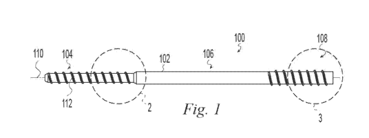
[0023] The term "transverse" is used herein to mean not parallel. FIGS. 1-4
depict a bone
screw 100 having an elongate body 102 with a distal portion 104, a mid-portion
106 and a
proximal portion 108 spaced longitudinally relative to a longitudinal axis
110. The distal
portion 104 includes a helical thread 112 having a major diameter 114, a minor
diameter 116,
and a pitch 128. The mid-portion 106 has a non-threaded outer surface 118 with
an outer
diameter 120. In the illustrative example of FIGS. 1-4, the mid-portion outer
diameter 120 is
equal to or greater than the thread major diameter 114. The distal threaded
portion 104 is
operable to bend as it is threaded into a bone to follow a curved path. For
example, the
bending stiffness of the distal threaded portion 104 is such that it will bend
to follow a curved
path in human bone. Such a curved path may be defined, for example, by a
curved hole in
the bone, a guide wire, or a natural bone feature such as a non-linear
intramedullary canal
bounded by cortical bone. This is distinct from prior art screws which if
started on a curved
path in human bone would, when advanced, continue in a straight line and thus
deviate from
the curved path and form their own, straight, path through the bone.
Preferably the bending
stiffness of the threaded distal portion 104 is lower than the bending
stiffness of the mid-
4
CA 02991756 2018-01-08
WO 2017/011244
PCT/US2016/041162
portion 106. The relatively lower bending stiffness of the threaded distal
portion 104 causes
the threaded distal portion to bend to follow a curved path while the
relatively higher bending
stiffness of the mid-portion causes the mid-portion to remain straight to
stabilize first and
second bone portions relative to one another at a bone interface such as at a
fracture,
osteotomy, or fusion site. The difference in bending stiffness between the
threaded distal
portion 104 and the mid-portion 106 may be achieved in different ways. For
example, the
threaded distal portion 104 and the mid-portion 106 may be made of different
materials
and/or may have different sectional moduli. In the illustrative example of
FIGS. 1-4, the
threaded distal portion 104 and the mid-portion 106 have different sectional
moduli. The
threaded distal portion minor diameter 116 is less than the outer diameter 120
of the mid-
portion 106 and the threaded distal portion major diameter is less than or
equal to the outer
diameter 120 of the mid-portion 106. Preferably, the ratio of the bending
stiffness of the mid-
portion 106 to the bending stiffness of the threaded distal portion 104 is in
the range of 1.5:1
to 100:1. More preferably, the ratio is in the range of 2:1 to 20:1. For
example, screws
suitable for internal fixation of a clavicle fracture and that fall within
these ranges may have a
major diameter 114 in the range of 4-6.5mm, a minor diameter 116 in the range
of 2.5-3.5
and a cannulation 101 with a diameter in the range of 1-2mm. Preferably, the
screw 100 is
made of a polymer.
[0024] Table 1 compares the calculated load required to bend a cantilevered
tube of 3mm
outside diameter and 1.5mm inside diameter around a radius of 50mm and an arc
length of
26mm. The titanium and stainless steel alloys are predicted to have a required
load
approximately 10 times that of the PEEK and PLLA. These loads would be greater
than the
bone could withstand and a threaded device made of those materials would not
follow a
curved path in the bone but would instead cause the bone to fail. In the case
of the highly
CA 02991756 2018-01-08
WO 2017/011244
PCT/US2016/041162
cold worked stainless steel, even if the bone could withstand the load, the
screw would fail
since the minimum bend radius before failure of the screw is greater than
50mm.
Table 1: Load at 50mm bend radius
Material Yield Failure Yield Failure Flexural
Load
Stress Stress Strain Strain Modulus (N)
(MPa) (MPa) (%) (%) (MPa)
PEEK
100 115 2.5% 20% 4
9.8
ASTM F2026
PLLA 90 100 2.6% 25% 3.5 8.7
Ti-6A1-4V ELI 91.7
880 990 0.8% 14% 114
ASTM F136
316LVM Not
Stainless Steel 1468 1696 0.7% 3% 197 possible
ASTM F899
[0025] Another way to quantify the bending stiffness of the threaded distal
portion 104 is by
the amount of torque required to turn the threaded distal portion 104 into a
curved bone hole
having a specified radius of curvature. For example, the threaded distal
portion 104
preferably requires a torque less than 20 in-lbs to turn the distal threaded
portion 104 into a
bone to follow a curved path having a radius of curvature of 50mm. More
preferably the
required torque is less than 10 in-lbs. More preferably the required torque is
less than
in-lbs. More preferably the required torque is approximately 2 in-lbs.
[0026] Table 2 compares the measured torque required to advance a threaded
tube 25mm into
a 50mm threaded radius formed in a rigid test block. The tubes were all
machined to the
same geometry but of different materials. The thread major diameter was
4.25mm, the minor
diameter was 3.0mm and the inner diameter of the tube was 1.5mm. A rigid block
was
prepared having a curved, threaded path. Such a path has a pitch that is wider
on the outside
of the curve and a pitch that is narrower on the inside of the curve
corresponding to the shape
of the screw thread when it is curved. Multiple samples of each screw were
inserted into the
block over an arc length of 25mm. The maximum torque for each revolution was
measured
6
CA 02991756 2018-01-08
WO 2017/011244
PCT/US2016/041162
and it was found that the torque increased for each revolution. In Table 2,
the range is the
range of torque values from the first to the last revolution. The average is
the average of the
torque values for all revolutions. The peak is the highest torque value and in
all cases
occurred in the last revolution. However, the torque values for each material
were relatively
constant over the last few revolutions. The titanium and stainless steel
alloys had measured
torque values approximately 10 times that of the PEEK. These tests were
conducted using a
threaded block made of tool steel with a strength greater than that of the
materials being
tested in order to compare the torque values. As pointed out relative to Table
1, the loads
generated from the metal implants would be greater than the bone could
withstand and a
threaded device as described herein made of these metals would not follow a
curved path in
the bone but would instead cause the bone to fail.
Table 2: Torque to thread around rigid 50mm radius
Material Range Average Peak
(in-lbs) (in-lbs) (in-lbs)
PEEK
0-2.0 1.4 2.0
ASTM F2026
Ti-6A1-4V ELI
0.7-25 16 25
ASTM F136
316LVM
Stainless Steel 0.5-20 13 20
ASTM F899
[0027] In addition to bending stiffness advantages, having the threaded distal
portion major
diameter less than or equal to the outer diameter 120 of the mid-portion 106
allows the distal
threaded portion 104 to pass through a passage in a bone that will be a
sliding or press fit
with the mid-portion 106. A screw so configured, as shown in the illustrative
example of
FIGS. 1-4, can have an intramedullary canal filling mid-portion 106 providing
solid support
to a bone interface and a relatively bendable distal threaded portion 104
following a curved
path such as for threading into a distal portion of a curved bone to secure
the screw in the
bone.
7
CA 02991756 2018-01-08
WO 2017/011244
PCT/US2016/041162
[0028] The proximal portion 108 may be identical to the mid-portion 106.
Alternatively, the
proximal portion may have a positive driver engagement feature (not shown)
such as internal
or external non-circular surfaces, profiles, or holes. For example, an
internal or external
slotted, threaded, triangular, square, hexagonal, hexalobular, or other drive
feature may be
provided. In addition, as shown in the illustrative example of FIGS. 1-4, the
proximal portion
108 may include an optional external helical thread 122 able to engage a bone
portion to
provide proximal fixation of the screw. For example, the proximal thread 122
may have a
major diameter 124, a minor diameter 126, and a pitch 130 wherein the proximal
thread
minor diameter 126 is equal to the mid-portion outer diameter 120. In the
illustrative
example of FIGS. 1-4, the mid-portion outer diameter 120 is equal to the
proximal thread
minor diameter 126 and the distal thread major diameter 114. The proximal
portion may
alternatively, or in addition, receive a locking member such as a pin or screw
transverse to the
longitudinal axis to lock a proximal bone portion to the nail. The locking
member may be
drilled through the proximal portion. Preferably, the proximal portion has one
or more
transverse holes formed through it for receiving the locking member.
[0029] The distal and proximal thread pitches 128, 130 may advantageously be
the same or
different depending on the application. For example, to stabilize a fracture,
the screw 100
may be inserted into a bone across the fracture so that the distal thread 112
is engaged with
bone distal to the fracture and the proximal thread 122 is engaged with bone
proximal to the
fracture. If the bone portions on either side of the fracture are reduced to a
desired final
position prior to inserting the screw 100, then it is advantageous for the
thread pitches 128,
130 to be equal so that insertion of the screw does not change the relative
positions of the
bone portions. If on the other hand, it is desirable to move the bone portions
relative to one
another by the action of inserting the screw then it is advantageous for the
pitches 128, 130 to
be different. For example, to move the bone portions closer together to reduce
the fracture,
8
CA 02991756 2018-01-08
WO 2017/011244
PCT/US2016/041162
the distal thread pitch 128 may be made greater than the proximal thread pitch
130 so that
with the distal thread 112 engaged distally and the proximal thread 122
engaged proximally,
further advancing the screw causes the distal bone portion to move proximally
relative to the
screw faster than the proximal bone portion moves proximally and thus move the
bone
portions closer together. Alternatively, to move the bone portions further
apart to distract the
fracture, the distal thread pitch 128 may be made smaller than the proximal
thread pitch 130
so that with the distal thread 112 engaged distally and the proximal thread
122 engaged
proximally, further advancing the screw causes the distal bone portion to move
proximally
relative to the screw more slowly than the proximal bone portion moves
proximally and thus
move the bone portions further apart. Preferably, the bone screw 100 has a
through bore, or
cannulation 101, coaxial with the longitudinal axis 110 to permit the bone
screw 100 to be
inserted over a guide wire.
[0030] The bone screw 100 of FIGS. 1-4, may advantageously be provided in a
set
containing a plurality of bone screws as shown in the illustrative example of
FIGS. 5-7. For
example, it is advantageous in a surgical procedure to minimize the number of
steps and the
amount of time needed to complete the procedure. In a bone fixation procedure,
a surgeon
often makes an initial sizing decision based on medical imaging. During the
procedure, it
may become expedient to change the predetermined size based on observation of
the surgical
site or the fit of trial implants or instruments. For example, a surgeon may
determine initially
that a smaller bone screw is appropriate. However, during preparation of the
site, the surgeon
may determine that a larger screw will better grip the bone or fill, for
example, a canal in the
bone. The illustrative set of bone screws shown in FIGS. 5-7 facilitates
changing between
sizes. Each screw 140, 150, 160 in the set has a minor diameter 142, 152, 162,
a major
diameter 144, 154, 164, and a pitch 146, 156, 166. The minor diameters 142,
152, 162 are
equal to one another so that a single diameter drill will provide an initial
bore hole
9
CA 02991756 2018-01-08
WO 2017/011244
PCT/US2016/041162
appropriate for all the screws in the set. The pitches 146, 156, 166 are equal
to one another
so that all of the screws in the set will threadably engage a helical thread
of the same pitch.
The major diameters 144, 154, 164 may increase to provide progressively more
bone
purchase or, for example, to span increasing larger intramedullary canals. For
example, with
the set of screws of the illustrative example of FIGS. 5-7, a surgeon may
drill a hole equal to
the minor diameters 142, 152, 162 and then tap the hole with a tap
corresponding to the
thread of the smallest major diameter screw 140. The tactile feedback received
by the
surgeon as the tap is inserted will indicate to the surgeon if the thread
major diameter is
sufficient to provide a desired level of bone engagement. For example, the
surgeon can feel
if the tap is engaging the cortical walls of an intramedullary canal or if the
tap is in softer
cancellous bone. If the surgeon determines that greater engagement is desired,
the surgeon
can next tap the hole with a tap corresponding to the thread of the next
larger major diameter
screw 150. Since the minor diameters 142, 152, 162 and thread pitches 146,
156, 166 are the
same for all of the screws in the set, the next tap will thread into the
previously tapped hole
and increase the bone thread major diameter without damaging the bone thread.
Once the
desired bone engagement is achieved, the surgeon may then insert the desired
screw 140,
150, 160. If in tapping the larger major diameter thread, the surgeon
determines that the bone
is providing too much resistance, the surgeon may revert to the smaller sized
screw since the
threads are still compatible. Alternatively to using a separate tap, the screw
threads may be
configured as self-tapping so that the screws may be threaded directly into
the bored hole.
[0031] In addition to the sizing advantages of having the same minor diameter
142, 152, 162
across a family of screws, it is also advantageous because the distal threaded
portion of each
screw will have a similar bending stiffness to each of the other screws 140,
150, 160 since the
continuous wall of the minor diameter contributes much more to the bending
stiffness than
CA 02991756 2018-01-08
WO 2017/011244
PCT/US2016/041162
the helical thread itself This similar bending stiffness means that they can
be inserted around
a similar bending radius with a similar torque.
[0032] In the illustrative example of FIGS. 5-7, each screw 140, 150, 160 has
a mid-portion
diameter 148, 158, 168 equal to the corresponding major diameter 144, 154,
164. The
increasing mid-portion diameters provide progressively less flexible mid-
portions across the
set of screws and, for example, canal filling for increasingly larger bones if
used in the
intramedullary canal. If the screws incorporate the optional increasing mid-
portion diameter
as shown, then it is desirable to re-drill the mid-portion of the bone hole to
accommodate the
mid-portion when an increase in screw size is desired. However, the distal,
threaded portion
of the bone hole does not need to be re-drilled so the screw threads will not
be damaged by
drilling.
[0033] Alternatively to, or in addition to, the threaded distal portion 104
and mid-portion 106
having different sectional moduli, the threaded distal portion 104 and mid-
portion 106 may
have different material properties such as two different materials or
different conditions of
the same material to produce a difference in bending stiffness between them.
[0034] In the illustrative example of FIGS. 36-41, a screw 170 has separate
first and second
members 172, 174 permanently joined together. The first member 172 includes an
elongate
body 176 with a proximal end 178, a distal end 180, a longitudinal axis 182,
and an axial
through bore 184. The proximal end 178 of the first member includes a pair of
transverse
through bores 181, 183. Each transverse bore 181, 183 defines a longitudinal
axis and the
axes form an angle 185 between them about the longitudinal axis 182 as best
seen in FIG. 39.
Providing more than one transverse through bore increases options for
attaching the screw to
bone fragments and options for fixation direction. Both bores may be used for
fixation or the
one that is most conveniently located. Preferably the angle 185 is in the
range of 0 to 90
degrees. More preferably the angle 185 is in the range of 20 to 90 degrees. In
the illustrative
11
CA 02991756 2018-01-08
WO 2017/011244
PCT/US2016/041162
example of FIGS. 36-41, the angle 185 is 45 degrees. The proximal end 178 also
includes
opposed flats 187 for engaging a driver in torque transmitting relationship.
An internal
thread 189 within the bore 184 is engageable with, e.g., a threaded draw bar
to secure the first
member to a driver.
[0035] The second member 174 includes an elongate body 186 with a proximal end
188, a
distal end 190, a longitudinal axis 192, an external helical thread 194, and
an axial through
bore 196. The distal end 180 of the first member 172 and the proximal end 188
of the second
member 174 may have complementary geometries to aid in joining them. In the
illustrative
example of FIGS. 36-41, the distal end 180 of the first member has a stepped
conical taper
and the proximal end 188 of the second member has a corresponding stepped
conical socket
198. The mating surfaces may be any suitable shape as determined by the
materials and
joining technique including but not limited to plug and socket joints (as
shown), scarf joints,
butt joints, dovetail joints, finger joints, and lap joints. The joint may be
reinforced with a
third component such as an adhesive, pin, or key. The joint may be formed by
mechanical
interlock, chemical bonding, molding, welding or other suitable joining
process. The final
assembled screw 170, has a distal portion 191, a mid-portion 193, and a
proximal portion 195
and may have the thread forms, diameters, and relationships as described
relative to the
examples of FIGS. 1-7.
[0036] The first and second components 172, 174 may be made of different
materials or
different conditions of the same material. For example, they may be made of
polymers,
metals, or ceramics. Metals may include stainless steel alloys, titanium,
titanium alloys,
cobalt-chromium steel alloys, nickel-titanium alloys, and/or others. Polymers
may include
nonresorbable polymers including polyolefins, polyesters, polyimides,
polyamides,
polyacrylates, poly(ketones), fluropolymers, siloxane based polymers, and/or
others.
Polymers may include resorbable polymers including polyesters (e.g. lactide
and glycolide),
12
CA 02991756 2018-01-08
WO 2017/011244
PCT/US2016/041162
polyanhydrides, poly(aminoacid) polymers (e.g. tyrosine based polymers),
and/or others.
Other possible materials include nonresorbable and resorbable ceramics (e.g.
hydroxyapatite
and calcium sulfate) or biocompatible glasses. They may be made of homogenous
materials
or reinforced materials. They may be made of crystallographically different
materials such as
annealed versus cold worked. It is preferable for the mid portion 193 to have
a higher
bending stiffness than the distal portion 191 and the distal portion should
have a bending
stiffness low enough for it to be inserted along a curved path in bone.
[0037] In a first example, the first component may be made of a metal with a
relatively high
degree of cold work and the second component of a metal with a relatively low
amount of
cold work such as for example annealed and cold worked stainless steel. The
components
may be joined for example by welding. However, as discussed relative to Table
1, most
metals are far too stiff to allow threading along a curved path in a bone
within suitable
torsional loads.
[0038] Preferably the distal portion is made of a polymer. In a second
example, the first
component is made of a metal, such as stainless steel or a titanium alloy, and
the second
component is made of a polymer such as polyetheretherketone (PEEK) or a
polylactide
polymer (e.g. PLLA). The components may be joined such as for example by
threading them
together.
[0039] Preferably both components are made of polymers. In a third example,
the first and
second components are both made of non-resorbable polymers. For example, the
first
component may be made of fiber reinforced PEEK (e.g. Invibio PEEK-OptimaTM
Ultra-
Reinforced) and the second component may be made of neat (unreinforced) PEEK
(e.g.
Invibio PEEK-OptimaTM Natural). The fiber reinforced PEEK is strong while the
neat PEEK
is relatively flexible allowing it to be easily threaded around a curved path
even while having
a relatively large bone filling diameter. The components may be joined, e.g.
by molding the
13
CA 02991756 2018-01-08
WO 2017/011244
PCT/US2016/041162
components as a continuous matrix with first component fiber reinforcement and
second
component neat polymer with polymer chains extending across the joint
interface. In the
example of FIGS. 36-41, the second component is relatively more transparent to
laser
radiation than the first component and the parts are joined by laser welding
at the conical
interface. The laser energy passes relatively easily through the second
component and is
absorbed by the first component so that localized heating at the conical
interface takes place
causing the polymer constituent of the two components to fuse together.
[0040] In a fourth example, the mid-portion and distal portion are made of
resorbable
polymers. For example, the mid-portion may be made of a glass fiber reinforced
PLLA (e.g.
Corbion-Purac FiberLiveTM) and the distal portion may be made of neat PLLA.
[0041] Alternatively, the first member 172 and second member 174 may form one
continuous part with different properties between first and second portions.
The difference in
properties may be achieved, for example, by different processing (e.g. thermal
processing) or
blending materials. For example, different polymers may be combined in a
single injection
mold cavity and formed together. The polymers may be blended so that there is
a transition
between them. In another example, stiffening and/or strengthening material,
e.g. fibers,
whiskers, and/or granules, may be selectively incorporated in, e.g., the first
portion.
[0042] FIGS. 42 and 43 illustrate an example of a screw 270 similar to that of
FIGS. 36-41
except that the first member 272 is not cannulated, the first member 272
extends the full
length of the second member 274, and the transverse holes 281, 283 are
coplanar. The screw
270 may be assembled as with the prior example including by using
complimentary screw
threads in the proximal region of the second member 274 and mid portion of the
first member
272 as indicated by reference number 250. The screw 270 of the example of
FIGS. 42 and 43
may be include any of the materials and features described relative to the
prior examples. If,
for example, the first member 272 is made of a radiographically more opaque
material than
14
CA 02991756 2018-01-08
WO 2017/011244
PCT/US2016/041162
the second member 274, then the first member will provide a radiographic
marker over the
entire length of the screw 270 that may be radiographically visualized during
and after
surgery to confirm screw placement. For example, a metal first component and
polymer
second component would provide for radiographic visualization of the metal
first component.
It has been found by the present inventors that the bending stiffness of the
distal end of the
screw is not materially changed by eliminating the axial through bore of the
first component
and is essentially unchanged when the bending stiffness of a guide wire is
accounted for
which was optionally used with the previous cannulated screw examples. The
guide wire is
not necessary inasmuch as the screw 270 will follow a curved hole prepared to
receive it.
The transverse holes 181, 183 may be provided in any number or not at all as
desired but it
has been found that one is sufficient and two provides the user with
additional fixation
choice.
[0043] FIGS. 8-10 illustrate a bone screw according to the one example of the
invention,
such as bone screw 100, being inserted into first and second bone portions
200, 202 having a
bone interface 204 between them. A first or proximal bore 206 is formed in the
first bone
portion 200, across the bone interface 204, and into the second bone portion
202. A second
or distal bore 208 extends distally from the proximal bore 206 defining a
curved path 210.
The screw 100 is advanced through the proximal bore 206 until the distal screw
threads
engage the distal bore 208 as shown in FIG. 9. Further advancing the screw 100
causes it to
bend to follow the curved path 210 as shown in FIG. 10. Having a straight
portion of the
path, and thus the straight mid portion of the screw 100, spanning the bone
interface results in
a zero stress and strain state at the bone interface which prevents separation
of the bone
portions 200, 202 at the interface 204.
[0044] FIGS. 11-35 depict an illustrative method of using the screw of FIGS. 1-
4 to fix a
fractured clavicle. A patient is placed in a beach chair position with the
head rotated away
CA 02991756 2018-01-08
WO 2017/011244
PCT/US2016/041162
from the operative side. A bolster is placed between the shoulder blades and
head allowing
the injured shoulder girdle to retract posteriorly. A C-arm is positioned to
enable anterior-
posterior (AP) and cephalic views of the operative site. A 2-3cm incision 300
is made at the
fracture site along Langer's Lines running perpendicular to the long axis of
the clavicle to
expose the fracture site (FIG. 10). The platysma muscle is freed from the skin
and split
between its fibers. The middle branch of the supraclavicular nerve is
identified and retracted.
[0045] The medial end 302 of the lateral fragment 304 of the fractured
clavicle is elevated
from the fracture site incision (FIG. 12).
[0046] A K-wire 306, e.g. a 1.4mm K-wire, is drilled into the canal of the
lateral fragment
304 and advanced through the dorsolateral cortex 308 and out through the skin
(FIG. 13).
[0047] A wire driver is attached to the lateral portion of the K-wire and used
to back the wire
out until it is lateral to the fracture 310 (FIG. 14). Bone clamps are used at
the incision site to
reduce the fracture and clamp the bone fragments in position. Proper reduction
is confirmed
with AP and cephalic radiographic views.
[0048] The K-wire 306 is advanced until it is preferably at least 20 mm medial
to the fracture
(FIG. 15).
[0049] A first dilator 312, e.g. a 3.2mm dilator, is placed over the K-wire
and advanced until
it contacts the bone (FIGS. 16-17).
[0050] A second dilator 314, e.g. a 4.5mm dilator, is placed over the first
dilator 312 and
advanced until it contacts the bone (FIG. 18).
[0051] A drill guide 316 is placed over the second dilator 314 and advanced
until it contacts
the bone (FIG. 19).
[0052] The first dilator 312 is removed and a first lateral drill 318,
corresponding to the
minor diameter of the distal screw threads, e.g. a 3.2mm drill, is advanced
over the K-wire
16
CA 02991756 2018-01-08
WO 2017/011244
PCT/US2016/041162
into the bone, preferably at least 20mm medial to the fracture. A drill depth
mark readable
adjacent the drill guide may be noted as a reference for implant sizing (FIG.
20).
[0053] The K-wire is removed and replaced with a flexible guide wire 320, e.g.
a nitinol
guide wire, sized to fit within the screw cannulation, e.g. a 1.4mm guide
wire. The flexible
guide wire 320 is advanced through the first lateral drill and further along
the intramedullary
canal of the medial bone fragment and will curve to follow the intramedullary
canal to define
a curved path in the bone. Preferably, the guide wire is advanced
approximately 30mm
medial to the tip of the first lateral drill 318 (FIG. 21).
[0054] The first lateral drill 318 is removed and a flexible shaft reamer 322,
corresponding to
the minor diameter of the distal screw threads, is guided over the flexible
guide wire 320 to
ream the medial portion of the curved path (FIG. 22) The flexible reamer 322
and second
dilator 314 are then removed.
[0055] A second lateral drill 324, having a diameter corresponding to the
diameter of the
mid-portion of the screw, e.g. a 4.5mm drill, is guided over the flexible
guide wire to enlarge
the bone hole laterally to receive the mid-portion and proximal portion of the
screw 100. The
second lateral drill 324 is advanced the same distance as the first lateral
drill (FIG. 23). The
drilling step may be monitored in A/P and cephalic views with the C-arm to
avoid perforating
the bone cortex as the second lateral drill 324 is advanced into the medial
bone fragment 326.
[0056] A flexible tap 328, having cutting threads corresponding to the distal
threads of the
screw 100 is guided over the flexible guide wire to cut threads into the
medial bone fragment
along the curved path (FIG. 24). The tap may serve as a trial implant and
provides tactile
feedback regarding the fit of the implant in the bone. If it is determined
that a larger screw is
desirable, subsequent larger second drills may be used to re-drill the lateral
straight portion
and subsequent larger flexible taps may be used to increase the distal thread
major diameter
without having to re-ream the medial curved portion of the bone hole. Once a
desired level
17
CA 02991756 2018-01-08
WO 2017/011244
PCT/US2016/041162
of thread purchase and canal filling are achieved, a depth mark readable
adjacent the drill
guide may be noted as a reference for the required implant length. If a screw
100 with a
proximal threaded portion is used, a lateral tap may be used to tap the
lateral bone fragment
to receive the proximal threads.
[0057] The screw 100 is attached to an inserter 330 and guided over the
flexible guide wire
until it is fully seated in the prepared threads in the medial bone fragment
(FIGS. 25 and 26).
Optionally, the screw 100 may be axially driven with a mallet through the
lateral bone
fragment until just short of the distal thread engagement. The screw 100 may
then be
threaded into full engagement with the prepared threads in the medial
fragment.
Radiographic visualization may be used to ensure that the fracture is fully
reduced and
anatomically aligned in length and rotation.
[0058] If a proximally threaded screw has not been used, or if additional
fixation is otherwise
desired, cross fixation may be used. For example, a cross fixation guide 340
may be engaged
with the implant inserter 330 (FIG. 27). The cross fixation guide may include
a knob 342
that threadingly engages the implant inserter 330 and a cross fixation guide
sleeve 344 that
abuts the lateral bone fragment adjacent the bone hole entrance. Rotating the
knob 342
moves the cross fixation guide sleeve 344 and implant inserter 330 axially
relative to one
another. With the cross fixation guide sleeve 344 abutting the lateral bone
fragment 304, the
implant inserter, implant, and medial bone fragment 326 will be drawn
laterally and the
lateral bone fragment 304 will be pressed medially to apply compression across
the fracture.
[0059] Inner and outer drill sleeves 346, 348 are advanced through the guide
340 until they
abut the bone (FIG. 28). In the case of a screw such as screw 170 having one
or more
preformed transverse bores, the cross fixation guide may have one or more
targeting holes
positioned to align with the one or more transverse bores. In the case of a
screw such as
18
CA 02991756 2018-01-08
WO 2017/011244
PCT/US2016/041162
screw 100 not having preformed transverse bores, cross fixation may be
inserted directly
through the screw 100 forming a transverse bore intraoperatively.
[0060] For example, a cross fixation wire 350 may be guided through the drill
sleeves,
through the near cortex, through the mid or proximal portions of the screw,
and into the far
cortex of the lateral bone fragment (FIG. 29). If wire cross fixation is
adequate, the cross
fixation guide may be removed and the wire may be trimmed flush with the bone
surface.
[0061] However, if screw cross fixation is desired, a screw depth gauge 352
may be placed
over the cross fixation wire to measure the projecting portion of the guide
wire to determine
the required screw length for bi-cortical fixation (FIG. 30).
[0062] A countersink tool 354 may be used to create a countersink for a cross
fixation bone
screw 356 (FIG. 31).
[0063] The appropriate length cross fixation screw 356 may then be guided over
the cross
fixation wire 350 and seated into the bone (FIG. 32). These steps may be
repeated to place
additional screws if desired.
[0064] FIGS. 33 and 34 illustrate the location of the screw 100 and cross
fixation screws 356
relative to the lateral and medial bone fragments.
[0065] FIG. 35 illustrates the cross fixation screws 356 in the screw 100
without the bone to
obscure the view. Preferably the screw 100 is made of a relatively soft
material, e.g. a
polymer, that facilitates arbitrary placement of the cross fixation screws at
any desired
location.
19