Note: Descriptions are shown in the official language in which they were submitted.
CA 02991814 2018-01-09
1
WO 2017/013129
PCT/EP2016/067207
Her2 binding proteins based on di-ubiquitin muteins
Field of the invention
The present invention relates to new Her2 binding molecules based on di-
ubiquitin muteins. The invention further
refers to Her2 binding proteins optionally fused or conjugated to a moiety
modulating pharmacokinetics or to a
therapeutically or diagnostically active component. The invention further
relates to the use of these Her2 binding
proteins in medicine, preferably for use in the diagnosis or treatment of
cancer.
Background of the invention
Increased expression of the membrane-bound receptor tyrosine kinase Her2 plays
an important role in the
development and progression of many breast carcinomas, but also in ovarian,
stomach, and uterine cancer,
particularly with aggressive forms of cancer. Overexpression of this oncogene
is reported for malignancies,
predominantly in malignancies of epithelial origin, and is associated with
cancer recurrence and poor prognosis. The
three domain protein (extracellular, transmembrane, intracellular tyrosine
kinase domain) is mediating cell
proliferation and inhibiting apoptosis. Upon binding of a ligand to the
extracellular domain of Her2, Her2 forms dimers
with the receptor whereby the intracellular domain of Her2 is activated which
mediates cellular processes such as
proliferation, differentiation, migration, or apoptosis. Thus, modulating the
function of Her2 is an important approach
for the development of cancer therapeutics, in particular those based on
monoclonal antibodies binding to the
extracellular domain of Her2. Therapeutic anti-Her2 monoclonal antibodies such
as Trastuzumab or Pertuzumab are
available for treatments of cancer, in particular breast cancer.
Technical problems underlying the present invention
However, monoclonal antibodies have major disadvantages such as a complex
molecular structure, a large size, and
challenging production methods. Furthermore, treatment of diseases with
currently available Her2 binding molecules
is not effective in all patients and may have severe side effects.
Needless to say that there is a strong medical need to effectively treat
cancer with improved novel agents, in
particular efficient tumor targeted therapeutics and diagnostics. There is an
ongoing need to find alternative to current
therapies and diagnosis, i.e. to substitute Her2 monoclonal antibodies by
smaller and less complex Her2 specific
molecules such as non-immunoglobulin based Her2 binding agents.
To overcome the disadvantages of antibodies, novel Her2 binding molecules
suitable for diagnostic and therapeutic
applications should include characteristics such as affinity to Her2,
specificity to Her2, and high stability.
It is thus an objective of the present invention to provide novel Her2 binding
non-immunoglobulin molecules for new
and improved strategies in the treatment and diagnosis of cancer with Her2
overexpression. In particular, it is an
objective to provide novel binding proteins which have high affinity and
specificity to Her2, combined with a less
complex and smaller structure, for example for enabling a simplified molecular
engineering.
A solution is provided in this invention by small Her2 binding proteins such
as non-immunoglobulin based binding
agents, in particular by Her2 binding molecules based on ubiquitin muteins
(also known as Affilin molecules).
Compared to antibodies, a significant advantage of the Her2 binding proteins
of the invention is the reduced
complexity in terms of (i) reduced size (e.g. of maximal 152 amino acids),
(ii) simple molecular structure (one chain
compared to four chains of an antibody), and (iii) posttranslational
modifications possible but not required for full
functionality. The binding proteins of the invention provide molecular formats
with favorable physicochemical
properties (such as stability and solubility), high-level expression, and
allow easy production methods. The Her2
specific Affilin molecules of the invention are characterized by high affinity
for Her2, by specificity for a Her2, and by
high stability, and provide novel therapeutic and diagnostic possibilities.
CA 02991814 2018-01-09
2
WO 2017/013129
PCT/EP2016/067207
The above-described objectives and advantages are achieved by the subject-
matters of the enclosed independent
claims. The present invention meets the needs presented above by providing
examples for specific Her2 binding
proteins based on di-ubiquitin muteins with substitutions in at least 12 amino
acid positions of di-ubiquitin. Preferred
embodiments of the invention are included in the dependent claims as well as
in the following description, examples
and figures. The above overview does not necessarily describe all problems
solved by the present invention.
Summary of the invention
In a first aspect the present invention relates to a Her2 binding protein
wherein the Her2 binding protein comprises an
amino acid sequence wherein at least 12 amino acids selected from positions
R42,144, H68, V70, R72, L73, R74,
K82, L84, Q138, K139, E140, S141, and T142 of di-ubiquitin (SEQ ID NO: 4) are
substituted and wherein the Her2
binding protein has at least 85 % sequence identity to di-ubiquitin (SEQ ID
NO: 4) and wherein the Her2 binding
protein has a binding affinity (KD) of less than 700 nM for Her2, preferably
the binding affinity determined by ELISA or
by surface plasmon resonance assays.
Another aspect of the present invention relates to a Her2 binding protein
further comprising at least one additional
molecule, preferably selected from at least one member of the groups (i), (ii)
and (iii) consisting of (i) a moiety
modulating pharmacokinetic behavior selected for example from a polyethylene
glycol, a human serum albumin
(HSA), a human serum albumin binding protein, an albumin-binding peptide, or
an immunoglobulin or
immunoglobulin fragments, a polysaccharide, and, (ii) a therapeutically active
component, optionally selected for
example from a monoclonal antibody or a fragment thereof, a cytokine, a
chemokine, a cytotoxic compound, an
enzyme, or derivatives thereof, or a radionuclide, and (iii) a diagnostic
component, optionally selected for example
from a fluorescent compound, a photosensitizer, a tag, an enzyme, or a
radionuclide.
The present invention also provides, in further aspects, a nucleic acid or
nucleic acids encoding the Her2 binding
proteins comprising or consisting of a binding protein of the present
invention, as well as a vector or vectors
comprising said nucleic acid or nucleic acids, and a host cell or host cells
comprising said vector or vectors.
Another aspect relates to said Her2 binding protein for use in diagnostics or
medicine, preferably for use in the
diagnosis or treatment of cancer, or a nucleic acid molecule encoding said
Her2 binding protein, or a vector
comprising said Her2 binding protein, or a host cell comprising said Her2
binding protein, or a non-human host
comprising said Her2 binding protein.
Another aspect relates to a composition comprising the Her2 binding protein of
the invention, the nucleic acid
molecule of the invention, the vector of the invention, or the host cell of
the invention, preferably for use in the
diagnosis or treatment of cancer.
Another aspect of the present invention relates to a method for the production
of a Her2 binding protein of any of the
preceding aspects of the invention comprising culturing of host cells under
suitable conditions and optionally isolation
of the Her2 binding protein produced.
This summary of the invention does not necessarily describe all features of
the present invention. Other
embodiments will become apparent from a review of the ensuing detailed
description.
Brief description of the Figures
The Figures show:
FIG. 1 shows Her2 binding Affilin molecules.
FIG. 1 A lists positions of di-ubiquitin (SEQ ID NO: 4) that are substituted
in order to generate a Her2 binding protein.
In the first row, the corresponding amino acid position is listed. All Her2
binding proteins (for example, SEQ ID NOs:
5-38) are substituted at least in 12 positions selected from positions
R42,144, H68, V70, R72, L73, R74, K82, L84,
Q138, K139, E140, S141, and T142 of SEQ ID NO: 4. A "." In the table refers to
a wild type position (unchanged); for
example, as exemplified in SEQ ID Nos: 6, 31, 33, 34, 35, 36, and 37.
CA 02991814 2018-01-09
WO 2017/013129 3 PCT/EP2016/067207
FIG. 1 B shows the same amino acid exchanges as FIG. 1 A, however, the
exchanges are translated according to
the following code which groups amino acids with similar biophysical
properties. A waved line "¨" is the symbol for
polar amino acids (T, S, N, or Q) "H" is the symbol for hydrophobic amino
acids (e.g. A, M, L, V, I) "o" is the symbol
for aromatic amino acids (e.g. F, W, Y), "+"the symbol for basic amino acids
(e.g. K, R, H), "-"the symbol for acidic
amino acids (e.g. 0, E), and "G" corresponds to Glycine.
FIG. 1 C lists further substitutions; all Her2 binding proteins have 0, 1, 2,
3,4, 5, or 6 further modifications in addition
to the at least 12 substitutions selected from amino acid positions R42, 144,
H68, WO, R72, L73, R74, K82, 184,
Q138, 1<139, E140, S141, and T142 of SEQ ID NO: 4.
FIG. 1 D shows the same amino acid exchanges as FIG. 1 C, however, the
exchanges are translated according to
the code which groups amino acids with similar biophysical properties as
described in FIG. 1 B.
FIG. 2. Biochemical characterization of Her2 binding Affilin molecules (for
example, SEQ ID NOs: 5-38). Shown are
binding affinities (KO as obtained from SPR assay (Biacore; third column of
the table) and temperature stability (DSF;
fourth column of the table).
FIG. 3. Analysis of Her2 binding proteins via label-free interaction assays
using SPR (Biacore). Different
concentrations of Affilin proteins (0, 0.137, 0.4115, 1.2345, 3.7037, 11.11,
and 33.33 nM) were analyzed for binding
to Her2 immobilized on a chip (Biacore) to analyze the interaction between the
Affilin protein and Her2. FIG. 3 A
shows the binding kinetics of Affilin-142628 to Her2. FIG. 3 B shows the
binding kinetics of Affilin-144633 to Her2.
FIG. 4. Functional characterization of Her2 binding proteins confirming
binding to cellular Her2. The figure shows
binding to exogenously Her2 expressing SkBr3 cells as determined by FACS
analysis. Her2 binding proteins
("Affilin") show binding at 50 nM on SkBr3 cells (dark grey bars) and no
activity on HEK/293 cells (see FIG. 4 A and
FIG. 4 B). Weak or no binding to Her2 expressing SkBr3 was observed for
Affilin-142655 (referred to as "142655"),
Affilin-141965, Affilin-142465 (referred to as "142465"), Affilin-142502
(referred to as "142502"), and di-ubiquitin
(referred to as "di-ubi").
FIG. 5. Concentration dependent functional binding of Her2 binding proteins to
exogenously Her2 expressing SkBr3
cells as determined by flow cytometry analysis. Shown is a dilution series of
333 nM to 5.6 pM of binding protein.
Affilin-141926 (FIG. 5 A) and Affilin-141890 (FIG. 5 B) show a concentration
depending binding on SkBr3-cells.
FIG. 6. Functional characterization of Her2 binding proteins confirming
binding to exogenously Her2 overexpressing
CHO-K1 cells as determined by flow cytometry analysis. Her2 binding proteins
show binding on CHO-K1-Her2 cells
at concentrations of 50 nM, 5 nM, and 0.5 nM. FIG. 6 A shows the Her2-binding
of Affilin-142627, Affilin-142628,
Affilin-142654, and Affilin-141884; FIG. 6 B shows cellular Her 2 binding of
Affilin-144631, Affilin-144632, Affilin-
144633, Affilin-144634, Affilin-144635, Affilin-144636, Affilin-144637, and
FIG. 6 C shows cellular Her 2 binding of
Affilin-144567, and only low levels of binding of 142502 at 500 nM. Thus,
cellular Her2 binding was confirmed for all
binding molecules except 142502 even at the lowest concentration tested. Di-
ubiquitin showed no binding on CHO-
K1-Her2-cells (shown, for example, in FIG. 6 B).
FIG. 7. Concentration dependent binding of Affilin-142628. The figure shows
binding of Affilin-142628 to exogenously
Her2 expressing CHO-K1 cells as determined by flow cytometry (FACS
analysis)(control: empty vector CHO-K1-
pEntry cells). Histograms at different Affilin protein concentrations of 50
nM, 5 nM and 0.5 nM are shown in
comparison to di-ubiquitin 139090 (SEQ ID NO: 4). Affilin-142628 induces a
concentration depending shift on the
Her2-overexpressing cell line.
FIG. 8. Concentration dependent functional binding of Her2 binding proteins to
exogenously Her2 expressing SkBr3-
cells as determined by flow cytometry. A dilution series of 100 nM to 0.06 pM
of Affilin-142628 was used to analyze
the interaction with Her2 overexpressing SkBr3 cells. FIG. 8 shows a
concentration dependent binding of Affilin-
142628.
FIG. 9 shows the binding analysis of different Her2 binding proteins on Her2-
overexpressing SkBr3-cells by
irrimunofluorescence staining. FIG. 9A shows concentrations of 50 nM Affilin-
141884, Affilin-142628, Affilin-141926,
RECTIFIED SHEET (RULE 91) ISA/EP
CA 02991814 2018-01-09
4
WO 2017/013129
PCT/EP2016/067207
Affilin-144637, Affilin-142418. FIG. 9B shows concentrations of 50 nM Affilin-
144567, di-ubiquitin (139090), PBS, and
Trastuzumab (Herceptin). Affilin-141884, Affilin-142628, Affilin-141926,
Affilin-144637 and Affilin-142418 show a
strong binding on the Her2-overexpressing cell line, whereas Affilin-144567
and di-ubiquitin (139090) do not bind to
Her2 on SkBr-3 cells.
FIG. 10 confirms that Her2 binding proteins bind to SKOV-3 xenograft tumor
tissue. Shown is an immunohistological
staining of 50 nM Affilin-141884 and 50 nM Affilin-142628 on Her2-expressing
tumor tissue derived from human
ovarian adenocarcinoma cells. Affilin-141884 and Affilin-142628 show a strong
binding on Her2-expressing tissue.
Di-ubiquitin (139090) shows no binding on SKOV-3 tissue slides.
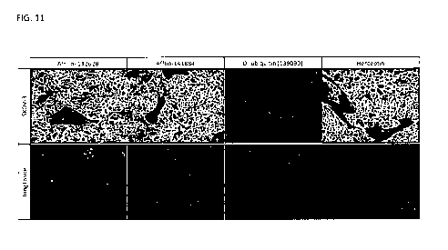
FIG. 11 shows an immunohistological binding analysis of Her2 binding proteins
on Her2-expressing SKOV-3-tumor
tissue slides and lung tissue slides without Her2 expression. Affilin-141884
and Affilin-142628 show strong binding at
nM on SKOV-3 tissue. No binding to lung tissue was observed. In addition, no
binding of Affilin-141884 and Affilin-
142628 to tissue obtained from liver, heart muscle, and ovary was observed.
FIG. 12 shows that Affilin-142628 and Affilin-143692 bind to different Her2
epitopes (competition analysis; binding
analysis SPR). These Her2 binding proteins do not compete for Her2 binding and
thus, use different or non-
15 overlapping epitopes of Her2.
FIG. 13 shows that the Her2 binding proteins Affilin-142628 and Affilin-143692
bind to different Her2 epitopes than
Trastuzumab (Herceptin).
FIG. 14 shows the simultaneous binding of a bispecific fusion protein to Her2
and EGFR. The fusion of a Her2
binding protein to an EGFR specific monoclonal antibody (Cetuximab) enables
bispecific targeting, as shown for
20 example for fusion proteins SEQ ID NOs: 44-47.
FIG. 15 shows the flow cytometric binding analysis of a bispecific fusion
protein comprising an Her2 specific Affilin
fused to the C-terminus of the light chain of Cetuximab (CL-141926; SEQ ID NO:
44) on Her2 overexpressing CHO
K1 cells (FIG. 15 B) and on EGFR overexpressing CHO K1 cells (FIG. 15 B). The
fusion protein shows binding to
both extracellular targets. The figure shows the median fluorescence intensity
(MFI), representing the binding of the
Affilin-antibody fusion protein to EGFR and to Her2 expressing cells at the
indicated concentrations.
Detailed Description of the Invention
Before the present invention is described in more detail below, it is to be
understood that this invention is not limited
to the particular methodology, protocols and reagents described herein as
these may vary. It is also to be understood
that the terminology used herein is for the purpose of describing particular
embodiments only, and is not intended to
limit the scope of the present invention which will be limited only by the
appended claims. Unless defined otherwise,
all technical and scientific terms used herein have the same meanings as
commonly understood by one of ordinary
skill in the art to which this invention belongs.
Preferably, the terms used herein are defined as described in "A multilingual
glossary of biotechnological terms:
(IUPAC Recommendations)", Leuenberger, H.G.W, Nagel, B. and Kolbl, H. eds.
(1995), Helvetica Chimica Acta, CH-
4010 Basel, Switzerland).
Throughout this specification and the claims which follow, unless the context
requires otherwise, the word "comprise",
and variants such as "comprises" and "comprising", will be understood to imply
the inclusion of a stated integer or
step or group of integers or steps but not the exclusion of any other integer
or step or group of integers or steps.
Several documents (for example: patents, patent applications, scientific
publications, manufacturers specifications,
instructions, GenBank Accession Number sequence submissions etc.) are cited
throughout the text of this
application. Nothing herein is to be construed as an admission that the
invention is not entitled to antedate such
disclosure by virtue of prior invention. Some of the documents cited herein
are characterized as being Incorporated
by reference". In the event of a conflict between the definitions or teachings
of such incorporated references and
definitions or teachings recited in the present specification, the text of the
present specification takes precedence.
CA 02991814 2018-01-09
WO 2017/013129
PCT/EP2016/067207
All sequences referred to herein are disclosed in the attached sequence
listing that, with its whole content and
disclosure, is a part of this specification.
General definitions of important terms used in the application
5 The terms "protein" and "polypeptide" refer to any chain of two or more
amino acids linked by peptide bonds, and
does not refer to a specific length of the product. Thus, "peptides",
"protein", "amino acid chain," or any other term
used to refer to a chain of two or more amino acids, are included within the
definition of "polypeptide," and the term
"polypeptide" may be used instead of, or interchangeably with any of these
terms. The term "polypeptide" is also
intended to refer to the products of post-translational modifications of the
polypeptide, including without limitation
glycosylation, acetylation, phosphorylation, amidation, proteolytic cleavage,
modification by non-naturally occurring
amino acids and similar modifications which are well known in the art. Thus,
binding proteins comprising two or more
protein moieties also fall under the definition of the term "protein" or
"polypeptides".
The term "ubiquitin" or õunmodified ubiquitin" refers to ubiquitin in
accordance with SEQ ID NO: 1 and to proteins with
at least 95 % identity, such as SEQ ID NO: 2 (point mutations in positions 45,
75, 76 which do not influence binding
to a target), to a di-ubiquitin according to SED ID NO: 4 and to proteins with
at least 95 % identity, such as , di-
ubiquitin according to SEQ ID NO: 48, and according to the following
definition. Particularly preferred are ubiquitin
molecules from mammals, e.g. humans, primates, pigs, and rodents. On the other
hand, the ubiquitin origin is not
relevant since according to the art all eukaryotic ubiquitins are highly
conserved and the mammalian ubiquitins
examined up to now are even identical with respect to their amino acid
sequence. In addition, ubiquitin from any other
eukaryotic source can be used. For instance ubiquitin of yeast differs only in
three amino acids from the wild-type
human ubiquitin (SEQ ID NO: 1).
The term "di-ubiquitin" refers to a protein comprising two unmodified
ubiquitin moieties linked to each other in head-
to-tail orientation. An example is given in SED ID NO: 4 (point mutations in
positions 45, 75, 76, 151, 152 of wildtype
ubiquitin; these point mutations do not influence binding to a target; clone
139090), and in SEQ ID NO: 48. The
amino acid sequence identity between SEQ ID NO: 4 and SEQ ID NO: 48 is 96.7
To. A di-ubiquitin according to the
present invention is an artificial protein of 152 amino acids consisting of
two ubiquitin moieties directly linked to each
other without a peptide linker between the two ubiquitin moieties. A di-
ubiquitin as understood herein is a protein with
at least 95 % identity to SEQ ID NO: 4.
The terms õmodified ubiquitin" and õubiquitin mutein" and "Affilin" are all
used synonymously and can be exchanged.
The term õmodified ubiquitin" or õubiquitin mutein" or "Affilin" as used
herein refers to derivatives of ubiquitin which
differ from said unmodified ubiquitin by amino acid exchanges, insertions,
deletions or any combination thereof,
provided that the ubiquitin mutein has a specific binding affinity to a target
epitope or antigen which is at least 10fold
lower or absent in unmodified ubiquitin. This functional property of an
ubiquitin mutein (Affilin; modified ubiquitin) is a
de novo created function.
The term "Affilin " (registered trademark of Scil Proteins GmbH) refers to non-
immunoglobulin derived binding
proteins based on ubiquitin muteins. An Affilin protein is not a natural
ubiquitin existing in or isolated from nature, for
example, as shown in SEQ ID NO: 1. The scope of the invention excludes
unmodified ubiquitin. An Affilin molecule
according to this invention comprises, essentially consists, or consists of
either two differently modified ubiquitin
moieties linked together in a head-to-tail fusion or an Affilin molecule that
comprises, essentially consists, or consists
of one modified ubiquitin moiety. A "head-to-tail fusion" is to be understood
as fusing two proteins together by
connecting them in the direction (head) N-C-N-C- (tail) (tandem molecule), as
described for example in
EP2379581B1 which is incorporated herein by reference. The head part is
designated as the first moiety and the tail
part as the second moiety. In this head-to-tail fusion, two moieties may be
connected directly without any linker (e.g.
SEQ ID NOs: 5-38). Alternatively, the fusion of two proteins can be performed
via linkers, for example, a polypeptide
linker, as described herein.
CA 02991814 2018-01-09
6
WO 2017/013129
PCT/EP2016/067207
The term "substitution" includes "conservative" and "non-conservative"
substitutions. "Conservative substitutions" may
be made, for instance, on the basis of similarity in polarity, charge, size,
solubility, hydrophobicity, hydrophilicity,
and/or the amphipathic nature of the amino acid residues involved. Amino acids
can be grouped into the following
standard amino acid groups: (1) hydrophobic side chains: Ala (A), Met (M), Leu
(L), Val (V), Ile (I); (symbol "H" in
Figure 1) (2) acidic polar side chain: Asp (D), Glu (E) (symbol "2 in Figure
1); (3) basic side chain polarity: Lys (K),
Arg (R), His (H) (symbol "+" in Figure 1); (4) aromatic amino acids: Trp (W),
Tyr (Y), Phe (F) (symbol "o" in Figure 1);
(5) polar amino acids: Thr (T), Ser (S), Asn (N), Gin (Q) (symbol "wave" in
Figure 1); (6) residues that influence chain
orientation: Gly (G), Pro (P); and (7) Cys (C). As used herein, "conservative
substitutions" are defined as exchanges
of an amino acid by another amino acid listed within the same group of the
standard amino acid groups shown
above. For example, the exchange of Asp by Glu retains one negative charge in
the so modified polypeptide. In
addition, Gly and Pro may be substituted for one another based on their
ability to disrupt a-helices. Some preferred
conservative substitutions within the above groups are exchanges within the
following sub-groups: (i) Ala, Val, Leu
and Ile; (ii) Ser and Thr; (ii) Asn and Gin; (iv) Lys and Arg; and (v) Tyr and
Phe. Given the known genetic code, and
recombinant and synthetic DNA techniques, the skilled scientist can readily
construct DNAs encoding the
conservative amino acid variants. As used herein, "non-conservative
substitutions" or "non-conservative amino acid
exchanges" are defined as exchanges of an amino acid by another amino acid
listed in a different group of the amino
acid groups (1) to (7) shown above.
The term "insertions" comprises the addition of amino acids to the original
amino acid sequence of ubiquitin wherein
the ubiquitin remains stable without significant structural change. Naturally,
loop regions connect regular secondary
structure elements. The structure of human unmodified ubiquitin (SEQ ID NO: 1)
reveals six loops at amino acid
regions 8-11, 17-22, 35-40, 45-47, and 50-63 which connect secondary structure
elements such as beta sheets and
alpha helix. In one embodiment of the invention, Her2 binding proteins are
disclosed comprising a ubiquitin mutein
having a combination of an insertion and substitutions. In one embodiment,
ubiquitin muteins have insertions of 2-10
amino acid residues, preferably within the most N-terminal loop within amino
acids 8-11. Specifically, the number of
amino acid residues to be inserted is 2, 3, 4, 5, 6, 7, 8, 9, 10, preferably 2
- 10 amino acid residues, most preferred 6-
9 amino acid residues.
The term "antibody" as used in accordance with the present invention comprises
monoclonal antibodies having two
heavy chains and two light chains (immunoglobulin or IgG antibodies).
Furthermore, also fragments or derivatives
thereof, which still retain the binding specificity, are comprised in the term
"antibody'. The term "antibody" also
includes embodiments such as chimeric (human constant domain, non-human
variable domain), single chain and
humanized (human antibody with the exception of non-human CDRs) antibodies.
Full-length IgG antibodies
consisting of two heavy chains and two light chains are most preferred in this
invention. Heavy and light chains are
connected via non-covalent interactions and disulfide bonds.
In the present specification, the terms "target antigen", "target", "antigen"
and "binding partner" are all used
synonymously and can be exchanged. Preferably the target is one of the targets
defined herein below. The term
"antigen", as used herein, is to be interpreted in a broad sense and includes
any target moiety that is bound by the
binding moieties of the binding proteins of the present invention.
The terms "protein capable of binding" or "binding protein" or "binding Her2"
or "binding affinity for" according to this
invention refer to a protein comprising a binding capability to a defined
target antigen. The term "Her2 binding
protein" refers to a protein with high affinity binding capability to Her2.
An "antigen binding site" refers to the site, i.e. one or more amino acid
residues, of an antigen binding molecule which
provide interaction with the antigen. A native immunoglobulin molecule
typically has two antigen binding sites, a Fab
molecule typically has a single antigen binding site.
The term "epitope" includes any molecular determinant capable of being bound
by an antigen binding protein as
defined herein and is a region of a target antigen that is bound by an antigen
binding protein that targets that antigen,
CA 02991814 2018-01-09
7
WO 2017/013129
PCT/EP2016/067207
and when the antigen is a protein, it may include specific amino acids that
directly contact the antigen binding protein.
In a conformational epitope, amino acid residues are separated in the primary
sequence, but are located near each
other on the surface of the molecule when the polypeptide folds into the
native three-dimensional structure. A linear
epitope is characterized by two or more amino acid residues which are located
adjacent in a single linear segment of
a protein chain. In other cases, the epitope may include determinants from
posttranslational modifications of the
target protein such as glycosylation, phosphorylation, sulfatation,
acetylation, fatty acids or others.
The term "fused" means that the components (e.g. an Affilin molecule and a
monoclonal antibody or a Fab fragment)
are linked by peptide bonds, either directly or via peptide linkers.
The term "fusion protein" relates to a protein comprising at least a first
protein joined genetically to at least a second
protein. A fusion protein is created through joining of two or more genes that
originally coded for separate proteins.
Thus, a fusion protein may comprise a multimer of different or identical
binding proteins which are expressed as a
single, linear polypeptide. It may comprise one, two, three or even more first
and/or second binding proteins. A fusion
protein as used herein comprises at least a first binding protein (e.g.
Affilin) which is fused with at least a second
binding protein, e.g. a monoclonal antibody or a fragment thereof. Such fusion
proteins may further comprise
additional domains that are not involved in binding of the target, such as but
not limited to, for example,
multimerization moieties, polypeptide tags, polypeptide linkers.
The term "conjugate" as used herein relates to a protein comprising or
essentially consisting of at least a first protein
attached chemically to other substances such as to a second protein or a non-
proteinaceous moiety. The conjugation
can be performed by means of organic synthesis or by use of enzymes including
natural processes of enzymatic
post-translational modifications. Examples for protein conjugates are
glycoproteins (conjugated protein with
carbohydrate component) or lipoproteins (conjugated protein with lipid
component). The molecule can be attached
e.g. at one or several sites through any form of a linker. Chemical coupling
can be performed by chemistry well
known to someone skilled in the art, including substitution (e.g. N-
succinimidyl chemistry), addition or cycloaddition
(e.g. maleimide chemistry or click chemistry) or oxidation chemistry (e.g.
disulfide formation). Some examples of non-
proteinaceous polymer molecules which are chemically attached to protein of
the invention are hydroxyethyl starch,
polyethylene glycol, polypropylene glycol, dendritic polymers, or
polyoxyalkylene and others.
A fusion protein or protein conjugate may further comprise one or more
reactive groups or peptidic or non-peptidic
moieties such as ligands or therapeutically or diagnostically relevant
molecules such as radionuclides or toxins. It
may also comprise small organic or non-amino acid based compounds, e.g. a
sugar, oligo- or polysaccharide, fatty
acid, etc. Methods for attaching a protein of interest to such non-
proteinaceous components are well known in the art,
and are thus not described in further detail here.
The terms "bispecific binding molecule" or "multispecific binding molecule"
mean that the antigen binding molecule is
able to specifically bind two or multiple different epitopes. Typically, a
bispecific antigen binding molecule comprises
two antigen binding sites, each of which is specific fora different epitope.
In certain embodiments the bispecific
antigen binding molecule is capable of simultaneously binding two epitopes,
particularly two epitopes expressed on
two distinct cells. The term "bispecific binding molecule" or "bispecific
binding protein" means that binding proteins of
the present invention are capable of specifically binding to two different
epitopes. Moreover, the bispecific binding
molecule of the present invention is capable of binding to two different
epitopes at the same time. This means that a
bispecific construct is capable of simultaneously binding to at least one
epitope "A" and at least one epitope "B",
wherein A and B are not the same. The two epitopes may be located on the same
or different target antigens which
means that the fusion molecules of the present invention can bind one target
at two different epitopes or two target
antigens each with its own epitope. Similarly, "multispecific binding
molecules" are capable of binding multiple
epitopes at the same time wherein the epitopes may be located on the same or
different antigens.
Alternatively, said binding proteins may bind to different, non-overlapping
epitopes on the same or different target
molecules and are thus classified as bispecific, trispecific, multispecific,
etc., for example af3, f3y, a6, af3y, af3y6 binding
CA 02991814 2018-01-09
8
WO 2017/013129
PCT/EP2016/067207
to epitopes AB, BC, AD, ABC or ABCD, respectively. For example, fusion
proteins with Her2-specific Affilin and anti-
EGFR-monoclonal antibody are bispecific.
The term "multimeric binding molecules" refers to fusion proteins that are
multivalent and / or multispecific,
comprising two or more moieties (i.e. bivalent or multivalent) of binding
protein a, f3 and/or y etc., e.g.
aa, f3f3f3, aaf3, aaf3f3, ayy, f3f3y, af3y66, etc.. For example, aaf3y is
trispecific and bivalent with respect to epitope A. For
example, the fusion proteins of Her2-specific Affilin and monoclonal
antibodies as described herein are at least
"bivalent" because they comprise at least two binding proteins (for example,
an Affilin and an monoclonal antibody).
Said binding proteins may bind specifically to the same or overlapping
epitopes on a target antigen (monospecific),
e.g. the composition of the binding protein may be described by (a)2, (03,
(a)4, (3)2, (f3)3, (f3)4 etc.. In this case, the
fusion molecules are monospecific but bivalent, trivalent, tetravalent, or
multivalent for the epitope A or epitope B,
respectively.
The term "amino acid sequence identity" refers to a quantitative comparison of
the identity (or differences) of the
amino acid sequences of two or more proteins. "Percent (%) amino acid sequence
identity" with respect to a
reference polypeptide sequence is defined as the percentage of amino acid
residues in a sequence that are identical
with the amino acid residues in the reference polypeptide sequence, after
aligning the sequences and introducing
gaps, if necessary, to achieve the maximum percent sequence identity.
To determine the sequence identity, the sequence of a query protein is aligned
to the sequence of a reference
protein, for example, to SEQ ID NO: 4 (di-ubiquitin) or to SEQ ID NO: 1
(ubiquitin). Methods for alignment are well
known in the art. For example, for determining the extent of an amino acid
sequence identity of an arbitrary
polypeptide relative to the amino acid sequence of SEQ ID NO: 4 or SEQ ID NO:
1, the SIM Local similarity program
is preferably employed (Xiaoquin Huang and Webb Miller (1991), Advances in
Applied Mathematics, vol. 12: 337-
357), that is freely available (see also: http://www.expasy.org/tools/sim-
prot.html). For multiple alignment analysis
ClustalW is preferably used (Thompson et al. (1994) Nucleic Acids Res.,
22(22): 4673-4680).
In the context of the present invention, the extent of sequence identity
between a modified sequence and the
sequence from which it is derived (also termed "parental sequence") is
generally calculated with respect to the total
length of the unmodified sequence, if not explicitly stated otherwise. Each
amino acid of the query sequence that
differs from the reference amino acid sequence at a given position is counted
as one difference. An insertion or
deletion in the query sequence is also counted as one difference. For example,
an insertion of a linker between two
ubiquitin moieties is counted as one difference compared to the reference
sequence. The sum of differences is then
related to the length of the reference sequence to yield a percentage of non-
identity. The quantitative percentage of
identity is calculated as 100 minus the percentage of non-identity. In
specific cases of determining the identity of
ubiquitin muteins aligned against unmodified ubiquitin, differences in
positions 45, 75 and/or 76 are not counted, in
particular, because they are not relevant for the novel binding capability of
the ubiquitin mutein. The ubiquitin moiety
can be modified in amino acid residues 45, 75 and/or 76 without affecting its
binding capability; said modifications
might, however, be relevant for achieving modifications in the biochemical
properties of the mutein. Generally, a
ubiquitin used as starting material for the modifications has an amino acid
identity of at least 95 %, of at least 96 % or
of at least 97 %, or of at least an amino acid sequence identity of 98 % to
SEQ ID NO: 1. Thus, a polypeptide which
is, for example, 95 % "identical" to a reference sequence may comprise, for
example, five point mutations or four
point mutations and one insertion etc., per 100 amino acids, compared to the
reference sequence.
The term "dissociation constant" or "K0" defines the specific binding
affinity. As used herein, the term "K0" (usually
measured in "mol/L", sometimes abbreviated as "M") is intended to refer to the
dissociation equilibrium constant of
the particular interaction between a first compound and a second compound. In
the context of the present invention,
the term KD is particularly used to describe the binding affinity between a
Her2-binding protein and Her2. A high
affinity corresponds to a low value of KD. Thus, the expression "a KD of at
least e.g. 10-7 M" means a value of 10-7M
or lower (binding more tightly). 1 x 10⁻⁷M corresponds to 100 nM. A value of
10-5 M and below down to 10-12 M can
CA 02991814 2018-01-09
9
WO 2017/013129
PCT/EP2016/067207
be considered as a quantifiable binding affinity. Depending on the application
a value of 10-7 to 10-12 M is preferred
for chromatographic applications or for diagnostic or therapeutic
applications. In accordance with the invention the
affinity for the target binding is in the range of 7 x 10⁻⁷M (700 nM) or less.
Final target binding affinity can be ideally
10-9M (1 nM) or less.
Binding proteins of the invention comprise two ubiquitin muteins linked
directly without any linker to result in
unique and high affinity Her2 binding proteins with substitutions at least in
12, 13, or 14 positions selected from 42,
44, 68, 70, 72, 73, 74, 82, 84, 138, 139, 140, 141, and 142 of di-ubiquitin
(SEQ ID NO: 4 or SEQ ID NO: 48), and
optionally in 0, 1, 2, 3, 4, 5, or 6 further substitutions.
Binding proteins of the invention can be fused, e.g. genetically, to other
functional protein moieties. In the context of
such fusion proteins of the invention the term "linker" refers to a single
amino acid or a polypeptide that joins at least
two other protein molecules covalently. The linker is e.g. genetically fused
to the first and second protein or protein
moieties to generate a single, linear polypeptide chain. The length and
composition of a linker may vary between at
least one and up to about 50 amino acids. Preferably, the linker length is
between one and 30 amino acids. More
preferably, the peptide linker has a length of between 1 and 20 amino acids;
e.g. 1, 2, 3, 4, 5, 6, 7, 8, 9, 10, 11, 12,
13, 14, 15, 16, 17, 18, 19, or 20 amino acids. It is preferred that the amino
acid sequence of the peptide linker is not
immunogenic to human beings, stable against proteases and optionally does not
form a secondary structure. An
example is a linker comprised of small amino acids such as glycine or serine.
The linkers can be glycine-rich (e.g.,
more than 50 % of the residues in the linker can be glycine residues).
Preferred are glycine-serine-linkers of variable
length consisting of glycine and serine residues only. In general, linkers of
the structure (SGGG)n or permutations of
SGGG, e.g. (GGGS)n, can be used wherein n can be any number between 1 and 6,
preferably 1 or 2 or 3. Also
preferred are linkers comprising further amino acids. Other linkers for the
genetic fusion of proteins are known in the
art and can be used. In one embodiment of the invention, the first binding
protein (e.g. Affilin) and the second binding
protein (e.g. monoclonal antibody or fragment thereof) are linked via a (G35)4
linker.
In case of chemical conjugates of the binding proteins of the invention, the
term "linker" refers to any chemical moiety
which connects the Her2 binding protein with other proteinaceous or non-
proteinaceous moieties either covalently or
non-covalently, e.g., through hydrogen bonds, ionic or van der Weals
interactions, such as two complementary
nucleic acid molecules attached to two different moieties that hybridize to
each other, or chemical polymers such as
polyethylene glycol or others. Such linkers may comprise reactive groups which
enable chemical attachment to the
protein through amino acid side chains, the N-terminal a-amino- or C-terminal
carboxy-group of the protein. Such
linkers and reactive groups are well-known to those skilled in the art and not
described further.
Her2 (Human Epidermal Growth Factor Receptor 2; synonym names are ErbB-2, Neu,
CD340 or p185) is a 185-kDa
receptor first described in 1984 (Schlechter et al (1984) Nature 312:513-516).
Amplification or over-expression of this
gene has been shown to play an important role in the pathogenesis and
progression of certain aggressive types of
breast cancer, and Her2 is known as an important biomarker and target of
therapy for the disease. Other tumors
where Her2 plays a role include ovarian cancer and gastric cancer. Human Her2
is represented by the NCB!
accession number NP_004439; the extracellular domain (residues 1-652) of Her2
is represented by the uniprot
Accession Number p04626. The term õHer2" comprises all polypeptides which show
a sequence identity of at least
70 %, 80 %, 85 %, 90 %, 95 %, 96 % or 97 % or more, or 100 % to NP_004439 and
have the functionality of Her2.
Detailed description of the embodiments of the invention
The Her2 binding protein of the invention comprises, essentially consists of
or consists of two differently modified
ubiquitin moieties directly connected without a linker in head-to-tail
orientation. The Her2 binding protein of the
invention has an amino acid identity of at least 85 % to di-ubiquitin (SEQ ID
NO: 4); i.e. a maximum of 23 amino acids
are modified in di-ubiquitin (SEQ ID NO: 4) (152 amino acids total) to
generate a novel binding property of di-ubiquitin
(SEQ ID NO: 4) to Her2. Further preferred amino acid identities of the novel
Her2 binding proteins are at least 86 %,
CA 02991814 2018-01-09
WO 2017/013129
PCT/EP2016/067207
at least 87 % (corresponding to 20 amino acids modified), at least 88 %, or at
least 89 %, at least 90 %
(corresponding to 15 amino acids modified), at least 91 % (corresponding to 14
amino acids modified), at least 92 %
(corresponding to 12 amino acids modified) to di-ubiquitin (SEQ ID NO: 4).
Thus, Her2 binding protein of the
invention show 85 % to 92 % identity to di-ubiquitin, more preferably between
87 % to 91 % identity to di-ubiquitin.
5 The Her2 binding protein of the invention with binding affinity (KD) of
less than 700 nM for Her2 comprises, essentially
consists, or consists of an amino acid sequence according to di-ubiquitin (SEQ
ID NO: 4) wherein amino acids
selected from at least 12, 13, or 14 amino acids selected from positions
R42,144, H68, V70, R72, L73, R74, K82,
L84, Q138, K139, E140, S141, and T142 of di-ubiquitin (SEQ ID NO: 4) are
substituted wherein the Her2 binding
protein has at least 85 % sequence identity to di-ubiquitin (SEQ ID NO: 4).
The Her2 binding proteins as described in
10 this invention show not more than 92 % sequence identity to SEQ ID NO:
4. The preferred Her2 binding proteins
comprise 152 amino acids with at least 85% to di-ubiquitin (SEQ ID NO: 4),
provided that at least 12, 13, or 14 amino
acids selected from positions R42,144, H68, V70, R72, L73, R74, K82, L84,
Q138, K139, E140, S141, and T142 are
substituted. All Her2 binding proteins have substitutions in positions R42,
V70, R72, L73, K82, L84, Q138, K139,
E140, and T142, and preferably in positions 144, H68, R74, and S141.
Surprisingly, the specific combination of
substitutions in said 12, 13, or 14 positions of SEQ ID NO: 4 results in high
affinity Her2 binding proteins. These
proteins are artificial proteins that are created de novo. The Her2 binding
proteins of the invention do not exist in
nature. Examples for de novo created Her2 binding proteins are provided in SEQ
ID NOs: 5-38.
The Her2 binding protein is substituted in at least 12 positions selected from
positions 42, 44, 68, 70, 72, 73, 74, 82,
84, 138, 139, 140, 141, and 142 of di-ubiquitin (SEQ ID NO: 4) and has no
further substitution, for example, SEQ ID
NOs: 29, 33, one additional substitution, for example, SEQ ID NOs: 27, 28, 31,
32, two additional substitutions, for
example, SEQ ID NOs: 14, 16, 21, 25, 26, 30, 35, three additional
substitutions, for example, SEQ ID NOs: 6,12,
13, 15, 17, 18, 20, 34, 36, four additional substitutions, for example, SEQ ID
NOs: 10, 11, 19, 22, 23, 24, five
additional substitutions, for example, SEQ ID NOs: 5, 7, 8, 9, or six
additional substitutions, for example, SEQ ID
NO: 37. For example, further 1, 2, 3, 4, 5, or 6 substitutions in addition to
the at least 12 substitutions in positions 42,
44, 68, 70, 72, 73, 74, 82, 84, 138, 139, 140, 141, and 142 of SEQ ID NO: 4
may be preferably selected from
positions 6, 10, 11, 15, 20, 21, 23, 27, 28, 31, 34, 36, 40, 46, 48, 49, 52,
58, 62, 63, 75, 78, 88, 92, 95, 96, 98, 114,
120, 124, 131, 133, 144, and/or 147 of SEQ ID NO: 4 (see Figure 1 and Table
1).
Table 1. Her2 binding proteins of the invention - number of substitutions and
degree of identity to SEQ ID NO: 4
SEQ ID NO: Number of substitutions in positions 42, Number of total
number of % identity to
44, 68, 70, 72, 73, 74, 82, 84, 138, 139, additional substitutions
SEQ ID NO:
140, 141, and 142 substitutions 4
5 14 6 20 86.8
7 14 6 20 86.8
8 14 6 20 86.8
9 14 6 20 86.8
10 14 5 19 87.5
11 14 5 19 87.5
19 14 5 19 87.5
22 14 5 19 87.5
23 14 5 19 87.5
24 14 5 19 87.5
12 14 4 18 88.2
13 14 4 18 88.2
15 14 4 18 88.2
17 14 4 18 88.2
18 14 4 18 88.2
CA 02991814 2018-01-09
11
WO 2017/013129
PCT/EP2016/067207
20 14 4 18 88.2
14 14 3 17 88.9
16 14 3 17 88.9
21 14 3 17 88.9
25 14 3 17 88.9
26 14 3 17 88.9
30 14 3 17 88.9
25 14 3 17 88.9
6 13 4 17 88.9
36 13 4 17 88.9
27 14 2 16 89.5
28 14 2 16 89.5
32 14 2 16 89.5
34 12 4 16 89.5
29 14 1 15 90.3
31 13 2 15 90.3
33 12 1 13 91.4
Many examples of Her2 binding proteins are provided in this invention (see,
for example, Figure la, SEQ ID NOs: 5-
38). The Her2 binding Affilin molecules of the invention bind to the isolated
extracellular domain of Her2 with
measurable binding affinity of less than 700 nM, less than 500 nM, less than
100 nM, less than 20 nM, less than 10
nM (for example, SEQ ID NOs: 6, 14, 15, 18, 22, 24, 25, 26, 28, 35, 36, 38),
and more preferred less than 1 nM (for
example, SEQ ID NOs: 7, 8, 9, 10, 11, 12, 13, 16, 17, 19, 20, 23)(binding
affinity as determined by Biacore; see, for
example, Figure 2). The di-ubiquitin (SEQ ID NO: 4) does not naturally bind to
Her2 with any measurable binding
affinity. All Her2 binding proteins of the invention show de novo created
binding to Her2 with high affinity.
Preferred substitutions of the Her2 binding protein based on di-ubiquitin (SEQ
ID NO: 4) are substitutions of amino
acids selected from position 70 and 140 by aromatic amino acids. Further
preferred substitutions of the Her2 binding
protein based on di-ubiquitin (SEQ ID NO: 4) are substitutions of amino acids
selected from position 42 by a polar
amino acid, position 44 is substituted by a hydrophobic or polar amino acid,
position 68 is substituted by an aromatic
amino acid, position 72 is substituted by a polar or aromatic amino acid,
position 73 is substituted by any amino acid
but not basic or acidic amino acid, position 74 is substituted by an aromatic,
basic or polar amino acid, position 82 is
substituted by any amino acid but not basic or acidic amino acid, position 84
is substituted by a basic or acidic amino
acid, position 138 is substituted by a basic or acidic or polar amino acid,
position 139 is substituted by acidic or
hydrophobic amino acid or Glycine, position 141 is substituted by hydrophobic
or polar or basic amino acid, and/or
position 142 is substituted by a hydrophobic or polar amino acid. Preferred
substitutions of the Her2 binding protein
based on di-ubiquitin (SEQ ID NO: 4) are selected from R42T, R425, R42L, I44A,
I44V, I44S, I44T, H68W, H68Y,
H68F, V70Y, V7OW, R72T, R72F, R72G, R72Y, L73W, L735, L73V, L73I, R74Y, R745,
R74N, R74K, K82T, K82L,
K82N, K82I, K82Y, L84H, L84D, L84E, L845, Q1385, Q138R, Q138E, K139E, K139G,
K139L, E140W, 5141A,
S141R, T142I, T142L, and/or T142N. Further preferred are Her2 binding proteins
with a specific combination of
amino acid substitutions in SEQ ID NO: 4, for example, at least R42T, I44A,
H68W, V70Y, R72T, L73W, R74Y,
K82T, L84H, as for example, in SEQ ID NOs: 7-29 and 38.
Other preferred Her2 binding proteins with a specific combination of amino
acid substitutions in di-ubiquitin (SEQ ID
NO: 4), are for example at least R425, I44V, H68Y, V70Y, R72F, L735, K82L,
L84D, as for example, in SEQ ID NOs:
34, 35, 36, and 37. Further preferred are Her2 binding proteins with a
specific combination of amino acid substitutions
in di-ubiquitin (SEQ ID NO: 4), for example, Q1385, K139E, E140W, 5141A, T142I
(for example, in SEQ ID NOs: 5,
CA 02991814 2018-01-09
12
WO 2017/013129
PCT/EP2016/067207
7-29, 33, 36, 37), or Q138R, K139G, E140W, T142L (for example, in SEQ ID NOs:
6, 34, 35), or Q138E, K139L,
E140W, 5141R, T142N (for example, in SEQ ID NOs: 30, 31, 32).
Her2 binding proteins of the invention comprise amino acid sequences selected
from the group consisting of SEQ ID
NO: 5-38. It is preferred that the Her2 binding proteins of the invention
comprise amino acid sequences that exhibit
at least 85 % or at least 87 % or at least 91 % or at least 94 % or at least
96 % sequence identity to one or more of
the amino acid sequences of SEQ ID NO: 5-38. Figure 1 shows examples for Her2
binding proteins.
In further embodiments, the Her2 binding protein based on SEQ ID NO: 1
comprises an insertion of amino acids
within a natural loop region, preferably within the first loop of the N-
terminal part, in addition to the substitutions in
positions 62, 63, 64, 65, 66 of SEQ ID NO: 1 and possibly further 1, 2, 3, 4,
5, or 6 modifications, for example in
positions 2, 4, 6, or 8. A preferred Her2 binding protein based on SEQ ID NO:
1 has substitutions in amino acid
region 62 - 66 of SEQ ID NO: 1 combined with an insertion of 2 - 10 amino
acids, preferably 4 - 9 amino acids, even
more preferred 6, 7, 8, or 9 amino acids, in a natural loop region of said SEQ
ID NO: 1, preferably in region 8 - 11,
more preferably between position 9 and 10 corresponding to SEQ ID NO: 1. For
example, Her2 binding Affilin-
144567 (SEQ ID NO: 39) has an insertion of 6 amino acids (PYETQV, SEQ ID NO:
42) at position 9 of SEQ ID NO: 1
in addition to substitutions in positions 2, 4, 6, 62, 63, 64, 65, 66 SEQ ID
NO: 1 (2R, 4G, 6G, 62R, 63F, 64W, 65K,
66K). Her2 binding Affilin-143692 (SEQ ID NO: 40) has an insertion of 9 amino
acids (AGNPSHMHH, SEQ ID NO:
43) at position 9 of SEQ ID NO: 1 in addition to substitutions in positions 2,
4, 6, 62, 63, 64, 65, 66 of SEQ ID NO: 1
(2D, 4D, 6M, 62H, 63W, 641, 65L, 66N). Her2 binding proteins of the invention
comprise amino acid sequences
selected from the group consisting of SEQ ID NO: 39 and SEQ ID NO: 40. It is
preferred that the Her2 binding
proteins comprise amino acid sequences that exhibit at least 85 % or at least
87 % or at least 91 % or at least 94 %
or at least 96 % sequence identity to one or more of the amino acid sequences
of SEQ ID NOs: 39-40.
The further characterization of Her2 binding proteins can be performed in the
form of soluble proteins. The
appropriate methods are known to those skilled in the art or described in the
literature. The methods for determining
the binding affinities are known per se and can be selected for instance from
the following methods known in the art:
Surface Plasmon Resonance (SPR) based technology, Bio-layer interferometry
(BLI), enzyme-linked immunosorbent
assay (ELISA), flow cytometry, fluorescence spectroscopy techniques,
isothermal titration calorimetry (ITC),
analytical ultracentrifugation, radioimmunoassay (RIA or IRMA) and enhanced
chemiluminescence (ECL). Some of
the methods are described in the Examples below.
For stability analysis, for example spectroscopic or fluorescence-based
methods in connection with chemical or
physical unfolding are known to those skilled in the art. Exemplary methods
for characterization of Her2 binding
proteins are outlined in the Examples section of this invention.
For example, the biochemical target binding analysis is summarized in Figure 2
and further described in the
Examples. All binding proteins of the invention have an affinity of less than
700 nM for Her2, as determined by SPR
based technology. In an embodiment of the first aspect, the Her2-binding
protein has a dissociation constant KD to
human Her2 in the range between 0.01 nM and 700 nM, more preferably between
0.05 nM and 500 nM, more
preferably between 0.1 nM and 100 nM, more preferably between 0.1 nM and 20
nM, more preferably between 0.1
nM and 10 nM. The dissociation constant KD can be determined by ELISA or by
surface plasmon resonance assays.
Typically, the dissociation constant KD is determined at 20 C, 25 C, or 30 C.
If not specifically indicated otherwise,
the KD values recited herein are determined at 25 C by surface plasmon
resonance.
In addition, temperature stability was determined by differential scanning
fluorimetry (DSF), as described in further
detail in the Examples and as shown in Figure 2. In addition to results shown
in Figure 2, solubility of at least 80 %
was confirmed for all Her2 binding molecules by size exclusion chromatography;
no Her2 binding molecule of the
invention shows aggregation. Figure 3 shows binding kinetics for two different
Her2 binding proteins.
Competitive binding experiments comparing Affilin molecules show that the
epitope that is bound by different Her2
binding proteins, for example Affilin-142628 and Affilin-143692, is not
identical or non-overlapping (see Figure 12).
CA 02991814 2018-01-09
13
WO 2017/013129
PCT/EP2016/067207
These Her2 binding proteins do not compete for Her2 binding. Further, Her2
binding proteins bind to different Her2
epitopes than the monoclonal antibody Trastuzumab. Figure 13 shows that
Affilin-142628 and Affilin-143692 bind to
different or non-overlapping Her2 epitopes than Trastuzumab. In addition,
Affilin-141926 (SEQ ID NO: 28), Affilin-
141884 (SEQ ID NO: 38), Affilin-141890 (SEQ ID NO: 30), and Affilin-141975
(SEQ ID NO: 37) bind to different or
non-overlapping epitopes of Her2 than Trastuzumab (Table 2). The first KID
shown in Table 2 shows binding to Her2,
the second KID in the Table 2 shows binding to Her2 in the presence of
Trastuzumab. Since both values are almost
identical, it can be concluded that Affilin-proteins bind to different or non-
overlapping epitops than Trastuzumab. In
contrast, similar or overlapping epitopes with Trastuzumab show Affilin-141931
(SEQ ID NO: 27), Affilin-141912
(SEQ ID NO: 31), and Affilin-141935 (SEQ ID NO: 32).
Table 2. Competition of Affilin-proteins with Trastuzumab
Affilin- Ko (nM) Ko (nM)
141884 4.9 4.6
141890 24.3 25.5
141926 6.5 8.9
141975 41.2 39.4
Additional functional characterization was performed by cellular Her2 binding
analysis with Her2 overexpressing cells,
for example SkBr3 cells and genetically engineered CHO-K1 cells. Different
concentrations of the Affilin molecules
were tested. Her2 cell target binding was confirmed, as shown in Figures 4-9.
Furthermore, Affilin binding proteins show binding to Her2 on tumor tissue
from cells of human origin (see Figure 10
and Figure 11). In particular and surprisingly, Affilin molecules show strong
binding to Her2 expressed on SKOV-3
tumor tissue. No binding was observed on tissue from lung, liver, heart
muscle, and ovary.
One embodiment of the invention covers a Her2 binding protein of the invention
and further at least one additional
protein or molecule. The additional protein can be a second binding protein
with identical or different specificity for an
antigen as the first binding protein. One embodiment of the invention covers a
fusion protein or a conjugate
comprising an Affilin-antibody fusion protein or conjugate, optionally further
fused with or conjugated to a moiety
preferably selected from at least one member of the groups (i), (ii) and (iii)
consisting of (i) a moiety modulating
pharmacokinetics selected from a polyethylene glycol (PEG), a human serum
albumin (HSA), a human serum
albumin, an albumin-binding peptide, or an immunoglobulin (Ig) or Ig
fragments, a polysaccharide, and, (ii) a
therapeutically active component, optionally selected from a monoclonal
antibody or a fragment thereof, a cytokine, a
chemokine, a cytotoxic compound, an enzyme, or derivatives thereof, or a
radionuclide, and (iii) a diagnostic
component, optionally selected from a fluorescent compound, a photosensitizer,
a tag, an enzyme or a radionuclide.
The conjugate molecule can be attached e.g. at one or several sites through a
peptide linker sequence or a carrier
molecule.
Further conjugation with proteinaceous or non-proteinaceous moieties to
generate protein conjugates according to
the invention can be performed applying chemical methods well-known in the
art. In particular, coupling chemistry
specific for derivatization of cysteine or lysine residues is applicable. In
case of the introduction of non-natural amino
acids further routes of chemical synthesis are possible, e.g. "click
chemistry" or aldehyde specific chemistry and
others.
Conjugates thus obtained can be selected from one or more of the following
examples: (i) conjugation of the protein
via lysine residues; (ii) conjugation of the protein via cysteine residues via
maleimide chemistry; in particular, cysteine
residues can be specifically introduced and can be located at any position
suitable for conjugation of further moieties,
(iii) peptidic or proteinogenic conjugations. These and other methods for
covalently and non-covalently attaching a
CA 02991814 2018-01-09
14
WO 2017/013129
PCT/EP2016/067207
protein of interest to other functional components are well known in the art,
and are thus not described in further
detail here.
A further embodiment relates to binding proteins according to the invention,
further comprising a moiety modulating
pharmacokinetics or biodistribution, preferably selected from PEG, HSA, or an
Ig or Ig fragments, for example an Fc
fragment. Several techniques for producing proteins with extended half-life
are known in the art.
The binding protein of the invention may also comprise a second binding
protein which comprises or consists of a
monoclonal antibody or fragment thereof. In one embodiment, the second binding
protein is a monoclonal antibody
with specificity for EGFR. It was surprisingly found that a bispecific binding
molecule consisting of an EGFR
monoclonal antibody and a Her2-specific Affilin is able to bind specifically
to both EGFR and Her2. The EGFR
binding level of the fusion protein is surprisingly higher than the EGFR-
binding level of Cetuximab.
In some embodiments of the invention, bispecific binding molecules are
provided comprising polypeptides specifically
binding to Her2 and to EGFR simultaneously. Figure 14 shows the simultaneous
binding of bispecific Affilin-antibody
binding proteins to both target antigens (Her2 and EGFR). Figure 15 shows the
flow cytometric binding analysis of
Affilin-antibody binding proteins (e.g. C-terminal fusion to light chain; CL-
141926, SEQ ID NO: 44) on Her2
overexpressing cells (Figure 15 a) and on EGFR overexpressing cells (Figure 15
b). The figure shows the mean
fluorescence intensity, representing the concentration dependent binding of
the Affilin-antibody fusion protein to Her2
and to EGFR overexpressing cells.
In a further aspect of the invention, a Her2 binding protein or fusion protein
or conjugate is used in medicine, in
particular in a method of medical treatment or diagnosis, preferably in
cancer. The membrane protein Her2 is known
to be upregulated in tumor cells, resulting in uncontrolled growth of tumor
cells and in the formation of metastases.
New therapies for cancer patients include an inhibition of Her2 by targeted
therapeutics such as for example the
monoclonal antibodies Trastuzumab (HerceptinO) or Pertuzumab (Perjeta ). T-
DM1, an antibody-drug conjugate, is
highly effective against breast, uterine, and ovarian carcinosarcoma
overexpressing Her2.
Overexpression of Her2 has been described in a wide variety of cancers. For
example, overexpression of Her2
occurs in approximately 15 % to 30 % of breast cancers and 10 % to 30 % of
gastric/gastroesophageal cancers, and
has also been observed in other cancers like ovary, endometrium, bladder, lung
colon, head and neck. Thus, the
pharmaceutical composition comprising the Her2 binding protein of the
invention, can be used for treatment of cancer
in which Her2 is relevant for the development of the disease including but not
limited to particularly breast, ovarian,
gastric, but also in lung, head and neck, cervical, prostate, pancreas, and
others.
The compositions contain a therapeutically or diagnostically effective dose of
the Her2 binding protein of the
invention. The amount of protein to be administered depends on the organism to
be treated, the type of disease, the
age and weight of the patient and further factors known per se.
Some embodiments of the invention describe Her2 binding proteins that bind
with high affinity of at least 700 nM to
the extracellular domain of Her2 but have no or only weak cellular binding.
Such Her2 binding proteins are
particularly useful for certain medical applications requiring a
differentiation of Her2-binding proteins between soluble
and cell-bound receptor. Soluble Her2 is often found in the blood of cancer
patients. The Her2 binding proteins that
bind to soluble Her2 (as for example in Biacore assays) but not to cell-bound
receptors can be used for diagnostic
applications where soluble Her2 is a predictive biomarker for disease
progression. Further, certain therapeutic
applications for Her2-binding Affilin proteins that only bind to soluble Her2
can be useful, in particular in combination
with a therapeutic antibody that binds soluble and cell bound receptor
receptor (e.g. Trastuzumab). In this case, the
Affilin would preferably bind the soluble Her2 molecules, leaving more
antibody molecules available for the
therapeutic intervention at the cell. This opens the opportunity to lower the
dose of the antibody known for its
cardiotoxic side effects. Examples of such Her2-binding proteins are provided
in this invention (e.g. Affilin-142465,
Affilin-142655, Affilin-142502, Affilin-141965, and Affilin-144567).
CA 02991814 2018-01-09
WO 2017/013129
PCT/EP2016/067207
The invention covers a pharmaceutical composition comprising the Her2 binding
protein, fusion protein or conjugate
or the nucleic acid molecule of the invention, the vector of the invention,
and/or the host cell or a virus and a
pharmaceutically acceptable carrier. The invention further covers a diagnostic
agent comprising the Her2 binding
protein or conjugate or the nucleic acid molecule of the invention, the vector
of the invention, and/or the host cell or
5 non-human host with a diagnostically acceptable carrier. The compositions
contain a pharmaceutically or
diagnostically acceptable carrier and optionally can contain further auxiliary
agents and excipients known per se.
These include for example but are not limited to stabilizing agents, surface-
active agents, salts, buffers, coloring
agents etc.
The pharmaceutical composition comprising the Her2 binding protein can be in
the form of a liquid preparation, a
10 lyophilisate, a cream, a lotion for topical application, an aerosol, in
the form of powders, granules, in the form of an
emulsion or a liposomal preparation. The compositions are preferably sterile,
non-pyrogenic and isotonic and contain
the pharmaceutically conventional and acceptable additives known per se. In
addition, reference is made to the
regulations of the U.S. Pharmacopoeia or Remington's Pharmaceutical Sciences,
Mac Publishing Company (1990).
In the field of human and veterinary medical therapy and prophylaxis
pharmaceutically effective medicaments
15 containing at least one Her2 binding protein in accordance with the
invention can be prepared by methods known per
se. Depending on the galenic preparation these compositions can be
administered parentally by injection or infusion,
systemically, intraperitoneally, intramuscularly, subcutaneously,
transdermally or by other conventionally employed
methods of application. The type of pharmaceutical preparation depends on the
type of disease to be treated, the
route of administration, the severity of the disease, the patient to be
treated and other factors known to those skilled
in the art of medicine.
In a still further aspect the invention discloses diagnostic compositions
comprising Her2 binding protein according to
the invention specifically binding specific targets/antigens or its isoforms
together with diagnostically acceptable
carriers. Since enhanced Her2 expression is correlated with tumor malignancy,
it is desirable to develop diagnostics
for non-invasive imaging in order to gain information about Her2 expression
status in patients. Furthermore, Her2
imaging could be useful for the assessment of the response of a patient to a
therapeutic treatment. For example,
using a protein of the invention labelled with a suitable radioisotope or
fluorophore can be used for non-invasive
imaging to determine the location of tumors and metastasis (for review see for
example Milenic et al. 2008 Cancer
Biotherapy & Radiopharmaceuticals 23: 619-631; Hoeben et al. 2011, Int.
Journal Cancer 129: 870-878). Due to their
pharmacokinetic characteristics, intact antibodies are not suitable for
routine imaging. Due to their small size and high
affinity, radiolabelled or fluorescently labelled fusion proteins of the
invention are expected to be much better suited
for use as diagnostics for imaging.
It is expected that a protein of the invention can be advantageously applied
in therapy. In particular, the molecules
are expected to show superior tumor targeting effect and desired
biodistribution and thus, reduced side effects.
Pharmaceutical compositions of the invention may be manufactured in any
conventional manner.
The derivatization of ubiquitin to generate a ubiquitin mutein that
specifically binds to a particular target antigen has
been described in the art. For example, a library can be created in which the
sequence as shown in SEQ ID NO: 4
has been altered. Preferably, the alterations comprise at least 12 amino acids
selected from positions R42,144, H68,
V70, R72, L73, R74, K82, L84, Q138, K139, E140, S141, and T142 of di-ubiquitin
(SEQ ID NO: 4). In other
embodiments, a library can be created in which the sequence as shown in SEQ ID
NO: 1 has been altered at least at
amino acids located in positions 62, 63, 64, 65, 66 of SEQ ID NO: 1 in
combination with an extension of 4-10 amino
acids in the N-terminal loop. Additional 1, 2, 3, 4, 5, or 6 amino acids can
be substituted to generate a protein with a
novel binding ability to Her2.
The step of modification of the selected amino acids is performed according to
the invention preferably on the genetic
level by random mutagenesis of the selected amino acids. Preferably, the
modification of ubiquitin is carried out by
means of methods of genetic engineering for the alteration of a DNA belonging
to the respective protein.
CA 02991814 2018-01-09
16
WO 2017/013129
PCT/EP2016/067207
Preferably, the alteration is a substitution, insertion or deletion as
described in the art. The substitution of amino acid
residues for the generation of the novel binding proteins derived from
ubiquitin can be performed with any desired
amino acid. This is described in detail in EP1626985131, EP2379581131, and
EP2721152, which are incorporated
herein by reference.
The substitution of amino acids for the generation of the novel binding
proteins based on SEQ ID NO: 4 or SEQ ID
NO: 1 can be performed with any desired amino acid. This is described in
detail for example in EP1626985B1 and
EP237958161, which are incorporated herein by reference. Assuming a random
distribution of the 20 natural amino
acids at e.g. 14 positions generates a pool of 20 to the power of 14 (2014)
theoretical ubiquitin muteins, each with a
different amino acid composition and potentially different binding properties.
This large pool of genes constitutes a
library of different Affilin binding proteins.
By way of example, starting point for the mutagenesis can be for example the
cDNA or genomic DNA coding for
proteins of SEQ ID NOs: 4 and 1. Furthermore, the gene coding for the protein
as shown in SEQ ID NOs: 4 and 1
can also be prepared synthetically. The DNA can be prepared, altered, and
amplified by methods known to those
skilled in the art. Different procedures known per se are available for
mutagenesis, such as methods for site-specific
mutagenesis, methods for random mutagenesis, mutagenesis using PCR or similar
methods. All methods are known
to those skilled in the art.
In a preferred embodiment of the invention the amino acid positions to be
mutagenized are predetermined. In each
case, a library of different mutants is generally established using methods
known per se. Generally, a pre-selection of
the amino acids to be modified can be performed based on structural
information available for the ubiquitin protein to
be modified. The selection of different sets of amino acids to be randomized
leads to different libraries.
The gene pool libraries obtained as described above can be combined with
appropriate functional genetic elements
which enable expression of proteins for selection methods such as display
methods. The expressed proteins are
contacted according to the invention with a target molecule to enable binding
of the partners to each other if a binding
affinity exists. This process enables identification of those proteins which
have a binding activity to the target
molecule. See, for example, EP237958161, which is herewith incorporated by
reference.
Contacting according to the invention is preferably performed by means of a
suitable presentation and selection
method such as the phage display, ribosomal display, mRNA display or cell
surface display, yeast surface display or
bacterial surface display methods, preferably by means of the phage display
method. For complete disclosure,
reference is made also to the following references: Hoess, Curr. Opin. Struct.
Biol. 3 (1993), 572-579; Wells and
Lowmann, Curr. Opin. Struct. Biol. 2 (1992), 597-604; Kay et al., Phage
Display of Peptides and Proteins-A
Laboratory Manual (1996), Academic Press. The methods mentioned above are
known to those skilled in the art.
The library can be cloned into a phagemid vector (e.g. pCD87SA (Paschke, M.
and W. Hohne (2005). "Gene 350(1):
79-88)). The library may be displayed on phage and subjected to repeated
rounds of panning against the respective
target antigen. Ubiquitin muteins from enriched phage pools are cloned into
expression vectors for individual protein
expression. Preferably, expression of the ubiquitin mutein enables screening
for specific binding proteins by
established techniques, such as ELISA on automated high-throughput screening
platforms. Identified clones with
desired binding properties are then sequenced to reveal the amino acid
sequences of Affilin molecules. The identified
binding protein may be subjected to further maturation steps, e.g. by
generating additional libraries based on
alterations of the identified sequences and repeated phage display, ribosomal
display, panning and screening steps
as described above.
Her2 binding molecules of the invention may be prepared by any of the many
conventional and well known
techniques such as plain organic synthetic strategies, solid phase-assisted
synthesis techniques, fragment ligation
techniques or by commercially available automated synthesizers. On the other
hand, they may also be prepared by
conventional recombinant techniques alone or in combination with conventional
synthetic techniques. Furthermore,
they may also be prepared by cell-free in-vitro transcription/translation.
Conjugates according to the present invention
CA 02991814 2018-01-09
17
WO 2017/013129
PCT/EP2016/067207
may be obtained by combining compounds by chemical methods, e.g. lysine or
cysteine-based chemistry, as
described herein above.
According to another aspect of the invention, an isolated polynucleotide
encoding a binding protein of the invention is
provided. The invention also encompasses polypeptides encoded by the
polynucleotides of the invention. The
invention further provides an expression vector comprising the isolated
polynucleotide of the invention, and a host
cell comprising the isolated polynucleotide or the expression vector of the
invention.
For example, one or more polynucleotides which encode for a Her2 binding
protein of the invention may be
expressed in a suitable host and the produced binding protein can be isolated.
Vectors comprising said
polynucleotides are covered by the invention. In a further embodiment the
invention relates to a vector comprising the
nucleic acid molecule of the invention. A vector means any molecule or entity
(e.g., nucleic acid, plasmid,
bacteriophage or virus) that can be used to transfer protein coding
information into a host cell.
The present invention furthermore relates to an isolated cell comprising the
nucleic acid molecule of the invention or
the vector of the invention. Suitable host cells include prokaryotes or
eukaryotes. Various mammalian or insect cell
culture systems can also be employed to express recombinant proteins.
The invention also relates in an embodiment to a host cell or a non-human host
carrying the vector of the invention. A
host cell is a cell that has been transformed, or is capable of being
transformed, with a nucleic acid sequence and
thereby expresses a gene of interest. The term includes the progeny of the
parent cell, whether or not the progeny is
identical in morphology or in genetic make-up to the original parent cell, so
long as the gene of interest is present.
In accordance with the present invention, the host may be a transgenic non-
human animal transfected with and/or
expressing the proteins of the present invention. In a preferred embodiment,
the transgenic animal is a non-human
mammal.
In another aspect, there is provided a method of producing the binding protein
of the invention, comprising the steps
of a) culturing the host cell of the invention under conditions suitable for
the expression of the binding protein and b)
isolating the produced binding protein. The invention also encompasses a
binding protein produced by the method of
the invention. Suitable conditions for culturing a prokaryotic or eukaryotic
host are well known to the person skilled in
the art.
One embodiment of the present invention is directed to a method for the
preparation of a binding protein according to
the invention as detailed above, said method comprising the following steps:
(a) preparing a nucleic acid encoding a
Her2 binding protein according to any aspect of the invention; (b) introducing
said nucleic acid into an expression
vector; (c) introducing said expression vector into a host cell; (d)
cultivating the host cell; (e) subjecting the host cell to
culturing conditions under which a Her2 binding protein is expressed, thereby
producing a Her2 binding protein as
described above; (f)optionally isolating the Her2 binding protein produced in
step (e); and (g) optionally conjugating
the Her2 binding protein with further functional moieties as described above.
Cultivation of cells and protein expression for the purpose of protein
production can be performed at any scale,
starting from small volume shaker flasks to large fermenters, applying
technologies well-known to any skilled in the
art.
Following the expression of the ubiquitin protein modified according to the
invention, it can be further purified and
enriched by methods known per se. The selected methods depend on several
factors known per se to those skilled in
the art, for example the expression vector used, the host organism, the
intended field of use, the size of the protein
and other factors.
In general, isolation of purified protein from the cultivation mixture can be
performed applying conventional methods
and technologies well known in the art, such as centrifugation, precipitation,
flocculation, different embodiments of
chromatography, filtration, dialysis, concentration and combinations thereof,
and others. Chromatographic methods
are well-known in the art and comprise without limitation ion exchange
chromatography, gel filtration chromatography
(size exclusion chromatography), or affinity chromatography.
CA 02991814 2018-01-09
18
WO 2017/013129
PCT/EP2016/067207
For simplified purification the protein modified according to the invention
can be fused to other peptide sequences
having an increased affinity to separation materials. Preferably, such fusions
are selected that do not have a
detrimental effect on the functionality of the ubiquitin mutein or can be
separated after the purification due to the
introduction of specific protease cleavage sites. Such methods are also known
to those skilled in the art.
EXAMPLES
The following Examples are provided for further illustration of the invention.
The invention is particularly exemplified
by particular modifications of di-ubiquitin (SEQ ID NOs: 4 or 48) or wild type
ubiquitin (SEQ ID NOs: 1 or 2) resulting
in binding to Her2. The invention, however, is not limited thereto, and the
following Examples merely show the
practicability of the invention on the basis of the above description. For a
complete disclosure of the invention
reference is made also to the literature cited in the application which is
incorporated completely into the application by
reference.
Example 1. Identification of binding proteins
Library Construction and Cloning Two ubiquitin moieties each comprising seven
randomized amino acid positions
were synthesized by triplet technology (MorphoSys Slonomics, Germany) to
achieve a well-balanced amino acid
distribution. A mixture of 19-amino acid coding premade double-stranded
triplets excluding cysteine was used for the
synthesis. Both ubiquitin moieties were directly linked (without linker
between the two ubiquitin moieties) in head to
tail orientation to result in a protein of 152 amino acids with 14 randomized
amino acid positions. The sequence of di-
ubiquitin with 14 randomized positions is shown in SEQ ID NO: 3:
MQIFVKTLTGKTITLEVEPSDTIENVKAKIQDKEGIPPDQQXLXFAGKQLEDGRTLSDYNIQKESTLXLXLXXXAAMQI
FV
XTXTGKTITLEVEPSDTIENVKAKIQDKEGIPPDQQRLIWAGKQLEDGRTLSDYNIXXXXXLHLVLRLRAA.
The 14 randomized amino acids correspond to positions 42, 44, 68, 70, 72, 73,
74, 82, 84, 138, 139, 140, 141, and
142 of di-ubiquitin. The sequence of di-ubiquitin is shown in SEQ ID NO: 4:
MQIFVKTLTGKTITLEVEPSDTIENVKAKIQDKEGIPPDQQRLIFAGKQLEDGRTLSDYNIQKESTLHLVLRLRAAMQI
FV
KTLTGKTITLEVEPSDTIENVKAKIQDKEGIPPDQQRLIWAGKQLEDGRTLSDYNIQKESTLHLVLRLRAA.
The construct was ligated with a modified pCD87SA phagemid (herein referred to
as pCD12) using standard
methods known to a skilled person. The pCD12 phagemid comprises a modified
torA leader sequence (deletion of
amino acid sequence QPAMA) to achieve protein processing without additional
amino acids at the N terminus.
Aliquots of the ligation mixture were used for electroporation of Escherichia
coli ER2738 (Lucigen). The library is
referred to as SPIF. Unless otherwise indicated, established recombinant
genetic methods were used, for example as
described in Sambrook et al.
Target: Recombinant human Her2 -Fc Chimera was purchased from R&D Systems. A
DNA sequence encoding the
extracellular domain of human Her2 (uniprot Accession Number p004626; residues
1-652) was genetically fused with
the Fc region of human IgG1 at the C-terminus.
TAT Phage Display Selection. The SPIF library was enriched against the given
protein target Her2 using TAT phage
display as selection system. After transformation of competent bacterial
ER2738 cells (Lucigene) with phagemid
pCD12 carrying the SPIF library, phage amplification and purification was
carried out using standard methods known
to a skilled person. For selection the target protein was provided as Fc-
fusion protein (Her2-Fc, R&D Systems)
immobilized on Dynabeads@ Protein A or G. The target concentration during
phage incubation varied from 200 nM
(first round) to 50 nM (third round). Target phage complexes were magnetically
separated from solution and washed
CA 02991814 2018-01-09
19
WO 2017/013129
PCT/EP2016/067207
several times. Target bound phages were eluted by trypsin. To deplete the
phage library for Fc-binding variants a
preselection of phages with immobilized Fc-fragment of IgG1 (Athens Research &
Technology) was performed prior
to round two and three.
To identify target specific phage pools, eluted and reamplified phages of each
selection round were analysed by
phage pool ELISA. Wells of a medium binding microtiter plate (Greiner bio-one)
were coated with Her2-Fc (2,5 pg/ml)
and Fc-fragment of IgG1 (5 pg/ml), respectively. Bound phages were detected
using a-M13 HRP-conjugated
antibody (GE Healthcare). Eluted and reamplified phages of round three showed
specific binding to the target and
were used subsequently for pool maturation by error prone PCR. Isolated
phagemid pools served as template for
error prone PCR (GeneMorph ll Random Mutagenesis Kit, Agilent Technologies).
The amplified pool of SPIF variants
carrying now additional substitutions compared to the library positions was
recloned into phagemid pCD12 and
transformed into ER2738 for phage amplification und purification. The phages
were again subjected to two rounds of
panning as described above. The target was employed at a concentration of 5 nM
and 1 nM in round one and two,
respectively. For both rounds a preselection with Fc-fragment of IgG1 was
performed. To analyze the matured and
selected pools for specific target binding a phage pool ELISA was performed as
described above.
Cloning of Target Binding Phage Pools into an Expression Vector. Upon
completion of the selection procedure the
target specific DNA pools of maturation selection round one and two were
amplified by PCR according to methods
known in the art, cut with appropriate restriction nucleases and ligated into
a derivative of the expression vector pET-
28a (Merck, Germany) comprising a Strep-Tag II (IBA GmbH).
Single Colony Hit Analysis. After transformation of BL21 (DE3) cells (Merck,
Germany) kanamycin-resistant single
colonies were grown. Expression of the target-binding modified ubiquitin
variants was achieved by cultivation in 384
well plates (Greiner BioOne) using auto induction medium (Studier, 2005).
Cells were harvested and subsequently
lysed chemically or enzymatically by BugBuster reagent (Novagen) or
mechanically by freeze/thaw cycles,
respectively. After centrifugation the resulting supernatants were screened by
ELISA with immobilized target on
highbind 384 micrrotiter plates (Greiner BioOne). Detection of bound protein
was achieved by Strep-Tactin HRP
Conjugate (IBA GmbH) in combination with TMB-Plus substrate (Biotrend,
Germany). The reaction was stopped by
addition of 0.2 M H2504 solution and measured in a plate reader at 450 nm
versus 620 nm.
Example 2. Expression and purification of Her2-binding proteins
Affilin molecules were cloned to an expression vector using standard methods
known to a skilled person, purified and
analyzed as described below. All Affilin proteins were expressed and highly
purified by affinity chromatography and
gel filtration. After affinity chromatography purification a size exclusion
chromatography (SE HPLC or SEC) has been
performed using an Akta system and a SuperdexTM 200 HiLoad 16/600 column (GE
Healthcare). The column has a
volume of 120 ml and was equilibrated with 2 CV. The samples were applied with
a flow rate of 1 ml/min purification
buffer B. Fraction collection starts as the signal intensity reaches 10 mAU.
Following SDS-PAGE analysis positive
fractions were pooled and their protein concentrations were measured.
Further analysis included SDS-PAGE, SE-HPLC and RP-HPLC. Protein
concentrations were determined by
absorbance measurement at 280 nm using the molar absorbent coefficient. RP
chromatography (RP HPLC) has
been performed using a Dionex HPLC system and a Vydac 214M554 C4 (4.6 x 250
mm, 5pm, 300 A) column (GE
Healthcare).
Example 3. Solubility analysis of Her2 binding proteins
Supernatants and resuspended pellets were analyzed by NuPage Novex 4-12 % Bis-
Tris SDS gels and stained with
Coomassie. Proteins were recovered from the pellets by addition of 8 M urea.
Her2 binding proteins displayed a high
solubility of at least 80 % soluble (SEQ ID NO: 5, 27, 30, 37, 38), at least
90 % soluble (SEQ ID NOs: 6, 20, 23, 28,
CA 02991814 2018-01-09
WO 2017/013129
PCT/EP2016/067207
34), at least 95 % soluble expression ( SEQ ID NOs: 7, 9, 10, 11, 22, 29), 100
% soluble (SEQ ID NOs: 8, 12, 13,
14, 15, 16, 17, 18, 19, 21, 25, 26, 33, 35, 36).
Example 4. Her2 binding proteins are stable at high temperatures
5 Thermal stability of the binding proteins of the invention was determined
by Differential Scanning Fluorimetry (DSF).
Each probe was transferred at concentrations of 0.1 pg/pL to a MicroAmp
Optical 384-well plate well plate, and
SYPRO Orange dye was added at suitable dilution. A temperature ramp from 25 to
95 C was programmed with a
heating rate of 1 C per minute (ViiA-7 Applied Biosystems). Fluorescence was
constantly measured at an excitation
wavelength of 520 nm and the emission wavelength at 623 nm (ViiA-7, Applied
Biosystems). The midpoints of
10 transition for the thermal unfolding (Tm, melting points) are shown for
selected variants in Figure 2. Her2 binding
proteins of the invention have similar melting temperatures. The stability of
all binding proteins is comparable to the
stability of the control proteins.
Example 5. Analysis of Her2 binding proteins (Surface Plasmon Resonance, SPR)
15 A 0M5 sensor chip (GE Healthcare) was equilibrated with SPR running
buffer. Surface-exposed carboxylic groups
were activated by passing a mixture of EDC and NHS to yield reactive ester
groups. 700-1500 RU Her2-Fc(on-
ligand) were immobilized on a flow cell, IgG-Fc (off- ligand) was immobilized
on another flow cell at a ratio of 1:3
(hIgG-Fc:Target) to the target. Injection of ethanolamine after ligand
immobilization was used to block unreacted NHS
groups. Upon ligand binding, protein analyte was accumulated on the surface
increasing the refractive index. This
20 change in the refractive index was measured in real time and plotted as
response or resonance units (RU) versus
time. The analytes were applied to the chip in serial dilutions with a flow
rate of 30 pl/min. The association was
performed for 30 seconds and the dissociation for 60 seconds. After each run,
the chip surface was regenerated with
pl regeneration buffer and equilibrated with running buffer. A dilution series
of Trastuzumab served as positive
control, whereas a dilution series of unmodified di-ubiquitin represents the
negative control. The control samples
25 were applied to the matrix with a flow rate of 30 pl/min, while they
associate for 60 seconds and dissociate for 120
seconds. Regeneration and re-equilibration were performed as previously
mentioned. Binding studies were carried
out by the use of the Biacore 3000 (GE Healthcare); data evaluation was
operated via the BlAevaluation 3.0
software, provided by the manufacturer, by the use of the Langmuir 1:1 model
(RI=0). Results of binding to Her2 are
shown in Figure 2. Evaluated dissociation constants (KD) were standardized
against off-target and indicated.
Example 6. Functional characterization: Binding to cell surface expressed Her2
(Flow Cytometry)
Flow cytometry was used to analyze the interaction of Her2 binding proteins
with surface-exposed Her2. Her2
overexpressing human mammary gland adenocarcinoma-derived SkBr3 cells, Her2
overexpressing transfected
OHO-K1 cells (chinese hamster ovary cells), Her2 non-expressing human
embryonic kidney cell line HEK/293 and
empty vector control OHO-K1 cells were used. Results are summarized in Figures
4 to 8.
Cells were trypsinized and resupended in medium containing FCS, washed and
stained in pre-cooled FACS blocking
buffer. A cell concentration of 2x10⁶cells/m1 was prepared for cell staining
and filled into a 96 well plate (Greiner) in
triplicate for each cell line.
Different concentrations of Affilin proteins were added to Her2 overexpressing
and control cells in several
experiments. 50 nM of each Affilin was tested on SkBr3 and Her2-negative
HEK/293-cells (Figure 4A and 4B). A
dilution series from 333 nM to 5.6 nM (Figure 5) and a dilution series from
100 nM to 0.06 pM (Figure 8) were added
to SkBr3-cells. On Her2-overexpressing OHO-K1 cells and the Her2-negative OHO-
Kl-pEntry cell line Affilin
concentrations of 500 nM to 0.5 nM (Figure 6 and Figure 7) were tested. After
45 min the supernatants were removed
and 100 p1/well rabbit anti-Strep-Tag antibody (obtained from GenScript;
A00626), 1:300 diluted in FACS blocking
buffer were added. After removal of the primary antibody goat anti-rabbit IgG
Alexa Fluor 488 antibody (obtained from
CA 02991814 2018-01-09
21
WO 2017/013129
PCT/EP2016/067207
Invitrogen; A11008) was applied in a 1:1000 dilution. Flow cytometry
measurement was conducted on the Guava
easyCyte 5HT device from Merck-Millipore at excitation wavelength 488 nm and
emission wavelength 525/30 nm.
Results on SkBr3 for binding of Affilin-141884, Affilin-141890, Affilin-
141912, Affilin-141926, Affilin-141931, Affilin-
141935, Affilin-141965, Affilin-141975 (Figure 4A), Affilin-142418, Affilin-
142437, Affilin-142465, Affilin-142502,
Affilin-142609, Affilin-142618, Affilin-142620, Affilin-142627, Affilin-
142628, Affilin-142654, Affilin-142655, Affilin-
142672, Affilin-141884 (Figure 4B) and Affilin-141926 and Affilin-141890
(Figure 5) are shown. Results on CHO-Kl-
Her2 cells for binding of Affilin-142628, Affilin-142654, Affilin-141884,
Affilin-142627, Affilin-144631, Affilin-144632,
Affilin-144633, Affilin-144634, Affilin-144635, Affilin-144636, Affilin-
144637, Affilin-144567, and Affilin-142502 are
depicted in Figure 6a-c. Concentration dependent binding of (50 nM to 0.5 nM)
Affilin-142628 to CHO-K1-Her2 cells
and CHO-Kl-pEntry cells is shown in Figure 7. Comparable amounts of di-
ubiquitin (139090) were used as negative
control in the experiments where applicable (e.g. in experiments as shown in
Figures 4A, 4B, 6A, 6B, and 7).
However, Affilin-142465 (SEQ ID NO: 33), Affilin-142655 (SEQ ID NO: 34),
Affilin-142502 (SEQ ID NO: 49), and
Affilin-141965 (SEQ ID NO: 50) showed only weak binding to Her2 overexpressing
SkBr3 cells. SEQ ID NOs: 34, 35,
49 and 50 have at least the following substitutions: 42S, 44V, 68Y, 70Y, 72F,
73S, 82L, 84D, 138R, 139G, 140W,
142L (of di-ubiquitin). Affilin-142418 and Affilin-142655 have further two
substitutions (K63I, Q78R) or further four
substitutions (Q31L, D58V, Q78R, P95S), respectively. Her2 binding proteins
that bind with high affinity of at least
700 nM to the extracellular domain of Her2 but without or with low cellular
binding are particularly useful for certain
applications, e.g. for certain diagnostic or therapeutic applications that
require Her-binding proteins with high affinity
only for soluble Her2, but not for cellular Her2. Affilin-142418 shows binding
to Her2 overexpressing SkBr3 cells
(Figure 4b, Figure 9).
Example 7. Binding to cell surface expressed Her2 (Immunocytochemistry and
fluorescence microscopy)
Binding of proteins of the invention on cells exogenously expressing human
Her2 was confirmed. 50 nM of Affilin-
141884, Affilin-142628, Affilin-141926, Affilin-144637, Affilin-142418 and
Affilin-144567 were tested on Her2-
expressing SkBr3 cells and the negative control cell line HEK/293. Di-
ubiquitin (139090) was used as negative
control and lOnM Trastuzumab served as positive control. Cells were seeded
with a concentration of lx1 05cells/m1 in
Lab-Tek Chamber-Slides (Sigma-Aldrich). After cultivation over 72 h the cells
were fixed with methanol (5 min., 20
C), followed by blocking (5 % Fetal Horse Serum in PBS, 1 h) and incubation
with 50 nM Affilin for 45 min at rt.
Affilin binding was detected by an incubation of rabbit-anti-Strep-Tag-
antibody (1:500) for 1h and subsequent
incubation with anti-rabbit-IgG-A1exa488-antibody (1:1000) for 1 h.
Trastuzumab binding were proved with anti-
human-IgG-A1exa488-antibody (1:1000). The nuclei were stained with 4
ug/m1DAPI. All incubation steps were done
at room temperature. Figure 9A shows strong binding of Affilin-141884, Affilin-
142628, Affilin-141926, Afflin-144637
and Affilin-142418. Weak or negative binding of Affilin-144567 and di-
ubiquitin is shown in Figure 9B. Comparable
binding was detected with Trastuzumab. No non-specific binding was observed on
Her2-negative cell line HEK/293.
Example 8. Functional characterization: Her2 binding proteins bind to
extracellular Her2 expressed on tumor
cells (immunohistochemistry and fluorescence microscopy)
Cryo-tissue sections of SKOV-3-tumor, lung, liver, heart muscle and ovary were
used to analyze the binding proteins
of the invention. Tissue slices were fixed with ice-cold Acetone for 10 min,
followed by blocking and incubation with
different concentrations (20 nM and 50 nM) of Affilin-141884 and Affilin-
142628 and an equal amount of di-ubiquitin
clone 139090 as negative or 10 nM Trastuzumab as positive control for 1 h.
After washing with PBS the tissue was
incubated with rabbit anti-Strep-Tag antibody (1:500) for 1 h at room
temperature, followed by an incubation with goat
anti-rabbit A1exa488 (1:1000) or goat anti-human IgG A1exa594 (1:1000) as
secondary antibody for Trastuzumab.
Nuclei of cells were visualized with DAR. Chamber slides were dissembled and
the glass slides were covered with
Mowiol and a cover glass. Slices were imaged at a Zeiss Axio Scope.A1
microscope and images were processed
CA 02991814 2018-01-09
22
WO 2017/013129
PCT/EP2016/067207
using standard software packages. Figure 10 and 11 show the specific binding
of Affilin-141884 and Affilin-142628 on
SKOV-3-tumor-slices compared to the non-binding protein clone 139090. No
binding on lung tissue was obtained
(Figure 11). Slices of further tissues (liver, heart muscle and ovarian
tissue) were tested; no specific staining was
observed.
Example 9. Competition Analysis that Her2 binding Affilin molecules bind to
other epitope than anti-Her2
monoclonal antibody Trastuzumab
Affilin proteins that bind to different Her2 epitopes of particular
Trastuzumab can be useful in certain medical
embodiments. To investigate whether the isolated Her2 binding proteins of the
invention can compete with the anti-
Her2 monoclonal antibody Trastuzumab, the following assay was performed: Her2
(from Acrobiosystems) was
immobilized on a CM5 Biacore chip using NHS/EDC chemistry resulting in 1000
response units (RU). In a first
experiment, all variants were injected at one defined concentration (2.5 pM)
at a flow of 30 pl/min PBST 0.005 %
Tween 20 (Figure 12). In the second experiment, the same flow channel was
first pre-loaded with Trastuzumab (200
nM) until the chip surface was saturated (Figure 13). After loading
Trastuzumab, the variants were identically applied
as in experiment 1 (2.5 pM). For better clarification both sensogram traces
were aligned at the last injected Her2
binding protein.
It was demonstrated that the binding of a Her2 binding protein was not
influenced by the presence of Trastuzumab.
Thus, no competition was observed, meaning that Trastuzumab and Her2 binding
proteins of the invention bind to
different or non-overlapping Her2-epitopes, i.e. to different surface exposed
amino acids, than Trastuzumab (see
Figure 13). This was observed for Affilin-142628 (SEQ ID NO: 19), Affilin-
143692 (SEQ ID NO: 39), Affilin-141926
(SEQ ID NO: 28), Affilin-141884 (SEQ ID NO: 38), Affilin-141890 (SEQ ID NO:
30), and Affilin-141975 (SEQ ID NO:
37). These binding proteins might be particularly useful in cancer treatments
with reported primary and acquired
resistance to Trastuzumab. Affilin-141931 (SEQ ID NO: 27), Affilin-141912 (SEQ
ID NO: 31), and Affilin-141935
(SEQ ID NO: 32) bind to identical or overlapping Her2-epitopes as Trastuzumab.
Example 10: Binding analysis of bispecific fusion proteins of Her2 binding
protein and EGFR-antibody
Cetuximab
A Her2 binding protein was linked to the C- or N-terminus of the light chain
or heavy chain of the anti-EGFR
monoclonal antibody Cetuximab. Fusion proteins were generated by fusing a Her2
binding protein (for example,
Affilin-141926) to the N-terminus of the heavy chain of the anti EGFR antibody
Cetuximab (referred to as NH-141926;
SEQ ID NO: 47), to the C- terminus of the heavy chain of the anti EGFR
antibody Cetuximab (referred to as CH-
141926; SEQ ID NO: 45), to the N-terminus of the light chain of the anti EGFR
antibody Cetuximab (referred to as
NL-141926; SEQ ID NO: 46), and to the C- terminus of the light chain of the
anti-EGFR antibody Cetuximab
(Erbitux ; referred to as CL-141926; SEQ ID NO: 44) respectively. The first up
to 20 amino acids of SEQ ID NOs: 44-
47 are signal sequences. The cDNA encoding the fusion proteins were
transiently transfected into FreeStyleTM 293-F
cells and expressed in serum-free/animal component-free media. Expression was
confirmed by Western Blot
analysis. Fusion proteins were purified from the supernatants by Protein A
affinity chromatography (GE-Healthcare
cat no 17-0402-01) with an AKTAxpress (GE Healthcare). Further purification
of the fusion proteins was achieved
by gel filtration. Further analysis included SDS-PAGE, SE-HPLC and RP-HPLC.
Thermal stability of the binding
proteins of the invention was determined by Differential Scanning Fluorimetry
as described above. The midpoint of
transition for the thermal unfolding (Tm, melting points) was determined for
fusion proteins; all fusions proteins have
thermal stabilities between Tm= 63.9 C and 67.9 C (see Tab. 3). The stability
of all binding proteins is comparable to
the stability of the control proteins.
Tab. 3: Midpoint of transition for the thermal unfolding of binding proteins
of the invention and of control proteins
CA 02991814 2018-01-09
23
WO 2017/013129
PCT/EP2016/067207
Fusion protein or control Tm [ C]
Cetuximab 69.0
CH-141926 67.4
CL-141926 63.9
NH-141926 67.5
NL-141926 67.9
Binding studies were carried out by the use of the Biacore 3000 (GE
Healthcare) as described above and as shown
in Figure 14. Further, FACS analysis of binding of the fusion proteins to the
target receptors expressed individually in
CHO-K1 cells confirmed cell binding (Figure 15). Results are summarized in
Tab. 4 and Tab. 5.
Tab. 4: Affinity data for Her2-ubiquitin-mutein-Cetuximab binding proteins of
the invention for EGFR (Biacore)
Fusion protein k0 [M-1. x koff [s-1] Ko [M]
Cetuximab 6.21 x 10⁵ 6.29 x 10⁻⁴ 1.01 x 10-
9
CH-ubiquitin 7.28 x 10⁵ 6.72 x 10⁻⁴ 9.23 x 10-
19
CH-141926 4.66 x 10⁵ 1.65 x 10⁻⁴ 3.53 x 10-
'9
CL-ubiquitin 7.96 x 10⁵ 7.12 x 10⁻⁴ 8.95 x 10-
19
CL-141926 5.84 x 10⁵ 5.91 x 10⁻⁴ 1.01 x 10-
9
NH-ubiquitin 3.02 x 10⁵ 6.5 x 10⁻⁴ 2.15 x 10-
9
NH-141926 3.79 x 10⁵ 4.12 x 10⁻⁴ 1.09 x 10-
9
NL-ubiquitin 2.79 x 10⁵ 1.54 x 10⁻⁵ 5.52 x 10-
11
NL-141926 1.08 x 10⁵ 1.72 x 10⁻⁴ 1.59 x 10-
9
Tab. 5: Affinity data for Her2-ubiquitin-mutein-Cetuximab binding proteins of
the invention for Her2 (Biacore)
Fusion protein k0 [M-1 x koff Ko [M]
CH-141926 7.53 x 10⁴ 4.96 x 10⁻⁴ 6.58 x 10-
9
CL-141926 3.11 x 10⁵ 4.46 x 10⁻⁴ 1.43 x 10-
9
NH-141926 9.03 x 10⁴ 3.05 x 10⁻⁴ 3.37 x 10-
9
NL-141926 8.23 x 10⁴ 2.36 x 10⁻⁴ 2.87 x 10-
9
It is known that a co-expression of the receptor proteins EGFR and Her2 on
various forms of cancer (e.g. breast,
colorectal and prostate cancer) is associated with poor prognosis for the
patients. In Figure 14, a Biacore chip with
immobilized extracellular domain Her2-Fc was used. The bispecific fusion
proteins were injected, after 350 sec,
extracellular domain EGFR-Fc was injected at concentrations between 100 nM and
25 nM in 1:2 dilution series.
Figure 14 shows the simultaneous binding of bispecific Affilin-antibody fusion
proteins to both targets. This effect
might increase the selectivity and efficiency of a product comprising a
bispecific EGFR-Her2-fusion protein of the
invention in therapeutic applications in molecular imaging. In Figure 15 the
binding of the two interaction moieties to
their respective targets is shown on Her2 (Figure 15a) and EGFR (Figure 15b)
overexpressing CHO cells by flow
cytometry measurements for variant CL 141926.