Note: Descriptions are shown in the official language in which they were submitted.
CA 02992122 2018-01-10
WO 2017/011670 PCT/US2016/042302
COMPOSITIONS AND METHODS FOR TREATING PERITONEAL CANCERS
CROSS-REFERENCE TO RELATED APPLICATIONS
[0001]
This application claims the benefit of priority to U.S. provisional
application No.
62/193,217, filed July 16, 2015 and U.S. provisional application No.
62/298,980, filed February 23,
2016, each of which is hereby incorporated by reference in its entirety.
REFERENCE TO SEQUENCE LISTING
[0002] A
Sequence Listing is being submitted electronically via EFS in the form of a
text file,
created July 13, 2016, and named "0962010125SequenceListing.txt" (13,957
bytes), the contents of
which are incorporated herein by reference in their entirety.
TECHNICAL FIELD
[0003]
The subject matter described herein relates to the design and use of T cells
engineered to
express on its surface a receptor protein which binds a tumor antigen and
which activates activities
of the T cell. Methods include the intraperitoneal administration of chimeric
antigen receptor T cells
(CAR-T cells) to inhibit growth and/or survival of tumor cells in the
peritoneal cavity.
BACKGROUND
[0004]
Pseudomyxoma peritonei (PMP) and peritoneal carcinomatosis (PC) are rare
diseases
with an estimated incidence of 1-2 per million per year worldwide. PC affects
15% of all colorectal
cancer patients at initial presentation with devastating effects (Coccolini et
al, 2013, World J
Gastroenterol, 19:6979-6994). These patients typically have a very poor
prognosis and suffer from
numerous complications of their disease, including progressive bowel
obstruction. Optimal
treatment involves cytoreductive surgery with hyperthermic intraperitoneal
chemotherapy (CRS-
HIPEC) which has been used with modest success in highly selected patients
with limited disease
burdens. During CRS-HIPEC, all visible intraperitoneal tumor is debulked and
residual microscopic
disease is treated with regionally delivered chemotherapy. CRS-HIPEC is most
effective when the
tumor burden is small following CRS to eliminate any tumor nodules larger than
2.5 mm. Outcomes
are dependent on tumor grade, with 5-year survival rates of 63-100% for low
grade, and 0%-65% for
high grade disease (Sugarbaker et al., 1999, Ann Surg Oncol, 6:727-731). A
randomized controlled
trial demonstrated that CRS-HIPEC for patients with colorectal cancer PC
resulted in significantly
improved survival compared to systemic chemotherapy (Verwaal et al., 2003, J
Clin Oncol,
21:3737-3743, Verwaal et al., 2008, Ann Surg Oncol, 15:2426-2432).
Unfortunately, most PC
patients are not candidates for CRS-HIPEC and ultimately progress and die of
disease (Coccolini et
1
CA 02992122 2018-01-10
WO 2017/011670 PCT/US2016/042302
al, 2013, World J Gastroenterol, 19:6979-6994; Cao et al., 2009, Ann Surg
Oncol, 16:2152-2165).
Even so, results with CRS-HIPEC for PC suggest that regionally delivered
therapeutics are a
promising approach to address this large unmet clinical need.
[0005] Immunotherapy for advanced solid tumors has gained considerable
traction in recent
years (Hodi et al., 2010, N Engl J Med, 363:711-723; Kantoff et al., 2010, N
Engl J Med, 363:411-
422; Khan et al., 2014, J Surg Res, 191:189-195; Saied et al., 2014, J Surg
Res, 187:525-535).
Several types of immunotherapy exist, including vaccines, antibodies, and
immune cell infusions.
Cellular immunotherapy for solid tumors has advanced largely through
application of chimeric
antigen receptor T cells (CAR-Ts). CAR-Ts are of particular interest based in
part on their broad
applicability since they can be produced for almost any patient and are not
restricted by major
histocompatibility complex types (Eshhar, 2010, Curr Opin Mol Ther, 12:55-63).
[0006] CAR-T targeting carcinoembryonic antigen (CEA) was recently tested
in Phase I Hepatic
Immunotherapy for Metastases (HITM) clinical trials (NCT01373047, NCT02416466)
examining
the safety and clinical activity of these cells against colorectal cancer LM
(Katz et al., 2015, Clin
Cancer Res, 21:3149-3159). As the peritoneal cavity is another common site of
failure in stage IV
CRC patients, it was worthwhile to test regional CAR-T delivery for PC. While
regional delivery
may enhance the anti-tumor efficacy of CAR-Ts, intratumoral immunosuppression
will likely
present additional challenges. The metastatic solid tumor microenvironment
contains many
immunosuppressive cell types that inhibit CAR-Ts, including myeloid-derived
suppressor cells
(MDSC) and regulatory T cells (Treg) (Kershaw et al., 2013, Nat Rev Cancer,
13:525-541). It has
been previously shown that MDSC suppress CAR-T cells, and inhibit the antigen
presentation
functions of liver B cells (Thorn et al., 2014, J Leukoc Biol, 96:883-894).
MDSC accomplish this
immunosuppressive function through the PD-1/PD-L1 axis and IDO (Burga et al.,
2015, Cancer
Immunol Immunother, 64:817-829). Treg are also well studied in tumor
microenvironments and
have been shown to suppress CAR-Ts via PD-Li and CTLA4 (Lee et al., 2011,
Cancer Res,
71:2871-2881).
[0007] Accordingly, provided herein is a method for infusing
immunoresponsive cells
expressing chimeric T cell receptors to treat subjects diagnosed with PMP/PC.
Data are provided
which indicate that these genetically programed cells attack tumors expressing
specific antigens,
such as antigens expressed or specifically expressed on adenocarcinoma cells
present in PMP or PC.
Moreover, the data support the idea that effective IP CAR-T therapy for PC
will be further enhanced
through inhibition of immunosuppressive cell populations.
[0008] The foregoing examples of the related art and limitations related
therewith are intended to
be illustrative and not exclusive. Other limitations of the related art will
become apparent to those of
skill in the art upon a reading of the specification and a study of the
drawings.
2
CA 02992122 2018-01-10
WO 2017/011670 PCT/US2016/042302
BRIEF SUMMARY
[0009] The following aspects and embodiments thereof described and
illustrated below are
meant to be exemplary and illustrative, not limiting in scope.
[0010] In one aspect, a method of treating an intraperitoneal tumor or
cancer in a subject is
provided, comprising infusing into the abdominal cavity of the subject a
population of genetically
engineered lymphocytes which express a chimeric T cell receptor which binds to
a tumor associated
antigen on malignant cells in the abdominal cavity.
[0011] In some embodiments, the population of lymphocytes comprises T
cells, B cells and/or
NK cells. In other embodiments, the T cells comprise CD4+ cells, CD8+ cells,
gamma delta T cells
(y6 T cells), NK T cells and/or regulatory T cells (Treg).
[0012] In some embodiments, the chimeric receptor is comprised of the
antigen-binding domain
of an immunoglobulin and a T-cell receptor signaling domain. In other
embodiments, the chimeric
receptor is comprised of a natural ligand to a protein expressed on the cell
surface of the malignant
cell and a T-cell receptor signaling domain.
[0013] In some embodiments, the method comprises administering the
genetically engineered
lymphocytes in an amount effective to reduce the number of malignant cells in
the abdominal cavity
of the subject. In other embodiments, the method comprises administering
genetically engineered
lymphocytes in an amount effective to reduce the mass of malignant cells in
the abdominal cavity of
the subject. In still other embodiments, the number and/or mass of malignant
cells in the abdominal
cavity is measured by imaging.
[0014] In some embodiments, the method comprises administering the
genetically engineered
lymphocytes in an amount effective to reduce the number of malignant cells
outside of the
abdominal cavity of the subject. In other embodiments, the method comprises
administering
genetically engineered lymphocytes in an amount effective to reduce the mass
of malignant cells
outside of the abdominal cavity of the subject. In still other embodiments,
the number and/or mass of
malignant cells outside the abdominal cavity is measured by imaging.
[0015] In some embodiments, the method comprising infusing the genetically
engineered
lymphocytes results in a decrease in the number of peritoneal tumor cells. In
other embodiments, the
method results in a decrease of at least 30%, 40%, 50%, 60%, 70%, 80% or 90%
of the tumor size at
or before the time of the first administration of the genetically engineered
lymphocytes.
[0016] In some embodiments, the method comprising infusing the genetically
engineered
lymphocytes results in a decrease in the size of peritoneal tumors. In other
embodiments, the method
results in a decrease of at least 30%, 40%, 50%, 60%, 70%, 80% or 90% of the
size of the peritoneal
tumors at or before the time of the first administration of the chimeric
receptor T cells.
3
CA 02992122 2018-01-10
WO 2017/011670 PCT/US2016/042302
[0017] In
some embodiments, the method comprising infusing the genetically engineered
lymphocytes results in a decrease of at least 30%, 40%, 50%, 60%, 70%, 80% or
90% of the
peritoneal volume as determined at or before the time of the first
administration of the genetically
engineered lymphocytes.
[0018] In
some embodiments, the genetically engineered lymphocytes are infused into the
abdominal cavity of the subject once every 1 week, once every 2 weeks, once
every 3 weeks, or once
every 4 weeks.
[0019] In
some embodiments the genetically engineered lymphocytes are autologous to the
subject. In other embodiments, the genetically engineered lymphocytes are not
autologous to the
subject.
[0020] In
some embodiments, the infusing into the abdominal cavity of the subject the
genetically engineered lymphocytes comprises infusing 106-1011 genetically
engineered
lymphocytes.
[0021] In
some embodiments, the method comprises infusing a composition the genetically
engineered lymphocytes and a pharmaceutically compatible solution comprising
the chimeric
receptor T cells in normal saline with or without 10% DMSO, wherein the total
volume of the
composition ranges from about 100 ml to 500 ml.
[0022] In
some embodiments, the chimeric T cell receptor protein comprises an
extracellular
domain which specifically binds to a tumor associated antigen expressed on the
surface of an
adenocarcinoma, sarcoma or neuroendocrine tumor cell. In
other embodiments, the
adenocarcinoma, sarcoma or neuroendocrine tumor cell is present in the
peritoneal cavity of the
subject. In other embodiments, the adenocarcinoma, sarcoma or neuroendocrine
tumor cell is present
outside of the peritoneal cavity of the subject.
[0023] In
some embodiments, the method further comprises infusing a second therapeutic
agent
into the abdominal cavity of the subject. In other embodiments, the second
therapeutic agent is an
immune suppressive cell inhibitor that blocks an immunoinhibitory pathway
within a suppressive
cell. In still other embodiments, the suppressive cell is a myeloid-derived
suppressor cell (MDSC) or
a regulatory T cell (Treg). In some embodiments, the second therapeutic agent
inhibits
immunosuppression mediated by PD-1, PD-L1, PD-L2, IDO, STAT3, GM-CSF, IL10 or
TGF13. In
yet other embodiments, the second therapeutic agent is an antibody or fragment
thereof that binds
PD-1, PD-L1, PD-L2, IDO, STAT3, GM-CSF, IL10 or TGF43.
[0024] In
some embodiments, the infusing the second therapeutic agent is performed
before,
during or after the infusion of the lymphocyte which expresses a chimeric
receptor protein. In other
embodiments, the second therapeutic agent is infused into the abdominal cavity
or intravenously.
4
CA 02992122 2018-01-10
WO 2017/011670 PCT/US2016/042302
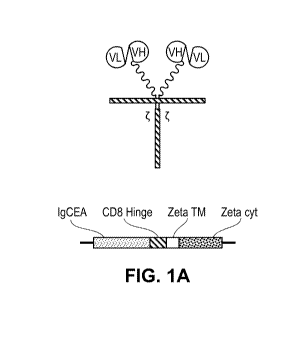
BRIEF DESCRIPTION OF DRAWINGS
[0025] FIGS. lA and 1B provide schematics of various anti-CEA CAR-T
constructs.
[0026] FIG. 2 shows lysis by untransduced splenic cells and chimeric
receptor transduced
lymphocytes.
[0027] FIG. 3A shows luminescence in animals harboring tumors and which had
been
administered chimeric receptor transduced lymphocytes by intraperitoneal (IP)
or tail vein (TV)
injections.
[0028] FIG. 3B shows reduction in tumor volume in animals harboring tumors
and which had
been administered chimeric receptor transduced lymphocytes by intraperitoneal
(IP) or tail vein (TV)
injections.
[0029] FIG. 4A shows luminescence in animals harboring tumors which had
been treated with
chimeric receptor transduced lymphocytes by intraperitoneal (IP) or tail vein
(TV) injections and
which were rechallenged with tumor cells.
[0030] FIGS. 4B and 4C show infiltration of tumors in vivo by leukocytes
expressing the
chimeric receptor protein (FIG. 4B) or by leukocytes having an effector memory
phenotype (FIG.
4C).
[0031] FIG. 5A illustrates therapeutic efficacy of IP chimeric receptor T
cell infusion on tumors
outside of the peritoneal cavity.
[0032] FIG. 5B shows IP tumor reduction via bioluminescence after TV vs. IP
administration of
chimeric receptor T cells.
[0033] FIG. 5C shows reduced flank tumor burden via measurement with
calipers after TV vs.
IP administration of chimeric receptor T cells.
[0034] FIG. 5D shows systemic IFNy levels after IP administration of
chimeric receptor T cells.
[0035] FIGS. 6A and 6B show the presence of CD1 lb+ and MDSC (Ly6G+) cells
within IP
tumor and spleen.
[0036] FIGS. 7A and 7B show the presence of MDSC Ly6G+ and MDSC PD-L1+
cells within
IP tumor and spleen.
[0037] FIGS. 8A and 8B show the presence of Treg (FoxP3+) and CD4 T cells
within IP tumor
and spleen.
[0038] FIG. 9A shows the effects of TV and IP chimeric receptor T cell
infusion on tumor
burden on Day 8 after infusion.
[0039] FIG. 9B shows the effects of administration of antibodies that bind
PD-L1, Gr-1 or GITR
on efficacy of IP chimeric receptor T cell infusion on Day 8 after infusion.
CA 02992122 2018-01-10
WO 2017/011670 PCT/US2016/042302
[0040] FIG. 10A shows the effects of TV and IP chimeric receptor T cell
infusion on tumor
burden on Day 14 after infusion.
[0041] FIG. 10B shows the effects of administration of antibodies that bind
PD-L1, Gr-1 or
GITR on efficacy of IP chimeric receptor T cell infusion on Day 14 after
infusion.
[0042] FIG. 11 shows the effects of administration of antibodies that bind
PD-L1, Gr-1 or GITR
on efficacy of IP chimeric receptor T cell infusion over a 14-day period after
infusion.
DETAILED DESCRIPTION
[0043] Various aspects now will be described more fully hereinafter. Such
aspects may,
however, be embodied in many different forms and should not be construed as
limited to the
embodiments set forth herein; rather, these embodiments are provided so that
this disclosure will be
thorough and complete, and will fully convey its scope to those skilled in the
art.
I. DEFINITIONS
[0044] As used in this specification, the singular forms "a," "an," and
"the" include plural
referents unless the context clearly dictates otherwise. Thus, for example,
reference to a "polymer"
includes a single polymer as well as two or more of the same or different
polymers, reference to an
"excipient" includes a single excipient as well as two or more of the same or
different excipients, and
the like.
[0045] Where a range of values is provided, it is intended that each
intervening value between
the upper and lower limit of that range and any other stated or intervening
value in that stated range
is encompassed within the disclosure. For example, if a range of 1 um to 8 um
is stated, it is
intended that 2 um, 3 um, 4 um, 5 um, 6 um, and 7 um are also explicitly
disclosed, as well as the
range of values greater than or equal to 1 um and the range of values less
than or equal to 8 um.
[0046] The term "substantially pure" or "substantially purified" as used
herein means that the
CAR-T cells are as pure as it is possible to obtain by standard techniques and
methods commonly
known to one of ordinary skill in the art to which this invention pertains.
However, a purity of 70%,
80%, 90% or greater is necessary for the monocytes to be substantially pure.
[0047] The term "peritoneal cavity" as used herein refers to the hollow or
space, or a potential
space, between the parietal and the visceral peritoneum.
[0048] The term "intraperitoneal cancer," "intraperitoneal tumor,"
"intraperitoneal malignancy"
or the like as used herein refers to a malignancy including for example a
tumor mass or one or more
tumor cells, which is located within the peritoneal cavity. A peritoneal
cancer, malignancy or tumor
is a malignancy which originated in the peritoneum or peritoneal cavity.
[0049] The terms "patient," "subject," "individual," and the like are used
interchangeably herein,
6
CA 02992122 2018-01-10
WO 2017/011670 PCT/US2016/042302
and refer to any animal or cells thereof whether in vitro or in situ, amenable
to the methods
described herein. In certain non-limiting embodiments, the patient, subject or
individual is a human.
[0050] As used herein the term "therapeutically effective" applied to dose
or amount refers to
that quantity of a compound or pharmaceutical composition (e.g., a composition
comprising immune
cells such as T lymphocytes and/or NK cells) comprising a chimeric receptor of
the disclosure, and
further comprising a drug resistance polypeptide that is sufficient to result
in a desired activity upon
administration to a subject in need thereof Within the context of the present
disclosure, the term
"therapeutically effective" refers to that quantity of a compound or
pharmaceutical composition that
is sufficient to delay the manifestation, arrest the progression, relieve or
alleviate at least one
symptom of a disorder treated by the methods of the present disclosure. Note
that when a
combination of active ingredients is administered the effective amount of the
combination may or
may not include amounts of each ingredient that would have been effective if
administered
individually.
[0051] The term "chimeric receptor" as used herein is defined as a cell-
surface receptor
comprising an extracellular ligand binding domain, a transmembrane domain and
one or more
cytoplasmic co-stimulatory signaling domains in a combination that is not
naturally found together
on a single protein. This particularly includes receptors wherein the
extracellular domain and the
cytoplasmic domain are not naturally found together on a single receptor
protein. The chimeric
receptors of the present disclosure are intended primarily for use with T
cells and natural killer (NK)
cells. A chimeric receptor described herein may also be referred to herein as
a chimeric antigen
receptor (CAR), a chimeric ligand receptor, or a chimeric T cell receptor.
[0052] The term "tumor associated antigen" or "antigen" as used herein
refers to an antigen
which is specifically expressed by tumor cells or expressed at a higher
frequency or density by tumor
cells than by non-tumor cells of the same tissue type. Tumor-associated
antigens may be antigens not
normally expressed by the host; they may be mutated, truncated, misfolded, or
otherwise abnormal
manifestations of molecules normally expressed by the host; they may be
identical to molecules
normally expressed but expressed at abnormally high levels; or they may be
expressed in a context
or milieu that is abnormal. Tumor-associated antigens may be, for example,
proteins or protein
fragments, complex carbohydrates, gangliosides, haptens, nucleic acids, or any
combination of these
or other biological molecules.
[0053] The term "immune suppressive cell inhibitor" refers to a substance
capable of reducing or
suppressing the number or function of immune suppressive cells of a mammal.
Examples of immune
suppressive cells include regulatory T cells ("T regs"), myeloid derived
suppressor cells (MDSCs),
and tumor-associated macrophages.
[0054] The term "antibody," as used herein, refers to an immunoglobulin
molecule which
7
CA 02992122 2018-01-10
WO 2017/011670 PCT/US2016/042302
specifically binds with an antigen. Antibodies can be intact immunoglobulins
derived from natural
sources or from recombinant sources and can be immunoreactive portions of
intact
immunoglobulins. The antibodies in the present invention may exist in a
variety of forms including,
for example, polyclonal antibodies, monoclonal antibodies, Fv, Fab and F(ab)2,
as well as single
chain antibodies and humanized antibodies (Harlow et al, 1999, In: Using
Antibodies: A Laboratory
Manual, Cold Spring Harbor Laboratory Press, NY; Harlow et al, 1989, In:
Antibodies: A
Laboratory Manual, Cold Spring Harbor, New York; Houston et al, 1988, Proc.
Natl. Acad. Sci.
USA 85:5879-5883; Bird et al., 1988, Science 242:423-426).
[0055] The term "antibody fragment" refers to a portion of an intact
antibody and refers to the
antigenic determining variable regions of an intact antibody.
[0056] The term "antibody-derived targeting domain" "or antigen binding
domain" as used
herein refers to the minimum antibody fragment which contains a complete
antigen-recognition and
binding site. An "Fv" domain also refers to the minimum antibody fragment
which contains a
complete antigen-recognition and ¨binding site and consists of a dimer of one
heavy chain and one
light chain variable domain in tight, non-covalent association. It is in this
configuration that the three
hypervariable regions of each variable domain interact to define an antigen-
binding site on the
surface of the VH-VL dimer. Collectively, the six hypervariable regions confer
antigen-binding
specificity to the antibody. However, even a single variable domain (or half
of an Fv comprising
only three hypervariable regions specific for an antigen) has the ability to
recognize and bind
antigen, although at a lower affinity than the entire binding site.
[0057] The term "natural ligand" as used herein refers to a naturally
occurring protein which
binds specifically to another naturally occurring protein. "Natural ligand"
encompasses both the full-
length protein and fragments thereof which bind specifically to the same
naturally occurring protein.
A natural ligand as used herein can be recombinantly produced or synthetic.
[0058] The term "antigen" or "Ag" as used herein is defined as a molecule
that provokes an
immune response. This immune response may involve either antibody production,
or the activation
of specific immunologically-competent cells, or both. The skilled artisan will
understand that any
macromolecule, including virtually all proteins or peptides, can serve as an
antigen. Furthermore,
antigens can be derived from recombinant or genomic DNA. A skilled artisan
will understand that
any DNA, which comprises a nucleotide sequences or a partial nucleotide
sequence encoding a
protein that elicits an immune response therefore encodes an "antigen" as that
term is used herein.
[0059] As used herein, the expression "specifically binds" in reference to
a chimeric T cell
receptor means that the chimeric T cell receptor binds to its target protein
with greater affinity that it
does to a structurally different protein(s).
[0060] As used herein, the expression "tumor load" or "tumor burden" refers
to the number of
8
CA 02992122 2018-01-10
WO 2017/011670 PCT/US2016/042302
cancer cells, the size of a tumor, or the amount of cancer in the body of a
subject.
INTRAPERITONEAL ADMINISTRATION OF CHIMERIC RECEPTOR IMMUNE CELLS
[0061] In developing therapies for treatment of disseminated tumors such as
intraperitoneal
tumors, it is advantageous to utilize a tumor-selective therapeutic.
Immunotherapeutic cells
engineered to express chimeric receptors (e.g., CAR T cells) that recognize
and bind to tumor
associated antigens is increasingly being proven as a promising approach to
cancer treatment.
Despite the ability of the engineered cells to target the tumor cells,
systemic intravascular
administration can nevertheless result in inadequate exposure of tumor cells
to the CAR-T cells and
adverse side effects due to binding of CAR-T cells to normal cells.
Accordingly, it is advantageous
to provide a method for administering the CAR-T cells directly to the organ or
anatomic space
containing the tumors. In some aspects of the present disclosure, methods are
provided comprising
intraperitoneal administration of chimeric receptor lymphocytes as described
herein. In some
embodiments, the lymphocytes are T cells.
[0062] Current CAR T therapies involve systemic infusion of the engineered
cells to the patient.
Such administration methods, however, may suffer from reduced concentrations
of the cells at the
disease site or presentation of adverse side effects due to activities of the
cells. Provided herein are
compositions and methods for intraperitoneal (IP) infusion of engineered
immune cells to treat
patients diagnosed with an intraperitoneal cancer as experiments described
below show that regional
IP infusion of the cells resulted in superior protection against peritoneal
tumors when compared to
systemically infused cells. Moreover, administration of immune pathway
inhibitors to the patients
receiving the IP cell (IPC) therapy further improved therapeutic efficacy for
treating peritoneal
metastases.
CHIMERIC RECEPTOR IMMUNE CELL THERAPY
[0063] Cancer research is increasingly focused on the use of immune system
components to
combat malignant disease. For example, numerous therapeutic antibodies have
proven successful in
treating cancers and are presently marketed throughout the world. More
recently, cell-based
immunotherapy is emerging as a promising approach to cancer treatment in which
a patient's own
immune cells are engineered to recognize and attack tumors in their body.
Diagnosis of a subject as
having malignant tumors may include determining what tumor antigen proteins
(tumor associated
antigens) are expressed on the tumor cell surface. The subject can then be
treated with anti-tumor
immune cells which have been engineered to target and bind to the tumor
associated antigen,
ultimately leading to the killing of the tumor cells by the immune cell and
possibly other co-
administered cells or therapeutic agents. Disclosed herein are compositions
and methods for treating
tumors in the abdominal cavity via intraperitoneal infusion of engineered
immune cells.
9
CA 02992122 2018-01-10
WO 2017/011670 PCT/US2016/042302
[0064] In one aspect are lymphocytes which have been engineered to express
a chimeric
receptor. The population of lymphocytes for use according to the present
methods include but are not
limited to T cells, B cells and NK cells. In some embodiments, the T cells
comprise CD4+ cells,
CD8+ cells, gamma delta T cells (y6 T cells), NK T cells and/or regulatory T
cells (Treg). Of
particular interest are T cells which express a chimeric receptor ("chimeric
receptor T cells). The
chimeric receptor immune cells are designed to bind, via the chimeric receptor
protein, to diseased
or malignant cells which express a cell surface protein. For example,
malignant cells in the
intraperitoneal cavity may express the carcinoembryonic antigen (CEA, GenBank
Acc. No.
NP 04354 and its related isoforms), the KIT tyrosine kinase receptor protein
(GenBank Acc. No.
P10721), the epithelial cell adhesion molecule protein (EpCAM; GenBank Acc.
No. NP 002345 and
its related isoforms), or the mucin 1 protein (MUC1, GenBank Acc. No. NP
001018016 and its
related isoforms) (e.g., Yamamoto et al., 2014, J Cancer Res Clin Oncol,
140:607-612; Joensuu,
2006, Ann Oncol, 17:x280-x286; Chauhan et al., 2009, J Ovarian Res, 2:21-29;
Flatmark et al.,
2013, Int J Cancer, 133:1497-1506). Other examples of antigen targets
expressed on cancer cells and
that are currently being studied for CAR-T cell therapy include CD20 or GD2
(follicular
lymphoma), CD171 (neuroblastoma), CD20 (non-Hodgkin lymphoma), CD19
(lymphoma),
IL13Ra2 (glioblastoma), and CD19 (chronic lymphocytic leukemia or CLL and
acute lymphocytic
leukemia or ALL). Virus specific CAR-T cells have also been developed to
attack cells harboring
virus such as HIV. For example, a clinical trial was initiated using a CAR
specific for Gp100 for
treatment of HIV (Chicaybam et al (2011) Int Rev Immunol 30:294-311). It is
understood that the
present methods and compositions include, but are not limited to, the antigen
targets listed above.
[0065] Generation of chimeric receptor proteins and immune cells expressing
these proteins is
well known in the art and combines the targeting function and specificity of a
ligand or antibody or
fragment thereof with the anti-tumor activity of an immune cell. See for
example, Sadelain et al.,
2013, Cancer Discov, 3:388-398. The chimeric receptor protein comprises in an
N-terminal to C-
terminal direction a target binding domain which specifically binds a protein
expressed on the
surface of a diseased target cell (e.g., a cancer cell or malignant cell
present in the peritoneal cavity),
a hinge domain, a transmembrane domain, and an immunomodulatory signaling
domain. In some
embodiments, the construct further comprises a signal peptide fused to the N-
terminus of the target
binding domain.
[0066] In some embodiments, the target binding domain of the chimeric
receptor protein
comprises the antigen-binding portion of an immunoglobulin wherein the
immunoglobulin binds a
protein on the surface of the diseased cell. This construct is alternatively
referred to herein as a
chimeric antigen receptor (CAR). The antigen binding domain can be any domain
that binds to the
cell surface antigen including but not limited to monoclonal antibodies,
polyclonal antibodies,
CA 02992122 2018-01-10
WO 2017/011670 PCT/US2016/042302
synthetic antibodies, human antibodies, humanized antibodies, and fragments
thereof In preferred
embodiments, the antigen-binding domain of the CAR is a fragment of an
antibody that is able to
specifically bind the antigen when part of a CAR construct. In some instances,
it is beneficial for the
antigen binding domain to be derived from the same species in which the CAR
will ultimately be
used in. For example, for use in humans, it may be beneficial for the antigen
binding domain of the
CAR to comprise a fragment of a human or humanized antibody. Accordingly, in
some
embodiments, the antigen binding domain portion of a CAR comprises a tumor
antigen binding
fragment of a human or humanized antibody. In each of these embodiments, the
antigen-binding
domain of an antibody, such as the single-chain variable fragment (scFV or
Fab) or is fused to a
transmembrane domain and a signaling intracellular domain (endodomain) of a T
cell receptor.
Often, a spacer or hinge is introduced between the extracellular antigen
binding domain and the
transmembrane domain to provide flexibility which allows the antigen-binding
domain to orient in
different directions to facilitate antigen recognition and binding.
[0067] In some embodiments, the antigen binding moiety portion of the
chimeric antigen T cell
receptor targets the CEA antigen and comprises the CEA-binding domain of an
antibody which has
been shown to bind CEA expressed on a cell surface. The chimeric receptor
construct can be
generated according to methods and compositions known to the ordinarily
skilled artisan. For
example, a CEA CAR-T construct used in the Examples below comprises portions
of the variable
domain of a humanized MN14 antibody (described in U.S. Patent No. 5,874,540,
the contents of
which are incorporated herein by reference it their entirety). A Fab or scFv
construct can be
generated from a CEA antibody according to the methods of Nolan et al. (1999,
Clinical Canc Res,
5:3928-3941) to include the CEA-binding domains of the CEA antibody. In some
embodiments, the
CEA CAR-T construct comprises the amino acid sequence of SEQ ID NO: 1 shown
below:
DIQLTQSPSSLSASVGDRVTITCKASQDVGISVAWYQQKPGKAPKLLIYWISTRHIGVPSRFSGSGS
GTDFTFTISSLQPEDIATYYCQQYSLYRSFGQGTKVEIKRTVAAPSVFIFPPSDEQLKSGTASVVCL
LNNFYPREAKVQWKVDNALQSGNSQESVTEQDSKDSTYSLSSTLTLSKADYEKHKVYACEVTHQGLS
SPVTKSFNRGEC (SEQ ID NO:1)
[0068] In some embodiments, the CEA CAR-T construct further comprises a
signal sequence at
the N-terminus of SEQ ID NO:1 which is cleaved from the construct after in
vivo expression of the
CEA CAR-T construct. In other embodiments, the signal sequence has the
sequence
MGWSCIILFLVATATGVHS (SEQ ID NO:2). The Fab or scFv domain can then be fused to
a
hinge domain such as that from the CD8 hinge domain (see GenBank Acc. No. NP
001759). The
hinge domain can then be fused at its C-terminus to a transmembrane domain. In
one embodiment,
the transmembrane domain is from the CD3 zeta chain (e.g., GenBank Acc. No. NP
000725 or from
the CD28 protein (e.g., GenBank Acc. No. NP 006130). The transmembrane domain
of the chimeric
11
CA 02992122 2018-01-10
WO 2017/011670 PCT/US2016/042302
construct can then be fused at its C-terminus to the signaling domain of the
CD3 zeta chain (e.g.,
GenBank Acc. No. NP 000725).
[0069] In some embodiments, the CEA-binding domain is a scFv or Fab domain
from an
antibody that binds CEA and the chimeric receptor construct comprises, in an N-
terminal to C-
terminal direction: the CEA-binding domain (e.g., SEQ ID NO:1), a CD8 hinge
domain, a zeta
transmembrane domain and a zeta cytoplasmic signaling domain. In other
embodiments, the
chimeric receptor construct comprises, in an N-terminal to C-terminal
direction: the CEA-binding
domain (e.g., SEQ ID NO:1), the CD8 hinge domain, a domain comprising (in an N-
terminal to C-
terminal direction) a portion of the CD28 extracellular domain, the CD28
transmembrane domain,
and the CD28 cytoplasmic co-stimulatory domain, and a zeta cytoplasmic
signaling domain.
[0070] In alternative embodiments, a known ligand to a protein expressed on
the surface of a
tumor cell is fused to a T cell receptor signaling domain to produce what is
alternatively referred to
herein as a "chimeric ligand T cell receptor" or "chimeric ligand receptor."
As with CAR-T cells, T
cells that express a chimeric ligand T cell receptor protein become activated
in the presence of a cell
expressing the target ligand receptor protein, resulting in the attack on the
targeted cell by the
activated T-cell in a non-MHC dependent manner. In some embodiments, a
chimeric ligand receptor
is specifically designed to include the extracellular domain of the KIT-
ligand, a cytokine that binds
to tyrosine-protein kinase KIT protein (cKIT receptor or CD117) expressed on
the surface of
gastrointestinal stromal tumor (GIST) cells. A chimeric T cell receptor was
engineered as described
in PCT Pub. No. WO 2014/121264 (see also Katz et al., J Transl Med., 2013,
11:46). The anti-MT
chimeric receptor was expressed on the surface of the T cells and the
engineered cells were able to
proliferate when co-cultured with KIT+ tumor cells and produce IFNy. Moreover,
mice with
established GIST xenografts and treated with the anti-MT chimeric ligand
receptor T cells showed
significant reductions in tumor growth rates. Accordingly, it is understood
that such chimeric ligand
receptor T cells can be used to treat intraperitoneal cancers according to the
methods described
herein. A schematic of two alternative CAR-T constructs for use in the methods
as described herein
are provided in FIGS. 1A and 1B.
CHIMERIC RECEPTOR INTRACELLULAR DOMAIN
[0071] The intracellular signaling domain of the chimeric T cell receptor
is activated upon
binding of the target antigen by the antigen-binding domain of the CAR or by
the ligand portion of
the chimeric ligand receptor. Generally, the domain of the endogenous CD3 T
cell receptor is used
as the signaling domain. More recently, however, second generation CAR
molecules have been
designed to further include another intracellular signaling domain from a
costimulatory receptor such
as CD28, 41BB, or ICOS to provide additional signals to the engineered T cell
which may improve
its efficacy and/or viability. Third generation chimeric T cell receptors
combine multiple signaling
12
CA 02992122 2018-01-10
WO 2017/011670 PCT/US2016/042302
domains or accessory regions to provide novel functionality. Accordingly in
some embodiments, the
cytoplasmic domain further comprises one or more co-stimulatory domains
selected from the group
consisting of an OX-40 costimulatory domain, an HVEM co-stimulatory domain, a
41BB co-
stimulatory domain, an ICOS co-stimulatory domain, an 0X40 co-stimulatory
domain and a CD27
co-stimulatory domain. In one embodiment, the additional co-stimulatory domain
is positioned
between a CD28 co-stimulatory domain and a CD3-zeta signaling domain.
CHIMERIC RECEPTOR LYMPHOCYTES FOR IP INFUSION
[0072] Lymphocytes engineered with chimeric receptors to enable highly
specific tumor
recognition and killing have gained considerable attention following promising
clinical results
(Grupp et al., 2013, N Eng J Med, 368:1509-1518; Porter et al., 2011, N Eng J
Med, 365:725-733;
Sadelain et al., 2009, Curr Opin Immunol, 21:215-223). Types of lymphocytes
that can be used in
the methods of the present disclosure include, without limitation, peripheral
donor lymphocytes
genetically modified to express chimeric receptors (Sadelain, M., et al. 2003,
Nat Rev Cancer 3:35-
45), lymphocyte cultures derived from tumor infiltrating lymphocytes (TILs) in
tumor biopsies
(Panelli, M. C., et al. 2000 J Immunol 164:495-504; Panelli, M. C., et al.
2000 J Immunol 164:4382-
4392), and selectively in vitro-expanded antigen-specific peripheral blood
leukocytes employing
artificial antigen-presenting cells (AAPCs) or pulsed dendritic cells (Dupont,
J., et al. 2005 Cancer
Res 65:5417-5427; Papanicolaou, G. A., et al. 2003 Blood 102:2498-2505). The T
cells may be
autologous, non-autologous (e.g., allogeneic), or derived in vitro from
engineered progenitor or stem
cells. T cells may prepared in bulk as commonly performed with Peripheral
blood lymphocytes
(PBL), or tumor infiltrating lymphocytes (TILs), T cells may be purified by
using, e.g. CD4, CD8,
CD62L.
[0073] Genetic modification of immunoresponsive cells (e.g., T cells, CTL
cells, NK cells) can
be accomplished by transducing a substantially homogeneous cell composition
with a recombinant
DNA or RNA construct. Preferably, a retroviral vector (either gamma retroviral
or lentiviral) is
employed for the introduction of the DNA or RNA construct into the host cell
genome. For example,
a polynucleotide encoding a receptor that binds an antigen (e.g., a tumor
antigen, or a variant, or a
fragment thereof), can be cloned into a retroviral vector and expression can
be driven from its
endogenous promoter, from the retroviral long terminal repeat, or from an
alternative internal
promoter. Non-viral vectors or RNA may be used as well. Random chromosomal
integration, or
targeted integration (e.g., using a nuclease, transcription activator-like
effector nucleases (TALENs),
Zinc-finger nucleases (ZFNs), and/or clustered regularly interspaced short
palindromic repeats
(CRISPRs), or transgene expression (e.g., using a natural or chemically
modified RNA) can be used.
[0074] For initial genetic modification of the cells to provide chimeric
receptor-expressing cells,
a retroviral vector is generally employed for transduction, however any other
suitable viral vector or
13
CA 02992122 2018-01-10
WO 2017/011670 PCT/US2016/042302
non-viral delivery system can be used. For subsequent genetic modification of
the cells to provide
cells comprising an antigen presenting complex comprising at least two co-
stimulatory ligands,
retroviral gene transfer (transduction) likewise proves effective.
Combinations of retroviral vector
and an appropriate packaging line are also suitable, where the capsid proteins
will be functional for
infecting human cells.
[0075] In yet another aspect, the disclosure is directed to pharmaceutical
compositions to
facilitate administration of transduced T cells as described herein to a
subject in need. The
transduced T cells according to the disclosure can be made into a
pharmaceutical composition or
made implant appropriate for administration in vivo, with appropriate carriers
or diluents, which
further can be pharmaceutically acceptable. The means of making such a
composition or an implant
have been described in the art (see, for instance, Remington's Pharmaceutical
Sciences, 16th Ed.,
Mack, ed. (1980)). Where appropriate, the transduced T cells can be formulated
into a preparation in
semisolid or liquid form, such as a capsule, solution, injection, inhalant, or
aerosol, in the usual ways
for their respective route of administration. Means known in the art can be
utilized to prevent or
minimize release and absorption of the composition until it reaches the target
tissue or organ, or to
ensure timed-release of the composition. Desirably, however, a
pharmaceutically acceptable form is
employed which does not ineffectuate the cells expressing the chimeric
receptor. Thus, desirably the
transduced T cells can be made into a pharmaceutical composition containing a
balanced salt
solution, preferably Hanks' balanced salt solution, or normal saline. For
instance, the compositions
can be formulated with a physiologically acceptable carrier or excipient to
prepare a pharmaceutical
composition. The carrier and composition can be sterile. The formulation
should suit the mode of
administration.
[0076] Suitable pharmaceutically acceptable carriers include but are not
limited to water, salt
solutions (e.g., NaC1), saline, buffered saline, alcohols, glycerol, ethanol,
gum arabic, vegetable oils,
benzyl alcohols, polyethylene glycols, gelatin, carbohydrates such as lactose,
amylose or starch,
dextrose, magnesium stearate, talc, silicic acid, viscous paraffin, perfume
oil, fatty acid esters,
hydroxymethylcellulose, polyvinyl pyrolidone, etc., as well as combinations
thereof The
pharmaceutical preparations can, if desired, be mixed with auxiliary agents,
e.g., lubricants,
preservatives, stabilizers, wetting agents, emulsifiers, salts for influencing
osmotic pressure, buffers,
coloring, flavoring and/or aromatic substances and the like that do not
deleteriously react with the
active compounds.
[0077] The composition, if desired, can also contain minor amounts of
wetting or emulsifying
agents, or pH buffering agents. The composition can be a liquid solution,
suspension, emulsion,
tablet, pill, capsule, sustained release formulation, or powder. The
composition can be formulated as
a suppository, with traditional binders and carriers such as triglycerides.
Oral formulation can
14
CA 02992122 2018-01-10
WO 2017/011670 PCT/US2016/042302
include standard carriers such as pharmaceutical grades of mannitol, lactose,
starch, magnesium
stearate, polyvinyl pyrollidone, sodium saccharine, cellulose, magnesium
carbonate, etc.
[0078]
The composition can be formulated in accordance with the routine procedures as
a
pharmaceutical composition adapted for administration to human beings.
For example,
compositions for intravenous administration typically are solutions in sterile
isotonic aqueous buffer.
Where necessary, the composition may also include a solubilizing agent and a
local anesthetic to
ease pain at the site of the injection. Generally, the ingredients are
supplied either separately or
mixed together in unit dosage form, for example, as a dry lyophilized powder
or water free
concentrate in a hermetically sealed container such as an ampule or sachette
indicating the quantity
of active compound. Where the composition is to be administered by infusion,
it can be dispensed
with an infusion bottle containing sterile pharmaceutical grade water, saline
or dextrose/water.
Where the composition is administered by injection, an ampule of sterile water
for injection or saline
can be provided so that the ingredients may be mixed prior to administration.
[0079]
Compositions of the invention comprising genetically modified immunoresponsive
cells
can be conveniently provided as sterile liquid preparations, e.g., isotonic
aqueous solutions,
suspensions, emulsions, dispersions, or viscous compositions, which may be
buffered to a selected
pH. Liquid preparations are normally easier to prepare than gels, other
viscous compositions, and
solid compositions. Additionally, liquid compositions are somewhat more
convenient to administer,
especially by injection. Viscous compositions, on the other hand, can be
formulated within the
appropriate viscosity range to provide longer contact periods with specific
tissues. Liquid or viscous
compositions can comprise carriers, which can be a solvent or dispersing
medium containing, for
example, water, saline, phosphate buffered saline, polyol (for example,
glycerol, propylene glycol,
liquid polyethylene glycol, and the like) and suitable mixtures thereof
[0080]
Those skilled in the art will recognize that the components of the
compositions should be
selected to be chemically inert and will not affect the viability or efficacy
of the genetically modified
immunoresponsive cells as described in the present invention. This will
present no problem to those
skilled in chemical and pharmaceutical principles, or problems can be readily
avoided by reference
to standard texts or by simple experiments (not involving undue
experimentation), from this
disclosure and the documents cited herein.
THERAPEUTIC METHODS
[0081]
The present disclosure describes compositions and methods for intraperitoneal
infusion of
lymphocytes which express chimeric receptor T cells and which thereby target
and bind malignant
cells in the peritoneal cavity, leading to inhibition of tumor cell growth or
death of tumor cells.
Intraperitoneal administration provides a higher concentration of therapeutic
agents to the tumor
CA 02992122 2018-01-10
WO 2017/011670 PCT/US2016/042302
location to maximize therapeutic efficacy and minimize systemic toxicity of
the therapeutic cells.
Data provided herein shows that genetically engineered lymphocytes have
significantly greater
efficacy when administered via IP infusion as compared to systemic infusion.
The therapeutic
efficacy of these cells in enhanced by use of inhibitors of immune suppressor
cells.
[0082] Therapeutic use of chimeric receptor lymphocytes involves harvesting
white blood cells
from a subject diagnosed with cancer, isolating and culturing the lymphocytes,
transforming the
lymphocytes with a vector containing the chimeric receptor gene, and
administering to the subject
the resultant engineered lymphocytes. Cells prepared for administration to a
subject can comprise a
purified population of cells, for example CD4+ T cells. Those having ordinary
skill in the art can
readily determine the percentage of genetically modified lymphocytes in a
population using various
well-known methods, such as fluorescence activated cell sorting (FACS).
[0083] The chimeric receptor T cells can be administered in any
physiologically acceptable
vehicle. In some embodiments, a dose of about 1 x 10⁶ to 1 x 10¹¹, 1 x 10⁶ to
1 x 10¹⁰, 1 x 10⁶ to 1 x
109, 1 x 10⁷ to 1 x 10¹¹, 1 x 10⁷ to 1 x 101 , 1 x 10⁷ to 1 x 10⁹ or 1 x 10⁸
to 1 x 10⁹ cells are
administered. In other embodiments, a dose of about 1x10⁶, 1x10⁷, 1x10⁸,
1X10⁹, 1X101 , or lx 1011
cells are administered. The precise determination of what would be considered
an effective dose may
be based on factors individual to each subject, including their size, age,
sex, weight, and condition of
the particular subject. Dosages can be readily ascertained and readily
adjusted by those skilled in the
art from this disclosure and the knowledge in the art. Preferable ranges of
purity in populations
comprising chimeric receptor T cells are about 70 to about 75%, about 75 to
about 80%, about 80 to
about 85%; and still more preferably the purity is about 85 to about 90%,
about 90 to about 95%,
and about 95 to about 100%. The cells can be administered by, for example,
injection or catheter.
Cells may also be administered by minimally invasive surgical techniques.
[0084] The chimeric receptor T cells are administered to the patient via
intraperitoneal infusion
once, twice, 3 times, 4 times or 5 times over a period of time. The period of
time may be about 1
month, 2 months, 3 months, 4 months or 5 months. For example, a dose of the
chimeric receptor T
cells are administered once, twice, 3 times or 4 times in a one-week period.
Furthermore, the one-
week dosing regimen is performed every week, every other week, or 3 weeks or
every month.
Alternatively, the one-week dosing regimen is performed every other week. In
one embodiment, the
dose of the chimeric receptor T cells is administered 3 times per week, every
other week. The dosing
regimen is continued until the tumor load is reduced by at least 5%, 10%, 15%,
20%, 25%, 50%,
60%, 70%, 80%, 90% or 95% relative to the tumor load prior to administration
of the first dose of
chimeric receptor T cells.
[0085] In some embodiments, the chimeric receptor T cells are administered
to a patient who has
undergone debulking surgery to render the patient as disease-free as is
surgically possible.
16
CA 02992122 2018-01-10
WO 2017/011670 PCT/US2016/042302
Immediately following surgery, or within 1, 2 or 5 days following surgery, the
patient receives
intraperitoneal infusion of the CAR-T cells.
[0086] Effective chimeric receptor T cell therapy is achieved in part by
determining an optimal
dose of the chimeric receptor T cells. A therapeutically effective dose for
chimeric receptor T cell
treatment can be determined, for example, by imaging the abdomen of the
patient by CT or PET
scans or MRI imaging. A therapeutically effective dose will decrease the
volume and/or number of
malignant tumors as determined by imagine by at least 10%, 20%, 30%, 40%, 50%,
60%, 70%,
80%, or 90% or by 100%. A therapeutically effective dose would be expected to
decrease the
volume and/or number of malignant tumors in the abdomen within about 5 days, 1
week, 2 weeks, 4
weeks, 6 weeks or 10 weeks after the first administered dose of chimeric
receptor T cells.
Alternatively, a therapeutically effective dose will decrease the amount or
volume of malignant
ascites and/or intraperitoneal mucin by at least 10%, 20%, 30%, 40%, 50%, 60%,
70%, 80%, or 90%
over the dosing period. A therapeutically effective dose will also decrease
serum tumor markers if
available for the targeted tumor type by at least 10%, 20%, 30%, 40%, 50%,
60%, 70%, 80%, or
90% over the dosing period.
INTRAPERITONEAL INFUSION OF CEA CAR-T CELLS
[0087] The efficacy of chimeric receptor T cells by IP infusion of chimeric
receptor lymphocytes
was shown using methods described herein. In mice treated by IP infusion of
unmodified T cells or
anti-CEA CAR-T cells, there was a significant reduction in tumor load as
compared to animals
untreated or treated with unmodified T cells. The ordinarily skilled artisan
would understand that the
methods described herein are useful for reducing tumor load using any chimeric
receptor T cell (e.g.,
CAR-T cell or chimeric ligand receptor T cell) which has been engineered to
specifically bind via
the chimeric T cell receptor to the target protein or antigen expressed on the
surface of the tumor
cell.
[0088] As shown in the Examples below, direct IP infusion of CAR-Ts in mice
with PC was
more effective at controlling tumor than systemic infusion. CAR-Ts within
peritoneal tumors were
detected following IP infusion, whereas CAR-Ts were not present in peritoneal
tumors following
systemic injection. Treatment of malignancies using IP CAR-T infusion methods
as described herein
results in a reduction in adverse side effects as well.
[0089] The compositions and methods described herein are used for treating
patients diagnosed
with intraperitoneal tumors. The patient first undergoes diagnostic
laparoscopy to lyse any peritoneal
adhesions in order to ensure optimal CAR-T distribution following IP infusion
of the CAR-T. This
diagnostic laparoscopy can also be used to assess the disease, acquire pre-
treatment cell or tissue
specimens, and/or for placement of a peritoneal dialysis catheter. The IP CAR-
T infusion can be
17
CA 02992122 2018-01-10
WO 2017/011670 PCT/US2016/042302
performed later the same day or on a following day.
[0090] IP infusion of CAR-T comprises infusion of an initial dose of about
1 x 10⁹ to 1 x 10¹¹, or
about 1 x 10¹⁰ cells into the peritoneal cavity. The CAR-T cells are suspended
in a physiological
solution such as normal saline. In some embodiments, the solution contains
about 5% to 15% or
about 10% dimethyl sulfoxide (DMSO). In some embodiments, immediately prior to
IP CAR-T
infusion, ascites fluid is drained from the peritoneal cavity. In other
embodiments, aspiration is
performed prior to injection of the dose of CAR-T cells to confirm the absence
of blood and/or
intestinal contents.
[0091] The IP infusion of a dose of CAR-T cells can be carried out manually
and at room
temperature. In some embodiments, the dose is infused over a time period of
about 5 min to 60 min,
about 30 min to 120 min, about 5 min to 30 min, about 5 min to 20 min, or
about 10 min, 15 min, 20
min, 25 min, 30 min, 45 min or 60 min. The infusion can be carried out in an
out-patient setting.
[0092] One or more additional doses of the CAR-T cells can be administered
after the initial IP
infusion. For example, an additional dose can be administered weekly, every 3
days or every 5 days
wherein the additional dose is administered once, twice, or three times. In
other embodiments an
additional dose is administered weekly, every 3 days or every 5 days until a
post-infusion assessment
fails to detect malignancy in the peritoneal cavity. In some embodiments, the
additional dose is equal
to the initial dose. In other embodiments, each of the additional doses is
about 75%, 90%, 120% or
150% of the initial dose in terms of the number of CAR-T cells. In a preferred
embodiment, an
additional dose of about 1 x 10¹⁰ is administered to the patient IP once per
week for 2 or 3 weeks.
[0093] In some embodiments, a method for treating malignancies in which
tumor cells are
located outside of the peritoneal cavity is provided. Studies were done to
determine if IP CAR-T
infusions could reduce or inhibit the growth of flank tumors in mice with
synchronous PC. IP CAR-
T infusions were able to significantly limit the growth of distant flank
tumors while inducing marked
IP responses (see Example 6). CAR-Ts were not detected within the flank
tumors, suggesting that
the flank tumor responses were due to IFNy surges which were detected 4 days
following IP CAR-T
treatment (FIG. 7D). IP infusion of CAR-Ts with profound destruction of
peritoneal tumors may
have induced a phenomenon similar to the abscopal effect seen with radiation
therapy (Park et al.,
2015, Cancer Immunol Res, 3:610-619). Alternatively, CAR-Ts may have
infiltrated the flank tumor
at earlier time points. Surprisingly, systemic infusion also did not lead to a
meaningful flank tumor
response, which may reflect inadequate CAR-T dosing by this route, as most
cells likely traffic to
nodes, lung, and spleen. Importantly, the response of distant subcutaneous
tumors was less durable
than the response of IP tumors in accordance with the brief surge in serum
IFNy levels. Sequential
regional and systemic therapy may offer improvements in efficacy for PC in the
context of extra-
abdominal disease. Accordingly, in some embodiments, a method of treatment is
provided wherein a
18
CA 02992122 2018-01-10
WO 2017/011670 PCT/US2016/042302
subject diagnosed with a peritoneal malignancy is treated with IP infusion of
a chimeric receptor
lymphocyte followed by treatment with systemic infusion of the chimeric
receptor lymphocyte.
[0094] As
PC can have a prolonged natural history, the durability of protection from IP
tumor
growth following IP CAR-T infusion was examined (see Example 4). Following
repeated IP CAR-T
dosing, mice were protected from repeat IP tumor challenge for up to 10
additional days. CAR-Ts
were detectable within the PC as late as 28 days. This finding suggests
persistence of CAR-Ts in the
peritoneal space, potentially with the CAR-Ts acquiring effector memory
features. CAR-Ts with an
effector memory phenotype (CD44+CD62L-CCR7-) were detected within IP tumors in
greater
proportion at day 28 compared to day 10. These data suggest that following
initial IP infusion, CAR-
Ts undergo effector memory programming, which may have accounted for the
prolonged anti-tumor
protection in the peritoneal space.
IMMUNOSUPPRESSOR AGENTS
[0095]
Therapeutic efficacy of chimeric receptor T cell infusions is likely to be
affected by
factors that lead to immunosuppression, e.g., suppression of tumor-killing
cells or decreased
expression of anti-tumor cytokines. It is important to consider the effects of
immune environment of
the intraperitoneal space in the presence of a carcinoma and to treat a
patient undergoing chimeric
receptor T cell therapy accordingly.
[0096]
The accumulation of immunosuppressive regulatory T cells (Tregs) and myeloid
derived
suppressor cells (MDSCs) within the tumor microenvironment represents a
potential major obstacle
for the development of effective antitumor immunotherapies (Weiss et al.,
2014, J Immunol.,
192:5821-5829). Elimination of MDSCs has been shown to significantly improve
immune
responses in tumor-bearing mice and in cancer patients (Ostrong-Rosenberg et
al., 2009, J Immunol,
182:4499-4506); Talmadge, 2007, Clin Cancer Rres, 13:5243-5248). Provided
herein are methods
for inhibiting immunosuppression by, for example, Treg and MDSC, in a patient
undergoing
chimeric receptor T cell therapy, wherein the patient is also administered an
agent which inhibits
functions of immunosuppressive cells.
[0097] To
examine the extent of immunosuppressive activity upon treatment with chimeric
receptor T cells, Treg and MDSC were characterized in C57BL/6 mice bearing
MC38 tumor cells.
Specifically, Treg and MDSC are characterized in terms of their cell surface
markers, cytokines and
enzymes believed to play a role in suppressive activity. As shown in Example 7
below, studies
showed that both MDSC and Treg could be detected within IP tumors. Both MDSC
and Treg have
been well described as inhibitors of endogenous T cell and CAR-T anti-tumor
responses (Khaled et
al., 2013, Immunol Cell Biol, 91:493-502; Burkholder et al., 2014, Biochim,
Biophys Acta,
1845:182-201). IP MDSC also expressed high levels of PD-Li (programmed death-1
receptor
19
CA 02992122 2018-01-10
WO 2017/011670 PCT/US2016/042302
ligand), which was previously demonstrated to be an important mediator of CAR-
T suppression
(Burga et al., 2015, 64:817-829). Addition of an MDSC depletion antibody which
binds Grl
(granulocytic myeloid marker protein) or a PD-Li blocking antibody treatment
enhanced IP CAR-T
performance in terms of tumor killing. The encouraging additive effects of IP
CAR-T and suppressor
cell targeting provide justification for combinatorial strategies in
developing solid tumor
immunotherapy. Accordingly, in some embodiments, a method for treating a
subject diagnosed with
a peritoneal cancer is provided, wherein the subject is administered a
population of lymphocytes
expressing a chimeric receptor as described herein via IP infusion and wherein
the subject is also
administered an immunosuppressing agent which suppresses the activity of
suppressor T cells such
as MDSCs or Tregs.
[0098] In some embodiments, the immunosuppressing agent is an antibody that
binds IL10, PD-
1 (programmed death-1 receptor), PD-Li (programed death-1 receptor ligand 1),
PD-L2 (programed
death-1 receptor ligand 2), IDO (Indolamine 2,3-dexoygenase), STAT3 (signal
transducer and
activator of transcription 3), GM-CSF, CD25, GITR (glucocorticoid-induced TNFR-
related protein),
TGF-0, or CTLA4. In other embodiments, the immunosuppressing agent is
administered to the
subject before IP administration of chimeric receptor lymphocytes. In still
other embodiments, the
immunosuppressing agent is administered to the subject after IP administration
of chimeric receptor
lymphocytes. The immunosuppressing agent can be administered multiple times,
for example, every
day, every 2 days, every 3 days, every 4 days, every 5 days, every 6 days or
once per week (every 7
days) after IP administration of the chimeric receptor lymphocytes. The
immunosuppressing agent
can be administered on the same day as the IP administration of the chimeric
receptor lymphocytes.
The immunosuppressing agent can be administered 1 day, 2 days, 3 days, 4 days,
5 days, 6 days, 7
days or more prior to IP administration of the chimeric receptor lymphocytes.
More than one
immunosuppressing agent can be administered to the patient, for example, the
subject may be co-
administered or serially administered antibodies which bind CD25 and
antibodies which bind GR1.
ADDITIONAL THERAPEUTIC AGENTS
[0099] The chimeric receptor T cells of the present disclosure can be used
alone or in
combination with other therapies. Immunomodulatory agents may include but are
not limited to
interleukins, e.g. IL-2, IL-3, IL-6, IL-11, IL7, IL12, IL21, as well as the
other 10 interleukins, the
colony stimulating factors, such as granulocyte colony stimulating factor (G-
CSF), and macrophage
colony stimulating factor (M-CSF), and interferons, such as y-interferon and
erythropoietin. Other
immunomodulatory agents may include monoclonal antibodies or small molecules
designed to target
immunoinhibitory pathways such as an antibody or fragment thereof which binds
TGF13 or IL10,
thereby blocking the function of TGFr3 or IL10, respectively.
CA 02992122 2018-01-10
WO 2017/011670 PCT/US2016/042302
[0100] In a preferred embodiment, administration of the chimeric receptor T
cells is coupled with
administration of one or more agents as listed above which inhibit chimeric
receptor T cell
suppressor pathways. For example, a patient in need thereof receives
intraperitoneal infusion of both
chimeric receptor T cells and an agent which increases in situ viability of
the chimeric receptor T
cells after intraperitoneal infusion. In a preferred embodiment, the patient
is administered chimeric
receptor T cells and a dose of IL2. Administration of the agent which
increases viability of the
chimeric receptor T cells may be performed before, during or after
administration of the chimeric
receptor T cells.
IV. EXAMPLES
[0101] The following examples are illustrative in nature and are in no way
intended to be
limiting.
EXAMPLE 1
PREPARATION OF CEA CAR-T CELLS
[0102] The anti-CEA scfv-CD28/CD3 (Tandem) chimeric antigen receptor used
in the
examples described herein was previously generated according to the method of
Emtage et al. (2008,
Clin Cancer Res, 14:8112-8122). Briefly, a tandem molecule was generated by
molecularly fusing
an hMN14 sFv-CD8 hinge segment of a monoclonal antibody which specifically
binds CEA
upstream of a construct encoding a cytoplasmic domain comprising in an N-
terminal to C-terminal
direction, a human CD28 extracellular domain, the CD28 cytoplasmic domain, and
the cytoplasmic
domain. The resultant chimeric construct was cloned into a retroviral vector
and verified by
restriction digestion and sequencing.
[0103] For the present studies, 6-8 week old B6.SJL-Ptprca Pepcb/BoyJ
(CD45.1) mice were
purchased from Jackson for the purpose of generating distinguishable CAR-Ts
when isolated from
tissues ex vivo. Mice were housed in the animal facility at Roger Williams
Medical Center in
pathogen-free conditions under guidelines from the Institutional Care and Use
Committee. CD45.1
mouse spleens were harvested in sterile fashion then pulverized. Red blood
cells were lysed and T
cells were isolated using MACS immunomagnetic bead isolation (Miltenyi). T
cells were cultured in
complete media with IL-2 (500 IU/mL) and anti-CD3/CD28 T-activator Dynabeads
(Life
Technologies) for 48 hours to achieve activation. Phoenix Ecotropic cells
harboring a hMN14 sFv-
CD8a-CD28/CD3 CAR (Emtage et al.,2008, Clin Cancer Res, 14:8112-8122) were
used to produce
supernatant for transduction. Activated T cells were cultured in the
retroviral supernatant and
underwent two spinfections. Transduced T cells were cultured and expanded in
the presence of IL-2
(500 IU/mL), and CAR expression levels were checked 48 hours after
transduction.
[0104] Transduction of murine splenocytes was confirmed 48 hours after
transduction by
21
CA 02992122 2018-01-10
WO 2017/011670 PCT/US2016/042302
measuring CAR expression on CD3+ cells using flow cytometry and an antibody
which specifically
binds the sFy portion of the CEA CAR-T molecule. A standard gating strategy
was used to identify
viable, single cells expressing CD3 and the chimeric anti-CEA CAR-T. The
results showed that viral
transduction efficiency was about 73% (data not shown).
EXAMPLE 2
IN VITRO KILLING OF TUMOR CELLS
[0105] Killing by the transduced CAR-T cells was tested in vitro using as
target cells MC38
cells which were stably transfected with a gene encoding human CEA and firefly
luciferase.
MC38CEA+ cells were first generated by stably transfecting MC38 cells with the
human CEA gene.
MC38-luc was generated by transfecting MC38CEA+ cells with pLenti-III-UbC-
Luciferase
(Applied Biological Materials Inc, Richmond, BC Canada). Effector cells were
either CEA CAR-T
cells generated as described in Example 1 or untransduced splenic T cells
which were used as a
negative control for the effector cells. Bioluminescence assays were performed
in which CAR-Ts or
untransduced T cells were co-cultured with MC38CEA-luc at various
Effector:Target ratios.
Effectors were cultured in complete media with IL-2 (500 IU/mL) prior to the
assays. Cells were
plated in complete media in 96 well optical plates at varying Effector:Target
ratios and incubated
overnight. After incubation, media was discarded and luciferin (150pg/mL) was
added to the wells.
Plates were analyzed in an IVIS 100. Supernatants were collected and measured
for luminescence
activity and Specific Lysis % was calculated as 100 x [(experimental killing ¨
spontaneous
luminescence)/(maximal killing ¨ spontaneous luminescence)]. As seen in FIG.
2, transduced CAR-
T cells caused lysis at a significantly higher rate than untransfected cells.
At an Effector:Target ratio
as low as 0.03:1, specific lysis was 40% and significantly higher than
activated untransduced T cells
(p=0. 02).
EXAMPLE 3
CAR-T CELL DELIVERY AND KILLING OF TUMOR CELLS
[0106] To show that IP delivery improves CAR-T efficacy in mice with
peritoneal cancers (PC)
compared to systemic tail vein (TV) infusion, both infusion methods were
studied in mice with
established IP tumors. Mice harboring CEA+ PC were generated by the IP
injection of the
MC38CEA-luc cells.
[0107] Six to eight week old C57B1/6J mice were purchased from Jackson
Laboratories (Bar
Harbor, ME) and were used in all in vivo models. Mice were injected
intraperitoneally with 2.5 x 10⁶
MC38CEA-luc cells on day 0 using a 26 gauge x 1/2" needle attached to a 1 ml
syringe. Cells had
been resuspended in normal saline for injection and the injection was
performed at room
temperature. The needle traverses the midline fascia 2-3 mm superior to the
pubic symphysis and
22
CA 02992122 2018-01-10
WO 2017/011670 PCT/US2016/042302
aspiration was performed prior to injection to confirm absence of blood or
intestinal contents. In
vivo work was carried out over the span of 14 days. On days 3 and 6, tumor-
bearing mice were
treated with CAR-Ts (2.5 x 10⁶ cells), either via IP or TV infusion. All mice
were administered IL-2
(1000 IU/injection) daily beginning with the first CAR-T injection on day 3.
Control mice were
treated with untransduced splenic cells on days 3 and 6 or treated with IL-2
alone. For the
bioluminescent mice were imaged on an IVIS 100 imaging station (Caliper Life
Sciences) on even
days during in vivo studies after being injected with 200 pL of 15 mg/mL
luciferin.
[0108] Data are presented in FIGS. 3A and 3B. In FIG. 3A each line on the
plot is representative
of the average of 4 mice. Fold reduction in tumor luminescence was calculated
between days 4 and
14 of the in vivo study, comparing TV to IP CAR-T delivery and the results are
shown in FIG. 3B.
Error bars in FIGS. 3A and 3B are representative of SEM values. P values were
calculated using
Student's t test.
[0109] A single treatment of regionally delivered IP CAR-Ts resulted in
significantly reduced
tumor burden (p<0.01), and this remained significant compared to untreated
animals at each
subsequent time point. IP infusion of CAR-Ts remained more efficacious than
systemic TV CAR-Ts
for up to 8 days following the second CAR-T treatment. In contrast to IP CAR-
Ts, TV CAR-Ts did
not have a significant impact on tumor growth until day 14 when compared to
untreated animals
(p=0.04). IP CAR-T treated mice exhibited a 37-fold reduction in tumor burden
between days 4 and
14, whereas TV CAR-T treated mice exhibited only a 3-fold reduction in tumor
burden over the
same time period (p=0.05) (FIG. 3B). In 4 mice treated with regionally
delivered IP CAR-Ts, there
was no detectable tumor upon necroscopy at day 14. Microscopic tumor was,
however, still
detectable by bioluminescence monitoring on the same day. In contrast, all of
the TV treated animals
had grossly visible IP tumor upon necroscopy.
EXAMPLE 4
DURABLE PROTECTION BY CAR-T CELLS
[0110] Having confirmed that IP CAR-T infusions are superior to systemic
administration,
studies were performed to assess the durability of the protection against IP
tumor challenge.
Following IP CAR-T infusion treatment, mice were re-challenged with IP tumor
injections and
tumor progression was monitored by bioluminescence. In this study, mice
received CAR-Ts on days
2, 4, 6 and 8, and received a rechallenge dose of 2.5x10⁶ MC38CEA-luc on Day
10. Tumor growth
was measured by bioluminescence as described in Example 3.
[0111] Mice that had received prior CAR-T IP infusions demonstrated a
significant decrease in
tumor growth compared to mice with no prior CAR-T treatment (p=0.02).
Protection from IP tumor
growth extended for up to 10 days following tumor re-challenge (p=0.01) (FIG.
4A). The
frequencies of CAR+ lymphocytes recovered from IP tumor tissue at both day 10
(n=5) and day 28
23
CA 02992122 2018-01-10
WO 2017/011670 PCT/US2016/042302
(n=3) time points were compared. Small amounts of visible tumor were harvested
and CAR-Ts were
found to comprise 69% of intratumoral leukocytes on day 10, and 47% on day 28
(FIG. 4B).
Memory phenotypes of CAR+ phenotypes were examined at both day 10 (n=5) and
day 28 (n=3)
time points using flow cytometry in which intratumoral CAR-Ts were
immunophenotyped. A
standard gating strategy was used with antibodies to CD62L (MEL-14, BD
Bioscience), CCR7
(4B12, BD Bioscience) and CD44 (IM7, BD Bioscience). An increase in the
proportion of CAR-Ts
with an effector memory phenotype (CAR+CD44+CD62L-CCR7-) was detected in the
intratumoral
CAR-T cells (FIG. 4C), suggesting that following initial IP infusion, CAR-T
cells undergo effector
memory programming.
EXAMPLE 5
PROTECTION AGAINST EXTRA-ABDOMINAL TUMOR GROWTH BY IP CAR-T
INFUSION
[0112] Considering that patients with IP tumors may have disease at other
anatomic sites, studies
were performed to determine if IP CAR-T infusions protected against
subcutaneous flank tumor
growth. Mice were simultaneously injected with 1.0 x 10⁶ MC38CEA-luc cells IP
and in the left
flank. Flank tumor size was measured in two dimensions (mm2) with calipers.
Mice were imaged on
an IVIS 100 on even days during in vivo studies, after being injected with 200
nL of 15 mg/mL
luciferin as described in Example 3.
[0113] Following two treatments on days 3 and 6, IP CAR-Ts led to decreased
IP and flank
tumor burden compared to untreated animals (p<0.05), as well as animals
receiving untransduced
splenic T cells (data not shown). Tumor reduction also trended favorably when
compared to mice
that received CAR-Ts via TV and mice that received IL-2 support only. This
corresponded with a
significantly less flank tumor area in IP CAR-T treated mice when compared to
untreated animals on
the same day (p=0.03, FIGS. 5A, 5B and 5C). CAR-Ts were not recovered after
flow cytometry
staining for trafficking in whole blood, flank tumor tissue, or left inguinal
lymph nodes. However, IP
CAR-T infusions did lead to high levels of systemic IFNy at 4 days following
treatment (FIG. 5D).
EXAMPLE 6
IP TUMOR INFILTRATION BY IMMUNOSUPPRESSIVE CELLS
[0114] Although IP CAR-T infusions mediated durable responses in mice with
PC, it was
worthwhile to consider that immunosuppressive cells could limit CAR-T
function. MDSC and Treg,
which we have previously shown to suppress CAR-Ts in colorectal cancer LM
models (Burga et al.,
2015, Cancer Immunol Immunother, 64:817-829), were detected within IP tumors.
[0115] Tumor leukocyte contents were immunophenotyped using flow cytometry
as described in
Example 3 to detect the presence of suppressive cell populations. Antibodies
used for these surface
24
CA 02992122 2018-01-10
WO 2017/011670 PCT/US2016/042302
markers: CD4 (RM4-5, BD Bioscience), CD11b (M1/17, BD Bioscience), Ly6C (AL-
21, BD
Bioscience), Ly6G (1AB, BD Bioscience), PD-Li (MIH5, BD Bioscience).
Intracellular FoxP3
staining was performed with Mouse FoxP3 Permeabilization Kit (BD Bioscience).
Single stain and
isotype controls were used for each experiment. Analysis of acquired flow
samples was performed
with FlowJo software (Tree Star Inc., Ashland OR).
[0116] Tumor leukocyte contents were immunophenotyped to detect the
presence of suppressive
cell populations. MDSC were found in the tumors after staining for CD11b, Ly6C
and Ly6G.
Representative dot plots show MDSC from the IP tumors, along with bar graphs
comparing MDSC
populations from the tumors and spleens of the same untreated animals. The
percentages of CD11b+
cells among all live cells and MDSC (Gr-l+) among CD11b+ cells are shown in
FIGS. 6A and 6B).
MDSC were also immunophenotyped for the expression of the immunosuppressive
marker PD-Li
(FIGS. 7A and 7B). Representative tumor dot plots show that Treg, expressed as
the percentage of
FoxP3+ cells among CD3+CD4+ T cells, were also found within the IP tumors.
Smaller populations
were found within the spleens of the same animals (FIGS. 8A and 8B). Bars are
representative of 3
mice per group. Error bars are representative of SEM values. P values were
calculated using
Student's t test.
[0117] On average, CD11b+ cells represented 57% of leukocytes in IP tumors,
compared to 11%
from the spleens of the same animals (p<0.01). Both Ly6G+ granulocytic MDSC
(gMDSC) and
Ly6C+ monocytic MDSC (mMDSC) were found within IP tumor (43%) and spleen (41%)
(FIGS.
6A and 6B). The immunosuppressive marker PD-Li was expressed on both MDSC
subsets, and was
expressed at equally high levels, whether they were derived from the tumor or
the spleen (FIGS. 7A
and 7B). Treg (FoxP3+) were found to comprise 82% of CD4 T cells within the
tumors, compared
with 7% in spleens from the same animals (p<0.01) (FIGS. 8A and 8B).
EXAMPLE 7
CAR-T ADMINISTRATION COMBINED WITH SUPPRESSOR CELL DEPLETION
[0118] Tests were performed to study the potential therapeutic efficacy of
IP CAR-T infusions in
combination with suppressor cell depletion or blockade of the PD-1/PD-L1
immunoinhibitory
pathway. IP CAR-Ts combined with depleting antibodies against MDSC and Treg,
or blocking
antibodies against the PD-Li pathway, were administered to mice that had been
injected with
MC38CEA-luc. The depleting antibodies administered were anti-PD-Li and anti-
Grl antibodies
(which bind the PD-Li and Grl proteins on the surface of MDSCs) and anti-GITR
antibodies (which
bind the GITR protein on the surface of Treg cells). Tumor reduction was
monitored by
bioluminescence over 14 days as described in Example 3.
[0119] Bar graphs compare the efficacy of regional IP CAR-Ts to systemic TV
CAR-Ts (FIG.
9A), and IP CAR-Ts alone to IP CAR-Ts with antibodies on day 8 after the
treatments (FIG. 9B) and
CA 02992122 2018-01-10
WO 2017/011670 PCT/US2016/042302
the efficacy of regional IP CAR-Ts to systemic TV CAR-Ts (FIG. 10A), and IP
CAR-Ts alone to IP
CAR-Ts with antibodies at the end of the study on day 14 (FIG. 10B). Bars are
representative of 4
animals per group. Error bars are representative of SEM values. P values were
calculated using
Student's t test. Gross inspection images and bioluminescence images were
analyzed as well (data
not shown).
[0120] IP CAR-Ts alone, and when used in combination with anti-PD-L1, anti-
Grl, or anti-
GITR antibodies, resulted in significant reductions in tumor burden compared
to untreated animals.
On day 14, CAR-Ts alone significantly diminished tumor burden when compared to
untreated mice,
mice that received untransduced T cells, and mice that received daily dose IL-
2 alone (FIG. 10A,
p<0.05). CAR-Ts combined with the depletion of Treg showed even further
reduced burden from
CAR-Ts alone (FIG. 10B, p<0.01), as did the combination of CAR-Ts and MDSC
depletion
(p=0.017) (FIGS. 10A and 10B). Tumor burden was measured through day 14 with
results shown in
FIG. 11. The combination of CAR-Ts and anti-Gr-1 was the most efficacious
overall, showing no
detectable bioluminescence on days 8 and 10. On day 14, there was no
detectable tumor found in
any mouse that received IP CAR-T upon gross inspection (data not shown).
EXAMPLE 8
PATIENT CAR-T CELL PRODUCTION
[0121] Leukapheresis is performed at a validated blood center. CAR-Ts are
prepared at a Good
Manufacturing Practice (GMP) facility with standard operating procedures
(SOPs) for processing,
manufacturing, expansion, dose harvesting, labeling, storage and distribution.
Briefly, patient
peripheral blood mononuclear cells (PBMCs) are isolated from leukapheresis
product using Ficoll.
PBMCs are then activated for 48-72 hours in tissue culture flasks containing
AIM V media (Life
Technologies, Grand Island, NY) supplemented with 5% sterile human AB serum,
50 ng/mL of anti-
CD3 monoclonal antibody and 300-3000 U/mL of IL2.
[0122] Using the spinoculation method (e.g., Quintas-Cardama et al., 2007,
Hum Gene Ther,
18:1253-1260), 7.2 ¨ 14.4 x 10⁸ T cells are transduced in retronectin coated 6-
well plates in AIM V
media with 5% human AB serum, 3000 U/ mL of IL2, and protamine at low speed
centrifugation for
1 hour at room temperature. The transduction step is carried out a total of
two-three times over 24-
hrs. After transduction, cells are washed in media and incubated for 48-72
hours at 37 C. CAR-Ts
are further expanded in Lifecell tissue culture bags (Baxter, Deerfield, IL)
for 10-14 days. CAR-T
growth curves and cell viability are examined periodically and cell growth
media is replaced as
required. CAR-Ts are examined by flow cytometry with fluorescently labeled
antibodies specific for
CD3, CD4, CD8, and anti-CAR antibodies. Flow cytometry is performed on a CyAn
(Beckman
Coulter, Brea, CA) or LSR-II (BD Biosciences, San Jose, CA) machine. In vitro
activity of patient
products is measured by bioluminescence cytotoxicity assay. Luciferase-
expressing tumor cells with
26
CA 02992122 2018-01-10
WO 2017/011670 PCT/US2016/042302
the appropriate target are mixed with specific CAR-T at various ratios in 96-
well round bottom
plates and loss of bioluminescence from each well is measured (Karimi et al.,
2014, PLoS One,
9:e89357).
[0123] Clinical doses are prepared using a Fenwal cell harvester system
(Baxter, Deerfield, IL)
in freezing media containing PlasmaLyte (Baxter), 20% human bovine albumin,
10% DMSO and
IL2. Bacterial and fungal cultures are monitored for 14 and 28 days
respectively. Assays for
bacterial endotoxin are performed using LAL Endotoxin assay kits (Lonza,
Walkersville, MD). The
clinical dose is stored in liquid nitrogen and thawed immediately prior to
infusion.
EXAMPLE 9
DOSE DETERMINATION IN A MOUSE MODEL
[0124] Animal studies are performed to identify a minimal dose of CAR-T
cells necessary to
achieve killing of IP tumor cells. A murine model of carcinomatosis is
generated by injecting
C57BL/6 mice with tumor antigen-expressing tumor cells. The antigen-expressing
tumor cells are
produced from the MC38 cell line, a colorectal carcinoma cell line derived
from primary mouse
colon carcinoma (Rosenberg et al., 1986, Science, 233:1318-1321). MC38 cells
are transduced with
full length human antigen cDNA using a retroviral expression vector. The MC38
cells are also stably
transfected with a luciferase gene. C57BL/6 mice are injected
intraperitoneally with 2.5x10⁶ murine
colorectal carcinoma cells. Seven days after injection of the tumor cells, the
mice are infused with
2.5 x 10⁶, 107, or 108 specific CAR-T cells using a needle inserted directly
into the peritoneal cavity.
Each mouse receives a subcutaneous injection of IL2 (200 p1 of 1.5 pg/mL) each
day following the
CAR-T infusion.
[0125] After infusion of the CAR-T cells, the mice are monitored for tumor
growth and response
to treatment by measuring bioluminescence using, e.g., an IVIS system
(PerkinElmer). To assess
extraperitoneal or off-target CAR-T delivery, flow cytometry is performed on
peripheral blood,
liver, lung, kidney, colon, and stomach to measure the frequency of CAR+ T
cells at these sites.
Animal survival is also carefully monitored and charted.
EXAMPLE 10
DURATION OF CAR-T PERSISTENCE AND MULTIPLE CAR-T INFUSIONS
[0126] If mice treated according to the study described in Example 3 fail
to achieve a complete
response to a single IP CAR-T infusion, studies are done to determine the
duration of CAR-T
persistence in IP tumors after a single IP infusion and to test the
therapeutic effect of multiple CAR-
T infusions.
[0127] The duration of CAR-T persistence in IP tumors after a single IP
infusion is determined
using the mouse model described in Example 3. Using an optimal dose as
determined in Example 3,
27
CA 02992122 2018-01-10
WO 2017/011670 PCT/US2016/042302
ten mice with established MC38 IP tumors are treated with an IP infusion of
the specific CAR-T.
Tumors and ascites fluid are analyzed by flow cytometry using a monoclonal
antibody specific for
CAR at 1, 2, 4, 7, 14, and 21 days following treatment.
[0128] Based on the duration of CAR-T persistence and the effects of the
single dose of anti-
CAR-T on tumor progression as determined according to Example 3, a dosing
schedule for multiple
CAR-T infusions is identified and used in the multiple infusion treatment
regimen.
[0129] If CAR-T persistence in IP tumors is particularly short-lived (<2-3
days), a total body
irradiation preconditioning strategy is employed to promote CAR-T engraftment
in the host animal.
EXAMPLE 11
IP CAR-T TREATMENT WITH CHEMOTHERAPEUTICS
[0130] IP delivery of CAR-T to patients is conducted in compliance with
Good Clinical Practice
guidelines. Patients first undergo a diagnostic laparoscopy in the operating
room for lysis of
peritoneal adhesions, disease assessment, acquisition of pre-treatment
biospecimens, and placement
of a peritoneal dialysis catheter. On postoperative Day 1, about 1 x 101 CAR-
T are infused in 200
ml normal saline (NS) with 10% dimethyl sulfoxide (DMSO). The infusion is
carried out by manual
injection at the bedside over a 15 minute period with continuous vital sign
monitoring. Two
additional CAR-T doses of 1 x 10¹⁰ cells are given at 1-week intervals.
[0131] Six weeks following the first CAR-T dose, the patient is returned to
the operating room
for a diagnostic laparoscopy to assess disease response and to acquire post-
treatment biospecimens.
EXAMPLE 12
IP CAR-T TREATMENT WITH CHEMOTHERAPEUTICS
[0132] Effects of the chemotherapeutic agent cyclophosphamide on
therapeutic efficacy of
CAR-T cells in the mouse model are studied using methods similar to those
described above.
C57BL/6 mice are injected intraperitoneally with 2.5x10⁶ tumor cells. Seven
days after this
injection, the mice receive IP injections of CAR-T cells generated as
described in Example 1. Mice
also receive IP injections of cyclophosphamide. The cyclophosphamide is
administered 1 day prior
to CAR-T infusion and then every 2 days after CAR-T administration for a total
of 4 doses of the
antibody. A control group of mice receive saline injection via the same dosing
schedule relative to
the CAR-T infusion. Efficacy of each treatment is measured by measuring
bioluminescence and
survival of the mice.
[0133] While a number of exemplary aspects and embodiments have been
discussed above,
those of skill in the art will recognize certain modifications, permutations,
additions and sub-
combinations thereof It is therefore intended that the following appended
claims and claims
hereafter introduced are interpreted to include all such modifications,
permutations, additions and
28
CA 02992122 2018-01-10
WO 2017/011670
PCT/US2016/042302
sub-combinations as are within their true spirit and scope.
29