Note: Descriptions are shown in the official language in which they were submitted.
CA 02993224 2018-01-19
WO 2017/023689 PCT/US2016/044453
EXOSOME COMPOSITIONS AND USE THEREOF FOR SOFT TISSUE REPAIR
CROSS REFERENCE TO RELATED APPLICATIONS
[0001] This application claims the benefit of U.S. provisional patent
application number
62/199,696, filed July 31, 2015, the disclosure of which is hereby
incorporated by reference in its
entirety.
TECHNICAL FIELD
[0002] The present disclosure relates to stem cell exosome compositions,
and preparation
thereof, for uses including repairing soft tissue damage, repairing
periodontal tissue and repairing
burns including burns resulting from radiation treatment.
BACKGROUND
[0003] Existing treatments for aging and wrinkled skin are temporary and
many treatments
are ineffective or have unwanted side effects. During the aging process, skin
loses thickness and
resiliency due to a loss of collagen and other elastic proteins in the dermal
layers. These losses can
result in fine lines and wrinkles. Common non-invasive methods for treating
fine lines and
wrinkles include application of formulations topically to the skin. The
formulations commonly
include alpha and beta hydroxyl acids, retinoic acids, argirelines, and
vitamins. None of these
formulations completely eliminate wrinkles and many are expensive. In
addition, while some
formulations irritate the skin to elicit a wound healing response, this does
not result in
replenishment of the thinning skin to sufficiently treat and/or prevent age-
related defects.
[0004] Skin aging is characterized by a decrease in collagen synthesis
and an increase in
collagen breakdown. It is generally accepted that the breakdown of collagen is
mediated by
metalloproteinases (1). The loss in dermal collagen is believed to contribute
to the appearance of
fine lines and wrinkles. It is believed that biological factors that stimulate
collagen production in
1
CA 02993224 2018-01-19
WO 2017/023689 PCT/US2016/044453
wound healing might provide a benefit for aging skin. As a result,
formulations for regulating skin
condition such as those for treating and/or reducing the appearance of fine
lines and wrinkles can
include growth factors, peptide fragments, and other biologically active
molecules.
[0005] Growth factors are typically peptides with diverse biological
effects. Some growth
factor families that have been identified as useful in wound healing and
epidermal remodeling
include, e.g., transforming growth factor-0 (TGF-0), epidermal growth factor
(EGF), insulin-like
growth factors (IGFs), platelet-derived growth factor (PDGF), and fibroblast
growth factors
(FGFs). One source of growth factors for regulating skin condition includes
those secreted by
cultured living cells. The growth factors and other extracellular molecules
including proteins and
peptides are secreted into the nutrient medium in which they are cultured.
Medium exposed to cells
in culture is referred to as "conditioned medium."
[0006] In addition to secreting extracellular proteins such as growth
factors, cultured
cells also secrete extracellular vesicles known as microvesicles or exosomes.
Once thought of as
contaminating debris in cell culture, these secreted microvesicles that are
also called exosomes
are packed with protein and RNA cargos. Exosomes contain functional mRNA,
miRNA, DNA,
and protein molecules that can be taken up by target cells. Proteomic and
genomic analysis of
exosome cargo has revealed a broad range of signaling factors that are both
cell type-specific as
well as differentially regulated based on the secreting cells' environment
[2]. HSP70 has been
previously shown to be a cargo constituent of exosomes [3, 4, 5]. The genetic
information
contained in exosomes may influence or even direct the fate of the target
cell, for example by
triggering target cell activation, migration, growth, differentiation or de-
differentiation, or by
promoting apoptosis or necrosis. As such, exosomes may provide additional cell
factors for
assistance in wound healing and epithelial remodeling.
[0007] Stem cell therapies also represent a compelling means for
repairing damaged
tissue, and several of these strategies have been evaluated for repair of oral
tissues and
craniomaxillofacial bone [6-8]. For example, mesenchymal stem cells (MSCs)
represent an
accessible, numerous and well-characterized source of stem cells. A range of
studies have
2
CA 02993224 2018-01-19
WO 2017/023689 PCT/US2016/044453
examined the ability of stem cells to regenerate periodontal tissues, with
studies including stem
cells derived from adipose tissue and bone marrow [9, 10]. However, while
these reports support
the potential for stem cell based therapeutics in gingivitis and
periodontitis, none are yet
commercially available.
[0008] Despite repeated demonstration of MSC-induced improvements in the
repair of
tissues such as bone, cartilage and tendon, a consensus mechanism for MSC-
induced repair
remains elusive. The intuitive concept that therapeutic stem cells engraft and
differentiate at sites
of tissue damage is not well supported given the low numbers of cells retained
over time at in
vivo injection sites, with a number of encapsulation and delivery technologies
such as
microbeads and cell sheets under development [11, 12]. Alternatively, MSCs
have been shown to
exert tissue repair effects through a paracrine modality, secreting factors
that trigger host-site
damage repair cascades [13-15]. Periodontal ligament cells have also been
shown to proliferate
in response to conditioned media derived from stem cells [16]. In addition,
environmental factors
such as pro-inflammatory cytokines and platelet lysate have been shown to
stimulate changes in
MSC paracrine factor composition and abundance [17, 18]. Concomitant with
growing interest in
MSC paracrine activity, MSC-derived exosomes have become a relatively new
target for
investigation [19]. The hypothesis that exosomes exert the primary paracrine
activities of stem
cells has garnered support through in vivo tissue repair models [20, 21].
[0009] Thus, an unmet need remains for more effective formulations for
repair of soft
tissue damage, including repair of periodontal tissue, and repair of burns
including burns
resulting from radiation treatment.
[00010] The presently disclosed subject matter provides improved exosome
compositions,
and methods of preparation and use thereof, for repairing soft tissue damage.
SUMMARY
[00011] In one embodiment, a method is provided for making stem cell
exosomes having
increased levels of heat shock stress-response molecules, the method
comprising: culturing stem
3
CA 02993224 2018-01-19
WO 2017/023689 PCT/US2016/044453
cells in a culture medium, wherein the culturing includes a step of heat
shocking the stem cells in
a serum-free culture media by increasing the culture temperature to about 41 C
to about 43 C for
about 1 hour to about 3 hours, and wherein the serum-free culture medium
contains the
exosomes having the increased levels of heat shock stress-response molecules.
[00012] In one embodiment, a composition is provided, the composition
comprising: i)
isolated stem cell exosomes having increased levels of heat shock stress-
response molecules,
wherein the stem cell exosomes are produced by a process comprising: (a)
culturing stem cells in
a culture medium, wherein the culturing includes a step of heat shocking the
stem cells in a
serum-free culture media by increasing the culture temperature to about 41 C
to about 43 C for
about 1 hour to about 3 hours; and (b) isolating the exosomes having increased
levels of heat
shock stress-response molecules from the serum-free culture medium.
[00013] In one embodiment, a method is provided for treating a skin
condition, the
method comprising one or more of: putting on, embedding into, or filling an
area on the skin of a
living body a composition comprising isolated stem cell exosomes having
increased levels of
heat shock stress-response molecules, wherein the stem cell exosomes are
produced by a process
comprising: (a) culturing stem cells in a culture medium, wherein the
culturing includes a step of
heat shocking the stem cells in a serum-free culture media by increasing the
culture temperature
to about 41 C to about 43 C for about 1 hour to about 3 hours; and (b)
isolating the exosomes
having increased levels of heat shock stress-response molecules from the serum-
free culture
medium, wherein the condition of the area of the skin is treated by the
putting on, embedding
into, or filling of the area with the composition.
[00014] In one embodiment, a method is provided for treating
periodontitis, the method
comprising one or more of putting on, embedding into, or filling an area of
the gum in the mouth
of a living animal a composition comprising isolated stem cell exosomes having
increased levels
of heat shock stress-response molecules, wherein the stem cell exosomes are
produced by a
process comprising: (a) culturing stem cells in a culture medium, wherein the
culturing includes
a step of heat shocking the stem cells in a serum-free culture media by
increasing the culture
4
CA 02993224 2018-01-19
WO 2017/023689 PCT/US2016/044453
temperature to about 41 C to about 43 C for about 1 hour to about 3 hours; and
(b) isolating the
exosomes having increased levels of heat shock stress-response molecules from
the serum-free
culture medium, wherein the periodontitis on the area of the gum is treated.
[00015] In one embodiment, a method is provided for repair of a soft
tissue in a living
body, the method comprising one of putting on, embedding into, and filling a
soft tissue wound
area of a living body the composition a composition comprising isolated stem
cell exosomes
having increased levels of heat shock stress-response molecules, wherein the
stem cell exosomes
are produced by a process comprising: (a) culturing stem cells in a culture
medium, wherein the
culturing includes a step of heat shocking the stem cells in a serum-free
culture media by
increasing the culture temperature to about 41 C to about 43 C for about 1
hour to about 3 hours;
and (b) isolating the exosomes having increased levels of heat shock stress-
response molecules
from the serum-free culture medium, wherein the wound area of the living body
is repaired.
BRIEF DESCRIPTION OF THE DRAWINGS
[00016] FIG. 1 is a graph showing the size distribution (mean 152nm, mode
107nm) of a
representative sample of isolated heat shock exosomes according to one or more
embodiments of
the present disclosure. The inset to FIG. 1 is a scanning electron microscopy
image of a separate
representative sample of the isolated heat shock exosomes according to one or
more
embodiments of the present disclosure showing the size and shape of the
exosome particles.
[00017] FIG. 2 is a bar graph of quantified Western Blot data that shows
the amount of
HSP70 protein relative to 13-actin protein in two separate preparations of
exosomes: 1) secreted
by cells cultured at 37 C without a heat shock step (Control; blank and
hatched bars represent the
separate preparations); and 2) secreted by cells subjected to a 2 hr heat
shock step at 43 C (Heat
Shock; blank and hatched bars represent the separate preparations), according
to one or more
embodiments of the present disclosure.
[00018] FIG. 3 is a graph of histograms of flow cytometry data from HPAE
cells
incubated with isolated exosomes showing transfer of dye loaded into the
exosomes to the HPAE
CA 02993224 2018-01-19
WO 2017/023689 PCT/US2016/044453
cells. The HPAE cells were incubated with dye-loaded exosomes at 4 C (left-
most histogram) or
at 37 C (right-most histogram). The isolated exosomes were prepared from stem
cells subjected
to a heat shock step according to one or more embodiments of the present
disclosure.
[00019] FIG. 4A is a graph showing the amount of cell proliferation in
periodontal
ligament fibroblasts after a 3 day incubation with serum free medium, various
growth factors, or
exosomes secreted from cells cultured with or without a heat shock step
according to one or
more embodiments of the present disclosure. Values shown on the Y axis are
relative
fluorescence units (RFU).
[00020] FIG. 4B is a graph showing the amount of cell proliferation in
dermal fibroblasts
after a 3 day incubation with serum free medium, various growth factors, or
exosomes secreted
from cells cultured with or without a heat shock step according to one or more
embodiments of
the present disclosure. Values shown on the Y axis are relative fluorescence
units (RFU).
[00021] FIG. 5A is a graph showing the amount of collagen I production in
periodontal
ligament fibroblasts after a 48 hour incubation with medium control, various
growth factors, or
exosomes secreted from cells cultured with or without a heat shock step
according to one or
more embodiments of the present disclosure. Values shown on the Y axis are
ng/ml of collagen.
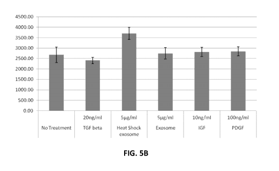
[00022] FIG. 5B is a graph showing the amount of collagen I production in
dermal
fibroblasts after a 48 hour incubation with medium control, various growth
factors, or exosomes
secreted from cells cultured with or without a heat shock step according to
one or more
embodiments of the present disclosure. Values shown on the Y axis are ng/ml of
collagen.
[00023] FIG. 6 is a graph showing quantified RT-qPCR data of the
inflammatory cytokine
IL6 from periodontal ligament fibroblasts (PDLF) after being incubated
overnight with the
following treatments: without HKPG or exosomes (No Tx), with 107/m1 HKPG and
without
exosomes (No Exosomes), or with 107/m1 HKPG in combination with adipose stem
cell-derived
isolated exosomes prepared from cell cultures with a heat shock step (Heat
shock Exosomes) and
without a heat shock step (Std Exosomes), according to one or more embodiments
of the present
disclosure.
6
CA 02993224 2018-01-19
WO 2017/023689 PCT/US2016/044453
[00024] FIG. 7A is an image showing the dorsal surface of a rodent having
four, 2 cm
diameter areas where the dermis was removed, the image taken immediately after
wounding (day
0), according to one or more embodiments of the present disclosure.
[00025] FIG. 7B is an image according to FIG. 7A taken two weeks post
injury (day 14)
where the wound on the lower left was treated with a saline control and the
three remaining
non-control wounds were treated with isolated exosomes secreted from stem
cells cultured with a
heat shock step (HEAT SHOCK) according to one or more embodiments of the
present
disclosure.
[00026] FIG. 7C is an image according to FIG. 7A taken two weeks post
injury (day 14)
where the wound on the lower left was treated with a saline control and the
three remaining
non-control wounds were treated with isolated exosomes secreted from stem
cells cultured with a
heat shock step, lyophilized, and then reconstituted for the treatment (LYO)
according to one or
more embodiments of the present disclosure.
[00027] FIG. 8 is a graph showing the percent wound closure versus the
number of days
post injury for the animals shown in FIG. 7A and FIG. 7B (mean value 3
animals) where percent
wound closure was calculated by dividing the wound diameter on the indicated
days by the
wound diameter at day 1, multiplying by 100, and then subtracting this number
from 100 (stars -
exosome-treated wounds; dots - saline treated control wounds) according to one
or more
embodiments of the present disclosure.
[00028] FIG. 9A is an image of a histological section stained for ki-67
taken from the
wound treated with saline as a control from an animal shown in FIG. 7B
according to one or more
embodiments of the present disclosure.
[00029] FIG. 9B is an image of a histological section stained for ki-67
taken from a wound
treated with isolated exosomes secreted from stem cells cultured with a heat
shock step from an
animal shown in FIG. 7B according to one or more embodiments of the present
disclosure.
[00030] FIG. 10A is an image of a histological section stained with EVG
taken from the
wound treated with saline as a control from an animal shown in FIG. 7B showing
only a small
7
CA 02993224 2018-01-19
WO 2017/023689 PCT/US2016/044453
amount of collagen present that is lacking structural organization (indicated
by arrows; x100
magnification) according to one or more embodiments of the present disclosure.
[00031] FIG. 10B is an image of a histological section stained with EVG
taken from the
wound treated with isolated exosomes secreted from stem cells cultured with a
heat shock step
from an animal shown in FIG. 7B showing a moderate amount of collagen present
with mild
structural organization (indicated by arrows; x100 magnification) according to
one or more
embodiments of the present disclosure.
[00032] FIG. 11 is a graph showing reduction in IL-8 production by human
adult
keratinocytes in the absence of UVB radiation (No UVB) with various amounts of
the heat shock
exosomes compared to a media control (Media Only) according to one or more
embodiments of
the present disclosure.
[00033] FIG. 12 is a graph showing reduction in IL-8 production by human
adult
keratinocytes in the presence of UVB radiation (40 mJ/cm2 UVB) with various
amounts of the
heat shock exosomes compared to a media control (Media Only) according to one
or more
embodiments of the present disclosure.
[00034] FIG. 13 is a graph showing a side-by-side comparison of the data
in the FIG. 11
and FIG. 12 graphs.
[00035] FIG. 14 is a graph showing the amount of TNF-a produced in the
presence of
various concentrations of heat shock exosomes in the presence (40 mJ/cm2 UVB)
and absence
(No UVB) of UVB radiation as compared to a media only control (Media Only)
according to one
or more embodiments of the present disclosure.
[00036] FIG. 15A is an image of a dissected rat Achilles tendon thin-
section which was
histochemically stained to show collagen deposition and collagen fiber
organization and serves
as a contralateral intact control for FIG. 15B according to one or more
embodiments of the
present disclosure.
[00037] FIG. 15B is an image of a dissected rat Achilles tendon thin-
section which was
histochemically stained to show collagen deposition and collagen fiber
organization fourteen
8
CA 02993224 2018-01-19
WO 2017/023689 PCT/US2016/044453
days after collagenase injection into the tendon followed by vehicle injection
three days later
according to one or more embodiments of the present disclosure.
[00038] FIG. 15C is an image of a dissected rat Achilles tendon thin-
section which was
histochemically stained to show collagen deposition and collagen fiber
organization and serves
as a contralateral intact control for FIG. 15D according to one or more
embodiments of the
present disclosure.
[00039] FIG. 15D is an image of a dissected rat Achilles tendon thin-
section which was
histochemically stained to show collagen deposition and collagen fiber
organization fourteen
days after collagenase injection into the tendon followed by heat shock
exosome injection three
days later according to one or more embodiments of the present disclosure.
DETAILED DESCRIPTION
[00040] For the purposes of promoting an understanding of the principles
of the present
disclosure, reference will now be made to preferred embodiments and specific
language will be
used to describe the same. It will nevertheless be understood that no
limitation of the scope of the
disclosure is thereby intended, such alteration and further modifications of
the disclosure as
illustrated herein, being contemplated as would normally occur to one skilled
in the art to which
the disclosure relates.
[00041] There is an unmet need for more effective topical formulations for
regulating skin
condition such as the treatment and prevention of skin damage, wrinkles, and
other defects
including scars, keloids, skin discolorations, and skin abrasions. Another
important and unmet
need remains for more effective formulations to repair soft tissue damage,
including repair of
periodontal tissue, and repair of burns including burns resulting from
radiation treatment. To
solve these unmet needs, the presently disclosed subject matter provides
improved stem
cell-derived exosome compositions, including mesenchymal stem cell (MSC)-
derived exosome
compositions, and methods for their preparation and use, to regulate skin
condition and repair
soft tissue damage.
9
CA 02993224 2018-01-19
WO 2017/023689 PCT/US2016/044453
[00042] Exosomes represent a compelling therapeutic for a range of
indications, especially
those requiring delivery to tissues with reduced vasculature or prominent
necrosis. Exosomes,
unlike stem cells, do not require an oxygenated blood supply to exert their
impact. And, because
exosomes fuse with cell membranes directly, there is no requirement for
receptor mediated
uptake of their pro-healing cargos. Accordingly, the isolated exosomes
produced according to the
methods provided herein can have advantages over existing systemic
pharmaceuticals or direct
application of stem cells for regulating skin condition and repairing soft
tissue damage.
[00043] The improved exosome-containing compositions of the present
disclosure are
based on the context-dependency of the loading of exosomes. More specifically,
the present
disclosure provides methods demonstrating that exosome loading can be
engineered to result in
exosomes having enhanced healing activities, such as and including increased
proliferative and
anti-inflammatory activities. The isolated exosomes of the present disclosure
are prepared from
stem cell cultures in a highly controlled environment, and various stimuli are
delivered to the
stem cell cultures to manipulate the exosomal cargo. In one example of
providing exosomes
engineered for pro-healing activity, stem cell cultures are subjected to high
temperature
(otherwise known as "heat shock") to produce exosomes having increased levels
of heat shock
stress-response molecules, including the stress-response protein, HSP70. It is
demonstrated
herein that the isolated exosomes having increased heat shock stress-response
molecules can
have enhanced healing activity in a rodent model, and can have increased
proliferative and
anti-inflammatory activity in cell cultures.
[00044] The terms "exosomes", "microvesicles", "secreted microvesicles",
"extracellular
vesicles", and "secreted vesicles" are used interchangeably herein for the
purposes of the
specification and claims.
[00045] The terms "freeze drying" and "lyophilization" are used
interchangeably herein
for the purposes of the specification and claims.
[00046] The terms "stress-response molecules" and "heat shock stress-
response molecules"
are used interchangeably herein for the purposes of the specification and
claims. These terms are
CA 02993224 2018-01-19
WO 2017/023689 PCT/US2016/044453
meant to include molecules present in exosomes that are secreted by cultured
stem cells
subjected to high temperature (otherwise known as "heat shock"). Similarly,
the terms
"exosomes" and "heat shock exosomes" and "heat shocked exosomes" are used
interchangeably
herein for the purposes of the specification and claims to represent exosomes
that are secreted by
cultured stem cells subjected to high temperature (otherwise known as "heat
shock").
[00047] The terms "a," "an," and "the" refer to "one or more" when used in
this
application, including the claims.
[00048] Throughout this specification and the claims, the terms
"comprise," "comprises,"
and "comprising" are used in a non-exclusive sense, except where the context
requires otherwise.
Likewise, the term "include" and its grammatical variants are intended to be
non-limiting, such
that recitation of items in a list is not to the exclusion of other like items
that can be substituted
or added to the listed items.
[00049] For the purposes of this specification and claims, the term
"about" when used in
connection with one or more numbers or numerical ranges, should be understood
to refer to all
such numbers, including all numbers in a range and modifies that range by
extending the
boundaries above and below the numerical values set forth. The recitation of
numerical ranges
by endpoints includes all numbers, e.g., whole integers, including fractions
thereof, subsumed
within that range. For example, the recitation of about 41 to about 43
includes 41, 42, and 43, as
well as fractions thereof, for example, but not limited to, 40.5, 40.6, 40.7,
40.8, 40.9, 41.5, 42.25,
42.5, 43.1, 43.2, 43.3, 43.4, 43.5 and the like, and the recitation of 1 to 3
includes 1, 2, and 3, as
well as fractions thereof, for example, but not limited to, 0.6, 0.7, 0.8,
0.9, 1.5, 2.25, 3.5, and the
like and any range within that range.
[00050] In one embodiment of the present disclosure a composition is
provided, the
composition including isolated stem cell exosomes having increased levels of
heat shock
stress-response molecules, wherein the stem cell exosomes are produced by a
process including:
(a) culturing stem cells in a culture medium, wherein the culturing includes a
step of heat
shocking the stem cells in a serum-free culture media by increasing the
culture temperature to
11
CA 02993224 2018-01-19
WO 2017/023689 PCT/US2016/044453
about 41 C to about 43 C for about 1 hour to about 3 hours; and (b) isolating
the exosomes
having increased levels of heat shock stress-response molecules from the serum-
free culture
medium. The composition can further include a carrier. The carrier can be a
pharmaceutically
acceptable carrier.
[00051] The composition can be in the form of a liquid, lotion, cream,
gel, foam, mousse,
spray, paste, powder, or solid.
[00052] In the composition, isolating the exosomes can be carried out by
one or more
centrifugation steps. The one or more centrifugation steps can include
centrifugation at 100,000X
g or greater. In the composition, isolating the exosomes can further include
freeze drying the
isolated exosomes. In the composition, the process can further comprise
culturing the stem cells
in the serum-free culture medium at a temperature of about 36 C to 38 C for
about 24hr to about
72hr subsequent to the step of heat shocking. The serum-free medium can be
free of animal
products. The stem cells can be mesenchymal stem cells. The mesenchymal stem
cells can be of
placental or adipose origin. The stress-response molecules can include HSP70.
[00053] In one embodiment, a method is provided for making stem cell
exosomes having
increased levels of heat shock stress-response molecules, the method
including: culturing stem
cells in a culture medium, wherein the culturing includes a step of heat
shocking the stem cells in
a serum-free culture media by increasing the culture temperature to about 41 C
to about 43 C for
about 1 hour to about 3 hours, and wherein the serum-free culture medium
contains the
exosomes having the increased levels of heat shock stress-response molecules.
[00054] The method can further include isolating the exosomes from the
serum-free
culture medium. The isolating can be carried out by one or more centrifugation
steps. The one or
more centrifugation steps can include centrifugation at 100,000 X g or
greater.
[00055] The method can further include freeze drying the isolated
exosomes, such that the
exosomes can be stored at room temperature.
[00056] The method can further include culturing the stem cells in the
serum-free culture
medium at a temperature of about 36 C to 38 C for about 24hr to about 72hr
subsequent to the
12
CA 02993224 2018-01-19
WO 2017/023689 PCT/US2016/044453
step of heat shocking.
[00057] In the method, the serum-free medium can be free of animal
products. The stem
cells can be mesenchymal stem cells. The mesenchymal stem cells can be of
placental or adipose
origin. The stress-response molecules can include HSP70.
[00058] Characterization of the size and shape of the isolated exosomes
produced
according to the methods of the present disclosure is described in Example 3
and the results are
shown in the graph in FIG. 1. FIG. 1 shows the size distribution of a
representative sample of
isolated exosomes with mean of 152 nm and a mode of 107 nm. A scanning
electron microscopy
(SEM) micrograph from another isolated exosome preparation according to one or
more
embodiments of the present disclosure is shown in the inset for FIG. 1.
[00059] Example 4 describes analysis of the isolated exosomes produced
according the
methods of the present disclosure for specific protein markers including
Hsp70. The resulting
data are shown in FIG. 2. FIG. 2 is a bar graph of quantified Western Blot
data showing the
amount of HSP70 relative to 13-actin in the exosomes secreted by stem cells
cultured at 37 C
without a heat shock step (Control) and exosomes from the same stem cells
subjected to a 2 hr
heat shock step at 43 C (Heat Shock). The data in FIG. 2 indicate that there
is a significant
up-regulation in exosomal HSP70 relative to 13-actin in the exosomes from the
heat shocked cells
as compared to the exosomes from the cells cultured without the heat shock
step.
[00060] The capability of the isolated exosomes prepared according to the
methods of the
present disclosure to deliver cargo to cells was assessed by monitoring the
ability of the isolated
exosomes to transfer a lipophilic dye to cells in culture. The experiment is
described in Example
and the results are shown in FIG. 3. The results indicate an efficient
transfer of the dye from
the isolated exosomes to the Human pulmonary artery endothelial (HPAE) cells
with 75% of the
cells being labeled.
[00061] The effects of the isolated exosomes produced according to the
methods of the
present disclosure on cultured periodontal and dermal cells are described in
Example 6. FIG' s
4A and 4B show that treatment with the isolated exosomes from the heat shocked
cells
13
CA 02993224 2018-01-19
WO 2017/023689 PCT/US2016/044453
significantly increased proliferation of both periodontal ligament fibroblasts
(PDLFs) and dermal
fibroblasts (DFs), as compared to the isolated exosomes prepared from cells
that were not
subjected to a heat shock step. In addition, the level of proliferation of the
PDLFs and DFs
induced by the isolated exosomes from the heat shocked cells approached or
surpassed that
induced by complete medium and the individual growth factors.
[00062] In addition to degradation of collagen fiber and the extracellular
matrix associated
with skin aging and its relationship to wound repair, periodontal disease is
associated with
degradation of the extracellular matrix and collagen fiber degeneration.
Additional experiments
described in Example 6 demonstrate that the isolated exosomes prepared from
heat shocked cells
according to the methods of the present disclosure can induce collagen I
synthesis in PDLFs and
DFs. The graphs in FIG. 5A and 5B show that treatment with the isolated
exosomes from the
heat shocked cells increased collagen I production of both PDLFs and DFs, as
compared to the
isolated exosomes prepared from cells that were not subjected to a heat shock
step. In addition,
the increase in collagen I production of the PDLFs and DFs induced by the
isolated exosomes
from the heat shocked cells surpassed that of the individual growth factors.
These data indicate
that the isolated exosomes can have a role in regulating skin condition and
repair of soft tissue
damage.
[00063] P. gingivalis is one of the bacterial species known to contribute
to periodontitis
pathogenesis by secreting various toxins lethal to oral soft tissue cells.
Previous reports indicate
the induction of inflammatory cascades in gingival keratinocytes (GKs) and
PDLFs in response
to P. gingivalis lysates, including the inflammatory molecules IL6 and IL8 [22-
24]. In the
experiment described in Example 7, PDLF cells were concomitantly exposed to
lyophilized heat
killed P. gingivalis (HKPG, 107/m1) and the isolated exosomes from medium from
heat shocked
cell cultures according to the methods of the present disclosure. The results
indicate a
statistically significant elevation in IL-6 gene expression in HPLF cells
induced by heat-killed P.
gingivalis (HKPG) at lx10^7 /ml. The elevation is significantly reduced by the
isolated standard
exosomes, and even more so by the isolated exosomes secreted from cultured
cells with a heat
14
CA 02993224 2018-01-19
WO 2017/023689 PCT/US2016/044453
shock step. These data indicate that the isolated heat shock exosomes of the
present disclosure
can inhibit the production of inflammatory cytokines including IL6 that act
locally to recruit
monocytes to the site of inflammation.
[00064] Example 8 describes an experiment showing that the MSC-derived
isolated
exosomes produced according to the methods of the present disclosure can
improve skin wound
healing in rodents. FIG. 7A through 7C are images of the dorsal surface of a
rodent with four
separate wounds and show the increased rate of healing provided by the exosome
compositions
of the present disclosure. Images of the rodent were taken immediately after
wounding (FIG. 7A)
and two weeks post injury (FIG. 7B-7C). The wound on the lower left in each
image was treated
with a saline control. For the image shown in FIG. 7B, the three remaining non-
control wounds
were treated with isolated exosomes secreted from stem cells cultured with a
heat shock step
according to the methods of the present disclosure. For the image shown in
FIG. 7C, the three
remaining non-control wounds were treated with exosomes secreted from stem
cells that were
isolated, lyophilized, and then reconstituted for the treatment. The images
taken after 2 weeks
show that both the control and exosome-treated wounds are substantially
healed, but the wounds
treated with the MSC-derived isolated heat shock exosomes produced according
to the methods
of the present disclosure healed substantially faster. FIG. 8 shows a graph of
the percent wound
closure versus the number of days post injury for the animals shown in FIG. 7A
and FIG. 7B. In
FIG. 8, the line designated with stars represents the exosome-treated wounds,
which were
completely closed at the end of the time course of 19 days post-treatment, and
the saline treated
control wounds are represented by the dotted line. The control wounds remained
open at the end
of the time course, with approximately 25% of the wound surface area
remaining.
[00065] In addition, sections were taken from the animals shown in FIG. 7B
and
histologically stained for markers known to be involved in cell proliferation
and wound healing.
Specifically, the sections were histologically stained for ki-67, a protein
indicating cell
proliferation. The results are shown in FIG. 9A (Control 1) and FIG. 9B (Test
1). The section
shown in FIG. 9A was taken from the wound treated with saline as a control and
the section
CA 02993224 2018-01-19
WO 2017/023689 PCT/US2016/044453
taken from FIG. 9B was taken from the wound treated with the isolated exosomes
secreted from
stem cells cultured with a heat shock step as described herein. The section
shown in FIG. 9B is
darker compared to the saline control shown in FIG. 9A. This darker staining
of the ki-67 protein
indicates that cells are proliferating more in the wound treated with the heat
shock exosomes
than in the control. Increased proliferation is key to wound healing, and is
one possible
explanation for the reduced time to closure in the exosome treated wound.
[00066] In addition to the sections stained for ki-67 protein, sections
taken from the animals
shown in FIG. 7B were also analyzed for collagen deposition and organization
by staining with
EVG and the results are shown in FIG. 10A (Control) and FIG. 10B (Test). The
section shown in
FIG. 10A taken from the saline control shows weak staining by EVG and only a
small amount of
collagen present that is lacking structural organization (indicated by
arrows). In contrast, the
section shown in FIG. 10B taken from the wound treated with the heat shock
exosomes shows an
increase in staining of collagen bundles by EVG revealing a moderate amount of
collagen
present with mild structural organization (indicated by arrows). Greater
collagen deposition and
organization in the heat shock exosome treated skin wound indicates improved
and faster healing.
These data support the gross observation that the wounds closed more rapidly
in the heat shock
exosome-treated samples, and indicate a possible molecular mechanism for the
improved healing.
[00067] Example 9 describes the protective effect of MSC-derived isolated
exosomes
produced according to the present disclosure against UVB radiation on human
adult
keratinocytes. In this study, keratinocytes were incubated with media
containing the heat shock
exosomes for 1 hour and the keratinocytes were subsequently exposed to 40
mJ/cm2 UVB
radiation. Control cells underwent the same protocol with the exception of UVB
exposure. The
effects of the heat shock exosomes were assessed by measuring reduced
production of IL-8 and
TNF-a by the cells, and the results are shown in FIG. 11, FIG. 12, FIG. 13,
and FIG. 14.
Specifically, FIG. 11 is a graph showing reduction in IL-8 production by the
human adult
keratinocytes in the absence of UVB radiation (No UVB) with various amounts of
the heat shock
exosomes compared to a media control. The results show that the heat shock
exosomes at all
16
CA 02993224 2018-01-19
WO 2017/023689 PCT/US2016/044453
concentrations tested reduced the production of IL-8. In addition, a
concentration of 8.23E+05
heat shock exosomes significantly reduced IL-8 production.
[00068] FIG. 12 is a graph showing reduction in IL-8 production by human
adult
keratinocytes in the presence of UVB radiation (40 mJ/cm2 UVB) with various
amounts of the
heat shock exosomes compared to a media control. The results were similar to
the experiment in
the absence of UVB where heat shock exosomes at all concentrations tested,
with the exception
of 2.00E+08, reduced the production of IL-8.
[00069] FIG. 13 is a graph showing a side-by-side comparison of the data
in the FIG. 11
and FIG. 12 graphs. The comparison shows that both UVB (40 mJ/cm2 UVB) and Non-
UVB
(No UVB) exposed samples follow the same trend. Exosome concentrations of
8.23E+05,
2.47E+06, 7.41E+06, and 2.22E+07 exosomes /mL in the UVB exposed cells
resulted in no
significant difference compared to cells in the No UVB media control. These
results demonstrate
the protective effect of heat shock exosomes against UVB induced inflammation.
[00070] FIG. 14 is a graph showing the amount of TNF-a produced in the
presence of
various concentrations of the heat shock exosomes in the presence (40 mJ/cm2
UVB) and
absence (No UVB) of UVB radiation as compared to a media only control (Media
Only). The
data shown that TNF-a release in the UVB exposed samples follow the same trend
as observed
for IL-8, with a maximum decrease in TNF-a of about 3-fold with heat shock
exosomes at a
concentration of 2.22E+07 exosomes /mL. The values of TNF-a are at the lower
limit of the
assay detection, which is why no data are shown for a majority of the No UVB
samples.
[00071] In summary, the results indicate that IL-8 release from UVB
exposed cells treated
with heat shock exosome concentrations of 8.23E+05, 2.47E+06, 7.41E+06, and
2.22E+07
exosomes /mL showed no significant difference compared to the No UVB media
control, which
shows the protective effect of the heat shock exosomes against UVB induced
inflammation. In
addition, IL-8 release was significantly reduced from cells treated with the
heat shock exosomes
at concentration of 8.23+E05 exosomes /mL in the absence of UVB radiation as
compared to the
No UVB media control. Further, TNF-a release from cells treated with the heat
shock exosomes
17
CA 02993224 2018-01-19
WO 2017/023689 PCT/US2016/044453
was decreased as much as 3-fold. The above results demonstrate the protective
effect of heat
shock exosomes against UVB induced inflammation.
[00072] Example 10 describes the soft tissue healing activity of MSC-
derived isolated
exosomes produced according to the methods described herein in a rodent model
of tendon
healing. Specifically, tendon injury was induced by injecting collagenase into
the right Achilles
tendon and the left tendons were left intact. Three days post-injury, exosomes
or vehicle control
were injected in the injury site. Fourteen days post-injury, the Achilles
tendons were removed
and stained for collagen content and collagen bundle orientation. FIG. 15B
shows the effect of
collagenase treatment in the absence of exosomes compared to the contralateral
intact control
tendon (FIG. 15A). Collagenase induces severe collagen degeneration and loss
of oriented
collagen bundles as shown by loss of dark and striated staining in FIG. 15B.
Exosome treatment
of the collagenase-injected tendon (FIG. 15D) shows no significant difference
in collagen
content or collagen fiber orientation from the contralateral intact control
tendon (FIG. 15C).
These data show that MSC-derived isolated heat shock exosomes greatly reduce
or inhibit
collagen degeneration and/or promote soft tissue and tendon healing after an
induced tendon
injury.
[00073] In one embodiment, a method is provided for treating
periodontitis, the method
including one or more of putting on, embedding into, or filling an area of the
gum in the mouth
of a living animal a composition including: isolated stem cell exosomes having
increased levels
of heat shock stress-response molecules, wherein the stem cell exosomes are
produced by a
process including: (a) culturing stem cells in a culture medium, wherein the
culturing includes a
step of heat shocking the stem cells in a serum-free culture media by
increasing the culture
temperature to about 41 C to about 43 C for about 1 hour to about 3 hours; and
(b) isolating the
exosomes having increased levels of heat shock stress-response molecules from
the serum-free
culture medium, wherein the periodontitis on the area of the gum is treated.
The composition can
further include a carrier. The carrier can be a pharmaceutically acceptable
carrier.
[00074] In one embodiment, a method is provided for repair of a soft
tissue in a living
18
CA 02993224 2018-01-19
WO 2017/023689 PCT/US2016/044453
body, the method comprising one of putting on, embedding into, and filling a
soft tissue wound
area of a living body a composition including isolated stem cell exosomes
having increased
levels of heat shock stress-response molecules, wherein the stem cell exosomes
are produced by
a process including: (a) culturing stem cells in a culture medium, wherein the
culturing includes
a step of heat shocking the stem cells in a serum-free culture media by
increasing the culture
temperature to about 41 C to about 43 C for about 1 hour to about 3 hours; and
(b) isolating the
exosomes having increased levels of heat shock stress-response molecules from
the serum-free
culture medium, wherein the wound area of the living body is repaired. The
composition can
further include a carrier. The carrier can be a pharmaceutically acceptable
carrier.
[00075] In one embodiment, a method is provided for treating a skin
condition, the
method including one or more of putting on, embedding into, or filling an area
on the skin of a
living body a composition of the present disclosure including isolated stem
cell exosomes having
increased levels of heat shock stress-response molecules, wherein the
condition of the skin is
treated.
[00076] In one embodiment, a method is provided for treating a skin
condition, the
method comprising one or more of: putting on, embedding into, or filling an
area on the skin of a
living body a composition comprising isolated stem cell exosomes having
increased levels of
heat shock stress-response molecules, wherein the stem cell exosomes are
produced by a process
comprising: (a) culturing stem cells in a culture medium, wherein the
culturing includes a step of
heat shocking the stem cells in a serum-free culture media by increasing the
culture temperature
to about 41 C to about 43 C for about 1 hour to about 3 hours; and (b)
isolating the exosomes
having increased levels of heat shock stress-response molecules from the serum-
free culture
medium, wherein the condition of the area of the skin is treated by the
putting on, embedding
into, or filling of the area with the composition. The composition can further
include a carrier.
The carrier can be a pharmaceutically acceptable carrier.
[00077] The skin condition can include, for example, one or more of a
wound, a burn, a
burn resulting from radiation treatment, a discoloration, a scar, and a
keloid.
19
CA 02993224 2018-01-19
WO 2017/023689 PCT/US2016/044453
[00078] The composition can be in the form of a liquid, lotion, cream,
gel, foam, mousse,
spray, paste, powder, or solid.
[00079] In the composition, isolating the exosomes can be carried out by
one or more
centrifugation steps. The one or more centrifugation steps can include
centrifugation at 100,000
X g or greater. In the composition, isolating the exosomes can further include
freeze drying the
isolated exosomes. In the composition, the process can further comprise
culturing the stem cells
in the serum-free culture medium at a temperature of about 36 C to 38 C for
about 24hr to about
72hr subsequent to the step of heat shocking. The serum-free medium can be
free of animal
products. The stem cells can be mesenchymal stem cells. The mesenchymal stem
cells can be of
placental or adipose origin.
[00080] In one embodiment, a topical composition is provided for
regulating skin
condition, the composition comprising an effective amount of isolated exosomes
having
increased levels of heat shock stress-response molecules and a carrier.
[00081] In one embodiment a topical composition is provided for regulating
skin
condition, the composition including: i) an effective amount of isolated
exosomes having
increased levels of heat shock stress-response molecules; and ii) a carrier,
wherein the isolated
exosomes are isolated from a serum-free culture medium conditioned by
culturing stem cells
under conditions that include a heat shock of the stem cells in the serum-free
culture medium at a
temperature of about 41 C to about 43 C for about 1 hour to about 3 hours.
[00082] In one embodiment, a method is provided for making a topical
composition for
regulating skin condition, the method including: combining isolated exosomes
having increased
levels of heat shock stress-response molecules with a carrier, wherein the
exosomes are isolated
from a serum-free culture medium conditioned by culturing stem cells under
conditions
including a heat shock of the stem cells at a temperature of about 41 C to
about 43 C for about 1
hour to about 3 hours.
[00083] The compositions provided for regulating skin condition can be in
the form of a
liquid, lotion, cream, gel, foam, mousse, spray, paste, powder, or solid.
CA 02993224 2018-01-19
WO 2017/023689 PCT/US2016/044453
[00084] In the compositions provided for regulating skin condition,
regulating skin
condition can include one or more of inducing increased skin integrity by cell
renewal;
enhancing water content or moisture of skin; reducing trans epidermal water
loss, skin flaking,
and scaling; improving skin thickness; enhancing skin tensile properties;
reducing the
appearance of dermal fine lines and wrinkles; improving skin texture; reducing
skin pores size;
enhancing skin smoothness; improving skin age spots; improving skin tone; or
improving the
appearance of scars and skin abrasions.
[00085] In the compositions provided for regulating skin condition, the
composition can
further include from about 0.1 to about 20% of a moisturizing agent. The
moisturizing agent can
include one or more of panthenol, pantothenic acid derivatives, glycerin,
glycerol, dimethicone,
petrolatum, hyaluronic acid, or ceremides, and mixtures thereof.
[00086] In the compositions provided for regulating skin condition, the
composition can
further include a vitamin B3 compound. The vitamin B3 compound can include
tocopherol
nicotinate.
[00087] In the compositions provided for regulating skin condition, the
composition can
further include an anti-oxidant. The anti-oxidant can include one or a
combination of tocopherol
or esters of tocopherol.
[00088] In the compositions provided for regulating skin condition, the
isolated exosomes
can be freeze dried.
[00089] In one embodiment, a method is provided for regulating a human
skin condition
which includes applying to human skin at least once a day over at least seven
days a topical
composition according to the present disclosure comprising isolated exosomes
having increased
levels of heat shock stress-response molecules. In one embodiment, a method is
provided for
regulating a human skin condition which includes applying to human skin at
least once a day over
at least seven days a topical composition according to the present disclosure
comprising isolated
exosomes having increased levels of heat shock stress-response molecules. The
method can
further include applying the topical composition according to the present
disclosure to human skin
21
CA 02993224 2018-01-19
WO 2017/023689 PCT/US2016/044453
at least twice a day over at least fourteen days.
[00090] In one embodiment, a coating composition is provided for
conditioning skin or
hair, the coating composition including: i) isolated stem cell exosomes having
increased levels of
heat shock stress-response molecules; and ii) a carrier, wherein the stem cell
exosomes are
produced by a process including: (a) culturing stem cells in culture medium,
wherein the
culturing includes a step of heat shocking the stem cells in a serum-free
culture medium by
increasing the culture temperature to about 41 C to about 43 C for about 1
hour to about 3 hours,
and wherein the serum-free culture medium contains the exosomes having the
increased levels of
heat shock stress-response molecules; and (b) isolating the exosomes having
increased levels of
heat shock stress-response molecules from the serum-free medium.
[00091] In the coating compositions for conditioning skin or hair of the
present disclosure,
the process for producing the isolated stem cell exosomes can further include
freeze drying the
isolated exosomes.
[00092] In the coating compositions for conditioning skin or hair of the
present disclosure,
the process for producing the isolated stem cell exosomes can further include
freeze drying the
isolated exosomes and the carrier can be a dry powder.
[00093] The coating compositions for conditioning skin or hair of the
present disclosure
can be a dry powder coating composition applied to the inside of a glove.
[00094] The coating compositions for conditioning skin or hair of the
present disclosure
can be in the form of a liquid, lotion, cream, gel, foam, mousse, spray,
paste, powder, or solid.
[00095] In one embodiment, a glove is provided for conditioning the skin,
the glove
having a coating composition on the inside thereof, the coating composition
including: i) isolated
stem cell exosomes having increased levels of heat shock stress-response
molecules; and ii) a
powder carrier, wherein the isolated stem cell exosomes are produced by a
process including: (a)
culturing stem cells in culture medium, wherein the culturing includes a step
of heat shocking the
stem cells in a serum-free culture medium by increasing the culture
temperature to about 41 C to
about 43 C for about 1 hour to about 3 hours, and wherein the serum-free
culture medium
22
CA 02993224 2018-01-19
WO 2017/023689 PCT/US2016/044453
contains the exosomes having the increased levels of heat shock stress-
response molecules; (b)
isolating the exosomes having increased levels of heat shock stress-response
molecules from the
serum-free medium; and (c) freeze drying the isolated exosomes.
EXAMPLES
[00096] The following Examples have been included to provide guidance to
one of
ordinary skill in the art for practicing representative embodiments of the
presently disclosed
subject matter. In light of the present disclosure and the general level of
skill in the art, those of
skill can appreciate that the following Examples are intended to be exemplary
only and that
numerous changes, modifications, and alterations can be employed without
departing from the
scope of the presently disclosed subject matter.
Example 1
Preparation of Exosomes with Increased Levels of Heat shock Stress-Response
Molecules
using Heat Shock
[00097] The following experiments describe the production of isolated
exosomes having
increased levels of stress-response molecules to provide enhanced
proliferative and
anti-inflammatory activity.
[00098] Mesenchymal stem cells (placental or adipose origin) were cultured
in a hollow
fiber cartridge bioreactor (FIBERCELL BIOSYSTEMS) to produce exosomes having
increased
levels of heat shock stress-response molecules as follows. Prior to seeding,
the bioreactor was
conditioned with complete culture medium (DMEM/F12 containing 10% FBS) for 24
hr at 37 C
in a humidified, 5% CO2 containing atmosphere. The bioreactor was seeded with
300 x 106
mesenchymal stem cells (placental or adipose origin) and maintained at 37 C in
a humidified, 5%
CO2 containing atmosphere. Cells were grown for 2 weeks before beginning
exosome harvest.
Prior to harvesting exosome-containing medium, the bioreactor was washed 5
times with
serum-free DMEM/F12 to remove bovine exosomes. After washing, the cells were
subjected to a
23
CA 02993224 2018-01-19
WO 2017/023689 PCT/US2016/044453
heat shock step as follows. The medium in the bioreactor was replaced with
serum-free
DMEM/F12 medium warmed to 41 C, and the bioreactor was placed in a 41 C,
humidified, 5%
CO2 containing atmosphere for 1 hr. Next the 41 C medium was replaced with the
same medium
warmed to 37 C, and the bioreactor was placed in a 37 C, humidified, 5% CO2
containing
atmosphere for 48 hr. After the 48 hr incubation, the conditioned serum-free
DMEM/F12 medium
was recovered, and in some instances, stored at -80 C for future processing.
[00099] In separate preparations of isolated exosomes having increased
levels of heat
shock stress-response molecules, the same procedure as described above was
followed except
that the temperature of the medium used in the heat shock step was about 42 C
in some
preparations and about 43 C in other preparations and the time period of heat
shock ranged from
as short as about 1 hour to as long as about 3 hours.
[000100] After thawing or fresh collection of the conditioned medium
described above, the
exosomes were isolated from the conditioned media by centrifugation of the
medium at 3000 xg
for 20 min at room temperature to pellet cell debris (in 50, 250, or 500 mL
screw cap vessels). The
clarified supernatant was collected and centrifuged at 100,000 xg (Avg. RCF)
for 2 hrs at 4 C. The
supernatant was aspirated and the pellet(s) resuspended in minimum volume of
DPBS (300-1000
i.t.L). Manufacturer's instructions were followed to estimate protein and RNA
concentration using
a NANODROP (THERMO FISHER, Corp) spectrophotometer. The number of particles
(exosomes) per mL and the particle (exosome) size were determined using the
QNANO (IZON
SCIENCE, Ltd) following manufacturer's instructions. The isolated exosomes
were aliquoted into
appropriate volumes into 1.5 mL screw cap tubes.
[000101] It was discovered that the isolated exosomes described above could
be stored at
-80 C and then thawed at a later date for use without a detectable decrease in
activity for. It was
also discovered that the isolated exosomes could be could be freeze dried and
stored at room
temperature without a detectable decrease in activity.
24
CA 02993224 2018-01-19
WO 2017/023689 PCT/US2016/044453
Example 2
Preparation of Exosomes with Enhanced Proliferative and Anti-Inflammatory
Activity
using Medium Supplementation
[000102] The following experiments describe the production of isolated
exosomes having
increased proliferative and anti-inflammatory activity.
[000103] Mesenchymal stem cells (placental or adipose origin) are cultured
in a bioreactor
to produce exosomes having increased proliferative and anti-inflammatory
activity according to
the procedure described above in Example 1 with the following exceptions.
After the 2 week
period of cell growth, the bioreactor is washed multiple times with serum-free
DMEM/F12 to
remove bovine exosomes. After washing, the medium in the bioreactor is
replaced with serum-free
DMEM/F12 medium supplemented with one or a combination of platelet lysate,
human platelet
lysate, PDGF-BB, TGF-03, TGF-01, or other pro- and anti-inflammatory cytokines
and the
bioreactor is placed in a 37 C, humidified, 5% CO2 containing atmosphere for
48 hr. After the 48
hr incubation, the conditioned serum-free, supplemented DMEM/F12 medium is
recovered and in
some instances stored at -80 C for future processing.
[000104] After thawing or fresh collection of the conditioned medium
described above, the
exosomes are isolated from the conditioned media and stored for future use as
described in
Example 1.
Example 3
Size Characterization of Heat Shock Isolated Exosomes
[000105] The isolated exosomes produced according to Example 1 were
characterized as
described in the following experiments.
[000106] To determine the size of the stem cell-derived exosomes produced
according to
Example 1, the isolated exosomes were analyzed using the QNANO (IZON SCIENCE,
Ltd)
following manufacturer's instructions. The graph in FIG. 1 shows the resulting
size distribution
of a representative sample with mean of 152 nm and a mode of 107 nm. An
exosome sample
CA 02993224 2018-01-19
WO 2017/023689 PCT/US2016/044453
taken from a separate exosome preparation was analyzed by scanning electron
microscopy
(MARBLE LABORATORIES) to determine the relative size and shape of the exosome
particles.
Exosomes were prepared for SEM by drying on mounting studs, coated with
platinum, and
visualized by SEM (see FIG. 1 inset). While the resulting particle size
calculated by SEM was
larger than that determined by the QNANO, the difference is likely due to SEM
preparation and
drying artifacts rather than a significant size variation in the exosome
preparations.
Example 4
HSP70 is Up-Regulated in Isolated Heat Shock Exosomes
[000107] As part of the characterization process, the exosomes prepared
according to
Example 1 were analyzed by Western blot analysis for specific protein markers
including CD63,
Hsp70 and TSG101. Specifically, exosomes produced by cells at both normal
culture
temperature (37 C) (i.e., without a heat shock step) and exosomes produced by
cells at culture
conditions that include culturing the cells at 43 C for 2 hours according to
Example 1 were
examined by Western Blot analyses for the presence of stress-response proteins
including HSP70.
FIG. 2 is a bar graph of the quantified Western Blot data that shows the
amount of HSP70
protein relative to 13-actin protein in two separate preparations of exosomes:
1) secreted by cells
cultured at 37 C without a heat shock step (Control; blank and hatched bars
represent the separate
preparations); and 2) secreted by cells subjected to a 2 hr heat shock step at
43 C as described in
Example 1 (Heat Shock; blank and hatched bars represent the separate
preparations). The data in
FIG. 2 indicate that there is a significant up-regulation in exosomal HSP70
relative to 13-actin in
the heat shock exosomes as compared to the exosomes from the cells cultured
without the heat
shock step.
Example 5
Dye Transfer of Isolated Heat Shock Exosomes to HPAE Cells
[000108] The capability of the isolated exosomes prepared according to
Example 1 to
26
CA 02993224 2018-01-19
WO 2017/023689 PCT/US2016/044453
deliver cargo to cells was assessed by monitoring the ability of the isolated
exosomes to transfer
a lipophilic dye to cells in culture. The experiments were performed as
described below.
[000109] An aliquot of isolated exosomes produced according to Example 1
was labeled
with VYBRANT DII (LIFE TECHNOLOGIES) cell labeling solution for 20 minutes at
37 C
and at 4 C and the dye transfer was assessed at each temperature using flow
cytometry [25]. FIG.
3 shows histograms of the data from the cells incubated at 4 C (left-most
histogram) and those
incubated at 37 C (right-most histogram) with dye-loaded exosomes. The results
indicate an
efficient transfer of the dye from the exosomes to the human pulmonary artery
endothelial
(HPAE) cells with 75% of the cells being labeled.
Example 6
Effects of Isolated Heat Shock Exosomes on Cultured Periodontal Cells
[000110] The effects of the isolated exosomes produced according to Example
1 on
cultured periodontal cells were determined as described below.
[000111] Adipose-derived stem cell isolated exosomes produced by cells at
both normal
culture temperature (37 C) (i.e., without a heat shock step) and isolated
exosomes produced by
cells at culture conditions that included culturing the cells with a heat
shock step according to
Example 1 were added to low density periodontal ligament fibroblasts (PDLFs)
and dermal
fibroblasts (DFs) (3,000 cells/well) in 96-well culture plates in serum free
medium and incubated
for 3 days. To compare the proliferative effects of the isolated exosomes, the
cells were also
treated with other inducers, including 10% FBS, PDGF, TGF-01, or IGF-1. After
3 days, the
cells were treated with CELL TITER BLUE REAGENT (PROMEGA) for 2 hours to
assess
proliferation. The data are shown in FIG. 4A (PDLFs) and 4B (DFs). FIG. 4A and
4B show that
treatment with the isolated exosomes from the heat shocked cells significantly
increased
proliferation of both PDLFs and DFs, as compared to the isolated exosomes
prepared from cells
that were not subjected to a heat shock step. In addition, the level of
proliferation of the PDLFs
and DFs induced by the isolated exosomes from the heat shocked cells
approached or surpassed
27
CA 02993224 2018-01-19
WO 2017/023689 PCT/US2016/044453
that induced by complete medium and the individual growth factors.
[000112] Periodontal disease is associated with degradation of the
extracellular matrix and
collagen fiber degeneration. The following experiments were performed to
determine if the
isolated exosomes prepared from heat shocked cells could induce collagen I
synthesis in PDLFs
and DFs. For the experiment, isolated exosomes produced by MSC's at both
normal culture
temperature (37 C) (i.e., without a heat shock step) and isolated exosomes
produced by MSC's
at culture conditions that included culturing the cells with a heat shock step
according to
Example 1 were tested along with serum-free conditioned medium from vehicle
and growth
factors using a procollagen I C-peptide ELISA (TAKARA) assay. PDLF cells were
treated for
48 hours with the media control (No Treatment), 2Ong/m1 TG93-1, lOng/m1 IGF,
10Ong/m1
PDGF, or the isolated exosomes. After the 48 hrs, the conditioned medium was
removed,
clarified by centrifugation, and diluted into the ELISA assay. The resulting
data are shown in
FIG. 5A (PDLFs) and FIG. 5B (DFs). The graphs in FIG. 5A and 5B show that
treatment with
the isolated exosomes from the heat shocked cells increased collagen I
production of both PDLFs
and DFs, as compared to the isolated exosomes prepared from cells that were
not subjected to a
heat shock step. In addition, the increase in collagen I production of the
PDLFs and DFs induced
by the isolated exosomes from the heat shocked cells surpassed that of the
individual growth
factors. These data indicate that the isolated exosomes can have a role in
periodontal ligament
repair.
Example 7
ASC-Derived Heat Shock Exosomes Inhibit Expression of Inflammatory Cytokines
[000113] The potential of ASC-derived isolated exosomes produced according
to Example
1 to inhibit IL6 expression in periodontal ligament fibroblasts (PDLFs) was
examined as
described below.
[000114] P. gin givalis is one of the bacterial species known to contribute
to periodontitis
pathogenesis by secreting various toxins lethal to oral soft tissue cells.
Previous reports indicate
28
CA 02993224 2018-01-19
WO 2017/023689 PCT/US2016/044453
the induction of inflammatory cascades in GKs and PDLFs in response to P. gin
givalis lysates,
including the inflammatory molecules IL6 and IL8 [22-24]. To evaluate the anti-
inflammatory
impact of treatment with the isolated exosomes prepared from cells cultured
with a heat shock
step, PDLF cells were concomitantly exposed to lyophilized heat killed P. gin
givalis (HKPG,
107/m1) and the isolated exosomes from medium from heat shocked cell cultures.
[000115] For the experiment, isolated exosomes produced by ASC's at both
normal culture
temperature (37 C) (i.e., without a heat shock step) and isolated exosomes
produced by ASC's at
culture conditions that included culturing the cells with a heat shock step
according to Example 1
were tested. Specifically, to measure inflammatory response, RT-qPCR for the
inflammatory
cytokine IL6 mRNA was performed. PDLFs were seeded in 6-well plates and
incubated
overnight: without HKPG and without exosomes (No Tx), with 107/m1 HKPG and
without
exosomes (No Exosomes), with 107/m1 HKPG in combination with adipose stem cell-
derived
isolated exosomes prepared from cell cultures with a heat shock step (Heat
shocked Exosomes)
and without a heat shock step (Std Exosomes). The quantified RT-qPCR data are
shown in the
graph in FIG. 6. The results indicate a statistically significant elevation in
IL-6 gene expression in
HPLF cells induced by heat-killed P. gingivalis (HKPG) at lx10^7 /ml. The
elevation is
significantly reduced by the isolated standard exosomes, and even more so by
the isolated cell
exosomes produced with a heat shock step. These data indicate that the
isolated exosomes of the
present disclosure can inhibit the production of inflammatory cytokines
including IL6 that act
locally to recruit monocytes to the site of inflammation.
Example 8
Effect of Isolated Heat ShockExosomes on Wound Healing in Rodents
[000116] The skin wound healing activity of MSC-derived isolated exosomes
produced
according to Example 1 was examined in a rodent model as described below.
[000117] The skin wound healing experiment with the isolated exosomes was
performed as
follows. Four, 2 cm diameter areas of dermis were completely removed from the
dorsal surface
29
CA 02993224 2018-01-19
WO 2017/023689 PCT/US2016/044453
of a rodent to create four separate wounds. Images of the rodent were taken
immediately after
wounding (FIG. 7A) and two weeks post injury FIG. 7B and FIG. 7C. The wound on
the lower
left in each image was treated with a saline control. For the image shown in
FIG. 7B, the three
remaining non-control wounds were treated with isolated exosomes secreted from
stem cells
cultured with a heat shock step. For the image shown in FIG. 7C, the three
remaining
non-control wounds were treated with exosomes secreted from stem cells that
were isolated,
lyophilized, and then reconstituted for the treatment. The images taken after
2 weeks show that
both the control and exosome-treated wounds are substantially healed, but the
wounds treated
with the MSC-derived isolated exosomes produced according to Example 1 appear
to have
healed substantially faster.
[000118] FIG. 8 shows a graph of the percent wound closure versus the
number of days
post injury for the animals shown in FIG. 7A and FIG. 7B. Wound diameters were
measured at
the indicated days. Percent wound closure was calculated by dividing the wound
diameter on the
indicated days by the wound diameter at day 1, multiplying by 100, and then
subtracting this
number from 100. The data points in FIG. 8 represent the mean values for 3
animals. In FIG. 8,
the line designated with stars represents the exosome-treated wounds, which
were completely
closed at the end of the time course of 19 days post-treatment, and the saline
treated control
wounds are represented by the dotted line. The control wounds remained open at
the end of the
time course, with approximately 25% of the wound surface area remaining.
[000119] Sections were taken from the animals shown in FIG. 7B and
histologically stained
for markers known to be involved in cell proliferation and wound healing.
Specifically, the
sections were histologically stained for ki-67, a protein indicating cell
proliferation. The results
are shown in FIG. 9A (Control 1) and FIG. 9B (Test 1). The section shown in
FIG. 9A was taken
from the wound treated with saline as a control and the section taken from
FIG. 9B was taken
from the wound treated with the isolated exosomes secreted from stem cells
cultured with a heat
shock step as described above. The section shown in FIG. 9B is darker compared
to the saline
control shown in FIG. 9A. This darker staining of the ki-67 protein indicates
that cells are
CA 02993224 2018-01-19
WO 2017/023689 PCT/US2016/044453
proliferating more in the wound treated with the heat shock exosomes than in
the control.
Increased proliferation is key to wound healing, and is one possible
explanation for the reduced
time to closure in the exo some treated wound.
[000120] In addition, sections taken from the animals shown in FIG. 7B were
analyzed for
collagen deposition and organization by staining with VERHOEFF'S VAN GIESON
(EVG;
POLYSCIENCES, INC, Warrington, PA) according to manufacturer protocol and the
results are
shown in FIG. 10A and FIG. 10B. Specifically, the section shown in FIG. 10A
taken from the
saline control shows weak staining by EVG and only a small amount of collagen
present that is
lacking structural organization (indicated by arrows; x100 magnification). In
contrast, the section
shown in FIG. 10B taken from the wound treated with the heat shock exosomes
shows an
increase in staining of collagen bundles by EVG revealing a moderate amount of
collagen
present with mild structural organization (indicated by arrows; x100
magnification). Greater
collagen deposition and organization in the heat shock exosome treated skin
wound indicates a
faster and better rate of healing.
[000121] These data support the gross observation that the wounds closed
more rapidly in the
exosome-treated samples, and suggest a possible molecular mechanism as to how
this occurs.
Example 9
Protective Effect of Heat Shock Exosomes against UVB Light in Human Adult
Keratinocytes
[000122] The protective effect of MSC-derived isolated exosomes produced
according to
Example 1 against UVB radiation on human adult keratinocytes was examined as
described
below.
[000123] Solar Ultraviolet (UV) light exposure on skin causes photo aging,
sunburn, DNA
damages, and carcinogenesis. UVB (290-320 nm) induces erythema and DNA damage
such as
cyclobutane pyrimidine dimers (CPDs) in the epidermis. UVB radiation also
results in
inflammation, which can be measured in vitro by proinflammatory mediators
e.g., TNF-a, IL-8,
31
CA 02993224 2018-01-19
WO 2017/023689 PCT/US2016/044453
and PGE2. Furthermore, UVB could damage cells irreversibly (sunburn cells)
which are
eliminated by induction of apoptosis.
[000124] In vitro biological methods provide an excellent tool with which
to assess the
molecular damage caused by UVB and to evaluate the efficacy of sunscreen
products and topical
formulations containing chemical or biological technologies in protecting skin
from UVB.
Human Adult Keratinocyte culture models have been well established as research
tools to
evaluate the protective effect of small molecules and other formulations, and
to overcome the
limitations of testing on human subjects.
[000125] In this study, human adult keratinocytes were used for the
assessment of
UVB-induced cell damage and protective activity of the heat shock exosomes
described herein in
KM-2 media. Media containing the exosomes were applied to keratinocytes for 1
hour then
aspirated. PBS was then placed on the keratinocytes and exposed to 40 mJ/cm2
UVB. Following
exposure, PBS was aspirated and fresh, stock KM-2 media were applied to cells
(200 lL). Media
were collected at 24 hours. The non-UVB radiated samples underwent the same
protocol with
the exception of UVB exposure.
[000126] The effects of the heat shock exosomes were assessed by measuring
reduced
production of IL-8 and TNF-a by the cells, and the results are shown in FIG.
11, FIG. 12, FIG.
13, and FIG. 14. Specifically, FIG. 11 is a graph showing IL-8 reduction in
the human adult
keratinocytes in the absence of UVB radiation (No UVB) with various amounts of
the heat shock
exosomes compared to a media control. The results show that the heat shock
exosomes at all
concentrations tested reduced the production of IL-8. In addition, a
concentration of 8.23E+05
heat shock exosomes significantly reduced IL-8 production (t-test, 2 tails,
unequal variance).
[000127] FIG. 12 is a graph showing IL-8 reduction in the human adult
keratinocytes in the
presence of UVB radiation (40 mJ/cm2 UVB) with various amounts of the heat
shock exosomes
compared to a media control. The results were similar to the experiment in the
absence of UVB
where heat shock exosomes at all concentrations tested, with the exception of
2.00E+08, reduced
the production of IL-8. Specifically, concentrations of 2.74E+05, 2.47E+06,
7.41E+06,
32
CA 02993224 2018-01-19
WO 2017/023689 PCT/US2016/044453
2.22E+07, and 6.67E+07 heat shock exosomes /mL significantly reduced IL-8
production (t-test,
2 tails, unequal variance).
[000128] FIG. 13 is a graph showing a side-by-side comparison of the data
in the FIG. 11
and FIG. 12 graphs. The comparison shows that both UVB (40 mJ/cm2 UVB) and Non-
UVB
(No UVB) exposed samples follow the same trend. Basal levels of IL-8
production (No UVB,
Media Only) are 202 pg/mL (marked by the dashed line). Basal levels of IL-8
production in the
presence of the UVB radiation (40 mJ/cm2 UVB, Media Only) are 685 pg/mL
(marked by the
dotted line). Exosome concentrations of 8.23E+05, 2.47E+06, 7.41E+06, and
2.22E+07
exosomes /mL in the UVB exposed cells resulted in no significant difference
compared to cells
in the No UVB media control. These results demonstrate the protective effect
of heat shock
exosomes against UVB induced inflammation.
[000129] FIG. 14 is a graph showing the amount of TNF-a in the presence of
various
concentrations of the heat shock exosomes in the presence (40 mJ/cm2 UVB) and
absence (No
UVB) of UVB radiation as compared to a media only control (Media Only). The
data shown that
TNF-a release in the UVB exposed samples follow the same trend as observed for
IL-8, with a
maximum decrease in TNF-a of about 3-fold with heat shock exosomes at a
concentration of
2.22E+07 exosomes /mL. The values of TNF-a are at the lower limit of the assay
detection,
which is why no data are shown for a majority of the No UVB samples.
[000130] In summary, the results indicate that IL-8 release from UVB
exposed cells treated
with heat shock exosome concentrations of 8.23E+05, 2.47E+06, 7.41E+06, and
2.22E+07
exosomes /mL showed no significant difference compared to the No UVB media
control, which
shows the protective effect of the heat shock exosomes against UVB induced
inflammation. In
addition, IL-8 release was significantly reduced from cells treated with the
heat shock exosomes
at concentration of 8.23+E05 exosomes /mL in the absence of UVB radiation as
compared to the
No UVB media control. Further, TNF-a release from cells treated with the heat
shock exosomes
was decreased as much as 3-fold. The above results demonstrate the protective
effect of heat
shock exosomes against UVB induced inflammation.
33
CA 02993224 2018-01-19
WO 2017/023689 PCT/US2016/044453
Example 10
Effect of Isolated Heat Shock Exosomes on Collagenase-Induced Tendon
Degeneration in
Rodents
[000131] The soft tissue healing activity of MSC-derived isolated exosomes
produced
according to Example 1 was examined in a rodent model as described below.
[000132] The soft tissue healing experiment with the isolated heat shock
exosomes was
performed as follows. The right leg of a rat was held in position and the skin
incised to expose
the Achilles tendon. Tendon injury was induced by injecting a solution of 0.3
mg of collagenase
in 25 0_, saline into the middle part of the tendon. The left Achilles tendons
were left intact.
Three days post-injury, exosomes or vehicle control were injected in the
injury site in a volume
of 100 t.L. Fourteen days post-injury, the Achilles tendons were removed and
fixed with 4%
paraformaldehyde for histological evaluation. Fixed tendon tissues were
embedded in paraffin
blocks and thin sections stained using Masson's trichrome which stains
collagen dark blue
revealing collagen content and collagen bundle orientation. FIG. 15B shows the
effect of
collagenase treatment in the absence of exosomes compared to the contralateral
intact control
tendon (FIG. 15A). Collagenase induces severe collagen degeneration and loss
of oriented
collagen bundles as shown by loss of dark and striated staining in FIG. 15B.
Exosome treatment
of the collagenase-injected tendon (FIG. 15D) shows no significant difference
in collagen
content or collagen fiber orientation from the contralateral intact control
tendon (FIG. 15C).
These data show that MSC-derived isolated exosomes produced according to
Example 1 greatly
reduce or inhibit collagen degeneration and/or promote soft tissue and tendon
healing after an
induced tendon injury.
References
1. Arch. Dermatol. 138(11):1462-70, 2002.
2. Webber, J., et al., Proteomics analysis of cancer exosomes using a novel
modified
34
CA 02993224 2018-01-19
WO 2017/023689 PCT/US2016/044453
aptamer-based array (SOMAscan) platform. Mol Cell Proteomics, 2014. 13(4): p.
1050-64. PMID 24505114.
3. Mears, R., et al., Proteomic analysis of melanoma-derived exosomes by two-
dimensional
polyacrylamide gel electrophoresis and mass spectrometry. Proteomics, 2004.
4(12): p.
4019-31. PMID 15478216.
4. Ogawa, Y., et al., Proteomic analysis of two types of exosomes in human
whole saliva.
Biol Pharm Bull, 2011. 34(1): p. 13-23. PMID 21212511.
5. Clayton, A., et al., Induction of heat shock proteins in B-cell exosomes. J
Cell Sci, 2005.
118(Pt 16): p. 3631-8. PMID 16046478.
6. Du, J., et al., Allogeneic bone marrow mesenchymal stem cell
transplantation for
periodontal regeneration. J Dent Res, 2014. 93(2): p. 183-8. PMID 24226426.
7. Li, Y., et al., Adult stem cell-based apexogenesis. World J Methodol, 2014.
4(2): p.
99-108. PMID 25332909.
8. Monsarrat, P., et al., Concise review: mesenchymal stromal cells used for
periodontal
regeneration: a systematic review. Stem Cells Transl Med, 2014. 3(6): p. 768-
74. PMID
24744392.
9. Cai, X., et al., Influence of bone marrow-derived mesenchymal stem cells
pre-implantation differentiation approach on periodontal regeneration in vivo.
J Clin
Periodontol, 2015. PMID 25692209.
10. Tobita, M. and H. Mizuno, Adipose-derived stem cells and periodontal
tissue engineering.
Int J Oral Maxillofac Implants, 2013. 28(6): p. e487-93. PMID 24278946.
11. Iwata, T., et al., Cell sheet engineering and its application for
periodontal regeneration. J
Tissue Eng Regen Med, 2013. PMID 23881816.
12. Lee, C.S., et al., Adipose stem cell microbeads as production sources for
chondrogenic
growth factors. J Stem Cells Regen Med, 2014. 10(2): p. 38-48. PMID 25705097.
13.Inukai, T., et al., Novel application of stem cell-derived factors for
periodontal
regeneration. Biochem Biophys Res Commun, 2013. 430(2): p. 763-8. PMID
23206704.
14. Katagiri, W., et al., Novel cell-free regeneration of bone using stem cell-
derived growth
factors. Int J Oral Maxillofac Implants, 2013. 28(4): p. 1009-16. PMID
23869359.
15. Osugi, M., et al., Conditioned media from mesenchymal stem cells enhanced
bone
regeneration in rat calvarial bone defects. Tissue Eng Part A, 2012. 18(13-
14): p.
1479-89. PMID 22443121.
16. Kinoshita, Y., et al., Periodontal ligament cell culture on the
hydrophobic substrate
coated with proteins of periodontal ligament fibroblast-conditioned medium. J
Biomater
Sci Polym Ed, 1998. 9(5): p. 489-505. PMID 9648029.
17. van Buul, G.M., et al., Mesenchymal stem cells secrete factors that
inhibit inflammatory
processes in short-term osteoarthritic synovium and cartilage explant culture.
Osteoarthritis Cartilage, 2012. 20(10): p. 1186-96. PMID 22771777.
18. Vezina Audette, R., et al., Inflammatory stimuli differentially modulate
the transcription
of paracrine signaling molecules of equine bone marrow multipotent mesenchymal
CA 02993224 2018-01-19
WO 2017/023689 PCT/US2016/044453
stromal cells. Osteoarthritis Cartilage, 2013. 21(8): p. 1116-24. PMID
23685224.
19. Maumus, M., C. Jorgensen, and D. Noel, Mesenchymal stem cells in
regenerative
medicine applied to rheumatic diseases: role of secretome and exosomes.
Biochimie,
2013. 95(12): p. 2229-34. PMID 23685070.
20. Zhang, J., et al., Exosomes released from human induced pluripotent stem
cells-derived
MSCs facilitate cutaneous wound healing by promoting collagen synthesis and
angiogenesis. J Transl Med, 2015. 13(1): p. 49. PMID 25638205.
21. Zhou, Y., et al., Exosomes released by human umbilical cord mesenchymal
stem cells
protect against cisplatin-induced renal oxidative stress and apoptosis in vivo
and in vitro.
Stem Cell Res Ther, 2013. 4(2): p. 34. PMID 23618405.
22. Liu, J., Y. Wang, and X. Ouyang, Beyond toll-like receptors: Porphyromonas
gingivalis
induces IL-6, IL-8, and VCAM-1 expression through NOD-mediated NF-kappaB and
ERK signaling pathways in periodontal fibroblasts. Inflammation, 2014. 37(2):
p. 522-33.
PMID 24162780.
23. Morandini, A.C., et al., Toll-like receptor 2 knockdown modulates
interleukin (IL)-6 and
IL-8 but not stromal derived factor-1 (SDF-1/CXCL12) in human periodontal
ligament
and gingival fibroblasts. J Periodontol, 2013. 84(4): p. 535-44. PMID
22680301.
24. Morandini, A.C., et al., Periodontal ligament and gingival fibroblasts
participate in the
production of TGF-beta, interleukin (IL)-8 and IL-10. Braz Oral Res, 2011.
25(2): p.
157-62. PMID 21537641.
25. Deregibus, M.C., et al., Endothelial progenitor cell derived microvesicles
activate an
angiogenic program in endothelial cells by a horizontal transfer of mRNA.
Blood, 2007.
110(7): p. 2440-8. PMID 17536014.
[000133] Any patents or publications mentioned in this specification are
indicative of the
levels of those skilled in the art to which the present disclosure pertains.
These patents and
publications are herein incorporated by reference in their entirety to the
same extent as if each
individual publication was specifically and individually indicated to be
incorporated by
reference.
[000134] One skilled in the art will readily appreciate that the present
disclosure is well
adapted to carry out the objects and obtain the ends and advantages mentioned,
as well as those
inherent therein. The present Examples along with the methods described herein
are presently
representative of preferred embodiments, are exemplary, and are not intended
as limitations on
the scope of the invention. Changes therein and other uses will occur to those
skilled in the art
which are encompassed within the spirit of the present disclosure as defined
by the scope of the
36
CA 02993224 2018-01-19
WO 2017/023689
PCT/US2016/044453
claims.
37