Note: Descriptions are shown in the official language in which they were submitted.
SURGICAL INSTRUMENTS INCLUDING SENSORS
BACKGROUND
1. Technical Field
[0001] The present disclosure relates to surgical instruments and,
more
particularly, to surgical instruments for grasping tissue and determining
characteristics of
the grasped tissue in preparation for performing various surgical procedures.
2. Background of Related Art
[0002] Surgical procedures sometimes involve the cutting and closure
of tissue.
For example, colorectal surgery sometimes requires anastomosis, which involves
resecting a piece of diseased bowel tissue and creating a new connection
between
presumably two healthy bowel segments. Typically, before performing the
anastomosis,
the amount of tissue to be resected is estimated using visual indicia of the
bowel. A goal
of performing the anastomosis is to preserve as much healthy tissue as
possible while at
the same time removing all of the diseased tissue.
[0003] A risk involved in performing an anastomotic procedure is
anastomotic
leaks. The anastomotic leaks are typically caused by a failure to resect all
of the diseased
tissue. Current methods used in estimating the amount of tissue to be resected
during an
anastomotic procedure are sometimes inadequate in preventing all anastomotic
leaks.
[0004] Accordingly, a need exists for surgical instruments that can
sense, either
sequentially or simultaneously, one or more parameters of the bowel tissue to
aid a
clinician in performing a more successful anastomotic surgical procedure.
SUMMARY
[0005] In one aspect of the present disclosure, a surgical instrument
is provided
and includes a handle portion, a shaft coupled to the handle portion, a pair
of jaw
members operably coupled to the shaft, an inflatable member, and a first
sensor. The jaw
1
CA 2993570 2018-01-31
members are movable relative to one another between spaced and approximated
positions
in response to an actuation of the handle portion. The inflatable member is
associated
with one of the jaw members and is configured to apply pressure to tissue
disposed
between the pair of jaw members. The first sensor is associated one of the jaw
members
and is configured to measure local perfusion pressure in the tissue disposed
between the
jaw members.
[0006] In some embodiments, the surgical instrument may further
include a
second sensor in communication with the inflatable member. The second sensor
is
configured to measure a pressure within the inflatable member.
[0007] In some embodiments, one of the jaw members may include a
tissue-
contacting surface on which the inflatable member is disposed. The inflatable
member is
clamped between the jaw members when the jaw members are in the approximated
position.
[0008] In some embodiments, the inflatable member may be disposed
within a
cavity defined in one of the jaw members.
[0009] In some embodiments, the jaw members may be configured to
staple tissue
disposed therebetween.
[00010] In some embodiments, a first jaw member may include a tissue-
contacting
surface that defines staple-forming pockets, and a second jaw member may
include a
tissue-contacting surface that defines staple-receiving channels.
[00011] In some embodiments, the inflatable member may be a balloon
fabricated
from an elastomer.
[00012] In some embodiments, the inflatable member may have a tube
configured
to be in fluid communication with a pump for inflating the inflatable member.
2
CA 2993570 2018-01-31
[00013] In some embodiments, the inflatable member may be configured to
transition between a collapsed state, in which the inflatable member is
substantially flat,
and an expanded state, in which the inflatable member is bulbous.
[00014] In some embodiments, the inflatable member may define a hollow
inner
chamber therein.
[00015] In some embodiments, the handle portion may include a manual
trigger for
effecting a closing of the pair of jaw members.
[00016] In some embodiments, the handle portion may be configured to
effect a
closing of the pair of jaw members via an internal power source.
[00017] In some embodiments, the surgical instrument may further
include an
adapter assembly interposed between the handle portion and the shaft.
[00018] In another aspect of the present disclosure, a surgical loading
unit is
provided and includes an elongate body and an end effector coupled to a distal
portion of
the elongate body. The end effector includes a pair of jaw members, an
inflatable
member, and a first sensor. The jaw members are movable relative to one
another
between spaced and approximated positions. The inflatable member is associated
with
one of the jaw members and is configured to apply pressure to tissue disposed
between
the jaw members. The first sensor is associated with one of the jaw members
and is
configured to measure local perfusion pressure in the tissue disposed between
the jaw
members.
[00019] In another aspect of the present disclosure, a method of
performing a
surgical procedure is provided and includes positioning tissue between a pair
of jaw
members of a surgical instrument. An inflatable member associated with one of
the jaw
members is inflated, thereby applying pressure on the tissue with the
inflatable member.
Local perfusion pressure of the tissue is measured using a perfusion sensor
associated
3
CA 2993570 2018-01-31
with one of the jaw members. It is determined whether the tissue is in
condition for
stapling using the measured local perfusion pressure of the tissue.
[00020] These and other objects will be more clearly illustrated below
by the
description of the drawings and the detailed description of the preferred
embodiments.
BRIEF DESCRIPTION OF THE DRAWINGS
[00021] The accompanying drawings, which are incorporated in and
constitute a
part of this specification, illustrate embodiments of the present disclosure
and, together
with the detailed description of the embodiments given below, serve to explain
the
principles of the disclosure.
[00022] FIG. 1 is a perspective view of one embodiment of a surgical
system
including a display and a surgical instrument;
[00023] FIG. 2 is a front, perspective view of a surgical loading unit
of the surgical
instrument of FIG. 1;
[00024] FIG. 3 is an enlarged perspective view of the surgical loading
unit of FIG.
2 illustrating an inflatable member of the surgical loading unit in an
inflated state;
[00025] FIG. 4 is a side view of the surgical loading unit of FIG. 3
positioned at a
surgical site via an access port;
[00026] FIG. 5 is a partial, side view of the surgical loading unit of
FIG. 3
illustrating tissue disposed between jaw members of the surgical loading unit
with the
inflatable member in a collapsed state and the jaw members in a partially open
position;
[00027] FIG. 6 is a partial, side view of the surgical loading unit of
FIG. 3
illustrating tissue disposed between the jaw members with the inflatable
member in an
expanded state and the jaw members in the partially open position;
4
CA 2993570 2018-01-31
[00028] FIG. 7 is a partial, side view of the surgical loading unit of
FIG. 3
illustrating the jaw members clamped about the tissue with the inflatable
member in the
expanded state;
[00029] FIG. 8 is a perspective view of another embodiment of a
surgical system
including a display and a surgical instrument including the surgical loading
unit of FIG.
3;
[00030] FIG. 9 is yet another embodiment of a surgical system including
a display
and a surgical instrument;
[00031] FIG. 10 is a perspective view, with parts separated, of a
surgical loading
unit and an adapter assembly of the surgical instrument of FIG. 9;
[00032] FIG. 11 is an enlarged, perspective view of the surgical
loading unit of
FIG. 10;
[00033] FIG. 12 is yet another embodiment of a surgical system
including a display
and a surgical instrument; and
[00034] FIG. 13 is a perspective view, with parts separated, of a
handle portion and
an adapter assembly/jaw assembly of the surgical instrument of FIG. 12.
DETAILED DESCRIPTION
[00035] Embodiments of the presently disclosed surgical instruments and
systems
will now be described in detail with reference to the drawing figures wherein
like
reference numerals identify similar or identical elements. As used herein and
as is
traditional, the term "distal" will refer to that portion which is further
from the user while
the term "proximal" will refer to that portion which is closer to the user.
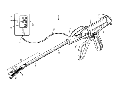
[00036] FIG. 1 illustrates a surgical system 1 which generally includes
a surgical
instrument such as a surgical stapler 10 in communication with a display 20
for
displaying measurements taken by the surgical stapler 10. The surgical stapler
10 is
CA 2993570 2018-01-31
configured to staple grasped tissue and to sense biological parameters of the
tissue to
assist a clinician in determining whether to effect a stapling function of the
surgical
stapler 10, as will be described in detail herein. For example, the surgical
stapler 10 is
configured to determine a surface perfusion pressure of a subject tissue. In
embodiments,
the surgical stapler 10 is configured for use in laparoscopic surgical
procedures. The
surgical stapler can be arranged for use in a robotic surgical system.
[00037] The surface perfusion pressure of tissue is measured by
applying clamping
pressure on the tissue until there is no perfusion (i.e., no blood flow)
through the tissue,
and then slowly reducing the clamping pressure until perfusion through the
grasped tissue
restarts. The pressure at which the perfusion restarts is known as "surface
perfusion
pressure."
[00038] For a detailed description of a method of measuring surface
perfusion
pressure, reference may be made to U.S. Patent No. 7,618,376, the entire
contents of
which are incorporated by reference herein.
[00039] The surgical stapler 10 generally includes a handle portion 12,
an adapter
assembly 14, an elongated shaft 30, and a surgical loading unit 40. The handle
portion 12
of the surgical stapler 10 includes a stationary handle 16 and a pivoting or
movable
handle 18 pivotably coupled to the stationary handle 16. Manipulation of the
pivoting
handle 18 relative to the stationary handle 16 effects a closing of jaw
members 42a, 42b
of the surgical loading unit 40 to grasp tissue disposed between the jaw
members 42a,
42b. For a detailed description of the various functions of the surgical
stapler 10,
reference may be made to, for example, U.S. Patent No. 7,172,104, the entire
contents of
which are incorporated by reference herein.
[00040] The surgical loading unit 40 has an elongate body portion 44
and an end
effector 46 coupled to the body portion 44. The body portion 44 is detachably
coupled to
6
CA 2993570 2018-01-31
a distal portion of the elongated shaft 30 or, in some embodiments, may be
fixedly
coupled to the distal portion of the elongated shaft 30. The end effector 46
of the surgical
loading unit 40 is pivotably coupled to a distal portion of the body portion
44 such that
the end effector 46 may be articulated about an axis transverse to a
longitudinal axis of
the body portion 44 between a linear orientation and an angled orientation
relative to the
longitudinal axis. In some embodiments, the end effector 46 may be fixedly
attached to
the distal portion of the elongated shaft 30.
[00041] With reference to FIGS. 1-3, the end effector 46 includes a
pair of
opposing jaw members 42a, 42b, wherein the jaw member 42a is configured as a
staple
cartridge and the jaw member 42b is configured as an anvil. The staple
cartridge 42a and
the anvil 42b each define a respective tissue-contacting surface 48a, 48b. The
tissue-
contacting surfaces 48a, 48b oppose one another such that when the end
effector 46 is in
the closed configuration, tissue is gasped between the tissue contacting
surfaces 48a,
48b. The tissue-contacting surface 48a of the staple cartridge 42a defines a
plurality of
staple-receiving channels 50, and the tissue-contacting surface 48b of the
anvil 42b
defines a plurality of staple forming pockets 52. As such, the staple
cartridge 42a and the
anvil 42b are configured to clamp and then staple tissue disposed therebetween
in
response to an actuation of the movable handle of the handle portion 12.
[00042] The end effector 46 of the surgical loading unit 40 includes an
inflatable
member 56 (FIG. 3), such as, for example, a balloon, disposed on the tissue-
contacting
surface 48b of the anvil 42b. In other embodiments, the inflatable member 46
may be
disposed on the tissue-contacting surface 48a of the staple cartridge 42a. In
still other
embodiments, each of the staple cartridge 42a and the anvil 42b may have an
inflatable
member disposed on its respective tissue-contacting surface 48a, 48b. The
inflatable
member 56 may be attached to the tissue-contacting surface 48b via an
adhesive, a hook
7
CA 2993570 2018-01-31
and loop fastener, a suture, or the like. In some embodiments, the inflatable
member 56
may be detachably connected to the tissue-contacting surface 48b. In other
embodiments,
the inflatable member 56 may be detachably connected to the tissue-contacting
surface
48b such that upon an actuation of the stapling function of the end effector
46, the staples
ejected from the staple cartridge 42a release the inflatable member 56 from
the tissue-
contacting surface 48b, for example, by severing a suture that attaches the
inflatable
member 56 to the tissue-contacting surface 48b.
[00043] The inflatable member 56 has a generally rectangular shape
dimensioned
to cover the tissue-contacting surface 48b of the anvil 42b such that the
staple forming
pockets 52 defined in tissue-contacting surface 48b are covered by the
inflatable member
56. The inflatable member 56 is fabricated from a biocompatible material such
as natural
or synthetic elastomers, natural or synthetic rubbers and silicone materials,
and/or
compliant polyurethanes. The inflatable member 56 may be made of a material
that is
penetrable by the staples ejected from the staple cartridge 42a so as to not
inhibit the
stapling function of the end effector 46.
[00044] The inflatable member 56 defines a hollow inner chamber or void
60 that
receives a fluid to change or move the inflatable member 56 from a collapsed
configuration, in which the inflatable member 56 is substantially flat and
rectangular
(FIG. 5), to an expanded configuration, in which the inflatable member 56 is
larger than
in the collapsed configuration and assumes a bulbous configuration (FIG. 6).
In some
embodiments, the inflatable member 56 may be configured to assume any suitable
shape
when in the expanded configuration, such as, for example, rectangular, dome-
shaped,
elliptical, oblong, tubular, square, triangular, cylindrical, rod-shaped, or
the like.
[00045] The inflatable member 56 may have a hose or tube 58 (FIG. 1)
extending
therefrom and in fluid communication with the hollow inner chamber 60 (FIG.
3). The
8
CA 2993570 2018-01-31
tube 58 may extend from the inflatable member 56, proximally through the body
portion
44 of the surgical loading unit 40, the elongated shaft 30, and out of the
adapter assembly
14. The tube 58 may have an end 62 (FIG. 1) coupled to a source of fluid, such
as, for
example, a pump (not explicitly shown), for delivering a fluid, such as liquid
and/or gas,
into the hollow inner chamber 60 (FIG. 3) of the inflatable member 56. The end
62 of
tube 58 may be in communication with the display 20 (FIG. 1) or a processor of
the
display 20.
[00046] The end effector 46 of the surgical loading unit 40 includes a
first sensor
64 (FIG. 3) and a second sensor 66 (FIG. 3) each associated with the anvil
42b. In
particular, the first and second sensors 64, 66 are each attached to the
inflatable member
56 of the anvil 42b and in communication with the display 20. In some
embodiments, the
first sensor 64 may be attached to one or both of the tissue-contacting
surfaces 48a, 48b
of the respective staple cartridge 42a and anvil 42b. The first sensor 64 is a
perfusion
sensor, for example, a Doppler flow sensor, configured to measure local
perfusion (i.e.,
blood flow) through tissue grasped between the staple cartridge 42a and the
anvil 42b.
The first sensor 64 may measure perfusion of the grasped tissue on the basis
of known
techniques, such as Laser-Doppler Flowmetry ("LDF"), measuring light
scattering, and/or
measuring absorption of light from one or more LED's or other light sources.
For a
detailed description of LDF technology, reference may be made to U.S. Patent
Nos.
4,109,647 and 4,862,894, the entire contents of each of which are incorporated
by
reference herein.
[00047] The first sensor 64 is in communication, via lead wires or
wireless
connection, with the display 20 such that upon the first sensor 64 measuring
perfusion in
grasped tissue, the first sensor 64 transmits the measurement data to a first
display section
20a of the display 20, which displays the measurement using a number, word, or
image.
9
CA 2993570 2018-01-31
In some embodiments, the first sensor 64 may also be in communication, via
lead wires
or wireless connection, with a computing device or processor (not shown) such
as a laser
Doppler monitor, which processes the information collected by the first sensor
64 to
calculate the tissue perfusion. The computing device (e.g., a laser Doppler
monitor) may
also be in communication, via lead wires or wireless connection, with the
first display
section 20a to send the processed information related to the tissue perfusion
to the first
display section 20a so that the first display section 20a can display the
tissue perfusion.
[00048] The second sensor 66 of the end effector 46 is a pressure
sensor or
pressure measuring device, for example, a MEMS device. For a detailed
description of
various MEMS devices, reference may be made to U.S. Patent No. 8,808,311, the
entire
contents of which are incorporated by reference herein. In embodiments, the
second
sensor 66 is disposed within the hollow inner chamber 60 of the inflatable
member 56 and
is configured to measure the amount of pressure applied by the end effector 46
to the
gasped tissue (i.e., the clamping pressure) by measuring the pressure within
the inflatable
member 56. In addition to or in the alternative of inflatable member 56 having
a pressure
sensor, tissue contacting surface 48a of the staple cartridge 42a may also
have a pressure
sensor for measuring the amount of pressure applied by the end effector 46 to
the grasped
tissue.
[00049] The second sensor 66 (FIG. 3) is in electrical communication,
via lead
wires or wireless connection, with a second display section 20b (FIG. 1) of
the display 20.
After the second sensor 66 measures the clamping pressure applied to the
grasped tissue,
the second sensor 66 transmits the measurement data to the second display
section 20b,
which displays the measurement. Additionally or alternately, the second sensor
66 may
send the measured clamping pressure to the computing device (e.g., a laser
Doppler
monitor) for processing, which then sends the information to the display 20.
CA 2993570 2018-01-31
[00050] The display 20 may have multiple display sections, for example,
three
display sections 20a, 20b, 20c. It is contemplated that the display 20 may
include more or
less than three discrete display sections arranged in any suitable
configuration. In
embodiments, the first display section 20a of the display 20 is configured to
display a
visual indication of a measured tissue perfusion of tissue grasped by the end
effector 46.
The second display section 20b of the display 20 is configured to display a
visual
indication of a measured amount of pressure being applied to tissue grasped by
the end
effector 46. A third display section 20c of the display 20 is configured to
display an
index representative of the ratio of the surface perfusion pressure determined
using the
first and second sensors 64, 66 of the surgical stapler 10, and a systemic
blood pressure
measured by a blood pressure cuff (not shown), as will be described in detail
below.
[00051] In some embodiments, the display 20 (FIG. 1) may display ranges
of
numbers or various numeral outputs to display the measurements of first and
second
sensors 64, 66. In particular, the first, second, and third display sections
20a, 20b, 20c
may display the number ranges 0 to 3, 0 to 5, 0 to 10, 0 to 100, or any other
suitable
range, to illustrate information about the tissue being grasped by the end
effector 46. For
example, when the first display section 20a displays the number 0, this may be
an
indication that the grasped tissue has very little or no perfusion (i.e., no
blood flow),
whereas when the first display section 20a displays the number 100, this may
be an
indication that the gasped tissue has a high perfusion (i.e., ideal blood
flow).
[00052] In some embodiments, the display 20 may convey information
about a
characteristic of the grasped tissue utilizing any suitable indicia, for
example, words such
as poor, satisfactory, or good.
[00053] In some embodiments, the surgical system 1 may not include the
display
20, and instead, surgical stapler 10 may be configured to be connected to or
be in
11
CA 2993570 2018-01-31
communication with another type of display, for example, a tablet, a cell
phone, a
computer monitor, a laptop, or any suitable display device. The surgical
stapler 10 may
be connected to any of the aforementioned display devices via USB wires, Wi-
Fi, or the
like.
[00054] In operation, the surgical system 1 may be used in a surgical
procedure in
which tissue is to be stapled, for example, an anastomotic surgical procedure,
to gather
various data about the subject tissue prior to effecting stapling. In some
anastomotic
surgical procedures, unhealthy or diseased bowel tissue is resected and the
ends of the
remaining healthy segments of bowel are stapled together to recreate a
continuous bowel.
Prior to stapling the ends of the separate bowel segments to one another, the
viability of
the ends of the separate bowel segments should be assessed in order to predict
the
likelihood of post-surgery anastomotic leaks or other adverse outcomes. To aid
in
making this viability assessment, a clinician may make use of the surgical
system 1 of the
present disclosure.
[00055] With reference to FIG. 4, in use of the surgical system 1, the
surgical
loading unit 40 is positioned through an access port 70 to gain entry to a
surgical site in a
minimally invasive manner. With the inflatable member 56 of the end effector
46 in a
collapsed or un-inflated state, tissue "T" is disposed between the tissue
contacting surface
48a of the staple cartridge 42a and the inflatable member 56 disposed on the
tissue-
contacting surface 48b of the anvil 42b with the staple cartridge 42a and the
anvil 42b in a
partially open position, as shown in FIG. 5. With the tissue "T" disposed
between the
staple cartridge 42a and the anvil 42b, the pump of the surgical system 1
conveys a fluid
(e.g., air) into the hollow inner chamber 60 of the inflatable member 56 via
the tube 58 to
change the inflatable member 56 from its collapsed state toward its expanded
or inflated
CA 2993570 2018-01-31 12
state, as shown in FIG. 6. With the inflatable member 56 in the expanded
state, the staple
cartridge 42a and the anvil 42b are clamped about the tissue "T," as shown in
FIG. 7.
[00056] In
one embodiment, the staple cartridge 42a and the anvil 42b may be
clamped about the tissue "T" prior to expanding the inflatable member 56. As
the
pressure inside of the inflatable member 56 increases, the pressure applied to
the tissue
"T" disposed between staple cartridge 42a and the anvil 42b increases to
inhibit blood
flow through the tissue.
[00057]
With the inflatable member 56 in the inflated state and the tissue "T" being
grasped by the end effector 46, the first sensor 64 of the end effector 46
collects
information about the perfusion through the grasped tissue. This information
is
transmitted to the first display section 20a of the display 20, which displays
this
infoi ____________________________________________________________________
illation as an image of the blood flow or as a number representative of the
degree of
perfusion through the tissue "T." The second sensor 66 measures the pressure
within the
inflatable member 56 and sends this information to the second display section
20b of the
display 20, which displays this information as a number. While a clinician
monitors the
perfusion reading (i.e., blood flow) displayed on the first display section
20a and the
pressure reading displayed on the second display section 20b, expansion of the
inflatable
member 56 is gradually continued to gradually increase the clamping pressure
between
the staple cartridge 42a and the anvil 42b, until the perfusion reading
indicates that no
blood flow or virtually no blood flow is moving through the grasped tissue
"T."
[00058] In
embodiments, instead of gradually increasing the clamping pressure on
the tissue "T" by inflating the inflatable member 56, the pivoting handle 16
is actuated to
gradually close the staple cartridge 42a and the anvil 42b about the tissue
"T." A
clinician will cease approximating the staple cartridge 42a and the anvil 42b
when the
13
CA 2993570 2018-01-31
display section 20a indicates that no blood or virtually no blood is flowing
through the
tissue "T."
[00059] When the first display section 20a indicates that perfusion
through the
grasped tissue has ceased, a clinician continuously monitors both the first
and second
display sections 20a, 20b while the pump is activated to gradually withdraw
the fluid
from the inflatable member 56, decreasing the pressure applied to the grasped
tissue "T."
In embodiments, the pressured applied to the tissue "T" may be decreased by
separating
the staple cartridge 42a and the anvil 42b rather than by deflating the
inflatable member
56. The clamping pressure is reduced until the first display section 20a
displays a
perfusion reading indicating that blood flow has returned to the grasped
tissue. At the
moment that the perfusion reading indicates that the blood flow is returned,
the pressure
reading (e.g., the pressure in inflatable member 56) displayed by the second
display
section 20b is noted, which is marks the local perfusion pressure of the
grasped tissue.
[00060] The local perfusion pressure determined using the above-noted
technique
may be used to assess the viability of the grasped tissue by, for example,
comparing the
measured local perfusion pressure with a known local perfusion pressure
associated with
healthy or viable tissue. Additionally or alternately, the measured local
perfusion
pressure may be used in combination with other measurements, for example, a
systemic
blood pressure reading, to aid in making the determination of the viability of
the tissue.
The systemic blood pressure may be taken using any suitable device, for
example, a blood
pressure cuff, applied to any suitable body portion of the patient, for
example, an arm of
the patient. An index may be calculated by taking a ratio of the local
perfusion pressure
measured by the surgical stapler 10 and the systemic blood pressure taken
using the blood
pressure cuff The index may be calculated by the computing device in the
display 20
and displayed as a number on the third display section 20c of the display 20.
14
CA 2993570 2018-01-31
[00061] The calculated index is predictive of whether an anastomotic
leak may
occur and/or the grade of an anastomotic leak. As such, a clinician can use
the index to
make a determination on whether the two ends of the presumed healthy bowel
segments
are healthy enough to be stapled together or whether more tissue needs to be
resected.
For example, the calculated index may be compared to a known index that is
associated
with healthy tissue. For a detailed description of a method of calculating a
perfusion
index and using the calculated index to assess tissue viability, reference may
be made to
U.S. Patent No. 7,618,376, the entire contents of which were incorporated by
reference
above.
[00062] After determining that the grasped tissue "T" is viable, the
movable handle
18 of the surgical stapler 10 may be actuated to eject staples from the staple-
receiving
channels 50 of the staple cartridge 42a into the tissue "T." The staples
contact the staple-
forming pockets 52 of the anvil 42b to close the staples about the tissue "T."
As the
staples are ejected, a cutting blade (not shown) moves through the end
effector 46 to cut
the stapled tissue "T," thereby dividing the tissue "T" into two tissue
segments.
[00063] In this way, a clinician only needs to use one instrument,
namely the
surgical stapler 10 of the present disclosure, to both determine tissue
viability and to
effect a stapling procedure once it is determined that the tissue is viable.
One benefit to
having only one instrument to accomplish each of these tasks is that once the
surgical
instrument determines that a particular segment of tissue is viable, the
clinician can
immediately staple the tissue without having to use a separate surgical
stapler.
[00064] In some embodiments, the surgical stapler 10 may be pre-
programmed to
inflate the inflatable member 56 to exert a predetermined clamping pressure
that is known
to result in a ceasing of perfusion through the grasped tissue. The surgical
stapler 10 may
also be pre-programmed to reduce the clamping pressure at a predetermined rate
via
CA 2993570 2018-01-31
deflation of the inflatable member 56 and automatically send the pressure
reading to the
computing device at the moment when perfusion through the grasped tissue
restarts. The
perfusion pressure reading may also be displayed on the display 20. This
automated
process eliminates human error in operating the surgical stapler 10 by
controlling the
amount of clamping pressure being applied to the tissue at any given time
instead of the
clinician.
[00065] In addition to the surgical system 1 being used to ensure that
the tissue is
in condition for stapling or acceptable for stapling, the surgical system 1
may also be used
as a check after the staples have been fired to ensure that the tissue is
healthy (e.g., has
good blood flow, is healing properly, etc.).
[00066] The surgical system 1 or components thereof may be configured
to be
incorporated into a robotic surgical system (not shown). The robotic surgical
system is
powered locally or remotely, and has electronic control systems localized in a
console or
distributed within or throughout the robotic surgical system. The robotic
surgical system
permits a clinician to remotely manipulate the surgical stapler 10 to more
precisely
control the movement of the surgical stapler 10. The surgical stapler 10 may
be
configured to send the measurements gathered by the sensors 64, 66 of the end
effector 46
to an interface of the robotic surgical system on which the measurements may
be
displayed for the clinician to read. In any of the embodiments disclosed
herein, the
surgical instrument can be a surgical stapler other than a linear endoscopic
surgical
stapler. For example, a circular stapler can incorporate a perfusion system
disclosed
herein.
[00067] As illustrated in FIG 8, another embodiment of a surgical
instrument 100
for use with the display 20 is provided. The surgical instrument 100 is
similar to surgical
CA 2993570 2018-01-31 16
stapler 10 described with reference to FIGS. 1-7, and will therefore only be
described
with the detail necessary to elucidate differences.
[00068] Surgical instrument 100 is a surgical stapler configured to
both staple
tissue and to determine tissue viability using an inflatable member (not
explicitly shown),
a perfusion sensor (not explicitly shown), and a pressure sensor (not
explicitly shown).
As described above in regard to the surgical stapler 10, the surgical
instrument 100
includes a handle portion 112, an adapter assembly 114, an elongated shaft
130, and a
surgical loading unit 140. However, rather than being manually-powered as is
the
surgical stapler 10 of FIGS. 1-7, the surgical instrument 100 of the present
embodiment
actuates functions of the surgical loading unit 140 (e.g., stapling, cutting,
closing/opening
of the jaws, etc.) via an internal-power source disposed within the handle
portion 112.
For a detailed description of an exemplary internally-powered handle portion
of a surgical
stapler, reference may be made to U.S. Patent Application Publication No.
2016/0118201,
filed on July 24, 2015, the entire contents of which being incorporated by
reference
herein.
[00069] As illustrated in FIGS. 9-11, another embodiment of a surgical
instrument
200 for use with the display 20 is provided. The surgical instrument 200 is
similar to the
surgical stapler 10 described with reference to FIGS. 1-7, and will therefore
only be
described with the detail necessary to elucidate differences.
[00070] Surgical instrument 200 is a non-stapling tissue grasper
configured to
determine tissue viability of grasped tissue. The tissue grasper 200 includes
a handle
portion 212, an adapter assembly 214, and a surgical loading unit 240. The
adapter
assembly 214 includes a housing or knob 214a and an elongated shaft 214b
extending
distally therefrom. The knob 214a of the adapter assembly 214 is configured to
detachably couple to the handle portion 212.
17
CA 2993570 2018-01-31
[00071] The surgical loading unit 240 has an elongated body portion 244
and a jaw
assembly 246 coupled to the body portion 244. The body portion 244 is
detachably
coupled to a distal portion of the elongated shaft 214b of the adapter
assembly 214. The
jaw assembly 246 includes a first jaw 242a and a second jaw 242b each coupled
to the
distal portion of the body portion 244. The first jaw 242a is pivotably
coupled to the
body portion 244 such that the first jaw member 242a is movable relative to
the second
jaw member 242b between a spaced condition and an approximated condition. In
some
embodiments, one or both of the jaw members 242a, 242b may be pivotably
coupled to
the body portion 244 of the surgical loading unit 240.
[00072] The first jaw member 242a has a tissue-contacting surface 248a
and the
second jaw member 242b has an inner surface 248b that defines an elongated
cavity 250
(FIG. 11) therein. The jaw assembly 246 includes an inflatable member 256,
similar to
inflatable member 56 described above, received within the cavity 250 of the
second jaw
member 242b. The inflatable member 256 may be dimensioned for a snap-fit
engagement with the inner surface 248b of the second jaw member 242b. The
inflatable
member 256 has a generally rectangular shape dimensioned to contact the tissue-
contacting surface 248a of the first jaw member 242a when the jaw assembly 244
is in the
approximated state.
1000731 The inflatable member 256 is configured to be changed or moved
from a
collapsed configuration, in which the inflatable member 256 is substantially
flat and
rectangular, to an expanded configuration, in which the inflatable member 256
is larger
than in the collapsed configuration and assumes a bulbous configuration. The
inflatable
member 256 may have a hose or tube 258 (FIG. 9) extending therefrom and in
fluid
communication with a hollow inner chamber 260 defined in the inflatable member
256.
The tube 258 may have an end 262 coupled to a source of fluid, such as, for
example, a
18
CA 2993570 2018-01-31
pump (not explicitly shown), for delivering a fluid such as liquid and/or gas
into hollow
inner chamber 260 of the inflatable member 256.
[00074] The jaw assembly 140 includes a perfusion sensor 264 (FIG. 11),
similar
to the first sensor 64 described above, and a pressure sensor 266, similar to
the second
sensor 66 described above. The perfusion sensor 264 is attached to the
inflatable member
256 (e.g., an inner or outer surface thereof) of the second jaw member 242b
and in
communication with the display 20. In some embodiments, the perfusion sensor
264 may
be attached to the tissue-contacting surface 248a of the first jaw ember 242a
or the inner
surface 248b of the second jaw member 242b rather than the inflatable member
256.
[00075] In embodiments, the pressure sensor 266 (FIG. 11) of the jaw
assembly
246 is disposed within the hollow inner chamber 260 of the inflatable member
256 and is
configured to measure the amount of pressure applied by the jaw assembly 246
to the
grasped tissue (i.e., the clamping pressure) by measuring the pressure within
the inflatable
member 256. In addition to or in the alternative of inflatable member 256
having a
pressure sensor, the tissue contacting surface 248a of the first jaw member
242a may also
have a pressure sensor for measuring the amount of pressure applied by the jaw
assembly
246 to the grasped tissue.
[00076] The tissue grasper 200 is used in a similar manner as described
above with
respect to the surgical stapler 10 except that the tissue grasper 200 does not
effect a
stapling of the grasped tissue after the viability of the grasped tissue is
assessed.
[00077] As illustrated in FIGS. 12 and 13, another embodiment of a
surgical
instrument 300 for use with the display 20 is provided. The surgical
instrument 300 is
similar to the surgical instrument 200 described above with reference to FIGS.
9-11, and
will therefore only be described with the detail necessary to elucidate
differences.
19
CA 2993570 2018-01-31
[00078] The surgical instrument 300 is a tissue grasper configured to
grasp tissue
and determine viability of the grasped tissue using an inflatable member 256,
a perfusion
sensor (not explicitly shown), and a pressure sensor (not explicitly shown).
The tissue
grasper 300 includes a handle portion 312, an adapter assembly 314, and a jaw
assembly
340. Rather than the tissue grasper 300 having a detachable loading unit, the
jaw
assembly 340 is fixed to the elongated shaft 314b of the adapter assembly 314,
and a
housing or knob 314a of the adapter assembly 314 is detachably coupled to the
handle
portion 312.
[00079] Although the illustrative embodiments of the present disclosure
have been
described herein, it is understood that the disclosure is not limited to those
precise
embodiments, and that various other changes and modifications may be affected
therein
by one skilled in the art without departing from the scope or spirit of the
disclosure. All
such changes and modifications are intended to be included within the scope of
the
disclosure.
CA 2993570 2018-01-31 20