Note: Descriptions are shown in the official language in which they were submitted.
CA 02993629 2018-01-23
CELL MEASUREMENT METHOD
Technical Field
[0001]
The present invention relates to a method for measuring a cell amount.
Background Art
[0002]
In a susceptibility test for an anticancer agent against epithelial malignant
tumor, sarcoma,
etc., a cancer cell brought into contact with an anticancer agent and a cancer
cell not brought into
contact with the anticancer agent are cultured under the same condition, and
the proliferation
degrees of the cancer cells after cultivation are compared so as to evaluate
susceptibilities of the
cancer cells to the anticancer agent. The less proliferation of the cancer
cell is, the better the
anticancer agent is.
[0003]
As a method for culturing cancer cells, Patent Documents 1 to 5 describe
methods for
culturing cancer cells by embedding them in a collagen gel. This collagen gel
embedding
cultivation is known to proliferate cancer cells better compared to a surface
cultivation in which
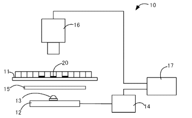
cancer cells are cultured on a surface of agar or the like.
[0004]
As a method for quantitating a cultured cancer cell, Patent Document 1
describes a
method in which a proliferated cancer cell is imaged with a TV camera or the
like, and then
obtained image information is electronically image-analyzed to calculate
estimated volume
values of cancer cell colonies. In addition, Patent Document 3 describes a
method in which a
cancer cell cultured in a collagen gel is stained with a dye, imaged, and
quantitated based on an
image density.
Prior Art Documents
Patent Documents
[0005]
Patent Document 1: JP H03-285696 A
1
1
,
= CA 02993629 2018-01-23
Patent Document 2: WO 95/18216
Patent Document 3: JP H10-115612 A
Patent Document 4: JP Pat. No. 3363445
Patent Document 5: JP 2008-11797 A
Summary of Invention
Problem to be solved
[0006]
The cancer cell quantitating methods described in Patent Document 1 and Patent
Document 3 had problems of further improvement for quantitative precision. The
susceptibility tests to anticancer agents have been conventionally performed
using surgical
materials taken from cancer patients as starting materials. In recent years,
there has been
growing demand for an anticancer agent susceptibility test using a biopsy
material as a starting
material, in which cells are sampled with a puncture needle or the like for
the purpose of
reducing physical burden of a patient. However, for the biopsy material, since
tissue pieces
that can be sampled are smaller than surgical materials, it is required in the
anticancer agent
susceptibility test to precisely quantitate less than or equal to one-tenth
cell amount of that in the
conventional test. In Patent Document 1 and Patent Document 3, it was
difficult to precisely
quantitate such a small amount of cancer cell.
[0007]
The present invention has been made in view of the above circumstance, and an
object of
the present invention is to provide a cell measurement method with higher
quantitative precision.
Solution to Problem
[0008]
The cell measurement method of the present invention comprises: a step of
staining a
cultured target cell with a dye; a step of obtaining a first image and a
second image which are
transmission images for a first light and a second light to which the dye has
different
absorbance; a step of dividing each of the first image and the second image
into a plurality of
divided regions and comparing the first image and the second image for each of
the divided
regions so as to eliminate noises; and a step of integrating an indicator of a
cell amount in each
2
1
= CA 02993629 2018-01-23
of the divided regions in the images from which the noises were eliminated so
as to evaluate a
target cell amount.
[0009]
Herein, the target cell means a cell to be measured. In addition, the noise
means
unnecessary image information not derived from the stained target cell.
Furthermore, the
indicator of the cell amount means an indicator which increases or decreases
depending on the
amount of the cell, such as a density of the image or an absorbance calculated
from the density
of the image. This method eliminates the influence of the noises resulting in
errors, so that the
cell amount can be precisely measured.
[0010]
Preferably, in the step of eliminating the noises, the first image and the
second image are
compared for each of the divided regions, and when a difference or a ratio of
luminosity between
the divided regions subjected to the comparison is less than a predetermined
value, the divided
regions are excluded from the data as a basis for evaluation of the target
cell amount.
[0011]
Alternatively, preferably, in the step of eliminating the noises, the first
image and the
second image are compared for each of the divided regions, and when a
difference or a ratio of
absorbance between the divided regions subjected to the comparison is less
than a predetermined
value, the divided regions are excluded from the data as a basis for
evaluation of the target cell
amount.
[0012]
Preferably, the target cell is a cancer cell.
[0013]
Preferably, the target cell is a three-dimensionally cultured cell, and more
preferably a
cell cultured by embedding the cell in a collagen gel.
[0014]
Preferably, the first image and the second image are obtained by color-
separating an
image taken using one color camera while concurrently applying the first light
and the second
light.
3
1
, CA 02993629 2018-01-23
[0015]
Alternatively, preferably, the first image and the second image are obtained
by
independently taking each image using one camera while sequentially applying
the first light and
the second light
[0016]
Preferably, the target cell amount is evaluated by calculating an absorbance
from the
image luminosity for each of the divided regions, and integrating the obtained
absorbance over
the plurality of divided regions to calculate an estimated volume value of the
target cell.
Effects of Invention
[0017]
According to the cell measurement method of the present invention, the cell
amount can
be precisely evaluated even when the amount of the cultured target cell is
small.
Brief Description of Drawings
[0018]
Figure 1 shows a configuration example of a cell measuring apparatus used in a
first
embodiment of the present invention.
Figure 2 is a flow chart of a cancer cell quantitating method according to the
first
embodiment of the present invention.
Figure 3 is a diagram for explaining the luminosity of the image.
Figure 4 is a picture for explaining an original image obtained by the cancer
cell
quantitating method according to the first embodiment of the present
invention.
Figure 5 shows an absorption spectrum of a neutral red.
Figure 6 is an original image of a sample in which a cancer cell was
quantitated in
Example.
Figure 7 is an original image of a sample in which a cancer cell was
quantitated in
Example.
Detailed Description of Embodiments
[0019]
As a first embodiment of the cell measurement method of the present invention,
a method
4
= CA 02993629 2018-01-23
of quantitating a cancer cell in an anticancer agent susceptibility test will
be described below.
[0020]
Prior to the cultivation, tissues sampled from a living body are subjected to
dispersion
treatment such as chopping and digestion of intercellular substances by a cell
dispersion enzyme
treatment. In some cases, separation treatment is subsequently carried out in
which
unnecessary cells such as blood is removed by preliminary cultivation and
living cells are
collected.
[0021]
Various known methods can be used to prepare a cultured sample. Above all, a
three-
dimensional cultivation is preferably used. More preferably, a collagen gel
embedding
cultivation is used. This method allows preferable cultivation and subsequent
quantitation of
the cancer cell even when the amount of the cancer cell used for cultivation
is small.
[0022]
The procedure according to the collagen gel embedding cultivation is as
follows. A
separated and dispersed cell is blended into a collagen solution. At this
time, besides collagen,
various components necessary for cultivation can be added to the collagen
solution. For
example, a buffer solution which is the same as or similar to the
physiological condition of the
target cell can be added to the collagen solution. The collagen solution
containing the cancer
cell is dropped onto the supporting surface in the culture container to form a
collagen gel in a
form of droplet, and the liquid medium is added into the culture container.
Similarly, several
samples are prepared. For some samples, an anticancer agent is added to the
culture container,
and after a predetermined time, the anticancer agent is washed away, and
cultivation is carried
out again.
[0023]
After completion of the cultivation, a dye is added to the culture container
to stain the
cancer cell as a target cell. As a staining method, a staining method in
conventional cancer cell
cultivation can be applied. Specific examples include a Giemsa solution dyeing
method, a
crystal violet dyeing method, a neutral red (NR) dyeing method, a fluorescein
diacetate (FDA)
dyeing method, and dyeing methods using other fluorescent reagents. As a
staining method, a
CA 02993629 2018-01-23
method in which cancer cells can be selectively stained and components other
than cancer cells
are stained as little as possible, is preferable. Use of a living cell-
staining method for
selectively staining a living cell is suitable for measuring susceptibility to
an anticancer agent, or
the like. The NR staining method is preferable as a method capable of
selectively staining only
living cells among cancer cells.
[0024]
After completion of staining, the dye is fixed within the cell with formalin
and dried. In
the dried collagen gel, moisture is released from the droplet-like collagen
gel, so that the gel is in
a form of flat face.
[0025]
Next, a method for imaging a sample including a target cell and processing the
image will
be described. A flowchart of the process is shown in Figure 2.
[0026]
In Figure 1, a measuring apparatus 10 according to the present embodiment
comprises: a
sample stage 11 on which a sample 20 is placed; an illumination 12 for
illuminating the sample
from below; a color camera 16 for imaging a transmission image of the sample;
and an image
processor 17. The illumination 12 comprises one LED package 13 and is
connected to the
illumination power supply 14. A light diffusion plate 15 is inserted between
the illumination
and the sample stage. In each LED package, an LED chip for emitting first
light (not shown)
and an LED chip for emitting second light (not shown) are incorporated.
[0027]
Between the first light and the second light, there is a difference in
absorbance by the dye
which has stained the sample. In the present embodiment, the first light and
the second light
are concurrently applied to the sample, and the sample is imaged by one color
camera to obtain
one original image. This original image is color-separated, so that a first
image as a
transmission image for the first light and a second image as a transmission
image for the second
light can be obtained.
[0028]
For the first light and the second light, it is preferable that the difference
in absorbance by
6
CA 02993629 2018-01-23
the dye therebetween is greater. In order to obtain sufficient measurement
precision, a ratio of
transmission loss between the first light and the second light in transmitting
through the sample
is preferably 1:1.5 or more, more preferably 1:2 or more. For that purpose,
the difference in
absorbance therebetween is preferably log1.5 0.18 or more, more preferably
log2 0.30 or
more. Since the absorbance varies depending on the measurement conditions, it
is preferable to
select wavelengths of the first light and the second light such that such a
difference can be
obtained under actual measurement conditions.
[0029]
For example, Figure 5 shows absorption spectrum of neutral red (NR) at pH =
7.1 (made
from: Rika Obata et al, " Neutralization titration, and visible absorption
spectrum of acid-base
indicator", The Hiyoshi review of Natural Science, Keio University, No. 50,
pp. 77-102,
September 2011). The NR has an absorption band in a range of about 380 nm to
600 nm at this
pH, and has an absorption peak at 462 nm and 518 nm. In this case, green light
whose
wavelength distribution overlaps with this absorption band can be selected for
the first light, and
red light whose wavelength distribution does not overlap with this absorption
band can be
selected for the second light.
[0030]
As a light source for illumination, an LED is preferably used. This is because
the
wavelength distribution of LED is narrow and a difference between the first
image and the
second image is easy to clearly appear. Note that the physical form of
illumination is not
particularly limited. For example, the number of LED packages is not
particularly limited. In
addition, for example, an LED chip emitting the first light and an LED chip
emitting the second
light may be incorporated in one LED package as in the present embodiment, or
an LED
package emitting the first light and an LED package emitting the second light
may be alternately
arranged.
[0031]
An image is constituted as an aggregate of many pixel data. Each pixel
includes
information representing a luminosity corresponding to a light intensity
captured by image
sensor elements of the camera. For example, if a gradation for inputting
images is 8-bit
7
CA 02993629 2018-01-23
gradation, the luminosity is represented by 256 different values from 0 to
255. If light is
absorbed when transmitting through the sample, the relevant portion is dark on
the transmission
image, that is, the luminosity is low.
[0032]
In the first image which is a transmission image for the first light,
absorption by the NR is
large, and thus if there are cancer cells stained with the NR in the cultured
sample, the intensity
of the transmitted light on the relevant portion is low. In addition, the
larger the thickness of
the cancer cell is, the lower the intensity of the transmitted light is, and
the lower the luminosity
of the image is. On the other hand, the second image which is a transmission
image for the
second light does not significantly reflect the presence amount of the cancer
cells.
[0033]
Herein, each of the first image and the second image is divided into a
plurality of divided
regions by the same method. The division by the same method means that a
divided region of
the first image and a corresponding divided region of the second image are the
same in size, and
imaged on the same place of the sample. The image processing described below
is carried out
in each of the divided regions. In the present embodiment, one pixel is
defined as one divided
region. Since the first image and the second image are obtained from one
original image, each
pixel is a region obtained by dividing both images by the same method.
[0034]
First, a blank image luminosity W obtained from image information of a sample
containing no cancer cell is defined as an upper limit, and a dark image
luminosity B obtained
from image information in a dark state is defined as a lower limit, and
relative values of the
luminosity with respect to the upper and lower limit values are determined for
each pixel to
correct the first image and the second image. A blank image is an image in the
brightest state
obtained by imaging a blank sample treated through the same process as for the
cultured sample
of the cancer cell except that the cancer cell is not added. However, the
blank image is not a
complete white image because of the presence of a collagen gel matrix and the
like. A dark
image is an image in the darkest state in which light is prevented from
entering by closure of a
shutter of an imaging lens or the like. As shown in Figure 3, the luminosity
Ti of the first
8
=
CA 02993629 2018-01-23
image and the luminosity T2 of the second image are between the luminosity W
of the blank
image and the luminosity B of the dark image.
[0035]
Next, influence of noises is eliminated by comparing the first image and the
second
image.
[0036]
Respective pixels are compared between the first image and the second image.
If the
difference or the ratio of the luminosity is less than a predetermined
threshold value, the region
of the relevant pixel is judged to have no cancer cell, and the pixel is
excluded. In more detail,
the data of the pixel is excluded from the data which is the basis for
evaluating the cancer cell
amount later. Specifically, for example, the first image is corrected so that
the luminosity of the
pixel is overwritten with the luminosity of the blank image. Thereby, the
luminosity of the
pixel does not affect the evaluation of the cancer cell amount and is
substantially excluded.
[0037]
When the difference in luminosity is defined as a reference for the threshold
value, for
example the threshold value can be set to one eighth of the gradation number
of luminosity.
That is, in a case that the luminosity is represented by 8 bits/256 grades,
when the difference in
luminosity between the first image and the second image is smaller than 32,
the relevant pixel is
excluded. Alternatively, in a case that the ratio of the luminosity is defined
as a reference,
when the ratio in luminosity between the first image and the second image is
lower than a
predetermined threshold value, it had better exclude the relevant pixel. More
preferably, these
threshold values are previously determined by a preliminary experiment.
[0038]
Alternatively, when an absorbance is determined from the luminosity of each
pixel and
the difference or the ratio of the absorbance is less than a predetermined
threshold value, the
region of the relevant pixel may be judged to have no cancer cell.
[0039]
Since opaque dusts do not transmit light regardless of the wavelength, it
looks dark
similarly in both the first image and the second image. In addition, since
bubbles contained in
9
CA 02993629 2018-01-23
the dried collagen gel look dark on the image due to light refraction, the
bubbles also look dark
similarly in both the first image and the second image regardless of the
wavelength of the light
source. Consequently, these noises can be eliminated by excluding regions
where there is no
difference in luminosity between the first image and the second image.
[0040]
Note that bubbles are particularly problematic when the cell amount is small
in collagen
gel embedding cultivation. If the cell amount is small, bubbles may remain in
the dried
collagen gel. Although the reason is unclear, it is considered that when the
cell amount is large,
a gas in the gel passes through the interface between the cell and the matrix
in the gel droplet
mass to exit outside, whereas when the cell amount is small, the gas in the
gel does not
thoroughly exit but remains.
[0041]
Figure 4 shows a transmission image (original image) of a sample stained with
NR. The
first light was green light with a dominant wavelength of 528 nm and the
second light was red
light with a dominant wavelength of 625 nm. Note that Figure 4 is a picture
obtained by
converting the color original image into a monochrome image, in which the
resolution is also
converted. The circular area at the center is the sample (dried collagen gel).
Many fine dark
spots scattered on the sample are cancer cells or colonies thereof, which are
red in the original
image, dark in the first image, and do not appear in the second image. Note
that the dark spots
surrounded by the dotted line are dusts, which are gray in the original image,
and dark in the first
image and the second image. The upper solid ellipse and the lower hollow
ellipse indicate
noises due to bubbles, which are gray in the original image and dark in the
first image and the
second image.
[0042]
Another cause of noise is contamination of a fibroblast. The influence of the
fibroblast
can be eliminated by the method described in Patent Document 3. The fibroblast
is stained
with a dye such as NR together with the cancer cell, but the fibroblast is
much more difficult to
stain than the cancer cell, and its luminosity in the image is obviously
higher than that of the
cancer cell. Thus, when the luminosity of a pixel exceeds a predetermined
threshold value in
1
. CA 02993629 2018-01-23
the first image, the region of the relevant pixel is judged to have the
fibroblast, and the pixel is
excluded. Specifically, for example, the first image is corrected so that the
luminosity of the
pixel is overwritten with the luminosity of the blank image. The threshold
value can be
previously determined by a preliminary experiment.
[0043]
Alternatively, as another method of eliminating the influence of the
fibroblast, the cancer
cell and the fibroblast are distinguished depending on their shapes by image
analysis as
described in Patent Document 1, and information about only the cancer cell may
be extracted.
[0044]
The above treatment is repeated over the entire area of the sample, so that
the influence of
the noises not resulting from light absorption by the cancer cell can be
eliminated.
[0045]
Next, cancer cells are quantitated from the image from which the noises have
been
eliminated.
[0046]
The cancer cell amount can be evaluated by integrating an indicator of the
cell amount for
each pixel. Preferably, the cancer cell amount is evaluated by an estimated
volume value.
This is because colonies of the cancer cells three-dimensionally develop by
the collagen gel
embedding cultivation, and thus their thicknesses can be taken into
consideration for more
accurate evaluation. The estimated volume value is obtained by determining an
absorbance
from the luminosity of each pixel and integrating the absorbance over the
entire area of the
sample. This is because the absorbance linearly correlates with the cell
thickness in each
region.
[0047]
According to the Lambert-Beer law, if the intensity of the incident light to
the sample is
represented by 10, and the intensity of the transmitted light is represented
by I, the following
relationship is established;
I/I0 = exp (-aL)
wherein, a represents an absorption coefficient of the stained cancer cell,
and L represents a
11
1
CA 02993629 2018-01-23
distance through which light passes in the cancer cell, i.e., a thickness of
the cancer cell. An
absorbance A by the cancer cell in each pixel is represented by the following
equation:
A = -log(I/Io)
= (aL)/2.303
and therefore the absorbance A is proportional to the thickness L of the
cancer cell. The
absorbance A is an indicator of the cell amount in the pixel, and the
absorbance A is integrated
over the entire area of the sample to determine the cell amount. Note that log
is common
logarithm.
[0048]
On the other hand, from the corrected first image, the absorbance A is
determined by the
following equation:
A = log {(W-B)/(Ti -B)}
wherein, W represents the luminosity of the pixel in the blank image, B
represents the
luminosity of the pixel in the dark image, and T1 represents the luminosity of
the pixel in the
corrected first image.
[0049]
Based on the above, the estimated volume value V of the cancer cell amount is
determined by the following equation:
V = IL = CA = CE [log I(W-B)/(Ti-B)}] (Equation 1)
wherein C is a constant. Thus, the absorbance is determined from the
luminosity in each pixel,
and the absorbance is integrated over the entire area of the sample to
determine the estimated
volume value of the cell.
[0050]
Note that, when the luminosity T1 of the pixel in the corrected first image
equals to the
luminosity B of the pixel in the dark image (Ti = B) for any reason, the
denominator of the
antilogarithm of the right-side logarithm in Equation 1 is 0, and thus
calculation is impossible.
In response to this, it is preferable that the luminosity etc. of the light
source are adjusted so that
the sample image is not too dark, and an exception handling suitable in the
case of T1 = B is
carried out.
12
= CA 02993629 2018-01-23
[0051]
For simplicity, the luminosity of each pixel may be integrated to determine
the
absorbance from the integrated value. The estimated volume value Vp is
represented by the
following equation:
Vp = CAp = Clog
wherein, Cp represents a constant, and Ap represents an absorbance. In this
equation, the
absorbance is determined considering the entire area of the sample as one
region, but if the cell
amount is large, sufficient precision can be obtained e.g., in a case of using
a surgical material as
a starting material. Also when using this equation, the influence of noise due
to dusts and the
like has already been eliminated by the image processing.
[0052]
In the anticancer agent susceptibility test, the susceptibility to the
anticancer agent is
evaluated by comparing the cancer cell amounts after cultivation between the
control sample to
which the anticancer agent has not been added and the sample to which the
anticancer agent has
been added.
[0053]
The effect of the cancer cell-quantitating method of this embodiment will be
described
again.
[0054]
Noises due to dusts and bubbles have been difficult to eliminate by
conventional
techniques. According to the method of the present embodiment, the first light
and the second
light are used to eliminate the influences of contamination of dust and
remaining bubbles, so that
the cancer cell can be precisely quantitated. Since opaque dusts are
misrecognized as cancer
cells only with the first image and furthermore misrecognized as thick cancer
cells because of
dark shadow in the image, quantitative precision is significantly impaired.
Also bubbles are
misrecognized as cancer cells only with the first image, many of which are
larger than colonies
of cancer cells, and thus quantitative precision is significantly impaired.
[0055]
Furthermore, the absorbance is determined and integrated for each of the
divided regions
13
1
=
= CA 02993629 2018-01-23
in the sample image according to the above equation 1, so that the estimated
volume value of the
cancer cell can be calculated more precisely.
[0056]
Next, a second embodiment of the cell measurement method of the present
invention will
be described.
[0057]
This embodiment relates to a method for quantitating cancer cells in an
anticancer agent
susceptibility test as in the first embodiment. In the method of this
embodiment, the method
for taking the first image and the second image is different from that in the
first embodiment.
The other steps are the same as in the first embodiment.
[0058]
In this embodiment, the first light source emitting the first light and the
second light
source emitting the second light are sequentially lighted, and one camera
takes an image each
time each light source is lighted. Thereby, the first image is obtained by
imaging at the time of
lighting the first light source, and the second image is obtained by imaging
at the time of lighting
the second light source. Also in this embodiment, the physical form of the
light source is not
particularly limited. For example, an LED chip as a first light source and an
LED chip as a
second light source may be incorporated in one LED package, or otherwise
separate LED
packages as a first light source and a second light source may be used and
alternately arranged.
[0059]
In this embodiment, a monochrome camera can be used. In that case, finer
images can
be obtained, because monochrome cameras with higher resolution are available
than color
cameras.
Example
[0060]
The first embodiment will be further specifically described with reference to
Example.
[0061]
A human colon cancer-derived cell line HCT-116 was used as a cancer cell, and
cultured
by a collagen gel embedding method. As a collagen gel solution for embedding
the cell, 1
14
CA 02993629 2018-01-23
volume of a ten-time concentrated Ham's F12 medium (containing no sodium
bicarbonate) and 1
volume of a buffer solution for reconstitution (50 mM-NaOH solution containing
260 mM of
sodium bicarbonate and 200 mM of HEPES) were added to 8 volumes of Cell Matrix
Type CD
(KURABO INDUSTRIES LTD.), and stored in ice. The HCT-116 strain was added to
the
collagen solution so that its final density was 4x10⁴ cells/mL, and mixed well
to prepare a
collagen mixture. 10 tiL of this collagen mixture was dropped into each of
three wells of a 24-
well plate with appropriate intervals using a micro pipette. Thereafter, the
mixture was warmed
in a CO2 incubator at 37 C for 1 hour to prepare a collagen matrix containing
the cancer cell.
To the resulting collagen gel matrix, 1 mL of DF medium containing 10% FBS was
added, and
cultured for 16 hours. Then, an NR stain was injected into the wells, followed
by formalin
fixation and drying, to obtain a dried collagen gel.
[0062]
The resulting dried collagen gel was placed on a sample stage and illuminated
from
below with an illumination, and a transmission image was imaged by a color
camera. For the
illumination, one LED package (MC-E Color, CREE Inc.) was used. RGB three-
color LED
chips were mounted in the LED package, and among them, only R chip and G chip
were lighted
for use. The first light was green light with a dominant wavelength of 528 nm,
and the second
light was red light with a dominant wavelength of 625 nm. For the color camera
(XCL5005CR,
Sony Corporation), the pixel number was 2448 x 2050, each of the RGB chips was
constituted
with 8-bit gradation, and a lens of 1.3 optical magnifications was used. At
this time, the
resolution of the image was about 2.7 p.m.
[0063]
In Figure 6 (sample containing no bubble) and Figure 7 (sample containing many
bubbles), the imaged original images were converted into monochrome images.
The samples
shown in Figures 6 and 7 contain almost the same amount of cancer cell. Note
that the above
Figure 4 also shows an image obtained by the same method as this Example. The
original
image was color-separated into three colors of RGB, and the G image was
defined as a first
image and the R image was defined as a second image. For each pixel, the first
image and the
second image were compared, and when a difference in luminosity was within 35,
the pixel was
1
. CA 02993629 2018-01-23
judged to have no cancer cell. Absorbance was calculated for each pixel
according to the
above Equation 1, and integrated over the entire area of the sample to
determine an estimated
volume value of the cancer cell. At this time, a value of the constant C in
Expression 1 was
2.0x10⁻⁴.
[0064]
As Comparative Example, the absorbance was calculated from the luminosity of
the first
image without using the second image, and similarly integrated over the entire
area of the
sample to determine an estimated volume value of the cancer cell.
[0065]
The estimated volume values obtained by the method of Example were 0.42 in
Figure 6
and 0.44 in Figure 7. In the method of Comparative Example, the estimated
volume values
were 0.47 in Figure 6 and 1.54 in Figure 7. In Figure 6 without bubbles,
Example and
Comparative Example showed equivalent estimated volume values. On the other
hand, in
Figure 7 with many bubbles, the estimated volume value according to
Comparative Example
was about three times that of Example. This was attributed to the influence of
the noise due to
the bubbles, and in Example, the noise due to the bubbles could be eliminated.
[0066]
The cell measurement method of the present invention is not limited to the
above-
described Embodiments and Example, and can be variously modified within the
scope of the
technical idea of the invention.
[0067]
For example, in the above-described Embodiment, relativization of the
luminosity (blank
correction), elimination of noises such as dusts and bubbles by comparison
between the first
image and the second image, and elimination of noises due to fibroblasts are
carried out in this
order, but their turns may be replaced.
[0068]
In addition, images may be taken using e.g., a white illumination while
sequentially
switching color filters installed on the front of the camera, to obtain the
first and second images.
[0069]
16
1
= CA 02993629 2018-01-23
In addition, images may be taken by a color camera using e.g., a white light
source
having continuous spectrum as an illumination, and color-separated to obtain
the first and
second images. However, since image sensor elements of the color camera
generally have
wide sensitivity spectra and partially overlap with each other, it had better
use two light sources
having different wavelengths for obtaining clear difference between the first
and second images.
Reference Numerals
[0070]
measuring apparatus
11 sample stage
12 illumination
13 LED package
14 illumination power supply
light diffusion plate
16 color camera
17 image processor
sample
17