Note: Descriptions are shown in the official language in which they were submitted.
CA 02993653 2018-01-24
WO 2017/019939 PCT/US2016/044647
WOUND THERAPY DEVICE PRESSURE MONITORING AND CONTROL SYSTEM
CROSS-REFERENCE TO RELATED APPLICATIONS
[0001]This application claims priority to U.S. Provisional Patent Application
No.
62/198,514, filed on July 29, 2015, titled, "WOUND THERAPY DEVICE PRESSURE
MONITORING AND CONTROL SYSTEM" and U.S. Provisional Patent Application No.
62/296,679, filed on February 18, 2016, titled "WOUND THERAPY DEVICE PRESSURE
MONITORING AND CONTROL SYSTEM". The entirety of each of these applications is
hereby incorporated by reference.
BACKGROUND
[0002]Negative pressure wound therapy includes a vacuum source connected to a
wound dressing. Various porous dressings comprising gauze, felts, foams, beads
and/or
fibers can be used in conjunction with a semi-permeable cover and a controlled
vacuum
source. A collection container may be used to collect wound exudate and fluid
that drains
from the wound.
[0003]In addition to using negative pressure wound therapy, many devices
employ
concomitant wound irrigation. For example, a known wound healing apparatus
includes
a porous dressing made of polyurethane foam placed adjacent a wound and
covered by
a semi-permeable and flexible plastic sheet. The dressing further includes
fluid supply
and fluid drainage connections in communication with the cavity formed by the
cover and
foam. The fluid supply is connected to a fluid source that can include an
aqueous-based
topical antibiotic solution or isotonic saline, for example, for use in
providing therapy to
the wound. The fluid drainage can be connected to a vacuum source where fluid
can be
removed from the cavity and subatmospheric pressures can be maintained inside
the
cavity.
[0004] Other devices use vacuum sealing of wound dressings including polyvinyl
alcohol
foam cut to size and stapled to the margins of the wound. The dressings are
covered by
a semi-permeable membrane while suction and fluid connections are provided by
small
plastic tubes introduced subcutaneously into the cavity formed by the foam and
cover.
CA 02993653 2018-01-24
WO 2017/019939 PCT/US2016/044647
Such devices alternate in time between vacuum drainage and the introduction of
aqueous
medicaments to the wound site.
[0005] However, such devices may fail to address the problems caused by
standing fluid
and occlusions in a tube connecting the wound dressing to the collection
container.
SUMMARY
[0006]A wound therapy system is described, which comprises a wound dressing, a
pressure sensor, a container having an internal chamber, a vacuum source
pneumatically
associated with the internal chamber of the container, and a tube set
comprising a first
tube and a second tube positioned inside a lumen of the first tube. A space
between the
first tube and the second tube forms a fluid channel, and a lumen of the
second tube
forms a sensor channel in the tube set. The wound dressing may be
pneumatically
associated with the internal chamber of the container by the fluid channel of
the tube set.
The wound dressing may be pneumatically associated with the pressure sensor by
the
sensor channel of the tube set. A crushing force required to occlude the fluid
channel is
greater than a crushing force required to occlude a fluid channel of a
comparison tube set
that does not include a second tube positioned inside a lumen of the first
tube.
[0007]A method of wound therapy is also described, which comprises applying a
dressing
to a wound, wherein the dressing is coupled to a tube set comprising a fluid
channel and
a sensor channel; applying a vacuum to the fluid channel, wherein the vacuum
draws
exudate from the wound into the fluid channel; and providing a restrictor
pneumatically
associated with the wound dressing by the sensor channel, the restrictor
having a hole
through which air can leak into the sensor channel, wherein the air pushes
exudate from
the wound into the fluid channel of the tube set.
[0008]A device for creating an air leak is also described, the device
comprising a body
having a first port in communication with a second port; a cap coupled to the
first port of
the body, the cap having a hole; and a porous material positioned between the
cap and
the first port of the body. Air may be configured to enter the device through
the hole in
the cap and pass through the porous material before entering the body via the
first port.
2
CA 02993653 2018-01-24
WO 2017/019939 PCT/US2016/044647
BRIEF DESCRIPTION OF THE FIGURES
[0009] FIGS. 1A-1C show schematic depictions of various configurations of a
negative
pressure wound therapy system.
[0010]FIG. 2A shows a perspective view of a first embodiment of a collection
container
used in the negative pressure wound therapy system shown in FIGS. 1A-1C.
[0011]FIG. 2B shows a top view of the collection container shown in FIG. 2A.
[0012]FIG. 20 shows a cross-sectional view of the collection container shown
in FIGS.
2A-2B, taken along line 20.
[0013]FIG. 3A shows a perspective view of a second embodiment of a collection
container used in the negative pressure wound therapy system shown in FIGS. 1A-
1C.
[0014]FIG. 3B shows a perspective view of a lid of the second embodiment of
the
collection container shown in FIG. 3A.
[0015]FIG. 30 shows a top view of the lid shown in FIG. 3B.
[0016]FIG. 3D shows a cross-sectional view of the lid shown in FIGS. 3B-3C,
taken along
line 3d.
[0017] FIG. 3E shows a cross-sectional view of the lid shown in FIGS. 3B-3C,
taken along
line 3e. For simplicity, the sensor tube connected to the patient port (shown
in FIG. 3D)
is not shown in FIG. 3E.
[0018] FIG. 4 shows a perspective view of a first embodiment of a patient tube
set used
in the negative pressure wound therapy system shown in FIGS. 1A-1C.
[0019]FIG. 5 shows a perspective view of a second embodiment of a patient tube
set
used in the negative pressure wound therapy system shown in FIGS. 1A-1C.
[NM FIG. 6 shows a perspective view of a collection container, wound dressing,
and
associated tubing used in the negative pressure wound therapy system shown in
FIGS.
1A-1C.
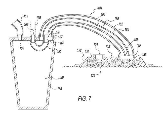
[0021]FIG. 7 shows a cross-sectional view of the collection container, wound
dressing,
and associated tubing shown in FIG. 6, taken along line 7.
[0022] FIG. 8 shows a perspective view of a first embodiment of an adjustable
restrictor
used in the negative pressure wound therapy system shown in FIGS. 1A-1C.
[0023]FIG. 9 shows a cross-sectional view of the adjustable restrictor shown
in FIG. 8,
taken along line 9.
3
CA 02993653 2018-01-24
WO 2017/019939 PCT/US2016/044647
[0024]FIG. 10 shows a perspective view of a second embodiment of an adjustable
restrictor used in the negative pressure wound therapy system shown in FIGS.
1A-1C.
[0025]FIG. 11 shows a cross-sectional view of the adjustable restrictor shown
in FIG. 10,
taken along line 11.
[0026] FIG. 12 shows a perspective view of an adapter used in the negative
pressure
wound therapy system shown in FIGS. 1B-1C.
[0027] FIG. 13 shows a top view of the adapter shown in FIG. 12.
[0028] FIG. 14 shows a cross-sectional view of the adapter shown in FIGS. 12-
13, taken
along line 14.
[0029] FIG. 15A shows a perspective view of a gasket used in the negative
pressure
wound therapy system shown in FIGS. 1B-1C.
[0030] FIG. 15B shows a cross-sectional view of the gasket shown in FIG. 15A,
taken
along line 15b.
[0031] FIG. 16A shows a perspective view of the adapter shown in FIG. 12
connected to
the lid shown in FIG. 3B.
[0032] FIG. 16B shows a cross-sectional view of the lid and adapter shown in
FIG. 16A,
taken along line 16b, and further includes the gasket shown in FIG. 15A (which
is not
visible in FIG. 16A).
[0033] FIG. 160 shows a detailed cross-sectional view of the lid, adapter, and
gasket
shown in FIG. 16B.
[0034] FIG. 17 shows a perspective view of a y-connector used in the negative
pressure
wound therapy system shown in FIG. 10.
[0035] FIG. 18 shows a top view of the y-connector shown in FIG. 17.
[0036] FIG. 19 shows a cross-sectional view of the y-connector shown in FIGS.
17-18,
taken along line 19.
[0037] FIG. 20 shows a cross-sectional view of the y-connector shown in FIG.
17, taken
along line 20.
[0038] FIG. 21 shows a perspective view of a valve (in an open position) used
in the
configurations of the negative pressure wound therapy system shown in FIGS. 18-
1C.
[0039] FIG. 22 shows a side view of the valve (in an open position) shown in
FIG. 21.
4
CA 02993653 2018-01-24
WO 2017/019939 PCT/US2016/044647
[0040] FIG. 23 shows a cross-sectional view of the valve (in an open position)
shown in
FIG. 21, taken along line 23.
[0041] FIG. 24 shows a cross-sectional view of the valve (in an open position)
shown in
FIG. 21, taken along line 24.
[0042] FIG. 25 shows a perspective view of the valve of FIGS. 21-24, now shown
in a
closed position.
[0043] FIG. 26 shows a side view of the valve (in a closed position) shown in
FIG. 25.
[0044] FIG. 27 shows a cross-sectional view of the valve (in a closed
position) shown in
FIG. 25, taken along line 27.
[0045] FIG. 28 shows a cross-sectional view of the valve (in a closed
position) shown in
FIG. 25, taken along line 28.
[0046] FIG. 29 shows a perspective view of a housing used in the valve of
FIGS. 21-28.
[0047] FIG. 30 shows a cross-sectional view of the housing shown in FIG. 29,
taken along
line 30.
[0048] FIG. 31 shows a cross-sectional view of the housing shown in FIG. 29,
taken along
line 31.
[0049] FIG. 32 shows a perspective view of a slide switch used in the valve of
FIGS. 21 -
2 8
[0050] FIG. 33 shows a side view of the slide switch shown in FIG. 32.
[0051] FIG. 34 shows a perspective view of a valve seat used in the valve of
FIGS. 21 -
2 8 .
[0052] FIG. 35 shows a cross-sectional view of the valve seat shown in FIG.
34, taken
along line 35.
[0053] FIG. 36 shows a cross-sectional view of the valve seat shown in FIG.
34, taken
along line 36.
[0054] FIG. 37 shows a cross-sectional view of the valve seat shown in FIG.
34, taken
along line 37.
[0055] FIG. 38 shows a perspective view of a wound dressing subassembly used
in the
negative pressure wound therapy system shown in FIGS. 18-1C.
[0056] FIG. 39 shows a perspective view of a y-connector subassembly used in
the
negative pressure wound therapy system shown in FIG. 1C.
CA 02993653 2018-01-24
WO 2017/019939 PCT/US2016/044647
[0057] FIG. 40 shows a side view of a Comparison A tube set.
[0058] FIG. 41 shows a side view of a Comparison B tube set.
[0059] FIG. 42 shows a side view of an Example 1 tube set.
[0060] FIG. 43 shows a side view of test set-up including a mechanical test
system and a
sample tube set.
[0061] FIG. 44 shows a front view of the test set-up of FIG. 43.
[0062] FIGS. 45-46 shows two test configurations used to test the sample tube
sets in the
EXAMPLE section.
DETAILED DESCRIPTION
[0063]The detailed description set forth below, in connection with the
appended
drawings, is intended as a description of various configurations and is not
intended to
represent the only configurations in which the concepts described herein may
be
practiced. The detailed description includes specific details for the purpose
of providing
a thorough understanding of the various concepts. However, it will be apparent
to those
skilled in the art that these concepts may be practiced without these specific
details.
[0064] Various aspects of a negative pressure wound therapy system may be
illustrated
by describing components that are coupled, attached, connected, pneumatically
associated, and/or joined together. As used herein, the terms "coupled",
"attached",
"connected", "pneumatically associated-, "in communication with", and/or
"joined" are
interchangeably used to indicate either a direct connection between two
components or,
where appropriate, an indirect connection to one another through intervening
or
intermediate components. In contrast, when a component is referred to as being
"directly
coupled", "directly attached", "directly connected" and/or "directly joined"
to another
component there are no intervening elements shown in said examples.
[0065] As illustrated in FIGS. 1A-1C, a negative pressure wound therapy system
100 may
include a microcontroller 101, a membrane keypad and display 160, one or more
vacuum
pumps 105 and/or 107, a collection container 165, one or more fluid barriers
129 and/or
113, a wound dressing 123, a battery 127, a muffler 128, a patient tube set
181, an
adjustable restrictor 200, a solenoid 177, an optional orifice restrictor 178,
a pump
pressure sensor 109, and a wound pressure sensor 173. These components may be
6
CA 02993653 2018-01-24
WO 2017/019939 PCT/US2016/044647
connected through a series of adapters, connectors, pneumatic tubes and
electrical
cables. Various configurations of the system 100 are contemplated, and example
configurations of the system 100 are shown in FIGS. 1B and 1C. The system 100
may
also include a one or more valves 500, additional wound dressings 123, y-
connector 400,
and various adapters 300.
[0066] Many of the components may be provided as part of a pump unit 120,
which may
include one or more of the microcontroller 101, membrane keypad and display
160,
vacuum pumps 105 and/or 107, fluid barrier 113, battery 127, muffler 128,
adjustable
restrictor 200, solenoid 177, orifice restrictor 178, pump pressure sensor
109, wound
pressure sensor 173, and related pneumatic tubes and electrical cables.
Pneumatic
tubes 176 and 115 may be separate components used to connect the collection
container
165 to the pump unit 120, or they may preferably be provided inside the pump
unit 120.
The pump unit 120 may have a vacuum port which connects to pneumatic tube 115,
and
a sensor port which connects to pneumatic tube 176. If pneumatic tubes 176 and
115
are provided inside the pump unit 120, the pump unit 120 may have a vacuum
port at the
end of pneumatic tube 115 that interfaces with the canister 165, and a sensor
port at the
end of pneumatic tube 176 that interfaces with the canister 165.
CONTROLLER
[0067]As illustrated in FIGS. 1A-1C, the negative pressure wound therapy
system 100
generally includes a microcontroller 101 having an embedded microprocessor
102,
Random Access Memory (RAM) 103 and Read Only Memory (ROM) 104. ROM 104
contains programming instructions for a control algorithm 150. ROM 104 may be
non-
volatile and may retain its programming when the power is terminated. RAM 103
is utilized
by the control algorithm 150 for storing variables such as pressure
measurements, alarm
counts and the like, which the control algorithm 150 uses while generating and
maintaining the vacuum.
VACUUM SOURCES
[0068] IVIicrocontroller 101 is electrically associated with, and controls the
operation of, a
first vacuum pump 105 and an optional second vacuum pump 107 through
electrical
7
CA 02993653 2018-01-24
WO 2017/019939 PCT/US2016/044647
cables 106 and 108 respectively. To increase the airflow of the system,
additional vacuum
pumps may also be included. Electrical cables used with system 100 may be
multi-
conductor ribbon cables or flat flexible cables (FFC), or any cable that
allows
communication between two or more system components. First vacuum pump 105 and
optional second vacuum pump 107 may be one of many types including, for
example, the
pumps sold under the trademarks Hargraves and Thomas . Vacuum pumps 105 and
107 may use, for example, a reciprocating diaphragm or piston to create vacuum
and are
typically powered by a D.C. motor that may also optionally use a brushless
commutator
for increased reliability and longevity. Vacuum pumps 105 and 107 may also be,
for
example, a rotary diaphragm pump which is a hybrid of a rotary pump and a
diaphragm
pump. Although some embodiments include one or more pumps as the vacuum
source,
the system 100 may use any type of vacuum source, including a squeeze bulb, a
spring-
loaded suction device, or hospital-supplied "wall suction" with pressure
regulator/controller.
[0069] Vacuum pumps 105 and/or 107 may be capable of producing vacuum
pressures,
which are pressures that have lower absolute values compared to the
atmospheric
pressure of the surrounding environment. The vacuum pumps 105 and/or 107 may
be
able to produce vacuum pressures that range from about 70 mmHg below
atmospheric
pressure to about 150 mmHg below atmospheric pressure, where vacuum pressures
of
150 mmHg below atmospheric pressure are stronger vacuums compared to vacuum
pressures of 70 mmHg below atmospheric pressure. For example, at standard
atmospheric pressure of 760 mmHg, vacuum pumps 105 and/or 107 may generate a
vacuum pressure having an absolute value ranging from about 610 mmHg to about
690
mmHg, where vacuum pressures of 610 mmHg are stronger vacuums compared to
vacuum pressures of 690 mmHg. In addition, vacuum pumps 105 and/or 107 may
also
be capable of producing vacuum pressures outside this range. For example,
vacuum
pumps 105 and/or 107 may be able to produce vacuum pressures that range from
about
50 mmHg below atmospheric pressure to about 200 mmHg below atmospheric
pressure.
[0070] An acoustic muffler 128 may be pneumatically associated with the
exhaust ports
of vacuum pumps 105 and/or 107 through pneumatic exhaust tubing 138 and is
configured to reduce exhaust noise produced by the pumps during operation. An
8
CA 02993653 2018-01-24
WO 2017/019939 PCT/US2016/044647
activated carbon odor trap may also be associated with the exhaust ports of
vacuum
pumps 105 and/or 107 through pneumatic exhaust tubing 138.
[0071] In normal operation of the negative pressure wound therapy system 100,
first
vacuum pump 105 (and optionally one or more additional vacuum pumps, such as a
second vacuum pump 107) may be used to generate an initial or "draw-down"
vacuum
while the optional second vacuum pump 107 may be used to maintain a desired
vacuum
within the system 100, compensating for leaks or pressure fluctuations. The
second
vacuum pump 107 may be smaller and quieter than the first vacuum pump 105
providing
a means to maintain the desired pressure without significantly disturbing the
patient.
DISPLAY
[0072]A membrane keypad and display 160 may be electrically associated with
microcontroller 101 through electrical cable 164. Membrane switches 161
provide power
control, while membrane switches 162 may be used to preset the desired vacuum
levels.
Light emitting diodes (LEDs) 163 may be provided to indicate alarm conditions
associated
with collection container 165 fluid level and wound dressing 123 leaks.
Preferably, an
LCD display could be used in place of the LEDs 163 to indicate alarm
conditions.
POWER
[0073] The system 100 may be powered by an external source of power. A battery
127
is optionally provided to permit portable operation of the negative pressure
wound therapy
system 100. Battery 127, which may be Lithium Ion, Nickel-Metal-Hydride
(NiMH), Nickel-
Cadmium, (NiCd) or their equivalent, is electrically associated with
microcontroller 101
through electrical cables 136 and 137. Battery 127 is charged by circuits
related with
microcontroller 101 while an external source of power is available such as
would typically
be supplied by a low-voltage A.C. adapter. When an external source of power is
not
available and the unit is to operate in a portable mode, battery 127 supplies
power to the
negative pressure wound therapy system 100.
9
CA 02993653 2018-01-24
WO 2017/019939 PCT/US2016/044647
COLLECTION CONTAINER
[0074] The negative pressure wound therapy system 100 includes a collection
container
165. A first embodiment of the collection container 165 is shown in FIGS. 2A-
2C, and a
second embodiment of a collection container 165 is shown in FIG. 3A. The
collection
container 165 encloses an internal chamber 166 into which fluid and exudate
may drain.
The collection container 165 may be a canister, an in-line vessel, or any
container capable
of collecting exudate. The volume of the collection container 165 may vary.
The collection
container 165 may be formed as a cylinder (as shown in FIG. 3A), an inverted
truncated
cone (as shown in FIG. 2A), or any number of other shapes. In a preferred
embodiment,
the collection container 165 may be substantially cylindrical. In some
embodiments, the
volume of the collection container 165 may be between about 300 mL and about
1200
mL. It may be preferable that collection container 165 has a volume which does
not
exceed about 1500 mL in order to prevent accidental exsanguination of a
patient in the
event hemostasis has not yet been achieved at the wound site.
[0075] The embodiments shown in FIGS. 2A-2C and 3A-3E have three openings in
the
collection container 165: a patient port 167, a vacuum port 168, and a sensor
port 169.
However, fewer or additional openings are possible. In FIGS. 2A-2C, 6, and 7,
the patient
port 167, vacuum port 168, and sensor port 169 are linearly arranged on the
collection
container 165. However, other port arrangements are also possible. For
example, the
patient port 167, vacuum port 168, and sensor port 169 may form a triangle on
the
collection container 165, as shown in FIGS. 3A-3E. Beneficially, the vacuum
port 168
and sensor port 169 may be positioned on the collection container 165 such
that they
may be able to connect directly to ports on the pump unit 120 without the use
of additional
pneumatic tubing. The vacuum port 168 and/or sensor port 169 may include a
locking
feature 168a that allows the collection container to connect to the pump unit
120 (for
example, a groove which may receive a latch on one of the ports on the pump
unit 120).
However, pneumatic tubing 176, 115 outside the pump unit 120 (as shown in
FIGS. 1A-
1C) may also be used to connect the vacuum port 168 and sensor port 169 on the
collection container 165 to ports on the pump unit 120.
[0076]The internal chamber 166 of the collection container 165 may be
pneumatically
associated with vacuum pumps 105 and/or 107 through a tube 115 connected to
the
CA 02993653 2018-01-24
WO 2017/019939 PCT/US2016/044647
vacuum port 168 of the collection container 165. Tube 115 may connect to first
vacuum
pump 105 and optional second vacuum pump 107 through "T- connectors 111 and
112,
respectively.
[0077]A fluid barrier 129 may be provided with the collection container 165.
The fluid
barrier 129 may be proximate to the vacuum port 168 and may be configured to
prevent
fluids collected in the collection container 165 from escaping through the
vacuum port
168 into tube 115, which could potentially damage vacuum pumps 105 and 107.
The
fluid barrier 129 may be a porous polymer hydrophobic filter such as those
available under
the trademark Porex . Alternatively, the fluid barrier 129 may have a
mechanical float
design or may have one or more membranes of hydrophobic material such as those
available under the trademark GoreTexTm. A secondary barrier 113 may include a
hydrophobic membrane which may be provided in line with tube 115 to prevent
fluid
ingress into the system 100 in the event fluid barrier 129 fails to operate as
intended. The
fluid barrier 129 may be included on the outside of the collection container
165 as shown
in FIGS. 1A-1C, or it may be positioned inside the internal chamber 166 of the
collection
container 165. Preferably, the fluid barrier 129 may be connected to a lid
165a of the
collection container 165 as shown in FIG. 3E.
[0078]The patient port 167 of collection container 165 may include a first
attachment 195
and a second attachment 194. As shown in the exemplary embodiments of FIGS. 2A-
2C
and 3A-3E, the second attachment 194 may be positioned inside the first
attachment 195,
with a web 196 connecting the first attachment 195 and the second attachment
194. The
inner walls of the second attachment 194 form a sensor channel 171, and the
space
between the first attachment 195 and the second attachment 194 forms a fluid
channel
172. A sensor tube 190, shown in FIG. 7, may be coupled to collection
container 165 and
connects the second attachment 194 of the patient port 167 to the sensor port
169 such
that air flowing through the sensor channel 171 of the patient port 167
remains separate
from the air in the internal chamber 166. Although sensor tube 190 is shown as
a
separate tube in FIG. 7, sensor tube 190 may be integrally formed with, or
provided as
part of, one or more of the collection container 165, tube 176, or second tube
182 of
patient tube set 181.
11
CA 02993653 2018-01-24
WO 2017/019939 PCT/US2016/044647
[0079]The collection container 165 may be formed as a single component (as
shown in
the first embodiment, FIGS. 2A-2C), or preferably, the collection container
165 may be
an assembly of a lid 165a and a base 165b (as shown in the second embodiment.
FIG.
3A). If the collection container 165 is an assembly of a lid 165a and a base
165b, the lid
165a and the base 165b together may enclose the internal chamber 166. The lid
165a
of the second embodiment of the collection container 165 is shown in FIGS. 3B-
3E. A
snap 165c may be included on the lid 165a (see FIG. 3E), which interlocks with
a groove
in the base 165b (or alternatively, a snap in the base 165b may interlock with
a groove
on the lid 165a) to prevent the lid 165a and base 165b from being separated
during use.
Additional ribs 165d or sealing rings 165e may be included on the lid 165a or
the base
165b to provide a seal between the lid 165a and the base 165b. The patient
port 167,
vacuum port 168, and/or sensor port 169 may be provided on either the lid 165a
or the
base 165b of the collection container 165. Preferably, the patient port 167,
vacuum port
168, sensor port 169, sensor tube 190, and fluid barrier 129 may all be
provided on a lid
165a as shown in FIGS. 3A-3E.
[0080]The collection container 165 may be configured to allow the patient tube
set 181
to be directly connected to the patient port 167 (as shown in the first
embodiment, FIGS.
2A-2C), or preferably, the collection container 165 may be configured to allow
the patient
tube set 181 to connect to the patient port 167 via an adapter 300 (as shown
in the second
embodiment, FIGS. 3A-3E). If the patient port 167 is configured to connect to
the patient
tube set 181 via an adapter 300, one or more pins 167a may be included on an
outer
surface of the first attachment 195. The pins 167a may be able to interlock
with one or
more slots 329 on the adapter 300.
WOUND DRESSING
[0081]A wound dressing 123 may include a sterile porous substrate 131, a
semipermeable adhesive cover 132, an optional inlet port 134, and a suction
port 135.
The porous substrate 131 may be polyurethane foam, polyvinyl alcohol foam,
gauze, felt
or any other suitable material. The semipermeable adhesive cover 132 may be
made of
a material sold under the trademark Avery Dennison or an adhesive film
product made
by DermaMed . There may be two openings in the semipermeable adhesive cover
132:
12
CA 02993653 2018-01-24
WO 2017/019939 PCT/US2016/044647
an inlet port 134 and a suction port 135. Suction may be applied to the wound
dressing
123 through suction port 135. Irrigation fluid may be applied to the wound
dressing 123
through inlet port 134, as is further discussed in U.S. Patent No. 7,608,066,
the entirety
of which is hereby incorporated by reference. If irrigation fluid is not
desired, the inlet port
134 may be omitted from the wound dressing 123.
[0082]As shown in FIG. 7, when wound dressing 123 is applied to the patient,
the
semipermeable adhesive cover 132 forms a seal with the patient's skin around
the
periphery of the wound dressing 123, thus creating a cavity enclosed by the
semipermeable adhesive cover 132 and the wound 124. The periphery of the
semipermeable adhesive cover 132 can be sealed to the patient's skin around
the
periphery of the wound. The porous substrate 131 is positioned between the
wound 124
and the semipermeable adhesive cover 132. The porous substrate 131 may contact
the
wound 124, but because the surface of the wound 124 may be uneven, the porous
substrate 131 may not contact the entire surface area of the wound 124.
[0083]When a vacuum is applied to the wound dressing 123 through the suction
port 135,
the vacuum is maintained in the cavity. The porous substrate 131 is provided
within the
cavity to distribute vacuum pressure evenly throughout the entire wound bed
and prevent
collapse of the cavity. The porous substrate 131 includes mechanical
properties suitable
for promoting the formation of granular tissue and approximating the wound
margins. In
addition, when vacuum is applied to wound dressing 123, porous substrate 131
creates
micro- and macro-strain at the cellular level of the wound stimulating the
production of
various growth factors and other cytokines and promoting cell proliferation.
PATIENT TUBE SET
[0084]As shown in FIGS. 6-7, the suction port 135 of the wound dressing 123
may be
pneumatically associated with the collection container 165. Further, the wound
pressure
sensor 173, solenoid 177, orifice restrictor 178, and adjustable restrictor
200 may be
pneumatically associated with the wound dressing 123 by a patient tube set
181, typically
in combination with one or more of a sensor tube 190 connecting the patient
port 167 and
the sensor port 169 of the collection container 165 and a tube 176 connected
to the sensor
port 169. The patient tube set 181 may include a fluid channel 189 that
pneumatically
associates wound dressing 123 with the internal chamber 166 of collection
container 165
13
CA 02993653 2018-01-24
WO 2017/019939 PCT/US2016/044647
for applying suction to wound dressing 123, thereby providing a path for fluid
to be moved
from the wound 124 to the collection container 165. The patient tube set 181
may include
a sensor channel 188 that pneumatically associates the wound dressing 123 with
one or
more of the wound pressure sensor 173, solenoid 177, orifice restrictor 178,
and
adjustable restrictor 200. The fluid channel 189 and sensor channel 188 may be
formed
from a plurality of tubes.
[0085]The patient tube set 181 may have a tube-within-a-tube design as shown
in FIG.
4, including a first tube 185 and a second tube 182. Second tube 182 may be
positioned
inside the lumen of first tube 185 such that the second tube 182 becomes an
inner tube
and the first tube 185 becomes an outer tube. Second tube 182 has a patient
end 183
and a device end 184. First tube 185 has a patient end 186 and a device end
187.
[0086]The second tube 182 and first tube 185 may have any cross-sectional
shape,
including a circle; oval, rectangle, square, or any other shape, although a
substantially
circular cross-sectional shape may be preferred. Preferably, the first tube
185 and the
second tube 182 may have the same length. The overall length of the patient
tube set
181 may vary. Patient tube sets 181 connected to the wound dressing 123 may be
longer
than patient tube sets 181 used to connect other components. For example, a
patient
tube set 181 used to connect the wound dressing 123 with a valve 500 may be
longer
than a patient tube set 181 used to connect the valve 500 with an adapter 300.
[0087]The patient tube set 181 may therefore include two channels. The lumen
of the
second tube 182 may be a sensor channel 188 which pneumatically associates the
wound dressing 123 with one or more of the wound pressure sensor 173, solenoid
177,
orifice restrictor 178, and adjustable restrictor 200. The space between the
inner surface
of the first tube 185 and the outer surface of the second tube 182 may form a
fluid channel
189. The fluid channel 189 pneumatically associates wound dressing 123 with
the
internal chamber 166 of collection container 165.
[0088] During use, a patient tube set 181 may be connected to the suction port
135 of the
wound dressing 123 and the patient port 167 of the collection container 165 as
shown in
FIGS. 6-7. One or both of the patient end 183 of the second tube 182 and the
patient
end 186 of the first tube 185 may be connected to the suction port 135 of the
wound
dressing 123. The device end 184 of the second tube 182 may be connected to
the
14
CA 02993653 2018-01-24
WO 2017/019939 PCT/US2016/044647
second attachment 194 of the patient port 167. The device end 187 of the first
tube 185
may be connected to the first attachment 195 of the patient port 167. The
fluid channel
189 of patient tube set 181 may be in communication with the internal chamber
166 of
the collection container 165 via the fluid channel 172 of the patient port
167. The sensor
channel 188 of patient tube set 181 may be in communication with sensor tube
190 via
the sensor channel 171 of the patient port 167. Sensor tube 190 communicates
with tube
176 via the sensor port 169 of collection container 165. Therefore, sensor
channel 188
of patient tube set 181 communicates with tube 176, which communicates with
one or
more of the wound pressure sensor 173, solenoid 177, orifice restrictor 178,
and
adjustable restrictor 200. In the exemplary embodiment shown in FIG. 7, the
sensor
channel 188 of the patient tube set 181 does not open into the internal
chamber 166 of
the collection container 165; rather, the sensor channel 188 of the patient
tube set 181
may be in communication with the sensor port 169 of the collection container
165 via
sensor tube 190. However, other configurations are possible in which the
sensor channel
188 opens into the internal chamber 166 of the collection container 165.
[0089J The patient tube set 181 may be manufactured using standard
manufacturing
techniques. In a preferred embodiment, the first tube 185 and the second tube
182 may
be coextruded and joined by a web 193 extending between the inner surface of
first tube
185 and the outer surface of second tube 182. Connecting second tube 182 and
first
tube 185 with a web 193 may facilitate the assembly process by ensuring that
the second
tube 182 and the first tube 185 remain connected. Although only one web 193 is
shown
in FIG. 4, a plurality of webs 193 may be used. Alternatively, first tube 185
and second
tube 182 could be manufactured separately, and the second tube 182 could be
inserted
into the lumen of the first tube 185.
[0090]The patient tube set 181 may be made from polyvinylchloride (PVC),
silicone, low
density polyethylene (LDPE), polyurethane, or any other material that is
flexible enough
to allow the patient tube set 181 to bend, yet rigid enough that the first
tube 185 and
second tube 182 do not collapse if a vacuum is applied within the tubes.
Preferably, the
patient tube set 181 may be made from PVC. Likewise, the thicknesses of the
walls of
the first tube 185 and the second tube 182 may be selected such that the tubes
are flexible
and compliant while still providing enough structural integrity that the tubes
do not
CA 02993653 2018-01-24
WO 2017/019939 PCT/US2016/044647
collapse if a vacuum is applied within the tubes. Preferably, the thickness of
the first tube
185 may be about 0.035 inches, and the thickness of the second tube 182 may be
about
0.030 inches. The thickness of the web 193 may be about 0.030 inches.
[0091]lncreasing the cross-sectional area of the sensor channel 188 compared
to
conventional designs may reduce the likelihood of fluid entering and occluding
the sensor
channel 188 due to capillary action. The cross-sectional area of the sensor
channel 188
may be calculated based on the dimension of the inner surface of the second
tube 182.
For example, the cross-sectional area of a sensor channel formed by a
cylindrical tube
may be calculated as the area of a circle formed by the inner diameter of the
tube. In
some embodiments, the sensor channel 188 may have a cross-sectional area that
is at
least about 0.75 mm2. In some embodiments, the sensor channel 188 may have a
cross-
sectional area in the range of between about 0.75 mm2 and about 7 mm2. In some
embodiments, the sensor channel 188 may have a cross-sectional area of at
least about
1.75 mm2. In some embodiments, the sensor channel 188 may have a cross-
sectional
area in the range of between about 175 mm2 and about 7 mm2. In some
embodiments,
the sensor channel 188 may have a cross-sectional area of at least about 2.5
mm2. In
some embodiments, the sensor channel 188 may have a cross-sectional area in
the
range of about 2.5 mm2 to about 7 mm2. In some embodiments, the sensor channel
188
may have a cross-sectional area in the range of about 2.5 mm2 to about 5 mm2.
In a
preferred embodiment, the sensor channel 188 may have a cross-sectional area
of about
3.25 mm2. However, the cross-sectional area of the sensor channel 188 could be
increased until the patient tube set 181 becomes too bulky for customer
acceptance.
[0092]The cross-sectional area of the fluid channel 189 of patient tube set
181 may be
determined by calculating the cross-sectional area between the inner surface
of first tube
185 and the outer surface of the second tube 182. For example, the cross
sectional area
of a fluid channel formed by the space between a cylindrical first tube and a
cylindrical
second tube may be determined by calculating the area of a circle formed by
the inner
diameter of the first tube, and subtracting the area of a circle formed by the
outer diameter
of the second tube. In some embodiments, the fluid channel 189 may have a
cross-
sectional area of at least about 10 mm2. In some embodiments, the fluid
channel 189
may have a cross-sectional area in the range of about 10 mm2 to about 30 mm2.
In some
16
CA 02993653 2018-01-24
WO 2017/019939 PCT/US2016/044647
embodiments, the fluid channel 189 may have a cross-sectional area of at least
about 15
mm2. In some embodiments, the fluid channel 189 may have a cross-sectional
area in
the range of about 15 mm2 to about 20 mm2. In a preferred embodiment, the
fluid channel
189 may have a cross-sectional area of about 17.75 mm2.
[0093] Generally, first tube 185 has a larger cross-sectional area compared to
second
tube 182 such that second tube 182 may fit inside the lumen of the first tube
185 while
leaving sufficient space between the first tube 185 and the second tube 182 to
create a
fluid channel 189. In the case where both second tube 182 and first tube 185
are
cylindrical, the inner diameter of first tube 185 may be larger than the outer
diameter of
the second tube 182. In some embodiments, the ratio of the cross-sectional
area of the
fluid channel 189 to the cross-sectional area of the sensor channel 188 may be
in the
range of about 4:1 to about 7:1. In some embodiments, the ratio of the cross-
sectional
area of the fluid channel 189 to the cross-sectional area of the sensor
channel 188 may
be in the range of about 5:1 to about 6:1. In a preferred embodiment, the
ratio of the
cross-sectional area of the fluid channel 189 to the cross-sectional area of
the sensor
channel 188 may be about 5.5:1.
[0094] In an alternative embodiment of a patient tube set 181' as shown in
FIG. 5, the
second tube 182' and first tube 185' may be positioned side-by-side instead of
positioning
the second tube inside the lumen of the first tube. In patient tube set 181',
the lumen of
the second tube 182' would form the sensor channel 188', and the lumen of the
first tube
185' would form the fluid channel 189'. In this embodiment, second tube 182'
and first
tube 185' may be co-extruded or second tube 182' and first tube 185' may be
manufactured separately. The collection container 165 and wound dressing 123
may be
modified to accommodate the different configurations of the first tube 185'
and second
tube 182' of patient tube set 181'. The patient port 167 of the collection
container 165
and the suction port 135 of the wound dressing 123 may be modified to
accommodate
the side-by-side tubes. Furthermore, one or more of the second attachment 194
in the
patient port 167, sensor tube 190, and the sensor port 169 of the collection
container 165
may be eliminated, as the second tube 182' which forms the sensor channel 188'
could
connect to tube 176 without the need for to sensor tube 190 inside the
collection container
165.
17
CA 02993653 2018-01-24
WO 2017/019939 PCT/US2016/044647
[0095] Although the first tube and second tube may be positioned side-by-side,
the tube-
within-a-tube design of the patient tube set 181 shown in FIG. 4 may be
preferred because
it may reduce kinking of the first tube 185 and/or the second tube 182. If the
patient tube
set 181 is accidentally bent, crushed, or otherwise deformed, the force may
cause the
first tube 185 to deform before the second tube 182 begins to deform, thereby
reducing
the changes of blocking the second tube 182. Furthermore, as the patient tube
set 181
deforms, the second tube 182 may act as a support structure that prevents the
first tube
185 from kinking and prevents the fluid channel 189 from becoming blocked.
Therefore,
air may flow through the fluid channel 189 and vacuum may be applied to the
wound
dressing 123 even if the patient tube set 181 is accidentally bent, crushed,
or otherwise
deformed.
[0096] The first and second tubes of the patient tube set are made of a semi-
rigid material
and are able to maintain their shape when a vacuum is applied by vacuum pumps
105
and 107. Therefore, the fluid channel 189 and the sensor channel 188 may be
substantially unobstructed because the first and second tubes are able to
resist collapsing
when a vacuum is applied by vacuum pumps 105 and 107.
[0097] Patient tube set 181 may include any number of sensor channels 188. In
some
embodiments, patient tube set 181 may include only one sensor channel 188. In
other
embodiments, patient tube set 181 may include a plurality of sensor channels
188. The
plurality of sensor channels 188 may be positioned inside the lumen of the
first tube 185.
The plurality of sensor channels 188 may be formed from a plurality of second
tubes, or
they may be formed from a single extrusion having multiple coaxial lumens.
However,
providing a plurality of sensor channels 188 may increase the overall size of
the cross-
section of patient tube set 181. Alternatively, if the overall size of the
cross-section of
patient tube set 181 is maintained and a plurality of sensor channels 188 are
used, the
cross-sectional area of each of the plurality of sensor channels 188 would
decrease,
making the sensor channels 188 more likely to occlude via capillary action.
Therefore, it
may be advantageous to use a patient tube set 181 with a single sensor channel
188 in
order to prevent occlusions while minimizing the overall size of patient tube
set 181.
Using multiple sensor channels 188 requires the microcontroller 101 to
determine which
of the sensor channels 188 is free of occlusions and providing accurate data,
and which
18
CA 02993653 2018-01-24
WO 2017/019939 PCT/US2016/044647
of the sensor channels 188 is occluded and therefore providing inaccurate
data.
Therefore, if multiple sensor channels 188 are used, the microcontroller 101
may show
an average measurement of the vacuum pressure applied across all sensor
channels
188.
[0098] Standing fluid in any of the tubes connecting pumps 105 and/or 107 to
the wound
dressing 123 (for example, the fluid channel 189 of patient tube set 181) may
create
hydrostatic forces that cause a difference in the pressure experienced at the
wound
dressing 123 (also referred to as the therapeutic pressure) compared to the
pressure
being created by pumps 105 and/or 107. Depending on the position of pumps 105
and/or
107 relative to the wound, the therapeutic pressure may be increased or
decreased
compared to the pressure measured at pumps 105 and/or 107. However, the system
100
may be able to compensate for these variations in therapeutic pressure
resulting from
hydrostatic forces. The therapeutic pressure may be monitored by wound
pressure
sensor 173, and the activity of pumps 105 and/or 107 may be adjusted based on
any
pressure fluctuations. This monitoring may occur in real time, such that pumps
105 and/or
107 may be able to compensate quickly when the position of the wound changes
relative
to pumps 105 and/or 107.
[0099] If the vertical position of pumps 105 and/or 107 is higher than the
wound, fluid in
any of the tubes connecting pumps 105 and/or 107 to the wound dressing 123
(for
example, the fluid channel 189 of patient tube set 181) may cause the absolute
value of
the therapeutic pressure applied to the wound to increase, such that a weaker
vacuum is
being applied to the wound. For example, if the pressure at vacuum pumps 105
and/or
107 is 70 mmHg below atmospheric pressure, the therapeutic pressure at the
wound
dressing 123 may be only 60 mmHg below atmospheric pressure. The increase in
the
absolute value of the therapeutic pressure may be detected by wound pressure
sensor
173 and communicated to microprocessor 102. Control algorithm 150 contains
instructions that will instruct pumps 105 and/or 107 to run, or continue to
run, in order to
compensate for the increase in the absolute value of the therapeutic pressure
at the
wound.
[0100]Conversely, if the vertical position of pumps 105 and/or 107 is lower
than the
wound, fluid in any of the tubes connecting pumps 105 and/or 107 to the wound
dressing
19
CA 02993653 2018-01-24
WO 2017/019939 PCT/US2016/044647
123 (for example, the fluid channel 189 of patient tube set 181) may cause the
absolute
value of the therapeutic pressure applied to the wound to decrease, such that
a stronger
vacuum is being applied to the wound. For example, if the pressure at vacuum
pumps
105 and/or 107 is 70 mmHg below atmospheric pressure, the therapeutic pressure
at the
wound dressing 123 may be 80 mmHg below atmospheric pressure. The decrease in
the
absolute value of the therapeutic pressure may be detected by wound pressure
sensor
173. Control algorithm 150 contains instructions that will instruct pumps 105
and/or 107
to turn off, or run less frequently, in order to compensate for the decrease
in the absolute
value of the therapeutic pressure at the wound. Control algorithm 150 may also
contain
instructions to open the solenoid 177 to relieve pressure in order to
compensate for the
decrease in the absolute value at the therapeutic pressure at the wound, if
necessary.
ADAPTER
[0101] An adapter 300, shown in FIGS. 12-14, may be provided on one or both
ends of
the patient tube set 181. Preferably, an adapter 300 may be provided on at
least the
device end of the patient tube set 181. The adapter 300 may have a first end
313 and a
second end 323. The adapter 300 may have at least two ports: a first port 310
at the first
end 313, configured to couple to a patient tube set 181, and a second port 320
at the
second end 323, configured to couple to the patient port 167 of the collection
container
165 and/or the second or third ports 420, 430 of a y-connector 400 (described
below).
The first port 310 and the second port 320 may meet at an interface 330. The
second
port 320, as described below, has a female fitting: however, the second port
could also
have a male fitting.
[0102]The adapter 300 may have an outer wall 301 and an inner wall 302
connected to
the outer wall 301 by one or more webs 303. The outer wall 301 may have an
outer
surface 306 and an inner surface 307. The inner wall 302 may have an outer
surface 308
and an inner surface 309. The outer wall 301 may extend along the first port
310 and the
second port 320, having a first end 311 at the first port 310 and a second end
321 at the
second port 320. The inner surface 307 of the outer wall 301 may have a larger
diameter
at the second port 320 and a smaller diameter at the first port 310. A
horizontal ledge
327 may be formed in the outer wall 301 at the interface 330 between the first
port 310
CA 02993653 2018-01-24
WO 2017/019939 PCT/US2016/044647
and the second port 320, where the inner surface 307 of the outer wall 301
transitions
from the larger diameter at the second port 320 to the smaller diameter at the
first port
310. The inner vvall 302 may extend along the first port 310, having a first
end 312
proximate the first end 313 of the adapter 300, and a second end 322 at the
interface 330
between the first port 310 and the second port 320. The inner wall 302 does
not
necessarily extend into the second port 320, and preferably it may not extend
into the
second port 320. Preferably, the ledge 327 and the second end 322 of the inner
wall 302
may be coplanar.
[0103] The inner surface 309 of the inner wall 302 may form a sensor channel
305. The
sensor channel 305 may have a first opening 315 on the first end 313 of the
adapter 300
and a second opening 325 at the interface 330 between the first port 310 and
the second
port 320. The space between the outer surface 308 of the inner wall 302 and
the inner
surface 307 of the outer wall 301 may form a fluid channel 304. The fluid
channel 304
may have a first opening 314 on the first end 313 of the adapter 300 and a
second opening
324 at the interface 330 between the first port 310 and the second port 320.
[0104] The cross-sections of the inner wall 302 and the outer wall 301 may be
circular,
elliptical, or various other shapes. However, in a preferred embodiment, the
inner wall
302 and the outer wall 301 may both be substantially circular. More
specifically, the inner
surface 307 of the outer wall 301 and the outer surface 308 of the inner wall
302 may
have a substantially circular cross-section. The inner surface 307 of the
outer wall 301
and the outer surface 308 of the inner wall 302 may be substantially
concentric.
[0105] One or more slots 329 may be provided in the outer wall 301 of the
second port
320 to receive pins 167a on the first attachment 195 of the patient port 167.
One or more
notches 328 may also be provided in the outer wall 301 at the second port 320
to receive
pins 390 on the gasket 380.
[0106]A gasket 380, shown in FIGS. 15A-15B, may be provided with the adapter
300.
The gasket 380 may include an outer sealing rib 381 and an inner sealing rib
382
connected to the outer sealing rib 381 by one or more webs 383. The outer
sealing rib
381 may have a cross-sectional shape similar to the cross-sectional shape of
the ledge
327 on the outer wall 301 of the adapter 300. The inner sealing rib 382 may
have a cross-
sectional shape that is similar to the cross-sectional shape of the second end
322 of the
21
CA 02993653 2018-01-24
WO 2017/019939 PCT/US2016/044647
inner vvall 302 of the adapter 300. The inner diameter of the inner sealing
rib 382 may
form a sensor channel 385, and the space between the outer sealing rib 381 and
the inner
sealing rib 382 may form a fluid channel 384. Preferably, the cross-sections
of the inner
sealing rib 382 and the outer sealing rib 381 may be substantially circular,
and even more
preferably they may be substantially concentric.
[0107] The gasket 380 may have a first surface and a second surface opposite
the first
surface, such that the outer sealing rib 381 has a first surface 386 and a
second surface
387, and the inner sealing rib 382 has a first surface 388 and a second
surface 389. The
gasket 380 may have one or more pins 390 extending outwardly from the outer
sealing
rib 381. Preferably, there may be the same number of pins 390 on the gasket
380 and
notches 328 in the adapter 300. The gasket 380 may be made of any number of
materials, including silicone, thermoplastic elastomers, natural rubber, or
any other
elastomeric, compressible, non-porous material. In a preferred embodiment, the
gasket
380 may be made of silicone.
[0108] The gasket 380 may be inserted into the second port 320 of the adapter
300. The
pins 390 on the gasket 380 may be inserted into the notches 328 in the adapter
300. The
first surface 386 of the outer sealing rib 381 of the gasket 380 may be in
contact with the
ledge 327 on the outer wall 301 of the adapter 300. Likewise, the first
surface 388 of the
inner sealing rib 382 of the gasket 380 may be in contact with the second end
322 of the
inner wall 302 of the adapter 300. The sensor channel 385 of the gasket 380
may be
aligned with the sensor channel 305 of the adapter 300. Each second opening
324 of the
fluid channel 304 of the adapter 300 may be aligned with a fluid channel 384
in the gasket
380. In order to provide a continuous path for fluids passing through the
fluid channels
304, 384 of the adapter 300 and the gasket 380, the pins 390 may be positioned
on the
gasket 380 to allow the webs 383 on the gasket 380 to substantially overlie
the webs 303
on the adapter 300.
[0109]A patient tube set 181 may be connected to the first port 310 of the
adapter 300.
The device end 187 of the first tube 185 may mate with the outer wall 301 of
the adapter
300. Preferably, the outer surface of the first tube 185 may be in contact
with the inner
surface 307 of the outer wall 301. The device end 184 of the second tube 182
may mate
with the inner wall 302 of the adapter 300. Preferably, the inner diameter of
the second
22
CA 02993653 2018-01-24
WO 2017/019939 PCT/US2016/044647
tube 182 may be in contact with the outer surface 308 of the inner wall 302.
Thus, the
fluid channel 304 of the adapter 300 may be in communication with the fluid
channel 189
of the patient tube set 181. The sensor channel 305 of the adapter 300 and the
sensor
channel 385 of the gasket 380 may be in communication with the sensor channel
188 of
the patient tube set 181.
[0110]The second port 320 of the adapter 300 may be connected to the patient
port 167
of the collection container 165, as shown in FIGS. 16A-160. The patient port
167 may
be inserted into the outer wall 301 of the second port 320 of the adapter 300.
The pins
167a on the patient port 167 may be inserted into the slots 329 in the outer
wall 301 of
the adapter 300. The gasket 380, positioned in the second port 320 of the
adapter 300,
may provide for sealing engagement between the second port 320 of the adapter
300
and the patient port 167 of the collection container 165. The ledge 327 on the
outer wall
301 of the adapter 300 may be in sealing engagement with the first attachment
195 of the
patient port 167 via the outer sealing rib 381 on the gasket 380. The second
end 322 of
the inner wall 302 of the adapter 300 may be in sealing engagement with the
second
attachment 194 of the patient port 167 via the inner sealing rib 382 on the
gasket 380.
[0111 ]Thus, if the adapter 300 is connected to both a patient port 167 on a
collection
container 165 and a patient tube set 181, as shown in FIGS. 16A-160, the
sensor channel
188 of patient tube set 181 may be in communication with the sensor channel
171 of the
patient port 167, and the fluid channel 189 of patient tube set 181 may be in
communication with the fluid channel 172 of the patient port 167. Using an
adapter may
be preferred because it may simplify the user set-up process by allowing the
user to
connect the adapter 300 to the patient port 167 of the collection container
165, as shown
in FIGS. 16A-160, instead of individually connecting first tube 185 and second
tube 182
to the patient port 167, as shown in FIG. 7.
PUMP PRESSURE SENSOR
[0112] Pump pressure sensor 109 may be pneumatically associated with first
vacuum
pump 105 and an optional second vacuum pump 107 as shown in FIGS. 1A-1C. Pump
pressure sensor 109 may be electrically associated with microcontroller 101
through
electrical cable 110. Pump pressure sensor 109 provides a vacuum-pressure
signal to
23
CA 02993653 2018-01-24
WO 2017/019939 PCT/US2016/044647
the microprocessor 102 enabling control algorithm 150 to monitor the vacuum
pressure
at the outlet of vacuum pumps 105 and/or 107.
WOUND PRESSURE SENSOR
[0113]A wound pressure sensor 173 may be pneumatically associated with the
sensor
port 169 of the collection container 165 through a tube 176 as shown in FIGS.
6-7. Tube
176 may be a single lumen tube as shown in FIG. 7. Because tube 176 is
pneumatically
associated with the wound dressing 123 via one or more of the sensor tube 190
and the
sensor channel 188 in the patient tube set 181, the wound pressure sensor 173
is able to
monitor the therapeutic pressure in the wound dressing 123 more accurately
than the
pump pressure sensor 109 can. Wound pressure sensor 173 may be electrically
associated with microcontroller 101 through electrical cable 174 and provides
a vacuum-
pressure signal to microprocessor 102 enabling control algorithm 150 to
monitor the
therapeutic pressure at the wound site.
SOLENO1D/VALVE
[01114] A solenoid 177 and optional orifice restrictor 178 may be
pneumatically associated
with the sensor port 169 of the collection container 165 through tube 176 as
shown in
FIGS. 1A-1C. lithe orifice restrictor 178 is not provided, solenoid 177 may
connected to
tube 176 by "T" connector 175. If the orifice restrictor 178 is provided, the
orifice restrictor
178 may be connected to tube 176 by a 1." connector 175, and vacuum-pressure
relief
solenoid 177 may be connected to the orifice restrictor 178. Together, the
solenoid 177
and optional orifice restrictor 178 act to relieve pressure in the wound
dressing 123 and
in the collection container 165 in the event of an alarm condition, if the set
pressure is
decreased (during intermittent mode, for example), or if power is turned off.
Solenoid 177
may be, for example, one available under the trademark Pneutronics , or Air
Logic .
Solenoid 177 is electrically associated with, and controlled by,
microprocessor 102
through electrical cable 130. Solenoid 177 may be configured to vent vacuum to
atmosphere when the power is turned off, for example. Orifice restrictor 178,
if it is
provided, is positioned in line with solenoid 177 and tube 176 to regulate the
rate at which
24
CA 02993653 2018-01-24
WO 2017/019939 PCT/US2016/044647
vacuum is relieved to atmospheric pressure when solenoid 177 is de-energized.
Orifice
restrictor 178 is, for example, available under the trademark Air Logic .
ADJUSTABLE RESTRICTOR
[0115] As shown in FIGS. 1A-1C, an optional adjustable restrictor 200 may be
pneumatically associated with tube 176. Two embodiments of an adjustable
restrictor
200 are shown in greater detail in FIGS. 8-11, and include a cap 210, a body
220, and a
porous material 230. Cap 210 has a base 211 and a flange 213. The base 211
and/or
flange 213 of cap 210 has at least one hole 212 that opens to the atmosphere
to allow air
to enter the body 220 and create a controlled air leak in the system 100.
Flange 213 has
a threaded portion 214 that engages the body 220. Body 220 includes at least
one tube
port 221 and a threaded port 223. The threaded port 223 on the body 220
engages the
threaded portion 214 of the cap 210, thereby coupling the cap 210 and the body
220.
[0116]The porous material 230 may be positioned between the threaded port 223
on the
body 220 and the base 211 of the cap 210. The porous material 230 may be
compressible. The porous material 230 may have minimal water absorbency (i.e.,
hydrophobic), which may prevent the flow rate of the air leak from changing
when the
restrictor 200 is exposed to increased or decreased humidity. A number of
common filter
materials may be used, including plastic foams, or synthetic membrane
materials such as
those used in cigarette filters. The porous material 230 may be disc-shaped
and may
cover the hole 212 on cap 210.
[0117]Air enters the adjustable restrictor 200 at the hole 212 in the cap 210
to create the
air leak. Air then travels through the porous material 230 before entering the
body 220 of
the adjustable restrictor 200 via the threaded port 223. The flow rate of the
air leak may
be controlled by compressing or decompressing the porous material 230.
Compressing
the porous material 230 by tightening the connection between the body 220 and
the cap
210 decreases the flow rate of the air leak. Decompressing the porous material
230 by
loosening the connection between the body 220 and the cap 210 increases the
flow rate
of the air leak.
[0118] In one embodiment shown in FIGS. 8-9, adjustable restrictor 200 may
include two
tube ports 221. If two tube ports 221 are provided, adjustable restrictor 200
may be
CA 02993653 2018-01-24
WO 2017/019939 PCT/US2016/044647
positioned in line with tube 176, and a "T" connector may not be needed to
connect
adjustable restrictor 200 to tube 176. In another embodiment shown in FIGS. 10-
11,
adjustable restrictor may include only one tube port 221 which may be
connected to tube
176 by a "T" connector 191 as shown in FIGS. 1A-1C.
[0119] Adjustable restrictor 200 may be designed to create an air leak that
allows air to
flow into tube 176. Because sensor channel 188 of the patient tube set 181 is
pneumatically associated with tube 176, the air leak also allows air to flow
into the sensor
channel 188 towards wound dressing 123, thereby preventing occlusions in the
sensor
channel 188. Any fluid that may have entered the sensor channel 188 (through
capillary
action, for example) is pushed toward the wound dressing 123, and fluid from
the wound
dressing 123 is prevented from flowing into the sensor channel 188 due to the
pressure
gradient. Furthermore, the air leak in the sensor channel 188 may cause air to
flow into
the fluid channel 189 at the suction port 135 of the wound dressing 123. The
air leak
would therefore provide a force that is additive to the suction force being
supplied by
pumps 105 and/or 107 to ensure fluid does not enter sensor channel 188 or
remove
occlusions from sensor channel 188. In addition, air from the air leak may
flow out of the
patient end 183 of the second tube 182 and into the patient end 186 of the
first tube 185,
thereby providing a force that is additive to the suction force being supplied
by pumps
105 and/or 107 to ensure that fluid in the fluid channel 189 would be forced
toward the
collection container 165. An advantage of using the adjustable restrictor 200
is that the
air leak may be substantially uninterrupted when a vacuum is applied to
collection
container 165 and/or wound dressing 123, such that the air leak may
continually prevent
occlusions and help to clear otherwise stationary fluid from the sensor
channel 188 and/or
fluid channel 189, instead of only acting in a reactive manner after an
occlusion has
formed.
[0120] The flow rate of the air leak should be low enough such that the air
leak does not
substantially affect the therapeutic pressure being applied to the wound
dressing 123,
causing a leak alarm to be triggered. The flow rate of the air leak should be
low enough
that pumps 105 and/or 107 are able to compensate for the small increase in the
absolute
value of the pressure in system 100 resulting from the leak. Therefore, the
therapeutic
pressure applied to the wound dressing 123 may be substantially maintained
despite the
26
CA 02993653 2018-01-24
WO 2017/019939 PCT/US2016/044647
air leak. For example, the leak may have a flow rate ranging from 0.05 ¨ 0.1
liters per
minute.
[0121 ]Alternatively, instead of using the adjustable restrictor 200, an air
leak may be
created by sporadically opening the solenoid 177, thereby venting tube 176 to
atmosphere such that air flows from solenoid 177 through tube 176, through one
or more
of sensor tube 190 and sensor channel 188 of patient tube set 181 toward wound
dressing
123. Any occlusions in the sensor channel 188 of patient tube set 181 may be
forced
toward wound dressing 123, and any fluid in the fluid channel 189 of patient
tube set 181
may be forced toward collection container 165. In this case, the solenoid 177
may
function as a time-variable restrictor.
Y-CONNECTOR
[0122]A y-connector 400, shown in FIGS. 17-20, may be provided to allow two
patient
tube sets 181 connected to two separate wound dressings 123 to be connected to
the
same patient port 167 of the collection container 165. The y-connector 400 may
have a
first end 413, a second end 423, and a third end 433. The y-connector 400 may
have at
least three ports: a first port 410 at the first end 413, configured to
connect to the collection
container 165, and second and third ports 420, 430 at the second and third
ends 423,
433, configured to connect to the two separate wound dressings 123. The second
and
third ports 420, 430, as described below, have male fittings; however they
could also have
female fittings.
[0123]The y-connector 400 may have an outer wall 401 and an inner wall 402
connected
to the outer wall 401 by one or more webs 403. The outer wall 401 may have an
outer
surface 406 and an inner surface 407. The inner wall 402 may have an outer
surface 408
and an inner surface 409. The outer wall 401 may extend along all three ports,
having a
first end 411 at the first end 413 of the y-connector 400, a second end 421 at
the second
end 423 of the y-connector 400, and a third end 431 at the third end 433 of
the y-connector
400. The inner wall 402 may extend along two of the ports, having a first end
412 at the
first end 413 of the y-connector 400 and a second end 422 at the second end
423 of the
y-connector 400, as shown in FIG. 19.
27
CA 02993653 2018-01-24
WO 2017/019939 PCT/US2016/044647
[01 24] The inner surface 409 of the inner wall 402 may form a sensor channel
405 that
extends from a first opening 415 on the first port 410 to a second opening 425
on the
second port 420. A fluid channel 404 may extend into all three ports, having a
first
opening 414 at the first port 410, a second opening 424 at the second port
420, and a
third opening 434 at the third port 430. The fluid channel 404 in the first
port 410 and the
second port 420 may be formed by the space between the outer surface 408 of
the inner
wall 402 and the inner surface 407 of the outer wall 401. The fluid channel
404 in the
third port 430 may be formed by the inner surface 407 of the outer wall 401.
[0125] The cross-sections of the inner wall 402 and the outer wall 401 may be
circular,
elliptical, or various other shapes. However, in a preferred embodiment, the
inner wall
402 and the outer wall 401 may both be substantially circular. More
specifically, the inner
surface 407 of the outer wall 401 and the outer surface 408 of the inner wall
402 may
have a substantially circular cross-section. The inner surface 407 of the
outer wall 401
and the outer surface 408 of the inner wall 402 may be substantially
concentric.
[0126] Any of the first port 410, second port 420, and third port 430 may be
designed to
connect to a patient tube set 181 directly, or indirectly using an adapter
300. In a preferred
embodiment, the y-connector 400 may be designed such that the first port 410
directly
connects with a patient tube set 181, while the second port 420 and third port
430 each
connect with a patient tube set 181 via an adapter 300.
[0127]A patient tube set 181 may be connected to the first port 410 of the y-
connector
400. The patient end 186 of the first tube 185 may mate with the outer wall
401 of the y-
connector 400. Preferably, the outer surface of the first tube 185 may be in
contact with
the inner surface 407 of the outer wall 401. The patient end 183 of the second
tube 182
may mate with the inner wall 402 of the y-connector 400. Preferably, the inner
surface of
the second tube 182 may be in contact with the outer surface 408 of the inner
wall 402.
Thus, the fluid channel 404 of the y-connector 400 may be in communication
with the fluid
channel 189 of the patient tube set 181. The sensor channel 405 of the y-
connector 400
may be in communication with the sensor channel 188 of the patient tube set
181.
[0128] In some embodiments, the second port 420 and third port 430 of the y-
connector
400 interface with an adapter 300 connected to a patient tube set 181. The
second port
320 of the adapter 300 may be connected to the second port 420 or the third
port 430 of
28
CA 02993653 2018-01-24
WO 2017/019939 PCT/US2016/044647
the y-connector 400. The second or third port 420, 430 of the y-connector 400
may be
inserted into the outer wall 301 of the second port 320 of the adapter 300.
The second
and third ports 420, 430 of the y-connector 400 may each have one or more pins
429,
439 on the outer wall 401 that may be inserted into the slots 329 in the
adapter 300. The
gasket 380, positioned in the second port 320 of the adapter 300, may provide
for sealing
engagement between the second port 320 of the adapter 300 and the second or
third port
420, 430 of the y-connector 400.
[0129] If the adapter 300 is being connected to the second port 420 of the y-
connector
400 (which includes an outer wall 401 and an inner wall 402), the ledge 327 on
the outer
wall 301 of the adapter 300 may be in sealing engagement with the second end
421 of
the outer wall 401 of the y-connector 400 via the outer sealing rib 381 on the
gasket 380.
The second end 322 of the inner wall 302 of the adapter 300 may be in sealing
engagement with the second end 422 of the inner wall 402 of the y-connector
400 via the
inner sealing rib 382 on the gasket 380.
[0130] If the adapter 300 is being connected to the third port 430 of the y-
connector 400
(which may include an outer wall 401 but not in inner wall 402), the ledge 327
on the outer
wall 301 of the adapter 300 may be in sealing engagement with the second end
431 of
the outer wall 401 of the y-connector 400 via the outer sealing rib 381 on the
gasket 380.
The second end 322 of the inner wall 302 of the adapter 300 may not be in
sealing
engagement with the y-connector 400.
[0131] When patient tube sets 181 are connected to each of the first, second,
and third
ports 410, 420, 430 of the y-connector 400, the fluid channels 189 of the
patient tube sets
181 connected to the second and third ports 420, 430 communicate with the
fluid channel
404 of the y-connector 400. The sensor channel 188 of the patient tube set 181
connected to the second port 420 of the y-connector 400 (into which the inner
wall 402
extends) communicates with the sensor channel 405 of the y-connector 400.
However,
the sensor channel 188 of the patient tube set 181 connected to the third port
430 of the
y-connector 400 (into which the inner wall 402 does not extend) communicates
with the
fluid channel 404 of the y-connector 400.
[0132]When using a y-connector 400, it is preferable that the sensor channel
405 extends
from the first port 410 to only one of the second port 420 or the third port
430. As
29
CA 02993653 2018-01-24
WO 2017/019939 PCT/US2016/044647
described above and shown in FIGS. 17-20, the sensor channel 405 may extend
from the
first port 410 to the second port 420, but not to the third port 430. If the
sensor channel
405 of the y-connector 400 also extended into the third port 430, the wound
pressure
sensor 173 may only detect a blockage when the patient tube sets 181 connected
to the
second and third ports 420, 430 of the y-connector 400 were both occluded. If
a blockage
or occlusion occurred in only one of the patient tube sets 181 connected to
the second
and third ports 420, 430 of the y-connector 400, the wound pressure sensor 173
would
still detect the vacuum from the unoccluded patient tube set 181, and the
blockage alarm
would not be triggered. Therefore, it may be advantageous for the y-connector
400 to
have a sensor channel 405 extending between only the first port 410 and the
second port
420. In this configuration, the wound pressure sensor 173 may detect a
blockage if the
patient tube set 181 connected to the second port 420 of the y-connector 400
was
occluded and the patient tube set 181 connected to the third port 430 of the y-
connector
400 was not occluded, or if the patient tube sets 181 connected to the second
and third
ports 420, 430 of the y-connector 400 were both occluded.
[0133] Alternatively, instead of providing the y-connector 400 and the adapter
300 as
separate components, they may be formed as a single component. In this case,
patient
tube sets 181 may be connected to the second and third ports 420, 430, and the
first port
410 may be integrated with the second port 320 of the adapter 300. The outer
wall 301
of the second port 320 of the adapter 300 may be continuous with the outer
wall 401 of
the first port 410 of the y-connector 400, and the inner wall 302 of the
second port 320 of
the adapter 300 may be continuous with the inner wall 402 of the first port
410 of the y-
connector 400. Potential methods for manufacturing a combined y-connector 400
and
adapter 300 may include 3D printing or molding.
VALVE
[0134]At certain points during use, it may be desirable to close the fluid
channel 189
and/or the sensor channel 188 of the patient tube set 181, thereby preventing
vacuum
pressures from being transmitted along the channel. For example, it may be
desirable to
close the fluid channel 189 and sensor channel 188 when replacing a collection
container
165 while maintaining the wound dressing 123 over the wound, or when
troubleshooting
CA 02993653 2018-01-24
WO 2017/019939 PCT/US2016/044647
leaks and blockages in the system 100. As discussed above, the patient tube
set 181
may be resistant to kinking, which may beneficially prevent the fluid channel
189 from
becoming occluded if the patient tube set 181 is accidentally bent, crushed,
or otherwise
deformed. However, as a result, the user may not be able to close the fluid
channel 189
using conventional means such as a clamp.
[0135] Therefore, in order to close the fluid channel 189 and/or the sensor
channel 188,
a valve 500 may be connected in-line with the patient tube set 181 to allow
the user to
occlude the fluid channel 189 and/or the sensor channel 188 when desired. The
valve
500 may include a valve housing 510, a slide switch 550, a valve seat 570, a
first indicator
band 501, and a second indicator band 502.
[0136]The valve housing 510 may have a longitudinal axis 516 and a transverse
axis 515
substantially perpendicular to the longitudinal axis 516. The valve housing
510 may have
a first longitudinal end 512, a second longitudinal end 513, a first
transverse end 517, and
a second transverse end 518. A channel 514 may extend from the first
longitudinal end
512 toward the second longitudinal end 513 along the longitudinal axis 516.
One or more
grooves 519 may extend longitudinally along the inner surface of the channel
514, starting
at the second longitudinal end 513. Preferably, the one or more grooves 519 do
not
extend all the way to the first longitudinal end 512 of the housing 510.
[0137] A first port 520 and a second port 530 may be included on the valve
housing 510.
The first port 520 and second port 530 may be provided at the first transverse
end 517
and second transverse end 518 of the valve housing 510, respectively. The
first and
second ports 520, 530 may each include an outer wall 521, 531 and an inner
wall 522,
532 connected to the outer wall 521, 531 by a web 523, 533. A web 523, 533 may
extend
between the inner wall 522, 532 and the outer wall 521, 531 along a line
substantially
parallel to the longitudinal axis 516 of the valve housing 510. The inner
surface of the
inner wall 522, 532 may form a sensor channel 525, 535. A fluid channel 524,
534 may
be formed by the space between the outer surface of the inner wall 522, 532
and the inner
surface of the outer wall 521, 531. Preferably, the first and second ports
520, 530 may
both be substantially parallel with the transverse axis 515.
31
CA 02993653 2018-01-24
WO 2017/019939 PCT/US2016/044647
[0138] The first and second ports 520, 530 may be connected to patient tube
sets 181 by
mating the outer wall 521, 531 with the first tube 185 and the inner wall 522,
532 with the
second tube 182.
[0139] The valve housing 510 may alternatively be formed together with an
adapter 300
to form a single component. In this case, a patient tube set 181 may be
connected to the
first port 520, and the second port 530 of the valve housing 510 may be
connected with
the second port 320 of the adapter 300. The outer wall 301 of the second port
320 of the
adapter 300 may be continuous with the outer wall 531 of the second port 530
of the valve
housing 510, and the inner wall 302 of the second port 320 of the adapter 300
may be
continuous with the inner wall 532 of the second port 530 of the valve housing
510.
Potential manufacturing techniques may include 3D printing or molding.
[0140]The valve 500 may include a slide switch 550, shown in FIGS. 32-33. The
slide
switch 550 may have a longitudinal axis 556 and a transverse axis 555
substantially
perpendicular to the longitudinal axis 556. The slide switch 550 may be
elongated along
the longitudinal axis 556, having a first end 552 and a second end 553. A
channel 554
may extend through the slide switch 550 along the transverse axis 555. A first
annular
groove 557 may be included on the slide switch 550 proximate the first end
552, and a
second annular groove 558 may be included on the slide switch 550 proximate
the second
end 553. One or more pins 559 may be included proximate the second end 553 of
the
slide switch 550.
[0141]The valve 500 may also include a valve seat 570, shown in FIGS. 34-37.
The
valve seat 570 may have a longitudinal axis 576 and a transverse axis 575
substantially
perpendicular to the longitudinal axis 576. The valve seat 570 may be
elongated along
the longitudinal axis 576, having a first longitudinal end 572 and a second
longitudinal
end 573. The valve seat 570 may also be elongated along the transverse axis
575, having
a first transverse end 577 and a second transverse end 578. The valve seat 570
may be
made of any number of materials, including silicone, thermoplastic elastomers,
natural
rubber, or any other elastomeric, compressible, non-porous material. In a
preferred
embodiment, the valve seat 570 may be made of silicone. Additionally, although
the valve
seat 570 and the slide switch 550 are described as separate components, they
may also
32
CA 02993653 2018-01-24
WO 2017/019939 PCT/US2016/044647
be manufactured as a single part (for example, by overmolding the valve seat
570 onto
the slide switch 550).
[0142]A sensor channel 585 may extend through the valve seat 570 from the
first
transverse end 577 to the second transverse end 578. Preferably, the sensor
channel
585 may extend through the valve seat 570 in a direction substantially
parallel to the
transverse axis 575. The sensor channel 585 may have a first opening 586 on
the first
transverse end 577 of the valve seat 570 and a second opening 587 on the
second
transverse end 578 of the valve seat 570. A preferred embodiment of a valve
seat 570
is shown in FIGS. 34-37, and includes one sensor channel 585; however, one or
more
sensor channels 585 may be included.
[0143]One or more fluid channels 584 may extend through the valve seat 570
from the
first transverse end 577 to the second transverse end 578. Preferably, the one
or more
fluid channels 584 may extend through the valve seat 570 in a direction
substantially
parallel to the transverse axis 575. The fluid channel 584 may have a first
opening 581
on the first transverse end 577 of the valve seat 570 and a second opening 582
on the
second transverse end 578 of the valve seat 570. A preferred embodiment of a
valve
seat 570, shown in FIGS. 34-37, includes two fluid channels 584; however, one
or more
fluid channels 584 may be included.
[0144]Two indicator bands 501, 502 may be included in the valve 500. These
indicator
bands 501, 502 provide a visual indication of when the valve 500 is open, and
when the
valve 500 is closed. As shown in FIGS. 21-23 and 25-27, the indicator bands
501, 502
may be separate components (for example, 0-rings) inserted into the annular
grooves
557, 558 on the slide switch 550. However, the indicator bands do not need to
be
separate components from the slide switch 550. For example, the indicator
bands 501,
502 could be strips that are painted on appropriate sections of the slide
switch 550. As
shown in FIGS. 21-23, when the valve 500 is in the open position, the second
indicator
band 502 may be visible to the user. Optionally, the second indicator band 502
may be
green in color to indicate that the valve 500 is open. As shown in FIGS. 25-
27, when the
valve 500 is in the closed position, the first indicator band 501 may be
visible to the user.
Optionally, the first indicator band 501 may be red in color to indicate that
the valve 500
is closed.
33
CA 02993653 2018-01-24
WO 2017/019939 PCT/US2016/044647
[0145] To assemble the valve 500, the valve seat 570 may be inserted into the
channel
554 of the slide switch 550. The transverse axis 575 of the valve seat 570 may
be
substantially parallel with the transverse axis 555 of the slide switch 550. A
first indicator
band 501 may be positioned in the first annular groove 557, and a second
indicator band
502 may be positioned in the second annular groove 558. The slide switch 550
may then
be inserted into channel 514 of the valve housing 510. The longitudinal axis
516 of the
valve housing 510 may be substantially parallel with the longitudinal axis 556
of the slide
switch 550 and the longitudinal axis 576 of the valve seat 570. The transverse
axis 515
of the valve housing 510 may be substantially parallel with the transverse
axis 555 of the
slide switch 550 and the transverse axis 575 of the valve seat 570
[0146] In operation, the valve 500 may have two positions: an open position
and a closed
position. The valve 500 may be moved between the open position and the closed
position
by moving the valve seat 570 longitudinally relative to the valve housing 510.
The valve
500 may be moved to the open position by moving the valve seat 570
longitudinally
toward the first longitudinal end 512 of the valve housing 510. When the valve
500 is in
the open position, the second indicator band 502 may be located outside the
channel 514
on the valve housing 510 such that it is visible to the user. The valve 500
may be moved
to the closed position by moving the valve seat 570 longitudinally toward the
second
longitudinal end 513 of the valve housing 510. When the valve 500 is in the
closed
position, the first indicator band 501 may be located outside the channel 514
on the valve
housing 510 such that it is visible to the user.
[0147] When the valve 500 is in the open position, as shown in FIGS. 21-24,
the sensor
channel 585 of the valve seat 570 may be in communication with the sensor
channel 525
of the first port 520 and the sensor channel 535 of the second port 530 of the
valve
housing 510. Therefore, a vacuum applied to the sensor channel 525 of the
first port 520
may be transmitted to the sensor channel 535 of the second port 530, and vice
versa.
Likewise, the fluid channel 584 of the valve seat 570 may be in communication
with the
fluid channel 524 of the first port 520 and the fluid channel 534 of the
second port 530.
Therefore, a vacuum applied to the fluid channel 524 of the first port 520 may
be
transmitted to the fluid channel 534 of the second port 530, and vice versa.
34
CA 02993653 2018-01-24
WO 2017/019939 PCT/US2016/044647
[0148] When the valve 500 is in the closed position, as shown in FIGS. 25-28,
the sensor
channel 585 of the valve seat 570 is not in communication with at least one of
the sensor
channel 525 of the first port 520 and the sensor channel 535 of the second
port 530 of
the valve housing 510. Therefore, the valve seat 570 may block a vacuum
applied to the
sensor channel 525 of the first port 520 from being transmitted to the sensor
channel 535
of the second port 530, and vice versa. Likewise, the fluid channel 584 of the
valve seat
570 is not in communication with at least one of the fluid channel 524 of the
first port 520
and the fluid channel 534 of the second port 530. Therefore, the valve seat
570 may block
a vacuum applied to the fluid channel 524 of the first port 520 from being
transmitted to
the fluid channel 534 of the second port 530, and vice versa.
[0149] Preferably, the valve 500 may be designed to allow the fluid channels
524, 534 to
communicate with one another, and the sensor channels 525, 535 to communicate
with
one another, while preventing cross-communication between the fluid channels
524, 534
and the sensor channels 525, 535. In order to prevent cross-communication, the
openings 586, 587 of the sensor channel 585 and the openings 581, 582 of the
fluid
channel 584 may be carefully positioned on the valve seat 570. A line starting
at any
point on the first opening 586 of the sensor channel 585 and extending
substantially
parallel to the longitudinal axis 576 should not pass through or over the
first opening 581
of a fluid channel 584, and a line starting at any point on the first opening
581 of a fluid
channel 584 and extending substantially parallel to the longitudinal axis 576
should not
pass through or over the first opening 586 of the sensor channel 585.
Similarly, a line
starting at any point on the second opening 587 of the sensor channel 585 and
extending
substantially parallel to the longitudinal axis 576 should not pass through or
over the
second opening 582 of a fluid channel 584, and a line starting at any point on
the second
opening 582 of a fluid channel 584 and extending substantially parallel to the
longitudinal
axis 576 should not pass through or over the second opening 587 of the sensor
channel
585. When the valve 500 is moved between the open position and the closed
position by
moving the valve seat 570 along its longitudinal axis 576 relative to the
valve housing
510, the fluid channels 524, 534 and the sensor channels 525, 535 may be
prevented
from cross-communicating.
CA 02993653 2018-01-24
WO 2017/019939 PCT/US2016/044647
[0150]The valve 500 may be designed with features that prevent the valve seat
570 and
slide switch 550 from being inadvertently removed from the channel 514 of the
housing,
and also provide tactile feedback to the user. The first end 552 of the slide
switch 550
may be larger than the opening of the channel 514 on the first longitudinal
end 512 of the
housing 510. As the slide switch 550 and valve seat 570 are moved toward the
second
longitudinal end 513 of the housing 510, the first end 552 of the slide switch
550 hits the
first longitudinal end 512 of the housing 510, which prevents the valve seat
570 and slide
switch 550 from being removed from the second longitudinal end 513 of the
housing 510.
Additionally, each pin 559 on the slide switch 550 is inserted into a groove
519 in the
channel 514 of the housing 510. As the slide switch 550 and valve seat 570 are
moved
toward the first longitudinal end 512 of the housing 510, the pin 559 hits the
end of the
groove 519, which prevents the slide switch 550 and valve seat 570 from being
removed
from the first longitudinal end 512 of the housing 510.
ASSEMBLY
[0151JA wound dressing subassembly 601, shown in FIG. 38, may be provided. The
wound dressing subassembly 601 may include a wound dressing 123, a valve 500,
an
adapter 300, and two patient tube sets 181. One patient tube set 181 may be
connected
to the fluid port 135 of the wound dressing 123 at one end and the first port
520 of the
housing 510 of the valve 500 at the other end. Another patient tube set 181
may be
connected to the second port 530 of the housing 510 of the valve 500 at one
end and the
first port 310 of the adapter 300 at the other end. The valve 500 and one
patient tube set
181 may be omitted from the wound dressing subassembly 601 if desired, in
which case
a single patient tube set 181 may connect the fluid port 135 of the wound
dressing 123 to
the first port 310 of the adapter 300. The fluid channel 304 in the adapter
300, the fluid
channel 189 in the patient tube set 181, and optionally the fluid channels
524, 534, 584
in the valve 500 may form one continuous fluid channel (when the valve 500, if
included,
is in the open position). The sensor channel 305 in the adapter 300, the
sensor channel
188 in the patient tube set 181, and optionally the sensor channels 525, 535,
585 in the
valve 500 may form one continuous fluid channel (when the valve 500, if
included, is in
the open position). If only one wound dressing 123 is being connected to the
patient port
36
CA 02993653 2018-01-24
WO 2017/019939 PCT/US2016/044647
167 of the collection container 165, the second port 320 of the adapter 300 of
the wound
dressing subassembly 601 may be connected to the patient port 167 of the
collection
container 165.
[0152]Alternatively, if a plurality of wound dressings 123 are being connected
to the
patient port 167 of the collection container 165, a y-connector subassembly
602, shown
in FIG. 39, may be provided. The y-connector subassembly 602 may include a y-
connector 400, an adapter 300, and a patient tube set 181. One end of the
patient tube
set 181 may be connected to the first port 410 of the y-connector 400. Another
end of
the patient tube set 181 may be connected to the first port 310 of the adapter
300. The
fluid channel 304 in the adapter 300, the fluid channel 189 in the patient
tube set 181,
and the fluid channel 404 in the y-connector 400 may form one continuous fluid
channel.
The sensor channel 305 in the adapter 300, the sensor channel 188 in the
patient tube
set 181, and the sensor channel 405 in the y-connector 400 may form one
continuous
sensor channel. Optionally, a valve 500 could be placed in-line with the
patient tube set
181; however, it may be preferred to place the valve 500 on the wound dressing
subassembly 601 instead.
[0153] The y-connector subassembly 602 may be connected to two wound dressing
subassemblies 601 during use. Each of the second and/or third ports 420, 430
of the y-
connector 400 may be connected to the second port 320 of the adapter 300 on
each
wound dressing subassembly 601. The second port 320 of the adapter 300 on the
y-
connector subassembly 602 may be connected to the patient port 167 on the
collection
container 165. The sensor channel of the wound dressing subassembly 601
connected
to the second port 420 of the y-connector 400 may be in communication with the
sensor
channel 171 on the patient port 167 of the collection container 165, while the
fluid channel
may be in communication with the fluid channel 172 on the patient port 167 of
the
collection container 165. Both the fluid channel and the sensor channel of the
wound
dressing assembly 601 connected to the third port 430 of the y-connector 400
may be in
communication with the fluid channel 172 on the patient port 167 of the
collection
container 165.
[0154]There may be two pneumatic pathways leading away from the wound dressing
123. A first pneumatic pathway pneumatically may associate the wound dressing
123
37
CA 02993653 2018-01-24
WO 2017/019939 PCT/US2016/044647
with one or more of the wound pressure sensor 173, the adjustable restrictor
200, the
solenoid 177, and the optional orifice restrictor 178 via a series of tubes
(for example, the
second tube 182 of the patient tube set 181, sensor tube 190, and tube 176.)
In an
exemplary embodiment, the wound dressing 123 may be in fluid communication
with the
sensor channel 171 on the patient port 167 of the collection container 165 via
the sensor
channel 188 of the patient tube set 181. The sensor channel 171 on the patient
port 167
may be in fluid communication with the sensor port 169 via sensor tube 190.
The sensor
port 169 of collection container 165 may be in fluid communication with one or
more of
the wound pressure sensor 173, the adjustable restrictor 200, the solenoid
177, and the
optional orifice restrictor 178 via tube 176. Although the second tube 182 of
the patient
tube set 181, sensor tube 190, and tube 176 are described as separate
components, one
or more of these tubes may be formed as a single tube. In the exemplary
embodiments,
vacuum is not applied to the cavity of the wound dressing 123 through the
sensor channel
188 of the patient tube set 181, so fluid does not flow from the wound
dressing 123 into
the sensor channel 188.
[0155]A second pneumatic pathway allows vacuum to be applied to the wound
dressing
by pneumatically associating the wound dressing 123 with the internal chamber
166 of
the collection container 165 and ultimately with the vacuum pumps 105 and/or
107 and
the pump pressure sensor 109. The wound dressing 123 may be in fluid
communication
with the internal chamber 166 of the collection container 165 via the fluid
channel 189 of
the patient tube set 181 and the fluid channel 172 of the patient port 167 of
collection
container 165. The internal chamber 166 of the collection container 165 may be
in fluid
communication with the vacuum pumps 105 and/or 107 via tube 115 which is
connected
to the vacuum port 168 of the collection container 165.
[0156] The first pneumatic pathway and the second pneumatic pathway may be in
fluid
communication at the suction port 135 of the wound dressing 123. Therefore, a
suction
force may be applied to the sensor channel 188 at the suction port 135 of the
wound
dressing 123 which helps to draw any fluid in the sensor channel 188 back
toward the
wound dressing 123, and eventually through the fluid channel 189. As such, the
vacuum
is applied to the cavity of the wound dressing 123 through the fluid channel
189 of the
patient tube set 181, causing fluid in the wound dressing 123 to
preferentially flow into the
38
CA 02993653 2018-01-24
WO 2017/019939 PCT/US2016/044647
fluid channel 189 instead of flowing into the sensor channel 188. The air
leak, optionally
created by the adjustable restrictor 200, provides a force that is additive to
the vacuum.
While fluid is being drawn away from the wound dressing 123 by the vacuum in
the fluid
channel 189 of the patient tube set 181, the air leak in the sensor channel
188 prevents
fluid from entering the sensor channel 188, pushes any fluid that may enter
the sensor
channel 188 back towards the wound dressing 123, and pushes fluid in the wound
dressing 123 into fluid channel 189. The additive forces generated by the air
leak and
the vacuum help to keep the sensor channel 188 clear of fluid, which ensures
that the
pressure measured by the wound pressure sensor 173 accurately reflects the
therapeutic
pressure applied at the wound dressing 123. The additive forces also
advantageously
prevent standing fluid and occlusions from occurring in the fluid channel 189,
which may
affect the therapeutic pressure provided at the wound dressing 123.
OPERATION
[01573 When a user is ready to use the system 100, a wound dressing 123 may be
applied
to the wound site. The wound dressing 123 may be connected to the collection
container
165 via patient tube set 181. The internal chamber 166 of the collection
container 165
may be pneumatically associated with vacuum pumps 105 and/or 107 via tube 115.
A
vacuum pressure may be created by vacuum pumps 105 and/or 107. This vacuum
pressure may be applied to the internal chamber 166 of the collection
container 165 via
tube 115. The fluid channel 189 of patient tube set 181 may pneumatically
associate the
internal chamber 166 of the collection container 165 with the suction port 135
of the
wound dressing 123. Thus, the fluid channel 189 of patient tube set 181 may be
pneumatically associated with vacuum pumps 105 and/or 107, thereby allowing
vacuum
pressure created by vacuum pumps 105 and/or 107 to be applied in the cavity of
the
wound dressing 123.
[01 58] During use, it may be desirable to monitor the pressure being applied
at various
points in the system 100. The pump pressure sensor 109 may measure the vacuum
pressure created by vacuum pumps 105 and/or 107. However, the pressure
measured
by the pump pressure sensor 109 may not be equal to the therapeutic pressure
applied
to the cavity of the wound dressing 123 for several reasons. Standing fluid in
the fluid
39
CA 02993653 2018-01-24
WO 2017/019939 PCT/US2016/044647
channel 189 of the patient tube set 181 may create hydrostatic forces that
increase or
decrease the therapeutic pressure of the vacuum being applied to the cavity of
the wound
dressing 123, depending on the position of the wound relative to vacuum pumps
105
and/or 107. The patient tube set 181 may become kinked, crushed, or otherwise
deformed, which may decrease the therapeutic pressure. The patient tube set
181 may
become completely blocked with viscous fluids and/or wound exudate, which may
decrease the therapeutic pressure. Therefore, the pump pressure sensor 109 may
not
necessarily be an accurate indication of the therapeutic pressure applied to
the wound
dressing 123. A pressure sensor pneumatically associated with the internal
chamber 166
of the collection container 165 may have the same disadvantage, because the
pressure
in the internal chamber 166 of the collection container 165 may not be equal
to the
therapeutic pressure applied to the cavity of the wound dressing 123.
[01591However, the system 100 may measure the therapeutic pressure applied at
the
wound dressing 123 using a wound pressure sensor 173 pneumatically associated
with
the cavity of the wound dressing 123 through at least one of tube 176, sensor
tube 190,
and the sensor channel 188 of the patient tube set 181. Fluid is not intended
to travel
inside the sensor channel 188, and therefore pressure differentials between
the wound
pressure sensor 173 and the cavity of the wound dressing 123 may be avoided
because
there is no standing fluid (or minimal amounts of standing fluid) in sensor
channel 188 to
create hydrostatic forces.
[01601One advantage of the system 100 is that it enables the microcontroller
101 to
detect standing fluid and occlusions in the patient tube set 181. The
microcontroller 101
may compare the pressures measured by the wound pressure sensor 173 and the
pump
pressure sensor 109. If there is a discrepancy between the two measurements,
control
algorithm 150 may contain instructions that alert the user and/or cause the
system 100
make adjustments to ensure that the intended therapeutic pressure is being
applied at
the wound dressing 123.
[0161] If the absolute pressure measured by the wound pressure sensor 173 is
greater
than the absolute pressure measured by the pump pressure sensor 109, the
control
algorithm 150 may contain instructions that instruct pumps 105 and/or 107 to
run, or
CA 02993653 2018-01-24
WO 2017/019939 PCT/US2016/044647
continue to run, in order to compensate for the increase in the absolute value
of the
therapeutic pressure at the wound.
[016211f the absolute pressure measured by the wound pressure sensor 173 is
less than
the absolute pressure measured by the pump pressure sensor 109, control
algorithm 150
may contain instructions that will instruct pumps 105 and/or 107 to turn off,
or run less
frequently, in order to compensate for the decrease in the absolute value of
the
therapeutic pressure at the wound. Control algorithm 150 may also contain
instructions
to open the solenoid 177 to relieve pressure in order to compensate for the
decrease in
the absolute value of the therapeutic pressure at the wound, if necessary.
[0163] The system 100 may use the above steps to try to resolve differences
between the
pressure measured by the wound pressure sensor 173 and the pressure measured
by
the pump pressure sensor 109. However, if these steps are unable to resolve
the
difference, the control algorithm 150 may contain instructions to active a
blockage alarm
(optionally, via the display 160). The user would then know to inspect the
patient tube
set 181 for kinking, crushing, standing fluid, or other blockages.
[016411n addition to being able to detect occlusions and standing fluid and
take reactive
measures to correct the therapeutic pressure at the wound dressing 123, the
system 100
also prevents kinking and crushing of the patient tube set 181, and prevents
blockages
and standing fluid from occurring in the patient tube set 181 in the first
place. As
discussed above, the tube-within-a-tube design for the patient tube set 181
may reduce
the likelihood that crushing or bending the patient tube set 181 will cause
the tube to kink
and become occluded. Furthermore, the air leak, optionally created by the
adjustable
restrictor 200 and/or solenoid 177, may provide a force that is additive to
the vacuum
pressures, helping to move fluid along the fluid channel 189 of the patient
tube set 181.
Standing fluid and occlusions in the patient tube set 181 are not only
unsightly, but they
may cause the user to think that the system is not working.
[01651Generally speaking, the patient port 167 of the collection container
165, the second
port 320 of the adapter 300, and the second and third ports 420, 430 of the y-
connector
400 may each have either a male fitting or a female fitting. Preferably, the
patient port
167 of the collection container 165 and the second and third ports 420, 430 of
the y-
connector 400 may have a male fitting, and the second port 320 of the adapter
300 may
41
CA 02993653 2018-01-24
WO 2017/019939 PCT/US2016/044647
have a female fitting. Alternatively, the patient port 167 of the collection
container 165
and the second and third ports 420,430 of the y-connector 400 may have a
female fitting,
and the second port 320 of the adapter 300 may have a male filling.
EXAMPLES
[0166]Samples: Three types of tube sets (Example 1, Comparison A, and
Comparison
B) were tested to determine the crushing force required to occlude the fluid
channel of
each tube set. As shown in FIG. 42, Example 1 tube sets 710c were dual-lumen
tube
sets having a tube-within-a-tube design according to certain embodiments
described in
the present disclosure. The tube sets of Example 1 had a first tube 713c and a
second
tube 714c, and the second tube was positioned in the lumen of the first tube.
The lumen
of the second tube formed the sensor channel 712c, and the space between the
inner
surface of the first tube and the outer surface of the second tube formed the
fluid channel
711c.
[0167]Unlike Example 1, neither Comparison A tube sets nor Comparison B tube
sets
included a first tube and a second tube, where the second tube was positioned
in the
lumen of the first tube. As shown in FIG 40, Comparison A tube sets 710a were
multi-
lumen tube sets manufactured by KCIO. Comparison A tube sets had one fluid
channel
711a and four sensor channels 712a parallel to, but located entirely outside
of, the fluid
channel. Comparison A tube sets included one tube 715a ¨ the fluid channel was
the
lumen of the tube, and the sensor channels extended inside the tube wall,
along the length
of the tube. As shown in FIG. 41, Comparison B tube sets 710b were single-
lumen tube
sets manufactured by Cardinal Health . Comparison B tube sets had a single
tube 716b,
and the lumen of the tube formed a fluid channel 711b. Comparison B tube sets
did not
include a sensor channel.
[0168]Test Set-up: In order to determine the crushing force required to
occlude the fluid
channel of each sample tube set, test equipment was configured to a) apply and
measure
a crushing force to the sample tube set, and b) objectively determine whether
the fluid
channel was occluded at any given point in time. The test configurations
described below
and shown in FIGS. 43-46 were used in these Examples; however, various test
42
CA 02993653 2018-01-24
WO 2017/019939 PCT/US2016/044647
configurations with additional, fewer, or different components may be used to
achieve the
same results.
[0169]A mechanical testing machine 740 was used to apply and measure a
crushing
force to the sample tube set 710 as shown in FIGS. 43-44. The mechanical
testing
machine used in these experiments was a Zwick Z005 testing machine, but any
device
capable of applying and measuring compression forces could be used instead.
FIGS.
43-44 refer to a sample tube set 710 which could represent any one of
Comparison A
tube set 710a, Comparison B tube set 710b, or Example 1 tube set 710c
depending on
the sample being tested.
[0170]As shown in FIGS. 43-44, the mechanical testing machine 740 had a sample
platform 741 and two jaws 742 that were moveably connected to the sample
platform 741
by a support structure (not shown in FIGS. 43-44). A driving feature (also not
shown in
FIGS. 43-44) causes the jaws 742 to travel in a direction perpendicular to the
sample
platform 741. A thin plate 743 having a thickness of 1.27 mm was clamped
between the
jaws 742. The thin plate 743 was substantially perpendicular to the sample
platform 741
of the mechanical testing machine. The sample tube set 710 was positioned on
the
sample platform 741 such that the thin plate 743 was substantially
perpendicular to the
length of the sample tube set 710, as shown in FIGS. 43-44. A load cell (not
shown in
FIGS. 43-44) on the mechanical testing machine measured the crushing force
generated
as the jaws 742 traveled toward the sample platform 741, causing the thin
plate 743 to
crush the sample tube set 710.
[0171]The test configurations 700, 700 shown in FIGS. 45-46 allowed the user
to
objectively determine whether the fluid channel in the sample tube set was
occluded. In
each test configuration 700, 700', a pump unit 720 applied a vacuum to the
device end of
the fluid channel of the sample tube set, and the patient end of the fluid
channel was open
to atmosphere. A pressure sensor 730 was used to monitor the pressure in the
fluid
channel. When the fluid channel was not occluded, the pressure along the
entire fluid
channel was equal to atmospheric pressure. However, when the fluid channel was
crushed to the point of occlusion, the absolute pressure in the device end of
the fluid
channel decreased, signaling to the user that the fluid channel was occluded.
43
CA 02993653 2018-01-24
WO 2017/019939 PCT/US2016/044647
[0172]Test configuration 700 (shown in FIG. 45) was used to test Example 1
tube sets,
and included a pump unit 720, a pressure sensor 730, a collection container
165, a
sample tube set 710c, a T-connector 725, and pneumatic tubing 701. The pump
unit 720
was a Cardinal HealthTM NPWT PRO pump unit, but the test configuration could
be
modified to use any vacuum source. The pressure sensor 730 was a digital
manometer,
but any device capable of measuring vacuum pressures could be used instead.
The
collection container 165 was discussed above and is shown in FIGS. 3A-3E.
[0173] In test configuration 700, the sample tube set 710c was connected to
the patient
port 167 of the collection container 165, allowing the fluid channel 711c of
the sample
tube set 710c to communicate with the internal chamber 166 of the collection
container
165. The sensor port 169 of the collection container 165 was connected to a
sensor port
722 on the pump unit 720. The vacuum port 168 of the collection container 165
was
connected to a T-connector 725, and the T-connector 725 was connected to a
vacuum
port 721 on the pump unit 720 and the pressure sensor 730. Therefore, the
pressure
sensor 730 was in pneumatic communication with the internal chamber 166 of the
collection container 165 and the fluid channel of sample tube set 710c.
[0174]Test configuration 700' (shown in FIG. 46) was used to test Comparison A
tube
sets and Comparison B tube sets (collectively shown in FIG. 46 as 710'). Test
configuration 700' included a pump unit 720, a pressure sensor 730, a
collection container
765, a sample tube set 710', a T-connector 725, and pneumatic tubing 701. The
pump
unit 720 and pressure sensor 730 were the same as those used in test
configuration 700.
The collection container 765 used in test configuration 700' was similar to
collection
container 165. Collection container 765 also included a patient port 767, a
vacuum port
768, and a sensor port 769. The vacuum port 768 and sensor port 769 of
collection
container 765 were similar to the vacuum port 168 and sensor port 169 of
collection
container 165. However, there were two differences between collection
container 165
and collection container 765: 1) the patient port 167 of collection container
165 was a
dual-lumen port having a fluid channel and a sensor channel, whereas the
patient port
767 of collection container 765 was a single-lumen port having only a fluid
channel, and
2) the collection container 165 included a sensor tube 190 that connected the
sensor port
169 to the sensor channel of the patient port 167, whereas collection
container 765 did
44
CA 02993653 2018-01-24
WO 2017/019939 PCT/US2016/044647
not include a sensor tube, and the sensor port 769 was instead open to the
internal
chamber of the collection container 765.
[0175] In test configuration 700', the fluid channel of the sample tube set
710' was
connected to the patient port 767 of the collection container 765, allowing
the fluid channel
of the sample tube set 710' to communicate with the internal chamber of the
collection
container 765. The sensor port 769 of the collection container 765 was
connected to a
sensor port 722 on the pump unit 720. The vacuum port 768 of the collection
container
765 was connected to a T-connector 725, and the T-connector 725 was connected
to a
vacuum port 721 on the pump unit 720 and the pressure sensor 730. Therefore,
the
pressure sensor 730 was in pneumatic communication with the internal chamber
of the
collection container 765 and the fluid channel of sample tube set 710'.
[0176] Based on the set-up described above, when testing Comparison A tube
sets and
Example 1 tube sets, the vacuum source and the pressure sensor were
pneumatically
associated with the fluid channel, and were not pneumatically associated with
the sensor
channels. When testing Comparison B tube sets, the vacuum source and the
pressure
sensor were pneumatically associated with the fluid channel. Therefore, the
pressure
sensor 730 measured the pressure in the fluid channel 711a, 711b, 711c of the
sample
tube set 710a, 710b, 710c for all samples.
[0177]Test procedure: The pressure sensor was calibrated to produce a reading
of 0
mmHg when exposed to standard atmospheric pressure (which corresponds to an
absolute pressure of 760 mmHg). Vacuum pressures (which have an absolute
pressure
less than 760 mmHg) resulted in a negative reading on the pressure sensor. For
example, when exposed to an absolute pressure of 610 mmHg, the pressure sensor
would produce a reading of -150 mmHg. For the purposes of this discussion,
pressures
will be described as absolute pressures.
[0178]The vacuum source was turned on and set to generate a vacuum having an
absolute pressure of 610 mmHg. When the vacuum source was running but no load
was
placed on the tube set (crushing force = 0 N), the pressure sensor would
measure an
absolute pressure in the fluid channel that was approximately equal to
atmospheric
pressure (760 mmHg) because the patient end of the tube set was open to
atmosphere.
CA 02993653 2018-01-24
WO 2017/019939 PCT/US2016/044647
[0179]The thin plate was moved toward the sample platform at a rate of 0.03
mm/second,
thereby applying a crushing force to the tube set. As the crushing force
increased, the
fluid channel began to close, and the absolute pressure in the fluid channel
decreased
from atmospheric pressure to a value that was less than atmospheric pressure.
The test
was stopped when the absolute pressure in the fluid channel was equal to or
less than
625 mmHg (indicating that the fluid channel had been occluded), and the peak
crushing
force was recorded. Three sample tube sets of each of Comparison A, Comparison
B,
and Example 1 were tested, and each sample was tested at three locations along
the
length of the tube set. The results are shown in Table 1.
Table 1
Crushing force required to occlude fluid channel of sample tube sets
Sample Location Crushing Force (N) required to occlude fluid channel
No. No. Comparison A Comparison B Example 1
1 40.36 24.22 120.00
2 39.80 25.55 119.39
1 3 40.01 26.70 128.10
Sample
40.06 25.49 122.50
Average
1 40.46 25.12 128.79
2 38.02 25.85 105.16
2 3 44.43 25.16 121.91
Sample
40.97 25.38 118.62
Average
1 49.40 24.49 141.32 =
2 47.05 25.93 122.41
3 3 47.06 22.64 125.42
Sample 47.84 24.35 I 129.72
Average -I'
OVERALL 42.95 25.07 1123.61
AVERAGE
[0180]As shown in Table 1, the average crushing force required to occlude the
fluid
channel of Example 1 tube sets (123.61 N) was significantly higher than the
average
crushing force required to occlude the fluid channel of Comparison A tube sets
(42.95 N)
and the average crushing force required to occlude the fluid channel of
Comparison B
tube sets (25.07 N). A two sample unpaired student's t-test at a 95%
confidence level
46
CA 02993653 2018-01-24
WO 2017/019939 PCT/US2016/044647
(a=0.05) was used to individually compare Example 1 tube sets to Comparison A
tube
sets, and Example 1 tube sets to Comparison B tube sets. The force
measurements
measured at the three locations on the same sample were averaged, and this
sample
average was treated as one data point (n=3 for each of Comparison A,
Comparison B,
and Example 1). These t-tests showed that the crushing forces required to
occlude the
fluid channels of Example 1 tube sets was statistically significantly higher
than the
crushing forces required to occlude the fluid channels of Comparison A tube
sets and
Comparison B tube sets (the p-value for each comparison was less than 0.0001).
[0181 ]The foregoing description is provided to enable any person skilled in
the art to
practice the various example implementations described herein. Various
modifications to
these variations will be readily apparent to those skilled in the art, and the
generic
principles defined herein may be applied to other implementations. All
structural and
functional equivalents to the elements of the various illustrious examples
described
throughout this disclosure that are known or later come to be known to those
of ordinary
skill in the art are expressly incorporated herein by reference.
47