Note: Descriptions are shown in the official language in which they were submitted.
LIPOSOMES WITH GINSENOSIDE AS MEMBRANE MATERIAL AND
PREPARATIONS AND USE THEREOF
Background of the Invention
[2] A liposome is a spherical vesicle having at least one lipid bilayer. It
has been used
as a targeted drug carrier and belongs to a new formulation of targeted drug
delivery system.
It can embed powder or solution of a drug in particle with a diameter at
micron or
nanometer level, the particle is similar to a bilayer micro vesicle of a
biological membrane
structure and has good biocompatibility. The reason for calling it targeted
drug delivery
system is, on one hand, after the surface-unmodified liposome enters a human
body, it is
usually susceptible to be phagocytized by the reticulo endothelial system,
thereby activating
the body's own immune function and changing the distribution of the embedded
drug in the
human body, which leads to the drug mainly accumulated in liver, spleen and
other tissues
and organs. Due to the enhanced permeability and retention effect of tumors
([PR effect),
the liposome with a particle size at nanometer level can accumulate
effectively at the tumor
sites, this property can be called passive target of the liposome. On the
other hand, the
surface of the liposome can be modified by specific ligands in a covalent or
non-covalent
manner. The ligands include antibodies, polypeptides, aptamers, glycosyl and
small
molecules and so on. The liposome is efficiently absorbed by specific targeted
cells
through an interaction between ligands and receptors, which is called
initiative target, often
compared to "biological missile". The targeted drug delivery capability of the
liposome can
increase the therapeutic index of the drug, reduce the dosage and the toxicity
of the drug.
[3] A structure of the liposome is different from a micelle that is
constructed by the
surfactant. The latter is composed of a monomolecular layer, while the
liposome is
composed of bilayer which can embed a lipophilic drug or a water-soluble drug.
The main
components of the liposome are lipid (e.g., phospholipid) and cholesterol.
Phospholipid is
CA 2994032 2019-06-21
CA 02994032 2018-01-29
WO 2017/028811
PCT/CN2016/096005
an amphiphilic material and contains phosphate groups and amino-containing
basic groups
(both hydrophilic), and two relatively long hydrophobic hydrocarbon chains.
Cholesterol is
an amphipathic material and has both a hydrophobic group and a hydrophilic
group, but its
hydrophobicity is stronger than its hydrophilicity. When the
phospholipid forms a
liposome, there are two hydrophobic chains pointing to the interior, the
hydrophilic groups
are on both inside and outside surfaces of the membrane. The phospholipid
bilayer
constitutes a closed compartment, which contains an aqueous solution. The
aqueous
solution in the compartment is surrounded by the phospholipid bilayer and
independent,
the phospholipid bilayer forms a vesicle and is separated by the aqueous
medium. The
cholesterol increases the stability of the liposome membrane, and other
excipients have
special functional effects.
[4] The main
components of a conventional liposome are phospholipid and cholesterol
forming the liposome's membrane. The main components (except for the Taxol
drug) of a
paclitaxel liposome supplied by Cisco Nanjing (marketed as"Paclitaxel
Liposome", herein also
referred to simply as "paclitaxel") include lecithin (first main component),
cholesterol
(second main component), threonine (an amino acid generally used as an
antioxidant or a
buffer reagent), and glucose (glucide generally used as a freeze-dried
excipient or a
cryoprotectant). Other excipients can also be added into the liposome, such as
adding a
heat-sensitive excipient to prepare a heat-sensitive liposome, adding a pH-
sensitive
excipient to prepare a pH-sensitive liposome, adding a cation or an anion to
prepare a
cationic liposome or an anionic liposome, or adding a surfactant etc.
According to different
purposes, different excipients are added. Generally, the liposome before
frozen dried has
the phospholipid (generally with a proportion of 50% to 95%) and the
cholesterol (about 5%
to 50%) as main components, but during the process of freeze-drying, a certain
amount of
excipient will be added according to the specific conditions. Depending on
different uses
of the liposome, the proportion of the added excipient is greatly different.
Some do not
need to add excipients during freeze-drying, while some even require an
addition of 50%
excipients during freeze-drying. In addition, there are many modified
phospholipids in the
market at present, such as phospholipid modified by PEG or amino acid, which
contains very
small amounts of excipients. Therefore, mutual reference does not have
significant
2
CA 02994032 2018-01-29
WO 2017/028811
PCT/CN2016/096005
meaning between liposomes with different uses and in different types since the
specific
components and the amount of each component are greatly different (Note: the
percentage
mentioned above refers to a percentage of each component relative to the total
mass of the
raw materials of the liposome).
[5] There are corresponding technical requirements for preparation due to
differences in
physical and chemical properties of different types of drugs, such as
structure, solubility,
stability and so on. Meanwhile, it is necessary to continue improving
technology,
membrane materials, pilot scaling and other aspects. There are eight main
indicators to
evaluate the quality of liposomes, including morphology and particle size of
liposomes
(including dispersion), encapsulation efficiency, drug loading, burst and
permeability, release
in vitro, oxidation degree of the phospholipids, residual of organic solvent
and functional
evaluation in vivo and in vitro. But over all, the present liposomes still
have shortcomings
such as that the target-specific ability needs to be further improved,
encapsulation
efficiency is low, stability is poor and the process for preparation is
complicated etc.
[6] Therefore, it is always a key point and direction of the liposome to
research a
liposome with high efficiency, safety, stability, enhanced targetability, good
uniformity,
stable and reliable quality, and simple process for preparation.
[7] Ginsenoside is a material having special amphipathic property with a
glycosyl in the
hydrophilic end and a long terminal chain in the lipophilic end. Ginsenoside
has wide
pharmaceutical uses, for example that ginsenoside F4 and Rg6 are used for
treating
lymphoma, ginsenoside Rg3 is used for treating dysmenorrhea and vitiligo,
ginsenoside Rh1
can be used to improve steroid resistant induced by using dexamethasone and
increase the
anti-inflammatory effect of the dexamethasone, the ginsenoside Rb1 can be used
to prevent
and treat hypertension (see CN201310165926, CN201310165907, CN201210501652,
CN201310011400 and CN201210486959). CN201210151597.0 discloses a liposome of
ginsenoside Rg3 and its preparation method. The liposome of ginsenoside Rg3 is
obtained
by encapsulating the drug ginsenoside Rg3 into a liposome, which significantly
increases the
absorption and bioavailability of ginsenoside Rg3 and enhances its
targetability to the tumor
tissues, therefore, improves the drug efficacy.
[8] In the prior art, there is no report on that the ginsenoside, as a
liposome membrane
3
CA 02994032 2018-01-29
WO 2017/028811 PCT/CN2016/096005
material and meanwhile a targeted material and a drug, can be used to prepare
a blank
liposome and encapsulate drugs and other components.
Brief Summary of the Invention
[9] To overcome the disadvantages of the current liposome technology, such
as low
encapsulation efficiency, poor stability, complicated preparation process and
the
targetability which needs to be further improved, the present invention, among
others,
provides blank liposomes with ginsenoside as membrane material, preparation
methods
therefor, uses thereof, and loaded liposomes containing active substances and
the blank
liposomes of this invention. The blank liposomes of the present invention have
the
advantages of high efficiency, safety, stability, enhanced targetability, good
uniformity, stable
and reliable quality, and convenient preparation processes. They can be used
to
encapsulate drugs, cosmetically active substances, or substances with
healthcare function to
form a liposome loaded with active substances. When a blank liposome of the
present
invention is used to encapsulate such active substances, e.g., antitumor
drugs, the loaded
liposome thus prepared exhibited unexpectedly much better targeting effect on
tumor cells,
anti-multi-drug resistance effect, synergism effect, attenuation effect and
drug synergism.
Specifically, when compared to the ordinary liposomes, this liposome of the
present
invention has much more excellent indicators, especially as candidates for
loading drugs and
as drug carriers, targetability, anti-multi-drug resistance, synergism and
attenuation and
drug synergism etc.
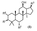
[10] In some aspect, the present invention provides a blank liposome having
a membrane,
wherein the membrane comprises lipid and a ginsenoside of Formula I:
OH R3
R1
R2
In Formula I,
each of RI- and R2 independently is H, OH, or R5, and RI- and R2 are not both
H at the
4
CA 02994032 2018-01-29
WO 2017/028811 PCT/CN2016/096005
same time;
R9
R1
R3 is R11 or
R4 is H, OH, or R5;
RS is R6, R7, or R8;
R6 is selected from the group consisting of: -0-G1c, -0-Rha, -0-Lyx, -0-Xyl, -
0-Ara(p),
-0-Ara(f), -0-Glc(241)Glc, -0-Glc(641)G lc, -0-
Glc(241)Rha, -0-Glc(241)Xyl,
-0-G1c(6-)1)Rha, -0-G1c(2-->1)Ara(p), -0-Glc(6-)1)Ara(p), -0-G1c(241)Ara(f),
-0-Glc(641)Ara(f), -0-
Glc(241)Glc(21)Glc, -0-Glc(241)Glc(241)Xyl,
-0-Glc(641)Glc(641)Xyl, -0-Glc(241)Glc(441)Xyl, -0-Glc(241)Lyx, -0-
Glc(641)Lyx,
-0-Glc(21)Glc(21)Rha, -0-Glc(21)Glc(21)Lyx, -0-
Glc(21)Glc(21)Ara(f),
-0-Glc(241)Glc(241)Ara(p), -0-
Glc(21)Glc(61)Glc, -0-G Ic(21)Glc(641)Rha,
-0-Glc(241)Glc(641)Xyl, -0-
Glc(241)Glc(641)Lyx, Ic(241)Glc(641)Ara(f),
-0-Glc(21)Glc(61)Ara(p), -0-Glc(61)Glc(21)Glc, -0-G
Ic(61)Glc(21)Rha,
-0-Glc(641)Glc(241)Xyl, -0-
Glc(61)Glc(21)Lyx, -0-G Ic(61)Glc(21)Ara(f),
-0-Glc(641)Glc(241)Ara(p), -0-
Glc(641)Glc(641)Glc, -0-G Ic(641)Glc(641)Rha,
-0-Glc(61)Glc(61)Lyx, -0-Glc(61)Glc(61)Ara(f) or -0-Glc(61)Glc(61)Ara(p);
R7 is a group formed by replacing one or more than one OH groups in R6 with R8
and
each of the one or more than one R8 groups independently can be the same as or
different
from each other;
R8 is:
I) -mPEG, -Z-mPEG, -mPEO, -Z-PEO, -mPVP, -Z-PVP, -mEPEG, or -Z-EPEG, wherein m
is H,
alkyl, or acyl; Z is-CO(CH2)9C0-, -NH(CH2)9CO-, -NH(CH2)bX-, or -CO-Ar-CH2-; X
is 0, S. or NH; a
is 1, 2, 3,4, 5, 6, 7, or 8; and b is 1, 2, 3, 4, 5, 6, 7, 8,9, or 10; or
II) C4_22 aliphatic acyl, a phosphate group, a succinic acid ester group, a n-
butyl acid
ester group, a sulfonate group, a malic acid ester group, or a sodium sulfate
salt; or
Ill) a group formed by dehydrogenizing the carboxyl contained in Boc-glycine,
Boc-alanine, Boc-arginine, Boc-lysine, Boc-serine, Acetyl phenylalanine,
Acetyl-proline,
Asparagine, Aspartic acid, Cysteine, Glutamine, Glutamic acid, Histidine,
lsoleucine, Leucine,
CA 02994032 2018-01-29
WO 2017/028811 PCT/CN2016/096005
Methionine, Phenylalanine, Proline, Threonine, Tryptophan, Tyrosine, or
Valine; or
IV) -0-PEO, -0-PVP, -0-PEG, -0-MPEG, -0-EPEG, -0-Glc (21)G1c(641)Mal or -0-Glc
(2 1)G1c(6 1)Ac;
each of R9, R10, R11, R12 and
K independently, is C1_3 alkyl;
each of d and e, independently, is 1, 2, or 3; and
the ginsenoside of Formula I can be optionally modified by replacing one or
more OH
groups therein with one or more R8 groups, and each of the R8 replacement
groups (when 2
or more) independently can be the same as or different from each other.
[11] As used herein, in a group such as -0-Glc(21)Glc, the numbers
indicating carbon
position, and the arror
indicates the connection relationship; Glc refers to glucopyranosyl;
Xyl refers to xylopyranosyl; Rha refers to rhamnopyranosyl; Ara(p) refers to
arabinopyranosyl;
Ara(f) refers to arabinofuranosyl; Lyx is lyxosyl; Ar refers to aryl; Mal
refers to a malonyl; Ac
refers to an acetyl; PEG refers to polyethylene glycol; PEO refers to
polyoxyethylene or
polyethylene oxide; MPEG refers to monomethoxy-terminated polyethylene glycol;
EPEG
refers to epoxy-terminated polyethylene glycol; PVP refers to povidone.
H07
fF=1,1v
OH
[12] In -0-
Glc- group, the structure of Glc is: OH ; in -0-Ara(p) group, the
OH
CC4-_21
structure of Ara(p) is: OH ; in -
0-Lyx group, the structure of Lyx
OH HO¨
,0 I-151
is: ; in -0-Ara(f) group, the structure of Ara(f) is: OH
; in
OH 0
-0-Rha group, the structure of Rha is: OHOH ; in -0-Xyl group, the structure
of Xyl
rO
;OH ?1,
\I 75ssinrOH
is: OH OH ; the structure of Mal is: 0 0
[13] In some embodiments, the molecular weight of PEG, PEO, PVP, or [PEG is
6
independently in the range of 200 to 20,000.
[14] In some embodiments, the aliphatic acyl group can be an acyl of a
natural saturated
or unsaturated aliphatic acid, and an acyl of artificially synthesized
saturated or unsaturated
aliphatic acid, preferably a stearyl or a palmityl.
'
[15] R3 is preferably or
[16] In the blank liposome, the ginsenoside of Formula I can be ginsenoside
Rg5,
ginsenoside Rg6, ginsenoside Rk1, ginsenoside Rk2, ginsenoside Rk3,
ginsenoside Rk4,
ginsenoside Rh3, ginsenoside Rh4, ginsenoside F4, ginsenoside Rs4, ginsenoside
Rs5,
ginsenoside Rs6, ginsenoside Rs7, notoginsenoside T5, damulin A, or damulin B.
[17] As mentioned above, the ginsenoside of Formula I contained in the
blank liposome
of the present invention can be modified by replacing one or more hydroxyl
(OH) groups in
the ginsenoside with le, and each of R8 groups (when more than one) can be the
same as or
different from each other, and R8 is as defined above.
[18] In the blank liposome of the present invention, the HPLC purity of the
ginsenoside
(including that as modified as described above) is preferably greater than or
equal to 90%,
more preferably greater than 95%, where the percentage is mass percentage.
[19] Preferably, in the blank liposome, the ginsenoside of Formula I can
also be in the
form ofmicelle. Ginsenoside nano micelle refers to that the ginsenoside is in
the form of
micelle, specifically refers to CN Patent Application CN201310155639.2 filed
on April 28,
2013 and PCT Application PCT/CN2013/088558 filed on December 4, 2013.
[20] Preferably, in the blank liposome of the present invention, the
lipid in
the membrane comprises phospholipid; and the mass ratio of the phospholipid
to the ginsenoside of Formula I is usually in the range of 0.5:1 to 100:1,
preferably in the
range of 0.5:1 to 20:1, more preferably in the range of 0.5:1 to 4:1 (such as
in the range of
0.5:1 to 2:1).
[21] Preferably, in the blank liposome of the present invention, the lipid
in the membrane
comprises phospholipid; the membrane furthercomprises cholesterol. The mass
ratio of the
phospholipid to the ginsenoside of Formula I is preferably in the range of
1:0.01 to 1:3
7
CA 2994032 2019-06-21
CA 02994032 2018-01-29
WO 2017/028811
PCT/CN2016/096005
(such as in the range of 1:0.03 to 1:1), more preferably in the range of
1:0.05 to 1:0.9 (such
as in the range of 1:0.3 to 1:0.75), most preferably in the range of 1:0.1 to
1:0.9 (such as in
the range of 1:0.1 to 1:0.5). The mass ratio of the ginsenoside of Formula I
to the
cholesterol is preferably in the range of 0.1:1 to 100:1, preferably in the
range of 0.5:1 to
50:1, more preferably in the range of 0.5:1 to 10:1 (such as in the range of
1.5:1 to 6:1, or
5:1).
[22] In the blank liposome of the present invention, a mass percentage of
the ginsenoside
of Formula I in the membrane is preferably in the range of 0.01% to 80%, a
mass percentage
of the phospholipid in the membrane is preferably in the range of 5% to 99.9%,
a mass
percentage of the cholesterol in the membrane is preferably lower than 50%;
the
percentage (%) mentioned above refers to the percentage of the mass of each
component
relative to the total mass of the membrane.
[23] The mass percentage of the ginsenoside of Formula I in the membrane is
preferably
in the range of 10% to 80%, more preferably in the range of 10% to 40%, most
preferably in
the range of 20% to 40% (such as in the range of 25% to 40%, preferably in the
range of 25%
to 35%). The mass percentage of the phospholipid in the membrane is preferably
in the
range of 10% to 70%, more preferably in the range of 30% to 70%, most
preferably in the
range of 30% to 60%. The mass percentage of the cholesterol in the membrane is
preferably in the range of 0.5% to 50%, more preferably in the range of 5% to
40%, most
preferably in the range of 5% to 30% (such as in the range of 10% to 20%).
[24] In a preferred embodiment of the present invention, the blank liposome
can further
comprise and encapsulate within the membrane an antioxidant. A mass percentage
of the
antioxidant in the blank liposome is usually no more than 25%, preferably in
the range of
0.001% to 15%, more preferably in the range of 0.01% to 10%, most preferably
in the range
of 0.01% to 5% (such as in the range of 0.1% to 1%), the percentage (%) refers
to the
percentage of the mass of the antioxidant relative to the total mass of the
blank liposome.
[25] In a preferred embodiment of the present invention, the blank liposome
can further
comprise and encapsulate within the membrane a cryoprotectant. A mass
percentage of
the cryoprotectant in the blank liposome is usually no more than 80%,
preferably in the
range of 0.5% to 60%, more preferably in the range of 5% to 60%, most
preferably in the
8
CA 02994032 2018-01-29
WO 2017/028811
PCT/CN2016/096005
range of 30% to 60%, the percentage (%) refers to the percentage of the mass
of the
cryoprotectant relative to the total mass of the blank liposome.
[26] In a preferred embodiment of the present invention, the blank liposome
can further
comprise and encapsulate within the membrane soybean oil and/or sodium oleate.
A
mass percentage of the "soybean oil and/or sodium oleate" in the blank
liposome is usually
in the range of 1% to 90%, preferably in the range of 15% to 80%, more
preferably in the
range of 20% to 70% (such as in the range of 25% or 62.5%), most preferably in
the range of
20% to 30%, or 60% to 70%, the percentage refers to the mass of the "soybean
oil and/or
sodium oleate" relative to the total mass of the blank liposome. A mass ratio
of the
"soybean oil and/or sodium oleate" to the phospholipid in the blank liposome
is preferably
in the range of 1:0.1 to 1:10, more preferably in the range of 1:0.5 to 1:5,
most preferably in
the range of 1:0.5 to 1:4 (such as in the range of 1:1 to 1:2).
[27] In a preferred embodiment of the present invention, the blank liposome
comprises
the following components: phospholipid and the ginsenoside of Formula I, or
the
ginsenoside of Formula I, phospholipid and an antioxidant, or the ginsenoside
of Formula I,
phospholipid and a cryoprotectant, or the ginsenoside of Formula I, "soybean
oil and/or
sodium oleate" and phospholipid, or the ginsenoside of Formula I, "soybean oil
and/or
sodium oleate", phospholipid and an antioxidant, or the ginsenoside of Formula
I, "soybean
oil and/or sodium oleate", phospholipid and a cryoprotectant, or the
ginsenoside of Formula
I, "soybean oil and/or sodium oleate", phospholipid, an antioxidant and a
cryoprotectant, or
the ginsenoside of Formula I, phospholipid and cholesterol, or the ginsenoside
of Formula I,
phospholipid, cholesterol and an antioxidant, or the ginsenoside of Formula I,
phospholipid,
cholesterol and a cryoprotectant, or the ginsenoside of Formula I, "soybean
oil and/or
sodium oleate", phospholipid and cholesterol, or the ginsenoside of Formula I,
"soybean oil
and/or sodium oleate", phospholipid, cholesterol and an antioxidant, or the
ginsenoside of
Formula I, "soybean oil and/or sodium oleate", phospholipid, cholesterol and a
cryoprotectant, or the ginsenoside of Formula I, "soybean oil and/or sodium
oleate",
phospholipid, cholesterol, an antioxidant and a cryoprotectant.
[28] In a preferred embodiment of the present invention, the blank liposome
consists of
the components mentioned-above.
9
CA 02994032 2018-01-29
WO 2017/028811
PCT/CN2016/096005
[29] In a preferred embodiment of the present invention, the blank liposome
comprises
the following components: the ginsenoside of Formula I, the phospholipid, the
cholesterol,
the soybean oil and/or the sodium oleate, the antioxidant and the
cryoprotectant. The
mass ratio of the soybean oil and/or the sodium oleate to the cholesterol in
the blank
liposome is preferably in the range of 1:0.1 to 1:10, more preferably in the
range of 1:0.5 to
1:5, most preferably in the range of 1:0.5 to 1:1. A mass percentage of the
cholesterol in
the membrane is preferably in the range of 1% to 20%, more preferably in the
range of 10%
to 20%, a mass percentage of the soybean oil and/or the sodium oleate in the
blank
liposome is preferably in the range of 1% to 90%, more preferably in the range
of 15% to
80%, most preferably in the range of 20% to 70% (such as in the range of 25%
or 62.5%, 20%
to 30%, or 60% to 70%).
[30] In a preferred embodiment of the present invention, the blank liposome
consists of
the phospholipid and the ginsenoside of Formula I.
[31] In a preferred embodiment of the present invention, the blank liposome
consists of
the ginsenoside of Formula I, the phospholipid and the cholesterol.
[32] In a preferred embodiment of the present invention, the blank liposome
consists of
the ginsenoside of Formula I, the phospholipid, the cholesterol, the
antioxidant and the
cryoprotectant.
[33] In a preferred embodiment of the present invention, the blank liposome
consists of
the ginsenoside of Formula I, the phospholipid, the cholesterol, the soybean
oil and/or
sodium oleate, the antioxidant and the cryoprotectant.
[34] The phospholipid can be a conventional phospholipid in this field,
preferably
comprises a natural phospholipid, semisynthetic phospholipid or fully
synthetic
phospholipid.
[35] The natural phospholipid is typically derived from soybean, yolk,
brain or organs of
an animal, preferably comprises natural lecithin, soybean lecithin egg
lecithin or cephalin.
[36] The semisynthetic phospholipid or the fully synthetic phospholipid can
be a
conventional semisynthetic phospholipid or fully synthetic phospholipid in
this field,
preferably comprises a phospholipid of phosphatidylchollines,
phosphatidylserine (PS),
phosphatidylinositol (PI), a phospholipid of phosphatidylethanolamine,
phosphatidylglycerol
CA 02994032 2018-01-29
WO 2017/028811
PCT/CN2016/096005
(DSPG), dicetyl phosphate (DCP), a PEG-modified phospholipid, cholesterol
succinate (CHS)
or 1-palmitoy1-2-oleoyl-sn-glycero-3-phosphocholine (16:0 to 18:1 PC, wherein
16:0 to 18:1
refers to thecarbonchain of PC). Due to the heat-sensitivity of the
semisynthetic or fully
synthetic phospholipids such as dipalmitoyl phosphatidylcholine and distearoyl
phosphatidylcholine etc., they can be used as heat-sensitive excipients at the
same time.
[37] The phospholipid of phosphatidylcholline can be a conventional
phospholipid of
phosphatidylcholline in this field, preferably comprises hydrogenated soybean
lecithin
(HSPC), dipalmitoyl phosphatidylcholine (DPPC), distearoyl phosphatidylcholine
(DSPC),
dimyristoyl phosphatidylcholine (DMPC), dilauroyl phosphatidylcholine (DLPC),
dioleoyl
phosphatidylcholine (DOPC), phosphatidylcholine (SPC),
monopalmitoyl
phosphatidylcholine (MPPC) or glycerol phosphatidylcholine (GPC).
[38] The phospholipid of phosphatidylethanolamine can be a conventional
phospholipid
of phosphatidylcholline in this field, preferably comprises 1-palmitoy1-2-
oleoyl
phosphatidylethanolamine (POPE), dilauroyl phosphatidylethanolamine (DLPE),
dierucoyl
phosphatidylethanolamine (DEPE), dioleoyl phosphatidylethanolamine (DOPE),
distearoyl
phosphatidylethanolamine (DSPE), dipalmitoyl phosphatidylethanolamine (DPPE)
or
dimyristoyl phosphatidylethanolamine (DMPE).
[39] The PEG-modified phospholipid can be a conventional PEG-modified
phospholipid in
this field, preferably comprises phosphatidylethanolamine-PEG (DMPE-PEG),
dipalmitoyl
phosphatidylethanolamine-PEG (DPPE-PEG), distearoyl phosphatidyletha nola mine-
PEG
(DSPE-PEG), dioleoyl phosphatidylethanolamine-PEG (DOPE-PEG), C8 ceramide-PEG
(C8
ceramide-PEG), C16 ceramide-PEG (C16 ceramide-
PEG), distearoyl
phosphatidylethanolamine-PEG-succinyl (DSPE-PEG
succinyl), distea royl
phosphatidylethanolamine-PEG -carboxyl (DSPE-PEG
carboxylic acid), distearoyl
phosphatidylethanolamine-PEG-maleimide (DSPE-PEG ma lei m
ide), distea royl
phosphatidylethanolamine-PEG-propionamide bis-mercaptopyridine (DSPE-PEG PDP),
distearoyl phosphatidylethanolamine-PEG-cyanuric chloride (DSPE-PEG cyanur),
distearoyl
phosphatidylethanolamine-PEG-amino (DSPE-PEG amine),
distearoyl
phosphatidylethanolamine-PEG -biotin (DSPE-PEG biotin),
distearoyl
phosphatidylethanolamine-PEG -folate (DSPE-PEG folate),
distearoyl
CA 02994032 2018-01-29
WO 2017/028811
PCT/CN2016/096005
phosphatidylethanolamine-PEG -folate (DSPE-PEG folate),
dilauroyl
phosphatidylethanolamine-PEG (DLPE-PEG),
distearoyl
phosphatidylethanolamine-PEG-active ester (DSPE-
PEG-N HS),
phosphatidylethanolamine-PEG-active ester (DMPE-
PEG-N HS), dipalmitoyl
phosphatidylethanolamine-PEG-active ester (DPPE-
PEG-NHS), dilauroyl
phosphatidylethanolamine-PEG-active ester (DLPE-
PEG-NHS), distearoyl
phosphatidylethanolamine-PEG-maleimide (DSPE-
PEG-maleimide),
phosphatidylethanolamine-PEG-maleimide (DMPE-PEG-maleimide),
dipalmitoyl
phosphatidylethanolamine-PEG-maleimide (DPPE-PEG-maleimide),
dilauroyl
phosphatidylethanolamine-PEG-maleimide(DLPE-PEG-maleimide),
distearoyl
phosphatidylethanolamine-PEG-biotin (DSPE-PEG-biotin),
distearoyl
phosphatidylethanolamine-PEG-fluorescein (DSPE-PEG-FITC),
distearoyl
phosphatidylethanolamine-PEG-hydroxyl (DSPE-PEG-OH),
distearoyl
phosphatidylethanolamine-PEG-amino (DSPE-
PEG-NH2),
phosphatidylethanolamine-PEG-amino (DMPE-PEG-NH2),
dipalmitoyl
phosphatidylethanolamine-PEG-amino (DPPE-PEG-NH2), dila
uroyl
phosphatidylethanolamine-PEG-amino(DLPE-PEG-NH2), distea
royl
phosphatidylethanolamine-PEG-carboxyl (DSPE-
PEG-COOH),
phosphatidylethanolamine-PEG-carboxyl (DMPE-PEG-COOH),
dipalmitoyl
phosphatidylethanolamine-PEG-carboxyl (DPPE-PEG-COOH),
dilauroyl
phosphatidylethanolamine-PEG-carboxyl (DLPE-PEG-COOH),
distearoyl
phosphatidylethanolamine-PEG-thiol (DSPE-PEG-SH), distea
royl
phosphatidylethanolamine-PEG-si lane (DSPE-PEG-silane), distea
royl
phosphatidylethanolamine-PEG-azide (DSPE-PEG-N3), cholesterol-PEG (cholesterol
PEG),
methoxyl-PEG-cholesterol (mPEG-CLS), cholesterol-PEG-active ester (cholesterol
PEG NHS
ester), cholesterol-PEG-maleimide (CLS-PEG-Mal), cholesterol-PEG-biotin
(cholesterol PEG
biotin), cholesterol-PEG-fluorescein (cholesterol PEG fluorescein),
cholesterol-PEG-carboxyl
(cholesterol PEG COOH), cholesterol-PEG-amino (cholesterol PEG NH2) or
cholesterol-PEG-thiol (Cholesterol PEG SH). The relative molecular weight of
the PEG
mentioned above is preferably in the range of 300 to 50000õmore preferably in
the range of
12
CA 02994032 2018-01-29
WO 2017/028811
PCT/CN2016/096005
500 to 10000, e g. at about 300, 350, 500, 550, 1000, 2000, 3400, 5000, 10000,
20000,
30000, 40000 or 50000.
[40] A number-average molecular weight of the DMPE-PEG is preferably 350, 550,
750,
1000, 2000, 3000 or 5000. A number-average molecular weight of the DPPE-PEG is
preferably 350, 550, 750, 1000, 2000, 3000 or 5000. A number-average molecular
weight
of the DSPE-PEG is preferably 350, 550, 750, 1000, 2000, 3000, 5000, 10000,
20000, 30000
or 40000. A number-average molecular weight of the DOPE-PEG is preferably 350,
550,
750, 1000, 2000, 3000 or 5000. A number-average molecular weight of the C8
Ceramide-PEG is preferably 750, 2000 or 5000. A number-average molecular
weight of the
DLPE-PEG is preferably 2000 or 5000. A number-average molecular weight of the
DSPE-PEG-NHS is preferably 1000, 2000, 5000, 10000, 20000, 30000 or 40000. A
number-average molecular weight of the DMPE-PEG-NHS is preferably 3400 or
5000. A
number-average molecular weight of the DPPE-PEG-NHS is preferably 3400 or
5000. A
number-average molecular weight of the DLPE-PEG-NHS is preferably 3400 or
5000. A
number-average molecular weight of the DSPE-PEG-Maleimide is preferably 1000,
2000,
3400, 5000 or 10000. A number average-molecular weight of the DMPE-PEG-
Maleimide is
preferably 1000, 2000, 3400, 5000 or 10000. A number-average molecular weight
of the
DPPE-PEG-Maleimide is preferably 1000, 2000, 3400, 5000 or 10000. A number-
average
molecular weight of the DLPE-PEG-Maleimid is preferably 1000, 2000, 3400, 5000
or 10000.
A number-average molecular weight of the DSPE-PEG-Biotin is preferably 1000,
2000, 3400,
5000 or 10000. A number-average molecular weight of the DSPE-PEG-FITC is
preferably
1000, 2000, 3400, 5000 or 10000. A number-
average molecular weight of the
DSPE-PEG-OH is preferably 2000, 3400 or 5000. A number-average molecular
weight of
the DSPE-PEG-NH2 is preferably 2000, 3400 or 5000. A number-average molecular
weight
of the DMPE-PEG-NH2 is preferably 2000, 3400 or 5000. A number-average
molecular
weight of the DPPE-PEG-NH2 is preferably 2000, 3400 or 5000. A number-average
molecular weight of the DLPE-PEG-NH2 is preferably 2000, 3400 or 5000. A
number-average molecular weight of the DSPE-PEG-COOH is preferably 2000, 3400
or 5000.
A number-average molecular weight of the DMPE-PEG-COOH is preferably 2000,
3400 or
5000. A number-average molecular weight of the DPPE-PEG-COOH is preferably
2000,
13
CA 02994032 2018-01-29
WO 2017/028811
PCT/CN2016/096005
3400 or 5000. A number-average molecular weight of the DLPE-PEG-COOH is
preferably
2000, 3400 or 5000. A number-average molecular weight of the DSPE-PEG-SH is
preferably
5000. A number-average molecular weight of the DSPE-PEG-Silane is preferably
3400. A
number-average molecular weight of the DSPE-PEG-N3 is preferably 2000, 3400 or
5000. A
number-average molecular weight of the mPEG-CLS is preferably 1000, 2000,
5000, 10000
or 20000. A number-average molecular weight of the Cholesterol PEG NHS ester
is
preferably 1000, 2000, 3400, 5000 or 10000. A number-average molecular weight
of the
CLS-PEG-Mal is preferably 2000, 3400, 5000 or 10000. A number-average
molecular
weight of the CLS-PEG-Biotin is preferably 2000, 3400 or 5000. A number-
average
molecular weight of the CLS-PEG-FITC is preferably 2000, 3400 or 5000. A
number-average
molecular weight of the Cholesterol PEG COOH is preferably 3400. A number-
average
molecular weight of the Cholesterol PEG amine is preferably 3400. A number-
average
molecular weight of the Cholesterol PEG Thiol/Sulfhydril is preferably 3400.
[41] The antioxidant can be a conventional antioxidant in this field,
preferably comprises
sodium metabisulfite, sodium thiosulfate, propyl gallate, ascorbic acid, a-
tocopherol,
a-hydroxyl acid, flavonoid, a phenylpropanoid phenolic compounds, vitamin E,
vitamin C,
fumaric acid, cysteine, methionine, butyl hydroxy anisole (BHA), butyl
hydroxytoluene (BHT),
thiodipropionic acid, sulfites (e.g., sodium sulfite), hydrosulphite (e.g.,
sodium hydrosulfite),
dithioaminobenzoic acid compounds, citric acid, malic acid, sorbitol,
glycerol, propylene
glycol, hydroquinone, hydroxycoumarin, ethanolamine, phosphoric acid or
phosphorous
acid.
[42] The cryoprotectant can be a conventional cryoprotectant in this field,
generally
comprises a glucide, a polyol, an amino acid or a buffer reagent; wherein the
glucide
preferably comprises a monosaccharide, a disaccharide or a polysaccharide.
The
monosaccharide preferably comprises glucose, mannitol, xylitol or sorbitol.
The
disaccharide preferably comprises sucrose, lactose, galactose or maltose. The
polysaccharide preferably comprises trehalose. The polyol preferably comprises
mannitol,
sorbitol or glycerol. The amino acid preferably comprises a-amino acid
selected from the
group consisting of threonine, glycine, glutamic acid, arginine and histidine.
The buffer
reagent generally refers to a buffer solution. The buffer solution can be a
conventional
14
buffer solution in this field, whose pH value is preferably in the range of 3
to 10, more
preferably in the range of 5 to 7. The buffer solution preferably comprises an
ethanol-acetic acid buffer solution, a tris (hydroxymethyl) aminomethane
buffer solution, a
barbital buffer solution, a sodium formate buffer solution, a phthalate buffer
solution, a
citrate buffer solution, a citric acid-disodium hydrogen phosphate buffer
solution, an
ammonia-ammonium chloride buffer solution, a borax-calcium chloride buffer
solution, an
acetate buffer solution, an acetic acid-lithium salt buffer solution, an
acetic acid-sodium
acetate buffer solution, an acetic acid-ammonium acetate buffer solution, a
phosphoric
acid-triethylamine buffer solution or a phosphate buffer solution.
[43] Preferably, the blank liposome can further comprise and encapsulate
within the
membrane other excipients. The excipients can be conventional excipients used
for
preparing liposome in this field except for the antioxidant and the
cryoprotectant, such as
the excipient comprise a surfactant, a heat-sensitive excipient, a pH-
sensitive material or an
ion additive.
[44] The surfactant preferably comprises polyethylene glycol (PEG),
polysorbate, Tween"
surfactant, or a brijTM surfactant. Wherein a number-average molecular weight
of the
polyethylene glycol is preferably in the range of 200 to 8000 (e g. at about
300, 350, 500,
550, 1000, 2000, 3400 or 5000). The polysorbate preferably comprises
polyoxyethylene
sorbitan monolaurate, polyoxyethylene sorbitan monopalmitate, polyoxyethylene
sorbitan
monostearate, polyoxyethylene sorbitan trioleate, PEG-
phosphatidylethanolamine,
PEG-polylactic acid, polylysine-poly(lactic-co-glycolic) acid, polyetherimide-
polylactic acid,
PEG-polycaprolactone, PEG-poly(lactic¨co-glycolic) acid, poloxamer 188,
polyoxyethylene
fatty acid ester, polyoxyethylene fatty acid ether or polyoxyethylene methyl
castor oil ether.
[45] The heat-sensitive excipient generally comprises a polymer, a drug or
a surfactant
which can bring heat-sensitivity to the liposome. The polymer preferably
comprises
polyprene acrylamide, polyprene acrylic acid, polyphosphate, or poly
phospholipid-amide
copolymer. The drug preferably comprises zedoary turmeric oil, elemene or
brucea
javanica oil. The surfactant is preferably a Tween' surfactant (such as
TweenTm-80)
and/or a brijTM surfactant.
[46] The ion additive preferably comprises a cationic additive (such as
octadecylamine)
CA 2994032 2019-06-21
CA 02994032 2018-01-29
WO 2017/028811
PCT/CN2016/096005
and/or an anion additive (such as phosphatidic acid and/or
phosphatidylserine).
[47] A mass percentage of the above excipients can be selected according to
the mass
percentage of such kind of excipients contained in the ordinary liposome in
the art. For
example, when the blank liposome includes the surfactant, a mass percentage of
the
surfactant in the blank liposome is preferably in the range of 0% to 50%,
excluding 0%.
When the blank liposome includes the ion additive, a mass percentage of the
ion additive in
the blank liposome is preferably in the range of 0% to 10%, excluding 0%.
[48] The blank liposome can be prepared by conventional methods of preparing a
liposome. Commonly, an injection method, a reverse evaporation method, a
freezing and
thawing method, a double emulsion method, an initiative encapsulation method,
a
precursor liposome preparation method, a film dispersion method, a freeze-
drying method,
an ammonium sulfate gradient method or a pH gradient method, as well as any
combination
of above two methods can be adopted. The present invention preferably adopts
the first
method or the second method as follows, wherein the first method or the blank
liposome
prepared thereby does not include a cryoprotectant, and the second method or
the blank
liposome prepared thereby includes a cryoprotectant.
[49] The first method includes the steps of:
(1) mixing a lipid and a ginsenoside of Formula I, optionally, a
cholesterol, a
hydrophobic antioxidant, soybean oil and/or sodium oleate, a hydrophobic
surfactant, a
hydrophobic heat-sensitive excipient, a hydrophobic pH sensitive material,
and/or a
hydrophobic ion additive in an organic solvent to obtain a clear solution; and
(2) removing the organic solvent of the clear solution obtained in step
(1), filming,
mixing the film with water optionally containing a hydrophilic antioxidant, a
hydrophilic
surfactant, a hydrophilic heat-sensitive excipient, a hydrophilic pH sensitive
material, and/or
a hydrophilic ion additive to obtain an aqueous mixture, filtering the mixture
after an
operation of ultrasound, high pressure homogenization or pushing through a
membrane to
obtain an aqueous solution containing a blank liposome, drying to get the
blank liposome;
[50] The second method includes the steps of:
(1) mixing a lipid and a ginsenoside of Formula I, optionally, a
cholesterol, a
hydrophobic antioxidant, soybean oil and/or sodium oleate, a hydrophobic
surfactant, a
16
CA 02994032 2018-01-29
WO 2017/028811 PCT/CN2016/096005
hydrophobic heat-sensitive excipient, a hydrophobic pH sensitive material,
and/or a
hydrophobic ion additive in an organic solvent to obtain a clear solution, and
(2) removing the organic solvent of the clear solution obtained in step (1),
filming,
mixing the film with an aqueous solution containing a cryoprotectant and
optionally a
hydrophilic antioxidant, a hydrophilic surfactant, a hydrophilic heat-
sensitive excipient, a
hydrophilic pH sensitive material, and/or a hydrophilic ion additive to give a
mixture,
filtering the mixture after an operation of ultrasound, high pressure
homogenization or
pushing through a membrane to obtain an aqueous solution containing a blank
liposome,
drying to get the blank liposome.
[51] In the first method or in the second method, wherein the lipid, the
ginsenoside of
Formula I, the cholesterol, the antioxidant, the soybean oil and/or sodium
oleate, the
cryoprotectant, the surfactant, the heat-sensitive excipient, the pH sensitive
material, and
the ion additive are as defined above.
[52] In step (1) of the first or second method, the organic solvent can be
a conventional
organic solvent used in the preparation of a liposome in the art, which
preferably comprises
a nitrile solvent, a C1_4 alcohol solvent, a ketone solvent, an alkane
solvent, an ether solvent,
a halogenated hydrocarbon solvent, a sulfoxide solvent, or an aldehyde
solvent, more
preferably comprises a Ci_4 alcohol solvent, a nitrile solvent, an ether
solvent or a
halogenated hydrocarbon solvent. The nitrile solvent preferably comprises
acetonitrile.
The C14 alcohol solvent preferably comprises methanol, ethanol, isopropanol or
n-butanol.
The ether solvent preferably comprises tetrahydrofuran or diethyl ether. The
halogenated
hydrocarbon solvent preferably comprises chloroform or dichloromethane. The
ketone
solvent preferably comprises acetone or butanone. The alkane solvent
preferably
comprises petroleum ether. An amount of the organic solvent can be a
conventional
amount used in the preparation of a liposome in the art, without particularly
limited, a
general requirement of which is capable of obtaining a clear solution after
the mixing of the
organic solvent and all the components. Preferably, a ratio of the organic
solvent's volume
to the total mass of all the components dissolved in the organic solvent in
step (1) of the
first or second method is 5 to 20mL/g.
[53] Step (1) of the first or second method is generally carried out at the
temperature of 0
17
CA 02994032 2018-01-29
WO 2017/028811 PCT/CN2016/096005
to 802C, preferably 10 to 802C, more preferably 10 to 652C. Based on the
common
knowledge in this field, in some cases, in order to reach 802C, heating is
required. Or,
when the blank liposome prepared thereby includes a heat-sensitive material,
such as
protein materials, step (1) of the first or second method is generally carried
out at the
temperature of e 02C.
[54] In step (2) of the first or second method, the operation of removing
the organic
solvent of the clear solution obtained in step (1) can be a conventional
operation in this field,
which is usually conducted with a rotary evaporator or a membrane evaporator.
The
temperature at which the organic solvent is removed can be selected according
to the
organic solvent to be removed, generally is 25 to 802C.
[55] In step (2) of the first or second method, the operation of
ultrasound, high pressure
homogenization or pushing through a membrane can be a conventional operation
in this
field. After the operation of ultrasound, high pressure homogenization or
pushing through
a membrane, a particle size of the liposome is generally ranging from 0.05 to
0.3 microns.
[56] In step (2) of the first or second method, the operation of filtration
can be a
conventional operation in the preparation of the liposome in this field, the
purpose of which
is to remove bacteria, solid particles and particularly large liposome (in a
method of
preparing a liposome loaded with the active substance, an unencapsulated free
drug can
also be removed) etc. In the present invention, the filtration is preferably
microporous
membrane filtration. The pore size of the microporous membrane is preferably
0.22
micron.
[57] In step (2) of the second method, the aqueous cryoprotectant solution
refers to an
aqueous solution formed by mixing the cryoprotectant and water. The aqueous
cryoprotectant solution is preferably a 5% to 10% aqueous solution of the
cryoprotectant,
the percentage refers to the percentage of the mass of the cryoprotectant
relative to the
total mass of the aqueous solution of the cryoprotectant. An amount of the
aqueous
cryoprotectant solution is without particular limitation, as long as there is
no influence on
the formation of the blank liposome, preferably, it is the same as that of the
organic solvent
used in step (1).
[58] In a preferred embodiment of the present invention, in the second
method, when
18
CA 02994032 2018-01-29
WO 2017/028811 PCT/CN2016/096005
the cryoprotectant is a buffer reagent, after the filming operation in step
(2), the
cryoprotectant is mixed directly.
[59] In step (2) of the first or second method, the operation of drying can
be a
conventional operation in this field, preferably is freeze-drying which
generally utilizes a
freeze dryer. The temperature and time required by the freeze-drying are
conventional
temperature and time in this field which is without particular limitation.
[60] In the first or second method, for easy storage, the aqueous solution
containing the
blank liposome obtained in step (2) is split charging into vials, dried, swept
with protective
gas (argon or nitrogen) and sealed.
[61] The blank liposome can be used to prepare a liposome loaded with and
encapsulate
within the membrane an active substance, wherein the active substance
comprises a drug, a
cosmetically active substance or a substance with healthcare function.
Therefore, the
present invention also provides a loaded liposome which comprises a blank
liposome and an
active substance loaded to and encapsulating within the liposome's membrane,
wherein the
active substance comprises a drug, a cosmetically active substance, or a
substance with
healthcare function.
[62] In the loaded liposome, a mass ratio of the active substance to the
ginsenoside of
Formula I is 1:0.1 to 1:10, more preferably 1:2 to 1:6 (such as 1:3 or 1:4).
[63] The drug can be a conventional drug in the art, preferably comprises
an antitumor
drug, an antifungal drug, an antiviral drug, antibiotics, a non-steroidal anti-
inflammatory
drug, a calcium ion antagonist, an immunosuppressive agent, an anesthetic, a
cardiovascular
and vasodilation drug, a gastrointestinal drug, an antidepressant drug, a
biological agent, a
polynucleotide or an oligonucleotide (including a ribonucleotide and a
deoxyribonucleotide).
[64] The antitumor drug can be a conventional antitumor drug in the art,
preferably
comprises paclitaxel, docetaxel, cabazitaxel, irinotecan hydrochloride,
hydroxycamptothecin,
aminocamptothecin, 7-ethyl-10-hydroxy camptothecin, topotecan hydrochloride,
lurtotecan,
topotecan, belotecan, cisplatin, carboplatin, oxaliplatin, nedaplatin,
lobaplatin, satraplatin,
miriplatin, amyl platinum, aroplatin (L-NDDP), carmustine, chlorambucil,
melphalan,
harringtonine, homoharringtonine, triptolide, tacrolimus, daunorubicin,
pingyangmycin,
19
CA 02994032 2018-01-29
WO 2017/028811 PCT/CN2016/096005
doxorubicin hydrochloride, idarubicin, fluorouracil, cytarabine, methotrexate,
etoposide
phosphate, desoxy-podophyllotoxin, huperzine-A, vinorelbine tartrate,
vincristine sulfate,
vinblastine sulfate, vinorelbine, vindesine sulfate, temozolomide, tegafur,
cyclophosphamide,
ifosfamide, dacarbazine, epothilone A, epothilone B, epothilone C, epothilone
D, epothilone
E, epothilone F, bortezomib, gemcitabine hydrochloride, fludarabine phosphate,
capecitabine, decitabine, pemetrexed disodium, sorafenib, recombinant human
interferon
a2b, cytosine arabinoside, all trans retinoic acid, interleukin-2, etoposide,
thymidylate
synthase inhibitor, mitoxantrone, minoxidil, azithromycin, epirubicin
hydrochloride,
doxorubicin hydrochloride (adriamycin), amrubicin hydrochloride, 5-
aminolevulinic acid
(5-ALA), gefitinib, imatinib, erlotinib, sunitinib, dasatinib, lapatinib,
axitinib, apatinib,
nilotinib, bosutinib, vandetanib, telatinib, neratinib, canertinib,
saracatinib, octenidine,
sorafenib, icotinib, mubritinib, lestaurtinib, tandutinib, dovitinib, 3',5'-
dipalmitotyl
cyclocytidine or curcumenol.
[65] The antifungal drug preferably comprises amphotericin B, gentamicin,
indomethacin,
penicillin G, econazole nitrate, flucytosine, fluconazole, itraconazole,
voriconazole,
posaconazole, ravuconazole, caspofungin, micafungin, anidulafungin,
cefpiramide sodium,
cefotaxime sodium, ceftriaxone, cefoperazone, cefditoren pivoxil, cefoxitin
sodium, cefalexin,
cefuroxime sodium, cefixime, cefpodoxime, cefmenoxime, cefodizime, cefsulodin,
cefazonam, ceftizoxime, cefetamet pivoxil, cefterampivoxil, ceftibuten,
cefdinir, cefamandole,
cefotiam, ceforanide, cefonicid, ceftazidime, cefradine, cefprozil, cefazolin
sodium, cefadroxil,
cephalothin, cefathiamidine, cefaloridine, cephacetrile, ceftezole, cefapirin,
cefpirome,
cefclidin, cefepime, fusidate sodium, florfenicol or tigecycline.
[66] The
antiviral drug preferably comprises ribavirin, acyclovir, cytarabine,
idoxuridine,
acyclovir laurate, acyclovir palmitate, iododeoxyuridine, cyclocytidine,
dipalmitoyl
cyclocytidine, phosphoric acid formate, phosphoric acid acetate, cimetidine,
dipyridamole,
rifampin, isoniazid, praziquantel, doxycycline, saquinavir, indinavir,
ritonavir, nelfinavir,
amprenavir, tipranavir, BM5232632, lamivudine, zidovudine, didanosine (ddi),
zalcitabine
(ddc), stavudine (d4t), abacavir, adefovirdipivoail (pmea), tenofovir (pmpa),
fluoro
lamivudine (ftc), nevirapine, delavirdin, efavirens, interleukin-2
tilmicosin or diclazuril.
[67] The antibioticis preferably comprises penicillin, penicillin V,
amoxicillin, ampicillin,
CA 02994032 2018-01-29
WO 2017/028811
PCT/CN2016/096005
oxacillin, cloxacillin, procaine penicillin, benzathine penicillin,
piperacillin, mezlocillin,
ticarcillin, azlocillin, mezlocillin, carbenicillin, sulbenicillin,
furbucillin, nafcillin, dicloxacillin,
pivampicillin, apalcillin, aspoxicillin, pivmecillinam, methicillin,
lenampicillin, fomidacillin,
flucloxacillin, kanamycin, natamycin, mitomycin, amikacin, tylosin,
verteporfin, cefpiramide
sodium, netilmicin sulfate, azithromycin, ofloxacin, ciprofloxacin, enoxacin,
lomefloxacin,
pefloxacin, rufloxacin, spa rfloxacin, fleroxacin, moxifloxacin,
grepafloxacin, trovafloxacin,
norfloxacin, gemifloxacin, gatifloxacin, tosufloxacin, pazufloxacin,
sparfloxacin,
clarithromycin, clindamycin, polymyxin, tobramycin, vancomycin , azithromycin,
doxycycline,
tetracycline, oxytetracycline, minocycline, aureomycin, guamecycline,
demeclocycline,
metacycline, etimicin, netilmicin,sisomicin, amikacin, arbekacin, dibekacin,
aztreonam,
meropenem, imipenem, thienamycin, panipenem, ertapenem, neomycin, paromomycin
or
spectinomycin.
[68] The calcium ion antagonist preferably comprises nimodipine,
nifedipine, nicardipine,
nitrendipine, verapamil, amlodipine, diltiazem, flunarizine, prenvlamine,
gallopamil or
tiapamil.
[69] The non-steroidal anti-inflammatory drug preferably comprises
indomethacin,
aspirin, paracetamol, naproxen, diclofenac, ibuprofen, nimesulide, rofecoxib
or celecoxib.
[70] The immunosuppressive agent preferably comprises cyclosporin,
alprostadil (also
known as prostate E-1), cyclosporine, tacrolimus, rapamycin, mycophenolate
mofetil or
mizoribine.
[71] The anesthetic preferably comprises halothane, sevoflurane,
isoflurane, enflurane,
propofol, fentanyl, urethane, lidocaine, procaine, tetracaine, bupivacaine,
pelltobarbitalum
natricum, chloral hydrate, ketamine, chloralose or morphine.
[72] The cardiovascular and vasodilation drug preferably comprises
dabigatran etexilate,
alogliptin, polysaccharide sodium, ginkgolides, gingko flavonoid, ginkgo
biloba extract,
asarone, olmesartan medoxomi, repaglinide, lipoic acid, breviscapine,
urapldil, niacin,
captopril, losartan, puerarin, tanshinone IIA, sarpogrelate hydrochloride,
fluvastatin,
pravastatin, simvastatin, lovastatin, simvastatin, mevastatin, cerivastatin,
rosuvastatin,
atorvastatin calcium or rosuvastatin calcium.
[73] The gastrointestinal drug preferably comprises omeprazole,
lansoprazole, ilaprazole,
21
CA 02994032 2018-01-29
WO 2017/028811
PCT/CN2016/096005
pantoprazole, rabeprazole, terazosin, esomeprazole, tenatoprazole,
leminoprazole,
tenatoprazole, disuprazole or lafutidine.
[74] The antidepressant drug preferably comprises agomelatine, fluoxetine,
paroxetine,
duloxetine, sertraline, fluvoxamine, citalopram, escitalopram, venlafaxine,
mirtazapine,
imipramine, amitriptyline, clomipramine, doxepin, remeron, venlafaxime,
phenelzine,
isocarboxazid or tranylcypromine.
[75] The polynucleotide and oligonucleotide preferably comprises a fragment
having
genetic functions and consisting of the basic groups such as A, T, C, Gor U,
for example,
SiRNA, RNAi sequence of antisense nucleic acid or microglia NLRP3 gene.
[76] The biological agent preferably comprises a conventional mono-antibody
drug in this
field, insulin, gamma globulin, antitoxic serum, interferon, interleukin,
tumor necrosis factor,
active factor of skin, epidermal growth factor, influenza vaccine, hepatitis A
vaccine, cancer
vaccine, recombinant human acidic fibroblast growth factor or vascular
endothelial growth
factor 2 monoclonal antibody (VEGFR-2 monoclonal antibody).
[77] The cosmetically active substance generally refers to an active
substance which has
functions of nourishing, improving the condition of skin and preventing skin
disease,
preferably comprises ursolic acid, superoxide dismutase (SOD), biological
protein T4N5,
vitamin D2, methyl nicotinate, refined snake oil, hyaluronic acid, essential
oil or ceramide.
[78] The substance with healthcare function can be a conventional substance
with
healthcare function in this filed, preferably comprises glycyrrhizin,
glycyrrhizic acid,
disodiumglycyrrhizinate, methyl glycyrrhizinate, diammoniumglycyrrhizinate,
vitamin E,
resveratrol, coenzyme 010, silymarin, anthocyanins, proanthocyanidins, lutein,
folic acid,
folinic acid, curcumin, emodin, tea polyphenols, epigallocatechin gallate
(EGCG), catechin,
blueberry extract, glutathione or oxymatrine.
[79] In a preferred embodiment of the present invention, in the loaded
liposome, the
drug comprises paclitaxel, docetaxel or irinotecan hydrochloride, the liposome
comprises
phospholipid and a ginsenoside of Formula I, the ginsenoiside comprises
ginsenoside Rg5,
the mass ratio of the phospholipid to the ginsenoside Rg5 is in the range of
0.5:1 to 100:1,
preferably in the range of 0.5:1 to 20:1, more preferably in the range of
0.5:1 to 4:1 (such as
in the range of 0.5:1 to 2:1).
22
CA 02994032 2018-01-29
WO 2017/028811 PCT/CN2016/096005
[80] In another preferred embodiment of the present invention, the liposome
further
comprises a cholesterol, the mass ratio of the phospholipid to the ginsenoside
Rg5 is in the
range of 1:0.01 to 1:3 (such as in the range of 1:0.03 to 1:1), more
preferably in the range of
1:0.05 to 1:0.9 (such as in the range of 1:0.3 to 1:0.75), most preferably in
the range of 1:0.1
to 1:0.9 (such as in the range of 1:0.1 to 1:0.5), the mass ratio of the
ginsenoside Rg5 to the
cholesterol is in the range of 0.1:1 to 100:1, preferably in the range of
0.5:1 to 50:1, more
preferably in the range of 0.5:1 to 10:1 (such as in the range of 1.5:1 to
6:1, or 5:1).
[81] In another preferred embodiment of the present invention, the mass
percentage of
ginsenoside Rg5 in the membrane is in the range of 0.01% to 80%, preferably in
the range of
10% to 80%, more preferably in the range of 10% to 40%, most preferably in the
range of
20% to 40%, the mass percentage of the phospholipid in the membrane is in the
range of 5%
to 99.9%, preferably in the range of 10% to 70%, more preferably in the range
of 30% to 70%,
most preferably in the range of 30% to 60%, the mass percentage of the
cholesterol in the
membrane is in the range of 0% to 50%, preferably in the range of 0.5% to 50%,
more
preferably in the range of 5% to 40%, most preferably in the range of 5% to
30% (such as in
the range of 10% to 20%).
[82] In another preferred embodiment of the present invention, the liposome
can further
comprise and encapsulate within the liposome's membrane an antioxidant, a mass
percentage of the antioxidant in the blank liposome is no more than 25%,
preferably in the
range of 0.001% to 15%, more preferably in the range of 0.01% to 10%, most
preferably in
the range of 0.01% to 5%,
[83] In another preferred embodiment of the present invention, the liposome
can further
comprise and encapsulate within the liposome's membrane a cryoprotectant, a
mass
percentage of the cryoprotectant in the blank liposome is no more than 80%,
preferably in
the range of 0.5% to 60%, more preferably in the range of 5% to 60%, most
preferably in the
range of 30% to 60%.
[84] In another preferred embodiment of the present invention, the liposome
can further
comprise and encapsulate within the liposome's membrane soybean oil and/or
sodium
oleate, a mass percentage of the soybean oil and/or sodium oleate in the blank
liposome is
usually in the range of 1% to 90%, preferably in the range of 15% to 80%, more
preferably in
23
CA 02994032 2018-01-29
WO 2017/028811
PCT/CN2016/096005
the range of 20% to 70% (such as in the range of 25% or 62.5%), most
preferably in the
range of 20% to 30%, or 60% to 70%, the percentage refers to the mass of the
"soybean oil
and/or sodium oleate" relative to the total mass of the blank liposome. A mass
ratio of the
"soybean oil and/or sodium oleate" to the phospholipid in the blank liposome
is preferably
in the range of 1:0.1 to 1:10, more preferably in the range of 1:0.5 to 1:5,
most preferably in
the range of 1:0.5 to 1:4 (such as in the range of 1:1 to 1:2).
[85] In another preferred embodiment of the present invention, the liposome
can further
comprise and encapsulate within the liposome's membrane other excipients. The
other
excipients and an amount of the other excipients are the same as that in the
blank liposome.
[86] In a preferred embodiment of the present invention, the liposome
comprises
soybean oil and/or sodium oleate, the ginsenoside Rg5 and phospholipid; or
soybean oil
and/or sodium oleate, the ginsenoside Rg5, phospholipid and a cryoprotectant;
or soybean
oil and/or sodium oleate, the ginsenoside Rg5, phospholipid, a cryoprotectant
and an
antioxidant.
[87] In another preferred embodiment of the present invention, the liposome
consists of
the components mentioned-above.
[88] In another preferred embodiment of the present invention, in the
liposome, the
phospholipid preferably comprises soyabean lecithin, egg lecithin or
dimyristoyl
phosphatidylcholine, the antioxidant preferably comprises ascorbic acid,
vitamin E, vitamin
C or threonine, the cryoprotectant preferably comprises glucose, mannitol,
xylitol, sucrose,
lactose, trehaloseor propanediol.
[89] The present invention also provides a process for preparing the loaded
liposome.
When the liposome includes a cryoprotectant, the process for preparing the
loaded
liposome comprises any of Methods A, B, C, and D, wherein when the liposome
does not
contain or include a cryoprotectant, the process for preparing the loaded
liposome
comprises any one of Methods Al, B1, Cl, and Dl:
[90] Method A comprises:
(1) mixing the lipid, the ginsenoside of Formula I, and the active
substantive, and
optionally, a cholesterol, a hydrophobic antioxidant, soybean oil and/or
sodium oleate, a
hydrophobic surfactant, a hydrophobic heat-sensitive excipient, a hydrophobic
pH sensitive
24
CA 02994032 2018-01-29
WO 2017/028811 PCT/CN2016/096005
material, and/or a hydrophobic ion additive in an organic solvent to obtain a
clear solution;
and
(2) removing the organic solvent of the clear solution obtained in step (1),
filming,
mixing the film with an aqueous solution containing a cryoprotectant and
optionally a
hydrophilic antioxidant, a hydrophilic surfactant, a hydrophilic heat-
sensitive excipient, a
hydrophilic pH sensitive material, and/or a hydrophilic ion additive to give a
mixture,
filtering the mixture after an operation of ultrasound, high pressure
homogenization of the
mixure or pushing the mixture through a membrane to obtain an aqueous solution
containing the liposome loaded with the active substance, drying to give the
loaded
Ii posome;
[91] Method B comprises:
(1) mixing the lipid and the ginsenoside of Formula I, optionally, a
cholesterol, a
hydrophobic antioxidant, soybean oil and/or sodium oleate, a hydrophobic
surfactant, a
hydrophobic heat-sensitive excipient, a hydrophobic pH sensitive material,
and/or a
hydrophobic ion additive in an organic solvent to obtain a clear solution; and
(2) removing the organic solvent of the clear solution obtained in step (1),
filming,
mixing the film with an active substance and an aqueous solution containing a
cryoprotectant and optionally a hydrophilic antioxidant, a hydrophilic
surfactant, a
hydrophilic heat-sensitive excipient, a hydrophilic pH sensitive material,
and/or a hydrophilic
ion additive to give a mixtureõ obtaining a solution of a loaded liposome
after an operation
of ultrasound, high pressure homogenization of the mixture or pushing the
mixture through
a membrane, dialyzing and filtering to obtain an aqueous solution containing
the liposome
loaded with the active substance, drying to give the loaded liposome;
[92] Method C comprises:
(1) mixing the lipid and the ginsenoside of Formula I, and optionally, a
cholesterol, a
hydrophobic antioxidant, soybean oil and/or sodium oleate, a hydrophobic
surfactant, a
hydrophobic heat-sensitive excipient, a hydrophobic pH sensitive material,
and/or a
hydrophobic ion additive in an organic solvent to obtain a clear solution in
an organic
solvent to obtain a clear solution, and
(2) removing the organic solvent of the clear solution obtained in step (1),
filming,
CA 02994032 2018-01-29
WO 2017/028811
PCT/CN2016/096005
mixing the film with an aqueous solution containing ammonium sulfate and a
cryoprotectant, and optionally a hydrophilic antioxidant, a hydrophilic
surfactant, a
hydrophilic heat-sensitive excipient, a hydrophilic pH sensitive material,
and/or a hydrophilic
ion additive to give a mixture, obtaining a solution of a blank liposome after
an operation of
ultrasound, high pressure homogenization of the mixture or pushing the mixture
through a
membrane, dialyzing, then mixing with an active substance, filtering to obtain
an aqueous
solution containing a liposome loaded with the active substance, drying to
give the loaded
Ii posome;
[93] Method D comprises:
(1) mixing the lipid and the ginsenoside of Formula I, and optionally, a
cholesterol, a
hydrophobic antioxidant, soybean oil and/or sodium oleate, a hydrophobic
surfactant, a
hydrophobic heat-sensitive excipient, a hydrophobic pH sensitive material,
and/or a
hydrophobic ion additive in an organic solvent to obtain a clear solution, and
(2) removing the organic solvent of the clear solution obtained in step (1),
filming,
mixing the film with citric acid and an aqueous solution containing a
cryoprotectant and
optionally a hydrophilic antioxidant, a hydrophilic surfactant, a hydrophilic
heat-sensitive
excipient, a hydrophilic pH sensitive material, and/or a hydrophilic ion
additive to give a
mixture, obtaining a solution of a blank liposome after an operation of
ultrasound, high
pressure homogenization of the mixture or pushing the mixture through a
membrane,
mixing the solution with an active substance and an aqueous solution of
disodium hydrogen
phosphate, filtering to obtain an aqueous solution containing a liposome
loaded with the
active substance, drying to give the loaded liposome;
[94] Method Al comprises:
(1) mixing the lipid, the ginsenoside of Formula I and an active substantive,
and
optionally, a cholesterol, a hydrophobic antioxidant, soybean oil and/or
sodium oleate, a
hydrophobic surfactant, a hydrophobic heat-sensitive excipient, a hydrophobic
pH sensitive
material, and/or a hydrophobic ion additive in an organic solvent to obtain a
clear solution,
and
(2) removing the organic solvent of the clear solution obtained in step (1),
filming,
mixing the film with water to obtain an aqueous mixture, optionally adding to
the aqueous
26
CA 02994032 2018-01-29
WO 2017/028811
PCT/CN2016/096005
mixture a hydrophilic antioxidant, a hydrophilic surfactant, a hydrophilic
heat-sensitive
excipient, a hydrophilic pH sensitive material, and/or a hydrophilic ion
additive, filtering the
mixture after an operation of ultrasound, high pressure homogenization of the
mixture or
pushing the mixture through a membrane to obtain an aqueous solution
containing a
liposome loaded with the active substance, drying to give the loaded liposome;
[95] Method B1 comprises:
(1) mixing the lipid and the ginsenoside of Formula I, and optionally, a
cholesterol, a
hydrophobic antioxidant, soybean oil and/or sodium oleate, a hydrophobic
surfactant, a
hydrophobic heat-sensitive excipient, a hydrophobic pH sensitive material,
and/or a
hydrophobic ion additive in an organic solvent to obtain a clear solution, and
(2) removing the organic solvent of the clear solution obtained in step (1),
filming,
mixing the film with an active substance and optionally an aqueous solution
containing a
hydrophilic antioxidant, a hydrophilic surfactant, a hydrophilic heat-
sensitive excipient, a
hydrophilic pH sensitive material, and/or a hydrophilic ion additive,
obtaining a solution
containing a liposome loaded with an active substance after an operation of
ultrasound,
high pressure homogenization or pushing through a membrane, dialyzing and
filtering to
obtain an aqueous solution containing the liposome loaded with the active
substance,
drying to give the loaded liposome;
[96] Method Cl comprises:
(1) mixing the lipid and the ginsenoside of Formula I, and optionally, a
cholesterol, a
hydrophobic antioxidant, soybean oil and/or sodium oleate, a hydrophobic
surfactant, a
hydrophobic heat-sensitive excipient, a hydrophobic pH sensitive material,
and/or a
hydrophobic ion additive in an organic solvent to obtain a clear solution, and
(2) removing the organic solvent of the clear solution obtained in step (1),
filming,
mixing the film with an aqueous solution containing ammonium sulfate and
optionally a
hydrophilic antioxidant, a hydrophilic surfactant, a hydrophilic heat-
sensitive excipient, a
hydrophilic pH sensitive material, and/or a hydrophilic ion additive,
obtaining a solution of a
blank liposome after an operation of ultrasound, high pressure homogenization
or pushing
through a membrane, dialyzing, then mixing the solution with an active
substance, filtering
to obtain an aqueous solution containing a liposome loaded with the active
substance,
27
CA 02994032 2018-01-29
WO 2017/028811 PCT/CN2016/096005
drying to give the loaded liposome;
[97] Method D1 comprises:
(1) mixing the lipid and the ginsenoside of Formula I, and optionally, a
cholesterol, a
hydrophobic antioxidant, a hydrophobic surfactant, a hydrophobic heat-
sensitive excipient, a
hydrophobic pH sensitive material, and/or a hydrophobic ion additive in an
organic solvent
to obtain a clear solution,
(2) removing the organic solvent of the clear solution obtained in step (1),
filming,
mixing the film with an aqueous solution containing citric acid and optionally
a hydrophilic
antioxidant, soybean oil and/or sodium oleate, a hydrophilic surfactant, a
hydrophilic
heat-sensitive excipient, a hydrophilic pH sensitive material, and/or a
hydrophilic ion
additive, obtaining a solution of a blank liposome after an operation of
ultrasound, high
pressure homogenization or pushing through a membrane, then mixing the blank
liposome
solution with an active substance and an aqueous solution of disodium hydrogen
phosphate,
filtering to obtain an aqueous solution containing a liposome loaded with the
active
substance, drying to give the loaded liposome.
[98] In the Method A, B, C, D, Al, B1, Cl or D1, each condition or
parameter is as defined
in the first or the second method for preparing the blank liposome. The mass
ratio of the
active substance to the ginsenoside of Formula I is preferably 1:0.1 to 1:10
or 1:2 to 1:6; and
the lipid, the ginsenoside of Formula I, the cholesterol, the antioxidant, the
soybean oil
and/or sodium oleate, the cryoprotectant, the surfactant, the heat-sensitive
excipient, the
pH sensitive material, the ion additive and the active substantive are each as
defined above.
[99] In step (2) of Method A, B, C or D, the cryoprotectant can be added
after an aqueous
solution containing the liposome loaded with the active substance is obtained
before drying.
[100] In Method B, C, B1 or Cl, the operation of dialysis can be a
conventional operation in
the process for preparing a liposome in this field, preferably comprise
putting a blank
liposome solution or a loaded liposome solution in an aqueous solution of
glucose (such as
0.15mol/L) or pure water to give a mixed solution . The time cost by dialysis
can be
conventional in the process for preparing a liposome in this field, preferably
is 5 to 20 hours,
more preferably 12 hours. In Method B, C, B1 or Cl, the operation of dialysis
can be
carried out before the operation of ultrasound, high pressure homogenization
or pushing
28
CA 02994032 2018-01-29
WO 2017/028811
PCT/CN2016/096005
through a membrane.
[101] In Method C or Cl, a mass fraction of the ammonium sulfate in the
aqueous solution
of ammonium sulfate and the cryoprotectant or the aqueous solution of ammonium
sulfate
is without particular limitation, which can be a conventional mass fraction
used for
preparing a liposome through an ammonium sulfate gradient method in this
field. The
mass fraction of the ammonium sulfate in the aqueous solution of ammonium
sulfate and
the cryoprotectant or the aqueous solution of ammonium sulfate is preferably
in the range
of 1% to 15%, more preferably 6.6%, the percentage refers to the mass of the
ammonium
sulfate relative to the total mass of the aqueous solution above.
[102] In Method C or Cl, there preferably comprises an operation of warm-
keeping before
filtering. The operation of warm-keeping preferably comprises keeping warm at
302C to
802C (such as 372C) for 5 minutes to 1 hour (such as 30 minutes).
[103] In Method D or D1, a concentration and an amount of the aqueous solution
of citric
acid are without particular limitation, which can be conventional
concentration and amount
used for preparing a liposome through a pH-gradient method in this field. In
the present
invention, the mass concentration of citric acid in its aqueous solution is
preferably in the
range of 1% to 15%, more preferably 5.76%, the percentage refers to the mass
of the citric
acid relative to the total mass of the aqueous solution of citric acid. A
concentration and
an amount of the aqueous solution of disodium hydrogen phosphate are without
particular
limitation, which can be conventional concentration and amount for preparing a
liposome
through a pH-gradient method in this field. In the
present invention, the mass
concentration of disodium hydrogen phosphate in its aqueous solution is
preferably in the
range of 5% to 20%, more preferably 7.1%. An amount of the aqueous solution of
disodium hydrogen phosphate is generally capable of keeping the pH of the
aqueous
solution containing the liposome loaded with the active substance between 6.5
and 7.5
(such as 7.3). In order to reach the desired pH quickly, pure water is added
to adjust the
pH of the aqueous solution of the liposome loaded with the active substance
between 6.5
and 7.5 (such as 7.3) before filtering.
[104] In Method D or D1, there preferably comprises an operation of warm-
keeping before
filtering. The operation of warm-keeping preferably comprises keeping warm at
302C to
29
CA 02994032 2018-01-29
WO 2017/028811 PCT/CN2016/096005
809C (such as 60 2C) for 5 minutes to 1 hour (such as 30 minutes).
[105] In each method above, the active substance may also preferably be used
in the form
of an aqueous solution of the active substance or an organic solution of the
active substance
according to the lipid solubility or water solubility of the active substance.
The mass
concentration of the aqueous solution of the active substance or the organic
solution of the
active substance may be without particular limitation, preferably a mass
volume percentage
of the aqueous solution or the organic solution is in the range of 1% to 20%,
the percentage
refers to the mass (g) of the active substance relative to the total volume
(mL) of the
aqueous solution of the active substance or the organic solution of the active
substance.
The organic solvent contained in the organic solution of the active substance
can be a
conventional organic solvent in this field, which is capable of dissolving the
active substance
well. In the present invention, the organic solvent is preferably a sulfoxide
solvent, such as
dimethyl sulfoxide (DMSO).
[106] A process for preparing the loaded liposome, comprising Method c),
Method 0,
Method 0, Method 0, Method 0, or Method 0, wherein
Method 0 comprises: adding soybean lecithin, ginsenoside Rg5 and paclitaxel
into
acetonitrile and stirring to form a clear solution; wherein a mass ratio of
the soybean
lecithin, ginsenoside Rg5 and paclitaxel is 10:6:3, a ratio of the volume of
the acetonitrile to
the mass of the ginsenoside Rg5 is 100mL/3g; removing the organic solvent in a
thermostatic
water bath at 50 to 602C to form a film, and adding purified water, a ratio of
the volume of
the purified water to the mass of the ginsenoside Rg5 is 100mL/3g, carrying
out an
operation of ultrasound until the particle size of the liposome is between 0.1
and 0.3 micron,
filtering through a 0.22 micron microporous membrane thereby obtaining an
aqueous
solution containing ginsenoside Rg5 paclitaxel liposome, freeze drying the
aqueous solution
containing ginsenoside Rg5 paclitaxel liposome, introducing protective gas,
sealing to give
the ginsenoside Rg5 paclitaxel liposome;
Method 0 comprises: adding egg lecithin, ginsenoside Rg5, paclitaxel and
threonine
into methanol and stirring to form a clear solution, wherein a mass ratio of
the egg lecithin,
ginsenoside Rg5, paclitaxel, cholesterol and threonine is 13:12:4:5:5, a ratio
of the volume of
the methanol to the mass of the ginsenoside Rg5 is 100mL/3g, removing the
organic solvent
CA 02994032 2018-01-29
WO 2017/028811
PCT/CN2016/096005
in a thermostatic water bath at 60 to 709C to form a film, and adding 5%
glucose aqueous
solution, a ratio of the volume of the glucose aqueous solution to the mass of
the
ginsenoside Rg5 is 100mL/3g, carrying out an operation of ultrasound until the
particle size
of the liposome is between 0.1 and 0.3 micron, filtering through a 0.22 micron
microporous
membrane thereby obtaining an aqueous solution containing ginsenoside Rg5
paclitaxel
liposome, freeze drying the aqueous solution containing ginsenoside Rg5
paclitaxel
liposome, introducing a protective gas, sealing to give the ginsenoside Rg5
paclitaxel
liposome;
Method comprises: adding egg lecithin, ginsenoside Rg5, paclitaxel, soybean
oil and
vitamin C into chloroform and stirring to form a clear solution, wherein a
mass ratio of the
egg lecithin, ginsenoside Rg5, paclitaxel, soybean oil and vitamin C is 8: 6:
1.5: 4: 0.5, a ratio
of the volume of the chloroform to the mass of the ginsenoside Rg5 is
100mL/3g, the organic
solvent is removed at 30 to 602C to form a film, and adding 10% trehalose
aqueous solution,
a ratio of the volume of the trehalose aqueous solution to the mass of the
ginsenoside Rg5
is 100mL/3g, carrying out an operation of homogenization by a high pressure
homogenizer
until the particle size of the liposome is between 0.1 and 0.3 micron,
filtering through a 0.22
micron microporous membrane thereby obtaining an aqueous solution containing
ginsenoside Rg5 paclitaxel liposome, freeze drying the aqueous solution
containing
ginsenoside Rg5 paclitaxel liposome, introducing a protective gas, and sealing
to give the
ginsenoside Rg5 paclitaxel liposome;
Method 0 comprises: adding egg lecithin, ginsenoside Rg5, paclitaxel, soybean
oil,
cholesterol and vitamin E into chloroform and stirred to form a clear
solution, wherein a
mass ratio of the egg lecithin, ginsenoside Rg5, paclitaxel, soybean oil,
cholesterol and
vitamin E is 14:12:4:8:0.5:0.1, a ratio of the volume of the chloroform to the
mass of the
ginsenoside Rg5 is 100mL/3g, removing the organic solvent at 30 to 602C to
form a film, and
adding 5% saccharose aqueous solution, a ratio of the volume of the saccharose
aqueous
solution to the mass of the ginsenoside Rg5 is 100mL/3g, carrying out an
operation of
homogenization by a high pressure homogenizer until the particle size of the
liposome is
between 0.1 and 0.3 micron, filtering through a 0.22 micron microporous
membrane
thereby obtaining an aqueous solution containing ginsenoside Rg5 paclitaxel
liposome, then
31
CA 02994032 2018-01-29
WO 2017/028811
PCT/CN2016/096005
freeze drying the aqueous solution containing ginsenoside Rg5 paclitaxel
liposome, then
introducing protective gas, sealing to give the ginsenoside Rg5 paclitaxel
liposome;
Method 0 comprises: adding egg lecithin, ginsenoside Rg5, docetaxel, soybean
oil,
and vitamin C into chloroform and stirred to form a clear solution, wherein a
mass ratio of
the egg lecithin, ginsenoside Rg5, docetaxel, soybean oil, and vitamin C is 8:
6: 3: 4: 5, a ratio
of the volume of the chloroform to the mass of the ginsenoside Rg5 is
100mL/3g, removing
the organic solvent at 30 to 602C to form a film, and adding 10% trehalose
aqueous solution,
a ratio of the volume of the trehalose aqueous solution to the mass of the
ginsenoside Rg5
is 100mL/3g, carrying out an operation of ultrasound until the particle size
of the liposome is
between 0.1 and 0.3 micron, filtering through a 0.22 micron microporous
membrane
thereby obtaining an aqueous solution containing ginsenoside Rg5 docetaxel
liposome,
freeze drying the aqueous solution containing ginsenoside Rg5 docetaxel
liposome,
introducing protective gas, sealing to give the ginsenoside Rg5 docetaxel
liposome;
Method comprises: adding egg lecithin, ginsenoside Rg5, irinotecan
hydrochloride
and soybean oil into chloroform and stirring to form a clear solution, wherein
a mass ratio of
the egg lecithin, ginsenoside Rg5, irinotecan hydrochloride and soybean oil is
8: 6: 2: 4:
5, a ratio of the volume of the chloroform to the ginsenoside Rg5 is 100mL/3g,
removing the
organic solvent at 30 to 602C to form a film, and adding 10% trehalose aqueous
solution, a
ratio of the volume of the trehalose aqueous solution to the mass of the
ginsenoside Rg5 is
100mL/3g, carrying out an operation of ultrasound until the particle size of
the liposome is
between 0.1 and 0.3 micron, filtering through a 0.22 micron microporous
membrane to
obtain an aqueous solution containing ginsenoside Rg5 docetaxel liposome,
freeze drying
the aqueous solution to contain ginsenoside Rg5 docetaxel liposome,
introducing protective
gas, and sealing to give the ginsenoside Rg5 docetaxel liposome.
[107] In the process for preparing the loaded liposome, a mass ratio of the
active
substance can be a conventional mass ratio in this field, preferably, the mass
ratio of the
active substance to the ginsenoside of Formula I is 1:0.1 to 1:10, more
preferably 1:2 to 1:6
(such as 1:3 or 1:4).
[108] A particle size of the blank liposome or the loaded liposome can be a
conventional
particle size in this field, preferably is in the range of 30 to 2000nm, more
preferably in the
32
CA 02994032 2018-01-29
WO 2017/028811
PCT/CN2016/096005
range of 30 to 300nm, most preferably in the range of 50 to 300nm. The
encapsulated
efficiency of the loaded liposome is preferably 80% or more, more preferably
90% or more,
most preferably 95% or more.
[109] When the active substance of the loaded liposome is a drug or a
substance with
healthcare function, an administration of the loaded liposome can be a
conventional
administration in this field, preferably is an injection administration, an
oral administration
or cutaneous penetration used for the treatment of diseases and/or medical
health care.
Therefore, the loaded liposome is generally prepared in the form suitable for
injection,
lyophilized injection, oral administration, or topical administration. The
injection
administration preferably includes intravenous injection, intramuscular
injection,
intraperitoneal injection, intradermal injection or subcutaneous injection.
Commonly, the
loaded liposome is added into normal saline, phosphate buffered solution or 5%
glucose
aqueous solution to prepare an injection solution for injection.
[110] In the loaded liposome, when the active substance is an antitumor drug,
the loaded
liposome generally has targeting effect on tumor cells, anti-multi-drug
resistance effect,
synergism and attenuation effects and synergism of drug.
[111] Shown in the table are the structures of exemplary ginsenosides that are
particularly
suitable for the prevention invention:
Name Structure
Ginsenoside Rg4 OH
00,H
HO lie
A
HO's'
OH
33
CA 02994032 2018-01-29
WO 2017/028811 PCT/CN2016/096005
Ginsenoside Rg5
HO .011H
H
HOro,6
HOµs.C('''''OH
OH
OHH
Ginsenoside Rg6
...,1H
:
_
R =
HOIT
0 6 0 õJDH
HO"' .940 , OH
OH OH
OHH
Ginsenoside Rkl
HICkt ....1H
HOA0 . _
z _
HO'1
)=,,
0 i
i H
HO'-0,...15
-4"-'
HOlyOH
OH
Ginsenoside Rk2 OHH
HO. '"H
_
HO4f.).''0 IT
0- H
OHH
Ginsenoside Rk3 s..
..niH
z
=
HO .
R A
HO:IXL)
HO* .
OH
34
CA 02994032 2018-01-29
WO 2017/028811
PCT/CN2016/096005
Ginsenoside Rk4
-.11F1
171 f. s.r
HO-""
0 OH
HONss' y '''00H
OH aH
)\_,,
Ginsenoside Rh3
Hal -111-1
HO .0 .
--)
HO..., ''0 .
121 z
E
(5H
''',
Ginsenoside Rh4 (20E) OH H ' ---
ono 01H
HO illi
I:1 1
HO' a a.
HO"
OH
Ginsenoside F4 (20E)
/
0 H ---
1i*
HO 11111161111111 '-'-
H 0 0 0 .,,...õ0OH
L....
H H
CA 02994032 2018-01-29
WO 2017/028811
PCT/CN2016/096005
Ginsenoside Rs5 OHH ,.
H04,. 0 s...)
:
_
HO ''90
H
Ac0-/- 1:5
HOY.'90H
OH
Ginsenoside Rs6
...,1H
E.
Ac0--4''`="`"
HCf".Y.''''OH
OH
Ginsenoside Rs7 OH
H ' \
...1111
HO _
I-I '
A co'*--()6
HCrs'y'''OH
OH
Notoginsenoside T5 OH
H ----- \
...1114
:
_
110
HCP''' y=-,0,--.y.'9'0H
OH OH
36
CA 02994032 2018-01-29
WO 2017/028811
PCT/CN2016/096005
OHH
Damulin A HO
HO
0 '
HO
Hes. ."OH
OH
OHH
Damulin B
H0,11
0
HO,õ
'
Hoa=--'4)
OH
[112] In the present invention, the mentioned optimized conditions can be
optionally
combined based on the common knowledge in this field to obtain preferred
embodiments.
[113] In the present invention, room temperature refers to 10 to 302C.
[114] In the present invention, a density of the aqueous solution of the
cryoprotectant or
the aqueous solution of the active substance is 1g/mL (i.e. the density of
water), therefore,
the total mass of the aqueous solution of the cryoprotectant or the aqueous
solution of the
active substance is calculated by m=p*V.
[115] In the present invention, a density of the organic solution of the
active substance
depends on the kind of the organic solvent, for example, when the organic
solvent is DMSO,
the density of the organic solution of the active substance is 1.1g/mL.
[116] In the present invention, the reagents and raw materials are
commercially available.
[117] The positive effects of the present invention are:
[118] The blank liposome of the present invention has the advantages of high
efficiency,
good safety, good stability, enhanced targeting, good uniformity, stable and
reliable quality,
and convenient preparation process. When the active substance is an antitumor
drug, the
loaded liposome generally has targeting effect on tumor cells, anti-multi-drug
resistance
effect, synergism and attenuation effects and synergism of drug.
37
CA 02994032 2018-01-29
WO 2017/028811 PCT/CN2016/096005
Brief Descriptions of the Drawings
[119] Fig.1 is a particle size distribution figure of the ginsenoside Rg5
blank liposome
prepared by embodiment 1.
[120] Fig. 2 is a particle size distribution figure of the ginsenoside Rg5
paclitaxel liposome
prepared by embodiment 2.
[121] Fig. 3 is a particle size distribution figure of the ginsenoside Rg5
paclitaxel liposome
prepared by embodiment 3.
[122] Fig. 4 is a particle size distribution figure of the ginsenoside Rg5
paclitaxel liposome
prepared by embodiment 4.
[123] Fig. 5 is a particle size distribution figure of the ginsenoside Rg5
paclitaxel liposome
prepared by embodiment 5.
[124] Fig. 6 is a particle size distribution figure of the ginsenoside Rg5
paclitaxel liposome
prepared by embodiment 6.
[125] Fig. 7 is a particle size distribution figure of the ginsenoside Rg5
paclitaxel liposome
prepared by embodiment 7.
[126] Fig. 8 is a particle size distribution figure of the ginsenoside Rg5
paclitaxel liposome
prepared by embodiment 8.
[127] Fig. 9 is a particle size distribution figure of the ginsenoside Rg5
docetaxel liposome
prepared by embodiment 9.
[128] Fig. 10 is a particle size distribution figure of the ginsenoside Rg5
irinotecan
hydrochloride liposome prepared by embodiment 10.
[129] Fig. 11 is a particle size distribution figure of the ginsenoside Rg5
hydroxycamptothecine liposome prepared by embodiment 11.
[130] Fig. 12 is a particle size distribution figure of the ginsenoside Rg5
doxorubicin
hydrochloride liposome prepared by embodiment 12.
[131] Fig. 13 is a particle size distribution figure of the ginsenoside Rg5
amphotericin B
liposome prepared by embodiment 13.
[132] Fig. 14 is a particle size distribution figure of the ginsenoside Rg5
doxorubicin
hydrochloride liposome prepared by embodiment 14.
[133] Fig. 15 is a particle size distribution figure of the ginsenoside Rg5
vincristine sulfate
38
liposome prepared by embodiment 15.
[134] Fig. 16 is a particle size distribution figure of the ginsenoside Rg5
oxaliplatin
liposome prepared by embodiment 16.
[135] Fig. 17 is a particle size distribution figure of the ginsenoside Rg5
cisplatin liposome
prepared by embodiment 17.
[136] Fig. 18 is a particle size distribution figure of the ginsenoside Rg5
fluorouracil
liposome prepared by embodiment 18.
[137] Fig. 19 is a particle size distribution figure of the ginsenoside Rg5
conventional
SiRNA liposome prepared by embodiment 19.
[138] Fig. 20 is a particle size distribution figure of the ginsenoside Rg5
doxorubicin
hydrochloride liposome prepared by embodiment 21.
[139] Fig. 21 is a particle size distribution figure of the ginsenoside Rg5
amphotericin B
liposome prepared by embodiment 22.
[140] Fig. 22 is a particle size distribution figure of the ginsenoside Rg5
epirubicin
hydrochloride liposome prepared by embodiment 23.
[141] Fig. 23 is a particle size distribution figure of the ginsenoside Rg5
vincristine sulfate
liposome prepared by embodiment 24.
[142] Fig. 24 is a particle size distribution figure of the ginsenoside Rg5
oxaliplatin
liposome prepared by embodiment 25.
[143] Fig. 25 is a particle size distribution figure of the ginsenoside Rg5
cisplatin liposome
prepared by embodiment 26.
[144] Fig. 26 is a particle size distribution figure of the ginsenoside Rg5
fluorouracil
liposome prepared by embodiment 27.
[145] Fig. 27 is a particle size distribution figure of the ginsenoside Rg5
conventional
SiRNA liposome prepared by embodiment 28.
[146] Fig. 28 is a cell survival rate graph of TaxolTm, blank Rg5 and
Taxorm +Rg5 against
human lung cancer cell line (A549).
[147] Fig. 29 is a cell survival rate graph of Taxol', blank Rg5 and
TaxolT" +Rg5 against
paclitaxel-resistant human lung cancer cell line (A549/T).
[148] Fig. 30 is a fluorescent inverted microscope observation figure of
tumor cell uptake,
39
CA 2994032 2019-06-21
CA 02994032 2018-01-29
WO 2017/028811 PCT/CN2016/096005
wherein Fig. 30-A, Fig 30-B, Fig 30-C are respectively the fluorescent
inverted microscope
observation figures of Nil, Nil-lip, Rg5-Nil-lip against paclitaxel-resistant
human lung cancer
cell line (A549/T).
[149] Fig. 31 is a distribution in vivo figure of IR783 fluorescence that
recorded by a live
imager, wherein Fig. 31-A, Fig. 31-B and Fig. 31-C are respectively the
distribution in vivo
figure of IR783 fluorescence of the control group at 2nd, 6th and 10th hour
that recorded by
the live imager, Fig. 31-D, Fig. 31-E and Fig. 31-F are respectively the
distribution in vivo
figure of IR783 fluorescence of the experimental group at 2nd, 6th and 10th
hour that
recorded by the live imager, Fig31-G is a fluorescence ruler, wherein
according to the
fluorescence intensity, color is red, yellow, green and blue in sequence, red
indicates the
strongest fluorescence, blue indicates weak fluorescence.
[150] Fig. 32 is a fluorescence figure of the isolated viscera of the
control group mice and
the experimental group mice, wherein Fig. 32-A and Fig. 32-B are respectively
the
fluorescence figure of the isolated viscera of the control group mice and the
experimental
group mice, Fig. 32-C is a fluorescence ruler, wherein according to the
fluorescence intensity,
color is red, yellow, green and blue, red indicates the strongest
fluorescence, blue indicates
weak fluorescence.
[151] Fig. 33 is an antitumor graph of the control group, Taxol +Rg5 group
and Abraxane
group against human lung cancer cell line (A549).
[152] Fig. 34 is an antitumor graph of the control group, Taxol +Rg5 group
and Abraxane
group against paclitaxel-resistant human lung cancer cell line (A549/T).
[153] Fig. 35 is a microscope observation figure of the paraffin sections
of lung, liver and
tumor tissue of normal mice, control group and Taxol +Rg5 group mice after
stained by
hematoxylin-eosin, wherein Fig. 35-A, Fig. 35-D and Fig.35-G are respectively
the
microscope observation figure of the paraffin sections of lung, liver and
tumor tissue of
normal mice after stained by hematoxylin-eosin, Fig. 35-B, Fig. 35-E and
Fig.35-H are
respectively the microscope observation figure of the paraffin sections of
lung, liver and
tumor tissue of the control group tumor-bearing mice after stained
byhematoxylin-eosin, Fig.
35-C, Fig. 35-F and Fig.35-I are respectively the microscope observation
figure of the
paraffin sections of lung, liver and tumor tissue of the experimental group
mice after stained
CA 02994032 2018-01-29
WO 2017/028811 PCT/CN2016/096005
by hematoxylin-eosin.
[154] Fig. 36 is a cell survival rate graph of Taxol, blank Rg5 and Taxol
+Rg5 against human
breast cancer cell line (MCF-7).
[155] Fig. 37 is a cell survival rate graph of Taxol, blank Rg5 and Taxol
+Rg5 against
paclitaxel-resistant human breast cancer cell line (MCF-7/T).
[156] Fig. 38 is an antitumor graph of the control group, Taxol +Rg5 group
and Abraxane
group against human breast cancer cell line (MCF-7).
[157] Fig. 39 is an antitumor graph of the control group, Taxol +Rg5 group
and Abraxane
group against paclitaxel-resistant human breast cancer cell line (MCF-7/T).
[158] Fig. 40 is a cell survival rate graph of Taxol, blank Rg5, Taxol +Rg5
and Abraxane
against human ovarian cancer cell line (A2780).
[159] Fig. 41 is a cell survival rate graph of Taxol, blank Rg5, Taxol +Rg5
and Abraxane
against paclitaxel-resistant human ovarian cancer cell line (A2780/T).
[160] Fig. 42 is a cell survival rate graph of Taxol, blank Rg5, Taxol +Rg5
and Abraxane
against human prostate cancer cell line (PC-3).
[161] Fig. 43 is a cell survival rate graph of Taxol, blank Rg5, Taxol +Rg5
and Abraxane
against paclitaxel-resistant human prostate cancer cell line (PC-3/T).
[162] Fig. 44 is a cell survival rate graph of Taxol, blank Rg5 and Taxol
+Rg5 against human
pancreatic cancer cell line (BxPC-3).
[163] Fig. 45 is a cell survival rate graph of Taxol, blank Rg5 and Taxol
+Rg5 against
paclitaxel-resistant human pancreatic cancer cell line (BxPC-3/T).
[164] Fig. 46 is a cell survival rate graph of Doc, blank Rg5 and Doc+Rg5
against human
breast cancer cell line (MCF-7).
[165] Fig. 47 is a cell survival rate graph of Doc, blank Rg5 and Doc+Rg5
against
paclitaxel-resistant human breast cancer cell line (MCF-7/T).
[166] Fig. 48 is an antitumor graph of the control group, Doc+Rg5 group and
Abraxane
group against breast cancer cell line (MCF-7).
[167] Fig. 49 is an antitumor graph of the control group, Doc+Rg5 group and
Abraxane
group against paclitaxel-resistant human breast cancer cell line (MCF-7/T).
[168] Fig. 50 is a cell survival rate graph of Cab, blank Rg5 and Cab+Rg5
against human
41
CA 02994032 2018-01-29
WO 2017/028811 PCT/CN2016/096005
prostate cancer cell line (PC-3).
[169] Fig. 51 is a cell survival rate graph of Cab, blank Rg5 and Cab+Rg5
against
paclitaxel-resistant human prostate cancer cell line (PC-3/T).
[170] Fig. 52 is a cell survival rate graph of FU+Rg5, blank Rg5 and FU
against
fluorouracil-resistant human liver cancer cell line (Bel/FU).
[171] Fig. 53 is an antitumor graph of the blank Rg5, FU and FU+Rg5 against
fluorouracil-resistant human liver cancer cell line (Bel/FU).
[172] Fig. 54 is a cell survival rate graph of blank Rg5, DDP and DDP+Rg5
against
cisplatin-resistant human gastric cancer cell line (SGC-7901/DDP).
[173] Fig. 55 is an antitumor graph of the blank Rg5, DDP and DDP+Rg5
against
cisplatin-resistant human gastric cancer cell line (SGC-7901/DDP).
[174] Fig. 56 is a cell survival rate graph of blank Rg5, V and V+Rg5
against
vincristine-resistant human colon cancer cell line (HCT-8/V).
[175] Fig. 57 is an antitumor graph of the blank Rg5, V and V+Rg5 against
vincristine-resistant human colon cancer cell line (HCT-8/V).
[176] Fig. 58 is a cell survival rate graph of blank Rg5, Taxol and
Rg5+Taxol against human
gastric cancer cell line (SGC-7901).
[177] Fig. 59 is a cell survival rate graph of blank Rg5, Taxol and
Rg5+Taxol against
paclitaxel-resistant human gastric cancer cell line (SGC-7901/T).
[178] Fig. 60 is an antitumor graph of control group, Abraxane group and
Rg5+Taxol
group against human gastric cancer cell line (SGC-7901).
[179] Fig. 61 is an antitumor graph of control group, Abraxane group and
Rg5+Taxol
group against paclitaxel-resistant human gastric cancer cell line (SGC-
7901/T).
Detailed Description of the Invention
[180] The present invention is further illustrated by the following
embodiments, but not
limited by the following embodiments thereby. In the following embodiments,
the
experimental methods without giving specific condition, are carried out
according to
conventional ways and conditions or commodity specification.
[181] 1. Experimental drugs: ginsenoside Rg5, ginsenoside Rh2, ginsenoside
Rg3,
ginsenoside Rk1, Paclitaxel, docetaxel, cabazitaxel, cisplatin, oxaliplatin,
irinotecan
42
hydrochloride, hydroxycamptothecine, doxorubicin hydrochloride, amphotericin
B,
epirubicin hydrochloride, vincristine sulfate, fluorouracil are commercially
available in this
field, for example which are supplied by Shanghai Ginposome Pharma Tech Co.,
Ltd..
[182] Conventional SiRNA is supplied by RiboBio.
[183] The process for preparing a conventional blank liposome: Soy lecithin
1g,
cholesterol 0.6g, soybean oil 0.1g were added into 20mL chloroform and stirred
to form a
clear solution at room temperature. The organic solvent was removed by a
rotary
evaporation in a thermostatic water bath at 40 to 502C to form a film, and
20mL 5% glucose
aqueous solution (the percentage refers to the mass of the glucose relative to
the total mass
of the glucose aqueous solution) was added. An operation of ultrasound was
carried out
until the particle size of the liposome was between 0.1 and 0.3 micron, and
filtered through
a 0.22 micron microporous membrane to obtain an aqueous solution containing
the
conventional blank liposome, then split charging into vials and each vial
contained 180mg
liposome. The aqueous solution was placed in a freeze-dryer to freeze dry 72
hours, and
the protective gas (argon or nitrogen) was introduced, sealed to give the
conventional blank
liposome.
[184] 2. The instruments used in the following embodiments and the
application
embodiments are self-owned by College of Pharmacy of Southwest University, the
model
and source information of the instruments are as follows:
High performance liquid chromatography (AgilentTM 1100),
Electronic balance (TB-215, Denver Instrument, US),
Ultrasonic cleaner (SB3200DT, NingboTm new MacBookTM Biotechnology Co., Ltd.)
TermovapTm Sample Concentrator (HGC-12A, Tianjin Hengao Technology
Co., LtdOevelopment
Rotary evaporator (RE-2000A, Shanghai Yarong Biochemical Instrument Factory)
Ultra-pure water production system (ULUP-IV-10T, Sichuan U & P Ultra
Technology
Co., Ltd.)
Thermostatic oscillator (SHA-C, Changzhou Aohua Instrument Co., Ltd.)
Ultrasonic cell crusher (JY92-II, NingboTm new MacBookTM Biotechnology Co.,
Ltd.) High pressure homogenizer (1315, AVESTINTm, Canada)
43
CA 2994032 2019-06-21
Laser particle size analyzer (Nano ZSTM, Malvern instruments Ltd. England)
Miniature extruder (Mini-extruder, Avanti Polar Lipids Inc)
Photoelectric Microscope (XDS-1B, Chongqing Optical Instrument Co., Ltd.)
Clean bench (SW-CJ-1FD, Suzhou Aetna air Technology Co., Ltd.)
Cell incubator (CCL-170B-8, ESCO, Singapore)
Flurescence inverted microscope (IX-73, Olympus, Japan)
Small animal imaging system in vivo (EX PRO, Bruker Corporation, US)
[185] 3. Experimental cell lines:
A549 human lung cancer cell line (Nanjing KeyGEN Bio)
A549/1 paclitaxel-resistant human lung cancer cell line (Nanjing KeyGEN Bio)
MCF-7 human breast cancer cell line (Nanjing KeyGEN Bio)
A2780 human ovarian cancer cell line (Nanjing KeyGEN Bio)
A2780/T paclitaxel-resistant human ovarian cancer cell line (Nanjing KeyGEN
Bio)
PC-3 human prostate cancer cell line (Nanjing KeyGEN Bio)
BxPC-3 human pancreatic cancer cell line (Nanjing KeyGEN Bio)
Bel/FU fluorouracil-resistant human liver cancer cell line (Nanjing KeyGEN
Bio)
SGC-7901 human gastric cancer cell line (Nanjing KeyGEN Bio)
SGC-7901/DDP cisplatin-resistant human gastric cancer cell line (Nanjing
KeyGEN
Bio)
HCT-8/V vincristine-resistant human colon cancer cell line (Nanjing KeyGEN
Bio)
[186] The process for establishing SGC-7901/T paclitaxel-resistant human
gastric cancer
cell line:
[187] A low-concentration amount-increasing continuous inducing method was
applied to
induce parental SGC-7901 cells to establish drug resistant human gastric
cancer cell line
SGC-790iipaclitaxel. The new recovery SGC-7901 cells were cultured for two
generations or
three generations under conventional conditions to make the cells grow stably.
When the
culture-medium was renewed the next day that the cells subcultured through
digestion,
paclitaxel was added with an initial concentration of one tenth of IC50 to
parental SGC-7901.
The culture-medium was renewed the next day that the drug had been added, and
the
44
CA 2994032 2019-06-21
CA 02994032 2018-01-29
WO 2017/028811 PCT/CN2016/096005
concentration of paclitaxel was maintained while conventional subculture was
carried out.
After the cells could stably grow under each concentration of paclitaxel, the
concentration of
the drug was increased. Continue to culture until cells could stably grow in a
culture-medium containing 2.5 mg/L paclitaxel. The period lasted for 12
months.
[188] The process for establishing MCF-7/T paclitaxel-resistant human
breast cancer cell
line:
[189] A low-concentration amount-increasing continuous inducing method was
applied to
induce parental MCF-7 cells to establish drug resistant human breast cancer
cell line
MCF-7/paclitaxel. The new recovery MCF-7 cells were cultured for two
generations or three
generations under conventional conditions to make the cells grew stably. When
the
culture-medium was renewed the next day that the cells subcultured through
digestion,
paclitaxel was added with an initial concentration of one tenth of IC50 to
parental MCF-7.
The culture-medium was renewed the next day that the drug had been added, and
the
concentration of paclitaxel was maintained while conventional subculture was
carried out.
After the cells could stably grow under each concentration of paclitaxel, the
concentration of
the drug was increased. Continued to culture until cells could stably grow in
a culture
medium containing 2.5 mg/L paclitaxel. The period lasted for 12 months.
[190] The process for establishing PC-3/T paclitaxel-resistant human
prostate cancer cell
line:
[191] A low-concentration amount-increasing continuous inducing method was
applied to
induce parental PC-3 cells to establish drug resistant human prostate cancer
cell line
PC-3/paclitaxel. The new recovery PC-3 cells were cultured for two generations
or three
generations under conventional conditions to make the cells grow stably. When
the
culture-medium was renewed the next day that the cells subcultured through
digestion,
paclitaxel was added with an initial concentration of one tenth of IC50 to
parental PC-3. The
culture-medium was renewed the next day that the drug had been added, and the
concentration of paclitaxel was maintained while conventional subculture was
carried out.
After the cells could stably grow under each concentration of paclitaxel, the
concentration of
the drug was increased. Continued to culture until cells could stably grow
in a
culture-medium containing 0.5 mg/L paclitaxel. The period lasted for 10
months.
CA 02994032 2018-01-29
WO 2017/028811 PCT/CN2016/096005
[192] The process for establishing BxPC-3/T paclitaxel-resistant human
pancreatic cancer
cell line:
[193] A low-concentration amount-increasing continuous inducing method was
applied to
induce parental BxPC-3 cells to establish drug resistant human pancreatic
cancer cell line
BxPC-3/paclitaxel. The new recovery BxPC-3 cells were cultured for two
generations or three
generations under conventional conditions to make the cells grow stably. When
the
culture-medium was renewed the next day that the cells subcultured through
digestion,
paclitaxel was added with an initial concentration of one tenth of IC50 to
parental BxPC-3.
The culture-medium was renewed the next day that the drug had been added, and
the
concentration of paclitaxel was maintained while conventional subculture was
carried out.
After the cells could stably grow under each concentration of paclitaxel, the
concentration of
the drug was increased. Continued to culture until cells could stably grow in
a
culture-medium containing 3 mg/L paclitaxel. The period lasted for 12 months.
[194] 4. Experimental animals: Kunming mice (or named normal mice), which
were
purchased from the Animal Center of the Third Military Medical University,
[195] BALB/C-nu/nu mice (or named nude mice), which were purchased from
Shanghai
Slack Laboratory Animal Co., Ltd.
[196] 5. Cell Culture method: the related cell line was placed into a 372C
incubator
containing 5% CO2, and cultured by DMEM or RPMI1640 complete culture-medium
(containing 10% fetal bovine serum, 100U/mL penicillin, 100pg/mL
streptomycin), 0.25%
trypsin-EDTA was used to digest and subculture 2 to 3 times per week.
[197] 6. Administration: a negative control group, a positive control group
(e.g.
ginsenoside blank liposome control group, paclitaxel control group,
conventional
liposome-paclitaxel control group, or albumin-paclitaxel control group) and a
ginsenoside
liposome loading a drug group are set for each experiment. No less than 6
concentration
gradients are set, half dilution or 5 times dilution, 3 wells for each
concentration.
[198] 7. Assay of inhibition concentration IC50 of the tumor cell: tumor
cells at logarithmic
growth phase were digested with trypsin thereby giving cell sap with a certain
concentration,
then inoculated into a 96-well plate with a density of 5000 cells per well,
100 I for each well.
A fresh culture-medium containing different concentration of sample and
corresponding
46
CA 02994032 2018-01-29
WO 2017/028811 PCT/CN2016/096005
solvent control were added, 100111 for each well (a final concentration of
DMSO < 0.5%).
Each sample set 10 dose groups, each group set 3 parallel wells, removing the
supernatant
after cultured for 72h in an incubator at 372C. 104.1 PBS and 101.11CCK-8 were
added to each
well, shaked by a micro oscillator to become uniform, continued to culture for
3h. An
optical density (OD) is determined by a microplate reader at a reference
wavelength of
630nm and a detection wavelength of 450nm. Tumor cells treated with a solvent
were as a
control group, IC50 was calculated according to an equation of the median
effect.
[199] 8. Assay of cell experiment in vitro: tumor cells at logarithmic
growth phase were
collected, resuspended in a DMEM complete culture-medium (containing 10% fetal
bovine
serum, 100U/m1 penicillin, 100p.g/m1 streptomycin) and a final concentration
was 4x10⁴
cells/ml. In a 96-well cell culture plate, 200 1 above cell suspension was
added into each
well (8x10³ cells/well), and cultured for 48h in a cell incubator at 372C
filled with 5% CO2.
The DMEM complete culture-medium was respectively replaced with 2001A
antitumor drug
with different concentrations. The final concentrations of the drugs were set
no less than
six groups. The DMEM complete culture-medium was as the negative control
group. Each
concentration was set 4 wells and the experiment was repeated for 3 times. 20
15mg/mL
MTT solution was added into each well after the cells were cultured for 72h in
a cell
incubator at 372C filled with 5% CO2. The supernatant was discarded after it
was further
cultured for 4h in a cell incubator. 154.d DMSO was added into each well,
shaked for
10min. An OD was determined at 490nm by a continuous spectrum multifunctional
microplate reader (Tecan infinite M 200 TECAN, Switzerland). The cell survival
rate was
calculated by the following formula: (the cell survival rate (%)
=0Ddrug/Opcontroix100%).
[200] Cell survival rate (%)=0D490(sarnple)/0D490(control)X100%;
[201] Wherein OD490(sarnple) is the OD of the experimental group,
0D490(control)IS the OD of
the blank control group.
[202] 9. Assay of pharmacological efficacy experiment in vivo: 1x10⁷ to
10x107 cells/mL of
tumor cell at logarithmic growth phase was injected subcutaneously in right
armpit of an 18
to 20g nude mouse slowly by a 1mL syringe, each nude mouse was injected with
100 1.
The growth of the tumor was observed until the volume of the tumor was about
100mm3.
Animals were randomly grouped and administered. The mice were weighed and the
47
CA 02994032 2018-01-29
WO 2017/028811
PCT/CN2016/096005
volume of the tumor was measured every two days. The longest diameter and the
shortest
diameter of the tumor were measured with a vernier caliper. The nude mouse was
sacrified and the volume of tumor was measured. The relative tumor volume
(RTV), the
relative tumor proliferation rate (T/C) and the inhibition percentage of tumor
were
calculated for statistical analysis.
[203] The calculation formula of the tumor volume: V=abh/2, wherein a is
the diameter of
the tumor, b is the transverse diameter of the tumor, h is the height of the
tumor.
[204] The calculation formula of the relative tumor volume: RTV=Vt/VO,
wherein Vt is the
volume of the tumor at a certain time, VO is the volume of the tumor at the
time of
administration.
[205] The calculation formula of the relative tumor proliferation rate: T/C
( % )
=TRTV/CRTVx100%, wherein TRTV is the RTV of the treatment group, CRTV is the
RTV of the
solvent control group.
[206] The calculation formula of the tumor inhibition percentage: the tumor
inhibition
percentage = (the tumor weight of the solvent control group - the tumor weight
of the drug
administration group)/ the tumor weight of the solvent control groupx100%.
[207] The valuation standard of curative effect: T/C(%)>60 indicates no
effect; TiC(%)60
and compared to the solvent control group, P<0.05 when the tumor volume is
processed
with statistics indicates having effect.
[208] In the following embodiments, the temperature and the pressure for
the operation,
unless otherwise specified, generally refer to room temperature and ordinary
pressure,
wherein room temperature refers to 10 to 30 2C; ordinary pressure refers to a
standard
atmospheric pressure.
[209] Embodiment 1 The preparation of ginsenoside Rg5 blank liposome
[210] Soybean lecithin 1g, ginsenoside Rg5 0.6g and soybean oil 0.1g were
added into
20mL chloroform and stirred to form a clear solution at room temperature. The
organic
solvent was removed by a rotary evaporation in a thermostatic water bath at 40
to 502C to
form a film, and 20mL 5% glucose aqueous solution (the percentage refers to
the mass of
the glucose relative to the total mass of the glucose aqueous solution) was
added. An
operation of ultrasound was carried out until the particle size of the
liposome was between
48
CA 02994032 2018-01-29
WO 2017/028811
PCT/CN2016/096005
0.1 and 0.3 micron. A 0.22 micron microporous membrane was used to filtration
thereby
obtaining an aqueous solution containing ginsenoside Rg5 blank liposome. Then
the
aqueous solution was split charging into vials and each vial contained 180mg
liposome.
The aqueous solution was placed in a freeze-dryer to freeze dry for 72 hours,
then protective
gas (argon or nitrogen) was introduced, sealed to give the ginsenoside Rg5
blank liposome.
By test, the average particle size of the liposome was 57.43nm (see Table 1
and Figure 1).
[211] Table 1 Particle size, distribution of particle size, intensity and
width of the
ginsenoside Rg5 blank liposome
Particle size (d.nm) Intensity (%) Width (d.nm)
Peak 1 76.02 97.6 38.63
Peak 2 4227 2.4 994.4
Peak 3 0.000 0.0 0.000
Average particle size (Z-Averaged.nm) 57.43
Polydispersity index (Pd1) 0.273
Intercept 0.934
Result quality good
[212] Embodiment 2 The preparation of ginsenoside Rg5 paclitaxel liposome
[213] Soybean lecithin 1g, ginsenoside Rg5 0.6g and paclitaxel 0.3g were
added into 20mL
acetonitrile and stirred to form a clear solution at room temperature. The
organic solvent
was removed by a rotary evaporation in a thermostatic water bath at 50 to 609C
to form a
film, and 20mL purified water was added. An operation of ultrasound was
carried out until
the particle size of the liposome was between 0.1 and 0.3 micron. A 0.22
micron
microporous membrane was used to filtration thereby obtaining an aqueous
solution
containing ginsenoside Rg5 paclitaxel liposome. Then the aqueous solution was
split
charging into vials and each vial contained 30mg paclitaxel. The aqueous
solution was
placed in a freeze-dryer to freeze dry for 72 hours, then protective gas
(argon or nitrogen)
was introduced, sealed to give the ginsenoside Rg5 paclitaxel liposome. By
test, the
average particle size of the liposome was 113.6nm (see Table 2 and Figure 2).
The
encapsulated efficiency was more than 90%.
[214] Table 2 Particle size, distribution of particle size, intensity and
width of the
49
CA 02994032 2018-01-29
WO 2017/028811 PCT/CN2016/096005
ginsenoside Rg5 paclitaxel liposome
Particle size (d.nm) Intensity (%) Width (d.nm)
Peak 1 131.2 97.4 58.95
Peak 2 4514 2.6 871.1
Peak 3 0.000 0.0 0.000
Average particle size (Z-Averaged.nm) 113.6
Polydispersity index (Pdl) 0.210
Intercept 0.940
Result quality good
[215] Embodiment 3 The preparation of ginsenoside Rg5 paclitaxel liposome
[216] Egg lecithin 0.75g, ginsenoside Rg5 0.6g, paclitaxel 0.2g,
cholesterol 0.25g and
threonine 0.25g were added into 20mL methanol and stirred to form a clear
solution at
room temperature. The organic solvent was removed by a rotary evaporation in a
thermostatic water bath at 60 to 702C to form a film, and 20mL 5% glucose
aqueous
solution (the percentage refers to the mass of the glucose relative to the
total mass of the
glucose aqueous solution) was added. An operation of ultrasound was carried
out until the
particle size of the liposome was between 0.1 and 0.3 micron. A 0.22 micron
microporous
membrane was used to filtration thereby obtaining an aqueous solution
containing
ginsenoside Rg5 paclitaxel liposome. Then the aqueous solution was split
charging into
vials and each vial contained 30mg paclitaxel. The aqueous solution was placed
in a
freeze-dryer to freeze dry for 72 hours, then protective gas (argon or
nitrogen) was
introduced, sealed to give the ginsenoside Rg5 paclitaxel liposome. By test,
the average
particle size of the liposome was 132.6nm (see Table 3 and Figure 3). The
encapsulated
efficiency was more than 90%.
[217] Table 3 Particle size, distribution of particle size, intensity and
width of the
ginsenoside Rg5 paclitaxel liposome
Particle size (d.nm) Intensity (%) Width (d.nm)
Peak 1 156.9 95.5 78.12
Peak 2 3983 4.5 1088
CA 02994032 2018-01-29
WO 2017/028811 PCT/CN2016/096005
Peak 3 0.000 0.0 0.000
Average particle size (Z-Averaged.nm) 132.6
Polydispersity index (Pdl) 0.253
Intercept 0.922
Result quality good
[218] Embodiment 4 The preparation of ginsenoside Rg5 paclitaxel liposome
[219] Soybean lecithin 8g, ginsenoside Rg5 6g, paclitaxel 1g, soybean oil
4g and vitamin C
2.5g were added into 200mL ethanol and stirred to form a clear solution at
room
temperature. The organic solvent was removed by a film evaporator at 60 to
702C to form
a film, and 200mL 10% trehalose aqueous solution (the percentage refers to the
mass of the
trehalose relative to the total mass of the trehalose aqueous solution) was
added. An
operation of homogenization by a high pressure homogenizer was carried out
until the
particle size of liposome was between 0.1 and 0.3 micron. A 0.22 micron
microporous
membrane was used to filtration thereby obtaining an aqueous solution
containing
ginsenoside Rg5 paclitaxel liposome. Then the aqueous solution was split
charging into
vials and each vial contained 30mg paclitaxel. The aqueous solution was placed
in a
freeze-dryer to freeze dry for 72 hours, then protective gas (argon or
nitrogen) was
introduced, sealed to give the ginsenoside Rg5 paclitaxel liposome. By test,
the average
particle size of the liposome was 116.4nm (see Table 4 and Figure 4). The
encapsulated
efficiency was more than 90%.
[220] Table 4 Particle size, distribution of particle size, intensity and
width of the
ginsenoside Rg5 paclitaxel liposome
Particle size (d.nm) Intensity (%) Width (d.nm)
Peak 1 125.7 96.7 47.69
Peak 2 4749 3.3 749.0
Peak 3 0.000 0.0 0.000
Average particle size (Z-Averaged.nm) 116.4
Polydispersity index (Pdl) 0.212
Intercept 0.943
Result quality good
51
CA 02994032 2018-01-29
WO 2017/028811
PCT/CN2016/096005
[221] Embodiment 5 The preparation of ginsenoside Rg5 paclitaxel liposome
[222] Soybean lecithin 7g, ginsenoside Rg5 6g, paclitaxel 2g, soybean oil
4g, cholesterol
2.5g and vitamin E 0.5g were added into 200mL diethyl ether and stirred to
form a clear
solution at room temperature. The organic solvent was removed by a film
evaporator at
30 to 40 C to form a film, and 200mL 5% saccharose aqueous solution (the
percentage
refers to the mass of the saccha rose relative to the total mass of the saccha
rose aqueous
solution) was added. An operation of homogenization by a high pressure
homogenizer was
carried out until the particle size of the liposome was between 0.1 and 0.3
micron. A 0.22
micron microporous membrane was used to filtration thereby obtaining an
aqueous
solution containing ginsenoside Rg5 paclitaxel liposome. Then the aqueous
solution was
split charging into vials and each vial contained 30mg paclitaxel. The aqueous
solution was
placed in a freeze-dryer to freeze dry for 72 hours, then protective gas
(argon or nitrogen)
was introduced, sealed to give the ginsenoside Rg5 paclitaxel liposome. By
test, the
average particle size of the liposome was 109.8nm (see Table 5 and Figure 5).
The
encapsulated efficiency was more than 90%.
[223] Table 5. Particle size, distribution of particle size, intensity and
width of the
ginsenoside Rg5 paclitaxel liposome
/ Particle size (d.nm) Intensity (%) Width (d.nm)
Peak 1 119.9 97.3 46.33
Peak 2 4752 2.7 747.0
Peak 3 0.000 0.0 0.000
Average particle size (Z-Averaged.nm) 109.8
Polydispersity index (Pd1) 0.201
Intercept 0.967
Result quality good
[224] Embodiment 6 The preparation of ginsenoside Rg5 paclitaxel liposome
[225] HSPC 8g, ginsenoside Rg5 6g, paclitaxel 2g, soybean oil 4g and
vitamin E 0.5g were
added into 200mL chloroform and stirred to form a clear solution at room
temperature.
The organic solvent was removed by a film evaporator at 35 to 452C to form a
film, and
200mL 5% mannitol aqueous solution (the percentage refers to the mass of the
mannitol
52
CA 02994032 2018-01-29
WO 2017/028811 PCT/CN2016/096005
relative to the total mass of the mannitol aqueous solution) was added. An
operation of
homogenization by a high pressure homogenizer was carried out until the
particle size of the
liposome was between 0.1 and 0.3 micron. A 0.22 micron microporous membrane
was
used to filtration thereby obtaining an aqueous solution containing
ginsenoside Rg5
paclitaxel liposome. Then the aqueous solution was split charging into vials
and each vial
contained 30mg paclitaxel. The aqueous solution was placed in a freeze-dryer
to freeze dry
for 72 hours, then protective gas (argon or nitrogen) was introduced, sealed
to give the
ginsenoside Rg5 paclitaxel liposome. By test, the average particle size of the
liposome was
186.7nm (see Table 6 and Figure 6). The encapsulated efficiency was more than
90%.
[226] Table 6 Particle size, distribution of particle size, intensity and
width of the
ginsenoside Rg5 paclitaxel liposome
Particle size (d.nm) Intensity (%) Width (d.nm)
Peak 1 209.8 98.6 87.28
Peak 2 5015 1.4 600.9
Peak 3 0.000 0.0 0.000
Average particle size (Z-Averaged.nm) 186.7
Polydispersity index (Pdl) 0.176
Intercept 0.950
Result quality good
[227] Embodiment 7 The preparation of ginsenoside Rg5 paclitaxel liposome
[228] DMPC 8g, ginsenoside Rg5 6g, paclitaxel 1g, soybean oil 4g and
ascorbic acid 0.1g
were added into 200mL ethanol and stirred to form a clear solution at 55 to
652C. The
organic solvent was removed by a film evaporator at 60 to 709C to form a film,
and 200mL
5% xylitol aqueous solution (the percentage refers to the mass of the xylitol
relative to the
total mass of the xylitol aqueous solution) was added. An operation of
homogenization by
a high pressure homogenizer was carried out until the particle size of the
liposome was
between 0.1 and 0.3 micron. A 0.22 micron microporous membrane was used to
filtration
thereby obtaining an aqueous solution containing ginsenoside Rg5 paclitaxel
liposome.
Then the aqueous solution was split charging into vials and each vial
contained 30mg
53
CA 02994032 2018-01-29
WO 2017/028811
PCT/CN2016/096005
paclitaxel. The aqueous solution was placed in a freeze-dryer to freeze dry
for 72 hours,
then protective gas (argon or nitrogen) was introduced, sealed to give the
ginsenoside Rg5
paclitaxel liposome. By test, the average particle size of the liposome was
146.7nm (see
Table 7 and Figure 7). The encapsulated efficiency was more than 90%.
[229] Table 7 Particle size, distribution of particle size, intensity and
width of the
ginsenoside Rg5 paclitaxel liposome
Particle size (d.nm) Intensity (%) Width (d.nm)
Peak 1 163.3 100.0 45.93
Peak 2 0.000 0.0 0.000
Peak 3 0.000 0.0 0.000
Average particle size (Z-Averaged.nm) 146.7
Polydispersity index (Pdl) 0.101
Intercept 0.947
Result quality good
[230] Embodiment 8 The preparation of ginsenoside Rg5 paclitaxel liposome
[231] DSPE-PEG(2000) 8g, ginsenoside Rg5 6g, paclitaxel 2g, sodium oleate
4g and
propylene glycol 0.1g were added into 200mL chloroform and stirred to form a
clear solution
at 10 to 202C. The organic solvent was removed by a film evaporator at 40 to
502C to form
a film, and 200mL 5% lactose aqueous solution (the percentage refers to the
mass of the
lactose relative to the total mass of the lactose aqueous solution) was added.
An
operation of homogenization by a high pressure homogenizer was carried out
until the
particle size of the liposome was between 0.1 and 0.3 micron. A 0.22 micron
microporous
membrane was used to filtration thereby obtaining an aqueous solution
containing
ginsenoside Rg5 paclitaxel liposome. Then the aqueous solution was split
charging into
vials and each vial contained 30mg paclitaxel. The aqueous solution was placed
in a
freeze-dryer to freeze dry for 72 hours, then protective gas (argon or
nitrogen) was
introduced, sealed to give the ginsenoside Rg5 paclitaxel liposome. By test,
the average
particle size of the liposome was 217.2nm (see Table 8 and Figure 8). The
encapsulated
efficiency was more than 90%.
54
CA 02994032 2018-01-29
WO 2017/028811
PCT/CN2016/096005
[232] Table 8 Particle size, distribution of particle size, intensity and
width of the
ginsenoside Rg5 paclitaxel liposome
Particle size (d.nm) Intensity (%) Width (d.nm)
Peak 1 237.4 100.0 73.02
Peak 2 0.000 0.0 0.000
Peak 3 0.000 0.0 0.000
Average particle size (Z-Averaged.nm) 217.2
Polydispersity index (Pdl) 0.219
Intercept 0.975
Result quality good
[233] Embodiment 9 The preparation of ginsenoside Rg5 docetaxel liposome
[234] Soybean lecithin 0.8g, ginsenoside Rg5 0.6g, docetaxel 0.2g, soybean
oil 0.4g and
vitamin C 0.5g were added into 20mL ethanol and stirred to form a clear
solution at room
temperature. The organic solvent was removed by a rotatory evaporator at 60 to
709C to
form a film, and 20mL 10% trehalose aqueous solution was added. An operation
of
ultrasound was carried out until the particle size of the liposome was between
0.1 and 0.3
micron. A 0.22 micron microporous membrane was used to filtration thereby
obtaining an
aqueous solution containing ginsenoside Rg5 docetaxel liposome. Then the
aqueous
solution was split charging into vials and each vial contained 20mg docetaxel.
The aqueous
solution was placed in a freeze-dryer to freeze dry for 72 hours, then
protective gas (argon
or nitrogen) was introduced, sealed to give the ginsenoside Rg5 docetaxel
liposome. By
test, the average particle size of the liposome was 158nm (see Table 9 and
Figure 9). The
encapsulated efficiency was more than 90%.
[235] Table 9 Particle size, distribution of particle size, intensity and
width of the
ginsenoside Rg5 docetaxel liposome
Particle size (d.nm) Intensity (%) Width (d.nm)
Peak 1 176.2 100.0 67.84
Peak 2 0.000 0.0 0.000
Peak 3 0.000 0.0 0.000
CA 02994032 2018-01-29
WO 2017/028811
PCT/CN2016/096005
Average particle size (Z-Averaged.nm) 158.0
Polydispersity index (Pdl) 0.172
Intercept 0.946
Result quality good
[236] Embodiment 10 The preparation of ginsenoside Rg5 irinotecan
hydrochloride
liposome
1237] Soybean lecithin 0.8g, ginsenoside Rg5 0.6g, irinotecan hydrochloride
0.2g, soybean
oil 0.4g and vitamin C 0.5g were added into 20mL ethanol and stirred to form a
clear
solution at room temperature. The organic solvent was removed by a rotary
evaporator at
60 to 702C to form a film, and 20mL 10% trehalose aqueous solution (the
percentage refers
to the mass of the trehalose relative to the total mass of the trehalose
aqueous solution)
was added. An operation of ultrasound was carried out until the particle size
of the
liposome was between 0.1 and 0.3 micron. A 0.22 micron microporous membrane
was
used to filtration thereby obtaining an aqueous solution containing
ginsenoside Rg5
irinotecan hydrochloride liposome. Then the aqueous solution was split
charging into vials
and each vial contained 100mg irinotecan hydrochloride. The aqueous solution
was placed
in a freeze-dryer to freeze dry for 72 hours, then protective gas (argon or
nitrogen) was
introduced, sealed to give the ginsenoside Rg5 irinotecan hydrochloride
liposome. By test,
the average particle size of the liposome was 121.6nm (see Table 10 and Figure
10).
[238] Table 10 Particle size, distribution of particle size, intensity and
width of the
ginsenoside Rg5 irinotecan hydrochloride liposome
Particle size (d.nm) Intensity (%) Width (d.nm)
Peak 1 175.4 97.6 102.0
Peak 2 4611 2.4 824.5
Peak 3 0.000 0.0 0.000
Average particle size (Z-Averaged.nm) 121.6
Polydispersity index (Pdl) 0.319
Intercept 0.940
Result quality good
56
CA 02994032 2018-01-29
WO 2017/028811
PCT/CN2016/096005
[239] Embodiment 11 The preparation of ginsenoside Rg5 HCPT liposome
[240] Soybean lecithin 0.8g, ginsenoside Rg5 0.6g, HCPT 0.2g, soybean oil
0.4g and
vitamin C 0.5g were added into 20mL ethanol and stirred to form a clear
solution at room
temperature. The organic solvent was removed by a rotary evaporator at 60 to
702C to
form a film, and 20mL 10% trehalose aqueous solution (the percentage refers to
the mass of
the trehalose relative to the total mass of the trehalose aqueous solution)
was added. An
operation of ultrasound was carried out until the particle size of the
liposome was between
0.1 and 0.3 micron. A 0.22 micron microporous membrane was used to filtration
thereby
obtaining an aqueous solution containing ginsenoside Rg5 HCPT liposome. Then
the
aqueous solution was split charging into vials and each vial contained 10mg
HCPT. The
aqueous solution was placed in a freeze-dryer to freeze dry for 72 hours, then
protective gas
(argon or nitrogen) was introduced, sealed to give the ginsenoside Rg5 HCPT
liposome. By
test, the average particle size of the liposome was 177.9nm (see Table 11 and
Figure 11).
[241] Table 11 Particle size, distribution of particle size, intensity and
width of the
ginsenoside Rg5 HCPT liposome
Particle size (d.nm) Intensity (%) Width (d.nm)
Peak 1 201.2 97.7 86.15
Peak 2 4630 2.3 815.1
Peak 3 0.000 0.0 0.000
Average particle size (Z-Averaged.nm) 177.9
Polydispersity index (Pdl) 0.195
Intercept 0.966
Result quality good
[242] Embodiment 12 The preparation of ginsenoside Rg5 doxorubicin
hydrochloride
liposome
[243] Soybean lecithin 0.8g, ginsenoside Rg5 0.6g, doxorubicin
hydrochloride 0.2g,
soybean oil 0.4g and vitamin C 0.5g were added into 20mL ethanol and stirred
to form a
clear solution at room temperature. The organic solvent was removed by a
rotary
evaporator at 60 to 702C to form a film, and 20mL 10% trehalose aqueous
solution (the
57
CA 02994032 2018-01-29
WO 2017/028811 PCT/CN2016/096005
percentage refers to the mass of the trehalose relative to the total mass of
the trehalose
aqueous solution) was added. An operation of ultrasound was carried out until
the particle
size of the liposome was between 0.1 and 0.3 micron. A 0.22 micron microporous
membrane was used to filtration thereby obtaining an aqueous solution
containing
ginsenoside Rg5 doxorubicin hydrochloride liposome. Then the aqueous solution
was split
charging into vials and each vial contained 10mg doxorubicin hydrochloride.
The aqueous
solution was placed in a freeze-dryer to freeze dry for 72 hours, then
protective gas (argon
or nitrogen) was introduced, sealed to give the ginsenoside Rg5 doxorubicin
hydrochloride
liposome. By test, the average particle size of the liposome was 144.5nm (see
Table 12 and
Figure 12).
[244] Table 12 Particle size, distribution of particle size, intensity and
width of the
ginsenoside Rg5 doxorubicin hydrochloride liposome
Particle size (d.nm) Intensity (%) Width (d.nm)
Peak 1 150.4 100.0 46.83
Peak 2 0.000 0.0 0.000
Peak 3 0.000 0.0 0.000
Average particle size (Z-Averaged.nm) 144.5
Polydispersity index (Pdl) 0.194
Intercept 0.970
Result quality good
[245] Embodiment 13 The preparation of ginsenoside Rg5 amphotericin B
liposome
[246] Soybean lecithin 0.8g, ginsenoside Rg5 0.6g, amphotericin B 0.2g,
soybean oil 0.4g
and vitamin C 0.5g were added into 20mL ethanol and stirred to form a clear
solution at
room temperature. The organic solvent was removed by a rotatory evaporator at
60 to
702C to form a film, and 20mL 10% trehalose aqueous solution (the percentage
refers to the
mass of the trehalose relative to the total mass of the trehalose aqueous
solution) was
added. An operation of ultrasound was carried out until the particle size of
the liposome
was between 0.1 and 0.3 micron. A 0.22 micron microporous membrane was used to
filtration thereby obtaining an aqueous solution containing ginsenoside Rg5
amphotericin B
58
CA 02994032 2018-01-29
WO 2017/028811 PCT/CN2016/096005
liposome. Then the aqueous solution was split charging into vials and each
vial contained
10mg amphotericin B. The aqueous solution was placed in a freeze-dryer to
freeze dry for
72 hours, then protective gas (argon or nitrogen) was introduced, sealed to
give the
ginsenoside Rg5 amphotericin B liposome. By test, the average particle size of
the
liposome was 119.2nm (see Table 13 and Figure 13).
1247] Table 13 Particle size, distribution of particle size, intensity and
width of the
ginsenoside Rg5 amphotericin B liposome
Particle size (d.nm) Intensity (%) Width (d.nm)
Peak 1 147.4 100.0 50.35
Peak 2 0.000 0.0 0.000
Peak 3 0.000 0.0 0.000
Average particle size (Z-Averaged.nm) 119.2
Polydispersity index (Pdl) 0.199
Intercept 0.947
Result quality good
[248] Embodiment 14 The preparation of ginsenoside Rg5 doxorubicin
hydrochloride
liposome
[249] Soybean lecithin 0.8g, ginsenoside Rg5 0.6g, doxorubicin
hydrochloride 0.2g,
soybean oil 0.4g and vitamin C 0.5g were added into 20mL ethanol and stirred
to form a
clear solution at room temperature. The organic solvent was removed by a
rotary
evaporation at 60 to 702C to form a film, and 20mL 10% trehalose aqueous
solution (the
percentage refers to the mass of the trehalose relative to the total mass of
the trehalose
aqueous solution) was added. An operation of ultrasound was carried out until
the particle
size of the liposome was between 0.1 and 0.3 micron. A 0.22 micron microporous
membrane was used to filtration thereby obtaining an aqueous solution
containing
ginsenoside Rg5 doxorubicin hydrochloride liposome. Then the aqueous solution
was split
charging into vials and each vial contained 10mg doxorubicin hydrochloride.
The aqueous
solution was placed in a freeze-dryer to freeze dry for 72 hours, then
protective gas (argon
or nitrogen) was introduced, sealed to give the ginsenoside Rg5 doxorubicin
hydrochloride
CA 02994032 2018-01-29
WO 2017/028811
PCT/CN2016/096005
liposome. By test, the average particle size of the liposome was 158.2nm (see
Table 14 and
Figure 14).
1250] Table 14 Particle size, distribution of particle size, intensity and
width of the
ginsenoside Rg5 doxorubicin hydrochloride liposome
Particle size (d.nm) Intensity (%) Width (d.nm)
Peak 1 191.7 100.0 88.33
Peak 2 0.000 0.0 0.000
Peak 3 0.000 0.0 0.000
Average particle size (Z-Averaged.nm) 158.2
Polydispersity index (Pdl) 0.159
Intercept 0.965
Result quality good
[251] Embodiment 15 The preparation of ginsenoside Rg5 vincristine sulfate
liposome
[252] Soybean lecithin 0.8g, ginsenoside Rg5 0.6g, vincristine sulfate
0.2g, soybean oil
0.4g and vitamin C 0.5g were added into 20mL ethanol and stirred to form a
clear solution at
room temperature. The organic solvent was removed by a rotary evaporator at 60
to 702C
to form a film, and 20mL 10% trehalose aqueous solution (the percentage refers
to the mass
of the trehalose relative to the total mass of the trehalose aqueous solution)
was added.
An operation of ultrasound was carried out until the particle size of the
liposome was
between 0.1 and 0.3 micron. A 0.22 micron microporous membrane was used to
filtration
thereby obtaining an aqueous solution containing ginsenoside Rg5 vincristine
sulfate
liposome. Then the aqueous solution was split charging into vials and each
vial contained
1mg vincristine sulfate. The aqueous solution was placed in a freeze-dryer to
freeze dry for
72 hours, then protective gas (argon or nitrogen) was introduced, sealed to
give the
ginsenoside Rg5 vincristine sulfate liposome. By test, the average particle
size of the
liposome was 177.9nm (see Table 15 and Figure 15).
[253] Table 15 Particle size, distribution of particle size, intensity and
width of the
ginsenoside Rg5 vincristine sulfate liposome
Particle size (d.nm) Intensity (%) Width (d.nm)
CA 02994032 2018-01-29
WO 2017/028811
PCT/CN2016/096005
Peak 1 201.2 97.7 86.15
Peak 2 4630 2.3 815.1
Peak 3 0.000 0.0 0.000
Average particle size (Z-Averaged.nm) 177.9
Polydispersity index (Pdl) 0.195
Intercept 0.966
Result quality good
[254] Embodiment 16 The preparation of ginsenoside Rg5 oxaliplatin liposome
[255] Soybean lecithin 0.8g, ginsenoside Rg5 0.6g, oxaliplatin 0.2g,
soybean oil 0.4g and
vitamin C 0.5g were added into 20mL ethanol and stirred to form a clear
solution at room
temperature. The organic solvent was removed by a rotatory evaporator at 60 to
702C to
form a film, and 20mL 10% trehalose aqueous solution (the percentage refers to
the mass of
the trehalose relative to the total mass of the trehalose aqueous solution)
was added. An
operation of ultrasound was carried out until the particle size of the
liposome was between
0.1 and 0.3 micron. A 0.22 micron microporous membrane was used to filtration
thereby
obtaining an aqueous solution containing ginsenoside Rg5 oxaliplatin liposome.
Then the
aqueous solution was split charging into vials and each vial contained 50mg
oxaliplatin.
The aqueous solution was placed in a freeze-dryer to freeze dry for 72 hours,
then protective
gas (argon or nitrogen) was introduced, sealed to give the ginsenoside Rg5
oxaliplatin
liposome. By test, the average particle size of the liposome was 122.7nm (see
Table 16 and
Figure 16).
[256] Table 16 Particle size, distribution of particle size, intensity and
width of the
ginsenoside Rg5 oxaliplatin liposome
Particle size (d.nm) Intensity (%) Width (d.nm)
Peak 1 171.3 100.0 108.6
Peak 2 0.000 0.0 0.000
Peak 3 0.000 0.0 0.000
Average particle size (Z-Averaged.nm) 122.7
Polydispersity index (Pdl) 0.261
61
CA 02994032 2018-01-29
WO 2017/028811
PCT/CN2016/096005
Intercept 0.923
Result quality good
[257] Embodiment 17 The preparation of ginsenoside Rg5 cisplatin liposome
[258] Soybean lecithin 0.8g, ginsenoside Rg5 0.6g, cisplatin 0.2g, soybean
oil 0.4g and
vitamin C 0.5g were added into 20mL ethanol and stirred to form a clear
solution at room
temperature. The organic solvent was removed by a rotatory evaporator at 60 to
702C to
form a film, and 20mL 10% trehalose aqueous solution (the percentage refers to
the mass of
the trehalose relative to the total mass of the trehalose aqueous solution)
was added. An
operation of ultrasound was carried out until the particle size of the
liposome was between
0.1 and 0.3 micron. A 0.22 micron microporous membrane was used to filtration
thereby
obtaining an aqueous solution containing ginsenoside Rg5 cisplatin liposome.
Then the
aqueous solution was split charging into vials and each vial contained 30mg
cisplatin. The
aqueous solution was placed in a freeze-dryer to freeze dry for 72 hours, then
protective gas
(argon or nitrogen) was introduced, sealed to give the ginsenoside Rg5
cisplatin liposome.
By test, the average particle size of the liposome was 124.3nm (see Table 17
and Figure 17).
[259] Table 17 Particle size, distribution of particle size, intensity and
width of the
ginsenoside Rg5 cisplatin liposome
Particle size (d.nm) Intensity (%) Width (d.nm)
Peak 1 137.8 98.7 53.70
Peak 2 5042 1.3 585.3
Peak 3 0.000 0.0 0.000
Average particle size (Z-Averaged.nm) 124.3
Polydispersity index (Pdl) 0.175
Intercept 0.956
Result quality good
[260] Embodiment 18 The preparation of ginsenoside Rg5 fluorouracil
liposome
[261] Soybean lecithin 0.8g, ginsenoside Rg5 0.6g, fluorouracil 0.2g,
soybean oil 0.4g and
vitamin C 0.5g were added into 20mL ethanol and stirred to form a clear
solution at room
temperature. The organic solvent was removed by a rotatory evaporator at 60 to
702C to
62
CA 02994032 2018-01-29
WO 2017/028811
PCT/CN2016/096005
form a film, and 20mL 10% trehalose aqueous solution (the percentage refers to
the mass of
the trehalose relative to the total mass of the trehalose aqueous solution)
was added. An
operation of ultrasound was carried out until the particle size of the
liposome was between
0.1 and 0.3 micron. A 0.22 micron microporous membrane was used to filtration
thereby
obtaining an aqueous solution containing ginsenoside Rg5 fluorouracil
liposome. Then the
aqueous solution was split charging into vials and each vial contained 250mg
fluorouracil.
The aqueous solution was placed in a freeze-dryer to freeze dry for 72 hours,
then protective
gas (argon or nitrogen) was introduced, sealed to give the ginsenoside Rg5
fluorouracil
liposome. By test, the average particle size of the liposome was 140.3nm (see
Table 18 and
Figure 18).
[262] Table 18 Particle size, distribution of particle size, intensity and
width of the
ginsenoside Rg5 fluorouracil liposome
Particle size (d.nm) Intensity (%) Width (d.nm)
Peak 1 182.1 98.3 103.6
Peak 2 4846 1.7 703.6
Peak 3 0.000 0.0 0.000
Average particle size (Z-Averaged.nm) 140.3
Polydispersity index (Pdl) 0.261
Intercept 0.945
Result quality good
[263] Embodiment 19 The preparation of ginsenoside Rg5 conventional SiRNA
liposome
[264] Soybean lecithin 0.8g, ginsenoside Rg5 0.6g, conventional SiRNA 0.2g,
soybean oil
0.4g and vitamin C 0.5g were added into 20mL ethanol and stirred to form a
clear solution at
room temperature. The organic solvent was removed by a rotatory evaporator at
60 to
702C to form a film, and 20mL 10% trehalose aqueous solution (the percentage
refers to the
mass of the trehalose relative to the total mass of the trehalose aqueous
solution) was
added. An operation of ultrasound was carried out until the particle size of
the liposome
was between 0.1 and 0.3 micron. A 0.22 micron microporous membrane was used to
filtration thereby obtaining an aqueous solution containing ginsenoside Rg5
conventional
63
CA 02994032 2018-01-29
WO 2017/028811 PCT/CN2016/096005
SiRNA liposome. Then the aqueous solution was split charging into vials and
each vial
contained 20mg conventional SiRNA. The aqueous solution was placed in a freeze-
dryer to
freeze dry for 72 hours, then protective gas (argon or nitrogen) was
introduced, sealed to
give the ginsenoside Rg5 conventional SiRNA liposome. By test, the average
particle size of
the liposome was 84.58nm (see Table 19 and Figure 19).
1265] Table 19 Particle size, distribution of particle size, intensity and
width of the
ginsenoside Rg5 conventional SiRNA liposome
Particle size (d.nm) Intensity (%) Width (d.nm)
Peak 1 101.7 98.1 49.29
Peak 2 4577 1.9 836.5
Peak 3 0.000 0.0 0.000
Average particle size (Z-Averaged.nm) 84.58
Polydispersity index (Pdl) 0.219
Intercept 0.932
Result quality good
[266] Embodiment 20 Ginsenoside Rg5 cabazitaxel liposome
[267] Soybean lecithin 0.8g, ginsenoside Rg5 0.6g, cabazitaxel 0.2g,
soybean oil 0.4g and
vitamin C 0.5g were added into 20mL ethanol and stirred to form a clear
solution at room
temperature. The organic solvent was removed by a rotatory evaporator at 60 to
702C to
form a film, and 20mL 10% trehalose aqueous solution (the percentage refers to
the mass of
the trehalose relative to the total mass of the trehalose aqueous solution)
was added. An
operation of ultrasound was carried out until the particle size of the
liposome was between
0.1 and 0.3 micron. A 0.22 micron microporous membrane was used to filtration
thereby
obtaining an aqueous solution containing ginsenoside Rg5 cabazitaxel liposome.
Then the
aqueous solution was split charging into vials and each vial contained 250mg
cabazitaxel.
The aqueous solution was placed in a freeze-dryer to freeze dry for 72 hours,
then protective
gas (argon or nitrogen) was introduced, sealed to give the ginsenoside Rg5
cabazitaxel
liposome.
[268] Embodiment 21 The preparation of ginsenoside Rg5 doxorubicin
hydrochloride
64
CA 02994032 2018-01-29
WO 2017/028811
PCT/CN2016/096005
liposome
[269] Soybean lecithin 0.8g, ginsenoside Rg5 0.6g and vitamin C 0.5g were
added into
20mL ethanol and stirred to form a clear solution at room temperature. The
organic
solvent was removed by a rotatory evaporator at 35 to 402C to form a film, and
20mL 6.6%
ammonium sulfate aqueous solution (the percentage refers to the mass of the
ammonium
sulfate relative to the total mass of the ammonium sulfate aqueous solution)
was added.
An operation of ultrasound was carried out until the particle size of the
blank liposome was
between 0.1 and 0.3 micron, thereby obtaining a solution of the blank
liposome. The
solution of the blank liposome was dialyzed for 12 hours in a 0.15M
(0.15mol/L) glucose
solution, then a corresponding mass of trehalose was added according to a
volume of the
dialyzed blank liposome solution to make a mass fraction of the trehalose in
the blank
liposome solution reach 10%, the percentage refers to the mass of the
trehalose relative to
the total mass of the blank liposome solution. 1mL doxorubicin hydrochloride
aqueous
solution with a mass fraction of 20% (doxorubicin hydrochloride 0.2g) was
added, and kept
for 30 minutes in a water bath at 372C. A 0.22 micron microporous membrane was
used to
filtration thereby obtaining an aqueous solution containing ginsenoside Rg5
doxorubicin
hydrochloride liposome. Then the aqueous solution was split charging into
vials and each
vial contained 10mg doxorubicin hydrochloride. The aqueous solution was placed
in a
freeze-dryer to freeze dry for 72 hours, then protective gas (argon or
nitrogen) was
introduced, sealed to give the ginsenoside Rg5 doxorubicin hydrochloride
liposome. By
test, the average particle size of the liposome was 180.8nm (see Table 20 and
Figure 20).
[270] Table 20 Particle size, distribution of particle size, intensity and
width of the
ginsenoside Rg5 doxorubicin hydrochloride liposome
Particle size (d.nm) Intensity (%) Width (d.nm)
Peak 1 195.4 100.0 63.16
Peak 2 0.000 0.0 0.000
Peak 3 0.000 0.0 0.000
Average particle size (Z-Averaged.nm) 180.8
Polydispersity index (Pdl) 0.180
CA 02994032 2018-01-29
WO 2017/028811
PCT/CN2016/096005
Intercept 0.879
Result quality good
[271] Embodiment 22 The preparation of ginsenoside Rg5 amphotericin B
liposome
[272] Soybean lecithin 0.8g, ginsenoside Rg5 0.6g and vitamin C 0.5g were
added into
20mL ethanol and stirred to form a clear solution at room temperature. The
organic
solvent was removed by a rotatory evaporator at 35 to 402C to form a film, and
1mL
amphotericin B in DMSO solution with a mass fraction of 20% (the mass of
amphotericin B
was 0.22g) and 20mL trehalose aqueous solution with a mass fraction of 10%
(trehalose 2g)
were added. An operation of ultrasound was carried out until the particle size
of the
liposome was between 0.1 and 0.3 micron, thereby obtaining a solution of the
liposome
loaded with an active substance. The solution of the liposome loaded with the
active
substance was poured into a dialysis bag and was dialized in pure water whose
volume is
100 times that of the solution for 12 hours at room temperature. A 0.22 micron
microporous membrane was used to filtration thereby obtaining an aqueous
solution
containing ginsenoside Rg5 amphotericin B liposome. Then the aqueous solution
was split
charging into vials and each vial contained 10mg amphotericin B. The aqueous
solution
was placed in a freeze-dryer to freeze dry for 72 hours, then protective gas
(argon or
nitrogen) was introduced, sealed to give the ginsenoside Rg5 amphotericin B
liposome. By
test, the average particle size of the liposome was 216.4nm (see Table 21 and
Figure 21).
[273] Table 21 Particle size, distribution of particle size, intensity and
width of the
ginsenoside Rg5 amphotericin B liposome
Particle size (d.nm) Intensity (%) Width (d.nm)
Peak 1 249.6 100.0 92.14
Peak 2 0.000 0.0 0.000
Peak 3 0.000 0.0 0.000
Average particle size (Z-Averaged.nm) 216.4
Polydispersity index (Pdl) 0.165
Intercept 0.951
Result quality good
66
CA 02994032 2018-01-29
WO 2017/028811
PCT/CN2016/096005
[274] Embodiment 23 The preparation of ginsenoside Rg5 epirubicin liposome
[275] Soybean lecithin 0.8g, ginsenoside Rg5 0.6g and vitamin C 0.5g were
added into
20mL ethanol and stirred to form a clear solution at room temperature. The
organic
solvent was removed by a rotatory evaporator at 35 to 402C to form a film, and
20mL
ammonium sulfate aqueous solution with a mass fraction of 6.6% was added. An
operation of ultrasound was carried out until the particle size of the blank
liposome was
between 0.1 and 0.3 micron, thereby obtaining a solution of the blank
liposome. The
blank liposome solution was dialyzed for 12 hours in a 0.15M glucose solution,
then a
corresponding mass of trehalose was added according to a volume of the
dialyzed blank
liposome solution to make a mass fraction of the trehalose in the blank
liposome solution
reach 10%, the percentage refers to the mass of the trehalose relative to the
total mass of
the blank liposome solution. 1mL epirubicin aqueous solution with a mass
fraction of 20%
(epirubicin 0.2g) was added, and kept for 30 minutes in a water bath at 372C.
A 0.22
micron microporous membrane was used to filtration thereby obtaining an
aqueous
solution containing ginsenoside Rg5 epirubicin liposome. Then the aqueous
solution was
split charging into vials and each vial contained 10mg epirubicin. The aqueous
solution
was placed in a freeze-dryer to freeze dry for 72 hours, then protective gas
(argon or
nitrogen) was introduced, sealed to give the ginsenoside Rg5 epirubicin
liposome. By test,
the average particle size of the liposome was 187.6nm (see Table 22 and Figure
22).
[276] Table 22 Particle size, distribution of particle size, intensity and
width of the
ginsenoside Rg5 epirubicin liposome
Particle size (d.nm) Intensity (%) Width (d.nm)
Peak 1 203.7 100.0 64.72
Peak 2 0.000 0.0 0.000
Peak 3 0.000 0.0 0.000
Average particle size (Z-Averaged.nm) 187.6
Polydispersity index (Pdl) 0.142
Intercept 0.922
Result quality good
67
CA 02994032 2018-01-29
WO 2017/028811
PCT/CN2016/096005
[277] Embodiment 23 The preparation of ginsenoside Rg5 vincristine sulfate
liposome
[278] Soybean lecithin 0.8g, ginsenoside Rg5 0.6g and vitamin C 0.5g were
added into
20mL ethanol and stirred to form a clear solution at room temperature. The
organic
solvent was removed by a rotary evaporator at 60 to 702C to form a film, and
20mL a mixed
aqueous solution of trehalose with a mass fraction of 10% and citric acid with
a mass
fraction of 5.76% was added. An operation of ultrasound was carried out until
the particle
size of the blank liposome was between 0.1 and 0.3 micron, thereby obtaining a
solution of
the blank liposome. 1mL vincristine sulfate aqueous solution with a mass
fraction of 20%
(vincristine sulfate 0.2g) and 6 mL disodium hydrogen phosphate aqueous
solution with a
mass fraction of 7.1% were added, pure water was added to adjust pH of the
external
aqueous layer reaching 7.30, kept for 30 minutes in a water bath at 602C. A
0.22 micron
microporous membrane was used to filtration thereby obtaining an aqueous
solution
containing ginsenoside Rg5 vincristine sulfate liposome. Then the aqueous
solution was
split charging into vials and each vial contained 1mg vincristine sulfate. The
aqueous
solution was placed in a freeze-dryer to freeze dry for 72 hours, then
protective gas (argon
or nitrogen) was introduced, sealed to give the ginsenoside Rg5 vincristine
sulfate liposome.
By test, the average particle size of the liposome was 188.3nm (see Table 23
and Figure 23).
[279] Table 23 Particle size, distribution of particle size, intensity and
width of the
ginsenoside Rg5 vincristine sulfate liposome
Particle size (d.nm) Intensity (%) Width (d.nm)
Peak 1 211.7 100.0 86.37
Peak 2 0.000 0.0 0.000
Peak 3 0.000 0.0 0.000
Average particle size (Z-Averaged.nm) 188.3
Polydispersity index (Pdl) 0.199
Intercept 0.919
Result quality good
[280] Embodiment 25 The preparation of ginsenoside Rg5 oxaliplatin liposome
[281] Soybean lecithin 0.8g, ginsenoside Rg5 0.6g, soybean oil 0.4g and
vitamin C 0.5g
68
CA 02994032 2018-01-29
WO 2017/028811 PCT/CN2016/096005
were added into 20mL ethanol and stirred to form a clear solution at room
temperature.
The organic solvent was removed by a rotatory evaporator at 60 to 702C to form
a film, and
20mL mixed aqueous solution of oxaliplatin with a mass fraction of 1% and
trehalose with a
mass fraction of 10% (oxaliplatin 0.2g, trehalose 2g) was added. An operation
of
ultrasound was carried out until the particle size of the liposome was between
0.1 and 0.3
micron. A 0.22 micron microporous membrane was used to filtration thereby
obtaining an
aqueous solution containing ginsenoside Rg5 oxaliplatin liposome. Then the
aqueous
solution was split charging into vials and each vial contained 50mg
oxaliplatin. The
aqueous solution was placed in a freeze-dryer to freeze dry for 72 hours, then
protective gas
(argon or nitrogen) was introduced, sealed to give the ginsenoside Rg5
oxaliplatin liposome.
By test, the average particle size of the liposome was 180.8nm (see Table 24
and Figure 24).
1282] Table 24 Particle size, distribution of particle size, intensity and
width of the
ginsenoside Rg5 oxaliplatin liposome
Particle size (d.nm) Intensity (%) Width (d.nm)
Peak 1 195.4 100.0 63.16
Peak 2 0.000 0.0 0.000
Peak 3 0.000 0.0 0.000
Average particle size (Z-Averaged.nm) 180.8
Polydispersity index (Pdl) 0.180
Intercept 0.879
Result quality good
[283] Embodiment 26 The preparation of ginsenoside Rg5 cisplatin liposome
[284] Soybean lecithin 0.8g, ginsenoside Rg5 0.6g, soybean oil 0.4g and
vitamin C 0.5g
were added into 20mL ethanol and stirred to form a clear solution at room
temperature.
The organic solvent was removed by a rotatory evaporator at 60 to 702C to form
a film, and
20mL mixed aqueous solution of cisplatin with a mass fraction of 1% and
trehalose with a
mass fraction of 10% (cisplatin 0.2g, trehalose 2g) was added. An operation of
ultrasound
was carried out until the particle size of the liposome was between 0.1 and
0.3 micron. A
0.22 micron microporous membrane was used to filtration thereby obtaining an
aqueous
69
CA 02994032 2018-01-29
WO 2017/028811 PCT/CN2016/096005
solution containing ginsenoside Rg5 cisplatin liposome. Then the aqueous
solution was
split charging into vials and each vial contained 30mg cisplatin. The aqueous
solution was
placed in a freeze-dryer to freeze dry for 72 hours, then protective gas
(argon or nitrogen)
was introduced, sealed to give the ginsenoside Rg5 cisplatin liposome. By
test, the average
particle size of the liposome was143.6nm (see Table 25 and Figure 25).
[285] Table 25 Particle size, distribution of particle size, intensity and
width of the
ginsenoside Rg5 cisplatin liposome
Particle size (d.nm) Intensity (%) Width (d.nm)
Peak 1 195.6 100 89.8
Peak 2 0.000 0.0 0.000
Peak 3 0.000 0.0 0.000
Average particle size (Z-Averaged.nm) 143.6
Polydispersity index (Pdl) 0.266
Intercept -0.937
Result quality good
[286] Embodiment 27 The preparation of ginsenoside Rg5 fluorouracil
liposome
[287] Soybean lecithin 0.8g, ginsenoside Rg5 0.6g, soybean oil 0.4g and
vitamin C 0.5g
were added into 20mL ethanol and stirred to form a clear solution at room
temperature.
The organic solvent was removed by a rotatory evaporator at 60 to 702C to form
a film, and
20mL mixed aqueous solution of fluorouracil with a mass fraction of 1% and
trehalose with a
mass fraction of 10% (fluorouracil 0.2g, trehalose 2g) was added. An operation
of
ultrasound was carried out until the particle size of the liposome was between
0.1 and 0.3
micron. A 0.22 micron microporous membrane was used to filtration thereby
obtaining an
aqueous solution containing ginsenoside Rg5 fluorouracil liposome. Then the
aqueous
solution was split charging into vials and each vial contained 250mg
fluorouracil. The
aqueous solution was placed in a freeze-dryer to freeze dry for 72 hours, then
protective gas
(argon or nitrogen) was introduced, sealed to give the ginsenoside Rg5
fluorouracil liposome.
By test, the average particle size of the liposome was 145.6nm (see Table 26
and Figure 26).
[288] Table 26 Particle size, distribution of particle size, intensity and
width of the
CA 02994032 2018-01-29
WO 2017/028811 PCT/CN2016/096005
ginsenoside Rg5 fluorouracil liposome
Particle size (d.nm) Intensity (%) Width (d.nm)
Peak 1 195.5 100 90.68
Peak 2 0.000 0.0 0.000
Peak 3 0.000 0.0 0.000
Average particle size (Z-Averaged.nm) 145.6
Polydispersity index (Pdl) 0.258
Intercept 0.934
Result quality good
[289] Embodiment 28 The preparation of ginsenoside Rg5 conventional SiRNA
liposome
[290] DOTAP 0.5g, Soybean lecithin 0.3g, ginsenoside Rg5 0.6g, soybean oil
0.4g and
vitamin C 0.5g were added into 20mL ethanol and stirred to form a clear
solution at room
temperature. The organic solvent was removed by a rotatory evaporator at 60 to
70.9C to
form a film, and 20mL mixed aqueous solution of conventional SiRNA with a mass
fraction of
1% and trehalose with a mass fraction of 10% (SiRNA0.2g, trehalose 2g) was
added. An
operation of ultrasound was carried out until the particle size of the
liposome was between
0.1 and 0.3 micron. A 0.22 micron microporous membrane was used to filtration
thereby
obtaining an aqueous solution containing ginsenoside Rg5 conventional SiRNA
liposome.
Then the aqueous solution was split charging into vials and each vial
contained 20mg
conventional SiRNA. The aqueous solution was placed in a freeze-dryer to
freeze dry for 72
hours, then protective gas (argon or nitrogen) was introduced, sealed to give
the
ginsenoside Rg5 conventional SiRNA liposome. By test, the average particle
size of the
liposome was 215.0nm (see Table 27 and Figure 27).
[291] Table 27 Particle size, distribution of particle size, intensity and
width of the
ginsenoside Rg5 conventional SiRNA liposome
Particle size (d.nm) Intensity (%) Width (d.nm)
Peak 1 219.3 97.1 74.35
Peak 2 5181 2.9 485.0
Peak 3 0.000 0.0 0.000
71
CA 02994032 2018-01-29
WO 2017/028811 PCT/CN2016/096005
Average particle size (Z-Averaged.nm) 215.0
Polydispersity index (Pdl) 0.237
Intercept 0.946
Result quality good
[292] Embodiment 29 The preparation of ginsenoside Rg5 paclitaxel liposome
[293] Egg lecithin 8g, ginsenoside Rg5 6g, paclitaxel 1.5g, soybean oil 4g
and vitamin C
0.5g were added into 200mL chloroform and stirred to form a clear solution at
room
temperature. The organic solvent was removed by a film evaporator at 35 to
452C to form
a film, and 200mL 10% trehalose aqueous solution (the percentage refers to the
mass of the
trehalose relative to the total mass of the trehalose aqueous solution) was
added. An
operation of homogenization by a high pressure homogenizer was carried out
until the
particle size of the liposome was between 0.1 and 0.3 micron. A 0.22 micron
microporous
membrane was used to filtration thereby obtaining an aqueous solution
containing
ginsenoside Rg5 paclitaxel liposome. Then the aqueous solution was split
charging into
vials and each vial contained 30mg paclitaxel. The aqueous solution was placed
in a
freeze-dryer to freeze dry for 72 hours, then protective gas (argon or
nitrogen) was
introduced, sealed to give the ginsenoside Rg5 paclitaxel liposome.
[294] Embodiment 30 The preparation of ginsenoside Rg5 paclitaxel liposome
[295] Egg lecithin 14g, ginsenoside Rg5 12g, paclitaxel 4g, soybean oil 8g,
cholesterol 0.5g
and vitamin E 0.5g were added into 400mL chloroform and stirred to form a
clear solution at
room temperature. The organic solvent was removed by a rotatory evaporator at
35 to
459C to form a film, and 400mL 5% saccharose aqueous solution (the percentage
refers to
the mass of the saccharose relative to the total mass of the saccharose
aqueous solution)
was added. An operation of homogenization by a high pressure homogenizer was
carried
out until the particle size of the liposome was between 0.1 and 0.3 micron. A
0.22 micron
microporous membrane was used to filtration thereby obtaining an aqueous
solution
containing ginsenoside Rg5 paclitaxel liposome. Then the aqueous solution was
split
charging into vials and each vial contained 30mg paclitaxel. The aqueous
solution was
placed in a freeze-dryer to freeze dry for 72 hours, then protective gas
(argon or nitrogen)
72
CA 02994032 2018-01-29
WO 2017/028811 PCT/CN2016/096005
was introduced, sealed to give the ginsenoside Rg5 paclitaxel liposome.
[296] Embodiment 31 The preparation of ginsenoside Rg5 docetaxel liposome
[297] Egg lecithin 8g, ginsenoside Rg5 6g, docetaxel 3g, soybean oil 4g and
vitamin C 0.5g
were added into 200mL chloroform and stirred to form a clear solution at room
temperature.
The organic solvent was removed by a membrane evaporator at 35 to 452C to form
a film,
and 200mL 10% trehalose aqueous solution (the percentage refers to the mass of
the
trehalose relative to the total mass of the trehalose aqueous solution) was
added. An
operation of ultrasound was carried out until the particle size of the
liposome was between
0.1 and 0.3 micron. A 0.22 micron microporous membrane was used to filtration
thereby
obtaining an aqueous solution containing ginsenoside Rg5 docetaxel liposome.
Then the
aqueous solution was split charging into vials and each vial contained 20mg
docetaxel. The
aqueous solution was placed in a freeze-dryer to freeze dry for 72 hours, then
protective gas
(argon or nitrogen) was introduced, sealed to give the ginsenoside Rg5
docetaxel liposome.
[298] Embodiment 32 The preparation of ginsenoside Rg5 irinotecan
hydrochloride
liposome
[299] Egg lecithin 8g, ginsenoside Rg5 6g, irinotecan hydrochloride 2g and
soybean oil 4g
were added into 200mL chloroform and stirred to form a clear solution at room
temperature.
The organic solvent was removed by a rotatory evaporator at 35 to 452C to form
a film, and
200mL 10% trehalose aqueous solution (the percentage refers to the mass of the
trehalose
relative to the total mass of the trehalose aqueous solution) was added. An
operation of
ultrasound was carried out until the particle size of the liposome was between
0.1 and 0.3
micron. A 0.22 micron microporous membrane was used to filtration thereby
obtaining an
aqueous solution containing ginsenoside Rg5 irinotecan hydrochloride liposome.
Then the
aqueous solution was split charging into vials and each vial contained 100mg
irinotecan
hydrochloride. The aqueous solution was placed in a freeze-dryer to freeze dry
for 72
hours, then protective gas (argon or nitrogen) was introduced, sealed to give
the
ginsenoside Rg5 irinotecan hydrochloride liposome.
[300] Application embodiments
[301] In the following application embodiments, C( M) means concentration,
wherein a
concentration of Taxol+Rg5 refers to the concentration of paclitaxel and
ginsenoside Rg5 in
73
CA 02994032 2018-01-29
WO 2017/028811 PCT/CN2016/096005
the ginsenoside Rg5 paclitaxel liposome, for example, 5+30 means that in the
ginsenoside
Rg5 paclitaxel liposome, the concentration of the paclitaxel is 5p.M and the
concentration of
the ginsenoside Rg5 is 30p.M. Time (d) means time (day).
[302] In the following application embodiments, unless otherwise specified,
the
ginsenoside Rg5 blank liposome refers to the ginsenoside Rg5 blank liposome
(abbreviated
as Rg5 blank) prepared according to embodiment 1; unless otherwise specified,
the
ginsenoside Rg5 paclitaxel liposome refers to the ginsenoside Rg5 paclitaxel
liposome
prepared according to embodiment 4 (abbreviated as Taxol+Rg5); the
conventional
paclitaxel injection (Taxol) is commercially available (abbreviated as Taxol).
[303] Application embodiment 1: Cell experiment in vitro and animal
experiment in vivo
for ginsenosides Rg5 paclitaxel liposome against human lung cancer cell line
(A549)/paclitaxel-resistant human lung cancer cell line (A549/T)
[304] 1.Cell experiment in vitro
[305] According to the assay of cell experiment in vitro, cell survival
rates of human lung
cancer cell line (A549) or paclitaxel-resistant human lung cancer cell line
(A549/T) were
determined respectively regarding conventional paclitaxel injection (Taxol),
ginsenoside Rg5
blank liposome (blank Rg5) and ginsenoside Rg5 paclitaxel liposome
(Taxol+Rg5). 10
Different drug concentrations were set as shown in Table 28 and Table 29. The
specific
experimental data are shown in Table 28, 29 and Figure 28, 29. Figure 28 is
the cell
survival rate graph of Taxol, blank Rg5 and Taxol+Rg5 against human lung
cancer cell line
(A549); Figure 29 is the cell survival rate graph of Taxol, blank Rg5 and
Taxol+Rg5 against
paclitaxel-resistant human lung cancer cell line (A549/T).
Table 28 Cell survival rates of Taxol, blank Rg5 and Taxol+Rg5 against human
lung cancer cell
line (A 549)
C (p.M) Cell Viability
Taxol+Rg5 Taxol Blank Taxol+Rg5 Taxol Blank Rg5
Rg5
5+30 5 30 0.084 0.093 0.669
2.5+15 2.5 15 0.092 0.112 0.698
1.25+7.5 1.25 7.5 0.103 0.124 0.762
0.625+3.75 0.625 3.75 0.117 0.131 0.783
0.3125+1.875 0.3125 1.875 0.131 0.167 0.776
74
CA 02994032 2018-01-29
WO 2017/028811 PCT/CN2016/096005
0.15625+0.9375 0.15625 0.9375 0.206 0.257 0.802
0.078125+0.46875 0.078125 0.46875 0.379 0.409 0.819
0.039063+0.23437 0.039063 0.23437 0.557 0.583 0.839
0.019531+0.11718 0.019531 0.11718 0.747 0.761 0.861
8 8
0.009766+0.05859 0.009766 0.05859 0.851 0.862 0.867
4 4
[306] Table 28 and Figure 28 show that activity of ginsenoside Rg5 blank
liposome was
relatively weak against human lung cancer cell line (A549), and the activity
of Taxol+Rg5 was
slightly improved relative to conventional paclitaxel injection.
Table 29 Cell survival rates of Taxol, blank Rg5 and Taxol+Rg5 against
paclitaxel-resistant
human lung cancer cell line (A549/T)
C (p.M) Cell Viability
Taxol+Rg5 Taxol Blank Rg5 Taxol+Rg5 Taxol Blank Rg5
400+2400 400 2400 0.074 0.101 0.079
200+1200 200 1200 0.083 0.132 0.098
100+600 100 600 0.09 0.32 0.152
50+300 50 300 0.097 0.483
0.231
25+150 25 150 0.101 0.605
0.419
12.5+75 12.5 75 0.136 0.708 0.632
6.25+37.5 6.25 37.5 0.279 0.806 0.769
3.125+18.75 3.125 18.75 0.527 0.832 0.849
1.5625+9.375 1.5625 9.375 0.739 0.874 0.881
0.78125+4.6875 0.78125 4.6875 0.868 0.901 0.896
[307] Table 29 and Figure 29 show that the ginsenoside Rg5 blank liposome
had better
activity against paclitaxel-resistant human lung cancer cell line (A549/T),
Taxol+Rg5 had
enhanced activity against paclitaxel-resistant human lung cancer cell line
(A549/T) relative to
conventional paclitaxel injection which showed a lower cell survival rate,
especially in low
doses.
[308] 2. According to the assay of ICso, the ICso of blank Rg5, Taxol and
Taxol+Rg5 against
human lung cancer cell line (A549) and paclitaxel-resistant human lung cancer
cell line
(A549/T) were tested respectively. The experimental data are shown in Table
30.
Table 30
Cell line Blank Rg5 Taxol Taxol+Rg5
A549/T 106.05 M 29.99 M 4.357 M (26.1411M)
CA 02994032 2018-01-29
WO 2017/028811 PCT/CN2016/096005
A549 10.68p.M 77.46nM 65.73nM (0.3944p.M)
Notes: In 4.357 p.M (26.14 p.M) of the corresponding IC50 value of Taxol+Rg5:
4.357 p.M
means the ICsovalue of Taxol and 26.14 p.M means the ICso value of blank Rg5.
[309] Table 30 showed that the activity of Taxol+Rg5 was enhanced 1.1-1.2
times that of
conventional Taxol injection against human lung cancer cell line (A549), and
the activity of
Taxol+Rg5 was enhanced 6-8 times that of conventional paclitaxel injection
against
Taxol-resistant human lung cancer cell line (A549/1).
[310] 3.Cell uptake experiment in vitro
[311] DAPI is a kind of fluorescent dye which can penetrate cell membrane
and bind with
double-strand DNA in cell nucleus so that it could have a mark function and
produce 20
times the fluorescence of DAPI itself. Compared to EB, the sensitivity of
staining
double-strand DNA is enhanced for multiple times. Cells with blue fluorescence
could be
observed through microscope and high efficiency of cell labeling is observed
by a
fluorescence microscope (almost 100%). DAPI is often used for normal cell
nuclear staining
and double-strand DNA staining in particular cases. Cells are stained with
DAPI for 3
minutes after heat shock treatment, a morphological change of the cell nucleus
could be
observed through a fluorescence microscope. After being stained with DAPI,
cell nucleus
with blue fluorescence could be observed through a fluorescence microscope.
Under
specific condition, when the cells stained by DIPA were put into a Nile red
culture-medium,
the cells can absorb Nile red. After superposition of red fluorescence and
blue
fluorescence, blue-violet fluorescence could be generated. An uptake amount of
Nile red
could be determined according to the strength of the blue-violet fluorescence.
[312] Paclitaxel-resistant human lung cancer cell line (A549/T) was
inoculated in a 24-well
plate and cultured overnight. Nil (free Nile red), Nil-Lip and Rg5-Nil-Lip
were used to
prepare a culture-medium containing equivalent amount of Nil (10p.g.mL-1) with
serum-free
culture medium, and were added into each well while light was avoided. Each
group was
for 8 wells. The culture medium was discarded after the cells were cultured
for 2 hours
while light was avoided, then washed for 3 times by PBS. The first 4 wells in
each group
were fixed for 30 minutes at 37 2C with 4% paraformaldehyde, then washed with
PBS for 3
76
CA 02994032 2018-01-29
WO 2017/028811
PCT/CN2016/096005
times, stained with DAPI (4,6-diamidino-2-phenylindol), washed with PBS
(phosphate buffer)
for 3 times and observed with fluorescent inverted microscope, see Figure 30.
Fig. 30-A,
Fig 30-B, Fig 30-C were respectively the fluorescent inverted microscope
observation figures
of Nil, Nil-lip, Rg5-Nil-lip. Nil= Free Nil red; Nil-lip= Nil red liposome
(i.e. the Nil red
encapsulated by a conventional blank liposome); Rg5-Nil-lip= Nil red Rg5
liposome (i.e. the
Nil red encapsulated by ginsenoside Rg5 blank liposome, see embodiment 4 in
detail
wherein paclitaxel was replaced by Nil red).
[313] Fig. 30-A shows blue fluorescence, Fig. 30-B shows a little fuchia
fluorescence and
Fig. 30-C shows almost fuchia fluorescence. It can be seen that the uptake of
ginsenoside
Rg5 liposome by paclitaxel-resistant human lung cancer cell line (A549/T) was
increased
significantly.
[314] 4. Small animal imaging system in vivo
[315] BALB/C-nujnu mice bearing tumors in uniform size of 100mm3 at left
forelimbs
without hemorrhagic necrosis, intravenously injected at tail with a
ginsenoside Rg5
liposome containing 10% of near-infrared fluorescent probe (IR783)
(hereinafter abbreviated
as the experimental group, which was obtained by encapsulating near-infrared
fluorescent
probe (IR783) into the ginsenoside Rg5 blank liposome, see embodiment 4 in
detail wherein
paclitaxel was replaced by near-infrared fluorescent probe (IR783)) and an
conventional
liposome containing near-infrared fluorescent probe (IR783) (hereinafter
abbreviated as the
control group, which was obtained by encapsulating near-infrared fluorescent
probe (IR783)
into a conventional blank liposome, the process was a conventional process for
preparing a
liposome loading near-infrared fluorescent probe (IR783) in this field)
respectively. The
distributions in vivo of IR783 fluorescence were recorded by a live imager at
2nd, 6th and
10th hour, see Figure 31 and Figure 32. Fig. 31-A, Fig. 31-B and Fig. 31-C
were respectively
the figures of distribution in vivo of IR783 fluorescence of the control group
recorded at 2nd,
6th and 10th hour by the live imager. Fig. 31-G is a fluorescence ruler,
wherein according
to the fluorescence intensity, color is red, yellow, green and blue in
sequence, red indicates
the strongest fluorescence, blue indicates weak fluorescence. Fig. 31-D, Fig.
31-E and
Fig.31-F were respectively the figures of distribution in vivo of IR783
fluorescence of the
experimental group recorded at 2nd, 6th and 10th hour by the live imager. It
can be seen
77
from Figure 31 that the left forelimbs of the mice in the control group had no
fluorescence,
while the left forelimbs of the mice in the experimental group had strong
fluorescence,
which showed that ginsenoside Rg5 blank liposome had very strong ability to
target tumor
cells.
[316] Figure 32 was the fluorescence figure of the isolated viscera of the
control group
mice and the experimental group mice. After the imaging experiment in vivo,
the main
viscera and tumors were taken out from mice of the control and experimental
groups to
make further fluorescence detection in vitro. Fig. 32-A and Fig. 32-B were
respectively the
fluorescence figures of the isolated viscera of the control group mice and the
experimental
group mice. Fig. 32-C is a fluorescence ruler, wherein according to the
fluorescence
intensity, color is red, yellow, green and blue in sequence, red indicates the
strongest
fluorescence, blue indicates weak fluorescence. The tumors fluorescence in
Fig. 32-6 was
strong, which suggested ginsenoside Rg5 blank liposome had strong
targetability to tumor
cells.
[317] Figure 31 and 32 showed an enhanced targetability of ginsenoside Rg5
blank
liposome to A549 lung cancer.
[318] 5. Pharmacological efficacy experiment in vivo
[319] According to the assay of pharmacological efficacy experiment in vivo,
27
subcutaneous tumor-burdened nude mice were randomly divided into 3 groups (9
in each
group), a control group (Control group, 0.9% NaCI), a Taxol+Rg5 group
(ginsenoside Rg5
paclitaxel liposome) and an AbraxanetTM group (albumin-Taxol group,
abbreviated as Abr).
Corresponding preparations were injected via tail vein (a dose of 25mg=kg-1).
The changes
of body weights of mice in each group were recorded every 2 days, and the
longest diameter
and the shortest diameter of tumors were measured with a vernier caliper. The
tumor
volume is calculated by the following formula: V=(dmaxxdmin2)/2, wherein dmin
and dmax
are respectively the shortest diameter and the longest diameter (mm) of the
tumor; a
relative tumor volume (RTV) is calculated according to the measurement
results, the
calculation formula is: RTV=Vt/VO. Wherein VO was the tumor volume measured
when the
mouse started to be administered and Vt was the tumor volume measured every 2
days.
[320] 5.1 Compare the antitumor effect (pharmacological efficacy) of the
control group,
78
CA 2994032 2019-06-21
CA 02994032 2018-01-29
WO 2017/028811 PCT/CN2016/096005
Taxol+Rg5 group and Abraxane group against human lung cancer cell line (A549).
The
detailed experimental data were shown in Table 31 and Figure 33. Wherein
Figure 33 was
the antitumor graph of the control group, Taxol+Rg5 group and Abraxane group
against
human lung cancer cell line (A549).
Table 31 Antitumor effect of the control group, Taxol+Rg5 group and Abraxane
group against
human lung cancer cell line (A549)
A549 Relative tumor volume
Time (d) Control SD Taxol+Rg5 SD Abraxane SD
0 1 0.171 1 0.247 1 0.159
3 2.136 0.308 1.307
0.372 1.308 0.406
6 2.752 1.029 1.581
0.341 1.804 0.454
9 4.478 0.453 1.805
0.315 2.473 0.367
12 5.289 0.768 2.217 0.353 2.853 0.454
15 7.043 1.089 2.33 0.519 3.273 0.607
18 9.675 1.385 2.505 0.604 3.704 0.595
21 12.274 1.734 2.77
0.852 4.198 0.783
[321] Table 31 and Figure 33 showed that after the same period of time, the
volume of
tumor in the control group was the maximum while in the Taxol+Rg5 group was
the
minimum. With time delaying, the volume of tumor in the control group reached
12.274,
while the Taxol+Rg5 group only 2.77, and the Abraxane group was 4.198 on 21st
day. This
suggested that the pharmacological efficacy of Taxol+Rg5 was slightly stronger
than
Abraxane for human lung cancer A549 tumor-bearing mice.
[322] 5.2. Compare antitumor effect (pharmacological efficacy) of the
control group,
Taxol+Rg5 group and Abraxane group against paclitaxel-resistant human lung
cancer cell line
(A549/T). The detailed experimental data were shown in Table 32 and Figure 34.
Figure
34 was the antitumor graph of the control group, Taxol+Rg5 group and Abraxane
group
against paclitaxel-resistant human lung cancer cell line (A549/T).
Table 32 antitumor effect of the control group, Taxol+Rg5 group and Abraxane
group against
paclitaxel-resistant human lung cancer cell line (A549/T)
A549/T Relative tumor volume
Time (d) Control SD Taxol+Rg5 SD Abraxane SD
0 1 0.271 1 0.247 1 0.259
4 3.166 0.308 2.107
0.272 2.308 0.306
7 4.752 1.029 2.381
0.441 3.604 0.454
79
CA 02994032 2018-01-29
WO 2017/028811 PCT/CN2016/096005
11 7.978 0.453 3.605 0.315 4.773 0.567
14 11.289 0.768 4.217
0.553 6.273 0.454
18 16.543 1.789 4.383 0.619 8.533 0.807
21 20.975 2.485 4.825
0.804 11.504 1.165
[323] Table 32 and Figure 34 showed that after the same period of time, the
volume of
tumor in the control group was the maximum while in the Taxol+Rg5 group was
the
minimum. With time delaying, the volume of tumor in the control group reached
20.975,
while the Taxol+Rg5 group only 4.825, and the Abraxane group was 11.504 on
21st day.
This suggested that the pharmacological efficacy of the Taxol+Rg5 on
paclitaxel-resistant
human lung cancer A549/T tumor-bearing mice was significantly improved
compared to
Abraxane.
[324] 6. Anti-tumor metastasis experiment of ginsenoside Rg5 paclitaxel
liposome
(A549/T)
[325] After the mice bearing paclitaxel-resistant human lung cancer cell
line (A549/T) and
the normal mice in the control group and Taxol+Rg5 group in the
pharmacological
experiment in vivo were sacrified, the corresponding viscera or tumor tissues
were taken for
paraffin section, stained by hematoxylin-eosin and observed under an optical
microscope,
see Figure 35. Fig.35-A, Fig. 35-D and Fig.35-G were respectively the
microscope
observation figures of the paraffin sections of lung, liver and tumor tissue
of normal mice
after stained by hematoxylin-eosin, Fig. 35-B, Fig. 35-E and Fig.35-H were
respectively the
microscope observation figures of the paraffin sections of lung, liver and
tumor tissue of
tumor-bearing mice in the control group after stained by hematoxylin-eosin,
Fig. 35-C, Fig.
35-F and Fig.35-I were respectively the microscope observation figures of
lung, liver and
tumor tissue of the paraffin sections of the mice in the experimental group
(after the
treatment with ginsenoside Rg5 paclitaxel liposome) after stained by
hematoxylin-eosin.
[326] Fig. 35-A, Fig.35-B and Fig.35-C showed that after the paraffin
sections of lung
tissue of the mice in the experimental group were stained by hematoxylin-
eosin, the color
distribution was the same as that of normal mice, which suggested that there
was no tumor
metastasis in the experimental group mice. However, after the paraffin
sections of mice in
the control group were stained by hematoxylin-eosin, the color distribution
was not uniform,
which suggested that there was tumor metastasis in the control group mice.
CA 02994032 2018-01-29
WO 2017/028811 PCT/CN2016/096005
[327] Figure 35 showed that the Taxol+Rg5 had a better inhibition effect on
lung tumor
metastasis of A549/T.
[328] Application embodiment 2:
[329] According to the assay of cell experiment in vitro, the effects of
Taxol, blank Rg5 and
Taxol+Rg5 against human breast cancer cell line (MCF-7) and paclitaxel-
resistant human
breast cancer cell line (MCF-7/T) were tested in cell experiment in vitro and
in animal
experiment in vivo.
[330] 1. Pharmacological efficacy experimental assay in vitro
[331] According to the assay of cell experiment in vitro, 9 different
concentrations were
set as shown in Table 33 and Table 32. The specific survival rate data and
graphs were
shown in Table 33-34 and Figure 36-37. Figure 36 was the cell survival rate
graph of Taxol,
blank Rg5 and Taxol+Rg5 against human breast cancer cell line (MCF-7)
respectively. Figure
37 was the cell survival rate graph of Taxol, blank Rg5 and Taxol+Rg5 against
paclitaxel-resistant human breast cancer cell line (MCF-7/T) respectively.
Table 33 Cell survival rates of Taxol, blank Rg5 and Taxol+Rg5 against human
breast cancer
cell line (MCF-7)
C (p.M) Cell Viability
Taxol+Rg5 Taxol Blank Rg5 Taxol+Rg5 Taxol Blank Rg5
1+6 1 6 0.109 0.112 0.751
0.5+3 0.5 3 0.115 0.132 0.785
0.25+1.5 0.25 1.5 0.198 0.205 0.807
0.125+0.75 0.125 0.75 0.302 0.331 0.813
0.0625+0.375 0.0625 0.375 0.503 0.529 0.821
0.03125+0.1875 0.03125 0.1875 0.614 0.637 0.846
0.015625+0.09375 0.015625 0.09375 0.721 0.744 0.853
0.007813+0.046875 0.007813 0.046875 0.828 0.849 0.879
0.003906+0.023438 0.003906 0.023438 0.863 0.878 0.8583
[332] Table 33 and Figure 36 showed that activity of ginsenoside Rg5 blank
liposome was
relatively weak against MCF-7, and tumor cells had high survival rate, while
the activity of
Taxol+Rg5 was slightly stronger than conventional paclitaxel injection against
MCF-7 live
cells in vitro, and tumor cells had low survival rate.
Table 34 Cell survival rates of Taxol, blank Rg5 and Taxol+Rg5 against
paclitaxel-resistant
human breast cancer cell line (MCF-7/T)
81
CA 02994032 2018-01-29
WO 2017/028811
PCT/CN2016/096005
C (p.M) Cell Viability
Taxol+Rg5 Taxol Blank Rg5 Taxol+Rg5 Taxol Blank Rg5
400+2400 400 2400 0.09 0.109 0.091
200+1200 200 1200 0.093 0.1222 0.105
100+600 100 600 0.095 0.157 0.117
50+300 50 300 0.104 0.237 0.203
25+150 25 150 0.164 0.419 0.361
12.5+75 12.5 75 0.227 0.584 0.5256
6.25+37.5 6.25 37.5 0.431 0.709 0.6553
3.125+18.75 3.125 18.75 0.762 0.856 0.7979
1.5625+9.375 1.5625 9.375 0.865 0.893 0.8583
[333] Table 34 and Figure 37 showed that ginsenoside Rg5 blank liposome had
better
activity against Taxol-resistant human breast cancer cell line (MCF-7/T), and
Taxol+Rg5 had
stronger activity than conventional paclitaxel injection against paclitaxel-
resistant human
breast cancer cell line (MCF-7/T), cell survival rate of which is lower,
especially in low doses.
[334] 2. According to the assay of ICso, the IC50 of blank Rg5, Taxol and
Taxol+Rg5 against
human breast cancer cell line (MCF-7) and paclitaxel-resistant human breast
cancer cell line
(MCF-7/T) was tested. The experimental data were shown in Table 35.
Table 35
Cell line Blank Rg5 Taxol Taxol+Rg5
MCF-7/T 86.241.tM 18.77 M 7.800
pM (46.80p.M)
MCF-7 11.291.tM 58.90nM 52.41M
(0.3145 M)
[335] Table 35 showed that activity of Taxol+Rg5 was enhanced 1.1-1.2 times
that of
conventional paclitaxel injection against human breast cancer cell line (MCF-
7), and the
activity of Taxol+Rg5 was enhanced 2-3 times that of conventional paclitaxel
injection
against paclitaxel-resistant human breast cancer cell line (MCF-7/T).
[336] 3. Pharmacological efficacy experiment in vivo
[337] 27 subcutaneous tumor-bearing mice were randomly divided into 3
groups (9 in
each group), the control group (Control group, 0.9% NaCI), the Taxol+Rg5 group
(ginsenoside
Rg5 paclitaxel liposome) and the Abraxane group (albumin-Taxol group,
abbreviated as Abr).
Corresponding preparations were injected via tail vein (a dose of 25mg.kg-1).
The changes
of body weights of mice in each group were recorded every 2 days, and the
longest diameter
and the shortest diameter of tumors were measured with a vernier caliper. The
tumor
volume is calculated by the following formula: V= (dmaxxdmin2)/2, wherein dmin
and dmax
82
CA 02994032 2018-01-29
WO 2017/028811 PCT/CN2016/096005
are respectively the shortest diameter and the longest diameter (mm) of the
tumor; a
relative tumor volume (RTV) is calculated according to the measurement
results, the
calculation formula is: RTV=Vt/VO, wherein, VO was the tumor volume measured
when the
mouse started to be administered and Vt was the tumor volume measured every 2
days.
[338] 3.1 Compare antitumor effects (pharmacological efficacy) of the
control group,
Taxol+Rg5 group and Abraxane group against human breast cancer cell line (MCF-
7). The
detailed experimental data were shown in Table 36 and Figure 38. Figure 38
showed the
antitumor graph of the control group, Taxol+Rg5 group and Abraxane group
against human
breast cancer cell line (MCF-7).
Table 36 Antitumor effect of the control group, Taxol+Rg5 group and Abraxane
group against
human breast cancer cell line (MCF-7)
MCF-7 Relative tumor volume
Time (d) Control SD Taxol+Rg5 SD Abraxane SD
0 1 0.592 1 0.247 1 0.269
4 1.736 0.651 1.251 0.27 1.34 0.173
7 3.358 0.824 1.32 0.327 1.58 0.248
11 6.127 0.833 1.76 0.292 2.19 0.28
14 6.783 1.163 1.82 0.25 2.61 0.317
18 7.472 1.056 2.17 0.231 2.99 0.282
21 8.214 1.403 2.64 0.243 3.51 0.293
[339] Table 36 and Figure 38 showed that after the same period of time, the
volume of
tumor in the control group was the maximum while in the Taxol+Rg5 group was
the
minimum. With time delaying, the volume of tumor in the control group reached
8.214,
while the Taxol+Rg5 group only 2.64, and the Abraxane group was 3.51 on 21st
day. This
suggested that the pharmacological efficacy of Taxol+Rg5 was slightly stronger
than
Abraxane for human breast cancer MCF-7 tumor-bearing mice.
[340] 3.2 Compare antitumor effect (pharmacological efficacy) of the
control group,
Taxol+Rg5 group and Abraxane group against paclitaxel-resistant human breast
cancer cell
line (MCF-7/T). The detailed experimental data were shown in Table 37 and
Figure 39.
Figure 39 was the antitumor graph of the control group, Taxol+Rg5 group and
Abraxane
group against paclitaxel-resistant human breast cancer cell line (MCF-7/T).
Table 37 Antitumor effect of the control group, Taxol+Rg5 group and Abraxane
group against
83
CA 02994032 2018-01-29
WO 2017/028811 PCT/CN2016/096005
Paclitaxel-resistant human breast cancer cell line (MCF-7/T)
MCF-7/T Relative tumor volume
Time (d) Control SD Taxol+Rg5 SD Abraxane SD
0 1 0.592 1 0.336 1 0.375
2 2.136 0.65 1.35 0.54 1.34 0.286
4 3.758 0.824 1.42
0.367 2.58 0.357
6 5.127 0.833 2.16
0.492 3.19 0.54
8 7.783 1.163 2.32 0.45 4.64 0.847
10.072 1.956 2.87 0.831 5.79 1.582
12 15.214 1.603 3.64
0.853 6.81 1.293
14 22.157 2.429 4.32
1.358 8.78 1.685
[341] Table 37 and Figure 39 showed that after the same period of time, the
volume of
tumor in the control group was the maximum while in the Taxol+Rg5 group was
the
minimum. With time delaying, the volume of tumor in the control group reached
22.157,
while the Taxol+Rg5 group only 4.32, and the Abraxane group was 8.78 on 14th
day. This
suggested that the pharmacological efficacy of Taxol+Rg5 on paclitaxel-
resistant human
breast cancer MCF-7/T tumor-bearing mice was significantly improved compared
to
Abraxane.
[342] Application embodiment 3: Cell experiment in vitro for ginsenosides
Rg5 paclitaxel
liposome against human ovarian cancer cell line (A2780)/paclitaxel-resistant
human ovarian
cancer cell line (A2780/T)
[343] 1. Pharmacological efficacy experimental assay in vitro
[344] According to the assay of cell experiment in vitro, survival rates of
Taxol, blank Rg5,
Taxol+Rg5 and Abraxane against human ovarian cancer cell line
(A2780)/paclitaxel-resistant
human ovarian cancer cell line (A2780/T) respectively were tested. 10
Different drug
concentrations were set as shown in Table 30 and Table 31. Detailed survival
rate data and
graphs were shown in Table 38 and Table 39, Figure 40 and Figure 41. Figure 40
was the
cell survival rate graph of Taxol, blank Rg5, Taxol+Rg5 and Abraxane against
human ovarian
cancer cell line (A2780) respectively. Figure 41 was the cell survival rate
graph of Taxol,
blank Rg5, Taxol+Rg5 and Abraxane against paclitaxel-resistant human ovarian
cancer cell
line (A2780/T) respectively.
Table 38 Cell survival rates of Taxol, blank Rg5, Taxol+Rg5 and Abraxane
against ovarian
cancer cell line (A2780)
84
CA 02994032 2018-01-29
WO 2017/028811 PCT/CN2016/096005
C (p.M) Cell Viability
Taxol+Rg5 Taxol Blank Abraxane Taxol+ Taxol Blank Abraxane
Rg5 Rg5 Rg5
1+6 1 6 1 0.128 0.133
0.834 0.148
0.5+3 0.5 3 0.5 0.174 0.185
0.847 0.184
0.25+1.5 0.25 1.5 0.25 0.198 0.204 0.824
0.228
0.125+0.75 0.125 0.75 0.125 0.274 0.282 0.844 0.314
0.0625+0.375 0.062 0.375 0.0625 0.304 0.324 0.853 0.374
0.03125+0.18 0.031 0.1875 0.03125 0.413 0.443 0.848 0.463
75 25
0.015625+0.0 0.015 0.09375 0.015625 0.634 0.673 0.846 0.724
9375 625
0.007813+0.0 0.007 0.04687 0.007813 0.806 0.818 0.868 0.836
46875 813 5
0.003906+0.0 0.003 0.02343 0.003906 0.827 0.847 0.871 0.866
2343 906 8
0.001953+0.0 0.001 0.01171 0.001953 0.853 0.866 0.882 0.874
11719 953 9
[345] Table 38 and Figure 40 showed that activity of ginsenoside Rg5 blank
liposome was
relatively weak against human ovarian cancer cell line (A2780), and the
activity of Taxol+Rg5
was slightly stronger than conventional paclitaxel injection and Abraxane
against human
ovarian cancer cell line (A2780) in vitro.
Table 39 Cell survival rates of Taxol, blank Rg5, Taxol+Rg5 and Abraxane
against
paclitaxel-resistant human ovarian cancer cell line (A2780/T)
C (p.M) Cell Viability
Taxol+Rg5 Taxol Blank Abraxane Taxol+R Taxol Blank Abrax
Rg5 g5 Rg5 ane
400+2400 400 2400 400 0.086 0.139 0.094 0.148
200+1200 200 1200 200 0.091 0.174 0.097 0.184
100+600 100 600 100 0.095 0.198 0.124 0.228
50+300 50 300 50 0.099 0.274 0.168 0.314
25+150 25 150 25 0.108 0.304 0.203 0.374
12.5+75 12.5 75 12.5 0.121 0.393 0.288
0.463
6.25+37.5 6.25 37.5 6.25 0.17 0.654 0.436
0.724
3.125+18.7 3.125 18.75 3.125 0.436 0.806 0.648 0.836
5
1.5625+9.3 1.5625 9.375 1.5625 0.733 0.856 0.811 0.866
0.78125+4. 0.7812 4.6875 0.78125 0.846 0.874 0.882 0.874
CA 02994032 2018-01-29
WO 2017/028811 PCT/CN2016/096005
6875 5
[346] Table 39 and Figure 41 showed that ginsenoside Rg5 blank liposome had
better
activity against paclitaxel-resistant human ovarian cancer cell line
(A2780/T), and the activity
of Taxol+Rg5 was stronger than that of conventional paclitaxel injection and
Abraxane
against paclitaxel-resistant human ovarian cancer cell line (A2780/T).
[347] 2. According to the assay of IC50, the IC50 of blank Rg5, Taxol,
Taxol+Rg5 and
Abraxane against human ovarian cancer cell line (A2780) and paclitaxel-
resistant human
ovarian cancer cell line (A2780/T) was tested. The experimental data were
shown in Table
40.
Table 40
Cell line Blank Rg5 Taxol Taxol+Rg5 Abraxane
A2780/T 44.44 M 14.11 p.M 3.764 pM (22.59 M) 17.88pM
A2780 40.79p.M 37.81M 33.64nM (0.2018 M ) 48.44nM
[348] Table 40 showed that the activity of Taxol+Rg5 was enhanced 1.1-1.5
times that of
paclitaxel and Abraxane injection against human ovarian cancer cell line
(A2780), and the
activity of Taxol+Rg5 was enhanced 3-5 times that of paclitaxel and Abraxane
injection
against paclitaxel-resistant human ovarian cancer cell line (A2780/T).
[349] Application embodiment 4 Cell experiment in vitro for ginsenosides
Rg5 paclitaxel
liposome against human prostate cancer cell line (PC-3)/paclitaxel-resistant
human prostate
cancer cell line (PC-3/T)
[350] 1.Pharmacological efficacy experimental assay in vitro
[351] According to the assay of cell experiment in vitro, survival rates of
Taxol, blank Rg5,
Taxol+Rg5 and Abraxane against human prostate cancer cell line (PC-
3)/paclitaxel-resistant
human prostate cancer cell line (PC-WT) respectively were tested. 10 Different
drug
concentrations were set as shown in Table 41 and Table 42. Detailed survival
rate data and
graphs were shown in Table 41, 42 and Figure 42, 43. Figure 42 was the cell
survival rate
graph of Taxol, blank Rg5, Taxol+Rg5 and Abraxane against human prostate
cancer cell line
(PC-3) respectively. Figure 43 was the cell survival rate graph of Taxol,
blank Rg5,
Taxol+Rg5 and Abraxane against paclitaxel-resistant human prostate cancer cell
line (PC-3/T)
respectively.
Table 41 Cell survival rates of Taxol, Rg5, Taxol+Rg5 and Abraxane against
human prostate
86
CA 02994032 2018-01-29
WO 2017/028811
PCT/CN2016/096005
cancer cell line (PC-3)
C (pM) Cell Viability
Taxol+Rg5 Taxol Blank Abraxane Taxol+R Taxol Blank Abraxane
Rg5 g5 Rg5
15+90 15 90 15 0.108 0.113 0.133
0.123
7.5+45 7.5 45 7.5 0.109 0.116 0.284
0.125
3.75+22.5 3.75 22.5 3.75 0.113 0.125 0.55 0.134
1.875+11.2 1.875 11.25 1.875 0.131 0.143 0.714 0.158
0.9375+5.6 0.937 5.625 0.9375 0.171 0.187 0.803 0.197
25 5
0.46875+2. 0.468 2.812 0.46875 0.213 0.223 0.826 0.261
8125 75 5
0.234375+1 0.234 1.406 0.234375 0.311 0.307 0.838 0.381
.40625 375 25
0.117188+0 0.117 0.703 0.117188 0.422 0.389 0.843 0.469
.703125 188 125
0.058594+0 0.058 0.351 0.058594 0.607 0.593 0.844 0.633
.351563 594 563
0.029297+0 0.029 0.175 0.029297 0.793 0.813 0.845 0.837
.175781 297 781
[352] Table 41 and Figure 42 showed that the activity of ginsenoside Rg5
blank liposome
was relatively weak against human prostate cancer cell line (PC-3) in low
doses, but stronger
in high doses. The activity of Taxol+Rg5 had no significant difference with
that of
conventional paclitaxel injection and Abraxane injection against human
prostate cancer cell
line (PC-3).
Table 42 Cell survival rates of Taxol, blank Rg5, Taxol+Rg5 and Abraxane
against
paclitaxel-resistant human prostate cancer cell line (PC-3/T)
C (pM) Cell Viability
Taxol+zRg5 Taxol Blank Abraxane Taxol+R Taxol Blank Abraxane
Rg5 g5 Rg5
50+300 50 300 50 0.108 0.484 0.107
0.464
25+150 25 150 25 0.146 0.563 0.128
0.513
12.5+75 12.5 75 12.5 0.143 0.713 0.235 0.673
6.25+37.5 6.25 37.5 6.25 0.181 0.807 0.55 0.787
3.125+18.7 3.125 18.75 3.125 0.191 0.843 0.749 0.833
5
1.5625+9.3 1.562 9.375 1.5625 0.293 0.853 0.853 0.843
75 5
0.78125+4. 0.781 4.687 0.78125 0.441 0.868 0.865 0.848
87
CA 02994032 2018-01-29
WO 2017/028811 PCT/CN2016/096005
6875 25 5
0.390625+2 0.390 2.343 0.390625 0.522 0.882 0.878 0.892
.34375 625 75
0.195313+1 0.195 1.171 0.195313 0.607 0.881 0.883 0.871
.171875 313 875
0.097656+0 0.097 0.585 0.097656 0.693 0.887 0.884 0.857
.585938 656 938
[353] Table 42 and Figure 43 showed that activity of ginsenoside Rg5 blank
liposome was
relatively strong against Taxol-resistant human prostate cancer cell line (PC-
WT) in high doses.
The activity of Taxol+Rg5 had obvious advantages compared to that of
conventional
paclitaxel injection and Abraxane injection against paclitaxel-resistant human
prostate
cancer cell line (PC-3/T).
[354] 2. According to the assay of IC50, the IC50 of blank Rg5, Taxol,
Taxol+Rg5 and
Abraxane against human prostate cancer cell line (PC-3) and paclitaxel-
resistant human
prostate cancer cell line (PC-3/T) was tested respectively. The experimental
data were
shown in Table 43.
Table 43
Cell line Blank Rg5 Taxol Taxol+Rg5 Abraxane
PC-?/T 29.011iM 16.201iM 0.6685p.M (4.0111iM) 13.981iM
PC-3 12.70 M 164.6nM 160.4nM (0.962711M) 205.2nM
[355] Table 43 showed that activity of Taxol+Rg5 was enhanced 1.0-1.5 times
that of
paclitaxel injection and Abraxane injection against human prostate cancer cell
line (PC-3),
and the activity of Taxol+Rg5 was enhanced 20-30 times that of paclitaxel
injection and
Abraxane injection against paclitaxel-resistant human prostate cancer cell
line (PC-WT).
[356] Application embodiment 5 Cell experiment in vitro for ginsenosides
Rg5 paclitaxel
liposome against human in situ pancreatic cancer cell line (BxPC-3)/paclitaxel-
resistant
pancreatic cancer cell line (BxPC-3/T)
[357] 1. Pharmacological efficacy experimental assay in vitro
[358] According to the assay of cell experiment in vitro, survival rates of
Taxol, blank Rg5
and Taxol+Rg5 against human pancreatic cancer cell line (BxPC-3)/paclitaxel-
resistant human
pancreatic cancer cell line (BxPC-WT) respectively were tested. 9
different drug
concentrations were set as shown in Table 44 and Table 45. Detailed survival
rate data and
graphs were shown in Table 44 and Table 45, Figure 44 and Figure 45. Figure 44
was the
88
CA 02994032 2018-01-29
WO 2017/028811 PCT/CN2016/096005
cell survival rate graph of Taxol, blank Rg5 and Taxol+Rg5 against human
pancreatic cancer
cell line (BxPC-3). Figure 45 was the cell survival rate graph of Taxol, blank
Rg5 and
Taxol+Rg5 against paclitaxel-resistant human pancreatic cancer cell line (BxPC-
3/T).
[359] Table 44 Cell survival rates of Taxol, blank Rg5 and Taxol+Rg5
against human
pancreatic cancer cell line (BxPC-3)
C (pM) Cell Viability
Taxol+Rg5 Taxol Blank Rg5 Taxol+Rg5 Taxol Blank
Rg5
400+2400 400 2400 0.096 0.097 0.132
200+1200 200 1200 0.115 0.149 0.166
100+600 100 600 0.132 0.166 0.228
50+300 50 300 0.135 0.202 0.281
25+150 25 150 0.242 0.254 0.341
12.5+75 12.5 75 0.297 0.339 0.434
6.25+37.5 6.25 37.5 0.387 0.426 0.632
3.125+18.75 3.125 18.75 0.588 0.632 0.749
1.5625+9.375 1.5625 9.375 0.85 0.85 0.822
[360] Table 44 and Figure 44 showed that activity of ginsenoside Rg5 blank
liposome was
relatively strong against human pancreatic cancer cell line (BxPC-3). The
activity of
Taxol+Rg5 was stronger than that of conventional paclitaxel injection against
human
pancreatic cancer cell line (BxPC-3).
Table 45 Cell survival rates of Taxol, blank Rg5 and Taxol+Rg5 against
paclitaxel-resistant
human pancreatic cancer cell line (BxPC-3/1)
C ( M) Cell Viability
Taxol+Rg5 Taxol Blank Rg5 Taxol+Rg5
Taxol Blank Rg5
400+2400 400 2400 0.096 0.134 0.12
200+1200 200 1200 0.115 0.15 0.161
100+600 100 600 0.132 0.166 0.178
50+300 50 300 0.135 0.272 0.231
25+150 25 150 0.242 0.454 0.321
12.5+75 12.5 75 0.297 0.639 0.504
6.25+37.5 6.25 37.5 0.388 0.756 0.702
3.125+18.75 3.125 18.75 0.588 0.832 0.789
1.5625+9.375 1.5625 9.375 0.85 0.85 0.802
[361] Table 45 and Figure 45 showed that activity of ginsenoside Rg5 was
relatively strong
against paclitaxel-resistant human pancreatic cancer cell line (BxPC-3,r1).
The activity of
ginsenoside Rg5 paclitaxel liposome was stronger than that of conventional
Taxol injection
89
CA 02994032 2018-01-29
WO 2017/028811 PCT/CN2016/096005
against paclitaxel-resistant human pancreatic cancer cell line (BxPC-3/T).
[362] 2. According to the assay of ICso, the IC50 of blank Rg5, Taxol and
Taxol+Rg5 against
human pancreatic cancer cell line (BxPC-3) and paclitaxel-resistant human
pancreatic cancer
cell line (BxPC-3/T) was tested respectively. The experimental data were shown
in Table 46.
Table 46
Cell line Blank Rg5 Taxol Taxol+Rg5
BxPC-3/T 91.601.IM 20.98p.M 7.850 p.M (47.10p.M)
BxPC-3 90.661.tM 9.483p.M 7.850p.M (47.10p.M)
[363] Table 46 showed that activity of Taxol+Rg5 was enhanced 1.1-1.5 times
that of
conventional paclitaxel against human pancreatic cancer cell line (BxPC-3),
and activity of
Taxol+Rg5 was enhanced 2-3 times that of paclitaxel injection against
paclitaxel-resistant
human pancreatic cancer cell line (BxPC-WT).
[364] Application embodiment 6
[365] Cell experiment in vitro and animal experiment in vivo for
ginsenosides Rg5
docetaxel liposome against breast cancer cell line (MCF-7)/paclitaxel-
resistant human breast
cancer cell line (MCF-7/T)
[366] 1. Pharmacological efficacy experimental assay in vitro
[367] According to the assay of cell experiment in vitro, survival rates of
conventional
docetaxel injection (Doc), ginsenoside Rg5 blank liposome (blank Rg5),
ginsenoside Rg5
docetaxel liposome (Doc+Rg5, prepared according to embodiment 9) against human
breast
cancer cell line (MCF-7)/paclitaxel-resistant human breast cancer cell line
(MCF-7/T) were
tested. 9 Different drug concentrations were set as shown in Table 47 and
Table 48 and
detailed cell survival rate data and graph were shown in Table 47 and Table
48, Figure 46
and Figure 47. Figure 46 was the cell survival rate graph of Doc, blank Rg5
and Doc+Rg5
against breast cancer cell line (MCF-7); Figure 47 was the cell survival rate
graph of Doc,
blank Rg5 and Doc+Rg5 against paclitaxel-resistant human breast cancer cell
line (MCF-7/T).
Table 47 Cell survival rates of Doc, blank Rg5 and Doc+Rg5 against human
breast cancer cell
line (MCF-7)
C ( M) Cell Viability
Doc+Rg5 Doc Blank Rg5 Doc+Rg5 Doc Blank Rg5
1+6 1 6 0.101 0.107 0.751
CA 02994032 2018-01-29
WO 2017/028811 PCT/CN2016/096005
0.5+3 0.5 3 0.111 0.122 0.785
0.25+1.5 0.25 1.5 0.178 0.238 0.807
0.125+0.75 0.125 0.75 0.282 0.317 0.813
0.0625+0.375 0.0625 0.375 0.463 0.521 -- 0.821
0.03125+0.1875 0.03125 0.1875 0.584 -- 0.636 -- 0.846
0.015625+0.09375 0.015625 0.09375 0.671 0.758 0.853
0.007813+0.046875 0.007813 0.046875 0.801 0.832 0.879
0.003906+0.023438 0.003906 0.023438 0.859 0.866 0.8583
[368] Table 47 and Figure 46 showed that the activity of ginsenoside Rg5
blank liposome
was relatively weak against breast cancer cell line (MCF-7), and the activity
of Doc+Rg5 was
slightly stronger than that of conventional Doc injection against breast
cancer cell line
(MCF-7).
Table 48 Cell survival rates of Doc, blank Rg5 and Doc+Rg5 against paclitaxel-
resistant human
breast cancer cell line (MCF-7/T)
C (p.M) Cell Viability
Doc+Rg5 Doc Blank Rg5 Doc+Rg5 Doc Blank Rg5
400+2400 400 2400 0.072 0.099 0.091
200+1200 200 1200 0.088 0.102 0.105
100+600 100 600 0.091 0.107 0.117
50+300 50 300 0.101 0.137 0.203
25+150 25 150 0.144 0.319 0.361
12.5+75 12.5 75 0.217 0.484 0.5256
6.25+37.5 6.25 37.5 0.401 0.609 0.6553
3.125+18.75 3.125 18.75 0.562 0.756 0.7979
1.5625+9.375 1.5625 9.375 0.845 0.853 0.8583
[369] Table 48 and Figure 47 showed that the activity of ginsenoside Rg5
blank liposome
was relatively strong against paclitaxel-resistant human breast cancer cell
line (MCF-7/T), and
the activity of Doc+Rg5 is stronger than that of conventional Doc injection
against
paclitaxel-resistant human breast cancer cell line (MCF-7r).
[370] 2. According to the assay of IC50, the IC50 of Doc, blank Rg5 and
Doc+Rg5 against
human breast cancer cell line (MCF-7) and paclitaxel-resistant human breast
cancer cell line
(MCF-7/T) was tested. The experimental data were shown in Table 49.
[371] Table 49
Cell line Blank Rg5 Doc Doc+Rg5
MCF-7/T 86.24p.M 12.11 M 6.251 p.M (37.511iM)
91
CA 02994032 2018-01-29
WO 2017/028811 PCT/CN2016/096005
MCF-7 11.291.iM 57.92nM 45.44nM (0.2726 M)
[372] Table 49 showed that the activity of Doc+Rg5 was enhanced 1.2-1.5 times
that of
conventional docetaxel against human breast cancer cell line (MCF-7), and the
activity of
Doc+Rg5 was enhanced 1.5-2 times that of docetaxel against paclitaxel-
resistant human
breast cancer cell line (MCF-7/1).
[373] 3.Pharmacological efficacy experiment in vivo
[374] 27 Subcutaneous tumor-bearing mice were randomly divided into 3
groups (9 in
each group), a control group (Control group, 0.9% NaCI), a Doc+Rg5 group and
an Abraxane
group. Corresponding preparations were injected via tail vein (a dose of
25mg=kg-1). The
change of body weights of mice in each group was recorded every 2 days, and
the longest
diameter and the shortest diameter of tumors were measured with a vernier
caliper. The
tumor volumes is calculated by the following formula: V= (dmaxxdmin2)/2,
wherein dmin
and dmax are respectively the shortest diameter and the longest diameter (mm)
of the
tumor; relative tumor volume (RTV) is calculated according to the measurement
results, the
calculation formula is: RTV=Vt/VO, wherein VO was the volume measured when the
mouse
started to be administered and Vt w the tumor volume measured every 2 days.
[375] 3.1 Compare antitumor effect (pharmacological efficacy) of the
control group,
Doc+Rg5 group and Abraxane group against human breast cancer cell line (MCF-
7). The
detailed experimental data were shown in Table 50 and Figure 48. Wherein
Figure 48 is the
antitumor graph of the control group, Doc+Rg5 group and Abraxane group against
human
breast cancer cell line (MCF-7).
Table 50 Antitumor effect of the control, Doc+Rg5 and Abraxane group against
human
breast cancer cell line (MCF-7)
MCF-7 Relative tumor volume
Time (d) Control SD Taxol+Rg5 SD Abraxane SD
0 1 0.592 1 0.247 1 0.269
4 1.736 0.65 1.251 0.27 1.34 0.173
7 3.358 0.824 1.32 0.327 1.58 0.248
11 6.127 0.833 1.66 0.292 2.19 0.28
14 6.783 1.163 1.72 0.25 2.6 0.317
18 7.472 1.056 2.07 0.231 2.99 0.282
21 8.214 1.403 2.14 0.243 3.51 0.293
92
CA 02994032 2018-01-29
WO 2017/028811
PCT/CN2016/096005
[376] Table 50 and Figure 48 showed that the pharmacological efficacy of
Doc+Rg5 was
slightly stronger than that of Abraxane injection in treating tumor-bearing
mice of human
breast cancer cell line (MCF-7).
[377] 3.2 Compare antitumor effect (pharmacological efficacy) of the
control group,
Doc+Rg5 group and Abraxane group against Taxol-resistant human breast cancer
cell line
(MCF-7/T). The detailed experimental data were shown in Table 51 and Figure
49. Figure
49 was the antitumor graph of the control group, Doc+Rg5 group and Abraxane
group
against paclitaxel-resistant human breast cancer cell line (MCF-7/T).
Table 51 Antitumor effect of the control group, Doc+Rg5 group and Abraxane
group against
paclitaxel-resistant human breast cancer cell line (MCF-7/T)
MCF-7/T Relative tumor volume
Time (d) Control SD Taxol+Rg5 SD Abraxane SD
0 1 0.592 1 0.336 1 0.375
2 2.136 0.65 1.251 0.24 1.34 0.286
4 3.758 0.824 1.62
0.367 2.58 0.357
6 5.127 0.833 2.16
0.392 3.19 0.54
8 7.783 1.163 2.92 0.45 4.64 0.847
10.072 1.956 2.85 0.531 5.79 1.582
12 15.214 1.603 3.64
0.753 6.81 1.293
[378] Table 51 and Figure 49 showed that the pharmacological efficacy of
Doc+Rg5 had
obvious advantages compared to Abraxane injection in treating tumor-bearing
mice of
Paclitaxel-resistant human breast cancer cell line (MCF-7/T).
[379] Application embodiment 7
[380] Cell experiment in vitro for ginsenosides Rg5 cabazitaxel liposome
against human
prostate cancer cell line (PC-3)/paclitaxel-resistant human prostate cancer
cell line (PC-3/T)
[381] 1. Pharmacological efficacy experimental assay in vitro
[382] According to the assay of cell experiment in vitro, survival rates of
conventional
cabazitaxel injection (Cab), ginsenoside Rg5 blank liposome(blank Rg5) and
ginsenoside Rg5
cabazitaxel liposome (Cab+Rg5, prepared according to embodiment 20) against
human
prostate cancer cell line (PC-3)/paclitaxel-resistant human prostate cancer
cell line (PC-WT)
were tested respectively. 10 Different drug concentrations were set as shown
in Table 44
and Table 45. Detailed survival rate data and graphs were shown in Table 52,
53 and Figure
93
CA 02994032 2018-01-29
WO 2017/028811 PCT/CN2016/096005
50, 51. Figure 50 was the cell survival rate graph of Cab, blank Rg5 and
Cab+Rg5 against
human prostate cancer cell line (PC-3). Figure 51 was the cell survival rate
graph of Cab,
blank Rg5 and Cab+Rg5 against paclitaxel-resistant human prostate cancer cell
line (PC-3/T).
Table 52 Cell survival rates of Cab, blank Rg5 and Cab+Rg5 against human
prostate cancer
cell line (PC-3)
C (p.M) Cell Viability
Cab+Rg5 Cab Blank Rg5 Cab+Rg5 Cab Blank Rg5
15+90 15 90 0.063 0.084 0.133
7.5+45 7.5 45 0.072 0.091 0.284
3.75+22.5 3.75 22.5 0.087 0.094 0.55
1.875+11.25 1.875 11.25 0.091 0.101 0.714
0.9375+5.625 0.9375 5.625 0.101 0.113 0.803
0.46875+2.8125 0.46875 2.8125 0.123 0.137 0.826
0.234375+1.40625 0.234375 1.40625 0.151 0.187 0.838
0.117188+0.703125 0.117188 0.703125 0.172 0.212 0.843
0.058594+0.351563 0.058594 0.351563 0.267 0.313 0.844
0.029297+0.175781 0.029297 0.175781 0.493 0.569 0.845
[383] Table 52 and Figure 50 showed that the activity of ginsenoside Rg5
blank liposome
was relatively weak against human prostate cancer cell line (PC-3) in low
doses, but strong in
high doses. The activity of Cab+Rg5 was slightly stronger than that of
conventional
cabazitaxel injection against human prostate cancer cell line (PC-3).
Table 53 Cell survival rates of Cab, blank Rg5 and Cab +Rg5 against paclitaxel-
resistant
human prostate cancer cell line (PC-WT)
C (p.M) Cell Viability
Cab+Rg5 Cab Rg5 Cab+Rg5 Cab
Rg5
50+300 50 300 0.088 0.134 0.107
25+150 25 150 0.096 0.143 0.128
12.5+75 12.5 75 0.103 0.213 0.235
6.25+37.5 6.25 37.5 0.121 0.307 0.55
3.125+18.75 3.125 18.75 0.141 0.503 0.749
1.5625+9.375 1.5625 9.375 0.153 0.653 0.853
0.78125+4.6875 0.78125 4.6875 0.161 0.768 0.865
0.390625+2.34375 0.390625 2.34375 0.202 0.852 0.878
0.195313+1.171875 0.195313 1.171875 0.307 0.881 0.883
0.097656+0.585938 0.097656 0.585938 0.623 0.887 0.884
[384] Table 53 and Figure 51 showed that activity of ginsenoside Rg5 blank
liposome was
relatively weak against paclitaxel-resistant human prostate cancer cell line
(PC-WT) in low
94
CA 02994032 2018-01-29
WO 2017/028811 PCT/CN2016/096005
doses, but strong in high doses. The activity of Cab+Rg5 was slightly stronger
than that of
conventional cabazitaxel injection against paclitaxel-resistant human prostate
cancer cell line
(PC-PIT).
[385] 2.
According to the assay of IC50, the IC50 of Cab, blank Rg5 and Taxol+Rg5
against
human prostate cancer cell line (PC-3) and paclitaxel-resistant human prostate
cancer cell
line (PC-3/T) was tested respectively. The experimental data were shown in
Table 54.
[386] Table 54
Cell line Blank Rg5 Cab Cab+Rg5
PC-WT 29.01p.M 2.809p.M 0.2618 p.M (1.5711.tM)
PC-3 12.70p.M 72.85nM 58.96nM (0.3538p.M)
[387] Table 54 showed that the activity of Cab +Rg5 was enhanced 1.1-1.5
times that of
conventional cabazitaxel injection against human prostate cancer cell line (PC-
3), and the
activity of Cab+Rg5 was enhanced 10-20 times that of conventional cabazitaxel
injection
against paclitaxel-resistant human prostate cancer cell line (PC-PIT).
[388] Application embodiment 8
[389] Cell experiment in vitro and animal experiment in vivo for
ginsenosides Rg5
fluorouracil liposome against fluorouracil-resistant human liver cancer cell
line (Bel/FU)
[390] 1. Pharmacological efficacy experimental assay in vitro
[391] According to the assay of cell experiment in vitro, cell survival
rates and inhibition
rates of ginsenoside Rg5 blank liposome (blank Rg5), conventional fluorouracil
liposome (FU)
and ginsenoside Rg5 fluorouracil liposome (FU+Rg5, prepared according to
embodiment 18)
against fluorouracil-resistant human liver cancer cell line (Bel/FU) were
tested. 10
Different drug concentrations were set as shown in Table 55 and detailed cell
survival rate
data and cell inhibition data were shown in Table 55 and Figure 52. Figure 52
was the cell
survival rate graph of FU+Rg5, blank Rg5 and FU against fluorouracil-resistant
human liver
cancer cell line (Bel/FU).
Table 55 Cell survival rates and inhibition rates of FU+Rg5, blank Rg5 and FU
against
fluorouracil-resistant human liver cancer cell line (Bel/FU)
FU Fluorouracil-resistant human liver cancer cell line (Bel/FU)
Cell survival rate Cell inhibition rate
CA 02994032 2018-01-29
WO 2017/028811 PCT/CN2016/096005
Blank FU+RG Blank
LogC FU+Rg5 FU FU
Rg5 5 Rg5
100 2 0.002 0.90091
0.009 0.998 0.09909 0.991
50 1.69897 0.0092 0.91284 0.0708 0.9908 0.08716 0.9292
25 1.39794 0.03 0.9395 0.217 0.97 0.0605 0.783
12.5 1.09691 0.1026 0.9193 0.327 0.8974 0.0807 0.673
6.25 0.79588 0.241 0.9092 0.458 0.759 0.0908 0.542
3.125 0.49485 0.397 0.9387 0.652
0.603 0.0613 0.348
1.562 0.2532
0.19382 0.5198 0.9179 0.74673 0.4802 0.0821
7
0.781 -0.1072
25 1 0.679 0.9061
0.7981 0.321 0.0939 0.2019
0.390 -0.4082 0.2413
63 4
0.75866 0.9273 0.8684 0.0727 0.1316
4
0.195 -0.7092
31 7 0.8596 0.9193 0.899 0.1404 0.0807 0.101
0.097
-1.0103 0.9093 0.9093 0.9096 0.0907 0.0907 0.0904
66
[392] 2. According to the assay of IC50, the IC50 of FU+Rg5, blank Rg5 and
FU against
Fluorouracil-resistant human liver cancer cell line (Bel/FU) was tested
respectively. The
experimental data were shown in Table 56.
Table 56
Cell line Blank Rg5 FU FU+Rg5
Bel/FU 114.3p.M 32.49 pM 11.89 pM (2.017 pM)
[393] Table 55, Table 56 and Figure 52 showed that the activity of
ginsenoside Rg5 blank
liposome was relatively weak against fluorouracil-resistant human liver cancer
cell line
(Bel/FU), and the activity of FU+Rg5 had obvious advantages compared to that
of
conventional fluorouracil liposome against fluorouracil-resistant human liver
cancer cell line
(Bel/FU). The activity of FU+Rg5 was enhanced 3 times that of conventional
fluorouracil
liposome against fluorouracil-resistant human liver cancer cell line (Bel/FU).
[394] 3. Pharmacological efficacy experiment in vivo: Diameters of tumors
transplanted
subcutaneously in nude mice were measured with a vernier caliper. When tumors
grew to
an average volume of about 100mm3, the animals were divided into groups
randomly. The
dose for conventional fluorouracil liposome (FU) single-use group was 5mg/kg,
and the mice
were injected once per week intravenously for three weeks. The dose for
ginsenoside Rg5
blank liposome (blank Rg5) single-use group was 150mg/kg, and the mice were
injected
96
CA 02994032 2018-01-29
WO 2017/028811
PCT/CN2016/096005
once per week intravenously for three weeks. The dose of ginsenoside Rg5
fluorouracil
liposome (FU+Rg5) group was 5+150mg/kg, also injected once per week
intravenously for
three weeks. The solvent control group was injected with equivalent amount of
normal
saline. Throughout this experiment, transplanted tumor diameters were measured
every 2
days. The formula for calculating tumor volume (TV) is: TV=1/2xaxb2' wherein
"a" and "b"
represent length and width respectively. Relative tumor volume (RTV) is
calculated
according to the measurement results, the calculation formula is: RTV=Vt/VO,
wherein VO is
the tumor volume measured when the mice were divided into groups (i.e. dO) and
Vt is the
tumor volume measured each time.
[395] Compare antitumor effect (pharmacological efficacy) of blank Rg5, FU
and FU+Rg5
against fluorouracil-resistant human liver cancer cell line (Bel/FU). The
detailed
experimental data were shown in Table 57 and Figure 53. Wherein Figure 53 was
the
antitumor graph of blank Rg5, FU and FU+Rg5 against fluorouracil-resistant
human liver
cancer cell line (Bel/FU).
Table 57 Antitumor effects of blank Rg5, FU+Rg5 and FU against fluorouracil-
resistant
human liver cancer cell line (Bel/FU)
Bel-FU Relative tumor volume
Time/ d Blank Rg5 SD FU SD FU+Rg5 SD
0 1 0.5212 1 0.5212 1
0.512
2 2.3 0.5215 1.9 0.522 1.4 0.513
4 2.6 0.5321 2.1 0.529 1.3 0.512
6 4.1 0.542 2.3 0.534 1.6
0.515
8 6.8 0.945 4.1 0.639 1.9
0.516
8.9 0.93 4.3 0.843 2.1 0.518
12 14.3 0.86 5.8 0.746 2.5
0.514
14 16.6 0.967 6.2 0.9237 3.3
0.519
[396] Table 57 and Figure 53 showed that the pharmacological efficacy of
FU+Rg5 had
obvious advantages compared to that of conventional fluorouracil in treating
tumor-bearing
mice of fluorouracil-resistant human liver cancer cell line (Bel/FU).
[397] Application embodiment 9
[398] Cell experiment in vitro and animal experiment in vivo for
ginsenosides Rg5
cisplatin liposome against cisplatin-resistant human gastric cancer cell line
(SGC-7901/DDP)
[399] 1. Pharmacological efficacy experimental assay in vitro
97
CA 02994032 2018-01-29
WO 2017/028811
PCT/CN2016/096005
[400] According to the assay of cell experiment in vitro, cell survival
rates and inhibition
rates of ginsenoside Rg5 blank liposome (blank Rg5), conventional cisplatin
liposome (DDP)
and ginsenoside Rg5 cisplatin liposome (DDP+Rg5, prepared according to
embodiment 17)
against cisplatin-resistant human gastric cancer cell line (SGC-7901./DDP)
were tested. 11
Different drug concentrations were set as shown in Table 58 and detailed cell
survival rate
data and cell inhibition rate data were shown in Table 58 and Figure 54.
Figure 54 was the
cell survival rate graph of blank Rg5, DDP and DDP+Rg5 against cisplatin-
resistant human
gastric cancer cell line (SGC-7901/DDP).
[401] Table 58 Cell survival rates and cell inhibition rates of blank Rg5,
DDP and DDP+Rg5
against cisplatin-resistant human gastric cancer cell line (SGC-7901/DDP)
DDP Cisplatin-resistant human gastric cancer cell line (SGC-
7901/DDP)
/ / Cell survival rate Cell inhibition
rate
LogC C DDP+Rg5 Rg5 DDP DDP+Rg5 Rg5 DDP
2 100 0.002 1.001 0.008 0.998 -0.001 0.992
1.69897 50 0.006 1.009 0.012 0.994 -0.009 0.988
1.39794 25 0.004 0.993 0.247 0.996 0.007 0.753
1.09691 12.5 0.019 0.991 0.429 0.981 0.009 0.571
0.79588 6.25 0.157 0.997 0.621 0.843 0.003 0.379
0.49485 3.125 0.328 0.982 0.773 0.672 0.018 0.227
0.19382 1.5625 0.589 0.973 0.874 0.411 0.027 0.126
-0.10721 0.78125 0.793 0.965 0.926 0.207 0.035 0.074
-0.40823 0.39063 0.854 0.994 0.987 0.146 0.006 0.013
-0.70928 0.19531 0.925 0.999 0.994 0.075 0.001 0.006
-1.01028 0.09766 0.991 1.003 1.002 0.009 -0.003 -0.002
[402] 2. According to the assay of Icso, the ICso of blank Rg5, DDP and
DDP+Rg5 against
cisplatin-resistant human gastric cancer cell line (SGC-7901/DDP) was tested.
The
experimental data were shown in Table 59.
[403] Table 59
Cell line Blank Rg5 DDP DDP+Rg5
SGC-7901./DDP 204.71.tM 27.02 pM 5.839 p.M (2.285 p.M)
[404] Table 58, Table 59 and Figure 54 showed that the pharmacological
activity of
ginsenoside Rg5 was relatively weak against cisplatin-resistant human gastric
cancer cell line
(SGC-7901/DDP), and the activity of DDP+Rg5 had obvious advantages compared to
that of
conventional cisplatin liposome against cisplatin-resistant human gastric
cancer cell line
98
CA 02994032 2018-01-29
WO 2017/028811
PCT/CN2016/096005
(SGC-7901/DDP). The activity of DDP+Rg5 was enhanced 5 times that of
conventional
cisplatin liposome against cisplatin-resistant human gastric cancer cell line
(SGC-7901/DDP).
[405] 3. Pharmacological efficacy experiment in vivo: Diameters of tumors
transplanted
subcutaneously in nude mice were measured with a vernier caliper. When tumors
grew to
an average volume of about 100mm3, the animals were divided into groups
randomly. A
dose for conventional cisplatin liposome (DDP) single-use group was 6mg/kg,
and the mice
were injected once per week intravenously for three weeks. A dose for
ginsenoside Rg5
blank liposome (blank Rg5) single-use group was 150mg/kg, and the mice were
injected
once per week intravenously for three weeks. A dose of ginsenoside Rg5
cisplatin
liposome (DDP+Rg5) group was 6+150mg/kg, the mice were also injected once per
week
intravenously for three weeks. The solvent control group was injected with
equivalent
amount of normal saline. Throughout this experiment, tumor diameters were
measured
every 2 days. The formula for calculating tumor volume (TV) is: TV=1/2xax132,
wherein "a"
and "b" represent length and width respectively. Relative tumor volume (RTV)
is calculated
according to the measurement results, the calculation formula is: RTV=Vt/VO,
wherein VO
was the tumor volume measured when the mice were divided into groups and Vt
was the
tumor volume measured each time.
[406] Compare antitumor effect (pharmacological efficacy) of blank Rg5, DDP
and
DDP+Rg5 against cisplatin-resistant human gastric cancer cell line (SGC-
7901/DDP). The
detailed experimental data were shown in Table 60 and Figure 55. Figure 55 was
the
antitumor graph of blank Rg5, DDP and DDP+Rg5 against cisplatin-resistant
human gastric
cancer cell line (SGC-7901/DDP).
Table 60 Antitumor effects of blank Rg5, DDP and DDP+Rg5 against cisplatin-
resistant human
gastric cancer cell line (SGC-7901/DDP)
SGC-7901/DDP Relative tumor volume
Time/d Rg5 SD DDP SD DDP+Rg5 SD
0 1 0.212 1 0.212 1 0.212
2 1.4 0.215 1.3 0.22 1.2 0.213
4 1.8 0.321 1.6 0.29 1.35 0.212
6 2.1 0.42 1.9 0.34 1.5 0.215
8 3.2 0.45 2.4 0.39 1.6 0.216
4.9 0.63 3.6 0.43 2.3 0.218
99
CA 02994032 2018-01-29
WO 2017/028811 PCT/CN2016/096005
12 6.3 0.86 4.3 0.46 2.5 0.314
14 9.1 0.967 5.5 0.59237 3.2 0.419
[407] Table 60 and Figure 55 showed that the pharmacological efficacy of
DDP+Rg5 had
obvious advantages compared to that of conventional cisplatin in treating
tumor-bearing
mice of cisplatin-resistant human gastric cancer cell line (SGC-7901/DDP).
[408] Application embodiment 10
[409] Cell experiment in vitro and animal experiment in vivo for
ginsenosides Rg5
vincristine sulfate liposome against vincristine-resistant human colon cancer
cell line
(HCT-8/V)
[410] 1.Pharmacological efficacy experimental assay in vitro
[411] According to the assay of cell experiment in vitro, cell survival
rates and inhibition
rates of ginsenoside Rg5 blank liposome (blank Rg5), conventional vincristine
liposome (V)
and ginsenoside Rg5 vincristine sulphate liposome (V+Rg5, prepared according
to
embodiment 15) against vincristine-resistant human colon cancer cell line (HCT-
WV) were
tested. 11 Different drug concentrations were set as shown in Table 61 and
detailed cell
survival rate data and cell inhibition rate data were shown in Table 61 and
Figure 56.
Figure 56 was the cell survival rate graph of blank Rg5, V and V+Rg5 against
vincristine-resistant human colon cancer cell line (HCT-8/V).
Table 61 Cell survival rates and cell inhibition rates of blank Rg5, V and
V+Rg5 against
vincristine-resistant human colon cancer cell line (HCT-WV)
V Vincristine-resistant human colon cancer cell line (HCT-WV)
Cell survival rate Cell inhibition rate
Blank Blank
LogC V+Rg5 Rg5 Rg5 V V+Rg5 V
32 1.50515 0.009 1.002 0.021 0.991 -0.002 0.979
16 1.20412 0.012 1.01 0.037 0.988 -0.01 0.963
8 0.90309 0.037 0.983 0.094 0.963 0.017 0.906
4 0.60206 0.127 0.989 0.236 0.873 0.011 0.764
2 0.30103 0.239 0.996 0.45283 0.761 0.004 0.54717
1 0 0.427 0.992 0.6135 0.573 0.008 0.3865
0.5 -0.30103 0.5672 0.969 0.734 0.4328 0.031 0.266
0.25 -0.60206 0.763 0.984 0.843 0.237 0.016 0.157
0.125 -0.90309 0.864 1.007 0.946 0.136 -0.007 0.054
0.062
-1.20412 0.951 0.979 0.986 0.049 0.021 0.014
100
CA 02994032 2018-01-29
WO 2017/028811 PCT/CN2016/096005
0.031
-1.50515 0.984 1.005 0.997 0.016 -0.005 0.003
25
[412] 2. According to the assay of IC50, the IC50values of blank Rg5, V and
V+Rg5 against
vincristine-resistant human colon cancer cell line (HCT-8/V) were tested. The
experimental
data were shown in Table 62.
Table 62
Cell line Blank Rg5 V V+Rg5
HCT-8/V 65.94p.M 1.673 p.M 0.8444 pM (0.9082 p.M)
[413] Table 61, Table 62 and Figure 56 showed that the activity of
ginsenoside Rg5 blank
liposome was relatively weak against vincristine-resistant human colon cancer
cell line
(HCT-8/V), and the activity of V+Rg5 had obvious advantages compared to that
of
conventional vincristine against vincristine-resistant human colon cancer cell
line (HCT-8/V).
The activity of V+Rg5 was enhanced 2 times that of conventional vincristine
liposome
against vincristine-resistant human colon cancer cell line (HCT-8/V).
[414] 3.Pharmacological efficacy experiment in vivo
[415] Diameters of tumors transplanted subcutaneously in nude mice were
measured
with a vernier caliper. When tumors grew to an average volume of about 100mm3,
the
animals were divided into groups randomly. A dose of conventional vincristine
liposome (V)
single-use group was 0.5mg/kg, and the mice were injected once per week
intravenously for
three weeks. A dose of ginsenoside Rg5 blank liposome (blank Rg5) single-use
group was
150mg/kg, and the mice were injected once per week intravenously for three
weeks. A
dose of ginsenoside Rg5 vincristine liposome (V+Rg5) was 0.5+150 mg/kg, the
mice were
also injected once per week intravenously for three weeks. The solvent control
group was
injected with equivalent amount of normal saline. Throughout this experiment,
tumor
diameters were measured every 2 days. The formula for calculating tumor volume
(TV) is:
TV=1/2xaxb2, wherein "a" and "b" represent length and width respectively.
Relative tumor
volume (RTV) is calculated according to the measurement results, the
calculation formula is:
RTV=Vt/VO, wherein VO is the tumor volume measured when the mice were divided
into
groups (i.e. dO) and Vt is the tumor volume measured each time.
[416] Compare antitumor effects (pharmacological efficacy) of blank Rg5, V
and V+Rg5
against vincristine-resistant human colon cancer cell line (HCT-WV). The
detailed
101
CA 02994032 2018-01-29
WO 2017/028811 PCT/CN2016/096005
experimental data were shown in Table 63 and Figure 57. Figure 57 was the
antitumor
graph of blank Rg5, V and V+Rg5 against vincristine-resistant human colon
cancer cell line
(HCT-8/V).
[417] Table 63 Antitumor effects of blank Rg5, V and V+Rg5 against
vincristine-resistant
human colon cancer cell line (HCT-8/V)
H CT-8/V Relative tumor volume
Time/ d Blank Rg5 SD V SD V+Rg5 SD
0 1 0.212 1 0.212 1 0.212
2 1.4 0.215 1.3 0.22 1.1 0.213
4 1.8 0.321 1.6 0.29 1.3 0.32
6 2.7 0.42 1.9 0.34 1.6 0.315
8 4.3 0.45 3.5 0.39 2.3 0.516
7.9 0.63 5.2 0.743 2.5 0.618
12 9.6 0.86 6.8 0.6 3.6 0.714
14 13.1 0.967 9.7 0.9237 4.3
0.819
[418] Table 63 and Figure 57 showed that the pharmacological efficacy of V+Rg5
had
obvious advantages compared to that of conventional vincristine liposome in
treating
tumor-bearing mice of vincristine-resistant human colon cancer cell line (HCT-
WV).
[419] Application embodiment 11
[420] Cell experiment in vitro and animal experiment in vivo (oral
administration) for
ginsenosides Rg5 paclitaxel liposome against gastric cancer cell line
(SGC-7901)/paclitaxel-resistant human gastric cancer cell line (SGC-7901/T)
[421] 1. Pharmacological efficacy experiment in vitro
[422] According to the assay of cell experiment in vitro, cell survival
rates of ginsenoside
Rg5 blank liposome (blank Rg5), conventional paclitaxel injection (Taxol) and
ginsenoside
Rg5 paclitaxel liposome (Taxol+Rg5) against human gastric cancer (SGC-7901)
cell line and
paclitaxel-resistant human gastric cancer cell line (SGC-7901/T) were tested
respectively.
10 Different drug concentrations were set as shown in Table 64 and 66, and
detailed cell
survival rate data and graph were shown in Table 64 and Table 66, Figure 58
and Figure 59.
102
CA 02994032 2018-01-29
WO 2017/028811 PCT/CN2016/096005
[423] Table 64 Cell survival rates of blank Rg5, Taxol, and Rg5+Taxol
against human gastric
cancer cell line (SGC-7901)
Human gastric cancer cell line (SGC-7901)
Cell survival rate
C LOGC (ug/ml) Rg5 SD Taxol SD Rg5+Taxol SD
50 1.69897 1.011 0.016
0.006 0.017 0.006 0.002
25 1.39794 0.979 0.021
0.004 0.019 0.004 0.004
12.5 1.09691 0.978 0.037
0.019 0.019 0.019 0.017
6.25 0.79588 0.971 0.042 0.157 0.016
0.077 0.019
3.125 0.49485 0.977 0.024 0.308 0.021
0.168 0.029
1.5625 0.19382 0.996 0.027 0.419 0.017
0.279 0.016
0.78125 -0.10721 0.983 0.047 0.638 0.022 0.428 0.021
0.39063 -0.40823 0.985 0.029 0.864 0.034 0.664 0.037
0.19531 -0.70928 0.984 0.029 0.925 0.047 0.825 0.043
0.09766 -1.01028 0.979 0.036 0.991 0.049 0.991 0.035
[424] 2. According to the assay of IC50, the IC50of blank Rg5, Taxol and
Rg5+Taxol against
human gastric cancer cell line (SGC-7901) was tested. The experimental data
were shown
in Table 65.
Table 65
Blank Rg5 Taxol Rg5+Taxol
IC50 (p.M) 77.4 1.51 0.523 (3.50)
[425] Table 64, Table 65 and Figure 58 showed that activity of ginsenoside
Rg5 blank
liposome was relatively weak against human gastric cancer cell line (SGC-
7901), and the
activity of Rg5+Taxol was stronger than that of conventional paclitaxel
injection against
human gastric cancer cell line (SGC-7901).
Table 66 Cell survival rates of blank Rg5, Taxol, and Rg5+Taxol against
paclitaxel-resistant
human gastric cancer cell line (SGC-7901/T)
paclitaxel-resistant human gastric cancer cell line
TAX
(SGC-7901/T)
Cell survival rate
C LOGC (ug/ml) Rg5 SD Taxol SD
Rg5+Taxol SD
100 2 1.011 0.016 0.015 0.017 0.002 0.002
103
CA 02994032 2018-01-29
WO 2017/028811 PCT/CN2016/096005
50 1.69897 0.979 0.021 0.028 0.019 0.006 0.004
25 1.39794 0.983 0.007 0.217 0.019 0.004 0.017
12.5 1.09691 0.971 0.012 0.339 0.016 0.019
0.019
6.25 0.79588 0.987 0.024 0.531 0.021 0.157
0.009
3.125 0.49485 0.993 0.027 0.713 0.017 0.328
0.016
1.5625 0.19382 0.983 0.017 0.844 0.022 0.499
0.021
0.78125 -0.10721 0.985 0.029 0.894 0.034 0.738 0.037
0.39063 -0.40823 0.994 0.029 0.942 0.017 0.864 0.023
0.19531 -0.70928 0.979 0.016 0.984 0.019 0.925 0.025
0.09766 -1.01028 0.99 0.035 1.012 0.009 0.991 0.038
[426] 3. According to the assay of IC50, the IC50 of blank Rg5, Taxol and
Rg5+Taxol against
paclitaxel-resistant human gastric cancer cell line (SGC-7901/T) was tested.
The
experimental data were shown in Table 67.
[427] Table 67
Blank Rg5 Taxol Rg5+Taxol
IC50 (p.M) 127.4 9.735 2.0 (15.65)
[428] Table 66, Table 67 and Figure 59 showed that the activity of
ginsenoside Rg5 blank
liposome was relatively weak against paclitaxel-resistant human gastric cancer
cell line
(SGC-7901/T), and the activity of Rg5+Taxol had obvious advantages compared to
that of
conventional paclitaxel injection against paclitaxel-resistant human gastric
cancer cell line
(SGC-7901/1).
[429] 4. Experiment in vivo
[430] 27 Subcutaneous tumor-bearing mice were randomly divided into 3
groups (9 in
each group), a control group (Control group, 0.9% NaCI), a Taxol+Rg5 group and
an Abraxane
group. Corresponding preparation was administered orally (a dose of 50mg=kg-
1). The
changes of body weights of mice were recorded every 2 days, and the longest
diameters and
the shortest diameters of tumors were measured with a vernier caliper. The
tumor
volumes were calculated by the following formula: V= (dmaxxdmin2)/2, wherein
dmin and
dmax were respectively the shortest diameter and the longest diameter (mm) of
the tumor;
104
CA 02994032 2018-01-29
WO 2017/028811 PCT/CN2016/096005
relative tumor volume (RTV) is calculated according to the measurement
results, the
calculation formula is: RTV=Vt/VO, wherein VO was the tumor volume measure
when the
mice started to be administered and Vt was the tumor volume measured every 2
days.
[431] Compare antitumor effects (pharmacological efficacy) of Abraxane and
Taxol+Rg5
against human gastric cancer cell line (SGC-7901) and paclitaxel-resistant
human gastric
cancer cell line (SGC-7901/T). The detailed experimental data were shown in
Table 68 and
Table 69, Figure 60 and Figure 61. Figure 60 and Figure 61 were the antitumor
graphs of
the control group, Abraxane group and Taxol+Rg5 group against human gastric
cancer cell
line (SGC-7901) and paclitaxel-resistant human gastric cancer cell line (SGC-
7901/T).
[432] Table 68 Antitumor effects of the control group, Abraxane group and
Taxol+Rg5
group against human gastric cancer cell line (SGC-7901)
SGC-7901 Relative tumor volume
Time (d) Control SD Taxol+Rg5 SD Abraxane SD
0 1 0.592 1 0.346 1 0.375
2 2.136 0.465 1.35
0.454 1.64 0.286
4 3.758 0.624 1.51
0.457 2.08 0.357
6 4.127 0.703 2.21
0.392 2.59 0.454
8 5.783 1.049 2.32
0.345 3.04 0.647
7.072 1.856 2.87 0.931 3.79 1.082
12 10.814 1.583 3.04 0.853 3.91 0.93
14 14.157 2.289 3.32
1.048 4.58 1.185
[433] Table 68 and Figure 60 showed that the pharmacological efficacy of
Taxol+Rg5 had
obvious advantages compared to that of conventional paclitaxel injection in
treating
tumor-bearing mice of human gastric cancer cell line (SGC-7901).
[434] Table 69 Antitumor effects of the control group, Abraxane group and
Rg5+Taxol
group against paclitaxel-resistant human gastric cancer cell line (SGC-7901/T)
SGC-7901/T Relative tumor volume
Time (d) Control SD Taxol+Rg5 SD Abraxane SD
0 1 0.392 1 0.346 1 0.375
105
CA 02994032 2018-01-29
WO 2017/028811 PCT/CN2016/096005
2 1.936 0.375 1.235
0.254 1.64 0.386
4 2.758 0.424 1.63
0.353 2.08 0.457
6 4.527 0.603 2.01
0.392 2.79 0.354
8 5.383 1.049 2.32
0.345 3.54 0.543
6.772 0.856 2.59 0.631 4.09 0.812
12 8.914 1.083 3.27
0.753 5.21 0.743
14 10.157 1.489 3.12
0.748 6.48 1.085
[435] Table 69 and Figure 61 showed that the pharmacological efficacy of
Taxol+Rg5 had
obvious advantages compared to that of conventional paclitaxel injection in
treating
tumor-bearing mice of paclitaxel-resistant human gastric cancer cell line (SGC-
7901/T).
[436] Effect embodiment
[437] 1. Ginsenoside Rg5 paclitaxel liposome
[438] Egg lecithin 30mg, ginsenoside Rg5, paclitaxel 3mg and soybean oil
15mg were
added into 2mL acetonitrile and stirred to form a clear solution at room
temperature. The
organic solvent was removed by a rotary evaporation in a thermostatic water
bath at 40 to
509C to form a film, and 20mL 5% glucose aqueous solution (the percentage
refers to the
mass of the glucose relative to the total mass of the glucose aqueous
solution) was added.
An operation of ultrasound was carried out until the particle size of the
liposome was
between 0.1 and 0.3 micron. A 0.22 micron microporous membrane was used to
filtration
thereby obtaining an aqueous solution containing ginsenoside Rg5 paclitaxel
liposome.
Then the aqueous solution was split charging into vials. The data of the
dosage of
ginsenoside Rg5, the appearance, the encapsulated efficiency of paclitaxel,
the average
particle size and the stability of the prepared ginsenoside Rg5 paclitaxel
liposome are shown
in the table below.
Encapsulate Average
The dosage
Appearance d efficiency particle Stability
g5
(%) size(nm)
1)placing for 7 days, stable,
9.5 encapsulated efficiency90%;
c/0
3mg Bright 68.0 2) placing for 15 days,
stable,
encapsulated efficiency90%;
3) placing for 30 days, little
106
CA 02994032 2018-01-29
WO 2017/028811
PCT/CN2016/096005
precipitation, encapsulated
efficiency80%.
1) placing for 7 days, stable,
encapsulated efficiency90%;
2) placing for 15 days, stable,
6mg Bright '95% 90.78 encapsulated efficiency90%;
3) placing for 30 days, little
precipitation, encapsulated
efficiency80%.
1) placing for 7 days, stable,
encapsulated efficiency 95%;
2) placing for 15 days, stable,
Relatively
9mg 98% 91.96 encapsulationbright efficiency95%;
3) placing for 30 days, little
precipitation, encapsulated
efficiency80%.
1) placing for 7 days, stable,
encapsulated efficiency95%;
12mg 98% 102.6
Relatively 2) placing for 15 days, stable,
bright encapsulated efficiency95%;
3) placing for 30 days, stable,
encapsulated efficiency90%.
1) placing for 7 days, stable,
encapsulated efficiency90%;
2) placing for 15 days, stable,
15mg Bright 95% 103.7 encapsulated efficiency90%;
3) placing for 30 days, little
precipitation, encapsulated
efficiency was80%.
1) placing for 7 days, stable,
encapsulated efficiency90%;
18mg Bright 95% 115.6 2) placing for 15 days, little
precipitation, encapsulated
efficiency80%.
1) placing for 7 days, stable,
encapsulated efficiency-90%;
21mg Bright 90% 135.7 2) placing for 15 days, little
precipitation, encapsulated
efficiency80%.
1) placing for 7 days, stable,
encapsulated efficiency90%;
24mg Bright 205.2 2) placing for 15 days, little
precipitation, encapsulated
efficiency80%.
107
CA 02994032 2018-01-29
WO 2017/028811 PCT/CN2016/096005
[439] 2. Ginsenoside Rg5 paclitaxel liposome
[440] Egg lecithin 30mg, ginsenoside Rg5, paclitaxel 3mg, soybean oil 15mg
and
cholesterol 7.5mg were added into 2mL acetonitrile and stirred to form a clear
solution at
room temperature. The organic solvent was removed by a rotary evaporation in a
thermostatic water bath at 40 to 502C to form a film, and 20mL 5% glucose
aqueous
solution (the percentage refers to the mass of the glucose relative to the
total mass of the
glucose aqueous solution) was added. An operation of ultrasound was carried
out until the
particle size of the liposome was between 0.1 and 0.3 micron. A 0.22 micron
microporous
membrane was used to filtration thereby obtaining an aqueous solution
containing
ginsenoside Rg5 paclitaxel liposome. Then the aqueous solution was split
charging into
vials. The data of the dosage of the ginsenoside Rg5, the appearance, the
encapsulated
efficiency of the paclitaxel, the average particle size and the stability of
the prepared
ginsenoside Rg5 paclitaxel liposome are shown in the table below.
The Average
Encapsulated
dosage of Appearance efficiency (%) particle Stability
Rg5 size(nm)
1) placing for 7 days, stable,
encapsulated efficiency90%;
2) placing for 15 days, stable,
3mg Bright 95% 92.43 encapsulated efficiency90%;
3) placing for 30 days, little
precipitation, encapsulated
efficiency-80%.
1) placing for 7 days, stable,
encapsulated efficiency90%;
2) placing for 15 days, stable,
6mg Bright 95% 103.5 encapsulated efficiency90%;
3) placing for 30 days, little
precipitation, encapsulated
efficiency80%.
1) placing for 7 days, stable,
encapsulated efficiency95%;
Relat l y 2) placing for 15 days, stable,
ive
9mg 98% 115.1 encapsulated efficiency90%;
bright
3) placing for 30 days, little
precipitation, encapsulated
efficiency-80%.
108
CA 02994032 2018-01-29
WO 2017/028811 PCT/CN2016/096005
1) placing for 7 days, stable,
encapsulated efficiency90%;
2) placing for 15 days, stable,
12mg Bright 98% 133.5 encapsulated efficiency90%;
3) placing for 30 days, little
precipitation, encapsulated
efficiency80%.
1) placing for 7 days, stable,
encapsulated efficiency90%;
2) placing for 15 days, stable,
15mg Bright ?2.90% 138.7 encapsulated efficiency-80%;
3) placing for 30days, little
precipitation, encapsulated
efficiency80%.
1) placing for 7 days, stable,
encapsulated efficiency90%;
18mg Bright 90% 193.9 2) placing for 15 days, little
precipitation, encapsulated
efficiency80%.
1) placing for 7 days, stable,
encapsulated efficiency90%;
21mg Bright 90% 265.7 2) placing for 15 days, little
precipitation, encapsulated
efficiency80%.
1) placing for 7 days, little
24mg Bright 90% 389.1 precipitation, encapsulated
efficiency80%.
[441] 3. Ginsenoside Rg3 blank liposome or Ginsenoside Rh2 blank liposome
[442] The raw materials of the liposomes disclosed in CN201210151597.0 must
contain
cholesterol, i.e. ginsenoside Rg3: cholesterol = 1:2.5; ginsenoside Rg3 :
phospholipid = 1:10
to 1:20. However, when the mass of the ginsenoside Rg3 relative to 10% the
mass of the
egg lecithin, the encapsulated efficiency was less than 80%, which cannot meet
the
medicinal standards.
[443] The ginsenoside Rg3 and the ginsenoside Rh2 in the following
preparation methods
were R configuration.
[444] 3.1. The preparation method:
[445] Egg lecithin 30mg, ginsenoside Rg3 or ginsenoside Rh2, and soybean
oil 15mg were
added into 2mL chloroform and stirred to form a clear solution at room
temperature. The
109
CA 02994032 2018-01-29
WO 2017/028811
PCT/CN2016/096005
organic solvent was removed by a rotary evaporation in a thermostatic water
bath at 40 to
502C to form a film, and 2mL 5% glucose aqueous solution (the percentage
refers to the
mass of the glucose relative to the total mass of the glucose aqueous
solution) was added.
An operation of ultrasound was carried out until the particle size of the
liposome was
between 0.1 and 0.3 micron. A 0.22 micron microporous membrane was used to
filtration
thereby obtaining an aqueous solution containing ginsenoside Rg3 blank
liposome or an
aqueous solution containing ginsenoside Rh2 blank liposome. Then the aqueous
solution
was split charging into vials. The data of the dosage of the ginsenoside Rg3
or ginsenoside
Rh2, the appearance, the average particle size and the stability of the
prepared ginsenoside
Rg3 blank liposome or ginsenoside Rh2 blank liposome are shown in the table
below.
The dosage
Average particle
of R-Rg3 or Appearance Stability
R-Rh2
size (nm)
Relatively 356 4 1)
placing for 7 days, little precipitation,
g .
0.9m
the brightest encapsulated efficiency-80%.
Not very 1) placing for 7 days, large amount of
1.5mg 823.1
bright precipitation.
2.4mg-18mg Muddy
[446] 3.2. The preparation method:
[447] Egg lecithin 30mg, ginsenoside Rg3 or ginsenoside Rh2, soybean oil
15mg and
cholesterol 7.5mg were added into 2mL chloroform and stirred to form a clear
solution at
room temperature. The organic solvent was removed by a rotary evaporation in a
thermostatic water bath at 40 to 502C to form a film, and 2mL 5% glucose
aqueous solution
(the percentage refers to the mass of the glucose relative to the total mass
of the glucose
aqueous solution) was added. An operation of ultrasound was carried out until
the particle
size of the liposome was between 0.1 and 0.3 micron. A 0.22 micron microporous
membrane was used to filtration thereby obtaining an aqueous solution
containing
ginsenoside Rg3 blank liposome or an aqueous solution containing ginsenoside
Rh2 blank
liposome. Then the aqueous solution was split charging into vials. The data of
the
dosage of the ginsenoside Rg3 or ginsenoside Rh2, the appearance, the average
particle size
and the stability of the prepared ginsenoside Rg3 blank liposome or
ginsenoside Rh2 blank
liposome are shown in the table below.
110
CA 02994032 2018-01-29
WO 2017/028811 PCT/CN2016/096005
The dosage of R-Rg3 Average particle
Appearance Stability
or R-Rh2 size (nm)
1) placing for 7 days, stable,
encapsulated efficiency-80%;
Relatively the 2) placing for 15 days, turning
0.9mg 105.2
brightest into not bright, little
precipitation, encapsulated
efficiency80%.
1) placing for 7 days, stable,
encapsulated efficiency90%;
2) placing for 15 days, turning
1.5mg Bright 268.3
into not bright, little
precipitation, encapsulated
efficiency-80%.
1) placing for 7 days, stable,
encapsulated efficiency90%;
2) placing for 15 days, turning
2.4mg Bright 635.4
into not bright, little
precipitation, encapsulated
efficiency-80%.
1) placing for 7 days, turning into
3mg Not very bright 1F1m not bright, little
precipitation,
encapsulated efficiency80%.
1) placing for 7 days, large
6mg Not bright
amount of precipitation.
9mg-18mg Muddy
[448] 4. Ginsenoside Rg3 paclitaxel liposome or Ginsenoside Rh2 paclitaxel
liposome
[449] The preparation method:
[450] Egg lecithin 30mg, ginsenoside Rg3 or ginsenoside Rh2, paclitaxel
3mg, soybean oil
111
CA 02994032 2018-01-29
WO 2017/028811 PCT/CN2016/096005
15mg and cholesterol 7.5mg were added into 2mL chloroform and stirred to form
a clear
solution at room temperature. The organic solvent was removed by a rotary
evaporation
in a thermostatic water bath at 40 to 502C to form a film, and 2mL 5% glucose
aqueous
solution (the percentage refers to the mass of the glucose relative to the
total mass of the
glucose aqueous solution) was added. An operation of ultrasound was carried
out until the
particle size of the liposome was between 0.1 and 0.3 micron. A 0.22 micron
microporous
membrane was used to filtration thereby obtaining an aqueous solution
containing
ginsenoside Rg3 paclitaxel liposome or an aqueous solution containing
ginsenoside Rh2
paclitaxel liposome. Then the aqueous solution was split charging into vials.
The data of
the dosage of the ginsenoside Rg3 or ginsenoside Rh2, the appearance, the
encapsulated
efficiency of the paclitaxel, the average particle size and the stability of
the prepared
ginsenoside Rg3 paclitaxel liposome or ginsenoside Rh2 paclitaxel liposome are
shown in the
table below.
Average
The dosage of Encapsulated
Appearance particle Stability
R-Rg3 or R-Rh2 efficiency (%)
size (nm)
1) placing for 7 days, little
Relatively the precipitation,
0.9mg 80% 11.J.m
brightest encapsulated
efficiency80%.
Not very 1)
placing for 7 days, large
1.5mg 80% 11.J.m
bright amount of
precipitation.
2.4mg-18mg Muddy / / /
[451] While
specific embodiments of the present invention are described above, those
skilled in this field should understand that these are only illustrative, on
condition of without
departing from the principles and spirit of the present invention, various
alterations or
modifications can be made to these embodiments. Therefore, the scope of
protection of the
invention is defined by the appended claims.
112