Note: Descriptions are shown in the official language in which they were submitted.
CA 02994509 2018-02-01
WO 2017/021909
PCT/1B2016/054687
NEUROMODULATION DEVICE
BACKGROUND
Efficient bladder function, mediated by continence and micturition reflexes,
is accomplished through
coordinated sympathetic, parasympathetic and somatic neural activity [Beckel
and Holstege
Neurophysiology of the Lower Urinary Tract, in Urinary Tract (2011) Springer
Berlin Heidelberg, 149-
169]].
Treatments for bladder dysfunction include behavioural therapy, exercise
therapy, and
pharmacotherapy. Behavioural and exercise therapy have limited efficacy, and
pharmacotherapy has
dose-limiting side effects. Overactive bladder (OAB), resulting in urgency,
frequency and
incontinence, is a highly prevalent condition that leads to medical
complications and decreased
quality of life [Latini & Giannantoni (2011), Expert Opinion on
Pharmacotherapy 12:1017-1027].
Parasympathetic control of the urinary bladder originates from the sacral
spinal cord segments S2-
S4. Mechanoreceptors in the bladder wall supply visceral afferent information
to the spinal cord and
higher autonomic centres in the brainstem. Efferent innervation is supplied to
the visceral motor
neurons in parasympathetic ganglia in or near the bladder wall. The bladder
receives sympathetic
innervation from the T10-L2 region of the spinal cord via postganglionic
fibres travelling in the
hypogastric and pelvic nerves to the bladder. Sympathetic activity causes the
internal urethra to
close and inhibits contraction of the detrusor. Filling of the bladder
increases parasympathetic tone
and decreases sympathetic activity, ultimately causing the internal urethral
sphincter to relax and
the detrusor to contract [Purves et al., Neuroscience 2nd Ed. (2001) Sinauer
Associates].
In patients who are non-responsive or whose condition is inadequately
controlled by conservative
treatments, attempts have been made to control the functioning of the urinary
bladder using
electrical devices, as summarized by Gaunt and Prochazka (Progress in Brain
Research 152:163-94
(2006)). The FDA-approved use of sacral neuromodulation (SNM) targeting the
sacral spinal nerves
(INTERSTIMTm therapy of Medtronic, Inc (Minneapolis, MN)) has proved partially
successful. The
Medtronic system uses a cylindrical electrode inserted in the S3 sacral
foramen (a bony tunnel in the
pelvis) adjacent to the S3 spinal nerve. Approximately half of screened
subjects go on to receive an
implant, and only around 75% of implant recipients experience a .50% reduction
in leaking episodes
(Schmidt, et al. Sacral nerve stimulation for treatment of refractory urinary
urge incontinence,
(1999) J Urol. 162(2):352-7). Further, in a multi-centre clinical trial of 98
implanted patients, surgical
revision was required in 32.5% of recipients, illustrating the complexity of
the spinal nerve approach
(Van Voskuilen AC, et al. Medium-term experience of sacral neuromodulation by
tined lead
implantation. BJU Int 2007;99:107-10; Pham K, et al. Unilateral versus
bilateral stage I
neuromodulator lead placement for the treatment of refractory voiding
dysfunction. Neurourol
Urodyn 2008; 27:779-81).
Bladder function is comprised of two phases: a filling phase (urine storage)
and a voiding phase
(urine evacuation). Despite this biphasic process, artificial electric
stimulation protocols do not
differentiate between the phases, even though the goals of these phases are
diametrically opposed.
Instead, it is customary to stimulate with a fixed stimulus amplitude, rate
and pulse width
1
CA 02994509 2018-02-01
WO 2017/021909
PCT/1B2016/054687
throughout the day. Advanced features allow for intervening periods of
stimulation and no
stimulation (cycling), although this is principally to prolong battery life
rather than to specifically
target urine storage or voiding (Medtronic INTERSTIMTm Programming Guide), and
is not timed with
respect to periods of continence (storage) or voiding (micturition).
The Finetech-Brindley bladder control system (Finetech Medical, Hertfordshire,
UK) combines cuff-
type electrodes implanted on the sacral ventral roots to activate the bladder,
combined with surgical
transection of the sacral dorsal roots to eliminate hyperactive reflexes. This
approach is highly
effective in restoring bladder function, but requires a highly invasive
surgical procedure and
irreversible transection of the sacral sensory nerves, and is limited to
persons with complete spinal
cord injury.
Others have attempted to modulate the peripheral nerves to control bladder
function. Dalmose
(Scand J Urol Nephrol Suppl 210: 34-45 (2002)) describes the stimulation of
the efferent fibres of the
pelvic nerve, prompting bladder contraction. Jezernik et al. J Urol. 163:1309-
14 (2000) described
electrical recording from the pelvic nerve in pigs as a means to detect
changes in bladder pressure.
Grill et al. (US 6,907,293) and Boggs II et al. (US 7,571,000 and US
8,396,555) describe apparatus for
stimulating the pudendal nerve or branches thereof, or sacral roots, to
control function in the lower
urinary tract. Boggs II et al. describe an apparatus in which a stimulatory
electrode is placed near
afferent nerve fibres in the deep perineal nerve and/or a urethral afferent of
the pudendal nerve
and using modulations in the frequency of a stimulation waveform to inhibit
bladder contraction or
conversely to evoke contraction.
Rijkhoff et al. (US 6,836,684) describe a method to control OAB in which a
recording electrode is
positioned on a nerve in a manner to sense the onset of bladder contraction,
and a stimulatory
electrode is positioned on a nerve in a manner to activate an inhibitory
neural circuit. The inhibitory
neural circuit is activated by the stimulatory electrode in response to an
undesired bladder
contraction. The inventors propose that the recorded nerve signal comes from
afferent nerve fibres
innervating mechanoreceptors in the bladder wall and/or the detrusor, and that
the stimulatory
electrode activates afferent nerve fibres innervating the glans of the penis
or clitoris, which has a
strong inhibitory effect on the bladder.
It would be desirable to provide improved apparatus and methods to provide for
control of bladder
function.
SUMMARY OF INVENTION
The present inventors have characterised and quantified the dysfunction that
results in animal
models of overactive bladder, and have identified peripheral nerves (the
pelvic and optionally
pudendal nerves) innervating the bladder and/or lower urinary tract as targets
for treatment of
bladder dysfunction. The pelvic nerve is an autonomic (parasympathetic) nerve
derived from the
sacral spine which innervates the bladder and internal urethral sphincter and
carries afferent and
efferent nerve signals (Figure 1). The pudendal nerve is a somatic nerve (i.e.
not autonomic) that
innervates the urethra, external urethral sphincter, external anal sphincter,
and perineal skin and
2
CA 02994509 2018-02-01
WO 2017/021909
PCT/1B2016/054687
carries afferent and efferent signals (Figure 1). Other peripheral nerves
innervating the bladder and
lower urinary tract include the hypogastric nerve, an autonomic (sympathetic)
nerve that innervates
the bladder and carries afferent and efferent signals (Figure 1).
The apparatuses and methods provided herein address the problem of treating
bladder dysfunction
using electrical devices by targeting the afferent fibres of a pelvic nerve
and optionally the afferent
fibres of a pudendal nerve of the patient. These apparatuses and methods have
the advantage of
providing greater control of bladder function, whilst not requiring
significant and potentially
dangerous spinal surgery in order to position devices in signalling contact
with these nerves. in
addition, the inventors have further found that modulating (optionally
increasing) the neural activity
in the afferent fibres of at least the pelvic nerve results in the surprising
effects of increasing bladder
capacity and increasing voiding efficiency., The present inventors have
therefore shown that
numerous aspects of normal bladder activity can be restored in animal models
of abnormal bladder
function, thereby showing that such modulation provides effective treatment of
bladder dysfunction
without the need for acting on spinal nerves. Moreover, selective modulation
of (optionally
increasing) the neural activity in the afferent fibres of the pelvic nerve is
particularly advantageous.
Such selective modulation (optionally selective stimulation) of neural
activity in the afferent fibres of
the pelvic nerve is characterised by the signal modulating (e.g. increasing)
neural activity in the
afferent fibres, but not modulating neural activity in the efferent nerve
fibres of the pelvic nerve to a
threshold level at which activity of the external urethral sphincter
increases, or urethral pressure
decreases. Preferably such selective modulation (e.g. stimulation) does not
modulate (e.g. increase)
neural signalling in the efferent nerve fibres of the pelvic nerve.
Therefore, in a first aspect, the invention provides an apparatus for
modulating the neural activity of
afferent fibres of at least one pelvic nerve of a patient, the apparatus
comprising: a first actuator
configured to apply a first signal to said at least one pelvic nerve of the
patient; and a controller
coupled to the first actuator, the controller controlling the signal to be
applied by the actuator, such
that the signal modulates the neural activity of the afferent fibres of said
at least one pelvic nerve to
produce a physiological response in the patient.
In certain embodiments, the apparatus comprises a second actuator coupled to
the controller and
configured to apply a second signal to a pudendal nerve of the patient,
wherein the controller
controls the signal to be applied by the second actuator such that the signal
modulates the neural
activity of afferent fibres of the pudendal nerve to produce a physiological
response in the patient.
In certain embodiments, the modulation is an increase in neural activity of
the afferent fibres of the
nerve. In certain embodiments the modulation is a selective increase in neural
activity of the
afferent fibres of the pelvic nerve. That is, in an embodiment, the modulation
of neural activity does
not increase neural signalling in the efferent nerve fibres of the pelvic
nerve, or alternatively, does
not increase neural activity in the efferent nerve fibres of the pelvic nerve
to a threshold level at
which bladder pressure increases.
In a second aspect the invention provides a method of treating bladder
dysfunction, optionally
overactive bladder (urgency, frequency and incontinence), neurogenic bladder,
stress incontinence,
urinary retention, in a patient comprising: implanting in the patient an
apparatus according to the
3
CA 02994509 2018-02-01
WO 2017/021909
PCT/1B2016/054687
first aspect; positioning the first actuator of the apparatus in signalling
contact with at least one
pelvic nerve of the patient; and activating the apparatus.
In certain embodiments the method further comprises positioning the second
actuator of the
apparatus in signalling contact with at least one pudendal nerve of the
patient.
In a third aspect, the invention provides a method of treating bladder
dysfunction in a patient, the
method comprising applying a first signal to at least one pelvic nerve of said
patient to modulate the
neural activity of afferent nerve fibres of said nerve in the patient.
In certain embodiments the method is a method of treating overactive bladder
(urgency, frequency
and incontinence), neurogenic bladder, stress incontinence, urinary retention.
In certain
embodiments a second signal is applied to a pudendal nerve of the patient, to
modulate the neural
activity of afferent nerve fibres of said nerve in the patient. In certain
embodiments the signal or
signals is/are applied by a neuromodulation apparatus comprising at least one
actuator configured
to apply each signal.
In certain embodiments, the modulation is an increase in neural activity of
the afferent fibres of the
nerve. In certain embodiments the modulation is a selective increase in neural
activity of the
afferent fibres of the pelvic nerve. That is, in an embodiment, the modulation
of neural activity does
not increase neural signalling in the efferent nerve fibres of the pelvic
nerve, or alternatively, does
not increase neural activity in the efferent nerve fibres of the pelvic nerve
to a threshold level at
which bladder pressure decreases.
In a fourth aspect the invention provides a neuromodulatory electrical
waveform for use in treating
bladder dysfunction in a patient, wherein the waveform is a sub-kilohertz
frequency, pulsatile AC
waveform having a pulse repetition frequency of 0.5-20 Hz, such that, when
applied to a pelvic nerve
of the patient, the waveform increases neural signalling in the afferent nerve
fibres of the pelvic
nerve to which the signal is applied, optionally selectively increases neural
signalling in the afferent
nerve fibres of the pelvic nerve to which the signal is applied. That is, in
an embodiment, the
waveform does not increase neural signalling in the efferent nerve fibres of
the pelvic nerve, or
alternatively, does not increase neural activity in the efferent nerve fibres
of the pelvic nerve to a
threshold level at which bladder pressure increases.
In a fifth aspect the invention provides the use of a neuromodulation
apparatus for treating bladder
dysfunction in a patient by stimulating neural activity in at least one pelvic
nerve of a patient,
optionally selectively stimulates neural activity in afferent nerve fibres of
the at least one pelvic
nerve of the patient.
In a sixth aspect the invention provides a neuromodulation system, the system
comprising a plurality
of apparatuses according to the first aspect. In such a system, each apparatus
may be arranged to
communicate with at least one other apparatus, optionally all apparatuses in
the system. In certain
embodiments, the system is arranged such that, in use, the apparatuses are
positioned to bilaterally
modulate the neural activity of the afferent fibres of the pelvic nerves of a
patient. In certain
embodiments, the system is arranged such that, in use, the apparatuses are
positioned to modulate
4
CA 02994509 2018-02-01
WO 2017/021909
PCT/1B2016/054687
the neural activity of the afferent fibres of at least one pelvic nerve of a
patient and to modulate the
activity of the afferent fibres of a pudendal nerve of the patient.
In a seventh aspect, the invention provides a pharmaceutical composition
comprising a compound
for treating bladder dysfunction, for use in a method of treating bladder
dysfunction in a subject,
wherein the method is a method according to the second aspect of the invention
or according to the
third aspect of the invention, the method further comprising the step of
administering the
pharmaceutical composition to the subject.
In an eighth aspect, the invention provides a pharmaceutical composition
comprising a compound
for treating bladder dysfunction, for use in treating bladder dysfunction in a
subject, the subject
having an apparatus according to the first aspect implanted.
In certain embodiments of the seventh or eighth aspect, the compound for
treating bladder
dysfunction is an antimuscarinic compound, for example darifenacin,
hyoscyamine, oxybutynin,
tolterodine, solifenacin, trospium, or fesoterodine. In certain embodiments,
the compound for
treating bladder dysfunction is a B-adrenergic receptor agonist, optionally a
133-adrenergic receptor
agonist, for example mirabegron. In an alternative embodiment, the compound is
botulinum toxin.
Figures
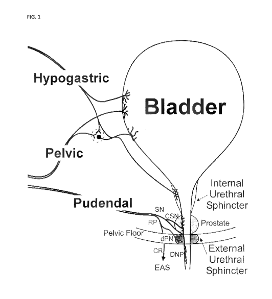
Figure 1: Schematic drawing showing innervation of the bladder, internal
urethral sphincter (IUS),
external urethral sphincter (EUS) and prostate. Sensory branch of the pudendal
nerve
(SN), rectal perineal branch of the pudendal nerve (RP), cranial sensory
branch of the
pudendal nerve (CSN), dorsal nerve of the penis branch of the pudendal nerve
(DNP; or
clitoris), deep perineal branch of the pudendal nerve (dPN) and caudal rectal
branch of
the pudendal nerve (CR).
Figure 2: Schematic drawings showing how apparatuses, devices and methods
according to the
invention can be put into effect.
Figure 3: A ¨ mean voiding efficiency in PGE2 and SHR models of bladder
dysfunction. In this
cohort, PGE2 rats exhibited a statistically non-significant reduction in
voiding efficiency
versus controls. Data is expressed as mean SE. N values as indicated. B ¨
Voiding
efficiency in individual PGE2 and SHR rats represented in A. Each PGE2 rat
prior to PGE2
installation is used as its own control and each data point represents the
mean for each
experiment.
Figure 4: A ¨ maximum bladder pressure (top) and threshold bladder pressure
(bottom) in PGE2
and SHR models of bladder dysfunction. PGE2 and SHR rats exhibited a reduction
in
threshold pressure and maximum pressure versus controls. Data is expressed as
mean
SE. N values as indicated. B - Maximum bladder pressure (top) and threshold
bladder
pressure (bottom)in individual PGE2 and SHR rats represented in A. Each PGE2
rat prior
to PGE2 installation is used as its own control and each data point represents
the mean
for each experiment.
5
CA 02994509 2018-02-01
WO 2017/021909
PCT/1B2016/054687
Figure 5: A ¨ mean bladder capacity in PGE2 and SHR models of bladder
dysfunction. PGE2 rats
exhibited a reduction in bladder capacity versus controls. Data is expressed
as mean
SE. N values as indicated. B ¨ Bladder capacity in individual PGE2 and SHR
rats
represented in A. Each PGE2 rat prior to PGE2 installation is used as its own
control and
each data point represents the mean for each experiment.
Figure 6: A ¨ mean Abladder pressure in PGE2 and SHR models of bladder
dysfunction. PGE2 and
SHR rats exhibited a reduction in Abladder pressure versus controls. Abladder
pressure
was calculated by subtracting baseline bladder pressure from the threshold
bladder
pressure. Data is expressed as mean SE. N values as indicated. B ¨ Abladder
pressure in
individual PGE2 and SHR rats represented in A. Each PGE2 rat prior to PGE2
installation is
used as its own control and each data point represents the mean for each
experiment.
Figure 7: A ¨ mean bladder compliance in PGE2 and SHR models of bladder
dysfunction. SHR rats
exhibited an increase in bladder compliance versus controls. Bladder
compliance was
calculated by dividing the bladder capacity by the Abladder pressure . The red
box
indicates which parameters used to calculate bladder compliance were decreased
when
compared to control. Data is expressed as mean SE. N values as indicated. B
¨ Bladder
compliance in individual PGE2 and SHR rats represented in A. Each PGE2 rat
prior to
PGE2 installation is used as its own control and each data point represents
the mean for
each experiment.
Figure 8: A ¨ mean non-voiding contraction (NVC) magnitude (as measured by
bladder pressure
area under the curve) in PGE2 and SHR models of bladder dysfunction. PGE2 and
SHR
rats exhibited a reduction in NVC magnitude versus controls. Data is expressed
as mean
SE. N values as indicated. B ¨ NVC magnitude in individual PGE2 and SHR rats
represented in A. Each PGE2 rat prior to PGE2 installation is used as its own
control and
each data point represents the mean for each experiment.
Figure 9: A ¨ NVC duration in PGE2 and SHR models of bladder dysfunction.
PGE2 and SHR rats
exhibited a reduction in NVC duration versus controls. Data is expressed as
mean SE. N
values as indicated. B ¨ NVC duration in individual PGE2 and SHR rats
represented in A.
Each PGE2 rat prior to PGE2 installation is used as its own control and each
data point
represents the mean for each experiment.
Figure 10: A ¨ NVC frequency (as measured by the number of NVC events counted
during the filling
phase of the cystometrogram) in PGE2 and SHR models of bladder dysfunction.
SHR rats
exhibited an increase in NVC frequency versus controls. Data is expressed as
mean SE.
N values as indicated. B ¨ NVC frequency in individual PGE2 and SHR rats
represented in
A. Each PGE2 rat prior to PGE2 installation is used as its own control and
each data point
represents the mean for each experiment.
Figure 11: A ¨ Mean external urethral sphincter (EUS) activity during the
phases of a CMG event as
measured by electromyography (EMG) in PGE2 and SHR models of bladder
dysfunction
6
CA 02994509 2018-02-01
WO 2017/021909
PCT/1B2016/054687
(n values as indicated). Data is expressed as mean SE. PGE2 and SHR rats
exhibit an
increase in EUS EMG activity during the filling and voiding phases versus
controls.
Figure 12: A ¨ Pelvic nerve stimulation restores bladder capacity in PGE2
rats. The indicated
electrical signals were applied to the pelvic nerves of PGE2 treated rats (n
values as
indicated). Data is expressed as mean SE. Stimulation of neural activity in
the pelvic
nerve as a result of the applied signal restores the bladder capacity. I3 ¨
The data
presented in A shown for individual rats to which the signals indicated were
applied to
the pelvic nerve. Each data point represents the mean for each experiment.
Figure 13: A ¨ Pelvic nerve stimulation restores voiding efficiency in PGE2
rats. The indicated
electrical signals were applied to the pelvic nerves of PGE2 treated rats (n
values as
indicated). Data is expressed as mean SE. Stimulation of neural activity in
the pelvic
nerve as a result of the applied signal restores the voiding efficiency. 13 ¨
The data
presented in A shown for individual rats to which the signals indicated were
applied to
the pelvic nerve. Each data point represents the mean for each experiment.
Figure 14: A ¨ Pelvic nerve stimulation restores bladder capacity in SHR rats.
Application of the
indicated electrical signals to stimulate the pelvic nerve of SHR rats
increased the
bladder capacity compared to unstimulated controls. Data is expressed as mean
SE.
Stimulation of neural activity in the pelvic nerve as a result of the applied
signal restores
the bladder capacity. 13 ¨ The data presented in A shown for individual rats
to which the
signals indicated were applied to the pelvic nerve. Each data point represents
the mean
for each experiment. C ¨ Pelvic nerve stimulation restores voiding efficiency
in SHR rats.
Application of the indicated electrical signals to stimulate the pelvic nerve
of SHR rats
increased the voiding efficiency compared to unstimulated controls. Data is
expressed as
mean SE. Stimulation of neural activity in the pelvic nerve as a result of
the applied
signal restores the voiding efficiency. D ¨ The data presented in C shown for
individual
rats to which the signals indicated were applied to the pelvic nerve. Each
data point
represents the mean for each experiment.
Figure 15: Mean bladder capacity pre- and post-stimulation of the pelvic nerve
in PGE2 model of
bladder dysfunction. PGE2 rats exhibited an increase in bladder capacity post
stimulation versus PGE2 (no stimulation) condition. Each PGE2 rat prior to
PGE2
installation is used as its own control. Data is expressed as mean SE. n =
11. * p < 0.05
vs 100 uM PGE2.
Figure 16: Voiding efficiency pre- and post-stimulation of the pelvic nerve in
PGE2 model of bladder
dysfunction. PGE2 rats exhibited an increase in voiding efficiency post
stimulation versus
PGE2 (no stimulation) condition. Data is expressed as mean SE. N values as
indicated.
N = 11. Each PGE2 rat prior to PGE2 installation is used as its own control.
Figure 17: Mean bladder capacity in cat PGE2 model of bladder dysfunction.
PGE2 cats exhibited a
dose dependent reduction in bladder capacity versus controls. Data is
expressed as
7
CA 02994509 2018-02-01
WO 2017/021909
PCT/1B2016/054687
mean SE. N values as indicated. Each PGE2 cat prior to PGE2 installation is
used as its
own control. N = 3. * p < 0.05 vs control.
Figure 18: Mean bladder capacity pre- and post-stimulation of the pelvic nerve
in cat PGE2 model
of bladder dysfunction. Preliminary experiment in PGE2 cat shows a stimulation
dependent increase in bladder capacity versus 5 u.M PGE2. Data is expressed as
mean of
multiple experiments (in a single subject) SE. N =1.
The terms as used herein are given their conventional definition in the art as
understood by the
skilled person, unless otherwise defined below. In the case of any
inconsistency or doubt, the
definition as provided herein should take precedence.
As used herein, application of a signal may equate to the transfer of energy
in a suitable form to
carry out the intended effect of the signal. That is, application of a signal
to a nerve or nerves may
equate to the transfer of energy to (or from) the nerve(s) to carry out the
intended effect. For
example, the energy transferred may be electrical, mechanical (including
acoustic, such as
ultrasound), electromagnetic (e.g. optical), magnetic or thermal energy. It is
noted that application
of a signal as used herein does not include a pharmaceutical intervention.
As used herein, "actuator" is taken to mean any element of applying a signal
to the nerve or plexus,
for example an electrode, diode, Peltier element or ultrasound actuator.
As used herein, "neural activity" of a nerve is taken to mean the signalling
activity of the nerve, for
example the amplitude, frequency and/or pattern of action potentials in the
nerve.
Modulation of neural activity, as used herein, is taken to mean that the
signalling activity of the
nerve is altered from the baseline neural activity ¨ that is, the signalling
activity of the nerve in the
patient prior to any intervention. Such modulation may increase, inhibit,
block, or otherwise change
the neural activity compared to baseline activity.
Where the modulation of neural activity is an increase of neural activity,
this may be an increase in
the total signalling activity of the whole nerve, or that the total signalling
activity of a subset of nerve
fibres of the nerve is increased, compared to baseline neural activity in that
part of the nerve. In a
preferred embodiment, the modulation of neural activity is an increase in the
signalling activity of
the afferent fibres of the nerve, optionally a selective increase in the
signalling activity of the
afferent fibres of the nerve. A selective increase in neural activity of the
afferent fibres does not
increase neural signalling in the efferent nerve fibres of the pelvic nerve,
or alternatively, does not
increase neural activity in the efferent nerve fibres of the pelvic nerve to a
threshold level at which
bladder pressure increases.
Where the modulation of neural activity is inhibition of neural activity, such
inhibition may be partial
inhibition. Partial inhibition may be such that the total signalling activity
of the whole nerve is
partially reduced, or that the total signalling activity of a subset of nerve
fibres of the nerve is fully
reduced (i.e. there is no neural activity in that subset of fibres of the
nerve), or that the total
8
CA 02994509 2018-02-01
WO 2017/021909
PCT/1B2016/054687
signalling of a subset of nerve fibres of the nerve is partially reduced
compared to baseline neural
activity in that subset of fibres of the nerve. Where the modulation of neural
activity is inhibition of
neural activity, this also encompasses full inhibition of neural activity in
the nerve ¨ that is, there is
no neural activity in the whole nerve.
Where inhibition of neural activity is a block on neural activity, such
blocking may be a partial block ¨
i.e. blocking of neural activity in a subset of nerve fibres of the nerve.
Alternatively, such blocking
may be a full block ¨ i.e. blocking of neural activity in the whole nerve. A
block on neural activity is
understood to be blocking neural activity from continuing past the point of
the block. That is, when
the block is applied, action potentials may travel along the nerve or subset
of nerve fibres to the
point of the block, but not beyond the point of the block.
Modulation of neural activity may also be an alteration in the pattern of
action potentials. It will be
appreciated that the pattern of action potentials can be modulated without
necessarily changing the
overall frequency. For example, modulation of the neural activity may be such
that the pattern of
action potentials is altered to more closely resemble a healthy state rather
than a disease state ¨ i.e.
to more closely resemble the pattern in a healthy individual.
Modulation of neural activity may comprise altering the neural activity in
various other ways, for
example increasing or inhibiting a particular part of the neural activity
and/or stimulating new
elements of activity, for example in particular intervals of time, in
particular frequency bands,
according to particular patterns and so forth. Such altering of neural
activity may for example
represent both increases and/or decreases with respect to the baseline
activity.
Modulation of the neural activity may be temporary. As used herein,
"temporary" is taken to mean
that the modulated neural activity (whether that is an increase, inhibition,
block or other modulation
of neural activity or change in pattern versus baseline activity) is not
permanent. That is, the neural
activity following cessation of the signal is substantially the same as the
neural activity prior to the
signal being applied ¨ i.e. prior to modulation.
Modulation of the neural activity may be persistent. As used herein,
"persistent" is taken to mean
that the modulated neural activity (whether that is an increase, inhibition,
block or other modulation
of neural activity or change in pattern versus baseline activity) has a
prolonged effect. That is, upon
cessation of the signal, neural activity in the nerve remains substantially
the same as when the signal
was being applied ¨ i.e. the neural activity during and following modulation
is substantially the same.
Modulation of the neural activity may be corrective. As used herein,
"corrective" is taken to mean
that the modulated neural activity (whether that is an increase, inhibition,
block or other modulation
of neural activity or change in pattern versus baseline activity) alters the
neural activity towards the
pattern of neural activity in a healthy individual. That is, upon cessation of
the signal, neural activity
in the nerve more closely resembles the pattern of action potentials in the
nerve observed in a
healthy subject than prior to modulation, preferably substantially fully
resembles the pattern of
action potentials in the nerve observed in a healthy subject.
9
CA 02994509 2018-02-01
WO 2017/021909
PCT/1B2016/054687
Such corrective modulation caused by the signal can be any modulation as
defined herein. For
example, application of the signal may result in a block on neural activity,
and upon cessation of the
signal, the pattern of action potentials in the nerve resembles the pattern of
action potentials
observed in a healthy subject. By way of further example, application of the
signal may result
modulation such that the neural activity resembles the pattern of action
potentials observed in a
healthy subject, and upon cessation of the signal, the pattern of action
potentials in the nerve
resembles the pattern of action potentials observed in a healthy individual.
As used herein, a "healthy individual" or "healthy subject" is an individual
not exhibiting any
disruption or perturbation of normal bladder activity.
As used herein, "bladder dysfunction" is taken to mean that the patient or
subject is exhibiting
disruption of bladder function compared to a healthy individual. Bladder
dysfunction may be
characterised by symptoms such as nocturia, increased urinary retention,
increased incontinence,
increased urgency of urination or increased frequency of urination compared to
a healthy individual.
Bladder dysfunction includes conditions such as overactive bladder (OAB),
neurogenic bladder,
stress incontinence, and chronic urinary retention.
As used herein, an "improvement in a measurable physiological parameter" is
taken to mean that
for any given physiological parameter, an improvement is a change in the value
of that parameter in
the patient towards the normal value or normal range for that value ¨ i.e.
towards the expected
value in a healthy individual.
For example, in a patient with bladder dysfunction, an improvement in a
measurable parameter may
be: a reduction in number of incontinence episodes, a decrease in urgency of
urination, a decrease
in frequency of urination, an increase in bladder capacity, an increase in
bladder voiding efficiency,
and/or a change in external urethral sphincter (EUS) activity towards that of
a healthy individual,
assuming the patient is exhibiting abnormal values for the respective
parameter.
As used herein, a physiological parameter is not affected by modulation of the
neural activity if the
parameter does not change as a result of the modulation from the average value
of that parameter
exhibited by the subject or patient when no intervention has been performed ¨
i.e. it does not
depart from the baseline value for that parameter.
The skilled person will appreciate that the baseline for any neural activity
or physiological parameter
in an individual need not be a fixed or specific value, but rather can
fluctuate within a normal range
or may be an average value with associated error and confidence intervals.
Suitable methods for
determining baseline values would be well known to the skilled person.
As used herein, a measurable physiological parameter is detected in a patient
when the value for
that parameter exhibited by the patient at the time of detection is
determined. In addition, the
detection of the physiological parameter may include detection of a
characteristic of the measured
signal, for example amplitude or power, e.g., over a range of frequencies. A
detector is any element
able to make such a determination.
CA 02994509 2018-02-01
WO 2017/021909
PCT/1B2016/054687
A "predefined threshold value" for a physiological parameter is the value for
that parameter where
that value or beyond must be exhibited by a subject or patient before the
intervention is applied. For
any given parameter, the threshold value may be defined as a value indicative
of a pathological state
(e.g. the patient is experiencing abnormal retention of urine) or a particular
physiological state (e.g.
the patient being asleep). Examples of such predefined threshold values
include parasympathetic or
sympathetic tone (neural, hemodynamic (e.g. heart rate, blood pressure, heart
rate variability) or
circulating plasma/urine biomarkers) greater than a threshold parasympathetic
or sympathetic tone;
abnormal bladder pressure compared to a healthy individual, abnormal bladder
capacity compared
to a healthy individual, bladder voiding efficiency lower than a healthy
individual, abnormal pelvic
nerve activity compared to a healthy individual (for instance a decrease in
pelvic nerve activity),
abnormal EUS activity compared to a healthy individual (for instance an
increase in EUS activity),
abnormal pudendal nerve activity (for instance a decrease in pudendal afferent
activity), abnormal
hypogastric nerve activity (for instance an increase in hypogastric nerve
activity), or abnormal rate of
change, e.g., increase in bladder pressure. Such a threshold value for a given
physiological
parameter is exceeded if the value exhibited by the patient is beyond the
threshold value ¨ that is,
the exhibited value is a greater departure from the normal or healthy value
for that parameter than
the predefined threshold value.
The measurable physiological parameter may comprise an action potential or
pattern of action
potentials in one or more nerves of the patient, wherein the action potential
or pattern of action
potentials is associated with bladder dysfunction. Suitable nerves in which to
detect an action
potential or pattern of action potentials include a pelvic nerve, a pudendal
nerve and/or a
hypogastric nerve. In a particular embodiment, the measurable physiological
parameter comprises
the pattern of action potentials in the pelvic nerve. The measureable
physiological parameter may
be muscle electromygraphic activity, wherein the electromyographic activity is
indicative of the level
of activity in the muscle. Such activity could typically be measured from the
bladder detrusor muscle,
the internal urethral sphincter, the external urethral sphincter, and the
external anal sphincter.
Treatment of bladder dysfunction, as used herein may be characterised by any
one or more of a
reduction in number of incontinence episodes, a decrease in urgency of
urination, a decrease in
frequency of urination, an increase bladder capacity, an increase in bladder
voiding efficiency, a
decrease in urinary retention, a change in external urethral sphincter (EUS)
activity towards that of a
healthy individual, and/or a change in the pattern of action potentials or
activity of the pelvic nerve,
pudendal nerve or hypogastric nerve towards that of a healthy individual.
Treatment of bladder dysfunction may be prophylactic or therapeutic.
A "neuromodulation apparatus" as used herein is an apparatus or device
configured to modulate the
neural activity of a nerve. Neuromodulation apparatuses or devices as
described herein comprise at
least one actuator capable of effectively applying a signal to a nerve. In
those embodiments in which
the neuromodulation apparatus is at least partially implanted in the patient,
the elements of the
apparatus that are to be implanted in the patient are constructed such that
they are suitable for
such implantation. Such suitable constructions would be well known to the
skilled person. Indeed,
various fully implantable neuromodulation devices have been implanted into
human patients, such
11
CA 02994509 2018-02-01
WO 2017/021909
PCT/1B2016/054687
as the INTERSTIMIm devices of Medtronic, Inc (Minneapolis, MN), the Finetech-
Brindley bladder
control system (Finetech Medical, Hertfordshire, UK) and the BIONTM devices of
Advanced Bionics
Corp.
As used herein, "implanted" is taken to mean positioned within the patient's
body. Partial
implantation means that only part of the apparatus is implanted ¨ i.e. only
part of the apparatus is
positioned within the patient's body, with other elements of the apparatus
external to the patient's
body. Wholly implanted means that the entire apparatus is positioned within
the patient's body.
For the avoidance of doubt, the apparatus being "wholly implanted" does not
preclude additional
elements, independent of the apparatus but in practice useful for its
functioning (for example, a
remote wireless charging unit or a remote wireless manual override unit),
being independently
formed and external to the patient's body.
As used herein, "charge-balanced" in relation to a DC current is taken to mean
that the positive or
negative charge introduced into any system (e.g. a nerve) as a result of a DC
current being applied is
balanced by the introduction of the opposite charge in order to achieve
overall (i.e. net) neutrality.
As used herein, a "pharmaceutical composition" is a composition suitable for
administration to a
subject or patient.
As used herein, a "compound for treating bladder dysfunction" is taken to mean
a pharmacological
compound capable of treating bladder dysfunction. Such compounds include
antimuscarinic
compounds, for example darifenacin, hyoscyamine, oxybutynin, tolterodine,
solifenacin, trospium,
or fesoterodine. Other examples are B-adrenergic receptor agonist compounds,
optionally a 133-
adrenergic receptor agonist, for example mirabegron. Another example is
botulinum toxin.
DETAILED DESCRIPTION
In accordance with a first aspect of the invention there is provided an
apparatus for modulating the
neural activity of the afferent fibres of at least one pelvic nerve of a
patient, the apparatus
comprising: a first actuator configured to apply a first signal to said at
least one nerve; and a
controller coupled to the first actuator and controlling the signal to be
applied by the first actuator,
such that the signal modulates the neural activity of the afferent fibres of
the pelvic nerve to
produce a physiological response in the patient.
In certain embodiments, the apparatus comprises a second actuator coupled to
the controller and
configured to apply a second signal to a pudendal nerve of the patient,
wherein the controller
controls the signal to be applied by the second actuator such that the signal
modulates the neural
activity of the afferent nerve fibres in the pudendal nerve to produce a
physiological response in the
patient.
In certain embodiments, the apparatus comprises one actuator configured to
apply said first signal
to only one of the left or right pelvic nerves of said patient. In this
embodiment, the apparatus may
further comprise a second actuator configured to apply said second signal to
only one of the left or
right pudendal nerves of said patient.
12
CA 02994509 2018-02-01
WO 2017/021909
PCT/1B2016/054687
In certain embodiments, the modulation of the neural activity of the afferent
fibres of the nerve or
nerves is selective. A signal selectively modulates the neural activity of the
afferent fibres of the
pelvic nerve and/or pudendal nerve if that signal does not modulate the neural
activity of the
efferent fibres of those nerve(s), or if that signal modulates the neural
activity of the efferent fibres
of those nerves below a degree of modulation of the efferent fibres which
leads to an increase in
bladder pressure or voiding of the bladder (e.g. below the degree of
modulation which leads to an
increase in bladder pressure to the threshold pressure as defined in Andersson
et al. Neurourology
and Urodynamics, 30: 636-646 (2011)).
In certain embodiments, the signal applied by the one or more actuators is a
non-destructive signal.
As used herein, a "non-destructive signal" is a signal as defined above that,
when applied, does not
irreversibly damage the underlying neural signal conduction ability. That is,
application of a non-
destructive signal maintains the ability of the nerve or nerves (or fibres
thereof) to conduct action
potentials when application of the signal ceases, even if that conduction is
in practice inhibited or
blocked as a result of application of the non-destructive signal.
In those embodiments in which the apparatus has at least a first actuator and
a second actuator, the
signal which each of the actuators is configured to apply is independently
selected from the signal to
be applied by the other actuator.
In the passages below, the described embodiments of the signal apply to the
first signal and, where
applicable, may also apply to the second signal, independently of the first
signal.
In certain embodiments, the signal which the first or second actuator is
configured to apply is of a
modality selected from an electrical signal, an optical signal, an ultrasonic
signal, and a thermal
signal. That is, each actuator may be configured to apply a different modality
of signal. Alternatively,
in certain embodiments each actuator is configured to apply the same modality
of signal.
In certain embodiments, each actuator may be comprised of one or more
electrodes, one or more
photon sources, one or more ultrasound transducers, one more sources of heat,
or one or more
other types of actuator arranged to put the signal into effect.
In certain embodiments, the actuator is an electrode and the signal applied by
the actuator is an
electrical signal, for example a voltage or current. In certain such
embodiments the signal applied
comprises a direct current (DC) waveform, such as a charge balanced direct
current waveform, or an
alternating current (AC) waveform, or both a DC and an AC waveform.
In certain embodiments the signal comprises a sub-kilohertz frequency AC
waveform. In certain such
embodiments the signal comprises an AC waveform having a frequency of 0.1-500
Hz, preferably
0.1-50Hz, preferably 1-50Hz or 0.5-20 Hz, preferably 1-15Hz, for example 1, 2,
3, 4, 5, 6, 7, 8, 9, 10,
11, 12, 13, 14, 15 Hz, preferably 1-10Hz, for example 1, 2, 3, 4, 5, 6, 7, 8,
9, 10 Hz, preferably 1 or 10
Hz.
Typically, effective treatment of the symptoms of bladder dysfunction in
accordance with the
invention (for example overactive bladder (OAB), neurogenic bladder or other
dysfunction of the
lower urinary tract) requires the selection of appropriate stimulation
parameters. Stimulation
13
CA 02994509 2018-02-01
WO 2017/021909
PCT/1B2016/054687
parameters include the stimulation pulse amplitude/intensity, the stimulation
pulse duration, and
stimulation frequency.
Relative stimulation pulse intensity can be expressed as multiples (0.1, 0.8,
1, 2, 5, etc.) of "T". "T" is
the threshold stimulation intensity required to evoke a response a reflex
electromyogram (EMG)
response in the external urethral sphincter (EUS).
By way of example, T may be determined as follows: a low frequency electrical
signal, typically 1 Hz,
is applied and the intensity of stimulation is increased (either by increasing
the voltage or the
current of the signal, preferably the current) until the pelvic nerve
stimulation pulse produces a
reflex EMG response in the EUS. This stimulation intensity is designated T.
The absolute threshold
stimulation intensity may vary across individuals, and subsequent experimental
or therapeutic
intensities are designated as multiples of T to provide equivalent relative
stimulation intensities.
The desired stimulation intensity (i.e. the desired multiple of threshold
intensity "T") can be
achieved through controlled variation of the current or voltage of the signal,
preferably the current.
In certain embodiments the electrical signal has an amplitude value of from
0.1 T to 5.0 T, where T is
a threshold obtained through empirical measurement of the threshold for the
stimulation signal to
evoke a reflex response in the external urethral sphincter or external anal
sphincter, following
application of stimulus to the pelvic nerve or pudendal nerve. In certain
embodiments, the electrical
signal has a T value of 0.1T-5.0T, 0.5T-2.5T or 0.2-3.0T, 0.25T-2.0T, for
example 0.8T or 2.0T. In
certain preferred embodiments the signal has a T value of 0.8T or 2.0T.
In certain preferred embodiments, the signal is an electrical signal
comprising an AC waveform of
0.8T 1Hz, 0.8T 10Hz, 2.0T 1Hz, or 2.0T 10Hz.
In certain embodiments wherein the signal applied by the one or more actuators
is a thermal signal,
the signal reduces the temperature of the nerve (i.e. cools the nerve). In
certain alternative
embodiments, the signal increases the temperature of the nerve (i.e. heats the
nerve). In certain
embodiments, the signal both heats and cools the nerve.
In those embodiments in which the signal applied by the one or more actuators
is a thermal signal,
at least one of the one or more actuators is configured to apply a thermal
signal. In certain such
embodiments, all the actuators are configured to apply a thermal signal,
optionally the same
thermal signal.
In certain embodiments, one or more of the one or more actuators comprise a
Peltier element
configured to apply a thermal signal, optionally all of the one or more
actuators comprise a Peltier
element. In certain embodiments, one or more of the one or more actuators
comprise a laser diode
configured to apply a thermal signal, optionally all of the one or more
actuators comprise a laser
diode configured to apply a thermal signal (e.g. a diode configured to emit
infrared radiation). In
certain embodiments, one or more of the one or more actuators comprise an
electrically resistive
element configured to apply a thermal signal, optionally all of the one or
more actuators comprise
an electrically resistive element configured to apply a thermal signal.
14
CA 02994509 2018-02-01
WO 2017/021909
PCT/1B2016/054687
In certain alternative embodiments, the signal applied by the one or more
actuators is not a thermal
signal.
In certain embodiments the signal applied by the one or more actuators is a
mechanical signal,
optionally an ultrasonic signal. In certain alternative embodiments, the
mechanical signal applied by
the one or more actuators is a pressure signal.
In certain embodiments the signal applied by the one or more actuators is an
electromagnetic signal,
optionally an optical signal. In certain such embodiments, the one or more
actuators comprise a
laser and/or a light emitting diode configured to apply the optical signal. In
some embodiments, the
apparatus further comprises a fibre optic interface configured to apply said
signal from said one or
more of the actuators to said at least one nerve.
It will be appreciated that in embodiments in which the apparatus comprises
more than one
actuator, the signal to be applied by each actuator is independently selected
from the signal applied
by the other actuator(s). For example, an apparatus according to the invention
may comprise one
actuator configured to apply a sub-kilohertz frequency AC waveform to the
pelvic nerve in order to
stimulate neural activity in the afferent fibres of the pelvic nerve, and a
second actuator configured
to apply a high frequency AC waveform to the pudendal nerve in order to
inhibit or block signalling
in the pudendal nerve. Alternatively, a first and a second actuator may be
configured to stimulate
neural activity in the pelvic nerve and in the pudendal nerve, respectively.
In certain embodiments, the physiological response produced in the patient is
one or more of: a
reduction in number of incontinence episodes, a reduction in the length and/or
severity of
incontinence episode(s), a decrease in urgency of urination, a decrease in
frequency of urination, an
increase bladder capacity, an increase in bladder voiding efficiency, a
decrease in urinary retention
and/or a change in external urethral sphincter (EUS) activity towards that of
a healthy individual.
In certain embodiments, the apparatus further comprises one or more detector
elements to detect
one or more physiological parameters in the patient. Such a detector element
may be configured to
detect the one or more physiological parameters. That is, in such embodiments
each detector may
detect more than one physiological parameter, for example all the detected
physiological
parameters. Alternatively, in such embodiments each of the one or more
detector elements is
configured to detect a separate parameter of the one or more physiological
parameters detected.
In certain embodiments, the one or more detected physiological parameters are
selected from:
parasympathetic tone, sympathetic tone, bladder pressure, bladder volume,
external urethral
sphincter activity, and the rate of change of any one of these parameters. In
addition, the one or
more detected physiological parameters may be selected from: nerve activity in
the pelvic nerve, the
hypogastric nerve or the pudendal nerve; muscle activity in the bladder
detrusor muscle, the
internal urethral sphincter, the external urethral sphincter or the external
anal sphincter; and the
rate of change of any one of these parameters. The skilled person will
appreciate that the detection
of the physiological parameter may include detection of the absolute value of
that parameter, or a
characteristic of the detection signal, for example amplitude or power, e.g.,
over a range of
frequencies.
CA 02994509 2018-02-01
WO 2017/021909
PCT/1B2016/054687
In an embodiment, the detector element is configured to detect nerve activity
in the pelvic nerve
(optionally, where the actuator is configured to apply a signal to one of the
left or right pelvic nerve,
the detector element is configured to detect nerve activity in the other of
the left or right pelvic
nerve). In another embodiment, the detector element is configured to detect
nerve activity in the
hypogastric nerve. In another embodiment, the detector element is configured
to detect nerve
activity in the pudendal nerve. In another embodiment, the detector element is
configured to
detect muscle activity in the bladder detrusor muscle. In another embodiment,
the detector element
is configured to detect muscle activity in the internal urethral sphincter. In
another embodiment, the
detector element is configured to detect muscle activity in the external
urethral sphincter. In
another embodiment, the detector element is configured to detect muscle
activity in the external
anal sphincter.
In such embodiments, the controller is coupled to the detector element
configured to detect one or
more physiological parameters, and causes the actuator or actuators to apply
the signal when the
physiological parameter is detected to be meeting or exceeding a predefined
threshold value.
The inventors have observed, in an animal model of bladder dysfunction (in
particular OAB), a
decrease in pelvic nerve activity and an increase in hypogastric activity.
Therefore, in certain
embodiments, the one or more detected physiological parameters comprise an
action potential or
pattern of action potentials in one or more nerves of the patient, wherein the
action potential or
pattern of action potentials is associated with bladder dysfunction. In
certain such embodiments, the
one or more nerves are selected from a pelvic nerve, a pudendal nerve and a
hypogastric nerve,
preferably a pelvic nerve. In a preferred embodiment, the detected
physiological parameter is a
decrease in pelvic nerve activity, and/or an increase in hypogastric nerve
activity.
It will be appreciated that any two or more of the indicated physiological
parameters may be
detected in parallel or consecutively. For example, in certain embodiments,
the controller is coupled
to a detector or detectors configured to detect the pattern of action
potentials in the pelvic nerve at
the same time as the bladder pressure in the patient.
In certain embodiments, the modulation in neural activity as a result of
applying the signal is
stimulation or an increase in neural activity in the nerve to which the signal
is applied. That is, in
such embodiments, application of the signal results in the neural activity in
at least the afferent
fibres of at least part of the nerve being stimulated or increased compared to
the baseline neural
activity in that part of the nerve. In certain embodiments, neural activity is
increased across the
whole nerve. In certain preferred embodiments, neural activity is selectively
stimulated in the
afferent fibres of the nerve to which the signal is applied (e.g. the pelvic
nerve).
In certain embodiments, the modulation in neural activity as a result of
applying the signal is an
alteration to the pattern of action potentials in the nerve to which the
signal is applied. In certain
such embodiments, the neural activity is modulated such that the resultant
pattern of action
potentials in the nerve resembles the pattern of action potentials in the
nerve or nerves observed in
a healthy subject.
16
CA 02994509 2018-02-01
WO 2017/021909
PCT/1B2016/054687
Modulation of neural activity may comprise altering the neural activity in
various other ways, for
example increasing or inhibiting a particular part of the activity and
stimulating new elements of
activity, for example in particular intervals of time, in particular frequency
bands, according to
particular patterns and so forth. Such altering of neural activity may for
example represent both
increases and/or decreases with respect to the baseline activity.
In certain embodiments, the controller causes the signal to be applied
intermittently. In certain such
embodiments, the controller causes the signal to applied for a first time
period, then stopped for a
second time period, then reapplied for a third time period, then stopped for a
fourth time period. In
such an embodiment, the first, second, third and fourth periods run
sequentially and consecutively.
The series of first, second, third and fourth periods amounts to one
application cycle. In certain such
embodiments, multiple application cycles can run consecutively such that the
signal is applied in
phases, between which phases no signal is applied. In certain embodiments, the
signal applied for
the first time period and the signal applied for the third time period are of
the same parameters
(frequency, amplitude, etc.) and the same modality. In other embodiments, the
signal applied for
the first and third time periods are of different parameters, and/or different
modality.
In such embodiments, the duration of the first, second, third and fourth time
periods is
independently selected. That is, the duration of each time period may be the
same or different to
any of the other time periods. In certain such embodiments, the duration of
each of the first,
second, third and fourth time periods is any time from 5 seconds (5s) to 24
hours (24h), 30s to 12 h,
1 min to 12 h, 5 min to 8 h, 5 min to 6 h, 10 min to 6 h, 10 min to 4 h, 30
min to 4 h, 1 h to 4 h. In
certain embodiments, the duration of each of the first, second, third and
fourth time periods is 5s,
10s, 30s, 60s, 2 min, 5 min, 10 min, 20 min, 30 min, 40 min, 50 min, 60 min,
90 min, 2 h, 3 h, 4 h, 5 h,
6 h, 7 h, 8 h, 9 h, 10 h, 11 h, 12 h, 13 h, 14 h, 15 h, 16 h, 17 h, 18 h, 19
h, 20 h, 21 h, 22 h, 23 h, 24 h.
In certain embodiments wherein the controller causes the signal to be applied
intermittently, the
signal is applied for a specific amount of time per day. In certain such
embodiments, the signal is
applied for 10 min, 20 min, 30 min, 40 min, 50 min, 60 min, 90 min, 2 h, 3 h,
4 h, 5 h, 6 h, 7 h, 8 h, 9
h, 10 h, 11 h, 12 h, 13 h, 14 h, 15 h, 16 h, 17 h, 18 h, 19 h, 20 h, 21 h, 22
h, 23 h per day. In certain
such embodiments, the signal is applied continuously for the specified amount
of time. In certain
alternative such embodiments, the signal may be applied discontinuously across
the day, provided
the total time of application amounts to the specified time.
In certain embodiments wherein the controller causes the signal to be applied
intermittently, the
signal is applied only when the patient is in a specific physiological state.
In certain such
embodiments, the signal is applied only when the patient exhibits a particular
bladder pressure. In
certain such embodiments, the signal is applied only when the patient is in a
continent state. In
certain such embodiments, the signal is applied only when the patient is in a
state of bladder
emptying. In certain such embodiments, the signal is applied only when the
patient is asleep.
In certain such embodiments, the apparatus further comprises a communication,
or input, element
via which the status of the patient (e.g. that they about to go to sleep) can
be indicated by the
patient or a physician. In alternative embodiments, the apparatus further
comprises a detector
17
CA 02994509 2018-02-01
WO 2017/021909
PCT/1B2016/054687
configured to detect the status of the patient, wherein the signal is applied
only when the detector
detects that the patient is in the specific state.
In certain embodiments of the apparatus, the modulation in neural activity
caused by the application
of the signal (whether that is an increase, inhibition, block or other
modulation of neural activity) is
temporary. That is, upon cessation of the signal, neural activity in the nerve
or nerves returns
substantially towards baseline neural activity within 1-60 seconds, or within
1-60 minutes, or within
1-24 hours, optionally 1-12 hours, optionally 1-6 hours, optionally 1-4 hours,
optionally 1-2 hours. In
certain such embodiments, the neural activity returns substantially fully to
baseline neural activity.
That is, the neural activity following cessation of the signal is
substantially the same as the neural
activity prior to the signal being applied ¨ i.e. prior to modulation.
In certain alternative embodiments, the modulation in neural activity caused
by the application of
the signal or signals is substantially persistent. That is, upon cessation of
the signal, neural activity in
the nerve or nerves remains substantially the same as when the signal was
being applied ¨ i.e. the
neural activity during and following modulation is substantially the same.
In certain embodiments, the modulation in neural activity caused by the
application of the signal is
partially corrective, preferably substantially corrective. That is, upon
cessation of the signal, neural
activity in the nerve or nerves more closely resembles the pattern of action
potentials in the nerve(s)
observed in a healthy subject than prior to modulation, preferably
substantially fully resembles the
pattern of action potentials in the nerve(s) observed in a healthy subject. In
such embodiments, the
modulation caused by the signal can be any modulation as defined herein. For
example, application
of the signal may result in a block on neural activity, and upon cessation of
the signal, the pattern of
action potentials in the nerve or nerves resembles the pattern of action
potentials observed in a
healthy individual. By way of further example, application of the signal may
result in modulation
such that the neural activity resembles the pattern of action potentials
observed in a healthy
subject, and upon cessation of the signal, the pattern of action potentials in
the nerve or nerves
resembles the pattern of action potentials observed in a healthy subject
In certain embodiments, the apparatus is suitable for at least partial
implantation into the patient. In
certain such embodiments, the apparatus is suitable to be fully implanted in
the patient.
In certain embodiments, the apparatus further comprises one or more power
supply elements, for
example a battery, and/or one or more communication elements.
In a second aspect, the invention provides a method for treating bladder
dysfunction in a patient,
the method comprising implanting an apparatus according to the first aspect,
positioning the first
actuator of the apparatus in signalling contact with a pelvic nerve of the
patient, and activating the
apparatus. In such embodiments, the actuator is in signalling contact with the
nerve when it is
positioned such that the signal can be effectively applied to the nerve. The
apparatus is activated
when the apparatus is in an operating state such that the signal will be
applied as determined by the
controller.
18
CA 02994509 2018-02-01
WO 2017/021909
PCT/1B2016/054687
In certain embodiments, the actuator or actuators positioned in signalling
contact with a pelvic
nerve apply a signal to stimulate afferent neural activity in said pelvic
nerve, preferably to selectively
stimulate the afferent fibres of said pelvic nerve. In certain embodiments,
the first actuator is
positioned in signalling contact with only one of the left or right pelvic
nerve. A detector element
may be placed in signalling contact with the other of the left or right pelvic
nerve.
In certain embodiments, the method comprises implanting an apparatus according
to the first
aspect, positioning the first actuator in signalling contact with a pelvic
nerve of the patient to
modulate the neural activity of said pelvic nerve, and positioning a second
actuator in signalling
contact with a pudendal nerve of the patient to modulate the neural activity
of said pudendal nerve.
In certain embodiments, the first actuator is positioned in signalling contact
with only a pelvic nerve
and the second actuator is positioned in signalling contact only with a
pudendal nerve. In certain
such embodiments the first actuator is configured to apply a signal to
stimulate neural activity in
said pelvic nerve, and the second actuator is configured to apply a signal to
inhibit or block neural
activity in said pudendal nerve. In certain alternative embodiments, the first
actuator is configured
to apply a signal to stimulate neural activity in said pelvic nerve, and the
second actuator is
configured to apply a signal to stimulate neural activity in said pudendal
nerve.
In certain embodiments, the method is a method for treating overactive bladder
or neurogenic
bladder.
Implementation of all aspects of the invention (as discussed both above and
below) will be further
appreciated by reference to Figures 2A-2C.
Figures 2A-2C show how the invention may be put into effect using one or more
neuromodulation
apparatuses which are implanted in, located on, or otherwise disposed with
respect to a patient in
order to carry out any of the various methods described herein. In this way,
one or more
neuromodulation apparatuses can be used to treat bladder dysfunction in a
patient, by modulating
neural activity in at least a pelvic nerve, optionally also a pudendal nerve.
In Figure 2A a separate neuromodulation apparatus 100 is provided for
unilateral neuromodulation,
although as discussed above and below a apparatus could be provided for
bilateral neuromodulation
(100, Fig. 28 and 2C). Each such neuromodulation apparatus may be fully or
partially implanted in
the patient, or otherwise located, so as to provide neuromodulation of the
respective nerve or
nerves. Figure 2A also schematically shows in the cutaway components of one of
the
neuromodulation apparatuses 100, in which the apparatus comprises several
elements, components
or functions grouped together in a single unit and implanted in the patient. A
first such element is an
actuator 102 which is shown in proximity to a pelvic nerve 90 of the patient.
The apparatus may
optionally further comprise further actuators (not shown) implanted proximally
to a pudendal nerve.
Alternatively, the pudendal nerve may be provided with a separate apparatus
100 (not shown). The
actuator 102 may be operated by a controller element 104. The apparatus may
comprise one or
more further elements such as a communication element 106, a detector element
108, a power
supply element 110 and so forth. Each neuromodulation apparatus 100 may
operate independently,
or may operate in communication with each other, for example using respective
communication
elements 106.
19
CA 02994509 2018-02-01
WO 2017/021909
PCT/1B2016/054687
Each neuromodulation apparatus 100 may carry out the required neuromodulation
independently,
or in response to one or more control signals. Such a control signal may be
provided by the
controller 104 according to an algorithm, in response to output of one or more
detector elements
108, and/or in response to communications from one or more external sources
received using the
communications element. As discussed herein, the detector element(s) could be
responsive to a
variety of different physiological parameters.
Figure 28 illustrates some ways in which the apparatus of Figure 2A may be
differently distributed.
For example, in Figure 28 the neuromodulation apparatuses 100 comprise
actuators 102 implanted
proximally to a pelvic nerve 90, optionally further comprising further
actuators (not shown)
implanted proximally to a pudendal nerve, but other elements such as a
controller 104, a
communication element 106 and a power supply 110 are implemented in a separate
control unit
130 which may also be implanted in, or carried by the patient. The control
unit 130 then controls the
actuators in both of the neuromodulation apparatuses via connections 132 which
may for example
comprise electrical wires and/or optical fibres for delivering signals and/or
power to the actuators.
In the arrangement of Figure 28 one or more detectors 108 are located
separately from the control
unit, although one or more such detectors could also or instead be located
within the control unit
130 and/or in one or both of the neuromodulation apparatuses 100. The
detectors may be used to
detect one or more physiological parameters of the patient, and the controller
element or control
unit then causes the actuators to apply the signal in response to the detected
parameter(s), for
example only when a detected physiological parameter meets or exceeds a
predefined threshold
value. Physiological parameters which could be detected for such purposes
include parasympathetic
or sympathetic tone (neural, hemodynamic (e.g. heart rate, blood pressure,
heart rate variability) or
circulating plasma/urine biomarkers) greater than a threshold parasympathetic
or sympathetic tone;
abnormal bladder pressure compared to a healthy individual, abnormal bladder
capacity compared
to a healthy individual, bladder voiding efficiency lower than a healthy
individual, abnormal pelvic
nerve activity compared to a healthy individual (for instance a decrease in
pelvic nerve activity),
abnormal EUS activity compared to a healthy individual (for instance an
increase in EUS activity),
abnormal pudendal nerve activity (for instance a decrease in pudendal afferent
activity), abnormal
hypogastric nerve activity (for instance an increase in hypogastric nerve
activity), or an abnormal
rate of increasing bladder pressure. Physiological parameters which could be
detected also include
the power spectrum of the detected signal (for example via a fast fourier
transform (FFT) or similar
transform).
A variety of other ways in which the various functional elements could be
located and grouped into
the neuromodulation apparatuses, a control unit 130 and elsewhere are of
course possible. For
example, one or more sensors of Figure 28 could be used in the arrangement of
Figures 2A or 2C or
other arrangements.
Figure 2C illustrates some ways in which some functionality of the apparatus
of Figures 2A or 28 is
provided not implanted in the patient. For example, in Figure 2C an external
power supply 140 is
provided which can provide power to implanted elements of the apparatus in
ways familiar to the
skilled person, and an external controller 150 provides part or all of the
functionality of the
CA 02994509 2018-02-01
WO 2017/021909
PCT/1B2016/054687
controller 104, and/or provides other aspects of control of the apparatus,
and/or provides data
readout from the apparatus, and/or provides a data input facility 152. The
data input facility could
be used by a patient or other operator in various ways, for example to input
data relating to the
activity status or bladder pressure.
Each neuromodulation apparatus may be adapted to carry out the neuromodulation
required using
one or more physical modes of operation which typically involve applying a
signal to a pelvic nerve,
optionally also to a pudendal nerve, such a signal or signals typically
involving a transfer of energy to
(or from) the nerve(s). As already discussed, such modes may comprise
modulating the nerve or
nerves using an electrical signal, an optical signal, an ultrasound or other
mechanical signal, a
thermal signal, a magnetic or electromagnetic signal, or some other use of
energy to carry out the
required modulation. Such signals may be non-destructive signals. Such
modulation may comprise
increasing, inhibiting, blocking or otherwise changing the pattern of neural
activity in the nerve or
nerves. Preferably the modulation comprises stimulating, optionally
selectively stimulating, neural
activity in the afferent fibres of the nerve or nerves. To this end, the
actuator 90 illustrated in Figure
2A could be comprised of one or more electrodes, one or more photon sources,
one or more
ultrasound transducers, one more sources of heat, or one or more other types
of actuators arranged
to put the required neuromodulation into effect.
The neural modulation device(s) or apparatus may be arranged to stimulate
(i.e. increase or induce)
neural activity of a nerve, for example a pelvic nerve or a pudendal nerve, by
using the actuator(s) to
apply a voltage or current, for example a direct current (DC) such as a charge
balanced direct
current, or an AC waveform, or both. The device or apparatus may be arranged
to use the
actuator(s) to apply an AC waveform preferably 0.1-50Hz, preferably 1-50Hz or
0.5-20 Hz, preferably
1-15Hz, for example 1, 2, 3, 4, 5, 6, 7, 8, 9, 10, 11, 12, 13, 14, 15 Hz,
preferably 1-10Hz, for example
1, 2, 3, 4, 5, 6, 7, 8, 9, 10 Hz, preferably 1 or 10 Hz.
In certain embodiments the AC waveform has an amplitude of from 0.5 T to 2.5
T, where "T" is the
intensity of stimulation at which, for a given frequency (typically 1Hz), a
reflex EMG response in the
EUS is produced. The skilled person would be readily able to determine the
appropriate value of T in
any given patient.
In certain embodiments, the electrical signal has a T value of 0.1T-5.0T, 0.5T-
2.5T, 0.2T-3.0T, 0.25T-
2.0T, or 0.8T-2.0T, for example 0.8T, 0.9T, 1.0T, 1.1T, 1.2T, 1.3T, 1.4T,
1.5T, 1.6T, 1.8T, 1.9T, 2.0T,
2.5T, 3.0T. In certain preferred embodiments the signal has a T value of 0.8T
or 2.0T, in particular
0.8T.
In certain preferred embodiments, the signal is an electrical signal
comprising an AC waveform of
0.8T 1Hz, 0.8T 10Hz, 2.0T 1Hz, or 2.0T 10Hz.
The neural modulation device(s) or apparatus may be arranged to inhibit neural
activity of a nerve,
for example a pelvic nerve or a pudendal nerve, by using the actuator(s) to
apply a voltage or
current, for example a direct current (DC) such as a charge balanced direct
current, or an AC
waveform, or both. The device or apparatus may be arranged to use the
actuator(s) to apply a DC
ramp, then apply a first AC waveform, wherein the amplitude of the waveform
increases during the
21
CA 02994509 2018-02-01
WO 2017/021909
PCT/1B2016/054687
period the waveform is applied, and then apply a second AC waveform. The AC
waveform(s) may
have a frequency of 5 to 50 KHz, optionally 5-10 KHz.
Thermal methods of neuromodulation typically manipulate the temperature of a
nerve to inhibit
signal propagation. For example, Patberg et al. (Blocking of impulse
conduction in peripheral nerves
by local cooling as a routine in animal experimentation. Journal of
Neuroscience Methods
1984;10:267-75, which is incorporated herein by reference) discuss how cooling
a nerve blocks
signal conduction without an onset response, the block being both reversible
and fast acting, with
onsets of up to tens of seconds. Heating the nerve can also be used to block
conduction, and is
generally easier to implement in a small implantable or localised actuator or
device, for example
using infrared radiation from laser diode or a thermal heat source such as an
electrically resistive
element, which can be used to provide a fast, reversible, and spatially very
localised heating effect
(see for example Duke et al. J Neural Eng. 2012 Jun;9(3):036003. Spatial and
temporal variability in
response to hybrid electro-optical stimulation, which is incorporated herein
by reference). Either
heating, or cooling, or both could be provided using a Peltier element.
Optogenetics is a technique that genetically modifies cells to express
photosensitive features, which
can then be activated with light to modulate cell function. Many different
optogenetic tools have
been developed that can be used to inhibit neural firing. A list of
optogenetic tools to suppress neural
activity has been compiled (Epilepsia. 2014 Oct 9. doi: 10.1111/epi.12804.
WONOEP
appraisal: Optogenetic tools to suppress seizures and explore the mechanisms
of epileptogenesis.
Ritter LM et al., which is incorporated herein by reference). Acrylamine-
azobenzene-quaternary
ammonium (AAQ) is a photochromic ligand that blocks many types of K+channels
and in the cis
configuration, the relief of K+ channel block inhibits firing (Nat Neurosci.
2013 Jul;16(7):816-23. doi:
10.1038/nn.3424. Optogenetic pharmacology for control of native neuronal
signaling proteins.
Kramer RH et al, which is incorporated herein by reference). By adapting
Channelrhodopsin-2 and
introducing it into mammalian neurons with the lentivirus, it is possible to
control inhibitory synaptic
transmission (Boyden ES 2005). Instead of using an external light source such
as a laser or light
emitting diode, light can be generated internally by introducing a gene based
on firefly luciferase
(Land BB 2014). The internally generated light has been sufficient to generate
inhibition.
Mechanical forms of neuromodulation can include the use of ultrasound which
may conveniently be
implemented using external instead of implanted ultrasound transducers. Other
forms of
mechanical neuromodulation include the use of pressure (for example see "The
effects of
compression upon conduction in myelinated axons of the isolated frog sciatic
nerve" by Robert Fern
and P. J. Harrison Br.j. Anaesth. (1975), 47,1123, which is incorporated
herein by reference).
Some electrical forms of neuromodulation may use direct current (DC), or
alternating current (AC)
waveforms applied to a nerve using one or more electrodes. A DC block may be
accomplished by
gradually ramping up the DC waveform amplitude (Bhadra and Kilgore, IEEE
Transactions on Neural
systems and rehabilitation engineering, 2004 12(3) pp313-324, which is
incorporated herein by
reference). Some AC techniques include HFAC or KHFAC (high-frequency or
kilohertz frequency) to
provide a reversible block (for example see Kilgore and Badra, 2004, Medical
and Biological
Engineering and Computing, the content of which is incorporated herein by
reference for all
22
CA 02994509 2018-02-01
WO 2017/021909
PCT/1B2016/054687
purposes). In the work of Kilgore and Bhadra, a proposed waveform was
sinusoidal or rectangular at
3-5 kHz, and typical signal amplitudes that produced block were 3 - 5 Volts or
0.5 to 2.0 milli
Amperes peak to peak.
HFAC may typically be applied at a frequency of between 1 and 50 kHz at a duty
cycle of 100%
(Bhadra, N. et al., Journal of Computational Neuroscience, 2007, 22(3), pp 313-
326, which is
incorporated herein by reference). Methods for selectively blocking activity
of a nerve by application
of a waveform having a frequency of 5 - 10 kHz are described in US 7,389,145
(incorporated herein
by reference). Similarly, US 8,731,676 (incorporated herein by reference)
describes a method of
ameliorating sensory nerve pain by applying a 5-50 kHz frequency waveform to a
nerve.
Some commercially available nerve blocking systems include the Maestro (RTM)
system available
from Enteromedics Inc. of Minnesota, USA. Similar neuromodulation devices are
more generally
discussed in US2014/214129 and elsewhere.
The techniques discussed above principally relate to the blocking of neuronal
activity. Where
modulation by increasing activity or otherwise modifying activity in various
ways is required,
electrodes adjacent to or in contact with the nerve or particular parts of the
nerve for example in
contact with specific nerve fibres may be used to impart an electrical signal
to stimulate activity in
various ways, as would be appreciated by the skilled person and as is
described herein. By way of
further example, devices for stimulating nerve activity in the pudendal nerve
are described in
U57571000 and U58396555, each of which are incorporated herein by reference.
In a third aspect, the invention provides a method of treating bladder
dysfunction in a patient, the
method comprising applying a first signal to at least one pelvic nerve of said
patient to modulate the
neural activity of said pelvic nerve in the patient. In certain embodiments,
the method further
comprises applying a second signal to a pudendal nerve of the patient to
modulate the neural
activity of said pudendal nerve.
In certain embodiments, the signal is applied by a neuromodulation apparatus
comprising one or
more actuators configured to apply the signal. In certain preferred
embodiments the
neuromodulation apparatus is at least partially implanted in the patient. In
certain preferred
embodiments, the neuromodulation apparatus is wholly implanted in the patient.
For the avoidance
of doubt, the apparatus being "wholly implanted" does not preclude additional
elements,
independent of the apparatus but in practice useful for its functioning (for
example, a remote
wireless charging unit or a remote wireless manual override unit), being
independently formed and
external to the patient's body.
In certain embodiments, the method is applied unilaterally. That is, in such
embodiments the signal
or signals are applied only to the left or only to the right pelvic nerve. In
those embodiments in
which a signal is also applied to a pudendal nerve, it may be applied to only
to the corresponding
pudendal nerve (i.e. a signal is applied to the left pelvic nerve and the left
pudendal nerve, or the
right pelvic nerve and the right pudendal nerve). In these embodiments, one or
more detector
element may be configured to detect nerve activity in the other of the left or
right pelvic nerve (the
nerve to which the signal is not applied). In certain alternative embodiments,
the method is applied
23
CA 02994509 2018-02-01
WO 2017/021909
PCT/1B2016/054687
bilaterally. That is, in such embodiments, a signal is applied to the left and
to the right pelvic nerve.
In certain such embodiments wherein a signal is also applied to a pudendal
nerve of the patient,
such signal or signals may be applied bilaterally or unilaterally (i.e. to
only the left or right, or to both
pudendal nerves), preferably to both pudendal nerves.
In certain preferred embodiments, the modulation of neural activity as a
result of applying the signal
is an increase in neural activity.
In certain preferred embodiments, the signal or signals modulate (preferably
stimulate) neural
activity in the afferent fibres of the nerve to which the signal is applied.
In certain preferred
embodiments, the signal selectively modulates (preferably selectively
stimulates) neural activity in
the afferent fibres of the nerve to which the signal is applied (i.e. pelvic
nerve and optionally the
pudendal nerve). In certain preferred embodiments, the signal selectively
stimulates neural activity
in the afferent fibres of the pelvic nerve. A selective increase in neural
activity of the afferent fibres
does not increase neural signalling in the efferent nerve fibres of the pelvic
nerve, or alternatively,
does not increase neural activity in the efferent nerve fibres of the pelvic
nerve to a threshold level
at which bladder pressure increases.
In certain embodiments, the method is a method of treating overactive bladder.
In certain
embodiments, the method is a method of treating neurogenic bladder. In certain
embodiments, the
method is a method of treating nocturia. In certain embodiments, the method is
a method of
treating urinary incontinence. In certain embodiments, the method is a method
of treating urine
retention. It will be appreciated that the method may treat more than one of
these conditions
exhibited by a single patient ¨ that is, the method may treat both nocturia
and urine retention in the
same patient.
In certain embodiments, the treatment of bladder dysfunction is prophylactic
treatment. That is, the
methods of the invention prevent the onset of bladder dysfunction. For
example, the method may
prevent or ameliorate the onset of bladder dysfunction in at risk patients,
for example patients
having diabetes, vitamin B12 deficiency, diseases of the central nervous
system (e.g. brain tumours,
multiple sclerosis, spina bifida), those in pregnancy or patients undergoing
spinal surgery.
Prophylactic treatment may also be such that it prevents an episode of bladder
dysfunction. That is,
in patients known to have bladder dysfunction, the methods of the invention
may be used to
prevent commencement of an episode of bladder dysfunction, for example by
using the method to
prevent onset of an episode of incontinence.
In certain embodiments, the treatment of bladder dysfunction is therapeutic
treatment. That is, the
methods of the invention at least partially restore the bladder function of
the patient. For example,
methods according to the invention may result in the patient exhibiting levels
of urinary retention,
incontinence, nocturia, urgency and/or frequency of urination closer to those
levels of a healthy
patient.
24
CA 02994509 2018-02-01
WO 2017/021909
PCT/1B2016/054687
In certain embodiments of therapeutic methods, the methods may be
interventional. That is,
application of the method during an episode of incontinence, for example,
results in the length
and/or severity of the episode being reduced, or the episode stopped entirely.
In certain embodiments, treatment of bladder dysfunction is indicated by an
improvement in a
measurable physiological parameter, for example a reduction in number of
incontinence episodes, a
reduction in the length and/or severity of incontinence episode(s), a decrease
in urgency of
urination, a decrease in frequency of urination, an increase in bladder
capacity, an increase in
bladder voiding efficiency, a decrease in urinary retention, and/or a change
in external urethral
sphincter (EUS) activity towards that of a healthy individual.
Suitable methods for determining the value for any given parameter would be
appreciated by the
skilled person.
In certain embodiments, treatment of the condition is indicated by an
improvement in the profile of
neural activity in the nerve or nerves to which the signal is applied. That
is, treatment of the
condition is indicated by the neural activity in the nerve(s) approaching the
neural activity in a
healthy individual.
In those embodiments in which more than one signal may be applied (for example
a first signal to a
pelvic nerve and a second signal to a pudendal nerve, and/or the left and
right signals when the
method is applied bilaterally), each signal is selected independently of the
others. As described
below, the embodiments of the signal may apply to each such signal and are
selected independently.
In certain embodiments, the modulation in neural activity as a result of
applying the signal is an
increase in neural activity in the nerve to which the signal is applied. That
is, in such embodiments,
application of the signal results in stimulation such that the neural activity
in at least part of the
nerve is increased compared to the baseline neural activity in that part of
the nerve.
In certain preferred embodiments, the modulation in neural activity as a
result of applying the signal
is an increase in neural activity in the afferent fibres of the nerve or
nerves. In certain preferred
embodiments, the modulation in neural activity as a result of applying the
signal is a selective
increase in neural activity in the afferent fibres of the nerve or nerves. A
signal that selectively
increases neural activity in the afferent fibres does not stimulate neural
activity in the efferent fibres
of those nerve(s), or if that signal does stimulate neural activity in the
efferent fibres of those nerves,
it is below the degree of stimulation of the efferent fibres which leads to an
increase in bladder
pressure or voiding of the bladder (e.g. a degree of modulation which leads to
an increase in bladder
pressure to the threshold pressure as defined in Andersson et al. Neurourology
and Urodynamics,
30: 636-646 (2011)).
In certain alternative embodiments, the modulation in neural activity as a
result of applying the
signal is an increase in neural activity in both the efferent and the afferent
fibres of the nerve. In
certain embodiments, a result of applying the signal is an increase in neural
activity across the whole
nerve. In certain embodiments, the neural activity in the efferent fibres may
be increased but to a
degree which is below the level of neural activity which leads to an increase
in activity of the EUS.
CA 02994509 2018-02-01
WO 2017/021909
PCT/1B2016/054687
It will be appreciated by the skilled person that stimulation of afferent
neural activity may result in
downstream reflex efferent neural activity. For the avoidance of doubt, such
reflex efferent neural
activity is not considered part of the modulation of neural activity as a
result of the signal being
applied ¨ the modulation in neural activity is taken to be the activity
directly caused by application of
the signal, not any reflex response. For example, selective stimulation of
afferent fibres of the pelvic
nerve would not stimulate efferent neural activity directly (at least not to
the extent necessary to
increase bladder pressure and/or decrease urethral pressure, see above), but
may result in
subsequent efferent activity in the nerve due to a reflex response. It is
within the ability of the skilled
person to differentiate between direct neuromodulation as a result of the
signal being applied and
that induced by a reflex response.
In certain embodiments the modulation in neural activity as a result of
applying the signal is
inhibition of neural activity in the nerve to which the signal is applied.
That is, in such embodiments,
application of the signal results in the neural activity in at least part of
the nerve being reduced
compared to the baseline neural activity in that part of the nerve. In certain
embodiments, the
modulation in neural activity as a result of applying the signal is an
inhibition in neural activity in the
efferent fibres of the nerve. In certain embodiments, the modulation in neural
activity as a result of
applying the signal is an inhibition in neural activity in the afferent fibres
of the nerve. In certain
embodiments, the modulation in neural activity as a result of applying the
signal is an inhibition in
neural activity in both the efferent and the afferent fibres of the nerve.
In certain embodiments, the inhibition in neural activity as a result of
applying the signal is a block
on neural activity in the nerve to which the signal is applied. That is, in
such embodiments, the
application of the signal blocks action potentials from travelling beyond the
point of the block in at
least a part of the nerve. In certain such embodiments, the modulation is a
partial block. In certain
alternative embodiments, the modulation is a full block. In certain
embodiments, the modulation in
neural activity as a result of applying the signal is a block in neural
activity in the efferent fibres of
the nerve. In certain embodiments, the modulation in neural activity as a
result of applying the
signal is a block in neural activity in the afferent fibres of the nerve. In
certain embodiments, the
modulation in neural activity as a result of applying the signal is a block in
neural activity in both the
efferent and the afferent fibres of the nerve.
In certain embodiments, the modulation in neural activity as a result of
applying the signal is an
alteration to the pattern of action potentials in nerve to which the signal is
applied. In certain such
embodiments, the neural activity is modulated such that the resultant pattern
of action potentials in
the nerve resembles the pattern of action potentials in the nerve observed in
a healthy subject.
In certain embodiments, the signal is applied intermittently. In certain such
embodiments, the signal
is applied for a first time period, then stopped for a second time period,
then reapplied for a third
time period, then stopped for a fourth time period. In such an embodiment, the
first, second, third
and fourth periods run sequentially and consecutively. The series of first,
second, third and fourth
periods amounts to one application cycle. In certain such embodiments,
multiple application cycles
can run consecutively such that the signal is applied in phases, between which
phases no signal is
applied.
26
CA 02994509 2018-02-01
WO 2017/021909
PCT/1B2016/054687
In such embodiments, the duration of the first, second, third and fourth time
periods is
independently selected. That is, the duration of each time period may be the
same or different to
any of the other time periods. In certain such embodiments, the duration of
each of the first,
second, third and fourth time periods is any time from 5 seconds (5s) to 24
hours (24h), 30s to 12 h,
1 min to 12 h, 5 min to 8 h, 5 min to 6 h, 10 min to 6 h, 10 min to 4 h, 30
min to 4 h, 1 h to 4 h. In
certain embodiments, the duration of each of the first, second, third and
fourth time periods is 5s,
10s, 30s, 60s, 2 min, 5 min, 10 min, 20 min, 30 min, 40 min, 50 min, 60 min,
90 min, 2 h, 3 h, 4 h, 5 h,
6 h, 7 h, 8 h, 9 h, 10 h, 11 h, 12 h, 13 h, 14 h, 15 h, 16 h, 17 h, 18 h, 19
h, 20 h, 21 h, 22 h, 23 h, 24 h.
In certain embodiments wherein the signal is applied intermittently, the
signal is applied for a
specific amount of time per day. In certain such embodiments, the signal is
applied for 10 min, 20
min, 30 min, 40 min, 50 min, 60 min, 90 min, 2 h, 3 h, 4 h, 5 h, 6 h, 7 h, 8
h, 9 h, 10 h, 11 h, 12 h, 13 h,
14 h, 15 h, 16 h, 17 h, 18 h, 19 h, 20 h, 21 h, 22 h, 23 h per day. In certain
such embodiments, the
signal is applied continuously for the specified amount of time. In certain
alternative such
embodiments, the signal may be applied discontinuously across the day,
provided the total time of
application amounts to the specified time.
In certain embodiments wherein the signal is applied intermittently, the
signal is applied only when
the patient is in a specific state.. For example, the signal is applied only
when the patient exhibits a
particular bladder pressure, or when the patient is experiencing an episode of
incontinence. In
certain such embodiments, the signal is applied only when the patient is
asleep.. In certain such
embodiments, the signal is applied only when the patient is in a continent
state. In certain such
embodiments, the signal is applied only when the patient is in a state of
bladder emptying. In such
embodiments, the status of the patient (e.g. that they are about to go to
sleep, or are experiencing
an episode of incontinence) can be indicated by the patient. In alternative
such embodiments, the
status of the patient can be detected independently from any input from the
patient. In certain
embodiments in which the signal is applied by a neuromodulation apparatus, the
apparatus further
comprises a detector configured to detect the status of the patient, wherein
the signal is applied
only when the detector detects that the patient is in the specific state.
In certain embodiments of methods according to the invention, the method
further comprises the
step of detecting one or more physiological parameters of the patient, wherein
the signal is applied
only when the detected physiological parameter meets or exceeds a predefined
threshold value. In
such embodiments wherein more than one physiological parameter is detected,
the signal may be
applied when any one of the detected parameters meets or exceeds its threshold
value, alternatively
only when all of the detected parameters meet or exceed their threshold
values. In certain
embodiments wherein the signal is applied by a neuromodulation apparatus, the
apparatus further
comprises at least one detector element configured to detect the one or more
physiological
parameters.
In certain embodiments, the one or more detected physiological parameters are
one or more of:
parasympathetic tone, sympathetic tone, bladder pressure, bladder volume,
external urethral
sphincter activity, and the rate of change of any one of these parameters. The
measurable
physiological parameter may comprise an action potential or pattern of action
potentials in one or
27
CA 02994509 2018-02-01
WO 2017/021909
PCT/1B2016/054687
more nerves of the patient, wherein the action potential or pattern of action
potentials is associated
with bladder dysfunction. Suitable nerves in which to detect an action
potential or pattern of action
potentials include a pelvic nerve, a pudendal nerve and a hypogastric nerve.
In a particular
embodiment, the measurable physiological parameter comprises the pattern of
action potentials in
the pelvic nerve. The measureable physiological parameter may be muscle
electromyographic
activity or the rate of change of muscle electromyographic activity, wherein
the electromyographic
activity is indicative of the level of activity in the muscle, and such
activity could be measured from
the bladder detrusor muscle, the internal urethral sphincter, the external
urethral sphincter, and the
external anal sphincter.
In certain embodiments, the detected physiological parameter is an action
potential or pattern of
action potentials in one or more nerves of the patient, wherein the action
potential or pattern of
action potentials is associated with bladder dysfunction. In certain such
embodiments, the nerve or
nerves are selected from a pelvic nerve, a hypogastric nerve and/or a pudendal
nerve. In certain
such embodiments, the detected physiological parameter is a decrease in pelvic
nerve activity
and/or an increase in hypogastric nerve activity.
The skilled person will appreciate that the detection of the physiological
parameter may include
detection of the absolute value of that parameter, or a characteristic of the
detection signal, for
example amplitude or power, e.g., over a range of frequencies.
It will be appreciated that any two or more of the indicated physiological
parameters may be
detected in parallel or consecutively. For example, in certain embodiments,
the pattern of action
potentials in the pelvic nerve can be detected at the same time as bladder
pressure.
In certain alternative embodiments, the signal is permanently applied. That
is, once begun, the signal
is continuously applied to the nerve or nerves. It will be appreciated that in
embodiments wherein
the signal is a series of pulses, gaps between pulses do not mean the signal
is not continuously
applied.
In certain embodiments of the methods, the modulation in neural activity
caused by the application
of the signal (whether that is an increase, inhibition, block or other
modulation of neural activity) is
temporary. That is, upon cessation of the signal, neural activity in the nerve
or nerves returns
substantially towards baseline neural activity within 1-60 seconds, or within
1-60 minutes, or within
1-24 hours, optionally 1-12 hours, optionally 1-6 hours, optionally 1-4 hours,
optionally 1-2 hours. In
certain such embodiments, the neural activity returns substantially fully to
baseline neural activity.
That is, the neural activity following cessation of the signal is
substantially the same as the neural
activity prior to the signal being applied ¨ i.e. prior to modulation.
In certain alternative embodiments, the modulation in neural activity caused
by the application of
the signal is substantially persistent. That is, upon cessation of the signal,
neural activity in the nerve
or nerves remains substantially the same as when the signal was being applied
¨ i.e. the neural
activity during and following modulation is substantially the same.
28
CA 02994509 2018-02-01
WO 2017/021909
PCT/1B2016/054687
In certain embodiments, the modulation in neural activity caused by the
application of the signal is
partially corrective, preferably substantially corrective. That is, upon
cessation of the signal, neural
activity in the nerve or nerves more closely resembles the pattern of action
potentials observed in a
healthy subject than prior to modulation, preferably substantially fully
resembles the pattern of
action potentials observed in a healthy subject. In such embodiments, the
modulation caused by the
signal can be any modulation as defined herein. For example, application of
the signal may result in a
block on neural activity, and upon cessation of the signal, the pattern of
action potentials in the
nerve or nerves resembles the pattern of action potentials observed in a
healthy subject. By way of
further example, application of the signal may result in modulation such that
the neural activity
resembles the pattern of action potentials observed in a healthy subject, and
upon cessation of the
signal, the pattern of action potentials in the nerve resembles the pattern of
action potentials
observed in a healthy subject. It is hypothesised that such a corrective
effect is the result of a
positive feedback loop.
In certain such embodiments, once first applied, the signal may be applied
intermittently or
permanently, as described in the embodiments above.
In certain embodiments wherein the modulation is bilateral, each signal is
applied by a single
neuromodulation apparatus. In certain alternative embodiments, the left-side
signal(s) is applied by
one neuromodulation apparatus and right-side signal(s) is applied by another
neuromodulation
apparatus.
In certain embodiments, the signal applied is a non-destructive signal.
In certain embodiments of the methods according to the invention, the signal
applied is an electrical
signal, an electromagnetic signal (optionally an optical signal), a mechanical
(optionally ultrasonic)
signal, a thermal signal, a magnetic signal or any other type of signal.
In certain embodiments in which the signal is applied by a neuromodulation
apparatus comprising at
least one actuator, the actuator may be comprised of one or more electrodes,
one or more photon
sources, one or more ultrasound transducers, one more sources of heat, or one
or more other types
of actuator arranged to put the signal into effect.
In certain embodiments, the signal is an electrical signal, for example a
voltage or current. In certain
such embodiments the signal comprises a direct current (DC) waveform, such as
a charge balanced
DC waveform, or an alternating current (AC) waveform, or both a DC and an AC
waveform. In those
embodiments in which the signal is an electrical signal and is applied by a
neuromodulation
apparatus, the actuator is an electrode.
In certain embodiments, the signal is an AC waveform of 0.1-500 Hz, preferably
0.1-50Hz, preferably
1-50Hz or 0.5-20 Hz, preferably 1-15Hz, for example 1, 2, 3, 4, 5, 6, 7, 8, 9,
10, 11, 12, 13, 14, 15 Hz,
preferably 1-10Hz, for example 1, 2, 3, 4, 5, 6, 7, 8, 9, 10 Hz, preferably 1
or 10 Hz. Such signals are
particularly suitable for increasing or stimulating neural activity.
In certain embodiments the electrical signal has an intensity value of from
0.1 T to 5.0 T, where "T" is
the intensity of stimulation at which, for a given frequency (typically 1Hz),
a reflex EMG response in
29
CA 02994509 2018-02-01
WO 2017/021909
PCT/1B2016/054687
the EUS is produced.. In certain embodiments, the electrical signal has a T
value of 0.1T-5.0T, 0.5T-
2.5T, 0.2-3.0T, 0.25T-2.0T, or 0.8T-2.0T, for example 0.8T, 0.9T, 1.0T, 1.1T,
1.2T, 1.3T, 1.4T, 1.5T,
1.6T, 1.8T, 1.9T, 2.0T, 2.5T, 3.0T. In certain preferred embodiments the
signal has a T value of 0.8T or
2.0T.
In certain preferred embodiments, the signal is an electrical signal
comprising an AC waveform of
0.8T 1Hz, 0.8T 10Hz, 2.0T 1Hz, or 2.0T 10Hz.
In certain embodiments the signal comprises a DC ramp followed by a plateau
and charge-balancing,
followed by a first AC waveform, wherein the amplitude of the first AC
waveform increases during
the period in which the first AC waveform is applied, followed by a second AC
waveform having a
lower amplitude and/or lower frequency than the first AC waveform. In certain
such embodiments,
the DC ramp, first AC waveform and second AC waveform are applied
substantially sequentially. In
certain embodiments in which the signal comprises one or more AC waveforms, at
least one of the
AC waveforms has a frequency of 1 to 50 kHz, optionally 5 to 50 KHz,
optionally 5-10 KHz. Such
signals are particularly suitable for inhibiting or blocking neural activity.
In certain embodiments wherein the signal is a thermal signal, the signal
reduces the temperature of
the nerve (i.e. cools the nerve). In certain alternative embodiments, the
signal increases the
temperature of the nerve (i.e. heats the nerve). In certain embodiments, the
signal both heats and
cools the nerve.
In certain embodiments wherein the signal is a mechanical signal, the signal
is an ultrasonic signal. In
certain alternative embodiments, the mechanical signal is a pressure signal.
In certain preferred embodiments, the invention provides a method of treating
bladder dysfunction
(in particular overactive bladder), the method comprising applying a sub-
kilohertz frequency AC
electrical signal to a part or all of a pelvic nerve of said patient to
increase the neural activity of said
nerve, preferably in the afferent fibres of said nerve, preferably selectively
increase neural activity in
the afferent fibres of said nerve, wherein the signal is applied by a
neuromodulation apparatus at
least partially implanted in the patient. In certain embodiments, the method
may further comprise
applying a sub-kilohertz frequency AC electrical signal to a part or all of a
pudendal nerve of the
patient to stimulate neural activity in said pudendal nerve, preferably the
afferent fibres of said
pudendal nerve.
In certain such embodiments, the signal applied to the pudendal nerve may be
applied by the same
neuromodulation apparatus as applies the signal to the pelvic nerve, or
alternatively by a second
neuromodulation apparatus.
In a fourth aspect, the invention provides a neuromodulatory electrical
waveform for use in treating
bladder dysfunction in a patient, wherein the waveform is an AC waveform
having a frequency of
0.5-20 Hz and intensity of 0.1T-5.0T, optionally 0.25-2.0T, optionally 0.8T-
2.0T, such that, when
applied to a pelvic nerve of the patient, the waveform selectively stimulates
afferent neural
signalling in the nerve. In certain embodiments, the waveform, when applied to
the nerve, improves
the patient's bladder function.
CA 02994509 2018-02-01
WO 2017/021909
PCT/1B2016/054687
In a fifth aspect, the invention provides use of a neuromodulation apparatus
for treating bladder
dysfunction in a patient by increasing neural activity in a pelvic nerve of
the patient, preferably
increasing the neural activity in the afferent fibres of said nerve,
preferably selectively increasing the
neural activity in the afferent fibres of said nerve.
In a sixth aspect the invention provides a neuromodulation system, the system
comprising a plurality
of apparatuses according to the first aspect. In such a system, each apparatus
may be arranged to
communicate with at least one other apparatus, optionally all apparatuses in
the system. In certain
embodiments, the system is arranged such that, in use, the apparatuses are
positioned to bilaterally
modulate the neural activity of the afferent fibres of the pelvic nerves of a
patient. In certain
embodiments, the system is arranged such that, in use, the apparatuses are
positioned to modulate
the neural activity of the afferent fibres of at least one pelvic nerve of a
patient and to modulate the
activity of the afferent fibres of a pudendal nerve of the patient.
In such embodiments, the system may further comprise additional components
arranged to
communicate with the apparatuses of the system, for example a processor, a
data input facility, a
and/or a data display module. In certain such embodiments, the system further
comprises a
processor. In certain such embodiments, the processor is comprised within a
mobile device (for
example a smart phone) or computer.
In a seventh aspect, the invention provides a pharmaceutical composition
comprising a compound
for treating bladder dysfunction, for use in a method of treating bladder
dysfunction in a subject,
wherein the method is a method according to the second aspect of the invention
or according to the
third aspect of the invention, the method further comprising the step of
administering an effective
amount of the pharmaceutical composition to the subject. It is a preferred
embodiment that the
pharmaceutical composition is for use in a method of treating bladder
dysfunction wherein the
method comprises applying a first signal to a part or all of a pelvic nerve of
said patient to stimulate
the neural activity of said nerve in the patient, the signal being applied by
a neuromodulation
apparatus.
In an eighth aspect, the invention provides a pharmaceutical composition
comprising a compound
for treating bladder dysfunction, for use in treating bladder dysfunction in a
subject, the subject
having an apparatus according to the first aspect implanted. That is, the
pharmaceutical composition
is for use in treating a subject that has had an apparatus as described
according to the first aspect
implanted. The skilled person will appreciate that the apparatus has been
implanted in a manner
suitable for the apparatus to operate as described. Use of such a
pharmaceutical composition in a
patient having an apparatus according to the first aspect implanted will be
particularly effective as it
permits a cumulative or synergistic effect as a result of the combination of
the compound for
treating bladder dysfunction and apparatus operating in combination.
In certain embodiments of the seventh or eighth aspect, the compound for
treating bladder
dysfunction is selected from an antimuscarinic compound and a 13-adrenergic
receptor agonist,
optionally a 133-adrenergic receptor agonist. In certain embodiments, the
antimuscarinic compound
is selected from darifenacin, hyoscyamine, oxybutynin, tolterodine,
solifenacin, trospium, or
fesoterodine. In certain embodiments, theI3-adrenergic receptor agonist is a
133-adrenergic receptor
31
CA 02994509 2018-02-01
WO 2017/021909
PCT/1B2016/054687
agonist, for example mirabegron. In another embodiment, the pharmaceutical
composition is
botulinum toxin. In certain embodiments, the pharmaceutical composition is for
use in treating OAB.
In certain embodiments, the pharmaceutical composition may comprise a
pharmaceutical carrier
and, dispersed therein, a therapeutically effective amount of the compounds
for treating bladder
dysfunction. The composition may be solid or liquid. The pharmaceutical
carrier is generally chosen
based on the type of administration being used and the pharmaceutical carrier
may for example be
solid or liquid. The compounds of the invention may be in the same phase or in
a different phase
than the pharmaceutical carrier.
Pharmaceutical compositions may be formulated according to their particular
use and purpose by
mixing, for example, excipient, binding agent, lubricant, disintegrating
agent, coating material,
emulsifier, suspending agent, solvent, stabilizer, absorption enhancer and /
or ointment base. The
composition may be suitable for oral, injectable or infusible (e.g., directly
into the bladder), rectal or
topical administration.
For example, the pharmaceutical composition may be administered orally, such
as in the form of
tablets, coated tablets, hard or soft gelatine capsules, solutions, emulsions,
or suspensions.
Administration can also be carried out rectally, for example using
suppositories, locally or
percutaneously, for example using ointments, creams, gels or solution, or
parenterally, for example
using injectable solutions.
For the preparation of tablets, coated tablets or hard gelatine capsules, the
compounds for treating
bladder dysfunction may be admixed with pharmaceutically inert, inorganic or
organic excipients.
Examples of suitable excipients include lactose, maize starch or derivatives
thereof, talc or stearic
acid or salts thereof. Suitable excipients for use with soft gelatine capsules
include, for example,
vegetable oils, waxes, fats and semi-solid or liquid polyols.
For the preparation of solutions and syrups, excipients include, for example,
water, polyols,
saccharose, invert sugar and glucose. For injectable solutions, excipients
include, for example, water,
alcohols, polyols, glycerine and vegetable oil. For suppositories and for
local and percutaneous
application, excipients include, for example, natural or hardened oils, waxes,
fats and semi-solid or
liquid polyols.
The pharmaceutical compositions may also contain preserving agents,
solublizing agents, stabilizing
agents, wetting agents, emulsifiers, sweeteners, colorants, odorants, buffers,
coating agents and / or
antioxidants.
Thus, a pharmaceutical formulation for oral administration may, for example,
be granule, tablet,
sugar coated tablet, capsule, pill, suspension or emulsion. For parenteral
injection for, for example,
intravenous, intramuscular or subcutaneous use, a sterile aqueous solution may
be provided that
may contain other substances including, for example, salts and / or glucose to
make to solution
isotonic. The compound may also be administered in the form of a suppository
or pessary, or may
be applied topically in the form of a lotion, solution, cream, ointment or
dusting powder.
32
CA 02994509 2018-02-01
WO 2017/021909
PCT/1B2016/054687
In a preferred embodiment of all aspects of the invention, the subject or
patient is a mammal, more
preferably a human. In certain embodiments, the subject or patient is
suffering from bladder
dysfunction.
The foregoing detailed description has been provided by way of explanation and
illustration, and is
not intended to limit the scope of the appended claims. Many variations in the
presently preferred
embodiments illustrated herein will be apparent to one of ordinary skill in
the art, and remain within
the scope of the appended claims and their equivalents.
Examples
Example 1: Model validation
In the following examples, two accepted animal models of bladder dysfunction
were used. The first
was a PGE2 (prostoglandin E2) model, in which installation of PGE2 into rats
induces a hyperactive
bladder response. The second was a spontaneous hypertensive rat (SHR) model,
in which the
animals inherently exhibit abnormal bladder activity (compared to e.g. Wistar
rats of their parent
strain).
Animal models of bladder dysfunction need to take into account the fact that
rodent (e.g. rat)
urination behaviour differs from that of humans. However, the following
parameters (as defined in
Andersson et al. Neurourology and Urodynamics, 30: 636-646 (2011), which is
incorporated herein
by reference) are accepted measures against which therapeutic interventions
can be assessed
(adapted from Andersson et al.):
(a) Baseline pressure: minimum pressure between two micturitions. This
parameter is
important as an internal control for the procedure.
(b) Intermicturition pressure: mean pressure between two micturitions. The IMP
is related to the
occurrence of non-voiding contractions (NVCs). The more frequent and with
higher amplitude
the NVCs, the higher the IMP will be.
(c) Threshold pressure: intravesical pressure immediately before micturition.
(d) Maximum pressure: maximum bladder pressure during a micturition cycle.
Also known as
peak pressure, maximum voiding pressure, or maximum intravesical pressure. It
should be
noted that this pressure is not identical with the micturition pressure. The
true micturition
pressure is the pressure at which fluid starts to flow.
(e) Spontaneous activity (SA) or mean intermicturition oscillatory pressure:
intermicturition
pressure minus baseline pressure. This parameter is an approximate index of
spontaneous
bladder contractions between micturitions. SA will increase if there are
frequent NVCs
between micturitions.
(f) Non-voiding contractions (NVCs): defined as increases in intravesical
pressure during
cystometry, not associated with release of fluid. They are sometimes used as a
surrogate for
33
CA 02994509 2018-02-01
WO 2017/021909
PCT/1B2016/054687
the involuntary contractions during filling that can be demonstrated in
detrusor overactivity
patients.
(g) Bladder capacity (BC): the volume held at micturation, calculated as
voided volume + residual
volume.
(h) Voided volume: volume voided in a given micturation (urination) event.
(i) Residual volume (RV): the volume remaining in the bladder after
voiding.
(j) Delta (A) pressure: Threshold pressure minus baseline pressure.
(k) Bladder compliance: bladder capacity divided by A pressure. Since
threshold pressure is the
pressure at onset of micturition, subtraction of baseline pressure from
threshold pressure
gives the pressure variation between micturitions.
(l) Voiding efficiency: the voided volume expressed as a percentage of bladder
capacity (i.e.
voided volume/bladder capacity *100)
(m) External urethral sphincter electromyography: electrodes are placed into
the external
urethral sphincter and electromyography is recorded during bladder filling and
voiding.
A number of the parameters above were measured for both PGE2 and SHR rats.
These are shown in
Figures 3-11.
Methods:
Surgical procedure:
A PE-90 polyethylene catheter was inserted into the bladder lumen through a
small incision in the
apex of the bladder dome. The bladder catheter was connected an infusion pump
and a pressure
transducer to measure bladder pressure. Additionally, two PFA-coated platinum-
iridium wires
(0.0055 inch diameter) were bilaterally inserted percutaneously into the
external urethral sphincter
(EUS) to record EUS EMG. Alternatively, a bipolar paddle electrode was placed
intra-abdominally
between the external urethral sphincter (EUS) and pubic symphysis (platinum-
iridium contacts facing
the urethra) to record EUS EMG.
Cystometrograms (CMG's):
At the start of an experiment, the bladder was continuously filled with
physiological saline at room
temperature using an infusion pump with an open urethra for 45 minutes to
allow recovery post-
surgery. The bladder was subsequently emptied, and CMG's were recorded. A
single CMG event was
characterized by a quiet period, a filling period, and a voiding period.at the
end of the voiding event,
the infusion pump was turned off. The bladder was then emptied and subsequent
CMG's were
conducted.
34
CA 02994509 2018-02-01
WO 2017/021909
PCT/1B2016/054687
Animal Models:
PGE2: Control CMG's were conducted under saline infusion. After 2-3 subsequent
control
CMG events, the bladder was then infused continuously with PGE2 solution for 1
hour to allow for
the bladder to reach drug related steady state. Multiple CMG's were then
taken.
SHR: All CMG's were conducted under saline infusion.
Measured Parameters
CMG parameters recorded were previously described by Andersson et al. as
outlined above in
example1: model validation.
Non-voiding contraction (NVC) parameters: during the filling phase for each
CMG, non-voiding
contractions (as described by Andersson et al. ) were identified and defined
as follows:
Bladder pressure area under the curve (AUC): it is the integral of the bladder
pressure during a NVC.
NVC Duration: the time span between the start and end of a NVC event.
NVC Frequency: the number of NVC events during the filling phase of a CMG
event.
These data show that PGE2 rats exhibit decreased voiding efficiency (i.e. they
exhibit some urinary
retention), exhibit a reduction in threshold and maximum bladder pressure, a
reduction in Apressure
and a reduction in bladder capacity. SHR rats exhibited a reduction in
threshold and maximum
pressure, a reduction in Apressure, and an increase in bladder compliance. In
both models, these
changes are indicative of an animal model of the symptoms of overactive
bladder.
PGE2 and SHR rats also exhibited decreased magnitude and duration of NVCs,
with SHR rats
exhibiting an increased frequency of NVCs. Both PGE2 and SHR rats also
exhibited abnormal external
urethral sphincter (EUS) activity (Figure 11).
These figures show that these two models are characterised by disruption of at
least one relevant
parameter, making them suitable models for the assessment of the apparatuses
and methods of the
invention.
Example 2 ¨ Therapeutic effect of neuromodulation
The PGE2 and SHR rates of the models validated in Example 1 underwent
treatment in which the
neural activity in the pelvic nerve was modulated by application of a signal
to the nerve.
Surgical isolation of the pelvic nerve:
The right pelvic (PeIN, central to the major pelvic ganglion) nerve was
isolated and a bipolar
stimulation electrode was placed (Teflon-coated Pt/Ir wire, 10Ir7T). Prior to
placing the PeIN onto
the electrode, the abdominal skin was tied to a metal frame to create a bowl.
The abdominal cavity
was then filled with warm mineral oil. In some rat experiments, the right
pelvic (PeIN, central to the
CA 02994509 2018-02-01
WO 2017/021909
PCT/1B2016/054687
major pelvic ganglion) nerve was isolated and a bipolar stimulation cuff
electrode from CorTec
(AirRay research Micro Cuff Tunnel 2001.IM with perpendicular leads, CorTec
GmbH, Freiburg
Germany). After surgery was completed, the abdomen was covered with cellophane
to help
maintain moisture and temperature.
Stimulation and CMG's:
Stimulation of regulated current 100 us per phase biphasic pulses were
delivered across a range of
frequencies (1- 30 Hz) and amplitudes (0.25- 2.0 times relative to the
threshold to evoke reflex
activity in the EUS EMG (Pelvic-EUS EMG). After control CMG's, in SHR and PGE2
CMG's in the PGE2
model, stimulation of the pelvic nerve was evaluated during subsequent CMG's.
Stimulation was
started at the onset of the filling phase and was subsequently turned off
after the voiding event was
completed. Voiding parameters described above were then compared to control.
Figure 12 shows that stimulation of a pelvic nerve in PGE2 rats restores the
loss in bladder capacity
back to the level in control rats. Signals of 0.8T 10Hz and 2.0T 1Hz were
particularly effective.
Figure 13 shows that stimulation of the pelvic nerve was able to restore the
voiding efficiency in rats
given PGE2.
Figure 14 shows that in SHR rats, stimulation of the pelvic nerve resulted in
an increase in bladder
capacity (A), and in some instances was able to increase voiding efficiency (B
¨ 0.8T 1Hz).
Further experimentation using larger sample sets supports the therapeutic
effect of stimulating the
pelvic nerve. Figure 15 echoes the data of Figure 12 and shows that
stimulation of the pelvic nerve
increases the bladder capacity of PGE2 rats.
Figure 16 again demonstrates that stimulation of the pelvic nerve is able to
improve voiding
efficiency in some instances. Efficacy is variable between individuals but
Figure 16 demonstrates a
trend towards increased voiding efficiency as a result of pelvic nerve
stimulation.
Cat model of OAB
Pelvic nerve stimulation is also effective in other models of OAB. Cats
exhibit a urinary function
similar to humans, and thus can represent a representative model for human
disease.
Administration of PGE2 to cats results in a dose dependent reduction in
bladder capacity (Figure 17).
However, stimulation of the pelvic nerve resulted in an increase in bladder
capacity close to that
observed in the control (Figure 18).
These data indicate that neuromodulation of the pelvic nerve is able to at
least partially treat signs
and symptoms of bladder dysfunction, and in certain cases fully restore
bladder parameters to
healthy levels. The data therefore shows that neuromodulation apparatuses and
methods according
to the invention offer effective treatments for bladder dysfunction.
Based on the data presented herein, it is expected that co-modulation of a
pudendal nerve of a
subject together with modulation of the pelvic nerve would be advantageous in
the treatment of
bladder dysfunction. Modulation of the pudendal nerve to reduce detrusor over-
activity has been
36
CA 02994509 2018-02-01
WO 2017/021909
PCT/1B2016/054687
described in US7571000, and based on the surprising effect of modulating
neural activity of the
pelvic nerve as described herein, the inventors expect co-modulation, for
example stimulation, of
the pudendal nerve together with the pelvic nerve would also be effective
treatment for bladder
dysfunction.
37