Note: Descriptions are shown in the official language in which they were submitted.
WO 2017/027473
PCT/US2016/046051
METHODS FOR PREDICTING PROSTATE CANCER RELAPSE
1. INTRODUCTION
The present invention relates to methods for determining whether a subject
having
prostate cancer is at increased risk for relapse or rapid relapse.
2. BACKGROUND OF THE INVENTION
Prostate cancer is one of the leading causes of death for men in the United
States,
and about 30,000 patients die of prostate cancer annually (4). Since the
implementation of
serum prostate specific antigen (PSA) screening, the clinical detection rate
of prostate
cancer has increased substantially due primarily to the identification of
small, low grade
cancers that would likely not progress (1). Several treatment options are
available for
prostate cancer patients including watchful waiting, radiation, hormonal/chemo-
therapy
and radical prostateetomy. Gleason grading, alone or in combination with other
clinical
indicators such as serum PSA levels, and pathological or clinical staging, has
been the
guiding tool in selecting these treatment options. However, prostate cancer
has
considerable heterogeneity in biological aggressiveness and clinical prognosis
(1-3) and
accurate prediction of the aggressive behavior of prostate cancer remains
difficult. In
addition, a significant number of prostate cancer patients experience
recurrence after
surgical resection of the prostate gland. Therefore, there is a need in the
art for methods
for more accurately determining the prognosis of prostate cancer.
1
CA 2994848 2019-08-08
Date Recue/Date Received 2020-06-26
CA 02994848 2018-02-05
WO 2017/027473 PCT/US2016/046051
3. SUMMARY OF THE INVENTION
The present invention relates to methods for determining whether a prostate
cancer
patient is at increased risk of suffering a relapse or a rapid relapse of his
cancer and further
relates to kits for performing such methods. It is based, at least in part, on
the results of a
comprehensive genome analysis performed on 273 prostate cancer samples, which
indicate that the percentage of large size CNVs predicts prostate cancer
relapse.
The present invention provides methods for determining whether a prostate
cancer
patient is at an increased risk of suffering a relapse or a rapid relapse. In
certain
embodiments, the method comprises deteimining the number and size of CNVs in a
sample and determining the large size ratio, where if the large size ratio
(LSR) exceeds a
particular threshold, the patient is deemed to be at an increased risk for
relapse or rapid
relapse (relative to subjects having a LSR below that threshold). In certain
embodiments,
the sample can be a blood sample or a tumor sample. In certain embodiments,
the large
size ratio is calculated by dividing the number of CNVs that are larger in
size than a cut-
off value by the total number of CNVs. In certain embodiments, the cut-off
value is about
kb or about 30 kb, and a large size ratio equal to or greater than about 0.28
is indicative
that the patient is at an increased risk for relapse. In certain embodiments,
the cut-off
value is about 400 or about 500 kb, and a large size ratio equal to or greater
than about
0.02 is indicative that the patient is at an increased risk for rapid relapse.
20 The present invention further provides methods for determining whether a
prostate
cancer patient is at a decreased risk of suffering a relapse or a rapid
relapse. In certain
embodiments, the method comprises determining the number and size of CNVs in a
sample and determining the large size ratio, where if the large size ratio is
less than a
particular threshold, the patient is deemed to be at a decreased risk for
relapse or rapid
25 relapse. In certain embodiments, the sample can be a blood sample or a
tumor sample. In
certain embodiments, the large size ratio is calculated by dividing the number
of CNVs
that are larger in size than a cut-off value by the total number of CNVs. In
certain
embodiments, the cut-off value is about 25 kb or about 30 kb, and a large size
ratio less
than about 0.28 is indicative that the patient is at a decreased risk for
relapse. In certain
embodiments, the cut-off value is about 400 or about 500 kb, and a large size
ratio less
than about 0.02 is indicative that the patient is at a decreased risk for
rapid relapse.
The present invention further provides a method for treating a prostate cancer
patient that includes determining whether the prostate cancer patient is at
increased risk for
relapse or rapid relapse, where if the prostate cancer patient is deemed to be
at an
2
CA 02994848 2018-02-05
WO 2017/027473 PCMJS2016/046051
increased risk for relapse or rapid relapse, then perfoiming a prophylactic
and/or treatment
regimen. In certain embodiments, determining whether the prostate cancer
patient is at an
increased risk for relapse or rapid relapse comprises determining the number
and size of
copy number variations (CNVs) in a sample from the patient and deteimining a
large size
ratio, where if the large size ratio exceeds a particular threshold, the
patient is deemed to
be at an increased risk for relapse or rapid relapse. In certain embodiments,
the large size
ratio is calculated by dividing the number of CNVs that are larger in size
than a cut-off
value by the total number of CNVs. In certain embodiments, the cut-off value
is about 25
kb or about 30 kb. Alternatively, the cut-off value is about 400 or about 500
kb. In
certain embodiments, a large size ratio equal to or greater than about 0.28 is
indicative that
the patient is at an increased risk for relapse. In certain embodiments, a
large size ratio
equal to or greater than about 0.02 is indicative that the patient is at an
increased risk for
rapid relapse. In certain embodiments, the prophylactic and/or treatment
regimen is
selected from the group consisting of cryotherapy, radiation therapy,
chemotherapy,
hormone therapy, biologic therapy, bisphosphonate therapy, high-intensity
focused
ultrasound, frequent monitoring, frequent prostate-specific antigen (PSA)
checks, radical
prostatectomy and combinations thereof
The present invention further provides a method for treating a prostate cancer
patient comprising determining whether the prostate cancer patient is at a
decreased risk
for relapse or rapid relapse, where if the prostate cancer patient is deemed
to be at a
decreased risk for relapse or rapid relapse, then performing one or more of
the following:
high-intensity focused ultrasound, watchful waiting, frequent monitoring,
frequent PSA
checks and/or a biopsy. In certain embodiments, determining whether the
prostate cancer
patient is at a decreased risk for relapse or rapid relapse can include
determining the
number and size of copy number variations (CNVs) in a sample from the patient
and
determining a large size ratio, where if the large size ratio is less than a
particular
threshold, the patient is deemed to be at a decreased risk for relapse or
rapid relapse. In
certain embodiments, the large size ratio is calculated by dividing the number
of CNVs
that are larger in size than a cut-off value by the total number of CNVs. In
certain
embodiments, the cut-off value is about 25 kb or about 30 kb. Alternatively,
the cut-off
value is about 400 or about 500 kb. In certain embodiments, a large size ratio
less than
about 0.28 is indicative that the patient is at a decreased risk for relapse.
In certain
embodiments, a large size ratio less than about 0.02 is indicative that the
patient is at a
decreased risk for rapid relapse.
3
CA 02994848 2018-02-05
WO 2017/027473 PCMJS2016/046051
In certain embodiments, a method of determining that a prostate cancer patient
is at
an increased risk for relapse comprises determining the number and size of
copy number
variations (CNVs) in a sample from the patient and determining a large size
ratio, where if
the large size ratio is greater than or equal to about 0.28, the patient is
deemed to be at an
increased risk for relapse. In certain embodiments, the large size ratio is
calculated by
dividing the number of CNVs that are larger in size than a cut-off value of
about 25 kb or
about 30 kb by the total number of CNVs.
In certain embodiments, a method of determining that a prostate cancer patient
is at
an increased risk for rapid relapse comprises determining the number and size
of copy
number variations (CNVs) in a sample from the patient and determining a large
size ratio,
where if the large size ratio is greater than or equal to about 0.02, the
patient is deemed to
be at an increased risk for rapid relapse. In certain embodiments, the large
size ratio is
calculated by dividing the number of CNVs that are larger in size than a cut-
off value of
about 400 or about 500 kb by the total number of CNVs.
In certain embodiments, a method of determining that a prostate cancer patient
is
at a decreased risk for relapse comprises determining the number and size of
copy number
variations (CNVs) in a sample from the patient and determining a large size
ratio, where if
the large size ratio is less than about 0.28, the patient is deemed to be at a
decreased risk
for relapse. In certain embodiments, the large size ratio is calculated by
dividing the
.. number of CNVs that are larger in size than a cut-off value of about 25 kb
or about 30 kb
by the total number of CNVs.
In certain embodiments, a method of determining that a prostate cancer patient
is at
a decreased risk for rapid relapse comprises determining the number and size
of copy
number variations (CNVs) in a sample from the patient and determining a large
size ratio,
.. where if the large size ratio is less than about 0.02, the patient is
deemed to be at a
decreased risk for rapid relapse. In certain embodiments, the large size ratio
is calculated
by dividing the number of CNVs that are larger in size than a cut-off value of
about 400 or
about 500 kb by the total number of CNVs.
In certain embodiments, methods of the present invention can further include
.. determining the Gleason grade of the cancer, generating a nomogram and/or
determining
fusion gene status of the cancer. In certain embodiments, the fusion gene is
selected from
the group consisting of TRMT11-GRIK2, SLC45A2-AMACR, MTOR-TP53BP1,
LRRC59-FLJ60017, TMEM135-CCDC67, KDM4B-AC011523.2, CCNH-05orf30,
MAN2A1-FER and combinations thereof.
4
CA 02994848 2018-02-05
WO 2017/027473 PCMJS2016/046051
The present invention further provides kits for determining whether a prostate
cancer patient is at an increased risk for relapse and/or rapid relapse. In
certain
embodiments, the kit can include a means for analyzing the number and size of
copy
number variations (CNVs) in one or more genes. In certain embodiments, the
means for
analyzing the number and size of CNVs can comprise an array and/or microarray
suitable
for detecting the CNVs. In certain embodiments, the method can further include
a
software or internet access to software, in electronically readable form, that
determines the
number and size of CNVs in the one or more genes represented in the array
and/or
microarray. For example, and not by way of limitation, the software can (a)
determine
whether the CNVs exceed or fall below a size cut-off value and (b) determine
the large
size ratio. In certain embodiments, the large size ratio is calculated by
dividing the
number of CNVs that are larger in size than the cut-off value by the total
number of
CNVs. In certain embodiments, the kit can further comprise a means for
detecting one or
more fusion genes within a sample of the prostate cancer patient. In certain
embodiments,
the means for detecting the one or more fusion genes can include one or more
fusion gene-
specific probe and/or primer sets, arrays/microarrays or antibodies for
detecting the one or
more fusion genes. In certain embodiments, the one or more fusion genes are
selected
from the group consisting of TRMT11-GRIK2, SLC45A2-AMACR, MTOR-TP53BP1,
LRRC59-FLJ60017, TMEM135-CCDC67, KDM4B-AC011523.2, CCNH-05orf30,
MAN2A1-FER and combinations thereof.
4. BRIEF DESCRIPTION OF THE FIGURES
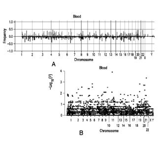
Figure 1A-B. Copy number variations (CNV) in blood and prostate cancer from
prostate cancer patients. Figure 1A. Histogram of frequency of amplification
(light gray)
or deletion (dark gray) of genome sequences of leukocytes (upper panel, n=273)
from
prostate cancer patients. Figure 1B. Manhattan plots of p-values in
association with
prostate cancer recurrence of each gene CNV from leukocytes.
Figure 2A-C. Large size ratio (LSR) of CNVs from leukocytes from prostate
cancer patients are correlated with aggressive behavior of prostate cancer.
Figure 2A.
Schematic diagram of L SR model of leukocyte CNV. Figure 2B. LSRs from
leukocytes
are associated with aggressive prostate cancer recurrence behavior. Upper
panel:
Correlation of LSRs from leukocyte genomes with prostate cancers that were
recurrent;
Lower panel: Correlation of LSRs from leukocyte genomes with prostate cancers
that
were non-recurrent 90 months after radical prostatectomy. Figure 2C. LSRs from
5
CA 02994848 2018-02-05
WO 2017/027473 PCMJS2016/046051
leukocytes are associated with short prostate specific antigen doubling time
(PSADT).
Upper panel: Correlation of LSRs from leukocyte genomes with prostate cancers
that had
recurrent serum PSADT of 4 months or less; Lower panel. Correlation of LSRs
from
leukocyte genomes with prostate cancers that were not recurrent or recurrent
but having
PSADT of 15 months or more.
Figure 3A-B. LSR of genome CNV from leukocytes to predict prostate cancer
recurrence. Figure 3A. LSR derived from leukocyte genome CNV predicts prostate
cancer recurrence. Receiver operating curve (ROC) analyses using LSRs derived
from
leukocyte CNVs as prediction parameter (dark gray, dashed line) to predict
prostate cancer
recurrence, versus Nomogram (dotted line), Gleason's grade (dash-dotted line)
and the
status of 8 fusion transcripts (14) (light gray, dashed line). The samples
were equally split
randomly into training and testing sets 10 times. The ROC analysis represents
the results
from the most representative split. Figure 3B. Combination of LSR (L),
Gleason's grade
(G), Nomogram (N) and the status of fusion transcripts (F) to predict prostate
cancer
recurrence. ROC analysis of a model combining LSR, fusion transcripts,
Nomogram and
Gleason's grade using linear discriminant analysis (LDA) is indicated by a
black solid
line. ROC analysis of a model combining fusion transcripts, Nomogram and
Gleason's
grade using LDA is indicated by a dark gray dashed line. ROC analysis of a
model
combining LSR, fusion transcripts and Gleason's grade using LDA is indicated
by a
dotted line. ROC analysis of a model combining LSR, fusion transcripts and
Nomogram
using LDA is indicated by a dash-dotted line. ROC analysis of a model
combining LSR,
Nomogram and Gleason's grade is indicated by a light gray dashed line. Similar
random
splits of training and testing data sets were performed as of (A).
Figure 4. Large LSRs of genome CNVs from leukocytes correlated with lower
PSA-free survival. Kaplan-Meier analysis on patients predicted by LSR based on
CNV of
patients' leukocytes as likely recurrent versus likely non-recurrent (upper
left). Similar
survival analyses were also performed on case segregations based on Gleason's
grades
(upper middle), Nomogram probability (upper right), the status of 8 fusion
transcripts
(lower left), or a model by combining LSR, Nomogram and fusion transcript
status using
LDA (lower middle), or a model by combining LSR, Nomogram, Gleason grade and
fusion transcript status using LDA (lower right). Number of samples analyzed
and p
values are indicated.
Figure 5A-B. LSR of genome CNV from leukocytes to predict prostate cancer
recurrence with short PSADT. LSR derived from leukocyte genome CNV predicts
6
CA 02994848 2018-02-05
WO 2017/027473 PCMJS2016/046051
PSADT of 4 months or less. Figure 5A. ROC analysis using LSRs derived from
leukocyte
CNVs as a prediction parameter (dark gray, dashed line) to predict PSADT 4
months or
less, versus Nomogram (dotted line), Gleason's grade (dash-dotted line) and
the status of 8
fusion transcripts (14) (light gray, dashed line). Samples were analyzed by
the same
procedure as Figure 3. Figure 5B. Combination of LSR (L), Gleason's grade (G),
Nomogram (N) and the status of fusion transcripts (F) to predict prostate
cancer recurrent
PSADT 4 months or less. ROC analysis of a model combining LSR, fusion
transcripts,
Nomogram and Gleason's grade using LDA is indicated by a black solid line. ROC
analysis of a model combining fusion transcripts, Nomogram and Gleason's grade
using
LDA is indicated by a dark gray dashed line. ROC analysis of a model combining
LSR,
fusion transcripts and Gleason's grade using LDA is indicated by a dotted
line. ROC
analysis of a model combining LSR, fusion transcripts and Nomogram using LDA
is
indicated by a dash-dotted line. ROC analysis of a model combining LSR,
Nomogram
and Gleason's grade is indicated by a light gray dashed line.
Figure 6. Genome CNVs from leukocytes predicting short PSADT correlated with
lower PSA-free survival. Kaplan-Meier analysis on patients predicted by LSR
based on
CNV of patients' leukocytes as likely recurrent and having PSADT 4 months or
less
versus likely non-recurrent or recurrent but having PSADT of 15 months or more
(upper
left). Similar survival analyses were also performed on case segregations
based on
Gleason's grades (upper middle), Nomogram probability (upper right), the
status of 8
fusion transcripts (lower left), or a model by combining LSR, Nomogram and
fusion
transcript status using LDA (lower middle), or a model by combining LSR,
Nomogram,
Gleason grade and fusion transcript status using LDA (lower right). Number of
samples
analyzed and p values are indicated.
Figure 7. Correlation of area under the curve (AUC) with LSR in predicting
prostate cancer recurrence (left panel) or in predicting recurrent PSADT of <4
months
(right panel).
Figure 8A-B. LSR of genome CNV from leukocytes to predict prostate cancer
likely lethality. Figure 8A. LSR derived from leukocyte genome CNV predicts
prostate
cancer likely lethality (recurrent within 12 months of radical prostatectomy
and PSADT of
<4 months). Receiver operating curve (ROC) analyses using LSRs derived from
leukocyte CNVs as prediction parameter (dark gray, dashed line) to predict
prostate cancer
likely lethality, versus Nomogram (dotted line), Gleason's grade (dash-dotted
line) and the
status of 8 fusion transcripts (14) (light gray, dashed line). The samples
were equally split
7
CA 02994848 2018-02-05
WO 2017/027473 PCMJS2016/046051
randomly into training and testing sets 10 times. The ROC analysis represents
the results
from the most representative split. Figure 8B. Combination of LSR (L),
Gleason's grade
(G), Nomogram (N) and the status of fusion transcripts (F) to predict prostate
cancer likely
lethality. ROC analysis of a model combining LSR, fusion transcripts, Nomogram
and
Gleason's grade using LDA is indicated by a black solid line. ROC analysis of
a model
combining fusion transcripts, Nomogram and Gleason's grade using LDA is
indicated by a
dark gray dashed line. ROC analysis of a model combining LSR, fusion
transcripts and
Gleason's grade using LDA is indicated by a dotted line. ROC analysis of a
model
combining LSR, fusion transcripts and Nomogram using LDA is indicated by a
dash-
dotted line. ROC analysis of a model combining LSR, Nomogram and Gleason's
grade is
indicated by a light gray dashed line. Similar random splits of training and
testing data sets
were performed as of (A).
Figure 9. Large LSRs of genome CNVs from leukocytes correlated with lower
PSA-free survival. Kaplan-Meier analysis on patients predicted by LSR based on
CNV of
patients' leukocytes as likely lethal (recurrent within 12 months of radical
prostatectomy
and PSADT<4 months) versus likely non-recurrent (upper left). Similar survival
analyses
were also performed on case segregations based on Gleason's grades (upper
middle),
Nomogram probability (upper right), the status of 8 fusion transcripts (lower
left), or a
model by combining LSR, Nomogram and fusion transcript status using LDA (lower
middle), or a model by combining LSR, Nomogram, Gleason grade and fusion
transcript
status using LDA (lower right). Number of samples analyzed and p values are
indicated
5. DETAILED DESCRIPTION OF THE INVENTION
The present invention provides methods for assessing whether a subject having
prostate cancer is at an increased risk of relapse and/or at an increased risk
of rapid
relapse. In certain embodiments, the present invention utilizes the size and
number of the
CNVs detected in a sample from the subject to assess the risk of relapse. The
present
invention further provides methods of treating subjects having an increased
risk and/or
decreased risk of relapse or rapid relapse.
For clarity of description, and not by way of limitation, the detailed
description of
the invention is divided into the following subsections:
(i) definitions;
(ii) methods of assessing risk of relapse or rapid relapse;
(iii) methods of treatment;
8
CA 02994848 2018-02-05
WO 2017/027473 PCMJS2016/046051
(iv) detection methods; and
(v) kits.
5.1. DEFINITIONS
Unless defined otherwise, all technical and scientific terms used herein have
the
meaning commonly understood by a person skilled in the art to which this
invention
belongs. The following references provide one of skill with a general
definition of many of
the terms used in this invention: Singleton et al., Dictionary of Microbiology
and
Molecular Biology (2nd ed. 1994); The Cambridge Dictionary of Science and
Technology
(Walker ed., 1988); The Glossary of Genetics, 5th Ed., R. Rieger et al.
(eds.), Springer
Verlag (1991); and Hale & Marham, The Harper Collins Dictionary of Biology
(1991). As
used herein, the following terms have the meanings ascribed to them below,
unless
specified otherwise.
As used herein, the term "about" or "approximately" means within an acceptable
error range for the particular value as determined by one of ordinary skill in
the art, which
will depend in part on how the value is measured or determined, i.e., the
limitations of the
measurement system. For example, "about" can mean within 3 or more than 3
standard
deviations, per the practice in the art. Alternatively, "about" can mean a
range of up to
20%, preferably up to 10%, more preferably up to 5%, and more preferably still
up to 1%
of a given value. Alternatively, particularly with respect to biological
systems or
processes, the term can mean within an order of magnitude, preferably within 5-
fold, and
more preferably within 2-fold, of a value.
The terms "prostate cancer patient" or "subject having prostate cancer," as
used
interchangeably herein, refer to a subject having or who has had a carcinoma
of the
prostate. The use of the term "patient" does not suggest that the subject has
received any
treatment for the cancer, but rather that the subject has at some point come
to the attention
of the healthcare system. The patient/subject, prior to or contemporaneous
with the
practicing of the invention, may be untreated for prostate cancer, may have
received
treatment or are currently undergoing treatment, including but not limited to,
surgical,
chemotherapeutic, anti-androgen or radiologic treatment.
The term "sample," as used herein, includes, but is not limited to, cells in
culture,
cell supernatants, cell lysates, serum, blood plasma, biological fluid (e.g.,
blood, plasma,
serum, stool, urine, lymphatic fluid, ascites, ductal lavage, saliva and
cerebrospinal fluid)
and tissue samples. The source of the sample may be solid tissue (e.g., from a
fresh,
9
CA 02994848 2018-02-05
WO 2017/027473
PCT/1JS2016/046051
frozen, and/or preserved organ, tissue sample, biopsy or aspirate), blood or
any blood
constituents, e.g., leukocytes, bodily fluids (such as, e.g., urine, lymph,
cerebral spinal
fluid, amniotic fluid, peritoneal fluid or interstitial fluid), or cells from
the individual,
including circulating cancer cells. In certain non-limiting embodiments, the
sample is
obtained from a prostate tumor. In certain embodiments, the sample may be a
"biopsy
sample" or "clinical sample," which are samples derived from a subject. In
certain
embodiments, the sample includes one or more prostate cancer cells from a
subject. In
certain embodiments, the sample is a blood sample, e.g., buffy coat sample,
from a
subject. In certain embodiments, the sample contains one or more leukocytes
from a
subject.
The term "relapse," as used herein, refers to a clinical course including one
or
more of the following: (i) where the cancer had been removed or put into
remission,
relapse refers to a recurrence of prostate cancer at the original site or
occurrence at a new
site, including metastatic spread; (ii) where the cancer had not been removed
or put into
remission, relapse refers to an extension of the cancer and/or metastatic
spread; (iii)
whether or not the cancer had been treated, relapse refers to an advancement
in the clinical
grade (for example, the Gleason grade), of the cancer; and/or a prostate
specific antigen
("PSA") doubling time (PSADT) of 15 months.
The terms "rapid" or "relapse quickly," as used interchangeably herein, means
that
the relapse occurs within a period of 5 years In certain embodiments, patients
suffering a
rapid relapse can also manifest a PSADT of 3 months or less or 4 months or
less
In certain non-limiting embodiments, "increased risk" means that a relapse or
a
rapid relapse occurs in more than about 50%, more than about 60%, more than
about 70%,
more than about 80% or more than 90% of individuals that have a large size
ratio (LSR)
greater than a particular threshold.
5.2 METHODS OF ASSESSING RISK OF RELAPSE OR RAPID RELAPSE
The present invention provides methods for determining whether a prostate
cancer
patient has an increased and/or decreased risk for relapse or rapid relapse.
In certain embodiments, the present invention utilizes the size and number of
the
CNVs to assess the likelihood that a prostate cancer will relapse or rapid
relapse. For
example, and not by way of limitation, the present invention can utilize the
percentage of
CNVs detected in a sample that are larger in size than a particular cut-off
value to assess
the likelihood that a prostate cancer will relapse or rapid relapse. In
certain embodiments,
CA 02994848 2018-02-05
WO 2017/027473
PCMJS2016/046051
the percentage of CNVs detected in a sample that are larger in size than a
particular cut-off
value can be represented by a large size ratio (see Figure 2A). "Large size
ratio," as used
herein, refers to the ratio of CNVs that have a size larger than a cut-off
value to the total
number of CNVs detected in a sample of a subject. In certain embodiments, the
large size
ratio (LSR) can be represented by the following formula: LSR = large size
number / total
number of CNVs, where large size number is the number of CNVs that are larger
in size
than a cut-off value.
In certain embodiments, the cut-off value for determining the LSR for a
subject
can be about 20 kilobases (kb), about 25 kb, about 30 kb, about 31 kb, about
32 kb, about
33 kb, about 34 kb, about 35 kb, about 40 kb, about 45 kb, about 50 kb, about
55 kb, about
60 kb, about 65 kb, about 70 kb, about 75 kb, about 80 kb, about 85 kb, about
90 kb, about
95 kb, about 100 kb, about 150 kb, about 200 kb, about 250 kb, about 300 kb,
about 350
kb, about 400 kb, about 450 kb, about 500 kb, about 501 kb or about 550 kb. In
certain
embodiments, the cut-off value can be about 31,622 base pairs (bp) or about
501,187 bp.
In certain embodiments, in methods for determining that a prostate cancer
patient
is at increased and/or decreased risk for relapse, the LSR can be calculated
by dividing the
number of CNVs that are larger than about 25 kb or about 30 kb in size by the
total
number of CNVs (e.g., LSR = (number of CNVs larger than about 25 kb or about
30 kb in
size) / total number of CNVs).
In certain embodiments, in methods for determining that a prostate cancer
patient
is at an increased and/or decreased risk for rapid relapse, the LSR can be
calculated by
dividing the number of CNVs that are larger than about 400 kb or about 500 kb
in size by
the total number of CNVs (e.g., LSR = (number of CNVs larger than about 400 kb
or
about 500 kb in size) / total number of CNVs).
In certain embodiments, CNVs across the genome can be determined and used to
determine the LSR. CNVs can be detected using methodology known in the art,
including
hybridization to gene arrays and the analysis of the results of such
hybridization using
software that determines copy number variation, as disclosed herein. In
certain
embodiments, CNV size can be determined using the same genotyping analysis
techniques
as described below and as are known in the art. In certain embodiments of the
invention,
using the Partek software described below, segments with changes in copy
number can be
obtained (including amplification and deletions), and those with the following
criteria:
p<0.001, length >2 kb and >10 markers can be selected. The length of the
selected CNVs
can also be determined.
11
CA 02994848 2018-02-05
WO 2017/027473
PCMJS2016/046051
The presently disclosed subject matter provides methods for determining
whether a
prostate cancer patient is at an increased risk for relapse or rapid relapse.
In certain
embodiments, the method comprises determining the number and size of CNVs in a
sample and determining the large size ratio, where if the large size ratio
exceeds a
particular threshold, the patient is deemed to be at an increased risk for
relapse or rapid
relapse. In certain embodiments, the sample can be a blood sample from the
patient, e.g.,
a buffy coat sample. In certain embodiments, the sample can comprise one or
more
leukocytes from the patient.
In certain embodiments, a large size ratio of about 0.28 or greater is
consistent
with a likelihood that the prostate cancer will relapse, e.g., when the cut-
off value for
calculating the large size ratio is about 25 kb or about 30 kb. Accordingly,
the present
invention provides for a method of determining that a prostate cancer patient
is at an
increased risk for relapse comprising determining the number and size of CNVs
in a
sample of the patient and determining the large size ratio, where if the large
size ratio is
about 0.28 or greater, the patient is deemed to be at an increased risk for
relapse.
In certain embodiments, a large size ratio of about 0.02 or greater is
consistent with
a likelihood that the prostate cancer will rapidly relapse, e.g., when the cut-
off value for
calculating the large size ratio is about 500 kb. In certain embodiments,
alarge size ratio
between about 0.02 and about 0.28 can indicate that the prostate cancer will
rapidly
relapse. Accordingly, the present invention provides for a method of
determining that a
prostate cancer patient is at an increased risk for relapse comprising
determining the
number and size of CNVs in a sample of the patient and determining the large
size ratio,
where if the large size ratio is about 0.02 or greater, the patient is deemed
to be at an
increased risk for rapid relapse.
The presently disclosed subject matter further provides methods for
determining
whether a prostate cancer patient is at a decreased risk for relapse or rapid
relapse. In
certain embodiments, the method comprises determining the number and size of
CNVs in
a sample and determining the large size ratio, where if the large size ratio
is less than a
particular threshold, the patient is deemed to be at a decreased risk for
relapse or rapid
relapse.
In certain embodiments, a large size ratio of less than about 0.28 is
consistent with
a likelihood that the prostate cancer will be at a decreased risk of relapse,
e.g., when the
cut-off value for calculating the large size ratio is about 25 kb or about 30
kb. In certain
embodiments, a large size ratio between about 0.02 and about 0.28 can indicate
that the
12
WO 2017/027473
PCT/US2016/046051
prostate cancer will be at a decreased risk of relapse. Accordingly, the
present invention
provides for a method of determining that a prostate cancer patient is at a
decreased risk
for relapse comprising determining the number and size of CNVs in a sample of
the
patient and determining the large size ratio, where if the large size ratio is
less than about
0.28, the patient is deemed to be at a decreased risk for relapse.
In certain embodiments, a large size ratio of less than about 0.02 is
consistent with
a likelihood that the prostate cancer will be at a decreased risk of rapid
relapse, e.g., when
the cut-off value for calculating the large size ratio is about 400 kb or
about 500 kb.
Accordingly, the present invention provides for a method of determining that a
prostate
cancer patient is at a decreased risk for relapse comprising determining the
number and
size of CNVs in a sample of the patient and determining the large size ratio,
where if the
large size ratio is less than about 0.02, the patient is deemed to be at a
decreased risk for
rapid relapse.
In certain embodiments, the method can further include determining one or more
of the following: the Gleason grade of the prostate cancer, nomogram and
fusion gene
status. For example, and not by way of limitation, the method of determining
whether a
subject is at increased risk or decreased risk of relapse or rapid relapse of
prostate cancer
can further comprise determining the Gleason grade of a prostate cancer sample
from a
subject.
In certain embodiments, the method of determining whether a subject is at
increased risk or decreased risk of relapse or rapid relapse of prostate
cancer can further
comprise generating a nomogram. In certain embodiments, the nomogram can be
determined using the prediction tool.
In certain embodiments, the method of determining whether a subject is at
increased risk or decreased risk of relapse or rapid relapse of prostate
cancer can further
comprise determining whether a sample of the subject contains one or more
fusion genes.
The term "fusion gene," as used herein, refers to a nucleic acid or protein
sequence, which
combines elements of the recited genes or their RNA transcripts in a manner
not found in
the wild type/normal nucleic acid or protein sequences. For example, but not
by way of
.. limitation, in a fusion gene in the form of genomic DNA, the relative
positions of portions
of the genomic sequences of the recited genes is altered relative to the wild
type/normal
sequence (for example, as reflected in the NCBI chromosomal positions or
sequences set
forth herein). In a fusion gene in the form of mRNA, portions of RNA
transcripts arising
13
CA 2994848 2019-08-08
CA 02994848 2018-02-05
WO 2017/027473 PCMJS2016/046051
from both component genes are present (not necessarily in the same register as
the wild-
type transcript and possibly including portions normally not present in the
nounal mature
transcript). In non-limiting embodiments, such a portion of genomic DNA or
mRNA may
comprise at least about 10 consecutive nucleotides, or at least about 20
consecutive
nucleotides, or at least about 30 consecutive nucleotides, or at least 40
consecutive
nucleotides. In a fusion gene in the form of a protein, portions of amino acid
sequences
arising from both component genes are present (not by way of limitation, at
least about 5
consecutive amino acids or at least about 10 amino acids or at least about 20
amino acids
or at least about 30 amino acids). In certain embodiments, portions arising
from both
genes, transcripts or proteins do not refer to sequences which may happen to
be identical
in the wild type forms of both genes (that is to say, the portions are
"unshared"). As such,
a fusion gene represents, generally speaking, the splicing together or fusion
of genomic
elements not normally joined together. Non-limiting examples of such fusion
genes
include TRMT11-GRIK2, SLC45A2-AMACR, MTOR-TP53BP1, LRRC59-FLJ60017,
TMEM135-CCDC67, KDM4B-AC011523.2, CCNH-05orf3 0 and MAN2A1-FER.
The fusion gene TRMT11-GRIK2 refers to a fusion between the tRNA
methyltransferase 11 homolog ("TRMT11") and glutamate receptor, ionotropic,
kainate 2
("GRIK2") genes. The human TRMT11 gene is typically located on chromosome
6q11.1
and the human GRIK2 gene is typically located on chromosome 6q16.3. In certain
embodiments, the TRMT11 gene is the human gene having NCBI Gene ID No: 60487,
sequence chromosome 6; NC 000006.11 (126307576..126360422) and/or the GRIK2
gene is the human gene having NCBI Gene ID No:2898, sequence chromosome 6,
NC 000006.11 (101841584..102517958).
The fusion gene SLC45A2-AMACR refers to a fusion between the solute carrier
family 45, member 2 ("SLC45A2") and alpha-methylacyl-CoA racemase ("AMACR")
genes. The human SLC45A2 gene is typically located on human chromosome 5p13.2
and
the human AMACR gene is typically located on chromosome 5p13. In certain
embodiments, the SLC45A2 gene is the human gene having NCBI Gene ID No: 51151,
sequence chromosome 5; NC 000005.9 (33944721..33984780, complement) and/or the
AMACR gene is the human gene having NCBI Gene ID No:23600, sequence chromosome
5; NC 000005.9 (33987091..34008220, complement).
The fusion gene MTOR-TP53BP1 refers to a fusion between the mechanistic target
of rapamycin ("MTOR") and tumor protein p53 binding protein 1 ("TP53BP1")
genes.
The human MTOR gene is typically located on chromosome 1p36.2 and the human
14
CA 02994848 2018-02-05
WO 2017/027473 PCMJS2016/046051
TP53BP1 gene is typically located on chromosome 15q15 - q21. In certain
embodiments,
the MTOR gene is the human gene having NCBI Gene ID No:2475, sequence
chromosome 1 NC 000001.10 (11166588..11322614, complement) and/or the
TP53BP1gene is the human gene having NCBI Gene ID No: 7158, sequence
chromosome
15; NC 000015.9 (43695262..43802707, complement).
The fusion gene LRRC59-FLJ60017 refers to a fusion between the leucine rich
repeat containing 59 ("LRRC59") gene and the "F1160017" nucleic acid. The
human
LRRC59 gene is typically located on chromosome 17q21.33 and nucleic acid
encoding
human FLJ60017 is typically located on chromosome 11q12.3. In certain
embodiments,
the LRRC59 gene is the human gene having NCBI Gene ID No:55379, sequence
chromosome 17; NC 000017.10 (48458594..48474914, complement) and/or FLJ60017
has a nucleic acid sequence as set forth in GeneBank AK_296299.
The fusion gene TMEM135-CCDC67 refers to a fusion between the
transmembrane protein 135 (`T1VIEM135") and coiled-coil domain containing 67
("CCDC67") genes. The human TMEM135 gene is typically located on chromosome
11q14.2 and the human CCDC67 gene is typically located on chromosome 11q21. In
certain embodiments, the TMEM135 gene is the human gene having NCBI Gene ID
No:
65084, sequence chromosome 11; NC_000011.9 (86748886..87039876) and/or the
CCDC67 gene is the human gene having NCBI Gene ID No: 159989, sequence
chromosome 11; NC 000011.9 (93063156..93171636).
The fusion gene CCNH-05orf30 refers to a fusion between the cyclin H
("CCNH") and chromosome 5 open reading frame 30 ("C5orf30") genes. The human
CCNH gene is typically located on chromosome 5q13.3-q14 and the human
C5orf30gene
is typically located on chromosome 5q21.1. In certain embodiments, the CCNH
gene is
.. the human gene having NCBI Gene ID No: 902, sequence chromosome 5; NC
000005.9
(86687310..86708850, complement) and/or the C5orf30gene is the human gene
having
NCBI Gene ID No: 90355, sequence chromosome 5; NC 000005.9
(102594442..102614361).
The fusion gene KDM4B-AC011523.2 refers to a fusion between lysine (K)-
specific demethylase 4B ("KDM4B") and chromosomal region "AC011523.2." The
human KDM4B gene is typically located on chromosome 19p13.3 and the human
AC011523.2 region is typically located on chromosome 19q13.4. In certain
embodiments,
the KDM4B gene is the human gene having NCBI Gene ID NO: 23030, sequence
chromosome 19; NC 000019.9 (4969123..5153609).
CA 02994848 2018-02-05
WO 2017/027473 PCMJS2016/046051
The fusion gene MAN2A1-FER refers to a fusion between mannosidase, alpha,
class 2A, member 1 ("MAN2A1") and (fps/fes related) tyrosine kinase ("FER").
The
human MAN2A1 gene is typically located on chromosome 5q21.3 and the human FER
gene is typically located on chromosome 5q21. In certain embodiments, the
MAN2A1gene is the human gene having NCBI Gene ID NO: 4124, sequence
chromosome 5; NC 000005.9 (109025156..109203429) or NC 000005.9
(109034137..109035578); and/or the FER gene is the human gene having NCBI Gene
ID
NO: 2241, sequence chromosome 5: NC 000005.9 (108083523..108523373).
In certain embodiments, to predict prostate cancer relapse by the combination
of
the LSR, Nomogram, fusion gene status and Gleason grading, it is postulated
that samples
from relapse or non-relapse groups follow normal distribution with different
means but
same covariance matrix. For example, and not by limitation, based on training
data, the
mean value for relapse samples is mu_relapse=(0.462 0.8714 0.571 7.107) for
(LSR,
Nomogram, fusion, Gleason) and the mean for non-relapse samples is mu non-
relapse=(0.318 0.907 0.214 7.214). In certain embodiments, the pooled
covariance matrix
can be represented as follows:
sigma=
LSR nomogram fusion gleason
LSR 8.491034e-03
8.507772e-05 0.004008907 -0.012312703
nomo 8.507772e-05 1.307571e-02 -0.002607143 -0.063142857
fusion 4.008907e-03 -2.607143e-03 0.230357143 -0.008928571
gleason -1.231270e-02 -6.314286e-02 -0.008928571 0.525892857
In certain embodiments, for a testing sample x=[xl,x2,x3,x4]', its posterior
probability can be estimated by the following:
p(relapse x)= p_0(x)*p(relapse) / (p_0(x)*p(relapse) + p_1(x)*p(non relapse))
p(non-relapselx)= p_1(x) *p(non_relapse) / (p 0(x)*p(relapse) + p
1(x)*p(non_relapse))
In certain embodiments, the cut-off value of the posterior probability can be
set to
be a suitable value to increase or maximize the Youden index, which can be,
for example
and without limitation and as embodied herein, about 0.544. In certain
embodiments, a
testing sample with a posterior probability that is greater than about 0.5,
greater than about
0.54 or greater than about 0.544 can be predicted to be relapse, or otherwise
the testing
sample can be predicted to be non-relapse.
In certain embodiments, the techniques described above can be applied to
classify
fast relapse versus non-fast relapse. For example, and not by limitation, the
mean values
16
CA 02994848 2018-02-05
WO 2017/027473 PCMJS2016/046051
for the fast relapse group is mu fast-relapse=(0.031 0.828 0.667 7.267) for
(LSR,
Nomogram, fusion, Gleason) and the mean for the non-fast relapse samples is mu
non-
fast re1apse=(0.023 0.905 0.269 7.192). In certain embodiments, the pooled
covariance
matrix can be represented as follows:
sigma=
LSR nomogram fusion gleason
LSR 0.0007088535
0.0001202635 0.0006518215 0.0003571929
nomo 0.0001202635 0.0125151282 0.0129487179 -0.0740256410
fusion 0.0006518215 0.0129487179 0.2166337936 -0.1028928337
gleason 0.0003571929 -0.0740256410 -0.1028928337 0.6915844839
In certain embodiments, the cut-off value of the posterior probability can be
set to
about 0.396. For example, and not by way of limitation, a testing sample with
a posterior
probability greater than about 0.35, greater than about 0.39 or greater than
about 0.396 can
be predicted to be fast relapse, or otherwise the testing sample can be
predicted to be non-
fast relapse.
5.3. METHODS OF TREATMENT
In certain embodiments, use of the present invention can inform a health care
practitioner how to better advise a prostate cancer patient on whether or not
to undergo
more aggressive forms of therapy or whether watchful waiting would be an
appropriate
recommendation Accordingly, the present invention provides methods for
treating
prostate cancer patients that are at an increased and/or decreased risk for
relapse or rapid
relapse.
In certain embodiments, if it is determined that the patient is at an
increased risk
for relapse or rapid relapse, as disclosed herein, a healthcare provider can
take the further
step of recommending and/or performing a prophylactic and/or treatment
regimen. For
example, and not by way of limitation, one or more of the following can be
recommended
and/or performed: cryotherapy, radiation therapy, chemotherapy, hormone
therapy,
biologic therapy, bisphosphonate therapy, high-intensity focused ultrasound,
frequent
monitoring, frequent prostate-specific antigen (PSA) checks and radical
prostatectomy.
In certain embodiments, if it is determined that the patient is not at an
increased
risk and/or is at a decreased risk for relapse or rapid relapse, as disclosed
herein, a
healthcare provider can recommend and/or perform one or more of the following:
high-
17
CA 02994848 2018-02-05
WO 2017/027473
PCMJS2016/046051
intensity focused ultrasound, watchful waiting, frequent monitoring, frequent
PSA checks
and a biopsy.
In certain embodiments, one or more of the prophylactic and/or treatment
regimens, disclosed herein, can be performed at about 1 month, about 2 months,
about 3
months, about 4 months, about 5 months, about 6 months, about 7 months, about
8
months, about 9 months, about 10 months, about 11 months, about 12 months,
about 18
months, about 2 years, about 3 years, about 4 years or about 5 years following
the
assessment of the risk of relapse or rapid relapse for the prostate cancer
patient.
A non-limiting example of a biologic therapeutic is Sipuleucel-T.
Bisphosphonate
therapy includes, but is not limited to, clodronate or zoledronate. Hormone
therapy can
include one or more of orchiectomy and the administration of luteinizing
hormone-
releasing hormone (LHRH) analogs and/or agonists, LHRH antagonists, anti-
androgens or
androgen-suppressing drugs. Non-limiting examples of LHRH analogs and/or
agonists
include leuprolide, goserelin and buserelin. Non-limiting examples of LHRH
antagonists
include abarelix, cetrorelix, ganirelix and degarelix. Anti-androgen drugs
include, but are
not limited to, flutamide, bicalutamide, enzalutamide and nilutamide. Non-
limiting
examples of androgen-suppressing drugs include estrogens, ketoconazole and
aminoglutethimide. Frequent monitoring can include PSA blood tests, digital
rectal
exams, ultrasounds and/or transrectal ultrasound-guided prostate biopsies at
regular
intervals, e.g., at about 3 to about 6 month intervals, to monitor the status
of the prostate
cancer. Radical prostatectomy is a surgical procedure that involves the
removal of the
entire prostate gland and some surrounding tissue. Prostatectomies can be
performed by
open surgery or it may be performed by laparoscopic surgery.
In certain embodiments, these prophylactic and/or treatment regimens can be
used
to produce an anti-cancer effect in a subject. For example, and not by way of
limitation,
the present invention provides methods of treating a prostate cancer patient
to produce an
anti-cancer effect in the patient. An "anti-cancer effect" refers to one or
more of a
reduction in aggregate cancer cell mass, a reduction in cancer cell growth
rate, a reduction
in cancer progression, a reduction in cancer cell proliferation, a reduction
in tumor mass, a
reduction in tumor volume, a reduction in tumor cell proliferation, a
reduction in tumor
growth rate and/or a reduction in tumor metastasis. In certain embodiments, an
anti-
cancer effect can refer to a complete response, a partial response, a stable
disease (without
progression or relapse), a response with a later relapse or progression-free
survival in a
patient diagnosed with cancer.
18
CA 02994848 2018-02-05
WO 2017/027473 PCMJS2016/046051
5.4. DETECTION METHODS
The present invention provides methods for detecting the number and size of
CNVs across the genome of a subject. The present invention further provides
methods for
detecting the presence of one or more fusion genes, disclosed herein, within a
sample of a
subject.
5.4.1 COPY NUMBER VARIATION DETECTION
The present invention provides methods for determining the size and number of
CNVs within a sample of a subject. In certain embodiments, CNVs can be
detected in one
or more samples of a subject. For example, and not by way of limitation, the
sample can
be a sample of malignant tumor (or presumptively malignant tumor, where a
diagnosis has
not yet been made) tissue. In certain embodiments, microdissection can be
performed to
achieve a tumor purity of at least about 70% or at least about 80% or greater
than 80%. In
certain embodiments, the sample can be tissue adjacent to a malignant tumor
tissue (e.g.,
prostate tissue that is not identified as a tumor located in a prostate gland
that contains a
tumor). In certain embodiments, a sample can be a tissue sample which is
considered by a
skilled artisan to appear abnormal (microscopically and/or macroscopically)
and is to be
tested to determine whether it is cancerous. In certain embodiments, a sample
can be a
blood sample that contains at least some nucleated cells to serve as a source
of DNA, e.g.,
a whole blood or buffy coat blood sample. In certain embodiments, the sample
can
comprise one or more leukocytes from the subject. In certain embodiments,
multiple
samples can be prepared for a single subject For example, but not by way of
limitation,
samples of tumor (i.e., malignant) tissue, tissue adjacent to a tumor tissue
and blood can be
prepared and each of the samples can be analyzed for CNVs and compared.
In certain embodiments, DNA can be extracted from the sample, e.g., using a
Qiagen kit or other method known in the art. In certain embodiments,
genotyping of the
extracted DNA can be performed to identify CNVs across the genome or a portion
of the
genome. For example, and not by way of limitation, genotyping can be performed
by
fragmenting the DNA using restriction enzymes (e.g., Styl and/or Nspl),
ligating the
DNA fragments to adaptors, amplifying the adaptor-DNA fragments using primers
that
correspond to the adaptor sequences and, optionally, performing an additional
fragmentation step (e.g., by digestion with DNAseI). In certain embodiments,
the
genotyping technique can further include labeling the amplified (or optionally
further
fragmented) DNA product (e.g., with biotinylated nucleotides) and then
hybridizing the
resulting labeled DNA to a plurality of test nucleic acid, e.g., DNA,
molecules
19
WO 2017/027473
PCT/US2016/046051
representative of the genome or a genome portion of interest under appropriate
conditions
(for example, as described by the array manufacturer). Additional non-limiting
examples
of genotyping techniques are disclosed in International Application No, WO
2013/106737.
In certain embodiments, the plurality of test nucleic acid molecules can be
provided in an
.. array such as, but not limited to, the Affymetrix Genomcwide Human SNP
Array 6.0
(Affymetrix, CA). The terms "array," "microarray" and "DNA chip" are used
herein
interchangeably to refer to an array of distinct polynucleotides affixed to a
substrate, such
as glass, plastic, paper, nylon or other type of membrane, filter, chip, bead,
or any other
suitable solid support. The polynucleotides can be synthesized directly on the
substrate, or
synthesized separate from the substrate and then affixed to the substrate. The
arrays can
be prepared using known methods. In certain non-limiting embodiments, the one
or more
test nucleic acid molecules set forth above may constitute at least 10 percent
or at least 20
percent or at least 30 percent or at least 40 percent or at least 50 percent
or at least 60
percent or at least 70 percent or at least 80 percent of the species of
polynucleotides
represented on the microarray.
In certain embodiments, the results from the array can then be interpreted to
determine the number or approximate number and/or size or approximate size of
the
CNVs in the genome or portion thereof. For example, and not by way of
limitation,
software such as Partek GenomeSuite 6.6 can be used.
5.4.2 FUSION GENE DETECTION
The present invention provides methods for detecting one or more fusion genes
in
a sample of a subject. The fusion genes can be detected by detecting a fusion
gene
manifested in a DNA molecule, an RNA molecule or a protein. In certain
embodiments, a
fusion gene can be detected by determining the presence of a DNA molecule, an
RNA
molecule or protein that is encoded by the fusion gene. For example, and not
by way of
limitation, the presence of a fusion gene may be detected by determining the
presence of
the protein encoded by the fusion gene. In certain embodiments, the fusion
gene can be
detected in a sample of a subject.
In certain non-limiting embodiments, the fusion gene is detected by nucleic
acid
hybridization analysis.
In certain non-limiting embodiments, the fusion gene is detected by
fluorescent in
situ hybridization (FISH) analysis. FISH is a technique that can directly
identify a specific
sequence of DNA or RNA in a cell or biological sample and enables visual
determination
CA 2994848 2019-08-08
CA 02994848 2018-02-05
WO 2017/027473
PCMJS2016/046051
of the presence and/or expression of a fusion gene in a tissue sample. In
certain non-
limiting embodiments, where a fusion gene combines genes not typically present
on the
same chromosome, FISH analysis may demonstrate probes binding to the same
chromosome. For example, and not by way of limitation, analysis may focus on
the
chromosome where one gene normally resides and then hybridization analysis may
be
performed to determine whether the other gene is present on that chromosome as
well.
In certain non-limiting embodiments, the fusion gene is detected by DNA
hybridization, such as, but not limited to, Southern blot analysis
In certain non-limiting embodiments, the fusion gene is detected by RNA
hybridization, such as, but not limited to, Northern blot analysis. In certain
embodiments,
Northern blot analysis can be used for the detection of a fusion gene, where
an isolated
RNA sample is run on a denaturing agarose gel, and transferred to a suitable
support, such
as activated cellulose, nitrocellulose or glass or nylon membranes.
Radiolabeled cDNA or
RNA is then hybridized to the preparation, washed and analyzed by
autoradiography to
detect the presence of a fusion gene in the RNA sample.
In certain non-limiting embodiments, the fusion gene is detected by nucleic
acid
sequencing analysis.
In certain non-limiting embodiments, one or more fusion genes can be detected
by
probes present on a DNA array, chip or a microarray. For example, and not by
way of
limitation, oligonucleotides corresponding to one or more fusion genes can be
immobilized on a chip which is then hybridized with labeled nucleic acids of a
sample
obtained from a subject Positive hybridization signal is obtained with the
sample
containing the fusion gene transcripts. In certain non-limiting embodiments,
the one or
more probes set forth above can constitute at least 10 percent or at least 20
percent or at
least 30 percent or at least 40 percent or at least 50 percent or at least 60
percent or at least
70 percent or at least 80 percent of the species of probes represented on the
microarray.
In certain non-limiting embodiments, the fusion gene is detected by a method
comprising Reverse Transcription Polymerase Chain Reaction ("RT-PCR").
In certain non-limiting embodiments, the fusion gene is detected by antibody
binding analysis such as, but not limited to, Western Blot analysis and
immunohistochemistry.
21
CA 02994848 2018-02-05
WO 2017/027473 PCMJS2016/046051
5.5. KITS
The present invention further provides kits that can be used to practice the
invention. For example, and not by way of limitation, a kit of the present
invention can
comprise an array that allows the analysis of CNVs across the whole genome. A
non-
limiting embodiment of such an array is the Affymetrix SNP Array 6Ø In
certain non-
limiting embodiments, the nucleic acid molecules for detecting CNVs may
constitute at
least 10 percent or at least 20 percent or at least 30 percent or at least 40
percent or at least
50 percent or at least 60 percent or at least 70 percent or at least 80
percent of the species
of polynucleotides represented on the microarray.
In certain embodiments, a kit of the present invention can optionally comprise
software or internet access to software, in electronically readable form, that
determines the
number and size of CNVs in the genes represented in the array. In certain
embodiments,
the kit can optionally comprise software or internet access to software, in
electronically
readable form, that determines whether CNVs in a DNA sample exceed or fall
below a
size threshold and can further determine the large size ratio, set forth
herein, which
indicates whether or not a prostate cancer patient is at an increased risk of
relapse or an
increased risk of rapid relapse.
The present invention further provides kits for detecting one or more of the
fusion
genes disclosed herein within a sample of a subject. Types of kits include,
but are not
limited to, packaged fusion gene-specific probe and primer sets (e.g., TaqMan
probe/primer sets), arrays/microarrays or antibodies for detecting one or more
fusion
genes. In certain embodiments, a kit of the present invention can include
packaged fusion
gene-specific probe and primer sets (e.g., TaqMan probe/primer sets),
arrays/microarrays
or antibodies for detecting one or more fusion genes selected from the group
consisting of
TRMT11-GRIK2, SLC45A2-AMACR, MTOR-TP53BP1, LRRC59-FLJ60017,
TMEM135-CCDC67, KDM4B-AC011523.2, CCNH-05orf30 and MAN2A1-FER. In
certain non-limiting embodiments, the one or more probes and/or primers for
detecting
fusion genes indicated above can constitute at least 10 percent or at least 20
percent or at
least 30 percent or at least 40 percent or at least 50 percent or at least 60
percent or at least
70 percent or at least 80 percent of the species of probes and/or primers
represented on the
microarray.
The following Example is offered to more fully illustrate the disclosure, but
is not
to be construed as limiting the scope thereof
22
CA 02994848 2018-02-05
WO 2017/027473 PCT/US2016/046051
6. EXAMPLE 1: ANALYSIS OF SIZE AND NUMBER OF CNVS IN
PROSTATE CANCER PATIENTS.
6.1 INTRODUCTION
Accurate prediction of prostate cancer clinical courses remains elusive. In
this
study, we performed whole genome copy number analysis on leukocytes of 273
prostate
cancer patients using Affymetrix SNP 6.0 chip. Copy number variations (CNV)
were
found across all chromosomes of the human genome. An average of 152 CNV
fragments
per genome was identified in the leukocytes from prostate cancer patients. The
size
distributions of CNV in the genome of leukocytes were highly correlative with
prostate
cancer aggressiveness. A prostate cancer outcome prediction model was
developed based
on large size ratio of CNV from the leukocyte genomes. This prediction model
generated
an average prediction rate of 75.2%, with sensitivity of 77.3% and specificity
of 69.0% for
prostate cancer recurrence. When combined with Nomogram and the status of
fusion
transcripts, the average prediction rate was improved to 82.5% with
sensitivity of 84.8%
.. and specificity of 78.2%. In addition, the leukocyte prediction model was
62.6% accurate
in predicting short prostate specific antigen doubling time. When combined
with
Gleason's grade, Nomogram and the status of fusion transcripts, the prediction
model
generated a correct prediction rate of 77.5% with 73.7% sensitivity and 80.1%
specificity.
To our knowledge, this is the first study showing that CNVs in leukocyte
genomes are
predictive of clinical outcomes of a human malignancy.
Previous cytogenetic and other genome studies suggested a clear link between
genome abnormalities and prostate cancer (5-21). Recent analyses of genome
copy
number of prostate cancer, benign tissues adjacent to cancer and blood samples
from
prostate cancer patients suggested that genome deletion and amplification of
certain
regions in prostate cancer samples were associated with poor clinical outcomes
(14;22).
Whole genome and transcriptome sequencing revealed fusion transcripts in
prostate cancer
predictive of prostate cancer recurrence (23). In this study, whole genome
copy number
analyses on leukocytes from prostate cancer patients were performed.
Significant copy
number variations (CNV) were identified in the genome of leukocytes of
prostate cancer
patients. It was found that sizes of CNVs in leukocytes of prostate cancer
samples were
highly correlative to prostate cancer recurrence. Prediction models were built
to predict
prostate cancer outcomes based on the size of CNVs of the leukocytes.
23
CA 02994848 2018-02-05
WO 2017/027473 PCMJS2016/046051
6.2 MATERIALS AND METHODS
Tissue processing, DNA extraction, amplicon generation, labeling,
hybridization,
washing and scanning of SNP 6.0 chips.
Prostate cancer samples were obtained from University of Pittsburgh Medical
Center Tissue Bank. These samples were collected from 1998-2012. Two hundred
seventy-three buffy coat samples from prostate cancer patients were analyzed.
Among
these samples, 143 samples were followed at least 90 months, 35 patients were
non-
recurrent for 90 months or more, 55 patients experiencing recurrence with
short PSADT
(PSA doubling time <4 months), and 53 patients experiencing recurrence with
long
PSADT (PSA doubling time >15 months) after radical prostatectomy (Table 3).
The
Gleason's scores of all prostate cancer samples were reassessed by UPMC
pathologists
before the study. Clinical follow-up was conducted by office examination
record, blood
PSA survey and radiographic follow-up. These follow-ups were carried out for
up to a 15
year period after the patient had a radical prostatectomy. The protocol was
approved by
"University of Pittsburgh Institutional Review Board". Five hundred nanograms
of
genomic DNA were digested with Styl and Nspl for 2 hours at 37 C. The digested
DNA
was purified and ligated with primer/adaptors at 16 C for 12-16 hours.
Amplicons were
generated by performing PCR using primers provided by the manufacturer
(Affymetrix,
CA) on the ligation products using the following program. 94 C for 3 min, then
35 cycles
of 94 C 30 second, 60 C for 45 sec and 65 C for 1 minute. This was followed by
extension
at 68 C for 7 min. The PCR products were then purified and digested with
DNAseI for 35
min at 37 C to fragment the amplified DNA. The fragmented DNA was then labeled
with
biotinylated nucleotides through terminal deoxynucleotide transferase for 4
hours at 37 C.
Two hundred fifty micrograms of fragmented DNA were hybridized with a pre-
equilibrated Affymetrix chip SNP 6.0 at 50 C for 18 hours. Procedures of
washing and
scanning of SNP 6.0 chips followed the manuals provided by Affymetrix, Inc.
Raw data
information of SNP6.0 from these samples was deposited in "Gene Expression
Omnibus"
(GEO, accession number GSE70650).
Statistical analysis:
Copy number variation analysis: CEL files were analyzed with Genotyping
Console for quality control (QC) analysis. Samples with QC call above 80% and
QC
contrast ratio above 0.4 were admitted into the analysis. To analyze CNV, CEL
files were
imported into Partek Genome Suite 6.6 to generate copy number from raw
intensity. To
plot the histograms, deletion or amplification of genomes were analyzed by
first limiting
24
WO 2017/027473
PCT/US2016/046051
to the regions with p-value less than 0.001. The selected regions were
subsequently
filtered by limiting to the regions with at least 10 markers and 2 kb in size.
The regions
were then mapped to known genes. The frequencies of amplification and
deletions were
plotted to the genome corresponding to the gene locations (Figure 1A). For
each gene,
Fisher's exact test was applied to test the association between CNV
involvement and
sample recurrence status. Then the minus log p-values were plotted on the
Manhattan plot
with their corresponding gene chromosome locations to generate Figure 1B.
Benjamini-
Hochberg (BH) method was applied to correct the p-values. The CNV-gene
enriched
pathways were selected by Kolmogorov-Smirnov test on the gene adjusted p-
values.
Pathway p-values were also corrected by BH method.
Machine learning methods to predict recurrent and fast-recurrent status:
prediction
models for two types of clinical comparisons were constructed: (1) non-
recurrent versus
recurrent; (2) non-fast recurrent (i.e., non-recurrent or recurrent but having
prostate
specific antigen doubling time [PSADT]>15 months) versus fast-recurrent
(recurrent
PSADT< 4 months). For each comparison, the models were constructed using
Gleason
score (6), Nomogram score (N), fusion transcript status (F) or blood CNV
information (L)
separately. For Gleason score discrimination, binary prediction was used (0
meaning
Gleason score <7 and 1 meaning Gleason score > 7). For Nomogram score, the 7
year
survival probability was used (24). For fusion status, eight fusion
transcripts (TRMT11-
GRIK2, SLC45A2-AMACR, MTOR-TP53BP1, LRRC59-FLJ60017, TMEM135-
CCDC67, KDM4-AC011523.2, MAN2A1-FER and CCNH-05orf30) previously
identified and validated in a multi-center study (23) were applied. A binary
fusion score
was used (0 meaning none of the eight fusions detected; 1 meaning one or more
fusion
transcripts detected).
For prediction using gene CNV of leukocytes, little predictive power from gene-
based association was found (Figure 1B). As a result, a large size ratio (LSR)
model was
developed based on the assumption that untargeted CNV aberrations in blood
played a
significant role in predisposing prostate tumors to aggressiveness. As shown
in Figure 2A,
LSR was defined as the proportion of large size CNV identified in the blood
genome of a
given patient, where large size was defined by threshold 8. In each two-fold
cross-
validation, samples were randomly and equally split into two data sets. In the
first dataset
treated as training data, the best 8 parameter in LSR model and the best
cutoffs of
Nomogram and LSR scores were selected by maximizing the highest AUC (area
under the
curve) and Youden index (i.e., sensitivity+specificity-1). The models were
then applied to
CA 2994848 2019-08-08
CA 02994848 2018-02-05
WO 2017/027473 PCMJS2016/046051
the second dataset as testing data. The cross-validation was then repeated
using the second
dataset as training data and the first dataset as test data. ROC curves were
plotted by
varying the cutoffs in both the training and testing datasets. The
corresponding overall
accuracy, sensitivity, specificity, Youden index and AUC were calculated to
evaluate the
performance. The equal-splitting validation was repeated for 14 times and the
top 2 and
bottom 2 splitting with the highest and lowest sum of AUCs were removed to
avoid
accidentally extreme training/testing assignment. The remaining 10 cross-
validation
results were finally averaged (Table 1 and Table 2). ROC and Kaplan-Meier
survival
curves in Figure 3-6 are the representative results of the 10 predictions
closest to the
averaged values.
To test whether combining multiple data information improves the prediction
result, we applied linear discriminant analysis (LDA) to combine two or more
predictive
factors. All possible combinations were performed. Models using (1) L+N+F; (2)
L+N+G;
(3) N+F+G; (4) L+F+G; (5) L+N+F+G are shown in Figure 3 and 5.
Kaplan-Meier curve analysis: For the survival evaluation (Figures 4 and 6),
the
two-fold cross validation of "Training=>Testing" result was combined to
compare the
performance of different methods, except for Gleason score that we used (<7 VS
>7 as
cut-off for the whole samples). Kaplan-Meier curves were truncated at 90
months follow-
up. Log-rank test was performed to calculate the p-value between survival
curves of two
predicted outcomes. To evaluate whether the survival difference for one model
was
significantly better than the other, we define a test statistics U as the
absolute difference of
the log-rank test statistics from the two models. Theoretically under the null
hypothesis
(two models were non-discriminant), the test statistics U followed a
distribution of
absolute difference of two independent chi-squared (degree of freedom = 1)
distributions.
As a result, 10,000,000 times from the absolute difference of two independent
chi-squared
distributions were sampled to foini null distribution and evaluate the p-
values.
6.3 RESULTS
Genome copy abnormalities are some of the hallmarks for prostate cancer.
However, little is known about the genome copy abnormalities in non-cancerous
tissues
from prostate cancer patients. To analyze the regions of amplification and
deletion in the
genome of leukocytes from prostate cancer patients, 273 buffy coats from
prostate cancer
patients were analyzed for CNV across the entire genome using Affymetrix
SNP6Ø
Using the cutoff criteria of size>2 Kb, marker number >10 and p<0.001, a total
of 41589
CNV fragments were identified, including 24213 segments of deletion and 17376
of
26
CA 02994848 2018-02-05
WO 2017/027473 PCMJS2016/046051
amplification, involving 17865 genes based on the Partek gene annotation
(Figure 1A).
This translates to an average of about 152 CNVs per sample. The average size
of CNV in
the genome of the leukocytes is about 147 Kb. On average, 256 genes were found
to have
either copy number gain or loss per genome. Among the 273 blood samples, 143
blood
samples have more than 90 months of clinical follow-ups in terms of prostate
cancer
recurrence. Interestingly, when categorizing the blood samples based on the
status of
prostate cancer recurrence, CNV of leukocytes from patients who experienced
recurrence
after radical prostatectomy had an average of >3.2 fold larger size of CNV
versus CNV
from patients who had no recurrence for at least 90 months. Two-sided t test
showed a
strong correlation between the size of CNV in leukocytes and prostate cancer
recurrence
(p=2.2 x 10⁻¹⁶), suggesting that the size of germ line CNV may play a
significant role in
predisposing prostate cancer to aggressive clinical courses. However, no
specific
(FDR=0.05) gene involved in CNV of genome fragment reaches the threshold that
differentiates recurrent prostate cancer versus those of non-recurrent (Figure
1B).
Together, the results indicate that the gene-based prediction model is
unlikely to succeed
in the leukocyte CNV analysis but size distribution of CNVs can be predictive.
To examine whether germ line CNV is predictive of recurrence of prostate
cancer,
an algorithm utilizing ratios of the number of large size fragments was
developed. As
illustrated in Figure 2A, for each sample, large size ratio (LSR) is defined
as the ratio of
CNV fragments whose sizes are greater than a size cutoff (6) over the total
number of
CNV fragments. For example, 3 of the 7 detected CNVs in Figure 2A are found
"large
size fragments" (size > 5) and the LSR of this patient is calculated as
3/7=0.43. In Figure
2B, the distribution of LSR from patients who experienced prostate cancer
recurrence
showed significantly higher values than those who did not experience
recurrence.
Similarly, the distribution of LSR from patients with fast recurrence (PSADT<
4 months)
was significantly higher than those from non-fast recurrent patients (non-
recurrent or
recurrent but having PSADT> 15 months, Figure 2C). In the LSR model, the size
threshold 6 is determined by maximizing the AUC. When 6 values were optimized
(Figure
7, 6=104.5=31622 bp for recurrent prediction model and 1B selected
6=105.7=501187 bp
for fast recurrent prediction), it predicts prostate cancer recurrence with
accuracy of
77.6%, with sensitivity of 80.4% and specificity of 68.6%, while fast
recurrence with
accuracy of 62.4%, with sensitivity of 72.9% and specificity of 54.1%.
To validate this model, 143 blood samples (Table 3) from prostate cancer
patients
were randomly split into a training set (72 samples) and a testing set (71
samples). The
27
CA 02994848 2018-02-05
WO 2017/027473 PCMJS2016/046051
optimized large-size cutoff 6 and LSR-cutoff were obtained from the training
analysis by
maximizing the Youden index. The parameters were then applied to the testing
data set to
assess the prediction accuracy. The validation was then repeated 14 times and
the best 2
and worst 2 were removed to avoid extreme randomization. The remaining 10
results from
these training and testing analyses were averaged (Table 1). As shown in
Figure 3A
(representative analyses in Table 4) and Table 1, the training accuracy of LSR
model in
predicting prostate cancer recurrence reaches 76.5%, with 77.8% sensitivity
and 72.4%
specificity. When the parameters were applied to the testing set, the
prediction accuracy
reaches 73.9%, with 76.8% sensitivity and 65.6% specificity. These prediction
rates are
better than those of Nomogram (66.00/0 accuracy for training and 61.3% for
testing, Table
1), and are significantly higher than those of Gleason grade's with single
cutoff (40.3% for
training and 39.4% for testing; p=8.6x10⁻³ for training and p=5.8x10⁻³ for
testing by ROC
comparison, see Table 1 and Table 5).
To examine whether combination of different modalities will improve the
prediction model, blood LSR, Nomogram, Gleason's grade and the status of 8
fusion
transcripts (TRMT11-GRIK2, SLC45A2-AMACR, MTOR-TP53BP1, LRRC59-
FLJ60017, TMEM135 ¨CCDC67, KDM4-AC011523.2, MAN2A1-FER and CCNH-
05orf30) (23) in the prostate cancer samples were combined through linear
discriminant
analysis (LDA) to train the prediction model in the training set. Such model
generated a
prediction accuracy of 87.9%, with 88.8% sensitivity and 85.4% specificity for
prostate
cancer recurrence in the training set, and accuracy of 75.7%, with 81.7%
sensitivity and
64.0% specificity in the testing set (Figure 3B and Table 1). Interestingly,
the combination
of LSR, Nomogram and the status of fusion transcripts appears to produce the
best
prediction results: 86.4% accuracy in the training set and 78.6% accuracy in
the testing set.
These prediction rates appear significantly better than those generated from
any single
modality (Table 1). To evaluate the contribution of each of these modalities
to the
combination model, subtraction of one of each modality at a time was made on
the model
to evaluate their impacts respectively. As shown in Figure 3B and Table 1,
subtraction of
LSR modality appeared to have the most significant impact on prediction of
prostate
cancer recurrence: The prediction accuracy rates drop from 87.9% to 75.1% (ROC
p=0.044, see Table 5) in the training sets and from 75.7% to 64.0% (ROC
p=0.037) in the
testing sets. This was followed by fusion genes (p-value between the two ROC
curves was
0.109 for training and 0.159 for testing). On the other hand, subtraction of
Nomogram or
28
CA 02994848 2018-02-05
WO 2017/027473 PCMJS2016/046051
Gleason grade had no appreciable impact on the prediction performance of the
model
(Table 1, Figure 3 and Table 5).
To examine the prediction performance of LSR score on PSA-free survival of
prostate cancer patients, Kaplan-Meier analyses were performed on 143 patients
who had
definitive clinical information (Table 3). Recurrence status for testing
samples were
predicted by the model trained from the training set, and the prediction model
of training
samples was trained from testing set. The merged two-fold cross-validation
prediction
results were used to divide the 143 patients into predicted recurrent group
and non-
recurrent group. As shown in Figure 4, when patients were predicted by LSR as
high risk
for prostate cancer recurrence, only 12.1% of the patients survived for 90
months without
recurrence, while over 52.3% patients with LSR model predicted to be likely
non-
recurrent survived 90 months without any sign of recurrent prostate cancer
(average p=9.9
x 10-5 by log-rank test, Figure 4 and Table 6). In contrast, Gleason score
failed to produce
statistically significant different results for recurrent and non-recurrent
groups (p=0.113 by
log-rank test). Nomogram, however, generated statistically significant better
clinical
outcomes (33.9% versus 18.4% survival rate and p=0.0038 for log-rank test)
when patients
were segregated based on predicted recurrent versus non-recurrent by Nomogram.
When
fusion transcripts, leukocyte genome LSR and Nomogram were combined, it
improved the
outcomes of prostate cancer prediction to 58.1% PSA-free survival if they were
predicted
to be non-recurrent by the model versus 16.9% if they were predicted as likely
recurrent
by the combined model (p=2.9x10⁻⁶ for the two survival curves). This combined-
modality
model significantly outperforms any single modality prediction model (p=6.6x10-
3 versus
LSR, p=1.8x10⁻⁵ versus Gleason, p=3.5x10⁻⁴ versus Nomogram, p=0.017 versus
fusion
transcripts, see Table 7). When Gleason grading was added to model, it did not
improve
the accuracy of prediction, but improved the survival curves.
Prostate cancer related death is closely associated with rising velocity of
recurrent
seral PSA Short PSADT (<4 months) had been used as a surrogate for prostate
cancer
related death for the last 15 years (25; 26). To examine whether LSR in the
genome of
leukocytes is also predictive of short PSADT, blood samples (Table 3) were
randomly
split into training (65 samples) and testing (64 samples) sets. Similar
processes were
performed on these samples as described in recurrence prediction. As shown in
Table 2,
the LSR model in the training and testing data sets yielded an accuracy of
prediction of
PSADT=<4 months as 67.7% and 57.5%, respectively. The ROC curve of LSR model
versus the diagonal line (random guess) has p-value=0.016 for the training set
and 0.017
29
CA 02994848 2018-02-05
WO 2017/027473 PCMJS2016/046051
for the testing set (Figure 5, Table 2 and Table 8). The prediction based on
Gleason scores
yielded 42.3% accuracy for training set, and 44.5% for the testing data set.
On the other
hand, Nomogram generated a prediction accuracy of 67.8 /o and ROC p-value of
0.0082 in
the training set and 64.5% accuracy and 0.0014 ROC p-value in the testing set.
The status
of fusion transcripts in the prostate cancer samples produced an accuracy of
68.8% and
68.4% in training and testing data sets, respectively. These 4 methods did not
appear to be
significantly better than one another when pairwise proportion tests were
performed.
However, when all 4 methods were combined, it yielded an accuracy of 83.0%
(ROC
p=5.3 x 10⁻⁹) for the training set and 72.0% (ROC p=1.3x10⁻⁴) for the testing
set. These
results were better than any single prediction modality in terms of accuracy,
AUC and
Youden Index values (Table 2). To investigate the impact of each of these
modalities on
the prediction model, each modality was individually subtracted from the
combined
prediction model. The prediction results showed a range of 72.8-82.5% accuracy
in the
training data set and 65.0-73.6% accuracy in the testing data set, when one
modality was
subtracted. Interestingly, when either blood LSR or cancer fusion transcript
status was
subtracted, the combined models yielded no significantly better predictions
than any single
modality prediction except Gleason's (Table 9), suggesting that blood LSR and
fusion
transcript status were the most significant contributors in the combined
prediction model
To analyze the impact of short PSADT prediction on prostate cancer PSA-free
survivals, Kaplan-Meier analyses were performed on samples segregated based on
the
PSADT prediction by leukocyte genome LSR As shown in Figure 6 and Table 10,
when
samples predicted by blood LSR to have PSADT<4 months, the PSA-free survival
rate
was 17.1% at 90th-month after radical prostatectomy, while the survival rate
improved to
41.5% for those predicted to have PSADT>15 months or non-recurrent (log-rank
test
p=0.0039, see Figure 6 and Table 10). In contrast, survival curves predicted
by Gleason
score ended up with similar survival rate at 90-month, and the p-value between
two curves
was 0.0816 by log-rank test. Nomogram had the PSA-free survival rate of 21.4%
when
patients were predicted to have PSADT<4 months. This survival rate was 31.5%
when
patients were predicted to be non-recurrent (p=0.0021 by log-rank test).
However, when
the model combining Gleason, Nomogram, fusion transcripts and blood LSR was
applied,
the PSA-free survival rate was only 7.9% when patients were predicted to have
PSADT<4
months, while the survival rate was 52.1% when the patients were predicted to
have
PSADT>4 months or non-recurrent (p=1.6x10⁻⁷). The model combining 4 modalities
significantly outperformed the prediction models based on Gleason grade
(p=1.5x10⁻⁶) or
CA 02994848 2018-02-05
WO 2017/027473 PCMJS2016/046051
Nomogram (p=3.0x10⁻⁵) or LSR (p=1.9x10⁻⁵) or fusion transcripts (p=0.0018)
alone
(Table 11). These analyses clearly indicate that the sizes of copy number
variation of
human leukocytes are correlative with clinical behavior of prostate cancer.
The
combination of the genome CNV of leukocytes with clinical information of
prostate
cancer patients would yield much improved prediction models for prostate
cancer
behavior.
6.4. DISCUSSION
Extensive presence of CNV is one of the important features of human
malignancies. CNV in normal tissues of healthy individuals was also well
documented
(14; 27; 28). Since CNV analysis is largely insensitive to small
contamination, it may
require more than 25% contamination to detect an alteration of copy number in
the
genome. Small contamination of the blood stream by prostate cancer cells is
generally
undetected. The CNVs detected from the buffy coats in our study probably
represent the
genome CNVs from leukocytes. Our studies suggest that the sizes of CNV from
leukocytes of prostate cancer patients are highly correlative with the
clinical outcomes of
prostate cancer. These CNVs spreads across all the chromosomes. Most of these
CNVs
overlap with the gene coding sequences of the genome. Interestingly, neither
specific
CNV fragment nor gene involved by these CNVs is significantly associated with
the
outcome of prostate cancer, suggesting that the impact of CNVs on prostate
cancer is of
collective nature. However, pathway analysis on genes that were involved in
leukocyte
genome CNV revealed enrichment of olfactory signaling pathways in recurrent-
high risk
patients from REACTOIVIE (adjusted p=5.0x10⁻¹⁰ using Kolmogorov-Smimov test)
and
KEGG (adjusted p=6.9x10⁻¹⁰) databases. The significance of leukocyte genome
CNV
enriched in this pathway is not clear. A recent study also suggests that
higher copy number
of mitochondria DNA is associated with the risk of prostate cancer. But it is
unclear
whether mitochondria DNA copy number is correlated with prostate cancer
metastasis
(29). There is no clear link of leukocyte CNV with the severity of
infiltrating lymphocytes
in the prostate cancer samples.
The widespread and sporadic nature of these CNVs indicates that the leukocyte
CNVs are of germline origin. As a result, our study implies that high numbers
of large size
germline CNVs predispose prostate cancer to aggressive behavior. These large
size CNVs
frequently overlap with multiple genes. The larger the size of the CNV is, the
higher the
number of genes could be impacted, and thus more metabolic and signaling
pathways
would be hit. Interestingly, one of the most frequent genes detected in large
size CNVs is
31
CA 02994848 2018-02-05
WO 2017/027473 PCMJS2016/046051
UDP glucuronosyltransferase 2 family, polypeptide B17 (UGT2B17). This gene
encodes
an enzyme responsible for transferring of glucuronic acid from uridine
diphosphoglucuronic acid to a diverse array of substrates including steroid
hormones and
lipid-soluble drugs. UGT2B17 is essential for steroid metabolism. Genome
deletion of
UGT2B17 is associated with higher testosterone level (30). As a result,
germline CNV of
UGT2B17 may have an impact on sex hormone metabolism, and thus affects the
clinical
course of prostate cancer. The expression levels of genes involved in CNV may
be altered
even in normal cells due to higher or lower copy number of the transcription
units. Such
subtle alterations could be exacerbated when cells become malignant because of
the loss
of the off-set mechanism. Indeed, higher numbers and larger sizes of CNVs and
bigger
CNV burden in prostate cancer samples are correlative with prostate cancer
aggressiveness
(14; 31). As a result, germline CNV is possibly a pre-condition and down-
stream
mechanism leading to aggressive behavior of prostate cancer.
Prostate cancer is highly heterogeneous with various clinical outcomes. Most
prostate cancers do not develop into life-threatening disease. Only a small
fraction of
prostate cancers are lethal and require aggressive treatment. When prostate
cancer samples
were segregated as likely lethal (recurrence occurred <12 months after radical
prostatectomy and PSD.AT<4 months) versus those with no recurrence at all for
90
months, leukocyte LSR correctly predicted 78.3% accuracy with 73.9%
sensitivity and
82.9% specificity for training and 66.9% accuracy with 59.4% sensitivity and
73.9%
specificity for testing (Table 12-16; Figures 8 and 9). The model combining
leukocyte
LSR with Nomogram and fusion transcript status has an accuracy of 95.7% with
96.6%
sensitivity and 94.7% specificity for training and an accuracy of 82.9% with
79.6%
sensitivity and 85.5% specificity for testing. The multi-modality model
outperformed all
model based on single criteria in judging the lethality of prostate cancer.
Gleason's grading has been the mainstay in judging the potential behavior of
prostate cancer for many years. The accuracy of Gleason's prediction is
generally good
when Gleason's grade is high (8 and above). However, the prediction rates for
prostate
cancers with mid-range scores such as 7, are much less accurate. Furthermore,
final
Gleason's grades cannot be determined until the entire prostate gland is
examined. Thus,
the determination of treatment modality of prostate cancer could be
problematic. Even
though genomic or epigenomic analyses of cancer cells from the blood (32) or
from
prostate (14; 33; 34) can offer significant insight into the prognosis of
prostate cancer,
leukocyte CNV represents the most non-invasive and least laborious approach to
assess
32
CA 02994848 2018-02-05
WO 2017/027473 PCMJS2016/046051
the metastatic potential of cancer. Conceivably, leukocyte CNV analysis offers
an
attractive alternative model in predicting prostate cancer clinical outcomes.
There are
several salient potentials for clinical application using the leukocyte CNV
tests: For a
patient being diagnosed of prostate cancer, CNV analysis done on the blood
samples from
the patient would eliminate the need for additional invasive procedure to
decide a
treatment mode. For a patient already having a radical prostatectomy, the CNV
analysis on
the blood sample, combined with information of fusion transcript status and
Nomogram,
may help to decide whether additional treatment is warranted to prevent
prostate cancer
recurrence. Since the leukocyte genome CNV test required no prostate cancer
sample, it
would be extremely useful if a patient has only a limited number of prostate
cancer cells
and Gleason's grading or other pathological features cannot be determined. The
only
limitation of leukocyte CNV test is its slightly higher cost. In addition, the
leukocyte CNV
test is highly complement to clinical prediction parameters such as Gleason's
grade and
Nomogram, and it enhances the prediction precision of these clinical
parameters. As a
result, the CNV analysis on the genome of leukocytes of prostate cancer
patients may hold
promise to become an important way to predict the behavior of prostate cancer.
7. REFERENCES
1. Isaacs JT (1997) Molecular markers for prostate cancer metastasis.
Developing diagnostic methods for predicting the aggressiveness of prostate
cancer. The
American journal of pathology 150: 1511-1521.
2. Potosky AL, Miller BA, Albertsen PC, Kramer BS (1995) The role of
increasing detection in the rising incidence of prostate cancer. Jama 273: 548-
552.
3. Gittes RF (1991) Carcinoma of the prostate. The New England journal of
medicine 324: 236-245.
4. Siegel R, Naishadham D, Jemal A (2012) Cancer statistics, 2012. CA: a
cancer journal for clinicians 62: 10-29.
5. Pang ST, Weng WH, Flores-Morales A, Johansson B, Pourian MR et al.
(2006) Cytogenetic and expression profiles associated with transformation to
androgen-
resistant prostate cancer. The Prostate 66: 157-172.
6. Matsui S, LaDuca J, Rossi MR, Nowak NJ, Cowell JK (2005) Molecular
characterization of a consistent 4.5-megabase deletion at 4q28 in prostate
cancer cells.
Cancer genetics and cytogenetics 159: 18-26.
33
CA 02994848 2018-02-05
WO 2017/027473
PCMJS2016/046051
7. Bettendorf 0, Schmidt H, Eltze E, Gockel I, Semjonow A et al.
(2005)
Cytogenetic changes and loss of heterozygosity in atypical adenomatous
hyperplasia, in
carcinoma of the prostate and in non-neoplastic prostate tissue using
comparative genomic
hybridization and multiplex-PCR. International journal of oncology 26: 267-
274.
8. Teixeira MR, Ribeiro FR, Eknaes M, Waehre H, Stenwig AE et al. (2004)
Genomic analysis of prostate carcinoma specimens obtained via ultrasound-
guided needle
biopsy may be of use in preoperative decision-making. Cancer 101: 1786-1793.
9. Macoska JA, Paris P, Collins C, Andaya A, Beheshti B et al. (2004)
Evolution of 8p loss in transformed human prostate epithelial cells. Cancer
genetics and
cytogenetics 154: 36-43.
10. Kraus J, Pantel K, Pinkel D, Albertson DG, Speicher MR (2003) High-
resolution genomic profiling of occult micrometastatic tumor cells. Genes,
chromosomes
& cancer 36: 159-166.
ii. Lin F, Yu YP, Woods J, Cieply K, Gooding B et al. (2001)
Myopodin, a
synaptopodin homologue, is frequently deleted in invasive prostate cancers.
American
Journal of Pathology 159: 1603-1612.
12. Ren B, Yu G, Tseng GC, Cieply K, Gavel T et al. (2006) MCM7
amplification and overexpression are associated with prostate cancer
progression.
Oncogene 25: 1090-1098,
13 Yu G, Tseng GC, Yu YP, Gavel T, Nelson Jet al. (2006) CSR1 suppresses
tumor growth and metastasis of prostate cancer ........................
American Journal of Pathology 168: 597-
607.
14. Yu YP, Song C, Tseng G, Ren BG, Laframboise W et al. (2012) Genome
abnormalities precede prostate cancer and predict clinical relapse. The
American journal
of pathology 180: 2240-2248.
15. Luo JH, Yu YP, Cieply K, Lin F, Deflavia P et al. (2002) Gene
expression
analysis of prostate cancers. Molecular carcinogenesis 33: 25-35.
16. Luo JH, Yu YP (2003) Genetic factors underlying prostate cancer. Expert
reviews in molecular medicine 5: 1-26.
17. Yu YP, Tseng GC, Luo JH (2006) Inactivation of myopodin expression
associated with prostate cancer relapse. Urology 68: 578-582.
18. Ren B, Yu YP, Tseng GC, Wu C, Chen K et al. (2007) Analysis of
integrin
a1pha7 mutations in prostate cancer, liver cancer, glioblastoma multiforme,
and
leiomyosarcoma. Journal of the National Cancer Institute 99: 868-880.
34
CA 02994848 2018-02-05
WO 2017/027473 PCMJS2016/046051
19. Yu YP, Luo JH (2007) Pathological factors evaluating prostate cancer.
Histology and histopathology 22: 1291-1300.
20. Yu YP, Yu G, Tseng G, Cieply K, Nelson J et al. (2007) Glutathione
peroxidase 3, deleted or methylated in prostate cancer, suppresses prostate
cancer growth
and metastasis. Cancer research 67: 8043-8050.
21. Liu W, Laitinen S, Khan S, Vihinen M, Kowalski J et al. (2009) Copy
number analysis indicates monoclonal origin of lethal metastatic prostate
cancer. Nature
medicine 15: 559-565.
22. Taylor BS, Schultz N, Hieronymus H, Gopalan A, Xiao Y et al. (2010)
Integrative genomic profiling of human prostate cancer. Cancer cell 18: 11-22.
23. Yu YP, Ding Y, Chen Z, Liu S, Michalopoulos A et al. (2014) Novel
fusion
transcripts associate with progressive prostate cancer. The American journal
of pathology
184: 2840-2849.
24. Partin AW, Yoo J, Carter HB, Pearson JD, Chan DW et al. (1993) The use
of prostate specific antigen, clinical stage and Gleason score to predict
pathological stage
in men with localized prostate cancer, The Journal of urology 150: 110-114.
25. Freedland Si, Humphreys EB, Mangold LA, Eisenberger M, Dorey FJ et al.
(2007) Death in patients with recurrent prostate cancer after radical
prostatectomy:
prostate-specific antigen doubling time subgroups and their associated
contributions to all-
cause mortality. J Clin Oncol 25: 1765-1771.
26 Antonarakis ES, Zahurak ML, Lin J, Keizman D, Carducci MA et
al.
Changes in PSA kinetics predict metastasis- free survival in men with PSA-
recurrent
prostate cancer treated with nonhormonal agents: combined analysis of 4 phase
II trials.
Cancer 118: 1533-1542.
27. Sebat J, Lakshmi B, Troge J, Alexander J, Young J et al. (2004) Large-
scale copy number polymorphism in the human genome. Science (New York, NY 305:
525-528.
28. Zarrei M, MacDonald JR, Merico D, Scherer SW A copy number
variation
map of the human genome. Nat Rev Genet 16: 172-183.
29. Zhou W, Zhu M, Gui M, Huang L, Long Z et al. (2015) Peripheral blood
mitochondrial DNA copy number is associated with prostate cancer risk and
tumor
burden. PloS one 9: e109470.
CA 02994848 2018-02-05
WO 2017/027473
PCMJS2016/046051
30. Yang TL, Chen XD, Guo Y, Lei SF, Wang JT et al. (2008) Genome-wide
copy-number-variation study identified a susceptibility gene, UGT2B17, for
osteoporosis.
American journal of human genetics 83: 663-674.
31. Hieronymus H, Schultz N, Gopalan A, Carver BS, Chang MT et al. (2014)
Copy number alteration burden predicts prostate cancer relapse. Proceedings of
the
National Academy of Sciences of the United States of America 111: 11139-11144.
32. Xia S, Kohli M, Du M, Dittmar RL, Lee A et al. (2015) Plasma genetic
and
genomic abnormalities predict treatment response and clinical outcome in
advanced
prostate cancer. Oncotarget.
33. Luo JH, Ding Y, Chen R, Michalopoulos G, Nelson J et al. (2013)
Genome-wide methylation analysis of prostate tissues reveals global
methylation patterns
of prostate cancer. The American journal of pathology 182: 2028-2036.
34. Yu YP, Paranjpe S, Nelson J, Finkelstein S, Ren B et al. (2005) High
throughput screening of methylation status of genes in prostate cancer using
an
oligonucleotide methylation array. Carcinogenesis 26: 471-479.
Various references are cited in this document, which are hereby incorporated
by
reference in their entireties herein.
36
CA 02994848 2018-02-05
WO 2017/027473 PCMJS2016/046051
Table 1: Prediction of prostate cancer recurrence based on leukocyte LSR,
Gleason, Nomogram and
fusion transcript status
Model Accuracy Sensitivity Specificity
Youden index AUC ROC p-value
Equal split training data (n=72)
LSR 0.765 0.778 0.724 0.502 0.779 2.15 x 10⁻⁵
Nomogram 0.660 0.675 0.612 0.286 0.630 3.67
x 10-2
Gleason 0.403 0.296 0.747 0.043 0.538 3.28
x 101
-
Fusion 0.642 0.537 0.897 0.434 0.717 5.84
x 10-4
L+N+F 0.864 0.856 0.885 0.742 0.917 2.12
x 10-13
L+N+G 0.768 0.767 0.771 0.538 0.803 1.69 x 10⁻⁶
5
N+F+G 0.751 0.698 0.870 0.568 0.799 3.05
x 10-
L+F+G 0.863 0.867 0.850 0.717 0.910 3.33
x 10-12
L-i-N+F+G 0.879 0.888 0.854 0.742 0.923 3.75
x 10-14
Equal split testing data (n=71)
LSR 0.739 0.768 0.656 0.423 0.760 1.38
x 10-4
Nomogram 0.613 0.653 0.494 0.147 0.589 1.93
x 10-1
Gleason 0.394 0.277 0.739 0.016 0.513 3.52
x 101
-
Fusion 0.647 0.530 0.892 0.422 0.711 9.11
x 10-4
L+N+F 0.786 0.839 0.678 0.517 0.879 4.19 x 10⁻⁹
L+N+G 0.692 0.719 0.611 0.330 0.722 1.77
x 10-3
N+F+G 0.640 0.641 0.650 0.292 0.709 8.82
x 10-3
L+F+G 0.760 0.812 0.660 0.472 0.856 1.61
x 10-7
L+N+F+G 0.757 0.817 0.640 0.457 0.853 3.94
x 10-7
L-LSR; N-Nomogram; F-fusion transcript status; G-Gleason grade.
L+N+F: LDA model to combine LSR, Nomogram and fusion transcript status;
L+N+G: LDA model to combine LSR, Nomogram and Gleason grade;
N+F+G: LDA model to combine Nomogram, fusion transcript status and Gleason
grade;
L+N+F+G: LDA model to combine LSR, Nomogram, fusion transcript status and
Gleason grade.
The results represent the average of the analyses on 10 random equal splits of
training and testing
results.
37
CA 02994848 2018-02-05
WO 2017/027473 PCT/US2016/046051
Table 2: Prediction of prostate cancer recurrent PSADT<4 months based on
leukocyte LSR, Gleason,
Nomogram and fusion transcript status
Model Accuracy Sensitivity Specificity
Youden index AUC ROC p-value
Equal split training data (n=65)
LSR 0.655 0.739 0.592 0.331 0.662 1.63 x 10⁻²
Nomogram 0.678 0.593 0.743 0.336 0.676 8.19
x 10-3
Gleason 0.423 0.300 0.743 0.043 0.550 4.63
x 10-1
Fusion 0.688 0.626 0.725 0.351 0.676 1.89
x 10-2
L+N+F 0.825 0.788 0.850 0.638 0.860 8.00
x 10-9
L+N+G 0.728 0.779 0.689 0.468 0.743 1.97 x 10⁻⁴
N+F+G 0.791 0.710 0.845 0.555 0.794 2.55
x 10-4
L+F+G 0.809 0.822 0.798 0.620 0.839 5.34
x 10-7
L+N+F+G 0.830 0.806 0.846 0.652 0.866 5.29
x 10-9
Equal split testing data (n=64)
LSR 0.595 0.636 0.564 0.200 0.660 1.67
x 102
Nomogram 0.645 0.611 0.670 0.281 0.707 1.39
x 10-3
Gleason 0.445 0.324 0.754 0.078 0.532 5.68
x 10-1
Fusion 0.684 0.613 0.731 0.344 0.672 1.96
x 10-2
L+N+F 0.736 0.669 0.782 0.451 0.799 4.84 x 10⁻⁵
L+N+G 0.650 0.678 0.630 0.308 0.715 1.45
x 10-3
N+F+G 0.699 0.598 0.764 0.362 0.764 5.97
x 10-4
L+F+G 0.698 0.668 0.723 0.390 0.768 4.79
x 10-4
L+N+F+G 0.720 0.667 0.756 0.423 0.788 1.26
x 10-4
L-LSR; N-Nomogram; F-fusion transcript status; G-Gleason grade.
L+N+F: LDA model to combine LSR, Nomogram and fusion transcript status;
L+N+G: LDA model to combine LSR, Nomogram and Gleason grade;
N+F+G: LDA model to combine Nomogram, fusion transcript status and Gleason
grade;
L+N+F+G: LDA model to combine LSR, Nomogram, fusion transcript status and
Gleason grade.
The results represent the average of the analyses on 10 random equal splits of
training and testing
results.
38
Table 3: Clinical information for 143 blood samples.
Pre- Prostate
Time to 0
r.4
operative Gleason Pathological 5-year cancer Fast
relapse Surgical =
---11
Case name Age Race PSA grad stage
Nomogram recurrence recurrence (Month) PSADT year ,
=
NO
11563B 70s W 8.4 3+4=7 TlcNOMX 0.97 no
nf >90 N/A 1998 -4
..,.
--4
1199B 50s W 40 3+5=8 T3bNOMX 0.88 yes
nf 15 33.7 1999 (.4
13745B 60s W 6.8 3+4=7 T1cN0MX 0.97 no
nf >90 N/A 1998
16464B 60s W 8.5 3+4=7 T3bNOMX 0.92 yes
nf 88.5 24.6 1999
18176B 50s W 8.8 3+3=6 T2bNOMX 0.98 yes
nf 87 26.9 1999
1942B 60s W 7.5 4+5=9 T3bNOMX 0.73 yes
nf 80.1 14.8 1998
25313B 50s W 9.5 5+3=8 T3bNOMX 0.83 no
nf >90 N/A 1998
27086B 50s W 9.5 3+3=6 T2BNOMX 0.98 no
nf >90 N/A 1998
P
28685B 50s W 56.6 4+3=7 T3ANOMX 0.75 yes
nf 77.5 17.7 1998 .
2868582 50s W 50.2 4+3=7 T3ANOMX 0.76 yes
nf 79.6 17.7 1998 .
0
(.4
..
sz
0
4308B 60s W 12.4 3+3=6 T1CNOMX 0.98 no
nf >90 N/A 1998
4336B 60s W 2.5 3+3=6 T1cN0MX 0.99 yes
nf 21.7 22.0 1997 .
00
,
4851B 60s W 7 4+3=7 T1CNOMX 0.94 no
nf >90 N/A 1998 .
o,
5396B 60s W 9.1 5+4=9 T2bN1MX 0.88 no
nf >90 N/A 2003
562B 60s W 5.5 3+3=6 T2ANOMX 0.98 no
nf >90 N/A 1998
6634B 50s U 18.2 3+3=6 T2bNOMX 0.98 no
nf >90 N/A 1998
6634B2 50s U 18.2 3+3=6 T2bNOMX 0.98 no
nf >90 N/A 1998
678B 70s W 10.8 4+5=9 T3bNOMX 0.71 no
nf >90 N/A 2000
7270B 70s W 4.1 3+4=7 T3BN1MX 0.94 no
nf >90 N/A 2000 -o
n
7504B 70s U 10.5 4+5=9 T3bNOMX 0.71 no
nf >90 N/A 1999
;=-1-
9122B 50s W 13 3+4=7 T1CNOMX 0.97 no
nf >90 N/A 1997 .. u)
t..)
=
912262 50s W 14.4 3+4=7 T1CNOMX 0.96 no
nf >90 N/A 1997 .
c"
DB237B 70s W 6.3 3+3=6 T2bN0MX 0.98 yes
nf 46 25.97 2001 -I-
r-
c=
DB237B2 70s W 6.1 3+3=6 T2bNOMX 0.98 yes
nf 42.3 26.24 2000 =
'A
FB104 605 W 16.6 4+4=8 T3b NO MX 0.78 yes f
22.5 3.2 2003
FB120B 60s W 61.1 3+4=7 T3aNOMX 0.88 yes nf
1.3 20.84 2003
0
FB174B 60s W 6.9 3+4=7 T3aNOMX 0.93 yes f
30.5 3.21 2003 r.)
=
FB183B 60s W 9.7 3+4=7 T2cNOMX 0.97 yes nf
78.8 25.6 2003 ---11
=
FB222B 50s W 25.9 4+3=7 T3a NO MX 0.73 yes f
1.2 2.4 2003 NO
-,1
.F.,
FB238B 60s W 15.9 3+4=7 T3bNOMX 0.91 yes nf
30 29.97 2003 --4
FB41B 60s AA 7.9 3+4=7 T2c NO MX 0.97 yes f
82.1 4.1 2003
FB421B 60s W 4.5 3+4=7 T3aNOMX 0.94 yes f
1.3 4.37 2003
FB493B 50s AA 7.1 3+3=6 T3aNOMX 0.96 yes nf
62.5 17.84 2003
FB586B 50s W 7.2 3+4=7 T3aNOMx 0.93 yes nf
46.6 15.6 2004
FB94B 60s W 12.9 3+4=7 T2cNOMX 0.97 yes nf
3.4 15.16 2003
FB95 60s W 2.9 4+5=9 T3a NO MX 0.81 yes N/A
17 N/A 2003
P
GB195B 60s W 10.1 3+4=7 T2cNOMX 0.97 yes nf
53.2 23.8 2006 2
GB222 60s W 6.8 3+3=6 T2c NO MX 0.98 yes f
34.9 3.9 2004 -
0
r-
..
GB368 60s W 5.5 4+3=7 T3a NO MX 0.86 yes nf
70.1 18 2004 '
GB400B 60s W 3.5 3+4=7 T3bNOMX 0.94 yes f
29.6 4.22 2005 .
00
,
HB021B 50s W 5.9 3+3=6 T2bNOMX 0.98 yes f
24.2 3.99 2004
o,
HB033B 50s W 8.4 3+4=7 T2cNOMX 0.97 no nf
>90 N/A 2004
HB207B 60s W 6.3 4+5=9 T3bNOMX 0.75 yes f
5.5 0.58 2005
HB235B 60s W 4.6 4+5=9 T3bN1MX 0.67 yes nf
1.3 20.76 2010
HB261B 50s W 5.4 3+4=7 T3aNOMX 0.94 no nf
>90 N/A 2005
HB303 60s W 31.3 3+4=7 T2c NO MX 0.96 no nf
>90 N/A 2005
HB305B 60s W 10.1 3+3=6 T3bNOMX 0.95 yes f
1.4 3.9 2005 -o
HB312B 70s W 1.1 4+4=8 T3bNOMX 0.86 yes nf
7.4 15.23 2005 n
HB327 60s W 9.5 4+4=8 T2c NO MX 0.88 no nf
>90 N/A 2005
u)
t..)
HB340 60s W 9.57 3+4=7 T2c NO MX 0.97 yes N/A
4.54 N/A 2005
c.,
HB346 60s W 17.2 3+4=7 T3a NO MX 0.91 no nf
>90 N/A 2005 -I-
r-
c=
HB46B 60s W 4.7 4+4=8 T3bNOMX 0.77 yes nf
20.1 15.28 2005 =
'A
HB492 60s W 7.4 3+4=7 T2c NO MX 0.97 yes nf
82.3 24 2005
H65046 50s U 70 4+4=8 T3bNOMX 0.57 yes
f 4.3 0.69 2006
H65266 60s W 8.7 3+3=6 T3bNOMX 0.95 yes
f 1.4 2.66 2009
0
HB5686 60s W 4.4 3+4=7 T3bNOMX 0.94 yes
f 22.4 4.19 2005 r.)
=
HB5916 60s W 13.6 3+4=7 T3bN1MX 0.87 yes
f 1.3 4.48 2007 ---11
=
H66036 60s W 8.4 3+4=7 T3aN1MX 0.89 yes
f 22.1 11.91 2005 NO
-,1
.F.,
H6658 60s W 20.6 4+3=7 T3b NO MX 0.79 no
nf >90 N/A 2005 --4
H6705 60s W 9.8 4+3=7 T2c NO MX 0.93 no
nf >90 N/A 2005
160716 60s W 2.6 3+4=7 T3aN0MX 0.95 yes
f 4.3 1.58 2007
16111 60s W 9.5 3+4=7 T2c NO MX 0.97 no
nf >90 N/A 2006
161126 60s U 4.7 3+4=7 T3aN0MX 0.94 yes
nf 55.8 30.59 2006
161136 70s W 5.6 3+4=7 T3bNOMX 0.93 yes
nf 47.3 20.62 2005
113133 60s W 4.6 3+4=7 T2c NO MX 0.97 yes
N/A 34.9 N/A 2005
P
161346 70s W 15.7 4+5=9 T3bN0MX 0.68 no
nf >90 N/A 2005 2
16135 60s W 31.9 4+3=7 T3b Ni MX 0.67 yes
f 35.2 2.2 2006 -
0
r-
..
11313613 50s W 19.6 4+4=8 T3bN1MX 0.54 yes
f 1.8 2.23 2005 '
16180 60s W 3 3+4=7 T2c NO MX 0.98 no
nf >90 N/A 2006 .
00
,
16289 60s W 9.96 3+4=7 T2a NO MX 0.97 no
nf >90 N/A 2006
o,
11329813 60s W 5.3 3+4=7 T3bNOMX 0.93 yes
nf 34.3 20.4 2006
113378 60s W 2.8 4+3=7 T3b NO MX 0.88 no
nf >90 N/A 2006
11348313 50s W 5.2 3+4=7 T2bNOMX 0.97 yes
f 1.4 1.7 2007
JB608 60s W 6.76 3+4=7 T3a NO MX 0.93 yes
f 1.3 0.6 2007
113627 60s W 7.86 3+4=7 T2c NO MX 0.97 yes
N/A 10.5 N/A 2006
113673 60s W 5.7 4+4=8 T3a NO MX 0.77 yes
N/A 22.8 N/A 2006 -o
11368413 60s W 4.1 3+4=7 T3bNOMX 0.94 yes
nf 60.9 77.4 2006 n
JB3786 60s W 5 3+3=6 T2bNOMX 0.99 yes
nf 18.4 45.8 2008
u)
t..)
113426B 60s W 5.7 3+4=7 T2cNOMX 0.97 yes
1 17.4 2.26 2007
c"
113770B 60s W 2.4 4+4=8 T2cNOMX 0.92 yes
f 33.8 2.99 2008 -I-
r-
c=
KB1706 70s W 14.1 3+4=7 T3bN1MX 0.87 yes
f 1.8 4.22 2008 =
'A
PRO1813 60s W 9 3+4=7 T3aN0MX 0.93 yes
nf 78 55.02 1999
PRO48 605 W 5.9 4+3=7 T3a NO MX 0.86 no nf
>90 N/A 2002
PR065 60s W 10.2 4+5=9 T4 NO MX 0.88 yes f
16.7 2.1 2001
0
PRO73 60s W 7.8 3+5=8 T3a NO MX 0.93 yes f
36.6 0.2 2000 "
=
PRO79B 60s W 5.1 3+4=7 T3aNOMX 0.94 yes nf
85.3 17.32 2000 ---11
=
PR150 60s W 14.98 3+4=7 T2b NO MX 0.96 yes
N/A 36.1 N/A 2001 NO
-,1
.F.,
PR151B 60s W 8.1 4+3=7 T2bNOMX 0.93 yes nf
35.5 35.19 2001 --4
PR151B2 60s W 8.9 4+3=7 T2bNOMX 0.93 yes nf
36.9 26.65 2001
PR227 60s W 4.46 3+4=7 T2c NO MX 0.97 no nf
>90 N/A 2002
PR236B 60s W 9.9 5+5=10 T3bNOMX 0.71 yes f
1.3 3.91 2006
PR300B 50s W 20.3 3+4=7 T3bN1MX 0.85 yes f
59 3.87 2003
PR303B 70s W 10.5 3+3=6 T3bNOMX 0.95 yes nf
54.6 43.29 2004
PR304B 60s W 5.9 4+4=8 T3bNOMX 0.75 yes nf
47.4 32.75 2002
P
PR306B 60s W 11.5 3+4=7 T3bNOMX 0.92 yes nf
16.4 52.93 2002 2
PR310B 60s W 5.1 3+4=7 T3bNOMX 0.93 yes f
22.8 1.58 2007 -
0
r-
..
" PR311B 60s W 10.2 4+4=8 T3bNOMX 0.71 yes nf
61.6 160 2002 '
PR363B 60s W 12.5 3+4=7 T2bNOMx 0.97 yes nf
54 26 2002 .
00
,
PR372 60s W 11.2 4+4=8 T3a NO MX 0.72 yes f
4.5 1.4 2001
o,
PR375B 50s W 11.3 3+4=7 T3bN1MX 0.87 yes f
1.2 1.13 2002
PR434B 60s W 6.4 3+4=7 T3aNOMX 0.93 yes nf
72.8 30.81 2000
PR485 60s W 7.7 3+4=7 T2b NO MX 0.97 yes f
35.2 2.1 2001
PR490B 60s W 5.7 3+4=7 T2ANOMX 0.97 yes nf
45.5 35.6 1999
PR521B 50s W 6.4 3+4=7 T2bNOMX 0.97 yes nf
79.2 15.51 2001
PR524 60s W 8.5 3+2=5 T2b NO MX 0.98 yes N/A
1.6 N/A 2000 -o
PR525 60s W 6.3 3+3=6 T2a NO MX 0.98 yes N/A
18.4 N/A 2000 n
PR527 60s AA 9.1 3+4=7 T2b NO MX 0.97 yes f
3.78 3.78 2001
u)
t..)
PR528 60s W 1.3 3+3=6 T3a NO MX 0.98 yes N/A
36.8 N/A 2000
c"
PR529 60s W 6.7 3+4=7 T2b NO MX 0.97 yes N/A
16.6 N/A 2002 -I-
r-
c=
PR530 60s W 4.4 3+4=7 T2c NO MX 0.98 yes N/A
30 N/A 2002 =
'A
PR535 60s W 7 3+4=7 T2b NO MX 0.97 no
nf >90 N/A 2000
PR536 605 W 5.4 3+4=7 T2b NO MX
0.97 no nf >90 N/A 2002
PR537 60s W 5.4 3+3=6 T2b NO MX
0.98 no nf >90 N/A 2001
0
PR541 60s W 29.4 4+4=8 T3b NO MX
0.64 no nf >90 N/A 2002 r.)
=
PR542 60s W 11.6 4+4=8 T3b NO MX
0.7 no nf >90 N/A 2000 ---11
=
PR543 60s W 20.8 4+4=8 T3a NO MX
0.68 no nf >90 N/A 2000 NO
-,1
.F.,
TP08-500262 60s W 22.8 4+5=9 T3b NO MX
0.66 yes f 1.6 0.2 2008 --4
TP08-
2009
S00268B 60s W 2 3+4=7 T2bNOMX 0.98 yes
f 21.4 3.8
TP08-
2008
S00530B 60s W 11.1 3+4=7 T3bNOMX 0.92 yes
f 1.3 3.31
TP08- 2009
S00542B 50s W 4.3 3+4=7 T2cNOMX 0.98 yes
f 1.9 3.61
TP09-5000613 50s W 4.9 4+4=8 T3bN1MX 0.66 yes
f 4.6 1.23 2009 P
TP09-5040813 70s U 2.9 4+4=8 T3aNOMX 0.81 yes
f 1.5 3.18 2010 .
2009
TP09-50420B 50s W 14.6 3+4=7 T3bN1MX 0.86 yes
f 1.4 3.7 .
0
r-
..
TP09- 2009
S0420B2 50s W 12.8 3+4=7 T3bN1MX 0.87 yes
f 3.2 2.6 .
00
,
TP09-50638B 50s W 9.2 3+4=7 T3bN1MX 0.88 yes
f 1.4 1.83 1999
o,
TP09-50721B 50s W 29.3 3+4=7 T3bN1MX 0.84 yes
f 1.4 0.93 2010
TP09-50928 60s W 5.9 4+4=8 T3b Ni MX
0.64 yes f 1.3 0.1 2012
TP10-509313 60s W 4.1 3+4=7 T3aNOMX 0.94 yes
nf 43.8 39.96 2000
TP12-50740 50s W 25 4+5=9 T3b NO MX
0.65 yes f 1.6 0.4 2012
TP12-50786 60s W 4.5 4+3=7 T3b Ni MX
0.8 yes f 1.2 0.6 2012
TP12-50790 60s W 24.2 4+3=7 T3a NO MX
0.8 yes f 11.6 3.7 2012 -o
n
TP12-50799 50s W 6.4 4+3=7 T33 NO MX
0.86 yes N/A 13.1 N/A 2012
;=-1-
TP12-50805 50s W 7.6 4+3=7 T3a Ni MX
0.78 yes N/A 6.18 N/A 2013 u)
t..)
TP12-50918 60s W 6.8 5+4=9 T3b Ni MX
0.63 yes f 0.9 2.3 2012 =
c.,
TP12-50945 50s W 10.3 4+5=9 T3a Ni MX
0.61 yes f 9.1 3.1 2012 -I-
r-
c,
TP12-50996 60s W 6.3 4+3=7 T3a Ni MX
0.79 yes f 1.4 0.4 2012
'A
TP12-51059 60s W 10.6 4+4=8 T3a NO MX
0.73 yes f 1.3 0.43 2012
TP12-S1303 605 W 9.87 4+5=9 T3b Ni MX
0.6 yes f 1.78 0.5 2012
TP13-S0048 60s W 22 4+4=8 T3a NO MX
0.68 yes f 1.54 4 2012
TP13-S0109 60s W 21.46 4+4=8 T3b Ni MX
0.53 yes I 1.7 1.4 2013
1P13-S0147 60s W 14.1 4+3=7 T2c NO MX
0.92 yes N/A 5.4 N/A 2013
1P13-S0248 60s W 6.8 4+4=8 T3b Ni MX
0.63 yes I 2.1 0.52 2013
TP13-S0456 50s W 29.9 4+5=9 T3a NO MX
0.66 yes I 1.8 1.87 2013
0
01
c.)
c=
CA 02994848 2018-02-05
WO 2017/027473 PCMJS2016/046051
Table 4: Prediction of prostate cancer recurrence based on leukocyte LSR,
Gleason, Nomogram and
fusion transcript status (the representative result for Figure 3)
Model Accuracy Sensitivity Specificity Youden index
AUC ROC p-value
Equal split training data (n=72)
LSR 0.778 0.800 0.706 0.506 0.775 1.52 x 10⁻⁴
Nomogram 0.681 0.691 0.647 0.338 0.619 1.43
x 101
Gleason 0.347 0.218 0.765 -0.017 0.496 9.54
x 10-1
Fusion 0.651 0.586 0.786 0.372 0.686 1.53
x 10-2
L+N+F 0.837 0.793 0.929 0.722 0.897 2.60
x 10-9
L+N+G 0.639 0.545 0.941 0.487 0.778 7.34 x 10⁻⁵
N+F+G 0.721 0.586 1.000 0.586 0.787 1.39
x 10-4
L+F+G 0.814 0.759 0.929 0.687 0.897 5.44
x 10-9
L+N+F+G 0.860 0.897 0.786 0.682 0.906 1.99
x 10-9
Equal split testing data (n=71)
LSR 0.761 0.792 0.667 0.459 0.768 8.10
x 10-s
Nomogram 0.648 0.736 0.389 0.125 0.596 2.06
x 101
Gleason 0.451 0.358 0.722 0.081 0.558 4.33
x 10-1
Fusion 0.638 0.485 1.000 0.485 0.742 1.68
x 10-5
L+N+F 0.872 0.909 0.786 0.695 0.898 1.13 x 10⁻⁹
L+N+G 0.634 0.604 0.722 0.326 0.761 3.68
x 10-4
N+F+G 0.596 0.424 1.000 0.424 0.714 5.89
x 10-3
L+F+G 0.745 0.727 0.786 0.513 0.890 2.68
x 10-9
L+N+F+G 0.851 0.909 0.714 0.623 0.892 1.34
x 10-9
L-LSR; N-Nomogram; F-fusion transcript status; G-Gleason grade;
L+N+F: LDA model to combine LSR, Nomogram and fusion transcript status;
L+N+G: LDA model to combine LSR, Nomogram and Gleason grade;
N+F+G: LDA model to combine Nomogram, fusion transcript status and Gleason
grade;
L+N+F+G: LDA model to combine LSR, Nomogram, fusion transcript status and
Gleason grade.
45
CA 02994848 2018-02-05
WO 2017/027473
PCMJS2016/046051
Table 5: Pairwise ROC p-value for prostate cancer recurrent status prediction
(the geometric mean of
the 10 cross-validations)
Training => Training
LSR Nomogram Gleason Fusion L+F+N+G F+N+G L+F+G L+F+N L+N+G
LSR 1
8.09E-2 8.63E-3 4.56E-1 5.38E-2 5.96E-1 8.84E-2 6.73E-2 4.07E-1
Nomogram 1 2.79E-
2 2.73E-1 7.47E-4 8.87E-2 1.62E-3 1.07E-3 3.37E-2
Gleason 1 5.52E-
2 1.18E-5 7.73E-3 3.40E-5 1.83E-5 2.65E-3
Fusion 1 1.50E-
3 9.60E-2 2.68E-3 1.94E-3 2.77E-1
L+F+N+G 1 4.41E-
2 5.15E-1 5.42E-1 1.09E-1
F+N+G 1 8.49E-
2 6.62E-2 6.84E-1
L+F+G 1 6.53E-
1 1.61E-1
L+F+N 1 1.33E-
1
L+N+G 1
L-LSR; N-Nomogram; F-fusion transcript status; G-Gleason grade;
L+N+F: LDA model to combine LSR, Nomogram and fusion transcript status;
L+N+G: LDA model to combine LSR, Nomogram and Gleason grade;
N+F+G: LDA model to combine Nomogram, fusion transcript status and Gleason
grade;
L+N+F+G: LDA model to combine LSR, Nomogram, fusion transcript status and
Gleason grade.
Training => Testing
LSR Nomogram Gleason Fusion L+F+N+G F+N+G L+F+G L+F+N L+N+G
LSR 1
7.28E-2 5.78E-3 5.15E-1 2.86E-1 4.61E-1 2.57E-1 1.58E-1 2.52E-1
Nomogram 1 1.51E-
1 1.87E-1 6.84E-3 2.37E-1 5.94E-3 2.10E-3 1.65E-1
Gleason 1 2.90E-
2 3.21E-4 5.01E-2 2.64E-4 5.73E-5 3.33E-3
Fusion 1 2.51E-
2 4.47E-1 1.55E-2 8.23E-3 6.08E-1
L+F+N+G 1 3.71E-
2 3.56E-1 2.56E-1 1.59E-1
F+N+G 1
4.51E-2 2.06E-2 6.07E-1
L+F+G 1
1.77E-1 1.37E-1
L+F+N 1 7.87E-
2
L+N+G 1
L-LSR; N-Nomogram; F-fusion transcript status; G-Gleason grade;
L+N+F: LDA model to combine LSR, Nomogram and fusion transcript status;
L+N+G: LDA model to combine LSR, Nomogram and Gleason grade;
N+F+G: LDA model to combine Nomogram, fusion transcript status and Gleason
grade;
L+N+F+G: LDA model to combine LSR, Nomogram, fusion transcript status and
Gleason grade.
46
CA 02994848 2018-02-05
WO 2017/027473 PCT/US2016/046051
Table 6: Survival p-values for the predicted prostate cancer recurrent and non-
recurrent groups (the
geometric mean of the 10 cross-validations).
Model Survival p-value between two groups
LSR 9.85 x 10⁻⁵
Nomogram 3.83 x 10⁻³
Gleason 1.13 x 10⁻¹
Fusion 6.75 x 10⁻⁵
LSR + Nomogram + Fusion 2.88 x 10⁻⁶
LSR + Nomogram + Gleason 2.67 x 10⁻⁴
Nomogram + Fusion + Gleason 3.42 x 10⁻⁴
s
LSR + Fusion + Gleason 4.75 x 10
LSR + Nomogram + Fusion + Gleason 9.40 x 10⁻⁵
47
CA 02994848 2018-02-05
WO 2017/027473 PCT/US2016/046051
Table 7: Pairwise survival p-value for prostate cancer recurrent status
prediction (the geometric
mean of the 10 cross-validations)
LSR Nomogram Gleason Fusion L+F+N+G F+N+G L+F+G L+F+N L+N+G
LSR 1
9.61E-3 5.29E-4 8.02E-2 3.63E-2 1.26E-2 9.07E-2 6.58E-3 8.47E-2
Nomogram 1 2.14E-
2 8.48E-3 1.25E-2 1.42E-2 6.00E-3 3.45E-4 3.67E-2
Gleason 1
3.54E-4 4.98E-4 1.64E-3 2.52E-4 1.82E-5 1.44E-3
Fusion 1 7.46E-
2 2.19E-2 8.70E-2 1.69E-2 7.18E-2
L+F+N+G 1 5.70E-
2 1.37E-1 1.21E-2 3.47E-2
F+N+G 1
2.78E-2 2.40E-3 2.20E-2
L+F+G 1 2.13E-
2 3.95E-2
L+F+N 1 3.18E-
3
L+N+G 1
L-LSR; N-Nomogram; F-fusion transcript status; G-Gleason grade;
L+N+F: LDA model to combine LSR, Nomogram and fusion transcript status;
L+N+G: LDA model to combine LSR, Nomogram and Gleason grade;
N+F+G: LDA model to combine Nomogram, fusion transcript status and Gleason
grade;
L+N+F+G: LDA model to combine LSR, Nomogram, fusion transcript status and
Gleason grade.
48
CA 02994848 2018-02-05
WO 2017/027473 PCT/US2016/046051
Table 8: Prediction of prostate cancer recurrent PSADT<4 months based on
leukocyte LSR, Gleason,
Nomogram and fusion transcript status (the representative result for Figure
5).
Model Accuracy Sensitivity Specificity
Youden index AUC ROC p-value
Equal split training data (n=65)
LSR 0.662 0.500 0.784 0.284 0.674 1.33 x 10⁻²
Nomogram 0.677 0.536 0.784 0.319 0.668 1.65
x 10-2
Gleason 0.415 0.292 0.765 0.056 0.555 4.69
x 10-1
Fusion 0.667 0.579 0.731 0.310 0.655 4.00
x 10-2
L+N+F 0.822 0.842 0.808 0.650 0.858 1.26
x 10-7
L+N+G 0.754 0.750 0.757 0.507 0.766 4.86 x 10⁻⁵
N+F+G 0.800 0.632 0.923 0.555 0.764 1.99
x 10-3
L+F+G 0.867 0.842 0.885 0.727 0.857 6.91
x 10-7
L+N+F+G 0.800 0.842 0.769 0.611 0.864 2.39
x 10-8
Equal split testing data (n=64)
LSR 0.547 0.259 0.757 0.016 0.650 3.49
x 102
Nomogram 0.672 0.593 0.730 0.322 0.716 7.66
x 10-4
Gleason 0.453 0.333 0.737 0.070 0.530 6.74
x 10-1
Fusion 0.707 0.667 0.731 0.397 0.699 1.37
x 10-2
L+N+F 0.707 0.733 0.692 0.426 0.782 3.16 x 10⁻⁴
L+N+G 0.656 0.593 0.703 0.295 0.727 6.57
x 10-4
5
N+F+G 0.707 0.533 0.808 0.341 0.801 8.37
x 10-
L+F+G 0.610 0.400 0.731 0.131 0.717 9.56
x 10-3
L+N+F+G 0.707 0.733 0.692 0.426 0.785 2.52
x 10-4
L-LSR; N-Nomogram; F-fusion transcript status; G-Gleason grade;
L+N+F: LDA model to combine LSR, Nomogram and fusion transcript status;
L+N+G: LDA model to combine LSR, Nomogram and Gleason grade;
N+F+G: LDA model to combine Nomogram, fusion transcript status and Gleason
grade;
L+N+F+G: LDA model to combine LSR, Nomogram, fusion transcript status and
Gleason grade.
49
CA 02994848 2018-02-05
WO 2017/027473 PCMJS2016/046051
Table 9: Pairwise ROC p-value for prostate cancer fast-recurrent status
prediction (the geometric
mean of the 10 cross-validations)
Training => Training
LSR Nomogram Gleason Fusion L+F+N+G F+N+G L+F+G L+F+N L+N+G
LSR 1
8.04E-1 1.80E-1 5.71E-1 2.81E-2 2.35E-1 6.94E-2 3.22E-2 2.73E-1
Nomogram 1 1.89E-
1 5.44E-1 1.99E-2 1.98E-1 5.59E-2 2.25E-2 1.58E-1
Gleason 1 2.08E-
1 6.26E-4 1.92E-2 2.46E-3 7.82E-4 4.44E-2
Fusion 1 5.93E-
3 7.92E-2 1.00E-2 6.61E-3 3.90E-1
L+F+N+G 1 2.07E-
1 4.63E-1 7.23E-1 1.13E-1
F+N+G 1
4.55E-1 2.50E-1 4.90E-1
L+F+G 1
4.69E-1 2.26E-1
L+F+N 1 1.24E-
1
L+N+G 1
L-LSR; N-Nomogram; F-fusion transcript status; G-Gleason grade;
L+N+F: LDA model to combine LSR, Nomogram and fusion transcript status;
L+N+G: LDA model to combine LSR, Nomogram and Gleason grade;
N+F+G: LDA model to combine Nomogram, fusion transcript status and Gleason
grade;
L+N+F+G: LDA model to combine LSR, Nomogram, fusion transcript status and
Gleason grade.
Training => Testing
LSR Nomogram Gleason Fusion L+F+N+G F+N+G L+F+G L+F+N L+N+G
LSR 1
3.76E-1 2.99E-1 5.36E-1 1.10E-1 1.68E-1 1.61E-1 8.17E-2 1.36E-1
Nomogram 1 6.40E-
2 5.25E-1 3.68E-1 4.38E-1 4.69E-1 2.93E-1 6.03E-1
Gleason 1 1.61E-
1 1.07E-2 2.15E-2 2.10E-2 7.48E-3 5.90E-2
Fusion 1 1.01E-
1 1.29E-1 9.96E-2 6.30E-2 5.55E-1
L+F+N+G 1 2.48E-
1 3.95E-1 3.52E-1 4.32E-1
F+N+G 1
5.11E-1 2.31E-1 5.50E-1
L+F+G 1
3.85E-1 5.49E-1
L+F+N 1 3.61E-
1
L+N+G 1
L-LSR; N-Nomogram; F-fusion transcript status; G-Gleason grade;
Li-N-i-F: LDA model to combine LSR, Nomogram and fusion transcript status;
L+N+G: LDA model to combine LSR, Nomogram and Gleason grade;
N+F+G: LDA model to combine Nomogram, fusion transcript status and Gleason
grade;
L+N+F+G: LDA model to combine LSR, Nomogram, fusion transcript status and
Gleason grade.
CA 02994848 2018-02-05
WO 2017/027473 PCT/US2016/046051
Table 10: Survival p-values for the predicted prostate cancer fast-recurrent
and non-fast-recurrent
groups (the geometric mean of the 10 cross-validations).
Model Survival p-value between two groups
LSR 3.94 x 10 3 _________________________
Nomogram 2.14 x 10⁻³
Gleason 8.16 x 10⁻²
Fusion 3.50 x 10⁻⁵
LSR + Nomogram + Fusion 1.48 x 10⁻⁵
LSR + Nomogram + Gleason 5.24 x 10⁻⁵
Nomogram + Fusion + Gleason 2.83 x 10⁻⁵
LSR + Fusion + Gleason 3.43 x 10⁻⁵
LSR + Nomogram + Fusion + Gleason 1.55 x 10⁻⁷
Table 11: Pairwise survival p-value for prostate cancer fast-recurrent status
prediction (the
geometric mean of the 10 cross-validations)
LSR Nomogram Gleason Fusion L+F+N+G F+N+G L+F+G L+F+N L+N+G
LSR 1
5.73E-2 3.05E-2 4.23E-3 1.93E-5 3.52E-4 3.11E-4 1.77E-4 3.74E-3
Nomogram 1 1.59E-
2 7.73E-3 3.03E-5 5.88E-4 7.40E-4 2.99E-4 9.52E-3
Gleason 1
2.41E-4 1.50E-6 2.09E-5 2.75E-5 1.16E-5 3.68E-4
Fusion 1 1.83E-
3 1.75E-2 9.05E-3 2.16E-2 7.07E-2
L+F+N+G 1 8.64E-
3 9.32E-3 4.50E-2 7.81E-4
F+N+G 1
7.12E-3 2.55E-2 7.03E-3
L+F+G 1 7.05E-
3 4.54E-3
L+F+N 1 8.65E-
3
L+N+G 1
L-LSR; N-Nomogram; F-fusion transcript status; G-Gleason grade;
L+N+F: LDA model to combine LSR, Nomogram and fusion transcript status;
Li-Ni-G: LDA model to combine LSR, Nomogram and Gleason grade;
N+F+G: LDA model to combine Nomogram, fusion transcript status and Gleason
grade;
L+N+F+G: LDA model to combine LSR, Nomogram, fusion transcript status and
Gleason grade.
51
CA 02994848 2018-02-05
WO 2017/027473 PCT/US2016/046051
Table 12: Prediction of lethal prostate cancer recurrent (PSADT<4 months and
relapse time <12
months) VS non-recurrence based on leukocyte LSR, Gleason, Nomogram and fusion
transcript
status (the average result).
Model Accuracy Sensitivity Specificity
Youden index AUC ROC p-value
Equal split training data (n=35)
LSR 0.783 0.739 0.829 0.568 0.818 7.88
x 10-5
Nomogram 0.749 0.694 0.806 0.500 0.778 3.45
x 10-4
Gleason 0.617 0.500 0.741 0.241 0.640 8.75
x 10-2
Fusion 0.727 0.553 0.889 0.442 0.721 1.07
x 10-2
L+N+F 0.957 0.966 0.947 0.913 0.979 1.25 x 10-
21
L+N+G 0.891 0.917 0.865 0.781 0.914 4.42
x 10-io
N+F+G 0.840 0.836 0.848 0.684 0.862 1.21
x 10-5
L+F+G 0.938 0.992 0.887 0.880 0.968 8.88
x 10-18
L+N+F+G 0.977 0.983 0.971 0.954 0.991 7.24
x 10-26
Equal split testing data (n=35)
LSR 0.669 0.594 0.739 0.333 0.705 2.49
x 10-2
Nomogram 0.686 0.676 0.694 0.371 0.788 1.12
x 10-4
Gleason 0.640 0.529 0.744 0.274 0.659 5.11
x 10-2
Fusion 0.768 0.594 0.900 0.493 0.747 4.19 x 10⁻³
L+N+F 0.829 0.796 0.855 0.651 0.921 2.80
x 10-11
L+N+G 0.743 0.741 0.744 0.486 0.800 2.98
x 10-4
N+F+G 0.755 0.778 0.733 0.510 0.847 2.06
x 10-5
L+F+G 0.773 0.758 0.787 0.545 0.903 7.01
x 10-9
L+N+F+G 0.829 0.755 0.884 0.639 0.908 4.91 x 10⁻⁸
L-LSR; N-Nomogram; F-fusion transcript status; G-Gleason grade;
L+N+F: LDA model to combine LSR, Nomogram and fusion transcript status;
L+N+G: LDA model to combine LSR, Nomogram and Gleason grade;
N+F+G: LDA model to combine Nomogram, fusion transcript status and Gleason
grade;
L+N+F+G: LDA model to combine LSR, Nomogram, fusion transcript status and
Gleason grade.
52
CA 02994848 2018-02-05
WO 2017/027473 PCT/US2016/046051
Table 13: Prediction of lethal prostate cancer recurrent (PSADT<4 months and
relapse time <12
months) VS non-recurrence based on leukocyte LSR, Gleason, Nomogram and fusion
transcript
status (the representative result for Figure 8).
Model Accuracy Sensitivity Specificity
Youden index AUC ROC p-value
Equal split training data (n=35)
LSR 0.743 0.500 1.000 0.500 0.827 3.71 x 10⁻⁵
Nomogram 0.743 0.667 0.824 0.490 0.791 1.07 x 10⁻⁴
Gleason 0.571 0.444 0.706 0.150 0.592 3.34 x 10⁻¹
Fusion 0.714 0.538 0.867 0.405 0.703 2.46 x 10⁻²
L+N+F 0.929 1.000 0.867 0.867 0.959 2.71 x 10⁻¹³
L+N+G 0.914 1.000 0.824 0.824 0.951 3.46 x 10⁻¹⁴
N+F+G 0.821 0.769 0.867 0.636 0.833 6.62 x 10⁻⁴
L+F+G 0.893 1.000 0.800 0.800 0.938 4.14 x 10⁻¹⁰
L+N+F+G 0.964 1.000 0.933 0.933 0.995 <i
Equal split testing data (n=35)
LSR 0.686 0.471 0.889 0.359 0.717 2.60 x 10⁻²
Nomogram 0.743 0.824 0.667 0.490 0.778 2.29 x 10⁻⁴
Gleason 0.686 0.588 0.778 0.366 0.722 7.25 x 10⁻³
Fusion 0.783 0.600 0.923 0.523 0.762 8.35 x 10⁻³
L+N+F 0.913 1.000 0.846 0.846 1.000 <iO3
L+N+G 0.800 0.765 0.833 0.598 0.810 3.14 x 10⁻⁴
N+F+G 0.783 0.800 0.769 0.569 0.873 1.30 x 10⁻⁴
L+F+G 0.826 0.900 0.769 0.669 0.950 1.06 x 10⁻¹⁰
L+N+F+G 0.870 0.800 0.923 0.723 0.892 8.63 x 10⁻⁵
L-LSR; N-Nomogram; F-fusion transcript status; G-Gleason grade;
L+N+F: LDA model to combine LSR, Nomogram and fusion transcript status;
L+N+G: LDA model to combine LSR, Nomogram and Gleason grade;
N+F+G: LDA model to combine Nomogram, fusion transcript status and Gleason
grade;
L+N+F+G: LDA model to combine LSR, Nomogram, fusion transcript status and
Gleason grade.
53
CA 02994848 2018-02-05
WO 2017/027473 PCMJS2016/046051
Table 14: Pairwise ROC p-value for prostate cancer lethal-recurrent and non-
recurrent status
prediction (the geometric mean of the 10 cross-validations)
Training => Training
LSR Nomogram Gleason Fusion L+F+N+G F+N+G L+F+G L+F+N L+N+G
LSR 1
6.05E-1 9.74E-2 2.21E-1 2.60E-2 3.37E-1 6.06E-2 3.94E-2 1.66E-1
Nomogram 1 1.19E-
2 3.46E-1 8.71E-3 3.29E-1 2.47E-2 1.47E-2 4.29E-2
Gleason 1 2.24E-
1 3.11E-4 3.25E-2 9.01E-4 5.15E-4 1.34E-3
Fusion 1 4.98E-
4 6.97E-2 7.19E-4 5.85E-4 3.79E-2
L+F+N+G 1 9.06E-
2 4.09E-1 5.55E-1 1.64E-1
F+N+G 1
1.93E-1 1.22E-1 3.72E-1
L+F+G 1
4.80E-1 3.11E-1
L+F+N 1 2.40E-
1
L+N+G 1
L-LSR; N-Nomogram; F-fusion transcript status; G-Gleason grade;
Li-N-i-F: LDA model to combine LSR, Nomogram and fusion transcript status;
L+N+G: LDA model to combine LSR, Nomogram and Gleason grade;
N+F+G: LDA model to combine Nomogram, fusion transcript status and Gleason
grade;
L+N+F+G: LDA model to combine LSR, Nomogram, fusion transcript status and
Gleason grade.
Training => Testing
LSR Nomogram Gleason Fusion L+F+N+G F+N+G L+F+G L+F+N L+N+G
LSR 1
3.75E-1 5.97E-1 2.47E-1 6.64E-2 1.26E-1 5.83E-2 4.39E-2 1.37E-1
Nomogram 1 1.30E-
2 3.36E-1 1.69E-1 3.13E-1 1.29E-1 1.03E-1 4.31E-1
Gleason 1 1.74E-
1 1.52E-2 4.21E-2 1.28E-2 8.93E-3 5.76E-2
Fusion 1 4.07E-
2 2.19E-1 2.35E-2 1.20E-2 2.81E-1
L+F+N+G 1 2.31E-
1 4.59E-1 4.72E-1 2.64E-1
F+N+G 1
3.14E-1 1.78E-1 3.49E-1
L+F+G 1
2.66E-1 2.33E-1
L+F+N 1 1.77E-
1
L+N+G 1
L-LSR; N-Nomogram; F-fusion transcript status; G-Gleason grade;
Li-Ni-F: LDA model to combine LSR, Nomogram and fusion transcript status;
L+N+G: LDA model to combine LSR, Nomogram and Gleason grade;
N+F+G: LDA model to combine Nomogram, fusion transcript status and Gleason
grade;
.. L+N+F+G: LDA model to combine LSR, Nomogram, fusion transcript status and
Gleason grade.
54
CA 02994848 2018-02-05
WO 2017/027473 PCMJS2016/046051
Table 15: Survival p-values for the predicted prostate cancer lethal-recurrent
and non-recurrent
groups (the geometric mean of the 10 cross-validations).
Model Survival p-value between two groups
LSR 5.79 x 10⁻⁴ _________________________
Nomogram 2.79 x 10⁻³
Gleason 5.40 x 10⁻²
Fusion 9.26 x 10⁻⁴
LSR + Nomogram + Fusion 1.24 x 10⁻⁵
LSR + Nomogram + Gleason 5.12 x 10⁻⁵
Nomogram + Fusion + Gleason 3.49 x 10⁻⁴
LSR + Fusion + Gleason 2.37 x 10⁻⁴
LSR + Nomogram + Fusion + Gleason 4.24 x 10⁻⁶
Table 16: Pairwise survival p-value for prostate cancer lethal-recurrent and
non-recurrent status
prediction (the geometric mean of the 10 cross-validations)
LSR Nomogram Gleason Fusion Li-F+N+G F+N+G L+F+G L+F+N L+N+G
LSR
1 4.41E-2 6.20E-3 6.16E-2 1.48E-3 4.87E-2 3.56E-2 3.67E-3 2.89E-2
Nomogram 1
3.22E-2 1.17E-1 7.15E-4 4.34E-2 1.90E-2 2.12E-3 8.86E-3
Gleason 1 9.72E-3 4.50E-5
3.82E-3 2.50E-3 , 1.25E-4 5.26E-4
Fusion 1
1.56E-3 4.12E-2 3.79E-2 4.76E-3 2.75E-2
L+F+N+G 1
3.15E-3 8.83E-3 6.16E-2 1.07E-2
F+N+G 1
2.46E-2 6.80E-3 5.16E-2
L+F+G
1 2.54E-2 6.61E-2
L+F+N , 1
2.80E-2
L+N+G 1
[-[SR; N-Nomogram; F-fusion transcript status; G-Gleason grade;
L+N+F: LDA model to combine LSR, Nomogram and fusion transcript status;
L+N+G: LDA model to combine LSR, Nomogram and Gleason grade;
N+F+G: LDA model to combine Nomogram, fusion transcript status and Gleason
grade;
L+N+F+G: LDA model to combine LSR, Nomogram, fusion transcript status and
Gleason grade.