Note: Descriptions are shown in the official language in which they were submitted.
CA 02994951 2018-02-06
WO 2017/027325 PCT/US2016/045580
ANTIGEN BINDING CONSTRUCTS TO TARGET MOLECULES
CROSS REFERENCE TO RELATED APPLICATIONS
[0001] This application claims the benefit of U.S. Provisional
Application No.
62/202,665, filed on August 7, 2015, which is hereby incorporated by reference
in its
entirety.
REFERENCE TO SEQUENCE LISTING
[0002] The present application is being filed along with a Sequence
Listing in
electronic format. The Sequence Listing is provided as a file entitled
IGNAB030WOSEQUENCE.TXT, which was created and last modified on August 3, 2016,
which is 270,446 bytes in size. The information in the electronic Sequence
Listing is hereby
incorporated by reference in its entirety.
FIELD
[0003] Embodiments described herein relate generally to hinge
structures in
antigen binding constructs (such as any scFv fusion proteins, such as
minibodies), as well as
the antigen binding constructs themselves, as well as methods for their use.
BACKGROUND
[0004] There are a wide variety of antigen binding constructs known in
the art.
Such constructs frequently vary by the sequences in their CDR sections, less
frequently with
variations in their framework regions and other sections of the antibodies
(such as CH3 and
hinge regions).
SUMMARY
[0005] In some aspects, an amino acid hinge region comprising a
sequence of
SEQ ID NO: 1 (XTACX.2X.3CX.4XT,5C) is provided. Xi can be any amino acid that
does not
-1-
CA 02994951 2018-02-06
WO 2017/027325 PCT/US2016/045580
naturally form a covalent crosslinking bond. Xn2 is one of: A, R, N, D, E, Q,
G, H, I, L, K,
M, F, P, S, T, W, Y, or V. Xn3 can be any amino acid. Xn4 can be any amino
acid. X15 can
be any amino acid.
[0006] In some aspects, Xi does not form a covalent crosslinking bond
with
another amino acid (SEQ ID NO: 191). In some aspects, Xi is not a cysteine
(SEQ ID NO:
192). In some aspects, Xi is one of: A, R, N, D, E, Q, G, H, I, L, K, M, F, P,
S, T, W, Y, or
V (SEQ ID NO: 193). In some aspects, Xn2 is P, V, or E (SEQ ID NO: 194). In
some
aspects, Xn2 is P or V (SEQ ID NO: 195). In some aspects, Xn4 is P, V, or E
(SEQ ID NO:
196). In some aspects, Xn4 P or V (SEQ ID NO: 197). In some aspects, Xn3 is P
or E (SEQ
ID NO: 198). In some aspects, X15 is P or E (SEQ ID NO: 199). In some aspects,
Xn3P or E
(SEQ ID NO: 200). In some aspects, Xn2Xn3 is YE (SEQ ID NO: 201). In some
aspects,
Xn2Xn3 is PP (SEQ ID NO: 202). In some aspects, Xn4X15 is YE (SEQ ID NO: 203).
In some
aspects, Xn4X15 is PP (SEQ ID NO: 204). In some aspects, Xn2Xn3 is YE and
Xn4X15 is PP
(SEQ ID NO: 205). In some aspects, Xn2Xn3 is PP and Xn4X15 is PP or YE (SEQ ID
NO:
206). In some aspects, Xn2Xn3 is YE and Xn4X15 is YE or PP (SEQ ID NO: 207).
In some
aspects, the hinge further comprises an extension or lower hinge sequence C-
terminal to the
last cysteine in XniCX.2X,13CX.4Xn5C (SEQ ID NO: 1). In some aspects, the
extension or
lower hinge sequence comprises at least one of S, G, A, P, or V. In some
aspects, the
extension sequence comprises at least GGGSSGGGSG (SEQ ID NO: 59). In some
aspects,
the linker sequence comprises at least APPVAGP (SEQ ID NO: 60). In some
aspects, the
hinge region of claim 1 is part of a core hinge region. In some aspects, the
hinge further
comprises an upper hinge region adjacent to the core hinge region. In some
aspects, the
hinge further comprises a lower hinge or extension region adjacent to the core
hinge region.
In some aspects, it further comprises an upper hinge region adjacent to the
core hinge region.
In some aspects, Xi comprises a serine, a threonine, or an alanine (SEQ ID NO:
209). In
some aspects, Xi comprises a serine (SEQ ID NO: 210). In some aspects, Xi
comprises an
alanine (SEQ ID NO: 211). In some aspects, the amino acid hinge region
comprises at least
one of the following sequences: SCVECPPCP (SEQ ID NO: 56) or TCPPCPPC (SEQ ID
NO: 166). In some aspects, the amino acid hinge region comprises at least one
of the
following sequences: ERKSCVECPPCP (SEQ ID NO: 167), EPKSSDKTHT (SEQ ID NO:
46), and CPPCPPC (SEQ ID NO: 52). In some aspects, the amino acid hinge region
-2-
CA 02994951 2018-02-06
WO 2017/027325 PCT/US2016/045580
comprises at least one of the following sequences: ERKSCVECPPCPGGGSSGGGSG (SEQ
ID NO: 34) or ERKSCVECPPCPAPPVAGP (SEQ ID NO: 33) or
EPKSSDKTHTCPPCPPCGGGSSGGGSG (SEQ ID NO: 26) or
EPKSSDKTHTCPPCPPCAPELLGGP (SEQ ID NO: 25).
[0007] In some aspects, an amino acid hinge region is provided. The
amino acid
hinge region comprises a sequence of SEQ ID NO: 2 (X.1Xn2Xn3Xn4Xn5
Xn6CXn7Xn8CXn9Xn10C).
Xi can be any m amino acids (where m is any number of amino
acids of any type). Xn2 can be any amino acid. Xn3 can be any amino acid. Xn4
can be any
amino acid. X15 can be any amino acid. Xn6 can be any amino acid other than a
cysteine.
Xn7 can be any amino acid. Xn8 can be any amino acid. Xn9 can be any amino
acid. Xnio can
be any amino acid (see, e.g., SEQ ID NO: 212). In some aspects, Xi is not a
cysteine (see,
e.g., SEQ ID NO: 213). In some aspects, Xn2 is not a cysteine (see, e.g., SEQ
ID NO: 214).
In some aspects, Xn2 is a D (see, e.g., SEQ ID NO: 215). In some aspects, Xn3
is a K (see,
e.g., SEQ ID NO: 216). In some aspects, Xn4 is a T (see, e.g., SEQ ID NO:
217). In some
aspects, X15 is a H (see, e.g., SEQ ID NO: 218). In some aspects, Xn6 is a T
(see, e.g., SEQ
ID NO: 219). In some aspects, Xn7 is a P or a V (see, e.g., SEQ ID NO: 220).
In some
aspects, Xn8 is a P or a E (see, e.g., SEQ ID NO: 221). In some aspects, Xn9
is a P or a V (see,
e.g., SEQ ID NO: 222). In some aspects, Xnio is a P or a E (see, e.g., SEQ ID
NO: 223). In
some aspects, the amino acid hinge region further comprises a CXXC (see, e.g.,
SEQ ID NO:
224) or CXXC (see, e.g., SEQ ID NO: 225) motif that is positioned in front of
Xi. In some
aspects, the amino acid hinge region further comprises a XniiXni2C sequence
immediately
attached to the c-terminal cysteine in SEQ ID NO: 2, wherein Xnii can be any
amino acid,
and wherein Xni2 can be any amino acid (see, e.g., SEQ ID NO: 226). In some
aspects, Xnii
is a P or a V, and Xni2 is a P or an E (see, e.g., SEQ ID NO: 227). In some
aspects, Xi is a
serine, Xn2 is a D, Xn3 is a K, Xn4 is a T, X15 is a H, Xn6 is a T, Xn7 is a
P, Xn8 is a P, Xn9 is a P,
and Xnio is a P (see, e.g., SEQ ID NO: 228). In some aspects, the hinge region
comprises at
least one of the following sequences: CPPCPPC (SEQ ID NO: 52), CPPCVECPPC (SEQ
ID
NO: 53), or CPPCPPCPPC (SEQ ID NO: 54). In some aspects, the hinge region
comprises
at least one of the following sequences: EPKSSDKTHTCPPCPPC (SEQ ID NO: 168),
EPKSSDKTHTCPPCVECPPC (SEQ ID NO: 169), or EPKSSDKTHTCPPCPPCPPC (SEQ
ID NO: 170). In some aspects, the hinge region comprises at least one of the
following
-3-
CA 02994951 2018-02-06
WO 2017/027325 PCT/US2016/045580
sequences: EPKSSDKTHTCPPCPPCGGGSSGGGSG (SEQ ID NO: 26),
EPKSSDKTHTCPPCVECPPCGGGSSGGGSG (SEQ ID NO: 28), or
EPKSSDKTHTCPPCPPCPPCGGGSSGGGSG (SEQ ID NO: 30).
[0008] In some aspects, an amino acid hinge region is provided. The
hinge
region comprises a core hinge sequence of at least one of: CVECPPCP (SEQ ID
NO: 57),
CPPCPPC (SEQ ID NO: 52), or CPPCPPCPPC (SEQ ID NO: 54), or CPPCVECPPC (SEQ
ID NO: 53) linked to an upper hinge sequence of ELKTPLGDTTHT (SEQ ID NO: 48)
or
EPKSSDKTHT (SEQ ID NO: 46).
[0009] In some aspects, an amino acid hinge region for an antibody is
provided, it
can comprise an upper hinge region that comprises no amino acids capable of
crosslinking
with a corresponding amino acid; and a core hinge region connected to a C-
terminus of the
upper hinge region, wherein the core hinge region comprises at least three
cysteines per
strand. In some aspects, the amino acid hinge region further comprises a lower
hinge or
extension region connected C-terminal to the core hinge region, wherein the
lower hinge or
extension sequence is at least one of: APPVAGP (SEQ ID NO: 60), APELLGGP (SEQ
ID
NO: 58), and/or GGGSSGGGSG (SEQ ID NO: 59). In some aspects, the upper hinge
region
comprises no cysteines that crosslink within the upper hinge region. In some
aspects, the
upper hinge region comprises no cysteines. In some aspects, it further
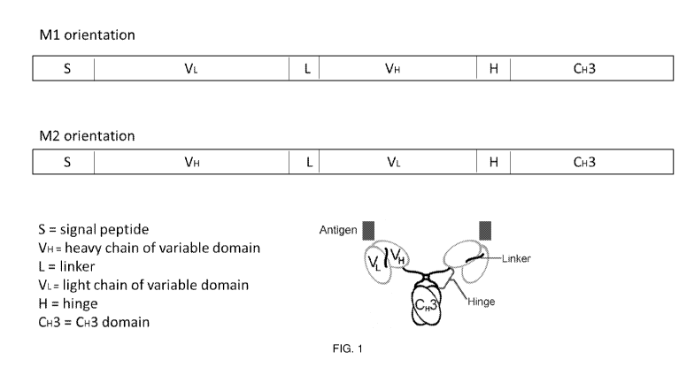
comprises a lower
hinge or extension region. In some aspects, the lower hinge or extension
region comprises at
least one of: GGGSSGGGSG (SEQ ID NO: 59) or APPVAGP (SEQ ID NO: 60) or
APELLGGP (SEQ ID NO: 58). In some aspects, when located within a minibody, and
wherein when the minibody is administered to a human subject, clearance of the
minibody
from the subject occurs primarily through a liver. In some aspects, when
located within a
minibody, and wherein when the minibody is administered to a human subject,
clearance of
the minibody from the subject does not occur primarily through a kidney. In
some aspects,
the hinge region is within an antibody. In some aspects, the hinge region is
within an
antibody binding fragment. In some aspects, the hinge region is within a
minibody. In some
aspects, the hinge region is within a monospecific antibody. In some aspects,
the hinge
region comprises at least three cysteines per strand. In some aspects, the
hinge region
comprises at least four cysteines per strand. In some aspects, the hinge
region comprises at
least five cysteines per strand. In some aspects, cysteines are distributed
throughout the
-4-
CA 02994951 2018-02-06
WO 2017/027325 PCT/US2016/045580
amino acid hinge region in a repeating CXX or CXY motif. In some aspects, the
hinge
region is within a bispecific antibody. In some aspects, the bispecific
antibody is assembled
in a 1:1 ratio. In some aspects, the bispecific antibody comprises an antibody
fragment. In
some aspects, the bispecific antibody is a minibody.
[0010] In some aspects, a pharmaceutical composition is provided. The
pharmaceutical composition can comprise the amino acid hinge region of any of
those
disclosed herein. In some embodiments, this results in less than 5%
aggregation of an
antibody is present in the composition.
[0011] In some aspects, a pharmaceutical composition comprising the
amino acid
hinge region of any of those provided herein is provided. In some aspects, at
least 1
microgram to 100 mg of the antibody is present.
[0012] In some aspects, a minibody comprising a core hinge region is
provided.
The core hinge region comprises at least three cysteines per strand forming at
least three
disulfide bonds within the core hinge region. In some aspects, the first
residue of the core
region is a serine. In some aspects, the core hinge region comprises SCVECPPCP
(SEQ ID
NO: 56).
[0013] In some aspects, a minibody is provided. The minibody can
comprise a
sequence XniCX,2X,3CX,4Xn5C (SEQ ID NO: 3). This sequence can be located as
the core
hinge region of the minibody. Xi can be any amino acid or no amino acid. Xn2
can be any
amino acid. Xn3 can be any amino acid. Xn4 can be any amino acid. X15 can be
any amino
acid. In some aspects, Xi is any amino acid other than a cysteine (SEQ ID NO:
229). In
some aspects, Xi is a serine (SEQ ID NO: 230).
[0014] In some aspects, a variant minibody hinge is provided. The
variant hinge
can comprise a first altered amino acid position. The first altered position
is an amino acid
that in a native antibody hinge would be a cysteine, and has been altered in
the first altered
position so that it does not form a disulfide bond. The variant hinge can also
comprise at
least three cysteines per strand C-terminal to the first altered amino acid
position. In some
aspects, the hinge region consists of SEQ ID NO: 1. In some aspects, SEQ ID
NO: 1 is a
core hinge region, and wherein the core hinge region essentially consists of
SEQ ID NO: 1.
In some aspects, the core hinge region consists of SEQ ID NO: 1.
-5-
CA 02994951 2018-02-06
WO 2017/027325 PCT/US2016/045580
[0015] In some aspects, a minibody is provided. The minibody that binds
to a
target antigen, wherein the target antigen is at least one of CD3, CD8, 5T4,
PSMA, or PSCA.
The minibody comprising a polypeptide that comprises: a single-chain variable
fragment
(scFv) that binds to the target antigen, the scFv comprising a variable heavy
(VH) domain
linked a variable light (VI) domain; and a variant hinge region comprising at
least three
cysteines on each strand of the hinge.
[0016] In some aspects, the minibody further comprises a human IgG CH3
sequence. In some aspects, the minibody further comprises a detectable marker
selected
from the group consisting of a radioactive substance, a dye, a contrast agent,
a fluorescent
compound, a bioluminescent compound, an enzyme, an enhancing agent, and a
nanoparticle.
[0017] In some aspects, the minibody comprises: a HCDR1 of the HCDR1 as
disclosed herein; a HCDR2 of the HCDR2 as disclosed herein; a HCDR3 of the
HCDR3 as
disclosed herein; a LCDR1 of the LCDR1 as disclosed herein; a LCDR2 of the
LCDR2 as
disclosed herein; and a LCDR3 of the LCDR3 as disclosed herein.
[0018] In some aspects, the variable heavy (VH) domain and the variable
light
(VI) domain are human sequences.
[0019] In some aspects, a nucleic acid encoding a minibody as disclosed
herein is
provided.
[0020] In some aspects, a cell line producing the minibody as disclosed
herein is
provided.
[0021] In some aspects, a kit comprising any of the minibodies provided
herein
and a detectable marker is provided.
[0022] In some aspects, a method of detecting the presence or absence
of a target
antigen is provided. The target antigen is at least one of CD3, CD8, 5T4,
PSMA, or PSCA.
The method comprises applying a minibody as disclosed herein to a sample; and
detecting a
binding or an absence of binding of the antigen binding construct thereof to
the target
antigen.
[0023] In some aspects, the minibody comprises a detectable marker
selected
from the group consisting of a radioactive substance, a dye, a contrast agent,
a fluorescent
compound, a bioluminescent compound, an enzyme, an enhancing agent, and a
nanoparticle.
In some aspects, applying the minibody comprises administering the minibody to
a subject.
-6-
CA 02994951 2018-02-06
WO 2017/027325 PCT/US2016/045580
In some aspects, detecting binding or absence of binding of the minibody
thereof to target
antigen comprises positron emission tomography. In some aspects, the method
further
comprising applying a secondary antibody or fragment thereof to the sample,
wherein the
secondary antibody or fragment thereof binds specifically to the minibody. In
some aspects,
the minibody thereof is incubated with the sample for no more than 1 hour.
[0024] In some aspects, a method of targeting a therapeutic agent to a
target
antigen is provided. The target antigen is at least one of CD3, CD8, 5T4,
PSMA, or PSCA.
The method comprises administering to a subject a minibody as disclosed
herein, wherein the
minibody is conjugated to a therapeutic agent.
[0025] In some aspects, a method of neutralizing a B or T lymphocyte
cell in a
subject in need thereof is provided. The method comprising administering to
the subject a
minibody as disclosed herein that binds to CD8 and/or CD3. In some aspects,
the subject has
at least one of the disorders as noted herein.
[0026] In some aspects, an antibody and/or minibody that binds to a
target
antigen (such as CD8, CD3, 5T4, PSMA, PSCA) is provided. The antibody and/or
minibody
comprises a hinge region, wherein the hinge region comprises at least one of
the following:
a) an amino acid hinge region comprising a sequence of SEQ ID NO: 1
(XniCXõ2Xn3CXn4XnsC), wherein Xi can be any amino acid that does not naturally
form a covalent crosslinking bond, wherein Xn2 is one of: A, R, N, D, E, Q, G,
H, I,
L, K, M, F, P, S, T, W, Y, or V, wherein Xn3 can be any amino acid, wherein
Xn4 can
be any amino acid, and wherein X15 can be any amino acid;
b) an amino acid hinge region comprising a sequence of SEQ ID NO: 2
(Xnj Xn2 Xn3 Xn4Xn5 Xn6CXn7Xn8CXn9Xn10C), wherein Xi can be any m amino acids
(where m is any number of amino acids of any type), wherein Xn2 can be any
amino
acid, wherein Xn3 can be any amino acid, wherein Xn4 can be any amino acid,
wherein
X15 can be any amino acid, wherein Xn6 can be any amino acid other than a
cysteine,
wherein Xn7 can be any amino acid, wherein Xn8 can be any amino acid, wherein
Xn9
can be any amino acid, and wherein Xnio can be any amino acid;
c) a core hinge sequence of at least one of: CVECPPCP (SEQ ID
NO: 57), CPPCPPC (SEQ ID NO: 52), or CPPCPPCPPC (SEQ ID NO: 54), or
-7-
CA 02994951 2018-02-06
WO 2017/027325 PCT/US2016/045580
CPPCVECPPC (SEQ ID NO: 53) linked to; an upper hinge sequence of
ELKTPLGDTTHT (SEQ ID NO: 48) or EPKSSDKTHT (SEQ ID NO: 46);
d) an upper hinge region that comprises no amino acids capable of
crosslinking with a corresponding amino acid; and a core hinge region
connected to a
C-terminus of the upper hinge region, wherein the core hinge region comprises
at
least three cysteines per strand;
e) an antibody and/or minibody comprising a core hinge region, wherein
the core hinge region comprises at least three cysteines per strand forming at
least
three disulfide bonds within the core hinge region; or
0 a first altered amino acid position, wherein the first
altered position is
an amino acid that in a native antibody hinge would be a cysteine, and has
been
altered in the first altered position so that it does not form a disulfide
bond; and at
least three cysteines per strand C-terminal to the first altered amino acid
position.
[0027] In some aspects, any minibody provided herein can instead be
formatted
as a full length antibody. In some embodiments, the minibody body comprises a
humanized
amino acid sequence.
[0028] In some aspects, a method of manufacturing the minibody provided
herein
comprises expressing the minibody in a cell line.
[0029] In some aspects, a method of treating a condition in a subject
in need
thereof is provided. The method comprises administering to the subject any one
or more of
the minibodies provided herein to treat any one or more of a CD3, CD8, PSCA,
PSMA,
and/or 5T4 disorder provided herein.
BRIEF DESCRIPTION OF THE DRAWINGS
[0030] FIG. 1 shows an illustration of an engineered minibody (Mb).
[0031] FIG. 2 shows an illustration of various embodiments of Mb hinges
based
on human IgG1 (y1 EH1 top and yl EH2 bottom).
[0032] FIG. 3 shows an illustration of various embodiments of Mb hinges
based
on human IgG2 (y2 EH1 top and y2 EH2 bottom).
[0033] FIG. 4 shows an illustration of various embodiments of
additional Mb
hinges based on human IgG2 (y2 NH1 top and y2 NH2 bottom).
-8-
CA 02994951 2018-02-06
WO 2017/027325 PCT/US2016/045580
[0034] FIG. 5A shows images of non-reduced SDS-PAGE analysis of IAB2M
hinge variants.
[0035] FIG. 5B shows protein sequence information for some embodiments
of
IAB2M 71 EH1.
[0036] FIG. 5C shows protein sequence information for some embodiments
of
IAB2M 71 EH2.
[0037] FIG. 5D shows protein sequence information for some embodiments
of
IAB2M 72 EH2.
[0038] FIG. 5E shows protein sequence information for some embodiments
of
IAB2M- 72 EH1.
[0039] FIG. 6A shows intact mass analysis of IAB2M 71 EH1 variant.
Upper
panel shows the total ion chromatogram under reverse phase conditions. Middle
panel shows
the deconvoluted intact masses confirming the presence of half molecules and
the lower
panel shows the full size masses that were identified.
[0040] FIG. 6B shows intact mass analysis of IAB2M 72 EH1 variant.
Upper
panel shows the total ion chromatogram under reverse phase conditions. Middle
panel shows
the deconvoluted intact masses confirming the presence of half molecules and
the lower
panel shows the full size molecular masses that were identified.
[0041] FIG. 6C shows intact mass analysis of IAB2M 71 EH2 variant.
Upper
panel shows the total ion chromatogram under reverse phase conditions. Middle
panel shows
the deconvoluted intact masses confirming the presence of half molecules and
the lower
panel shows the full size molecular masses that were identified.
[0042] FIG. 6D shows intact mass analysis of IAB2M 72 EH2 variant.
Upper
panel shows the total ion chromatogram. Lower panel shows the deconvoluted
intact masses
confirming the presence of the full-size mass molecules. No half molecules
were detected.
[0043] FIG. 7A shows binding curves and EC50 binding values of IAB2M
engineered hinge variants determined by FACS using LNCaP-AR cells.
[0044] FIG. 7B shows binding curves and EC50 binding values of IAB2M
engineered hinge variants determined by FACS using C4-2 XCL cells.
[0045] FIG. 7C shows protein sequence information for some embodiments
of
IAB2M 71 EH3 without a canonical signal sequence.
-9-
CA 02994951 2018-02-06
WO 2017/027325 PCT/US2016/045580
[0046] FIG. 7D shows protein sequence information for some embodiments
of
IAB2M 71 EH3 without a canonical signal sequence.
[0047] FIG. 7E shows DNA sequence information for IAB2M 71 EH3 without
a
canonical signal sequence.
[0048] FIG. 8 shows a list of disulfide-containing peptides identified
within
IAB2M 71 EH1 dimer.
[0049] FIG. 9 shows an illustration of a mapping of the disulfide bonds
of
IAB2M 71 EH1 indicating where mispaired cysteines are formed.
[0050] FIG. 10 shows an illustration of a mapping of the disulfide
bonds of
IAB2M y2 EH1 suggesting the absence of mispaired cysteines.
[0051] FIG. 11A shows images of PET/CT scans of 89Zr-Df-IAB2M y 1 EH1
in a
nude mouse harboring 22Rv1 (PSMA+) tumor xenograft.
[0052] FIG. 11B shows a graph of biodistribution of 89Zr-Df-IAB2M y 1
EH1 in
nude mice.
[0053] FIG. 12A shows images of PET/CT scans of 89Zr-Df-IAB2M y 1 EH2
in a
nude mouse harboring 22Rv1 (PSMA+) tumor xenograft.
[0054] FIG. 12B shows a graph of biodistribution of 89Zr-Df-IAB2M y 1
EH2 in
nude mice.
[0055] FIG. 13A shows images of PET/CT scans of 89Zr-Df-IAB2M y2 EH2 in
a
nude mouse harboring 22Rv1 (PSMA+) tumor xenograft.
[0056] FIG. 13B shows a graph of biodistribution of 89Zr-Df-IAB2M y2
EH2 in
nude mice.
[0057] FIG. 14A shows the intact mass analysis of IAB22M 72 EH1
variant.
Upper panel shows the total ion chromatogram. Middle panel shows the
deconvoluted intact
masses confirming the presence of half molecules and the lower panel shows the
full-size
masses that were identified.
[0058] FIG. 14B shows protein sequence information for some embodiments
of
IAB22M 72 EH1.
[0059] FIG. 15A shows intact mass analysis of IAB22 72 EH2 variant. The
protein was separated under reverse phase conditions. Upper panel shows the
full mass range
scan. The zoomed-in full-size mass region emphasizes the presence of the
intact, i.e.
-10-
CA 02994951 2018-02-06
WO 2017/027325 PCT/US2016/045580
disulfide-bridge bonded protein (middle panel) while the zoomed-in half-
molecule region
showed that essentially no half-molecule is present (lower panel).
[0060] FIG. 15B shows protein sequence information for some embodiments
of
IAB22M 72 EH2 variant.
[0061] FIG. 16A shows PET/CT scan images of mice harboring HPB-ALL
(CD8+) tumor xenograft comparing 89Zr-Df-IAB22M minibodies with human IgG1 and
human IgG2 derived hinge sequences.
[0062] FIG. 16B shows protein sequence information for some embodiments
of
IAB22M 71 EH'.
[0063] FIG. 16C shows protein sequence information for some embodiments
of
IAB22M 72 NH1.
[0064] FIG. 16D shows protein sequence information for some embodiments
of
IAB22M 72 NH2.
[0065] FIG. 17 shows PET/CT scan images comparing uptake of 89Zr-Df-
IAB22M 71 EH1 in a NOD-SCID mouse with antigen-positive HPB-ALL tumor and
antigen-negative Daudi tumor (right panel only).
[0066] FIG. 18 shows a graph of biodistribution of 89Zr-Df-IAB22M 71
EH1 in
mice with antigen-positive HPB-ALL tumor and antigen-negative Daudi tumor.
Numbers
are shown on the right.
[0067] FIG. 19 is an illustration of some embodiments of hinge variants
listed in
Table 3 (71 EH1 top, 71 EH3 middle, 71 EH4 bottom).
[0068] FIG. 20A shows a graph summarizing the percentage of half
molecules
present in different hinge variants for IAB2M as determined by mass
spectrometry.
[0069] FIG. 20B shows a graph summarizing the percentage of half
molecules
present in different hinge variants for IAB22M as determined by mass
spectrometry.
[0070] FIG. 20C shows protein sequence information for some embodiments
of
IAB22M 71 EH3.
[0071] FIG. 20D shows protein sequence information for some embodiments
of
IAB22M 71 EH5.
[0072] FIG. 20E shows protein sequence information for some embodiments
of
IAB22M 73/71 EH6.
-11-
CA 02994951 2018-02-06
WO 2017/027325 PCT/US2016/045580
[0073] FIG. 20F shows protein sequence information for some embodiments
of
IAB22M 73/71 EH7.
[0074] FIG. 20G shows protein sequence information for some embodiments
of
IAB22M 73/71 EH8.
[0075] FIG. 21A shows an image of non-reduced SDS-PAGE analysis (left
panel)
of IAB2M hinge Mb variants and percentage of half molecules quantified by
densitometry
(right panel).
[0076] FIG. 21B shows protein sequence information for some embodiments
of
IAB2M 71 EH5.
[0077] FIG. 21C shows protein sequence information for some embodiments
of
IAB2M 73/71 EH6.
[0078] FIG. 21D shows protein sequence information for some embodiments
of
IAB2M 73/71 EH7.
[0079] FIG. 21E shows protein sequence information for some embodiments
of
IAB2M 73/71 EH8.
[0080] FIG. 22A shows an image of non-reduced SDS-PAGE analysis (left
panel)
of IAB22M hinge Mb variants and percentage of half molecules quantified by
densitometry
(right panel).
[0081] FIG. 22B shows protein sequence information for some embodiments
IAB22M 71 EH2.
[0082] FIG. 23 shows the intact mass analysis of IAB22M 71 EH1 variant.
Upper
panel shows the total ion chromatogram. Middle panel shows the deconvoluted
intact masses
confirming the presence of half molecules and the lower panel shows the full-
size masses
that were identified.
[0083] FIG. 24 shows intact mass analysis of IAB22M 71 EH3 variant. The
protein was separated under reverse phase conditions. Upper panel shows the
full mass range
scan. Zoomed-in full-size mass region emphasizes the presence of the intact
protein (Middle
panel). Zoomed-in half-molecule region showed that essentially no half-
molecule is present
(lower panel).
-12-
CA 02994951 2018-02-06
WO 2017/027325 PCT/US2016/045580
[0084] FIGs. 25A and 25B shows intact mass analyses of IAB22M 72 hinge
variants. FIG. 25A shows the total ion chromatogram of the IAB22M 72 EH1
variant (FIG.
14B) under reverse phase conditions (upper panel). Middle panel shows the
deconvoluted
intact masses confirming the presence of half molecules and the lower panel
shows the full-
size masses that were identified. Note that the heterogeneity results from the
terminal lysine
clipping. FIG. 25B shows a deconvoluted full range scan (upper panel) of the
IAB22M 72
EH2 variant (FIG. 15B) identifies the sole species with m/z of 79053.1 Da.
Zoomed-in full-
size molecular weight is shown on the middle panel and the half molecule is of
low
abundance and just above the level of noise (bottom panel).
[0085] FIG. 26 shows binding curves and EC50 binding values of IAB2M
engineered hinge variants determined by FACS using C4-2 XCL cells.
[0086] FIG. 27 shows binding curves and EC50 binding values of IAB22M
engineered hinge variants determined by FACS using HPB-ALL cells.
[0087] FIG. 28A shows an image of non-reduced SDS-PAGE analysis of
IAB2OM hinge Mb variants (left panel) and a percentage of half molecules
quantified by
densitometry (right panel).
[0088] FIG. 28B shows protein sequence information for some embodiments
of
IAB2OM 71 EH'.
[0089] FIG. 28C shows protein sequence information for some embodiments
of
IAB2OM 71 EH3.
[0090] FIG. 28D shows protein sequence information for some embodiments
of
IAB2OM 71 EH2.
[0091] FIG. 28E shows protein sequence information for some embodiments
of
IAB2OM 71 EH5.
[0092] FIG. 29A shows an image of non-reduced SDS-PAGE analysis of
IAB1M
hinge Mb variants (left panel) and percentage of half molecules quantified by
densitometry
(right panel).
[0093] FIG. 29B shows protein sequence information for some embodiments
of
IAB1M 71 EH1 .
[0094] FIG. 29C shows protein sequence information for some embodiments
of
IAB1M 72 EH2.
-13-
CA 02994951 2018-02-06
WO 2017/027325 PCT/US2016/045580
[0095] FIG. 29D shows protein sequence information for some embodiments
of
IAB1M 71 EH3.
[0096] FIG. 30 shows PET/CT scan images comparing uptake of 89Zr
radiolabeled IAB22 Mbs with different hinge sequences in NOD-SCID mice bearing
antigen
positive HPB-ALL xenografts on the left shoulder.
[0097] FIG. 31 shows a graph of biodistribution data of 89Zr
radiolabeled IAB22
Mbs with different hinge sequences in NOD-SCID mice bearing antigen positive
HPB-ALL
xenografts on the left shoulder.
[0098] FIG. 32 shows protein sequence information for some embodiments
of
IAB2OM 72 EH2.
[0099] FIG. 33 shows protein sequence information for some embodiments
of
IAB25m-y2 EH2.
[0100] FIG. 34A shows the DNA sequence encoding some embodiments of the
IAB2M Mb.
[0101] FIG. 34B shows the DNA sequence encoding some embodiments of the
IAB2M Mb.
[0102] FIG. 34C shows the DNA sequence encoding some embodiments of the
IAB2M Mb.
[0103] FIG. 34D shows the DNA sequence encoding some embodiments of the
IAB2M Mb.
[0104] FIG. 34E shows the DNA sequence encoding some embodiments of the
IAB2M Mb.
[0105] FIG. 34F shows the DNA sequence encoding some embodiments of the
IAB2M Mb.
[0106] FIG. 35A shows protein sequence information of IAB2M Mb hinge
variants.
[0107] FIG. 35B shows protein sequence information of IAB2M Mb hinge
variants.
[0108] FIG. 35C shows protein sequence information of IAB2M Mb hinge
variants.
-14-
CA 02994951 2018-02-06
WO 2017/027325 PCT/US2016/045580
[0109] FIG. 36A shows protein sequence information of VL and VH domains
of
Mbs with different antigen-specificities.
[0110] FIG. 36B shows protein sequence information of VL and VH domains
of
Mbs with different antigen-specificities.
[0111] FIG. 36C shows protein sequence information of VL and VH domains
of
Mbs with different antigen-specificities.
[0112] FIG. 36D shows protein sequence information of VL and VH domains
of
Mbs with different antigen-specificities.
[0113] FIG. 36E shows protein sequence information of VL and VH domains
of
Mbs with different antigen-specificities.
[0114] FIG. 37 shows protein sequence information of various
embodiments of
linker sequences.
[0115] FIG. 38 shows protein sequence information of various
embodiments of
hinge regions.
[0116] FIG. 39 shows protein sequence information of various
embodiments of
CH3 domains.
[0117] FIG. 40 shows an alignment of protein sequences of an embodiment
each
of IAB22M 71 EH 1(M1) and IAB22M 71 EH3(M1). Sequence differences are shown in
boxes.
[0118] FIG. 41 shows the DNA and translated protein sequence of an
embodiment of IAB22M 71 EH3(M1). In boxes are shown the signal, CDR, linker
and hinge
sequences.
[0119] FIG. 42 shows the DNA and translated protein sequence of an
embodiment of IAB22M 71 EH5(M1).
[0120] FIG. 43 shows the DNA and translated protein sequence of an
embodiment of IAB22M 71 EH7(M1).
[0121] FIG. 44 shows the DNA and translated protein sequence of an
embodiment of IAB22M 71 EH8(M1).
[0122] FIG. 45 shows the DNA and translated protein sequence of an
embodiment of IAB22M 72 EH2(M1).
-15-
CA 02994951 2018-02-06
WO 2017/027325 PCT/US2016/045580
[0123] FIG. 46 shows the DNA and translated protein sequence of an
embodiment of IAB22M 72 EH2(M1) with VH-K67R polymorphism.
[0124] FIG. 47 shows an alignment of protein sequences of an embodiment
each
of IAB2M 71 EH 1(M2) and IAB2M 71 EH3(M2). Sequence differences are shown in
boxes.
[0125] FIG. 48 shows the DNA and translated protein sequence of an
embodiment of IAB2M 71 EH3(M2). In boxes are shown the signal, CDR, linker and
hinge
sequences.
[0126] FIG. 49 shows the DNA and translated protein sequence of an
embodiment of IAB2M 71 EH3 (M2) (G1m1).
[0127] FIG. 50 shows the DNA and translated protein sequence of an
embodiment of IAB2M 71 EH5(M2).
[0128] FIG. 51 shows the DNA and translated protein sequence of an
embodiment of IAB2M 71 EH7(M2).
[0129] FIG. 52 shows the DNA and translated protein sequence of an
embodiment of IAB2M 71 EH8(M2).
[0130] FIG. 53 shows the DNA and translated protein sequence of an
embodiment of IAB2M 71 EH3(M1).
[0131] FIG. 54 shows an alignment of protein sequences of an embodiment
each
of IAB2OM 71 EH1 (M2) and IAB2OM 71 EH3 (M2) with VL-Q79E, V83E; VH-Q1E, Q6E
polymorphisms. Sequence differences are shown in boxes.
[0132] FIG. 55 shows the DNA and translated protein sequence of an
embodiment of IAB2OM 71 EH3(M2) with VL-Q79E, V83E; VH-Q1E, Q6E
polymorphisms. In boxes are shown the signal, CDR, linker and hinge sequences.
[0133] FIG. 56 shows the DNA and translated protein sequence of an
embodiment of IAB2OM 71 EH3(M2).
[0134] FIG. 57 shows the DNA and translated protein sequence of an
embodiment of IAB2OM 71 EH3 (M2) with VL-A9D, T 10S, 512A, P 15L, L21I, 522N;
VH-
Q1E, Q6E polymorphisms.
-16-
CA 02994951 2018-02-06
WO 2017/027325 PCT/US2016/045580
[0135] FIG. 58 shows the DNA and translated protein sequence of an
embodiment of IAB2OM 71 EH5 (M2) with VL-Q79E, V83E; VH-Q1E, Q6E
polymorphisms.
[0136] FIG. 59 shows the DNA and translated protein sequence of an
embodiment of IAB2OM 71 EH5 (M2) with VL-A9D, T 10S, S 12A, P15L, L21I, S22N;
VH-
Q 1E, Q6E polymorphisms.
[0137] FIG. 60 shows an alignment of protein sequences of an embodiment
each
of IAB1M 71 EH1(M1) and IAB1M 71 EH3 (M1). Sequence differences are shown in
boxes.
[0138] FIG. 61 shows the DNA and translated protein sequence of an
embodiment of IAB1M 71 EH3(M1). In boxes are shown the signal, CDR, linker and
hinge
sequences.
[0139] FIG. 62 shows the DNA and translated protein sequence of an
embodiment of IAB 1M 72 EH2 (M1).
[0140] FIG. 63 shows the DNA and translated protein sequence of an
embodiment of IAB 1M 71 EH5 (M1).
[0141] FIG. 64 shows the DNA and translated protein sequence of an
embodiment of IAB 1M 71 EH7 (M1).
[0142] FIG. 65A shows the DNA and translated protein sequence of an
embodiment of IAB 1M 71 EH8(M1).
[0143] FIG. 65B shows the DNA and translated protein sequence of an
embodiment of IAB25M 72 NH(M1).
[0144] FIG. 65C shows the DNA and translated protein sequence of an
embodiment of IAB25M 72 EH(M1).
[0145] FIG. 66 shows the protein sequence of an embodiment of IAB22M VH
domain.
[0146] FIG. 67 shows the protein sequence of some embodiments of IAB20
VL
domain.
[0147] FIG. 68 shows the protein sequence of an embodiment of IAB20 VH
domain.
-17-
CA 02994951 2018-02-06
WO 2017/027325 PCT/US2016/045580
[0148] FIG. 69 shows the protein sequence of some embodiments of anti-
CD3
VL and VH domains.
[0149] FIG. 70 shows the protein sequence of an embodiment of PSMA
(Prostate-Specific Membrane Antigen) also known as glutamate carboxypeptidase
2 from
Homo sapiens.
[0150] FIG. 71 shows the protein sequence of an embodiment of PSCA
(Prostate
Stem Cell Antigen) from Homo sapiens.
[0151] FIG. 72 shows the protein sequence of an embodiment of 5T4
oncofetal
trophoblast glycoprotein from Homo sapiens.
[0152] FIG. 73 shows the protein sequence of an embodiment of T-cell
surface
glycoprotein CD8 alpha chain from Homo sapiens.
[0153] FIG. 74 shows the protein sequence of an embodiment of T-cell
surface
glycoprotein CD8 beta chain from Homo sapiens.
[0154] FIG. 75 shows the protein sequence of an embodiment of T-cell
surface
glycoprotein CD3 delta chain from Homo sapiens.
[0155] FIG. 76 shows the protein sequence of an embodiment of T-cell
surface
glycoprotein CD3 gamma chain from Homo sapiens.
[0156] FIG. 77 shows the protein sequence of an embodiment of T-cell
surface
glycoprotein CD3 epsilon chain from Homo sapiens.
[0157] FIG. 78 shows the protein sequence of an embodiment of T-cell
surface
glycoprotein CD3 zeta chain from Homo sapiens.
[0158] FIG. 79 shows protein sequence information for some embodiments
of
IAB25M- 72 EH2.
[0159] FIG. 80 shows protein sequence information for some embodiments
of
IAB20M- 72 EH2.
DETAILED DESCRIPTION
[0160] Described herein are components for antigen binding constructs,
including
antibodies and fragments thereof, such as minibodies, that bind to a target
molecule. In some
embodiments, these components are novel hinge sequences and/or sequences
associated with
and/or part of the hinge sequence. These hinge sequences can provide various
benefits. Also
-18-
CA 02994951 2018-02-06
WO 2017/027325 PCT/US2016/045580
provided herein are the antigen binding constructs (such as antibodies,
minibodies, etc.) that
include one or more of the hinge sequences or subsequences provided herein.
[0161] In some embodiments, the antigen binding constructs can be
useful for
targeting therapeutic agents to cells that express the target molecule. In
some embodiments,
methods are provided for detecting the presence or absence of a target
molecule (or "target")
using antigen binding constructs (including antibodies, and constructs such as
minibodies).
In some embodiments, methods are provided for using the antigen binding
constructs for
therapeutic purposes.
[0162] In some embodiments, scFv and minibody antibodies can have
superior
pharmacokinetic properties for faster diagnostic imaging while maintaining the
binding
specificity and affinity of the parental antibody. Current technology utilizes
imaging with the
full length antibodies which often requires significantly longer times (-7-8
days post-
injection) to produce high contrast images due to the slow serum clearance of
the intact
antibody. Some embodiments of the minibodies provided herein provide the
opportunity for
same-day or next-day imaging. Same-day or next-day imaging also provides a
logistical
solution to the problem facing many patients who travel great distances to
receive
treatment/diagnosis since the duration of travel stays or the need to return
one week later
would be eliminated when imaging with minibodies versus intact antibodies.
[0163] As detailed below, in some embodiments, the antigen binding
constructs
are for diagnostics. When labeled with an appropriate radionuclides (e.g., the
positron
emitter Iodine-124, Copper-64, Fluorine-18, Gallium-68 and/or Zirconium-89 for
PET
imaging) or fluorophore (for fluorescent imaging), the antibody fragments can
be used for
preclinical imaging as shown herein and for clinical imaging in patients.
These antigen
binding constructs can also be used as potential SPECT imaging agents by
simply changing
the radiolabel to single photon emitting radionuclides such as Indium-111,
Iodine-123 and
Lutitium-177.
[0164] In some embodiments, the antigen binding constructs can be
clinical
imaging agents (PET/SPECT) in humans. Accordingly, in some embodiments,
antigen
binding constructs can be used for targeted diagnostic detection for these
disorders. In some
embodiments, the antigen binding construct can be used as a therapeutic.
-19-
CA 02994951 2018-02-06
WO 2017/027325 PCT/US2016/045580
Definitions and various embodiments
[0165] The term "hinge" denotes at least a part of a hinge region for
an antigen
binding construct, such as an antibody or a minibody. A hinge region can
include a
combination of the upper hinge, core (or middle) hinge and lower hinge
regions. In some
embodiments, the hinge is defined according to any of the antibody hinge
definitions. Native
IgG 1 , IgG2, and IgG4 antibodies have hinge regions having of 12-15 amino
acids. IgG3 has
an extended hinge region, having 62 amino acids, including 21 prolines and 11
cysteines.
The functional hinge region of naturally occurring antibodies, deduced from
crystallographic
studies, extends from amino acid residues 216-237 of the IgG1 H chain (EU
numbering; ref.
12) and includes a small segment of the N terminus of the CH2 domain in the
lower hinge,
with the lower hinge being the N terminus of CH2 domain. The hinge can be
divided into
three regions; the "upper hinge," the "core," and the "lower hinge".
[0166] The term "artificial" or "non-natural" when modifying a hinge
(or a
subpart thereof) denotes that the sequence in question is not present, in the
noted state, in
nature. In the present context the hinges have been altered from their native
state, so that
their sequences are no longer those found in wild-type antibodies. As will be
appreciated by
those of skill in the art, minibodies do not naturally occur in nature, and
thus, any construct
which is a minibody construct is also not found in nature. This also applies
to at least some
of the constructs found in and/or incorporating the sequences of any of the
hinge sequence
tables provided herein (for example, Table 0.2). In some embodiments, any of
the hinge
subparts or full hinge sequences in Table 0.1 can be artificial hinge
sequences, as long as the
sequence (or resulting combination for the hinge) does not occur in nature.
[0167] The term "full hinge region" or "entire hinge region" denotes
the presence
of the entire upper, core, and lower hinge regions as a single construct. The
upper, core, and
lower regions can be positioned immediately adjacent to one another, or
additional residues
can be added between, or N- or C-terminal to the regions. In some embodiments,
the native
lower hinge can be replaced with an extension sequence. In some embodiments,
one can
combine a native lower hinge with the extension sequence. In some embodiments,
an
extension or other set of sequences can be added after the upper and/or core
sequences.
[0168] The phrase "effective hinge region" denotes that an adequate
amount of
part of at least one of the upper, core and lower hinge regions is present to
allow the hinge
-20-
CA 02994951 2018-02-06
WO 2017/027325 PCT/US2016/045580
region to be effective for its intended purpose. Thus, the phrase encompasses
variants of
hinge regions and fragments of the various hinge regions. In some embodiments,
the
function of the hinge region is one or more of the following: to link the scFv
with the CH3
domain, provide flexibility and spacing for the two scFvs to bind to the
target properly, to
link two half molecules together, to provide overall stability to the
molecule, and/or to
provide a site for site-specific conjugation due to its solvent exposure.
In some
embodiments, the hinge should be close to natural as to reduce potential
immunogenicity. In
some embodiments, the upper hinge provides flexibility to scFv (starts at
residue 216 in
native IgGs), the middle hinge provides stability, and the lower hinge
mediates flexibility to
CH3 (starts at residue 231 in native IgGs).
[0169] The
term "upper hinge" denotes the first part of the hinge that starts at the
end of the scFv. Examples of upper hinge regions can be found in Table 0.1.
The upper
hinge includes the amino acids from the end of the scFv up to, but not
including, the first
cysteine residue in the core hinge as shown in Table 0.1. As above, the term
"effective upper
hinge" denotes that enough of the sequence is present to allow the section to
function as an
upper hinge; the term encompasses functional variants and fragments of the
designated hinge
section.
[0170] The
term "core hinge" denotes the second part of the hinge region that is
C-terminal to the upper hinge. Examples of core hinge regions can be found in
Table 0.1.
The core hinge contains the inter-chain disulfide bridges and a high content
of prolines. As
above, the term "effective core hinge" denotes that enough of the sequence is
present to
allow the section to function as a core hinge; the term encompasses functional
variants and
fragments of the designated hinge section.
[0171] The
term "lower hinge" denotes the third part of the hinge region that is
C-terminal to the core hinge. Examples of lower hinge regions can be found in
Table 0.1. In
the context of a minibody or antibody fragment, the lower hinge connects to
the CH3 domain
Mb. As above, the term "effective lower hinge" denotes that enough of the
sequence is
present to allow the section to function as a lower hinge; the term
encompasses functional
variants and fragments of the designated hinge section. The term "lower hinge"
as used
herein can encompass various amino acid sequences including naturally
occurring IgG lower
hinge sequences and artificial extension sequences in place of one another or
a combination
-21-
CA 02994951 2018-02-06
WO 2017/027325 PCT/US2016/045580
thereof provided herein. In some embodiments, the various extensions can be
considered to
be a lower hinge region in its entirety or a replacement.
[0172] The
term "treating" or "treatment" of a condition can refer to preventing
the condition, slowing the onset and/or rate of development of the condition,
reducing the
risk of developing the condition, preventing and/or delaying the development
of symptoms
associated with the condition, reducing or ending symptoms associated with the
condition,
generating a complete or partial regression of the condition, or some
combination thereof.
The term "prevent" does not require the absolute prohibition of the disorder
or disease.
[0173] A
"therapeutically effective amount" or a "therapeutically effective dose"
is an amount that produces a desired therapeutic effect in a subject, such as
preventing,
treating a target condition, delaying the onset of the disorder and/or
symptoms, and/or
alleviating symptoms associated with the condition. This amount will vary
depending upon a
variety of factors, including but not limited to the characteristics of the
therapeutic compound
(including activity, pharmacokinetics, pharmacodynamics, and bioavailability),
the
physiological condition of the subject (including age, sex, disease type and
stage, general
physical condition, responsiveness to a given dosage, and type of medication),
the nature of
the pharmaceutically acceptable carrier or carriers in the formulation, and/or
the route of
administration. One skilled in the clinical and pharmacological arts will be
able to determine
a therapeutically effective amount through routine experimentation, for
example by
monitoring a subject's response to administration of a compound and adjusting
the dosage
accordingly, given the present disclosure. For additional guidance, see
Remington: The
Science and Practice of Pharmacy 21st Edition, Univ. of Sciences in
Philadelphia (USIP),
Lippincott Williams & Wilkins, Philadelphia, PA, 2005.
[0174] The
term "antigen binding construct" includes all varieties of antibodies,
including binding fragments thereof. Further included are constructs that
include 1, 2, 3, 4,
5, and/or 6 CDRs. In some embodiments, tandem scFvs can be provided, which can
provide
two arms with bivalent binding. In some embodiments, these CDRs can be
distributed
between their appropriate framework regions in a traditional antibody. In
some
embodiments, the CDRs can be contained within a heavy and/or light chain
variable region.
In some embodiments, the CDRs can be within a heavy chain and/or a light
chain. In some
embodiments, the CDRs can be within a single peptide chain. Unless otherwise
denoted
-22-
CA 02994951 2018-02-06
WO 2017/027325 PCT/US2016/045580
herein, the antigen binding constructs described herein bind to the noted
target molecule.
The term "target" or "target molecule" denotes the protein to which the
antigen binding
construct binds. Examples of target proteins are known in the art, and
include, for example
PSMA (such as FOLH1) (FIG. 70; SEQ ID NO: 131), PSCA (FIG. 71; SEQ ID NO:
132),
5T4 (such as TPBG) (FIG. 72; SEQ ID NO: 133), CD8 (such as the a-chain) (FIG.
73; SEQ
ID NO: 134), CD8 (such as the 13-chain) (FIG. 74; SEQ ID NO: 135), CD3 (such
as the 8-
chain) (FIG. 75; SEQ ID NO: 136), CD3 (such as the 7-chain) (FIG. 76; SEQ ID
NO: 137),
CD3 (such as the c-chain) (FIG. 77; SEQ ID NO: 138), CD3 (such as the c-chain)
(FIG. 78;
SEQ ID NO: 139).
[0175] The term "antibody" includes, but is not limited to, genetically
engineered
or otherwise modified forms of immunoglobulins, such as intrabodies, chimeric
antibodies,
fully human antibodies, humanized antibodies, antibody fragments, scFv, and
heteroconjugate antibodies (for example, bispecific antibodies, diabodies,
triabodies,
tetrabodies, etc.). The term "antibody" includes scFv and minibodies. Thus,
each and every
embodiment provided herein in regard to "antibodies" is also envisioned as
scFv and/or
minibody embodiments, unless explicitly denoted otherwise. The term "antibody"
includes a
polypeptide of the immunoglobulin family or a polypeptide comprising fragments
of an
immunoglobulin that is capable of noncovalently, reversibly, and in a specific
manner
binding a corresponding antigen. An exemplary antibody structural unit
comprises a
tetramer. In some embodiments, a full length antibody can be composed of two
identical
pairs of polypeptide chains, each pair having one "light" and one "heavy"
chain (connected
through a disulfide bond). The recognized immunoglobulin genes include the
kappa,
lambda, alpha, gamma, delta, epsilon, hinge, and mu constant region genes, as
well as the
myriad immunoglobulin variable region genes. For full length chains, the light
chains are
classified as either kappa or lambda. For full length chains, the heavy chains
are classified as
gamma, mu, alpha, delta, or epsilon, which in turn define the immunoglobulin
classes, IgG,
IgM, IgA, IgD, and IgE, respectively. The N-terminus of each chain defines a
variable
region of about 100 to 110 or more amino acids primarily responsible for
antigen
recognition. The terms variable light chain (VI) and variable heavy chain (VH)
refer to these
regions of light and heavy chains respectively. As used in this application,
an "antibody"
encompasses all variations of antibody and fragments thereof. Thus, within the
scope of this
-23-
CA 02994951 2018-02-06
WO 2017/027325 PCT/US2016/045580
concept are full length antibodies, chimeric antibodies, humanized antibodies,
single chain
antibodies (scFv), Fab, Fab', and multimeric versions of these fragments (for
example,
F(abl)2) with the same binding specificity. In some embodiments, the antibody
binds
specifically to a desired target.
[0176] The term "complementarity-determining domains" or
"complementarity-
determining regions ("CDRs") interchangeably refer to the hypervariable
regions of VL and
VH. The CDRs are the target molecule-binding site of the antibody chains that
harbors
specificity for such target molecule. In some embodiments, there are three
CDRs (CDR1-3,
numbered sequentially from the N-terminus) in each VL and/or VH, constituting
about
15-20% of the variable domains. The CDRs are structurally complementary to the
epitope of
the target molecule and are thus directly responsible for the binding
specificity. The
remaining stretches of the VL or VH, the so-called framework regions (FRs),
exhibit less
variation in amino acid sequence (Kuby, Immunology, 4th ed., Chapter 4. W.H.
Freeman &
Co., New York, 2000).
[0177] The positions of the CDRs and framework regions can be
determined
using various well known definitions in the art, for example, Kabat (Wu, T. T.
et al., "An
analysis of the sequences of the variable regions of Bence Jones proteins and
myeloma light
chains and their implications for antibody complementarity," J. Exp. Med.,
Vol. 132, No. 2,
pp. 211-250, 1970; Kabat, E. A. et al., "Sequences of Proteins of
Immunological Interest,"
5th Ed., NIH Publication No. 91-3242, Bethesda, MD, 1991, Chothia C. et al.,
"Canonical
structures for the hypervariable regions of immunoglobulins," J. Mol. Biol.,
Vol. 196, No. 4,
pp. 901-917, 1987; Chothia C. et al., "Conformations of immunoglobulin
hypervariable
regions," Nature, Vol. 342, No. 6252, pp. 877-883, 1989; Chothia C. et al.,
"Structural
repertoire of the human VH segments," J. Mol. Biol., Vol. 227, No. 3, pp. 799-
817, 1992; Al-
Lazikani B. et al., "Standard conformations for the canonical structures of
immunoglobulins," J. Mol. Biol., Vol. 273, No. 4, pp. 927-748, 1997),
ImMunoGeneTics
database (IMGT) (on the worldwide web at imgt.org/) (Giudicelli, V. et al.,
"IMGT/LIGM-
DB, the IMGT comprehensive database of immunoglobulin and T cell receptor
nucleotide
sequences," Nucleic Acids Res., Vol. 34 (Database Issue), pp. D781-D784, 2006;
Lefranc,
M. P. et al., "IMGT unique numbering for immunoglobulin and T cell receptor
variable
domains and Ig superfamily V-like domains," Dev. Comp. Immunol., Vol. 27, No.
1, pp. 55-
-24-
CA 02994951 2018-02-06
WO 2017/027325 PCT/US2016/045580
77, 2003; Brochet, X. et al., "IMGT/V-QUEST: the highly customized and
integrated system
for IG and TR standardized V-J and V-D-J sequence analysis," Nucleic Acids
Res., Vol. 36
(Web Server Issue), pp. W503-508, 2008; AbM (Martin, A. C. et al., "Modeling
antibody
hypervariable loops: a combined algorithm,"Proc. Natl. Acad. Sci. U.S.A., Vol.
86, No. 23,
pp. 9268-9272, 1989); the contact definition (MacCallum, R. M. et al.,
"Antibody-antigen
interactions: contact analysis and binding site topography," J. Mol. Biol.,
Vol. 262, No. 5, pp.
732-745, 1996), and/or the automatic modeling and analysis tool (Honegger, A.
et al.,
Accessible on the world wide web at
bioc.uzh.ch/plueckthun/antibody/Numbering/).
[0178] The term "binding specificity determinant" or "BSD"
interchangeably
refer to the minimum contiguous or non-contiguous amino acid sequence within a
complementarity determining region necessary for determining the binding
specificity of an
antibody. A minimum binding specificity determinant can be within one or more
CDR
sequences. In some embodiments, the minimum binding specificity determinants
reside
within (i.e., are determined solely by) a portion or the full-length of the
CDR3 sequences of
the heavy and light chains of the antibody. In some embodiments, CDR3 of the
heavy chain
variable region is sufficient for the antigen binding construct specificity.
[0179] An "antibody variable light chain" or an "antibody variable
heavy chain"
as used herein refers to a polypeptide comprising the VL or VH, respectively.
The
endogenous VL is encoded by the gene segments V (variable) and J (junctional),
and the
endogenous VH by V, D (diversity), and J. Each of VL or VH includes the CDRs
as well as
the framework regions. In this application, antibody variable light chains
and/or antibody
variable heavy chains may, from time to time, be collectively referred to as
"antibody
chains." These terms encompass antibody chains containing mutations that do
not disrupt the
basic structure of VL or VH, as one skilled in the art will readily recognize.
In some
embodiments, full length heavy and/or light chains are contemplated. In some
embodiments,
only the variable region of the heavy and/or light chains are contemplated as
being present.
[0180] Antibodies can exist as intact immunoglobulins or as a number of
fragments produced by digestion with various peptidases. Thus, for example,
pepsin digests
an antibody below the disulfide linkages in the hinge region to produce
F(ab)12, a dimer of
Fab which itself is a light chain (VL-CL) joined to VH-CH1 by a disulfide
bond. The F(ab)12
may be reduced under mild conditions to break the disulfide linkage in the
hinge region,
-25-
CA 02994951 2018-02-06
WO 2017/027325 PCT/US2016/045580
thereby converting the F(ab)12 dimer into an Fab monomer. The Fab' monomer is
a Fab with
part of the hinge region. (Paul, W. E., "Fundamental Immunology," 3d Ed., New
York:
Raven Press, 1993). While various antibody fragments are defined in terms of
the digestion
of an intact antibody, one of skill will appreciate that such fragments may be
synthesized de
novo either chemically or by using recombinant DNA methodology. Thus, the term
"antibody," as used herein, also includes antibody fragments either produced
by the
modification of whole antibodies, or those synthesized de novo using
recombinant DNA
methodologies (for example, single chain Fv) or those identified using phage
display libraries
(see, for example, McCafferty, J. et al., "Phage antibodies: filamentous phage
displaying
antibody variable domains," Nature, Vol. 348, No. 66301, pp. 552-554, 1990).
[0181] For preparation of monoclonal or polyclonal antibodies, any
technique
known in the art can be used (see, for example, Kohler, G. et al., "Continuous
cultures of
fused cells secreting antibody of predefined specificity," Nature, Vol. 256,
No. 5517, pp.
495-497, 1975; Kozbor, D. et al., "The production of monoclonal antibodies
from human
lymphocytes," Immunology Today, Vol. 4, No. 3, pp. 72-79, 1983; Cole, et al.,
"Monoclonal
Antibodies and Cancer Therapy," Alan R. Liss, Inc., pp. 77-96, 1985; Wang, S.,
"Advances
in the production of human monoclonal antibodies," Antibody Technology
Journal, Vol. 1,
pp. 1-4, 2011; Sharon, J. et al., "Recombinant polyclonal antibodies for
cancer therapy," J.
Cell Biochem., Vol. 96, No. 2, pp. 305-313, 2005;; Haurum, J. S., "Recombinant
polyclonal
antibodies: the next generation of antibody therapeutics?," Drug Discov.
Today, Vol. 11, No.
13-14, pp. 655-660, 2006). Techniques for the production of single chain
antibodies (U.S.
Pat. No. 4,946,778) can be adapted to produce antibodies to polypeptides of
this invention.
Also, transgenic mice, or other organisms such as other mammals, may be used
to express
fully human monoclonal antibodies. Alternatively, phage display technology can
be used to
identify high affinity binders to selected antigens (see, for example,
McCafferty et al., supra;
Marks, J. D. et al., "By-passing immunization: building high affinity human
antibodies by
chain shuffling," Biotechnology (N. Y.), Vol. 10, No. 7, pp. 779-783, 1992).
[0182] Methods for humanizing or primatizing non-human antibodies are
well
known in the art. Generally, a humanized antibody has one or more amino acid
residues
introduced into it from a source which is non-human. These non-human amino
acid residues
are often referred to as import residues, which are typically taken from an
import variable
-26-
CA 02994951 2018-02-06
WO 2017/027325 PCT/US2016/045580
domain. In some embodiments, the terms "donor" and "acceptor" sequences can be
employed. Humanization can be essentially performed following the method of
Winter and
co-workers (see, for example, Jones, P. T. et al., "Replacing the
complementarity-
determining regions in a human antibody with those from a mouse," Nature, Vol.
321, No.
6069, pp. 522-525, 1986; Riechmann, L. et al., "Reshaping human antibodies for
therapy,"
Nature, Vol. 332, No. 6162, pp. 323-327, 1988; Verhoeyen, M. et al.,
"Reshaping human
antibodies: grafting an antilysozyme activity," Science, Vol. 239, No. 4847,
pp. 1534-1536,
1988; Presta, L. G., "Antibody engineering,", Curr. Op. Struct. Biol., Vol. 2,
No. 4, pp. 593-
596, 1992), by substituting rodent CDRs or CDR sequences for the corresponding
sequences
of a human antibody. Accordingly, such humanized antibodies are chimeric
antibodies (U.S.
Pat. No. 4,816,567), wherein substantially less than an intact human variable
domain has
been substituted by the corresponding sequence from a non-human species. In
practice,
humanized antibodies are typically human antibodies in which some
complementarity
determining region ("CDR") residues and possibly some framework ("FR")
residues are
substituted by residues from analogous sites in rodent antibodies.
[0183] A "chimeric antibody" is an antibody molecule in which (a) the
constant
region, or a portion thereof, is altered, replaced or exchanged so that the
antigen binding site
(variable region) is linked to a constant region of a different or altered
class, effector function
and/or species, or an entirely different molecule which confers new properties
to the chimeric
antibody, for example, an enzyme, toxin, hormone, growth factor, and drug; or
(b) the
variable region, or a portion thereof, is altered, replaced or exchanged with
a variable region
having a different or altered antigen specificity.
[0184] Antibodies further include one or more immunoglobulin chains
that are
chemically conjugated to, or expressed as, fusion proteins with other
proteins. It also includes
bispecific antibodies. A bispecific or bifunctional antibody is an artificial
hybrid antibody
having two different heavy/light chain pairs and two different binding sites.
[0185] Other antigen-binding fragments or antibody portions of the
invention
include, bispecific scFv antibodies where the antibody molecule recognizes two
different
epitopes, single binding domains (sdAb or nanobodies), and minibodies.
[0186] The term "antibody fragment" includes, but is not limited to one
or more
antigen binding fragments of antibodies alone or in combination with other
molecules,
-27-
CA 02994951 2018-02-06
WO 2017/027325 PCT/US2016/045580
including, but not limited to Fab', F(abl)2, Fab, Fv, rIgG (reduced IgG), scFv
fragments,
single domain fragments (nanobodies), peptibodies, minibodies. The term "scFv"
refers to a
single chain Fv ("fragment variable") antibody in which the variable domains
of the heavy
chain and of the light chain of a traditional two chain antibody have been
joined to form one
chain.
[0187] A pharmaceutically acceptable carrier may be a pharmaceutically
acceptable material, composition, or vehicle that is involved in carrying or
transporting a
compound of interest from one tissue, organ, or portion of the body to another
tissue, organ,
or portion of the body. For example, the carrier may be a liquid or solid
filler, diluent,
excipient, solvent, or encapsulating material, or some combination thereof.
Each component
of the carrier is "pharmaceutically acceptable" in that it is be compatible
with the other
ingredients of the formulation. It also must be suitable for contact with any
tissue, organ, or
portion of the body that it may encounter, meaning that it must not carry a
risk of toxicity,
irritation, allergic response, immunogenicity, or any other complication that
excessively
outweighs its therapeutic benefits. The pharmaceutical compositions described
herein may
be administered by any suitable route of administration. A route of
administration may refer
to any administration pathway known in the art, including but not limited to
aerosol, enteral,
nasal, ophthalmic, oral, parenteral, rectal, transdermal (for example, topical
cream or
ointment, patch), or vaginal. "Transdermal" administration may be accomplished
using a
topical cream or ointment or by means of a transdermal patch. "Parenteral"
refers to a route
of administration that is generally associated with injection, including
infraorbital, infusion,
intraarterial, intracapsular, intracardiac, intradermal, intramuscular,
intraperitoneal,
intrapulmonary, intraspinal, intrasternal, intrathecal, intrauterine,
intravenous, subarachnoid,
subcapsular, subcutaneous, transmucosal, or transtracheal. In some
embodiments, the
antigen binding construct can be delivered intraoperatively as a local
administration during
an intervention or resection.
[0188] The term "target molecule dependent disorder" "or "target
molecule
associated disorder" includes any disorder in which the target molecule plays
a role in the
disorder itself. In some embodiments, this denotes over-expression of the
target molecule.
In some embodiments, the disorders can include any of the disorders discussed
herein. In
some embodiments, the disorder can be any for which there is a target molecule
that can be
-28-
CA 02994951 2018-02-06
WO 2017/027325 PCT/US2016/045580
targeted by binding, whose binding will result in the detection and/or
treatment of the
disorder. Without limitation, the target molecule can be CD8, CD3, 5T4, PSMA,
and/or
PSCA, for example.
[0189] A minibody is an antibody format that has a smaller molecular
weight than
the full-length antibody while maintaining the bivalent binding property
against an antigen.
Because of its smaller size, absence of CH2 domain that binds Fc-gamma and
FcRN
receptors, absence of glycosylation, the minibody has a faster clearance from
the system and
potentially enhanced penetration when targeting tumor tissue. With the ability
for strong
targeting combined with rapid clearance, the minibody is advantageous for
diagnostic
imaging and delivery of radioactive payloads for which prolonged circulation
times may
result in adverse patient dosing or dosimetry. In some embodiments, it can
also be
advantageous for delivery of a cytotoxic payload due to the above-mentioned
features such
as tumor penetration and faster clearance. A "minibody" as described herein,
encompasses a
homodimer, wherein each monomer is a single-chain variable fragment (scFv)
linked to a
human IgG CH3 domain by a hinge sequence. In some embodiments, a minibody is a
bivalent or bispecific, covalently bound homodimer of -80 kDa. In some
embodiments, each
monomer (half-molecule) is comprised of a variable heavy (VH) domain linked to
the
corresponding variable light (VI) domain by an approximate 15-18 amino acid
Gly-Ser-rich
linker sequence.
[0190] In some embodiments, each single-chain variable fragment (scFv)
is
linked to a human IgGl, IgG2, IgG3 or IgG4 CH3 domain by a hinge sequence.
[0191] In some embodiments a lower hinge/extension sequence can be a
native
IgG1 , 2, 3 or 4 lower hinge, an/or (G3)ST, or (G4)ST, (n can be any number of
S's; in some
embodiments it is 1 or 2) and/or no lower hinge and/or any combination of
amino acids
(doesn't have to be G's and S's). In some embodiments, the lower
hinge/extension sequence
can comprise GGGSSGGGSG (SEQ ID NO: 59).
[0192] In some embodiments, a linker can be any that will work. In some
embodiments, a linker sequence can include a motif that is (G3)ST, or (G4)ST,
(n can be any
number of S's; in some embodiments it is 1 or 2). In some embodiments, the
linker can
comprise GSTSGGGSGGGSGGGGSS (SEQ ID NO: 62).
-29-
CA 02994951 2018-02-06
WO 2017/027325 PCT/US2016/045580
[0193] The phrase "specifically (or selectively) bind," when used in
the context of
describing the interaction between an antigen, for example, a protein, to an
antibody or
antibody-derived binding agent, refers to a binding reaction that is
determinative of the
presence of the antigen in a heterogeneous population of proteins and other
biologics, for
example, in a biological sample, for example, a blood, serum, plasma or tissue
sample. Thus,
under designated immunoassay conditions, in some embodiments, the antibodies
or binding
agents with a particular binding specificity bind to a particular antigen at
least two times the
background and do not substantially bind in a significant amount to other
antigens present in
the sample. Specific binding to an antibody or binding agent under such
conditions may
require the antibody or agent to have been selected for its specificity for a
particular protein.
A variety of immunoassay formats may be used to select antibodies specifically
immunoreactive with a particular protein. For example, solid-phase ELISA
immunoassays
are routinely used to select antibodies specifically immunoreactive with a
protein (see, for
example, Harlow, E. & Lane D., "Using Antibodies, A Laboratory Manual," Cold
Spring
Harbor Laboratory Press, 1998, for a description of immunoassay formats and
conditions that
can be used to determine specific immunoreactivity). Typically a specific or
selective
binding reaction will produce a signal at least twice over the background
signal and more
typically at least than 10 to 100 times over the background.
[0194] The term "equilibrium dissociation constant (KD, M)" refers to
the
dissociation rate constant (kd, time-1) divided by the association rate
constant (ka, time-1 M-1).
Equilibrium dissociation constants can be measured using any known method in
the art. The
antibodies of the present invention generally will have an equilibrium
dissociation constant
of less than about 10-7 or 10-8 M, for example, less than about 10-9 M or 10-
10 M, in some
embodiments, less than about 10-11 M, 10-12 M, or 10-13 M.
[0195] The term "isolated," when applied to a nucleic acid or protein,
denotes that
the nucleic acid or protein is essentially free of other cellular components
with which it is
associated in the natural state. In some embodiments, it can be in either a
dry or aqueous
solution. Purity and homogeneity can be determined using analytical chemistry
techniques
such as polyacrylamide gel electrophoresis or high performance liquid
chromatography. A
protein that is the predominant species present in a preparation is
substantially purified. In
particular, an isolated gene is separated from open reading frames that flank
the gene and
-30-
CA 02994951 2018-02-06
WO 2017/027325 PCT/US2016/045580
encode a protein other than the gene of interest. The term "purified" denotes
that a nucleic
acid or protein gives rise to essentially one band in an electrophoretic gel.
In some
embodiments, this can denote that the nucleic acid or protein is at least 85%
pure, more
preferably at least 95% pure, and most preferably at least 99% pure of
molecules that are
present under in vivo conditions.
[0196] The term "nucleic acid" or "polynucleotide" refers to
deoxyribonucleic
acids (DNA) or ribonucleic acids (RNA) and polymers thereof in either single-
or double-
stranded form. Unless specifically limited, the term encompasses nucleic acids
containing
known analogues of natural nucleotides that have similar binding properties as
the reference
nucleic acid and are metabolized in a manner similar to naturally occurring
nucleotides.
Unless otherwise indicated, a particular nucleic acid sequence also implicitly
encompasses
conservatively modified variants thereof (for example, degenerate codon
substitutions),
alleles, orthologs, SNPs, and complementary sequences as well as the sequence
explicitly
indicated. Specifically, degenerate codon substitutions may be achieved by
generating
sequences in which the third position of one or more selected (or all) codons
is substituted
with mixed-base and/or deoxyinosine residues (Batzer, M. A. et al., "Enhanced
evolutionary
PCR using oligonucleotides with inosine at the 31-terminus," Nucleic Acid
Res., Vol. 19, No.
18, pp. 5081, 1991; Ohtsuka, E. et al., "An alternative approach to
deoxyoligonucleotides as
hybridization probes by insertion of deoxyinosine at ambiguous codon
positions," J. Biol.
Chem., Vol. 260, No. 5, pp. 2605-2608, 1985; Rossolini, G. M. et al., "Use of
deoxyinosine-
containing primers vs degenerate primers for polymerase chain reaction based
on ambiguous
sequence information," Mol. Cell. Probes, Vol. 8, No. 2, pp. 91-98, 1994).
[0197] The terms "polypeptide," "peptide," and "protein" are used
interchangeably herein to refer to a polymer of amino acid residues. The terms
apply to
amino acid polymers in which one or more amino acid residue is an artificial
chemical
mimetic of a corresponding naturally occurring amino acid, as well as to
naturally occurring
amino acid polymers and non-naturally occurring amino acid polymer.
[0198] The term "amino acid" refers to naturally occurring and
synthetic amino
acids, as well as amino acid analogs and amino acid mimetics that function in
a manner
similar to the naturally occurring amino acids. Naturally occurring amino
acids are those
encoded by the genetic code, as well as those amino acids that are later
modified, for
-31-
CA 02994951 2018-02-06
WO 2017/027325 PCT/US2016/045580
example, hydroxyproline, gamma-carboxyglutamate, and 0-phosphoserine. Amino
acid
analogs refer to compounds that have the same basic chemical structure as a
naturally
occurring amino acid, for example, an alpha-carbon that is bound to a
hydrogen, a carboxyl
group, an amino group, and an R group, for example, homoserine, norleucine,
methionine
sulfoxide, methionine methyl sulfonium. Such analogs have modified R groups
(for
example, norleucine) or modified peptide backbones, but retain the same basic
chemical
structure as a naturally occurring amino acid. Amino acid mimetics refers to
chemical
compounds that have a structure that is different from the general chemical
structure of an
amino acid, but that functions in a manner similar to a naturally occurring
amino acid.
[0199] The term "conservatively modified variants" applies to both
amino acid
and nucleic acid sequences. With respect to particular nucleic acid sequences,
conservatively
modified variants refers to those nucleic acids which encode identical or
essentially identical
amino acid sequences, or where the nucleic acid does not encode an amino acid
sequence, to
essentially identical sequences. Because of the degeneracy of the genetic
code, a large
number of functionally identical nucleic acids encode any given protein. For
instance, the
codons GCA, GCC, GCG and GCU all encode the amino acid alanine. Thus, at every
position where an alanine is specified by a codon, the codon can be altered to
any of the
corresponding codons described without altering the encoded polypeptide. Such
nucleic acid
variations are "silent variations," which are one species of conservatively
modified
variations. Every nucleic acid sequence herein which encodes a polypeptide
also describes
every possible silent variation of the nucleic acid. One of skill will
recognize that each
codon in a nucleic acid (except AUG, which is ordinarily the only codon for
methionine, and
TGG, which is ordinarily the only codon for tryptophan) can be modified to
yield a
functionally identical molecule. Accordingly, each silent variation of a
nucleic acid that
encodes a polypeptide is implicit in each described sequence.
[0200] As to amino acid sequences, one of skill will recognize that
individual
substitutions, deletions or additions to a nucleic acid, peptide, polypeptide,
or protein
sequence which alters, adds or deletes a single amino acid or a small
percentage of amino
acids in the encoded sequence is a "conservatively modified variant" where the
alteration
results in the substitution of an amino acid with a chemically similar amino
acid.
Conservative substitution tables providing functionally similar amino acids
are well known
-32-
CA 02994951 2018-02-06
WO 2017/027325 PCT/US2016/045580
in the art. Such conservatively modified variants are in addition to and do
not exclude
polymorphic variants, interspecies homologs, and alleles of the invention.
[0201] The following eight groups each contain amino acids that are
conservative
substitutions for one another: 1) Alanine (A), Glycine (G); 2) Aspartic acid
(D), Glutamic
acid (E); 3) Asparagine (N), Glutamine (Q); 4) Arginine (R), Lysine (K); 5)
Isoleucine (I),
Leucine (L), Methionine (M), Valine (V); 6) Phenylalanine (F), Tyrosine (Y),
Tryptophan
(W); 7) Serine (S), Threonine (T); and 8) Cysteine (C), Methionine (M) (see,
for example,
Creighton, T. E., "Proteins - Structures and Molecular Properties," W. H.
Freeman & Co.
Ltd., 1984).
[0202] The term "percentage of sequence identity" can be determined by
comparing two optimally aligned sequences over a comparison window, wherein
the portion
of the polynucleotide sequence in the comparison window may comprise additions
or
deletions (i.e., gaps) as compared to the reference sequence (for example, a
polypeptide of
the invention), which does not comprise additions or deletions, for optimal
alignment of the
two sequences. The percentage is calculated by determining the number of
positions at
which the identical nucleic acid base or amino acid residue occurs in both
sequences to yield
the number of matched positions, dividing the number of matched positions by
the total
number of positions in the window of comparison and multiplying the result by
100 to yield
the percentage of sequence identity.
[0203] The terms "identical" or percent "identity," in the context of
two or more
nucleic acids or polypeptide sequences, refer to two or more sequences or
subsequences that
are the same sequences. Two sequences are "substantially identical" if two
sequences have a
specified percentage of amino acid residues or nucleotides that are the same
(for example,
70%, 75%, 80%, 85%, 90%, 95%, 96%, 97%, 98% or 99% sequence identity over a
specified
region, or, when not specified, over the entire sequence of a reference
sequence), when
compared and aligned for maximum correspondence over a comparison window, or
designated region as measured using one of the following sequence comparison
algorithms or
by manual alignment and visual inspection. Some embodiments provided herein
provide
polypeptides or polynucleotides that are substantially identical to the
polypeptides or
polynucleotides, respectively, exemplified herein (for example, any one or
more of the
variable regions exemplified in any one of Tables 0.1, 0.2, 1, 2, or 3 and
FIGs. 5B-5E, 7C,
-33-
CA 02994951 2018-02-06
WO 2017/027325 PCT/US2016/045580
7D, 14B, 15B, 16B-16D, 20C-20G, 21B-21E, 22B, 28B-28E, 29B-29D, 32, 33, 35A-
35C,
36A-36E, 40-69, 79, 80 and any one or more of the nucleic acid sequences
exemplified in
any one of FIGs. 7E, 34A-34F, 41-46, 48-53, 55-59, 61-64, 65A, 65B, and 65C).
Optionally,
the identity exists over a region that is at least about 15, 25 or 50
nucleotides in length, or
more preferably over a region that is 100 to 500 or 1000 or more nucleotides
in length, or
over the full length of the reference sequence. With respect to amino acid
sequences, identity
or substantial identity can exist over a region that is at least 5, 10, 15 or
20 amino acids in
length, optionally at least about 25, 30, 35, 40, 50, 75 or 100 amino acids in
length,
optionally at least about 150, 200 or 250 amino acids in length, or over the
full length of the
reference sequence. With respect to shorter amino acid sequences, for example,
amino acid
sequences of 20 or fewer amino acids, in some embodiments, substantial
identity exists when
one or two amino acid residues are conservatively substituted, according to
the conservative
substitutions defined herein.
[0204] In some embodiments, the percent identity is over the hinge
regions noted
herein (the hinge region and/or its subparts of upper, core, and lower hinge
regions). In such
situations, the percent identity of the hinge region or its subpart can be
identified separately
from the rest of the protein or nucleic acid sequence. Thus, two hinge regions
(or upper,
core, and/or lower regions) can have a specified percentage of amino acid
residues or
nucleotides that are the same (for example, 70%, 75%, 80%, 85%, 90%, 95%, 96%,
97%,
98% or 99% sequence identity over a specified region, or, when not specified,
over the entire
sequence of a reference sequence), while allowing for the remainder of the
protein to either
stay 100% identical to the comparison protein, our while also allowing the
remainder of the
protein to also have variation by a specified percent identity.
[0205] For sequence comparison, typically one sequence acts as a
reference
sequence, to which test sequences are compared. When using a sequence
comparison
algorithm, test and reference sequences are entered into a computer,
subsequence coordinates
are designated, if necessary, and sequence algorithm program parameters are
designated.
Default program parameters can be used, or alternative parameters can be
designated. The
sequence comparison algorithm then calculates the percent sequence identities
for the test
sequences relative to the reference sequence, based on the program parameters.
-34-
CA 02994951 2018-02-06
WO 2017/027325 PCT/US2016/045580
[0206] A "comparison window", as used herein, includes reference to a
segment
of any one of the number of contiguous positions selected from the group
consisting of from
20 to 600, usually about 50 to about 200, more usually about 100 to about 150
in which a
sequence may be compared to a reference sequence of the same number of
contiguous
positions after the two sequences are optimally aligned. Methods of alignment
of sequences
for comparison are well known in the art. Optimal alignment of sequences for
comparison
can be conducted, for example, by the local homology algorithm of Smith and
Waterman
(1970) Adv. Appl. Math. 2:482c, by the homology alignment algorithm of
Needleman, S. B.
et al., "A general method applicable to the search for similarities in the
amino acid sequence
of two proteins," J. Mol. Biol., Vol. 48, No. 3, pp. 443-453, 1970, by the
search for similarity
method of Pearson, W. R. et al., "Improved tools for biological sequence
comparison," Proc.
Natl. Acad. Sci. U.S.A., Vol. 85, No. 8, pp. 2444-2448, 1988, by computerized
implementations of these algorithms (GAP, BESTFIT, FASTA, and TFASTA in the
Wisconsin Genetics Software Package, Genetics Computer Group, 575 Science Dr.,
Madison, Wis.), or by manual alignment and visual inspection (see, for
example, Ausubel, F.
M. et al., Current Protocols in Molecular Biology, Supplement, 1995).
[0207] Two examples of algorithms that are suitable for determining
percent
sequence identity and sequence similarity are the BLAST and BLAST 2.0
algorithms, which
are described in Altschul, S. F. et al., "Gapped BLAST and PSI-BLAST: a new
generation of
protein database search programs," Nucleic Acids Res., Vol. 25, No. 17, pp.
3389-3402,
1977, and Altschul, S. F. et al., "Basic local alignment search tool," J. Mol.
Biol., Vol. 215,
No. 3, pp. 403-410, 1990, respectively. Software for performing BLAST analyses
is publicly
available through the National Center for Biotechnology Information. This
algorithm
involves first identifying high scoring sequence pairs (HSPs) by identifying
short words of
length W in the query sequence, which either match or satisfy some positive-
valued threshold
score T when aligned with a word of the same length in a database sequence. T
is referred to
as the neighborhood word score threshold (Altschul, S. F. et al., supra).
These initial
neighborhood word hits act as seeds for initiating searches to find longer
HSPs containing
them. The word hits are extended in both directions along each sequence for as
far as the
cumulative alignment score can be increased. Cumulative scores are calculated
using, for
nucleotide sequences, the parameters M (reward score for a pair of matching
residues; always
-35-
CA 02994951 2018-02-06
WO 2017/027325 PCT/US2016/045580
>0) and N (penalty score for mismatching residues; always <0). For amino acid
sequences, a
scoring matrix is used to calculate the cumulative score. Extension of the
word hits in each
direction are halted when: the cumulative alignment score falls off by the
quantity X from its
maximum achieved value; the cumulative score goes to zero or below, due to the
accumulation of one or more negative-scoring residue alignments; or the end of
either
sequence is reached. The BLAST algorithm parameters W, T, and X determine the
sensitivity and speed of the alignment. The BLASTN program (for nucleotide
sequences)
uses as defaults a wordlength (W) of 11, an expectation (E) or 10, M=5, N=-4
and a
comparison of both strands. For amino acid sequences, the BLASTP program uses
as
defaults a wordlength of 3, and expectation (E) of 10, and the BLOSUM62
scoring matrix
(see, Henikoff, S. et al., "Amino acid substitution matrices from protein
blocks," Proc. Natl.
Acad. Sci. U.S.A., Vol. 89, No. 22, pp. 10915-10919, 1992) alignments (B) of
50,
expectation (E) of 10, M=5, N=-4, and a comparison of both strands.
[0208] The BLAST algorithm also performs a statistical analysis of the
similarity
between two sequences (see, for example, Karlin, S. et al., "Applications and
statistics for
multiple high-scoring segments in molecular sequences," Proc. Natl. Acad. Sci.
U.S.A., Vol.
90, No. 12, pp. 5873-5787, 1993). One measure of similarity provided by the
BLAST
algorithm is the smallest sum probability (P(N)), which provides an indication
of the
probability by which a match between two nucleotide or amino acid sequences
would occur
by chance. For example, a nucleic acid is considered similar to a reference
sequence if the
smallest sum probability in a comparison of the test nucleic acid to the
reference nucleic acid
is less than about 0.2, more preferably less than about 0.01, and most
preferably less than
about 0.001.
[0209] An indication that two nucleic acid sequences or polypeptides
are
substantially identical is that the polypeptide encoded by the first nucleic
acid is
immunologically cross reactive with the antibodies raised against the
polypeptide encoded by
the second nucleic acid, as described below. Thus, in some embodiments, a
polypeptide is
typically substantially identical to a second polypeptide, for example, where
the two peptides
differ only by conservative substitutions. Another indication that two nucleic
acid sequences
are substantially identical is that the two molecules or their complements
hybridize to each
other under stringent conditions, as described below. Yet another indication
that two nucleic
-36-
CA 02994951 2018-02-06
WO 2017/027325 PCT/US2016/045580
acid sequences are substantially identical is that the same primers can be
used to amplify the
sequence.
[0210] The terms "subject," "patient," and "individual" interchangeably
refer to
an entity that is being examined and/or treated. This can include, for
example, a mammal,
for example, a human or a non-human primate mammal. The mammal can also be a
laboratory mammal, for example, mouse, rat, rabbit, hamster. In some
embodiments, the
mammal can be an agricultural mammal (for example, equine, ovine, bovine,
porcine,
camelid) or domestic mammal (for example, canine, feline).
[0211] The term "co-administer" refers to the administration of two
active agents
in the blood of an individual or in a sample to be tested. Active agents that
are co-
administered can be concurrently or sequentially delivered.
Antigen binding constructs
[0212] It is herein appreciated that the sequences within the hinge
region can be
of special relevance for various antigen binding constructs. In some
embodiments, the value
of the hinge region can be especially high, such as in a minibody arrangement.
The
sequences within the hinge can include disulfide bonds which are useful for
hinge function
but can have varying results when altered. As disclosed herein, and shown in
the examples
below, in some embodiments, the hinge region sequences can be configured to
prevent
and/or reduce undesirable disulfide scrambling with cysteine residues present
in other
regions of the protein and/or contain sufficient numbers of cysteine pairs to
maintain dimer
integrity in vivo, prevent concatamers from forming and/or allow for site
specific conjugation
to one of the paired cysteines.
[0213] In some embodiments, the stability of a minibody dimer in vivo
can be
attributed to natural Van-der-Waals association between the CH3 domains,
formation of
disulfide bonds within the hinge regions, and/or interactions between VH and
VL. To date
most minibodies have been engineered using the human IgG1 upper and core hinge
regions
with an extension sequence linked to the human IgG1 CH3 domain. In addition to
the above
noted variables, both orientations of a minibody (e.g., VL-VH (M1) and VH-VL
(M2) scFv
variants) are herein provided and characterized, as the orientation of the
constructs can also
alter their characteristics (as shown in the examples below).
-37-
CA 02994951 2018-02-06
WO 2017/027325 PCT/US2016/045580
[0214] Some previous hinges (e.g., huIgG1 hinge-extension or 71 EH1)
have
included a native huIgG1 in the upper hinge (FIG. 2). This cysteine at
position 245 in the
context of human IgG1 as defined by Kabat and indicated by the first circle,
creates problems
with both protein heterogeneity and stability in the minibody format based on
the disulfide
mapping by LC/MS, SDS-PAGE analysis and in vivo biodistribution data. As shown
in the
examples below, in some embodiments, Cys245 can be mutated to serine,
resulting in an
artificial hinge or "EH2 hinge" as described herein. Thus, removal of this
cysteine from a
strand (as shown in the examples below), has resulted in an improved hinge
region. In some
embodiments, the resulting construct (which lacks the first cysteine in the
hinge region) is
not strong enough to maintain minibody dimer stability in vivo with the two
remaining
disulfide bridges (in regard to 71 EH/EH2). Thus, as shown in the examples
below, the
introduction of a third or greater number of cysteines per strand can be made
(for example,
within the core region) to create an improved construct. In some embodiments,
providing a
3rd (or more) cysteine in the hinge (an additional cysteine over IgG1 native
hinge, per strand)
increases disulfide bonding and increases protein stability. In some
embodiments, the added
stability afforded by increasing disulfide bonds in the hinge region allows
for site specific
conjugation to one or more than one of the cysteines with maintenance of
intact dimer. In
some embodiments, the huIgG2 minibodies of IAB2M, 20M, 1M, and IAB22M involve
human IgG2 (huIgG2) hinge/extension sequence linked to the huIgG2 CH3 domain.
Thus, in
some embodiments, the CH3 domain can be a huIgG2 CH3 domain. In some
embodiments,
any option for creating a covalent bond between the two strands of the peptide
can be
employed instead of cysteines for forming a disulfide bond.
[0215] In the present application, when a reference to an introduction
of a
cysteine is made (unless otherwise noted), it denotes the introduction of a
cysteine per strand
of the hinge (which comprises two strands). Thus, 3, 4, 5, or 6 cysteines
within a strand will
mean 6, 8, 10, or 12 cysteines within a hinge, and 3, 4, 5, or 6 possible
disulfide bonds being
present (of course, unless noted, the number of disulfide bonds can be
different, as some
could be used for labels and other uses).
[0216] In some embodiments, the IgG2 hinge/extension - 72 EH1 -
resulted in an
increase in aggregation, where the reactive first hinge cysteine was
responsible for
-38-
CA 02994951 2018-02-06
WO 2017/027325 PCT/US2016/045580
concatamer formation in IAB2M-y2 EH1 format. In such embodiments, the native
huIgG2
upper hinge can be employed.
[0217] The above aspects were then considered in developing a second
configuration of some of the embodiments of a hinge construct. In this second
arrangement,
the first hinge cysteine (one that pairs with the kappa light chain in a
native antibody) was
mutated to serine resulting in IgG2 EH2 (72 EH2). This construct expressed
well and had
good stability in vivo, had all 3 disulfide bonds formed properly as shown by
intact mass
analysis, and had stability demonstrated with both IAB2M and IAB22M constructs
(as
shown in the examples below). Thus, in some embodiments an IgG2 artificial or
modified
hinge, in which the first cysteine has been altered is provided herein and
provides for an
improved minibody construct.
[0218] As demonstrated in the examples below, the results showed that
it was
advantageous to mutate the first hinge cysteine of the IgG1 and IgG2 hinges to
prevent
cysteine mis-pairing. As demonstrated in the examples below, the results
showed that it was
advantageous to have more than 2 cysteine residues in core hinge region per
strand to
maintain structural integrity of protein. As demonstrated in the examples
below, the results
showed that the IgG2 hinge provides one solution to increase stability in
vivo. Thus, higher
in vivo stability antigen binding constructs, such as minibodies, are provided
herein.
[0219] In some embodiments, an IgG2 hinge can be employed. In some
embodiments, an IgG2 hinge can be employed where the first hinge cysteine is
mutated. In
some embodiments, mutation of the terminal lysine (K) in the Mb constructs did
not impact
protein expression but did generate protein with more uniform charge.
[0220] As discussed in the examples, additional Mb variants were
evaluated
based on upper IgG3 hinge and modified IgG1 core hinge sequences where at
least 3 cysteine
residues were present in the core hinge region per strand. As outlined in the
examples below,
IAB2M, IAB22M and IAB2OM constructs were evaluated to demonstrate universality
of
findings across various constructs having different orientations and different
targets. The
results indicate that the advantages described herein regarding the presently
disclosed hinges
would be available and applicable to any and all antigen binding constructs
and minibodies in
particular.
-39-
CA 02994951 2018-02-06
WO 2017/027325 PCT/US2016/045580
[0221] In some embodiments, the hinges can be peptide hinges and/or
nucleic
acid sequences that encode for the peptide hinges. All discussions regarding
the peptide
form of the hinges provided herein also designate corresponding structures and
functions for
nucleic acid sequences encoding the peptides. In some embodiments, the hinges
can be
incorporated into an antigen binding construct, such as an antibody, such as a
minibody. Any
hinge or subpart of a hinge (such as an upper hinge, core hinge, and/or lower
hinge region)
can be incorporated into any of the antigen binding constructs provided
herein, including, but
not limited to those in: Tables 0.1, 0.2, 1, 2, or 3 and FIGs. 5B-5E, 7C, 7D,
14B, 15B, 16B-
16D, 20C-20G, 21B-21E, 22B, 28B-28E, 29B-29D, 32, 33, 35A-35C, 36A-36E, 40-69,
79,
80 and any one or more of the nucleic acid sequences exemplified in any one of
FIGs. 7E,
34A-34F, 41-46, 48-53, 55-59, 61-64, 65A, 65B, and 65C. In some embodiments,
the hinge
can include any one or more of the sequences or subparts shown in Table 0.1.
In some
embodiments, any of the lower hinges can then be connected directly or
indirectly to a CH3
domain.
TABLE 0.1
Hinge variants (FIG. 38)
Full hinge Upper hinge Core hinge Lower hinge
Human IgG1 NH1 (SEQ EPKSCDKTHT (SEQ CPPCP (SEQ ID NO: APELLGGP (SEQ ID
ID NO: 21) ID NO: 45) 51) NO: 58)
Human IgG1 EH1 (SEQ EPKSCDKTHT (SEQ CPPC (SEQ ID NO: GGGSSGGGSG (SEQ
ID NO: 22) ID NO: 45) 50) ID NO: 59)
Human IgG1 NH2 (SEQ EPKSSDKTHT (SEQ CPPCP (SEQ ID NO: APELLGGP (SEQ ID
ID NO: 23) ID NO: 46) 51) NO: 58)
Human IgG1 EH2 (SEQ EPKSSDKTHT (SEQ CPPC (SEQ ID NO: GGGSSGGGSG (SEQ
ID NO: 24) ID NO: 46) 50) ID NO: 59)
Human IgG1 NH3 (SEQ EPKSSDKTHT (SEQ CPPCPPC (SEQ ID
APELLGGP (SEQ ID
ID NO: 25) ID NO: 46) NO: 52) NO: 58)
Human IgG1 EH3 (SEQ EPKSSDKTHT (SEQ CPPCPPC (SEQ ID GGGSSGGGSG (SEQ
ID NO: 26) ID NO: 46) NO: 52) ID NO: 59)
Human IgG1 NH4 (SEQ EPKSSDKTHT (SEQ CPPCVECPPC (SEQ APELLGGP (SEQ ID
ID NO: 27) ID NO: 46) ID NO: 53) NO: 58)
Human IgG1 EH4 (SEQ EPKSSDKTHT (SEQ CPPCVECPPC (SEQ GGGSSGGGSG (SEQ
ID NO: 28) ID NO: 46) ID NO: 53) ID NO: 59)
Human IgG1 NH5 (SEQ EPKSSDKTHT (SEQ CPPCPPCPPC (SEQ APELLGGP (SEQ ID
ID NO: 29) ID NO: 46) ID NO: 54) NO: 58)
Human IgG1 EH5 (SEQ EPKSSDKTHT (SEQ CPPCPPCPPC (SEQ GGGSSGGGSG (SEQ
ID NO: 30) ID NO: 46) ID NO: 54) ID NO: 59)
Human IgG2 NH1 (SEQCCVECPPCP (SEQ
APPVAGP (SEQ ID
ERK (SEQ ID NO: 47)
ID NO: 31) ID NO: 55) NO: 60)
Human IgG2 EH1 (SEQ ERK (SEQ ID NO: 47) CCVECPPCP (SEQ GGGSSGGGSG (SEQ
-40-
CA 02994951 2018-02-06
WO 2017/027325 PCT/US2016/045580
ID NO: 32) ID NO: 55) ID NO: 59)
Human IgG2 NH2 (SEQ ERK ID (SEQ NO: 47)
SCVECPPCP (SEQ APPVAGP (SEQ ID
ID NO: 33) ID NO: 56) NO: 60)
Human IgG2 EH2 (SEQERK (SE ID N 47)
SCVECPPCP (SEQ GGGSSGGGSG (SEQ
Q O:
ID NO: 34) ID NO: 56) ID NO: 59)
IgG3/IgG1 EH6 (SEQ ELKTPLGDTTHT (SEQ CVECPPC (SEQ ID GGGSSGGGSG (SEQ
ID NO: 35) ID NO: 48) NO: 57) ID NO: 59)
IgG3/IgG1 EH7 (SEQ ELKTPLGDTTHT (SEQ CPPCPPC (SEQ ID GGGSSGGGSG (SEQ
ID NO: 36) ID NO: 48) NO: 52) ID NO: 59)
IgG3/IgG1 EH8 (SEQ ELKTPLGDTTHT (SEQ CPPCPPCPPC (SEQ GGGSSGGGSG (SEQ
ID NO: 37) ID NO: 48) ID NO: 54) ID NO: 59)
IgG4 NH (SEQ ID NO: ESKYGPP (SEQ ID CPPCP
(SEQ ID NO: APEFLGGP (SEQ ID
38) NO: 49) 51)
NO: 61)
IgG4 EH (SEQ ID NO: ESKYGPP (SEQ ID CPPCP
(SEQ ID NO: GGGSSGGGSG (SEQ
39) NO: 49)
51) ID NO: 59)
[0222] In
some embodiments, the hinge comprises, consists of, or consists
essentially of any of the sequences in Table 0.1 above. In some embodiments,
the hinge
consists of each of a designated upper, core, and lower hinge region in Table
0.1. In some
embodiments, any one of the above subparts (such as the upper, core and/or
lower hinge) can
also be provided as a variant thereof. In some embodiments, the variants have
substantial
identity to the sequences above. In some embodiments, the variants have one,
two, or three
amino acids difference from the sequences noted above. In some embodiments,
the variants
have one, two, or three amino acids difference from the sequences noted above
and the
differences are conservative differences.
[0223] In
some embodiments the CPPC (SEQ ID NO: 50) motif in the core
region can be altered to any other effective core motif, as long as the number
and/or
effectiveness of the covalent bonds presented in the table above are
adequately replaced.
[0224] In
some embodiments, a peptide hinge region for an antibody is provided.
The hinge region can include an upper hinge region that comprises no amino
acids capable of
crosslinking with a corresponding amino acid. The amino acid hinge region can
further
include a core hinge region connected C-terminal to the upper hinge region. In
some
embodiments, the core hinge region comprises at least three cysteines on each
side of the
hinge (so at least six cysteines for the assembled structure). In some
embodiments, the
amino acid hinge region further comprises a lower hinge or extension region
connected
C-terminal to the core hinge region. In some embodiments, the lower hinge or
extension
-41-
CA 02994951 2018-02-06
WO 2017/027325 PCT/US2016/045580
region can include at least one of: APPVAGP (SEQ ID NO: 60), APELLGGP (SEQ ID
NO:
58), or GGGSSGGGSG (SEQ ID NO: 59).
[0225] In some embodiments, the upper hinge region comprises no
cysteines that
crosslink within the upper hinge region. In some embodiments, the upper hinge
region
comprises no cysteines. In some embodiments, the amino acid hinge region
further
comprises a lower hinge region. In some embodiments, these constructs are non-
naturally
occurring constructs.
[0226] In some embodiments, a variant minibody hinge region is
provided. The
variant minibody hinge region comprises a first altered amino acid position.
The first altered
amino acid position is an amino acid that in a native antibody hinge would be
a cysteine, and
has been altered in the first altered position so that it does not form a
disulfide bond. The
variant minibody hinge region further includes at least three cysteines C-
terminal to the first
altered amino acid position per strand. In some embodiments, 4, 5, 6, or more
cysteines can
be present C-terminal to the first altered amino acid position per strand. In
some
embodiments, these additional cysteines are fully contained within the core
region of the
hinge (of course, the core hinge can be modified and expanded beyond wild-type
sequences
such that the additional cysteines can be added). In some embodiments, the
hinge region
consists of SEQ ID NO: 1 (XTACX.2X.3CX.4XT,5C). In some embodiments, SEQ ID
NO: 1 is
a core hinge region, and the core hinge region essentially consists of SEQ ID
NO: 1. In some
embodiments, the core hinge region consists of SEQ ID NO: 1. In the
description noted
above, the number of cysteines relates to the number of cysteines in a single
hinge region of
the peptide chain. It will be understood that the number of cysteines in the
dimeric form of
the hinge (where the peptide chains are crosslinked to one another) will be
twice as many, as
the noted cysteines are each part of a disulfide bind involving two cysteines.
[0227] In some embodiments, Mb constructs contain antigen binding scFvs
with
variable linker lengths that can be in either VL-VH or VH-VL orientation. Any
disclosure
provided herein regarding one orientation also contemplates the reverse
orientation and both
orientations. In some embodiments, any hinge sequence described in Table 0.1
can be added
C-terminal to scFvs. In some embodiments, any CH3 domain from IgG 1, IgG2,
IgG3, or
IgG4 antibodies can be added C-terminal to scFvs and appropriate hinges. In
some
embodiments, to ensure appropriate disulfide bonding, the first hinge cysteine
(cysteine that
-42-
CA 02994951 2018-02-06
WO 2017/027325 PCT/US2016/045580
usually pairs with light chain in native IgG 1, IgG2, IgG3, and IgG4
antibodies) is mutated to
a serine or alanine or other appropriate amino acid to prevent disulfide
scrambling and/or
concatemerization. In some embodiments, to reduce the presence or prevent
formation of
half-molecules and ensure optimal stability in vivo, the hinge region can
contain at least 3
disulfide bonds, in some embodiments it can have more. In some embodiments, at
least 3
disulfide bonds are present in the hinge located at appropriate distances from
one another are
included to prevent improper disulfide bonds forming and ensure proper
disulfide bond
formation. In some embodiments, 3 disulfide bonds in the hinge are located at
appropriate
distances from one another are included for protein stability in vivo and
clearance through
liver rather than renal clearance. In some embodiments, at least 3 disulfides
bonds (for
example, 4 or more) in the hinge are provided to afford using a cysteine as a
handle,
following gentle reduction of the disulfides, of drugs, metal chelators,
radionuclides,
radioisotopes, site specific conjugation of a chelator then attachment of a
radioisotope to the
chelator, or fluorescent dyes using cysteine as a site of attachment. As
appreciated by one of
skill in the art, the presence of each single disulfide bond indicates the
presence of two
cysteines, one on each peptide sequence of the hinge.
[0228] In some embodiments, Mbs are constructed so as to include any of
the
hinge components provided herein and retain similar (or the same) affinity to
parent
antibodies. In some embodiments, Mbs constructed as described above can be
engineered to
contain two different antigen binding domains.
[0229] In some embodiments, superior hinges for antigen binding
constructs can
be obtained by removing a first cysteine in the hinge region, as noted in
Table 0.1 above and
in the examples below. Thus, in some embodiments, a hinge region in which this
cysteine
(which would otherwise be present in the natural hinge) is removed or altered
to another
residue (for example a residue that cannot form a covalent bond) can be
provided. In some
embodiments, this adjustment is made in a human IgG1 context, or any other
hinge region
that includes the cysteine in the noted position in Table 0.1 (for example, 72
EH1 altered in
72 EH2 (Cys to Ser), and 72 NH1 altered in 72 NH2 (Cys to Ser)).
[0230] In some embodiments, an amino acid hinge region is provided that
comprises a sequence of SEQ ID NO: 1 (XniCXn2Xn3CXn4XnsC). Xi can be any amino
acid
that does not form a covalent crosslinking bond. In some embodiments, Xn2 is
any one of:
-43-
CA 02994951 2018-02-06
WO 2017/027325 PCT/US2016/045580
A, R, N, D, E, Q, G, H, I, L, K, M, F, P, S, T, W, Y, or V. Xn3 can be any
amino acid. Xn4 is
any amino acid. X15 is any amino acid. In some embodiments, Xi comprises a
serine or an
alanine (SEQ ID NO: 235). In some embodiments, Xi comprises a serine (SEQ ID
NO:
210). In some embodiments, Xi comprises an alanine (SEQ ID NO: 211). In some
embodiments, Xi comprises a T (SEQ ID NO: 236). In some embodiments, the first
cysteine in SEQ ID NO: 1 that is altered is a cysteine in a y2 domain. In some
embodiments,
the amino acid hinge region further comprises additional amino acids in front
of SEQ ID NO:
1 (XniCXõ2Xn3CXn4Xn5C).
[0231] In some embodiments of Xn1CX,2Xn3CXn4Xn5C (SEQ ID NO: 1), Xi
does
not form a covalent crosslinking bond with another amino acid (SEQ ID NO:
191). In some
embodiments, Xi is not a cysteine (SEQ ID NO: 192). In some embodiments of SEQ
ID
NO: 1, Xn1 is one of: A, R, N, D, E, Q, G, H, I, L, K, M, F, P, S, T, W, Y, or
V (SEQ ID NO:
193). In some embodiments of SEQ ID NO: 1, Xn2 is P, V, or E (SEQ ID NO: 194).
In some
embodiments of SEQ ID NO: 1, Xn4 is P, V, or E (SEQ ID NO: 196). In some
embodiments
of SEQ ID NO: 1, Xn4 P or V (SEQ ID NO: 197). In some embodiments of SEQ ID
NO: 1,
Xn2 is P or V (SEQ ID NO: 195). In some embodiments of SEQ ID NO: 1, Xn3 is P
or E
(SEQ ID NO: 198). In some embodiments of SEQ ID NO: 1, X15 is P or E (SEQ ID
NO:
199). In some embodiments of SEQ ID NO: 1, Xn3 is P or E and X15 is P or E
(SEQ ID NO:
237). In some embodiments of SEQ ID NO: 1, Xn2Xn3 is VE (SEQ ID NO: 201). In
some
embodiments of SEQ ID NO: 1, Xn2Xn3 is PP (SEQ ID NO: 202). In some
embodiments of
SEQ ID NO: 1, Xn4X15 is VE (SEQ ID NO: 203). In some embodiments of SEQ ID NO:
1,
Xn4X15 is PP (SEQ ID NO: 204). In some embodiments of SEQ ID NO: 1, Xn2Xn3 is
VE and
Xn4X15 is PP (SEQ ID NO: 205). In some embodiments of SEQ ID NO: 1, Xn2Xn3 is
VE or
PP (SEQ ID NO: 238). In some embodiments of SEQ ID NO: 1, Xn2Xn3 is VE (SEQ ID
NO:
239).
[0232] In some embodiments, any of the above noted hinge regions
further
comprises a lower hinge or extension sequence C-terminal to the last cysteine
in
XniCXn2Xn3CXn4Xn5C (SEQ ID NO: 1). In some embodiments, any lower hinge region
can
be employed. In some embodiments, any of the above noted hinge regions further
comprises
an extension or lower hinge sequence C-terminal to the last cysteine in
XniCXn2Xn3CXn4Xn5C
(SEQ ID NO: 1).
-44-
CA 02994951 2018-02-06
WO 2017/027325 PCT/US2016/045580
[0233] In some embodiments, the hinge region of SEQ ID NO: 1 and/or 2
is a
core hinge region. In some embodiments, the hinge further comprises an upper
hinge region
adjacent to the core hinge region. In some embodiments, the hinge further
comprises a lower
hinge or extension region adjacent to the core hinge region. In some
embodiments, the
amino acid hinge region further comprises an upper hinge region adjacent to
the core hinge
region.
[0234] In some embodiments, the amino acid hinge region comprises the
following sequence: SCVECPPCP (SEQ ID NO: 56). In some embodiments, the amino
acid hinge region comprises the following sequence: ERKSCVECPPCP (SEQ ID NO:
167).
In some embodiments, the amino acid hinge region comprises the following
sequence:
EPKSSDKTHTCPPCPPC (SEQ ID NO: 168). In some embodiments, the amino acid hinge
region comprises at least one of the following sequences:
ERKSCVECPPCPGGGSSGGGSG
(SEQ ID NO: 34) or ERKSCVECPPCPAPPVAGP (SEQ ID NO: 33). In some
embodiments, the amino acid hinge region comprises at least TCPPCPPC (SEQ ID
NO: 166). In some embodiments, the amino acid hinge region comprises at least
EPKSSDKTHTCPPCPPCGGGSSGGGSG (SEQ ID NO: 26) or
EPKSSDKTHTCPPCPPCAPELLGGP (SEQ ID NO: 25).
[0235] In some embodiments, superior hinges can be provided by removing
the
hinge cysteine that is responsible for linking the heavy and light chains of
IgGl, IgG2, IgG3
and IgG4 respectively, as shown in Table 0.1 and 3 (for example, 71 EH1 to 71
EH2).
Furthermore, in some embodiments, using a core region with at least three
cysteines (e.g., 71
EH3, 71 EH4, and 71 EH5) per strand can result in a superior construct. In
some
embodiments, a hinge can be provided by removing a cysteine from an upper
hinge region,
as shown in table 0.1 and 3 (for example, 71 EH1 to 71 EH2).
[0236] In some embodiments, such constructs can be described as an
amino acid
hinge region comprising a sequence of SEQ ID NO: 2 (XniXn2Xn3Xõ4Xn5
Xn6CXõ7Xn8CX,9XnioC), wherein Xi is any amino acid that does not form a
covalent
crosslinking bond with another amino acid, wherein Xn2 is any amino acid,
wherein Xn3 is
any amino acid, wherein Xn4 is any amino acid, wherein X15 is any amino acid,
wherein Xn6 is
any amino acid, wherein Xn7 is any amino acid, wherein X8 isany amino acid,
wherein Xn9 is
any amino acid, and wherein Xnio is any amino acid (SEQ ID NO: 240).
-45-
CA 02994951 2018-02-06
WO 2017/027325 PCT/US2016/045580
[0237] In some embodiments, such constructs can be described as an
amino acid
hinge region comprising a sequence of SEQ ID NO: 2 (X.1Xn2Xn3Xn4Xn5
Xn6CXn7Xn8CXn9Xn10C), wherein Xi can be any amino acid, wherein X.2 can be any
amino
acid, wherein Xn3 can be any amino acid, wherein X.4 can be any amino acid,
wherein X15
can be any amino acid, wherein Xn6 can be any amino acid other than a
cysteine, wherein Xn7
can be any amino acid, wherein Xn8 can be any amino acid, wherein Xn9 can be
any amino
acid, and wherein Xnio can be any amino acid (SEQ ID NO: 241).
[0238] In some embodiments of SEQ ID NO: 2, X.1 is not a cysteine (SEQ
ID
NO: 213). In some embodiments of SEQ ID NO: 2, Xi is a serine or alanine (SEQ
ID NO:
242). In some embodiments of SEQ ID NO: 2, Xn2 is not a cysteine (SEQ ID NO:
214). In
some embodiments of SEQ ID NO: 2, Xn2 is a D (SEQ ID NO: 215). In some
embodiments,
Xn3 is a K (SEQ ID NO: 216). In some embodiments of SEQ ID NO: 2, Xn4 is a T
(SEQ ID
NO: 217). In some embodiments of SEQ ID NO: 2, X15 is a H (SEQ ID NO: 218). In
some
embodiments of SEQ ID NO: 2, X.6 is a T (SEQ ID NO: 219). In some embodiments
of
SEQ ID NO: 2, Xn7 is a P or a V (SEQ ID NO: 220). In some embodiments of SEQ
ID
NO: 2, Xn8 is a P or a E (SEQ ID NO: 221). In some embodiments of SEQ ID NO:
2, Xn9 is a
P or a V (SEQ ID NO: 222). In some embodiments of SEQ ID NO: 2, Xnio is a P or
a E
(SEQ ID NO: 223). In some embodiments of SEQ ID NO: 2, Xn2 is a D, Xn3 is a K,
X.4 is a
T, X15 is a H, X.6 is a T, X.7 is a P or a V, Xn8 is a P or a E, and Xn9 is a
P or a V (SEQ ID
NO: 243).
[0239] In some embodiments, the amino acid hinge region further
comprises a
XniiXn12C sequence. In some embodiments, this can be immediately attached to
the
C-terminal cysteine in SEQ ID NO: 1 (SEQ ID NO: 244) or SEQ ID NO: 2 (SEQ ID
NO:
245). In some embodiments of SEQ ID NO: 2, Xnii can be any amino acid, and
Xni2 can be
any amino acid (SEQ ID NO: 245). In some embodiments of SEQ ID NO: 2, Xnii is
a P or a
V, and wherein Xn12 is a P or an E (SEQ ID NO: 246). In some embodiments of
SEQ ID NO:
2, Xi is a serine, X.2 is a D, Xn3 is a K, X.4 is a T, X15 is a H, X.6 is a T,
Xn7 is a P, Xn8 is a P,
X9 isa P, and Xnio is a P (SEQ ID NO: 247).
[0240] In some embodiments, any of the hinge sequences can be preceded
by any
number of initial amino acids. Thus, additional linkers or spacers of amino
acids can be
added to the molecule before the noted sequence (such as SEQ ID NO: 1 and/or
NO: 2). In
-46-
CA 02994951 2018-02-06
WO 2017/027325 PCT/US2016/045580
some embodiments, any of the hinge sequences can be followed by any number of
additional
amino acids. Thus, additional linkers or spacers of amino acids can be added
to the molecule
after the noted sequence (such as SEQ ID NO: 1 and/or NO: 2).
[0241] In
some embodiments, the hinge region comprises at least one of the
following sequences: CPPCPPC (SEQ ID NO: 52), CPPCVECPPC (SEQ ID NO: 53), or
CPPCPPCPPC (SEQ ID NO: 54). In some embodiments, the hinge region comprises at
least
one of the following sequences: ERKSCVECPPCPGGGSSGGGSG (SEQ ID NO: 34),
EPKSSDKTHTCPPCPPC (SEQ ID NO: 168), EPKSSDKTHTCPPCVECPPC (SEQ ID
NO: 169), or EPKSSDKTHTCPPCPPCPPC (SEQ ID NO: 170). In some embodiments, the
hinge region comprises at least one of the following sequences:
EPKSSDKTHTCPPCPPCGGGSSGGGSG (SEQ ID NO: 26),
EPKSSDKTHTCPPCVECPPCGGGSSGGGSG (SEQ ID NO: 28), or
EPKSSDKTHTCPPCPPCPPCGGGSSGGGSG (SEQ ID NO: 30).
[0242] In
some embodiments, the lower hinge region/sequence can be an
extension sequence (e.g., 8-25 amino acids, such as 8-20, 10-25, 10-22, 12-20,
14-16, 8, 9,
10, 11, 12, 13, 14, 15, 16, 17, 18, 19, 20, 21, 22, 23, 24, or 25 amino
acids). Examples of the
lower hinge are shown in Table 0.1 and Table 3. In some embodiments, the lower
hinge can
be a native lower hinge from 71, 72, 73 or 74. In some embodiments, one can
have a native
hinge sequence and add one or more spacer amino acids (for example, 1, 2, 3, 4
or more
amino acids, such as G or S).
[0243] In
some embodiments, an amino acid hinge region comprises a core hinge
sequence of at least one of: CVECPPCP (SEQ ID NO: 57), CPPCPPC (SEQ ID NO:
52),
CPPCPPCPPC (SEQ ID NO: 54), or CPPCVECPPC (SEQ ID NO: 53) linked to an upper
hinge sequence of ELKTPLGDTTHT (SEQ ID NO: 48) or EPKSSDKTHT (SEQ ID
NO: 46).
[0244] In
some embodiments, any of the embodiments described herein can be
modified or supplemented by any one or more of the following options.
[0245] In
some embodiments, the hinge region comprises at least three cysteines
(on each chain, for a total of at least 6 cysteines between the two strands in
the dimeric hinge
construct). In some embodiments, the hinge region comprises at least four
cysteines (on each
strand). In some embodiments, the hinge region comprises at least five
cysteines (on each
-47-
CA 02994951 2018-02-06
WO 2017/027325 PCT/US2016/045580
strand). In some embodiments, 6, 7, 8, 9, 10 or more cysteines are present (on
each strand).
In some embodiments, the hinge region as a whole has the above noted number of
cysteines
(on each strand and/or in the dimeric construct). In some embodiments, all of
the noted
cysteines are present in the core hinge region. In some embodiments, all of
the noted
cysteines are present in the core hinge region and there are no additional
cysteines present in
the upper hinge region and/or the lower hinge region. In some embodiments, the
cysteines
are distributed throughout the amino acid hinge region in a repeating "CXX"
motif (such as
CXXCXXCXX (SEQ ID NO: 171) or CXXCXX (SEQ ID NO: 172), or
CXXCXXCXXCXX (SEQ ID NO: 173). In some embodiments, the cysteines are
distributed
throughout the core hinge region in a repeating CXX motif. In some
embodiments, the motif
repeats 1, 2, 3, 4, 5, 6, 7, 8, 9, 10 or more times. In some embodiments, each
X can be either
a P, V, or E (SEQ ID NO: 171, SEQ ID NO: 172, SEQ ID NO: 173). In some
embodiments,
X is not a cysteine (SEQ ID NO: 248, SEQ ID NO: 249, SEQ ID NO: 250).
[0246] In some embodiments, the core hinge region comprises at least
three
cysteines per strand forming at least three disulfide bonds within the core
hinge region. In
some embodiments, the constructs express much better in mammalian cells and
give higher
titers as well as lower aggregation and more uniform product. In some
embodiments, each of
the cysteines present in the core hinge region can form a disulfide bond with
a corresponding
cysteine on its paired chain in the hinge. In some embodiments, the disulfide
bond is
between corresponding cysteines on two separate protein chains, each being a
hinge region.
In some embodiments, at least 3 disulfide bonds are formed, for example, 3, 4,
5, 6, 7, 8, 9,
or more. In some embodiments, at least 3 disulfide bonds are capable of being
formed,
for example, 3, 4, 5, 6, 7, 8, 9, 10 or more disulfide bonds can be formed at
the same time
within the structure.
[0247] In some embodiments, the first residue of the core hinge region
is a serine.
In some embodiments, the core hinge region comprises SCVECPPCP (SEQ ID NO:
56).
[0248] In some embodiments, the CH3 domain for any construct can be
from 71,
72, or 73 and any naturally occurring allele thereof. As used herein "7" is an
abbreviation for
gamma.
[0249] In some embodiments, the extension or lower hinge region
comprises at
least one of S, G, A, P, or V. In some embodiments, the extension sequence
comprises at
-48-
CA 02994951 2018-02-06
WO 2017/027325 PCT/US2016/045580
least GGGSSGGGSG (SEQ ID NO: 59). In some embodiments, the extension or lower
hinge region comprises at least APPVAGP (SEQ ID NO: 60). In some embodiments,
the
lower hinge region comprises at least one of: GGGSSGGGSG (SEQ ID NO: 59) or
APP VAGP (SEQ ID NO: 60). In some embodiments, the lower hinge sequence can be
a GS
linker, extension sequence, and/or any native lower hinge region from 71, 72,
73 and 74.
Properties
[0250] In some embodiments, the hinges provided herein can allow for a
lower
percentage of the half-molecule to form and/or be present in any minibody or
other antigen
binding construct composition. For example, less can 7% of the composition can
be the half
molecule, including ranges such as less than 6, 5, 4, 3, 2, 1, and/or 0.5% of
composition
being the half-molecule.
[0251] In some embodiments when the hinge is located within a minibody,
and
when the minibody is administered to a human subject, clearance of the
minibody from the
subject may occur through liver and/or kidney. In some embodiments, clearance
through the
liver is at least 10% or more, for example 10, 20, 30, 40, 50, 60, 70, 80, 90,
95, 99, or 100%.
In some embodiments, when located within a minibody, and when the minibody is
administered to a human subject, clearance of the minibody from the subject
may occur
primarily through the kidneys. In some embodiments, at least 1, 2, 3, 4, 5, 6,
7, 8, 9, 10 fold
or more of the antibody is cleared through the kidney.
[0252] In some embodiments, less than 30% aggregation of an minibody is
present in an minibody composition. In some embodiments, less than 20%
aggregation of an
minibody is present in an minibody composition. In some embodiments, less than
10%
aggregation of an minibody is present in an minibody composition. In some
embodiments,
less than 5% aggregation of an minibody is present in an minibody composition.
In some
embodiments, less than 1% aggregation of a minibody is present in an minibody
composition.
[0253] When the minibody remains as a dimer in circulation in vivo,
then protein
clearance is predicted to occur through the liver primarily. In contrast, when
the half-
molecule is present, then its clearance will be through the kidney. Thus, in
some
embodiments, the above clearance aspects are a way of characterizing the
amount of
-49-
CA 02994951 2018-02-06
WO 2017/027325 PCT/US2016/045580
resulting half-molecule present in a subject, in vivo, allowing one to
determine if the
administered whole molecule remains as a whole molecule in vivo, or if it
breaks down to the
half molecule. In some embodiments, the minibody composition is one in which
clearance
through the liver is relatively high compared to clearance through the kidney,
and tumor
uptake of the minibody is relatively high. In some embodiments, the ratio of
kidney to liver
is less than 1. In some embodiments, at least 1% more uptake occurs through
the liver than
then kidney, for example, at least 1, 10, 20, 30, 40, 50, 60, 70 80, 90, 100,
or more uptake
occurs through the liver than through the kidney. In some embodiments, uptake
of kidney to
liver is 0.9:1, 1:10, 1:100, 1:000, 1:10,000, etc. In some embodiments, the
various hinge
constructs provided herein have lower clearance through the kidneys than the
EH1 hinge
from IgGl.
[0254] In some embodiments, mutation of the first hinge cysteine to
serine in
IgG1 hinge region (71 EH2) to prevent undesired interaction with other
unpaired cysteine
generated a protein with a better profile in vitro. However, the high
clearance through the
kidneys, compared to the liver, may be indicative of protein instability and
dissociation into
half molecules in vivo. In some embodiments, minibodies having an IgG2 hinge
are
provided wherein the first hinge cysteine is mutated to a serine (72 EH2 and
others), have the
property of being cleared through the liver as predicted for proteins of
approx. 80 kDa. This
indicates that the molecule remains intact in vivo. In some embodiments, the
molecule
remains intact for 48 hours or longer.
[0255] In some embodiments, the hinge construct provided herein have
less than
9% half molecules present due to an unpaired first hinge cysteine. In some
embodiments, an
IgG2 hinge with a first cysteine to serine has greater than 99% intact dimer
protein present,
such as that with 72 EH2. In some embodiments, a 72 hinge variant results in
lower kidney
uptake. In some embodiments, a Mb made with an huIgG1 hinge (71 EH1) is
rapidly cleared
through the kidney resulting in low tumor targeting. In some embodiments, a Mb
made with
huIgG2 hinge variants, either Extended Hinge (72 EH2) or Natural Hinge (72
NH1) shows
greater stability in vivo with high tumor targeting and lower kidney
clearance.
[0256] In some embodiments, 71 minibodies with EH2 hinge assemble
properly
into intact dimeric molecules but inclusion of only 2 cysteine yields protein
with a high
amount of half molecule. In some embodiments, 71 minibodies with EH3 hinge
assemble
-50-
CA 02994951 2018-02-06
WO 2017/027325 PCT/US2016/045580
properly into intact dimeric molecules and addition of 3rd cysteine per strand
yield protein
with very low levels of half molecule. In some embodiments, the level of half
molecule is
less than 15%, for example less than 14, 13, 12, 11, 10,9, 8,7, 6, 5,4, 3, 2,
or less than 1%
of half molecule is present.
[0257] In some embodiments, even though the hinge region has been
varied and
is different from that described in the past, the CDRs, VHNLs or full amino
acid sequences of
the minibodies still bind to the target molecule with the same affinity as an
antibody (such as
a minibody) with the previous style hinge. Thus, in some embodiments, the
presence of a
hinge as provided herein does not alter or negatively impact the binding
affinity of the
antigen binding construct, while still providing one or more of the benefits
and/or structures
provided herein.
[0258] In some embodiments, a minibody comprising a sequence
XraCXõ2Xn3CXn4Xn5C (SEQ ID NO: 3) is provided. In some embodiments, SEQ ID NO:
3 is
located as the core hinge region of the minibody. In some embodiments, Xi can
be any
amino acid or no amino acid, Xn2 can be any amino acid, Xn3 can be any amino
acid, Xn4 can
be any amino acid, and X15 can be any amino acid. In some embodiments, Xi
cannot be a
cysteine, Xn2 cannot be a cysteine, Xn3 cannot be a cysteine, Xn4 cannot be a
cysteine, and/or
X15 cannot be a cysteine (SEQ ID NO: 251). In some embodiments, Xi is any
amino acid
other than a cysteine (SEQ ID NO: 229). In some embodiments, Xi is a serine
(SEQ ID
NO: 230).
[0259] In some embodiments, a minibody comprising a sequence
XraCXõ2Xn3CXn4X.5CXn6 (SEQ ID NO: 4) is provided. In some embodiments, SEQ ID
NO:
4 is located as the core hinge region of the minibody. In some embodiments, Xi
can be any
amino acid or no amino acid, Xn2 can be any amino acid, Xn3 can be any amino
acid, Xn4 can
be any amino acid, and X15 can be any amino acid (SEQ ID NO: 252). In some
embodiments, Xi cannot be a cysteine, Xn2 cannot be a cysteine, Xn3 cannot be
a cysteine,
Xn4 cannot be a cysteine, and/or X15 cannot be a cysteine (SEQ ID NO: 253). In
some
embodiments, Xi is any amino acid other than a cysteine (SEQ ID NO: 254). In
some
embodiments, Xi is a serine (SEQ ID NO: 255). In some embodiments, Xn6 can be
any
amino acid, no amino acid, or P (SEQ ID NO: 256).
-51-
CA 02994951 2018-02-06
WO 2017/027325 PCT/US2016/045580
[0260] In some embodiments, any of the amino acid hinge regions or full
hinges
provided herein is within an antibody. In some embodiments, any of the amino
acid hinge
regions or full hinges provided herein is within an antibody binding fragment.
In some
embodiments, any of the amino acid hinge regions or full hinges provided
herein is within a
minibody.
[0261] In some embodiments, any of the amino acid hinge regions or full
hinges
provided herein is within a monospecific antibody.
[0262] In some embodiments, any of the amino acid hinge regions or full
hinges
provided herein is within a bispecific antibody. In some embodiments, any of
the amino acid
hinge regions or full hinges provided herein is assembled in a 1:1 ratio. In
some
embodiments, any of the amino acid hinge regions or full hinges provided
herein is part of an
antibody fragment that is a bispecific construct. In some embodiments, the
bispecific
antibody is a minibody.
Specific Antigen Binding Constructs
[0263] Antigen binding constructs that bind to the target are described
herein. An
antigen binding construct is a molecule that includes one or more portions of
an
immunoglobulin or immunoglobulin-related molecule that specifically binds to,
or is
immunologically reactive with the target molecule. In some embodiments, any of
the hinge
embodiments provided herein can be applied to any desired antigen binding
construct. In
some embodiments, any of the hinge embodiments provided herein can be applied
to a
minibody. In some embodiments, any of the hinge embodiments provided herein
can be
applied to a minibody that binds to CD8, CD3, 5T4, PSCA, or PSMA. A non-
limiting
embodiment of the PSMA antigen is shown in FIG. 70. A non-limiting embodiment
of the
PSCA antigen is shown in FIG. 71. A non-limiting embodiment of the 5T4 antigen
is shown
in FIG. 72. Some non-limiting embodiments of the CD8 antigen are shown in
FIGs. 73 and
74. Some non-limiting embodiments of the CD3 antigen are shown in FIGs. 75 ¨
78. In
some embodiments, any of the hinge embodiments provided herein can be applied
to a
minibody that specifically and/or selectively binds to CD8, CD3, 5T4, PSCA, or
PSMA. In
some embodiments, any of the hinge embodiments provided in Tables 0.1 or 3 can
be applied
to a minibody that binds to CD8, CD3, 5T4, PSCA, or PSMA. In some embodiments,
any of
the full hinge embodiments can be applied to a minibody that binds to CD8,
CD3, 5T4,
-52-
CA 02994951 2018-02-06
WO 2017/027325 PCT/US2016/045580
PSCA, or PSMA. In some embodiments, any of the upper hinge embodiments can be
applied to a minibody that binds to CD8, CD3, 5T4, PSCA, or PSMA. In some
embodiments, any of the core hinge embodiments can be applied to a minibody
that binds to
CD8, CD3, 5T4, PSCA, or PSMA. In some embodiments, any of the lower hinge
embodiments can be applied to a minibody that binds to CD8, CD3, 5T4, PSCA, or
PSMA.
In some embodiments, any of the upper and core hinge embodiments can be
applied to a
minibody that binds to CD8, CD3, 5T4, PSCA, or PSMA. In some embodiments, any
of the
core and lower hinge embodiments can be applied to a minibody that binds to
CD8, CD3,
5T4, PSCA, or PSMA. In some embodiments, any of the upper and lower hinge
embodiments can be applied to a minibody that binds to CD8, CD3, 5T4, PSCA, or
PSMA.
[0264]
Additional antibody fragments, such as minibodies are provided below.
The antibody fragments can be used, for example, for imaging or therapeutic
purposes.
Schematic representations of exemplary minibodies are illustrated in the
Figures and/or
outlined in Table 0.2 below (in either the Ml orientation or the M2
orientation).
TABLE 0.2
Minibodies (N-terminus to C-terminus) ¨ Ml structural orientation
1 2 3 4 5 6 7 8 9
Full hinge
Signal
Name peptide Region 1
Linker Region 2 Upper Core Lower Remainder
hinge hinge hinge
(SEQ ID NO: 22) IgG1
CH3
IAB2M (SEQ VL 18aa VH _________________________
domain (S
ID NO: (SEQ ID (SEQ ID (SEQ ID
(SEQ (SEQ (SEQ EQ ID NO:
71 EH1
69) NO: 13) NO:
62) NO: 14) ID NO: ID NO: ID NO: 40 or SEQ
45) 50)
59) ID NO: 41)
(SEQ ID NO: 24) IgG1
CH3
IAB2M(SEQ
(SEQ VL 18aa VH(SEQ EQ ID NO:
domain (S
ID NO: (SEQ ID (SEQ ID (SEQ ID (SEQ ID NO:
71 EH2
69) NO: 13) NO:
62) NO: 14) ID NO: 50\ ID NO: 40 or SEQ
46) 1
59) ID NO: 41)
SEQ V 18 V (SEQ ID NO: 32) IgG2 CH3
(L aa H
IAB2M 7 _______________________________________________________________
domain (S
ID NO: (SEQ ID (SEQ ID (SEQ ID (SEQ (SEQ (SEQ
2 EH1 EQ ID
NO:
69) NO: 13) NO: 62) NO: 14) ID NO: ID NO: ID
NO:
47) 55) 59)
42)
IAB2M 7 (SEQ VL 18aa VH (SEQ ID NO: 34) IgG2
CH3
-53-
CA 02994951 2018-02-06
WO 2017/027325 PCT/US2016/045580
2 EH2 ID NO: (SEQ ID (SEQ ID (SEQ
ID (SEQ (SEQ (SEQ domain (S
69) NO: 13) NO: 62) NO: 14) ID NO:
ID NO: ID NO: EQ ID NO:
47) 56) 59) 42)
(SEQ ID NO: 26) IgG1
CH3
IAB2M (SEQ VL 18aa VH domain (S
ID NO: (SEQ ID (SEQ ID (SEQ ID (SEQ
(SEQ (SEQ EQ ID NO:
71 EH3
69) NO: 13) NO: 62) NO:
14) ID NO: ID NO: ID NO: 40 or SEQ
46) 52) 59) ID
NO: 41)
(SEQ VL 18aa VH (SEQ ID NO: 32) IgG2 CH3
IAB22M domain (S
ID NO: (SEQ ID (SEQ ID (SEQ ID (SEQ (SEQ (SEQ
-y2 EHIEQ ID NO:
69) NO: 15) NO: 62) NO: 16) ID NO: ID NO: ID NO:
47) 55) 59) 42)
(SEQ VL 18aa VH (SEQ ID NO: 34) IgG2 CH3
IAB22M domain (S
ID NO: (SEQ ID (SEQ ID (SEQ ID (SEQ (SEQ (SEQ
1 y2 EH2EQ ID NO:
69) NO: 15) NO: 62) NO: 16) ID NO: ID NO: ID NO:
47) 56) 59) 42)
(SEQ ID NO: 22) IgG1
CH3
(SEQ VL 18aa VH domain (S
IAB22M
ID NO: (SEQ ID (SEQ ID (SEQ ID (SEQ
(SEQ (SEQ EQ ID NO:
71 EHI
69) NO: 15) NO: 62) NO:
16) ID NO: ID NO: ID NO: 40 or SEQ
45) 50) 59) ID
NO: 41)
IAB22M (SEQ VL 18aa VH (SEQ ID NO: 31)
IgG2 CH3
72 ID NO: (SEQ ID (SEQ
ID (SEQ ID (SEQ (SEQ (SEQ domain (S
NH1 69) NO: 15) NO: 62) NO: 16) ID NO:
ID NO: ID NO: EQ ID NO:
47) 55) 60) 42)
(SEQ VL 18aa VH (SEQ ID NO: 33) IgG2 CH3
IAB22M domain (S
ID NO: (SEQ ID (SEQ ID (SEQ ID (SEQ (SEQ (SEQ
y2 NH2EQ ID NO:
69) NO: 15) NO: 62) NO: 16) ID NO: ID NO: ID NO:
47) 56) 60) 42)
(SEQ ID NO: 26) IgG1
CH3
IAB22M (SEQ VL 18aa VH domain (S
ID NO: (SEQ ID (SEQ ID (SEQ ID (SEQ
(SEQ (SEQ EQ ID NO:
71 EH3
69) NO: 15) NO: 62) NO:
16) ID NO: ID NO: ID NO: 40 or SEQ
46) 52) 59) ID
NO: 41)
(SEQ ID NO: 30) IgG1
CH3
IAB22M (SEQ VL 18aa VH domain (S
ID NO: (SEQ ID (SEQ ID (SEQ ID (SEQ
(SEQ (SEQ EQ ID NO:
71 EH5
69) NO: 15) NO: 62) NO:
16) ID NO: ID NO: ID NO: 40 or SEQ
46) 54) 59) ID
NO: 41)
(SEQ ID NO: 35) IgG1
CH3
IAB22M (SEQ VL 18aa VH domain (S
73/71 ID NO: (SEQ ID (SEQ ID (SEQ ID (SEQ
(SEQ (SEQ EQ ID NO:
EH6 69) NO: 15) NO: 62) NO: 16) ID NO:
ID NO: ID NO: 40 or SEQ
48) 57) 59) ID
NO: 41)
(SEQ ID NO: 36) IgG1
CH3
IAB22M (SEQ VL 18aa VH domain (S
73/71 ID NO: (SEQ ID (SEQ ID (SEQ
ID (SEQ (SEQ (SEQ EQ ID NO:
EH7 69) NO: 15) NO: 62) NO: 16) ID NO:
ID NO: ID NO: 40 or SEQ
48) 52) 59) ID
NO: 41)
IAB22M (SEQ VL 18aa VH (SEQ ID NO: 37)
IgG1 CH3
73/71 ID NO: (SEQ ID (SEQ ID (SEQ ID
domain (S
EH8 69) NO: 15) NO: 62) NO: 16) (SEQ
(SEQ (SEQ EQ ID NO:
-54-
CA 02994951 2018-02-06
WO 2017/027325 PCT/US2016/045580
-11-_:\K-T' ID NO: ID NO: 40 or SEQ
48) 54) 59) ID
NO: 41)
(SEQ ID NO: 30) IgG1
CH3
(SEQ VL 18aa VH _________________________ domain
(S
IAB2M 7
1 EH5 = ID NO: (SEQ ID (SEQ ID (SEQ
ID (SEQ (SEQ (SEQ EQ ID NO:
69) NO: 13) NO: 62) NO:
14) ID NO: ID NO: ID NO: 40 or SEQ
46) 54) 59) ID
NO: 41)
(SEQ ID NO: 35) IgG1
CH3
IAB2M (SEQ VL 18aa VH _________________________ domain
(S
73/71 ID NO: (SEQ ID (SEQ ID (SEQ
ID (SEQ (SEQ (SEQ EQ ID NO:
EH6 69) NO: 13) NO: 62) NO:
14) ID NO: ID NO: ID NO: 40 or SEQ
48) 57) 59) ID
NO: 41)
(SEQ ID NO: 36) IgG1
CH3
IAB2M 7 (SEQ VL 18aa VH _________________________ domain
(S
ID NO: (SEQ ID (SEQ ID (SEQ
ID (SEQ (SEQ (SEQ EQ ID NO:
3/71 EH7 69)
NO: 13) NO: 62) NO: 14) ID NO: ID NO: ID NO: 40
or SEQ
48) 52) 59) ID
NO: 41)
(SEQ ID NO: 37) IgG1
CH3
IAB2M- (SEQ VL 18aa VH _________________________ domain
(S
73/71 ID NO: (SEQ ID (SEQ ID (SEQ
ID (SEQ (SEQ (SEQ EQ ID NO:
EH8 69) NO: 13) NO: 62) NO:
14) ID NO: ID NO: ID NO: 40 or SEQ
48) 54) 59) ID
NO: 41)
(SEQ ID NO: 24) IgG1
CH3
(SEQ VL 18aa VH _________________________ domain
(S
IAB22M
ID NO: (SEQ ID (SEQ ID (SEQ
ID (SEQ (SEQ (SEQ EQ ID NO:
71 EH2 69)
NO: 15) NO: 62) NO: 16) ID NO: ID NO: ID NO: 40
or SEQ
46) 50) 59) ID
NO: 41)
(SEQ ID NO: 22) IgG1
CH3
(SEQ VL 18aa VH _________________________ domain
(S
IAB2OM
ID NO: (SEQ ID (SEQ ID (SEQ
ID (SEQ (SEQ (SEQ EQ ID NO:
71 EH1
69) NO: 13) NO: 62) NO:
14) ID NO: ID NO: ID NO: 40 or SEQ
45) 50)
59) ID NO: 41)
(SEQ ID NO: 26) IgG1
CH3
(SEQ VL 18aa VH _________________________ domain
(S
IAB2OM
ID NO: (SEQ ID (SEQ ID (SEQ
ID (SEQ (SEQ (SEQ EQ ID NO:
71 EH3 69) NO: 17) NO: 62) NO:
18) ID NO: ID NO: ID NO: 40 or SEQ
46) 52)
59) ID NO: 41)
(SEQ ID NO: 24) IgG1
CH3
(SEQ VL 18aa VH _________________________ domain
(S
IAB2OM
ID NO: (SEQ ID (SEQ ID (SEQ
ID (SEQ (SEQ (SEQ EQ ID NO:
71 EH2 69)
NO: 17) NO: 62) NO: 18) ID NO: ID NO: ID NO: 40
or SEQ
46) 50) 59) ID
NO: 41)
(SEQ ID NO: 30) IgG1
CH3
(SEQ VL 18aa VH _________________________ domain
(S
IAB2OM
ID NO: (SEQ ID (SEQ ID (SEQ
ID (SEQ (SEQ (SEQ EQ ID NO:
71 EH5 69) NO: 17) NO: 62) NO:
18) ID NO: ID NO: ID NO: 40 or SEQ
46) 1 54)
1 59) ID NO: 41)
(SEQ ID NO: 22) IgG1
CH3
(SEQ VL 18aa VH _________________________ domain
(S
IAB1M 7
1 EHI = ID NO: (SEQ ID (SEQ ID (SEQ
ID (SEQ (SEQ (SEQ EQ ID NO:
69) NO: 67) NO: 62) NO:
68) ID NO: ID NO: ID NO: 40 or SEQ
45) 50) 59) ID
NO: 41)
IAB1M 7 (SEQ VL 18aa VH (SEQ ID NO: 24) IgG1
CH3
-55-
CA 02994951 2018-02-06
WO 2017/027325
PCT/US2016/045580
-
1 EH2 ID NO: (SEQ ID (SEQ ID (SEQ
ID domain (S
69) NO: 67) NO: 62) NO: 68) (SEQ (SEQ (SEQ
EQ ID NO:
ID NO: ID NO: ID NO:
a, 40 or SEQ
46) 50)
5`') ID NO: 41)
(SEQ ID NO: 26) IgG1
CH3
IAB1M (SEQ VL 18aa VH domain
(S
ID NO: (SEQ ID (SEQ ID (SEQ
ID (SEQ (SEQ (SEQ EQ ID NO:
71 EH3
69) NO: 67) NO: 62) NO:
68) ID NO: ID NO: ID NO: 40 or SEQ
46) 52)
59) ID NO: 41)
VH (SEQ ID NO: 34) IgG2
CH3
(SEQ VL 18aa
IAB2OM s (SEQ ID domain
(S
ID NO: (SEQ ID (SEQ ID (SEQ (SEQ (SEQ
-y2 EH2 NO: 18) EQ O:
ID N
69) NO: 17) NO: 62) ID NO: ID NO: ID NO:
47) 56) 59)
42)
(SEQ ID NO: 34) IgG2
T26CS1-13
VH L28A,
IAB2OM (SEQ VL 18aa
ID NO: (SEQ ID (SEQ ID (SEQ ID(SEQ (SEQ (SEQ M57V
-y2 EH2 NO: 18)
69) NO: 17) NO: 62) ID NO:
ID NO: ID NO: domain (S
47) 56) 59) EQ
ID NO:
181)
VH (SEQ ID NO: 34) IgG2
CH3
(SEQE VL 18aa 126W
IAB2OM ID NO: (SEQ ID (SEQ ID (SEQ
ID(SEQ (SEQ (SEQ domain (S
-y2 EH2 NO: 18)
69) NO: 17) NO: 62) ID NO:
ID NO: ID NO: EQ ID NO:
47) 56) 59) 182)
VH (SEQ ID NO: 34) IgG2
CH3
(SEQ VL 18aa
IAB25M s (SEQ ID
ID NO: (SEQ ID (SEQ ID (SEQ
(SEQ (SEQ domain (S
-y2 EH2 NO: 20) EQ ID
NO:
69) NO: 19) NO: 62) ID NO: ID NO: ID NO:
47) 56) 59) 42)
(SEQ ID NO: 34) IgG2
T26CS1-13
VH L28A,
IAB25M ('SEQ VL 18aa
(SEQ ID
ID NO: (SEQ ID (SEQ ID (SEQ
(SEQ (SEQ M57V
-y2 EH2 NO: 20)
69) NO: 19) NO: 62) ID NO:
ID NO: ID NO: domain (S
47) 56) 59) EQ
ID NO:
181)
VH (SEQ ID NO: 34) IgG2
CH3
(SEQE VL 18aa 126W
IAB25M ID NO: (SEQ ID (SEQ ID (SEQ
ID(SEQ (SEQ (SEQ domain (S
-y2 EH2 NO: 20)
69) NO: 19) NO: 62) ID NO:
ID NO: ID NO: EQ ID NO:
47) 56) 59) 182)
-56-
CA 02994951 2018-02-06
WO 2017/027325
PCT/US2016/045580
TABLE 0.2 CONTINUED
Minibodies (N- to C-terminal) ¨ M2 structural orientation
1 2 3 4 5 6 7 8 9
Full hinge
Signal
Name Region
1 Linker Region 2Remainder
peptide Upper Core Lower
hinge hinge hinge
(SEQ ID NO: 22) IgG1 CH3
VH 18aa VL domain
(SE
(SEQ Q ID NO: 40
IAB2M 71
EH1
(SEQ ID (SEQ ID (SEQ ID (SEQ ID
I D5N0. I D5N9)0.. or SOE.
Q41I)D
NO: 69) NO: 14) NO: 62) NO: 13) (SEQ ID (SEQ.
NO: 45) 0)
(SEQ ID NO: 24) IgG1
CH3
VH 18aa VL domain
(SE
IAB2M 71 ' (SEQ 1
(SEQ ID (SEQ ID (SEQ ID (SEQ ID(SEQ ID ID SEQ Q
ID NO' 40
ID 5N9)0: or SOE. Q4.;I)D
EH2
NO: (
NO: 69) NO: 14) NO: 62) NO: 13) NO: 46) 50)
(SEQ ID NO: 32)
VH 18aa VL IgG2
CH3
IAB2M 72 (SEQ ID
(SEQ ID (SEQ ID (SEQ ID (SEQ ID (SEQ (SEQ domain (SE
EH1 NO: 69) ID NO: ID NO: Q ID NO: 42)
NO: 14) NO: 62) NO: 13) NO: 47)
55) 59)
(SEQ ID NO: 34)
VH 18aa VL IgG2
CH3
IAB2M 72 (SEQ ID
(SEQ ID (SEQ ID (SEQ ID (SEQ ID (SEQ (SEQ domain (SE
EH2 NO: 69) ID NO: ID NO:
NO: 14) NO: 62) NO: 13) NO: 47) Q ID NO: 42)
1 56) 1 59)
(SEQ ID NO: 26) IgG1
CH3
VH 18aa VL domain
(SE
IAB2M 71 (SEQ ID
(SEQ ID (SEQ ID (SEQ ID (SEQ ID (SEQ (SEQ Q ID NO: 40
EH3 NO: 69) ID NO:
ID NO: or SEQ ID
NO: 14) NO: 62) NO: 13) NO: 46)
52) 59) NO: 41)
(SEQ ID NO: 32)
VH 18aa VL IgG2
CH3
IAB22M 72 (SEQ ID
(SEQ ID (SEQ ID (SEQ ID (SEQ ID (SEQ (SEQ domain (SE
EH1 NO: 69) ID NO: ID NO:
NO: 16) NO: 62) NO: 15) NO: 47) Q ID NO: 42)
55) 59)
(SEQ ID NO: 34)
VH 18aa VL IgG2
CH3
IAB22M1 y2 (SEQ ID
(SEQ ID (SEQ ID (SEQ ID (SEQ
(SEQ domain (SE
EH2 NO: 69) (SEQ ID
NO: 16) NO: 62) NO: 15) ID NO: ID NO: Q ID NO: 42)
NO: 47)
56) 59)
(SEQ ID NO: 22) IgG1
CH3
VH 18aa VL domain (SE
IAB22M 71 (SEQ ID i
(SEQ ID (SEQ ID (SEQ ID (SEQ ID (SEQ (SEQ Q ID NO: 40
EH1 NO: 69) ID NO:
ID NO: or SEQ ID
NO: 16) NO: 62) NO: 15) NO: 45)
50) 59) NO: 41)
(SEQ ID NO: 31)
VH 18aa VL IgG2
CH3
IAB22M 72 (SEQ ID
(SEQ ID (SEQ ID (SEQ ID (SEQ ID (SEQ (SEQ domain (SE
NH1 NO: 69) ID
NO: 16) NO: 62) NO: 15) NO: ID NO: Q ID NO:
42)NO: 47)
55) 60)
-57-
CA 02994951 2018-02-06
WO 2017/027325
PCT/US2016/045580
(SEQ ID NO: 33)
VH 18aa VL IgG2 CH3
IAB22M y2 (SEQ ID
NH2 NO: 69) (SEQ
ID (SEQ ID (SEQ ID t--pr-1 in (SEQ (SEQ domain (SE
NO: 16) NO: 62) NO: 15) \¨ ¨ ID NO: ID NO: Q ID NO: 42)
NO: 47) 56) 60)
(SEQ ID NO: 26) IgG1
CH3
VH 18aa VL domain (SE
IAB22M 71 (SEQ ID
EH3 NO: 69) (SEQ
ID (SEQ ID (SEQ ID (SEQ ID (SEQ (SEQ Q ID NO: 40
NO: 16) NO: 62) NO: 15) \¨ ¨ ID NO: ID NO: or SEQ ID
NO: 46) 52) 59,
) NO: 41)
(SEQ ID NO: 30) IgG1
CH3
VH 18aa VLdomain (SE
IAB22M 71 (SEQ ID
EH5 NO: 69) (SEQ
ID (SEQ ID (SEQ ID tqpn ini (SEQ I (SEQ Q ID NO: 40
`s---- ¨ ID NO: ID NO: or SEQ ID
NO: 16) NO: 62) NO: 15) NO: 46) 54) 59) NO: 41)
(SEQ ID NO: 35) IgG1
CH3
VH 18aa VL domain (SE
IAB22M 73/ (SEQ ID
71 EH6 NO: 69) (SEQ
ID (SEQ ID (SEQ ID t¨p-r-1 in (SEQ (SEQ Q ID NO: 40
NO: 16) NO: 62) NO: 15) \¨ ¨ ID NO: ID NO: or SEQ ID
NO: 48) 57) 59) NO: 41)
(SEQ ID NO: 36) IgG1
CH3
VH 1 8aa VL domain (SE
IAB22M 73/ (SEQ ID
71 EH7 NO: 69) (SEQ
ID (SEQ ID (SEQ ID t¨p-r-1 in (SEQ (SEQ Q ID NO: 40
NO: 16) NO: 62) NO: 15) \¨ ¨ ID NO: ID NO: or SEQ ID
NO: 48) 52) 59) NO: 41)
(SEQ ID NO: 37) IgG1
CH3
VH 18aa VL domain (SE
IAB22M 73/ (SEQ ID
71 EH8 NO: 69) (SEQ
ID (SEQ ID (SEQ ID t--pr-1 in (SEQ (SEQ Q ID NO: 40
NO: 16) NO: 62) NO: 15) \¨ ¨ ID NO: ID NO: or SEQ ID
NO: 48) õ,
') 59,
) NO: 41)
, '-
(SEQ ID NO: 30) IgG1
CH3
VH 18aa VL domain (SE
IAB2M 71 (SEQ ID
EH5 NO: 69) (SEQ
ID (SEQ ID (SEQ ID (SEQ ID (SEQ (SEQ Q ID NO: 40
NO: 14) NO: 62) NO: 13) \--- ¨ ID NO: ID NO: or SEQ ID
NO: 46) 54) 59) NO: 41)
(SEQ ID NO: 35) IgG1
CH3
VH 18aa VL domain (SE
IAB2M 73/7 (SEQ ID
1 EH6 NO: 69) (SEQ
ID (SEQ ID (SEQ ID (SEQ ID (SEQ (SEQ Q ID NO: 40
\¨ ¨ ID NO: ID NO: or SEQ ID
NO: 14) NO: 62) NO: 13) NO: 48) 57) 59) NO: 41)
(SEQ ID NO: 36) IgG1
CH3
VH 18aa VL domain (SE
IAB2M 73/7 (SEQ ID
1 EH7 NO: 69) (SEQ
ID (SEQ ID (SEQ ID (SEQ ID (SEQ (SEQ Q ID NO: 40
NO: 14) NO: 62) NO: 13) \¨ ¨ ID NO: ID NO: or SEQ ID
NO: 48) 52) 59) NO: 41)
(SEQ ID NO: 37) IgG1
CH3
VH 18aa VL domain (SE
IAB2M 73/7 (SEQ ID
1 EH8 NO: 69) (SEQ
ID (SEQ ID (SEQ ID (SEQ ID (SEQ (SEQ Q ID NO: 40
NO: 14) NO: 62) NO: 13) \¨ ¨ ID NO: ID NO: or SEQ ID
NO: 48) 54) 59,
) NO: 41)
(SEQ ID NO: 24) IgG1
CH3
VH 18aa VL domain (SE
IAB22M 71 (SEQ ID
EH2 NO: 69) (SEQ
ID (SEQ ID (SEQ ID (SEQ ID (SEQ (SEQ Q ID NO: 40
NO: 16) NO: 62) NO: 15) \--- ¨ ID NO: ID NO: or SEQ ID
NO: 46) 50) 59) NO: 41)
-58-
CA 02994951 2018-02-06
WO 2017/027325
PCT/US2016/045580
(SEQ ID NO: 22) IgG1
CH3
VH 18aa VL
domain (SE
IAB2OM 71 (SEQ ID (SEQ (SEQ Q ID
NO: 40
(SEQ ID (SEQ ID (SEQ ID
(SEQ ID ID NO: ID NO: or SEQ ID
EH1 NO: 69) NO: 14) NO: 62) NO: 13)
NO: 45) 50) 1 59) NO: 41)
(SEQ ID NO: 26) IgG1
CH3
VH 18aa VL
domain (SE
IAB2OM 71 (SEQ ID
(SEQ ID (SEQ ID (SEQ ID (SEQ
(SEQ Q ID NO: 40
EH3 NO: 69) (SEQ ID ID NO: ID NO: or SEQ ID
NO: 18) NO: 62) NO: 17) NO: 46) 52) 59)
NO: 41)
(SEQ ID NO: 24) IgG1
CH3
VH 18aa VL
domain (SE
IAB2OM 71 (SEQ ID
(SEQ ID (SEQ ID (SEQ ID (SEQ
(SEQ Q ID NO: 40
(SEQ ID ID NO: ID NO: or SEQ ID
EH2 NO: 69)
NO: 18) NO: 62) NO: 17) NO: 46) 50) 59)
NO: 41)
(SEQ ID NO: 30) Vali nC
H(S3
_______________________________________________________________ do E
VH 18aa VLl
IAB2OM 71 (SEQ ID
(SEQ ID (SEQ ID (SEQ ID I (SEQ
'(SEQ Q ID NO: 40
(SEQ ID ID NO: ID NO: or SEQ ID
EH5 NO: 69)
NO: 18) NO: 62) NO: 17) NO: 46) 54) 59)
NO: 41)
(SEQ ID NO: 22) Vali
nCH(S3
_______________________________________________________________ do E
VH 18aa VL l
IAB1M 71 (SEQ ID
(SEQ ID (SEQ ID (SEQ ID OD SEQ
Q ID NO: 40
EH1 NO: 69) (SEQ ID i(DS..--.:.
NO: 68) NO: 62) NO: 67) NU 1(ID
NO: or SEQ ID
NO: 45) 50) . 59) NO: 41)
(SEQ ID NO: 24) IgG1
CH3
IAB1M 71 VH 18aa VL domain
(SE
(SEQ ID (SEQ (SEQ Q ID NO: 40
EH2 (SEQ ID (SEQ ID (SEQ ID
(SEQ ID ID NO: ID NO: or SEQ ID
NO: 69) NO: 68) NO: 62) NO: 67) NO: 46) 50) .
59) NO: 41)
(SEQ ID NO: 26) IgG1
CH3
______________________________________________________________________ domain
(SE
VH 18aa VL
IAB1M 71 (SEQ ID (SEQ (SEQ Q ID
NO: 40
(SEQ ID (SEQ ID (SEQ ID
(SEQ ID ID NO: ID NO: or SEQ ID
EH3 NO: 69) NO: 68) NO: 62) NO: 67) NO: 46) 52)
59) NO: 41)
VH (SEQ ID NO: 34)
18aa VL IgG2
CH3
IAB2OM 72 (SEQ ID (SEQ ID
(SEQ ID (SEQ ID ______________________________________________________ domain
(SE
EH2 NO: 69) NO:
18) (SEQ Q ID NO: 42)
NO: 62) NO: 17) (SEQ ID i(r.OD
NU: ID NO:
NO: 47) 56) 59)
IgG2 CH3
VH (SEQ ID NO: 34) T26S,
L28A,
18aa VL
IAB2OM 72 (SEQ ID (SEQ ID M57V
(SEQ ID (SEQ ID ____________________________________________
EH2 NO: 69) NO:
18) domain (SE
S
EQ (SEQ
NO: 62) NO: 17) (SEQ ID
ID NO: ID NO: Q ID NO:
NO: 47) 56) 59) 181)
IgG2 CH3
VH 18aa VL (SEQ ID NO: 34) 126W
IAB2OM y2 (SEQ ID (SEQ ID
(SEQ ID (SEQ ID domain
(SE
EH2 NO: 69) NO: 18) Q ID NO:
NO: 62) NO: 17) (SEQ ID (SEQ (SEQ
182)
NO: 47) ID NO: ID NO:
-59-
CA 02994951 2018-02-06
WO 2017/027325 PCT/US2016/045580
56) 59)
VH (SEQ ID NO: 34)
18aa VL IgG2
CH3
IAB25M y2 (SEQ ID (SEQ ID
(SEQ ID (SEQ ID (SEQ ID (SEQ (SEQ domain (SE
EH2 NO: 69) NO: 20)
NO: 62) NO: 19) NO:
47) ID NO: ID NO: Q ID NO: 42)
56) 59)
(SEQ ID NO: 34) IgG2
CH3
VH T26S,
L28A,
18aa VL
IAB25M y2 (SEQ ID (SEQ ID M57V
(SEQ ID (SEQ ID (SEQ ID (SEQ (SEQ
EH2 NO: 69) NO: 20) ID NO:
ID NO: domain (SE
NO: 62) NO: 19) NO: 47) Q ID
NO:
56) 59)
181)
VH (SEQ ID NO: 34) IgG2
CH3
18aa VL 126W
IAB25M y2 (SEQ ID (SEQ ID
(SEQ ID (SEQ ID (SEQ ID (SEQ (SEQ domain (SE
EH2 NO: 69) NO: 20) ID NO:
ID NO: Q ID NO:
NO: 62) NO: 19) NO: 47)
56) 59) 182)
[0265]
Depicted in Table 0.2 are arrangements of sequences for monomers that
can be used in minibodies (Table 0.2). Each row of the table represents the
sequence of a
monomer construct, with left-to-right representing N-terminus to C-terminus.
In some
embodiments, the shown sequences of each monomer construct are directly linked
to each
other. Thus, in some embodiments, the construct can include any of the
constructs on a
single row in Table 0.2. In some embodiments, the constructs can include any
combination
in Table 0.2. In some embodiments, for example, the first item in the first
row, column 3 can
be combined with the first row, column 4 to the first row column 5, to the
first row column 6-
8, to the first row, column 9. In some embodiments, column 3 and column 5 can
be swapped
with one another. In some embodiments, the first item in the first row, column
3 can be
combined with the first row, column 4 to the second row column 5, to the
second row
column 6-8, to the second row, column 9. Thus, the tables represent all
possible
combinations, both within a single row and across various rows (and with
columns swapped).
[0266] In
some embodiments, an antigen binding construct against CD8 includes
a heavy chain CDR1 (HCDR1) of the HCDR 1 in SEQ ID NO: 75; a heavy chain CDR2
(HCDR2) of the HCDR2 in SEQ ID NO: 76; a heavy chain CDR3 (HCDR3) of the HCDR3
in SEQ ID NO: 77; a light chain CDR1 (LCDR1) of the LCDR1 in SEQ ID NO: 78; a
light
chain CDR2 (LCDR2) of the LCDR2 in SEQ ID NO: 79; and/or a light chain CDR3
(LCDR3) of the LCDR3 in SEQ ID NO: 80. In some embodiments, an antigen binding
construct against PSMA includes a heavy chain CDR1 (HCDR1) of the HCDR 1 in
SEQ ID
-60-
CA 02994951 2018-02-06
WO 2017/027325
PCT/US2016/045580
NO: 81; a heavy chain CDR2 (HCDR2) of the HCDR2 in SEQ ID NO: 82; a heavy
chain
CDR3 (HCDR3) of the HCDR3 in SEQ ID NO: 83; a light chain CDR1 (LCDR1) of the
LCDR1 in SEQ ID NO: 84; a light chain CDR2 (LCDR2) of the LCDR2 in SEQ ID NO:
85;
and/or a light chain CDR3 (LCDR3) of the LCDR3 in SEQ ID NO: 86. In some
embodiments, an antigen binding construct against 5T4 includes a heavy chain
CDR1
(HCDR1) of the HCDR 1 in SEQ ID NO: 87; a heavy chain CDR2 (HCDR2) of the
HCDR2
in SEQ ID NO: 88; a heavy chain CDR3 (HCDR3) of the HCDR3 in SEQ ID NO: 89; a
light
chain CDR1 (LCDR1) of the LCDR1 in SEQ ID NO: 90; a light chain CDR2 (LCDR2)
of
the LCDR2 in SEQ ID NO: 91; and/or a light chain CDR3 (LCDR3) of the LCDR3 in
SEQ
ID NO: 92. In some embodiments, an antigen binding construct against PSCA
includes a
heavy chain CDR1 (HCDR1) of the HCDR1 in SEQ ID NO: 93; a heavy chain CDR2
(HCDR2) of the HCDR2 in SEQ ID NO: 94; a heavy chain CDR3 (HCDR3) of the HCDR3
in SEQ ID NO: 95; a light chain CDR1 (LCDR1) of the LCDR1 in SEQ ID NO: 96; a
light
chain CDR2 (LCDR2) of the LCDR2 in SEQ ID NO: 97; and/or a light chain CDR3
(LCDR3) of the LCDR3 in SEQ ID NO: 98. In some embodiments, an antigen binding
construct against CD3 includes a heavy chain CDR1 (HCDR1) of the HCDR1 in FIG.
65B or
65C; a heavy chain CDR2 (HCDR2) of the HCDR2 in FIG. 65B or 65C; a heavy chain
CDR3 (HCDR3) of the HCDR3 in FIG. 65B or 65C; a light chain CDR1 (LCDR1) of
the
LCDR1 in FIG. 65B or 65C; a light chain CDR2 (LCDR2) of the LCDR2 in FIG. 65B
or
65C; and/or a light chain CDR3 (LCDR3) of the LCDR3 in FIG. 65B or 65C. Some
embodiments of CDR sequences that can be used with any one or more of the
hinge
sequences provided herein are shown in Table 8.
TABLE 8
CDR sequences
IAB22M (CD8) IAB2M (PSMA) IAB2OM (5T4) IAB1M (PSCA) IAB25M (CD3)
GFNIKDT GYTFTRY
HCDR GYTFTEY GYSFTGY GFNIKDY
(SEQ ID NO: (SEQ ID
NO:
1 (SEQ ID NO: 81) (SEQ ID NO: (SEQ ID NO:
75) 87) 93) 174)
DPANDN NPSRGY
HCDR NINPNNGG NPNNGV DPENGD
2 (SEQ ID NO:
(SEQ ID NO: 82) (SEQ ID NO: (SEQ ID NO: (SEQ ID
NO:
76) 88) 94) 175)
-61-
CA 02994951 2018-02-06
WO 2017/027325
PCT/US2016/045580
GYGYYVFDH STMITNYVM GG F YYDDHYSLDY
HCDR GWNFDY
(SEQ ID NO: DY (SEQ ID NO:
77)
3 (SEQ ID NO: 83) (SEQ ID NO: (SEQ ID NO:
176) 95)
89)
LCDR
RTSRSISQYLA KASQSVSND SASSSVRFIH SASSSVSYMN
KASQDVGTAVD
1
(SEQ ID NO: (SEQ ID NO 84) : VA (SEQ ID (SEQ ID NO:
(SEQ ID NO:
78) NO: 90) 96) 177)
SGSTLQS DTSKLAS
LCDR WASTRHT YTSSRYA DTSKLAS
2 (SEQ ID NO:
(SEQ ID NO: 85) (SEQ ID NO: (SEQ ID NO: (SEQ ID NO:
79) 91) 97) 97)
QQHNENPLT QQWSSNPFT
LCDR QQYNSYPLT QQDYNSPPT QQWGSSPFT
3 (SEQ ID NO:
(SEQ ID NO: 86) (SEQ ID NO: (SEQ ID NO: (SEQ ID NO:
80) 92) 98) 178)
[0267] These constructs can be in any of the forms provided herein,
including
minibodies, scFv, etc. In some embodiments, the antigen binding construct
includes 6, 5, 4,
3, 2, or 1, the above CDRs (some embodiments of the CDRs are indicated in FIG.
41, 48, 55,
61, 65B, or 65C). In some embodiments, any of the hinge sections provided
herein can be
combined with any of the CDRs provided herein. In some embodiments, any of the
upper
hinge sections provided herein can be combined with any of the CDRs provided
herein. In
some embodiments, any of the core hinge sections provided herein can be
combined with any
of the CDRs provided herein. In some embodiments, any of the lower hinge
sections
provided herein can be combined with any of the CDRs provided herein. In some
embodiments, any of the full hinge sections provided herein can be combined
with any of the
CDRs provided herein. In some embodiments, any of the upper and core hinge
sections
provided herein can be combined with any of the CDRs provided herein. In some
embodiments, any of the core and lower hinge sections provided herein can be
combined
with any of the CDRs provided herein. In some embodiments, any of these CDR
based
embodiments can be part of a minibody. In some embodiments, the antigen
binding
construct includes heavy chain CDR3 (HCDR3). In some embodiments, the antigen
binding
construct binds specifically to the target molecule. In some embodiments, the
antigen
binding construct competes for binding with one or more of the antibodies
having the herein
provided CDRs. In some embodiments, the antigen binding construct includes at
least the 3
heavy chain CDRs noted herein. In some embodiments, the antigen binding
construct
includes heavy chain CDR3. In some embodiments, the antigen binding construct
further
-62-
CA 02994951 2018-02-06
WO 2017/027325 PCT/US2016/045580
includes any one of the heavy chain CDR2 sequences provided herein. In some
embodiments, any of these embodiments can be applied to a minibody that binds
to CD8,
CD3, 5T4, PSCA, or PSMA.
[0268] In some embodiments, the antigen binding construct is human or
humanized. When used in the context of a minibody, the term human does not
denote that
the construct is identical to that found in nature in humans, but rather that
the construct has
sequences that are human. In some embodiments, the antigen binding construct
includes at
least one human framework region (for example, those sections shown before,
between, and
after the CDRs shown in FIG. 41, 48, 55, 61, 65B, or 65C, or a framework
region with at
least about 80% sequence identity, for example at least about 80%, 85%, 90%,
93%, 95%,
97%, or 99% identity to a human framework region. In some embodiments, the
antigen
binding construct includes 8, 7, 6, 5, 4, 3, 2, or 1 of the listed FRs. In
some embodiments,
any of the hinge sections provided herein can be combined with any of the FRs
provided
herein. In some embodiments, any of the upper hinge sections provided herein
can be
combined with any of the FRs provided herein. In some embodiments, any of the
core hinge
sections provided herein can be combined with any of the FRs provided herein.
In some
embodiments, any of the lower hinge sections provided herein can be combined
with any of
the FRs provided herein. In some embodiments, any of the full hinge sections
provided
herein can be combined with any of the FRs provided herein. In some
embodiments, any of
the upper and core hinge sections provided herein can be combined with any of
the FRs
provided herein. In some embodiments, any of the core and lower hinge sections
provided
herein can be combined with any of the FRs provided herein. In some
embodiments, any of
these FR based embodiments can be part of a minibody. In some embodiments, any
of these
embodiments can be applied to a minibody that binds to CD8, CD3, 5T4, PSCA, or
PSMA.
[0269] In some embodiments, the antigen binding construct has a heavy
chain
variable region of the heavy chain variable region in SEQ ID NOs: 14 (PSMA),
16 or 99
(CD8), 18 or 102 (5T4), 68 (PSCA), or, 20 or 104 (or FIGs. 65B and 65C) (CD3).
In some
embodiments, the antigen binding construct has a heavy chain variable region
that includes a
sequence with at least about 80% identity, for example at least about 80%,
85%, 86%, 87%,
88%, 89%, 90%, 91%, 92%, 93%, 94%, 95%, 96%, 97%, 98%, or 99% identity to SEQ
ID
NOs: 14 (PSMA), 16 or 99 (CD8), 18 or 102 (5T4), 68 (PSCA), or, 20 or 104 (or
FIGs. 65B
-63-
CA 02994951 2018-02-06
WO 2017/027325 PCT/US2016/045580
and 65C) (CD3). In some embodiments, the antigen binding construct has a light
chain
variable region that includes SEQ ID NO: 13 (PSMA), 15 (CD8), 17 or 100 or 101
(5T4), 67
(PSCA), or, 19 or 103 (or FIGs. 65B and 65C) (CD3). In some embodiments, the
antigen
binding construct has a light chain variable region that includes a sequence
with least about
80% identity, for example at least about 80%, 85%, 86%, 87%, 88%, 89%, 90%,
91%, 92%,
93%, 94%, 95%, 96%, 97%, 98%, or 99% identity to SEQ ID NO: 13 (PSMA), 15
(CD8), 17
or 100 or 101 (5T4), 67 (PSCA), or, 19 or 103 (or FIGs. 65B and 65C) (CD3). In
some
embodiments, the antigen binding construct is a human antigen binding
construct and has a
heavy chain variable region, a light chain variable region, or a heavy and
light chain that is at
least as identical as at least the heavy and/or light chain variable sequences
noted above. In
some embodiments, any one of the above embodiments is combined with any one of
the
hinge embodiments provided herein, including, for example, in a minibody
context. In some
embodiments, any of the hinge sections provided herein can be combined with
any of the VH
and VL pairings provided herein. In some embodiments, any of the upper hinge
sections
provided herein can be combined with any of the VH and VL pairings provided
herein. In
some embodiments, any of the core hinge sections provided herein can be
combined with any
of the VH and VL pairings provided herein. In some embodiments, any of the
lower hinge
sections provided herein can be combined with any of the VH and VL pairings
provided
herein. In some embodiments, any of the full hinge sections provided herein
can be
combined with any of the VH and VL pairings provided herein. In some
embodiments, any of
the upper and core hinge sections provided herein can be combined with any of
the VH and
VL pairings provided herein. In some embodiments, any of the core and lower
hinge sections
provided herein can be combined with any of the VH and VL pairings provided
herein. In
some embodiments, any of these VH and VL pairings based embodiments can be
part of a
minibody. In some embodiments, any of these embodiments can be applied to a
minibody
that binds to CD8, CD3, 5T4, PSCA, or PSMA.
[0270] In some embodiments, the antigen binding construct includes a
detectable
marker. In some embodiments, the antigen binding construct includes a
therapeutic agent.
[0271] In some embodiments, the antigen binding construct is bivalent.
Bivalent
antigen binding construct can include at least a first antigen binding domain,
for example a
first scFv, and at least a second antigen binding domain, for example a second
scFv. In some
-64-
CA 02994951 2018-02-06
WO 2017/027325 PCT/US2016/045580
embodiments, a bivalent antigen binding construct is a multimer that includes
at least two
monomers, for example at least 2, 3, 4, 5, 6, 7, or 8 monomers, each of which
has an antigen
binding domain. In some embodiments, the antigen binding construct is a
minibody. In
some embodiments, one or more subparts of the hinges provided herein are
included in the
bivalent construct. The minibody can include any of the CDR and heavy chain
variable
region and/or light chain variable region embodiments provided herein (for
example, the
CDR sequences provided in FIG. 41, 48, 55, 61, 65B, or 65C). In some
embodiments, the
antigen binding construct is a monovalent scFv. In some embodiments, a
monovalent scFv is
provided that includes the heavy chain CDR1 (HCDR1) in the HCDR1 of FIG. 41,
48, 55,
61, 65B, or 65C the heavy chain CDR2 (HCDR2) in the HCDR2 of FIG. 41, 48, 55,
61, 65B,
or 65C, the HCDR3 in the HCDR3 of FIG. 41, 48, 55, 61, 65B, or 65C, the light
chain CDR1
(LCDR1) in the LCDR1 of FIG. 41, 48, 55, 61, 65B, or 65C, the light chain CDR2
(LCDR2)
in the LCDR2 of FIG. 41, 48, 55, 61, 65B, or 65C, and the light chain CDR3
(LCDR3) in the
LCDR3 of FIG. 41, 48, 55, 61, 65B, or 65C. In some embodiments, the monovalent
scFv
includes the heavy chain variable region of the heavy chain variable region in
FIG. 36A,
36B, 36C, 36D, 36E, 65B, or 65C. In some embodiments, the monovalent scFv
includes the
light chain variable region of the light chain variable region in FIG. 36A,
36B, 36C, 36D,
36E, 65B, or 65C. In some embodiments, the monovalent scFv includes the heavy
chain
variable region of the heavy chain variable region in FIG. 66, 68 or 69, and
the light chain
variable region of the light chain variable region in FIG. 67 or 69.
[0272] In some embodiments, the antigen binding construct is
bispecific.
Bispecific constructs can include at least a first binding domain, for example
an scFv that
binds specifically to a first epitope, and at least a second binding domain,
for example an
scFv that binds specifically to a second epitope. Thus, bispecific antigen
binding constructs
can bind to two or more epitopes. In some embodiments, the first epitope and
the second
epitope are part of the same antigen, and the bispecific antigen binding
construct can thus
bind to two epitopes of the same antigen. In some embodiments, the first
epitope is part of a
first antigen, and the second epitope is part of a second antigen, and the
bispecific antigen
binding construct can thus bind to two different antigens. In some
embodiments, the antigen
binding construct binds to two epitopes simultaneously.
-65-
CA 02994951 2018-02-06
WO 2017/027325 PCT/US2016/045580
[0273] In some embodiments, the antigen binding construct has a heavy
chain
variable region of the heavy chain variable region in SEQ ID NO: 14, 16, 18,
20, 68, 99, 102
or 104, and/or the sequences in FIGs. 65B and 65C. In some embodiments, the
antigen
binding construct has a heavy chain variable region that includes a sequence
with at least
about 80% identity, for example at least about 80%, 85%, 86%, 87%, 88%, 89%,
90%, 91%,
92%, 93%, 94%, 95%, 96%, 97%, 98%, or 99% identity to SEQ ID NO: 14, 16, 18,
20, 68,
99, 102 or 104 and/or the sequences in FIGs. 65B and 65C. In some embodiments,
the
antigen binding construct has a light chain variable region that includes SEQ
ID NO: 13, 15,
17, 19, 67, 100, 101 or 103 and/or the sequences in FIGs. 65B and 65C. In some
embodiments, the antigen binding construct has a light chain variable region
that includes a
sequence with least about 80% identity, for example at least about 80%, 85%,
86%, 87%,
88%, 89%, 90%, 91%, 92%, 93%, 94%, 95%, 96%, 97%, 98%, or 99% identity to SEQ
ID
NO: 13, 15, 17, 19, 67, 100, 101 or 103 and/or the sequences in FIGs. 65B and
65C. In some
embodiments, the antigen binding construct is a human antigen binding
construct and has a
heavy chain variable region, a light chain variable region, or a heavy and
light chain that is at
least as identical as at least the heavy and/or light chain variable sequences
noted above.
[0274] Some embodiments provided herein include an antigen binding
construct
that competes for binding to the target molecule with one or more antigen
binding constructs
provided herein, and includes one or more of the hinge subparts provided
herein (such as in
Table 0.1). In some embodiments, the competing antigen binding construct binds
to the
same epitope on the target molecule as the reference antigen binding
construct. In some
embodiments, the reference antigen binding construct binds to a first epitope
of the target
molecule, and the competing antigen binding construct binds to a second
epitope of the target
molecule, but interferes with binding of the reference antigen binding
construct to the target
molecule, for example by sterically blocking binding of the reference antigen
binding
construct, or by inducing a conformational change in the target molecule. In
some
embodiments, the first epitope overlaps with the second epitope.
[0275] In some embodiments, the scFv and/or minibody formats have
advantageous pharmacokinetic characteristics for diagnostic imaging and
certain therapeutic
applications while maintaining the high binding affinity and specificity of a
parental
antibody. Compared to imaging with the full-length parental antibody, the
pharmacokinetics
-66-
CA 02994951 2018-02-06
WO 2017/027325 PCT/US2016/045580
are more desirable for these fragments in that they are able to target the
antigen and then
rapidly clear the system for rapid high-contrast imaging. In some embodiments,
the shorter
circulation half-lives for the minibody allow for optimal imaging over a range
of times from
approximately 6-48 72 hours (depends on clearance and level of antigen "sink"
and half-life
of isotope that is chelated to Mb) hours post injection for the minibody. The
rapid blood
clearance together with better tissue penetration can allow for same day
imaging, which can
provide a significant advantage in the clinic with respect to patient care
management.
[0276] In some embodiments, the antibody fragments can comprise one,
two, or
three of the variable light region CDRs and/or one, two, or three of the
variable heavy region
CDRs from a PSCA, 5T4, PSMA, CD3 or CD8 antibody. For example, an antibody
fragment may contain one, two or three of the variable region CDRs and/or one,
two, or three
of the variable heavy region CDRs of murine versions of the PSCA, 5T4, PSMA,
CD3 or
CD8 antibody. In some embodiments, an antibody fragment comprises one or more
CDR
regions from the variable heavy or light regions of a humanized PSCA, 5T4,
PSMA, CD3 or
CD8 antibody.
[0277] In some embodiments, antigen binding constructs that bind the
PSCA
antigen can be antibodies, minibodies and/or fragments thereof such as scFv.
Some non-
limiting embodiments of antigen binding constructs against PSCA are shown in
FIGs. 29B -
29D, 36E, 60 (SEQ ID NOs: 125 and 126), 61 (SEQ ID NO: 126), 62 (SEQ ID NO:
127), 63
(SEQ ID NO: 128), 64 (SEQ ID NO: 129), 65A (SEQ ID NO: 130). In some
embodiments,
antigen binding constructs that bind the 5T4 antigen can be antibodies,
minibodies and/or
fragments thereof such as scFv. Some non-limiting embodiments of antigen
binding
constructs against 5T4 are shown in FIGs. 28B - 28E, 32, 36C, 54 (SEQ ID NOs:
119 and
120), 55 (SEQ ID NO: 120), 56 (SEQ ID NO: 121), 57 (SEQ ID NO: 122), 58 (SEQ
ID NO:
123), 59 (SEQ ID NO: 124), 67, 68. In some embodiments, antigen binding
constructs that
bind the PSMA antigen can be antibodies, minibodies and/or fragments thereof
such as scFv.
Some non-limiting embodiments of antigen binding constructs against PSMA are
shown in
FIGs. 5B - 5E, 7C - 7E, 21B - 21E, 34A - 34F, 35A - 35C, 36A, 47 (SEQ ID NOs:
112 and
113), 48 (SEQ ID NO: 113), 49 (SEQ ID NO: 114), 50 (SEQ ID NO: 115), 51 (SEQ
ID NO:
116), 52 (SEQ ID NO: 117), 53 (SEQ ID NO: 118). In some embodiments, antigen
binding
constructs that bind the CD3 antigen can be antibodies, minibodies and/or
fragments thereof
-67-
CA 02994951 2018-02-06
WO 2017/027325 PCT/US2016/045580
such as scFv. Some non-limiting embodiments of antigen binding constructs
against CD3
are shown in FIGS. 33, 36D, 69, 65B, and 65C. In some embodiments, antigen
binding
constructs that bind the CD8 antigen can be antibodies, minibodies and/or
fragments thereof
such as scFv. Some non-limiting embodiments of antigen binding constructs
against CD8
are shown in FIGs. 14B, 15B, 16B - 16D, 20C - 20G, 22B, 36B, 40 (SEQ ID NOs:
105 and
106), 41 (SEQ ID NO: 106), 42 (SEQ ID NO: 107), 43 (SEQ ID NO: 108), 44 (SEQ
ID NO:
109), 45 (SEQ ID NO: 110), 46 (SEQ ID NO: 111), and 66.
Linker Options
[0278] In some embodiments, for individual antigen binding constructs,
the heavy
and light chain variable domains can associate in different ways. For this
reason, the use of
different linker lengths between the VH and VL domains allows for
conformational flexibility
and range-of-motion to ensure formation of the disulfide bonds.
[0279] In some embodiments, the two linker lengths can be somewhere
between
(and including) about 1 to 50 amino acids, for example, 2 to 15, 2 to 14, 3 to
13, 4 to 10, or 5
amino acids to 8 amino acids. In some embodiments, each linker within a pair
for a
minibody can be the same length. In some embodiments, each linker within the
pair can be a
different length. In some embodiments, any combination of linker length pairs
can be used,
as long as they allow and/or promote the desired combinations. In some
embodiments, a
modified amino acid can be used.
[0280] Table 0.2 provides minibody variants. Producing and testing the
expression and binding of the variants allows for identification of a desired
format for protein
production for each new minibody.
[0281] In some embodiments, the linker is a GlySer linker. The GlySer
linker
can be a polypeptide that is rich in Gly and/or Ser residues. In some
embodiments, at least
about 40% of the amino acid residues of the GlySer linker are Gly, Ser, or a
combination of
Gly and Ser, for example at least about 40%, 50%, 60%, 70%, 80%, or 90%. In
some
embodiments, the GlySer linker is at least about 2 amino acids long, for
example at least
about 3, 4, 5, 6, 7, 8, 9, 10, 11, 12, 13, 14, 15, 16, 17, 18, 19, 20, 25, 30,
35, or 40 amino
acids long. In some embodiments, the linker motif can be (G3)ST, or (G4)ST, (n
can be any
number of S's; and in some embodiments is 1 or 2). For example, a) GGGGS 5 aa,
(SEQ ID
NO: 66); b) GGGGSGGGGS 10 aa, (SEQ ID NO: 65); c) GGGGSGGGGSGGGGS 15 aa,
-68-
CA 02994951 2018-02-06
WO 2017/027325 PCT/US2016/045580
(SEQ ID NO: 64); d) GSTSGGGSGGGS 12 aa, (SEQ ID NO: 63); or e)
GSTSGGGSGGGSGGGGSS 18 aa (SEQ ID NO: 62). In some embodiments, the linker
comprises at least one threonine.
[0282] Tables 0.1 and 0.2 depict various optional sequences that can be
used in
some embodiments of the scFv and/or minibodies. In some embodiments, the scFv
can be
any of those provided herein, but with any of the sequences provided in Table
0.1. In some
embodiments, the minibody can be any of those provided herein, but with any of
the
sequences provided in a full row in Table 0.1. In some embodiments, any of the
antigen
binding constructs provided herein can include any of the hinge sequence
subparts provided
in Table 0.1.
[0283] A "minibody" as described herein, encompasses a homodimer,
wherein
each monomer is a single-chain variable fragment (scFv) linked to a human CH3
domain by a
hinge sequence. In some embodiments, the hinge sequence is a human IgG1 hinge
sequence.
In some embodiments, the hinge sequence is any one or more of the hinge
sequences
provided herein, such as any of those described herein and/or in Table 0.1
(including any
subpart or combination of subparts thereof).
[0284] In some embodiments, the hinge sequence is an artificial hinge
sequence.
In some embodiments, the hinge sequence can be a natural or artificial IgG
hinge, or subpart
or combination of subparts thereof, from any one or more of the four classes
of IgG hinges.
[0285] In some embodiments, the minibody sequence can include CDR
and/or
FR, and or variable region sequences that are similar a sequence described
herein (for
example, as found in the Figures or Table 0.2. In some embodiments, the
minibody has a
sequence (CDR, CDRs, full set of 6 CDRS, heavy chain variable region, light
chain variable
region, heavy and light chain variable regions, etc) that is at identical to a
scFv of a minibody
described herein.
[0286] In some embodiments, the polypeptide of the monomer includes
sequences as shown in Table 0.2. In some embodiments, the polypeptide of the
monomer
includes a sequence with least about 80% identity, for example at least about
80%, 85%,
86%, 87%, 88%, 89%, 90%, 91%, 92%, 93%, 94%, 95%, 96%, 97%, 98%, or 99%
identity
to the sequence of the VLVH minibody monomer shown in Table 0.2.
-69-
CA 02994951 2018-02-06
WO 2017/027325 PCT/US2016/045580
[0287] In some embodiments, the minibody has a variable chain region
(heavy,
light or heavy and light chain variable region) that is at least about 80%
identical to a heavy
chain sequence in SEQ ID NO: 14, 16, 18, 20, 68, 99, 102 or 104 (and/or the
relevant
sequences in FIG. 65B or 65C) and/or a light chain sequence in SEQ ID NO: 13,
15, 17, 19,
67, 100, 101 or 103 (and/or the sequences in FIG. 65B or 65C), for example at
least about
80%, 85%, 86%, 87%, 88%, 89%, 90%, 91%, 92%, 93, 94, 95%, 96%, 97%, 98%, or
99%
identity.
[0288] The scFv can have a VHVL or a VLVH orientation. In some
embodiments,
the VH and VL are linked to each other by an amino acid linker sequence. The
amino acid
linker can be a linker as described herein. In some embodiments, the linker is
Gly-Ser-rich
and approximately 15-20 amino acids in length. In another embodiment, the
linker is GlySer
rich and is 18 amino acids in length. In some embodiments, the linker length
varies between
(and including) about 1 to 50 amino acids, for example, 2 to 30, 3 to 20, 4 to
15, or 5 amino
acids to 8 amino acids. In some embodiments, the linker is GSTSGGGSGGGSGGGGSS
(SEQ ID NO. 62). In some embodiments, the minibody scFv has a sequence that is
at least
about 80% identical to a scFv described herein, for example at least about
80%, 85%, 86%,
87%, 88%, 89%, 90%, 91%, 92%, 93, 94, 95%, 96%, 97%, 98%, or 99% identity. The
scFv
can have a VHVL or a VLVH orientation.
[0289] In some embodiments, each monomer of the minibody includes the
following elements, from N-terminus to C-terminus: (a) an scFv sequence that
includes a VH
domain linked to a VL domain and that binds to the target molecule, (b) a
hinge domain (e.g.,
an upper and core hinge region combined with a lower hinge and/or extension
region), and
(c) a human IgG CH3 sequence. In some embodiments, each monomer of the
minibody
includes a IgG 1, IgG2, an IgG3, or an IgG4 CH3 domain. In some embodiments,
the
minibody is encoded by a nucleic acid can be expressed by a cell, a cell line
or other suitable
expression system as described herein. Thus, a signal sequence can be fused to
the N-
terminus of the scFv to enable secretion of the minibody when expressed in the
cell or cell
line. In some embodiments, other signal sequences such as human serum albumin
(SEQ ID
NO: 70) can be used. In some embodiments, different signal sequences can
improve
secretion depending on context and cell line used for expression. In some
embodiments, a
human immunoglobulin kappa light chain signal sequence (SEQ ID NO: 69) can be
-70-
CA 02994951 2018-02-06
WO 2017/027325 PCT/US2016/045580
employed and/or included in a nucleic acid encoding for an amino acid sequence
provided
herein. In some embodiments, a single sequence can be selected from the list
shown in Table
7 depending on the context and cell line used for expression.
TABLE 7
Signal peptide sequences
Signal
peptide Signal peptide sequence
source
Mouse Ig
kappa light METDTLLLWVLLLWVPGSTG (SEQ ID NO: 69)
chain
Human
Serum MKWVTFISLLFLFSSAYS (SEQ ID NO: 70)
Albumin
(HSA)
Human
Azurocidin MTRLTVLALLAGLLASSRA (SEQ ID NO: 71)
(HAZ)
Prolactin MDSKGSSQKGSRLLLLLWSNLLLCQGWS (SEQ ID NO: 72)
LCMV GPO MGQIVTMFEALPHIIDEVININVIIVLIIITSIKAVYNFATCGILALVSFLFLAGRSCG
-
(SEQ ID NO: 73)
MMTV -Rem MPNHQSGSPTGSSDLLLSGKKQRPHLALRRKRRREMRKINRKVRRMNLAPIKEKT
AWQHLQALISEAEEVLKTSQTPQNSLTLFLALLSVLGPPPVTG (SEQ ID NO: 74)
Accessible on the world wide web at
Mammalia
signalpeptide.de/index.php?m=listspdb_mammalia**
Accessible on the world wide web at
Drosophila
signalpeptide.de/index.php?m=listspdb_drosophila**
Accessible on the world wide web at
Bacteria
signalpeptide.de/index.php?m=listspdb_bacteria**
Accessible on the world wide web at
Viruses
signalpeptide.de/index.php?m=listspdb_viruses**
**Signal Peptides referred to at the world wide web locations referenced above
are described in the
Signal Peptide Database, which can be accessed on the world wide web at
signalpeptide.de and via
the hyperlinks above, is hereby incorporated by reference in its entirety.
[0290] In some embodiments, a chimeric minibody that binds to the
target
molecule is provided. In some embodiments, the chimeric minibody includes a
monomer in
the VLVH format, and includes the sequence of a light chain variable region
that includes
-71-
CA 02994951 2018-02-06
WO 2017/027325 PCT/US2016/045580
SEQ ID NO: 13, 15, 17, 19, 67, 100, 101 or 103 (and/or the relevant sequences
in FIG. 65B
or 65C) and the sequence of a heavy chain variable region that includes SEQ ID
NO: 14, 16,
18, 20, 68, 99, 102 or 104 (and/or the sequences in FIG. 65B or 65C) or a
monomer as shown
in Table 0.2, or a sequence having at least about 80% identity thereto, for
example at least
about 80%, 85%, 90%, 95%, 96%, 97%, 98%, or 99%% identity thereto. In some
embodiments, the chimeric minibody includes a monomer in the VHVL format, and
includes
the sequence of a light chain variable region that includes SEQ ID NO: 13, 15,
17, 19, 67,
100, 101 or 103 (and/or the relevant sequences in FIG. 65B or 65C) and the
sequence of a
heavy chain variable region that includes SEQ ID NO: 14, 16, 18, 20, 68, 99,
102 or 104
(and/or the relevant sequences in FIG. 65B or 65C) or a monomer as shown in
Table 0.2, or a
sequence having at least about 80% identity thereto, for example at least
about 80%, 85%,
90%, 95%, 96%, 97%, 98%, or 99%% identity thereto. In some embodiments, the
minibody
comprises one or more of the CDRs outlined in FIG. 41, 48, 55, 61, 65B, or
65C. In some
embodiments, the minibody comprises one or more of the variable regions in the
FIG. 36A,
36B, 36C, 36D, 36E, 66, 67, 68, 69, 65B, or 65C or Table 0.2.
[0291] In some embodiments, the minibody includes the heavy chain
variable
region as outlined in the FIG. 36A, 36B, 36C, 36D, 36E, 66, 68, 69, 65B, or
65C. In some
embodiments, the minibody includes the light chain variable region as outlined
in the FIG.
36A, 36B, 36C, 36D, 36E, 67, 69, 65B, or 65C.
[0292] In some embodiments, the minibody includes one or more of the
CDRs
provided in the CDRs in FIG. 41, 48, 55, 61, 65B, or 65C.
Nucleic Acids
[0293] In some embodiments, the polypeptides of the antigen binding
constructs
can be encoded by nucleic acids and expressed in vivo or in vitro, or these
peptide can be
synthesized chemically. Thus, in some embodiments, a nucleic acid encoding an
antigen
binding construct is provided. In some embodiments, the nucleic acid encodes
one part or
monomer of a minibody or other antigen binding construct. In some embodiments,
the
nucleic acid encodes two or more monomers, for example, at least 2 monomers.
Nucleic
acids encoding multiple monomers can include nucleic acid cleavage sites
between at least
two monomers, can encode transcription or translation start site between two
or more
monomers, and/or can encode proteolytic target sites between two or more
monomers.
-72-
CA 02994951 2018-02-06
WO 2017/027325 PCT/US2016/045580
[0294] In some embodiments, an expression vector contains a nucleic
acid
encoding an antigen binding construct as disclosed herein. In some
embodiments, the
expression vector includes pcDNA3.1Tm/myc-His (-) Version A vector for
mammalian
expression (Invitrogen, Inc.) or a variant thereof. The pcDNA3.1 expression
vector features
a CMV promoter for mammalian expression and both mammalian (Neomycin) and
bacterial
(Ampicillin) selection markers. In some embodiments, the expression vector
includes a
plasmid. In some embodiments, the vector includes a viral vector, for example
a retroviral or
adenoviral vector. In embodiments, the vector includes a cosmid, YAC, or BAC.
[0295] In some embodiments, the nucleotide sequence encoding at least
one of
the minibody monomers comprises at least one of SEQ ID NOs: provided herein,
including
those disclosed herein for binding to CD3, CD8, 5T4, PSMA, and PSCA, or a
sequence
having at least about 80% identity, for example about 80%, 85%, 86%, 87%, 88%,
89%,
90%, 91%, 92%, 93, 94, 95%, 96%, 97%, 98%, 99%, or greater identity thereto.
Cell Lines
[0296] In some embodiments, a cell line is provided that expresses at
least one of
the antigen binding constructs described herein (which can include at least a
subpart of a
hinge provided herein). In some embodiments, a mammalian cell line (for
example,
CHO-K1 cell line) is an expression system to produce the minibodies, scFv, or
other
antibodies as described herein. In some embodiments, the minibodies, scFv, and
other
antibodies or antibody fragments described herein are non-glycosylated, and a
mammalian
expression system is not required, as such post-translational modifications
are not needed.
Thus, in some embodiments, one or more of a wide variety of mammalian or non-
mammalian expression systems are used to produce the antigen binding
constructs disclosed
herein (for example, minibodies) including, but not limited to mammalian
expression
systems (for example, CHO-K1 cells), bacterial expression systems (for
example, E. coli, B.
subtilis) yeast expression systems (for example, Pichia, S. cerevisiae) or any
other known
expression system. Other systems can include insect cells and/or plant cells.
Antigen Binding Construct Modifications
[0297] In some embodiments, the antigen binding construct includes at
least one
modification. Exemplary modifications include, but are not limited to, antigen
binding
-73-
CA 02994951 2018-02-06
WO 2017/027325 PCT/US2016/045580
constructs that have been modified by glycosylation, acetyl ation, pegylation,
phosphorylation, amidation, derivatization by known protecting/blocking
groups, proteolytic
cleavage, and linkage to a cellular ligand or other protein. Any of numerous
chemical
modifications may be carried out by known techniques, including, but not
limited to, specific
chemical cleavage, acetylation, and metabolic synthesis of tunicamycin. In
some
embodiments, the derivative can contain one or more non-natural amino acids.
[0298] In
some embodiments, the antigen binding construct is conjugated to
another substance to form an anti-target conjugate. The conjugates described
herein can be
prepared by known methods of linking antigen binding constructs with lipids,
carbohydrates,
protein or other atoms and molecules. In some embodiments, the conjugate is
formed by
site-specific conjugation using a suitable linkage or bond. Site-specific
conjugation is more
likely to preserve the binding activity of an antigen binding construct. The
substance may be
conjugate or attached at the hinge region of a reduced antigen binding
construct via thioether
bond formation. In some embodiments, tyrosine conjugation can be employed.
Other
linkages or bonds used to form the conjugate can include, but are not limited
to, a covalent
bond, a non-covalent bond, a disulfide linkage, a hydrazone linkage, an ester
linkage, an
amido linkage, and amino linkage, an imino linkage, a thiosemicarbazone
linkage, a
semicarbazone linkage, an oxime linkage and a carbon-carbon linkage. In
some
embodiments, no cysteine or other linking aspect, need be included in the
antigen binding
construct.
Detectable markers
[0299] In
some embodiments, a modified antigen binding construct is conjugated
to a detectable marker. As used herein, a "detectable marker" includes an
atom, molecule, or
compound that is useful in diagnosing, detecting or visualizing a location
and/or quantity of a
target molecule, cell, tissue, organ and the like. Detectable markers that can
be used in
accordance with the embodiments herein include, but are not limited to,
radioactive
substances (for example, radioisotopes, radionuclides, radiolabels or
radiotracers), dyes,
contrast agents, fluorescent compounds or molecules, bioluminescent compounds
or
molecules, enzymes and enhancing agents (for example, paramagnetic ions). In
addition,
some nanoparticles, for example quantum dots and metal nanoparticles
(described below) can
-74-
CA 02994951 2018-02-06
WO 2017/027325
PCT/US2016/045580
be suitable for use as a detection agent. In some embodiments, the detectable
marker is
IndoCyanine Green (ICG), Zirconium-89, IR800, and/or another near infrared
dye.
[0300]
Exemplary radioactive substances that can be used as detectable markers
, 18
in accordance with the embodiments herein include, but are not limited to, 18F
F-FAC, 32P,
33 45 =47 52 59 62 64 67 67 68 75 77 86 90 89
89 94
P, Ti, Sc, Fe, Fe, Cu, Cu, Cu, Ga, Ga, Sc, As, Y, Y, Sr, Zr, Tc,
94 99 99 105 105 111 111 123 124 125 131 142 143 149 153 154-
Tc, mTc, Mo, Pd, Rh, Ag, In, I, I, I, I, Pr, Pr, Pm, Sm,
158 161 166 166 169 175 177 186 188 189 194
198 199 211
Gd, Tb, Dy, Ho, Er, Lu, Lu, Re, Re, Re, Ir, Au, Au, At,
2iipb, 212Bi, 212pb, 213Bi, 223
Ra and 225Ac. Exemplary paramagnetic ions substances that can
be used as detectable markers include, but are not limited to ions of
transition and lanthanide
metals (for example metals having atomic numbers of 6 to 9, 21-29, 42, 43, 44,
or 57-71).
These metals include ions of Cr, V, Mn, Fe, Co, Ni, Cu, La, Ce, Pr, Nd, Pm,
Sm, Eu, Gd, Tb,
Dy, Ho, Er, Tm, Yb and Lu.
[0301] When
the detectable marker is a radioactive metal or paramagnetic ion, in
some embodiments, the marker can be reacted with a reagent having a long tail
with one or
more chelating groups attached to the long tail for binding these ions. In
some embodiments
the reagent may carry a reactive group designed to covalently tether the
antibody fragment
chains. The long tail can be a polymer such as a polylysine, polysaccharide,
polyethylene
glycol (PEG) or other derivatized or derivatizable chain having pendant groups
to which may
be bound to a chelating group for binding the ions. Examples of chelating
groups that may
be used according to the embodiments herein include, but are not limited to,
ethylenediaminetetraacetic acid (EDTA), diethylenetriaminepentaacetic acid
(DTPA),
DOTA, NOTA, NODAGA, NETA, deferoxamine (Df, which may also be referred to as
DFO), porphyrins, polyamines, crown ethers, bis-thiosemicarbazones,
polyoximes, and like
groups. The same chelates, when complexed with non-radioactive metals, such as
manganese, iron and gadolinium are useful for MRI, when used along with the
antigen
binding constructs and carriers described herein. Macrocyclic chelates such as
NOTA,
NODAGA, DOTA, and TETA are of use with a variety of metals and radiometals
including,
but not limited to, radionuclides of gallium, yttrium and copper,
respectively. Other ring-
type chelates such as macrocyclic polyethers, which are of interest for stably
binding
radionuclides, such as Radium-223 for RAIT may be used. In certain
embodiments,
-75-
CA 02994951 2018-02-06
WO 2017/027325 PCT/US2016/045580
chelating moieties may be used to attach a PET imaging agent, such as an
Aluminum-18F or
Zirconium-89 complex, to a targeting molecule for use in PET analysis.
[0302] Exemplary contrast agents that can be used as detectable markers
in
accordance with the embodiments of the disclosure include, but are not limited
to, barium,
diatrizoate, ethiodized oil, gallium citrate, iocarmic acid, iocetamic acid,
iodamide,
iodipamide, iodoxamic acid, iogulamide, iohexyl, iopamidol, iopanoic acid,
ioprocemic acid,
iosefamic acid, ioseric acid, iosulamide meglumine, iosemetic acid, iotasul,
iotetric acid,
iothalamic acid, iotroxic acid, ioxaglic acid, ioxotrizoic acid, ipodate,
meglumine,
metrizamide, metrizoate, propyliodone, thallous chloride, or combinations
thereof.
[0303] Bioluminescent and fluorescent compounds or molecules and dyes
that
can be used as detectable markers in accordance with the embodiments of the
disclosure
include, but are not limited to, fluorescein, fluorescein isothiocyanate
(FITC), OREGON
GREENTM, rhodamine, Texas red, tetrarhodimine isothiocynate (TRITC), Cy3, Cy5,
and the
like, fluorescent markers (for example, green fluorescent protein (GFP),
phycoerythrin, and
the like), autoquenched fluorescent compounds that are activated by tumor-
associated
proteases, enzymes (for example, luciferase, horseradish peroxidase, alkaline
phosphatase,
and the like), nanoparticles, biotin, digoxigenin or combination thereof.
[0304] Enzymes that can be used as detectable markers in accordance
with the
embodiments of the disclosure include, but are not limited to, horseradish
peroxidase,
alkaline phosphatase, acid phoshatase, glucose oxidase, fl-galactosidase, fl-
glucoronidase or
fl-lactamase. Such enzymes may be used in combination with a chromogen, a
fluorogenic
compound or a luminogenic compound to generate a detectable signal.
[0305] In some embodiments, the antigen binding construct is conjugated
to a
nanoparticle. The term "nanoparticle" refers to a microscopic particle whose
size is
measured in nanometers, for example, a particle with at least one dimension
less than about
100 nm. Nanoparticles can be used as detectable substances because they are
small enough
to scatter visible light rather than absorb it. For example, gold
nanoparticles possess
significant visible light extinction properties and appear deep red to black
in solution. As a
result, compositions comprising antigen binding constructs conjugated to
nanoparticles can
be used for the in vivo imaging of T-cells in a subject. At the small end of
the size range,
nanoparticles are often referred to as clusters. Metal, dielectric, and
semiconductor
-76-
CA 02994951 2018-02-06
WO 2017/027325 PCT/US2016/045580
nanoparticles have been formed, as well as hybrid structures (for example core-
shell
nanoparticles). Nanospheres, nanorods, and nanocups are just a few of the
shapes that have
been grown. Semiconductor quantum dots and nanocrystals are examples of
additional types
of nanoparticles. Such nanoscale particles, when conjugated to an antigen
binding construct,
can be used as imaging agents for the in vivo detection of T-cells as
described herein.
[0306] In some embodiments, having a stable hinge will impart stability
to the
targeting agent. In some embodiments, this results in better biodistribution,
higher
sensitivity, and/or reduced renal clearance. Therefore, these constructs can
be more
applicable for higher degree of specific activity. In some embodiments, this
imparts
substantially improved sensitivity without high dose to the kidney, which is a
radiosensitive
organ. In some embodiments, these constructs are more suitable for RIT for the
same reason
¨ low renal uptake. In some embodiments, a site-specific conjugation to
cysteines can now
be employed because chemical reduction under mild condition will break 1 or 2
disulfide
bonds to use these as a handle but keep the remaining disulfides intact. In
some
embodiments, this will preserve the integrity of the minibody conjugate.
Therapeutic agents and Compositions
[0307] In some embodiments, the pharmaceutical composition can also
include a
pharmaceutically acceptable carrier. A pharmaceutically acceptable carrier can
be a
pharmaceutically acceptable material, composition, or vehicle that is involved
in carrying or
transporting a compound of interest from one tissue, organ, or portion of the
body to another
tissue, organ, or portion of the body. For example, the carrier can be a
liquid or solid filler,
diluent, excipient, solvent, or encapsulating material, or some combination
thereof. Each
component of the carrier is "pharmaceutically acceptable" in that it is
compatible with the
other ingredients of the formulation. It is also suitable for contact with any
tissue, organ, or
portion of the body that it can encounter, meaning that, ideally it will not
carry a significant
risk of toxicity, irritation, allergic response, immunogenicity, or any other
complication that
excessively outweighs its therapeutic benefits.
[0308] The pharmaceutical compositions described herein can be
administered by
any suitable route of administration. A route of administration can refer to
any
administration pathway known in the art, including but not limited to aerosol,
enteral, nasal,
ophthalmic, oral, parenteral, rectal, transdermal (e.g., topical cream or
ointment, patch), or
-77-
CA 02994951 2018-02-06
WO 2017/027325 PCT/US2016/045580
vaginal. "Transdermal" administration can be accomplished using a topical
cream or
ointment or by means of a transdermal patch. "Parenteral" refers to a route of
administration
that is generally associated with injection, including infraorbital, infusion,
intraarterial,
intracapsular, intracardiac, intradermal, intramuscular, intraperitoneal,
intrapulmonary,
intraspinal, intrasternal, intrathecal, intrauterine, intravenous,
subarachnoid, subcapsular,
subcutaneous, transmucosal, or transtracheal. In some embodiments, the antigen
binding
construct can be delivered intraoperatively as a local administration during
an intervention or
resection.
[0309] In some embodiments, an antigen binding construct is conjugated
to a
therapeutic agent. A "therapeutic agent" as used herein is an atom, molecule,
or compound
that is useful in the treatment of a disorder related to a target molecule.
Examples of
therapeutic agents include, but are not limited to, drugs, chemotherapeutic
agents, therapeutic
antibodies and antibody fragments, toxins, radioisotopes, enzymes (for
example, enzymes to
cleave prodrugs to a cytotoxic agent at the site of the antigen binding
construct binding),
nucleases, hormones, immunomodulators, antisense oligonucleotides, chelators,
boron
compounds, photoactive agents and dyes, and nanoparticles. Examples of
disorders include
those related to one or more target molecules. In some embodiments, the target
molecules
can be one or more of: CD8, CD3, PSMA, PSCA, and 5T4.
[0310] Chemotherapeutic agents are often cytotoxic or cytostatic in
nature and
may include alkylating agents, antimetabolites, anti-tumor antibiotics,
topoisomerase
inhibitors, mitotic inhibitors hormone therapy, targeted therapeutics and
immunotherapeutics.
In some embodiments the chemotherapeutic agents that may be used as detectable
markers in
accordance with the embodiments of the disclosure include, but are not limited
to, 13-cis-
Retinoic Acid, 2-Chlorodeoxyadenosine, 5-Azacitidine, 5-Fluorouracil, 6-
Mercaptopurine, 6-
Thioguanine, actinomycin-D, adriamycin, aldesleukin, alemtuzumab,
alitretinoin, all-
transretinoic acid, alpha interferon, altretamine, amethopterin, amifostine,
anagrelide,
anastrozole, arabinosylcytosine, arsenic trioxide, amsacrine,
aminocamptothecin,
aminoglutethimide, asparaginase, azacytidine, bacillus calmette-guerin (BCG),
bendamustine, bevacizumab, bexarotene, bicalutamide, bortezomib, bleomycin,
busulfan,
calcium leucovorin, citrovorum factor, capecitabine, canertinib, carboplatin,
carmustine,
cetuximab, chlorambucil, cisplatin, cladribine, cortisone, cyclophosphamide,
cytarabine,
-78-
CA 02994951 2018-02-06
WO 2017/027325 PCT/US2016/045580
darbepoetin alfa, dasatinib, daunomycin, decitabine, denileukin diftitox,
dexamethasone,
dexasone, dexrazoxane, dactinomycin, daunorubicin, decarbazine, docetaxel,
doxorubicin,
doxifluridine, eniluracil, epirubicin, epoetin alfa, erlotinib, everolimus,
exemestane,
estramustine, etoposide, filgrastim, fluoxymesterone, fulvestrant,
flavopiridol, floxuridine,
fludarabine, fluorouracil, flutamide, gefitinib, gemcitabine, gemtuzumab
ozogamicin,
goserelin, granulocyte - colony stimulating factor, granulocyte macrophage-
colony
stimulating factor, hexamethylmelamine, hydrocortisone hydroxyurea,
ibritumomab,
interferon alpha, interleukin ¨ 2, interleukin-11, isotretinoin, ixabepilone,
idarubicin, imatinib
mesylate, ifosfamide, irinotecan, lapatinib, lenalidomide, letrozole,
leucovorin, leuprolide,
liposomal Ara-C, lomustine, mechlorethamine, megestrol, melphalan,
mercaptopurine,
mesna, methotrexate, methylprednisolone, mitomycin C, mitotane, mitoxantrone,
nelarabine,
nilutamide, octreotide, oprelvekin, oxaliplatin, paclitaxel, pamidronate,
pemetrexed,
panitumumab, PEG Interferon, pegaspargase, pegfilgrastim, PEG-L-asparaginase,
pentostatin, plicamycin, prednisolone, prednisone, procarbazine, raloxifene,
rituximab,
romiplostim, ralitrexed, sapacitabine, sargramostim, satraplatin, sorafenib,
sunitinib,
semustine, streptozocin, tamoxifen, tegafur, tegafur-uracil, temsirolimus,
temozolamide,
teniposide, thalidomide, thioguanine, thiotepa, topotecan, toremifene,
tositumomab,
trastuzumab, tretinoin, trimitrexate, alrubicin, vincristine, vinblastine,
vindestine,
vinorelbine, vorinostat, or zoledronic acid.
[0311] Toxins that may be used in accordance with the embodiments of
the
disclosure include, but are not limited to, Auristatin E, Auristatin F,
Dolastatin 10, Dolastatin
15, combretastatin and their analogs, maytansinoid, calicheamicin, alpha-
amanitin,
pyrrolobenzodiazepine dimers, epothilones, duocarmycin and their analogs,
tubulysin D,
basillistatins, ricin, abrin, ribonuclease (RNase), DNase I, Staphylococcal
enterotoxin-A,
pokeweed antiviral protein, gelonin, diphtheria toxin, Pseudomonas exotoxin,
and
Pseudomonas endotoxin.
[0312] In some embodiments nanoparticles are used in therapeutic
applications as
drug carriers that, when conjugated to an antigen binding construct, deliver
chemotherapeutic
agents, hormonal therapeutic agents, radiotherapeutic agents, toxins, or any
other cytotoxic
or anti-cancer agent known in the art to cancerous cells that overexpress the
target on the cell
surface.
-79-
CA 02994951 2018-02-06
WO 2017/027325 PCT/US2016/045580
[0313] Any of the antigen binding constructs described herein may be
further
conjugated with one or more additional therapeutic agents, detectable markers,
nanoparticles,
carriers or a combination thereof. For example, an antigen binding construct
may be
radiolabeled with Iodine-131 and conjugated to a lipid carrier, such that the
anti-target
molecule-lipid conjugate forms a micelle. The micelle can incorporate one or
more
therapeutic or detectable markers. Alternatively, in addition to the carrier,
the antigen
binding construct may be radiolabeled with Iodine-131 I (for example, at a
tyrosine residue)
and conjugated to a drug (for example, at the epsilon amino group of a lysine
residue), and
the carrier may incorporate an additional therapeutic or detectable marker.
[0314] In some embodiments, antigen binding constructs are conjugated
to a
therapeutic agent. While these antigen binding constructs can have a shorter
circulation half-
life compared to a full-length antibody, in some embodiments, these formats
can exhibit
improved tumor penetration based on their smaller size and be therapeutically
effective when
appropriately armed with a cytotoxic drug or radioisotope. In some
embodiments, an
antibody drug-conjugate approach can be employed. In some embodiments, a
therapeutic
approach includes radioimmunotherapy by attaching an appropriate radiolabel
such as,
Iodine-131, a beta-emitter, such as, Yttrium-90, Lutetium-177, Copper-67,
Astatine-211,
Lead-212/Bismuth-212, Actinium-225/Bismuth-213õ and Thorium, which can deliver
cell
damage and death to a target tissue. In some embodiments, treatment with these
fragments
armed with a cytotoxic drug or radionuclide result in less nonspecific
toxicity as they will be
cleared from the body more rapidly.
[0315] In some embodiments, the antigen binding construct can be
connected to a
therapeutic agent to a disorder associated with the expression of the target
molecule.
[0316] In some embodiments, target molecule antigen binding constructs
are used
as a stand-alone medicament (e.g. antigen binding construct in the absence of
therapeutic
agent) in the treatment of a disorder associated with the expression of the
target molecule.
[0317] In some embodiments, a pharmaceutical composition is provided
comprising the amino acid hinge region of any of the embodiments provided
herein. In some
embodiments, less than 30% aggregation (or high molecular weight construct) of
a minibody
is present in the composition, for example, less than 20, 10, 5, or 1%
aggregation.
-80-
CA 02994951 2018-02-06
WO 2017/027325 PCT/US2016/045580
[0318] In some embodiments, a pharmaceutical composition comprising the
amino acid hinge region of any of the constructs provided herein is provided.
In some
embodiments, an amount of 1 micrograms to 100 mg can be used. In some
embodiments,
such a composition can have a reduced level of aggregation compared to
constructs without
the noted hinge arrangements.
[0319] In some embodiments, the disorder is one associated with 5T4,
CD8, CD3,
PSCA, or PSMA. However, the various hinges disclosed herein can be applied to
any target
molecule or antigen binding construct that includes a hinge.
Kits
[0320] In some embodiments, kits are provided. In some embodiments, the
kit
includes an antigen binding construct as described herein. In some
embodiments, the kit
includes a nucleic acid that encodes an antigen binding construct as described
herein. In
some embodiments, the kit includes a cell line that produces an antigen
binding construct as
described herein. In some embodiments, the kit includes a detectable marker as
described
herein. In some embodiments, the kit includes a therapeutic agent as described
herein. In
some embodiments, the kit includes buffers. In some embodiments, the kit
includes positive
controls, for example target specific cells, or fragments thereof. In some
embodiments, the
kit includes negative controls, for example a surface or solution that is
substantially free of
the target. In some embodiments, the kit includes packaging. In some
embodiments, the kit
includes instructions.
Methods of detecting the presence or absence of the target molecule
[0321] Antigen binding constructs can be used to detect the presence or
absence
of the target molecule in vivo and/or in vitro. Accordingly, some embodiments
include
methods of detecting the presence or absence of the target. The method can
include applying
an antigen binding construct to a sample. The method can include detecting a
binding or an
absence of binding of the antigen binding construct to the target molecule. In
some
embodiments, any target molecule could be detected through the options
provided herein. In
some embodiments, the target molecule to be detected is one or more of 5T4,
CD8, CD3,
PSCA, or PSMA. In some embodiments, the target molecule is detected using one
or more
-81-
CA 02994951 2018-02-06
WO 2017/027325 PCT/US2016/045580
of the minibody arrangements provided in the figures and/or in the Tables,
such as Table 0.2
(or at least employing a hinge arrangement as outlined in Table 0.1).
[0322] Methods of detecting the presence or absence of the target
molecule are
provided herein. It will be appreciated that the processes below can be
performed in any
sequence, and/or can be optionally repeated and/or eliminated, and that
additional steps can
optionally be added to the method. In some embodiments, an antigen binding
construct as
described herein can be applied to a sample. In some embodiments, an optional
wash can be
performed. Optionally, a secondary antigen binding construct can be applied to
the sample.
An optional wash can be performed. In some embodiments, a binding or absence
of binding
of the antigen binding construct to the target molecule can be detected.
[0323] In some embodiments, an antigen binding construct as described
herein is
applied to a sample in vivo. The antigen binding construct can be administered
to a subject.
In some embodiments, the subject is a human. In some embodiments, the subject
is a non-
human mammal, for example a rat, mouse, guinea pig, hamster, rabbit, dog, cat,
cow, horse,
goat, sheep, donkey, pig, monkey, or ape. In some embodiments, the antigen
binding
construct is infused into the subject. In some embodiments, the infusion is
intravenous. In
some embodiments, the infusion is intraperitoneal. In some embodiments, the
antigen
binding construct is applied topically or locally (as in the case of an
interventional or
intraoperative application) to the subject. In some embodiments, a capsule
containing the
antigen binding construct is applied to the subject, for example orally or
intraperitoneally. In
some embodiments, the antigen binding construct is selected to reduce the risk
of an
immunogenic response by subject. For example, for a human subject, the antigen
binding
construct can be humanized as described herein. In some embodiments, following
in vivo
application of the antigen binding construct, the sample, or a portion of the
sample is
removed from the host. In some embodiments, the antigen binding construct is
applied in
vivo, is incubated in vivo for a period of time as described herein, and a
sample is removed
for analysis in vitro, for example in vitro detection of antigen binding
construct bound to the
target molecule or the absence thereof as described herein.
[0324] In some embodiments, the antigen binding construct is applied to
a sample
in vitro. In some embodiments, the sample is freshly harvested from a subject,
for example a
biopsy. In some embodiments, the sample is incubated following harvesting from
a subject.
-82-
CA 02994951 2018-02-06
WO 2017/027325 PCT/US2016/045580
In some embodiments, the sample is fixed. In some embodiments the sample
includes a
whole organ and/or tissue. In some embodiments, the sample includes one or
more whole
cells. In some embodiments the sample is from cell extracts, for example
lysates. In some
embodiments, antigen binding construct in solution is added to a solution in
the sample. In
some embodiments, antigen binding construct in solution is added to a sample
that does not
contain a solution, for example a lyophilized sample, thus reconstituting the
sample. In some
embodiments, lyophilized antigen binding construct is added to a sample that
contains
solution, thus reconstituting the antigen binding construct.
[0325] In some embodiments, the antigen binding construct is optionally
incubated with the sample. The antigen binding construct can be incubated for
a period of no
more than about 14 days, for example no more than about 14 days, 13, 12, 11,
10, 9, 8, 7, 6,
5, 4, 3, 2, or 1 day, or no more than about 23 hours, for example no more than
about 23
hours, 22, 21, 20, 19, 18, 17, 16, 15, 14, 13, 12, 11, 10,9, 8, 7, 6, 5, 4, 3,
2, 1, 0.75, 0.5, 0.25,
or 0.1 hour, including ranges between any two of the listed values. In some
embodiments,
the incubation is within a subject to which the antigen binding construct was
administered.
In some embodiments, the incubation is within an incubator. In some
embodiments, the
incubator is maintained at a fixed temperature, for example about 21 C, room
temperature,
25 C, 29 C, 34 C, 37 C, or 40 C.
[0326] In some embodiments, the antigen binding construct that is not
bound to
the target is optionally removed from the sample. In some embodiments, the
sample is
washed. Washing a sample can include removing the solution that contains
unbound antigen
binding construct, and adding solution that does not contain antigen binding
construct, for
example buffer solution. In some embodiments, an in vitro sample is washed,
for example
by aspirating, pipetting, pumping, or draining solution that contains unbound
antigen binding
construct, and adding solution that does not contain antigen binding
construct. In some
embodiments, an in vivo sample is washed, for example by administering to the
subject
solution that does not contain antigen binding construct, or by washing a site
of topical
antigen binding construct administration. In some embodiments, the wash is
performed at
least two times, for example at least 2, 3, 4, 5, 6, 7, 8, 9, 10, 15, or 20
times. In some
embodiments, following the wash or washes, at least about 50% of unbound
antibody is
-83-
CA 02994951 2018-02-06
WO 2017/027325 PCT/US2016/045580
removed from the sample, for example at least about 50%, 60%, 70%, 75%, 80%,
85%, 90%,
95%, 97%, 98%, 99% or greater.
[0327] In some embodiments, unbound antigen binding construct is
eliminated
from the sample. Following application of the antigen binding construct to the
sample,
antigen binding construct bound to the target reaches an equilibrium with
antigen binding
construct unbound to the target, so that at some time after application of the
antigen binding
construct, the amount of antigen binding construct bound to the target does
not substantially
increase. After this time, at least part of the quantity of the antigen
binding construct that is
unbound to the target can be eliminated. In some embodiments, unbound antigen
binding
construct is eliminated by metabolic or other bodily processes of the subject
to whom the
antibody or fragment was delivered. In some embodiments, unbound antigen
binding
construct is eliminated by the addition of an agent that destroys or
destabilized the unbound
antigen binding construct, for example a protease or a neutralizing antibody.
In some
embodiments, 1 day after application of the antigen binding construct, at
least about 30% of
the antigen binding construct that was applied has been eliminated, for
example at least about
30%, 40%, 60%, 70%, 75%, 80%, 85%, 90%, 95%, 97%, 98%, 99%, or 99.9%. In some
embodiments, 2 days after application of the antigen binding construct, at
least about 40% of
the antigen binding construct that was applied has been eliminated, for
example at least about
40%, 50%, 60%, 70%, 75%, 80%, 85%, 90%, 95%, 97%, 98%, 99%, or 99.9%.
[0328] In some embodiments, the presence or absence of the target
molecule is
detected. The presence or absence of the target can be detected based on the
presence or
absence of the antigen binding construct in the sample. After removal and/or
elimination of
the antigen binding construct from the sample, for example by washing and/or
metabolic
elimination, remaining antigen binding construct in the sample can indicate
the presence of
the target, while an absence of the antigen binding construct in the sample
can indicate the
absence of the target.
[0329] In some embodiments, the antigen binding construct includes a
detectable
marker as described herein. Thus, the presence of the antigen binding
construct can be
inferred by detecting the detectable marker.
[0330] In some embodiments, a secondary antigen binding construct is
used to
detect the antigen binding construct. The secondary antigen binding construct
can bind
-84-
CA 02994951 2018-02-06
WO 2017/027325 PCT/US2016/045580
specifically to the antigen binding construct. For example, the secondary
antigen binding
construct can include a polyclonal or monoclonal antibody, diabody, minibody,
etc. against
the host type of the antibody, or against the antigen binding construct
itself. The secondary
antigen binding construct can be conjugated to a detectable marker as
described herein. The
secondary antigen binding construct can be applied to the sample. In some
embodiments, the
secondary antigen binding construct is applied to the sample in substantially
the same
manner as the antigen binding construct. For example, if the antigen binding
construct was
infused into a subject, the secondary antigen binding construct can also be
infused into the
subject.
[0331] In some embodiments, binding or the absence of binding of the
antigen
binding construct is detected via at least one of: positron emission
tomography (PET), single-
photon emission computed tomography (SPECT), magnetic resonance imaging (NMR),
or
detection of fluorescence emissions. PET can include, but is not limited to
small animal PET
imaging. In some embodiments, binding of the absence of binding of the antigen
binding
construct is detected via two or more forms of imaging. In some embodiments,
detection can
be via near-infrared (NIR) and/or Cerenkov.
[0332] In some embodiments, any combination of imaging modalities is
possible,
including, by way of example, PET + NIR, PET + SPECT etc.
Methods of targeting a therapeutic agent to a cell
[0333] Antigen binding constructs can be used to target a therapeutic
molecule,
for example a cytotoxin, to a target positive cell, such as a cell expressing
the target
molecule. Thus, some embodiments include methods of targeting a therapeutic
agent to a
target positive cell. The method can include administering an antigen binding
construct as
described herein to a subject. The subject can be a subject in need, for
example a subject in
need of elimination or neutralization of at least some target positive cells.
In some
embodiments, the antigen binding construct includes at least on therapeutic
agent as
described herein. In some embodiments, the therapeutic can be directly
conjugated to the
antigen binding construct via a covalent bond, such as a disulfide bond. In
some
embodiments, the subject can benefit from the localization of a target
molecule positive cell
to another cell or agent.
-85-
CA 02994951 2018-02-06
WO 2017/027325 PCT/US2016/045580
[0334] Optionally, before and/or after administration of the antigen
binding
construct that includes at least one therapeutic agent, the number and/or
localization of the
target positive cells of the patient is determined. For example, determining
the number
and/or localization of target positive cells prior to administration can
indicate whether the
patient is likely to benefit from neutralization and/or elimination of the
target positive cells.
Determining the number and/or localization of the target positive cells after
administration
can indicate whether the target positive cells were eliminated in the patient.
[0335] In some embodiments, the antigen binding construct can be used
as a
therapeutic agent as stand-alone construct (for example, without a toxin
conjugated thereto).
While the fragments have a shorter half-life compared to the intact antibody
which would be
less optimal for therapy, these formats can exhibit improved tumor penetration
based on their
smaller size and be therapeutically effective when appropriately armed with a
cytotoxic drug
or radioisotope.
[0336] In some embodiments, another therapeutic approach is
radioimmunotherapy via attaching an appropriate radiolabel such as the Iodine-
131, a beta-
emitter, such as, Yttrium-90, Lutetium-177, Copper-67, Astatine-211, Lead-
212/Bismuth-
212, Actinium-225/Bismuth-213õ and Thorium, which can deliver cell damage and
death.
[0337] In some embodiments, the antigen binding construct is used to
target cells
expressing the target molecule. In some embodiments, the therapeutic agent is
appropriate
for one or more of a disorder associated with at least one of the following:
CD8, CD3,
PSMA, PSCA, or 5T4, for example. In some embodiments, the therapeutic agent is
appropriate for treating one or more of the following disorders:
Specific Targets/Constructs
[0338] In some embodiments, any of the compositions, methods (for
example,
methods of treatment, methods of making, methods of detection, etc.), kits,
agents, antigen
binding construct modifications, cell lines, nucleic acids, etc. provided
herein can be used for
any target molecule. In some embodiments, the target molecule can be one
associated with
cancer immunotherapy. In some embodiments, the target molecule can be one or
more of
CD8, CD3, 5T4, PSCA, or PSMA, including variants thereof.
[0339] In some embodiments, one or more of the antigen binding
constructs, such
as the minibody, can be used for the treatment of a subject having a target
molecule
-86-
CA 02994951 2018-02-06
WO 2017/027325 PCT/US2016/045580
associated disorder. In some embodiments, the target molecule can be any
target molecule
for which one has an antigen binding construct that will bind. In some
embodiments, the
target molecule can be at least one of the following: CD8, CD3, PSMA, PSCA, or
5T4, and
thus, the disorder to be treated can be one relating to at least one of the
following: CD8, CD3,
PSMA, PSCA, or 5T4. In some embodiments, one or more of the antigen binding
constructs,
such as the minibody, can be used for the diagnostic of a subject as to
whether or not they
have a target molecule associated disorder.
[0340] As used herein, the term "subject" refers to any animal (e.g., a
mammal),
including but not limited to humans, non-human primates, rodents, dogs, pigs,
and the like.
[0341] Each of the sections below outlines various embodiments for some
of the
various antigen binding constructs provided herein. All of these embodiments
are provided
as various forms of antigen binding constructs, including minibodies and
scFvs. The below
sections are explicitly provided for target specific embodiments; however,
they are
contemplated as aspects that are combinable with any of the appropriate
options provided
elsewhere in the present specification. Thus, the present embodiments are not
exclusive of
the other embodiments, but instead are options that can be combined with any
of the other
embodiments provided herein. For example, any of the hinge arrangements
provided herein
can be used in any one or more of the noted constructs and/or methods.
Similarly, the
various embodiments outlined below in regard to any one of the five targets of
CD8, CD3,
PSMA, PSCA, or 5T4 can also be swapped with the other noted targets. Thus,
while the
PSCA discussion is directed to PSCA, it will be understood that the
embodiments can also be
applied to CD8, CD3, PSMA, or 5T4 constructs. Similarly, disclosures to CD8
can also be
applied to CD3, PSMA, PSCA, or 5T4, for example.
[0342] In some embodiments, chemotherapeutic agents can be used with
any one
or more of the CD8, CD3, PSMA, PSCA, or 5T4 constructs provided herein.
Chemotherapeutic agents are often cytotoxic or cytostatic in nature and can
include
alkylating agents, antimetabolites, anti-tumor antibiotics, topoisomerase
inhibitors, mitotic
inhibitors hormone therapy, targeted therapeutics and immunotherapeutics. In
some
embodiments the chemotherapeutic agents that can be used as diagnostic agents
in
accordance with the embodiments of the disclosure include, but are not limited
to,13-cis-
Retinoic Acid, 2-Chlorodeoxyadenosine, 5-Azacitidine, 5-Fluorouracil, 6-
Mercaptopurine, 6-
-87-
CA 02994951 2018-02-06
WO 2017/027325 PCT/US2016/045580
Thioguanine, actinomycin-D, adriamycin, aldesleukin, alemtuzumab,
alitretinoin, all-
transretinoic acid, alpha interferon, altretamine, amethopterin, amifostine,
anagrelide,
anastrozole,arabinosylcytosine, arsenic trioxide, amsacrine,
aminocamptothecin,
aminoglutethimide, asparaginase, azacytidine, bacillus calmette-guerin (BCG),
bendamustine, bevacizumab, bexarotene, bicalutamide, bortezomib, bleomycin,
busulfan,
calcium leucovorin, citrovorum factor, capecitabine, canertinib, carboplatin,
carmustine,
cetuximab, chlorambucil, cisplatin, cladribine, cortisone, cyclophosphamide,
cytarabine,
darbepoetin alfa, dasatinib, daunomycin, decitabine, denileukin diftitox,
dexamethasone,
dexasone, dexrazoxane, dactinomycin, daunorubicin, decarbazine, docetaxel,
doxorubicin,
doxifluridine, eniluracil, epirubicin, epoetin alfa, erlotinib, everolimus,
exemestane,
estramustine, etoposide, filgrastim, fluoxymesterone, fulvestrant,
flavopiridol, floxuridine,
fludarabine, fluorouracil, flutamide, gefitinib, gemcitabine, gemtuzumab
ozogamicin,
goserelin, granulocyte - colony stimulating factor, granulocyte macrophage-
colony
stimulating factor, hexamethylmelamine, hydrocortisone hydroxyurea,
ibritumomab,
interferon alpha, interleukin ¨ 2, interleukin-11, isotretinoin, ixabepilone,
idarubicin, imatinib
mesylate, ifosfamide, irinotecan, lapatinib, lenalidomide, letrozole,
leucovorin, leuprolide,
liposomal Ara-C, lomustine, mechlorethamine, megestrol, melphalan,
mercaptopurine,
mesna, methotrexate, methylprednisolone, mitomycin C, mitotane, mitoxantrone,
nelarabine,
nilutamide, octreotide, oprelvekin, oxaliplatin, paclitaxel, pamidronate,
pemetrexed,
panitumumab, PEG Interferon, pegaspargase, pegfilgrastim, PEG-L-asparaginase,
pentostatin, plicamycin, prednisolone, prednisone, procarbazine, raloxifene,
rituximab,
romiplostim, ralitrexed, sapacitabine, sargramostim, satraplatin, sorafenib,
sunitinib,
semustine, streptozocin, tamoxifen, tegafur, tegafur-uracil, temsirolimus,
temozolamide,
teniposide, thalidomide, thioguanine, thiotepa, topotecan, toremifene,
tositumomab,
trastuzumab, tretinoin, trimitrexate, alrubicin, vincristine, vinblastine,
vindestine,
vinorelbine, vorinostat, or zoledronic acid.
[0343] In some embodiments, any one or more of the constructs, for
example,
CD8, CD3, PSMA, PSCA, or 5T4 antigen binding constructs, can be linked via one
or more
of the cysteine in the hinge to chelators, drug, or any of the other
components described
herein.
-88-
CA 02994951 2018-02-06
WO 2017/027325 PCT/US2016/045580
PSCA-IAB1M
[0344] Prostate stem cell antigen (PSCA) is a cell surface glycoprotein
expressed
in normal human prostate and bladder and is over-expressed in prostate cancers
(40% of
primary tumors and 60-100% of lymph node and bone marrow metastases). It is
also highly
expressed in transitional carcinomas of the bladder and pancreatic carcinoma.
An example of
PSCA protein is shown with SEQ ID NO: 132. IG8, an anti-PSCA mouse monoclonal
antibody specific for PSCA demonstrated anti-tumor targeting activity in vivo
(Gu, Z. et al.,
"Anti-prostate stem cell antigen monoclonal antibody 1G8 induces cell death in
vitro and
inhibits tumor growth in vivo via a Fc-independent mechanism," Cancer Res.,
Vol. 65, No.
20, pp. 9495-9500, 2005). This antibody was humanized by grafting on a human
framework
(Trastuzumab) and named 2B3 (Olafsen, T. et al., "Targeting, imaging, and
therapy using a
humanized antiprostate stem cell antigen (PSCA) antibody," Immunotherapy, Vol.
30, No. 4,
pp. 396-405, 2007).
[0345] In some embodiments, embodiments involving antigen binding
constructs
to PSCA can provide agents that have appropriate pharmacodynamics properties
to target and
image tumors that express PSCA. There is a value in the field for effective
agents to image
cancers with sensitivity and specificity, particularly early stage tumors or
ones with early
metastasis not imageable by traditional means. As PSCA is highly expressed by
most
prostate, bladder and pancreatic tumors, it is a valuable target in the
detection, diagnosis,
prognosis, and treatment of these cancers. Some embodiments provided herein
provide
constructs with characteristics for tumor imaging and targeting. They can also
be used for
tumor targeting of gene therapy, radioactivity therapy, and can have
therapeutic utility by
themselves.
[0346] Provided herein are engineered antigen binding constructs that
recognize a
novel cell surface marker in prostate and other cancers with high affinity.
These genetically
engineered antigen binding constructs can be tailored specifically for in vivo
use for targeting
and detection. PSCA is highly expressed by most prostate, bladder and
pancreatic tumors
and is a promising target. Embodiments provided herein describe an innovative
molecule
with optimal characteristics for tumor imaging. It can also be useful for
tumor targeting of
gene therapy, radioactivity or can have therapeutic utility by itself.
-89-
CA 02994951 2018-02-06
WO 2017/027325 PCT/US2016/045580
[0347] In some embodiments, the PSCA targeted by the anti-PSCA antibody
is
human PSCA. In some embodiments, the immunoconjugate can be used for targeting
the
effector moiety to a PSCA-positive cell, particularly cells, which overexpress
the PCSA
protein. In some embodiments, the PSCA targeted by the anti-PSCA minibody is
human
PSCA.
[0348] In some embodiments, the constructs provided herein can provide
agents
that have appropriate pharmacodynamics properties to target and image tumors
that express
PSCA. In some embodiments, the minibody provides an effective agent to image
cancers
with sensitivity and specificity, particularly early stage tumors or ones with
early metastasis
not imageable by traditional means.
[0349] In some embodiments, antigen binding constructs that bind the
PSCA
antigen can be antibodies, minibodies and/or other fragments of antibodies
such as scFv.
Some non-limiting embodiments of antigen binding constructs against PSCA are
shown in
FIGs. 29B ¨ 29D, 36E, 60 (SEQ ID NOs: 125 and 126), 61 (SEQ ID NO: 126), 62
(SEQ ID
NO: 127), 63 (SEQ ID NO: 128), 64 (SEQ ID NO: 129), and 65A (SEQ ID NO: 130).
[0350] In some embodiments, the construct allows for imaging of cancer,
in early
diagnosis or diagnosis of metastatic disease. In particular, there is value in
better agents for
imaging prostate cancer for detection and staging. PSCA antigen binding
construct imaging
will be very useful for imaging bone metastases and assessing response to
treatment. In
some embodiments, a method of the detection of pancreatic cancer is provided.
In some
embodiments, a high-affinity, highly specific engineered minibody is tailored
for in vivo
targeting and detection of PSCA in prostate cancer, bladder cancer, and
pancreatic cancer
patients. In some embodiments, a "PSCA dependent disorder" can include a
prostate tumor,
a prostate cancer, a bladder tumor, a transitional carcinoma of the urinary
bladder, a
pancreatic tumor, a pancreatic carcinoma, any tumor associated with PSCA
expression, any
cancer associated with PSCA expression, or any disorder associated with PSCA
expression.
[0351] In some embodiments, a high affinity PSCA antigen binding
construct
which can be used in the treatment and detection of cancers which overexpress
PSCA is
provided.
[0352] In some embodiments, the antigen binding construct can also be
linked to
therapeutic agents or detectable markers. In some embodiments, the therapeutic
agent is a
-90-
CA 02994951 2018-02-06
WO 2017/027325 PCT/US2016/045580
cytotoxic agent. For instance, the agent can be ricin, ricin A-chain,
doxorubicin,
daunorubicin, taxol, ethidium bromide, mitomycin, etoposide, tenoposide,
vincristine,
vinblastine, colchicine, dihydroxy anthracin dione, actinomycin D, diphteria
toxin,
Pseudomonas exotoxin (PE) A, PE40, abrin, arbrin A chain, modeccin A chain,
alpha-sarcin,
gelonin mitogellin, retstrictocin, phenomycin, enomycin, curicin, crotin,
calicheamicin,
sapaonaria officinalis inhibitor, maytansinoids, or glucocorticoidricin. In
other embodiments,
the therapeutic agent is a radioactive isotope. The radioactive isotope can be
selected, for
instance, from the group consisting of 212Bi, 1311, 1 1 lin, 90Y and 186Re. In
other embodiments
the construct is linked to an anti-cancer pro-drug activating enzyme capable
of converting a
pro-drug to its active form.
[0353] In some embodiments, the anti-PSCA construct is labeled with a
detectable marker. The marker can be for instance, a radioisotope, a
fluorescent compound, a
bioluminescent compound, chemiluminescent compound, a metal chelator or an
enzyme.
,
Many radionuclides can be used as imaging labels, including without
limitation, 1241 86y, 18F,
94mTc, and the like. One of skill in the art will know of other radionuclides
particularly well
suited for use in the present embodiment. In some embodiments, the method can
kill the
cancer cell. In some embodiments, the construct recognizes and binds the PSCA
protein as
shown below beginning with leucine at amino acid position 22 and ending with
alanine at
amino acid position 99. In additional embodiments, the method further
comprises
administering to a chemotherapeutic drug, radiation therapy. In some
embodiments, the
subject is also treated with hormone ablation therapy or hormone antagonist
therapy.
[0354] In some embodiments, the treatments can be given to the patient
or subject
by intravenously, intraperitoneally, intramuscularly, intratumorally, or
intradermally. In some
embodiments, contacting comprises administering the construct directly into a
prostate
cancer, a bladder cancer, a pancreatic cancer or a metastasis thereof.
[0355] In some embodiments, methods of detecting a cancerous cell in a
subject
by contacting the cancer cell with a construct which bears a detectable marker
is provided.
The methods can be used in screening patients at increased risk of cancer or
to monitory
response to therapy or to develop a prognosis for the cancer (e.g., prostate,
bladder, or
pancreatic cancers. The methods are particularly advantageous in detecting
metastases of the
cancer. Provided herein are minibody fragments with tumor targeting/imaging
aptitude. In
-91-
CA 02994951 2018-02-06
WO 2017/027325 PCT/US2016/045580
some embodiments, with regard to the constructs provided herein, there is a
proviso that the
construct comprises a VH or VL domain that is not identical to a corresponding
domain or
the 2B3 antibody.
[0356] In some embodiments, the antigen binding construct is conjugated
to an
"effector" moiety. The effector moiety can be any number of molecules,
including labeling
moieties such as radioactive labels or fluorescent labels, or can be a
therapeutic moiety. In
one aspect the antibody modulates the activity of the protein. Such effector
moieties include,
but are not limited to, an anti-tumor drug, a toxin, a radioactive agent, a
cytokine, a second
antibody or an enzyme. Further, herein provided are embodiments wherein the
antigen
binding construct is linked to an enzyme that converts a prodrug into a
cytotoxic agent.
Examples of cytotoxic agents include, but are not limited to ricin,
doxorubicin, daunorubicin,
taxol, ethiduim bromide, mitomycin, etoposide, tenoposide, vincristine,
vinblastine,
colchicine, dihydroxy anthracin dione, actinomycin D, diphtheria toxin,
Pseudomonas
exotoxin (PE) A, PE40, abrin, and glucocorticoid and other chemotherapeutic
agents, as well
as radioisotopes. Suitable detectable markers include, but are not limited to,
a radioisotope, a
fluorescent compound, a bioluminescent compound, chemiluminescent compound, a
metal
chelator or an enzyme.
[0357] In some embodiments, the antigen binding protein constructs can
be used
to systemically to treat cancer alone or when conjugated with an effector
moiety. PSCA-
targeting constructs conjugated with toxic agents, such as ricin, as well as
unconjugated
antibodies can be useful therapeutic agents naturally targeted to PSCA bearing
cancer cells.
Such constructs can be useful in blocking invasiveness.
[0358] In some embodiments, the antigen-binding protein constructs can
be used
to treat cancer. In such a situation, the construct is joined to at least a
functionally active
portion of a second protein or toxic molecule having therapeutic activity. The
second protein
can include, but is not limited to, an enzyme, lymphokine, oncostatin or
toxin. Suitable
toxins include doxorubicin, daunorubicin, taxol, ethiduim bromide, mitomycin,
etoposide,
tenoposide, vincristine, vinblastine, colchicine, dihydroxy anthracin dione,
actinomycin D,
diphteria toxin, Pseudomonas exotoxin (PE) A, PE40, ricin, abrin,
glucocorticoid and
radioisotopes.
-92-
CA 02994951 2018-02-06
WO 2017/027325 PCT/US2016/045580
[0359] In some embodiments, treatment will generally involve the
repeated
administration of the constructs and their immunoconjugates via an acceptable
route of
administration such as intravenous injection (N), at an effective dose.
Dosages will depend
upon various factors generally appreciated by those of skill in the art,
including without
limitation the type of cancer and the severity, grade, or stage of the cancer,
the binding
affinity and half-life of the agents used, the desired steady-state antibody
concentration level,
frequency of treatment, and the influence of chemotherapeutic agents used in
combination
with the treatment method of the embodiments provided herein. Typical daily
doses can
range from about 0.1 to 100 mg/kg. Doses in the range of 10-500 mg of the
constructs or
their immunoconjugates per week can be effective and well tolerated, although
even higher
weekly doses can be appropriate and/or well tolerated. The principal
determining factor in
defining the appropriate dose is the amount of a particular agent necessary to
be
therapeutically effective in a particular context. Repeated administrations
can be required in
order to achieve tumor inhibition or regression. Initial loading doses can be
higher. The
initial loading dose can be administered as an infusion. Periodic maintenance
doses can be
administered similarly, provided the initial dose is well tolerated.
[0360] In some embodiments, direct administration of the constructs is
also
possible and can have advantages in certain contexts. For example, for the
treatment of
bladder carcinoma, the agents can be injected directly into the bladder.
[0361] In some embodiments, the compositions can be administered for
therapeutic or prophylactic treatments. In therapeutic applications,
compositions are
administered to a patient suffering from a disease (e.g., cancer) in a
"therapeutically effective
dose." Amounts effective for this use will depend upon the severity of the
disease and the
general state of the patient's health. Single or multiple administrations of
the compositions
can be administered depending on the dosage and frequency as required and
tolerated by the
patient. Other known cancer therapies can be used in combination with the
methods
provided herein. For example, the compositions provided herein can also be
used to target or
sensitize a cell to other cancer therapeutic agents such as 5FU, vinblastine,
actinomycin D,
cisplatin, methotrexate, and the like.
[0362] In other embodiments, the methods can be practiced together with
other
cancer therapies (e.g, radical prostatectomy), radiation therapy (external
beam or
-93-
CA 02994951 2018-02-06
WO 2017/027325 PCT/US2016/045580
brachytherapy), hormone therapy (e.g., orchiectomy, LHRH-analog therapy to
suppress
testosterone production, anti-androgen therapy), or chemotherapy.
[0363] In some embodiments, methods of imaging cancer cells or tumors
in vivo
through administration of antibodies, such as the minibodies provided herein,
are provided.
In one embodiment, a method of imaging a cancer cell in vivo is provided, the
method
comprising administering a labeled anti-PSCA antibody to a mammal and imaging
the
antibody in vivo. The methods can be used to image a cancer cell in mammal,
including
without limitation, a mouse, rat, hamster, rabbit, pig, human, and the like.
[0364] In some embodiments, provided herein are methods for treating a
subject
having cancer, or inhibiting the growth of a prostate cancer cell expressing a
Prostate Stem
Cell Antigen (PSCA) protein comprising contacting the cancer cell (e.g.,
prostate, bladder,
pancreatic cancer cell, with a construct as provided herein, in an amount
effective to inhibit
the growth of the cancer cell. In some embodiments, the methods find
particular application
in the diagnosis, prognosis and treatment of cancers which overexpress PSCA,
for example,
prostate, pancreatic and bladder cancers. In certain embodiments the methods
are applied to
hormone refractory or therapy resistant cancers. In certain embodiments the
methods are
applied to metastatic cancers.
[0365] In some embodiments, a minibody that binds to PSCA is provided.
The
minibody comprises a polypeptide that comprises a single-chain variable
fragment (scFv)
that binds to PSCA, the scFv comprising a variable heavy (VH) domain linked a
variable light
(VI) domain; and a variant hinge region comprising at least three cysteines on
each strand of
the hinge. In some embodiments, the minibody further comprises a human IgG CH3
sequence. In some embodiments, the minibody further comprises a detectable
marker
selected from the group consisting of a radioactive substance, a dye, a
contrast agent, a
fluorescent compound, a bioluminescent compound, an enzyme, an enhancing
agent, and a
nanoparticle.
[0366] In some embodiments, the minibody provided herein comprises: a
HCDR1
of the HCDR1 in SEQ ID NO: 93; a HCDR2 of the HCDR2 in SEQ ID NO: 94; a HCDR3
of
the HCDR3 in SEQID NO: 95; a LCDR1 of the LCDR1 in SEQ ID NO: 96; a LCDR2 of
the
LCDR2 in SEQ ID NO: 97; and a LCDR3 of the LCDR3 in SEQ ID NO: 98. In some
-94-
CA 02994951 2018-02-06
WO 2017/027325 PCT/US2016/045580
embodiments of the minibody provided herein the variable heavy (VH) domain and
the
variable light (VI) domain are human sequences.
[0367] In some embodiments, a nucleic acid encoding an antibody of any
of the
embodiments described herein is provided. In some embodiments, a cell line
producing any
of the minibody embodiments described herein is provided. In some embodiments,
a kit
comprising any of the embodiments of the minibody described herein and a
detectable
marker are provided.
[0368] In some embodiments, a method of detecting the presence or
absence of
PSCA is provided. The method comprises applying any of the minibody
embodiments
provided herein to a sample; and detecting a binding or an absence of binding
of the antigen
binding construct thereof to PSCA. In some embodiments of the method, the
minibody
comprises a detectable marker selected from the group consisting of a
radioactive substance,
a dye, a contrast agent, a fluorescent compound, a bioluminescent compound, an
enzyme, an
enhancing agent, and a nanoparticle. In some embodiments of the method,
applying the
minibody comprises administering the minibody to a subject. In some
embodiments of the
method, detecting binding or absence of binding of the minibody thereof to a
target antigen
comprises positron emission tomography. In some embodiments, the method
further
comprises applying a secondary antibody or fragment thereof to the sample,
wherein the
secondary antibody or fragment thereof binds specifically to the minibody. In
some
embodiments of the method, the minibody thereof is incubated with the sample
for no more
than 1 hour.
[0369] In some embodiments, a method of targeting a therapeutic agent
to PSCA
is provided. The method comprises administering to a subject any of the
embodiments of the
minibody provided herein, wherein the minibody is conjugated to a therapeutic
agent.
[0370] In some embodiments, a method of targeting PSCA in a subject in
need
thereof is provided. The method comprises administering to the subject a
minibody of any
one of the embodiments provided herein. In some embodiments of the method, the
subject
has at least one or more of a prostate tumor, a prostate cancer, a bladder
tumor, a transitional
carcinoma of the urinary bladder, a pancreatic tumor, a pancreatic carcinoma,
any tumor
associated with PSCA expression, any cancer associated with PSCA expression,
or any
disorder associated with PSCA expression.
-95-
CA 02994951 2018-02-06
WO 2017/027325 PCT/US2016/045580
[0371] In some embodiments, an antigen binding construct (such as a
minibody
or scFv) that binds to PSCA is provided, the antigen binding construct (such
as a minibody or
scFv) comprising a hinge region, wherein the hinge region comprises at least
one of the
following: a) a peptide sequence of SEQ ID NO: 1 (XniCX,2X,3CX,4X,sC), wherein
Xi can
be any amino acid that does not form a covalent crosslinking bond, wherein Xn2
is one of: A,
R, N, D, E, Q, G, H, I, L, K, M, F, P, S, T, W, Y, or V, wherein Xn3 can be
any amino acid,
wherein Xn4 can be any amino acid, and wherein X15 can be any amino acid; b) a
peptide
sequence of SEQ ID NO: 2 (XniXn2Xn3Xn4Xn5 Xn6CXn7Xn8CX0XnioC), wherein Xn1 can
be
any amino acid that does not form a covalent crosslinking bond with another
identical amino
acid, wherein Xn2 can be any amino acid, wherein Xn3 can be any amino acid,
wherein Xn4
can be any amino acid, wherein X15 can be any amino acid, wherein Xn6 can be
any amino
acid, wherein Xn7 can be any amino acid, wherein Xn8 can be any amino acid,
wherein Xn9
can be any amino acid, and wherein Xnio can be any amino acid (SEQ ID NO:
208); c) a core
hinge sequence of at least one of: CVECPPCP (SEQ ID NO: 57), CPPCPPC (SEQ ID
NO:
52), or CPPCPPCPPC (SEQ ID NO: 54), linked to an upper hinge sequence of
ELKTPLGDTTHT (SEQ ID NO: 48); or d) an upper hinge region that comprises no
amino
acids capable of crosslinking with a corresponding amino acid, and a core
hinge region
connected to a C-terminus of the upper hinge region, wherein the core hinge
region
comprises at least three cysteines. In some embodiments, the antigen binding
construct is a
full length antibody. In some embodiments, the antibody is a minibody. In some
embodiments of the antigen binding construct (such as a minibody or scFv),
apart from the
hinge region, the antigen binding construct (such as a minibody or scFv)
comprises a
humanized amino acid sequence. In some embodiments, the antigen binding
construct (such
as a minibody or scFv) comprises: a HCDR1 of the HCDR1 in SEQ ID NO: 93; a
HCDR2 of
the HCDR2 in SEQ ID NO: 94; a HCDR3 of the HCDR3 in SEQID NO: 95; a LCDR1 of
the
LCDR1 in SEQ ID NO: 96; a LCDR2 of the LCDR2 in SEQ ID NO: 97; and a LCDR3 of
the LCDR3 in SEQ ID NO: 98.
[0372] In some embodiments, a nucleic acid encoding the hinge region of
any of
the antigen binding construct (such as a minibody or scFv) embodiments
described herein is
provided. In some embodiments of the nucleic acid, apart from the sequence
encoding the
hinge region, the nucleic acid comprises a human sequence. In some
embodiments, a cell
-96-
CA 02994951 2018-02-06
WO 2017/027325 PCT/US2016/045580
line expressing the antigen binding construct (such as a minibody or scFv)
encoded by the
nucleic acid is provided.
[0373] In some embodiments, a method of manufacturing the antigen
binding
construct (such as a minibody or scFv) of any of the embodiments described
herein is
provided, the method comprising expressing the antibody in a cell line.
[0374] In some embodiments, a method of treating a condition in a
subject in
need thereof is provided, the method comprising administering to the subject
the antibody of
any of the embodiments provided herein.
PSMA-IAB2M
[0375] Full-length antibodies that target PSMA have been developed,
some of
which are in various stages of preclinical and clinical development. PSMA was
originally
defined by a murine antibody (mAb), 7E11, which recognized an intracellular
epitope of
PSMA (Olson, W. C. et al., "Clinical trials of cancer therapies targeting
prostate-specific
membrane antigen," Rev. Recent Clin. Trials, Vol. 2, No. 3, pp. 182-190,
2007). The 7E11
mAb was later developed into a FDA-approved SPECT imaging agent called
Prostascint for
the detection and imaging of prostate cancer in soft tissue. However, since
7E11 recognizes
an intracellular epitope, Prostascint is a relatively poor imaging agent which
is limited to
detecting necrotic tumor tissue (Olson, W. C. et al., 2007). Having the
pharmacokinetic
properties of a full-length antibody, Prostascint also requires a long period
of time between
injection and imaging .. Furthermore, Prostascint is a murine antibody which
elicits strong
immune responses that prevent multiple dosing (Olson, W. C. et al., 2007).
[0376] Another full-length antibody that targets PSMA, J591, was
discovered and
subsequently deimmunized, the deimmunized version known as huJ591 (Liu, H. et
al.,
"Monoclonal antibodies to the extracellular domain of prostate-specific
membrane antigen
also react with tumor vascular endothelium", Cancer Research, Vol. 57, No. 17,
pp. 3629-
3634, 1997; Bander, N. H. et al., "Targeting Metastatic Prostate Cancer with
Radiolabeled
Monoclonal Antibody J591 to the Extracellular Domain of Prostate Specific
Membrane
Antigen," J. Urol., Vol. 170, No. 5, pp. 1717-1721, 2003). The deimmunized
huJ591 is an
anti-human PSMA antibody that recognizes and binds an extracellular epitope on
PSMA
(Bander, N. H. et al., 2003). The huJ591 antibody is being developed as a
potential
radioimmunotherapy agent against prostate cancer. In Phase I trials, DOTA-
conjugated
-97-
CA 02994951 2018-02-06
WO 2017/027325 PCT/US2016/045580
huJ591 antibody labeled with gamma emitting isotopes Indium-111 and Lutetium-
177
demonstrated excellent targeting to metastatic sites, no immunogenicity, and
multiple doses
were well tolerated (Bander, N. H. et al., 2003, Milowsky, M. I. et al.,
"Phase I Trial of
yttrium-90-labeled anti-prostate-specific membrane antigen monoclonal antibody
J591 for
androgen-independent prostate cancer," J. Clin. Oncol., Vol. 22, No. 13, pp.
2522-2531,
2004; Bander, N. H. et al., "Phase I trial of 177 Lutetium-labeled J591, a
monoclonal
antibody to prostate-specific membrane antigen, in patients with androgen-
independent
prostate cancer," J. Clin. Oncol., Vol. 23, No. 21, pp. 4591-4601, 2005;
Olson, W. C. et al.,
2007). Beyond prostate cancer, Phase I studies with 1111n-DOTA huJ591
demonstrated
specific targeting of tumor neovasculature of advanced solid tumors (Milowsky,
M. I. et al.,
"Vascular targeted therapy with anti-prostate-specific membrane antigen
monoclonal
antibody J591 in advanced solid tumors," J. Clin. Oncol., Vol. 25, No. 5, pp.
540-547, 2007).
[0377] In some embodiments, antigen binding constructs that bind the
PSMA
antigen can be antibodies, minibodies and/or other fragments of antibodies
such as
scFv. Some non-limiting embodiments of antigen binding constructs against PSMA
are
shown in FIGs. 5B ¨ 5E, 7C - 7E, 21B - 21E, 34A - 34F, 35A - 35C, 36A, 47 (SEQ
ID NOs:
112 and 113), 48 (SEQ ID NO: 113), 49 (SEQ ID NO: 114), 50 (SEQ ID NO: 115),
51 (SEQ
ID NO: 116), 52 (SEQ ID NO: 117), 53 (SEQ ID NO: 118).
[0378] Prostate Specific Membrane Antigen (PSMA), a cell-surface
biomarker
that is associated with prostate cancer (Slovin, S. F., "Targeting novel
antigens for prostate
cancer treatment: focus on prostate-specific membrane antigen," Expert Opin.
Ther. Targets,
Vol. 9, No. 3, pp. 561-570, 2005), is a single-pass Type II transmembrane
protein possessing
glutamate carboxypeptidase activity, although the functional role of PSMA is
not well
understood (Olson, W. C. et al., 2007). Expression of PSMA is relatively
limited in normal
tissues outside of the prostate including the brain, small intestines, liver,
proximal kidney
tubules, and salivary gland (Olson, W. C. et al., 2007). An example of PSMA
protein is
shown with SEQ ID NO: 131.
[0379] In some embodiments, provided herein are antigen binding
constructs,
such as minibodies, that targets prostate specific membrane antigen (PSMA).
The PSMA
antigen binding construct thereof can be conjugated to a substance such as a
diagnostic agent,
a therapeutic agent or a nanoparticle to form an anti-PSMA conjugate. Also
disclosed are
-98-
CA 02994951 2018-02-06
WO 2017/027325 PCT/US2016/045580
methods that include the use of the PSMA antigen binding construct or the anti-
PSMA
conjugate for diagnosing, visualizing, monitoring, or treating cancer or other
conditions
associated with overexpression of PSMA.
[0380] PSMA antigen binding constructs are provided herein according to
the
embodiments described herein. A PSMA antigen binding construct is a molecule
that
includes one or more portions of an immunoglobulin or immunoglobulin-related
molecule
that specifically binds to, or is immunologically reactive with a PSMA.
[0381] The PSMA antigen binding construct or anti-PSMA conjugate can be
used
to target a PSMA positive cell, such as cancer cells that overexpress PSMA.
[0382] In some embodiments, a method for diagnosing a cancer associated
with
PSMA expression in a subject is provided. Such a method includes administering
an anti-
PSMA minibody conjugated to a diagnostic agent to a subject having or
suspected of having
a cancer associated with PSMA expression; exposing the subject to an imaging
method to
visualize the labeled minibody in vivo; and determining that the subject has a
cancer
associated with PSMA expression when the labeled minibody localizes to a tumor
site.
[0383] In some embodiments, a method for treating a cancer associated
with
PSMA expression in a subject is provided. Some embodiments include
administering a
therapeutically effective amount of a pharmaceutical composition to the
subject, the
composition comprising an anti-PSMA minibody. In some embodiments, the anti-
PSMA
minibody is conjugated to a therapeutic agent.
[0384] In some embodiments, the anti-PSMA conjugate can include a PSMA
antigen binding construct (such as a minibody or scFv) conjugated to a
therapeutic agent. A
"therapeutic agent" as used herein is an atom, molecule, or compound that is
useful in the
treatment of cancer or other conditions associated with PSMA. Examples of
therapeutic
agents include, but are not limited to, drugs, chemotherapeutic agents,
therapeutic antigen
binding constructs, toxins, radioisotopes, enzymes (e.g., enzymes to cleave
prodrugs to a
cytotoxic agent at the site of the tumor), nucleases, hormones,
immunomodulators, antisense
oligonucleotides, chelators, boron compounds, photoactive agents and dyes.
[0385] Chemotherapeutic agents are often cytotoxic or cytostatic in
nature and
can include alkylating agents, antimetabolites, anti-tumor antibiotics,
topoisomerase
inhibitors, mitotic inhibitors hormone therapy, targeted therapeutics and
immunotherapeutics.
-99-
CA 02994951 2018-02-06
WO 2017/027325 PCT/US2016/045580
In some embodiments the chemotherapeutic agents that can be used as diagnostic
agents in
accordance with the embodiments of the disclosure include, but are not limited
to,13-cis-
Retinoic Acid, 2-Chlorodeoxyadenosine, 5-Azacitidine, 5-Fluorouracil, 6-
Mercaptopurine, 6-
Thioguanine, actinomycin-D, adriamycin, aldesleukin, alemtuzumab,
alitretinoin, all-
transretinoic acid, alpha interferon, altretamine, amethopterin, amifostine,
anagrelide,
anastrozole,arabinosylcytosine, arsenic trioxide, amsacrine,
aminocamptothecin,
aminoglutethimide, asparaginase, azacytidine, bacillus calmette-guerin (BCG),
bendamustine, bevacizumab, bexarotene, bicalutamide, bortezomib, bleomycin,
busulfan,
calcium leucovorin, citrovorum factor, capecitabine, canertinib, carboplatin,
carmustine,
cetuximab, chlorambucil, cisplatin, cladribine, cortisone, cyclophosphamide,
cytarabine,
darbepoetin alfa, dasatinib, daunomycin, decitabine, denileukin diftitox,
dexamethasone,
dexasone, dexrazoxane, dactinomycin, daunorubicin, decarbazine, docetaxel,
doxorubicin,
doxifluridine, eniluracil, epirubicin, epoetin alfa, erlotinib, everolimus,
exemestane,
estramustine, etoposide, filgrastim, fluoxymesterone, fulvestrant,
flavopiridol, floxuridine,
fludarabine, fluorouracil, flutamide, gefitinib, gemcitabine, gemtuzumab
ozogamicin,
goserelin, granulocyte - colony stimulating factor, granulocyte macrophage-
colony
stimulating factor, hexamethylmelamine, hydrocortisone hydroxyurea,
ibritumomab,
interferon alpha, interleukin ¨ 2, interleukin-11, isotretinoin, ixabepilone,
idarubicin, imatinib
mesylate, ifosfamide, irinotecan, lapatinib, lenalidomide, letrozole,
leucovorin, leuprolide,
liposomal Ara-C, lomustine, mechlorethamine, megestrol, melphalan,
mercaptopurine,
mesna, methotrexate, methylprednisolone, mitomycin C, mitotane, mitoxantrone,
nelarabine,
nilutamide, octreotide, oprelvekin, oxaliplatin, paclitaxel, pamidronate,
pemetrexed,
panitumumab, PEG Interferon, pegaspargase, pegfilgrastim, PEG-L-asparaginase,
pentostatin, plicamycin, prednisolone, prednisone, procarbazine, raloxifene,
rituximab,
romiplostim, ralitrexed, sapacitabine, sargramostim, satraplatin, sorafenib,
sunitinib,
semustine, streptozocin, tamoxifen, tegafur, tegafur-uracil, temsirolimus,
temozolamide,
teniposide, thalidomide, thioguanine, thiotepa, topotecan, toremifene,
tositumomab,
trastuzumab, tretinoin, trimitrexate, alrubicin, vincristine, vinblastine,
vindestine,
vinorelbine, vorinostat, or zoledronic acid.
[0386] Therapeutic antibodies and functional fragments thereof, that
can be used
as diagnostic agents in accordance with the embodiments of the disclosure
include, but are
-100-
CA 02994951 2018-02-06
WO 2017/027325 PCT/US2016/045580
not limited to, alemtuzumab, bevacizumab, cetuximab, edrecolomab, gemtuzumab,
ibritumomab tiuxetan, panitumumab, rituximab, tositumomab, and trastuzumab.
[0387] The PSMA antigen binding construct or anti-PSMA conjugate can be
used
to target a PSMA positive cell, such as cancer cells that overexpress PSMA.
Therefore,
methods for diagnosing, detecting, visualizing, monitoring or treating a
cancer or other
condition associated with PSMA expression can include administering the PSMA
antigen
binding construct or anti-PSMA conjugate to a subject having or suspected of
having a
cancer or other condition associated with PSMA expression.
[0388] In some embodiments, methods for treating cancer or other
condition
associated with overexpression of PSMA are provided. Such methods include
administering
to a subject a therapeutically effective amount of a pharmaceutical
composition that includes
a PSMA antigen binding construct as described herein. In one embodiment, the
PSMA
antigen binding construct is a minibody, derived from a J591 antibody such as
those J591
minibodies described herein.
[0389] In some embodiments, the pharmaceutical composition can include
a
therapeutic anti-PSMA conjugate, wherein the conjugate includes a PSMA antigen
binding
construct conjugated to one or more therapeutic agent as described herein. In
some
embodiments, the PSMA antigen binding construct, derived from a J591 antibody
such as
those J591 minibodies described herein. For example, the J591 minibodies
described herein
can be used in a radioimmunotherapy approach, wherein one or more of the J591
minibodies
is radiolabeled with an appropriate beta-emitting radiolabel such as Yttrium-
90. The
radiolabeled J591 minibody or minibodies can be used to deliver cell damage
and death to
local cancerous tissue that expresses PSMA. Further, the use of radiolabeled
J591
minibodies would likely exhibit improved tumor penetration as compared to
radiolabeled
full-length parental huJ591 antibody.
[0390] The therapeutic anti-PSMA conjugate can be conjugated to or
associated
with one or more additional substances described herein, such as diagnostic
anti-PSMA
conjugates (described herein), unconjugated diagnostic agents, contrast
solutions, carrier
lipids or nanoparticles.
[0391] PSMA expression in prostate cancer increases with tumor
aggressiveness
and is the highest in high-grade tumors, metastatic lesions, and androgen-
independent disease
-101-
CA 02994951 2018-02-06
WO 2017/027325 PCT/US2016/045580
(Olson, W. C. et al., 2007). Therefore, PSMA is a cancer biomarker that is a
good candidate
for targeting by an imaging agent. PSMA expression is also upregulated in the
neovasculature of many non-prostatic solid tumors including lung, colon,
breast, renal, liver
and pancreatic carcinomas as well as sarcomas and melanoma (Olson, W. C. et
al., 2007).
[0392] The cancer associated with PSMA expression in a subject can be
lung
cancer, colorectal cancer, breast cancer, renal cancer, liver cancer, bladder
cancer, pancreatic
cancer or melanoma. Thus, in some embodiments "PSMA dependent disorder" can
include:
lung cancer, colorectal cancer, breast cancer, renal cancer, liver cancer,
bladder cancer,
pancreatic cancer or melanoma.
[0393] Furthermore, "PSMA dependent disorder" can also include those
cancers
that are associated with PSMA expression including those having a cancer tumor
tissue that
overexpresses PSMA (e.g., prostate cancer) or those having solid tumor
neovasculature that
overexpresses PSMA (e.g., prostate cancer, lung cancer, colon (or colorectal)
cancer, breast
cancer, renal cancer, liver cancer, bladder cancer and pancreatic cancer as
well as sarcomas
and melanoma). Most solid tumor neovasculature expresses PSMA, making PSMA a
neovasculature biomarker. Thus, in addition to cancer cells that express PSMA,
a cancer that
is associated with PSMA expression can include any cancer tissue with
neovasculature
including, but not limited to, carcinomas such as prostate cancer, lung
cancer, colon (or
colorectal) cancer, breast cancer, renal cancer, liver cancer, bladder cancer
and pancreatic
cancer as well as sarcomas and melanoma.
[0394] In some embodiments, a minibody that binds to PSMA is provided.
The
minibody comprises a polypeptide that comprises a single-chain variable
fragment (scFv)
that binds to PSMA, the scFv comprising a variable heavy (VH) domain linked a
variable
light (VI) domain; and a variant hinge region comprising at least three
cysteines on each
strand of the hinge. In some embodiments, the minibody further comprises a
human IgG
CH3 sequence. In some embodiments, the minibody further comprises a detectable
marker
selected from the group consisting of a radioactive substance, a dye, a
contrast agent, a
fluorescent compound, a bioluminescent compound, an enzyme, an enhancing
agent, and a
nanoparticle.
[0395] In some embodiments, the minibody provided herein comprises: a
HCDR1
of the HCDR1 in SEQ ID NO: 81; a HCDR2 of the HCDR2 in SEQ ID NO: 82; a HCDR3
of
-102-
CA 02994951 2018-02-06
WO 2017/027325 PCT/US2016/045580
the HCDR3 in SEQID NO: 83; a LCDR1 of the LCDR1 in SEQ ID NO: 84; a LCDR2 of
the
LCDR2 in SEQ ID NO: 85; and a LCDR3 of the LCDR3 in SEQ ID NO: 86. In some
embodiments of the minibody provided herein the variable heavy (VH) domain and
the
variable light (VI) domain are human sequences.
[0396] In some embodiments, a nucleic acid encoding an antibody of any
of the
embodiments described herein is provided. In some embodiments, a cell line
producing any
of the minibody embodiments described herein is provided. In some embodiments,
a kit
comprising any of the embodiments of the minibody described herein and a
detectable
marker are provided.
[0397] In some embodiments, a method of detecting the presence or
absence of
PSMA is provided. The method comprising: applying any of the minibody
embodiments
provided herein to a sample; and detecting a binding or an absence of binding
of the antigen
binding construct thereof to PSMA. In some embodiments of the method, the
minibody
comprises a detectable marker selected from the group consisting of a
radioactive substance,
a dye, a contrast agent, a fluorescent compound, a bioluminescent compound, an
enzyme, an
enhancing agent, and a nanoparticle. In some embodiments of the method,
applying the
minibody comprises administering the minibody to a subject. In some
embodiments of the
method, detecting binding or absence of binding of the minibody thereof to
target antigen
comprises positron emission tomography. In some embodiments, the method
further
comprises applying a secondary antibody or fragment thereof to the sample,
wherein the
secondary antibody or fragment thereof binds specifically to the minibody. In
some
embodiments of the method, the minibody thereof is incubated with the sample
for no more
than 1 hour.
[0398] In some embodiments, a method of targeting a therapeutic agent
to PSMA
is provided. The method comprises administering to a subject any of the
embodiments of the
minibody provided herein, wherein the minibody is conjugated to a therapeutic
agent.
[0399] In some embodiments, a method of targeting PSMA in a subject in
need
thereof is provided. The method comprises administering to the subject a
minibody of any
one of the embodiments provided herein. In some embodiments of the method, the
subject
has at least one of a tumor or a cancer or a disorder of prostate, brain,
small intestines, liver,
-103-
CA 02994951 2018-02-06
WO 2017/027325 PCT/US2016/045580
proximal kidney tubules, salivary gland. In some embodiments, a method of
targeting PSMA
in a tumor neovasculature of an advanced solid tumor is provided.
[0400] In some embodiments, an antibody that binds to PSMA is provided.
The
antibody comprises a hinge region, wherein the hinge region comprises at least
one of the
following: a) a peptide sequence of SEQ ID NO: 1 (XniCX,2X,3CX,4X,sC), wherein
Xi can
be any amino acid that does not form a covalent crosslinking bond, wherein Xn2
is one of: A,
R, N, D, E, Q, G, H, I, L, K, M, F, P, S, T, W, Y, or V, wherein Xn3 can be
any amino acid,
wherein Xn4 can be any amino acid, and wherein X15 can be any amino acid; b) a
peptide
sequence of SEQ ID NO: 2 (Xn1Xn2Xn3Xn4Xn5 Xn6CXn7Xn8CXn9Xn10C), wherein Xi can
be
any amino acid that does not form a covalent crosslinking bond with another
identical amino
acid, wherein Xn2 can be any amino acid, wherein Xn3 can be any amino acid,
wherein Xn4
can be any amino acid, wherein X15 can be any amino acid, wherein Xn6 can be
any amino
acid, wherein Xn7 can be any amino acid, wherein Xn8 can be any amino acid,
wherein Xn9
can be any amino acid, and wherein Xnio can be any amino acid (SEQ ID NO:
208); c) a core
hinge sequence of at least one of: CVECPPCP (SEQ ID NO: 57), CPPCPPC (SEQ ID
NO:
52), or CPPCPPCPPC (SEQ ID NO: 54), linked to an upper hinge sequence of
ELKTPLGDTTHT (SEQ ID NO: 48); or d) an upper hinge region that comprises no
amino
acids capable of crosslinking with a corresponding amino acid, and a core
hinge region
connected to a C-terminus of the upper hinge region, wherein the core hinge
region
comprises at least three cysteines. In some embodiments, the antibody is a
full length
antibody. In some embodiments, the antibody is a minibody. In some embodiments
of the
antibody, apart from the hinge region, the antibody comprises a humanized
amino acid
sequence. In some embodiments, the antibody comprises: a HCDR1 of the HCDR1 in
SEQ
ID NO: 81; a HCDR2 of the HCDR2 in SEQ ID NO: 82; a HCDR3 of the HCDR3 in
SEQID
NO: 83; a LCDR1 of the LCDR1 in SEQ ID NO: 84; a LCDR2 of the LCDR2 in SEQ ID
NO: 85; and a LCDR3 of the LCDR3 in SEQ ID NO: 86.
[0401] In some embodiments, a nucleic acid encoding the hinge region of
any of
the antibody embodiments described herein is provided. In some embodiments of
the nucleic
acid, apart from the sequence encoding the hinge region, the nucleic acid
comprises a human
sequence. In some embodiments, a cell line expressing the antibody encoded by
the nucleic
acid is provided.
-104-
CA 02994951 2018-02-06
WO 2017/027325 PCT/US2016/045580
[0402] In some embodiments, a method of manufacturing the antibody of
any of
the embodiments described herein is provided, the method comprising expressing
the
antibody in a cell line.
[0403] In some embodiments, a method of treating a condition in a
subject in
need thereof is provided, the method comprising administering to the subject
the antibody of
any of the embodiments provided herein.
CD8-IAB22M
[0404] CD8 (cluster of differentiation 8) is a transmembrane
glycoprotein that is a
specific marker for a subclass of T-cells including cytotoxic T-cells.
Expression of CD8 is
also present on some natural killer and dendritic cells as well as on a subset
of T cell
lymphomas. CD8 assembles as either a heterodimer of the CD8 alpha and CD8 beta
subunits
or a CD8 alpha homodimer. The assembled dimeric CD8 complex acts as a co-
receptor
together with the T-cell receptor (TCR) to recognize antigen presentation by
MHC class I
cells. CD8 plays a role in the development of T-cells and activation of mature
T-cells.
Changes in T-cell localization can reflect the progression of an immune
response and can
occur over time. Examples of CD8 subunits are shown with SEQ ID NOs: 134 and
135.
[0405] Described herein are antigen binding constructs, including
antibodies and
fragments thereof, such as minibodies that bind to a target molecule, CD8. In
some
embodiments, antigen binding constructs that bind the CD8 antigen can be
antibodies,
minibodies and/or other fragments of antibodies such as scFv. Some embodiments
of antigen
binding constructs against CD8 are shown in FIGs. 14E, 15D, 16B - 16D, 20C -
20G, 22B,
36B, 40 (SEQ ID NOs: 105 and 106), 41 (SEQ ID NO: 106), 42 (SEQ ID NO: 107),
43 (SEQ
ID NO: 108), 44 (SEQ ID NO: 109), 45 (SEQ ID NO: 110), 46 (SEQ ID NO: 111),
66.
[0406] Some embodiments provided herein relate to a method of targeting
a
therapeutic agent to a CD8. The method can include administering to a subject
an antigen
binding construct as described herein, for example a CD8 antigen binding
construct. In some
embodiments, the antigen binding construct is conjugated to a therapeutic
agent.
[0407] Antigen binding constructs can be useful for detecting the
presence,
localization, and/or quantities of the target molecule (CD8 and/or CD8+ cells,
for example,
certain classes of T-cells). Such antigen binding constructs can also be
useful for targeting
therapeutic agents to cells that express the target molecule. In some
embodiments, methods
-105-
CA 02994951 2018-02-06
WO 2017/027325 PCT/US2016/045580
are provided for detecting the presence or absence of the target molecule (or
"target") using
antigen binding constructs (including antibodies, and constructs such as
minibodies). In
some embodiments, methods are provided for using the antigen binding
constructs for
therapeutic purposes.
[0408] In some embodiments, the antigen binding constructs allow for
the
detection of human CD8 which is a specific biomarker found on the surface of a
subset of
T-cells for diagnostic imaging of the immune system. Imaging of CD8 allows for
the in vivo
detection of T-cell localization. Changes in T-cell localization can reflect
the progression of
an immune response and can occur over time as a result various therapeutic
treatments or
even disease states.
[0409] In addition, CD8 plays a role in activating downstream signaling
pathways
that are important for the activation of cytolytic T cells that function to
clear viral pathogens
and provide immunity to tumors. CD8 positive T cells can recognize short
peptides
presented within the MHCI protein of antigen presenting cells. In some
embodiments,
engineered fragments directed to CD8 can potentiate signaling through the T
cell receptor
and enhance the ability of a subject to clear viral pathogens and respond to
tumor antigens.
Thus, in some embodiments, the antigen binding constructs provided herein can
be agonists
and can activate the CD8 target.
[0410] In some embodiments, the presence or absence of the target, CD8,
is
detected. The presence or absence of the target can be detected based on the
presence or
absence of the antigen binding construct in the sample. After removal and/or
elimination of
the antigen binding construct from the sample, for example by washing and/or
metabolic
elimination, remaining antigen binding construct in the sample can indicate
the presence of
the target, while an absence of the antigen binding construct in the sample
can indicate the
absence of the target.
[0411] Some embodiments include detection of human CD8 which is a
specific
biomarker found on the surface of a subset of T-cells for diagnostic imaging
of the immune
system. Imaging of the target molecule can allow for the in vivo detection of
T-cell
localization. Changes in T-cell localization can reflect the progression of an
immune
response and can occur over time as a result various therapeutic treatments or
even disease
states. For example, imaging T-cell localization can be useful in
immunotherapy. Adoptive
-106-
CA 02994951 2018-02-06
WO 2017/027325 PCT/US2016/045580
immunotherapy is a form of therapy where a patient's own T-cells are
manipulated in vitro
and re-introduced into the patient. For this form of treatment, imaging of T-
cells can be
useful for monitoring and/or determining the status of the treatment. Thus, in
some
embodiments, monitoring the localization of the target molecule can be a
useful for analyzing
a mechanism of action, efficacy, and/or safety in the development of drugs
and/or can aid in
the clinical management of disease.
[0412] Some embodiments provided herein relate to a method of targeting
a
therapeutic agent to a CD8. The method can include administering to a subject
an antigen
binding construct as described herein, for example a CD8 antigen binding
construct. In some
embodiments, the antigen binding construct is conjugated to a therapeutic
agent.
[0413] Toxins that can be used as detectable markers in accordance with
the
embodiments of the disclosure include, but are not limited to, ricin, abrin,
ribonuclease
(RNase), DNase I, Staphylococcal enterotoxin-A, pokeweed antiviral protein,
gelonin,
diphtheria toxin, Pseudomonas exotoxin, and Pseudomonas endotoxin.
[0414] In some embodiments nanoparticles are used in therapeutic
applications as
drug carriers that, when conjugated to an antigen binding construct, deliver
chemotherapeutic
agents, hormonal therapeutic agents, radiotherapeutic agents, toxins, or any
other cytotoxic
or anti-cancer agent known in the art to cancerous cells that overexpress the
target on the cell
surface.
[0415] Antigen binding constructs can be used to target a therapeutic
molecule,
for example a cytotoxin to a target positive cell, such as a cell expressing
CD8. Thus, some
embodiments include methods of targeting a therapeutic agent to a target
positive cell. The
method can include administering an antigen binding construct as described
herein to a
subject. The subject can be a subject in need, for example a subject in need
of elimination or
neutralization of at least some target positive cells. In some embodiments,
the antigen
binding construct includes at least one therapeutic agent as described herein.
In some
embodiments, the therapeutic can be directly conjugated to the antigen binding
construct via
a covalent bond, such as a disulfide bond. In some embodiments, the subject
can benefit
from the localization of a CD8 positive cell to another cell or agent.
[0416] Optionally, before and/or after administration of the antigen
binding
construct that includes at least one therapeutic agent, the number and/or
localization of the
-107-
CA 02994951 2018-02-06
WO 2017/027325 PCT/US2016/045580
target positive cells of the patient is determined. For example, determining
the number
and/or localization of target positive cells prior to administration can
indicate whether the
patient is likely to benefit from neutralization and/or elimination of the
target positive cells.
Determining the number and/or localization of the target positive cells after
administration
can indicate whether the target positive cells were eliminated in the patient.
[0417] The term "CD8 dependent disorder" includes cancers for which
there is an
immunological component (including response to cancer immunotherapies),
autoimmune
disorders inflammation disorders, etc.
[0418] In some embodiments, a minibody that binds to CD8 is provided.
The
minibody comprises a polypeptide that comprises a single-chain variable
fragment (scFv)
that binds to CD8, the scFv comprising a variable heavy (VH) domain linked a
variable light
(VL) domain; and a variant hinge region comprising at least three cysteines on
each strand of
the hinge. In some embodiments, the minibody further comprises a human IgG CH3
sequence. In some embodiments, the minibody further comprises a detectable
marker
selected from the group consisting of a radioactive substance, a dye, a
contrast agent, a
fluorescent compound, a bioluminescent compound, an enzyme, an enhancing
agent, and a
nanoparticle.
[0419] In some embodiments, the minibody provided herein comprises: a
HCDR1
of the HCDR1 in SEQ ID NO: 75; a HCDR2 of the HCDR2 in SEQ ID NO: 76; a HCDR3
of
the HCDR3 in SEQID NO: 77; a LCDR1 of the LCDR1 in SEQ ID NO: 78; a LCDR2 of
the
LCDR2 in SEQ ID NO: 79; and a LCDR3 of the LCDR3 in SEQ ID NO: 80. In some
embodiments of the minibody provided herein the variable heavy (VH) domain and
the
variable light (VL) domain are human sequences.
[0420] In some embodiments, a nucleic acid encoding an antibody of any
of the
embodiments described herein is provided. In some embodiments, a cell line
producing any
of the minibody embodiments described herein is provided. In some embodiments,
a kit
comprising any of the embodiments of the minibody described herein and a
detectable
marker are provided.
[0421] In some embodiments, a method of detecting the presence or
absence of
CD8 is provided. The method comprising: applying any of the minibody
embodiments
provided herein to a sample; and detecting a binding or an absence of binding
of the antigen
-108-
CA 02994951 2018-02-06
WO 2017/027325 PCT/US2016/045580
binding construct thereof to CD8. In some embodiments of the method, the
minibody
comprises a detectable marker selected from the group consisting of a
radioactive substance,
a dye, a contrast agent, a fluorescent compound, a bioluminescent compound, an
enzyme, an
enhancing agent, and a nanoparticle. In some embodiments of the method,
applying the
minibody comprises administering the minibody to a subject. In some
embodiments of the
method, detecting binding or absence of binding of the minibody thereof to CD8
comprises
positron emission tomography. In some embodiments, the method further
comprises
applying a secondary antibody or fragment thereof to the sample, wherein the
secondary
antibody or fragment thereof binds specifically to the minibody. In some
embodiments of
the method, the minibody thereof is incubated with the sample for no more than
1 hour.
[0422] In some embodiments, a method of targeting a therapeutic agent
to CD8 is
provided. The method comprises administering to a subject any of the
embodiments of the
minibody provided herein, wherein the minibody is conjugated to a therapeutic
agent.
[0423] In some embodiments, a method of targeting a T lymphocyte cell
expressing CD8 in a subject in need thereof is provided. The method comprises
administering to the subject a minibody of any one of the embodiments provided
herein. In
some embodiments, a method of neutralizing a T lymphocyte cell expressing CD8
is
provided. In some embodiments, the subject has at least one of anergic CD8 T
cells,
dysfunctional CD8 T cells, auto-reactive CD8 T cells, over-reactive CD8 T
cells, inhibitory
CD8 T cells, mislocalized CD8 T cells, or CD8 T cell lymphoma. In some
embodiments, the
subject has CD8 T cells associated with at least one of rheumatoid arthritis,
multiple
sclerosis, diabetes, systemic lupus erythematosus, autoimmune, inflammatory
condition,
signaling defect, or co-stimulatory defect.
[0424] In some embodiments, an antibody that binds to CD8 is provided.
The
antibody comprises a hinge region, wherein the hinge region comprises at least
one of the
following: a) a peptide sequence of SEQ ID NO: 1 ( XniCXn2Xn3CXn4XnsC),
wherein Xi can
be any amino acid that does not form a covalent crosslinking bond, wherein Xn2
is one of: A,
R, N, D, E, Q, G, H, I, L, K, M, F, P, S, T, W, Y, or V, wherein Xn3 can be
any amino acid,
wherein Xn4 can be any amino acid, and wherein X15 can be any amino acid; b) a
peptide
sequence of SEQ ID NO: 2 (XniXn2Xn3Xn4Xn5 Xn6CXn7Xn8CX0XnioC), wherein Xn1 can
be
any amino acid that does not form a covalent crosslinking bond with another
identical amino
-109-
CA 02994951 2018-02-06
WO 2017/027325 PCT/US2016/045580
acid, wherein Xn2 can be any amino acid, wherein Xn3 can be any amino acid,
wherein Xn4
can be any amino acid, wherein X15 can be any amino acid, wherein Xn6 can be
any amino
acid, wherein Xn7 can be any amino acid, wherein Xn8 can be any amino acid,
wherein Xn9
can be any amino acid, and wherein Xnjo can be any amino acid (SEQ ID NO:
208); c) a core
hinge sequence of at least one of: CVECPPCP (SEQ ID NO: 57), CPPCPPC (SEQ ID
NO:
52), or CPPCPPCPPC (SEQ ID NO: 54), linked to an upper hinge sequence of
ELKTPLGDTTHT (SEQ ID NO: 48); or d) an upper hinge region that comprises no
amino
acids capable of crosslinking with a corresponding amino acid, and a core
hinge region
connected to a C-terminus of the upper hinge region, wherein the core hinge
region
comprises at least three cysteines. In some embodiments, the antibody is a
full length
antibody. In some embodiments, the antibody is a minibody. In some embodiments
of the
antibody, apart from the hinge region, the antibody comprises a humanized
amino acid
sequence. In some embodiments, the antibody comprises: a HCDR1 of the HCDR1 in
SEQ
ID NO: 75; a HCDR2 of the HCDR2 in SEQ ID NO: 76; a HCDR3 of the HCDR3 in
SEQID
NO: 77; a LCDR1 of the LCDR1 in SEQ ID NO: 78; a LCDR2 of the LCDR2 in SEQ ID
NO: 79; and a LCDR3 of the LCDR3 in SEQ ID NO: 80.
[0425] In some embodiments, a nucleic acid encoding the hinge region of
any of
the antibody embodiments described herein is provided. In some embodiments of
the nucleic
acid, apart from the sequence encoding the hinge region, the nucleic acid
comprises a human
sequence. In some embodiments, a cell line expressing the antibody encoded by
the nucleic
acid is provided.
[0426] In some embodiments, a method of manufacturing the antibody of
any of
the embodiments described herein is provided, the method comprising expressing
the
antibody in a cell line.
[0427] In some embodiments, a method of treating a condition in a
subject in
need thereof is provided, the method comprising administering to the subject
the antibody of
any of the embodiments provided herein.
5T4-IAB2OM
[0428] 5T4 is an oncofetal glycoprotein which has weak expression in
select adult
tissues, but strong expression in various types of carcinomas including
colorectal, renal,
breast, ovarian, gastric, lung, and prostate cancer. 5T4 expression is found
in both primary
-110-
CA 02994951 2018-02-06
WO 2017/027325 PCT/US2016/045580
and metastatic cancers and expression level correlates with progression of the
disease making
5T4 a very promising biomarker. An example of 5T4 protein is shown with SEQ ID
NO:
133.
[0429] A 5T4-specific imaging agent can provide a significant advantage
in
specificity over other imaging agents such as FDG which are based on detecting
changes in
metabolism. Furthermore, imaging agents which specifically target 5T4 can be a
valuable
tool during the development of therapies targeting 5T4. For example, the
imaging agent can
be used to monitor patients following treatment of therapy targeting 5T4. In
the case of
prostate cancer, a FDA approved SPECT imaging agent is on the market called
Prostascint
but has considerable limitations since the intact antibody is murine
(immunogenicity issue
with repeat administration) and the epitope is an internal epitope of the
biomarker PSMA
(limits accuracy). The present 5T4 antigen binding constructs can have
significant
advantages over Prostascint in that they can bind an extracellular epitope of
5T4, have
optimized pharmacokinetics for imaging, and are humanized.
[0430] The 5T4 protein is also known as trophoblast glycoprotein or
TPBG.
Examples of 5T4 proteins are known in the art, and include, for example the
5T4 protein of
SEQ ID NO: 133.
[0431] Therapeutic antibodies and antibody fusion proteins have been in
development against the human 5T4 target including the murine antibody "H8".
Also, a
cancer vaccine named Trovax is currently in clinical development against the
5T4 antigen by
Oxford Biomedica.
[0432] A 5T4-specific imaging agent can provide a significant advantage
in
specificity over other imaging agents such as FDG which are based on detecting
changes in
metabolism. Furthermore, imaging agents which specifically target 5T4 can be a
valuable
tool during the development of therapies targeting 5T4. For example, the
imaging agent can
be used to monitor patients following treatment of therapy targeting 5T4. In
the case of
prostate cancer, a FDA approved SPECT imaging agent is on the market called
Prostascint
but has considerable limitations since the intact antibody is murine
(immunogenicity issue
with repeat administration) and the epitope is an internal epitope of the
biomarker PSMA
(limits accuracy). The present antigen binding constructs can have significant
advantages
-111-
CA 02994951 2018-02-06
WO 2017/027325 PCT/US2016/045580
over Prostascint in that they can bind an extracellular epitope of 5T4, have
optimized
pharmacokinetics for imaging, and are humanized.
[0433] In some embodiments, antigen binding constructs that bind the
5T4
antigen can be antibodies, minibodies and/or other fragments of antibodies
such as
scFv. Some non-limiting embodiments of antigen binding constructs against 5T4
are shown
in FIGs. 28B - 28E, 32, 36C, 54 (SEQ ID NOs: 119 and 120), 55 (SEQ ID NO:
120), 56
(SEQ ID NO: 121), 57 (SEQ ID NO: 122), 58 (SEQ ID NO: 123), 59 (SEQ ID NO:
124), 67,
68.
[0434] In some embodiments, the antigen binding construct can be
connected to a
therapeutic to treat a form of cancer, including, but not limited to:
colorectal, renal, breast,
ovarian, gastric, lung, and/or prostate cancer.
[0435] In some embodiments, the 5T4 antigen binding construct can be
used as a
therapeutic agent as a stand-alone construct (for example, without a toxin
conjugated
thereto). While the fragments have a shorter half-life compared to the intact
antibody which
would be less optimal for therapy, these formats can exhibit improved tumor
penetration
based on their smaller size and be therapeutically effective when
appropriately armed with a
cytotoxic drug or radioisotope.
[0436] 5T4 is a rapidly internalizing antigen which can make it
suitable for
antibody drug-conjugate approaches. In some embodiments, another therapeutic
approach is
radio immunotherapy via attaching an appropriate radiolabel such as the beta-
emitter
Yttrium-90 which can deliver cell damage and death to local cancerous tissue.
[0437] In some embodiments, the antigen binding construct is used to
target cells
expressing 5T4 antigen, including, for example, colorectal, renal, breast,
ovarian, gastric,
lung, and/or prostate cancer cells.
[0438] The ability to image a patient's entire body for the presence of
an
antibody's target prior to and during treatment provides valuable information
for personalized
patient management. During the testing of an antibody therapy's safety and
efficacy, it is
useful to be able to select and test the treatment on patients who express the
antibody's target
as part of their disease progression.
[0439] 5T4 is overexpressed in many different carcinomas including
colorectal,
renal, breast, ovarian, gastric, lung, and prostate. Imaging agents such as
FDG-PET have
-112-
CA 02994951 2018-02-06
WO 2017/027325 PCT/US2016/045580
proven quite effective for detection of many of these cancer types but current
imaging
practices are not sufficiently accurate for ovarian and prostate cancer. In
some embodiments,
any of these disorders can be diagnosed and/or treated with various 5T4
constructs provided
herein.
[0440] In some embodiments, the antigen binding constructs can be
clinical
imaging agents (PET/SPECT) in humans. Since 5T4 is overexpressed in multiple
cancer
types (including colorectal, renal, breast, ovarian, gastric, lung, and
prostate, these 5T4
antigen binding constructs can be used for targeted diagnostic detection for
these cancers. In
some embodiments, this can be used for detection of ovarian and/or prostate
cancer.
[0441] The term "5T4 dependent disorder" includes any disorder in which
5T4
plays a role in the disorder itself. In some embodiments, this denotes over-
expression of
5T4. Examples of the disorders include, multiple cancer types, such as
colorectal, renal,
breast, ovarian, gastric, lung, and prostate cancer, for example.
[0442] In some embodiments, a minibody that binds to 5T4 is provided.
The
minibody comprises a polypeptide that comprises a single-chain variable
fragment (scFv)
that binds to 5T4, the scFv comprising a variable heavy (VH) domain linked a
variable light
(VL) domain; and a variant hinge region comprising at least three cysteines on
each strand of
the hinge. In some embodiments, the minibody further comprises a human IgG CH3
sequence. In some embodiments, the minibody further comprises a detectable
marker
selected from the group consisting of a radioactive substance, a dye, a
contrast agent, a
fluorescent compound, a bioluminescent compound, an enzyme, an enhancing
agent, and a
nanoparticle.
[0443] In some embodiments, the minibody provided herein comprises: a
HCDR1
of the HCDR1 in SEQ ID NO: 87; a HCDR2 of the HCDR2 in SEQ ID NO: 88; a HCDR3
of
the HCDR3 in SEQID NO: 89; a LCDR1 of the LCDR1 in SEQ ID NO: 90; a LCDR2 of
the
LCDR2 in SEQ ID NO: 91; and a LCDR3 of the LCDR3 in SEQ ID NO: 92. In some
embodiments of the minibody provided herein the variable heavy (VH) domain and
the
variable light (VL) domain are human sequences.
[0444] In some embodiments, a nucleic acid encoding an antibody of any
of the
embodiments described herein is provided. In some embodiments, a cell line
producing any
of the minibody embodiments described herein is provided. In some embodiments,
a kit
-113-
CA 02994951 2018-02-06
WO 2017/027325 PCT/US2016/045580
comprising any of the embodiments of the minibody described herein and a
detectable
marker are provided.
[0445] In some embodiments, a method of detecting the presence or
absence of
5T4 is provided. The method comprising: applying any of the minibody
embodiments
provided herein to a sample; and detecting a binding or an absence of binding
of the antigen
binding construct thereof to 5T4. In some embodiments of the method, the
minibody
comprises a detectable marker selected from the group consisting of a
radioactive substance,
a dye, a contrast agent, a fluorescent compound, a bioluminescent compound, an
enzyme, an
enhancing agent, and a nanoparticle. In some embodiments of the method,
applying the
minibody comprises administering the minibody to a subject. In some
embodiments of the
method, detecting binding or absence of binding of the minibody to a target
antigen
comprises positron emission tomography. In some embodiments, the method
further
comprises applying a secondary antibody or fragment thereof to the sample,
wherein the
secondary antibody or fragment thereof binds specifically to the minibody. In
some
embodiments of the method, the minibody thereof is incubated with the sample
for no more
than 1 hour.
[0446] In some embodiments, a method of targeting a therapeutic agent
to 5T4 is
provided. The method comprises administering to a subject any of the
embodiments of the
minibody provided herein, wherein the minibody is conjugated to a therapeutic
agent.
[0447] In some embodiments, a method of targeting 5T4 in a subject in
need
thereof is provided. The method comprises administering to the subject a
minibody of any
one of the embodiments provided herein. In some embodiments of the method, the
subject
has at least one of colorectal, renal, breast, ovarian, gastric, lung, or
prostate cancer tumor or
cancer. In some embodiment, the subject has at least one of a primary cancer,
or metastatic
cancer. In some embodiments, the subject has an early stage disorder. In some
embodiments,
the subject has an intermediate stage disorder. In some embodiments, the
subject has a late
stage disorder.
[0448] In some embodiments, an antibody that binds to 5T4 is provided.
The
antibody comprises a hinge region, wherein the hinge region comprises at least
one of the
following: a) a peptide sequence of SEQ ID NO: 1 (XTACXõ2XT,3CXõ4XT,5C),
wherein XT,1 can
be any amino acid that does not form a covalent crosslinking bond, wherein Xn2
is one of: A,
-114-
CA 02994951 2018-02-06
WO 2017/027325 PCT/US2016/045580
R, N, D, E, Q, G, H, I, L, K, M, F, P, S, T, W, Y, or V, wherein Xn3 can be
any amino acid,
wherein Xn4 can be any amino acid, and wherein X15 can be any amino acid; b) a
peptide
sequence of SEQ ID NO: 2 (XniXn2Xn3Xn4Xns Xn6CXn7XnsCXn9XnioC), wherein Xi can
be
any amino acid that does not form a covalent crosslinking bond with another
identical amino
acid, wherein Xn2 can be any amino acid, wherein Xn3 can be any amino acid,
wherein Xn4
can be any amino acid, wherein X15 can be any amino acid, wherein Xn6 can be
any amino
acid, wherein Xn7 can be any amino acid, wherein Xn8 can be any amino acid,
wherein Xn9
can be any amino acid, and wherein Xnio can be any amino acid (SEQ ID NO:
208); c) a core
hinge sequence of at least one of: CVECPPCP (SEQ ID NO: 57), CPPCPPC (SEQ ID
NO:
52), or CPPCPPCPPC (SEQ ID NO: 54), linked to an upper hinge sequence of
ELKTPLGDTTHT (SEQ ID NO: 48); or d) an upper hinge region that comprises no
amino
acids capable of crosslinking with a corresponding amino acid, and a core
hinge region
connected to a C-terminus of the upper hinge region, wherein the core hinge
region
comprises at least three cysteines. In some embodiments, the antibody is a
full length
antibody. In some embodiments, the antibody is a minibody. In some embodiments
of the
antibody, apart from the hinge region, the antibody comprises a humanized
amino acid
sequence.
[0449] In some embodiments, the antibody comprises: a HCDR1 of the
HCDR1
in SEQ ID NO: 87; a HCDR2 of the HCDR2 in SEQ ID NO: 88; a HCDR3 of the HCDR3
in
SEQID NO: 89; a LCDR1 of the LCDR1 in SEQ ID NO: 90; a LCDR2 of the LCDR2 in
SEQ ID NO: 91; and a LCDR3 of the LCDR3 in SEQ ID NO: 92.
[0450] In some embodiments, a nucleic acid encoding the hinge region of
any of
the antibody embodiments described herein is provided. In some embodiments of
the nucleic
acid, apart from the sequence encoding the hinge region, the nucleic acid
comprises a human
sequence. In some embodiments, a cell line expressing the antibody encoded by
the nucleic
acid is provided.
[0451] In some embodiments, a method of manufacturing the antibody of
any of
the embodiments described herein is provided. The method comprises expressing
the
antibody in a cell line.
-115-
CA 02994951 2018-02-06
WO 2017/027325 PCT/US2016/045580
[0452] In some embodiments, a method of treating a condition in a
subject in
need thereof is provided, the method comprising administering to the subject
the antibody of
any of the embodiments provided herein.
CD3-IAB25M
[0453] CD3 (cluster of differentiation 3) was discovered concurrently
with the
monoclonal antibody OKT3. Initially, OKT3 was found to bind to all mature,
peripheral T
cells, and later the CD3 epsilon subunit as part of the TCR-CD3 complex was
determined to
be the cell surface antigen bound by OKT3. (Kung, P. et al., "Monoclonal
antibodies
defining distinctive human T cell surface antigens," Science, Vol. 206, No.
4416, pp. 347-
349, 1979). Examples of CD3 subunits are shown with SEQ ID NOs: 136-139. OKT3
was
subsequently tested as an immunosuppressant for transplant rejection with the
initial trial
studying acute kidney allograft rejection (Cosimi, A. B. et al., "Treatment of
acute renal
allograft rejection with OKT3 monoclonal antibody," Transplantation, Vol. 32,
No. 6, pp.
535-539, 1981).
[0454] In some embodiments, antigen binding constructs, including
antibodies
and fragments thereof, such as minibodies, that bind to a target molecule,
CD3, are provided.
Such antigen binding constructs can be useful for detecting the presence,
localization, and/or
quantities of the target molecule (CD3 and/or CD3+ cells). Such antigen
binding constructs
can also be useful for modulating the biologic activity associated with CD3
expression on
immune cells and for targeting therapeutic agents to cells that express the
CD3 protein. In
some embodiments, methods are provided for detecting the presence or absence
of the target
molecule (or "target") using antigen binding constructs (including antibodies,
and constructs
such as minibodies). In some embodiments, methods are provided for using the
antigen
binding constructs for therapeutic purposes.
[0455] Some embodiments of the CD3 minibodies disclosed herein can also
be
used as a therapeutic minibody, for example to modulate immune system reaction
by
neutralizing T cells via the CD3 epsilon domain of the TCR complex and by
upregulating
T regulatory cells via upregulation of FOXP3 (Saruta, M. et al.,
"Characterization of
FOXP3+CD4+ regulatory T cells in Crohn's disease," Clin. Immunol., Vol. 125,
No. 3, pp.
281-290, 2007). Such therapeutics have utility in treating not only
tissue/organ allograft
-116-
CA 02994951 2018-02-06
WO 2017/027325 PCT/US2016/045580
transplants but also autoimmune diseases such as Rheumatoid Arthritis,
Multiple Sclerosis,
Type 1 Diabetes, and Lupus Erythematosus to name a few.
[0456] Initial proof-of-concept preclinical imaging has been performed
with a
humanized anti-CD3 antibody, Visilizumab which was not derived from OKT3
(Malviya, G.
et al., "Radiolabeled humanized anti-CD3 monoclonal antibody Visilizumab for
imaging
human T-lymphocytes," Vol. 50, No. 10, pp. 1683-1691, 2009). Imaging of CD3+ T-
cells is
useful for anti-CD3 therapy since the treatment is effective if the organ of
interested has been
entirely infiltrated with CD3+ T-cells. A potential CD3 imaging agent would
allow for the
selection of the patient and also a way to monitor treatment. Imaging with a
full-length
antibody typically requires a longer time postinjection for optimal imaging
than with the
fragments provided herein.
[0457] Anti-CD3 antigen binding constructs, such as minibodies are
provided in
some embodiments. The antigen binding constructs can be used, for example, for
imaging
and for treating a variety of disorders involving the immune system.
[0458] In some embodiments, antigen binding constructs that bind the
CD3
antigen can be antibodies, minibodies and/or other fragments of antibodies
such as scFv.
Some non-limiting embodiments of antigen binding constructs against CD3 are
shown in
FIGS. 33, 36D, 69.
[0459] In some embodiments, the antigen binding construct can be used
as a
therapeutic without linkage to another molecule such as a toxin (see, for
example,
Chatenoud, L. et al., "CD3-specific antibodies: a portal to the treatment of
autoimmunity,"
Nat. Rev. Immunol., Vol. 7, No. 8, pp. 622-632, 2007). Such antigen binding
constructs can
also be useful for modulating the biologic activity associated with CD3
expression on
immune cells to treat a variety of diseases including cancer, diabetes,
autoimmune and
inflammatory conditions. In some embodiments, the antigen binding construct
alone can be
used as an immunosuppressant and shows activity to inhibit CD3 signaling.
[0460] The CD3 antigen binding constructs disclosed herein can also be
used as a
therapeutic antigen binding construct, for example to modulate immune system
reaction by
neutralizing T cells via the CD3 epsilon domain of the TCR complex and by
upregulating
T regulatory cells via upregulation of FOXP3 (Saruta, M. et al., 2007). Such
therapeutics
have utility in treating not only tissue/organ allograft transplants but also
autoimmune
-117-
CA 02994951 2018-02-06
WO 2017/027325 PCT/US2016/045580
diseases such as Rheumatoid Arthritis, Multiple Sclerosis, Type 1 Diabetes,
and Lupus
Erythematosus to name a few.
[0461] In some embodiments an antigen binding construct, such as a
minibody,
can contain one or more CDRs from the variable heavy or light regions of
Teplizumab.
[0462] In some embodiments, one or more of the antigen binding
constructs
provided herein can be combined with other immune cell targeting agents such
as antibodies
directed to 0X40, CD134, CD40, CD154, CD80, CD86, ICOS, CD137 and/or IL-1
receptor
antagonists. In some embodiments, the minibody directed to CD3 decreases an
immune
response and no additional therapeutic agent need be conjugated to the antigen
binding
construct. Thus, in some embodiments, minibodies are provided for the
treatment of
autoimmune diabetes or other autoimmune conditions that involve T cells that
are not
conjugated to or involve a therapeutic agent.
[0463] In some embodiments, the CD3 antigen binding constructs can be
used as
a therapeutic antigen binding construct to modulate immune system reaction by
stimulating
and tolerizing T cells via the CD3 epsilon domain of the TCR complex and/or by
upregulating T regulatory cells via upregulation of FOXP3 (Saruta, M. et al.,
2007) Such
therapeutics can be useful in treating not only tissue/organ allograft
transplants but also
autoimmune diseases such as Rheumatoid Arthritis, Multiple Sclerosis, Type 1
Diabetes,
Lupus Erythematosus, etc.
[0464] In some embodiments, the antigen binding construct can be used
as a
therapeutic without linkage to another molecule such as a toxin (see, for
example,
Chatenoud, L. et al., 2007). Such antigen binding constructs can also be
useful for
modulating the biologic activity associated with CD3 expression on immune
cells to treat a
variety of diseases including cancer, diabetes, autoimmune and inflammatory
conditions. In
some embodiments, the antigen binding construct alone can be used as an
immunosuppressant and shows activity to inhibit CD3 signaling.
[0465] Without being limited to any one theory, in some embodiments, a
bispecific antigen binding construct binds to the target on the target
positive cell, and binds to
the first antigen (which can be different from CD3) on the first cell, and
thus brings the target
positive cell in proximity to the first cell. For example, a CD3+ cell can be
brought into
proximity of a cancer cell, and can facilitate an immune response against that
cancer cell.
-118-
CA 02994951 2018-02-06
WO 2017/027325 PCT/US2016/045580
[0466] In some embodiments, the minibody can be conjugated to a
therapeutic
agent for the treatment of the CD3 dependent disorder.
[0467] The subject can have any of a number of CD3 dependent disorders,
which,
for example, can be Rheumatoid Arthritis, Multiple Sclerosis, Type 1 Diabetes
or Lupus
Erythematosus.
[0468] In some embodiments, an antigen binding construct as provided
herein
allows for significant decrease in undesirable side effects, including, but
not limited to, pro-
inflammatory cytokine release, T cell activation, and/or cell proliferation.
[0469] In some embodiments, a method of treatment is provided whereby
the
subject can benefit from the application of CD3 directed antigen binding
constructs, but
rather than a full length construct, a minibody is instead administered, so as
to result in a
lower stimulation of immune cell activation after binding of the antigen
binding construct to
CD3 in vivo.
[0470] In some embodiments, the amount of the minibody in a
pharmaceutical
composition is greater than the amount that could otherwise be administered
for a full length
construct (for example, full length OKT3). In some embodiments, the amount
administered
would induce a cytokine storm in the subject, if the amount had been
administered as a full
length construct. In some embodiments, the amount that can be administered
without
resulting in a cytokine storm or in cytokine release syndrome (CRS), is at
least 10
micrograms/m2, for example, at least 10, 17, 20, 30, 40, 50, 60, 70, 80, 90,
100, 200, 300,
400, 500, 600, 700, 800, 900, or at least 1000 micrograms/m2 of the minibody
format can be
administered. In some embodiments, this amount will not result in cytokine
release
syndrome. In some embodiments, this amount will not result in Grade 1 and/or
Grade 2
cytokine release syndrome. In some embodiments, this amount will not result in
one or more
of the following symptoms: rash, headache, nausea, vomiting, and/or
chills/rigors/pyrexia. In
some embodiments, these symptoms, which would normally occur on Day 1 or Day 5
or 6 of
dosing if a full length antibody is used, will not be present if a minibody is
employed.
[0471] In some embodiments, a method is provided for the treatment of
an
individual in need of a CD3 blocking therapy, and wherein the individual is
also at risk of
developing and/or experiencing a cytokine storm. In some embodiments, the
method
comprises administering to the subject at risk of experiencing a cytokine
storm a minibody
-119-
CA 02994951 2018-02-06
WO 2017/027325 PCT/US2016/045580
construct of a full length antibody; however, the minibody format will allow
for a reduction
in the intensity of any immune cell activation that would otherwise occur, had
a full length
antibody been administered to the subject. In some embodiments, the minibody
can share the
same CDRs (or similar CDRs) and/or the same heavy and light chain variable
regions (or
similar heavy and light chain variable regions). In some embodiments, the
administration of
the minibody allows for lower stimulation of immune cell activation after
binding to CD3
occurs. In some embodiments, the administration of the minibody allows for a
reduction in
the level of cytokines released after the binding to CD3 occurs. In some
embodiments, the
administration of the minibody allows for lower amounts of activation to occur
following the
binding to CD3, than if a full length construct had been employed.
[0472] In some embodiments, one can administer a CD3 therapy without
having
to administer a counter therapy, wherein the counter therapy would be designed
to reduce or
block a cytokine storm. In some embodiments, this comprises administering a
minibody
antigen binding construct to the subject, instead of a full length construct.
[0473] In some embodiments, a method of reducing an intensity and/or
likelihood
of a cytokine storm is provided, the method can include identifying a subject
who is to
receive an antigen binding construct that is to bind CD3, and administering to
the subject an
effective amount of a minibody that binds to CD3. In some embodiments, the
construct is
one or more of those provided herein.
[0474] As described herein, the ability to reduce and/or avoid a
cytokine storm
(or other aspect, such as reducing activation and/or proliferation) can be
especially relevant
for antigen binding constructs that bind to CD3. In some embodiments, the
process (e.g.,
using a minibody format) to avoid such issues can be employed for other target
proteins, for
example, any target protein for which the binding of an antibody to the target
results in a
cytokine storm or activation/proliferation. In some embodiments, the method
can be
employed to allow one to avoid inappropriate and/or damaging activation in a
subject, in
which the activation occurs in response to a bivalent antigen binding protein.
In some
embodiments, rather than a CD3 antigen binding protein, one can employ a CD28
antigen
binding protein (e.g., minibody) that binds to CD28).
[0475] In some embodiments, the use of a CD3 minibody allows for a
subject to
receive adequate levels of the antigen binding construct to allow for
treatment of a CD3
-120-
CA 02994951 2018-02-06
WO 2017/027325 PCT/US2016/045580
dependent disorder. In some embodiments, the subject can receive the CD3
binding
molecules without also and/or subsequently having to receive insulin
injections. In some
embodiments, the use of a CD3 minibody construct allows for one to dampen a
CD3 driven
response without destroying too many B cells in the process. In some
embodiments, the
activation/proliferation that is reduced includes activation and/or
proliferation of a T cell
and/or a Natural Killer (NK) cell.
[0476] In some embodiments, providing an antigen binding construct to a
subject
in need comprises providing an effective amount of an antigen binding
construct to CD3,
wherein said antigen binding construct binds to CD3, but does not result in a
significant
increase in an amount of at least one of cytokine release, activation, or
proliferation.
[0477] In some embodiments, a method of administering a therapeutic
agent that
binds CD3 on an immune cell is provided. The method comprises providing an
antigen
binding construct as provided herein (e.g., any of the minibodies to CD3) to a
subject in need
of lowering an amount of CD3. The antigen binding construct binds to CD3;
however, as
shown by the data provided herein, the antigen binding construct does not
result in a
significant amount of an increase in at least one of cytokine release,
activation, or
proliferation. In some embodiments, the lowering of CD3 denotes a lowering of
the amount
of free CD3 protein available. In some embodiments, the lowering of CD3
denotes the
lowering of cells expressing CD3 protein.
[0478] In some embodiments, the construct does not result in a cytokine
storm at
a level that would be unacceptably detrimental to the subject.
[0479] In some embodiments, a method of treating a subject having a CD3
dependent disorder is provided. The method comprises providing to the subject
an effective
amount of an antigen binding construct. The antigen binding construct is a
minibody that
binds to CD3. In some embodiments, the antigen binding construct is conjugated
to a
therapeutic agent. In some embodiments, the antigen binding construct
comprises a bivalent
arrangement comprising a first binding site and a second binding site, wherein
the first
binding site binds to CD3, and wherein the second binding site binds to CD3.
The effective
amount is a Molar amount that, had the antigen binding construct been
administered in a
different format¨in particular in the full antibody format (for example OKT3),
the Molar
amount would induce an cytokine storm at an intensity that would make it
unacceptable for
-121-
CA 02994951 2018-02-06
WO 2017/027325 PCT/US2016/045580
therapeutic purposes. However, this same Molar amount, when the antigen
binding construct
is a minibody, is still effective for the CD3 based therapy, but does not
induce a cytokine
storm at unacceptable levels.
[0480] In some embodiments, as the present constructs can be used for
therapies,
even though they are bivalent and bind to CD3, therapeutic constructs of CD3
antigen
binding constructs can be employed. In some embodiments, an antigen binding
construct that
binds to CD3 in vivo, but does not result in a significant amount of at least
one of cytokine
release, activation, or proliferation is provided.
[0481] The term "CD3 dependent disorder" includes rheumatoid arthritis,
multiple sclerosis, type 1 diabetes, lupus erythematosus, inflammatory bowel
disease,
diabetes, organ transplant rejection, autoimmune diseases, allergies and other
disorders
where T and/or Natural Killer (NK) cells play a role in the pathology.
[0482] A "cytokine storm," also known as a "cytokine cascade" or
"hypercytokinemia" is a potentially fatal immune reaction that involves of a
positive
feedback loop between cytokines and immune cells, with highly elevated levels
of various
cytokines. Symptoms of a cytokine storm are high fever, swelling and redness,
extreme
fatigue and nausea. In some cases the immune reaction may be fatal. Typically,
some level
of cytokine release is acceptable and necessary for instance when fighting an
infection.
When one has fever chills associated with flu, such symptoms are the result of
the immune
system fighting the virus so such levels are acceptable and are not a
"cytokine storm".
However, when the activation arm is overstimulated and not balanced by an
inhibitory signal,
these cytokines can lead to organ and tissue damage and ultimately death. In
some
embodiments, the constructs provided herein can be useful for avoiding and/or
minimizing
cytokine release syndrome, and thus, can be applied to situations where anti-T
cell full length
antibodies might otherwise be used. In some embodiments, any of the methods
provided
herein directed to cytokine storms can also be applied to cytokine release
syndrome.
[0483] In some embodiments, the subject has an inflammatory and/or
autoimmune condition. In some embodiments, the condition is selected from at
least one of
rheumatoid arthritis, multiple sclerosis, type 1 diabetes, or lupus
erythematosus.
-122-
CA 02994951 2018-02-06
WO 2017/027325 PCT/US2016/045580
[0484] In some embodiments, the cytokines are those that are involved
in
cytokine storms induced from full length OKT3 antibody administration. In some
embodiments, the cytokines include at least one of IFN7, IL-2, TNF-a, or IL-
17.
[0485] In some embodiments, a minibody that binds to CD3 is provided.
The
minibody comprises a polypeptide that comprises a single-chain variable
fragment (scFv)
that binds to CD3, the scFv comprises a variable heavy (VH) domain linked a
variable light
(VI) domain; and a variant hinge region comprising at least three cysteines on
each strand of
the hinge. In some embodiments, the minibody further comprises a human IgG CH3
sequence. In some embodiments, the minibody further comprises a detectable
marker
selected from the group consisting of a radioactive substance, a dye, a
contrast agent, a
fluorescent compound, a bioluminescent compound, an enzyme, an enhancing
agent, and a
nanop article .
[0486] In some embodiments, a nucleic acid encoding an antibody of any
of the
embodiments described herein is provided. In some embodiments, a cell line
producing any
of the minibody embodiments described herein is provided. In some embodiments,
a kit
comprising any of the embodiments of the minibody described herein and a
detectable
marker are provided.
[0487] In some embodiments, a method of detecting the presence or
absence of
CD3 is provided. The method comprises: applying any of the minibody
embodiments
provided herein to a sample; and detecting a binding or an absence of binding
of the antigen
binding construct thereof to CD3. In some embodiments of the method, the
minibody
comprises a detectable marker selected from the group consisting of a
radioactive substance,
a dye, a contrast agent, a fluorescent compound, a bioluminescent compound, an
enzyme, an
enhancing agent, and a nanoparticle. In some embodiments of the method,
applying the
minibody comprises administering the minibody to a subject. In some
embodiments of the
method, detecting binding or absence of binding of the minibody thereof to
target antigen
comprises positron emission tomography. In some embodiments, the method
further
comprises applying a secondary antibody or fragment thereof to the sample,
wherein the
secondary antibody or fragment thereof binds specifically to the minibody. In
some
embodiments of the method, the minibody thereof is incubated with the sample
for no more
than 1 hour.
-123-
CA 02994951 2018-02-06
WO 2017/027325 PCT/US2016/045580
[0488] In some embodiments, a method of targeting a therapeutic agent
to CD3 is
provided. The method comprises administering to a subject any of the
embodiments of the
minibody provided herein, wherein the minibody is conjugated to a therapeutic
agent.
[0489] In some embodiments, a method of targeting a T lymphocyte cell
expressing CD3 in a subject in need thereof is provided. The method comprises
administering to the subject a minibody of any one of the embodiments provided
herein. In
some embodiments, a method of neutralizing a T lymphocyte cell expressing CD3
is
provided. In some embodiments, the subject has at least one of anergic CD3 T
cells,
dysfunctional CD3 T cells, auto-reactive CD3 T cells, over-reactive CD3 T
cells, inhibitory
CD3 T cells, mislocalized CD3 T cells, or CD3 T cell lymphoma. In some
embodiments, the
subject has CD3 T cells associated with at least one of rheumatoid arthritis,
multiple
sclerosis, diabetes, systemic lupus erythematosus, autoimmune, inflammatory
condition,
signaling defect, or co-stimulatory defect.
[0490] In some embodiments, an antibody that binds to CD3 is provided.
The
antibody comprises a hinge region, wherein the hinge region comprises at least
one of the
following: a) a peptide sequence of SEQ ID NO: 1 (XniCX,2Xn3CXn4XnsC), wherein
Xi can
be any amino acid that does not form a covalent crosslinking bond, wherein Xn2
is one of: A,
R, N, D, E, Q, G, H, I, L, K, M, F, P, S, T, W, Y, or V, wherein Xn3 can be
any amino acid,
wherein Xn4 can be any amino acid, and wherein X15 can be any amino acid; b) a
peptide
sequence of SEQ ID NO: 2 (Xn1Xn2Xn3Xn4Xn5 Xn6CXn7Xn8CXn9Xn10C), wherein Xi can
be
any amino acid that does not form a covalent crosslinking bond with another
identical amino
acid, wherein Xn2 can be any amino acid, wherein Xn3 can be any amino acid,
wherein Xn4
can be any amino acid, wherein X15 can be any amino acid, wherein Xn6 can be
any amino
acid, wherein Xn7 can be any amino acid, wherein Xn8 can be any amino acid,
wherein Xn9
can be any amino acid, and wherein Xnio can be any amino acid (SEQ ID NO:
208); c) a core
hinge sequence of at least one of: CVECPPCP (SEQ ID NO: 57), CPPCPPC (SEQ ID
NO:
52), or CPPCPPCPPC (SEQ ID NO: 54), linked to an upper hinge sequence of
ELKTPLGDTTHT (SEQ ID NO: 48); or d) an upper hinge region that comprises no
amino
acids capable of crosslinking with a corresponding amino acid, and a core
hinge region
connected to a C-terminus of the upper hinge region, wherein the core hinge
region
comprises at least three cysteines. In some embodiments, the antibody is a
full length
-124-
CA 02994951 2018-02-06
WO 2017/027325 PCT/US2016/045580
antibody. In some embodiments, the antibody is a minibody. In some embodiments
of the
antibody, apart from the hinge region, the antibody comprises a humanized
amino acid
sequence.
[0491] In some embodiments, the antibody comprises: a HCDR1 of the
HCDR1
in Table 8 SEQ ID NO: 174; a HCDR2 of the HCDR2 in Table 8 SEQ ID NO: 175; a
HCDR3 of the HCDR3 in Table 8 SEQ ID NO: 176; a LCDR1 of the LCDR1 in Table 8
SEQ ID NO: 177; a LCDR2 of the LCDR2 in Table 8 SEQ ID NO: 97; and a LCDR3 of
the
LCDR3 in Table 8 SEQ ID NO: 178 for CD3.
[0492] In some embodiments, the antibody comprises: a HCDR1 of the
HCDR1
in FIGs. 65B or 65C; a HCDR2 of the HCDR2 in FIGs. 65B or 65C; a HCDR3 of the
HCDR3 in FIGs. 65B or 65C; a LCDR1 of the LCDR1 in FIGs. 65B or 65C; a LCDR2
of the
LCDR2 in FIGs. 65B or 65C; and a LCDR3 of the LCDR3 in FIGs. 65B or 65C.
[0493] In some embodiments, antigen binding constructs that bind the
CD3
antigen can be antibodies, minibodies and/or other fragments of antibodies
such as scFv.
Some non-limiting embodiments of antigen binding constructs against CD3 are
shown in
FIGs. 65B and 65C.
[0494] In some embodiments, the minibody comprises: a HCDR1 of the
HCDR1
in FIGs. 65B or 65C; a HCDR2 of the HCDR2 in FIGs. 65B or 65C; a HCDR3 of the
HCDR3 in FIGs. 65B or 65C; a LCDR1 of the LCDR1 in FIGs. 65B or 65C; a LCDR2
of the
LCDR2 in FIGs. 65B or 65C; and a LCDR3 of the LCDR3 in FIGs. 65B or 65C. In
some
embodiments of the minibody provided herein the variable heavy (VH) domain and
the
variable light (VI) domain are human sequences.
[0495] In some embodiments, a nucleic acid encoding the hinge region of
any of
the antibody embodiments described herein is provided. In some embodiments of
the nucleic
acid, apart from the sequence encoding the hinge region, the nucleic acid
comprises a human
sequence. In some embodiments, a cell line expressing the antibody encoded by
the nucleic
acid is provided.
[0496] In some embodiments, a method of manufacturing the antibody of
any of
the embodiments described herein is provided. The method comprises expressing
the
antibody in a cell line.
-125-
CA 02994951 2018-02-06
WO 2017/027325 PCT/US2016/045580
[0497] In some embodiments, a method of treating a condition in a
subject in
need thereof is provided, the method comprising administering to the subject
the antibody of
any of the embodiments provided herein.
Additional Embodiments
[0498] In some embodiments, the scFv or minibody have superior
pharmacokinetic properties for diagnostic imaging. Current technology utilizes
imaging with
the intact antibody which requires significantly longer time (-7 days post-
injection) to
produce high contrast images due to the slow serum clearance of full length
antibodies. The
minibody provide the opportunity for same-day or next-day imaging. Each day is
vital for
patients with an aggressively progressing disease, and the ability to identify
the proper
therapeutic approach at an earlier time-point has the potential to improve
patient survival.
Same-day or next-day imaging also provides a logistical solution to the
problem facing many
patients who travel great distances to receive treatment/diagnosis since the
duration of travel
stays or the need to return one week later would be eliminated when imaging
with minibody
versus full length antibodies. In some embodiments, exposure to lower doses of
radiation
allows one to image multiple times and follow disease progression over time.
[0499] Additionally, in some embodiments, the minibody component
monomers
contain hinge cysteine residues that form disulfide bonds. These covalently
bound cysteine
residues can be opened via mild chemical reduction to provide an active thiol
groups for
cysteine specific conjugation while maintaining the integrity of the dimeric
minibody
molecule. Current chemical conjugation methods include the following site
specific
platforms: Introducing a cysteine in F(ab)'2 region of antibody, e.g. Thio-
Mabs (Roche); a
cysteine in the position of 239 in the Fc (DISH-Mab, Seattle Genetics), non-
natural amino-
acids, such as keto-phenylalanine (Ambrx) for alkoxyamine linker formation,
phenylalanineazidomethane (Sutro) for click-cycloaddition, glutamine in the Fc
region for
transglutaminase-dependent conjugation (Rinat-Pfizer). Hinge-cysteines in mAbs
provide a
chemical handle for non-site-specific thiol-conjugation chemistry. Conjugation
to lysines is
also non-site-specific or random and results in the highest heterogeneity. In
some
embodiments, one can introduce a site specific cysteine in the scFv, framework
or another
portion of the antigen binding construct provided herein.
-126-
CA 02994951 2018-02-06
WO 2017/027325 PCT/US2016/045580
[0500] The ability to image a patient's entire body for the presence of
an
antibody's target prior to and during treatment provides valuable information
for
personalized patient management. During the testing of an antibody therapy's
safety and
efficacy, it is useful to be able to select and test the treatment on patients
who express the
antibody's target as part of their disease progression.
[0501] In some embodiments, scFv or minibody diagnostic fragments
matching
available antibody therapies allow matching of the patient's disease state
with the appropriate
antibody therapy.
[0502] In some embodiments, a method of targeting a first antigen on a
target
molecule positive cell to is provided. The method can include applying a
bispecific antigen
binding construct to a sample. The bispecific antigen binding construct can
include an
antigen binding construct as described herein. The bispecific antibody or
fragment thereof
can include an antigen binding construct that binds to the first antigen, for
example 1, 2, 3, 4,
5, or 6 CDR's, a scFv, or a monomer of a minibody. In some embodiments, the
bispecific
antibody includes 1, 2, or 3 HCDR's of an antigen binding construct as
described herein,
and/or 1, 2, or 3 LCDR's of an antigen binding construct as described herein.
In some
embodiments, the bispecific antigen binding construct includes a scFv of an
antigen binding
construct as described herein. In some embodiment, the bispecific antigen
binding construct
includes a VH or VL sequence as described herein. In some embodiments, the
bispecific
antigen binding construct includes a minibody monomer as described herein. In
some
embodiments, the bispecific antigen binding construct is applied to a sample
in vivo, for
example an organ or tissue of a subject. In some embodiments, the bispecific
antigen
binding construct is applied to an in vitro sample. Without being limited to
any one theory,
in some embodiments, the bispecific antigen binding construct binds to the
target on the
target positive cell, and binds to the first antigen (which can be different
from the first target
molecule) on the first cell, and thus brings the target positive cell in
proximity to the first
cell. For example, a first target molecule+ cell can be brought into proximity
of a cancer cell,
and can facilitate an immune response against that cancer cell. In some
embodiments, a
bispecific antigen binding construct comprised of a first target molecule and
a second target
molecule fragment can bring a cytotoxic T cell in proximity of an activated
first target
molecule expressing immune cell or first target molecule expressing tumor cell
to result in
-127-
CA 02994951 2018-02-06
WO 2017/027325 PCT/US2016/045580
killing of the target cell. In some embodiments, one can target any immune
cells (NK or B
cell or dendritic cell) and bridge it to an antigen expressing cell as
determined by the
specificity of the second scFv.
[0503] Minibody constructs contain antigen binding scFvs with variable
linker
lengths that can be in either VL-VH or VH-VL orientation. scFvs can be linked
to any of 12
hinge sequences as described on Table 0.1. scFvs and appropriate hinges can be
linked with
CH3 domains from IgG 1, IgG2, IgG3, or IgG4 antibodies. In some embodiments,
the first
hinge cysteine that usually pairs with light chain in native IgGl, IgG2 and
IgG3 antibodies
should be mutated to a serine or alanine or other appropriate amino acid to
prevent disulfide
scrambling and/or concatamerization. To reduce the presence or reduce the
formation of
half-molecules and enhance stability in vivo, the hinge region should contain
at least three
cysteins to form at least three disulfide bonds with the other monomer. Three
cysteines per
strand in the hinge located at appropriate distances from one another are
useful to allow for
proper disulfide bond formation (Table 3). Three disulfide bonds in the hinge
within the
minibody located at appropriate distances from one another are useful for
protein stability in
vivo and clearance through liver rather than renal clearance. At least three
disulfides (or
more) in the hinge are beneficial for site specific conjugation of drugs,
chelators or
fluorescent dyes. Mbs constructed as described above retain similar affinity
to parent
antibodies. In some embodiments, involving the IgG1 construct, the first cys
can be left
intact and it does not seem too deleterious.
[0504] In some embodiments, the hinge of a minibody or a bispecific
minibody
comprising an upper hinge, a core hinge and a lower hinge can be generated by
combining
any one of the upper hinge, any one of the core hinge and any one of the lower
hinge
sequences as shown in Table 3.
[0505] In some embodiments, the first cysteine residue in the hinge can
be
changed to any amino acid. In some embodiments, one cysteine residue in the
upper hinge
can be changed to any amino acid. In some embodiments, one cysteine residue in
the core
hinge can be changed to any amino acid. In some embodiments, two cysteine
residues in the
core hinge can be changed to any amino acid. In some embodiments, one cysteine
residue in
the upper hinge and one cysteine residue in the core hinge can be changed to
any amino acid.
-128-
CA 02994951 2018-02-06
WO 2017/027325 PCT/US2016/045580
In some embodiments, one cysteine in the upper hinge and two cysteine residues
in the core
hinge can be changed to any amino acid.
[0506] The sequences of the constructs for the examples are discussed
in the
example sections below as appropriate and are provided in Table 0.2 as well.
EXAMPLE 1- MINIBODY STRUCTURE
[0507] The minibody is a bivalent, covalently bound homodimer of ¨80
kDa.
Each monomer (half-molecule) is comprised of a variable heavy (VH) domain
linked to the
corresponding variable light (VL) domain by an approximate 15-18 amino acid
Gly-Ser-rich
linker sequence. Each single-chain variable fragment (scFv) is linked to a
human IgG CH3
domain by a hinge sequence.
[0508] The sequences encompassing the disulfide bonds of the hinge are
important and were designed to prevent undesirable disulfide scrambling with
cysteine
residues present in other regions of the protein as well as contain sufficient
numbers of
cysteine pairs to maintain dimer integrity in vivo and also as a possible site
for site-specific
conjugation.
[0509] To date most minibodies have been engineered using the native
human
IgG1 upper and core hinge regions with an extension sequence linked to the
human IgG1
CH3 domain as shown in FIG. 1. As outlined in the following examples, scFv
variants with
both orientations ¨ VL-VH (M1) and VH-VL (M2) ¨ were often evaluated for the
various
target molecules.
Engineering Based On Human IgG1
[0510] In previous hinges (e.g., y 1 EH1), the first cysteine in the
hinge (FIG. 2
top) created problems resulted in protein heterogeneity as demonstrated by the
intact mass
analysis results using LC/MS.. Despite the clear importance of cysteines in
the hinge region,
it was decided to mutate this cysteine to a serine, resulting in y 1 EH2 hinge
(FIG. 2 bottom).
However, it was determined that this hinge construct did not achieve certain
desired aspects,
as it appeared that the two disulfide bonds that were formed between the two
remaining
cysteine residues in the core hinge (FIG. 2 bottom) were not adequate to
maintain dimer
stability in vivo.
-129-
CA 02994951 2018-02-06
WO 2017/027325 PCT/US2016/045580
IgG2 Mbs to Overcome Problems with IgG1 Hinge Sequence
[0511] To address the newly introduced aspects noted above, the
minibodies were
further engineered. Engineered minibodies based on huIgG2 with an extension
sequence
provided a third cysteine in the core hinge (an additional cysteine over IgG1
native core
hinge) to increase disulfide bonding and increase protein stability. HuIgG2
minibodies of
IAB2M and IAB22M were engineered using human IgG2 (huIgG2) hinge sequence
linked to
the huIgG2 CH3 domain.
[0512] In the native IgG2 upper and core hinge sequences combined with
the
extension sequence as the lower hinge (y2 EH1) an increase in aggregation was
observed.
The reactive cysteine (FIG. 3 top; first cysteine of the hinge) was
responsible for concatamer
formation in IAB2M- y2 EH1 format with huIgG2 native hinge (FIG. 3 top) as
observed by
SDS-PAGE analysis shown in FIG. 5A.
[0513] Thus, similar to what was done for the IgG1 hinge (y1 EH1) the
first
cysteine in the hinge (that pairs with light chain) was mutated to serine
resulting in IgG2 EH2
(y2 EH2) (FIG. 3 bottom). Antigen binding constructs with this hinge (y2 EH2)
expressed
well and had good stability in vivo. All three disulfide bonds in the core
hinge formed
properly as detected by mass spectrometry. Stability was demonstrated with
both IAB2M
and IAB22M constructs with y2 EH2.
[0514] Further engineered huIgG2 minibodies with the first cysteine
altered to a
serine and native upper and lower hinge sequence (y2 NH2) was evaluated and
the proteins
were found to be stable in vivo (FIG. 4 bottom).
EXAMPLE 2- IN VITRO DATA FOR IAB2M
[0515] Table 1 shows an overview of the IAB2M variants (SEQ ID NOS are
shown in FIGs. 5B ¨ 5E). Additional embodiments of IAB2M variants are shown in
FIGs.
47 - 53.
TABLE 1
Cys-Ser
Remove
Name Hinge N- to C-terminal (SEQ ID NOs)
Mut Term Lys
-130-
CA 02994951 2018-02-06
WO 2017/027325
PCT/US2016/045580
Cys-Ser
Remove
Name Hinge N- to C-terminal (SEQ ID NOs)
Mut Term Lys
VL (SEQ ID NO: 13); linker (SEQ ID NO: 62); VH (SEQ ID
IAB2M 1 NO: 14);
full hinge (SEQ ID NO: 22) [upper hinge (SEQ ID
-y
EH1 No No NO: 45),
core hinge (SEQ ID NO: 50), lower hinge (SEQ
ID NO: 59)]; IgG1 CH3 (SEQ ID NO: 40 or SEQ ID NO:
41)
VL (SEQ ID NO: 13); linker (SEQ ID NO: 62); VH (SEQ ID
IAB2M-y1 NO: 14);
full hinge (SEQ ID NO: 24) [upper hinge (SEQ ID
Yes Yes NO: 46),
core hinge (SEQ ID NO: 50), lower hinge (SEQ
EH2 ID NO: 59)]; IgG1 CH3 (SEQ ID NO: 40 or SEQ
ID NO:
41)
VL (SEQ ID NO: 13); linker (SEQ ID NO: 62); VH (SEQ ID
IAB2M-y2 Yes Yes NO: 14);
full hinge (SEQ ID NO: 32) [upper hinge (SEQ ID
EH1 NO: 47),
core hinge (SEQ ID NO: 55), lower hinge (SEQ
ID NO: 59)]; IgG2 CH3 (SEQ ID NO: 42)
VL (SEQ ID NO: 13); linker (SEQ ID NO: 62); VH (SEQ ID
IAB2M-y2 Yes Yes NO: 14);
full hinge (SEQ ID NO: 34) [upper hinge (SEQ ID
EH2 NO: 47),
core hinge (SEQ ID NO: 56), lower hinge (SEQ
ID NO: 59)]; IgG2 CH3 (SEQ ID NO: 42)
[0516] SDS-PAGE analysis (FIG. 5A) of IAB2M with hinge variants yl EH1,
yl
EH2 and y2 EH1 and y2 EH2 showed that the half molecule to dimer ratios were
greatly
improved (half molecule can be decreased from 20-30% to less than 1%) with
mutation of
the first hinge cysteine in the EH2 variants. The y2 EH2 version had proper
disulfide
bonding and no concatemers compared to y2 EH1 (SEQ IDS NOS. in FIGs. 5B ¨ 5E).
Due
to the presence of the additional cysteine in the core hinge (3 instead of 2),
the y2 EH2 hinge
variant exhibited improved stability and had less half-molecules present
compared to the yl
EH2 hinge variant.
[0517] Confirmation of the presence of half-molecules was performed by
intact
mass analysis. The protein samples were separated under reverse phase
conditions using
TSKgel Phenyl-5PW column (2 x 75 mm, Tosoh Biosciences) at 60 C and analyzed
by
Agilent ESI-QTOF model 6538. FIG. 6A shows intact mass analysis of IAB2M 71
EH1
-131-
CA 02994951 2018-02-06
WO 2017/027325 PCT/US2016/045580
variant. The total ion chromatograms of the separated half-molecule and full-
size molecule
are shown in the upper panel. The UV280 trace (not shown) was used for
quantitation of the
percent of half-molecules present in the minibody variant by peak integration.
The
deconvoluted masses were used for assignment and confirmed the identity of
half molecules
(Middle panel) and of the full size masses (Lower panel). FIG. 6B shows intact
mass
analysis of IAB2M 72 EH1 variant. Upper panel shows the total ion chromatogram
under
reverse phase conditions. Middle panel shows the deconvoluted intact masses
confirming the
presence of half molecules and the lower panel shows the full size molecular
masses that
were identified. FIG. 6C shows intact mass analysis of IAB2M 71 EH2 variant.
Upper panel
shows the total ion chromatogram under reverse phase conditions. Middle panel
shows the
deconvoluted intact masses confirming the presence of half molecules and the
lower panel
shows the full size molecular masses that were identified. FIG. 6D shows
intact mass
analysis of IAB2M 72 EH2 variant. Upper panel shows the total ion
chromatogram. Lower
panel shows the deconvoluted intact masses confirming the presence of the full-
size mass
molecules. No half molecules were detected.
[0518] FAC analysis (FIGs. 7A and 7B) of IAB2M variants showed that
changing
the hinge from the previously used standard hinge, 71 EH1, did not impact the
binding
affinity for target antigen expressed either on LNCap-AR cells (FIGs. 5B, SC,
5D) or C4-2
XCL cells (FIGs. 5B, SC, 5E, 7C).
[0519] For disulfide mapping to help understand the half-molecule
formation,
samples were prepared by denaturing the protein with 6 M guanidine HC1 in Tris
pH 7.5. To
this, 4-vinylpyridine was added to a concentration of 30 mM followed by a 1 hr
incubation in
the dark to cap free cysteine. The solution was diluted to bring the
concentration of
guanidine to 1 M followed by digestion using trypsin/Lys-C. The digestion was
allowed to
proceed overnight at 37 C. The samples were frozen and dried down prior to
separation.
This was followed by LC/MS in which peptide mix was separated in 0.05% aqueous
TFA in
the gradient of 0.05% TFA in 90% acetonitrile using gradient on the Waters
Xbridge
BEH130 C18 4.6 x 150 mm column, and masses were identified using ESI-QTOT mass
spectrometer 6538 (Agilent) in the positive mode and deconvoluted using the
Agilent Mass
Hunter software. Disulfide-containing peptides identified within IAB2M 71 EH1
dimer is
shown in FIG. 8.
-132-
CA 02994951 2018-02-06
WO 2017/027325 PCT/US2016/045580
[0520] Disulfide Mapping of IAB2M y 1 EH1 demonstrated the presence of
properly formed e.g. expected disulfides in the hinge region and also detected
disulfide
scrambling. FIG. 8 shows a table of the identified disulfides while FIG. 9
shows an
illustration of the results. The disulfides (FIG. 8) occurred between the
expected cysteine
residues (cysteines of SEQ ID NO: 183 and SEQ ID NO: 184; cysteines of SEQ ID
NO: 183
and SEQ ID NO: 185; SEQ ID NO: 186) and between unexpected cysteine residues
present
in other domains of the protein. It was hypothesized that the unpaired
cysteine in the hinge
could give rise to disulfide scrambling whereby conventional disulfide
formation (solid lines)
was discouraged (FIG. 9). Furthermore, the VH disulfides were not observed
(broken lines)
presumably due to the tryptic disulfide-bonded peptide not ionizing (and
hence, not detected)
in the mass spectrometer (FIG. 9).
[0521] Reformatting to huIgG2 EH1 (y2 EH1) did not improve stability of
IAB2M. FIG. 10 shows a graphical representation of the identified disulfides.
Significant
concatamer formation was observed for y2 EH1 by SDS-PAGE (FIG. 5A). As in
IAB2M yl
EH1, the VH disulfides were not observed (broken lines) presumably due to the
tryptic
disulfide-bonded peptide not ionizing efficiently and hence, not being
detected in the mass
spectrometer. IAB2M y2 EH1 (FIG. 5D) exhibited >10% of half-molecule (39,469.5
Da by
LC/MS) (FIG. 10 and FIG.6B). A dominant half molecule (peak 1) was seen in
chromatograms as shown on the panel (A) and (B) in FIG. 10. FIG. 6A (upper
panel) shows
total ion current (TIC) chromatogram of IAB2My1 EH1 where peaks 1 and 2 are
half
molecule and full-size molecule, respectively. FIG. 6A middle panel shows
UV280 channel
wth peak integration allowing determination of the percent half-molecule,
11.1%. FIG. 6A
(Lower panel) shows molecular masses comprising the peak 2.
EXAMPLE 3- IN VIVO DATA FOR IAB2M
[0522] PET/CT and biodistribution of 89Zr-Df-IAB2M-y1 EH1 (FIG. 5B) was
performed in nude mice. MIP PET/CT images of a nude mouse bearing a PSMA
positive
22Rv1 xenograft on the right shoulder after administration of 89Zr-Df-IAB2M-y
1 -EH1 at 24h
and 48h are shown in FIG. 11A. The average radioactive uptakes in the tissues
from 3 mice
at 48h are shown as percentage of injected dose per gram (ID/g) (FIG. 11B).
The tumor
uptake was relatively low (6.1 1.2% ID/g) and the kidney uptake was high (23.3
6.2%
-133-
CA 02994951 2018-02-06
WO 2017/027325 PCT/US2016/045580
ID/g); almost 2-fold higher than the liver uptake (14.1 1.6% ID/g). If the
minibody remains
as a dimer in vivo, then it would be expected that protein clearance would
occur through the
liver. The kidney signal being greater than the liver signal suggested
instability and half
molecule formation in vivo.
[0523] PET/CT and biodistribution of 89Zr-Df-IAB2M-y1 EH2 (FIG. 5C) was
performed in nude mice. MIP PET/CT images of nude mouse bearing a PSMA
positive
22Rv 1 xenograft on the right shoulder after administration of 89Zr-Df-IAB2M-
y1 EH2 at 24h
and 47h are shown in FIG. 12A. The radioactive uptakes in the tissues from 2
mice at 47h
are shown as percentage of injected dose per gram (ID/g) (FIG. 12B). Again a 2-
to 3-fold
higher radioactive uptake was seen in the kidneys. As above, the kidney signal
being greater
than the liver signal indicated instability and half molecule formation in
vivo.
[0524] PET/CT and biodistribution of 89Zr-Df-IAB2M-y2 EH2 (FIG. 5D) was
performed in nude mice. MIP PET/CT images of a nude mouse bearing a PSMA
positive
22Rv 1 xenograft on the right shoulder after administration of 89Zr-Df-IAB2M-
y2 EH2 at 24h
and 47h are shown in FIG. 13A. The radioactive uptakes in the tissues from the
2 mice at
47h are shown as percentage of injected dose per gram (ID/g) (FIG. 13B). Low
kidney
uptake along with kidney to liver ratios <1 were obtained which indicated that
the y 1 EH2
was a stable protein.
EXAMPLE 4- SUMMARY OF IAB2M IN VIVO STUDIES (TABLE 2)
[0525] Minibodies made with hinge sequences derived from hinge region
of IgG1
showed high clearance through the kidneys suggesting protein instability and
dissociation
into half molecules in vivo (FIG. 11).
[0526] Mutation of the first hinge cysteine to serine in the IgG1 hinge
region (y1
EH2) (FIG. 5C) to prevent undesired interaction with other unpaired cysteine
generated a
protein with a better profile in vitro (for example, SDS-PAGE data of FIG. 5).
However, the
high clearance through the kidneys suggested protein instability and
dissociation into half
molecules in vivo (FIG. 12).
[0527] Minibodies made with an IgG2 hinge wherein the first hinge
cysteine is
mutated to a serine (y2 EH2 (FIG. 5D) and others (Table 3, presenting summary
of hinge
regions)) were cleared through the liver as predicted for proteins of ¨80 kDa
and indicated
-134-
CA 02994951 2018-02-06
WO 2017/027325
PCT/US2016/045580
that the molecule remained intact in vivo (FIGs. 13A and 13B). Results are
also presented in
Table 2.
TABLE 2
Conjugation Tumor MouseTime Tumor Kidney Liver
N- to C-terminal (SEQ
Strategy Model strain % I Dig % IDig % I Dig ID
NOS)
VL (SEQ ID NO:
13); linker (SEQ ID
NO: 62); VH (SEQ
ID NO: 14); full
hinge (SEQ ID NO:
IAB2M- 89Zr-Df-
22) [upper hinge
22Rv1 Nude 48h 6.1% 23.3% 14% (SEQ ID
NO: 45),
71 EH1 Lys core
hinge (SEQ ID
NO: 50), lower hinge
(SEQ ID NO: 59)];
IgG1 CH3 (SEQ ID
NO: 40 or SEQ ID
NO: 41)
VL (SEQ ID NO:
13); linker (SEQ ID
NO: 62); VH (SEQ
ID NO: 14); full
hinge (SEQ ID NO:
IAB2M- 89Zr-Df-
22) [upper hinge
22Rv1 Nude 48h 9.3% 26% 16.3% (SEQ ID
NO: 45),
71 EH1 Lys core
hinge (SEQ ID
NO: 50), lower hinge
(SEQ ID NO: 59)];
IgG1 CH3 (SEQ ID
NO: 40 or SEQ ID
NO: 41)
VL (SEQ ID NO:
13); linker (SEQ ID
NO: 62); VH (SEQ
ID NO: 14); full
hinge (SEQ ID NO:
IAB2M- 89Zr-Df-
24) [upper hinge
22Rv1 Nude 48h 9.4% 23.5% 9.9% (SEQ ID
NO: 46),
yl EH2 Lys core
hinge (SEQ ID
NO: 50), lower hinge
(SEQ ID NO: 59)];
IgG1 CH3 (SEQ ID
NO: 40 or SEQ ID
NO: 41)
VL (SEQ ID NO:
IAB2M- 89Zr-Df-
13); linker (SEQ ID
22Rv1 Nude 48h 7.8% 3.6% 5.1% NO: 62);
VH (SEQ
EH2 Lys ID NO: 14); full
hinge (SEQ ID NO:
-135-
CA 02994951 2018-02-06
WO 2017/027325
PCT/US2016/045580
Conjugation Tumor MouseTime Tumor Kidney Liver
N- to C-terminal (SEQ
Strategy Model strain % I D/g % ID/g % I D/g ID
NOS)
34) [upper hinge
(SEQ ID NO: 47),
core hinge (SEQ ID
NO: 56), lower hinge
(SEQ ID NO: 59)];
IgG2 CH3 (SEQ ID
NO: 42)
TABLE 3
N- to C-terminal
Full hinge
Hinge Name CH3 domain
Upper hinge Core hinge Lower Hinge
Full hinge (SEQ ID NO: 22)
71 (SEQ ID NO: 40 or Native upper and
71 EH1 GGGSSGGGSG
EPKSCDKTHT SEQ ID NO: 41) core
IgG1 hinge
CPPC (SEQ ID NO: 50) (SEQ ID NO:
(SEQ ID NO: 45)
59)
Full hinge (SEQ ID NO: 24)
71 EH2 EPKSSDKTHT GGGSSGGGSG 71 (SEQ ID NO: 40 or C 4 S
SEQ ID NO: 41)
CPPC (SEQ ID NO: 50) (SEQ ID NO:
(SEQ ID NO: 46)
59)
Full hinge (SEQ ID NO: 26)
71 EH3 C 4 S
EPKSSDKTHT CPPCPPC (SEQ ID NO: GGGSSGGGSG 71 (SEQ ID NO: 40 or
SEQ ID NO: 41)
(SEQ ID NO:
(SEQ ID NO: 46) 52)
59)
Full hinge (SEQ ID NO: 28)
71 EH4 GGGSSGGGSG 71 (SEQ ID NO: 40 or C 4 S
EPKSSDKTHT CPPCVECPPC (SEQ ID SEQ ID NO: 41)
(SEQ ID NO:
(SEQ ID NO: 46) NO: 53)
59)
Full hinge (SEQ ID NO: 30)
71 EH5 EPKSSDKTHT CPPCPPCPPC (SEQ ID GGGSSGGGSG 71 (SEQ ID NO: 40 or C 4
S
SEQ ID NO: 41)
(SEQ ID NO:
(SEQ ID NO: 46) NO: 54)
59)
Full hinge (SEQ ID NO: 32)
72 EH1 GGGSSGGGSG 72 (SEQ ID NO: 42) Native
upper and
ERK (SEQ ID NO: CCVECPPCP (SEQ ID core
IgG2 hinge
(SEQ ID NO:
47) NO: 55)
59)
Full hinge (SEQ ID NO: 34)
72 EH2 72 (SEQ ID NO: 42) C 4 S
ERK (SEQ ID NO: SCVECPPCP (SEQ ID GGGSSGGGSG
-136-
CA 02994951 2018-02-06
WO 2017/027325 PCT/US2016/045580
47) NO: 56) (SEQ ID NO:
59)
Full hinge (SEQ ID NO: 34)
72 EH2 ERK (SEQ ID NO: SCVECPPCP (SEQ ID GGGSSGGGSG 72 (SEQ ID NO: 181)
T26S, L28A, M57V
(SEQ ID NO:
47) NO: 56)
59)
Full hinge (SEQ ID NO: 34)
72 EH2 ERK (SEQ ID NO: SCVECPPCP (SEQ ID GGGSSGGGSG 72 (SEQ ID NO: 182)
T26W
(SEQ ID NO:
47) NO: 56)
59)
Full hinge (SEQ ID NO: 31)
72 NH1 APPVAGP y2 (SEQ ID NO: 42)
Native IgG2 hinge
ERK (SEQ ID NO: CCVECPPCP (SEQ ID
(SEQ ID NO:
47) NO: 55)
60)
Full hinge (SEQ ID NO: 33)
72 NH2 ERK (SEQ ID NO: SCVECPPCP (SEQ ID APPVAGP 72 (SEQ ID NO: 42)
C 4 S
(SEQ ID NO:
47) NO: 56)
60)
Full hinge (SEQ ID NO: 35)
73/71 EH6 ELKTPLGDTTHT CVECPPCP (SEQ ID GGGSSGGGSG 71 (SEQ ID NO: 40 or Native
73 upper
SEQ ID NO: 41) hinge
(SEQ ID NO:
(SEQ ID NO: 48) NO: 57)
59)
Full hinge (SEQ ID NO: 36)
73/71 EH7 ELKTPLGDTTHT CPPCPPC (SEQ ID NO. GGGSSGGGSG 71 (SEQ ID NO: 40 or
Native 73 upper
SEQ ID NO: 41) hinge
(SEQ ID NO:
(SEQ ID NO: 48) 52)
59)
Full hinge (SEQ ID NO: 37)
73/71 E H 8 ELKTPLGDTTHT CPPCPPCPPC (SEQ ID GGGSSGGGSG 71 (SEQ ID NO: 40 or
Native 73 upper
SEQ ID NO: 41) hinge
(SEQ ID NO:
(SEQ ID NO: 48) NO: 54)
59)
APELLGGP
71 NH11 EPKSCDKTHT CPPC (SEQ ID NO: 71 (SEQ ID NO: 40
or
(SEQ ID NO: Native 71 hinge
NH1 (SEQ ID NO: 45) 50) SEQ ID NO: 41)
58)
CPRCP APELLGGP
73 NH13 ELKTPLGDTTHT
(EPKSCDTPPPCPRCP)3 (SEQ ID NO: 73 (SEQ ID NO: 43) Native 73 hinge
NH1 (SEQ ID NO: 48)
(SEQ ID NO: 256) 58)
EXAMPLE 5 - IN VITRO DATA FOR IAB22M IGG2 MINIBODIES
[0528]
Intact mass analysis (FIG. 14A) of the IAB22M- y2 EH1 variant (FIG.
14B) was performed by mass spectrometry. IAB22M with y2 EH1 hinge had a
potential
unpaired first hinge cysteine which resulted in ¨9.2% of half molecules (FIG.
20B). For
-137-
CA 02994951 2018-02-06
WO 2017/027325 PCT/US2016/045580
intact mass analysis of IAB22M y2 EH1, the samples were separated by a reverse
phase
chromatography column (TSKgel Phenyl-5PW (Tosoh, 2 x 75 mm) at 60 C and
analyzed by
Agilent ESI-QTOF mass spectrometer. FIG. 14A shows the total ion chromatograms
of the
separated half-molecule and full-size molecule in the upper panel. The UV280
trace (not
shown) was used for quantitation of the percent of half-molecules present in
the minibody
variant by peak integration. The deconvoluted masses were used for assignment
and
confirmed the identity of half molecules (middle panel) and of the full size
masses (lower
panel).
[0529] However, the intact mass analysis (FIG. 15A) of the IAB22M1-y2
EH2
variant (FIG. 15B) with the first cysteine in IgG2 hinge mutated to serine
showed greater
than 99% intact dimeric protein with y2 EH2 (theoretical MW = 79051.2;
obtained MW
79052). For intact mass analysis of IAB22M1 y2 EH2, a protein sample was
analyzed by
LC/MS consisting of the following: Waters Synapt G2 HDMS fitted with a Trizaic
nanoESI
source coupled to a Waters nanoAcquity UPLC. The protein was separated using
the reverse
phase C4 nanotile column (150 m ID x 50mm, Waters) operated at 3 1/min with
0.1%
formic acid in water and 0.1% formic acid in acetonitrile as the mobile
phases. FIG. 15A
upper panel shows full range scan showing the single dominant molecular mass
of the full-
size minibody. FIG. 15A middle panel shows zoomed-in region of the dimers e.g.
full size
protein. FIG. 15A lower panel shows zoomed in region of the half-molecules. A
half
molecule is detected but its relative abundance is nearly 3 orders of
magnitude lower than the
full size protein.
EXAMPLE 6 - IN VIVO DATA FOR IAB22M IGG2 MBS
[0530] Comparison of the IAB22M Mbs with huIgG1 and huIgG2 derived
hinge
sequences was performed by PET/CT of 89Zr-Df-IAB22M variants. MIP PET/CT
images of
NOD-SCID mice bearing CD8 positive HPB-ALL xenografts on the left shoulder
after
administration of 89Zr-Df-IAB22M-yl-EH1 (FIG. 16B), -y2 NH1 (FIG. 16C) and -y2
EH2
(FIG. 15B) variants are shown in FIG. 16A. The y2 hinge variants resulted in
lower kidney
uptake.
[0531] IAB22M Mb made with a huIgG1 hinge (y1 EH1) (FIG. 16B) was
rapidly
cleared through the kidney resulting in low tumor targeting. IAB22M Mbs made
with
huIgG2 hinge variants, either extended hinge (y2 EH2) (FIG. 15B) or natural
hinge (y2 NH1)
-138-
CA 02994951 2018-02-06
WO 2017/027325 PCT/US2016/045580
(FIG. 16C) showed greater stability in vivo with high tumor targeting and
lower kidney
clearance (FIG. 16A).
[0532] PET/CT of HPB-ALL tumors was performed for 89Zr-Df-IAB22M-y1
EH1 (FIG. 16B). Serial coronal images of one mouse with antigen-positive HPB-
ALL tumor
at 4, 24 and 41hrs are shown in FIG. 17. Radioactive uptake was clearly seen
in the tumor at
24 and 41hrs. Some background activity was also observed in the antigen
negative Daudi
tumor. High background activity was present in the abdominal region suggesting
undesired
clearance through the kidney.
[0533] Biodistribution analysis was performed for 89Zr-Df-IAb22M-y1 EH1
(FIG.
16B) in HPB-ALL tumor (FIG. 18). The tumor uptake in HPB-ALL was --6-fold
higher
compared to Daudi (Daudi tumor does not express target antigen). The ratio of
biodistribution between HPB-ALL tumor and blood was 13.5 and ratio of
biodistribution
between Daudi tumor and blood was 3.5. Kidney signal was higher than liver
signal
suggesting higher clearance through kidneys (FIG. 18) and a less stable in
vivo construct.
Lessons learned from initial constructs used for engineering optimized
minibodies
[0534] The above results showed that it was important to mutate the
first hinge
cysteine to prevent cysteine mis-pairing. However, more than 2 cysteine
residues are
beneficial in the hinge region to maintain the structural integrity of
protein. Employing the
IgG2 hinge provides a solution to increase stability in vivo. Mutation of the
terminal lysine
(K) in the Mb constructs did not impact protein expression but did generate
protein with a
more uniform charge.
[0535] Additional Mb variants were evaluated based on IgG3 and
modification of
IgG1 hinge sequences where the first hinge cysteine was mutated and at least 3
cysteine
residues were present in the hinge region. Table 3 shows a list of hinge
variants. The first
native cysteine in the hinge sequence can be mutated to serine or alanine or
any other amino
acid. The lower hinge sequence can be an extension sequence (8-25 amino acids)
or native
lower hinge from yl, y2, y3 or y4. CH3 domain for any construct can be from
yl, y2 or y3 or
y4 and any naturally occurring allele thereof. An illustration of some hinge
variants is shown
in FIG. 19. IAB2M, IAB22M, IAB2OM and IAB1M constructs were evaluated to
demonstrate universality of findings.
-139-
CA 02994951 2018-02-06
WO 2017/027325 PCT/US2016/045580
EXAMPLE 7 - IN VITRO DATA FOR HINGE VARIANTS OF
IAB2M, IAB22M, IAB2OM AND IAB1M
[0536] Intact mass analysis was performed on IAB2M (FIG. 20A) hinge
variants
(FIGs. 5B, 5C, 7C, 5E, 5D). Intact mass analysis was also performed on IAB22M
(FIG.
20B) hinge variants (FIGs. 14B, 15B, 20C, 20D, 20E, 20F, 20G). Expressed
minibodies with
engineered hinges (EH) were analyzed for intact mass and the amount of the
half molecule
(FIGs. 20A and 20B) by LC/MS using a Waters Synapt G2 HDMS fitted with a
Trizaic
nanoESI source coupled to a Waters nanoAcquity UPLC Waters C4 nanotile column
(150 m
ID x 50mm length) operated at 3 1/min with 0.1% formic acid in water and 0.1%
formic acid
in acetonitrile as the buffers. For some molecules the samples were separated
by reverse
phase chromatography column (TSKgel Phenyl-5PW (Tosoh, 2 x 75 mm) and analyzed
by
Agilent ESI-QTOF mass spectrometer.
[0537] SDS-PAGE analyses of IAB2M hinge Mb variants (FIGs. 5C, 7C, 21B,
21C, 21D, 21E) and IAB22M hinge Mb variants (FIGs. 16B, 15B, 20C, 20D, 20E,
20F, 20G)
were performed. The resulting data are shown in FIGs. 21A and 22A.
[0538] Mass spectrometry analysis was performed to obtained data to
support the
SDS-PAGE analyses data of FIGs. 21A and 22A. Two IAB22M constructs were
evaluated
using mass spectrometry to confirm the exact molecular weight, amount of half-
molecule and
post-translational modification, e.g. C-terminal lysine clipping. The protein
constructs
included minibodies with engineered hinges EH2, EH3 derived from IgG1 sequence
(y1 EH2
(FIG. 22B) and yl EH3 (FIG. 20C)). yl minibodies with EH2 hinge assembled
properly into
intact dimeric molecules but inclusion of only two cysteine yielded protein
with high amount
of half molecule. yl minibodies with EH3 hinge assembled properly into intact
dimeric
molecules and addition of third cysteine yielded protein with very low levels
of half
molecule.
[0539] Intact mass analysis by mass spectrometry of IAB22M-y1 EH1 (FIG.
16B)
is shown in FIG. 23. The obtained molecular masses indicated C-terminal
clipping. Of 3
species present (MW 79318.9, 79450.1 and 79575.1) only one matched the
predicted MW of
79576. The obtained molecular masses indicated C-terminal clipping. Of 3
species present
(MW 79318.9, 79450.1 and 79575.1) only one matched the predicted MW of 79576.
The
remainder were minibody with either 1 or both C-terminal lysines being
clipped. Mbs
-140-
CA 02994951 2018-02-06
WO 2017/027325
PCT/US2016/045580
constructed with only 2 disulfides in the hinge produced high levels of half
molecule (-15%).
For intact mass analysis of IAB22M y 1 EH1, the samples were separated by
reverse phase
chromatography column (TSKgel Phenyl-5PW (Tosoh, 2 x 75 mm) and analyzed using
an
Agilent ESI-QTOF mass spectrometer. FIG. 23 shows the total ion chromatograms
of the
separated half-molecule and full-size molecule (upper panel). The UV280 trace
(not shown)
was used for quantitation of the percent of half-molecules present in the
minibody variant by
peak integration. The deconvoluted masses were used for assignment and
confirmed the
identity of half molecules (middle panel) and of the full size masses (lower
panel).
[0540] Intact mass analysis by mass spectrometry of IAB22M-y1 EH3
variant
(FIG. 20C) is shown in FIG. 24. Theoretical MW was 79946 matched the obtained
MW of
79946. Modified IgG1 hinge constructed with three disulfide bonds in hinge
yielded a
minibody with greater than 99% full size protein. For intact mass analysis, a
protein sample
in acetate buffer was analyzed by LC/MS consisting of the following: Waters
Synapt G2
HDMS fitted with a Trizaic nanoESI source coupled to a Waters nanoAcquity
UPLC. The
protein was separated using the reverse phase C4 nanotile column (150 m ID x
50mm,
Waters) operated at 3 1/min with 0.1% formic acid in water and 0.1% formic
acid in
acetonitrile as the mobile phases. FIG. 24 (upper panel) shows full mass range
scan. Middle
panel shows zoomed in region showing intact molecular mass. Lower panel shows
zoomed
in region showing half-molecule.
[0541] Table 9 shows a listing summarizing the theoretical predicted
molecular
weight (MW) mass and obtained MW mass by intact mass analysis for five IAB2M
hinge
variants (top; FIGs. 5B ¨ 5E, 7C) and seven IAB22M hinge variants (bottom;
FIGs. 14B,
15B, 20C, 20D; 20E, 43, 44) and the respective content of half molecule.
Actual masses of
proteins matched their theoretically calculated molecular masses except where
C terminal
lysine residues were clipped leading to MW heterogeneity (FIGs. 6, 14A, 15A,
23 and 24).
TABLE 9
Half-molecule content obtained by intact mass analysis of IAB2M and IAB22M
(*C-terminal lysine clipping gives rise to heterogeneity)
Predicted
Minibody Construct Obtai Percent
ned mass Half
Mass
Molecule
-141-
CA 02994951 2018-02-06
WO 2017/027325 PCT/US2016/045580
IAB2M-71 EH1 79433.6 79176.6,* 79304,
79433.1 7.6
IAB2M-72 EH1 79197.6 78938.7, 79069.7,
79195.9 11.1
IAB2M IAB2M-71 EH2 79145 79148 7.1
IAB2M2-72 EH2 78909 78909.2 <0.5
IAB2M2-71 EH3 79739.8 79741.8 <0.5
IAB22M-72 EH1 79576 79318.9, 79450.1,
79575.1 9.2
IAB22M1-72 EH2 79051.5 79053 <0.5
IAB22M1-71 EH3 79946.4 79947.5 <0.1
IAB22M IAB22M1-71 EH5 80541.2 80540 <0.1
IAB22M1-73/71 EH6 80575.2 80576 <0.5
IAB22M1-71 EH7 80312.8 80313.3 <0.5
IAB22M1-71 EH8 80907.6 80907 <0.5
[0542] FAC analysis of was performed for IAB2M hinge variants (FIGs.
5E, 7C,
7D, 21B, 21C, 21D, 21E). All minibody hinge variants bound with similar
affinity to the
target antigen expressed on C4-2 XCL cells (FIG. 26).
[0543] FAC analysis was performed for IAB22M hinge variants (FIGs. 15B,
20C,
20D, 20E, 20F, and 20G). Minibodies made with same scFv and CH3 domains but
different
hinges all bound to cell surface CD8 on HPB-ALL cell with similar affinity
(FIG. 27).
[0544] SDS-PAGE analysis (FIG. 28A) was performed for IAB2OM hinge Mb
variants (FIGs. 28B - 28E) further confirming that the aspects of the hinge
region noted
herein can be applied across a wide range of target molecules (for example,
that it works for
a variety of minibodies directed to different target molecules). Additional
embodiments of
IAB2OM variants are shown in FIGs. 54 - 59.
[0545] SDS-PAGE analysis (FIG. 29A) was performed for IAB1M hinge Mb
variants (FIGs. 29B - 29D) further confirming that the aspects of the hinge
region noted
-142-
CA 02994951 2018-02-06
WO 2017/027325 PCT/US2016/045580
herein can be applied across a wide range of target molecules (for example,
that it works for
a variety of minibodies directed to different target molecules). Additional
embodiments of
IAB1M variants are shown in FIGs. 60 ¨ 65A, 65B, and 65C.
EXAMPLE 8- IN VIVO DATA FOR HINGE VARIANTS OF IAB22M
[0546] PET/CT analysis of 89Zr radiolabeled IAB22 Mbs with different
hinge
sequences (FIGs. 15B, 20C, 20D, 20F, 20G) was performed in NOD-SCID mice. MIP
PET/CT images of NOD-SCID mice bearing CD8 positive HPB-ALL xenografts on the
left
shoulder after administration 89Zr-labeled Df-IAB22M hinge variants at 24h are
shown in
FIG. 30. All new hinge variants resulted in lower kidney uptake. Good tumor
targeting that
ranged from 17-28% ID/g was obtained with all hinge variants. Overall, the
liver signal was
similar to the kidney signal.
[0547] Biodistribution analysis (FIG. 31) was performed for 89Zr
Radiolabeled
IAB22 Mbs with the different hinge variants of FIG. 30. Similar
biodistribution was
observed for the IAB22M hinge variants. Excellent tumor targeting and uptake
was detected
compared to yl EH 1. High clearance though liver was observed as predicted
based on the
data provided herein for a Mb that remained a dimer.
EXAMPLE 9
[0548] A PSCA minibody from Table 0.2 is conjugated with a relevant
chelating
agent via cysteine residues on the minibody and subsequently radiolabeled with
an isotope of
In-111 (or in the alternative, Zirconium-89 or Copper-64). Alternatively, the
minibody can
be radiolabeled by directly radiolabeling with iodine via tyrosine residues.
[0549] The minibody is infused intravenously into a healthy human
subject. The
minibody is incubated in the human subject for 10 minutes post-infusion. On
the same day
as the incubation, the localization of the minibody is detected via a PET scan
or external
scintillation system.
[0550] Localization of minibody is used to determine localization of
elevated
levels of PSCA in the subject.
-143-
CA 02994951 2018-02-06
WO 2017/027325 PCT/US2016/045580
EXAMPLE 10
[0551] A PSMA minibody from Table 0.2 is conjugated with a relevant
chelating
agent via cysteine residues on the minibody and subsequently radiolabeled with
an isotope of
In-111 (or in the alternative, Zirconium-89 or Copper-64). Alternatively, the
minibody can
be radiolabeled by directly radiolabeling with iodine via tyrosine residues.
[0552] The minibody is infused intravenously into a healthy human
subject. The
minibody is incubated in the human subject for 10 minutes post-infusion. On
the same day
as the incubation, the localization of the minibody is detected via a PET scan
or external
scintillation system.
[0553] Localization of minibody is used to determine localization of
elevated
levels of PSMA in the subject.
EXAMPLE 11
[0554] Upon successful imaging of PSMA positive tumors by a PSMA
minibody
from Table 0.2, the biodistribution of the minibody may be investigated
according to
embodiments of the disclosure. These biodistribution studies can investigate
the localization
of the minibody at the tumor site versus other selected tissues over time
following injection.
These studies may be used to demonstrate high tumor to background ratios. Use
of a
minibody would likely produce a high tumor to background ratio when imaging a
tumor that
overexpresses PSMA, such as in prostate cancer. Positive results from these
imaging and
biodistribution experiments may lead to toxicology experiments in preparation
for clinical
studies.
[0555] Further, the ability of a minibody to target human PSMA in vivo
by PET
imaging studies may be demonstrated through clinical trials in cancer
patients. Briefly,
radiolabeled minibody can be injected intravenously into cancer patients
having a form of
cancer that is known to overexpress PSMA. At specific time points post-
injection, each
patient may be serially scanned by PET. After the final scan, patients may be
scanned by CT
for anatomical reference. The PET and CT images for each patient may then be
analyzed to
evaluate tumor targeting and specificity.
-144-
CA 02994951 2018-02-06
WO 2017/027325 PCT/US2016/045580
EXAMPLE 12
[0556] A 5T4 minibody from Table 0.2 is conjugated with a relevant
chelating
agent via cysteine residues on the minibody and subsequently radiolabeled with
an isotope of
In-111 (or in the alternative, Zirconium-89 or Copper-64). Alternatively, the
minibody can
be radiolabeled by directly radiolabeling with iodine via tyrosine residues.
[0557] The minibody is infused intravenously into a healthy human
subject. The
minibody is incubated in the human subject for 10 minutes post-infusion. On
the same day
as the incubation, the localization of the minibody is detected via a PET scan
or external
scintillation system.
[0558] Localization of minibody is used to determine localization of
elevated
levels of 5T4 in the subject.
EXAMPLE 13
[0559] A 5T4 minibody of Table 0.2 is provided. The minibody is infused
intravenously into a subject having colorectal, renal, breast, ovarian,
gastric, lung, and/or
prostate cancer in an amount adequate to bind to sufficient levels of 5T4 in
the subject to
provide a lessening of the symptoms of colorectal, renal, breast, ovarian,
gastric, lung, and/or
prostate cancer in the subject.
EXAMPLES 14
[0560] A CD8 minibody from Table 0.2 is conjugated with a relevant
chelator via
cysteine residues on the minibody and subsequenty radiolabeled with an isotope
of In-111 (or
in the alternative, Zirconium-89 or Copper-64). Alternatively, the minibody
can be
radiolabeled by directly radiolabeling with Iodine via tyrosine residues.
[0561] The minibody is infused intravenously into a healthy human
subject. The
minibody is incubated in the human subject for 10 minutes post-infusion. On
the same day
as the incubation, the localization of the minibody is detected via a PET scan
or external
scintillation system.
[0562] Localization of minibody is used to determine localization of
CD8 in the
subject.
-145-
CA 02994951 2018-02-06
WO 2017/027325 PCT/US2016/045580
EXAMPLE 15
[0563] A CD8 minibody of Table 0.2 is provided. The minibody is infused
intravenously into a healthy human subject.
[0564] The minibody is incubated in the human subject for 1 hour post-
infusion.
A secondary antibody, a humanized minibody that binds specifically to the CD8
minibody
and is conjugated to 33P is provided. Within the same day as the incubation,
the secondary
antibody is infused into to subject. The secondary antibody is incubated for
one hour. The
localization of the minibody is detected via PET imaging, via a marker on the
secondary
antibody.
[0565] Localization of minibody is used to determine localization of
CD8 in the
subject.
EXAMPLE 16
[0566] A CD8 minibody of Table 0.2 is provided. The minibody is infused
intravenously into a subject having a CD8 related disorder in an amount
adequate to bind to
sufficient levels of CD8 in the subject to provide a lessening of the symptoms
of the CD8
related disorder. The minibody is conjugated to Yttrium-90.
EXAMPLE 17
[0567] A CD8 minibody of Table 0.2 is provided. The minibody is
injected into a
patient who has been vaccinated with an antigen to an infectious disease or
with a tumor
associated antigen. The CD8 directed fragments augment the immune response and
enhance
the cytolytic activity of CD8 expressing T cells.
EXAMPLE 18
[0568] A CD8 minibody of Table 0.2 is provided. The minibody is infused
intravenously into a subject having a CD8 related disorder in an amount
adequate to bind to
sufficient levels of CD8 in the subject to provide a lessening of the symptoms
of the CD8
related disorder. The minibody is conjugated to Lu-177. The CD8 minibody binds
to a cell
expressing CD8 and the Lu-177 results in the killing of the cell.
[0569] The results indicated that the constructs still bind to cellular
human CD8.
-146-
CA 02994951 2018-02-06
WO 2017/027325 PCT/US2016/045580
EXAMPLE 19
[0570] A CD3 minibody from Table 0.2 is conjugated with a relevant
chelating
agent via cysteine residues on the minibody and subsequently radiolabeled with
an isotope of
In-111 (or in the alternative, Zirconium-89 or Copper-64). Alternatively, the
minibody can
be radiolabeled by directly radiolabeling with iodine via tyrosine residues.
[0571] The minibody is infused intravenously into a healthy human
subject. The
minibody is incubated in the human subject for 10 minutes post-infusion. On
the same day
as the incubation, the localization of the minibody is detected via a PET scan
or external
scintillation system.
[0572] Localization of minibody is used to determine localization of
elevated
levels of CD3 in the subject.
EXAMPLE 20
[0573] A CD3 minibody from Table 0.2 is provided. The minibody is
infused
intravenously into a subject having rheumatoid arthritis in an amount adequate
to bind to
sufficient levels of CD3 in the subject to provide a lessening of the symptoms
of rheumatoid
arthritis in the subject.
EXAMPLE 21
[0574] A subject at risk of developing an unacceptably intense cytokine
storm
from the administration of OKT3 is identified. The subject has a need for
reducing the level
of CD3 proteins in his system as the subject has a CD3 dependent disorder. One
of the
minibodies to CD3 provided herein is selected and administered to the subject
at a level that
is effective for adequate binding to CD3 to occur, without a cytokine storm
occurring.
EXAMPLE 22
[0575] A humanized CD3 minibody of Table 0.2 is provided. The minibody
is
infused intravenously into a healthy human subject. The minibody is incubated
in the human
subject for 1 hour post-infusion. A secondary antibody, a humanized minibody
that binds
specifically to the CD3 minibody and is conjugated to 33P is provided.
Immediately after the
one-hour incubation, the secondary antibody is infused into to subject. The
secondary
-147-
CA 02994951 2018-02-06
WO 2017/027325 PCT/US2016/045580
antibody is incubated for one hour. Immediately after the one-hour incubation
of the
secondary antibody, the localization of the minibody is detected via PET
imaging.
[0576] Localization of minibody is used to determine localization of
CD3 in the
subject.
EXAMPLE 23
[05771 A subject at risk of developing an unacceptably intense cytokine
storm
from the administration of OKT3 is identified. The subject has a need for
reducing the level
of CD3 proteins in his system as the subject has a CD3 dependent disorder. One
of the
minibody or minibodies from the herein examples is selected and administered
to the subject
at a level that is effective for adequate binding to CD3 to occur, without a
cytokine storm
occurring.
[0578] If needed, the minibody or minibodies can be conjugated to a
therapeutic
agent for the treatment of the CD3 dependent disorder.
[0579] The subject can have any of a number of particular CD3 dependent
disorders, which, each in the alternative can be rheumatoid arthritis,
multiple sclerosis, type 1
diabetes, or lupus erythematosus.
EXAMPLE 24
[0580] A PSCA minibody of Table 0.2 is provided. The minibody is
infused
intravenously into a subject having a PSCA related disorder in an amount
adequate to bind to
sufficient levels of PSCA in the subject to provide a lessening of the
symptoms of the PSCA
related disorder. The minibody is conjugated to Yttrium-90.
EXAMPLE 25
[0581] A PSMA minibody of Table 0.2 is provided. The minibody is
infused
intravenously into a subject having a PSMA related disorder in an amount
adequate to bind
to sufficient levels of PSMA in the subject to provide a lessening of the
symptoms of the
PSMA related disorder. The minibody is conjugated to Yttrium-90.
[0582] In this application, the use of the singular can include the
plural unless
specifically stated otherwise or unless, as will be understood by one of skill
in the art in light
of the present disclosure, the singular is the only functional embodiment.
Thus, for example,
-148-
CA 02994951 2018-02-06
WO 2017/027325 PCT/US2016/045580
"a" can mean more than one, and "one embodiment" can mean that the description
applies to
multiple embodiments.
[0583] All references cited herein, including patents, patent
applications, papers,
text books, and the like, and the references cited therein, to the extent that
they are not
already, are hereby incorporated by reference in their entirety. In the event
that one or more
of the incorporated literature and similar materials differs from or
contradicts this
application; including but not limited to defined terms, term usage, described
techniques, or
the like, this application controls.
[0584] The foregoing description and examples detail certain
embodiments. It
will be appreciated, however, that no matter how detailed the foregoing may
appear in text,
the invention may be practiced in many ways and the invention should be
construed in
accordance with the appended claims and any equivalents thereof.
-149-