Note: Descriptions are shown in the official language in which they were submitted.
CA 02995114 2018-02-07
WO 2017/027577 PCT/US2016/046328
IMAGE GUIDED FOCUSED ULTRASOUND
TREATMENT DEVICE AND AIMING APPARATUS
BACKGROUND OF THE INVENTION
1. TECHNICAL FIELD
[0001] The present invention relates to the field of focused ultrasound (FUS),
and more particularly,
to the field of X-Ray guided FUS devices.
2. DISCUSSION OF RELATED ART
[0002] Figure 1 is a high level schematic illustration of a prior art
radiofrequency (RF) nerve
ablation procedure. The RF ablation procedure includes thermal destroying of
medial branch nerves
that innervate a painful and inflamed joint 70. The RF ablation procedure is
performed in a clinic or
a hospital setting with the guidance of X-Ray, which is used by the treating
physician to guide the
tip of a needle 92 to a junction of a transverse articular process 71 and a
superior articular process 72
of facet joint 73 of a targeted vertebra, placing the needle along the path of
medial nerve branch 91.
Needle 92 generates heat at its tip via the RF energy and thermally coagulates
the tissue in a small
cylindrical shape around its tip, which also contains the medial nerve branch.
The prior art ablation
procedure is an invasive, uncomfortable and painful procedure that carries
risk of infection and
bleeding for the patients.
SUMMARY OF THE INVENTION
[0003] The following is a simplified summary providing an initial
understanding of the invention.
The summary does not necessarily identify key elements nor limits the scope of
the invention, but
merely serves as an introduction to the following description.
[0004] An X-Ray guided apparatus for an image guided focused ultrasound
treatment, comprises: an
articulated arm attached at its base to a procedure platform; a cradle affixed
to the distal end of the
arm; an aiming apparatus affixed in the cradle; a focused ultrasound (FUS)
transducer having a
central axis that is affixed in to the cradle and configured to transmit an
ultrasonic therapeutic
energy beam to a treatment location within a patient, wherein the FUS
transducer is connected to a
1
CA 02995114 2018-02-07
WO 2017/027577 PCT/US2016/046328
controller to control application of focused ultrasound by the transducer; and
an imaging
workstation connected to an imaging unit configured to derive imaging data
from an X-Ray imaging
system.
[0005] The apparatus relies on an imaging device such as an X-ray system to
assist in aiming the
position and orientation of the PUS transducer to guide the focal spot to the
treatment location.
[00061 These, additional, and/or other aspects and/or advantages of the
present invention are set
forth in the detailed description which follows; possibly inferable from the
detailed description;
and/or learnable by practice of the present invention.
BRIEF DESCRIPTION OF THE DRAWINGS
[00071 For a better understanding of embodiments of the invention and to show
how the same may
be carried into effect, reference will now be made, purely by way of example,
to the accompanying
drawings in which like numerals designate corresponding elements or sections
throughout.
[0008] In the accompanying drawings:
[0009] Figure 1 is a high level schematic illustration of a prior art RF
ablation procedure.
[0010] Figure 2 is a high level schematic illustration of an X-Ray guided
focused ultrasound
treatment apparatus and its components, according to some embodiments of the
invention.
[0011] Figures 3A-3B are high level schematic illustrations and a lateral X-
ray image of a cradle
used in the X-Ray guided apparatus, according to some embodiments of the
invention.
[0012] Figure 4A-4B is high level schematic illustrations and images of an
aiming apparatus,
(Mock-up with the optical markers and x-ray markers) used in the X-Ray guided
apparatus
according to some embodiments of the invention.
[0013] Figures 5A-5B is a high level flowchart illustrating a method,
according to some
embodiments of the invention.
[0014] Figures 6A-6C is an example of the treatment application, according to
some embodiments
of the invention used in the X-Ray guided apparatus.
[0015] Figures 7A-7B is a high level schematic illustration of the aiming
markers of the aiming
apparatus.
2
CA 02995114 2018-02-07
WO 2017/027577 PCT/US2016/046328
[0016] Figures 8A-8B are high level schematic illustrations and images of
optical markers of
different design used in the X-Ray guided device according to some embodiments
of the invention
[0017] Figures 9A-9B are high level schematic illustrations of a modified x-
ray aim of different
design, used in the X-Ray guided apparatus according to some embodiments of
the invention.
[0018] Figure 10 is an X-Ray image of the modified x-ray aim at a suitable
alignment.
[0019] Figures 11A-11B is a high level flowchart illustrating another method,
according to some
embodiments of the invention.
[0020] Figures 12A-12H are X-ray-images of the FUS transducer including
different types of x-ray
aims and aiming apparatus, used in the X-Ray guided device according to some
embodiments of the
invention.
[0021] Figures 13A-13C are screen dumps of the baseline images (Figures 13A
and 13B) and the
result (Figure 13C) of the device imaging workstation image processing of AP
images with the
transducer in place, according to some embodiments of the invention
[0022] Figure 14A-14B is a schematic diagram showing the alignment of the
imaging and
therapeutic ultrasound probes in the ultrasound guided device thereby
positioning the therapeutic
acoustic focal point in the center of the ultrasound image.
DETAILED DESCRIPTION OF THE INVENTION
[0023] In the following description, various aspects of the present invention
are described. For
purposes of explanation, specific configurations and details are set forth in
order to provide a
thorough understanding of the present invention. However, it will also be
apparent to one skilled in
the art that the present invention may be practiced without the specific
details presented herein.
Furthermore, well known features may have been omitted or simplified in order
not to obscure the
present invention. With specific reference to the drawings, it is stressed
that the particulars shown
are by way of example and for purposes of illustrative discussion of the
present invention only, and
are presented in the cause of providing what is believed to be the most useful
and readily understood
description of the principles and conceptual aspects of the invention. In this
regard, no attempt is
made to show structural details of the invention in more detail than is
necessary for a fundamental
understanding of the invention, the description taken with the drawings making
apparent to those
skilled in the art how the several forms of the invention may be embodied in
practice.
3
CA 02995114 2018-02-07
WO 2017/027577 PCT/US2016/046328
[0024] Before at least one embodiment of the invention is explained in detail,
it is to be understood
that the invention is not limited in its application to the details of
construction and the arrangement
of the components set forth in the following description or illustrated in the
drawings. The invention
is applicable to other embodiments that may be practiced or carried out in
various ways as well as to
combinations of the disclosed embodiments. Also, it is to be understood that
the phraseology and
terminology employed herein is for the purpose of description and should not
be regarded as
limiting.
[0025] Unless specifically stated otherwise, as apparent from the following
discussions, it is
appreciated that throughout the specification discussions utilizing terms such
as "processing",
"computing", "calculating", "determining", "enhancing" or the like, refer to
the action and/or
processes of a computer or computing system, or similar electronic computing
device, that
manipulates and/or transforms data represented as physical, such as
electronic, quantities within the
computing system's registers and/or memories into other data similarly
represented as physical
quantities within the computing system's memories, registers or other such
information storage,
transmission or display devices.
[0026] An X-Ray guided apparatus and method for an image guided focused
ultrasound (FUS)
treatment are provided. The apparatus comprises an articulated arm attached at
its base to a
procedure platform, a cradle affixed to the distal end of the arm, an aiming
apparatus, a FUS
transducer and x-ray aim, having a central axis that is affixed in to the
cradle and configured to
transmit an ultrasonic therapeutic energy beam to a treatment location within
a target patient,
wherein the FUS transducer is connected to a controller configured to control
application of focused
ultrasound by the transducer, and an imaging workstation connected to an
imaging unit configured
to derive imaging data from an X-Ray imaging system. The apparatus may be used
in a clinical or
hospital setting that is equipped with appropriate imaging device, such as C-
Arm, Fluoroscopy or
any generic X-ray imaging system. The apparatus may be guided by a pre-
operative imaging system,
in which the images taken by different imaging system (e.g., CT, an MRI or any
other system) may
be fused, registered and overlaid with the images generated during the FUS
treatment procedure.
The apparatus may be used in combination with a C-Arm, an 0-Arm, a G-Arm, X-
Ray computed
tomography (CT) or any other X-Ray device. The apparatus may be compatible
with any ultrasound
imaging system.
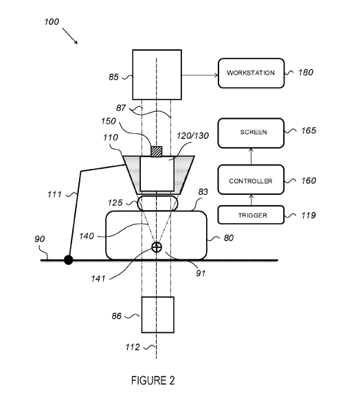
[0027] Figure 2 is a high level schematic illustration of an X-Ray guiding
apparatus 100 for an
image guided FUS treatment, according to some embodiments of the invention.
Apparatus 100
4
CA 02995114 2018-02-07
WO 2017/027577 PCT/US2016/046328
comprises an articulated arm 111 attached at its base to a procedure platform
90. In certain
embodiments, procedure platform 90 may comprise at least one of: an operating
room table, an
imaging table and a dedicated cart, wherein the cart is designed to carry the
electronics and other
device's accessories and wherein the cart wheels are designed to be locked to
avoid the cart's
movement. Apparatus 100 may further comprise a cradle 110 attached to the
distal end of arm 111.
Apparatus 100 may further comprise a coupling accessory 125 configured to
acoustically couple
transducer surface 120 to a surface 83 of a tissue 80.
[0028] Apparatus 100 may further comprise a FUS transducer 120 having a
central axis 112
configured to be affixed within cradle 110 and to transmit a FUS energy beam
140 to a treatment
location 141 within a patient. Apparatus 100 may further comprise a trigger
119, configured to
terminate the delivery of FUS energy 140. Apparatus 100 may further comprise a
controller 160
configured to control FUS energy delivery by therapeutic FUS transducer 120
which could be
controlled by user interface.. Apparatus 100 may further comprise a screen
165. Screen 165 provides
the physician technical information, such as, but not limited to, power level
chosen, sonication
duration, informative maintenance and service messages. Screen 165 may contain
the clinical
information which in essence the workstation 180 provides, and vice versa
workstation 180 may
provide the technical information. Apparatus 100 may further comprise an
aiming apparatus 130
configured to be affixed within cradle 110. In certain embodiments, cradle 110
may be further
configured such that both FUS transducer 120 and aiming apparatus 130 may be
affixed within it
simultaneously. In certain embodiments, an x-ray aim 150 may be attached to
the FUS transducer
120 to enable x-ray guidance. In certain embodiments, cradle 110 may comprise
several motion
degrees of freedom, such as, but not limited to, anterior-posterior (A-P),
superior-interior (S-I), left-
right (L-R). In certain embodiments, cradle 110 may be configured to
accommodate smoothly the
insertion, lock and release of the aiming apparatus and the FUS transducer. In
certain embodiments,
cradle 110, FUS transducer 120, aiming apparatus 130 and x-ray aim 150 are
built as a single unit.
100291 Apparatus 100 may further comprise an X-Ray imaging system, comprising
an X-Ray
intensifier 85 and an X-Ray source 86, wherein X-Ray intensifier 85 and X-ray
source 86 are
connected as an X-ray imaging system. In certain embodiments, the X-Ray
imaging system may be
configured to image a region 91 of tissue 80 that includes a treatment
location 141. In certain
embodiments, the X-ray imaging may be performed before and during the FUS
treatment. In certain
embodiments, apparatus 100 may configured to be compatible with at least one
of the following X-
ray types: a C-arm, an 0-arm, a G-arm and any other generic X-Ray type.
CA 02995114 2018-02-07
WO 2017/027577 PCT/US2016/046328
[0030] Apparatus 100 may further comprise a workstation 180 connected to X-ray
intensifier 85 of
the X-ray imaging system, wherein workstation 180 configured to derive an
imaging data from the
X-Ray imaging system. In certain embodiments, controller 160 and screen 165
may be combined
within workstation 180.
[0031] In certain embodiment, articulated arm 111 may be a mechanical arm or
robotic arm that is
attached to procedure platform 90. In certain embodiments, articulated arm 111
may comprise
several degrees of freedom, such as, but not limited to, anterior-posterior (A-
P), superior-interior (S-
I), left-right (L-R), and tilt such as, yaw, pitch and roll, to allow the
alignment of FUS energy beam
140 to a desired treatment location 141 within the patient. In certain
embodiments, articulated arm
111 may be adjusted manually and/or electronically and/or automatically to
align it in the predefined
orientation and position of cradle 110.
[0032] In certain embodiments, apparatus 100 may further comprise a manual or
controlled remote
maneuvering module configured to remotely control the position and the
orientation of articulated
arm 111. The maneuvering module may comprise at least one rod connected to
articulated arm 111
in a non-limiting manner, and a control unit configured to control the motion
of articulated arm 111.
The rod may be made of at least one of: a metal, a plastic, a wood and a
carbon. The remote control
of articulated arm 111 can minimize the exposure of the operating physician to
X-radiation. In
certain embodiments, the control unit of the maneuvering module may be
implemented within
controller 160 and/or workstation 180.
[0033] In certain embodiments, coupling accessory 125 is designed to mimic the
inner shape of FUS
transducer 120 to enhance the acoustic coupling quality and provide the
desired flexibility to
enhance the coupling with patient skin 83. In certain embodiments, coupling
accessory 125 may be a
balloon or membrane filled with fluid or gel. The balloon or membrane may be
affixed to cradle
110 using rubber and/or ring that secure coupling accessory 125 attached to
cradle 110 during the
procedure.
[0034] In certain embodiments, coupling accessory 125 may comprise a gel pad.
Gel pad 125 may
be designed to mimic the inner shape of FUS transducer 120 including its
margins in order to enable
angular maneuver flexibility. The margin may provide the operating physician
the possibility to
manipulate cradle 110 and FUS transducer 120 in different angular positions
without adversely
affecting the coupling between FUS transducer and gel pad 125. In certain
embodiments, gel pad
125 may be designed in a shape that wraps around cradle 110 in order to affix
gel pad 125 to cradle
6
CA 02995114 2018-02-07
WO 2017/027577 PCT/US2016/046328
110 during the insertion of FUS transducer 120. Gel pad 125 may also be
designed as a convex
shape on the side that is attached to patient skin 83. The convex shape may
provide the operating
physician the possibility to manipulate cradle 110 in different angular
position without affecting the
coupling between gel pad 125 and patient skin 83. In certain embodiments,
coupling accessory 125
may be at least one of: an optically transparent, an acoustically transparent
and radiologically
transparent. In certain embodiments, coupling accessory 125 may be designed to
guide the
positioning of the transducer 120 to a predefmed angle of penetration of the
acoustic beam 140 into
the tissue 80.
[0035] In certain embodiments, FUS transducer 120 may be configured to deliver
FUS energy 140
to different depths according to the position of treatment location 141 using
at least one of: different
sizes of coupling accessory 125 and / or by tuning phased array transducer
elements as electronic
steering.
[0036] In certain embodiments, FUS transducer 120 may be further configured to
project FUS beam
energy 140 in a focused manner onto treatment location 141 as the focal spot
location, utilizing
adjacent bone structures and avoiding damage to adjacent soft tissues. In
certain embodiments, FUS
transducer 120 may comprise at least one of: a single element or a phased
array of elements or two
or more annular elements. In certain embodiments, FUS transducer 120 may
comprise at least two
annular ring elements geometrically focused at a depth within a range 141A in
a closed environment
of treatment location 141 (see, e.g., Fig. 3B). The annular elements
arrangement of FUS transducer
120 allows locating the acoustic focus of FUS beam 140 either proximal or
distal to the geometric
focal depth by operating each of the at least two annular elements to vibrate
at different phase. This
allows a single FUS transducer 120 to mimic a series of transducers with the
same aperture size but
with different geometric focal lengths. This allows the operating physician to
adjust, during the
procedure, the depth of the acoustic focus of FUS beam 140 to match the depth
of treatment location
141, and thereby improve the efficacy of the treatment. In certain
embodiments, the different
annular elements of the transducer could be driven in slightly different
frequency (incoherent mode)
which results in continuous change of the relative phase between the elements
in order to create
elongated acoustic focus. In certain embodiments, at least one of the annular
ring elements of FUS
transducer 120 may be configured to be turned off in order to avoid from FUS
energy beam 140 to
hit vertebra bone protrusions or other acoustically absorbing structures in
the beam path which
should not be exposure to the high intensity acoustic energy. In certain
embodiments, central axis
112 of FUS transducer 120 may be tilted relatively to the patient back so that
energy beam 140 is
7
CA 02995114 2018-02-07
WO 2017/027577 PCMJS2016/046328
transmitted onto treatment location 141 on the vertebra at an angle to the
bone structure, thus
avoiding a situation where FUS energy 140 may be blocked (e.g., by the
vertebra protrusions and
lamina). Certain angles may be selected to allow the incidence angle with
respect to the bone surface
to be smaller than the refraction angle, such that most of FUS energy 140 is
absorbed by the bone
and not reflected. In certain embodiments, apparatus 100 and projected FUS
energy 140 may be
used to optimize the incidence angle of the acoustic energy with respect to
the bone to maximize
absorption of energy by the bone. When beam angle is perpendicular to the bone
the absorption of
acoustic energy by the bone is maximal.
[0037] Figure 3A is a high level schematic illustration of cradle 110. In
certain embodiments, cradle
110 is designed to have a geometrical conic shape such that the projections of
the cone boundaries
are consistent with FUS beams 140 generated by FUS transducer 120. In certain
embodiments, the
cone shape of cradle 110 is designed such that the lateral projected apex of
the cone (e.g., the
intersection point of the projections the cone boundaries) corresponds to the
focal depth of the FUS
energy beams 140. Accordingly, the conic shape of cradle 110 may be used as a
marker, visible on
the X-Ray image, in order to guide the focusing of FUS energy beam 140 onto
treatment location
141, as illustrated in Figure 3B. Figure 3B is a high level schematic
illustration of a lateral X-ray
image of cradle 110, according to some embodiments of the invention. In
certain embodiments,
workstation 180 may further comprise a software module configured to receive
the lateral X-ray
image of cradle 110, to send the lateral X-ray image of cradle 110 to screen
165 and, to recognize,
using image processing well known in the art, by means of at least one
computer processor, the
projections of the cone boundaries of cradle 110 and to display these
projections on the lateral X-ray
image of cradle 110. In the preferred embodiment, the intersection point of
the projections the cone
boundaries represents the lateral projected apex of the cone, which
corresponds to the focal depth of
the FUS energy beams 140. Accordingly, the lateral projected apex of the cone
may be used to assist
the operating physician in navigating FUS energy beam 140 accurately and
safely to treatment
location 141. The conical geometry of cradle 110 is invariant in wide range of
lateral projection
images of the lateral views. Accordingly, the cone shape including its apex
can be recovered from a
range of views. In certain embodiments, cradle 110 may comprise at least one
of: a radio opaque
material, a radiolucent material coated with radio opaque material and a semi
radio opaque material.
[0038] In certain embodiments, image guided interventional procedures, in
particular frameless
stereotactic procedures, involve a stereoscopic optical image sensor that
tracks object tagged with
special markers to aid registration and navigation of FUS energy beam 140 to a
target location 141.
8
CA 02995114 2018-02-07
WO 2017/027577 PCT/1.152016/046328
Such markers are typically large spheres that can be easily identified within
the field of view, or
encoded black and white barcode like labels that can also uniquely identify a
specific object and
track it within the field of view. Spheres are particularly popular because
its shape is almost
invariant to viewing angle transformations. In 3D imaging modalities like CT
or MR, markers are
one or two dimensional and are made of a radio opaque or magnetic material to
make them visible.
For X-Ray (fluoroscopy) guidance, 2D templates with radio opaque markers are
typically used for
registration with pre-operative 3D imaging data and tracking.
[0039] Figure 4A is a high level schematic illustration of an aiming apparatus
130 positioned in
cradle 110, according to some embodiments of the invention. In certain
embodiments, an aiming
apparatus 130 may comprise a mockup 115 configured to be positioned in cradle
110. In certain
embodiments, mockup 115 may comprise a transparent material (e.g., Perspex) to
allow the
operating physical to keep patient skin 83 in a field of view. In certain
embodiments, mockup 115
may comprise a radiolucent material (e.g., Perspex and Carbon Fibers) to
generate clear X-Ray
images of target location 141.
[0040] In certain embodiments, aiming apparatus 130 may further comprise at
least one optical
marker holder 113. In certain embodiments, optical marker holder 113 may
comprise at least one
laser pointer. In certain embodiments, at least one optical marker holder 113
may be aligned to
create a straight line along central axis 112 of FUS transducer 120 and cradle
110. In certain
embodiments, at least one optical marker holder 113 may be configured to
create additional lines to
verify the position of cradle 110 and FUS transducer 120 with respect to the
normal of the X-ray
imaging system field of view 85.
[0041] Figure 4B is a high level schematic illustration of mockup 115 and
optical marker holder
113 of aiming apparatus 130, according to some embodiments of the invention.
In certain
embodiments, aiming apparatus 130 may further comprise at least two x-ray
aiming markers 133,
134 positioned on the vertical axis of at least one optical marker holder 113.
In certain embodiments,
x-ray aiming markers 133, 134 may be rings. At least one x-ray aiming marker
133, 134 may
comprise at least one groove 133A. In certain embodiments, at least one of
mockup 115 and x-ray
aiming markers 133, 134 may be asymmetric, wherein the asymmetry may be
visible both optically
and on radiologically, enabling the operating physician to correlate both
views and conclude on
direction and angle of movement as needed to co-align cradle 110 with X-ray
intensifier 85 along
central axis 112.
9
CA 02995114 2018-02-07
WO 2017/027577 PCT/1JS2016/046328
[0042] In certain embodiments, at least one of mockup 115 and optical markers
holder 113, may
have at least one X-Ray fiducial marker to enable the finding of mockup 115
orientation in the X-
ray images. In certain embodiments, optical markers holder 113 may have
individual on and off
switches, affixed or placed adjacent to mockup 115.
[0043] Figures 5A-5B is a high level flowchart illustrating a method,
according to some
embodiments of the invention. At step 510, at least one radio opaque marker is
placed at center of
X-ray intensifier 85 (see, e.g., 70A in Figure 6A). At step 515, the patient
is positioned in a prone
position at procedure platform 90. After the patient is positioned on the
table, the relative height of
the table and C-Arm is adjusted so both the patient spine and the cradle can
be seen within the X-
Ray field of view. Once the height is set, it will remain locked throughout
the procedure. This
adjustment is done via lateral X-Ray image and manipulation of the table
height and C-Arm height.
[0044] At step 520, X-Ray arm 87 (see, e.g., Figure 2) is moved horizontally
to place radio opaque
marker 70A as seen in the X-Ray image to overlap treatment location 141 within
the patient (see,
e.g., 70A-2 in Figure 6A). In certain embodiments, X-Ray intensifier 85 may be
positioned in an
angle to the treatment location 141, to overlap the radio opaque marker 70A
onto treatment location
141. It is important to note that if an angle is set, it is done before step
520. This angle would be the
desired angle of view, which is also the angle of FUS energy penetration to
the patient body. At step
525, a radio opaque marker 70B is placed on patient's skin 83 in a specific
location that the
operating physician selects following verification of treatment location 141
using radio opaque
marker 70A-2 during an X-ray image by temporarily placing at least one
temporary marker 84 (e.g.,
tip of needle) on the patient skin 83 (see, e.g., Figure 6B). In certain
embodiments, marker 70B may
be only / also visual marker. This marker has no significant acoustic
absorption to avoid near field
heating and damage to the patient skin by the FUS energy.
[0045] At step 530, coupling accessory 125 is placed on skin 83 of the patient
above marker 70B, as
in step 525. At step 535, cradle 110 with mockup 115 is placed on coupling
accessory 125 (see e.g.,
Figure 6B).
[0046] At step 540, at least one optical marker holder 113 on mockup 115 is
turned on and cradle
110 is aligned using articulated arm 111 of apparatus 100 and pointing by co-
linear lasers to radio
opaque marker 70B on patient's skin 83 and radio opaque marker 70A on
intensifier 85. At step 545,
an X-Ray image is taken to verify the alignment of cradle 110 and mockup 115
to the normal of the
center of the X-ray imaging system field of view along axis 112. At step 550,
the verification of the
CA 02995114 2018-02-07
WO 2017/027577 PCT/US2016/046328
alignment is performed. If radio opaque markers 70A-2, 70B-1 on the X-Ray
image from step 545
are overlapped, it means that cradle 110 and mockup 115 are aligned with the
normal of the center
of the X-ray imaging system field of view along axis 112 (see, e.g., Figure
6C). If radio opaque
markers 70A-2, 70B-1 are not overlapped on the X-Ray image from step 545, the
step 535 should be
performed again. In certain embodiments, the alignment of cradle 110 and
mockup 115 with the
normal of the center of the X-ray imaging system field of view may be verified
also using at least
two x-ray aiming markers 133, 134 positioned on vertical axis of at least one
optical marker holder
113. Once cradle 110 and mockup 115 are aligned with the normal of the center
of the X-ray
imaging system field of view along axis 112, x-ray aiming markers 133, 134
will appear concentric
in the X-ray image from step 545 (see, e.g., Figure 7A). If x-ray aiming
markers 133, 134 are not
seem concentric in the X-Ray image from step 545 (see, e.g., Figure 7B), step
535 should be
repeated. A certain range of position and angular error of aiming apparatus
130 may be permitted.
An indication of the permitted error can be presented to the operating
physician by the shape and/or
size of x-ray aiming markers 133, 134, such as the gap between the aiming
markers diameters,
which must remain visible around inner x-ray aiming marker 133 to indicate
alignment within the
error limits. In certain embodiments the decision on the quality of alignment
of the cradle and
aiming apparatus, at this step, could be done based on optical markers alone
without the need for X-
Ray imaging.
[0047] In certain embodiments, the alignment of cradle 110 can be performed
based on depth
images produced by a depth camera located on cradle 110 or FUS transducer 120
facing intensifier
85. Cradle 110 may be aligned such that the flat face of intensifier 85 is
parallel to cradle 110
according to the depth image analysis, and the shape of intensifier 85 is
centered with the center of
cradle 110 or FUS transducer 120, such that cradle 110, intensifier 85 and
central axis 112 are
collinear. In certain embodiments, the alignment of cradle 110 can be
performed based on at least
two distance sensors, such as but not limited to ultrasonic, RF, IR or laser
sensors, located on cradle
110 or FUS transducer 120 facing intensifier 85. These sensors can measure the
distance from
intensifier 85 and indicate the alignment needed in order to bring cradle 110
to a parallel alignment
relative to intensifier 85 face. Complimentary to the distance sensors, a
camera located on cradle
110 or FUS transducer 120 facing intensifier 85 will produce an image of
intensifier 85 round shape
to indicate the position of cradle 110, relative to the intensifier 85, and
the direction to move cradle
110 in order to co-align central axis 112, intensifier 85 and cradle 110. In
certain embodiments,
alignment of cradle 110 can be performed based on at least two dual axis tilt-
meters or angulation
13.
CA 02995114 2018-02-07
WO 2017/027577 PCT/US2016/046328
sensors, located on cradle 110 or FUS transducer 120 and on intensifier 85.
These sensors can
measure the angle of cradle 110 or FUS transducer 120 and of intensifier 85
and indicate the
alignment needed in order to bring cradle 110 to a parallel alignment relative
to intensifier 85 face.
This could be done based on absolute angle measurements or following
calibration done at a
baseline parallel orientation. Complementary to the angle sensors, a camera
located on cradle 110
or FUS transducer 120 facing intensifier 85 will produce an image of
intensifier 85 round shape to
indicate the position of cradle 110, relative to intensifier 85, and the
direction to move cradle 110 in
order to co-align the central axis of intensifier 85 and cradle 110. The tilt-
meters or angulation
sensors can be wired or wireless and use any existing technology to measure
the required angle.
[0048] At step 555, C-Arm 87 of the X-Ray imaging system is tilted laterally,
preferably to an angle
perpendicular to cradle axis 112 to verify the depth of treatment location
141, using the FUS beam
path 140 recognized by the software module of workstation 180 (see, e.g.,
Figure 311). The tilting of
C-Arm 87 should be performed preferably on a single axis. When using other
types of imaging for
guidance, such as CT, Ultrasound and other, the location of the transducer
focus could be
extrapolated from the image. Once the treatment depth is verified, within the
applicable focus range,
C-Arm 87 should be moved back to its previous vertical position. C-Arm 87
should be re-positioned
in accordance with the angle of mockup 115, pointing optical markers holder
113 on radio opaque
markers 70A and 70B. In certain embodiments, an X-Ray image may be taken again
to verify the
alignment.
[0049] At step 560, mockup 115 is removed from cradle 110 and transducer 120
is inserted into
cradle 110. At step 565, an x-ray aim 150, is placed inside FUS transducer
120. At step 570, an X-
ray image is taken to verify that cradle 110 and FUS transducer 120 are
aligned with the normal of
the center of the X-ray imaging system field of view along axis 112, as in
step 550 using x-ray aim
150. At step 575, FUS acoustic energy beam 140 is deployed and the ablation of
target position 141
is performed. In certain embodiments, the FUS acoustic energy could be first
deployed at a low level
to verify targeting, per patient, feedback before deploying an ablation level
energy pulse.
[0050] Figure 8A-8B are high level schematic illustrations of optical marker
holder being located in
a different location, according to some embodiments of the invention. In these
embodiments of the
invention, since the laser beam originating from the optical marker 113 or
mirror 114 is aligned with
the central axis line of the C Arm 112, and the radio opaque marker in the
center of the intensifier
plate is adjusted to coincide with the treatment target on the X-ray image,
the use of a mockup 115 is
not required. Instead, an X-ray / optical aim attached directly to the FUS
transducer can be used.
12
CA 02995114 2018-02-07
WO 2017/027577 PCT/US2016/046328
[0051] The optical marker holder 113 (Figure 8A) or a mirror 114 (Figure 8B)
may be attached to
the center of C Arm (X-Ray) intensifier plate 85. The optical marker holder
113 or mirror 114 may
be designed to allow angular alignment relative to the intensifier plate,
either manually and/or
automatically, and to be aligned with the central axis 112 of the C Arm
(Figure 2) by projecting a
laser beam to the center of the C Arm source 86 (Figure 2). The optical marker
113 or mirror 114
may be attached to or consist of a radio opaque marker that is visible on X-
ray image. The optical
marker 113 or mirror 114 may be placed on the center of the radio opaque
marker as applicable. In
Figure 8A the mirror 114 has an angular alignment capability while the optical
marker 113 can be
adjusted to aim the center of this mirror.
[0052] Figure 9A-9B are a high level schematic illustrations of modified x-ray
aim 150 affixed in
FUS transducer 120, according to some embodiments of the invention. Modified x-
ray aim 150 may
be used as an optical aim and also an x-ray aim.
[0053] Modified x-ray aim 150, which is placed in the socket or recess of FUS
transducer 120 along
central axis 112 of the FUS transducer, may contain two or more x-ray aiming
markers, such as
rings 133, 134, that are placed along the vertical axis of the FUS transducer.
In order to align the
FUS transducer to point to the target, the optical marker needs to appear at
the center of the upper
and lower rings 133, 134. In order to verify that the FUS transducer is
aligned accurately to the C
Arm central axis 112, the radio opaque rings 133,134 need to appear concentric
on the X-ray image
(Figures 7A, Figure 10). If the rings do not seem concentric in the image
(Figure 7B) or the
physician identifies movement, the physician shall repeat the positioning
procedure.
[0054] A certain range of position and angular error of modified x-ray aim 150
may be permitted.
An indication of the permitted error can be presented to the physician by the
shape and/or size of the
x-ray aiming markers 133, 134, such as the gap between the ring diameters
(Figure 7A-7B), which
must remain visible around the inner ring 133 to indicate alignment within the
error limits.
[0055] Reference is now made to Figures 11A-11B, which is a schematic flow
diagram of a method
1100 for image guided focused ultrasound treatment to a patient, in some
embodiments of this
configuration.
[0056] At step 1110, a radio opaque marker may be placed at the center of the
X-ray intensifier
plate. An optical marker holder may then be placed at the center of the X-ray
intensifier as per step
1115, and aimed at the X-ray source.
[0057] At step 1120, the patient is positioned in a prone position at a
procedure platform 90. After
the patient is positioned on the table, the relative height of the table and C-
Arm is adjusted so that
both the patient spine and the cradle can be seen within the X-Ray field of
view. Once the height is
13
CA 02995114 2018-02-07
WO 2017/027577 PCT/US2016/046328
set, it will remain lock throughout the procedure. This adjustment is done via
lateral X-Ray image
and manipulation of the table height and C-Arm height.
[0058] At step 1125, X-ray arm 87 is moved horizontally to place the radio
opaque marker 70A as
seen in the X-ray image to overlap the treatment location 141 within the
patient (see, e.g., 70A-2 in
Figure 6A). In certain embodiments, X-Ray intensifier 85 may be positioned in
an angle to the
treatment location 141, to overlap the radio opaque marker 70A onto treatment
location 141. It is
important to note that, if an angle is set, it is done before step 520. This
angle would be the desired
angle of view, which is also the angle of FUS energy penetration to the
patient body.
[0059] At step 1135, coupling accessory 125 is placed on skin 83. At step
1140, the cradle 110 with
the FUS transducer 120 is placed on coupling accessory 125.At step 1145, the
modified x-ray aim
150 is placed inside the central hole of the FUS transducer 120.
[0060] At step 1150, the at least one optical marker holder (Figures 8A-8B) on
the X-ray intensifier
85 is turned on, and the alignment of the cradle is performed, using the laser
to point at the central
markers as per step 1155, one on the upper ring 133 of the modified x-ray aim
150 and the other at
the lower ring 134 of the modified x-ray aim 150 (Figure 9A). In case the
aiming markers 133, 134
appear concentric in the X-ray image, the cradle is aligned (Figure 6A). If
aiming markers 133, 134
are not seemed concentric in the X-ray image, step 1155 should be repeated. A
certain range of
position and angular error of the modified x-ray aim may be permitted. An
indication of the
permitted error can be presented to the physician by the shape and/or size of
the aiming markers
133, 134, such as the gap between the ring diameters (Figure 7A-7B), which
must remain visible
around the inner ring 133 to indicate alignment within the error limits. In
certain embodiments, the
decision on the quality of alignment of the cradle and aiming apparatus could
be done based on
optical markers alone without the need for X-Ray imaging.
[0061] At step 1170, the treatment depth should be verified. The X-ray arm
shall be tilted laterally,
preferably at 90 degrees to the Cradle axis 112 to verify the depth of the
treatment location, using
the imaging workstation beam path and focal point overlay (Fig 3B).
[0062] In case the treatment location depth is verified within the applicable
focus range, the
physician will deploy the acoustic energy, and ablate targeted tissue as per
step 1175. In certain
embodiments, the acoustic energy could be first deployed at a low level to
verify targeting per
patient feedback before deploying an ablation level energy pulse.
14
CA 02995114 2018-02-07
WO 2017/027577 PCT/US2016/046328
[0063] According to certain embodiments, the X-ray aim 150 and the aiming
apparatus 130 shape
may be designed in a manner that reduces the interference to the image
quality. Figures 12A-12G
are high level schematic illustrations of X-ray images of the FUS transducer
120 with various X-ray
aims 150 (12A-12C), of which Figure 12D-12G are high level schematic
illustrations of X-ray
images of aiming apparatus 130 at different designs, according to some
embodiments of the
invention. Image 12H shows as reference the transducer without any aim
inserted into it.
[0064] In all the X-ray aims presented, the design is optimized to minimize
artifacts by eliminating
non-aim related sharp interfaces between materials with different levels of
radio opaqueness to make
image as clear as possible. Similar effect, (to a bigger degree) can be seen
in the design of the
aiming apparatus, where Figure 12D shows a design with many artifacts, and
where Figure 12E
shows a clear design which is also optically transparent, as can be seen in
Figures 12F-12G.
[0065] In addition, the bottom of the X-ray aim 150 has a thick disk-shaped
plastic part which
increases the overall radio opaqueness of the aim and allows a more balanced
(in terms of gain and
image saturation), imaging of the anatomy through the FUS transducer 120
opening as seen in
Figures 12H-12G.
[0066] Figures 13A-13C are high level schematic illustrations of x-ray images
of the treatment
target with and without the FUS transducer in the cradle respectively,
according to some
embodiments of the invention. Figures 13A illustrate the A-P images of the FUS
transducer as
shown on the device workstation during the procedure.
[0067] After the positioning process is over and the cradle is aligned with
central axis 112 and fixed,
the workstation may identify the circular shape of the cradle in the image,
save it and use the clear
image of its inner area including the treatment target (Fig 13B) to replace
the dark area caused by
the radiopacity of the transducer (Fig 13A) using image processing, thereby
avoiding obstruction of
the patient anatomy. This produces a clear image of the treatment target with
the transducer inside
the cradle (Fig 13C) when ready for sonication. The physician may then observe
the image, which
shows now a radiologically "transparent transducer", which provides the
anatomical information that
was blocked by the opaque transducer. The importance of such image is to
assist the physician to
identify and verify the treatment location and alert in case of potential
patient movement. These
features are essential for the enhancement of the device safety profile and
efficacy outcome.
[0068] Another embodiment of this apparatus is using an ultrasound (US)
imaging probe instead of
using imaging of an X ray device, to view the treatment target and align the
FUS transducer to it.
Figure 14A is a schematic illustration of the US imaging probe mounted in the
center of the FUS
CA 02995114 2018-02-07
WO 2017/027577 PCT/US2016/046328
transducer. An alignment adaptor is used to align the US imaging probe to
conjoin with the
transducer central axis.
[0069] As the simultaneous operation of the imaging probe and transducer US
sonication
significantly degrades the quality of the ultrasound images and even
completely blocks the imaging
capabilities, an alternated pulsed method is described in Figure 14B. The PUS
energy will be pulsed
with short time cease periods in which an image without artifacts or
degradation would be captured
from the ultrasound imaging stream to be presented on the imaging workstation
until replaced by the
next non-distorted image, captured at the next energy cease time period. This
way the refresh rate of
the imaging would be lower but can still produce an image feedback during
sonication. The non-
distorted images can be identified using basic image processing techniques as
the predicted level of
image degradation is significant. Alternatively the pulse to create the
therapeutic sound wave may
be created in such a manner to minimize artifacts and degradation of
ultrasound image. It is
important to note that the uniqueness of the implementation above is that is
allows any generic
ultrasound imaging system with the required imaging characteristics for the
clinical indication to be
used, as is, without any need for modification or connection to a gate signal,
as guidance for a
Focused Ultrasound system.
[0070] In the above description, an embodiment is an example or implementation
of the invention.
The various appearances of "one embodiment", "an embodiment", "certain
embodiments" or "some
embodiments" do not necessarily all refer to the same embodiments. Although
various features of
the invention may be described in the context of a single embodiment, the
features may also be
provided separately or in any suitable combination. Conversely, although the
invention may be
described herein in the context of separate embodiments for clarity, the
invention may also be
implemented in a single embodiment. Certain embodiments of the invention may
include features
from different embodiments disclosed above, and certain embodiments may
incorporate elements
from other embodiments disclosed above. The disclosure of elements of the
invention in the context
of a specific embodiment is not to be taken as limiting their use in the
specific embodiment alone.
Furthermore, it is to be understood that the invention can be carried out or
practiced in various ways
and that the invention can be implemented in certain embodiments other than
the ones outlined in
the description above.
[0071] The invention is not limited to the diagrams or to the corresponding
descriptions. For
example, flow need not move through each illustrated box or state, or in
exactly the same order as
illustrated and described. Meanings of technical and scientific terms used
herein are to be commonly
16
CA 02995114 2018-02-07
WO 2017/027577 PCT/US2016/046328
understood as by one of ordinary skill in the art to which the invention
belongs, unless otherwise
defined. While the invention has been described with respect to a limited
number of embodiments,
these should not be construed as limitations on the scope of the invention,
but rather as
exemplifications of some of the preferred embodiments. Other possible
variations, modifications,
and applications are also within the scope of the invention. Accordingly, the
scope of the invention
should not be limited by what has thus far been described, but by the appended
claims and their
legal equivalents.
17