Note: Descriptions are shown in the official language in which they were submitted.
CATARACT PHACOEMULSIFICATION TIP
BACKGROUND
1. FIELD OF ART
[0001/2] This invention relates generally to the field of cataract surgery,
and particularly to a
phacoemulsification or aspiration tip used in cataract surgery.
2. DESCRIPTION OF THE RELATED ART
[0003] Cataract surgery involves removing the lens of a patient's eye that
has become
cloudy due to cataract foiniation, and replacing the lens with a clear
artificial lens. A
physician begins by making an incision in the eye to facilitate the insertion
of surgical
instruments. The physician uses surgical instruments such as choppers to break
a cataract
into smaller fragments so that a vacuum can aspirate the fragments to remove
them from the
eye via the incision. Once the fragments are removed, the physician inserts
the artificial lens
through the incision. Phacoemulsification is a type of cataract surgical
procedure that uses
ultrasound to emulsify the cataract. In particular, a physician inserts a
phacoemulsification
tip to the location of the cataract, and the phacoemulsification tip vibrates
at an ultrasonic
frequency to break down the cataract. The phacoemulsification tip includes a
lumen (a
hollow cavity) such that cataract fragments can be vacuumed out of the eye
through the
phacoemulsification tip. Phacoemulsification can also be completed without
ultrasound
using a phacoemulsification tip by mechanically breaking up the cataract and
aspirating
through the tip.
[0004] Existing phacoemulsification and other types of aspiration tips
often become
clogged with fragments of cataracts or other anatomical tissues that are too
large to pass
through the lumen of the tip, or become stuck to the tip. Thus, the physician
must dislodge
the blockages using irrigation, probing, or ultrasound power, which delays the
surgical
procedure. In some cases, fragments that are close in size to the diameter of
the lumen travel
slowly out of the tip. Thus, these phacoemulsification tips are inefficient at
removing
fragments from the eye and may require more ultrasound energy and power for a
vacuum to
aspirate the fragments. The application of ultrasound may cause damage to the
eye by killing
fragile non-reproducing endothelial cells of the cornea. Thus, it is desirable
to shorten the
- 1 -
Date Recue/Date Received 2023-01-23
duration of a phacoemulsification procedure by using phacoemulsification tips
that can
quickly remove the fragments from the eye and can do this without having
blockages.
SUMMARY
100051 A phacoemulsification or aspiration tip is designed for use in
cataract surgeries
such as phacoemulsification. The phacoemulsification tip (also referred to as
a phaco tip) can
reduce the duration of cataract surgeries because the phaco tip is designed to
reduce the
likelihood that cataract fragments from an eye of a patient will become
clogged inside the
phaco tip. Thus, the phaco tip can quickly remove fragments from the patient's
eye. For
example, the phaco tip has a lumen with variable sized diameters, which helps
filter out
larger cataract fragments that are more likely to clog the phaco tip. Further,
the phaco tip can
have a larger diameter at a bend to prevent blockages. The opening of the
phaco tip can have
a sharp edge to shear cataract fragments into smaller pieces that are less
likely to clog the
phaco tip. By preventing blockages using the phaco tip and reducing the
duration of cataract
surgeries, patients may recover faster from the cataract surgeries.
10005a1 Accordingly, in one aspect there is provided a phacoemulsification tip
for cataract
surgery comprising: an exterior wall and an interior wall forming a tubular
structure having a
length, the length including a first sub-length, a second sub-length, and a
third sub-length; a
lumen formed by the interior wall, comprising: a first section with a first
constant diameter
along the first sub-length; a second section adjacent to the first section,
the second section
having a second constant diameter along the second sub-length, the second
constant diameter
smaller than the first constant diameter; and a third section adjacent to the
second section, the
third section having a third constant diameter along the third sub-length, the
third constant
diameter smaller than the second constant diameter; and an opening into the
lumen, adjacent
to the third section, configured such that anatomical tissues from an eye of a
patient may
enter the lumen through the opening, wherein the exterior wall has a fourth
constant diameter
along the first sub-length, the second sub-length, and the third sub-length,
and wherein the
fourth constant diameter is greater than the first constant diameter.
- 2 -
Date Recue/Date Received 2023-01-23
BRIEF DESCRIPTION OF DRAWINGS
[0006] Figure (FIG.) lA shows a prior art phaco tip with a constant
diameter according to
one embodiment.
[0007] FIG. 1B shows a prior art phaco tip with a constant diameter and an
oblique
opening according to one embodiment.
[0008] FIG. 1C shows a prior art phaco tip with a flared opening according
to one
embodiment.
[0009] FIG. 2A shows a phaco tip with a tapered diameter at the opening
according to
one embodiment.
[0010] FIG. 2B shows another phaco tip with a tapered diameter at the
opening according
- 2a -
Date Recue/Date Received 2023-01-23
CA 02995336 2018-02-09
WO 2017/007655
PCT/US2016/040048
to one embodiment.
[0011] FIG. 3A shows a phaco tip with a constant outer diameter and a lumen
with a
tapered diameter at the opening according to one embodiment.
[0012] FIG. 3B shows another phaco tip with a constant outer diameter and a
lumen with
a tapered diameter at the opening according to one embodiment.
[0013] FIG. 4A shows a phaco tip with a smaller diameter opening according
to one
embodiment.
[0014] FIG. 4B shows a front view of the phaco tip shown in FIG. 4A
according to one
embodiment.
[0015] FIG. 5A shows a phaco tip with variable lumen diameters according to
one
embodiment.
[0016] FIG. 5B shows a side view of another phaco tip with variable lumen
diameters
according to one embodiment.
[0017] FIG. 5C shows a side view of yet another phaco tip with variable
lumen diameters
according to one embodiment.
[0018] FIG. 6A shows a side view of a phaco tip with a bend according to
one
embodiment.
[0019] FIG. 6B shows a side view of another phaco tip with a bend according
to one
embodiment.
[0020] FIG. 7 shows a phaco tip with a funnel opening according to one
embodiment.
[0021] The figures depict embodiments of the present invention for purposes
of
illustration only. One skilled in the art will readily recognize from the
following discussion
that alternative embodiments of the structures and methods illustrated herein
may be
employed without departing from the principles of the invention described
herein.
DETAILED DESCRIPTION
100221 Particular embodiments as described herein relate to
phacoemulsification tips,
which may also be referred to as phaco tips, phacoemulsification probes, phaco
probes,
phacoemulsification needles, phaco needles, vacuum tips, or aspiration tips.
The phaco tips
described herein may be used in surgical procedures with or without
ultrasound. For
example, in a phacoemulsification surgical procedure, the phaco tip is used
with ultrasound to
- 3 -
CA 02995336 2018-02-09
WO 2017/007655
PCT/US2016/040048
emulsify a cataract into smaller fragments. The cataract can also be
mechanically broken into
smaller fragments without using ultrasound and aspirated with an aspiration
tip. On the other
hand, in a laser cataract surgical procedure, the phaco tip is used without
ultrasound. Instead,
a laser is used to break down cataracts and the phaco tip helps remove the
resulting cataract
fragments via vacuum suction. The fragments of cataracts or fragments of other
anatomical
tissues (e.g., corneal tissue) that are produced during a surgical procedure
are referred to as
fragments herein. In some procedures, no energy is applied to the eye with the
tip, and in this
case the vacuum or aspiration tip only aspirates the fragments of the cataract
without also
performing an emulsification.
[0023] The figures are not necessarily drawn to scale. In particular,
certain features of
phaco tips have been enlarged for purposes of illustration and clarity. In
practice, the
diameter of the phaco tips described herein have a diameter of approximately
0.4 to 1.9
millimeters in the narrowest ranges and 0.5 to 2 millimeters in the widest
ranges. For
instance, the diameter of a phaco tip is, e.g., 0.7 millimeters, within the
narrowest range
toward the opening of the phaco tip and, e.g., 0.9 millimeters, within the
widest range toward
a proximal end, i.e., further away from the opening, of the phaco tip. The
diameter of the
opening of the phaco tip is approximately 0.4 to 1.9 millimeters or
approximately 0.6 to 1
millimeters. The thickness of the wall of the phaco tips is approximately 0.1
to 0.5
millimeters. The lumens of the phaco tips are typically in between 0.5 and 1.1
millimeters in
diameter.
I. PRIOR ART PHACO TIPS
[0024] FIG. lA shows a prior art phaco tip 100 with a constant diameter
according to one
embodiment. The phaco tip 100 includes an exterior wall 102 and an interior or
inner wall
103, which form a lumen 101 of the phaco tip 100. The lumen 101 also has a
constant
diameter. The opening 110 of the tip allows fragments 105 to enter the phaco
tip 100, e.g.,
via vacuum force. Further, the plane of the opening 110 is perpendicular to
the body of the
phaco tip 100. Fragments 105 that are similar to the size of the lumen 101 or
larger in size
than the lumen 101 may form a blockage in the lumen 101.
[0025] FIG. 1B shows a prior art phaco tip 120 with a constant diameter and
an oblique
opening 130 according to one embodiment. The phaco tip 120 is similar to the
phaco tip 100
shown in FIG. 1A. However, the phaco tip 120 has an opening 130 that is not
perpendicular,
i.e., oblique, to the body of the phaco tip 120.
- 4 -
CA 02995336 2018-02-09
WO 2017/007655
PCT/US2016/040048
[0026] FIG. 1C shows a prior art phaco tip 140 with a flared opening 150
according to
one embodiment. In particular, the diameter of the phaco tip 140 and the lumen
of the phaco
tip 140 gradually increase toward the flared opening 150. Compared to the
opening 110 of
the phaco tip 100 shown in FIG. 1A, the flared opening 150 has a larger
surface area. Thus,
the phaco tip 140 can vacuum larger sized fragments, relative to the phaco tip
100, but the
larger fragments tend to get stuck in the lumen of phaco tip 140 because the
lumen has a
smaller diameter than the opening 150.
II. NARROWING DIAMETER AT TIP OPENING
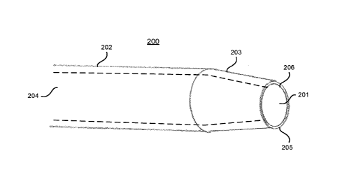
[0027] FIG. 2A shows a phaco tip 200 with a tapered diameter at the opening
201
according to one embodiment. In particular, the phaco tip 200 and the lumen
204 of the
phaco tip 200 include a straight section 202 and a tapered section 203. In the
straight section
202, the phaco tip 200 and the lumen 204 have a constant diameter. In the
tapered section
203, the diameter of the phaco tip 200 and the lumen 204 gradually decreases
toward the
opening 201. The thickness of the wall of the phaco tip 200 may either be
constant or differ
between the straight section 202 and the tapered section 203.
[0028] The opening 201 includes an outer edge 205 and an inner edge 206.
The outer
edge 205 and/or inner edge 206 may be sharp. Thus, the outer edge 205 and/or
inner edge
206 can shear fragments during a surgical procedure, including fragments that
become stuck
to the phaco tip 200 near the opening 201. As a result, there is a reduced
likelihood that
fragments will clog the phaco tip 200 (due to prevention of fragments staying
stuck near the
opening 201 and/or shearing fragments into smaller pieces), which helps reduce
the time
required to complete the surgical procedure. In some embodiments, the outer
edge 205
and/or the inner edge 206 are dull instead of sharp. Though the opening 201
shown in FIG.
2A is circular shaped, it should be noted that phaco tips may have openings of
different
shapes, e.g., elliptical, square, any other type of polygon, an arbitrary
shape, etc.
[0029] The tapered section 203 may be advantageous, e.g., because the
tapered section
203 reduces friction and resistance experienced by the phaco tip 200 during a
surgical
procedure. In particular, since the opening 201 at the end of the tapered
section 203 has a
smaller diameter than the straight section 202, the tapered section 203 is
less likely to contact
or move against surfaces inside an eye during the surgical procedure, compared
to a phaco tip
with a constant diameter. Further, a physician can more easily manipulate the
phaco tip 200
around more confined areas inside the eye. The tapered section 203 also
provides the
- 5 -
CA 02995336 2018-02-09
WO 2017/007655
PCT/US2016/040048
physician more visibility inside the eye when performing the surgical
procedure because the
smaller diameter of the tapered section 203 obscures less of the physician's
line of sight
relative to the straight section 202. Another advantage of the tapered section
203 is that it is
less likely that fragments will be clogged inside the lumen 204 because
fragments that enter
the lumen 204 through the opening 201 are smaller in diameter (at least in one
dimension)
than the diameter of the lumen 204 in the straight section 202.
[0030] FIG. 2B shows another phaco tip 220 with a tapered diameter at the
opening 221
according to one embodiment. The phaco tip 220 is substantially the same as
the phaco tip
200 shown in FIG. 2B, except that the phaco tip 220 has an oblique opening 221
while the
phaco tip 200 has an opening 201 perpendicular to the body of the phaco tip
200. Compared
to the opening 201 of the phaco tip 200, the oblique opening 221 has a larger
surface area.
Thus, the phaco tip 220 can vacuum larger sized fragments, relative to the
phaco tip 200. In
some embodiments, the angle of the oblique opening 221 is less than 60 degrees
(relative to a
plane perpendicular to the body of the phaco tip 220) such that the oblique
opening 221 does
not obscure the line of sight of a physician using the phaco tip 220 while
performing a
surgical procedure. Further, the oblique opening 221 provides a sharp end 222
of the phaco
tip 220, which helps a physician break down or manipulate (e.g., by impaling)
fragments.
III. NARROWING LuMEN DIAME l'ER AT TIP OPENING
[0031] FIG. 3A shows a phaco tip 300 with a constant outer diameter and a
lumen 303
with a tapered diameter at the opening 304 according to one embodiment.
Similar to the
phaco tip 200 shown in FIG. 2A, the lumen 303 includes a straight section 301
and a tapered
section 302. The diameter of the lumen 303 gradually decreases toward the
opening 304.
Unlike the phaco tip 200, the outer diameter of the phaco tip 300 remains
constant. Thus, the
thickness of the wall of the phaco tip 300 gradually increases toward the
opening 304.
[0032] FIG. 3B shows another phaco tip 320 with a constant outer diameter
and a lumen
303 with a tapered diameter at the opening 304 according to one embodiment.
The phaco tip
320 is similar to the phaco tip 300 shown in FIG. 3B, though the phaco tip 320
has an oblique
opening 321, while the phaco tip 300 has an opening 304 perpendicular to the
body of the
phaco tip 300. In addition, the oblique opening 321 has an inner edge 322 and
an outer edge
323, which may be sharp. The oblique opening 321 also has a raised edge 324
between the
inner edge 322 and the outer edge 323. The raised edge 324 can be a sharp
blade that helps
break down fragments.
- 6 -
CA 02995336 2018-02-09
WO 2017/007655
PCT/US2016/040048
[0033] FIG. 4A shows a phaco tip 400 with a smaller diameter opening 402
according to
one embodiment. In particular, the diameter of the opening 402 is smaller than
the diameter
of the lumen 401 of the phaco tip 400. An advantage of the smaller diameter
opening 402 is
that, similar to the phaco tip 200 shown in FIG. 2A, it is less likely for
fragments to become
clogged inside the phaco tip 400 because fragments that enter the lumen 401
through the
opening 402 are smaller in diameter (at least in one dimension) than the
diameter of the
lumen 401.
100341 FIG. 4B shows a front view of the phaco tip 400 shown in FIG. 4A
according to
one embodiment. FIG. 4B further illustrates that the diameter of the opening
402 is smaller
than the diameter of the lumen 401.
IV. VARIABLE LUMEN DIAME
[0035] FIG. 5A shows a phaco tip 500 with variable lumen diameters
according to one
embodiment. The phaco tip 500 includes an outer wall 502 with a constant
diameter and an
inner wall 504 that defines a lumen 516 of the phaco tip 500. The lumen 516
has a first
diameter 506, second diameter 508, and third diameter 510 along the length of
the phaco tip
500. In the example shown in FIG. 5A, the first diameter 506 is larger than
the second
diameter 508, which is larger than the third diameter 510. In other
embodiments, the lumen
516 may include additional, fewer, or different sized variable diameters,
e.g., four different
diameters where the first and third diameters are equal to each other, and
where the second
and fourth diameters are equal to each other. Each section of the lumen 516
corresponding to
the diameters 506, 508, and 510 are concentric to the phaco tip 500, e.g., the
cylinder defined
by the outer wall 502 with centerline 512 (e.g., a longitudinal axis). In
other embodiments,
one or more sections (with different diameters) of the lumen 516 may not
necessarily be
concentric to the phaco tip 500.
[0036] Typically, the diameter of the lumen 516 is smaller toward the
opening 514 of the
phaco tip 500. Thus, it is less likely for fragments to become clogged inside
the phaco tip
500 because fragments that enter the lumen 500 through the opening 514 are
smaller in
diameter (at least in one dimension) than one or more of the sections of the
lumen 516 with
variable diameters. In some embodiments, another advantage of the variable
diameter lumen
516 is that the edge between two sections of the lumen 516 with different
diameters, e.g.,
edge 518, is sharp. Thus, the sharp edge 518 can shear and chop fragments into
smaller
pieces as the fragments are vacuumed through the lumen 516 and/or prevent
fragments from
- 7 -
CA 02995336 2018-02-09
WO 2017/007655
PCT/US2016/040048
becoming stuck along the inner wall 504.
[0037] FIG. 5B shows a side view of another phaco tip 520 with variable
lumen
diameters according to one embodiment. Similar to the phaco tip 500 shown in
FIG. 5A, the
phaco tip 520 includes a lumen 532 with a first diameter 522, a second
diameter 524, and a
third diameter 526. The phaco tip 520 also includes a protrusion 530 at the
opening 528 of
the phaco tip 520. The protrusion 530 may have one or more sharp edges, e.g.,
to help break
fragments into smaller pieces or impale fragments.
[0038] FIG. 5C shows a side view of yet another phaco tip 540 with variable
lumen
diameters according to one embodiment. Similar to the phaco tip 500 shown in
FIG. 5A, the
phaco tip 540 includes a lumen 548 with a first diameter 542, a second
diameter 544, and a
third diameter 546. However, unlike the phaco tip 500, not all sections of the
lumen 548 are
concentric to the phaco tip 540. For example, section 558 of the lumen 548 is
not concentric
to the phaco tip 540, i.e., the center of the circle defined by the lumen 548
at section 558 does
not intersect the centerline 556 of the phaco tip 540 (e.g., a longitudinal
axis). The phaco tip
540 includes sharp edges 552 near the opening 550 of the phaco tip 540, e.g.,
to help break
fragments into smaller pieces as the fragments are vacuumed into the phaco tip
540. The
opening 550 also has a funnel shape that leads to the section of the lumen 548
that has the
third diameter 546.
V. PHACO TIPS WITH A BEND
[0039] FIG. 6A shows a side view of a phaco tip 600 with a bend according
to one
embodiment. The phaco tip 600 includes an exterior wall 602 and an inner wall
604, which
form the lumen 606 of the phaco tip 600. The lumen 606 has a constant diameter
608 in the
straight section 610 of the phaco tip 600. The diameter of the lumen 606
gradually increases
toward the opening 618 of the lumen 606. In particular, the diameter 614 of
the lumen 606 is
larger than the constant diameter 608. The diameter 614 occurs at the bend 612
of the lumen
606. Note that the center of the circular cross section of the lumen 606 at
the bend 612 does
not necessarily intersect with the centerline 616 of the straight section 610,
i.e., the diameter
of the lumen 606 does not need to remain symmetric about the centerline 616.
The increased
diameter 614 at the bend 612 reduces the likelihood that fragments will become
clogged in
the lumen 640. The diameter of the lumen 606 gradually decreases from the bend
612 to the
opening 618 to prevent larger fragments from entering the lumen 606 and
forming potential
blockages. In FIG. 6A, the diameter 608 of the lumen 606 at the opening 618 is
the same as
- 8 -
CA 02995336 2018-02-09
WO 2017/007655
PCT/US2016/040048
the diameter 608 in the straight section 610. In other embodiments, the
diameter at the
opening 618 may be smaller than the diameter 608.
[0040] The lumen 606 bends at an angle 0 relative to a line parallel to the
centerline 616
(e.g., a longitudinal axis). Typically, 0 is in between 0 and 30 degrees,
e.g., 12 degrees or 22
degrees. The lumen 606 includes a bevel 619 around the opening 618, e.g., an
oblique
opening. The angle of the bevel 619 may vary, e.g., 30 degrees of 45 degrees
relative to a
plane perpendicular to the opening 618. The bevel 619 may be advantageous,
e.g., because
the bevel 619 provides an angular opening 618 that allows fragments to more
easily enter the
lumen 606. In some embodiments, the bend 612 provides a larger field of view
of a patient's
eye for the physician using the phaco tip 600 while performing a surgical
procedure, e.g.,
because the physician can orient the phaco tip 600 to avoid obscuring certain
portions of the
eye. In some embodiments, the bevel 619 is rounded or smooth such that the
phaco tip 600 is
less likely to damage tissue of a patient's eye.
[0041] FIG. 6B shows a side view of another phaco tip 620 with a bend
according to one
embodiment. Similar to the phaco tip 600 shown in FIG. 6A, the phaco tip 620
includes a
lumen 640 that has a constant diameter 628 in the straight section 630 of the
phaco tip 620.
The lumen 640 also bends at an angle relative the straight section 630.
Further, the diameter
of the lumen 640 gradually increases toward the bend 632 of the lumen 640. In
particular,
the diameter 634 of the lumen 640 at the bend 632 is larger than the constant
diameter 628.
Unlike the lumen 606 shown in FIG. 6A, the diameter of the lumen 640, from the
straight
section 630 to the bend 632, remains symmetric about the centerline 636 of the
straight
section 630. In addition, the distal section 642 of the lumen 640 between the
bend 632 and
the opening 638 also has a constant diameter 628. In some embodiments, the
diameter of the
distal section 642 is not necessarily equal to the constant diameter 628 of
the straight section
630.
VI. PHACO TIP WITH A FUNNEL OPENING
[0042] FIG. 7 shows a phaco tip 700 with a funnel opening 714 according to
one
embodiment. The phaco tip 700 includes a lumen 702 with a constant diameter
704 along a
straight section 706 of the phaco tip 700. The diameter of the lumen 702
gradually decreases
until the section 708 of the phaco tip 700. The diameter 710 of the lumen 702
is smaller than
the constant diameter 704. From the section 708, the diameter of the lumen 702
gradually
increases, which forms the funnel opening 714. The diameter 716 of the funnel
opening 714
- 9 -
is larger than the diameter 710 and smaller than the constant diameter 704. In
other
embodiments, the diameter 716 is equal to the constant diameter 704. The
funnel opening
714 reduces the likelihood that fragments will become clogged inside the lumen
702 because
fragments that pass through the diameter 710 are smaller (at least in one
dimension) than the
constant diameter 704. In other words, the funnel opening 714 acts as a filter
to prevent
fragments that are too large from entering the straight section 706 of the
phaco tip 700. In
addition, a physician may be able to more easily aim or position the phaco tip
700 to aspirate
fragments inside a patient's eye because the funnel opening 714 has a larger
outer diameter
716.
100431 This presented invention can be used with or without an ultrasound
system.
Typically, a phaco tip includes a sleeve that surrounds the phaco tip and
provides an
irrigation solution, e.g., a balanced Salt Solution (BSS). The irrigation
solution enters the eye
through the sleeve to maintain intraocular pressure (e.g., to maintain the
anterior chamber
shape of the eye) and cool the phaco tip. The heat generated by ultrasound
during
phacoemulsification can burn the surrounding tissue if the phaco tip is not
cooled with the
irrigation solution. The phaco tips described herein reduce the likelihood
that fragments will
form blockages in the phaco tips, which allows irrigation solution to flow
through the phaco
tips to an operative site of a surgical procedure. In some embodiments, a
different source of
irrigation solution can be used, which may improve the fluid-dynamics of
cataract surgery.
Though not shown in the figures, the phaco tips described herein can be
connected to a pump
or other systems used to create vacuum or suction in the phaco tips to
aspirate fragments from
a patient's eye.
100441 Various different types of choppers can be used with any of the
phaco tip
embodiments of the invention described herein. In one embodiment, the chopper
is designed
to be used without application of ultrasound or laser energy to the eye, such
as the chopper
described in U.S. Provisional Application No. 62/190,190, filed on July 8,
2015. The
chopper has an angle at a bend of less than 90 degrees that allows for
posterior approach to a
cataract inside an eye and more efficient breakage such that energy is not
required in the eye
for further fragmentation or emulsification of the cataract.
VII. ALTERNATIVE CONSIDERATIONS
100451 Upon reading this disclosure, those of skill in the art will
appreciate still additional
- 10 -
Date Recue/Date Received 2023-01-23
CA 02995336 2018-02-09
WO 2017/007655
PCT/US2016/040048
alternative structural and functional designs through the disclosed principles
herein. Thus,
while particular embodiments and applications have been illustrated and
described, it is to be
understood that the disclosed embodiments are not limited to the precise
construction and
components disclosed herein. Various modifications, changes and variations,
which will be
apparent to those skilled in the art, may be made in the arrangement,
operation and details of
the apparatus disclosed herein without departing from the spirit and scope
defined in the
appended claims. In particular, features such as oblique openings (e.g., shown
in FIG. 1B),
tapered openings (e.g., shown in FIG. 2A), tapered lumens (e.g., shown in FIG.
3A),
narrower openings (e.g., shown in FIG. 4A), lumens with variable diameters
(e.g., shown in
FIGS. 5A-C), lumens with wider openings at bends (e.g., shown in FIGS. 6A-B),
or funnel
openings (e.g., shown in FIG. 7), may be used in any of the phaco tip
embodiments or
designs described herein.
[0046] As used herein any reference to "one embodiment" or "an embodiment"
means
that a particular element, feature, structure, or characteristic described in
connection with the
embodiment is included in at least one embodiment. The appearances of the
phrase "in one
embodiment" in various places in the specification are not necessarily all
referring to the
same embodiment.
[0047] As used herein, the terms "comprises," "comprising," "includes,"
"including,"
"has," "having" or any other variation thereof, are intended to cover a non-
exclusive
inclusion. For example, a process, method, article, or apparatus that
comprises a list of
elements is not necessarily limited to only those elements but may include
other elements not
expressly listed or inherent to such process, method, article, or apparatus.
Further, unless
expressly stated to the contrary, "or" refers to an inclusive or and not to an
exclusive or. For
example, a condition A or B is satisfied by any one of the following: A is
true (or present)
and B is false (or not present), A is false (or not present) and B is true (or
present), and both
A and B are true (or present).
[0048] In addition, use of the "a" or "an" are employed to describe
elements and
components of the embodiments herein. This is done merely for convenience and
to give a
general sense of the invention. This description should be read to include one
or at least one
and the singular also includes the plural unless it is obvious that it is
meant otherwise.
-11-