Note: Descriptions are shown in the official language in which they were submitted.
CA 02995719 2018-02-15
WO 2016/026038
PCT/CA2015/050779
SYSTEM AND METHOD FOR EMBEDDED IMAGES IN LARGE FIELD-OF-
VIEW MICROSCOPIC SCANS
BACKGROUND
[0001] In many clinical studies, the acquisition of large-field-of-view
microscopic
images is extremely beneficial. Many techniques are proposed using automated
microscopes [1] or manual stage microscopes [2]. In this document, a scan is
referred to as
a large image covering a large field-of-view of a specimen. A scan may be
composed of
many smaller images, such as in Figure 1A, or a unified image of a specimen
such as in
Figure 1B. In Figure 1A, the smaller images are referred to as keyframes. The
relative
locations of the keyframes are known a-priori. This may be performed using
automatic
scan system or image-based techniques [2]. Without loss of generality, for the
rest of this
document, it is assumed that a scan is composed of many keyframes with the
same size.
BRIEF DESCRIPTION OF THE DRAWINGS
[0002] Embodiments of the present disclosure will now be described, by
way of
example only, with reference to the attached Figures.
[0003] Fig. lA is an illustration of a scan of a specimen comprising
many smaller
images;
[0004] Fig. 1B is an illustration of a scan of a specimen comprising a
single
unified image;
[0005] Fig. 2 is an illustration of a scan having embedded scans;
[0006] Fig. 3 is a schematic diagram of a system, in accordance with an
embodiment of the present disclosure;
[0007] Fig. 4A is an illustration of a first scan with a new image captured
by an
objective with a magnification smaller than that of the original scan;
[0008] Fig. 4B is an illustration of a first scan with a new image
capture by an
objective with a magnification larger than that of the original scan;
[0009] Fig. 5 is a flowchart diagram illustrating a process of
localizing an image,
in accordance with an embodiment of the present disclosure;
1
CA 02995719 2018-02-15
WO 2016/026038
PCT/CA2015/050779
[0010] Fig. 6 is a flowchart diagram illustrating the process for
determining the
localization information for a frame, in accordance with an embodiment of the
present
disclosure;
[0011] Fig. 7 is a schematic representation of the selection of key
frames in
various iterations of an exhaustive search, in accordance with an embodiment
of the
present disclosure;
[0012] Fig. 8 is a schematic representation of the process of
correcting relative
magnification;
[0013] Figs. 9A and 9B illustrate a user interface of multi-objective
scans, in
accordance with an embodiment of the present disclosure;
[0014] Fig. 10 is a schematic diagram illustrating a system setup for
recording Z-
stack manually, in accordance with an embodiment of the present disclosure;
[0015] Fig. 11 is an illustration of a user interface for viewing a Z-
stack, in
accordance with an embodiment of the present disclosure;
[0016] Fig. 12 is an illustration of a user interface for viewing a scan,
in
accordance with an embodiment of the present disclosure; and
[0017] Fig. 13 is an illustration of a user interface for viewing a
scan showing the
location of Z-stacks, in accordance with an embodiment of the present
disclosure.
DETAILED DESCRIPTION
INTRODUCTION
Problem definition
[0018] Given the common use case, it can be beneficial to a technologist or
a
clinician to observe some part of the specimen in more resolution or explore a
portion in z-
axis. In other words, it would be beneficial to embed other images which are
acquired with
different magnification or depth into the main scan. The images are either a
collection of
images acquired by moving the stage spatially, or acquired by changing the
focus of the
microscope. For the rest of this document, the former is referred to as multi-
objective
scanning while the latter is referred to as Z-stack. Note that a prerequisite
for such features
are accurate localization of the images that are acquired by any arbitrary
objectives within
2
CA 02995719 2018-02-15
WO 2016/026038
PCT/CA2015/050779
a large field-of-view scan. Figure 2 shows a scan with embedded scan captured
with high
magnified objective and a Z-stack. As shown in Figure 2, an original scan may
contain
another scan which is captured with different objective magnification, or may
have Z-
stacks, which are images captured with different focus/depth.
[0019] The above mentioned features, together with the live acquisition of
the
images, are provided in microscopes with a motorized stage but are not
available in
manual stage microscopes. Some embodiments described herein rate to a system
that
collectively provides these features.
[0020] In the present disclosure, it is assumed that the stream of
images are
acquired from a camera mounted on a manual microscope, providing a live
digital image
of the specimen. The latest digital image of the camera is referred to as the
current image
frame hereafter. The user has control over the manual stage and the focusing
of the
microscope. The user notifies the system when he/she switches the objective.
The system
then automatically localizes the live images within the already captured scan.
The user
may also notify the system when he/she intends to change the focus to acquire
Z-stacks.
Figure 3 shows the overview of the system hardware. As shown in Figure 3, a
camera is
mounted on a manual microscope which streams real-time images to a processing
computer. Images are processed in real-time and the visualization is performed
on the
display.
[0021] This disclosure will cover three aspects of the embodiments
disclosed
herein. First, the localization of an image within a scan, which is presented
in the "Multi-
objective localization" section. Second is the proposed system for stitching
and embedding
such scans at different objectives within the original scan, which is
presented in the
"Multi-objective scanning" section. The third, is the proposed system for
storing and
managing Z-stacks embedded within a scan, which is illustrated in the "Z-
stack" section.
MULTI-OBJECTIVE LOCALIZATION
[0022] Given a scan, the multi-objective localization is defined as the
localization
of a stream of images captured by an objective different from the objective
that is used in
the reconstruction of the scan. Figures 4A and 4B show the two different
scenarios, where
the image (shown with stripes) is captured using a larger magnification or a
smaller
3
CA 02995719 2018-02-15
WO 2016/026038
PCT/CA2015/050779
magnification. In Figure 4A, the current image frame is captured by an
objective with
magnification smaller than that of the original scan. In Figure 4B, the
current image frame
is captured by an objective with magnification larger than that of the
original scan. The
image may have overlap with one or more keyframes of the scan. The image
originally has
the size (5"sv) , but can be scaled by relative magnification to the original
scan. For
example, if the original scan is captured by a 10x objective and the current
image frame is
captured by a 40x objective, the image can be scaled by a factor of 0.25. The
location of
the current frame which is captured at time t , with respect to the original
scan, is
represented by Pt .
[0023] The localization is performed via a series of image matching. In the
next section the matching process is explained.
Registration of two frames
Feature detection
[0024] Feature detection is performed on the current image frame. The
features are
used for image registration (linking). The result of the feature detection is
a set of features,
where each may include a set of properties:
= Position in image coordinate (x, y);
= Geometrical properties such as scale and orientation;
= Image properties that are used to describe the image pattern around the
feature.
Matching two frames
[0025] Matching of frames is performed by matching their features. Many
techniques are proposed for this purpose [2] [3]. Assuming that a long list of
features is
detected in both images, this part contains two steps (the frames are referred
to as
reference and matching frames):
1. For each feature in the reference frame, the closest feature in the
matching
frame is found. The closest feature should have the most similar properties.
2. A displacement is collectively found based on the matched features.
4
CA 02995719 2018-02-15
WO 2016/026038
PCT/CA2015/050779
Definition of tracking, linking, and localization
[0026] Given the stream of images, the term tracking in this document
refers to the
matching of the current frame to the previous frame. Assuming that the
matching results in
a displacement of d , the location of the current frame is estimated as P: =
Pr-i d . The
current frame is called tracked if it is successfully matched to the previous
frame.
[0027] The term "linking" as used herein refers to the matching of the
current
image frame to a keyframe. The current image frame is called linked, if it is
successfully
matched to at least one of the keyframes.
[0028] The term "localization" as used herein refers to determining
whether the
current frame location is correct based on the tracking and linking. The
current image
frame is called localized, if its location in the scan is correct.
Localization process
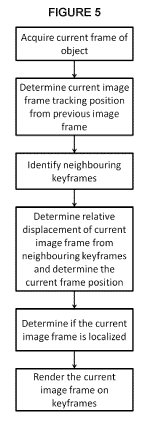
[0029] The localization process, which is a process of the localization
of the
current image frame within keyframes that are acquired with different
objective
magnification, is shown in Figure 5 and is outlined as follows:
1. The current image frame is preprocessed and the features are extracted.
2. The position, [(= Y,) and scale 5: of features in the new frame are scaled
according to the difference in magnification of this frame and keyframes.
Assuming that the new frame has a magnification of in and the keyframes have a
magnification of M k
Therefore, the position and scale are scaled as follows:
m
_
3. ()= , ¨ x 1..
= - X S D estimate
- -
and
4. Linking. Next, the current image frame is matched to the neighbouring
keyframes
to correct its location and remove the possibility of accumulation of
inaccurate
matching resulted from Tracking.
[0030] The linking may not always be successful in the case of multi-
objective
5
CA 02995719 2018-02-15
WO 2016/026038
PCT/CA2015/050779
matching. Therefore the tracking information is combined with the linking
information to determine the location of the current frame. The process is
described in
the next section.
Combining the tracking and linking for accurate localization
[0031] The
position of the current image frame is estimated based on the linking
and tracking information. The current image frame is localized if it is linked
or tracked
and the previous image frame is localized. The logic is shown in Figure 6,
which is a
diagram describing the combination of the tracking and linking information for
accurate
localization of the current image frame. Differences in the optical properties
of objectives
may introduce changes in the image. These changes may cause matching of images
between objectives to fail. To improve robustness of the localization
algorithm, tracking
can be added to the algorithm as an alternate method for image localization.
Exhaustive search
[0032] If the
current image frame is not localized in the previous step, the
algorithm enters the exhaustive search state. At this step, keyframes are
sorted according
to their distance to the current image frame. As opposed to the previous step,
not all but
only a portion of these keyframes are linked to the frame at this point. This
is performed to
prevent exhaustive search from hindering the real-time performance of the
system.
Assuming that keyframes are sorted based on their distance to the current
image frame:
ICo, K1, -= The first
time at the exhaustive search, only the first M elements
K0..., K11_1 are processed. If the linking is not successful, for the next
frame, the second
m elements Km. Kzm-i. are processed (see Figure 7) and so on. Figure 7
illustrates
exhaustive search in case the current image frame is not localized within its
neighboring
keyframes; all the keyframes are sorted with respect to their distance to the
current image
frame and, at each iteration, only a portion of keyframes are examined for
localization of
the current image frame. Since the current image frame is updated at each
iteration, the
reference frame does not remain the same. However, one can assume that they
don't move
as much since the exhaustive search can visit all the keyframes in a fraction
of a second.
6
CA 02995719 2018-02-15
WO 2016/026038 PCT/CA2015/050779
Correction of the relative magnification
[0033] The magnification indicated on an objective may not be exactly
true. For
example a 10x objective may have a magnification of 10.01. A true
magnification can be
achieved using physical calibration. However in absence of such information,
one can find
the "relative" magnification between different objectives in the process of
image matching.
Assuming that some of the features in the keyframe and the current image are
correctly
matched to each other. Note that each feature has a position and can be
represented as a
point. Matched features in the reference frame can be listed as ri.= = rti ,
and matched
features in the matching frame can be listed as "' --1/1/1. The features with
the same
indices are matched, i.e. r: corresponds to 7": . Figure 8 shows such
correspondences and
also our previous approach to find the displacement between the two frames. As
shown in
Figure 8, which illustrates correction of the relative magnification, this can
be performed
via Procrustes analysis [4] that is performed on the matched features of the
current image
frame and the matching keyframe. Although the frames are almost matched after
displacement, the relative scale still exists between two frames. Therefore,
the relative
scale between two frames should be recalculated properly. Assuming that each
point has
r v
both x and y components: = , . Initially the average of all components
is
calculated:
E v,-, l'rr,
= = =
11
/1
[0034] Next, the scale for each point set is calculated:
[(v:,. - Tr) + E(v:,.. - T.) [(Y:
S r; I =
= fl -
(Sr
.!
[0035] The true relative magnification is then calculated as
where S is the relative magnification which was calculated originally based on
a priori
knowledge of the objectives. For example for 10x and 40x objectives, S = 0.25
7
CA 02995719 2018-02-15
WO 2016/026038
PCT/CA2015/050779
MULTI-OBJECTIVE SCANNING
Linking multiple scans
[0036] The user can select to stitch the images captured with a
different objective
and create another scan. Many techniques are proposed for such stitching [2].
In this
situation, a parent-child relation is established between this scan and the
original scan. A
link is set up between two scans to relate the corresponding coordinate
spaces. Assuming
that n frames are captured at the child scan. The stitching of these frames
results in the
positions of (Iv Yi). = (µ Yn) . Also, by using multi-objective localization,
the positions
of these frames within the parent scan are found: (V1, 111). (Vn, 1713 . To
relate these
coordinate spaces, one can use Procrustes analysis [4], where the unknowns are
the
translation and the scale.
User interface
[0037] The user may switch to a different objective at any time. The
user may also
start scanning at the selected objective. At this point the previous scan
which was
captured by the parent objective, is shown semi-transparently in the
background. This will
provide a visual aid for the user to relate two scans to each other. After
finishing the scan,
the user may switch back to the parent objective. At this point, the scan
which was
captured by the different objective, is shown semi-transparent and is
clickable. By user
clicks, the scan view switches to make the child scan active. That is, the 40x
scan
becomes opaque while the 10x scan becomes semi-transparent. Figures 9A and 9B
show
the overview of the user interface of the multi-objective scan, in which the
user may
switch between objectives and modify each scan separately while the other scan
is visible
semi-transparently.
Recording the multi-objective scan
[0038] A parent scan and its child scans are saved using their own file
format. The
child scans can be linked to the parent scan using an additional file.
Information such as
the path to the child scan file and location of the child scan within the
parent scan is
recorded in this file.
8
CA 02995719 2018-02-15
WO 2016/026038
PCT/CA2015/050779
Z-STACK
[0039] The digitization of samples in microscopy is usually achieved by
capturing
a large 2D scan. While this solution satisfies most situations, it only allows
to capture a
narrow depth of field, stripping away valuable information for the analysis of
certain
samples. A solution to this problem is the capture of Z-stacks. A Z-stack is
defined as a
stack of images representing the same specimen at different focal planes. In
theory, one
could capture a Z-stack for an entire sample leading to a stack of scans.
However, due to
the high resolution of the images composing a scan, a stack of scans becomes
unpractical
as it necessitates too much memory space.
[0040] This section proposes a method for reducing the memory usage by
recording Z-stacks covering a limited area of a specimen and attaching the
stacks to a scan
covering the entire sample. This solution has the advantage of providing
enough depth
information of a scan for analysis while keeping the memory usage low.
[0041] The section is divided into two parts. The workflow for
recording and
visualizing a Z-stack using a microscope is described in the first section and
the
attachment of the Z-stacks to a scan is explained in the second section.
9
CA 02995719 2018-02-15
WO 2016/026038
PCT/CA2015/050779
Z-stack Recording
Hardware setup
[0042] As shown in Figure 10, a Z-stack can be recorded using a digital
video
camera that is mounted on a microscope. In Figure 10, the system setup
comprises a
microscope on which is mounted a camera that captures images while the
microscope
stage is moved at different depths. While the camera is capturing a specimen
placed under
the microscope at fixed time interval, one can move the microscope stage so
that the
specimen is viewed at different depths. As a result, the images captured by
the camera can
be regrouped to form a stack of images representing the same location of a
specimen at a
range of depth only limited by the amount of stage movement occurred during
the
recording. Note that this method is not necessarily limited to the analysis of
depth
information and can be used to record a region of a sample by moving the stage
laterally/spatially during the recording.
Z-stack Visualization
[0043] Z-stacks are visualized one frame at a time as shown in Figure
11, which
illustrates a user interface for viewing a Z-stack. There are different ways
to go through a
Z-stack. The first one is to play the Z-stack from beginning to end at the
same speed (or a
factor of the speed) as the recording speed in a similar way as playing a
video. The second
method is to scroll through the frames using the mouse's scroll wheel or
dragging the
current frame cursor with the mouse, allowing one to go either backward or
forward along
the Z-stack. The final method is to select any random frame to view within the
stack using
a slider as shown in Figure 11.
[0044] Note that the user interface may have other features such as
trimming
the beginning and the end of a Z-stack. For example, the user who manually
records a
Z-stack clicks on the "Record" button in the software, takes some time to get
ready on
the user's microscope, and then drives the focus knob or stage to capture the
focal planes
and regions of interest. The captured frames in between these operations can
be trimmed
to reduce the size of a Z-stack.
[0045] Since a Z-stack can use a lot of memory space, it is difficult to
keep in
memory the entire stack that is being visualized. To accommodate this problem,
it is
possible to keep the Z-stack in a file saved on the hard drive and only load
the frame that
CA 02995719 2018-02-15
WO 2016/026038
PCT/CA2015/050779
is currently being displayed. This, however, assumes that the file format used
for saving Z-
Stacks allows random access of frames within the stack. To resolve this issue,
a saving
technique is proposed in the next section.
Saving a Z-stack
[0046] The Z-stacks containing high resolution images can become costly in
terms
of memory space. Compressing the images of the stack then becomes an important
step in
the recording of a Z-stack. As mentioned in the previous section, the images
of a Z-stack
may be visualized in any order directly from a file. The compression algorithm
permits the
decoding of random frames within a Z-stack. According, use of a standard video
compression process is generally note suitable as such a process would
compress images
in a temporal manner, leading to the necessary dependency between neighbour
images in
the Z-stack. Although video compression algorithms offer great compression
ratios, the
decompression of any image n in a Z-stack would require decompression of the
previous
image n-1 which in turn would require the decompression of the previous images
until the
first frame of the Z-stack is reached. This method of decompression is only
appropriate
when reading a video in order from beginning to end. It is however not
suitable for
random access of frames throughout the Z-stack. One solution is to compress
the frames of
a Z-stack individually as separate images. This may not offer the best
compression ratio
but it satisfies the requirements for reading a Z-stack. These compressed
images can then
be saved in a multi-layered image file format such as TIFF.
Attaching a Z-stack to a scan
[0047] A Z-stack alone may not provide enough information for analyzing
a
specimen as it covers a limited region of the sample. However, it becomes a
powerful
feature when localized within a scan. This part proposes an apparatus for
embedding Z-
stacks into a sample scan recorded manually using a microscope and a digital
video
camera.
Z-stack Recording
[0048] This section assumes we have a system for manually scanning a sample
using a microscope and a digital camera. The user interface for such system
comprises a
view of the scan as well as the position of the current image frame captured
by the
11
CA 02995719 2018-02-15
WO 2016/026038
PCT/CA2015/050779
camera as shown in Figure 12. The box at the center shows the current position
of the
camera relative to the scan.
[0049] When a region of interest is found, the user can initiate the
recording of a
new Z-stack by clicking a button as described in "Z-stack Recording" section.
When
recorded, the position of the Z-stack is known using the localization
algorithm of the
manual scan system. Note that since the user is free to move the microscope
stage
laterally, the system sets the position of the entire Z-stack to the location
of the first frame
recorded. A link is established between the Z-stack and the scan by annotating
the latter
with a rectangle. The rectangle position and size matches the one of the Z-
stack and can be
clicked to open the Z-stack viewer described in "Z-stack Visualization"
section (see Figure
13). In Figure 13, the Z-stacks are localized in the scan and shown as an
outline rectangle
with a semi-transparent image. These rectangles are clickable, which opens
another
window for viewing the Z-stacks.
[0050] The localization algorithm described in "Multi-objective
localization"
section only provides an estimate of the position of the current frame when
recording a Z-
stack using an objective lens with a different magnification than the one used
for scanning.
This estimate cannot guarantee the accuracy of the position of the recorded Z-
stacks. A
solution to this issue is to allow the user to refine the position of a Z-
stack relative to a
scan by dragging the rectangle annotation representing the Z-stack within the
scan using
the mouse. Visual feedbacks can be provided to the user by drawing one of the
images of
the Z-stack semitransparent inside the rectangle annotation. This is
beneficial as one could
see the overlap between the Z-stack and the scan but it assumes that the frame
drawn
inside the rectangle is recorded at the same focal plane as the scan. There
are several ways
to ensure the chosen frame is as described. One can select the sharpest frame
within the Z-
stack to best match the scan, if the scan is carefully composed of sharp
images. Another
possibility is to always select the first frame recorded but it is assumed
that the Z-stack is
recorded starting from the same focal plane as the scan.
This is an acceptable assumption as the user will initiate recording once
he/she finds a
region of interest to record. The region can only be found by browsing the
scan, which is
moving the camera while staying at the same focal plane as the scan.
Saving the link between a Z-stack and a scan
12
CA 02995719 2018-02-15
WO 2016/026038
PCT/CA2015/050779
[0051] Both the scans and the Z-stacks are saved using their own file
format. This
structure should be kept for flexibility. Therefore, an additional file should
be created to
store the relationship between a scan and the Z-stacks recorded into that
scan. This file
should contain the path names to the files of the scan and the individual Z-
stacks. It should
also contain the position of the Z-stacks relative to the scan.
[0052] In the preceding description, for purposes of explanation,
numerous details
are set forth in order to provide a thorough understanding of the embodiments.
However, it
will be apparent to one skilled in the art that these specific details are not
required. In other
instances, well-known electrical structures and circuits are shown in block
diagram form
in order not to obscure the understanding. For example, specific details are
not provided as
to whether the embodiments described herein are implemented as a software
routine,
hardware circuit, firmware, or a combination thereof
[0053] Embodiments of the disclosure can be represented as a computer
program
product stored in a machine-readable medium (also referred to as a computer-
readable
medium, a processor-readable medium, or a computer usable medium having a
computer-
readable program code embodied therein). The machine-readable medium can be
any
suitable tangible, non-transitory medium, including magnetic, optical, or
electrical storage
medium including a diskette, compact disk read only memory (CD-ROM), memory
device
(volatile or non-volatile), or similar storage mechanism. The machine-readable
medium
can contain various sets of instructions, code sequences, configuration
information, or
other data, which, when executed, cause a processor to perform steps in a
method
according to an embodiment of the disclosure. Those of ordinary skill in the
art will
appreciate that other instructions and operations necessary to implement the
described
implementations can also be stored on the machine-readable medium. The
instructions
stored on the machine-readable medium can be executed by a processor or other
suitable
processing device, and can interface with circuitry to perform the described
tasks.
[0054] The above-described embodiments are intended to be examples
only.
Alterations, modifications and variations can be effected to the particular
embodiments by
those of skill in the art. The scope of the claims should not be limited by
the particular
embodiments set forth herein, but should be construed in a manner consistent
with the
specification as a whole.
13
CA 02995719 2018-02-15
WO 2016/026038
PCT/CA2015/050779
REFERENCES
The following references are incorporated herein by reference in their
entirety:
[1]"BZ-9000 All-in-one Fluorescence Microscope," Keyence Corporation,
[Online].
Available: hap ://www.keyence . corn/products/microscope/fluorescence -micro
scope/bz-
9000/index j sp.
[2]H. a. L. L. a. C. B. a. A. M. a. L. S. LO, "Apparatus and method for
digital microscopy
imaging". 2013.
[3]D. G. Lowe, "Object recognition from local scale-invariant features," in
The
proceedings of the seventh IEEE international conference on Computer vision,
1999.
[4] G. D. J.C. Gower, Procrustes Problems, Oxford University Press, 2004.
14