Note: Descriptions are shown in the official language in which they were submitted.
1
Description
Title of Invention: MODIFIED ONCOLYTIC VACCINIA VIRUSES
EXPRESSING A CYTOKINE AND A CARBOXYLESTERASE AND METHODS
OF USE THEREOF
Technical Field
[1] This application claims the benefit of U.S. Provisional Patent
Application No. 62/215,651,
filed September 8, 2015.
[2] The present disclosure relates generally to compositions and methods
related to oncolytic
vaccinia viruses that have been modified to express a cytokine and a car-
boxylesterase enzyme
and that preferably do not express an active thymidine kinase, optionally in
combinations with a
cancer co-drug, preferably a topoisomerase inhibitor.
Background Art
131 Normal tissue homeostasis is a highly regulated process of cell
proliferation and cell death.
An imbalance of either cell proliferation or cell death can develop into a
cancerous state. For
example, cervical, kidney, lung, pancreatic, colorectal, and brain cancer are
just a few examples
of the many cancers that can result. In fact, the occurrence of cancer is so
high that over 500,000
deaths per year are attributed to cancer in the United States alone.
[4] Currently, there are few effective options for the treatment of common
cancer types. The
course of treatment for a given individual depends on the diagnosis, the stage
to which the disease
has developed and factors such as age, sex, and general health of the patient.
The most
conventional options of cancer treatment are surgery, radiation therapy and
chemotherapy.
Chemotherapy is associated with substantial toxicity that can negatively
impact quality of life.
Surgery plays a central role in the diagnosis and treatment of cancer.
Typically, a surgical
approach is required for biopsy and to remove cancerous growths. However, if
the cancer has
metastasized and is widespread, surgery is unlikely to result in a cure and an
alternate approach
must be taken. New agents and therapies are needed to extend life and improve
quality of life in
patients with cancer.
[5] Replication-selective oncolytic viruses hold promise for the treatment
of cancer. These
viruses can cause tumor cell death through direct replication-dependent
oncolytic effects. In
addition, viruses are able to enhance the cell-mediated antitumoral immunity
within the host.
These viruses also can be engineered to express therapeutic transgenes within
the tumor to
enhance antitumoral efficacy. However, major limitations exist to this
therapeutic approach as
well.
Date Regue/Date Received 2023-01-20
CA 02996120 2018-02-20
WO 2017/043815 2 PCT/KR2016/009866
Disclosure of Invention
Technical Problem
[6] Therefore, additional therapies for the treatment of cancer are needed.
The use of
oncolytic viruses expressing factors that enhance the immune response and
increase
chemotherapeutic efficacy presents a potential area for development.
Solution to Problem
[7] Certain aspects of the compositions, combinations, and methods
disclosed herein are
based upon the targeted sequential multi-modal tumor killing effect of
oncolytic
vaccinia viruses disclosed herein that have been modified to express a
cytokine and a
carboxylesterase enzyme. In a preferred embodiment, the sequential multi-modal
treatment results in an improvement over existing therapies and more effective
tumor
debullcing. The selectivity of the oncolytic vaccinia virus means that
expression of the
cytokine and the carboxylesterase enzyme will be largely limited to the tumor
en-
vironment. The oncolytic vaccinia virus and the cytokine, such as interferon-
beta-1,
will each act to debulk the tumor mass with the combination being even more
effective
than either the virus or the cytokine being administered on its own. After
virus-
mediated cell lysis, the carboxylesterase enzyme expressed from the virus
genome will
be released into the local tumor environment where it will ideally convert the
cancer
co-drugs to their active form largely within the local tumor environment,
resulting in a
high local concentration of the active drug form. This mechanism potentially
allows
for lower (and therefore safer) cancer co-drug doses needed to achieve
effective
treatment of the remaining cancer cells in the tumor that are not eliminated
as a result
of the combined debulking effects of the virus-mediated cell lysis and the
cytokine.
These effects all combined will result in a multi-modal killing of the tumor
more
thoroughly and effectively than any one of the three by themselves. Finally,
each acts
by a different mode thereby reducing the likelihood of selecting for cancerous
cells
that are resistant to further treatment.
[8] An aspect of the invention includes compositions comprising a synthetic
oncolytic
vaccinia virus that expresses a cytokine and a carboxylesterase enzyme and
that does
not express an active thymidine kinase. In some embodiments, the
carboxylesterase
enzyme comprises a C-terminal retention sequence. In some embodiments, the C-
terminal retention sequence is HTEL (SEQ ID NO: 1). In some embodiments, the
car-
boxylesterase enzyme does not comprise a C-terminal retention sequence. In
some em-
bodiments, which can be combined with any of the preceding embodiments, the
car-
boxylesterase enzyme is CES2, preferably human CES2 (hCES2). In some em-
bodiments, which can be combined with any of the preceding embodiments, ex-
pression of the carboxylesterase enzyme is under control of a late-early VACV
p7.5
CA 02996120 2018-02-20
3
WO 2017/043815 PCT/ICR2016/009866
promoter, a vaccinia modified H5 (mH5) promoter, a vaccinia short synthetic
early-
late pS promoter, a pC11R promoter, a pF11L promoter, a psFJ1-10 synthetic
early
promoter, a pHyb synthetic early promoter, any native vaccinia early
promoters, and a
Late-Early Optimized (LEO) promoter. In some embodiments, which can be
combined
with any of the preceding embodiments, the cytokine is selected from the group
consisting of interferon-beta-1 (preferably human), IL-let, IL-2, IL-3, IL-4,
IL-5, IL-6,
IL-7, IL-8, IL-9, IL-10, IL-12, IL-13, IL-14, IL-15, IL-17, IL-18, IL-21, IL-
23, IL-24
CCL3, CCL5, and CXCR4. In some embodiments, which can be combined with any of
the preceding embodiments, the cytokine is interferon-beta-1 (preferably
human). In
some embodiments, which can be combined with any of the preceding embodiments,
expression of the cytokine is under control of a late-early VACV p7.5
promoter, a
vaccinia modified H5 (mH5) promoter, a vaccinia short synthetic early-late pS
promoter, a pC11R promoter, a pF11 L promoter, a psFJ1-10 synthetic early
promoter,
a pHyb synthetic early promoter, any native vaccinia early promoters, and a
Late-Early
Optimized (LEO) promoter. In some embodiments, which can be combined with any
of the preceding embodiments, the vaccinia virus is a Wyeth, Copenhagen,
Western
Reserve or Lister strain. In some embodiments, which can be combined with any
of the
preceding embodiments, the vaccinia virus expresses one or more of the
following: a
granulocyte-macrophage colony-stimulating factor (GM-CSF) (preferably human GM-
CSF), a cytosine deaminase protein, and somatostatin receptor type 2 protein.
In some
embodiments, which can be combined with any of the preceding embodiments, the
vaccinia virus does not express an active vaccinia growth factor (VGF) gene.
In some
embodiments, which can be combined with any of the preceding embodiments, the
composition comprises between 1 x 10⁶ and 1 x 10¹² plaque forming units (pfu),
preferably between 1 x 10⁷ and lx 10' pfu. In some embodiments, the vaccinia
virus is
the Western Reserve strain, the carboxylesterase is a human CES2 enzyme with a
C-
terminal retention sequence, and the cytokine is human interferon-beta-1. In
some em-
bodiments, the A34R gene comprises a K151E mutation. In some embodiments,
which
can be combined with any of the preceding embodiments, the composition further
comprises a biocompatible microparticle or hydrophilic polymer gel agent
suitable for
active embolization. In some embodiments, which can be combined with any of
the
preceding embodiments, the biocompatible microparticle or hydrophilic polymer
gel
agent is selected from the list consisting of: degradable starch, polyvinyl
alcohol,
gelatin foam, and sulfonated polyvinyl alcohol hydrogel. In some embodiments,
which
can be combined with any of the preceding embodiments, the microparticles of
the bio-
compatible microparticle agent are between 100 m and 2000pm, between 150 p.m
and
350pm, between 150pm and 200pm, between 200pm and 250pm in size, between
250pm and 300pm, or between 300 pm and 350pm in size. In some embodiments,
CA 02996120 2018-02-20
4
WO 2017/043815
PCT/KR2016/009866
which can be combined with any of the preceding embodiments, individual
particles of
the biocompatible microparticle agent vary in size from about 0 m to about 100
m,
from about 0 m to about 50 m, or from about Opm to about 25 m. In some em-
bodiments, which can be combined with any of the preceding embodiments,
individual
particles of the biocompatible microparticle agent have an average difference
in
diameter of 100 m or less, about 50 m or less, about 251,tm or less, about
1011.m or less
or about 5 m or less. In some embodiments, which can be combined with any of
the
preceding embodiments individual particles of the biocompatible microparticle
agent
are aggregates of particulates that are between 10 and 200 m or between 10 and
100 m. In some embodiments, which can be combined with any of the preceding em-
bodiments, the hydrophilic polymer gel agent comprises particulates that are
between
and 200 m or between 10 and 100 m. In some embodiments, which can be
combined with any of the preceding embodiments, the biocompatible
microparticle or
hydrophilic polymer gel agent is a temporary embolic agent or a permanent
embolic
agent.
[9] Another aspect of the invention includes methods for treating
cancer in a mammal,
comprising administering to the mammal an effective amount of a composition
comprising a synthetic oncolytic vaccinia virus that expresses a cytokine and
a car-
boxylesterase enzyme and that does not express an active thymidine kinase. In
some
embodiments, the carboxylesterase enzyme comprises a C-terminal retention
sequence.
In some embodiments, the C-terminal retention sequence is HTEL (SEQ ID NO: I).
In
some embodiments, the carboxylesterase enzyme does not comprise a C-terminal
retention sequence. In some embodiments, which can be combined with any of the
preceding embodiments, the carboxylesterase enzyme is CES2, preferably human
CES2 (hCES2). In some embodiments, which can be combined with any of the
preceding embodiments, expression of the carboxylesterase enzyme is under
control of
a late-early VACV p7.5 promoter, a vaccinia modified H5 (mH5) promoter, a
vaccinia
short synthetic early-late pS promoter, a pC11R promoter, a pF11L promoter, a
psFJ1-10 synthetic early promoter, a pHyb synthetic early promoter, any native
vaccinia early promoters, and a Late-Early Optimized (LEO) promoter. In some
em- -
bodiments, which can be combined with any of the preceding embodiments, the
cytokine is selected from the group consisting of interferon-beta-1
(preferably human),
IL-la, IL-2, IL-3, IL-4, IL-5, IL-6, IL-7, IL-8, IL-9, IL-10, IL-12, IL-13, IL-
14, IL-15,
IL-17, IL-18, IL-21, IL-23, IL-24 CCL3, CCL5, and CXCR4. In some embodiments,
which can be combined with any of the preceding embodiments, the cytokine is
in-
terferon-beta-1 (preferably human). In some embodiments, which can be combined
with any of the preceding embodiments, expression of the cytokine is under
control of
a a late-early VACV p7.5 promoter, a vaccinia modified H5 (mH5) promoter, a
CA 02996120 2018-02-20
WO 2017/043815 PCT/KR2016/009866
vaccinia short synthetic early-late pS promoter, a pC11R promoter, a pF1 1L
promoter,
a psFJ1-10 synthetic early promoter, a pHyb synthetic early promoter, any
native
vaccinia early promoters, and a Late-Early Optimized (LEO) promoter. In some
em-
bodiments, which can be combined with any of the preceding embodiments, the
vaccinia virus is a Wyeth, Copenhagen, Western Reserve or Lister strain. In
some em-
bodiments, which can be combined with any of the preceding embodiments, the
vaccinia virus expresses one or more of the following: a granulocyte-
macrophage
colony-stimulating factor (GM-CSF) (preferably human GM-CSF), a cytosine
deaminase protein, and somatostatin receptor type 2 protein. In some
embodiments,
which can be combined with any of the preceding embodiments, the vaccinia
virus
does not express an active vaccinia growth factor (VGF) gene. In some
embodiments,
the vaccinia virus is the Western Reserve strain, the carboxylesterase is a
human CES2
enzyme with a C-terminal retention sequence, and the cytokine is human
interferon-
beta-1. In some embodiments, the A34R gene comprises a K151E mutation. In some
embodiments, which can be combined with any of the preceding embodiments, the
composition comprises between 1 x 10⁶ and 1 x 10¹² plaque forming units (pfu),
preferably between 1 x 10⁷ and lx 1010 pfu. In some embodiments, which can be
combined with any of the preceding embodiments, the method further comprises a
bio-
compatible microparticle or hydrophilic polymer gel agent suitable for active
em-
bolization. In some embodiments, which can be combined with any of the
preceding
embodiments, the biocompatible microparticle or hydrophilic polymer gel agent
is
selected from the list consisting of: degradable starch, polyvinyl alcohol,
gelatin foam,
and sulfonated polyvinyl alcohol hydrogel. In some embodiments, which can be
combined with any of the preceding embodiments, the microparticles of the bio-
compatible microparticle agent are between 1001.1m and 2000 m, between 150 [un
and
350 m, between 1501im and 2001.trn, between 2001.tm and 250 rn in size,
between
250 m and 300 m, or between 300 [tm and 350[tm in size. In some embodiments,
which can be combined with any of the preceding embodiments, individual
particles of
the biocompatible microparticle agent vary in size from about Ow to about
100tim,
from about Olkm to about 50[tm, or from about 01,im to about 25 m. In some em-
bodiments, which can be combined with any of the preceding embodiments,
individual
particles of the biocompatible microparticle agent have an average difference
in
diameter of 100tim or less, about 50tim or less, about 25 m or less, about
lOpm or less
or about 5[tm or less. In some embodiments, which can be combined with any of
the
preceding embodiments, individual particles of the biocompatible microparticle
agent
are aggregates of particulates that are between 10 and 200iim or between 10
and
1 0011m. In some embodiments, which can be combined with any of the preceding
em-
bodiments, the hydrophilic polymer gel agent comprises particulates that are
between
CA 02996120 2018-02-20
6
WO 2017/043815
PCT/KR2016/009866
and 200[im or between 10 and 100 m. In some embodiments, which can be
combined with any of the preceding embodiments, the biocompatible
microparticle or
hydrophilic polymer gel agent is a temporary embolic agent or a permanent
embolic
agent. In some embodiments, which can be combined with any of the preceding em-
bodiments, the cancer is colorectal cancer, lung cancer, melanoma, pancreatic
cancer,
ovarian cancer, cervical cancer or liver cancer. In some embodiments, which
can be
combined with any of the preceding embodiments, the mammal is a human. In some
embodiments, which can be combined with any of the preceding embodiments, the
cancer is refractory to treatment with one or more chemotherapeutic agents
and/or is
refractory to treatment with one or more antibodies. In some embodiments,
which can
be combined with any of the preceding embodiments, the cancer is refractory to
treatment with a topoisomerase inhibitor, preferably irinotecan. In some
embodiments,
which can be combined with any of the preceding embodiments, the cancer is
melanoma. In some embodiments, which can be combined with any of the preceding
embodiments, the cancer is refractory to treatment comprising fluoropyrimidine
and
oxaliplatin and/or is refractory to treatment comprising cetuximab and/or pan-
itumumab. In some embodiments, which can be combined with any of the preceding
embodiments, the oncolytic vaccinia virus is administered intratumorally or
intra-
venously at one or more doses of between 1 x 10⁶ and 1 x 10'2 plaque forming
units
(pfu), preferably between 1 x 10⁷ and lx 1010 pfu. In some embodiments, which
can be
combined with any of the preceding embodiments, the method further comprises
ad-
ministering to the mammal one or more additional anti-cancer agents,
preferably
selected from 5-fluorouracil (FU), folinic acid (FA), methotrexate,
capecitabine, ox-
aliplatin, bevacizumab, cetuximab and any combination thereof.
[10] Another aspect of the invention includes methods for treating
cancer in a mammal,
comprising administering to the mammal an effective amount of a combination
comprising (a) a composition comprising a synthetic oncolytic vaccinia virus
that
expresses a cytokine and a carboxylesterase enzyme and that does not express
an
active thymidine kinase and (b) a cancer co-drug. In some embodiments, the
cancer co-
drug is a topoisomerase inhibitor. In some embodiments, the cancer co-drug is
an ac-
tivatable cancer co-drug. In some embodiments, the activatable cancer co-drug
is
selected from a topoisomerase inhibitor, paclitaxel-2-ethylcarbonate (which is
converted to paclitaxel), capecitabine (which is converted to 5'-Deoxy-5-
fluorocytidine
(5-FU)), and any tertiary amidomethyl ester prodrugs of existing
chemotherapeutics. In
some embodiments, the carboxylesterase enzyme comprises a C-terminal retention
sequence. In some embodiments, the C-terminal retention sequence is HTEL (SEQ
ID
NO: 1). In some embodiments, the carboxylesterase enzyme does not comprise a C-
terminal retention sequence. In some embodiments, which can be combined with
any
CA 02996120 2018-02-20
7
WO 2017/043815 PCT/KR2016/009866
of the preceding embodiments, the carboxylesterase enzyme is CES2, preferably
human CES2 (hCES2). In some embodiments, which can be combined with any of the
preceding embodiments, expression of the carboxylesterase enzyme is under
control of
a late-early VACV p7.5 promoter, a vaccinia modified H5 (mH5) promoter, a
vaccinia
short synthetic early-late pS promoter, a pC11R promoter, a pFI IL promoter, a
psFJ1-10 synthetic early promoter, a pHyb synthetic early promoter, any native
vaccinia early promoters, and a Late-Early Optimized (LEO) promoter. In some
em-
bodiments, which can be combined with any of the preceding embodiments, the
cytokine is selected from the group consisting of interferon-beta-1
(preferably human),
IL-1?, IL-2, IL-3, IL-4, IL-5, IL-6, IL-7, IL-8, IL-9, IL-10, IL-12, IL-13, IL-
14, IL-15,
IL-17, IL-18, IL-21, IL-23, IL-24 CCL3, CCL5, and CXCR4. In some embodiments,
which can be combined with any of the preceding embodiments, the cytokine is
in-
terferon-beta-1 (preferably human). In some embodiments, which can be combined
with any of the preceding embodiments, expression of the cytokine is under
control of
a a late-early VACV p7.5 promoter, a vaccinia modified H5 (rnH5) promoter, a
vaccinia short synthetic early-late pS promoter, a pC11R promoter, a pF11L
promoter,
a psFJ1-10 synthetic early promoter, a pHyb synthetic early promoter, any
native
vaccinia early promoters, and a Late-Early Optimized (LEO) promoter. In some
em-
bodiments, which can be combined with any of the preceding embodiments, the
vaccinia virus is a Wyeth, Copenhagen, Western Reserve or Lister strain. In
some em-
bodiments, which can be combined with any of the preceding embodiments, the
vaccinia virus expresses one or more of the following: a granulocyte-
macrophage
colony-stimulating factor (GM-CSF) (preferably human GM-CSF), a cytosine
dearninase protein, and somatostatin receptor type 2 protein. In some
embodiments,
which can be combined with any of the preceding embodiments, the vaccinia
virus
does not express an active vaccinia growth factor (VGF) gene. In some
embodiments,
the vaccinia virus is the Western Reserve strain, the carboxylesterase is a
human CES2
enzyme with a C-terminal retention sequence, and the cytokine is human
interferon-
beta-1. In some embodiments, the A34R gene comprises a K151E mutation. In some
embodiments, which can be combined with any of the preceding embodiments, the
composition comprises between 1 x 10⁶ and 1 x 10¹² plaque forming units (pfu),
preferably between 1 x 10⁷ and lx 1010 pfu. In some embodiments, which can be
combined with any of the preceding embodiments, the method further comprises a
bio-
compatible tnicroparticle or hydrophilic polymer gel agent suitable for active
em-
bolization. In some embodiments, which can be combined with any of the
preceding
embodiments, the biocompatible microparticle or hydrophilic polymer gel agent
is
selected from the list consisting of: degradable starch, polyvinyl alcohol,
gelatin foam,
and sulfonated polyvinyl alcohol hydrogel. In some embodiments, which can be
CA 02996120 2018-02-20
8
WO 2017/043815 PCT/KR2016/009866
combined with any of the preceding embodiments, the microparticles of the bio-
compatible microparticle agent are between 100pm and 2000pm, between 150 pm
and
350pm, between 150tim and 200pm, between 200 pm and 250tim in size, between
250pm and 300pm, or between 300 plin and 350pm in size. In some embodiments,
which can be combined with any of the preceding embodiments, individual
particles of
the biocompatible microparticle agent vary in size from about Opm to about
100m,
from about Opm to about 50m, or from about Op,m to about 25pm. In some em-
bodiments, which can be combined with any of the preceding embodiments,
individual
particles of the biocompatible microparticle agent have an average difference
in
diameter of 100pm or less, about 50m or less, about 25pm or less, about 10pm
or less
or about 5prn or less. In some embodiments, which can be combined with any of
the
preceding embodiments, individual particles of the biocompatible microparticle
agent
are aggregates of particulates that are between 10 and 200pm or between 10 and
100pm. In some embodiments, which can be combined with any of the preceding em-
bodiments, the hydrophilic polymer gel agent comprises particulates that are
between
and 200pm or between 10 and 100pm. In some embodiments, which can be
combined with any of the preceding embodiments, the biocompatible
microparticle or
hydrophilic polymer gel agent is a temporary embolic agent or a permanent
embolic
agent. In some embodiments, which can be combined with any of the preceding em-
bodiments, the cancer is colorectal cancer, lung cancer, melanoma, pancreatic
cancer,
ovarian cancer, cervical cancer or liver cancer. In some embodiments, which
can be
combined with any of the preceding embodiments, the mammal is a human. In some
embodiments, which can be combined with any of the preceding embodiments, the
cancer is refractory to treatment with one or more chemotherapeutic agents
and/or is
refractory to treatment with one or more antibodies. In some embodiments,
which can
be combined with any of the preceding embodiments, the cancer is refractory to
treatment with a topoisomerase inhibitor, preferably irinotecan. In some
embodiments,
which can be combined with any of the preceding embodiments, the cancer is
melanoma. In some embodiments, which can be combined with any of the preceding
embodiments, the cancer is refractory to treatment comprising fluoropyrimidine
and
oxaliplatin and/or is refractory to treatment comprising cetuximab and/or pan-
itumumab. In some embodiments, which can be combined with any of the preceding
embodiments, the oncolytic vaccinia virus is administered intratumorally or
intra-
venously at one or more doses of between 1 x 10⁶ and 1 x 10¹⁷ plaque forming
units
(pfu), preferably between 1 x 10⁷ and lx 1010 pfu. In some embodiments, which
can be
combined with any of the preceding embodiments, the method further comprises
ad-
ministering to the mammal one or more additional anti-cancer agents,
preferably
selected from 5-fluorouracil (FU), folinic acid (FA), methotrexate,
capecitabine, ox-
CA 02996120 2018-02-20
9
WO 2017/043815
PCT/KR2016/009866
aliplatin, bevacizumab, cetuximab and any combination thereof. In some em-
bodiments, which can be combined with any of the preceding embodiments, (a)
and (b)
are administered in synergistically effective amounts. In some embodiments,
which
can be combined with any of the preceding embodiments, the co-drug is a camp-
tothecin analogue, preferably selected from topotecan and irinotecan, more
preferably
irinotecan. In some embodiments, which can be combined with any of the
preceding
embodiments, (a) and (b) are sequentially, simultaneously or separately
administered.
In some embodiments, which can be combined with any of the preceding em-
bodiments, (a) and (b) are co-administered to the mammal in the same
formulation. In
some embodiments, which can be combined with any of the preceding embodiments,
(a) and (b) are co-administered to the mammal in different formulations. In
some em-
bodiments, which can be combined with any of the preceding embodiments, (a)
and (b)
are administered to the mammal by the same route, preferably wherein (a) and
(b) are
both administered by intravenous administration. In some embodiments, which
can be
combined with any of the preceding embodiments, a first dose of oncolytic
vaccinia
virus is administered prior to a first dose of cancer co-drug. In some
embodiments,
which can be combined with any of the preceding embodiments, the cancer co-
drug is
administered every other week at a dosage of from 120 to 250 mg/m2, preferably
wherein the cancer co-drug is irinotecan and is administered every other week
at a
dosage of about 180 mg/m2. In some embodiments, which can be combined with any
of the preceding embodiments, the oncolytic vaccinia virus is administered
weekly or
every other week and wherein the cancer co-drug is administered every other
week,
preferably wherein administration of the cancer co-drug is initiated one to
three days
after the second weekly dose of the oncolytic vaccinia virus.
[1 11 Another aspect of the invention includes methods for treating
cancer in a mammal,
comprising introducing into the vasculature of a mammal a composition
comprising a
synthetic oncolytic vaccinia virus that expresses a cytokine and a
carboxylesterase
enzyme and that does not express an active thymidine lcinase and a
biocompatible mi-
croparticle or hydrophilic polymer gel agent suitable for active embolization.
In some
embodiments, the carboxylesterase enzyme comprises a C-terminal retention
sequence.
In some embodiments, the C-terminal retention sequence is HTEL (SEQ ID NO: 1).
In
some embodiments, the carboxylesterase enzyme does not comprise a C-terminal
retention sequence. In some embodiments, which can be combined with any of the
preceding embodiments, the carboxylesterase enzyme is CES2, preferably human
CES2 (hCES2). In some embodiments, which can be combined with any of the
preceding embodiments, expression of the carboxylesterase enzyme is under
control of
a late-early VACV p7.5 promoter, a vaccinia modified H5 (mH5) promoter, a
vaccinia
short synthetic early-late pS promoter, a pC11R promoter, a pF11L promoter, a
CA 02996120 2018-02-20
WO 2017/043815 PCT/KR2016/009866
psFJ1-10 synthetic early promoter, a pHyb synthetic early promoter, any native
vaccinia early promoters, and a Late-Early Optimized (LEO) promoter. In some
em-
bodiments, which can be combined with any of the preceding embodiments, the
cytokine is selected from the group consisting of interferon-beta-1
(preferably human),
IL-la, 1L-2, IL-3, IL-4, IL-5, IL-6, 1L-7, IL-8, 1L-9, IL-10, IL-12, 1L-13, IL-
14, IL-15,
IL-17, IL-18, IL-21, IL-23, IL-24 CCL3, CCL5, and CXCR4. In some embodiments,
which can be combined with any of the preceding embodiments, the cytokine is
in-
terferon-beta-1 (preferably human). In some embodiments, which can be combined
with any of the preceding embodiments, expression of the cytokine is under
control of
a a late-early VACV p7.5 promoter, a vaccinia modified H5 (mH5) promoter, a
vaccinia short synthetic early-late pS promoter, a pC11R promoter, a pF11L
promoter,
a psFJ1-10 synthetic early promoter, a pHyb synthetic early promoter, any
native
vaccinia early promoters, and a Late-Early Optimized (LEO) promoter. In some
em-
bodiments, which can be combined with any of the preceding embodiments, the
vaccinia virus is a Wyeth, Copenhagen, Western Reserve or Lister strain. In
some em-
bodiments, which can be combined with any of the preceding embodiments, the
vaccinia virus expresses one or more of the following: a granulocyte-
macrophage
colony-stimulating factor (GM-CSF) (preferably human GM-CSF), a cytosine
deaminase protein, and somatostatin receptor type 2 protein. In some
embodiments,
which can be combined with any of the preceding embodiments, the vaccinia
virus
does not express an active vaccinia growth factor (VGF) gene. In some
embodiments,
the vaccinia virus is the Western Reserve strain, the carboxylesterase is a
human CES2
enzyme with a C-terminal retention sequence, and the cytokine is human
interferon-
beta-1. In some embodiments, the A34R gene comprises a K151E mutation. In some
embodiments, which can be combined with any of the preceding embodiments, the
composition comprises between 1 x 10⁶ and 1 x 10¹² plaque forming units (pfu),
preferably between 1 x 10⁷ and ix 10' pfu. In some embodiments, which can be
combined with any of the preceding embodiments, the method further comprises a
bio-
compatible microparticle or hydrophilic polymer gel agent suitable for active
em-
bolization. In some embodiments, which can be combined with any of the
preceding
embodiments, the biocompatible microparticle or hydrophilic polymer gel agent
is
selected from the list consisting of: degradable starch, polyvinyl alcohol,
gelatin foam,
and sulfonated polyvinyl alcohol hydrogel. In some embodiments, which can be
combined with any of the preceding embodiments, the microparticles of the bio-
compatible microparticle agent are between 100Rm and 2000m, between 150 [Am
and
350vm, between 150[tm and 200[tm, between 200Itm and 250Rm in size, between
250[1m and 300 m, or between 300 Ftm and 3501Am in size. In some embodiments,
which can be combined with any of the preceding embodiments, individual
particles of
CA 02996120 2018-02-20
11
WO 2017/043815
PCT/KR2016/009866
the biocompatible microparticle agent vary in size from about Oilm to about
100 m,
from about 01..tm to about 5011m, or from about 01.tm to about 251.1m. In some
em-
bodiments, which can be combined with any of the preceding embodiments,
individual
particles of the biocompatible microparticle agent have an average difference
in
diameter of 10011m or less, about 501.im or less, about 25 m or less, about
1011m or less
or about 51.tm or less. In some embodiments, which can be combined with any of
the
preceding embodiments, individual particles of the biocompatible microparticle
agent
are aggregates of particulates that are between 10 and 200ttm or between 10
and
100 m. In some embodiments, which can be combined with any of the preceding em-
bodiments, the hydrophilic polymer gel agent comprises particulates that are
between
and 200[tm or between 10 and 100pm. In some embodiments, which can be
combined with any of the preceding embodiments, the biocompatible
microparticle or
hydrophilic polymer gel agent is a temporary embolic agent or a permanent
embolic
agent. In some embodiments, which can be combined with any of the preceding em-
bodiments, the cancer is colorectal cancer, lung cancer, melanoma, pancreatic
cancer,
ovarian cancer, cervical cancer or liver cancer. In some embodiments, which
can be
combined with any of the preceding embodiments, the mammal is a human. In some
embodiments, which can be combined with any of the preceding embodiments, the
cancer is refractory to treatment with one or more chemotherapeutic agents
and/or is
refractory to treatment with one or more antibodies. In some embodiments,
which can
be combined with any of the preceding embodiments, the cancer is refractory to
treatment with a topoisomerase inhibitor, preferably irinotecan. In some
embodiments,
the cancer is melanoma. In some embodiments, which can be combined with any of
the
preceding embodiments, the cancer is refractory to treatment comprising
fluoropy-
rimidine and oxaliplatin and/or is refractory to treatment comprising
cetuximab and/or
panitumumab. In some embodiments, which can be combined with any of the
preceding embodiments, the oncolytic vaccinia virus is administered
intratumorally or
intravenously at one or more doses of between 1 x 10⁶ and 1 x 10¹² plaque
forming
units (pfu), preferably between 1 x 10⁷ and lx 1010 pfu. In some embodiments,
which
can be combined with any of the preceding embodiments, the method further
comprises administering to the mammal one or more additional anti-cancer
agents,
preferably selected from 5-fluorouracil (FU), folinic acid (FA), methotrexate,
capecitabine, oxaliplatin, bevacizumab, cetuximab and any combination thereof.
[12] Another aspect of the invention includes methods for treating
cancer in a mammal,
comprising (a) introducing into the vasculature of a mammal a composition
comprising
a synthetic oncolytic vaccinia virus that expresses a cytokine and a
carboxylesterase
enzyme and that does not express an active thymidine kinase and a
biocompatible mi-
croparticle or hydrophilic polymer gel agent suitable for active embolization
and (b)
CA 02996120 2018-02-20
WO 2017/043815 12 PCT/KR2016/009866
administering to the mammal a composition comprising an effective amount of a
cancer co-drug. In some embodiments, the cancer co-drug is a topoisomerase
inhibitor.
In some embodiments, the cancer co-drug is an activatable cancer co-drug. In
some
embodiments, the cancer co-drug is any cancer drug activatable by a
carboxylesterase,
including, without limitation, a topoisomerase inhibitor, paclitaxel-2-
ethylcarbonate
(which is converted to paclitaxel), capecitabine (which is converted to
5'-Deoxy-5-fluorocytidine (5-FU)), and any tertiary amidomethyl ester prodrugs
of
existing chemotherapeutics. In some embodiments, the carboxylesterase enzyme
comprises a C-terminal retention sequence. In some embodiments, the C-terminal
retention sequence is HTEL (SEQ ID NO: 1). In some embodiments, the car-
boxylesterase enzyme does not comprise a C-terminal retention sequence. In
some em-
bodiments, which can be combined with any of the preceding embodiments, the
car-
boxylesterase enzyme is CES2, preferably human CES2 (hCES2). In some em-
bodiments, which can be combined with any of the preceding embodiments, ex-
pression of the carboxylesterase enzyme is under control of a late-early VACV
p7.5
promoter, a vaccinia modified H5 (mH5) promoter, a vaccinia short synthetic
early-
late pS promoter, a pC11R promoter, a pF11L promoter, a psFJ1-10 synthetic
early
promoter, a pHyb synthetic early promoter, any native vaccinia early
promoters, and a
Late-Early Optimized (LEO) promoter. In some embodiments, which can be
combined
with any of the preceding embodiments, the cytokine is selected from the group
consisting of interferon-beta-1 (preferably human), IL-1 a, IL-2, IL-3, IL-4,
IL-5, IL-6,
IL-7, 1L-8, IL-9, IL-10, IL-12, IL-13, IL-14, 1L-15, 1L-17, IL-18, IL-21, IL-
23, IL-24
CCL3, CCL5, and CXCR4. In some embodiments, which can be combined with any of
the preceding embodiments, the cytokine is interferon-beta-1 (preferably
human). In
some embodiments, which can be combined with any of the preceding embodiments,
expression of the cytokine is under control of a a late-early VACV p7.5
promoter, a
vaccinia modified H5 (mH5) promoter, a vaccinia short synthetic early-late pS
promoter, a pC11R promoter, a pF11L promoter, a psFJ1-10 synthetic early
promoter,
a pHyb synthetic early promoter, any native vaccinia early promoters, and a
Late-Early
Optimized (LEO) promoter. In some embodiments, which can be combined with any
of the preceding embodiments, the vaccinia virus is a Wyeth, Copenhagen,
Western
Reserve or Lister strain. In some embodiments, which can be combined with any
of the
preceding embodiments, the vaccinia virus expresses one or more of the
following: a
granulocyte-macrophage colony-stimulating factor (GM-CSF) (preferably human GM-
CSF), a cytosine deaminase protein, and somatostatin receptor type 2 protein.
In some
embodiments, which can be combined with any of the preceding embodiments, the
vaccinia virus does not express an active vaccinia growth factor (VGF) gene.
In some
embodiments, the vaccinia virus is the Western Reserve strain, the
carboxylesterase is
CA 02996120 2018-02-20
W02017/043815 13 PCT/KR2016/009866
a human CES2 enzyme with a C-terminal retention sequence, and the cytokine is
human interferon-beta-1. In some embodiments, the A34R gene comprises a Kl 51E
mutation. In some embodiments, which can be combined with any of the preceding
embodiments, the composition comprises between 1 x 10⁶ and 1 x 10¹² plaque
forming
units (pfu), preferably between I x 107 and lx 1010 pfu. In some embodiments,
which
can be combined with any of the preceding embodiments, the method further
comprises a biocompatible microparticle or hydrophilic polymer gel agent
suitable for
active embolization. In some embodiments, which can be combined with any of
the
preceding embodiments, the biocompatible microparticle or hydrophilic polymer
gel
agent is selected from the list consisting of: degradable starch, polyvinyl
alcohol,
gelatin foam, and sulfonated polyvinyl alcohol hydrogel. In some embodiments,
which
can be combined with any of the preceding embodiments, the microparticles of
the bio-
compatible microparticle agent are between 100 m and 2000Rm, between 150 urn
and
350 m, between 150Rm and 2001.im, between 200[1m and 250Rm in size, between
2501.1m and 300m, or between 300 um and 3501Am in size. In some embodiments,
which can be combined with any of the preceding embodiments, individual
particles of
the biocompatible microparticle agent vary in size from about Opim to about
100pm,
from about Om to about 50pm, or from about Otim to about 25m. In some em-
bodiments, which can be combined with any of the preceding embodiments,
individual
particles of the biocompatible microparticle agent have an average difference
in
diameter of 100Itm or less, about 501.1m or less, about 25pm or less, about
101,,im or less
or about 51.an or less. In some embodiments, which can be combined with any of
the
preceding embodiments, individual particles of the biocompatible microparticle
agent
are aggregates of particulates that are between 10 and 200p.m or between 10
and
100 m. In some embodiments, which can be combined with any of the preceding em-
bodiments, the hydrophilic polymer gel agent comprises particulates that are
between
and 200 m or between 10 and 100[1,m. In some embodiments, which can be
combined with any of the preceding embodiments, the biocompatible
microparticle or
hydrophilic polymer gel agent is a temporary embolic agent or a permanent
embolic
agent. In some embodiments, which can be combined with any of the preceding em-
bodiments, the cancer is colorectal cancer, lung cancer, melanoma, pancreatic
cancer,
ovarian cancer, cervical cancer or liver cancer. In some embodiments, which
can be
combined with any of the preceding embodiments, the mammal is a human. In some
embodiments, which can be combined with any of the preceding embodiments, the
cancer is refractory to treatment with one or more chemotherapeutic agents
and/or is
refractory to treatment with one or more antibodies. In some embodiments,
which can
be combined with any of the preceding embodiments, the cancer is refractory to
treatment with a topoisomerase inhibitor, preferably irinotecan. In some
embodiments,
CA 02996120 2018-02-20
14
WO 2017/043815 PCT/KR2016/009866
the cancer is melanoma. In some embodiments, which can be combined with any of
the
preceding embodiments, the cancer is refractory to treatment comprising
fluoropy-
rimidine and oxaliplatin and/or is refractory to treatment comprising cetwdmab
and/or
panitumumab. In some embodiments, which can be combined with any of the
preceding embodiments, the oncolytic vaccinia virus is administered
intratumorally or
intravenously at one or more doses of between 1 x 10⁶ and 1 x 10¹² plaque
forming
units (pfu), preferably between 1 x 10⁷ and lx 1010 pfu. In some embodiments,
which
can be combined with any of the preceding embodiments, the method further
comprises administering to the mammal one or more additional anti-cancer
agents,
preferably selected from 5-fluorouracil (FU), folinic acid (FA), methotrexate,
capecitabine, oxaliplatin, bevacizumab, cetuximab and any combination thereof.
In
some embodiments, which can be combined with any of the preceding embodiments,
(a) and (b) are administered in synergistically effective amounts. In some em-
bodiments, which can be combined with any of the preceding embodiments, the co-
drug is a camptothecin analogue, preferably selected from topotecan and
irinotecan,
more preferably irinotecan. In some embodiments, which can be combined with
any of
the preceding embodiments, (a) and (b) are sequentially, simultaneously or
separately
administered. In some embodiments, which can be combined with any of the
preceding
embodiments, (a) and (b) are co-administered to the mammal in the same
formulation.
In some embodiments, which can be combined with any of the preceding em-
bodiments, (a) and (b) are co-administered to the mammal in different
formulations. In
some embodiments, which can be combined with any of the preceding embodiments,
(a) and (b) are administered to the mammal by the same route, preferably
wherein (a)
and (b) are both administered by intravenous administration. In some
embodiments,
which can be combined with any of the preceding embodiments, a first dose of
oncolytic vaccinia virus is administered prior to a first dose of cancer co-
drug. In some
embodiments, which can be combined with any of the preceding embodiments, the
cancer co-drug is administered every other week at a dosage of from 120 to 250
mg/m2
, preferably wherein the cancer co-drug is irinotecan and is administered
every other
week at a dosage of about 180 mg/m2. In some embodiments, which can be
combined
with any of the preceding embodiments, the oncolytic vaccinia virus is
administered
weekly or every other week and wherein the cancer co-drug is administered
every
other week, preferably wherein administration of the cancer co-drug is
initiated one to
three days after the second weekly dose of the oncolytic vaccinia virus.
[13] Other embodiments of the disclosure are discussed throughout this
application. Any
embodiment discussed with respect to one aspect of the disclosure applies to
other
aspects of the disclosure as well and vice versa. The embodiments in the
Example
section are understood to be embodiments of the disclosure that are applicable
to all
CA 02996120 2018-02-20
WO 2017/043815 PCT/KR2016/009866
aspects of the disclosure.
[14] The terms "inhibiting," "reducing," or "prevention," or any variation
of these winks,
when used in the claims and/or the specification includes any measurable
decrease or
complete inhibition to achieve a desired result.
[15] As used herein, the term "combination" means the combined
administration of the
anti-cancer agents, namely the oncolytic vaccinia virus and the cancer co-
drug, which
can be dosed independently or by the use of different fixed combinations with
dis-
tinguished amounts of the combination partners, i.e. simultaneously or at
different time
points. The term "combination" also defines a "kit" comprising the combination
partners which can e.g. be administered simultaneously or chronologically
staggered,
that is at different time points and with equal or different time intervals
for any part of
the kit. Preferably, the time intervals are chosen such that the combination
of agents
shows a synergistic effect. As used herein, the term "synergistic" or
"synergy" means
that the effect achieved with the combinations of anticancer agents
encompassed in this
disclosure is greater than the sum of the effects that result from using anti-
cancer
agents namely the oncolytic vaccinia virus and the cancer co-drug, as a
monotherapy.
Advantageously, such synergy provides greater efficacy at the same doses,
and/or
prevents or delays the build-up of multi-drug resistance.
[16] The term "cancer co-drug", includes any anti-cancer drug activatable
by a car-
boxylesterase and any topoisomerase. Topoisomerase inhibitors include
topoisomerase
I inhibitors and topoisomerase H inhibitors in free form or in the form of a
pharma-
ceutically acceptable salt. Examples of topoisomerase I inhibitors include,
but are not
limited to, irinotecan (e.g. irinotecan hydrochloride), also known as CPT-11;
topotecan
(e.g. topotecan hydrochloride), gimatecan (also known as LBQ707), camptothecin
and
its derivatives, 9-nitrocamptothecin and the camptothecin conjugate PNU-166148
(compound Al in WO 99/17804); 10-hydroxycamptothecin acetate salt; etoposide;
idarubicin hydrochloride; teniposide; doxorubicin; epirubicin hydrochloride;
mi-
toxantrorte hydrochloride; pentyl carbamate of p-aminobenzyl carbamate of doxa-
zolidine (PPD); and daunorubicin hydrochloride. 1rinotecan can be
administered, e.g.,
in the form as it is marketed, e.g., under the trademark CAMPTOSARTm.
Topotecan
can be administered, e.g., in the form as it is marketed, e.g., under the
trademark
HYCAMTINTm. Topoisomerase II inhibitors include, without limitation, the
anthra-
cyclines, such as doxorubicin, including liposomal formulation, e.g.,
CAELYXTM,
daunorubicin, including liposomal formulation, e.g., DAUNOSOMETm, epirubicin,
idarubicin and nemorubicin; the anthraquinones mitoxantrone and losoxantrone;
and
the podophillotoxines etoposide and teniposide. Etoposide is marketed as
ETOPOPHOSTm; teniposide as VM 26-BRISTOLTm; doxorubicin as
ADRIBLAST1NTm or ADR1AMYCINTm; epirubicin as FARMORUBICINTm;
CA 02996120 2018-02-20
16
WO 2017/043815 PCT/KR2016/009866
idarubicin as ZAVEDOSTM; and mitoxantrone as NOVANTRON?. In addition to
topoisomerase inhibitors, other anti-cancer agents activated by
carboxylesterases can
also be used including paclitaxel-2-ethylcarbonate (which is converted to
paclitaxel),
capecitabine (which is converted to 5'-Deoxy-5-fluorocytidine (5-FU)), and
generally
any tertiary amidomethyl ester prodrugs of existing chemotherapeutics (which
are
converted to their carboxylic acid or amine forms).
[17] The term "activatable cancer co-drug", includes any cancer drug that
is transformed
into its active form by a carboxylesterase, including topoisomerase inhibitors
such as
irinotecan. For example, the carboxylesterases catalyze the conversion of a
topoi-
somerase or cancer drug from its parent form to its active metabolite.
[18] The term "refractory cancer," as used herein refers to cancer that
either fails to
respond favorably to an anti-neoplastic treatment, or alternatively, recurs or
relapses
after responding favorably to an antineoplastic treatment. Accordingly, "a
cancer re-
fractory to a treatment" as used herein means a cancer that fails to respond
favorably
to, or resistant to, the treatment, or alternatively, recurs or relapses after
responding
favorably to the treatment. For example, such a prior treatment may be a
chemotherapy
regimen including irinotecan.
[19] The use of the word "a" or "an" when used in conjunction with the term
"comprising"
in the claims and/or the specification may mean "one," but it is also
consistent with the
meaning of "one or more," "at least one," and "one or more than one."
[20] It is contemplated that any embodiment discussed herein can be
implemented with
respect to any method, composition, or combination of the disclosure, and vice
versa.
Furthermore, compositions, combinations, and kits of the disclosure can be
used to
achieve methods of the disclosure.
[21] Throughout this application, the term "about" is used to indicate that
a value includes
the standard deviation of error for the device or method being employed to
determine
the value.
[22] The use of the term "or" in the claims is used to mean "and/or" unless
explicitly
indicated to refer to alternatives only or the alternatives are mutually
exclusive,
although the disclosure supports a definition that refers to only alternatives
and "and/
or."
[23] As used in this specification and claim(s), the words "comprising"
(and any form of
comprising, such as "comprise" and "comprises"), "having" (and any form of
having,
such as "have" and "has"), "including" (and any form of including, such as
"includes"
and "include") or "containing" (and any form of containing, such as "contains"
and
"contain") are inclusive or open-ended and do not exclude additional,
unrecited
elements or method steps.
[24] Other objects, features and advantages of the present disclosure will
become apparent
17
from the following detailed description. It should be understood, however,
that the detailed
description and the specific examples, while indicating specific embodiments
of the disclosure,
are given by way of illustration only, since various changes and modifications
within the spirit
and scope of the disclosure will become apparent to those skilled in the art
from this detailed
description.
Brief Description of Drawings
[25]
[26] The following drawings form part of the present specification and are
included to further
demonstrate certain aspects of the present disclosure. The disclosure may be
better understood by
reference to one or more of these drawings in combination with the detailed
description of
specific embodiments presented herein.
[27] FIGS. 1A & 1B show detection of the functional interferon protein in
different SJ-815 virus
clones by a h1FNb report cell assay. FIGS. 1C & 1D show detection of car-
boxylesterase
function in different isolates of SJ-815 vims by p-NPA assay. Activity units
were calculated by
measuring absorbance at 405 nm at 5 minutes after addition of pNPA assay
buffer minus
absorbance measured at 0 minutes.
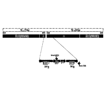
[28] FIG. 2 shows a genetic map demonstrating the use of primers
specifically targeting both
flanking sides of the interferon transgene.
[29] FIG. 3A shows images of plaques from U--2 OS cell seeded in 6--well
plates and infected
with the virus indicated after 24 hours of seeding. After 72 hours the cells
were stained with
crystal violet. FIG. 3B shows U--2 OS cells and BS--C--1 cells seeded in 6--
well plates and
infected with WR or SJ--815. Plaques were stained as in FIG. 3A. One picture
from each
representative experiment is shown.
[30] FIG. 4A shows SJ-815 EC50 (pfu/cell) on human pancreatic and cervix
cancer cell lines.
Pancreatic cancer cell lines and HeLa cells were treated with either SJ-815 or
control virus
(WR.A34R.TK-) labeled as TK-, at different multiplicities of infection. Cell
viability was
assessed after 48 hours post-infection by CCK-8. The EC50 was determined and
plotted. FIG. 4B
shows replication of SJ-815 on human pancreatic and cervix cancer cells.
Pancreatic cancer cell
lines and HeLa cells were infected with SJ- 815 or WR.A34R.TK- at 1 PFU/cell.
After 48 hours
post-infection the cells were harvested and the infectious virus produced in
each cancer cell was
determined by plaque assay in U-2 OS cells. The data from one representative
experiment
repeated 3 times in triplicate is presented.
[31] FIG. 5A shows SJ-815 EC50 (pfu/cell) on human colon cancer cell lines.
Colon cancer cell
lines were treated with either SJ-815 or control virus (WR.A34R.TK-)
Date Regue/Date Received 2023-01-20
CA 02996120 2018-02-20
18
WO 2017/043815 PCT/KR2016/009866
labeled as TK-, at different multiplicities of infection. Cell viability was
assessed after
48 hours post-infection by CCK-8. The EC50 was determined and plotted.
[32] FIG. 5B shows replication of SJ-815 on human colon cancer cells. Colon
cancer cell
lines were infected with SJ-815 or WR.A34R.TK- at a multiplicity of 1
PFU/cell. After
48 hours post-infection the cells were harvested and the infectious virus
produced in
each pancreatic cell was determined by plaque assay in U-2 OS cells. The data
from
one representative experiment repeated 3 times in triplicate is presented.
[33] FIG. 6A shows SJ-815 EC50 (pfu/cell) on human liver cancer cell lines.
Liver cancer
cell lines were treated with either SJ-815, mSJ-815 or control virus
(WR.A34R.TK-) at
different multiplicities of infection. Cell viability was assessed after 48
hours post-
infection by CCK-8. The EC50 was determined and plotted. FIG. 6B shows
replication of SJ-815 on human liver cancer cells. Liver cancer cell lines
were infected
with SJ-815, mSJ-815 and WR.A34R.TK- at a multiplicity of 1 PFU/cell. After 48
hours post-infection the cells were harvested and the infectious virus
produced in each
liver cell was determined by plaque assay in U-2 OS cells. The data from three
different experiments run in triplicate is presented. Unpaired t-test was used
to analyze
the data *P<0.05 and **P<0.01.
[34] FIGS. 7A-D show cell viability after increasing concentrations of
virus treatment in
myeloma and melanoma cancer cells. Myeloma and melanoma cancer cells SK-MEL 5
(FIG. 7A), SK-MEL 2 (FIG. 7B), RPMI8226 (FIG. 7C) and IM-9 (FIG. 7D) were
treated with either SJ-815, mSJ-815 or control virus (WR.A34R.TK-) at
different mul-
tiplicities of infection. Cell viability was assessed after 48 hours post-
infection by
CCK-8. The data from three different experiments run in triplicate is
presented.
[35] FIG. 8 shows replication of SJ-815 on human liver cancer cells.
Myeloma and
melanoma cancer cell lines were infected with SJ-815, mSJ-815 and WR.A34R.TK-
at
a multiplicity of 1 PFU/cell. After 48 hours post-infection the cells were
harvested and
the infectious virus produced in each cell line was determined by plaque assay
in U-2
OS cells. The data from three different experiments run in triplicate is
presented.
Unpaired t-test was used to analyze the data *P<0.05 and **P<0.01.
[36] FIGS. 9A-F shows cell viability after increasing concentrations of
virus treatment in
murine cancer cells: TB3-75 hepatocellular carcinoma (FIG. 9A), CT-26 colon
carcinoma (FIG. 9B), B16-F10 skin melanoma (FIG. 9C), MC-38 colon carcinoma (
FIG. 9D), RENCA renal adenocarcinoma (FIG. 9E), and 4T1 breast cancer (FIG.
9F)
were treated with either mSJ-815 WR.mGM-CSF at different multiplicities of
infection. Cell viability was assessed after 48 hours post-infection by CCK-8.
The data
from three different experiments run in triplicate is presented.
[37] FIG. 10A shows survival of B57BL/6 mice with MC-38 colon cancer tumors
treated
with mS1-815 and WR.TK-. mGMCSF virus via IV and IT. Kaplan-Meier curves for
CA 02996120 2018-02-20
19
WO 2017/043815 PCT/KR2016/009866
each treatment regimen are shown. Animals were sacrificed upon reaching the
endpoint tumor volume of 1,500 mm3. FIG. 10B shows average of body weight for
the
different groups followed during the days after the first treatment injection.
FIG. 10C
shows tumor size in percentage relative to initial tumor size over time
following
treatment of mice harboring MC-38 tumors with mSJ-815 and WR.TK-.mGMCSF
(Days 0, 3, 6 and 9), via IT and IV relative to control (PBS). Group averages
are
presented (n=5). None of the groups presented statistically significant
differences with
respect to the PBS (control) group, except on Days 21, 24 and 27, where the
mSJ-815
group's smaller tumor volume was statistically significant from the PBS group,
with a
P<0.05 for Day 21 and Day 24 and P<0.01 for Day 27.
[38] FIG. 11 shows tumor size over time following treatment of mice
harboring MIA
PaCa-2 tumors with SJ-815 intratumoral alone at increasing doses (1X10⁵, 1X10⁶
and
1X10⁷ PFU) (Days 0, 7 and 14), irinotecan intravenous treatment alone (Days 3,
10
and 17) and combination treatment with SJ-815 (1X10⁶) and irinotecan (above
schedules combined) relative to control (PBS). Group averages are presented
(n=3).
[39] FIG. 12A shows Kaplan-Meier curves for each treatment regimen
displayed.
Animals were sacrificed upon reaching the endpoint tumor volume of 1,500 mm3.
Data
as analyzed by Long-rank (Mantel-Cox) test was significantly different with a
P=0.002. FIG. 12B shows average and standard deviations of body weight for the
different groups followed during the days after the first treatment injection.
[40] FIG. 13A shows the average tumor size of B57BL/6 mice with B16-Fl 0
melanoma
tumors treated with different concentrations and doses of mSJ-815. The average
tumor
size per group was calculated and the SEM was plotted. The difference in the
tendency
of the plots was due to the animals that were sacrificed. Data was analyzed by
One-
way analysis of variance (ANOVA) followed by Dunnett's multiple comparison
test
and unpaired t test. The data was not statistically significant with a
P=0.3755. FIG.
13B shows the size of the tumor in individual mice at day 12 after treatment.
The data
is statistically significant by one-way ANOVA with a P=0.0119. All the groups
were
statistically different from the PBS groups by Dunnet's multiple comparison
test
(P<0.05).
[41] FIG. 14 shows Kaplan-Meier curves for each treatment regimen
displayed. Animals
were sacrificed upon reaching the endpoint tumor volume of 1,500 mm3. Data
analyzed by Long-rank (Mantel-Cox) test was significantly different with a
P<0.0001.
[42] FIG. 15A shows the average tumor size of B57BL/6 mice with B16-F10
melanoma
tumors treated with Vaccinia virus and CPT-11 alone and combination treatment
with
virus plus Irinotecan. Tumor volume values from a single dead mouse were
excluded
from this figure and analysis. The average in tumor size per group was
calculated and
the SEM was plotted. Data was analyzed by unpaired t-test. The data was
statistically
CA 02996120 2018-02-20
WO 2017/043815 PCT/KR2016/009866
different between PBS (control group) and animals treated either with WR.
A34R.TK-
(WR.TK-), mSJ-815, or virus + CPT-11 in combination on Day 9 and 13. FIG. 15B
shows the average size of the tumor per group until day 16 after treatment.
Tumor
volume values from a single dead mouse were excluded from this figure and
analysis.
The data was statistically significant by unpaired t-test. FIG. 15C shows the
average
tumor size of B57BL/6 mice with B16-F10 melanoma tumors treated with Vaccinia
virus and CPT-11 alone and combination treatment with virus plus Irinotecan.
The data
displayed is the same as that shown in FIG. 15A, with the exception that tumor
volume values from the single dead mouse are included. The average in tumor
size per
group was calculated and the SEM was plotted. Data was analyzed by unpaired t-
test.
FIG. 15D shows the average size of the tumor per group until day 16 after
treatment.
The data displayed is the same as that shown in FIG. 15C, with the exception
that
tumor volume values from the single dead mouse are included.
[43] FIG. 16 shows the average and standard deviations of body weight for
the different
groups of B57BL/6 mice with B16-F10 melanoma tumors treated with vaccinia
virus
and the combination with irinotecan. Mice were measured twice per week after
treatment. Data was analyzed by one-way analysis of variance (ANOVA) followed
by
Dunnett's multiple comparison test. The data was not statistically significant
with a
P=0.1845. There was not a significant difference between any of the groups
with
Dunnett's multiple comparison test.
[44] FIGS. 17A-D shows liver (FIG. 17A), kidney (FIG. 17B), brain (FIG.
17C), and
lung (FIG. 17D) weight of B57BL/6 mice with B16-F10 melanoma tumors treated
with mSJ-815 intratumoral or intravenous. Organs were collected at the
endpoint,
weighted and the weight was normalized against body weight.
[45] FIGS. 18A & 18B shows spleen weight of B57BL/6 mice with B16-F10
melanoma
tumors treated with mSJ-815 intravenous (FIG. 18A) or intratumoral (FIG. 18B).
Spleens were collected at the endpoint, weighted and the weight was normalized
against body weight.
[46] FIG. 19 shows virus titers on the day of animal death within tumor,
muscle, ovaries
and liver. Bars represent standard deviation of the mean (SD).
Mode for the Invention
[47] Certain aspects of the disclosures described herein are based upon the
surprising
discovery that oncolytic vaccinia virus that has been engineered to express a
cytokine
and a carboxylesterase enzyme results in an unexpected improvement in the
treatment
of cancer, including when combined with a cancer co-drug. Without wishing to
be
bound by theory, expression of a cytolcine such as the preferred human
interferon beta
1 (hIFNbl) by the oncolytic vaccinia virus stimulates the anti-cancer immune
response
CA 02996120 2018-02-20
21
WO 2017/043815 PCT/KR2016/009866
and enhances cancer selectivity.
[48] It has been found that combination therapy with the oncolytic vaccinia
virus
described herein and a cancer co-drug, preferably an activatable cancer co-
drug, results
in unexpected improvement in the treatment of cancer. When administered simul-
taneously, sequentially or separately, the oncolytic vaccinia virus and the
cancer co-
drug, especially activatable cancer co-drugs, interact to kill cancer cells to
a greater
degree than by administration of either component by itself. This unexpected
im-
provement should allow a reduction in the dose required of each component,
leading to
a reduction in the side effects and enhancement of the clinical effectiveness
of the
compounds and treatment. Cancer co-drugs such as irinotecan can cause
significant
side effects including frequent and severe gastrointestinal problems such as
diarrhea,
emesis, diaphoresis, abdominal cramping, hyperlacrimation, and rhinorrhea. The
frequency and severity of symptoms is dose-related, with patients received
higher
doses demonstrating more severe symptoms. Thus, potential reduction in the
cancer
co-drug dose required when used in combination with the oncolytic vaccinia
virus
described herein would lead to a reduction in clinical side effects.
[49] I.ONCOLYTIC VACCINIA Virus
[50] Certain aspects of the present disclosure relate to an oncolytic
vaccinia virus that
expresses a cytokine and a carboxylesterase enzyme and that preferably does
not
express an active thymidine kinase. Vaccinia virus is a large, complex
enveloped virus
having a linear double-stranded DNA genome of about 190K bp and encoding for
ap-
proximately 250 genes. Vaccinia virus is a large virus roughly 360nm by 250nm
in
size. Vaccinia is well-known for its role as a vaccine that eradicated
smallpox. Post-
eradication of smallpox, scientists have been exploring the use of vaccinia
virus as a
tool for delivering genes or as a vaccine into biological tissues (gene
therapy and
genetic engineering).
[51] Vaccinia virus preferentially infects through the basolateral surface
of cells, but its
viral progeny are released from the apical surface. Polarized cells include,
without
limitation, epithelial cells, endothelial cells, immune cells, osteoclasts,
neurons, and fi-
broblasts.
[52] Vaccinia virus is unique among DNA viruses as it replicates only in
the cytoplasm of
the host cell. Therefore, the large genome is required to code for various
enzymes and
proteins needed for viral DNA replication. During replication, vaccinia
produces
several infectious forms which differ in their outer membranes: the
intracellular mature
virion (IMV), the intracellular enveloped virion (IEV), the cell-associated
enveloped
virion (CEV) and the extracellular enveloped virion (EEV). IMV is the most
abundant
infectious form and is thought to be responsible for spread between hosts. On
the other
hand, the CEV is believed to play a role in cell-to-cell spread and the EEV is
thought
22
to be important for long range dissemination within the host organism. The
oncolytic vaccinia
virus of the present disclosure can optionally be modified to enhance EEV
output including by
mutating the ,434R gene, which is known to produce enhanced amounts of the
extracellular
enveloped form (EEV) of vaccinia virus
[53] Any known oncolytic strain of vaccinia virus may be used as the
vaccinia virus component of
the compositions and combinations of the disclosure. In preferred embodiments,
the oncolytic
vaccinia virus of the present disclosure is a Copenhagen, Western Reserve,
Lister, or Wyeth
strain, most preferably a Western Reserve or Wyeth strain. Other strains which
have been isolated
and characterized from infected individuals or through bioselection methods
selecting for tumor
specific targeting properties may also be used.
[54] The oncolytic vaccinia virus of the present disclosure can be
engineered to express a foreign
protein such as granulocyte-macrophage colony stimulating factor, or GM- CSF,
GM-CSF is a
protein secreted by macrophages that stimulates stem cells to produce
granulocytes (neutrophils,
eosinophils, and basophils) and macrophages. Human GM-CSF is glycosylated at
amino acid
residues 23 (leucine), 27 (asparagine), and 39 (glutamic acid) (see U.S.
Patent 5,073,627).
[55] In some embodiments, the vaccinia virus is engineered to express a
cytosine dearninase
protein. Cytosine deaminase catalyzes the hydrolysis of cytosine to uracil.
Cytosine deaminase
plays a role in the pyrimidine salvage pathway, which permits the cell to
utilize cytosine for
pyiimidine nucleotide synthesis. Cytosine deaminase is also able to catalyze
deamination of
isoguanine, a mutagenic oxidation product of adenine in DNA, and of
isocytosine and catalyzes
the conversion of 5-fluorocytosine (5FC) to 5-fluorouracil (5FU), allowing the
creation of a
cytotoxic chemotherapeutic agent from a non-cytotoxic precursor.
[56] In some embodiments, the vaccinia virus is engineered to express a
somatostatin receptor
type 2 protein. Somatostatin receptor type 2 is the receptor for somatostatin-
14 and -28.
Somatostatin receptor type 2 is coupled via pertussis toxin sensitive G
proteins to inhibition of
adenylyl cyclase. In also stimulates phosphotyrosine phosphatase and PLC,
inhibits calcium entry
by suppressing voltage-dependent calcium channels, inhibits cell growth
through enhancement of
MAPK1 and MAPK2 phosphorylation, stimulates neuronal migration and axon
outgrowth,
mediates negative regulation of insulin receptor signaling through PTPN6, and
inactivates SSTR3
receptor function following heterodimerization.
[57] The oncolytic vaccinia virus may be engineered to lack one or more
functional genes in order
to increase the cancer selectivity of the virus. In preferred embodiments, the
oncolytic vaccinia
virus is engineered to lack TK activity. A TK-deficient vaccinia virus
requires thymidine
triphosphate for DNA synthesis, which leads to preferential replication in
dividing cells
Date Regue/Date Received 2023-01-20
23
(particularly cancer cells). In another aspect, the oncolytic vaccinia virus
may be engineered to
lack vaccinia virus growth factor (VGF). This secreted protein is produced
early in the infection
process, acting as a mitogen to prime surrounding cells for infection. In
another aspect, the
oncolytic vaccinia virus may be engineered to lack both VFG and T activity. In
other aspects, the
oncolytic vaccinia virus may be engineered to lack one or more genes involved
in evading host
interferon (IFN) response such as E3L, 3L, B18R, or B8R. In some preferred
embodiments, the
oncolytic vaccinia virus is a Western Reserve or Wyeth strain and lacks a
functional TK gene. In
other embodiments, the oncolytic vaccinia virus is a Western Reserve strain
lacking a functional
B18R and/or B8R gene.
[58] In some embodiments, the oncolytic vaccinia virus lacks a functional
TK gene and expresses
human GM-CSF. In a preferred embodiment, the oncolytic vaccinia virus is a
Wyeth strain
oncolytic vaccinia virus that lacks a functional TK gene and expresses human
GM-CSF.
[59] A.Carboxylesterase Enzymes
[60] The oncolytic vaccinia virus component of the combination can be
engineered to express a
carboxylesterase. Carboxylesterases are serine esterase enzymes primarily in
the liver
(carboxylesterase 1) and intestine (carboxylesterase 2) thought to be involved
in the
detoxification of a variety of xenobiotics. Carboxylesterases as used herein
include any enzyme
that catalyzes the conversion of irinotecan to the active metabolite SN-38,
such as
butyrylcholinestcrase. SN-38, like irinotecan is a topoisomerase I inhibitor,
but is -100 to 1000
times more active than the parent drug. Only a small percentage of irinotecan
is converted to SN-
38 in human cancer patients receiving standard treatment with irinotecan. The
carboxy terminal
four amino acids of carboxylesterase 1 (IIIEL) and carboxylesterase 2 (H [EL),
cause the enzyme
to be retained in the cell. Removal of these terminal four amino acids causes
the enzyme to be
secreted.
[61] The present application demonstrates that combination therapy with
irinotecan and a vaccinia
virus genetically engineered to express a carboxylesterase results in a tumor-
specific increase in
the conversion of irinotecan to SN-38. A significant decrease in cell
viability in a variety of cell
lines following infection of the cells with vaccinia virus expressing
carboxylesterase 2 compared
to the same virus not expressing the carboxylesterase tans-gene was observed.
Superior results
were observed across several human cancer lines when the vaccinia vims
expressed
carboxylesterase 2.
[62] In a preferred embodiment, the oncolytic virus expresses human
carboxylesterase 2 (e.g.
UniProt Accession Number 000748). hi some embodiments, the vaccnia virus is
engineered to
express a polypeptide at least 80%, at least 85%, at least 90%, at least 95%
or at least 99%
Date Recue/Date Received 2023-01-20
24
identical to human carboxylesterase 2. In other embodiments, the onocolytic
vaccinia virus
expresses rabbit carboxylesterase 2 (UniProt Accession Number P14943) or a
polypeptide at least
80%, at least 85%, at least 90%, at least 95% or at least 99% identical
thereto. In related
embodiments, the vaccinia virus expresses carboxylesterase 2 comprising a
deletion of the
carboxy terminal four amino acids so that the carboxylesterase is secreted.
[63] In some preferred embodiments, the carboxylesterase enzyme comprises a
C-terminal
retention sequence. In some embodiments, the C-terminal retention sequence is
HTEL (SEQ ID
NO: 1). In some embodiments, the carboxylesterase enzyme does not comprise a C-
terminal
retention sequence. In some embodiments, the carboxylesterase enzyme is CES2,
preferably
human CES2 (hCES2).. In some embodiments, expression of the carboxylesterase
enzyme is
under control of a late-early VACV p7.5 promoter, a vaccinia modified H5 (mH5)
promoter, a
vaccinia short synthetic early-late pS promoter, a pC11R promoter, a pF1 IL
promoter, a psFJ1-10
synthetic early promoter, a pHyb synthetic early promoter, any native vaccinia
early promoters,
and a Late-Early Optimized (LEO) promoter.
[64] B.Cytokin es
[65] In certain embodiments, the oncolytic viruses for use in the
compositions, combinations and
methods of the present disclosure may be engineered to express a cytokine.
[66] Cytokines and chemokines can have potent antitumoral effects (Vicari
et al., 2002; Homey et
al., 2002). These effects can be on tumor cells themselves directly or they
can be indirect through
effects on non-cancerous cells. An example of the latter is TNF, which can
have antitumoral
effects by causing toxicity to tumor-associated blood vessels; this leads to a
loss of blood flow to
the tumor followed by tumor necrosis. In addition, cytokines and chemokines
can act to recruit
(and in some cases activate) immune effector cells such as neutrophils,
eosinophils, macrophages
and/or lymphocytes. These immune effector cells can cause tumor destruction by
a number of
mechanisms. These mechanisms include the expression of antitumoral cytokines,
expression of
fas-ligand, expression of perforin and granzyme, recruitment of natural killer
(NK) cells, etc. The
inflammatory response can eventually lead to the induction of systemic tumor-
specific immunity.
Finally, many of these cytokines or chemokines can act in combination with
chemotherapy or
radiation therapy to destroy tumors.
[67] Clinically effective systemic administration of recombinant versions
of these im-
munostimulatory proteins is not feasible due to (1) induction of severe
toxicity with systemic
administration and (2) local expression within tumor tissue is needed to
stimulate local infiltration
and antitumoral effects. Approaches are needed to achieve high local
concentrations of these
molecules within tumor masses while minimizing
Date Regue/Date Received 2023-01-20
CA 02996120 2018-02-20
WO 2017/043815 PCT/KR2016/009866
levels in the systemic circulation. Viruses can be engineered to express
cytokine or
chemokine genes in an attempt to enhance their efficacy. Expression of these
genes
from replication-selective vectors has potential advantages over expression
from non-
replicating vectors. Expression from replicating viruses can result in higher
local con-
centrations within tumor masses; in addition, replicating viruses can help to
induce an-
titumoral immunity through tumor cell destruction/oncolysis and release of
tumor
antigens in a proinflammatory environment.
[68] In some embodiments, the cytokine is selected from the group
consisting of in-
terferon-beta-1 (preferably human), IL-la, IL-2, IL-3, IL-4, IL-5, IL-6, IL-7,
IL-8, IL-
9, IL-10, IL-12, IL-13, IL-14, IL-15, IL-17, IL-18, IL-21, IL-23, IL-24 CCL3,
CCL5,
and CXCR4. In certain embodiments, the cytokine is interferon-beta-1,
preferably
human. Human interferon beta, a type I interferon, is a 166 amino acid
glycosylated
protein which is secreted by fibroblasts in response to viral infection or
exposure to
double-stranded RNA. Interferon beta signals through a receptor composed of
two
chains: interferon-alpha receptor 1 (IFNAR1) and interferon-alpha receptor 2
(IFNAR2). Upon binding to the receptor, IFNb activates the JAK/STAT pathways,
which leads to phosphorylation of STATI and STAT2. The STAT proteins sub-
sequently dimerize, associate with interferon regulatory factor 3 (IRF3), and
bind to in-
terferon response elements within the cell nucleus. These elements serve to
stimulate
interferon responsive genes, leading to the downstream effects of interferon
beta. In-
terferon beta possesses anti-viral and antiproliferative activity and has been
used for
the chemotherapy of certain types of tumors and in treating multiple
sclerosis. Ex-
pression of IFN-beta-1 by the oncolytic vaccinia virus described herein
stimulates the
anti-cancer immune response and enhances cancer selectivity.
[69] In some embodiments, expression of the cytokine is under control of a
late-early
VACV p7.5 promoter, a vaccinia modified H5 (mH5) promoter, a vaccinia short
synthetic early-late pS promoter, a pC11R promoter, a pF11L promoter, a psFJ1-
10
synthetic early promoter, a pHyb synthetic early promoter, any native vaccinia
early
promoters, and a Late-Early Optimized (LEO) promoter. Specific Exemplary Em-
bodiments
[70] In a particularly preferred embodiment, the oncolytic vaccinia virus
is SJ-815. SJ-815
is an attenuated transgenic oncolytic vaccinia virus derived from Western
Reserve
Vaccinia parental strain WR A34R K151E. WR A34R K151E contains the A34R gene
with a K151E mutation to enhance EEV production (Blasco 1993). SJ-815 was
derived
by inserting the genes for human CES2 and human IFNbetal into the thymidine
kinase
(TK) gene of the parental virus (under the control of the synthetic early-late
and p7.5
early-late promoters, respectively), thereby rendering the TK gene inactive.
Inac-
tivation of the TK gene has been shown to decrease the virulence of vaccinia
virus and
CA 02996120 2018-02-20
26
WO 2017/043815 PCT/KR2016/009866
to increase tumor specific replication.
[71] In another preferred embodiment, the oncolytic vaccinia virus is an
attenuated
transgenic oncolytic vaccinia virus derived from a Western Reserve Vaccinia
parental
strain with a wild-type (WT) A34R gene. This strain is derived by inserting
the genes
for human CES2 and human IFNbetal into the thymidine kinase (TK) gene of the
parental virus (under the control of the synthetic early-late and p7.5 early-
late
promoters, respectively), thereby rendering the TK gene inactive. Inactivation
of the
TK gene has been shown to decrease the virulence of vaccinia virus and to
increase
tumor specific replication.
[72] In a particularly preferred embodiment for use in mouse models, the
oncolytic
vaccinia virus is mSJ-815. mSJ-815 is an attenuated transgenic oncolytic
vaccinia virus
derived from Western Reserve Vaccinia parental strain WR A34R K151E. WR A34R
K151E contains the A34R gene with a K151E mutation to enhance EEV production
(Blasco 1993). SJ-815 was derived by inserting the genes for human CES2 and
mouse
IFNbetal into the thymidine kin ase (TK) gene of the parental virus (under the
control
of the synthetic early-late and p7.5 early-late promoters, respectively),
thereby
rendering the TK gene inactive. Inactivation of the TK gene has been shown to
decrease the virulence of vaccinia virus and to increase tumor specific
replication.
[73] H.METHODS OF USING THE DISCLOSED COMPOSITIONS
[74] Oncolytic vaccinia viruses according to the present disclosure may be
administered
locally, regionally or systemically. For example, without limitation,
oncolytic vaccinia
viruses according to the disclosure can be administered intravascularly
(intraarterially
or intravenously), intratumorally, intramuscularly, intradermally,
intraperitoneally,
subcutaneously, orally, parenterally, intranasally, intratracheally,
percutaneously, in-
traspinally, ocularly, or intracranially. Preferably, the vaccinia virus is
administered in-
travascularly and/or intratumorally. Intratumoral administration generally
entails
injection into a tumor mass or into tumor associated vasculature. In certain
aspects, the
tumor is imaged prior to or during administration of the virus.
[75] Oncolytic vaccinia viruses according to the disclosure may be
administered in a
single administration or multiple administrations. The virus may be
administered at
dosage of 1 x 10⁵ plaque forming units (PFU), 5 x 10⁵ PFU, at least 1 x 10⁶
PFU, 5 x
106 or about 5 x 10⁶ PFU, 1 x 10⁷, at least 1 x l0 PFU, 1 x 10⁸ or about 1 x
108 PFU, at
least 1 x 10⁸ PFU, about or at least 5 x 10⁸ PFU, 1 x 10⁹ or at least 1 x 10⁹
PFU, 5 x 10⁹
or at least 5 x 10 PFU, 1 x 10¹⁰ PFU or at least 1 x 10¹⁰ PFU, 5 x 101 or at
least 5 x 10
PFU, 1 x 10" or at least 1 x10", 1 x 10¹² or at least 1 x 10¹², 1 x 10¹³ or at
least 1 x
1013. For example, the virus may be administered at a dosage of between about
107-10"
pfu, between about 108-10" pfu, between about 109-1012 pfu, or between about
108-1012
. Preferably, the virus is administered at a dosage of between 107 and 101
pfu.
27
[76] It is contemplated that a single dose of virus refers to the amount
administered to a subject or
a tumor over a 0.1, 0.5, 1, 2, 5, 10, 15, 20, or 24 hour period, including all
values there between.
The dose may be spread over time or by separate injection. Typically, multiple
doses are
administered to the same general target region, such as in the proximity of a
tumor or in the case
of intravenous administration a particular entry point in the blood stream or
lymphatic system of a
subject. In certain aspects, the viral dose is delivered by injection
apparatus comprising a needle
providing multiple ports in a single needle or multiple prongs coupled to a
syringe, or a
combination thereof. A single dose of the vaccinia virus may be administered
or the multiple
doses may be administered over a treatment period which may comprise 1, 2, 3,
4, 5, 6, 7, 8, 9,
10, 11, 12 or more weeks. For example, the vaccinia vims may be administered
every other day,
weekly, every other week, every third week for a period of 1, 2, 3, 4, 5, 6 or
more months.
[77] Vaccinia virus may be propagated using the methods described by Earl
and Moss in Ausubel
etal., 1994 or the methods described in WIPO Publication No. W02013/022764.
[78] III.Cancer Co-DRUGS
[79] Cancer co-drugs useful in combination with an oncolytic vaccinia virus
include inhibitors of
topoisomerases I and II. It will be understood that reference to topoi-
somerase inhibitors is meant
to include their pharmaceutically acceptable salts. If the topoisomerase
inhibitor has at least one
basic group, it can form acid addition salts. Topoisomerase inhibitors having
an acid group can
also form salts with bases. Topoisomerase inhibitors and their salts may also
be used in the form
of a hydrate or include other solvents used e.g. for crystallization.
[80] Topoisomerase I inhibitors include camptothecin and its derivatives
such as irinotecan and
topotecan, marine alkaloids such as lamellarin D (3,11-Dihydroxy-14-(4-hydroxy-
3-
methoxypheny1)-2,12-dimethoxy-6H-chromeno[4',3':4,5]pyrrolo[2,1-a]isoquinolin-
6-one) and
idenoisoquinoline analgoues such as indotecan (also called LMP400 or 2,3-
dimethoxy-6-(3-
morpholinopropy1)-5H41,3]dioxolo[4',51:5,6]indeno[1,2-c]isoquin oline-5,12(6H)-
dione),
indimitecan (also called LMP776 or 6-(3-(1H-imidazol-1-yl)propy1)-2,3-
dimethoxy-6,6a-
dihydro-5H41,3]dioxolo[4',5':5,6]indeno[1,2-c]isoquinoline-5,12(12aH)-dione)
and NSC-
706744.
[81] Topoisomerase II inhibitors include etoposide (also called VP-16),
teniposide, doxorubicin,
daimorubicin, epirubicin, idarubucin, mitoxantrone, amsacrine, ellipticines,
aurintricarboxylic
acid, HU-331 and ICRF-193, ICRF-187, and merbarone. Etoposide phosphate may be
administered to a human in a dosage range varying from about 25 to
Date Regue/Date Received 2023-01-20
CA 02996120 2018-02-20
28
WO 2017/043815 PCT/ICR2016/009866
115 mg/m2. Teniposide may be administered to a human in a dosage range of
about 75
to 150 mg/m2 every two weeks. Doxorubucin may be administered to a human in a
dosage range of about 10 to 100 mg/m2. Epirubicin may be administered to a
human in
a dosage range varying from about 10 to 200 mg/m2 e.g. 100 rng/m2i.v. every 3-
4
weeks. Idarubucin may be administered to a human in a dosage of about 0.5 to
50 mg/
m2.
[82] Camptothecin derivatives are preferred topoisomerase inhibitors for
use in the com-
bination. Camptothecin derivatives are anticancer agents which inhibit
topoisomerase
I. These compounds are usually administered by injection, more particularly
intra-
venously in the form of a sterile solution or emulsion; however, they can also
be ad-
ministered orally, in solid or liquid formulations. Representative
camptothecin
derivatives useful in the present disclosure include irinotecan, topotecan
(also called
Hycamtin or
S)-10-[(dimethylamino)methy1]-4-ethyl-4,9-dihydroxy-1H-
pyrano[3',41:6,7]indolizino[
1,2-b]quinoline-3,14(4H,12H)-dione monohydrochloride), DB-67 (also called AR67
or
7-t-butyldimethylsily1-10-hydroxycamptothecin), BNP-1350
(7-[(2-trimethylsilypethyl]-20(S)-camptothecin), exatecan
((1S,9S)-1-Amino-9-ethy1-5-fluoro-1,2,3,9,12,15-hexahydro-9-hydroxy-4-methyl-
10H,
13H-benzo(de)pyrano(31,41:6,7)indolizino(1,2-b)quinol me-10,13-dione),
lurtotecan
(also called GI147211 or
7-(4-methylpiperazineomethylene)-10,11-ethylenedioxy-20(S)-camptothecin dihy-
drochloride), ST-1481 (7-t-butoxyiminomethylcamptothecin), and CKD-602
((20S)-7-(2-isopropylamino)-ethylcamptothecin). Topotecan can be administered
to a
human in a dosage range varying from about 1 to 5 mg/m2 e.g. at 1.5 mg/m2 by
in-
travenous infusion over 30 minutes daily for 5 consecutive days.
[83] Irinotecan (also known as CPT-11, Camptosar, or
(S)-4,11-diethy1-3,4,12,14-tetrahydro-4-hydroxy-3,14-dioxo1H-pyrano[31,4':6,71-
indoli
zino[1,2-b]quinolin-9-y141,4'bipiperidine1-1'-carboxylate) is a particularly
preferred
camptothecin derivative. Irinotecan is a prodrug that is converted by
carboxylesterase
enzymes to the active drug known as SN-38 (7-ethyl-10-hydroxy-camptothecin)
(Satoh
T. et al., Biol. Pharm. Bull., 17:662-664 (1994). It acts by preventing re-
ligation of the
DNA strand by binding to topoisomerase I-DNA complex and causes double-strand
DNA breakeage and cell death. Irinotecan has been demonstrated to be
particularly
effective in the treatment of cancer either alone or in combination with other
agents
such as 5-fluorouracil (5-FU) and oxaliplatin. Irinotecan is in fact the
reference
treatment in metastatic cancer after failure on prior 5-FU treatment. In this
respect,
irinotecan has been shown to be at least as active as the standard 5-
FU/folinic acid
(FA) in patients with metastatic cancer who had not previously received
chemotherapy.
CA 02996120 2018-02-20
29
WO 2017/043815 PCT/KR2016/009866
In addition to colon cancer, activity has been observed in ovarian cancer,
lung cancer,
gastric cancer, oesophageal cancer and cervical cancer.
[84] Irinotecan (preferably as a hydrochloride salt) may be administered
according to
treatment protocols known in the art taking into account the expression of the
car-
boxylesterase by the oncolytic vaccinia virus, which will increase the local
con-
centration in the tumor environment. For example, when used as a single agent
in the
treatment of cancer, irinotecan is usually administered at a starting dose of
350 mg/m2
intravenously over 90 minutes every three weeks which can be adjusted as low
as 200
mg/m2 in 50 mg/m2 decrements depending on a patient's tolerance, or
alternatively is
administered at a starting dose of 125 mg/m2 intravenously over 90 minutes
once a
week, which can adjusted as high as 150 mg/m2 or to as low as 50 mg/m2 in 25-
50 mg/
m2 decrements depending on a patient's tolerance, for as long as the patient
continues
to experience clinical benefit. When used in combination with 5-FU and FA,
irinotecan
is generally administered at 125 mg/m2 intravenously over 90 minutes once a
week for
four doses or is administered at a dose of 180 mg/m2 intravenously over 90
minutes
every other week for three doses. Thus, according to the disclosure,
irinotecan may be
administered at a dose range of 50-350 mg/m2 e.g. a 90 minute continuous
infusion
once per week at a dose of 125 mg/m2 or a 90 minute continuous infusion every
other
week at a dose of 180 mg/m2. Irinotecan dosing can continue for as long as a
clinical
benefit is experienced.
[85] In addition to topoisomerase inhibitors, other anti-cancer agents
activated by car-
boxylesterases can also be used including paclitaxel-2-ethylcarbonate (which
is
converted to paclitaxel), capecitabine (which is converted to 5'-Deoxy-5-
fluorocytidine
(5-Hi)), and generally any tertiary amidomethyl ester prodrugs of existing
chemother-
apeutics (which are converted to their carboxylic acid or amine forms). Dosing
can
suitably adapted for these other anti-cancer agents taking into account the
expression
of the carboxylesterase by the oncolytic vaccinia, which will increase the
local con-
centration in the tumor environment.
[86] IV.Treatment Regimens and Pharmaceutical Formulations oF THE COM-
BINATIONS
[87] Some aspects of the present disclosure relate to methods for treating
cancer in a
mammal by administering to the mammal an effective amount of a synthetic
oncolytic
vaccinia virus that expresses a carboxylesterase enzyme and a cytokine and
that
preferably does not express an active thymidine kinase, ideally for use in
conjunction
with a cancer co-drug. Some aspects of the present disclosure relate to
methods for
treating cancer in a mammal by administering an effective amount of a
combination
containing (a) a composition comprising a synthetic oncolytic vaccinia virus
that
expresses a cytokine and a carboxylesterase enzyme and that preferably does
not
CA 02996120 2018-02-20
WO 2017/043815 30 PCT/KR2016/009866
express an active thymidine kinase, and (b) cancer co-drug.
[88] The oncolytic vaccinia virus and the cancer co-drug may be
administered simul-
taneously, sequentially or separately. Simultaneous administration may, e.g.,
take place
in the form of one fixed combination comprising these agents, or by
simultaneously
administering each agent in independent formulations. Sequential use
(administration)
preferably means that the oncolytic vaccinia virus and the cancer co-drug are
ad-
ministered at different time points, that is, in a chronically staggered
manner,
preferably such that the combination is more effective than when the oncolytic
vaccinia virus and the cancer co-drug are independently administered. Separate
use
(administration) preferably means that the oncolytic vaccinia virus and the
cancer co-
drug are administered independently of each other at different time points.
The present
disclosure is therefore to be understood as embracing all such regimes of
simultaneous
or alternating treatment and the term "administering" is to be interpreted
accordingly.
[89] If the oncolytic virus and the cancer co-drug are not administered
simultaneously, the
order of administration of the oncolytic virus and the cancer co-drug may be
varied.
Thus, the oncolytic virus may be administered first followed by administration
of the
cancer co-drug or the cancer co-drug may be administered first followed by the
oncolytic virus. In embodiments where the oncolytic vaccinia virus and the
cancer co-
drug are not administered simultaneously, each agent is preferably
administered such
that an advantageously combined effect on the cell is obtained. In such
instances, it is
contemplated that the agents are administered within the same general time
frame and
preferably within two weeks, more preferably within one week, of each other.
In some
situations, it may be desirable to extend the time period for treatment
significantly
where several days (e.g. 2, 3, 4, 5, 6 or 7) lapse between the respective
administrations.
In a preferred combination, sequential administration of the oncolytic virus
and the
cancer co-drug comprises first administering one or more doses of the
oncolytic virus
followed by administration of one or more doses of the cancer co-drug,
preferably with
an intervening period of 14 or fewer (e.g. 13, 12, 11, 10, 9, 8, 7, 6, 5, 4,
3, 2, 1 or 0)
days between administration of the respective agents. By "intervening period"
it is
meant a time period beginning from the end of the last dose of the oncolytic
virus up
until the beginning of the first dose of cancer co-drug. In embodiments in
which the in-
tervening period is 0 days, the cancer co-drug is administered immediately
following
the last dose of the oncolytic virus.
[90] A particularly preferred sequential treatment protocol comprises
weekly admin-
istration of oncolytic virus staggered with every other week administration of
cancer
co-drug, wherein the oncolytic virus is administered first with an intervening
period of
from 1 to 3 days. For example, every other week administration of the cancer
co-drug
may begin l to 3 days after the second weekly dose of oncolytic virus:
CA 02996120 2018-02-20
31
WO 2017/043815 PCT/ICR2016/009866
[91] Oncolytic virus (Days 1, 8, 15, 22, 29?)
[92] Cancer co-drug (Days 9, 23, 37, 51, 65?).
[93] Administration of the oncolytic vaccinia virus and the cancer co-drug
will follow
general protocols for the administration of each particular therapy, taking
into account
the toxicity, if any, of the treatment. It is expected that the treatment
cycles would be
repeated as necessary. It also is contemplated that various standard
therapies, as well as
surgical intervention, may be applied in addition to therapy of the present
disclosure.
[94] Treatment regimens may vary and often depend on tumor type, tumor
location,
disease progression, and health and age of the patient. Certain types of tumor
will
require more aggressive treatment, while at the same time, certain patients
cannot
tolerate more taxing protocols. The clinician will be best suited to make such
decisions
based on the known efficacy and toxicity (if any) of the therapeutic
formulations.
[95] In certain embodiments, the tumor being treated may not, at least
initially, be re-
sectable. Treatment with a combination therapy of the disclosure may increase
the re-
sectability of the tumor due to shrinkage at the margins or by elimination of
certain
particularly invasive portions. Following treatment, resection may be
possible. Ad-
ditional treatments subsequent to resection will serve to eliminate
microscopic residual
disease at the tumor site
[96] Determining a synergistic interaction between one or more components,
the optimum
range for the effect and absolute dose ranges of each component for the effect
may be
definitively measured by administration of the components over different w/w
ratio
ranges and doses to patients in need of treatment. For humans, the complexity
and cost
of carrying out clinical studies on patients renders impractical the use of
this form of
testing as a primary model for synergy. However, the observation of synergy in
one
species can be predictive of the effect in other species and animal models
exist, as
described herein, to measure a synergistic effect and the results of such
studies can also
be used to predict effective dose and plasma concentration ratio ranges and
the
absolute doses and plasma concentrations required in other species by the
application
of pharmacokinetic/pharmacodynamic methods. Established correlations between
tumor models and effects seen in man suggest that synergy in animals may e.g.
be
demonstrated in the human xenograft tumor models as described in the Example
below.
[97] In certain aspects, the combination is used to treat cancer in mammal,
wherein the
cancer selected from the group consisting of brain cancer, head & neck cancer,
esophageal cancer, skin cancer, lung cancer, thymic cancer, stomach cancer,
colon
cancer, liver cancer, ovarian cancer, uterine cancer, bladder cancer, renal
cancer,
testicular cancer, rectal cancer, breast cancer, and pancreatic cancer. In
some em-
bodiments, the combination is used to treat a cancer selected from the group
consisting
CA 02996120 2018-02-20
32
WO 2017/043815
PCT/ICR2016/009866
of colorectal cancer, lung cancer, melanoma, pancreatic cancer, ovarian
cancer,
cervical and liver cancer.
[98] The methods include administering therapeutically effective amounts of
an oncolytic
vaccinia virus and a cancer co-drug. A therapeutically effective amount is
defined as
that amount sufficient to induce oncolysis - the disruption or lysis of a
cancer cell.
Preferably, the oncolytic vaccinia virus and the cancer co-drug are
administered in syn-
ergistically effective amounts. The term includes the slowing, inhibition, or
reduction
in the growth or size of a tumor and includes the eradication of the tumor in
certain
instances. In certain aspects an effective amount of vaccinia virus results in
systemic
dissemination of the therapeutic virus to tumors, e.g., infection of non-
injected tumors.
[99] V.Embolic Agents
[100] Some aspects of the present disclosure relate to methods for treating
cancer in a
mammal by introducing into the vasculature of a mammal a composition
containing a
synthetic oncolytic vaccinia virus that expresses a cytokine and a
carboxylesterase
enzyme and that preferably does not express an active thymidine kinase, and a
bio-
compatible microparticle or hydrophilic polymer gel agent suitable for active
em-
bolization. In some embodiments, the combination of an oncolytic vaccinia
virus and a
cancer co-drug are introduced into the vasculature. Some aspects of the
present
disclosure relate to methods for treating cancer in a mammal by (a)
administering by
introducing into the vasculature of a mammal a composition containing a
synthetic
oncolytic vaccinia virus that expresses a cytolcine and a carboxylesterase
enzyme and
that preferably does not express an active thymidine kinase, and a
bit:compatible mi-
croparticle or hydrophilic polymer gel agent suitable for active embolization
and (b)
administering to the mammal a composition comprising an effective amount of a
cancer co-drug. As above, the oncolytic vaccinia virus and the cancer co-drug
may be
administered simultaneously, sequentially or separately. Simultaneous
administration
may, e.g., take place in the form of one fixed combination comprising these
agents, or
by simultaneously administering each agent in independent formulations.
Sequential
use (administration) preferably means that the oncolytic vaccinia virus and
the cancer
co-drug are administered at different time points, that is, in a chronically
staggered
manner, preferably such that the combination is more effective than when the
oncolytic
vaccinia virus and the cancer co-drug are independently administered. Separate
use
(administration) preferably means that the oncolytic vaccinia virus and the
cancer co-
drug are administered independently of each other at different time points,
preferably
meaning that the oncolytic vaccinia virus and the cancer co-drug are
administered such
that no overlap of measurable blood levels of both agents are present in an
overlapping
manner (at the same time). Where administered independently, one or both may
be ad-
ministered via active embolization with a biocompatible microparticle or
hydrophilic
=
CA 02996120 2018-02-20
33
WO 2017/043815 PCT/ICR2016/009866
polymer gel agent suitable therefor. The present disclosure is therefore to be
un-
derstood as embracing all such regimes of simultaneous or alternating
treatment and
the term "administering" is to be interpreted accordingly.
[101] Numerous biocompatible microparticle or hydrophilic polymer gel
agents can be
used in the compositions, combinations, and methods of this disclosure. In a
preferred
embodiment, the biocompatible microparticle or hydrophilic polymer gel agents
are
selected from: degradable starch microparticles, polyvinyl alcohol
microparticles,
gelatin foam microparticles, and sulfonated polyvinyl alcohol hydrogel
microparticles.
[102] Biocompatible microparticle or hydrophilic polymer gel agents
("embolic agents")
can be either temporary or permanent. Exemplary temporary embolic agents
include
gelfoam, collagen, and thrombin. Exemplary permanent embolic agents include
particles, such as polyvinyl alcohol particles (PVA) and embospheres, coils,
such as
pushable, injectable, detachable, mechanical, electrolytic, and hydrolytic
coils, liquid
agents, such as glue, onyx, alcohol, and ALGELTM (a hydrogel, sugar-based
polymer
derived from alginate), and other agents, including amplatzer plugs, Gianturco-
Grifka
vascular occlusive device (GGVODs), and detachable balloons. Different embolic
agents can be used depending on the size of the vessel to be embolized, the
desired
length of vessel occlusion following embolization, and whether embolized
tissue
should remain viable after occlusion. Given the extensive use of embolization,
a skilled
interventional radiologist would have no difficulty in selecting the
appropriate type of
agent, size range of agent, etc. to achieve the desired embolization. Vessel
occlusion is
useful in clinical scenarios such as traumatic injury and hemorrhage, or when
repeated
embolization procedures are desired, such may be desirable as in tumor
embolization
with oncolytic viruses as disclosed in this specification.
[103] In one embodiment, the biocompatible microparticle or hydrophilic
polymer gel
agents are gelatin foam microparticles. Exemplary gelatin foam includes
Gelfoam,
produced by Alicon/Scion Medical Technologies. Gelfoam is a biological
substance
made from purified skin gelatin, and is formulated in sterile sheets or as a
powder.
Gelfoam has been used in embolization applications for over 30 years, and is a
low
cost, versatile embolic agent. Gelfoam slows blood flow by causing mechanical
ob-
struction. Gelfoam powder consists of particulates that range in size from 150-
1000?m
and can aggregate to form larger conglomerate particles upon water absorption.
Gelfoam sheets can be cut into numerous different sizes and shapes and
formulated
with other aqueous agents upon injection depending upon the desired
application.
Gelfoam slurry containing both a contrast agent and Gelfoam sponge can be used
to
form a "cast" of proximal embolized vessels, while Gelfoam torpedoes or cubes
can be
used for larger vessels. Gelfoam temporarily occludes vessels by slowing blood
flow,
increasing thrombus formation, and functioning as a scaffold for clots.
CA 02996120 2018-02-20
WO 2017/043815 34 PCT/KR2016/009866
[104] In one embodiment, the biocompatible microparticle or hydrophilic
polymer gel
agents are degradable starch microparticles. Exemplary degradable starch mi-
croparticles (DSM) are EMBOCEPTS particles produced by Pharmacept and
SPHEREX particles produced by Mangle Life Sciences. EMBOCEPTS particles
(Amilomer as the active substance) are cross-linked particles composed of
hydrolyzed
potato starch. These particles are suitable for temporary embolization, as
they have a
half-life of approximately 35 minutes and are degradable. SPHEREX particles
are
composed of DSM-S microparticles, sterilized and suspended in saline solution.
Starch
microparticles may be prepared from an aqueous solution of purified
amylopectin-
based starch of reduced molecular weight by forming an emulsion of starch
droplets in
an outer phase of polymer solution, converting the starch droplets to a gel,
and drying
the starch particles. A release-controlling shell is optionally also applied
to the
particles. Biodegradable microparticles, after parenteral administration, are
dissolved
in the body to form endogenic substances, ultimately, for example, glucose.
The
biodegradability can be determined or examined through incubation with a
suitable
enzyme, for example alpha-amylase, in vitro. The biodegradability can also be
examined through parenteral injection of the microparticles, for example subcu-
taneously or intramuscularly, and histological examination of the tissue as a
function
of time. Biodegradable starch microparticles disappear normally from the
tissue within
a few weeks and generally within one week. In those cases in which the starch
mi-
croparticles are coated with a release-controlling shell, for example coated,
it is
generally this shell which determines the biodegradability rate, which then,
in turn, de-
termines when alpha-amylase becomes available to the starch matrix.
[105] In one embodiment, the biocompatible microparticle or hydrophilic
polymer gel
agents are polyvinyl alcohol (PVA) microparticles. Exemplary polyvinyl alcohol
mi-
croparticles are produced by Boston Scientific Corporation (Natick, MA). PVA
particles are made from a PVA foam sheet that is vacuum dried and scraped into
particles. The particles are filtered with sieves and are available in sizes
ranging from
100?m to 1100?rn. Polyvinyl alcohol particles are irregular in size and shape,
which
promotes aggregation. After suspension, PVA particles can be oblong, oval,
irregular,
sharp, and angulated with small fragments after suspension. Polyvinyl alcohol
particles
deliver permanent occlusion by adhering to vessel walls and by blocking the
smallest
vessel into which they pass. PVA occlusion results in inflammatory reactions,
local
vessel necrosis, and subsequent vessel fibrosis.
[106] In one embodiment, the biocompatible microparticle or hydrophilic
polymer gel
agents are sulfonated polyvinyl alcohol hydrogel microparticles. Exemplary
sulfonated
polyvinyl alcohol hydrogel microparticles are DC-Beads produced by
Biocompatibles
(UK, Surrey, UK). DC Beads are embolic microparticle products based on a
polyvinyl
CA 02996120 2018-02-20
WO 2017/043815 35 PCT/ICR2016/009866
alcohol hydrogel that has been modified with sulfonate groups. DC Beads have
the
ability to actively sequester anthracycline compounds in their salt form, such
as dox-
orubicin HCI, from solution and release it in a controlled and sustained
manner. A drug
can be added immediately prior to embolization, allowing for a one-step
procedure in
which the drug and device are delivered at the same time, resulting in a
sustained local
delivery of the drug.
[107] As mentioned above, one of skill in the art can readily select the
appropriate size of
the biocompatible microparticle or hydrophilic polymer gel agents based upon,
amontg
other factors, the size of the tumor vasculature and the nature of the desired
em-
bolization. In a preferred embodiment, the biocompatible microparticle or
hydrophilic
polymer gel agents are between 100Rm and 2000Rm in size. In a preferred em-
bodiment, the biocompatible microparticle or hydrophilic polymer gel agents
are
between 150 and 350pm in size. In one embodiment, the biocompatible
microparticle
or hydrophilic polymer gel agents are between 150 and 2001i.m in size. In one
em-
bodiment, the biocompatible microparticle or hydrophilic polymer gel agents
are
between 200 and 250Rm in size. In one embodiment, the biocompatible
microparticle
or hydrophilic polymer gel agents are between 250 and 300Rm in size. In one em-
bodiment, the biocompatible microparticle or hydrophilic polymer gel agents
are
between 300 and 350Rm in size.
[108] In certain embodiments, the biocompatible microparticle or
hydrophilic polymer gel
agents are uniform in size. This means that the difference in diameter between
in-
dividual particles is from about 0 Rm to about 100 pm, from about 0 p.m to
about 50
pm, or from about 0 p.m to about 25 Rm. In some embodiments, the
microparticles
have differences in diameter of 100 p.m or less, about 50 p.m or less, about
25 pm or
less, about 10 p.m or less or about 5 pm or less.
[109] VI.Methods of Embolization
[110] In one aspect, the disclosure provides a method for active
embolization of a vascular
site in a mammal by introducing into the vasculature of a mammal an oncolytic
virus
of the disclosure and a biocompatible microparticle or hydrophilic polymer gel
suitable
for active embolization, optionally in combination with a cancer co-drug.
[111] Introduction of the biocompatible microparticle or hydrophilic
polymer gel agents,
the oncolytic viruses and the compositions and combinations of the present
disclosure
typically carried out by injection into blood vessels near and around tumors.
In certain
embodiments, the biocompatible microparticle or hydrophilic polymer gel
agents, the
oncolytic viruses and the compositions and combinations of the present
disclosure are
introduced by a catheter. In other embodiments, the biocompatible
microparticle or hy-
drophilic polymer gel agents, the oncolytic viruses and the compositions and
com-
binations of the present disclosure are introduced through injection by a
catheter
CA 02996120 2018-02-20
WO 2017/043815 36
PCT/ICR2016/009866
attached to a syringe. In some embodiments, introduction is into a blood
vessel that
directly feeds a tumor or portion of a tumor. In other embodiments,
introduction is
directly to the site of action, for example into a blood vessel at the
proximal end of the
tumor. The biocompatible microparticle or hydrophilic polymer gel agent
according to
the present disclosure can be introduced already loaded with the oncolytic
virus (i.e.,
the compositions of the present disclosure) and/or a cancer co-drug (i.e., the
com-
binations of the present disclosure). In other embodiments, the biocompatible
mi-
croparticle or hydrophilic polymer gel agents are introduced in combination
with the
oncolytic virus, wherein the virus is introduced prior, simultaneously or
after the in-
troduction of the biocompatible microparticle or hydrophilic polymer gel
agents. When
introduced, the biocompatible microparticle or hydrophilic polymer gel agents,
the
oncolytic viruses and the compositions and combinations of the present
disclosure are
suitable for injection. In specific embodiments, the biocompatible
microparticle or hy-
drophilic polymer gel agents, the oncolytic viruses and the compositions and
com-
binations of the present disclosure are sterile.
[112] The biocompatible microparticle or hydrophilic polymer gel
agents, the oncolytic
viruses and the compositions and combinations of the present disclosure may be
delivered using a catheter or microcatheter. The catheter delivering the
biocompatible
microparticle or hydrophilic polymer gel agents, the oncolytic viruses and the
com-
positions and combinations of the present disclosure may be a small diameter
medical
catheter. Catheter materials compatible with the biocompatible microparticle
or hy-
drophilic polymer gel agents, the oncolytic viruses and the compositions and
com-
binations of the present disclosure may include polyethylene, fluoropolymers
and
silicone. Once a catheter is in place, the biocompatible microparticle or
hydrophilic
polymer gel agents, the oncolytic viruses, the compositions, or combinations
of the
present disclosure are introduced through the catheters slowly, typically with
the as-
sistance of fluoroscopic guidance. The biocompatible microparticle or
hydrophilic
polymer gel agents, the oncolytic viruses and the compositions and
combinations of
the present disclosure may be introduced directly into critical blood vessels
or they
may be introduced upstream of target vessels. The amount of the biocompatible
mi-
croparticle or hydrophilic polymer gel agents or the compositions or
combinations of
the present disclosure introduced during an embolization procedure will be an
amount
sufficient to cause embolization, e.g., to reduce or stop blood flow through
the target
vessels. The amount of the biocompatible microparticle or hydrophilic polymer
gel
agents, the oncolytic viruses and the compositions and combinations of the
present
disclosure delivered can vary depending on, e.g., the total size or area of
the vas-
culature to be embolized and the size and nature of the tumor. After
embolization,
another arteriogram may be performed to confirm the completion of the
procedure.
CA 02996120 2018-02-20
WO 2017/043815 37 PCT/ICR2016/009866
Arterial flow will still be present to some extent to healthy body tissue
proximal to the
embolization, while flow to the diseased or targeted tissue is blocked.
Further, a va-
sodilator (e.g., adenosine) may be administered to the patient beforehand,
simul-
taneously, or subsequently, to facilitate the procedure.
[1131 One of skill in the medical or embolizing art will understand and
appreciate how the
biocompatible microparticle or hydrophilic polymer gel agents, the oncolytic
viruses
and the compositions and combinations of the present disclosure as described
herein
can be used in various embolization processes by guiding a delivery mechanism
to a
desired vascular body site, and delivering an amount of the biocompatible mi-
croparticle or hydrophilic polymer gel agents, the oncolytic viruses, the
compositions,
or the combinations of the present disclosure to the site, to cause
restriction, occlusion,
filling, or plugging of one or more desired vessels and reduction or stoppage
of blood
flow through the vessels. Factors that might be considered, controlled, or
adjusted for,
in applying the process to any particular embolization process might include
the
chosen biocompatible microparticle or hydrophilic polymer gel agent, oncolytic
virus,
composition and/or combination of the present disclosure (e.g., to account for
imaging,
tracking, and detection of a radiopaque particle substrate); the biocompatible
mi-
croparticle or hydrophilic polymer gel agents, the oncolytic viruses and the
com-
positions and combinations of the present disclosure delivered to the body
site; the
method of delivery, including the particular equipment (e.g., catheter) used
and the
method and route used to place the dispensing end of the catheter at the
desired body
site, etc. Each of these factors will be appreciated by one of ordinary skill,
and can be
readily dealt with to apply the described methods to innumerable embolization
processes.
[114] VII.Additional Anticancer Therapeutics
[115] One or more additional chemotherapeutic agents may be administered
with the com-
positions and combinations of the present disclosure, including, without
limitation,
5-fluorouracil (FU), folinic acid (FA) (or leucovorin), methotrexate,
capecitabine
(Xeloda; an oral prodrug of 5-FU), oxaliplatin (Eloxatin), bevacizumab
(Avastin),
cetuxirnab (Erbitux) and panitumumab (Vectibix), in any combination. These
agents
may be administered according to known treatment protocols. Generally, the
additional
chemotherapeutic agent is administered intravenously, with the exception of
capecitabine which is an oral formulation.
[116] 5-FU is typically administered with FA in order to increase 5-FU
activity. In one
aspect, 5-FU and FA are administered with the compositions or combinations of
the
present disclosure.
[117] In a related aspect, 5-FU, FA and oxaliplatin are administered with
the composition
or combinations of the present disclosure. For example, a FOLFOX treatment
protocol
CA 02996120 2018-02-20
WO 2017/043815 38 PCT/ICR2016/009866
may be administered to a mammal with the compositions of the present
disclosure.
FOLFOX treatment employs 5-FU (400 mg/m2 IV over 2 hours on day 1), FA (400
mg/m2 IV over 2 hours on day 1) and oxaliplatin (1200 mg/m2/day for 2 days
continuous infusion) repeated every 2 weeks for 4 cycles. Alternatively a FLOX
treatment protocol may be administered with the compositions of the present
disclosure (oxaliplatin 85 mg/m2 on days 1, 15 and 29 plus FA 500 mg/m2 on
days 1, 8,
15, 22, 29 and 36, followed by 5-FU 500 mg/m2 on days 1, 8, 15, 22, 29 and 39
for 2
cycles).
[118] In another related aspect, capecitabine and oxaliplatin are
administered with the com-
position or combinations of the present disclosure, for example as a XELOX
treatment
regimen.
[119] In another related aspect, a monoclonal antibody such as bevacizumab,
cetuximab or
panitumumab, optionally with 5-FU/FA, is administered with the composition or
com-
binations of the present disclosure. Bevacizumab, which targets and inhibits
vascular
endothelial growth factor (VEGF) is an approved first-line treatment for
patients with
metastatic cancer. Cetuximab and Panitumumab target epidermal growth factor
(EGFR).
[120] In other aspects, methods of the disclosure further comprise
administering an ad-
ditional cancer therapy such as radiotherapy, hormone therapy, surgery and com-
binations thereof.
[121] Radiotherapy includes, without limitation, 1-rays, X-rays, and/or the
directed
delivery of radioisotopes to tumor cells. Other forms of DNA damaging factors
are
also contemplated such as microwaves and UV-irradiation. It is most likely
that all of
these factors effect a broad range of damage on DNA, on the precursors of DNA,
on
the replication and repair of DNA, and on the assembly and maintenance of
chromosomes. Dosage ranges for X-rays range from daily doses of 50 to 200
roentgens
for prolonged periods of time (3 to 4 wk), to single doses of 2000 to 6000
roentgens.
Dosage ranges for radioisotopes vary widely, and depend on the half-life of
the
isotope, the strength and type of radiation emitted, and the uptake by the
neoplastic
cells.
[122] Approximately 60% of persons with cancer will undergo surgery of some
type,
which includes preventative, diagnostic or staging, curative and palliative
surgery.
Curative surgery is a cancer treatment that may be used in conjunction with
other
therapies, such as the treatment of the present disclosure, chemotherapy,
radiotherapy,
hormonal therapy, gene therapy, and/or alternative therapies.
[123] Curative surgery includes resection in which all or part of cancerous
tissue is
physically removed, excised, and/or destroyed. Tumor resection refers to
physical
removal of at least part of a tumor. In addition to tumor resection, treatment
by surgery
39
includes laser surgery, cryosurgery, electrosurgery, and microscopically
controlled surgery
(Mohs' surgery). It is further contemplated that the present disclosure may be
used in conjunction
with removal of superficial cancers, precancers, or incidental amounts of
normal tissue.
[124] Upon excision of part of or all of cancerous cells, tissue, or tumor,
a cavity may be formed in
the body. Treatment may be accomplished by perfusion, direct injection or
local application of
the area with an additional anti-cancer therapy. Such treatment may be
repeated, for example,
every 1, 2, 3, 4, 5,6, or 7 days, or every 1, 2, 3, 4, and 5 weeks or every
1,2, 3, 4, 5, 6, 7, 8,9, 10,
11, or 12 months. These treatments may be of varying dosages as well.
[125] Another form of therapy for use in conjunction with the current
methods includes
hyperthermia, which is a procedure in which a patients tissue is exposed to
high temperatures (up
to 106?F). External or internal heating devices may be involved in the
application of local,
regional, or whole-body hyperthermia. Local hyperthermia involves the
application of heat to a
small area, such as a tumor. Heat may be generated externally with high-
frequency waves
targeting a tumor from a device outside the body. Internal heat may involve a
sterile probe,
including thin, heated wires or hollow tubes filled with warm water, implanted
microwave
antennae, or radiofrequency electrodes.
[126] A patient's organ or a limb is heated for regional therapy, which is
accomplished using
devices that produce high energy, such as magnets. Alternatively, some of the
patient's blood may
be removed and heated before being perfused into an area that will be
internally heated. Whole-
body heating may also be implemented in cases where cancer has spread
throughout the body.
Warm-water blankets, hot wax, inductive coils, and thermal chambers may be
used for this
purpose.
[127] Hormonal therapy may also be used in conjunction with the present
disclosure or in
combination with any other cancer therapy previously described. The use of
hormones may be
employed in the treatment of certain cancers such as breast, prostate,
ovarian, or cervical cancer
to lower the level or block the effects of certain hormones such as
testosterone or estrogen.
[128] VIII.Compositions And Formulations
[129] A preferred method for the delivery of the oncolytic vaccinia virus
to cancer or tumor cells is
via intratumoral or intravascular injection. However, the pharmaceutical
compositions disclosed
herein may alternatively be administered parenterally, intradermally,
intramuscularly,
tansdermally or even intraperitoneally as described in U.S. Patent 5,543,158;
U.S. Patent
5,641,515 and U.S. Patent 5,399,363.
[130] Injection of the oncolytic vaccinia virus may be by syringe or any
other method used
Date Regue/Date Received 2023-01-20
40
for injection of a solution, as long as the expression construct can pass
through the particular
gauge of needle required for injection. A novel needleless injection system
has recently been
described (U.S. Patent 5,846,233) having a nozzle defining an ampule chamber
for holding the
solution and an energy device for pushing the solution out of the nozzle to
the site of delivery. A
syringe system has also been described for use in gene therapy that permits
multiple injections of
predetermined quantities of a solution precisely at any depth (U.S. Patent
5,846,225).
[131] Solutions of the active compounds as free base or pharmacologically
acceptable salts may be
prepared in water suitably mixed with a surfactant, such as hydroxypropyl-
cellulose. Dispersions
may also be prepared in glycerol, liquid polyethylene glycols, and mixtures
thereof and in oils.
Under ordinary conditions of storage and use, these preparations contain a
preservative to prevent
the growth of microorganisms. The pharmaceutical forms suitable for injectable
use include
sterile aqueous solutions or dispersions and sterile powders for the
extemporaneous preparation of
sterile injectable solutions or dispersions (U.S. Patent 5,466,468). In all
cases the form must be
sterile and must be fluid to the extent that easy syringability exists. It
must be stable under the
conditions of manufacture and storage and must be preserved against the
contaminating action of
microorganisms, such as bacteria and fungi. The earner can be a solvent or
dispersion medium
containing, for example, water, ethanol, polyol (e.g., glycerol, propylene
glycol, and liquid
polyethylene glycol, and the like), suitable mixtures thereof, and/or
vegetable oils. Proper fluidity
may be maintained, for example, by the use of a coating, such as lecithin, by
the maintenance of
the required particle size in the case of dispersion and by the use of
surfactants. The prevention of
the action of microorganisms can be brought about by various antibacterial and
antifungal agents,
for example, parabens, chlorobutanol, phenol, sorbic acid, thimerosal, and the
like. In many
cases, it will be preferable to include isotonic agents, for example, sugars
or sodium chloride.
Prolonged absorption of the injectable compositions can be brought about by
the use in the
compositions of agents delaying absorption, for example, aluminum monostearate
and gelatin.
[132] For parenteral administration in an aqueous solution, for example,
the solution should be
suitably buffered if necessary and the liquid diluent first rendered isotonic
with sufficient saline
or glucose. These particular aqueous solutions are especially suitable for
intravenous,
intramuscular, subcutaneous, intratumoral and intraperitoneal administration.
In this connection,
sterile aqueous media that can be employed will be known to those of skill in
the art in light of
the present disclosure. For example, one dosage may be dissolved in 1 ml of
isotonic NaCl
solution and either added to 1000 ml of hypodermoclysis fluid or injected at
the proposed site of
infusion, (see for example,
Date Regue/Date Received 2023-01-20
CA 02996120 2018-02-20
WO 2017/043815 41 PCT/ICR2016/009866
"Remington's Pharmaceutical Sciences" 15th Edition, pages 1035-1038 and
1570-1580). Some variation in dosage will necessarily occur depending on the
condition of the subject being treated. The person responsible for
administration will,
in any event, determine the appropriate dose for the individual subject.
Moreover, for
human administration, preparations should meet sterility, pyrogenicity,
general safety
and purity standards as required by FDA Office of Biologics standards.
[133] Sterile injectable solutions are prepared by incorporating the active
compounds in the
required amount in the appropriate solvent with various of the other
ingredients
enumerated above, as required. Generally, dispersions are prepared by
incorporating
the various sterilized active ingredients into a sterile vehicle which
contains the basic
dispersion medium and the required other ingredients from those enumerated
above. In
the case of sterile powders for the preparation of sterile injectable
solutions, the
preferred methods of preparation are vacuum-drying and freeze-drying
techniques
which yield a powder of the active ingredient plus any additional desired
ingredient
from a previously sterile-filtered solution thereof.
[134] The compositions disclosed herein may be formulated in a neutral or
salt form. Phar-
maceutically-acceptable salts, include the acid addition salts (formed with
the free
amino groups of the protein) and which are formed with inorganic acids such
as, for
example, hydrochloric or phosphoric acids, or such organic acids as acetic,
oxalic,
tartaric, mandelic, and the like. Salts formed with the free carboxyl groups
can also be
derived from inorganic bases such as, for example, sodium, potassium,
ammonium,
calcium, or ferric hydroxides, and such organic bases as isopropylamine,
trimethylamine, histidine, procaine and the like. Upon formulation, solutions
will be
administered in a manner compatible with the dosage formulation and in such
amount
as is therapeutically effective. The formulations are easily administered in a
variety of
dosage forms such as injectable solutions, drug release capsules and the like.
[135] As used herein, "carrier" includes any and all solvents, dispersion
media, vehicles,
coatings, diluents, antibacterial and antifungal agents, isotonic and
absorption delaying
agents, buffers, carrier solutions, suspensions, colloids, and the like. The
use of such
media and agents for pharmaceutical active substances is well known in the
art. Except
insofar as any conventional media or agent is incompatible with the active
ingredient,
its use in the therapeutic compositions is contemplated. Supplementary active
in-
gredients can also be incorporated into the compositions.
[136] The phrase "pharmaceutically-acceptable" or "pharmacologically-
acceptable" refers
to molecular entities and compositions that do not produce an allergic or
similar
untoward reaction when administered to a human. The preparation of an aqueous
com-
position that contains a protein as an active ingredient is well understood in
the art.
Typically, such compositions are prepared as injectables, either as liquid
solutions or
CA 02996120 2018-02-20
WO 2017/043815 42 PCT/ICR2016/009866
suspensions; solid forms suitable for solution in, or suspension in, liquid
prior to
injection can also be prepared.
[137] EXAMPLES
[138] The following are examples of methods and compositions of the present
disclosure.
It is understood that various other embodiments may be practiced, given the
general
description provided above.
[139] Example 1: Generation of a recombinant vaccinia virus sj-815
expressing human
carboxylesterase 2 and interferon beta
[140] Introduction
[141] A recombinant vaccinia virus, SJ-815, expressing human
carboxylesterase 2 and
human interferon beta 1 was generated by inserting these two genes into the
thymidine
kinase region of Western Reserve strain vaccinia virus. The parental vaccinia
virus
Western Reserve has a mutation in the A34R gene which is known to produce
enhanced amounts of the extracellular enveloped form (EEV) of vaccinia virus.
[142] The two foreign genes of interest were cloned into a transfer
plasrnid vector flanked
by thymidine kinase (TK) from the vaccinia virus genome J2R. The transfer
plasmid
was then transfected into 143B cells that had been infected with vaccinia
virus Western
Reserve A34R. Homologous recombination occurred and recombinant virus was
obtained in a cell lysate. For TK selection, the reagent 5-bromo-2'-deoxy
uridine
(BrdU) was used to isolate TK- virus, since in the presence of active TK, phos-
phorylated BrdU causes lethal mutations in viral DNA, thus theoretically
permitting
only recombinant virus to survive.
[143] Materials and Methods
[144] Virus
[145] Vaccinia virus Western Reserve bearing a K151E mutation in A34R (WR
A34R
K151E) was chosen as the backbone of SJ-815 since the Western Reserve (WR)
strain
has been demonstrated to be more potent than Wyeth strain and is expected to
improve
the therapeutic index of oncolytic vaccinia virus. The lysine (K) to glutamic
acid (E)
mutation at codon 151 of the A34R region reduces the A34R protein's ability to
retain
EEV particles at the cell membrane. This mutation was included with the
intention of
enhancing recombinant vaccinia virus SJ-815's ability to affect metastatic
tumor as
well as solid tumor in situ.
[146] The parental vaccinia virus was WR A34R K151E. The virus was further
propagated
in adherently cultured human cervix adenocarcinoma (HeLa) cells. The HeLa
cells,
taken from a cell bank derived from ATCC #CCL-2, were maintained in complete
growth medium (Dulbecco's Modified Eagle Medium (DMEM) containing 10% fetal
bovine serum (FBS, Hyclone , Cat# SH30919.03), 100 U/mL penicillin, and 100
u/mL
streptomycin) and were passaged using porcine-sourced Trypsin with EDTA
(Gibco,
CA 02996120 2018-02-20
WO 2017/043815 43 PCT/ICR2016/009866
cat#15400-054). Three days post infection (at target MOI of 0.1), HeLa cells
which
showed full cytopathic effect were collected, supernatant was discarded, and
intra-
cellular virus was released from cells by homogenization. Released virus was
semi-
purified by centrifugation (Beckman Avanti J-E High speed centrifuge #369001,
rotor
JA-20.1, 12,000 rpm, 80 min, 4 C), resuspended in 10 mM Tris pH 9.0, and
stored in a
deep freezer (-75 20 C). The amplified virus was titered on U-2 OS cells to
determine
the concentration of plaque forming units (pfu).
[147] Plasmid Vector
[148] The plasmid vector used for homologous recombination was composed of
the pSC65
plasmid bearing two transgenic inserts. For the two inserts, the human
carboxylesterase
2 (hCE2) and human interferon beta 1 (hIFM31) genes were synthesized de novo.
[149] The wild-type (WT) coding sequence (CDS) for Human carboxylesterase 2
(hCE2,
1978 base pairs, NM_003869.5; GI: 297632399) was modified to eliminate the
Small
Xmal and BamHI restriction sites and was placed under the control of a
vaccinia virus
synthetic early late promoter (pSE/L). The CDS for Human interferon beta
(hIFNb,
1734 base pairs, NM_002176.2; GI: 50593016) was placed under the control of
the
vaccinia virus early-late promoter p7.5.
[150] After synthesis, the two fragments were gel-purified and then ligated
into the
Thrnidine Kinase (TK) site of the pSC65 vector (which had previously been
digested
to remove LacZ and then gel-purified) by GENEWIZ, Inc. (South Plainfield, NJ)
to
generate the pSC65-hCE2-hIFNE3 construct. The first transgene hCE2, including
added
5' Xmal and 3' HindIII sequences, was digested with two restriction enzymes,
Xmal
and HindIII, and cloned into pSC65-LacZ (ampicillin) via 5' Xmal and 3'
HindIII to
make an intermediate pSC65-hCE2 construct for the next step. The second
transgene
hIFINIP, including added 5' HindIII and 3' BamHI sequences, was digested with
two re-
striction enzymes, HindIII and BamHI, and cloned into pSC65-hCE2 (ampicillin)
from
the previous step via 5' HindIII and 3' BamHI to make the final pSC65-hCE2-
hIFINT13
construct.
[151] The final construct was then prepared at a concentration of more than
1.0 ?g/uL by
maxi-scale DNA preparation. Half of the maxi-scale DNA preparation was
linearized
with BglII to increase the efficiency of homologous recombination. The quality
and
quantity of the linearized construct was determined by measuring optical
density (OD)
at the wavelengths of 260 nm and 280 nm and by agarose gel electrophoresis.
The
final transfer plasmid consisted of a purified and ethanol precipitated DNA
fragment.
[152] Construction of Recombinant Vaccinia Virus
[153] Generation of the recombinant virus was conducted following the
principles of ho-
mologous recombination. The detailed procedure draws on published methods.
[154] Selection offunctionally active recombinant virus: confirmation of
carboxylesterase
CA 02996120 2018-02-20
WO 2017/043815 44 PCT/KR2016/009866
and human interferon activity
[155] Interferon-13 expressed by recombinant vaccinia virus can be measured
by monitoring
the IFN-P-mediated activation of the JAKJSTAT/ISGF3 pathway in a human in-
terferon beta sensor cell line. JAK/STAT/ISGF3 stimulation in this sensor cell
line
subsequently induces the production of secreted alkaline phosphatase (SEAP).
The
level of SEAP in the suspension can be determined using the QUANTI-Bluem4
system.
In this system, the QUANTI-BlueTm medium turns purple/blue in the presence of
SEAP, and the colorimetfic change, proportional to the amount of SEAP, can be
assessed through OD measurement using a spectrophotometer at 620-655 nm.
[156] To select the clones expressing transgenic interferon-p, the isolated
clone samples
were analyzed with interferon sensor cells following the manufacturer's
instructions.
?L of sample clones were mixed with 50,000 cells/180 ?L of the sensor cell
suspension in a 96-well tissue culture plate. The plate was then incubated at
37 C, 5%
CO2 for 48 hours. The 20 uL supernatant from HEKBIueTM IFN ?/P sensor cells
was
added in a new 96-well tissue culture plate and 180 uL of QUANTI-blue solution
was
added. After 3 hours of incubation at 37 C, 5% CO2, the SEAP level was
measured
using a spectrophotometer (Synergy H1 Hybrid Multi-Mode Microplate Reader,
BioTek Instruments, Inc.) at 655 nm. Among 64 isolated clones, 7 clones showed
positive signal (FIGS. 1A&B).
[157] For the seven clones exhibiting interferon functionality, expression
of the car-
boxylesterase transgene was then assessed through a carboxylesterase enzyme
activity
assay, using p-nitrophenyl acetate as substrate. The phenol liberated from p-
nitrophenyl acetate in basic solution by the enzyme can be detected at 405 nm.
Ac-
cordingly, 20 ?L from recombinant clones resuspended in 100 ?L of DMEM serum
free medium was used to inoculate HeLa cells seeded 24 hours prior to
infection at
5.0 x 10⁴ cells/well in 24-well plates and incubated 48 hours at 37 C, 5% CO2.
Cells
were harvested after washing once with phosphate buffered saline and lysed by
in-
cubating for 20 minutes on ice with 50 ?L of lysis buffer, 1.5% N-docecyl B-
D-Maltoside. Cell lysate was collected by centrifugation at 12,000 rpm for 10
minutes
at 4 C and resuspended in phosphate buffered saline with the 4-fold volume of
lysis
buffer used. 10 ?L of the cell lysate was transferred to new 96-well plates
with 90 ?L
of assay buffer, 50 mM p-nitrophenyl acetate dissolved in methanol. The
absorbance
at 405 nm was measured every 100 seconds for 10 minutes. Activity Units were
calculated as the absorbance measured at 405 nm at 5 minutes after addition of
pNPA
assay buffer minus absorbance measured at 0 minutes.
[158] All seven clones showed positive signal for carboxylesterase (FIGS.
1C&D) and
were therefore used as seed clones for further clone purifications. Negative
controls for
this assay were the parental backbone virus (WR A34R K151E), a WR A34R TK-
CA 02996120 2018-02-20
WO 2017/043815 45 PCT/ICR2016/009866
virus, and other potential recombinant clones that had been isolated for
functionality
testing but that were found negative for IFN-13 activity. The seven clones
were
subjected to multiple rounds of clone purification using standard methods,
which
resulted in fourteen recombinant clones of which nine showed positive
interferon
activity using the previously described method.
[159] TK disruption confirmation by PCR
[160] Presence of interferon was confirmed by PCR product. Primers
specifically targeting
both flanking sides of the interferon transgene were designed, and
subsequently syn-
thesized by BIONEER, (Daejeon, Korea) (Forward: GCCTAGATCTGTC-
GACTTCGAGC. Reverse: AACGTATATGTGTACCGGGAGCAG) (FIG. 2). The
reaction mixture was prepared in 500 ?L tubes including 100 ng of template DNA
following manufacturer's instruction (TaKaRa Ex TaqTm). The tubes were put
into the
thermocycler (SureCycler 8800, G8800A, Agilent Technologies) pre-programmed
with the typical parameters.
[161] Example 2: In vitro characterization of SJ-815 virus
[162] Introduction
[163] The SJ-815 virus was evaluated using in vitro cell culture assays for
plaque size, viral
yield, infectious particle quantity, viral protein production, infected cell
mor-
phogenesis, and extracellular envelop virion (EEV) formation.
[164] Plaque Assays
[165] For plaque assays, U-2 OS or BS-C-1 cell monolayers in 6-well tissue
culture plates
were infected with 10--fold dilutions of WR, WR.A34R, SJ-815, or mSJ-815
virus.
After 2 hours of absorption, the virus inoculum was removed, the cells were
washed
with D--MEM or E--MEM with 2.5% FBS, and medium containing 3% methyl-
cellulose was added. The infection was allowed to proceed for 72 or 48 hours
at 37 C.
The monolayer was then stained with 0.1% crystal violet for 1 hour, washed,
and the
plaques were counted. The observed plaque size of SJ-815 was smaller than
other
viruses in U--2 OS (FIGS. 3A&B and BS-C--1 cells (FIG. 3B).
[166] OS cells had a smaller observed plaque size than BS C-1 cells. In
addition, the
plaque size of mSJ-815 was not reduced in human cell lines. This data suggests
that
components of the SJ-815 virus, particularly hIFN[3, impacted plaque formation
and
likely also impacted EEV release and infectivity of the virus.
[167] SJ-815 cytolysis and replication in pancreatic cancer cell lines
[168] Pancreatic and cervix cancer cell lines PANC-1, MIA PaCa-2, AsPc-1,
Capan-1,
Capan-2, BxPc-3 and HeLa were seeded at a concentration of 1X10⁴ cell/well in
96-well plates in a volume of 100?L of growth media. Plates were incubated in
a 37 C
incubator for 24 hours. The cells were infected with SJ-815 and WR.A34R.TK-
with a
multiplicity of infection dilution of 10 fold starting from 100 to 0.0001
PFU/cell. After
CA 02996120 2018-02-20
WO 2017/043815 46 PCT/ICR2016/009866
48 hours post-infection the cellular viability was assessed using CCK-8
(Dojindo)
according to the manufacturer's instructions. The EC50 was calculated using
GraphPad
Prism version 5.
[169] For viral replication (viral burst) assays, pancreatic and cervix
cancer cells were
seeded in 48-well plates. After 24 hours of incubation at 37 C, the cells were
counted
and infected with SJ-815 and WR.A34R.TK-. After 2 hours at 37 C, the inoculum
was
removed and the cells were washed. The cells were harvested after 48 hours
post-
infection. The production of infectious virus was determined by plaque assay
on U-2
OS cells. SJ-815 induced stronger cytolysis (FIG. 4A) and replicated less
(FIG. 4B)
than the control virus in pancreatic cancer cell lines.
[170] Of the six different pancreatic cancer cell lines tested in vitro, SJ-
815 was con-
sistently stronger than WRA34R TK- virus in three lines (BXPC-3, Capan-1 and
MiaPaca-2), as indicated by a lower EC-50 in CPE assays. In addition, in one
pancreatic tumor cell line (Capan-2), SJ-815 had a stronger CPE than WRA34R TK-
at
lower MOIs (0.001 to 0.1) and had a similar effect at higher MOIs. In a second
ex-
periment with the same cell line, SJ-815 was stronger than WRA34R TK- at the
different MOIs evaluated (data not shown). Although the CPE was stronger in
four
tumor cell lines, the total production of infectious particles per cell was
lower for SJ-
815 than WRA34R TK-. SJ-815 had a stronger CPE in BXPC-3 and Capan-1 than
WRA34R TK-, although the total production of PFU in these cells was lower than
20
PFU/cell. SJ-815 had a similar CPE and lower production of infectious virus
than
WRA34R TK- in AsPC-1 and PANC-1. Overall these results suggest that SJ-815 was
more effective than WRA34R TK- virus in killing pancreatic cancer cells in
vitro, even
though its replication was considerably lower than WRA34R TK-.
[171] SJ-815 cytolysis and replication in colon cancer cell lines
[172] Human colon cancer cell lines HCT-116, HCT-15, HCT-8 and SW620 were
seeded
at a concentration of 1X10⁴ cell/well in 96-well plates in a volume of 100?L
of growth
media. Plates were incubated in a 37 C incubator for 24 hours. The cells were
infected
with SJ-815 and WR.A34R.TK- with a multiplicity of infection dilution of 10
fold
starting from 100 to 0.0001 PFU/cell. After 48 hours post-infection the
cellular
viability was assessed using CCK-8 (Dojindo) according to the manufacturer's
in-
structions. The EC50 was calculated using GraphPad Prism version 5.
[173] For viral replication (viral burst) assays, colon cancer cells were
seeded in 48-well
plates. After 24 hours of incubation at 37 C, the cells were counted and
infected with
SJ-815 and WR.A34R.TK-. After 2 hours at 37 C, the inoculum was removed and
the
cells were washed. The cells were harvested after 48 hours post-infection. The
production of infectious virus was determined by plaque assay on U-2 OS cells.
SJ-815
induced stronger cytolysis (FIG. SA) and replicated less (FIG. 5B) than the
control
CA 02996120 2018-02-20
WO 2017/043815 47 PCT/ICR2016/009866
virus in colon cancer cell lines.
[174] Four colon cancer cell lines were evaluated (HCT-116, HCT-115, HCT-8
and
SW620). Two cell lines (HCT-15 and HCT-8) were more resistant to the vaccinia
virus
infection, with HCT-15 being slightly more susceptible to the infection with
SJ-815
than WR.A34R.TK-. HCT-116 and SW620 were in general more susceptible to the
vaccinia virus infection, with SJ-815 demonstrating a stronger killing effect
in SW620
cell lines than WR.A34R.TK virus. Despite the different susceptibility of
these colon
cancer cell lines to the vaccinia virus, the production of infectious
particles after 48
hours post-infection was lower than 1 PFU/cell in all cases. No significant
differences
were observed in the production of infectious particles between SJ-185 and
WR.A34R.TK- virus in the colon cancer cell lines tested. Overall this data
suggests
that SJ-815 was more effective than WRA34R TK- virus in killing some colon
cancer
cells in vitro, even though its replication was considerably lower.
[175] SJ-815 cytolysis and replication in liver cancer cell lines
[176] Human liver cancer cell lines SNU398, SNU449 and SNU739 were seeded
at a con-
centration of IX 104 cell/well in 96-well plates in a volume of 100?L of
growth media.
Plates were incubated in a 37 C incubator for 24 hours. The cells were
infected with
SJ-815, mSJ-815 and WR.A34R.TK- with a multiplicity of infection dilution of
10
fold starting from 10 to 0.0001 PFU/cell. After 48 hours post-infection the
cellular
viability was assessed using CCK-8 (Dojindo) according to the manufacturer's
in-
structions. The EC50 was calculated using GraphPad Prism version 5.
[177] For viral replication (viral burst) assays, liver cancer cells were
seeded in 48-well
plates. After 24 hours of incubation at 37 C, the cells were counted and
infected with
SJ-815, mSJ-815 and WR.A34R.TK-. After 2 hours at 37 C, the inoculum was
removed and the cells were washed. The cells were harvested after 48 hours
post-
infection. The production of infectious virus was determined by plaque assay
on U-2
OS cells. SJ-815 induced stronger cytolysis (FIG. 6A) and had a similar
replication
level (FIG. 6B) than the control virus in liver cancer cell lines.
[178] Three liver cancer cell lines were evaluated (SNU398, SNU449 and
SNU739). All
three viruses had similar cytotoxicity in SNU739 and demonstrated a dose
dependent
effect. The virus produced in these cells was between ¨10 and 20 PFU per cell,
with a
significant difference observed between the total virus levels produced by SJ-
815,
mSJ-815 and WRA34RTK-. Even though the number of infections particles produced
by SJ-815 was lower, it did not impact the potency of cell killing. SJ-815 had
a
stronger killing effect in SNU398 and SNU449 cells and the number of virus
produced
after 48 hours was similar between all viruses. These results suggest that the
three liver
cancer cell lines evaluated were sensitive to the viral infection and that the
killing
effect was increased by the human interferon carried in SJ-815, as mSJ-815 did
not
CA 02996120 2018-02-20
WO 2017/043815 48
PCT/KR2016/009866
demonstrate the same efficacy. The enhanced killing of liver cancer cell lines
correlated with a higher production of infectious viral particles by the tumor
cells.
[179] SJ-815 cytolysis and replication in myeloma and melanoma cell lines
[180] Human myeloma and melanoma cancer cell lines SK-MEL 5, SK-MEL 2,
RPMI8226 and IM-9 cells were seeded in 96-well plates in a volume of 100 L of
growth media. Plates were incubated in a 37 C incubator for 24 hours. The
cells were
infected with SJ-815, mSJ-815 and WR.A34R.TK- with 10 fold multiplicity of
infection dilutions from 10 to 0.0001 PFU/cell. After 48 hours post-infection
the
cellular viability was assessed using CCK-8 (Dojindo) according to the
manufacturer's
instructions. The production of infectious virus was determined by plaque
assay on U-
2 OS cells. SJ-815 induced stronger cytolysis in some myeloma and melanoma
cells
than the control virus, as indicated by decreased cell viability in cell lines
SK-MEL 5 (
FIG. 7A), SK-MEL 2 (FIG. 7B), RPMI8226 (FIG. 7C) and IM-9 (FIG. 7D).
[181] For viral replication (viral burst) assays, myeloma (RPMI8226 and IM-
9) and
melanoma (SK-MEL 5 and SK-MEL 2) cancer cells were seeded in 48-well plates.
After 24 hours of incubation at 37 C, the cells were counted and infected with
1 pfu/
cell of SJ-815, mSJ-815 and WR.A34R.TK-. The cells were incubated at 37 C in
5%
CO2 incubator and harvested after 48 hours post-infection. SJ-815 replicated
less than
the control virus in some myeloma and melanoma cancer cell lines (FIG. 8).
[182] Two melanoma cancer cell lines were evaluated (SK-MEL 2 and SK-MEL
5). The
viruses demonstrated similar cytotoxicity in SK-MEL 5 and a dose dependent
effect
was observed (FIG. 8). The virus produced in these cells was -10 PFU per cell,
and
there were no significant differences in the total viral production between
the different
viruses tested. SJ-815 demonstrated a stronger killing effect in SK-MEL 2
cells (FIG.
8) and there was no difference in the viral production after 48 hours post
infection
between SJ-815 and WR.A34R.TK- virus. The number of infectious particles
produced
in SK-MEL2 cells was higher than the number produced in SK-MEL 5 cells.
RPMI2886 and IM-9 myeloma cells (B lymphocytes) were susceptible to the virus
killing (FIG. 8). However RPMI2886 was more susceptible to SJ-815 than mSJ-815
and WRA34RTK- 9 (FIG. 8). Interestingly there was a significant difference
observed
in the total amount of SJ-815 infectious particles produced in the cancer
cells as
compared to the control WR.A34R.TK- virus. Suspension cells were harder to
infect,
leading to lower overall viral recovery after infection with 1 PFU per cell.
Overall
these results suggest that SJ-815 was more effective than WRA34R TK- virus at
killing some melanoma and myeloma cancer cells in vitro, even though its
replication
was considerably lower than WRA34R TK- in some instances.
[183] SJ-815 cytolysis and replication in marine cancer cell lines
[1841 Mouse
cancer cell lines TIB-75, CT-26, B16-F10, MC-38, RENCA and 4T1 were
CA 02996120 2018-02-20
WO 2017/043815 49 PCT/KR2016/009866
seeded in 96-well plates in a volume of 100?L of growth media. Plates were
incubated
in a 37 C incubator for 24 hour. The cells were infected with mSJ-815 and
WR.mGMCSF.TK- with 10 fold dilutions of multiplicity of infection from 100 to
0.0001 PFU/cell. After 48 hours post-infection the cellular viability was
assessed using
Cyto Tox-Glo assay (Promega) according to the manufacturer's instructions.
Increasing
concentrations of mSJ-815 virus induced cytolysis in some murine cancer cell
lines
including TIB-75 hepatocellular carcinoma (FIG. 9A), CT-26 colon carcinoma
(FIG.
9B), B16-F10 skin melanoma (FIG. 9C), MC-38 colon carcinoma (FIG. 9D), RENCA
renal adenocarcinoma (FIG. 9E), and 4T1 breast cancer (FIG. 9F).
[185] Example 3: In-vivo characterization of SJ-815 virus efficacy and
toxicity
[186] Introduction
[187] The SJ-815 virus was evaluated in-vivo by measuring the impact of
murine SJ-815
viral administration, both alone and in combination with irinotecan treatment,
on tumor
growth, animal survival, and organ weight.
[188] MC-38 murine colon carcinoma growth
[189] C57BL/6 mice were implanted subcutaneously with MC-38 mouse colon
cancer cells
(2X10⁶ cells per mouse). Once tumors had formed (100 - 200 mm3), mice were
randomized into 4 treatment groups (n=5 mice/group): (1) Phosphate buffered
saline
(PBS) alone, (2) WR.TK-.mGMCSF (four intravenous administrations of 1x10⁸ pfu
given every three days), (3) WR.TK-.mGMCSF (same schedule as 2 via
intratumoral),
and (4) treatment with mSJ-815 with the same treatment schedule as referenced
and
control administered via intratumoral. Subsequent tumor burden was followed by
caliper measurement and mice were sacrificed when their tumor reached 1,500
mm3.
Mice were weighted every 2 days per week. Data analysis was performed using
graphpad prism version 5. MC-38 murine colon carcinoma growth was delayed by
treatment with rnS.I-815, as indicated by decreased tumor size at days 21, 24,
and 27 (
FIG. 10C). No significant changes were observed in survival or body weight
(FIGS.
10A&B).
[190] The tumor growth rate of MC38 cells in C57BL/6 mice was evaluated.
Mice were
treated on day 13 after tumor cell injection at an average tumor size of 100
mm3. At 27
days after treatment, most of the groups reached a tumor size average of 1500
mm3.
The effect on tumor growth and overall survival of JX-594 and SJ-815 treatment
was
evaluated. There was no significant difference in survival between the control
and
treated groups. However there was a significant difference in tumor size
between the
PBS and mSJ-815 treated group at days 21, 24 and 27. Administering the
treatment IV
versus IT was evaluated using WR.mGMCSF and no significant different in tumor
size
was observed. Overall, the group treated with mSJ-815 had an increase tumor
reduction compared to WRmGMCSF.
CA 02996120 2018-02-20
WO 2017/043815 50 PCT/ICR2016/009866
[191] AllA PaCa-2 human pancreatic carcinoma growth
[192] Female nude mice were injected with MIA PaCa-2 human pancreatic
cancer cells
subcutaneously and developed tumors. Once tumors reached a volume between 100
to
200 mm3, mice were randomized into 8 treatment groups (n=3 mice/group): (1)
Phosphate buffered saline (PBS), (2) SJ-815 (1X10⁵), (3) SJ-815 (1X10⁶), (4)
SJ-815
(1X10⁷) administered intratumoral once per week (Days 0, 7 and 14), (5) CPT-11
(0.25
mg/kg), (6) CPT-11 (2.5 mg/kg), (7) CPT-11 (25 mg/kg) administered intravenous
on
days 3, 10 and 17, (8) SJ-815 (1X10⁶) + CPT-11 (25mg/kg). Tumor measurements
were performed with calipers twice per week through the endpoint. MIA PaCa-2
human pancreatic carcinoma growth was delayed by combination treatment with
mSJ-
815 and CPT-11 (irinotecan), as indicated by a decreased average tumor volume
(FIG.
11). This experiment shows that the expression of the carboxylesterase enzyme
in
combination with a topoisomerase inhibitor was having the desired effect
independent
of the cytokine (hIFN-b) since the nude mice have an inhibited immune system
due to
the near lack of T cells. Even if the human IFN-b could be affecting the mouse
immune system, the nude mice should be even less capable of responding to the
IFN-b.
[193] These data suggest that the virus monotherapy demonstrated a trend
towards
increased potency as compared to irinotecan monotherapy. Combination therapy
similarly demonstrated a trend towards increased potency than either
monotherapy.
Within the monotherapy groups, the highest doses generated the highest anti-
tumor
responses. Systemic delivery of virus to tumors generated an anti-tumor
response
comparable to that seen in directly injected tumors, suggesting that the IV
route may
be as effective as the IT route.
[194]
Survival of B57B1/6 mice harboring B16-F10 melanoma tumors =
[195] Female C57BL/6 mice were implanted subcutaneously with B16-F10 mouse
melanoma cells (1X10⁵ cells per mouse). Once tumor's had formed (50 - 100
mm3),
mice were randomized into 5 treatment groups (n=5 mice/group): (1) Phosphate
buffered saline (PBS), (2) mSJ-815 (1X10⁶) 1 dose at Day 0, (3) mSJ-815
(1X10⁷) 1
dose at Day 0, (4) mSJ-815 (1X10⁸) 1 dose at Day 0 and (5) ) mSJ-815 (1X10⁶) 3
doses
at Day 0, 7 and 14 intratumoral. Subsequent tumor burden was followed by
caliper
measurement and mice were sacrificed when their tumors reached 1,500 mm3. Mice
were weighted twice per week. Data analysis was performed using graphpad prism
version 5. Survival of B57BL/6 mice harboring B16-F10 melanoma tumors was sig-
nificantly enhanced by treatment with mSJ-815 (FIG. 12A).
[196] Body weight data was analyzed by one-way analysis of variance (ANOVA)
followed
by Dunnett's multiple comparison test (FIG. 12B). The data was statistically
sig-
nificant with a P=0.0267. There was no significant difference between the
different
groups with Dunnett's multiple comparison test. Homogeneity of variance was de-
,
CA 02996120 2018-02-20
WO 2017/043815 51 PCT/ICR2016/009866
termined using Bartlett's test, which indicated that the variances were not
significant
with a P=0.3973.
[197] B16-F10 melanoma tumor growth with intratumoral mSJ-815
[198] Female C57BL/6 mice were implanted subcutaneously with B16-Fl 0 mouse
melanoma cells (1X10⁵ cells per mouse). Once tumors had formed (50- 100 mm3),
mice were randomized into 5 treatment groups (n=5 mice/group): (1) Phosphate
buffered saline (PBS), (2) mSJ-815 (1X10⁶) 1 dose at Day 0, (3) mSJ-815
(1X10⁷) 1
dose at Day 0, (4) mSJ-815815 (1X10⁸) 1 dose at Day 0 and (5) ) mSJ-815
(1X10⁶) 3
doses at Day 0, 7 and 14 intratumoral. Subsequent rumor burden was followed by
caliper measurement and mice were sacrificed when their tumors reached 1,500
rnm3.
Data analysis was performed using graphpad prism version 5. B16-F10 melanoma
tumor growth was delayed by combination treatment with mSJ-815, as indicated
by a
decreased tumor volume (FIGS. 13A&B).
[199] The average tumor size per group was calculated and the SEM was
plotted. The
difference in the tendency of the plots was due to animals that were
sacrificed. Data
was analyzed by One-way analysis of variance (ANOVA) followed by Dunnett's
multiple comparison test and unpaired t test. The data was not statistically
significant
with a P=0.3755. Homogeneity of variance was determined using Bartlett's test
which
indicated that the variances were significantly different with a P=0.0247. By
unpaired
T test the data was not significantly different. If the data was analyzed at
day 12, which
corresponded to the day at which animals from the PBS group reached the
maximum
tumor size and were sacrificed, the data was statistically different by one-
way ANOVA
with a P=0.0119. At this time point, all groups were statistically different
from the PBS
group by Dunnett's multiple comparison test (P<0.05). A significant difference
was
observed between PBS and mSJ-815 (3e7) by unpaired t test with a P=0.0434.
[200] Survival of B57B116 mice harboring B16-F10 melanoma tumors
[201] Female C57BL/6 mice were implanted subcutaneously with B16-F10 mouse
melanoma cells (1X10⁵ cells per mouse). Once tumors had formed (50 - 100
min3),
mice were randomized into 8 treatment groups (n=8 mice/group): (1) Phosphate
buffered saline (PBS), (2) WR.A34R.TK- labeled as WR.TK- (3)SJ-815, (4) mSJ-
815
delivered via intratumoral, (5) mSJ-815 delivered via intravenous, (6) PBS+CPT-
11,
(7) WR.TK- + CPT-11 and (8) mSJ-815 + CPT-11. All the viruses were delivered
in-
tratumoral except group 5 where mSJ-815 was delivered via an intravenous
route, with
three treatments of 1X10⁷PFIT administered on days 0, 7 and 14. 25 mg/kg of
CPT-11
was delivered intravenously on days 3, 9 and 17. Survival was assessed every
day after
treatment. Data analysis was performed using graphpad prism version 5.
Survival of
B57BL/6 mice harboring B16-F10 melanoma tumors was significantly enhanced by
combination treatment with mSJ-815 and CPT-11 (Irinotecan). Kaplan-Meier
survival
1
CA 02996120 2018-02-20
WO 2017/043815 52 PCT/ICR2016/009866
curve were produced (FIG. 14). The data was analyzed by Long-rank (Mantel-Cox)
Test, and was significantly different with a P<0.0001. P values were also
calculated by
Log-rank (Matel-Cox) test for between-group comparisons. The statistically
significant
P-values were calculated as follow: G1 vs G2 P=0.0166, G1 vs G4 P=0.0025, G2
vs
G4 P=0.0199, G6 vs G7 P=0.0053, G6 vs G8 P=0.0007, G7 vs G8 P=0.0022. G4 vs
G8 was not statistically different.
[202] B16-F10 melanoma tumor growth with intratumoral combination treatment
with
mSJ-815 and 1rinotecan
[203] Female C57BL/6 mice were implanted subcutaneously with B16-F10 mouse
melanoma cells (1X10⁵ cells per mouse). Once tumors had formed (50 - 100 mm3),
mice were randomized into 8 treatment groups (n=8 mice/group): (1) Phosphate
buffered saline (PBS), (2) WR.A34R.TK- labeled as WR.TK- (3)SJ-815, (4) mSJ-
815
delivered via intratumoral, (5) mSJ-815 delivered via intravenous, (6) PBS+CPT-
11,
(7) WR.TK- + CPT-11 and (8) mSJ-815 + CPT-11. All the viruses were delivered
in-
tratumoral except group 5 where mSJ-815 was delivered via an intravenous
route, with
three treatments of 1X10⁷PFU administered on days 0, 7 and 14. 25 mg/kg of CPT-
11
was delivered intravenously on days 3, 9 and 17. Subsequent tumor burden was
followed by caliper measurement and mice were sacrificed when their tumors
reached
1,500 mm3. Data analysis was performed using graphpad prism version 5. One
mouse
died during the experiment. The tumor volume values from the dead mouse were
excluded from FIGS. 15A&B and included in FIGS. 15C&D. FIGS. 15A&B show
dips in the tumor volume, which represent artifacts due to exclusion of the
tumor
volume values from the dead mouse. Conversely, FIGS. 15C&D include the tumor
volume values from the dead mouse and no dips are observed. These data
indicate that
treatment with an oncolytic vaccinia virus, with or without a topoisomerase
inhibitor,
delayed tumor growth.
[204] Overall, the oncolytic vaccinia virus that did not express a
carboxylesterase enzyme,
WR.TK-, showed no improvement when combined with a topoisomerase inhibitor in-
dicating that there is no natural improvement when combining an oncolytic
vaccinia
virus with a topoisomerase inhibitor. Similarly, the WR.TK- virus produced
similar
results as the SJ-815 showing that neither expression of a carboxylesterase
without a
topoisomerase inhibitor nor expression of a human cytokine (not expected to
sig-
nificantly impact the mouse immune system) improve the effectiveness. On the
other
hand, expression of the murine cytokine improved the outcome as compared to
the
WR.TK- virus. Addition of the topoisomerase shows promising signs of further
im-
provement overall. The average tumor size per group was calculated and the SEM
was
plotted. The data was analyzed by unpaired T test for between-group analysis
at
different days of tumor measurement. The following combinations showed
statistically
CA 02996120 2018-02-20
WO 2017/043815 53 PCT/ICR2016/009866
significant differences: G1 vs G2 (D9) P=0.052, (D13) P=0.032; GI vs G3 (D9)
P=0.027, (D13) P=0.0097; G1 vs G4 (D13) P=0.012; G6 vs G7 (D9) P=0.027, (D13)
P=0.0105 and G6 vs G8 (D6) P=0.067, (D9) P=0.011, (D13) P=0.004.
[205] Body weight in animals treated with vaccinia virus alone or in
combination with
Irinotecan
[206] Female C57BL/6 mice were implanted subcutaneously with B16-F10 mouse
melanoma cells (1X10⁵ cells per mouse). Once tumors had formed (50 - 100 mm3),
mice were randomized into 8 treatment groups (n=8 mice/group): (1) Phosphate
buffered saline (PBS), (2) WR.A34R.TK- labeled as WR.TK- (3)SJ-815, (4) mSJ-
815
delivered via intratumoral, (5) mSJ-815 delivered via intravenous, (6) PBS+CPT-
11,
(7) WR.TK- + CPT-11 and (8) mSJ-815 + CPT-11. All the viruses were delivered
in-
tratumoral except group 5 where mSJ-815 was delivered via an intravenous
route, with
three treatments of 1X10⁷PFU administered on days 0, 7 and 14. 25 mg/kg of CPT-
11
was delivered intravenously on days 3, 9 and 17. Mice were weighted twice per
week.
Data analysis was performed using graphpad prism version 5. No significant
body
weight loss and therefore toxicity was observed in animals treated with
vaccinia virus
alone or in combination with Irinotecan (FIG. 16).
[207] Weight variation in nzajor organs in animals treated with mSJ-815
[208] Female C57BL/6 mice were implanted subcutaneously with B16-F10 mouse
melanoma cells (IX 05 cells per mouse). Once tumors had formed (50- 100 mm3),
mice were randomized and treated intratumoral or intravenous with mSJ-815.
Liver,
kidney, brain and lungs were collected and weighted at the end point for each
animal.
The data was normalized against each animal's body weight. Data analysis was
performed using graphpad prism version 5. No significant weight variation in
liver (
FIG. 17A), kidney (FIG. 17B), brain (FIG. 17C), or lung (FIG. 17D) was
detected in
animals treated with mSJ-815 intratumoral or intravenous.
[209] Spleen weight with IT and IV treatment with mSJ-815
[210] Female C57BL/6 mice were implanted subcutaneously with B16-F10 mouse
melanoma cells (1X10⁵ cells per mouse). Once tumors had formed (50 - 100 mm3),
mice were randomized and treated intratumoral or intravenous with mSJ-815.
Spleens
were collected and weighted at the end point for each animal. The data was
normalized
against each animal's body weight. Data analysis was performed using graphpad
prism
version 5. Spleen weight increased with IV (FIG. 18A) and IT (FIG. 18B)
treatment
with mSJ-815 and normal spleen weight was recovered after time.
[211] Virus quantitation within tumors and organs
[212] On the day of animal death tumor, muscle, ovaries and liver were
removed, placed in
2 mL of balanced salt solution containing 0.1% bovine serum albumin, and im-
mediately stored at -80 C until further use. Organs were thawed and
homogenized with
CA 02996120 2018-02-20
WO 2017/043815 54
PCT/ICR2016/009866
a Bead Ruptor 24 Homogenizer (OMNI International) using 1.5mL tubes with 1.4
mm
size ceramic beads. Tissue homogenates were sonicated for 45 seconds intervals
in
tubes immersed in ice water and then centrifuged for 10 seconds at 400 x g in
a micro-
centrifuge Sorvall Legend Micro 17R. Supernatants were aliquoted and virus
titers
were determined by plaque assay on U-2 OS cells. Viral levels were higher
within
tumors as compared to levels within muscle, ovaries, and liver. Virus was
found pre-
dominantly within tumors but not organs at later timepoints (FIG. 19).
[213] All of the compositions and/or methods disclosed and claimed
herein can be made
and executed without undue experimentation in light of the present disclosure.
While
the compositions and methods of this invention have been described in terms of
preferred embodiments, it will be apparent to those of skill in the art that
variations
may be applied to the compositions and/or methods and in the steps or in the
sequence
of steps of the method described herein without departing from the concept,
spirit and
scope of the invention. More specifically, it will be apparent that certain
agents which
are both chemically and physiologically related may be substituted for the
agents
described herein while the same or similar results would be achieved. All such
similar
substitutes and modifications apparent to those skilled in the art are deemed
to be
within the spirit, scope and concept of the invention as defined by the
appended claims.