Note: Descriptions are shown in the official language in which they were submitted.
CA 02996417 2018-02-22
WO 2017/035393
PCT/US2016/048769
DEVICES, SYSTEMS AND METHODS FOR DETECTING VIABLE
MICROORGANISMS IN A FLUID SAMPLE
CROSS-REFERENCE:TO RELATED APPLICATIONS
[00011 This application claims the benefit of U.S. Provisional Patent
:Application
Number 62/209,754 filed on August 25, 2015, the content of which is
incorporated. 'herein
by reference in its entirety.
FIELD OF THE INVENTION
10021 The present disclosure relates generally to in vitro detection of
microorganisms
and, more specifically, to devices, systems, and methods for detecting viable
microorganisms in a fluid sample,
DACKGROUND
[0003j Infections caused by anti-infective resistant microorganisms or
Microbes are a
significant problem for healthcare professionals in hospitals, nursing homes,
and other
healthcare environments. For example, such infections can lead to a
potentially life
-
threatening complication known as sepsis where chemicals released into the
bloodstream
by a microorganism can trigger a dangerous Whole-body inflammatory response as
well as
a vasonctive response causing fever, low blood pressure, and possibly death.
When faced
with such an infection, a preferred course of action is for a Clinician to use
anti-infective
compounds judiciously, preferably only those :necessary to alleviate the
infection,
However, what occurs most frequently today is that until the organism is
identified and
tested for drug sensitivity, broad spectrum anti-infectives, often multiple
drugs, are given to
the patient to insure adequacy of treatment. This tends to result in multiple
drug resistant
microorganisms. ideally, the sensitivity of the microorganism would he
detected soon after
its presence is identified. The present disclosure presents devices, systems,
and methods
for accomplishing this goal.
[00041 Existing methods and. instruments Lised to detect anti-infective
resistance in
microorganisms include costly and labor intensive microbial culturing
techniques to isolate
the microorganism and include tests such as agar disk diffusion or broth
microdilution
where anti-infectives are introduced as liquid suspensions, paper disks, or
dried gradients
CA 02996417 2018-02-22
WO 2017/035393
PCT/US2016/048769
on agar media. However, those methods require manual interpretation by skilled
personnel
and are prone to technical or clinician error.
100051 While automated inspection of such panels or media can reduce the
likelihood
of clinician error, current instruments used to conduct these inspections are
often costly and
require constant maintenance, in addition, current instruments often rely on
an optical read-
out of the investigated samples requiring bulky detection equipment and access
to power
supplies. Most importantly, these methods require days to obtain a result, as
the
microorganisms must reproduce several times in different media prior to being
exposed to
the anti-infective to determine their susceptibility.
100061 In addition, such methods and instruments often cannot conduct such
tests
directly on a patient's bodily fluids and require lengthy sample preparation
times.
100071 As a result of the above limitations and restrictions, there is a
need for improved
devices, systems, and methods to quickly and effectively detect anti-infective
resistant
microorganisms in a patient sample.
SUMMARY
[00081 Various devices., systems .aed methods for detecting the
susceptibility of a
thieroorgnniSto Ma patient sample: to one or more ariti4nfecnves are described
herein.
[00091 In one embodiment, a method for detecting the susceptibility of a
microorganism in a sample to one or more anti-infectiece can include
[00101 In one embodiment, a method for detecting the susceptibility of a
microorganism in a sample to one or more anti-infectives can include
introducing the
sample to a surface, such as a filter surface or a substrate surface, The
method can also
include exposing the microorganism to a first solution. The method can further
include
monitoring an electrical characteristic of a sensor upon separating the first
solution from
the microorganism and then introducing the first solution to the sensor. The
method can
also include exposing the surface comprising the microorganism with a second
solution,
wherein the second solution comprises an anti-infective. The method can
further include
separating the second solution from the microorganism after exposing the
surface.. The
method can also include detecting any changes in the electrical characteristic
of the sensor
after introducing the second solution. to the sensor. The method can further
include
2
CA 02996417 2018-02-22
WO 2017/035393
PCT/US2016/048769
assessing the susceptibility of the .mieroorganism to the anti-infective using
any detected
changes in the electrical characteristic of the sensor.
100111 The method can Ihrther include monitoring a first electrical
characteristic of a
first sensor, such as a control sensor, upon introducing the first solution to
the first sensor,
[90121 in a further embodiment, the method for detecting an anti-infective
to which the
microorganism is susceptible can include exposing a filter surface or a.
substrate surface
comprising a microorganism with a first solution, such as a nutrient solution.
The method
can also include incubating the filter comprising the microorganism and the
first solution.
The method can also include separating the first solution .from the
microorganism after
incubatinu. the filter. The method can also include monitoring a first
solution characteristic
of the first solution using a sensor or a sensor device, such as an ISFET
sensor. The method
can also include exposing the filter comprising the microorganism with a
second solution,
wherein the second solution comprises an anti-infective. The method can also
include
incubating the filter comprising the microorganism and the second solution.
The method
can also include separating the second solution from the microorganism after
incubating
the filter. The .method can also include monitoring a second solution
characteristic of the
second solution using the sensor. The method can further include comparing the
first
solution characteristic and the second solution characteristic to assess the
susceptibility of
the microorganism to the anti-infective.
[00131 in yet another embodiment, the method for detecting an anti-nit
ective resistant
microorganism in a fluid sample can include temporarily exposing a solution,
such as a
nutrient solution, to a microorganism, wherein the solution comprises an anti-
infective. The
method can also include providing a sensor in fluid communication with the
solution after
the solution is separated from the microorganism, wherein the sensor can
comprise an
electrical characteristic. The method can also include monitoring the sensor
for a change to
the electrical characteristic of the sensor after providing, the sensor in
fluid communication
with the solution. The method can also include providing an indication of the
susceptibility
of the microorganism to the anti-infective upon a failure to detect the change
to the
electrical characteristic of the sensor.
[001.4f In another embodiment, the method for detecting a susceptibility of
a
microorganism to an anti-infective can include delivering a first solution to
a surface, such
as a filter surface or a substrate surface. The first solution can be free of
the anti-infective
and a microorganism can be located on the surface. The method can also include
separating
the first solution from the microorganism and the surface. The method can
further include
3
CA 02996417 2018-02-22
WO 2017/035393
PCT/US2016/048769
fluidly coupling a sensor with the first solution. The method can also include
monitoring an
electrical characteristic oldie sensor while the sensor is .fluidly coupled to
the first solution.
The method can further include delivering a. second solution to the surface,
where the
second solution comprises the anti-infective. The method can also include
separating the
second solution from the microorganism and the surface. The method can further
include
monitoring the sensor for a change in the electrical characteristic while the
sensor is fluidly
coupled to the second solution to assess the susceptibility of the
microorganism to the anti-
infective.
BRIEF DESCRIPTION OF THE SEVERAL VIEWS OF THE DRAWINGS
[00151 Fig. I illustrates one embodiment of a system for detecting anti-
infective
susceptible microorganisms.
[00161 Fig, 2 illustrates another embodiment of the system for
deteeting7antiAtfoctive
susceptible microorganisms.
100171 Fig. 3 illustrates another embodiment of the system for detecting
anti-infective
susceptible microorganisms.
100:181 Fig. 4A. illustrates a side view of an embodiment of a substrate
having an active
sensor disposed on the substrate and an external reference.
1001.91 Fig. 413 illustrates a side view of an embodiment of a substrate
having the active
sensor and an on-chip reference electrode disposed on the substrate.
100201 Fig. 5A illustrates a side view of an embodiment of a. substrate
having the active
sensor and a control sensor disposed on the substrate and an external
reference electrode.
100211 Fig. 5B illustrates a side view of an embodiment of a substrate
having the active
sensor, the control sensor, and. the on-chip reference electrode disposed on
the substrate.
10022} Fig, 6.A illustrates a side view of an embodiment of the active
sensor and the
control sensor each having an extended. gate and an external reference
electrode.
[00231 Fig, 613 illustrates a side view of an embodiment of the active
sensor and the
control sensor each having an extended gate and an on-chip reference
electrode.
100241 Fig. 7 illustrates an embodiment of the system on a disposable
strip.
[00251 Fig. 8 illustrates the analyzer and the reader processing signals
outputted by the
active sensor and the control sensor.
4
CA 02996417 2018-02-22
WO 2017/035393
PCT/US2016/048769
100261 Fig. 9 illustrates experimental results of experiments conducted
using the
methods and systems described herein.
100271 Fig, 10 illustrates additional experimental results of experiments
conducted
using the methods and systems described herein.
100281 Fig. 11 illustrates an embodiment of a method for detecting a
susceptibility of a
microorganism to one or more anti-inactives.
100291 Fig. 12 illustrates another embodiment of the method for detecting a
susceptibility of a microorganism to one or more anti-infectives.
100301 Fig. 13 illustrates yet another embodiment of the method for
detecting a
susceptibility of a microorganism to one or more anti-infeetives.
100311 Fig. 14 illustrates another embodiment: of the method fur detecting
a
susceptibility of a microorganism to one or more anti-infectives.
100321 'Fig. 5 illustrates a further embodiment of the method .for
detecting a
susceptibility of a microorganism to one or more anti-infeetives.
100331 Fig. 16 illustrates another embodiment of the method for detecting a
susceptibility of a microorganism to one or more anti-infectives.
DETAILED DESCRIPTION
100341 Variations of the devices, systems, and methods described herein are
best
understood from the detailed description when read in conjunction. with the
accompanying
drawings. It is emphasized that, according to common practice, the various
features of the
drawings may not be to scale. On the contrary, the dimensions of the various
features may
be arbitrarily expanded or reduced for clarity and not all features may be
visible or labeled
in every drawing. The drawings are taken for illustrative purposes only and
are not
intended to define or limit the scope of the claims to that which is shown.
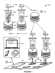
100351 Figure I illustrates an embodiment of a system 100 for detecting or
assessing a
susceptibility of a microorganism 102 to an anti-infective 104. The system 100
can
comprise a fluid delivery device 106, a filter housing 108 containing a filter
110, a.
substrate .112, and a reader 114. The substrate 112 can have one or more
sensors 116
disposed on a surface of the substrate 112. The substrate 112 can be comprised
of a
polymeric material, a .inetal, a ceramic, a semiconductor layer, an oxide
layer, an insulator,
or a combination thereof. The system 100 can also include an analyzer 118. In
the
embodiment shown in Figure 1, the analyzer 118 can be disposed on a surface of
the
CA 02996417 2018-02-22
WO 2017/035393
PCT/US2016/048769
substrate 112. In other embodiments, the analyzer 118 can be a standalone unit
or device
coupled to the substrate 112.
100361 The sensors 116 can include one or more active sensors 120, one or
more
control sensors 122, or a combination thereof As illustrated in the embodiment
shown in
Figure 1., the one or more active sensors 120 and control sensors 1.22 can be
disposed on
the same side surface of the substrate 112. in other embodiments not shown in
Figure 1, the
active sensors 120 and the control sensors 122 can be disposed on different
surfaces of the
substrate 142, different substrates 112, or a combination thereof. For
example, Figure 1
shows the substrate 1.12 having four sensors 116; however, it is contemplated
that the
substrate 112 can comprise any number of sensors 116. in one embodiment, at
least one of
the sensors 116 can be an ion-sensitive field effect transistor (ISFET). The
sensors 116 will
be discussed in more detail in the sections that follow_
100371 The system 100 can detect or assess the level of susceptibility of
the
microorganism 102 to an anti-infective 104 When the microorganism 102 is in a
fluid
sample 124. The fluid sample 124 can include a bodily fluid such as blood,
serum, plasma,
urine, saliva, joint fluid, semen, wound .material, spinal fluid, mucus, or a
combinatiou
thereof. In other embodiments, the fluid sample 124 can also include an
environmental
fluid such as liquids sampled from a stream, river, lake, ocean, contamination
site,
quarantine zone, or emergency area. The fluid sample 124 can also be a food
sample.
100381 The microorganism 102 can be any metabolizing single or mufti-
cellular
organism including bacteria or fungi. In certain embodiments, the
microorganism 102 can
be a bacteria selected from the genera consisting of Ae inetobacter,
Aeromonasõ
Biteteroides, Chrobacter, Enterobacter, Escherichia, Klebsiella, Morganelia,
.Pandoraea,
Proteus, Providencia, Pseudomonas, Ralstonia, Raoultelia, Salmonella,
Setratia,
.Shewanella, Shigel.la, Stenotrophomonas, Streptomyces, Staphylococcus,
Enterocoecus,
Clostridium or any combination thereof, In other embodiments, the
microorganism 102 can
be a fungi selected from the genera consisting of Candida, Cryptococcus, or
any
combination thereof. In another embodiment, the microorganism 102 can include
amoeba.
In further embodiments, the microorganism 102 can be cancer cells and the anti-
irtfectives
104 can be chemotherapeuties or other cancer treatments.
100391 As illustrated in Figure 1, the fluid delivery device 106 can
deliver or inject the
fluid. sample 12.4 comprising the microorganism 102 into the filter housing
108 in step IA.
In the example embodiment shown in Figure 1, the fluid delivery device 106 can
be a
syringe. In other embodiments not shown in Figure 1, the fluid delivery device
106 can be
6
CA 02996417 2018-02-22
WO 2017/035393
PCT/US2016/048769
an injection cartridge, a microfluidic channel, a pipette, a reaction tube, a
capillary, a test
gibe, a combination thereof, or a portion therein.
100401 The filter housing 108 can be a container or vessel configured to
secure or
enclose the filter 110. For example, the filter housing 108 can be a housing
of a syringe
filter. The filter 110 can be a mesh or matrix, for isolating or separating
the microorganism
:102 or other molecules or cells from the supernatant of the fluid. sample
124, In certain
embodiments, the :filter 110 can be selected from: the group consisting of
cellulose acetate,
regenerated cellulose, nylon, polystyrene, polyvinylidene fluoride (INDF),
polyethersulfone (PES), polytetrafluorethylene (PTFE), or a combination
thereof.
100411 The filter 110 can comprise a filter surface 126. The filter surface
126 can be
the portion of the filter 11.0 used to isolate or trap the microorganism 102.
The filter surface
126 can include an external surface, an internal surface extending into the
filter 11.0, or a
combination thereof. The filter housing 108 can have at least one opening 128
which allow
fluid or supernatant from the fluid sample 124 to evacuate the filter housing
108. For
example, step IA can include the additional step of discarding the fluid or
supernatant from
the fluid sample 124 through the opening 128 after isolating the microorganism
102 on the
filter surface 126.
100421 In an alternative embodiment not Shown in Figure I, the fluid sample
124 can
be pre-filtered in a step before step .1A. This pre-filtering step can involve
filtering the fluid
sample 124 using another instance of the filter 110, a microfluidic filter, or
a combination
thereof to filter out other larger cellular components including blood cells
or epithelial cells
from the fluid sample 124 when the fluid sample 124 is composed of bodily
fluid.
100431 The same fluid delivery device 106 or another fluid delivery device
.106 can
also be used to deliver or inject, a nutrient solution 130 to the filter
housing 108 in step 1B.
'The fluid delivery device 106 can continuously or periodically expose the
filter surface 126
containing the microorganism 102 with the nutrient solution 130. In one
embodiment, the
nutrient solution .130 can be composed of a butler containing bacto-tryptone,
yeast: extract,
sodium chloride and any combinations thereof. In another embodiment the
.nutrient
solution can include a growth inducer. The growth inducer can be selected from
the group
consisting of a carbon-based inducer, a nitrogen-based inducer, a mineral, a
trace element,
a biological growth factor, or any combination thereof For example, the growth
inducer
can include but is not limited to glucose, ammonia, magnesium, or a
combination thereof
For example, the nutrient solution 130 can be 0.1x Luria Broth supplemented
with 100 naM
glucose.
7
CA 02996417 2018-02-22
WO 2017/035393
PCT/US2016/048769
100441 The buffer in the nutrient: solution 130 can be an acidic buffer or
a basic buffer.
The 'buffer can be used to counteract. the buffering effects of ions or
substances present in
the fluid sample 124 when the fluid sample 124 is composed of a bodily fluid.
100451 The filter 110 comprising the microorganism 102 can be heated to a
temperature
of between 30 T. and 40 T: and allowed to incubate for an incubation period
132 in step
IC. In one embodiment, the filter 110 can be incubated, while in the filter
housing 108. In
another embodiment, the filter 110 can be removed from the filter housing 108
prior to
incnbation. In some embodiments, the filter 110 can be incubated with the
nutrient solution
130. The incubation period 132 can .range from .15 minutes to over one hour,
in other
embodiments, the incubation period 132 can be less than 15 minutes. The
incubation period
13.2 can be adjusted based on the type of microorganism 102, such as the type
of bacteria or
fungi.
0O461 The incubation period 132 can also be adjusted based on the amount
of the
microorganism 102 present in the fluid sample 124. For example, the incubation
period 132
can be increased when the amount of the microorganism 102 is below a threshold
amount.
The filter 110 can be allowed to incubate with the nutrient solution 130 in
order to promote
the proliferation of the microorganism 102 on the ..tilter surface 126. One
advantaee of
incubating the filter 110 is to increase the sensitivity of the system 100 to
small amounts of
the microorganism 102. For example, incubating the filter 110 can allow the
system 100 to
reduce its level of detection. in one embodiment, the system 100 can detect as
&w as 500
bacteria. per milliliter. in other embodiments, the system 1.00 can detect
fewer than 500
bacteria per milliliter. In further embodiments, the system 100 can detect 1*-
104 bacteria per
100471 After incubating the filter 110, the same fluid delivery device 106
or another
fluid delivery device 106 can then be used to expose the filter surface126
with additional
nutrient solution .130 in step ID. One advantage of exposing the filter 110
with the
additional .nutrient solution 130 is to prevent the filter housing 108, the
filter .110, Of the
environment housing the microorganism 102 from becoming overly acidified as a
result of
cellular activity, cellular metabolism,. or growth undertaken by the
microorganism -102. For
example, the filter housing 108 or the filter 110 comprising the microorganism
102 can
become overly acidified as result of the microorganism 102 undergoing cellular
metabolism or growth..
1.0048i As illustrated in the example embodiment shown in Figure 1, the
effluent or
outflow from the exposure step of step 10 can be introduced or applied, to one
or more of
8
CA 02996417 2018-02-22
WO 2017/035393
PCT/US2016/048769
the sensors 116 disposed on the substrate 112. This effluent or outflow can be
referred to as
a sample effluent 134.
100491 The sample effluent 134 can be introduced to one or more of the
sensors 116
disposed on the substrate 112 through the opening 128 in the .filter housing
108. The
opening 128 can include a channel, a capillary, a tube, or a combination
thereof. The.
sample effluent 134 can be separated from the microorganism 102 on the filter
surface 126
as the sample effluent 134 flows through the filter 110 on to the sensors 116.
in these
embodiments, the microorganism .102 can be kept separate or prevented from
contacting
any portion of the sensors 116 disposed on the substrate 112,
100501 The sample effluent 134 can comprise a solution characteristic 136.
The
solution characteristic 136 can refer to one or more attributes of the
solution making up the
sample effluent 134. For example, the solution characteristic .136 can include
a
concentration of a solute or an absolute number of solutes in solution. The
solution
characteristic 136 can include an amount or concentration of ions, organic
molecules such
as amino acids, minerals, or other inorganic compounds in the sample effluent
134.
100511 The solution characteristic 136 can vary as a result of ions,
organic molecules,
or minerals produced by or attributed to the microorganism 102 on the filter
surface 126.
'The solution characteristic 116 can be a direct or indirect byproduct of a
cellular activity
undertaken by the .microorganism 102 such as tell .metabolism or cell growth.
In one
embodiment, the sample effluent 134 can comprise hydrogen ions (W) as a
byproduct of
bacterial MI metabolism or growth. In other embodiments, the sample effluent
134 ean
comprise adenosine triphosphate (ATP), carbon dioxide (CO). lactic acid,
carbonic acid,
nitrates (NO3') or a combination thereof produced by or attributed to the
microorganism
102,
100521 After introducing the sample effluent 134 to the sensors 116, the
same fluid
delivery device 106 or another fluid delivery device .106 can be used to
introduce an anti-
infective 104 to the filter surface 126 comprising the microorganism .102 in
stop IF. In the
example embodiment shown in Figure 1, the anti-infective 104 can be mixed with
additional nutrient solution 130 and the filter surface 1.26 comprising the
microorganism
102 can be exposed to additional nutrient solution .130. In other embodiments,
the anti-
infective 104 can be introduced to the filter surface 126 separate from the
nutrient solution
130,
1.0531 The anti-infective 104 can comprise a bacteriostatic anti-infective,
a bactericidal
anti-infective, or a combination thereof. In certain embodiments, the
bacteriostatie anti-
9
CA 02996417 2018-02-22
WO 2017/035393
PCT/US2016/048769
infective can comprise P.-lac-tarns, Aminoglyeosides, .Ansamycins
Glycopeptides,
Lipopeptides, Quinolones, Streptogramins, or any combination thereof The
bactericidal
anti-infective can comprise .Chloramphenicols, Macrotides, Oxazoiidinones,
Sullbnamides,
Tetracyclines, any combination thereof, or future derivations thereof:
100541 In the example embodiment shown in Figure 1, the filter 110
incubated in step
1(. can be divided into two separate filters 110 with each filter 110 having
the
microorganism 102 on the filter surface 126. In this embodiment, nutrient
solution 130
containing the anti-infective 104 can be exposed or introduced to one of the
filters 110
comprising the microorganism 102 in step 1D and nutrient solution 130 without
the anti-
infective 104 can be exposed or introduced to the other filter 110 in step 1E,
In one
embodiment, step ID can occur concurrently or near in time with step 1E. In
other
embodiments, step ID and step 1E can occur sequentially.
0O551 In these embodiments, the sample effluent 134 resulting from the
exposure step
of step ID can be introduced to a different sensor 1.16 than the sensor 116
used to atullyze
the sample effluent 134 from step 1E. Also, in these embodiments, the sensor
116 receiving
the sample effluent 134 containing the anti-infective 104 can be referred to
as the active
sensor 120 and the sensor 116 receiving the sample effluent 134 without the
anti-infective
104 can be referred to as the control sensor 122_
[00561 In an alternative embodiment contemplated by the present disckisure,
the same
filter 110 exposed to the nutrient solution 130 in step 1,6 can be exposed to
a nutrient
solution 130 containing the anti-infective 104 at a later point in time.. In
this embodiment,
the sample effluent 134 from the exposure step comprising the anti-infective
104 can also
be introduced to the same sensor 116 as the sensor 116 -used to measure the
non-anti-
infective sample effluent 134 in step 1E.
100571 In yet another embodiment contemplated but not shown in Figure I,
portions of
the fluid sample 124 can be divided into multiple filter housings.] 08 prior
to step 1A. in
this embodiment, each filter housing I0 can contain a filter 110 comprising
microorganisms 102 from the fluid. sample 124 disposed on the filter surface
126. Each of
the filter housings 10S can he incubated and a variety of nutrient solutions
130, including
nutrient solutions 130 lacking in anti-infective 104 or containing different:
types of anti-
infretives 104, can be used to expose the various filters 110, in this
embodiment, the
sample effluent 134 from the various filter housings 1.08 can be introduced to
different
sensors 116 on the substrate 11.2.
CA 02996417 2018-02-22
WO 2017/035393
PCT/US2016/048769
100581 While Figure 1 illustrates two of the .fonr sensors 116 on the
substrate 112 being
used to analyze sample effluent 134 from the fluid sample 124, it is
contemplated that the
substrate 11.2 can accommodate any number of sensors 116 tbr receiving the
sample
effluent 134. For example, the substrate 1.12 can he a support or housing for
a high
throughput assay plate such as a 96 well plate, a 192 well plate, or a 384
well plate. In this
example, each of the well plates can be in fluid communication with one or
more sensors
116. in another embodiment, the sensors 1.16 can be positioned directly
underneath the
=filter housing 108.
100591 The reader 114, the analyzer .118, or a combination thereof can be
configured to
monitor an electrical Characteristic 800 (see Fig. 8) of the sensors 116 upon
introducing the
sample effluent 134 to the sensors 116. For example, the reader 11.4 can
monitor the
electrical characteristic 800 of the sensors 116 by receiving one or more
signals from the
analyzer 118 disposed on the substrate 112. In one embodiment, the analyzer
118 can
comprise a controller to execute logical commands concerning the detection or
comparison
of the electrical characteristic 800 of the sensors 116..th other embodiments,
the controller
can be intetzrated with the reader 114 or another device coupled to the
analyzer 118.
100601 The electrical characteristic 800 can include a current, a voltage,
a threshold
voltage, a capacitance, a resistance, a noise level, a subthreshold swing, a
level of
induction, or a combination thereof measured at or .near the sensor 116, The
reader 114 can
be electrically or communicatively coupled to the analyzer 118, the substrate
112, or a
combination thereof to monitor the electrical characteristic 800 of the
sensors 116 over
time. The reader 114 can also be configured to provide a read-out of the
electrical
characteristic 800 of the sensors 116.
100611 In certain embodiments, the reader 114 can be a mobile device, a
handheld
device, a tablet device, or a computing device such as a laptop or desktop
computer, in
other embodiments, the reader 114, the analyzer 118, a combination thereof, or
any portion
therein can be integrated into an ISFET probe or meter.
100621 In the example embodiment shown in Figure 1, the analyzer 118, the
reader
114, or a combination thereof can monitor the electrical characteristic 800 of
the sensors
116, such as the active sensor 120 and the control sensor 122 in step IF. The
analyzer 118,
the reader 114, or a combination thereof can monitor the electrical
characteristic 800 of the
active sensor 120 upon introducing the sample effluent 134 containing the anti-
infective
104 to the active sensor 1.20. in addition, the analyzer 118, the reader 1.14,
or a combination
thereof can also monitor the electrical characteristic 800 of the control
sensor 122 upon
11
CA 02996417 2018-02-22
WO 2017/035393
PCT/US2016/048769
introducing .the sample effluent 134 without the anti-infective 104 to the
control sensor
122. The analyzer 118, the reader 114, or a combination thereof can compare
the electrical
characteristic 800 of the active sensor 120 with the electrical characteristic
800 of the
control sensor 122 to assess the susceptibility of the microorganism 102 to
the anti-
infective 1.04.
[00631 The electric-al characteristic 800 of the sensors 116 can differ
when the solution
characteristic 136 of the sample effluents .134 differ as a result of
differences in the
concentration or the amount of solutes present in the sample effluents .134.
For example,
the electrical characteristic 800 of .the active sensor 120 and the control
sensor 122 can.
differ when the solution characteristic 136 of the sample effluent 134
introduced to the
active sensor 120 differ from the solution characteristic 136 of the sample
effluent 1.34
introduced to the control sensor 1.22. As a more specific example, the
electrical
characteristic 800 of the active sensor 120 and the control sensor 122 can
differ when the
solution characteristic 136 of the sample effluents 134 differ in their pH or
differ in the
concentration of another ion, an organic molecule, or a combination thereof
100641 in another embodiment contemplated but not shown in Figure 1, the
analyzer
118, the .reader 114, or a combination thereof can monitor the electrical
characteristic 800
of one sensor 116 upon introducing the sample effluent 134 without the anti-
infective 104.
to the sensor 116. In this embodiment, additional nutrient solution 130
comprising the anti-
infective 104 can be introduced or exposed to the filter surface 126
comprising the
nneroorganism 102 and additional sample effluent 134 resulting from this
exposure step
can be introduced to the sensor 116. The analyzer 118, the reader 114, or a
combination
thereof can detect any changes in the electrical characteristic 800 of the
sensor 116 after
introducing the additional sample effluent 134 to the sensor 116. The analyzer
118, the
reader 11.4, or a combination thereof can then assess the susceptibility of
the
microorganism 102 to the anti-infective 104 using any detected changes in the
electrical
characteristic 800 of the sensor 116.
[00651 In this embodiment, the change in the electrical characteristic 800
of the sensor
116 can indicate a change in the solution characteristic .136 of the sample
effluent 134
introduced to the sensor 116. For example, the change in the solution
characteristic 136 of
the sample effluent 134 can indicate a change in the concentration of an ion,
an organic
molecule, or a combination thereof in the sample effluent 134. As a more
specific example,
the change in the solution characteristic 136 of the sample effluent 134 can
be a change in
the pH of the sample effluent 134,
12
CA 02996417 2018-02-22
WO 2017/035393
PCT/US2016/048769
100661 In these and other embodiments, the analyzer 118, the reader 114, or
a.
combination thereof can assess the susceptibility of the microorganism 102 to
the anti-
infective 104 within a detection period 138, in one embodiment, the detection
period 138
can range .from 60 minutes to 240 minutes. In another embodiment, the
detection period
.138 can be less than 60 minutes. In yet another embodiment, the detection
period .138 can
be greater than 240 minutes.
[00.671 The reader 114 can produce an output signal. 808 (see
figgelqassessing the
susceptibility of the microorganism 102. In one embodiment, the output signal
808 can :be-
an electrical signal., In this embodiment, the output signal 808 can be
.rendered as a graphic,
such as a text string, a number, a symbol, or a combination thereof on a
display miit of the
reader 114. In another embodiment, the output signal 808 can be an audio
signal.
[00681 The analyzer 118, the reader 1.14, or a combination thereof can
assess the
susceptibility of the microorganism 102 to the anti-infective 104 as a binary
assessment or
a gradated or tiered assess=nt, in one embodiment, the analyzer 118, the
reader 114, or a
combination thereof can assess the susceptibility of the microorganism 102 as
either
resistant or non-resistant to the anti-infective 104. In this embodiment, the
system 100 can
introduce a set amount of the anti-infective 104 to the nutrient solution 130
and the reader
114 or the analyzer 118 can assess the susceptibility of the microorganism 102
as either
resistant or non-resistant based on any detected changes in the electrical
characteristic 800
of one sensor 116 or any detected differences in the electrical characteristic
800 of the
active sensor 120 and the control sensor 122.
10069} For example, the reader 114, the analyzer 118, or a combination
thereof can
assess the susceptibility of the microorganism .102 as resistant to the anti-
infective 104
when the analyzer 118 detects a change in the electrical characteristic 800 of
the one sensor
116 even after anti-infective 104 is introduced to the filter surface 126
comprisin.g the
microorganism 102. Also, for example, the reader 114, the analyzer 118, or a
combination
thereof can assess the susceptibility of the microorganism 102 as not
.resistant to the anti-
infective 104 when the analyzer 118 fails to detect a change in the electrical
characteristic
800 of the one sensor 116 when anti-infective 104 is introduced to the filter
surface 126
comprising the microorganism 102. Moreover, the reader .114, the analyzer 118,
or a
combination thereof can assess the susceptibility of the microorganism 102 as
not resistant
to the anti-infective 104 When the analyzer 118 fails to detect a
statistically significant
change or a change in the electrical characteristic. 800 of the one sensor 116
exceeding a
threshold value.
13
CA 02996417 2018-02-22
WO 2017/035393
PCT/US2016/048769
100701 As another example, the .reader 114, the analyzer 118, or a
combination -thereof
can assess the susceptibility of the microorganism 102 as resistant to the
anti-infective 104
when the analyzer 118 or the reader 114 thas to detect a statistically
significant difference
between the electrical characteristic 800 of the active sensor 120 and the
control sensor
.122. More specifically, this statistically significant difference in the
electrical characteristic
800 can be a difference exceeding a threshold value. In this example, the
system 100 can
introduce the sample effluent 134 from the nutrient solution 130 comprising
the anti-
infective 104 to the active sensor 120 and the sample effluent 134 free from
anti-infective
104 to the control sensor 122. In addition, the reader 114, the analyzer 118,
or a
combination thereof can assess the susceptibility of the microorganism 102 as
not resistant
to the anti-infective 104 when the reader 114 or the analyzer 118 detects a
statistically
significant difference between the electrical characteristic 800 of the,
active sensor .120 and.
the control sensor 122 over time.
100711 In other embodiments, the reader 114,.the.iln*nt 118. or a
combination:
thereof can assess the level of susceptibility of microorganism .102 on .a
gradated. or
tiered scale. For example, the reader 114 can assess the susceptibility of the
microorganism.
102 as being resistant, mildly susceptible, or susceptible to the anti-
infective 104. In these
embodiments, .anti-infeetives 104 of ditThrent concentrations can be
introduced to the filter
surface .126 comprising the microorganism 102 to assess the level of
susceptibility of the
microorganism 102 to the anti-in.fective 104.
100721 As a more specific example, when only one sensor 116 is used to
assess the
level of susceptibility of the microorganism 102, the system 100 can introduce
larger
amounts of the anti-infective 104 to the .fi.her surface 126 over time and
monitor the effects
of the additional anti-infective 104 on the electrical Characteristic 800 of
the sensor 116
over such a time period. As another exam*, when multiple active sensors 120
are
disposed on the substrate 112, the system 100 can introduce differing amounts
of the anti-
infective 104 to different active sensors 120 simultaneously or over time and
the reader
114, the analyzer 118, or a combination thereof can compare the electrical
characteristic
800 of the various active sensors 12.0 with the control sensor 1.22 to assess
the level of
susceptibility of the microorganism 102 to the anti-infective 1.04.
[00731 While three categories of susceptibility are discussed in the
section above, it
Should be understood by one of ordinary skill in the art that four or greater
categories of
susceptibility can be used to assess the level of susceptibility of the
microorganism 102 to
the anti-infective 104.
14
CA 02996417 2018-02-22
WO 2017/035393
PCT/US2016/048769
100741 Figure 2 illustrates another embodiment of .the system 100 for
detecting or
assessing the susceptibility of a microorganism 102 to an anti-infective 104.
The system
100 can comprise the fluid delivery device 106, the substrate 1.12 comprising
substrate
wells 200, and the reader 114. The substrate 12 can have one or more sensors
.116
disposed on a substrate surface 202. The system 100 can also include the
analyzer 118. In
the embodiment shown in Figure 2, the analyzer '118 can be disposed on the
substrate
surface 202. in other embodiments, the analyzer 118 can be a standalone unit
or device
coupled to the substrate 112.
[0075f The sensors 116 can include one or .more active sensors 120, one or
more
control sensors 122, or a combination thereof disposed on the substrate
surface 202. As
illustrated in the embodiment shown in Figure 2, the active sensors 120 and
control sensors
122 can be disposed on one side of the substrate 112. In other embodiments not
shown in
Figure 2, the active sensors 120 and the control sensors 122 can be disposed
on different
sides of the substrate .112 or on different substrates- For example, Figure 2
shows the
substrate 112 having three sensors 116; however, it is contemplated that the
substrate .112
can comprise any number of sensors 116. In one embodiment, at least one of the
sensors
116 can be ISFET.
10076.4 The substrate wells 200 can include a sample well 204, one or more
active wells
206, one or .1.110f0 control wells 208, or a combination thereof The sample
well 204, the one
or more active wells 206, the one or more control weds 208, or a combination
thereof can
be fluidly coupled to or be in fluid communication with one another through
substrate
channels 210, The substrate channels 210 can include tubes, capillaries,
microfluidie
channels, indentations, or holes disposed on or inside the substrate 112_
100771 The substrate wells 200 including the sample well 204, the active
well 206, the
control wells 208, or a combination thereof can be divots, indentations, or
openings on the
surface of the substrate 1.12. In another embodiment, the substrate wells 200
can be
enclosed spaces within the substrate 112. In other embodiments, the substrate
wells 200
can be receptacles or cartridges coupled to the substrate 112. The substrate
wells 200 can
also be fluidly coupled to or be in fluid communication with the sensors 116
through the
substrate channels 210,
100781 As illustrated in Figure 2, the fluid delivery device 106 can
deliver or inject the
fluid. sample 124 comprising the microorganism 102 into the sample well 204 in
step 2A..
In an alternative embodiment not shown in Figure 2, the fluid sample 124 can
be pre-
filtered in a step before step 2A. This pre-filtering step can involve
filtering the fluid.
CA 02996417 2018-02-22
WO 2017/035393
PCT/US2016/048769
sample 124 using the .flier 110, a .microfluidie filter, or a combination
thereof to filter out
other larger cellular components including blood cells or epithelial cells
from the fluid
sample 124.
[00791 The same fluid delivery device 106 or another fluid delivery device
106 can
also be used to deliver or inject the nutrient solution 1.30 to the sample
well 204 in step 213.
The fluid delivery device 106 can continuously or periodically introduce or
expose the
substrate surface 202 of the sample well 204 with the nutrient solution 130.
In one
embodiment, the nutrient solution 130 can be composed of a buffer containing
'bacto-
tryptorie, yeast extract, sodium chloride and any combinations thereof. In
another
embodiment the nutrient solution can include a growth inducer. The growth
inducer can be
selected from the group consisting of a carbon-based inducer, a nitrogen-based
inducer, a
mineral, a trace element, a biological growth factor, or any combination
thereof For
example, the growth inducer can include but is not limited to glucose,
ammonia,
magnesium, or a combination thereof. For example, the nutrient solution 130
can be 0.1x
Luria Broth supplemented with 100 inM glucose.
100801 The flow of the nutrient solution 130 can carry or deliver the
microorganism.
102 in the sample well 204 to the active well 206, the control well 208, or a
combination
thereof For example, the sample well 204, the active well 206, the control
well 208, or a
combination thereof can be shaped as a hemisphere having a rounded bottom, a
cubold
having a flat or planar bottom, a cone, a .frustoconical, a hyperboloid, or a
combination
thereof. The entire substrate 112 can be heated to a temperature between 30
A.: to 40"C
when the microorganism 102. is in the active well 206, the control well 208,
or a.
combination thereof and allowed to incubate for the incubation period 132. The
substrate
112 can be allowed to incubate in order to promote the proliferation,
metabolism, or growth
of the microorganism 102 in the active wells 206, the control wells 208, or a
combination.
thereof.
[00811 The substrate wells 200, including the sample well 204, the active
well 206, the
control well 208, or a combination thereof, can he covered by a well coating
212. The well
coating 212 can cover or coat the bottom or sides of the wells. The well
coating 212 can
include an anti-buffer coating such as an acidic coating or a basic coating.
[00821 The well coating 212 can also be a trapping coating configured to
trap the
microorganism 102 in the active wells 206, the control. wells 208, or a
combination thereof.
For example, the well coating 21.2 can be an extracelhdar matrix comprising
proteins such
.fibronectin, collagen, laminiri, osteopontin, poly-D-lysine, or a combination
thereof The
16
CA 02996417 2018-02-22
WO 2017/035393
PCT/US2016/048769
well coating 212 can also be a charged coating such as an amine surf.ace, a
carboxyl
surface, a charged peptide surface, or a combination thereof 11w well coating
212 can also
he an oxygen or nitrogen containing surfitce.. The well coating 212 can also
be a
polyurethane surface.
[00831 The active wells 206, the control wells 208, or a combination
thereof cm have a
physical barrier 214. The physical barrier 214 can be a physical feature or
design of the
well for trapping or isolating the microorganism .102 in the active well 206,
the. control. well
208, or a combination. thereof. For example, the physic& barrier 214 can be an
overhang or
lip protruding from a downstream section of the active well 206, the control
well 208, or a
combination thereof As another example: the physical barrier 214 can be a
sloping surface
of the active well 206, the control well 208, or a combination thereof in
another
embodiment contemplated but not shown in Figure 2, the physical barrier 214
can be the
filter 110 disposed at an opening of the active well 206, the control well
208, or a.
combination thereof downstream from the sample well 204.
[00841 Although the. example embodiment in Figure 2 shows the physic&
barrier 214
as a feature of the substrate wells 200, .the physical barrier 214 can also be
a feature of the
subsuate Channels 21Ø For example, the substrate channels 210 can be
inicrolluidic
channels, which narrow to a width or diameter which prevent the microorganism
102 from
proceeding down the substrate channels 210 toward the sensors 116. In this
example
embodiment, the substrate 112 can act as a .microfluidic chip or lab-on-chip
.(LOC).
[0085I The well coating 212, the physical barrier 214, or a combination
thereof can be
included as part of the system 100 to prevent or stop the microorganism 102
from
contacting or reaching the sensors 116. In another embodiment contemplated but
not
Shown in Figure 2, an .electtical or magnetic component can be used to trap or
isolate the
microorganism .102 in the active well 206, the control. well 208, or a
combination thereof.
[00861 The nutrient solution 130 delivered in step 2B or additional
nutrient solution
130 can be continuously or periodically delivered or injectixl into the sample
well 204, the
active well 206, the control well 208, or a combination thereof until the
microorganism 102
is carried or delivered into one or more active wells 206, control wells 208,
or a
combination thereof. The active wells 206, the control wells 208, or a
combination thereof
can comprise one or more opeaings, physical features õgeometries, or d.evice
features which
allow fluid or supernatant in the active wells 206, the control wells 208, or
a combination
thereof to evacuate or exit the wells into one or more substrate channels 210.
The fluid or
17
CA 02996417 2018-02-22
WO 2017/035393
PCT/US2016/048769
supernatant separated from the .microorganism 102 in the active wells 206, the
control
wells 208, or a combination thereof can be referred to as the sample effluent
134.
100871 As illustrated in the example embodiment shown in Figure 2, the
sample
effluent 134 can be introduced, carried, or delivered to one or more of the
sensors .1.16
disposed on the substrate 112. The sample effluent 134 can comprise a solution
characteristic 136. The solution characteristic 136 can include an amount or
concentration
of ions, organic molecules such as amino acids, minerals, or other inorganic
compounds in
the sample effluent 134.
100881 The solution characteristic 136 can vary as a result of ions,
organic molecules,
or minerals produced by or attributed to the microorganism 102 in the active
wells 206, the
control wells 208, or a combination thereof The solution characteristic 136
can be a direct
or indirect byproduct: of a cellular activity undertaken by the microorganism
1.02 such as
cell metabolism or cell growth. The sample effluent 134 can comprise H'. ATP,
CO. lactic
acid, carbonic acid:. NO3", or a combination thereof.
100891 The substrate channels 120 can deliver or introduce sample effluent
134 from
one or more active wells 206 to one or more active sensors 120. In addition,
separate
substrate channels 120 can deliver or introduce sample effluent 134 from one
or more
control wells 208 to one or more control sensors 122..
100901 After or prior to incubating the substrate 112, the same fluid
delivery device
106 or another fluid delivery device 106 can be used to introduce an anti-
infective 104 to
the active wells 206 in a step 2C. In the example embodiment shown in 'Figure
2, the anti-
infective 104 can be mixed with additional nutrient solution 130 and the
active wells 206
comprising the microorganism 102 can be exposed to additional nutrient
solution 130
comprising the anti-infective 104. In other embodiments, the anti-infective
104 can be
introduced to the active wells 206 separate from the nutrient solution .130..
100911 In the example embodiment shown in Figure .2, nutrient solution 130
containing
the anti-infective 104 can be delivered or introduced to the active well 206
comprising, the
microorganism 102 while nutrient solution 130 lacking the and-infective 104
can he
delivered or introduced to the control well 2.08 also comprising the
microorganism 102. In
these embodiments, the sample effluent 134 flowing from the active well 206
can be
introduced to the active sensor 120 and the sample effluent 134 flowing from
the control
well 208 can be introduced to the control sensor 122.
100.921 In an alternative embodiment contemplated hut not shown in Figure
2, one
active well 206 can initially be exposed to nutrient solution 130 lacking in
anti-infective
18
CA 02996417 2018-02-22
WO 2017/035393
PCT/US2016/048769
1.04 and the sample effluent 134 flowing from the active well 206 can be
introduced to a
sensor 1.16. In this erribodiment, the same active well 206 can be exposed at
a later time
with nutrient solution 130 comprising the anti-infective 104. By doing so, the
sample
effluent 134 from this second exposure step can he introduced to the same
sensor .116 as
the sensor 11.6 used to measure the non-anti-infeetive sample effluent 1.34.
[00931 While Figure 2 illustrates two of the three sensors 116 on the
substrate 112
being used to analyze sample effluent 134 from the fluid sample 124, it is
contemplated
that the substrate 112 can accommodate any number of sensors 116 for receiving
the
sample effluent 134. For example:, the substrate 112 can be a support or
housing for a high
throughput assay plate such as a 96-wel1 .plate, a 192-well plate, or a 384-
well. plate, In this
example, each of the well plates can be in fluid communication with at least
one sensor
116.
00941 The reader 114, the analyzer 118, or a combination thereof can he
configured to
monitor the electrical characteristic 800 of the sensors 116 upon introducing
the sample
effluent 134 to the sensors 116. For example, the reader 114 can monitor the
electrical
characteristic 800 of the smuts 116 by receivinu, one or more signals .from
the analyzer
118 disposed on the substrate 112.
100951 In the example embodiment shown in Figure 2, the analyzer 118, the
reader
114, or a combination thereof can monitor the electrical characteristic 800 of
the sensors
116, such as the active sensor 120 and the control sensor 122 in step 2D. The
analyzer 118,
the reader 114, or a combination thereof can monitor the electrical
characteristic 800 of the
active sensor 12.0 upon introducing, the sample effluent 134 front the active
well 206 to the
active sensor 120. In addition, the analyzer 118, the reader 11.4, or a
combination thereof
can also monitor the electrical characteristic 800 of the control sensor 122
upon introducing
the sample effluent 134 from the control well 208 to the control sensor 122.
The analyzer
.118, the reader 11.4, or a combination thereof can compare the electrical
characteristic 800
of the active sensor 120 with the electrical characteristic 800 of the control
sensor 122 to
assess the susceptibility of the microorganism 102 to the anti-infective .104.
10096} 'The electrical characteristic 800 of the sensors 116 can differ
when the solution
characteristic 136 of the sample effluents 134 differ as a result of
differences in the
concentration or the amount of solutes present in the sample effluents 134.
For example,
the electrical Characteristic 800 of the active sensor 120 and the control
sensor 122 can
differ when the solution characteristic 136 of the sample effluent134
introduced to the
19
CA 02996417 2018-02-22
WO 2017/035393
PCT/US2016/048769
active sensor 120 differ from .the solution characteristic 136 of the sample
effluent 134
introduced to the control sensor 122.
100971 In another embodiment contemplated but not shown in Figure 2, the
analyzer
118, the reader 11.4, or a combination thereof can monitor the electrical
characteristic 800
of one sensor 116 upon introducing, the sample effluent 134 without the anti-
infective 104.
to the sensor 116, in this embodiment, additional nutrient solution 130
comprising the anti-
infective 104 can be delivered or exposed to the same sensor .116 and
additional sample
effluent 134 resulting from this exposure step can be introduced to the sensor
116. The
analyzer 118, the reader 114, or a combination thereof can detect any changes
in the
electrical characteristic 800 of the sensor 116 after introducing the
additional sample
effluent 134 to the sensor 116. The analyzer .118, the reader 114; or a
combination thereof
can then assess the susceptibility of the microorganism 102 to the anti-
infective 104 using
any detected changes in the electrical .characteristic 800 of the sensor 116,
100981 In this embodiment, the change in the electrical. characteristic 800
of the sensor
116 can indicate a change in the solution characteristic 136 of the sample
effluent 134
introduced to the sensor 116. For example, the change in the solution
characteristic 136 of
the sample effluent 134 can indicate a change in the concentration of an ion,
an organic
molecule, or a combination thereof in the simple effluent 134, As a more
specific example,
the change in the solution characteristic. 136 of the sample effluent 134 can
be a change in
the pH of the sample effluent 134.
100991 In these and other embodiments, the analyzer 118, the reader 114, or
a
combination thereof can assess the susceptibility of the microorganism 102 to
the anti-
infective 104 within the detection period 138_
101001 The reader 114 can also produce the output signal 808 assessing the
susceptibility of the microorganism 102. The analyzer 118, the reader 11.4, or
a
combination thereof can assess the susceptibility of the microorganism 102 to
the anti-
infective 104 as the binary assessment or the gradated or tiered assessment.
[0.1.011 For example, the reader 114, the analyzer 118, or a combination
thereof can
assess the susceptibility of the microorganism 102 as resistant to the anti-
infective 104
when the analyzer 118 detects a change in the electrical characteristic 800 of
the active
sensor 120 even after anti-infective 104 is introduced to the active well 206
fluidly coupled.
to the active sensor 120. Also, for example, the reader 114, the analyzer 118,
or a
combination thereof can assess the susceptibility of the microorganism 1.02 as
not resistant
to the anti-in.lective 104 when the analyzer 118 fails to detect a change in
the electrical
CA 02996417 2018-02-22
WO 2017/035393
PCT/US2016/048769
characteristic 800 of the active sensor 120 when anti-infective 104 is
.introduced to the
active well 206 fluidly coupled to the active sensor 120. Moreover, the reader
114, the
analyzer 118, or a combination thereof can assess the susceptibility of the
microorganism
102 as not resistant to the anti-infective 104 when the analyzer 118 .fails to
detect a
statistically significant change or a change in the electrical characteristic
800 of the active
sensor 120 exceeding a threshold .value.
[01.021 As another example, the reader 114, the analyzer 118, or
a.eombina.tion thereof.
can assess the susceptibility of the .microorganism .102 as resistant to
theanti,nifective.:104.
When .the analyzer 1.18 or the reader 114 fails to detect a statistically
significant difference
between the electrical characteristic 8(X) of the active sensor 120 and the
control sensor
.122. More specifically, a statistically significant difference in the
electrical characteristic
800 can be a difference exceeding a threshold value. In this example, the
system 100 can
introduce the sample effluent 134 .from the active well 206 to the active
sensor 120 and. the
sample effluent 134 from the control well 208 to the control sensor 122. In
addition, the
reader 114, the analyzer 118, or a combination thereof can assess the
susceptibility of the
.microorganism 102 as not resistant to the anti-infective 104 when the reader
114 or the
analyzer 118 detects a statistically significant difference between the
electrical
characteristic 800 of the active sensor 120 and the control sensor 122.
[01031 In other embodiments, the reader 114, the analyzer 118, or a
combination
thereof can assess the level of susceptibility of the microorganism 102 on a
gradated or
tiered scale. For example, the reader 114, the analyzer 118, or a combination
thereof can.
assess the susceptibility of the microorganism .102 as being resistant, mildly
susceptible, or
susceptible to the anti-infective 104. In these embodiments, anti-intectives
104 of different
concentrations can be introduced to different active wells 206 comprising the
microorganism 102 to assess the level of susceptibility of the microorganism
102 to the
anti-infective 104.
[01041 As an example, when only one sensor 116 is used to assess the level
of
susceptibility of the microorganism 102, the system 100 can .introduce larger
amounts of
the anti-infective 104 to the active well 206 over time and monitor the
effects of the
additional anti-infective 104 on the electrical characteristic 800 of the
active sensor 120
fluidly coupled. to the active well 206 over such a time period. As another
example, when
multiple active sensors 120 are disposed on the substrate 112, the system 100
can introduce
differing amounts of the anti-infective 104 to different active wells 206 and
the reader 114,
the analyzer 118, or a combination thereof can compare the electrical
characteristic 800 of
21
CA 02996417 2018-02-22
WO 2017/035393
PCT/US2016/048769
the various active sensors 120 with one or more control sensors 122 to assess
the level of
susceptibility of the microorganism 102 to the anti-infective 104.
101051 Figure 3 illustrates another embodiment of the system 100 for
detecting or
assessing the susceptibility of a microorganism 102 to an anti -infective 104.
The system
.100 can comprise the fluid delivery device 106, the filter housing 108
containing the filter
/19, and a sensor device 300, in one embodiment, the sensor device 300 can be
a handheld
ISFET meter or probe.
[01.061 As illustrated in Figure 3, the fluid delivery device 106 can
deliver or inject the
fluid sample 124 comprising the microorganism 102 into the filter housing 108
in step 3A,
In the example embodiment shown in Fiiaire 3, the fluid delivery device 106
can be a
syringe. In other embodiments not shown in Figure 3, the fluid delivery device
106 can be
an injection cartridge, a microfluidie device, a pipette, a reaction tube, a
capillary, a test
tube, a combination thereof, or a portion therein.
101071 The filter housing 10$ can be a. container or vessel configured to
secure or
enclose the flu ter 110. For example, the filter housing 108 can be a housing
of a syringe
filter. The filter 110 can be a mesh or matrix for isolating or separating the
microorganism
102 Of other molecules or cells from the supernatant of the fluid sample 124.
101081 The filter 110 can comprise a filter surface 126. The filter surface
126 can be
the portion of the filter 110 used to isolate or trap the microorganism 102.
The filter surface
126 can include an external surface, an internal surface extending into the
filter 110, or a
combination thereof. Although not shown in Figure 3, the filter housing 108
c.an have at
least one opening 128 to allow fluid or supernatant from, the fluid sample 124
to evacuate
the filter housing .108. For example, step 3A can include the additional step
of discarding
the fluid or supernatant from the fluid sample 124 through the opening 128
after isolating
the microorganism 102 on the filter surface 126.
[01091 In an alternative embodiment not Shown in Figure 3, the fluid sample
124 can
be pre-filtered in a step before step 3A. This pre-filtering step can involve
filtering the fluid
sample 124 using another instance of the filter 110, a microfluidic filter, or
a combination
thereof to filter out other larger cellular components including blood cells
or epithelial cells
from the fluid sample 124 when the fluid sample 124 is composed of a bodily
fluid or
sample.
101101 The sarne fluid delivery device 106 or another fluid delivery device
106 can
also be used to deliver or inject a nutrient solution 130 to the filter
housing 108 in step 3W
The fluid delivery device 106 can continuously or periodically introduce or
expose the
22
CA 02996417 2018-02-22
WO 2017/035393
PCT/US2016/048769
nutrient solution 130 to the cater surface 126 containing the microorganism
102. in one
embodiment, the nutrient solution 1 30 can be composed of the growth inducer,
Luria
Broth, NaC1, and the buffer.
Nilli The filter housing 108 comprising the nutrient solution 130, the
.filter, and the
microorganism. 102 can be heated to a temperature of around 37 't and allowed
to incubate
for an incubation period 132 in a step 3C. The incubation period 132 can range
from 15
minutes to one hour. In other embodiments, the incubation period 132 can be
less than 15
minutes. The incubation period 132 can be adjusted based on the type of
microorganism
102.
101121 The incubation period 132 can also beadjitSted based on the amount
of the
microorganism 10.2 present in the fluid sample .124. For example, the
incubation period 132
can be increased when the amount of the microorganism 102 is below a threshold
amount.
The filter 110 can be allowed to incubate .with the nutrient solution 130 in
order to promote
the metabolism of the microorganism .102 on the filter 110, Furthermore, by
monitoring
the rate at which metabolites are produced using the sensor herein described,
it is possible
to identify the microorganism., as different microorganisms have
characteristic rates of
multiplication and metabolism. There is an additional feature hereby
disclosed, namely.
providing different nutrients to the microorganisms over time -while
monitoring the rate of
production of various .metabolites using the sensor herein described in order
to further
identify the microorganism.
[0113I After .incubating the filter housing 108, the filter 110 comprising
the
microorganism 102 can be separated from a solution representing the leftover
nutrient
solution 130 in the filter housing 108. This solution can be referred to as
the sample
effluent 134. The sensor device 300 can then be introduced or inserted in to
the sample
effluent 134 in step 3D to determine the solution characteristic 136 of the
sample effluent
.134. in another embodiment contemplated bat not shown in Figure 3, the sample
effluent
134 can be evacuated or removed from the filter housing 108 through an
opening, in the
filter housing 108 into another eoatainer Or veSSCI. The sensor device 300 can
then he used
to determine the solution characteristic 136 of the sample effluent 134 in
this other
container or vessel_
[01141 The solution characteristic 1.36 can refer to one or more attributes
of the
solution making up the sample effluent 134, For example, the solution
characteristic 136
can include a concentration of a solute or an absolute number of solute
molecules in
solution. The solution characteristic 136 can include an amount or
concentration of ions,
23
CA 02996417 2018-02-22
WO 2017/035393
PCT/US2016/048769
organic molecules such as amino acids, minerals, or other inorganic compounds
in the,
sample effluent 134.
101151 The solution characteristic 136 can vary as a result of ions,
organic molecules,
or minerals produced by or attributed to the microorganism 102 on the .filter
110. The
solution characteristic 136 can be a direct or indirect byproduct of a
cellular activity
undertaken by the microorganism 102 such as cell metabolism or cell growth. In
One
embodiment, the sample effluent 134 can comprise hydrogen ions (f') as a
byproduct of
bacterial cell meta.bolism or growth. ln other embodiments, the sample
effluent 134 can
comprise adenosine triphosphate (ATP), carlxm dioxide (CO lactic acid,
carbonic acid,
nitrates (N0), a combination thereof, or any other metabolic byproduct
produced by or
attributed to the microorganism 102,
[01161 After step 3C, the filter 110 comprising the microorganism .102 can
be removed
from the fliter housing 108 containing, the sample .effluent 134 and placed
into a new filter
housing 108> 'The same fluid delivery device i01 or another fluid delivery
device 106 can
then be used to introduce an anti-infective 104 to the new filter housing 108
containing the
filter 110 in a step 3E. :In an alternative embodiment, step 3E can involve
using the same
fluid delivery device 106 or another fluid delivery device 106 to introduce an
anti-infective
.104 to the filter housing 108 from step 3C after the sample effluent 134 has
been evacuated
or re.moved from the opening of the filter housing 108.
[01.171 In the example embodiment shown in Figure 3, the anti-in&ctive 104
can be
mixed with additional nutrient solution 130 and the filter 110 comprising the
microorganism 102 can be exposed to additional nutrient solution 130. In other
embodiments; the anti-infective 1.04 can be introduced to the filter 1.10
separate from the
nutrient solution 130,
101181 After introducing the anti-infective 104 to the filter housing 108,
the filter
housing 108 comprising the nutrient solution 130, the filter 110, the anti-
infective 104, and
the microorganism 102 can be heated to a temperature of around 37 T and
allowed to
incubate for an incubation period 132 in a step 3F.
101191 After incubating the filter housing 108õ the .filter 110 comprising
the
microorganism 102 can be separated from the sample effluent 134. A sensor
device 300
can then be introduced or inserted into the sample effluent 134 M step 36 to
determine the
solution characteristic 136 of the sample effluent 134. In another embodiment
contemplated but not shown in Figure 3, the sample effluent 134 can be
evacuated or
removed from the filter housing 108 through an opening in the filter housing
108 imo
24
CA 02996417 2018-02-22
WO 2017/035393
PCT/US2016/048769
another container or vessel. The sensor device 300 can then be used to
determine the
solution characteristic 136 of the sample effluent 134 in this other container
or vessel
10120j The reader 114 can then be used to compare the solution
characteristic 136 of
the sample effluent 134 from step 30 with the solution characteristic 136 of
the sample
effluent 134 from step 3D to assess the susceptibility of the microorganism
11)2 to the anti
infective 104 in step 3R. For example, the reader 114 can be used to compare
the two
solution characteristics 136 over time. The solution characteristic 136 from
step 31) and
step 30 can differ as a result of differences in the concentration or the
amount of solutes
present in .the sample effluents 134.. For example, the solution
characteristic 136 can differ
in their Ø1. or differ in the concentration of another ion, an organic
molecule, or a
combination thereof.
[01211 The reader 114 can assess the susceptibility of the microorganism
102 to the
anti-infective 104 within a detection period 138. In one embodiment, the
detection period
138 can ranee from 60 minutes to 240 minutes, In another embodiment, the
detection
period 138 can be less than 60 minutes. In yet another embodiment, the
detection period
138 can be greater than 240 .minutes.
101.22) The reader 114 can produce an output signal 808 assessing
susceptibility of
the microorganism 102. In one embodiment, the output 808 can be an
electrical
signal. In this embodiment, the output signal 808 can be rendered as a
graphic, such as a
text string, a number, a symbol, or a combination thereof on a display unit of
the reader
114. in another embodiment, the output signal 808 can be an audio signal,
0i.23} The reader 114 can assess the susceptibility of the microorganism
102 to the
anti-infective 1.04 as a binary assessment or a gradated or tiered assessment.
In one
embodiment, the reader 114 can assess the susceptibility of the microorganism
102 as
either resistant or neo-resistant to the anti-infective 104. In this
embodiment, the reader 114.
can assess the susceptibility of the microorganism 102 as either resistant or
non-resistant
based on any detected differences in the solution characteristic 136.
[0.1.241 For example, the reader 114 can assess the susceptibility of the
microorganism
102 as resistant to the anti-infective 104 when the reader 114 fails to detect
a statistically
significant difference between the solution characteristic 1.36 from step 3D
and the solution
characteristic 136 from step 30 over time. .A statistically. significant
difference can refer to
a difference exceeding a threshold value. Also, the reader 114 can assess the
susceptibility
of the microorganism 102 as sensitive to the anti-infective 104 when the
.reader 1.14 detects
CA 02996417 2018-02-22
WO 2017/035393
PCT/US2016/048769
a statistically significant difference between the solution characteristic 136
from step 3D
and the solution characteristic 136 from step 36 over time.
101251 In other embodiments, the reader 114 can assess the level of
susceptibility of
the microorganism 1.02 on a gradated or tiered scale. For example, the reader
114 can
assess the susceptibility of the microorganism 1.02 as being resistant, mildly
susceptible, or
susceptible to the anti-infective /04. In these embodiments, anti-infectives
104 of different
concentrations can be introduced to the filter housing 108 in step 3E to
assess the level of
susceptibility of the microorganism102 to the anti-infective 104. The reader
114 can.
compare the solution characteristic 136 of the various sample effluents 134
over .time to
assess the level of susceptibility of the microorganism. 102 to the aati-
infective 104,
101261 Figure 4A illustrates a side view of an embodiment of the substrate
112 haying
the active sensor 120 disposed on the substrate 112 and an external reference
electrode 400
extending into a measured. liquid 402 in contact with the active sensor 120,
As depicted in
Figure 4A, the substrate 112 can he comprised of a substrate carrier layer 404
and a base
dielectric layer 406, The substrate 112 can be a polymer layer, a metal layer,
a metalloid
layer, a ceramic layer, an organic semiconductor, an organic conductor such as
those
derived from polyacetylene, polyaniline. Quinacridone, or a combination
thereof For
example, the substrate 112 can he composed of silicon or an oxide of silicon
which allows
a voltage to be applied through the substrate 112 to the sensor channel 410.
[01271 The base dielectric layer 406 can be coupled or can be disposed on
the substrate
carrier layer 404 to electrically insulate or isolate each of the sensors 116
from one another.
In one embodiment, the base dielectric layer 406 can be composed of an oxide
material. In
other embodiments, the base dielectric layer 406 can be composed of any other
material
capable of providing insulation.
101281 In one or more embodiments: the sensors. 116 of the system 100:
including the
active sensor 120, the control sensor 122, or a combination thereof can be
fabricated -using
a complementary metal oxide semiconductor (CMOS) process. For example. the
active
sensor 120, the control sensor 122, or a combination thereof can be integrated
CMOS
MET sensors fabricated from p-type and n-type metal oxide semiconductor field-
effect
transistors (MOSFETs). in another embodiment, the sensors 116 can be organic
field-effect
transistors (OPM),
[01291 As depicted in Figure 4A, the active sensor 120 can comprise sensor
contacts
408, a sensor channel 410 in between the sensor contacts 408, a gate
dielectric layer 41.2
coupled to or on top of the sensor channel 410, and an encapsulating layer 414
partially
26
CA 02996417 2018-02-22
WO 2017/035393
PCT/US2016/048769
covering the gate dielectric layer 412 of the active sensor 120. The sensor
contacts 408 can
include a source contact and a drain contact. For example, the sensor contacts
408 can be
composed of highly doped p-type material. The sensor channel 410 can act. as a
bridge
between the two sensor contacts 408 and can be composed of any electrically
conductive
material or coating that allows for electrical communication between the
sensor contacts
408,
101301 The gate dielectric layer 412 can he Coupled to or disposed on top
of the sensor
channel 410. In certain embodiments, the gate dielectric layer 412 can be a
high-k
dielectric layer or a material layer having a high dielectric constant (k).
For example, the
gate dielectric layer 412 can comprise aluminum oxide, hafnium oxide, titanium
oxide,
zirconium oxide, yttrium oxide, tantalum oxide, hafnium silicate, zirconium
silicate, silicon
nitride, aluminum nitride, hafnium nitride, zirconium nitride, or a
combination thereof. As
a more specific example, the gate dielectric. layer 412 can comprise aluminum
dioxide,
hafnium dioxide, zirconium dioxide, or a combination thereof. In other
embodiments, the
gate dielectric layer 412 can comprise a silicon dioxide layer.
101311 Any of the sensors 116, including the active sensor 120 and the
control sensor
122, can be partially covered by the encapsulating layer 414. The
encapsulating layer 414
can be composed of any inert or non-conductive material for protecting the
sensor 116
=from being exposed to solutes or contaminants in the measured liquid 402 that
would
damage or degrade the sensor 116,
101321 As depicted in .Flute 4A, the system 100 can also comprise an
external
reference electrode 400 in liquid communication with the measured liquid 402
and the
sensor 1.16 itself. The measured liquid 402 can refer to any of the sample
effluent 134, the
nutrient solution 130, the fluid. sample 124, a. portion therein, or a
combination thereof The
fluid sample 124 can be introduced to the sensors 116 from the filter housing
108, the
substrate wells 200, or any other fluid delivery device 106. The fluid sample
124 can cover
the active sensor 120, the control sensor 122, or a combination thereof when
introduced to
the senson 116. In other embodiments, the fluid sample 124 can partially cover
or be in
liquid communication with the active sensor 120, the control sensor 122, or a
combination
thereof when introduced to the sensors 116,
101331 The external reference electrode 400 ean apply a potential, such as
a liquid gate
potential, to the measured liquid 402. In one embodiment, the external
reference electrode
400 can be a standalone probe or electrode. In other embodiments, the external
reference
electrode 400 can be coupled to the reader 114, the analyzer 118, or a
combination thereof
27
CA 02996417 2018-02-22
WO 2017/035393
PCT/US2016/048769
The external reference electrode 400 can have a stable and well-known internal
voltage and
can act as a differential noise filter for removing electrical noise from
measurements taken
by the sensors 116.
101341 in one embodiment, the external reference electrode 400 can be a
silver/silver
chloride .(AgfAsCI) electrode, in other embodiments, the external reference
electrode 400
can be a saturated calomel reference electrode (SCE) or a copper-copper (II)
sulfate
electrode (CSE.).
101351 The system 100 can use the external reference electrode 400 to
determine or
record a relative change in the electrical characteristic 800 of the active
sensor 120 rather
than having to ascertain an absolute change. The system 100 can also use the
external.
reference electrode 400 to determine or record a relative difference between
the electrical
characteristic 800 of the active sensor 12.0 and the control sensor 1.22.
101361 A back-gate voltage Vbg can he applied via the silicon substrate.
The electrical
characterization of ISFETs can be performed by applying a source-drain voltage
Vsd to
measure the source-drain current 'Is& In another embodiment, a liquid gate
voltage can be
applied to a solution via a reference electrode. The electrical
characterization of ISFETs
can be performed applying a. source-drain voltage Vsd to measure the source-
drain current
Isd. In another embodiment, a dual-gate approach can be used by applying gate
voltages
simultaneously to the back gate and to the liquid gate. This allows an
operator to Mlle the
device to .difforent working positions, optimizing the sensitivity. The back-
gate voltage
Vbg is applied to the Si substrate, while the liquid gate voltage \rig is
applied via a
reference electrode. At the same time, the liquid potential VI can be measured
by the
reference electrode. When the ion concentration in the solution is changing,
the ISFET
responds .with a change in the electrical characteristic. For example, in case
of proton (1-14-)
changes, the protons interact with the oxide surface of the 1SFET. This is the
expected
dependence for an oxide surface exposing hydroxl (-01-1) groups to the liquid.
The change
in surface charge density caused by a pH change is described by the site-
binding model,
which takes into account that -OH. groups can he protonated or deprotonated.
'This model
predicts an approximate linear relation between the surface charge density and
the proton.
concentration. Since the surface charge acts as an additional gate, the ISFET
is responding
to the additional gate effect.
101371 Figure 4B illustrates a side view of another embodiment of the
substrate 112
having the active sensor 120 and an on-chip reference electrode 416 disposed
on the
substrate 112. The on-chip reference electrode 416 can serve the same purpose
as the
28
CA 02996417 2018-02-22
WO 2017/035393
PCT/US2016/048769
external .reference electrode 400 except fabricated as a chip or sensor on the
substrate 112,
The on-chip reference electrode 416 can be located adjacent to or near the
active sensor
120, The on-chip reference electrode 416 can be coupled to the base dielectric
layer 406.
The on-chip reference electrode 416 can also be partially covered by the
encapsulating
layer 414. The on-chip reference electrode 41.6 can apply a liquid gate
voltage (VI.0) to the
measured liquid 402.
101.381 The on-chip reference electrode 416, the external reference
electrode 400, or a
combination thereof can he comprised of a metal, .a semiconductor material,
or:.0,
combination thereof, In one embodiment, the control sensor 122 can act: as the
on-chip
reference electrode 416. The .metal of the on-chip reference electrode 416 can
be covered
by an oxide layer, a silarie layer, or a combination thereof. Since metals or
other materials
used to fabricate such reference electrodes can often have an inhibitory or
harmful effect on
the microorganisms 102 under investigation, one advantage of the methods and
systems
100 disclosed herein is the separation of the microorganism 102 from the
components of
the system 100 in physical or fluid contact with these reference electrodes.
101391 For example, the external reference electrode 400 can be an AglAgC1
reference
electrode. In this example, silver ions or a silver surface making up the
external reference
electrode 400 can act as an anti-infective agent when placed into contact with
certain types
of bacteria or fungi. By separating the sample effluent 134 from the bacteria
OF fungi
representing the microorganism 102, the system 100 can prevent false positive
or false
negative results stemming from the antibacterial effects of the reference
electrode on the
microorganism 102 under investigation. For example, the filter 110 or the
substrate wells
200 can trap or isolate the microorganism 102 but permit the .nutrient
solution 130 or the
sample effluent 134 to reach the sensors 116 and the reference electrode.
101401 The on-chip reference electrode 416 can be a transistor with very
similar
electrical properties as compared to the sensor 116 but with a passivated
surface, the so-
called reference FET (RFET). The .RFET can be an 1SFET with a pH-passivatine
membrane, ion-blocking layers of photoresist material, or other polymers. The
on-chip
reference electrode 416 can comprise one or more pH-insensitive layers
covering an
ISFET. Such pH-insensitive layers can include slimes, self-assembled mono
layers
(SAMs), buffered. hydrogls, PVC, patylene, polyACE, or any other chemically
inert.
material. Also a metal, such as Ag or Pt, can be used as a quasi-reference
electrode
evaporated on the substrate carrier. In another embodiment, the on-chip
reference electrode
416 can be a .metal combined with a metal salt such as an AgiAgC1 reference
electrode.
29
CA 02996417 2018-02-22
WO 2017/035393
PCT/US2016/048769
101411 Figure 5A illustrates a side -view of yet another embodiment of the
substrate 112
having the active sensor 120 and. a control sensor 122 disposed on the
substrate 1.12 and the
external reference electrode 400 extending into the measured liquid 402 in
contact with the
active sensor .120 and the control sensor 122. Similar to the active sensor
.120, the control
sensor .122 can comprise a pair of sensor contacts 408, a sensor channel 410
in between the
sensor contacts 408, a gate dielectric layer 412 coupled to or on top of the
sensor channel
410, and an encapsulating layer 414 partially covering the gate dielectric.
layer 412 of the
control sensor 122_
[0142f The sensor contacts 408 can include a source contact and a drain
contact. The
sensor channel 410 can act as a bridge between the two sensor contacts 408 and
can be
composed of any electrically conductive material or coating that allows for
electrical
communication between the sensor contacts 408.
p1431 The gate dielectric layer 412 can be coupled to or disposed on top
of the sensor
channel 410, In certain embodiments, the gate dielectric layer 412 can be a
high-k
dielectric layer or a material layer having a high dielectric constant. For
example, the gate
dielectric layer 412 of the control sensor 122 can comprise aluminum oxide,
hafnium
oxide, titanium oxide, zirconium oxide, yttrium oxide, tantalum oxide, hafnium
silicate,
zirconium silicate, or a combination thereof As a more specific example, the
gate dielectric
layer 412 can comprise aluminum dioxide, hafnium dioxide, zirconium dioxide,
or a
combination thereof. In other embodiments, the gate dielectric layer 412 can
comprise a
silicon dioxide layer.
0144} 'The encapsulating layer 414 can be Cemposed of any inertor non-
conducti4
material for protecting the control sensor 122. from being exposed to salutes
or
contaminants in the measured liquid 402 that would damage or degrade the
control sensor
122,
[0145] In the example embodiment shown: it/ Figure 5A, the Conti*
SensOrl,22.eark
comprise a passivation layer 500 coupled to or diSposed on the gate dielectric
layer 412
The passiyation layer 500 can be composed of a .polymer layer, a metallic
layer, a self-
assembled monolayer (SAM), or a combination thereof The pa.ssivation layer 500
can he
used to prevent binding of ions or molecules to the surface of the control
sensor 122. In
other embodiments, the control sensor 122 can be without the passivation layer
500 and. the
makeup of the control sensor 122 can be identical to the active sensor 1.20.
For example.
the passivation layer 500 can be a pH-passivating membrane, an ion-blocking
layer, a
photoresist material, or any other 'polymer. In addition, the 'passivation
layer 500 can be a
CA 02996417 2018-02-22
WO 2017/035393
PCT/US2016/048769
pH-insensitive layer covering an 1SFET. Examplek of pH-insensitive layers
include silanes,
SAMs, buffered hydrouels, PVC, patylene, poIyACE, or a combination thereof
101461 Figure 5B illustrates a side view of another embodiment of the
snbstr.ate 112
having the active sensor 120, the control sensor 122, and the on-chip
reference electrode
416 disposed on the substrate 112. As shown in Figure 5B, the on-chip
reference electrode
416 can be disposed or located in between the active sensor 120 and the
control sensor 121
101471 Figure 6A illustrates a side view of an embodiment of the active
sensor 120 and
the control sensor 122 each having an extended gate 600. The extended gate 600
can be an
extension of the gate dielectric layer 412.
101481 Figure 6B illustrates a side view of Another embodiment of
theaCtive.SentOt
120 and the control sensor 122 each having the extended gate 600 and an on-
chip reference
electrode 416 adjacent to the active sensor 120. As shown in Figures 6A and
6f3, only the
extended gate is exposed to du:liquid The extended gate can interact .with
particles in the
solution. The extended gate can reduce the amotmt of material needed to make
the active
SOTISOT 120.
10.1.491 Figure 7 illustrates an embodiment of the system 100 .fabricated
or designed as a
disposable strip 700. The disposable strip 700 can comprise a number of active
sensors 120
and control sensors 122 and an analyzer 118 disposed on a snip substrate such
as the
substrate 112. In me embodiment, sample effluent 134 resulting from step 1E or
step ID
depicted in Figure 1. can be introduced .to one end of the .disposable strip
700 and the other
end of the disposable strip 700 eau be electrically coupled to or fed into the
reader 114. In
another embodiment, a fluid sample 124 can be introduced to one end of the
disposable
strip 700 as shown in step 2A of Figure 2 and sample effluent 134 can flow to
the active
sensors 120, the control sensors 122, or a cotribination thereof on the
disposable strip 700.
Although not shown in Figure 7, substrate wells NO such as the active wells
206 and. the
control wells 208 of Figure 2 can be disposed on the strip substrate upstream
from the
sensors 116. The reader 114 can then assess the susceptibility of a
.microorganism 102 in
the fluid sample 124 to the anti-infective M4 introduced to or coated on the
disposable
strip 700.
101.50f Figure 8 illustrates one embodiment of the analyzer 118 and the
reader 1.14
processing signals outputted by the active sensor 120 and the control sensor
122. The
analyzer 118, the reader 114, or a combination thereof can monitor the
electrical
characteristic 800 of the sensors 116 including the active sensor 1.20, the
control sensor
122, or a combination thereof
31
CA 02996417 2018-02-22
WO 2017/035393
PCT/US2016/048769
101511 The active sensor 120 can produce an active signal 802. The active
signal 802
can be indicative of a change in the electrical characteristic 800 of the
active sensor 120.
For example, the active signal 802 can be indicative of a change in the
current, the voltage,
the threshold voltage, the capacitance, or the resistance of the active sensor
.120. The active
sensor .120 can exhibit: a change in its electrical characteristic 800 due to
a change in the
solution characteristic 136 of a measured liquid 402 contacting or introduced
to the active.
sensor 120. For example, the active sensor 120 can exhibit a change in its
electrical
characteristic 800 due to a change in the solution characteristic 136 of the
sample effluent
134 introduced to .the active sensor 120. As a more specific example, the
change in the
solution characteristic 136 can he change in the concentration of an ion or a
change in the
pH of the measured liquid 402 contacting or introduced to the active sensor
1.20_
[01521 The control sensor 122 can produce a control signal 804. The control
signal can
be indicative cif a change in the electrical characteristic 800 of the control
sensor 122. The,
control signal can be analyzed relative to the reference electrode, For
example, the control
signal 804 can be indicative of the change in the current, the voltage, the
threshold voltage,
the capacitance, or the resistance of the control sensor 122. Similar to the
active sensor 120,
the control sensor 122 can exhibit a change in its electrical characteristic
800 due to a
change in the solution Chatacteristie 136 of a measured liquid 402 contacting
or introduced
to the control sensor 122.
[01531 The analyzer 118 can receive as inputs the active signal 802 from
the active
sensor 120 and the control signal 804 from the control sensor 122, The'
analyzer 118 can
produce a differential signal 806. In one embodiment, the differential signal
806 can be a
difference between the active signal 802 and the control signal 804. The
differential signal
806 or AS can also be indicative of a change in the electrical characteristic
800 of the active
sensor .120 or the control. sensor 122 or a difference between the electrical
characteristic
800 of the active sensor 120 and the control sensor 122. The reader 114 and
the analyzer
118 can also provide a feedback loop to control the active sensor 120.
101.541 The analyzer 118 can also convert the active signal 802 and the
control signal
804 from analog to digital. The differential signal 806 can be transmitted to
the reader 114,
and used to assess the susceptibility of the microorganism 102 in the fluid
sample 124 to
one or more anti-infectives 104, The reader 114 can also provide an output
signal 808
assessing the susceptibility of the microorganism 102 to one or more anti-
infeetives .104, in
one embodiment, the reader 114 can provide an output signal 808 indicating
whether the
microorganism 102 is resistant or sensitive to an anti-infective 104. In
another
32
CA 02996417 2018-02-22
WO 2017/035393
PCT/US2016/048769
embodiment the .reader 114 can provide an output signal 808 indicating a level
of
susceptibility of the microorganism 102 to one or more anti-infectives 104
such as
susceptible, mildly susceptible, or resistant.
[01551 Figure 9 illustrates experimental results of experiments conducted
using the
methods and system 100 described herein. The graphs in Figure 9 show a change
in the
solution characteristic 136 of the measured liquid 402, such as the sample
effluent 134,
monitored by the system 100 at a specific point in time.. In this case, the
graphs in Figure 9
show the change in the solution characteristic 136 of the measured liquid 402
sixty (60)
minutes after an anti-infective 104 is introduced to the filter 110, the
substrate wells 200, Or
a combination thereof comprising the microorganism 102.
[01561 As shown in Figure 9, the change in the solution characteristic 136
can be a
change in the pH of the measured liquid 402. For example, the graphs in Figure
9 show the
effects of various and-infectives 104 on E coll. in this example, E coil can
be one of the
microorganisms 102 present in the fluid sample 124 applied or introduced to
the system
100. As shown in Figure 9, a change in the solution Characteristic. .136, such
as a change in
the pH, can indicate resistance of the .microorganism 102 to the anti-
infective 104 while a
lack of a change or an insignificant Change in the solution characteristic 136
can indicate a
susceptibility of the microorganism 102 to the anti-inketive 104. An
insignificant change
in the solution characteristic 136 can be a change below a statistically
significant
percentage Or threshold value.
[01.571 For example, one of the graphs shows the effects of the anti-
infective 104.
nitrofitrantoin on the pH of the measured liquid 402, such as the sample
effluent 134, 60
minutes after E. coli from the fluid sample 124 is exposed to nitrofiirantoin
of various
concentrations. As can be seen in the graph, the E. con in the fluid. sample
124 can be
resistant to approximately 1 ggitrit of nitrofurantoin but can be susceptible
when exposed to
approximately it) ig/nil of nitrofurantoin,
101.581 Also, for example, another one of the graphs shows the effects of
the anti-
infective elprofloxacin on the pH of the measured liquid 402, such as the
sample effluent
134, 60 minutes after E con from the fluid. sample 124 is exposed to
eiprofloxacin of
various concentrations. This graph Shows that the systems, devices, and
methods disclosed
herein can be used to determine the minimal inhibitoty concentration (MIC.) of
an anti-
infective on a microorganism. As can be seen in the graph, the E. con in the
fluid sample
124 can he resistant to approximately 0.1 ignih of ciproftoxacin but can be
susceptible
33
CA 02996417 2018-02-22
WO 2017/035393
PCT/US2016/048769
when exposed to approximately 1 nginil of nitrofurantoin. in this ease, 1
ugiml. can be the
MKT of nitrolurantain on the E oaf isolated from the fluid sample 124.
101591 Figure 10 illustrates additional experimental results of experiments
conducted
using the methods and system 100 described herein. The graphs in Figure 10
show the
effects of various anti-infeetives 104 on the bacteria Staphylococcus
saprophyticus. in
these examples, Staphylococcus saprophyilcm can be one of the microorganisms
102
present in the fluid sample 124 applied or introduced to the system 100.
[01.601 For example, one of the graphs shows the effects of the anti-
infective 104.
ampicillin on the pH of the measured liquid 402., such as the sample effluent
134, 60
minutes after Staphylococcus s'aprophyticus from the fluid sample 124 is
exposed to
ampicillin of various concentrations. As can be seen in the graph, the
Staphylococcus
saprophyticus in the fluid sample 124 can be resistant to ampicillin when up
to 50 t.tglinl of
ampicillin is introduced to filters or wells comprising the microorganism 102,
101611 Also, for example, another one of the graphs shows the effects of
the anti
infective nitrofurantoin on the pH of the measured liquid 402, such as the
sample effluent.
.134, 60 minutes after Staphylococcus saproplvtims from the =fluid sample 124
is exposed
to nitroffiramoin of various concentrations. As can be seen in the graph, the
Staphylococcus
Romp/101cm in the fluid sample 124 can be resistant to approximately 1 usgiml
of
nitrofurantoin but can be susceptible when exposed to concentrations higher
than 1
of .nitrofurantoin.
[01.621 Figure 11 illustrates an embodiment of a method 1100 for detecting
a
susceptibility of a microorganism 102 to one or more anti-infectives 104. The
method 1100
can include exposing a surface, such as the filter surface 126 or the
substrate surface 202,
comprising the microorganism 102 with a first solution, such as the nutrient
solution 130,
in a step 1102. The method 1100 can also include separating the first solution
from the
microorganism 102 after exposing the surface in a step 1104. The method 1100
can further
include .monitoring an electrical characteristic 800 of a sensor 116 upon
.introducing the
first solution to the sensor 116 in a step 1.106. The method 1100 can also
include exposing
the surface comprising the microorganism 102 with a second solution, such as
additional
nutrient solution 130, wherein the second solution comprises an anti-infective
104 in a step
1108. The method 1100 can further include separating the second solution from
the
microorganism 102 after exposing the surface in a step 1110. The method 1100
can also
include detecting any changes in the electrical characteristic 800 of the
sensor 1.16 after
introducing the second solution to the sensor 116 in a step 1112. The method
1100 can
34
CA 02996417 2018-02-22
WO 2017/035393 PCT/US2016/048769
.thrther include assessing the susceptibility of the microorganism 102 to the
anti-infective
104 using any detected changes in the electrical characteristic 800 of the
sensor 116 in a
step 11.14.
10163i Figure 12 illustrates another method 1200 for detecting a
susceptibility of a
microorganism 102 to one or more anti-infectives .104. The method 1200. can
include
exposing a surface, such as the filter surface 126 or the substrate surface
202, comprising
the microorganism 102 with a first solution, such as the nutrient solution
130, in a step
1202. The method 1200 can also include separating the first solution from the
microorganism 102 after exposing the surface in a step 1204. The method 12.00
can further
include monitoring a first electrical characteristic of a first sensor, such
as the control
sensor .122, upon introducing the .first solution to the first sensor in a
step 1.206. The
method 1200 can also include exposing the surface comprising the microorganism
102
with a second solution, such as additional nutrient solution 130, wherein the
second.
solution comprises an anti-infective l.04 in a step 1208, The method 1200 can
further
include separating the second solution from the microorganism 102 after
exposing the
surface in a step 1210. The method 1200 can also include .monitoring a second
electrical
characteristic of a second sensor, such as the active sensor 120, after
introducing the second
solution to the second sensor in a step 1212. The method 1200 can further
include
comparing the first electrical characteristic and the second electrical
characteristic to assess
the susceptibility of the microorganism 102 to the anti-infective 104 in a
step 1214.
[01641 Figure 13 illustrates another method 1300 for detecting a
susceptibility of a
microorganism 102 to one or more anti-infectives 104. The method. 1300 can
include
exposing a filter 110 comprising a microorganism 102 to a first solution, such
as the
nutrient solution 130 in a step 1302. The method 1300 can also include
incubating the filter
110 comprisins,,, the microorganism 102 and the first solution in a step 1304.
The method
.1300 can also include separating the .first solution from the microorganism
102 after
incubating the filter 110 in a step 1306. The method 1300 can also include
.monitoring a
first solution characteristic of the first solution using a sensor or a sensor
device 300, such
as an ISFET sensor, in a step.1308: The. method 1300 can also include exposing
the filter
11.0 comprising the microorganism 102 to a second solution, wherein the second
solution
comprises an anti-infective 104 in a step 1310. 'The method 1300 can also
include
incubating the filter 110 comprising the microorganism 102 and the second
solution in a
step 131.2. The method 1300 can also include separating the second solution
from the
microorganism 102 after incubating the filter 110 in a step 1314. The method
1300 can also
CA 02996417 2018-02-22
WO 2017/035393
PCT/US2016/048769
include monitoring a second solution characteristic of the second solution
using the sensor
116 in a step 1316. The method 1300 can also include comparing the first
solution.
characteristic and the second solution characteristic to assess the
susceptibility of the
microorganism 102 to the anti-infective 104 in a step 1318.
[01651 Figure 14 illustrates another method 1400 for detecting a
susceptibility of a
microorganism 102 to one or more anti-infectives 104. The method 1400 can
include
temporarily exposing a solution, such as the nutrient solution 130, to the
.mieroorganism
102, wherein the solution comprises an anti-infective 104 in a step 1402. The
method 400
can also inchtde providing a SeTISOT 116 in fluid communication with the
solution after .the
solution is separated from the microorganism ..102, wherein the sensor 116
comprises an
electrical characteristic 800 in a step 1404. The method 1400 can also include
monitoring
the sensor 1.16 for a change to the electrical characteristic 800 of the
sensor 116 after
providing the sensor 116 in -fluid communication .with the solution in a step
1406. The
method 1400 can also include providing an indication of the susceptibility of
the
microorganism 102 to the anti-infective 104 upon a failure to detect the
change to the
electrical characteristic 800 of the sensor 116 in a step 1408.
101661 Figure 15 illustrates another method 1500 for detecting, a
susceptibility of a
microorganism 102 to one or inote anti-infectives 104.. The tnethod 1500 can
include
delivering a first solutio1 to a surface, such as the filter surface 126 or
the substrate surface
202, wherein the first solution does not contain an anti-infective 104 and
wherein a.
microorganism 102 is located on the surfke in a step 1502. The method 1500
can, also
include separating the first solution from the microorganism 1A/2 and the
surface in a step
1504. The method 1500 can further include fluidly coupling a sensor 116 with
the first
solution in a step 1506. The method 1500 can also include monitoring an
electric:al
characteristic 800 of the sensor 1.16 while the sensor 1.16 is fluidly coupled
to the first
solution in a step 1508. The method 1500 can further include .delivering a
second solution
to the surface, where the second solution comprises the anti-infective 1.04 in
a step 1.510.
The method 1500 can also include sepanitin,(_4 the second solution from the
microorganism
102 and the surface in a step .1512. The method 1500 can further include
monitoring the
sensor 11.6 for a. change in the decnical characteristic 800 while the sensor
116 is fluidly
coupled to the second solution to assess the susceptibility of the
microorganism 102 to the
anti-infective 104 in a step 1514.
101671 .Figure 16 illustrates another method 1600 for detecting a
susceptibility of a
microorganism 102 to one or more anti-infectives 104. The method 1600 can
include
36
CA 02996417 2018-02-22
WO 2017/035393
PCT/US2016/048769
introducing a fluid sample comprising .the microorganism 102 to a first
surface and a
second surtbee in a step .1602. The method 1600 can also include exposing the -
first surface
comprising the microorganism 1.02 to a first solution in a step 1604. The
method 1.600 can
also include exposing the second surface comprising the microorganism 102 to a
second
solution, wherein the second solution comprises an anti-infective 104 in a
step 1606. The
method 1600 can also include separating the first solution from the first
surface after
exposing the first surface to the first solution in a step 1608. The method
1600 can also
include separating the second solution from the second surface after exposing
the second
surface to the second solution in a step 1610. The method 1600 can also
include monitoring
a first electrical characteristic of a sensor 116 upon introducing the first
solution to the
sensor .116 in a step 1612. The method 16(X) can also include monitoring a
second electrical
characteristic of the sensor 116 after introducing the second solution to the
sensor 116 in a
step 1614. The method 1600 can also include comparing the first electrical
characteristic
and the second electrical characteristic to assess the susceptibility of the
microorganism
102 to the anti-infective .104.
101681 Each of the individual variations or embodiments described and
illustrated
herein has discrete components and features which may be .readily separated
from or
combined with the features of any of the other variations or embodiments.
.Modifications
may be made to adapt a particular situation, material, composition of matter,
process,
process act(s) or step(s) to the objective(s), spirit or scope of the present
invention.
101.691 'Methods recited herein may be carried out in any order of the
recited events that
is logically possible, as well as the recited order of events. For example,
the flowcharts or
process flows depicted in the figures do not require the particular order
shown to achieve
the desired result. Moreover, additional steps or operations may he provided
or steps or
operations may be eliminated to achieve the desired result,
[01701 It will be understood by one of ordinary skill in the art that all
or a portion of
the methods disclosed herein may be embodied in a non-transitory machine
readable or
accessible medium comprising instructions readable or executable by a
processor or
processing unit of a computing device or other type of machine.
10171! Furthermore, where a range of .values is provided, every intervening
value
between the upper and lower limit of that range and any other stated or
intervening value in
that stated range is encompassed within the invention. Also, any optional
feature of the
inventive variations described may be set forth and claimed independently, or
in
combination with any one or .more of' the features described herein.
37
CA 02996417 2018-02-22
WO 2017/035393
PCT/US2016/048769
101721 All existing subject matter .mentioned herein (e.g., publications,
patents, patent
applications and hardware) is incorporated by reference herein in its entirety
except insofar
as the subject matter may conflict with that of the present invention (in
which case what is
present herein shall prevail). The referenced items are provided solely for
their disclosure.
prior to the tiling date of the present application.. Nothing herein is to be
construed as an
admission that the present invention is not entitled to antedate such material
by virtue of
prior invention.
[01.731 Reference to a singuinr nein, includes the possibility that there
are plural of the
same items present. More specifically, as used herein and in .the appended
claims, the
singular forms "a," "an," "said" and "the" include plural. referents unless
the context clearly
dictates otherwise. It is further noted that the claims may be drafted to
exclude any optional
element. As such, this statement is intended to serve as antecedent basis for
use of such
exclusive terminology as -solely," "oniy" and the like in connection with the
recitation of
claim elements, or use of a "negative" limitation. Unless defined otherwise,
all technical
and scientific terms used herein have the same meaning as commonly understood
by one of
ordinary skill in the art to which this invention belongs.
101741 This disclosure .is not: intended to be limited to the scope of the
particular .forms
set forth, but is intended to cover alternatives, modifications, and
equivalents of the
variations or embodiments described herein. Further, the scope of the
disclosure fully
encompasses other variations or embodiments that may become obvious to those
skilled in.
the art in view of this disclosure. The scope of the present invention is
'limited only by the
appended claims.
38