Note: Descriptions are shown in the official language in which they were submitted.
CA 02996582 2018-02-23
WO 2017/040548 PCT/US2016/049530
PLURIPOTENT STEM CELL MANUFACTURING SYSTEM AND METHOD FOR
PRODUCING INDUCED PLURIPOTENT STEM CELLS
[Technical Field]
[0001]
The present invention relates to a cell technology and
relates to a pluripotent stem cell manufacturing system, a
method for inducing stem cells, a floating culture method for
stem cells, a floating culture vessel for stem cells, a method
for producing induced pluripotent stem cells, and a method for
producing particular somatic cells from animal cells.
[Background Art]
[0002]
Embryonic stem cells (ES cells) are stem cells established
from human or mouse early embryos. ES cells exhibit
pluripotency that permits their differentiation into every cell
in the organisms from which they were derived. Human ES cells
are currently utilized in cell transplantation therapy to treat
many diseases including: Parkinson's disease, juvenile diabetes,
and leukemia. However, there are drawbacks associated with
transplantation of ES cells. Notably, transplantation of ES
cells can trigger immune rejection in a manner similar to the
rejection which occurs subsequent to an unsuccessful organ
transplantation. Moreover, the use of ES cells established by
CA 02996582 2018-02-23
WO 2017/040548 PCT/US2016/049530
destroying human embryos has generated a large amount of
ethically-based criticism and a high degree of opposition.
[0003]
With these circumstances in the background, Shinya
Yamanaka, a professor at Kyoto University, successfully
established induced pluripotent stem cells (iPS cells) via the
transfer of four genes: Oct3/4, K1f4, c-Myc, and Sox2, into
somatic cells. For this, he was awarded the 2012 Nobel Prize in
Physiology or Medicine (see e.g., Patent Literature 1). iPS
cells are the ideal type of pluripotent cells because they
escape both immune rejection and the ethical problems. Thus, it
is expected that iPS cells will be used in cell transplantation
therapy.
[0004]
(Background Art of method for inducing stem cells,
floating culture method for stem cells, and floating culture
vessel for stem cells)
Induced pluripotent stem (iPS) cells have two
characteristic potentials. The first is a potential for
generating all somatic cells in the body. The second is the
ability to proliferate semipermanently. Because iPS cells
exhibit these two potentials, they can be used in
transplantation therapy without rejection by producing iPS
cells from an individual's own somatic cells and converting
these cells to the somatic cells of interest. Therefore, iPS
cells hold great promise in the field of regenerative medicine.
[0005]
(Background Art of method for producing induced
pluripotent stem cells)
-2-
CA 02996582 2018-3
WO 2017/040548 PCT/US2016/049530
Induced pluripotent stem (iPS) cells have two
characteristic potentials. The first is a potential for
generating all somatic cells in the body. The second is the
ability to proliferate semipermanently. Because iPS cells
exhibit these two potentials, they can be used in
transplantation therapy without rejection. This can be
accomplished by generating iPS cells from an individual's own
somatic cells and converting these cells to the somatic cells
of interest. Therefore, iPS cells hold great promise in the
field of regenerative medicine.
[0006]
Several methods for producing iPS cells have been
established to date. Typical examples of methods for producing
iPS cells include methods using retroviruses or lentiviruses,
and methods using episomal vectors.
[0007]
The methods using retroviruses or lentiviruses will be
described. The retrovirus or the lentivirus can infect somatic
cells so that genes encoding reprogramming factors are
transferred into the cells. Furthermore, the retrovirus or the
lentivirus can insert reprogramming factors into the genome of
somatic cells to induce the stable expression of the
reprogramming factors in the cells.
[0008]
Methods which rely on the use of retroviruses or
lentiviruses, however, are problematic. Firstly, the insertion
of reprogramming factors into the genome of somatic cells
damages existing genes or promoters and may therefore trigger
oncogenesis of the cells. Secondly, the reprogramming factors
-3-
CA 02996582 2018-02-23
WO 2017/040548 PCT/US2016/049530
inserted in the genome might be reactivated after conversion of
the iPS cells to somatic cells. Therefore, iPS cell-derived
cells for transplantation carry the risk of tumorigenesis. In
fact, it has been confirmed that the transferred reprogramming
factors are reactivated in the somatic cells of mouse models,
and the cells become cancerous (see e.g., Non Patent Literature
1).
[0009]
In addition, the iPS cells produced using retroviruses or
lentiviruses may retain residual viruses. When such iPS cells
are transplanted to a patient, the residual viruses might
infect the patient. Therefore, these iPS cells cannot be used
in transplantation. For reference, as a result of conducting
gene therapy of X-linked combined immunodeficiency disease (X-
SCID) in which a rc gene was transferred into hematopoietic
stem cells through retrovirus vectors, the patients have been
reported to develop leukemia due to the activation of the LMO2
gene by the insertion of the vectors (see e.g., Non Patent
Literatures 2 and 3).
[0010]
Thus, iPS cells produced using retroviruses or
lentiviruses are problematic for utilization in clinical
therapy.
[0011]
Next, the methods using episomal vectors will be described.
The methods for producing iPS cells using episomal vectors have
been developed in order to overcome the problems of the gene
transfer methods using retroviruses or lentiviruses (see e.g.,
Non Patent Literature 4). The episomal vectors are plasmids.
-4-
CA 02996582 2018-3
WO 2017/040548 PCT/US2016/049530
The episomal vectors are replicated concurrently with cell
division. Unlike retroviruses and lentiviruses, reprogramming
factors are not inserted into the genes of somatic cells.
Because of this characteristic, episomal vectors can achieve
intracellular expression of reprogramming factors over a long
period of time to generate iPS cells without inserting genes
into the deoxyribonucleic acid (DNA) of the targeted somatic
cells.
[0012]
Methods which exploit the use of episomal vectors, however,
are also problematic. Firstly, gene transfer into cells
requires electroporation, which largely damages the cells; a
high percentage of cells are damaged during even a single
electroporation event. Secondly, electroporation cannot be
performed repetitively. Furthermore, the gene transfer
efficiency of the methods which dictate the use of episomal
vectors is lower than that of retrovirus/lentivirus-based
methods.
[0013]
Recent research has revealed that the transfer of episomal
vectors may result in fragments of the vector DNA being
inserted into the genes of the target iPS cells. Therefore,
even when episomal vectors are used, there is a high
probability that the resulting iPS cells will contain vector
fragments that have been inserted into their genome. Thus, the
clinical application of such iPS cells remains controversial.
[0014]
For these reasons, the iPS cells produced using episomal
vectors are likewise difficult to utilize clinically.
-5-
CA 02996582 2018-3
WO 2017/040548 PCT/US2016/049530
[0015]
Since both the methods using retroviruses or lentiviruses
and those using episomal vectors are problematic as described
above, a method for producing iPS cells using RNA has been
proposed (see e.g., Non Patent Literature 6). However, there
has been no report on the successful induction of iPS cells
from adult human-derived somatic cells using RNA, though
successful iPS cell induction has resulted from the use of
fetal or newborn fibroblasts. Therefore, unless iPS cells can
be produced from adult human-derived somatic cells, their
clinical application is difficult.
[0016]
Further, for collecting fibroblasts necessary for the
production of iPS cells, a 1 cm squared piece of skin needs to
be harvested. This puts a great deal of burden on the skin
donor. After excision, the fibroblast cell culture line must be
established by expansion culture. As these fibroblasts
proliferate over the course of the expansion, there is a high
likelihood that they will incur genomic damage and/or
chromosomal aberrations.
[0017]
(Background Art of method for producing particular somatic
cells from animal cells)
Induced pluripotent stem cells (iPS cells) can generate
every somatic cell in the body. Therefore, iPS cells, which can
be converted to various types of somatic cells or tissues, are
expected to be utilized for cell transplantation therapy and
drug discovery research. For example, retinal cells produced
from iPS cells were used in transplantation therapy in 2014.
-6-
CA 02996582 2018-3
WO 2017/040548 PCT/US2016/049530
Numerous projects are underway around the world to generate
brain cells (and cells of various other organs) from iPS cells
for subsequent use in transplantation therapy.
[0018]
Heretofore, a wide range of methods for converting iPS
cells to somatic cells has been developed. However, in order to
use iPS cells for transplantation therapy, an efficient method
to induce iPS cell differentiation is of significant importance.
Specifically, it is necessary to develop an instrument for
inducing the differentiation of iPS cells into somatic cells to
improve the efficiency and accuracy of induced differentiation.
This instrument should produce functional somatic cells which
are amenable to transplantation therapy.
[0019]
Conventional methods for inducing the differentiation of
iPS and ES cells into somatic cells rely on various
combinations and concentrations of growth factors, hormones,
and/or small molecules to manipulate the cell's fate in an
attempt to recapitulate the process of natural development.
Natural development which occurs in vivo, however, is difficult
to replicate in vitro and is relatively inefficient. Moreover,
induced differentiation of iPS cells into human somatic cells
takes longer in humans than in mice. For example, a minimum of
three months is required for producing human mature neuronal
cells. Furthermore, the efficiency of induced differentiation
largely differs among ES/iPS cell lines, resulting in problems
such as inhomogeneous properties of induced somatic cells. This
phenomenon was evidenced when multiple ES clones from the same
source, treated with identical chemicals, produced differing
-7-
CA 02996582 2018-3
WO 2017/040548 PCT/US2016/049530
phenotypes. Some of these clones differentiated into spleen
cells, while others became cardiac cells, indicating that the
potentiality to differentiate differs among clones (see e.g.,
Non Patent Literature 6). Furthermore, when attempts were
undertaken to differentiate large quantities of iPS and ES cell
types into neuronal cells using a method called serum-free
floating culture of embryoid body-like aggregates with quick
reaggregation (SFEBq), it was found that though iPS cells and
ES cells were cultured in a serum-free medium free of neural
differentiating substances, some iPS and ES clones were
difficult to successfully convert to neuronal cells (see e.g.,
Non Patent Literature 7).
[0020]
Specifically, cells that were induced to differentiate
from human ES/iPS cells, through methods using hormones or
chemical substances, were confirmed to be analogous to fetal
somatic cells at the initial stage. Furthermore, induced
differentiation of ES/iPS cells into human mature somatic cells
is very difficult and requires long-term culture over several
months. However, for drug discovery or medical transplantation
in individuals which have completed development, it is critical
to produce somatic cells commensurate to the age of these
individuals.
[0021]
Neuronal cells include various subtypes of cells. Methods
using hormones or chemical substances to induce the
differentiation of ES/iPS cells into particular neuronal
subtypes have failed to produce homogeneous cell populations.
Therefore, drug discovery screening specific to a particular
-8-
CA 02996582 2018-3
WO 2017/040548 PCT/US2016/049530
neuronal cell subtype cannot be achieved. Consequently, the
effectiveness of drug discovery screening is low. Also, with
regards to medical transplantation, distinct neuronal cell
subtypes necessary for disease treatment cannot be enriched for
transplantation.
[0022]
By contrast, a method for producing somatic cells of
interest, by directly transferring into ES/iPS cells, a gene
containing the information to generate the properties of the
particular somatic cells, using a virus, has been proposed.
This method makes it possible to specifically produce mature
neuronal cells in a much shorter time (two weeks) than the
aforementioned methods which rely on the use of hormones or
chemical substances. For example, a homogeneous population of
excitatory neurons can be obtained by transfecting specific
genes into ES/iPS cells. Therefore, it is considered that drug
discovery screening specific for a particular neuronal cell
subtype can be achieved. Likewise, for medical transplantation,
specific neuronal cell subtypes can be enriched and
transplanted to treat disease.
[0023]
However, the method for inducing the differentiation of
stem cells into somatic cells, using a virus for the expression
of a particular gene, inserts that gene into the genome of
ES/iPS cells and damages endogenous genes. As a result,
disadvantageously, drug discovery screening is not necessarily
accurate, and transplantation imparts the risk of tumorigenesis
(see e.g., Non Patent Literatures 8 and 9).
-9-
CA 02996582 2018-02-23
WO 2017/040548 PCT/US2016/049530
[Citation List]
[Patent Literature]
[0024]
[Patent Literature 1] Japanese Patent No. 4183742
[Non Patent Literature]
[0025]
[Non Patent Literature 1] Nature 448, 313-317
[Non Patent Literature 2] N Eng J Med, 346: 1185-1193,
2002
[Non Patent Literature 3] Science 302: 415-419, 2003
[Non Patent Literature 4] Science 324: 797-801, 2009
[Non Patent Literature 5] Proc Jpn Acad Ser B Phys Biol
Sci. 2009; 85 (8): 348-62
[Non Patent Literature 6] Nature Biotechnol 26 (3): 313-
315, 2008
[Non Patent Literature 7] PNAS, 111: 12426-12431, 2014
[Non Patent Literature 8] N Eng J Med, 346: 1185-1193,
2002
[Non Patent Literature 9] Science 302: 415-419, 2003
[Summary of Invention]
[Technical Problem]
[0026]
Induced stem cells such as iPS cells are established by
the transfer of inducers, such as genes, into cells. These are
then expansion-cultured, and cryopreserved. However, production
and industrialization of clinical iPS cells (e.g., GLP or GMP
grade) present the following problems:
[0027]
-10-
CA 02996582 2018-02-23
WO 2017/040548 PCT/US2016/049530
1) Cost
The clinical iPS cells need to be produced and preserved
in a completely clean and sterile "clean room". It is very
expensive, however, to maintain the required level of
cleanliness. Therefore, the production of iPS cells is costly,
which presents a significant hurdle to industrialization.
[0028]
2) Quality
The procedures, from the establishment of stem cells to
the preservation thereof, are complicated and require many
manual techniques. In addition, the production of stem cells
partly depends on operator skills. Therefore, the iPS cells may
vary in quality depending on the producers, or the experimental
batch.
[0029]
3) Time
In order to prevent cross-contamination with iPS cells
belonging to individuals other than the specified donor, iPS
cells from only a single person are produced at any given time
period within a clean room. Furthermore, both the establishment
and quality evaluation of iPS cells take a long time. Since iPS
cells are only produced for one individual at a time per room,
the production of iPS cells for many individuals takes a very
long time.
[0030]
4) Human resources
As mentioned above, the production of iPS cells largely
depends on manual procedures at present. Meanwhile, only a
-11-
CA 02996582 2018-3
WO 2017/040548 PCT/US2016/049530
small number of technicians have the necessary skills to
produce clinical iPS cells.
[0031]
The series of procedures from the establishment of stem
cells to their preservation thereof is disadvantageously
complicated. In response to this, an objective of the present
invention is to provide a stem cell manufacturing system which
makes it possible to manufacture stem cells.
[0032]
(Objective as to method for inducing stem cells, floating
culture method for stem cells, and floating culture vessel for
stem cells)
The culture of iPS cells in an adherent culture system
requires a culture dish and therefore requires a very large
space, resulting in poor culture efficiency. After induction of
iPS cells or during expansion culture thereof, the iPS cells
must be detached from the culture dish. The process of
detaching iPS cells from the culture dish, however, largely
damages the iPS cells. In addition, these procedures are
complicated and unsuitable for mechanization.
[0033]
In the case of preparing mouse-derived feeder cells,
producing and expansion-culturing iPS cells on a layer of
feeder cells in a culture dish, the iPS cells are contaminated
with animal-derived components. Therefore, the iPS cells
cocultured with feeder cells are inappropriate for clinical
utilization. Alternatively, the production and expansion
culture of iPS cells without feeder cells (feeder-free
conditions) stress the iPS cells. This stress makes it likely
-12-
CA 02996582 2018-3
WO 2017/040548 PCT/US2016/049530
that the iPS cells develop karyotype abnormalities, or
chromosomal damage. Moreover, when the feeder cells are not
used, a special coating must be applied to the culture dish,
which further complicates the procedures.
[0034]
In the case of culturing iPS cells in an adherent culture
system, the iPS cells can proliferate merely two-dimensionally
and therefore disadvantageously exhibit poor growth efficiency.
[0035]
By contrast, it may be possible to culture iPS cells in a
three-dimensional culture (floating culture) system. In
conventional floating culture systems, however, the culture
solution must be continuously stirred to prevent the iPS cells
from sinking down. However, when the culture solution is
stirred, the iPS cells collide with each other, and are thus
damaged. This disadvantageously causes cell death or karyotype
abnormalities.
[0036]
In conventional floating culture systems, iPS cells
randomly aggregate and associate with each other to form cell
clusters (colonies) of various sizes. Therefore, a uniform size
distribution cannot be maintained among the colonies. If
colonies become too large, nutrients or growth factors are
unable to diffuse to the cells at the center of the colony,
which results in differentiation or cell death of these
innermost cells. Conversely, if colonies are too small, they
are unsuitable for subculture.
[0037]
-13-
CA 02996582 2018-3
WO 2017/040548 PCT/US2016/049530
iPS cells are derived from a single somatic cell.
Therefore, each iPS cell line, to a small extent, may have
distinctive properties. Thus, it is very important to
independently culture each colony and establish separate iPS
cell lines. In this regard, when culturing iPS cells in a
floating culture system, it is necessary to ensure that
colonies of the iPS cells grow independently and separate from
one another.
[0038]
In an adherent culture system, the iPS cells, each derived
from a single somatic cell, independently form colonies. As
mentioned above, however, in conventional floating culture
systems, iPS cells randomly aggregate with each other to form
colonies. Therefore, the clonality cannot be maintained for the
colonies produced in conventional floating systems. As a result,
no attempt at inducing and culturing iPS cells via conventional
floating culture systems has yet successfully produced iPS
colonies derived from an individual cell. Correspondingly, no
method for conventional floating culture has been developed
which makes it possible to establish independent iPS cell lines.
[0039]
Thus, another objective of the present invention is to
provide a method for inducing stem cells, a floating culture
method for stem cells, and a floating culture vessel for stem
cells which makes it possible to culture iPS cells with
isolated and separate colonies.
[0040]
(Objective as to method for producing induced pluripotent
stem cells)
-14-
CA 02996582 2018-3
WO 2017/040548 PCT/US2016/049530
Another objective of the present invention is to provide a
method for producing clinically available stem cells.
[0041]
(Objective as to method for producing particular somatic
cells from animal cells)
Another objective of the present invention is to provide a
method to efficiently produce, in a short period of time, and
without incurring genetic damage, a particular type of somatic
cell from another type of animal cell.
[Solution to Problem]
[0042]
An aspect of the present invention provides a stem cell
manufacturing system comprising: (a) a pre-transfer cell
solution sending channel through which a solution containing
cells flows; (b) an inducer solution sending mechanism which
sends a pluripotency inducer into the pre-transfer cell
solution sending channel; (c) an inducer transfer apparatus
which is connected to the pre-transfer cell solution sending
channel and transfers the pluripotency inducer into the cells
to produce cells harboring the inducer; (d) a cell cluster
production apparatus which cultures the cells harboring the
inducer to produce a plurality of cell clusters consisting of
stem cells; (e) a packaging apparatus which sequentially
packages the plurality of cell clusters; and (f) a container
which houses the pre-transfer cell solution sending channel,
the inducer solution sending mechanism, the inducer transfer
apparatus, the cell cluster production apparatus, and the
packaging apparatus.
-15-
CA 02996582 2018-02-23
WO 2017/040548 PCT/US2016/049530
[0043]
The above stem cell manufacturing system may further
comprise a separation apparatus which separates cells from
blood, wherein a solution containing the cells separated by the
separation apparatus may flow through the pre-transfer cell
solution sending channel.
[0044]
In the above stem cell manufacturing system, the cell
cluster production apparatus may comprise: a reprogramming
culture apparatus which cultures the cells harboring the
inducer produced by the inducer transfer apparatus; a first
division mechanism which divides cell clusters consisting of
stem cells established by the reprogramming culture apparatus
into a plurality of cell clusters; an expansion culture
apparatus which expansion-cultures the plurality of cell
clusters divided by the first division mechanism; a second
division mechanism which divides cell clusters consisting of
stem cells expansion-cultured by the expansion culture
apparatus into a plurality of cell clusters; and a cell cluster
delivery mechanism which sequentially sends the plurality of
cell clusters into the packaging apparatus.
[0045]
The reprogramming culture apparatus may comprise a first
culture solution replenishment apparatus which replenishes the
cells harboring the inducer with a culture solution, and the
expansion culture apparatus may comprise a second culture
solution replenishment apparatus which replenishes the
plurality of cell clusters with a culture solution.
[0046]
-16-
CA 02996582 2018-02-23
WO 2017/040548 PCT/US2016/049530
The above stem cell manufacturing system may further
comprise: a reprogramming culture photography apparatus which
photographs the cells cultured by the reprogramming culture
apparatus; and an expansion culture photography apparatus which
photographs the cells cultured by the expansion culture
apparatus, wherein a colorless culture solution may be used in
the reprogramming culture apparatus and the expansion culture
apparatus.
[0047]
In the above stem cell manufacturing system, the inside
wall of the pre-transfer cell solution sending channel may not
be adhesive to cells.
[0048]
In the above stem cell manufacturing system, the pre-
transfer cell solution sending channel and the inducer solution
sending mechanism may be disposed on a substrate.
[0049]
In the above stem cell manufacturing system, the packaging
apparatus may freeze the cell clusters using a Peltier device
or liquid nitrogen. Alternatively, the packaging apparatus may
freeze the cell clusters by a freezing method such as vapor
compression or vapor absorption.
[0050]
The above stem cell manufacturing system may further
comprise an air cleaning apparatus which cleans gas in the
container.
[0051]
-17-
CA 02996582 2018-02-23
WO 2017/040548 PCT/US2016/049530
The above stem cell manufacturing system may further
comprise a temperature control apparatus which controls the
temperature of gas in the container.
[0052]
The above stem cell manufacturing system may further
comprise a carbon dioxide concentration control apparatus which
controls the carbon dioxide concentration of gas in the
container.
[0053]
The above stem cell manufacturing system may further
comprise a sterilization apparatus which performs dry heat
sterilization or gas sterilization of the inside of the
container.
[0054]
In the above stem cell manufacturing system, the inducer
solution sending mechanism, the inducer transfer apparatus, the
cell cluster production apparatus, and the packaging apparatus
may be regulated on the basis of an operating procedure by a
server, and the server may monitor whether or not the inducer
solution sending mechanism, the inducer transfer apparatus, the
cell cluster production apparatus, and the packaging apparatus
are operated on the basis of the operating procedure, and make
an operation record.
[0055]
The above stem cell manufacturing system may further
comprise an apparatus which transfers the inducer into the stem
cells to differentiate the stem cells into somatic cells.
[0056]
-18-
CA 02996582 2018-02-23
WO 2017/040548 PCT/US2016/049530
An aspect of the present invention provides a method for
inducing stem cells, comprising inducing stem cells from
somatic cells floating-cultured in a gel medium.
[0057]
In the above method for inducing stem cells, the gel
medium may not be stirred. The gel medium may be a medium
gelled with deacetylated gellan gum.
[0058]
In the above method for inducing stem cells, the gel
medium may be free from a growth factor. Alternatively, the gel
medium may contain a growth factor at a concentration of 40% by
weight or lower.
[0059]
In the above method for inducing stem cells, the gel
medium may be free from bFGF. The gel medium may comprise a
human ES/iPS culture medium.
[0060]
An aspect of the present invention also provides a
floating culture method for stem cells, comprising floating-
culturing stem cells in a gel medium without a growth factor.
[0061]
An aspect of the present invention also provides a
floating culture method for stem cells, comprising floating-
culturing stem cells in a gel medium with a growth factor at a
concentration of 40% by weight or lower.
[0062]
An aspect of the present invention also provides a
floating culture method for stem cells, comprising floating-
culturing stem cells in a gel medium without bFGF.
-19-
CA 02996582 2018-02-23
WO 2017/040548 PCT/US2016/049530
[0063]
An aspect of the present invention also provides a
floating culture method for stem cells, comprising floating-
culturing stem cells in a gel medium with bFGF at a
concentration of 400 g/L or lower.
[0064]
In the above floating culture method for stem cells, the
gel medium may not be stirred. The gel medium may be a medium
gelled with deacetylated gellan gum. The gel medium may contain
a ROCK inhibitor. The concentration of the stem cells in the
gel medium may be 0.1 X 10⁵ cells/mL or higher.
[0065]
The above floating culture method for stem cells may
further comprise, before the floating culture, dissociating the
stem cells into single cells, and placing the stem cells
dissociated into single cells in the gel medium.
[0066]
In the floating culture in the above floating culture
method for stem cells, the single cells may form colonies while
maintaining their clonality.
[0067]
The above floating culture method for stem cells may
further comprise, before the floating culture, hanging drop-
culturing the stem cells using a grating plate to form colonies,
and placing the formed colonies in the gel medium.
[0068]
In the above floating culture method for stem cells, the
stem cells may proliferate while maintaining their
undifferentiated states.
-20-
CA 02996582 2018-02-23
WO 2017/040548 PCT/US2016/049530
[0069]
An aspect of the present invention also provides a
floating culture vessel for stem cells comprising: a dialysis
tube which accommodates stem cells and a gel medium; and a
container which accommodates the dialysis tube, wherein a gel
medium is placed around the dialysis tube.
[0070]
In the above floating culture vessel for stem cells, the
molecular weight cut off of the dialysis tube may be 0.1 kDa or
larger. The dialysis tube may be made of at least one member
selected from cellulose ester, cellulose ester derivatives,
regenerated cellulose, and cellulose acetate.
[0071]
An aspect of the present invention also provides a
floating culture method for stem cells comprising: placing stem
cells and a gel medium in a dialysis tube; placing the dialysis
tube in a container; placing a gel medium around the dialysis
tube in the container; and floating-culturing the stem cells in
the gel medium in the dialysis tube. The orders of placing the
stem cells and the gel medium in the dialysis tube, placing the
dialysis tube in the container, and placing the gel medium
around the dialysis tube in the container are not particularly
limited. For example, a dialysis tube may be placed in a
container, and then, the stem cells and the gel medium may be
placed in the dialysis tube.
[0072]
In the above floating culture method for stem cells, a
molecular weight cutoff of the dialysis tube may be 0.1 kDa or
larger. The dialysis tube may be made of at least one member
-21-
CA 02996582 2018-02-23
WO 2017/040548 PCT/US2016/049530
selected from cellulose ester, cellulose ester derivatives,
regenerated cellulose, and cellulose acetate.
[0073]
In the above floating culture method for stem cells, the
gel medium around the dialysis tube may be supplemented with a
ROCK inhibitor. The gel medium may not be stirred. The gel
medium may be a medium gelled with deacetylated gellan gum.
[0074]
In the above floating culture method for stem cells, the
gel medium may be free from a growth factor. Alternatively, the
gel medium may contain a growth factor at a concentration of
40% by weight or lower.
[0075]
In the above floating culture method for stem cells, the
gel medium may be free from bFGF.
[0076]
In the above floating culture method for stem cells, the
concentration of the stem cells in the gel medium may be 0.1 X
105 cells/mL or higher.
[0077]
The above floating culture method for stem cells may
further comprise, before the floating culture, dissociating the
stem cells into single cells, and placing the stem cells
dissociated into single cells in the gel medium.
[0078]
In the floating culture in the above floating culture
method for stem cells, the single cells may form colonies while
maintaining their clonality.
[0079]
-22-
CA 02996582 2018-3
WO 2017/040548 PCT/US2016/049530
The above floating culture method for stem cells may
further comprise, before the floating culture, hanging drop-
culturing the stem cells using a grating plate to form colonies,
and placing the formed colonies in the gel medium.
[0080]
In the above floating culture method for stem cells, the
stem cells may proliferate while maintaining their
undifferentiated states.
[0081]
The above floating culture method for stem cells may
further comprise replacing the gel medium around the dialysis
tube in the container with a fresh gel medium.
[0082]
The above floating culture method for stem cells may
further comprise supplementing the gel medium around the
dialysis tube in the container with a fresh gel medium.
[0083]
In the above floating culture method for stem cells, the
gel medium in the dialysis tube may not be replaced. The gel
medium may comprise a human ES/iPS culture medium.
[0084]
An aspect of the present invention also provides a method
for inducing stem cells by floating, comprising: placing
somatic cells and a gel medium in a dialysis tube; placing the
dialysis tube in a container; placing a gel medium around the
dialysis tube in the container; and inducing stem cells from
the somatic cells floating in the gel medium in the dialysis
tube. The orders of placing the somatic cells and the gel
medium in the dialysis tube, placing the dialysis tube in the
-23-
CA 02996582 2018-02-23
WO 2017/040548 PCT/US2016/049530
container, and placing the gel medium around the dialysis tube
in the container are not particularly limited. For example, the
dialysis tube may be placed in the container, and then, the
somatic cells and the gel medium may be placed in the dialysis
tube.
[0085]
In the above method for inducing stem cells by floating, a
molecular weight cutoff of the dialysis tube may be 0.1 kDa or
larger. The dialysis tube may be made of at least one member
selected from cellulose ester, cellulose ester derivatives,
regenerated cellulose, and cellulose acetate.
[0086]
In the above method for inducing stem cells by floating,
the gel medium may not be stirred. The gel medium may be a
medium gelled with deacetylated gellan gum.
[0087]
In the above method for inducing stem cells by floating,
the gel medium may be free from a growth factor.
[0088]
In the above method for inducing stem cells by floating,
the gel medium may be free from bFGF.
[0089]
The above method for inducing stem cells by floating may
further comprise, before the floating culture, dissociating the
somatic cells into single cells, and placing the somatic cells
dissociated into single cells in the gel medium.
[0090]
-24-
CA 02996582 2018-3
WO 2017/040548 PCT/US2016/049530
In the floating culture in the above method for inducing
stem cells by floating, the single cells may form colonies
while maintaining their clonality.
[0091]
The above method for inducing stem cells by floating may
further comprise replacing the gel medium around the dialysis
tube in the container with a fresh gel medium.
[0092]
The above method for inducing stem cells by floating may
further comprise supplementing the gel medium around the
dialysis tube in the container with a fresh gel medium.
[0093]
In the above method for inducing stem cells by floating,
the gel medium in the dialysis tube may not be replaced. The
gel medium may comprise a human ES/iPS culture medium.
[0094]
An aspect of the present invention also provides a method
for producing induced pluripotent stem cells, comprising:
preparing somatic cells; and transferring reprogramming factor
RNAs into the somatic cells by a lipofection method.
[0095]
In the above method for producing induced pluripotent stem
cells, the somatic cells may be blood cells. The blood cells
may be monocytes. The blood cells may be hematopoietic
stem/progenitor cells. The blood cells may be CD34-positive.
The blood cells may be blood cells separated on condition that
the cells are CD34-positive. The blood cells may be CD3-
positive. The blood cells may be separated on condition that
the cells are CD3-positive.
-25-
CA 02996582 2018-02-23
WO 2017/040548 PCT/US2016/049530
[0096]
In the above method for producing induced pluripotent stem
cells, the reprogramming factor RNAs may comprise Oct3/4 mRNA,
Sox2 mRNA, K1f4 mRNA, and c-Myc mRNA. The reprogramming factor
RNAs may further comprise at least one member selected from the
group consisting of GLIS1 mRNA, FOXH1 mRNA, L-MYC mRNA, and
p53-dn mRNA. The reprogramming factor RNAs may further comprise
LIN28A mRNA or LIN28B mRNA.
[0097]
In the above method for producing induced pluripotent stem
cells, an siRNA lipofection reagent or an mRNA lipofection
reagent may be used in the lipofection with the reprogramming
factor RNAs.
[0098]
In the above method for producing induced pluripotent stem
cells, at least one member selected from Lipofectamine(R)
RNAiMAX transfection reagent, Lipofectamine(R) MessengerMAX
transfection reagent, Stemfect(R) RNA transfection reagent, and
ReproRNA(R) transfection reagent may be used in the lipofection
with the reprogramming factor RNAs.
[0099]
In the above method for producing induced pluripotent stem
cells, the number of the blood cells for the lipofection with
the reprogramming factor RNAs may be 1 to 1 X 10⁸ cells. The
amounts of the reprogramming factor RNAs for the lipofection
with the reprogramming factor RNAs may be 5 ng to 50 g per run.
The amount of the lipofection reagent for the lipofection with
the reprogramming factor RNAs may be 0.1 L to 500 L per run.
The lipofection with the reprogramming factor RNAs may be
-26-
CA 02996582 2018-3
WO 2017/040548 PCT/US2016/049530
performed for 0.1 hours or longer and 24 hours or shorter per
run. The lipofection with the reprogramming factor RNAs may be
performed a plurality of times.
[0100]
In the above method for producing induced pluripotent stem
cells, the medium used in the lipofection with the
reprogramming factor RNAs may be Opti-MEM(R).
[0101]
The above method for producing induced pluripotent stem
cells may further comprise separating the monocytes from blood
using a filter.
[0102]
An aspect of the present invention also provides a method
for producing particular somatic cells from animal cells,
comprising: preparing animal cells; and transferring an inducer
RNA into the animal cells by lipofection, to differentiate the
animal cells into somatic cells.
[0103]
In the above method for producing particular somatic cells
from animal cells, the animal cells may be stem cells. The stem
cells may be induced pluripotent stem cells. The stem cells may
be iPS cells. The stem cells may be embryonic stem cells.
[0104]
In the above method for producing particular somatic cells
from animal cells, the animal cells may be human fibroblasts.
Alternatively, the animal cells may be blood cells.
[0105]
-27-
CA 02996582 2018-02-23
WO 2017/040548 PCT/US2016/049530
In the above method for producing particular somatic cells
from animal cells, the inducer RNA may comprise an mRNA
corresponding to a drug resistance gene.
[0106]
The above method for producing particular somatic cells
from animal cells may further comprise selecting cells that
exhibit drug resistance after the lipofection.
[0107]
The above method for producing particular somatic cells
from animal cells, the inducer RNA may comprise an mRNA
corresponding to puromycin resistance gene.
[0108]
The above method for producing particular somatic cells
from animal cells may further comprise selecting cells that
exhibit puromycin resistance after the lipofection.
[0109]
In the above method for producing particular somatic cells
from animal cells, the somatic cells may be neuronal cells. The
inducer RNA may comprise Ngn2 mRNA. The induced neuronal cells
may be Ngn2-positive. The induced neuronal cells may be p-III
Tubulin-, MAP2-, PsA-NCAM-, or vGlut-positive.
[0110]
In the above method for producing particular somatic cells
from animal cells, MessengerMAX(R) may be used in the
lipofection with the inducer RNA.
[0111]
In the above method for producing particular somatic cells
from animal cells, the number of the cells for the lipofection
with the inducer RNA may be 1 X 10⁴ to 1 X 10⁸ cells. The
-28-
CA 02996582 2018-02-23
WO 2017/040548 PCT/US2016/049530
amount of the inducer RNA for the lipofection with the inducer
RNA may be 200 ng to 5000 ng per run. The amount of the
lipofection reagent for the lipofection with the inducer RNA
may be 0.1 L to 100 L per run.
[0112]
In the above method for producing particular somatic cells
from animal cells, the medium used in the lipofection with the
inducer RNA may be Opti-MEM(R).
[0113]
In the above method for producing particular somatic cells
from animal cells, the animal cells may be differentiated into
the somatic cells within ten days from the lipofection with the
inducer RNA.
[0114]
In the above method for producing particular somatic cells
from animal cells, the transfer of the inducer RNA into the
animal cells by lipofection may be repeated a plurality of
times.
[0115]
In the above method for producing particular somatic cells
from animal cells, the animal cells may be cultured on a
substrate coated with basement membrane matrix.
[0116]
In the above method for producing particular somatic cells
from animal cells, the animal cells may be cultured in a medium
with B18R. Alternatively, the animal cells may be cultured in a
medium without B18R.
[Advantageous Effects of Invention]
-29-
CA 02996582 2018-02-23
WO 2017/040548 PCT/US2016/049530
[ 0 1 1 7 ]
The present invention makes it possible to provide a stem
cell manufacturing system which enables the manufacture of stem
cells.
[0118]
(Advantageous Effects of method for inducing stem cells,
floating culture method for stem cells, and floating culture
vessel for stem cells)
The present invention makes it possible to provide a
method for inducing stem cells, a floating culture method for
stem cells, and a floating culture vessel for stem cells which
enables iPS cells to be cultured with their colonies separated.
[0119]
(Advantageous Effects of method for producing induced
pluripotent stem cells)
The present invention makes it possible to provide a
method for producing clinically available induced pluripotent
stem cells.
[0120]
(Advantageous Effects of method for producing particular
somatic cells from animal cells)
The present invention makes it possible to provide a
method for producing particular somatic cells from animal cells
which enables the efficient production of the particular
somatic cells in a short period without damaging the genes of
the animal cells.
[Brief Description of Drawings]
[0121]
-30-
CA 02996582 2018-3
WO 2017/040548 PCT/US2016/049530
[Figure 1] Figure 1 is a schematic view of the stem cell
manufacturing system according to an embodiment of the present
invention.
[Figure 2] Figure 2 is a schematic cross-sectional view of one
example of a post-transfer cell solution sending channel in the
stem cell manufacturing system according to an embodiment of
the present invention.
[Figure 3] Figure 3 is a schematic cross-sectional view of one
example of a post-transfer cell solution sending channel in the
stem cell manufacturing system according to an embodiment of
the present invention.
[Figure 4] Figure 4 is a schematic view of a culture bag used
in the stem cell manufacturing system according to an
embodiment of the present invention.
[Figure 5] Figure 5 is a schematic view showing the floating
culture vessel for stem cells according to a second embodiment
of the present invention.
[Figure 6] Figure 6 is a photograph of the colonies of iPS
cells according to Example 1.
[Figure 7] Figure 7 is a photograph of the colonies of iPS
cells according to Example 1.
[Figure 8] Figure 8 is a photograph of the colonies of iPS
cells according to Example 1.
[Figure 9] Figure 9 is a graph showing the status of
differentiation of the colonies of iPS cells according to
Example 1.
[Figure 10] Figure 10 is a photograph of the colonies of iPS
cells according to Example 2.
-31-
CA 02996582 2018-02-23
WO 2017/040548 PCT/US2016/049530
[Figure 11] Figure 11 is a photograph of the colonies of iPS
cells according to Example 3.
[Figure 12] Figure 12 is a photograph of the colonies of iPS
cells according to Example 3.
[Figure 13] Figure 13 is a photograph of iPS cells according to
Example 4.
[Figure 14] Figure 14 is a graph showing the number of colonies
of iPS cells according to Example 4.
[Figure 15] Figure 15 is a photograph of the colonies of iPS
cells according to Example 4.
[Figure 16] Figure 16 is a photograph of the colonies of iPS
cells according to Example 5.
[Figure 17] Figure 17 is a graph showing the rate of colony
formation for each density of the iPS cells according to
Example 5.
[Figure 18] Figure 18 is a graph showing the rate of colony
formation for each amount of a medium according to Example 5.
[Figure 19] Figure 19 is a photograph of iPS cells according to
Example 6.
[Figure 20] Figure 20 is a graph showing the number of colonies
of iPS cells according to Example 6.
[Figure 21] Figure 21 is a photograph of the colonies of iPS
cells according to Example 7.
[Figure 22] Figure 22 is a graph showing the number of colonies
of iPS cells for each culture condition according to Example 7.
[Figure 23] Figure 23 is a photograph of the colonies of iPS
cells for each medium according to Example 7.
-32-
CA 02996582 2018-02-23
WO 2017/040548 PCT/US2016/049530
[Figure 24] Figure 24 is a graph showing the status of
differentiation of the colonies of iPS cells according to
Example 7.
[Figure 25] Figure 25 is a photograph of iPS cells according to
Example 8.
[Figure 26] Figure 26 is a graph showing the rate of colony
formation for each amount of a medium according to Example 8.
[Figure 27] Figure 27 is a photograph of a gel medium according
to Example 9.
[Figure 28] Figure 28 is a photograph of the colonies of iPS
cells according to Example 9.
[Figure 29] Figure 29 is a photograph of the colonies of iPS
cells according to Example 10.
[Figure 30] Figure 30 is a photograph of the colonies of iPS
cells according to Example 11.
[Figure 31] Figure 31 is a photograph of the colonies of iPS
cells according to Example 12.
[Figure 32] Figure 32 is a graph showing the size of the
colonies of iPS cells according to Example 12.
[Figure 33] Figure 33 is a photograph of the colonies of iPS
cells according to Example 12.
[Figure 34] Figure 34 is a graph showing the status of
differentiation of the colonies of iPS cells according to
Example 12.
[Figure 35] Figure 35 is a fluorescence microscope photograph
according to Example 13.
[Figure 36] Figure 36 is a graph showing analysis results using
a fluorescence-activated flow cytometer according to Example 13.
-33-
CA 02996582 2018-02-23
WO 2017/040548 PCT/US2016/049530
[Figure 37] Figure 37 is a photograph of cells according to
Example 14.
[Figure 38] Figure 38 is a photograph of cells according to
Example 14.
[Figure 39] Figure 39 is a graph showing the percentages of
transfection efficiency and survival rate according to Example
14.
[Figure 40] Figure 40 is a photograph of cells according to
Example 15.
[Figure 41] Figure 41 is a photograph taken by the observation
under a fluorescence microscope of cells according to Example
15.
[Figure 42] Figure 42 is a graph showing the percentage of TUJ-
1-positive cells according to Example 15.
[Figure 43] Figure 43 shows photographs of cells according to
Example 15.
[Figure 44] Figure 44 is a schematic view of a method for
transfection according to Example 16.
[Figure 45] Figure 45 is a photograph of cells according to
Example 16.
[Figure 46] Figure 46 shows photographs of cells according to
Example 16.
[Description of Embodiments]
[0122]
Hereinafter, embodiments of the present invention will be
described. In the following description of the drawings, the
same or similar reference signs will be used to designate the
same or similar portions. However, the drawings are schematic.
-34-
CA 02996582 2018-02-23
WO 2017/040548 PCT/US2016/049530
Thus, specific dimensions, etc., should be judged in light of
the description below. Also, it should be understood that
dimensional relationships or ratios may differ among the
drawings.
[0123]
(First embodiment)
The stem cell manufacturing system according to the first
embodiment of the present invention, as shown in Figure 1,
comprises: a separation apparatus 10 which separates cells from
blood; a pre-transfer cell solution sending channel 20 through
which a solution containing the cells separated by the
separation apparatus 10 flows; an inducer solution sending
mechanism 21 which sends a pluripotency inducer into the pre-
transfer cell solution sending channel 20; an inducer transfer
apparatus 30 which is connected to the pre-transfer cell
solution sending channel 20 and transfers the pluripotency
inducer into the cells to produce cells harboring the inducer;
a cell cluster production apparatus 40 which cultures the cells
harboring the inducer to produce a plurality of cell clusters
consisting of stem cells; and a packaging apparatus 100 which
sequentially packages the plurality of cell clusters.
[0124]
The stem cell manufacturing system further comprises a
container 200 which houses the separation apparatus 10, the
pre-transfer cell solution sending channel 20, the inducer
solution sending mechanism 21, the inducer transfer apparatus
30, the cell cluster production apparatus 40, and the packaging
apparatus 100.
[0125]
-35-
CA 02996582 2018-02-23
WO 2017/040548 PCT/US2016/049530
The stem cell manufacturing system may further comprise:
an air cleaning apparatus which cleans gas in the container
200: a temperature control apparatus which controls the
temperature of gas in the container 200; and a carbon dioxide
concentration control apparatus which controls the carbon
dioxide (CO2) concentration of gas in the container 200. The
air cleaning apparatus may comprise a cleanliness sensor which
monitors the cleanliness of gas in the container 200. The air
cleaning apparatus cleans air in the container 200 using, for
example, a HEPA (high efficiency particulate air) filter. The
air cleaning apparatus maintains the cleanliness of air in the
container 200 at a cleanliness of between ISO 1 and ISO 6
according to the ISO Standard 14644-1, for example. The
temperature control apparatus may comprise a temperature sensor
which monitors the temperature of gas in the container 200. The
CO2 concentration control apparatus may comprise a CO2
concentration sensor which monitors the CO2 concentration of gas
in the container 200.
[0126]
The container 200 is provided with, for example, a door.
In a state where the door is closed, the inside is completely
sealed so that the cleanliness, temperature, and CO2
concentration of inside air can be kept constant. The container
200 is preferably transparent so that the internal state of the
apparatus can be observed from the outside. The container 200
may be a glove box integrally comprising gloves such as rubber
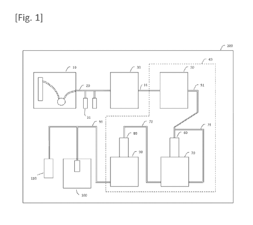
gloves.
[0127]
-36-
CA 02996582 2018-02-23
WO 2017/040548 PCT/US2016/049530
The separation apparatus 10 receives, for example, a vial
containing human blood. The separation apparatus 10 comprises,
for example, an anticoagulant tank which stores an
anticoagulant such as ethylenediamine tetraacetic acid (EDTA),
heparin, and Acid Citrate Dextrose Formula A solution (ACD-A
solution, Terumo Corp.). The separation apparatus 10 adds the
anticoagulant from the anticoagulant tank to the human blood
using a pump or the like.
[0128]
The separation apparatus 10 comprises, for example, a
reagent tank for separation which stores a reagent for monocyte
separation such as Ficoll-Paque PREMIUM(R) (GE Healthcare Japan
Corp.). The separation apparatus 10 dispenses the reagent for
monocyte separation at 5 mL/tube to, for example, two 15-mL
tubes from the reagent tank for separation using a pump or the
like. Note that a resin bag may be used instead of the tube.
[0129]
The separation apparatus 10 further comprises a buffer
solution tank which stores a buffer solution such as phosphate-
buffered saline (PBS). The separation apparatus 10 dilutes, for
example, 5 mL of the human blood by adding 5 mL of the buffer
solution from the buffer solution tank using a pump or the like.
In addition, the separation apparatus 10 adds 5 mL of the
diluted human blood onto the reagent for monocyte separation in
each tube using a pump or the like.
[0130]
The separation apparatus 10 further comprises a centrifuge
in which the temperature can be set. The centrifuge temperature
is set to, for example, 18 C. The separation apparatus 10
-37-
CA 02996582 2018-3
WO 2017/040548 PCT/US2016/049530
places each tube containing the reagent for monocyte separation
and the human blood, etc., in a holder of the centrifuge using
a transportation apparatus or the like. The centrifuge
centrifuges the solution in the tube, for example, at 400 X g
for 30 minutes. A resin bag may be centrifuged instead of the
tube.
[0131]
After the centrifugation, the separation apparatus 10
recovers a white cloudy intermediate layer composed of the
monocytes in the solution in the tube using a pump or the like.
The separation apparatus 10 sends the recovered monocyte
suspension into the pre-transfer cell solution sending channel
20 using a pump or the like. Alternatively, the separation
apparatus 10 further adds, for example, 12 mL of PBS to 2 mL of
the recovered monocyte solution and places the tube in a holder
of the centrifuge. The centrifuge centrifuges the solution in
the tube, for example, at 200 X g for ten minutes.
[0132]
After the centrifugation, the separation apparatus 10
removes the supernatant of the solution in the tube by
aspiration using a pump or the like, and suspends the monocyte
solution in the tube by adding 3 mL of a monocyte culture
medium such as X-VIVO 10(R) (Lonza Japan Ltd.). The separation
apparatus 10 sends the monocyte suspension into the pre-
transfer cell solution sending channel 20 using a pump or the
like. The separation apparatus 10 may separate the monocytes
from the blood using a dialysis membrane. Alternatively, in the
case of using somatic cells, such as fibroblasts, separated in
-38-
CA 02996582 2018-3
WO 2017/040548 PCT/US2016/049530
advance from the skin or the like, the separation apparatus 10
may be unnecessary.
[0133]
The separation apparatus 10 may separate cells suitable
for induction by a method other than centrifugal separation.
When the cells to be separated are, for example, T cells, cells
positive for any of CD3, CD4, and CD8 may be separated by
panning. When the cells to be separated are vascular
endothelial progenitor cells, cells positive for CD34 may be
separated by panning. When the cells to be separated are B
cells, cells positive for any of CD10, CD19, and CD20 may be
separated by panning. The separation approach is not limited to
panning, and the cells may be separated by a magnetic cell
separation method, flow cytometry, or other methods.
Alternatively, the separation apparatus 10 may separate cells
suitable for induction by methods described in embodiments
mentioned later. For example, as described in the fifth
embodiment, the cells suitable for induction may be separated
using a magnetic separation apparatus on the basis of a cell
surface marker. Alternatively, the cells suitable for induction
may be separated using a filter. The cells to be induced are
not limited to blood cells and may be fibroblasts or the like.
[0134]
The inducer solution sending mechanism 21 comprises an
inducer transfer reagent tank which stores an inducer transfer
reagent solution or the like. The inducer transfer reagent
solution such as a gene transfer reagent solution contains, for
example, an electroporation solution such as Human T Cell
Nucleofector(R) (Lonza Japan Ltd.) solution, a supplement
-39-
CA 02996582 2018-02-23
WO 2017/040548 PCT/US2016/049530
solution, and a plasmid set. The plasmid set contains, for
example, 0.83 g of pCXLE-hOCT3/4-shp53-F, 0.83 g of pCXLE-hSK,
0.83 g of pCE-hUL, and 0.5 g of pCXWB-EBNA1. Alternatively,
the inducer transfer reagent solution may contain reagents or
the like described in the fourth and fifth embodiments
mentioned later. For example, as described in the fifth
embodiment, an RNA encoding reprogramming factors may be
transferred into the cells by a lipofection method. The inducer
solution sending mechanism 21 sends the inducer transfer
reagent solution into the pre-transfer cell solution sending
channel 20 using a micropump or the like such that the monocyte
suspension is suspended in the inducer transfer reagent
solution.
[0135]
The inside wall of the pre-transfer cell solution sending
channel 20 may not be adhesive to cells by coating with poly-
HEMA (poly-2-hydroxyethyl methacrylate) so as to prevent cells
from adhering thereto. Alternatively, a material that resists
cell adhesion may be used as the material for the pre-transfer
cell solution sending channel 20. Also, a CO2-permeable
material having a high thermometric conductivity may be used as
the material for the pre-transfer cell solution sending channel
20 so that the internal conditions of the pre-transfer cell
solution sending channel 20 are equivalent to the controlled
temperature and 002 concentration in the container 200. The
pre-transfer cell solution sending channel 20 may be further
provided with a back-flow preventing valve from the viewpoint
of preventing contamination.
[0136]
-40-
CA 02996582 2018-02-23
WO 2017/040548 PCT/US2016/049530
The inducer transfer apparatus 30 connected to the pre-
transfer cell solution sending channel 20 is, for example, an
electroporator, which receives the mixed solution of the
inducer transfer reagent solution and the monocyte suspension
and carries out the electroporation of the monocytes with the
plasmids. After the electroporation, the inducer transfer
apparatus 30 adds a monocyte culture medium to a solution
containing the monocytes electroporated with the plasmids. The
inducer transfer apparatus 30 sends the solution containing the
monocytes electroporated with the plasmids (hereinafter,
referred to as "cells harboring the inducer") to a post-
transfer cell solution sending channel 31 using a pump or the
like. Note that the inducer transfer apparatus 30 is not
limited to an electroporator. The inducer transfer apparatus 30
may transfer the inducer into the cells by methods described in
the fourth and fifth embodiments mentioned later. The medium
may be a gel medium. In this case, the gel medium may be free
from, for example, a growth factor such as basic fibroblast
growth factor (bFGF). Alternatively, the gel medium may contain
a growth factor such as bFGF at a low concentration of 400 g/L
or lower, 40 g/L or lower, or 10 g/L or lower. The gel medium
may be free from tgf-P or may contain tgf-P at a low
concentration of 600 ng/L or lower, 300 ng/L or lower, or 100
ng/L or lower.
[0137]
The inside wall of the post-transfer cell solution sending
channel 31 may be rendered non-adhesive by coating with poly-
HEMA so as to prevent cells from adhering thereto.
Alternatively, a material that resists cell adhesion may be
-41-
CA 02996582 2018-02-23
WO 2017/040548 PCT/US2016/049530
used as the material for the post-transfer cell solution
sending channel 31. Also, a 002-permeable material having a
high thermometric conductivity may be used as the material for
the post-transfer cell solution sending channel 31 so that the
internal conditions of the post-transfer cell solution sending
channel 31 are equivalent to the controlled temperature and CO2
concentration in the container 200. The post-transfer cell
solution sending channel 31 may be further provided with a
back-flow preventing valve from the viewpoint of preventing
contamination. After the electroporation, many cells die, and
dead cells may form cell clusters. Therefore, the post-transfer
cell solution sending channel 31 may be provided with a filter
which removes dead cell clusters. Alternatively, as shown in
Figure 2, one or more walls which intermittently change the
inside diameter may be disposed in the inside of the post-
transfer cell solution sending channel 31. Alternatively, as
shown in Figure 3, the inside diameter of the post-transfer
cell solution sending channel 31 may be intermittently changed.
[0138]
The cell cluster production apparatus 40 connected to the
post-transfer cell solution sending channel 31 comprises: a
reprogramming culture apparatus 50 which cultures the cells
harboring the inducer produced by the inducer transfer
apparatus 30; a first division mechanism 60 which divides cell
clusters consisting of stem cells established by the
reprogramming culture apparatus 50 into a plurality of cell
clusters; an expansion culture apparatus 70 which expansion-
cultures the plurality of cell clusters divided by the first
division mechanism 60; a second division mechanism 80 which
-42-
CA 02996582 2018-3
WO 2017/040548 PCT/US2016/049530
divides cell clusters consisting of stem cells expansion-
cultured by the expansion culture apparatus 70 into a plurality
of cell clusters; and a cell cluster delivery mechanism 90
which sequentially sends the plurality of cell clusters into
the packaging apparatus 100.
[0139]
The reprogramming culture apparatus 50 can house a well
plate therein. The reprogramming culture apparatus 50 also
comprises a pipetting machine. The reprogramming culture
apparatus 50 receives a solution containing the cells harboring
the inducer from the post-transfer cell solution sending
channel 31 and distributes the solution to wells by the
pipetting machine. The reprogramming culture apparatus 50 adds
a stem cell culture medium such as StemFit(R) (Ajinomoto Co.,
Inc.), for example, three, five, and seven days after the cells
harboring the inducer are distributed into wells. Basic
fibroblast growth factor (basic FGF) may be added as a
supplement to the medium. Note that sustained-release beads,
such as StemBeads FGF2 (Funakoshi Co., Ltd.), which
continuously supply FGF-2 (basic FGF, bFGF, or FGF-b) to the
medium may be added to the medium. Since FGF is sometimes
unstable, the FGF may be stabilized by coupling a heparin-
mimicking polymer to the FGF. The reprogramming culture
apparatus 50 further replaces the medium, for example, nine
days after the cells harboring the inducer are distributed into
wells and subsequently replaces the medium every two days until
cell clusters (colonies) of iPS cells exceed 1 mm.
[0140]
-43-
CA 02996582 2018-02-23
WO 2017/040548 PCT/US2016/049530
After formation of the cell clusters, the reprogramming
culture apparatus 50 recovers the cell clusters by the
pipetting machine and adds a recombinant enzyme alternative to
trypsin, such as TrypLE Select(R) (Life Technologies Corp.), to
the recovered cell clusters. The reprogramming culture
apparatus 50 further places a container containing the
recovered cell clusters in an incubator where the cell clusters
react with the recombinant enzyme alternative to trypsin at 37 C
for ten minutes in a 5% CO2 environment. Alternatively, the
reprogramming culture apparatus 50 may disrupt the cell
clusters by pipetting using the pipetting machine. As another
alternative, the reprogramming culture apparatus 50 may disrupt
the cell clusters by passing the cell clusters through a pipe
provided with a filter or a pipe whose inside diameter
intermittently changes, as with the post-transfer cell solution
sending channel 31 shown in Figure 2 or 3. Then, the
reprogramming culture apparatus 50 adds a medium for
pluripotent stem cells, such as StemFit(R) (Ajinomoto Co.,
Inc.), to a solution containing the disrupted cell clusters.
[0141]
The culture in the reprogramming culture apparatus 50 may
be performed in a 002-permeable bag rather than in the well
plate. The culture may be an adherent culture or may be a
floating culture. In the case of floating culture, stirring the
culture may be performed. The medium may be in an agar form.
Examples of the medium in an agar form include gellan gum
polymer. When the medium in an agar form is used, even in the
form of floating culture, stirring is not required and it
possible to produce a single cell cluster derived from one cell
-44-
CA 02996582 2018-3
WO 2017/040548 PCT/US2016/049530
because the cells neither sink down nor adhere. The culture in
the reprogramming culture apparatus 50 may be a hanging drop
culture.
[0142]
The reprogramming culture apparatus 50 may comprise a
first culture solution replenishment apparatus which
replenishes the well plate or the CO2-permeable bag with a
culture solution. The first culture solution replenishment
apparatus may recover the culture solution in the well plate or
the 002-permeable bag, filter the culture solution using a
filter or a dialysis membrane, and recycle the purified culture
solution. In this case, a growth factor or the like may be
added to the culture solution to be recycled. The reprogramming
culture apparatus 50 may further comprise, for example, a
temperature control apparatus which controls the temperature of
the culture solution, and a humidity control apparatus which
controls humidity near the culture solution.
[0143]
In the reprogramming culture apparatus 50, for example,
the cells may be placed in a culture solution-permeable bag 301,
such as a dialysis membrane, as shown in Figure 4, and the
culture solution-permeable bag 301 may be placed in a culture
solution-impermeable and CO2-permeable bag 302, while a culture
solution may be placed in the bags 301 and 302. A plurality of
bags 302 containing a fresh culture solution may be prepared,
and the reprogramming culture apparatus 50 may replace the bag
302 in which the bag 301 containing the cells is placed, with
another bag 302 containing a fresh culture solution at a
predetermined time interval. Note that the culture method in
-45-
CA 02996582 2018-02-23
WO 2017/040548 PCT/US2016/049530
the reprogramming culture apparatus 50 is not limited to the
methods described above, and the culture may be performed by
methods described in the second and third embodiments mentioned
later. For example, as described in the second embodiment, a
gel medium may be used. In this case, the gel medium may be
free from, for example, a growth factor such as basic
fibroblast growth factor (bFGF). Alternatively, the gel medium
may contain a growth factor such as bFGF at a low concentration
of 400 g/L or lower, 40 g/L or lower, or 10 g/L or lower.
The gel medium may be free from tgf-P or may contain tgf-P at a
low concentration of 600 ng/L or lower, 300 ng/L or lower, or
100 ng/L or lower. As described in the third embodiment, a
floating culture vessel comprising: a dialysis tube which
accommodates stem cells and a gel medium; and a container which
accommodates the dialysis tube, wherein a gel medium is placed
around the dialysis tube, may be used.
[0144]
The stem cell manufacturing system may further comprise a
reprogramming culture photography apparatus which photographs
the culture in the reprogramming culture apparatus 50. Note
that when a colorless culture solution is used as the culture
solution for the reprogramming culture apparatus 50, it is
possible to suppress diffuse reflection or autofluorescence
that may occur when a colored culture solution is used. Since
induced cells and uninduced cells differ in cell shape and size,
etc., the stem cell manufacturing system may further comprise
an induction status monitor apparatus which calculates the
percentage of induced cells by photographing the cells in the
reprogramming culture apparatus 50. Alternatively, the
-46-
CA 02996582 2018-02-23
WO 2017/040548 PCT/US2016/049530
induction status monitor apparatus may identify the percentage
of induced cells by an antibody immunostaining method or an RNA
extraction method. The stem cell manufacturing system may
further comprise an uninduced cell removal apparatus which
removes uninduced cells by a magnetic cell separation method,
flow cytometry, or the like.
[0145]
A first cell cluster solution sending channel 51 is
connected to the reprogramming culture apparatus 50. The
reprogramming culture apparatus 50 sends a solution containing
the recombinant enzyme alternative to trypsin and the cell
clusters into the first cell cluster solution sending channel
51 using a pump or the like. When the cell clusters can be
physically disrupted, the recombinant enzyme alternative to
trypsin may be unnecessary. The first cell cluster solution
sending channel 51 may be connected to a branched channel which
has an inside diameter that permits passage of only induced
cells having less than a predetermined size and removes
uninduced cells having the predetermined size or larger.
[0146]
The inside wall of the first cell cluster solution sending
channel 51 may not be adhesive to cells by coating with poly-
HEMA so as to prevent cells from adhering thereto.
Alternatively, a material that resists cell adhesion may be
used as the material for the first cell cluster solution
sending channel 51. Also, a CO2-permeable material having a
high thermometric conductivity may be used as the material for
the first cell cluster solution sending channel 51 so that the
internal conditions of the first cell cluster solution sending
-47-
CA 02996582 2018-02-23
WO 2017/040548 PCT/US2016/049530
channel 51 are equivalent to the controlled temperature and 002
concentration in the container 200. The first cell cluster
solution sending channel 51 may be further provided with a
back-flow preventing valve from the viewpoint of preventing
contamination.
[0147]
The first cell cluster solution sending channel 51 is
connected to the first division mechanism 60. The first
division mechanism 60 comprises, for example, a mesh. When
passing through the mesh by hydraulic pressure, the cell
clusters contained in the solution are divided into a plurality
of cell clusters corresponding to the size of each pore of the
mesh. For example, when the mesh has uniform sizes of pores,
the sizes of the plurality of cell clusters thus divided are
also almost uniform. Alternatively, the first division
mechanism 60 may comprise a nozzle. For example, the inside of
a substantially conical nozzle is microfabricated in a
staircase pattern. When flowing through the nozzle, the cell
clusters contained in the solution are divided into a plurality
of cell clusters. The expansion culture apparatus 70 is
connected to the first division mechanism 60. The solution
containing the cell clusters divided by the first division
mechanism 60 is sent to the expansion culture apparatus 70.
[0148]
The expansion culture apparatus 70 can house a well plate
therein. The expansion culture apparatus 70 also comprises a
pipetting machine. The expansion culture apparatus 70 receives
a solution containing the plurality of cell clusters from the
first division mechanism 60 and distributes the solution into
-48-
CA 02996582 2018-02-23
WO 2017/040548 PCT/US2016/049530
wells by the pipetting machine. After the cell clusters are
distributed into wells, the expansion culture apparatus 70
cultures the cell clusters at 37 C, for example, for
approximately eight days, in a 5% 002 environment. The
expansion culture apparatus 70 replaces the medium as necessary.
[0149]
Then, the expansion culture apparatus 70 adds a
recombinant enzyme alternative to trypsin, such as TrypLE
Select(R) (Life Technologies Corp.), to the cell clusters. The
expansion culture apparatus 70 further places a container
containing the cell clusters in an incubator where the cell
clusters react with the recombinant enzyme alternative to
trypsin at 37 C for one minute in a 5% CO2 environment. Then,
the expansion culture apparatus 70 adds a medium such as a
maintenance culture medium to the solution containing the cell
clusters. The expansion culture apparatus 70 further detaches
the cell clusters from the container using an automatic cell
scraper or the like and sends a solution containing the cell
clusters into the first division mechanism 60 via an expansion
culture solution sending channel 71.
[0150]
The culture in the expansion culture apparatus 70 may be
performed in a CO2-permeable bag, not in the well plate. The
culture may be an adherent culture or may be a floating culture.
Alternatively, the culture may be a hanging drop culture. In
the case of floating culture, stirring culture may be performed.
The medium may be in an agar form. Examples of the medium in an
agar form include gellan gum polymer. When the medium in an
agar form is used, even in the form of floating culture,
-49-
CA 02996582 2018-02-23
WO 2017/040548 PCT/US2016/049530
stirring is not required because the cells neither sink down
nor adhere.
[0151]
The expansion culture apparatus 70 may comprise a second
culture solution replenishment apparatus which replenishes the
well plate or the 002-permeable bag with a culture solution.
The second culture solution replenishment apparatus may recover
the culture solution in the well plate or the 002-permeable bag,
filter the culture solution using a filter or a dialysis
membrane, and recycle the purified culture solution. In this
case, a growth factor or the like may be added to the culture
solution to be recycled. The expansion culture apparatus 70 may
further comprise, for example, a temperature control apparatus
which controls the temperature of the culture solution, and a
humidity control apparatus which controls humidity near the
culture solution.
[0152]
In the expansion culture apparatus 70 as well, for example,
the cells may be placed in a culture solution-permeable bag 301,
such as a dialysis membrane, as shown in Figure 4, and the
culture solution-permeable bag 301 may be placed in a culture
solution-impermeable and CO2-permeable bag 302, while a culture
solution may be placed in the bags 301 and 302. A plurality of
bags 302 containing a fresh culture solution may be prepared,
and the expansion culture apparatus 70 may replace the bag 302
in which the bag 301 containing the cells is placed, with
another bag 302 containing a fresh culture solution at a
predetermined time interval. The culture method in the
expansion culture apparatus 70 is not limited to the methods
-50-
CA 02996582 2018-02-23
WO 2017/040548 PCT/US2016/049530
described above, and the culture may be performed by methods
described in the second and third embodiments mentioned later.
[0153]
The stem cell manufacturing system may further comprise an
expansion culture photography apparatus which photographs the
culture in the expansion culture apparatus 70. Note that when a
colorless culture solution is used as the culture solution used
in the expansion culture apparatus 70, it is possible to
suppress diffuse reflection or autofluorescence that may occur
when a colored culture solution is used. Since induced cells
and uninduced cells differ in cell shape and size, etc., the
stem cell manufacturing system may further comprise an
induction status monitor apparatus which calculates the
percentage of induced cells by photographing the cells in the
expansion culture apparatus 70. Alternatively, the induction
status monitor apparatus may identify the percentage of induced
cells by an antibody immunostaining method or an RNA extraction
method. The stem cell manufacturing system may further comprise
an uninduced cell removal apparatus which removes uninduced
cells by a magnetic cell separation method, flow cytometry, or
the like.
[0154]
The cell clusters divided by the first division mechanism
60 are cultured again in the expansion culture apparatus 70.
The division of the cell clusters in the first division
mechanism 60 and the culture of the cell clusters in the
expansion culture apparatus 70 are repeated until a necessary
amount of cells is obtained.
[0155]
-51-
CA 02996582 2018-02-23
WO 2017/040548 PCT/US2016/049530
A second cell cluster solution sending channel 72 is
connected to the expansion culture apparatus 70. The expansion
culture apparatus 70 sends a solution containing the expansion-
cultured cell clusters detached from the container into the
second cell cluster solution sending channel 72 using a pump or
the like. The second cell cluster solution sending channel 72
may be connected to a branched channel which has an inside
diameter that permits passage of only induced cells having less
than a predetermined size and removes uninduced cells having
the predetermined size or larger.
[0156]
The inside wall of the second cell cluster solution
sending channel 72 may not be adhesive to cells by coating with
poly-HEMA so as to prevent cells from adhering thereto.
Alternatively, a material that resists cell adhesion may be
used as the material for the second cell cluster solution
sending channel 72. Also, a 002-permeable material having a
high thermometric conductivity may be used as the material for
the second cell cluster solution sending channel 72 so that the
internal conditions of the second cell cluster solution sending
channel 72 are equivalent to the controlled temperature and CO2
concentration in the container 200. The second cell cluster
solution sending channel 72 may be further provided with a
back-flow preventing valve from the viewpoint of preventing
contamination.
[0157]
The second cell cluster solution sending channel 72 is
connected to the second division mechanism 80. The second
division mechanism 80 comprises, for example, a mesh. When
-52-
CA 02996582 2018-3
WO 2017/040548 PCT/US2016/049530
passing through the mesh by hydraulic pressure, the cell
clusters contained in the solution are divided into a plurality
of cell clusters corresponding to the size of each pore of the
mesh. For example, when the mesh has uniform sizes of pores,
the sizes of the plurality of cell clusters thus divided are
also almost uniform. Alternatively, the second division
mechanism 80 may comprise a nozzle. For example, the inside of
a substantially conical nozzle is microfabricated in a
staircase pattern. When flowing through the nozzle, the cell
clusters contained in the solution are divided into a plurality
of cell clusters.
[0158]
The cell cluster delivery mechanism 90 which sequentially
sends a plurality of cell clusters to the packaging apparatus
100 is connected to the second division mechanism 80. A pre-
packaging cell channel 91 connects between the cell cluster
delivery mechanism 90 and the packaging apparatus 100. The cell
cluster delivery mechanism 90 sequentially sends the cell
clusters divided by the second division mechanism 80 to the
packaging apparatus 100 via the pre-packaging cell channel 91
using a pump or the like.
[0159]
The pre-packaging cell channel 91 may be coated with poly-
HEMA so as to prevent cells from adhering thereto.
Alternatively, a material that resists cell adhesion may be
used as the material for the pre-packaging cell channel 91.
Also, a CO2-permeable material having a high thermometric
conductivity may be used as the material for the pre-packaging
cell channel 91 so that the internal conditions of the pre-
-53-
CA 02996582 2018-02-23
WO 2017/040548 PCT/US2016/049530
packaging cell channel 91 are equivalent to the controlled
temperature and CO2 concentration in the container 200. The
pre-packaging cell channel 91 may be further provided with a
back-flow preventing valve from the viewpoint of preventing
contamination.
[0160]
A cryopreservation solution sending mechanism 110 is
connected to the pre-packaging cell channel 91. The
cryopreservation solution sending mechanism 110 sends a cell
cryopreservation solution into the pre-packaging cell channel
91. As a result, the cell clusters are suspended in the cell
cryopreservation solution in the pre-packaging cell channel 91.
[0161]
The packaging apparatus 100 sequentially freezes the
plurality of cell clusters sent via the pre-packaging cell
channel 91. For example, every time a cell cluster is received,
the packaging apparatus 100 places the cell cluster in a
CryoTube and instantly freezes the cell cluster solution, for
example, at -80 C or lower. If a CryoTube having a small
surface area per volume is used, freezing tends to be time-
consuming. Therefore, it is preferred to use a CryoTube having
a large surface area per volume. It is possible to increase the
survival rate of cells after thawing by using a CryoTube having
a large surface area per volume. Examples of the shape of the
CryoTube include, but are not limited to, capillary and
spherical shapes. Depending on the required survival rate of
cells after thawing, the instant freezing is not necessarily
required.
[0162]
-54-
CA 02996582 2018-3
WO 2017/040548 PCT/US2016/049530
For example, a vitrification method is used in the
freezing. In this case, DAP213 (Cosmo Bio Co., Ltd.) or
Freezing Medium (ReproCELL Inc.) can be used as the cell
cryopreservation solution. The freezing may be performed by an
ordinary method other than the vitrification method. In this
case, CryoDefend-Stem Cell (R&D Systems, Inc.), STEM-
CELLBANKER(R) (Nippon Zenyaku Kogyo Co., Ltd.), or the like can
be used as the cell cryopreservation solution. The freezing may
be performed using liquid nitrogen or may be performed using a
Peltier device. By using the Peltier device, it is possible to
regulate the change in temperature and suppress temperature
variations. The packaging apparatus 100 exports the CryoTube to
the outside of the container 200. In the case of clinically
using the frozen cells, the CryoTube is preferably a completely
sealed system. However, the packaging apparatus 100 may package
the stem cells in the CryoTube without freezing the stem cells.
[0163]
The stem cell manufacturing system may further comprise a
packaging step photography apparatus which photographs the
packaging step in the packaging apparatus 100.
[0164]
The stem cell manufacturing system may further comprise a
sterilization apparatus which sterilizes the inside of the
container 200. The sterilization apparatus may be a dry heat
sterilization apparatus. Note that the wiring of apparatuses,
such as the separation apparatus 10, the pre-transfer cell
solution sending channel 20, the inducer solution sending
mechanism 21, the inducer transfer apparatus 30, the cell
cluster production apparatus 40, and the packaging apparatus
-55-
CA 02996582 2018-3
WO 2017/040548 PCT/US2016/049530
100, which employ electricity is preferably wiring having heat
resistance. Alternatively, the sterilization apparatus may
sterilize the inside of the container 200 by emitting
sterilization gas such as ozone gas, hydrogen peroxide gas, or
formalin gas into the container 200.
[0165]
The stem cell manufacturing system may transmit operation
records of the separation apparatus 10, the pre-transfer cell
solution sending channel 20, the inducer solution sending
mechanism 21, the inducer transfer apparatus 30, the cell
cluster production apparatus 40, and the packaging apparatus
100, etc., and images taken by the photography apparatuses to
an external server by wire or wirelessly. The external server
may analyze, for example, the association of conditions (e.g.,
inducer transfer conditions, culture conditions, and freezing
conditions) with results (e.g., the incomplete reprogramming of
stem cells, the failure of stem cell differentiation and
proliferation, and chromosomal aberration) using a neural
network to extract a condition leading to the results or
predict the results. The external server may further regulate
the separation apparatus 10, the inducer solution sending
mechanism 21, the inducer transfer apparatus 30, the cell
cluster production apparatus 40, and packaging apparatus 100,
etc. in the stem cell manufacturing system on the basis of a
standard operating procedure (SOP), monitor whether or not each
apparatus is operated on the basis of SOP, and automatically
make an operation record of each apparatus.
[0166]
-56-
CA 02996582 2018-3
WO 2017/040548 PCT/US2016/049530
The stem cell manufacturing system described above makes
it possible to achieve the induction, establishment, expansion
culture, and cryopreservation of stem cells such as iPS cells
in a lump with full automation.
[0167]
(Other embodiments)
For example, the inducer transfer apparatus 30 may induce
cells by RNA transfection, not by electroporation.
Alternatively, the cells may be induced by virus (e.g.,
retrovirus, lentivirus, and Sendai virus) vectors or plasmids.
The pre-transfer cell solution sending channel 20, the post-
transfer cell solution sending channel 31, the cell cluster
solution sending channel 51, the expansion culture solution
sending channel 71, the cell cluster solution sending channel
72, and the pre-packaging cell channel 91 may be disposed on a
substrate by a microfluidics technique. An apparatus which
transfers an inducer ribonucleic acid (RNA) into induced stem
cells by lipofection to differentiate the stem cells into
somatic cells may be connected to the stem cell manufacturing
system. For example, a method described in the sixth embodiment
described later can be used as a method for transferring an
inducer ribonucleic acid (RNA) into induced stem cells by
lipofection to differentiate the stem cells into somatic cells.
The somatic cells may be, for example, neuronal cells.
[0168]
(Second embodiment)
The floating culture method for stem cells according to
the second embodiment of the present invention comprises
floating-culturing stem cells in a gel medium. The stem cells
-57-
CA 02996582 2018-02-23
WO 2017/040548 PCT/US2016/049530
are, for example, induced pluripotent stem (iPS) cells or
embryonic stem cells (ES cells). The gel medium is not stirred.
The gel medium is free from feeder cells. The stem cells
proliferate in the gel medium while remaining in their
undifferentiated states.
[0169]
For example, before the floating culture, the stem cells
are dissociated into single cells, and the stem cells
dissociated into single cells are placed in the gel medium. The
single cells proliferate while maintaining their clonality to
form colonies in the gel medium.
[0170]
The gel medium is prepared, for example, by adding
deacetylated gellan gum at a final concentration of 0.5% by
weight to 0.001% by weight, 0.1% by weight to 0.005% by weight,
or 0.05% by weight to 0.01% by weight to a medium for stem
cells.
[0171]
The gel medium may contain at least one polymer compound
selected from the group consisting of gellan gum, hyaluronic
acid, rhamsan gum, diutan gum, xanthan gum, carrageenan,
fucoidan, pectin, pectic acid, pectinic acid, heparan sulfate,
heparin, heparitin sulfate, keratosulfate, chondroitin sulfate,
dermatan sulfate, rhamnan sulfate, and salts thereof. The gel
medium may contain methylcellulose. Methylcellulose contained
therein suppresses the aggregation among the cells.
[0172]
Alternatively, the gel medium may contain at least one
temperature-sensitive gel selected from poly(glycerol
-58-
CA 02996582 2018-3
WO 2017/040548 PCT/US2016/049530
monomethacrylate) (PGMA), poly(2-hydroxypropyl methacrylate)
(PHPMA), poly(N-isopropylacrylamide) (PNIPAM), amine terminated,
carboxylic acid terminated, maleimide terminated, N-
hydroxysuccinimide (NHS) ester terminated, triethoxysilane
terminated, poly(N-isopropylacrylamide-co-acrylamide), poly(N-
isopropylacrylamide-co-acrylic acid), poly(N-
isopropylacrylamide-co-butylacrylate), poly(N-
isopropylacrylamide-co-methacrylic acid), poly(N-
isopropylacrylamide-co-methacrylic acid-co-octadecyl acrylate),
and N-isopropylacrylamide.
[0173]
A human ES/iPS culture medium, for example, Primate ES
Cell Medium (ReproCELL Inc.), can be used as the medium for
stem cells.
[0174]
However, the medium for stem cells is not limited thereto,
and various stem cell culture media can be used. For example,
Primate ES Cell Medium, Reprostem, ReproFF, ReproFF2, ReproXF
(ReproCELL Inc.), mTeSR1, TeSR2, TeSRE8, ReproTeSR (STEMCELL
Technologies Inc.), PluriSTEM(R) Human ES/iPS Medium (Merck
KGaA), NutriStem(R) XF/FF Culture Medium for Human iPS and ES
Cells, Pluriton reprogramming medium (Stemgent Inc.),
PluriSTEM(R), StemFit AKO2N, StemFit AK03 (Ajinomoto Co., Inc.),
ESC-Sure(R) serum and feeder free medium for hESC/iPS (Applied
StemCell, Inc.), and L7(R) hPSC Culture System (Lonza Japan
Ltd.) may be used.
[0175]
For example, a ROCK inhibitor is added at a final
concentration of 1000 mol/L or higher and 0.1 mol/L or lower,
-59-
CA 02996582 2018-02-23
WO 2017/040548 PCT/US2016/049530
100 mol/L or higher and 1 mol/L or lower, or 5 mol/L or
higher and 20 mol/L or lower to the gel medium every day. The
ROCK inhibitor added to the gel medium promotes the formation
of colonies by the stem cells.
[0176]
The gel medium may be free from, for example, a growth
factor such as basic fibroblast growth factor (bFGF).
Alternatively, the gel medium may contain a growth factor such
as bFGF at a low concentration of 400 g/L or lower, 40 g/L or
lower, or 10 g/L or lower. The gel medium without a growth
factor such as bFGF or the gel medium with a growth factor such
as bFGF at a low concentration tends to promote the formation
of colonies by the stem cells, as compared with a gel medium
with a growth factor such as bFGF at a high concentration.
[0177]
The gel medium may be free from tgf-P or may contain tgf-P
at a low concentration of 600 ng/L or lower, 300 ng/L or lower,
or 100 ng/L or lower.
[0178]
The concentration of the stem cells in the gel medium may
be, for example, 2 X 10⁵ cells/mL or higher, 2.25 X 10⁵ cells/mL
or higher, or 2.5 X 10⁵ cells/mL or higher. If the
concentration of the stem cells in the gel medium is lower than
2 X 10⁵ cells/mL, the rate of colony formation tends to be
decreased.
[0179]
The floating culture method for stem cells according to
the second embodiment of the present invention makes it
possible to form stem cell colonies from single cells. Although
-60-
CA 02996582 2018-02-23
WO 2017/040548 PCT/US2016/049530
this method is a floating culture method, the stem cells do not
collide with each other because the floating culture method for
stem cells does not require stirring the medium. Therefore, it
is possible to maintain the clonality of the colonies. Thus,
when the stem cells are, for example, iPS cells, it is possible
to ensure the clonality of iPS cells derived from one somatic
cell. Further, since the stem cells do not collide with each
other, the stem cell colonies can maintain homogeneous sizes.
Moreover, the floating culture method makes it possible to
culture a large number of colonies in a small space as compared
with the adherent culture method. Note that stem cell clusters
may be maintenance-cultured in the floating culture.
[0180]
A growth factor such as bFGF or Tgf-P has been thought to
be essential for the culture of ES/iPS cells since the
discovery of ES/iPS cells. However, a growth factor such as
bFGF is rapidly decomposed under a culture condition of
approximately 37 C. Therefore, it is necessary to replace a
culture solution with bFGF or Tgf-P with a fresh one every day
or to add bFGF or Tgf-P, etc. thereto every day. The bFGF used
for culture is usually a recombinant protein. A recombinant
protein at a clinical grade needs to be produced according to
very strict rules.
[0181]
When the concentration of bFGF is, for example, a low
concentration of 10 ng/mL, it has been considered that mouse-
derived fibroblasts need to be used as feeder cells. However,
stem cells cocultured with feeder cells derived from an animal
such as a mouse cannot be used in transplantation or
-61-
CA 02996582 2018-02-23
WO 2017/040548 PCT/US2016/049530
regenerative medicine. This has been a bottleneck in the
clinical utilization of stem cells.
[0182]
Although a feeder-free culture solution for stem cells,
which does not employ feeder cells, has also been developed,
the feeder-free culture solution usually contains bFGF in 25
times the amount of that contained in a culture solution using
feeder cells and contains bFGF at a very high concentration
such as 100 ng/mL. However, it is difficult to culture ES/iPS
cells without karyotype abnormalities using a feeder-free
culture solution containing a high concentration of bFGF. Thus,
many iPS cells are destroyed. ES/iPS cells cultured in the
feeder-free culture solution containing a high concentration of
bFGF tend to be less likely to differentiate into particular
somatic cells. Therefore, the feeder-free culture solution is
partly responsible for decreasing the efficiency of production
of somatic cells necessary for transplantation from stem cells.
[0183]
By contrast, the floating culture method for stem cells
according to the second embodiment of the present invention
makes it possible to culture and proliferate the stem cells
without the use of feeder cells while maintaining their
undifferentiated states. Even though feeder cells are not used,
the floating culture method for stem cells according to the
second embodiment of the present invention makes it possible to
culture and proliferate the stem cells without the use of a
growth factor such as bFGF or with the use of a growth factor
such as bFGF at a low concentration while maintaining their
undifferentiated states.
-62-
CA 02996582 2018-3
WO 2017/040548 PCT/US2016/049530
[ 0 1 8 4 ]
(Third embodiment)
The floating culture vessel for stem cells according to
the third embodiment of the present invention, as shown in
Figure 5, comprises: a dialysis tube which accommodates stem
cells and a gel medium; and a container which accommodates the
dialysis tube, wherein a gel medium is placed around the
dialysis tube.
[0185]
The dialysis tube is permeable to, for example, a ROCK
inhibitor. A molecular weight cutoff of the dialysis tube is
0.1 kDa or larger, 10 kDa or larger, or 50 kDa or larger. The
dialysis tube is made of, for example, cellulose ester,
ethylcellulose, cellulose ester derivatives, regenerated
cellulose, polysulfone, polyacrylonitrile, polymethyl
methacrylate, an ethylene-vinyl alcohol copolymer, a polyester-
based polymer alloy, polycarbonate, polyamide, cellulose
acetate, cellulose diacetate, cellulose triacetate,
cuprammonium rayon, saponified cellulose, a hemophan membrane,
a phosphatidylcholine membrane, or a vitamin E-coated membrane.
A conical tube such as a centrifugal tube can be used as the
container. The container is made of, for example, polypropylene.
[0186]
The stem cells to be placed in the dialysis tube are the
same as in the second embodiment. Likewise, the gel medium to
be placed in the dialysis tube is the same as in the second
embodiment. However, the gel medium to be placed in the
dialysis tube may be free from a ROCK inhibitor. The gel medium
to be placed around the dialysis tube in the container is the
-63-
CA 02996582 2018-02-23
WO 2017/040548 PCT/US2016/049530
same as in the second embodiment. The gel medium to be placed
around the dialysis tube in the container contains a ROCK
inhibitor.
[0187]
During the floating culture of the stem cells in the
dialysis tube, the gel medium around the dialysis tube in the
container is replaced or supplemented with a fresh gel medium.
However, the replacement of the gel medium in the dialysis tube
may be unnecessary.
[0188]
For a conventional floating culture system, it may be
difficult to replace a medium without aspirating the cells.
However, waste products may accumulate unless the medium is
replaced with a fresh medium. Furthermore, the medium may be
short of a medium component unless the medium is replaced or
supplemented with a fresh medium.
[0189]
By contrast, by using the floating culture vessel for stem
cells according to the third embodiment of the present
invention, it is possible to avoid aspirating the stem cells
even if the medium around the dialysis tube is replaced with a
fresh medium, because the stem cells are in the dialysis tube.
In addition, the amount of the medium in the dialysis tube is
hardly changed even when the medium around the dialysis tube is
replaced with a fresh medium. Therefore, the density of the
stem cells in the dialysis tube is not changed. A high
concentration of waste products in the dialysis tube moves to
the outside of the dialysis tube. The medium component moves
from the outside medium of the dialysis tube into the dialysis
-64-
CA 02996582 2018-3
WO 2017/040548 PCT/US2016/049530
tube with decrease in the concentration of the medium component
of the medium in the dialysis tube. Therefore, it is possible
to keep the medium around the stem cells fresh.
[0190]
(Fourth embodiment)
The method for inducing stem cells according to the fourth
embodiment of the present invention comprises inducing stem
cells from somatic cells floating-cultured in a gel medium. The
somatic cells are, for example, fibroblasts. The stem cells are,
for example, iPS cells. The gel medium is not stirred. The gel
medium is free from feeder cells.
[0191]
The gel medium is prepared, for example, by adding
deacetylated gellan gum at a final concentration of 0.5% by
weight to 0.001% by weight, 0.1% by weight to 0.005% by weight,
or 0.05% by weight to 0.01% by weight to a medium for stem
cells.
[0192]
The gel medium may contain at least one polymer compound
selected from the group consisting of gellan gum, hyaluronic
acid, rhamsan gum, diutan gum, xanthan gum, carrageenan,
fucoidan, pectin, pectic acid, pectinic acid, heparan sulfate,
heparin, heparitin sulfate, keratosulfate, chondroitin sulfate,
dermatan sulfate, rhamnan sulfate, and salts thereof. The gel
medium may contain methylcellulose. Methylcellulose contained
therein suppresses the aggregation among the cells.
[0193]
Alternatively, the gel medium may contain at least one
temperature-sensitive gel selected from poly(glycerol
-65-
CA 02996582 2018-02-23
WO 2017/040548 PCT/US2016/049530
monomethacrylate) (PGMA), poly(2-hydroxypropyl methacrylate)
(PHPMA), poly(N-isopropylacrylamide) (PNIPAM), amine terminated,
carboxylic acid terminated, maleimide terminated, N-
hydroxysuccinimide (NHS) ester terminated, triethoxysilane
terminated, poly(N-isopropylacrylamide-co-acrylamide), poly(N-
isopropylacrylamide-co-acrylic acid), poly(N-
isopropylacrylamide-co-butylacrylate), poly(N-
isopropylacrylamide-co-methacrylic acid), poly(N-
isopropylacrylamide-co-methacrylic acid-co-octadecyl acrylate),
and N-isopropylacrylamide.
[0194]
A human ES/iPS culture medium, for example, Primate ES
Cell Medium (ReproCELL Inc.), can be used as the medium for
stem cells.
[0195]
However, the medium for stem cells is not limited thereto,
and various stem cell culture media can be used. For example,
Primate ES Cell Medium, Reprostem, ReproFF, ReproFF2, ReproXF
(ReproCELL Inc.), mTeSR1, TeSR2, TeSRE8, ReproTeSR (STEMCELL
Technologies Inc.), PluriSTEM(R) Human ES/iPS Medium (Merck
KGaA), NutriStem(R) XF/FF Culture Medium for Human iPS and ES
Cells, Pluriton reprogramming medium (Stemgent Inc.),
PluriSTEM(R), StemFit AKO2N, StemFit AK03 (Ajinomoto Co., Inc.),
ESC-Sure(R) serum and feeder free medium for hESC/iPS (Applied
StemCell, Inc.), and L7(R) hPSC Culture System (Lonza Japan
Ltd.) may be used. The gel medium is placed in, for example, a
tube.
[0196]
-66-
CA 02996582 2018-02-23
WO 2017/040548 PCT/US2016/049530
The gel medium may be free from, for example, a growth
factor such as basic fibroblast growth factor (bFGF).
Alternatively, the gel medium may contain a growth factor such
as bFGF at a low concentration of 400 g/L or lower, 40 g/L or
lower, or 10 g/L or lower.
[0197]
The gel medium may be free from tgf-P or may contain tgf-P
at a low concentration of 600 ng/L or lower, 300 ng/L or lower,
or 100 ng/L or lower.
[0198]
(Example 1)
500 mL of Primate ES Cell Medium (ReproCELL Inc.) and 0.2
mL of bFGF (Gibco PHG0266) having a concentration of 10 g/mL
were mixed to prepare a human iPS medium with bFGF.
[0199]
Deacetylated gellan gum (Nissan Chemical Industries Ltd.)
was added at a concentration of 0.02% by weight to the human
iPS medium with bFGF to prepare a human iPS gel medium with
bFGF. Further, 5 mL of trypsin having a concentration of 2.5%
by weight, 5 mL of collagenase IV having a concentration of 1
mg/mL, 0.5 mL of CaC12 having a concentration of 0.1 mol/L, 10
mL of KnockOut Serum Replacement(R) (Invitrogen 10828-028), and
30 mL of purified water were mixed to prepare a dissociation
solution generally called a CTK solution.
[0200]
The CTK solution was added at 300 L/well to a 6-well dish
(Thermo Fisher Scientific 12-556-004) containing iPS cells in
the process of culture on feeder cells, and the 6-well dish was
incubated for three minutes in a 002 incubator. Three minutes
-67-
CA 02996582 2018-02-23
WO 2017/040548 PCT/US2016/049530
later, the dish was taken out of the incubator. After
confirmation that only the feeder cells were detached from the
dish, the CTK solution was removed using an aspirator. After
the removal of the CTK solution, the iPS cells were washed by
the addition of PBS (Santa Cruz Biotech sc-362183) at 500
L/well to the 6-well dish, followed by the removal of PBS from
the 6-well dish. A dissociation solution (Accutase(R)) was
added at 0.3 mL/well to the 6-well dish, which was then placed
in a 002 incubator and incubated for five minutes. Then, the
iPS medium with bFGF was added at 0.7 mL/well to the 6-well
dish so that the iPS cells were suspended until becoming single
cells.
[0201]
After the suspension of the iPS cells, 4 mL of the human
iPS medium with bFGF was added to a 15-mL centrifugal tube, and
the iPS cell suspension was centrifuged at 270 g using a
centrifuge. After the centrifugation, the supernatant was
removed, and 1 mL of the human iPS medium with bFGF was added
to the 15-mL centrifugal tube. The number of the cells was
calculated using a hemocytometer. After the cell counting, 5 X
105 iPS cells were seeded to 15-mL Falcon Tube(R) (Corning
352096) or a non-adherent dish and subsequently floating-
cultured without stirring.
[0202]
In the 15-mL tube, 2 mL of the human iPS gel medium with
bFGF was used. In the non-adherent dish, 2 mL of the human iPS
medium with bFGF and without gellan gum was used. A ROCK
inhibitor (Selleck Chemicals S1049) was added at 10 mol/L to
each medium. Then, 500 L of the human iPS gel medium with bFGF
-68-
CA 02996582 2018-02-23
WO 2017/040548 PCT/US2016/049530
was added to the 15-mL tube and the non-adherent dish every day,
and 500 L of the human iPS medium with bFGF was added to the
non-adherent dish every day. Also, the ROCK inhibitor was added
at a final concentration of 10 mol/L to the 15-mL tube and the
non-adherent dish every day, and the floating culture was
continued for seven days.
[0203]
The results are shown in Figure 6. As shown in Figure 6(b),
aggregation among iPS cell colonies was notably observed when
the iPS cells were cultured using the human iPS medium with
bFGF and without gellan gum in the non-adherent dish. By
contrast, as shown in Figure 6(a), conspicuous aggregation was
not observed when the iPS cells were cultured using the human
iPS gel medium with bFGF in the 15-mL tube. Figure 7(a) is a
photograph taken at day 1 when the iPS cells were cultured
using the human iPS gel medium with bFGF in the 15-mL tube.
Figure 7(b) is a photograph taken at day 9 when the iPS cells
were cultured using the human iPS gel medium with bFGF in the
15-mL tube. From the photographs of Figures 7(a) and 7(b), the
iPS cells of different lines were confirmed to form their
respective colonies without being aggregated with each other.
[0204]
Figure 8(a) is a photograph taken immediately before the
colonies of the iPS cells floating-cultured for seven days in
the gel medium were reseeded over feeder cells. Figure 8(b) is
a photograph taken three days later when the colonies were
morphologically confirmed. As a result, as shown in Figure 9,
95% more of the colonies were confirmed to be undifferentiated.
-69-
CA 02996582 2018-02-23
WO 2017/040548 PCT/US2016/049530
These results demonstrated that iPS cells can be cultured in a
gel medium while maintaining their undifferentiated states.
[0205]
(Example 2)
The same human iPS medium with bFGF and human iPS gel
medium with bFGF as in Example 1 were prepared. The CTK
solution was added at 300 L/well to a 6-well dish containing
iPS cells in the process of culture on feeder cells, and the 6-
well dish was incubated for three minutes in a CO2 incubator.
Three minutes later, the dish was taken out of the incubator.
After confirmation that only the feeder cells were detached
from the dish, the CTK solution was removed using an aspirator.
After the removal of the CTK solution, the iPS cells were
washed by the addition of PBS at 500 L/well to the dish,
followed by the removal of PBS from the dish. Accumax was added
at 0.3 mL/well to the dish, which was then placed in a CO2
incubator and incubated for five minutes. Then, the iPS medium
with bFGF was added at 0.7 mL/well to the dish so that the iPS
cells were suspended until becoming single cells.
[0206]
After the suspension of the iPS cells, 4 mL of the human
iPS medium with bFGF was added to a 15-mL centrifugal tube, and
the iPS cell suspension was centrifuged at 270 g using a
centrifuge. After the centrifugation, the supernatant was
removed, and 1 mL of the human iPS medium with bFGF was added
to the 15-mL centrifugal tube. The number of the cells was
calculated using a hemocytometer. After the cell counting, 5 X
105 iPS cells were seeded to a 15-mL tube and subsequently
floating-cultured without stirring.
-70-
CA 02996582 2018-02-23
WO 2017/040548 PCT/US2016/049530
[ 0 2 0 7 ]
In the 15-mL tube, 2 mL of the human iPS gel medium with
bFGF was used. A ROCK inhibitor was added at 10 mol/L to each
medium. Then, 500 L of the human iPS gel medium with bFGF was
added to the 15-mL tube every day. This gel medium (500 L)
contained 0.5 L of the ROCK inhibitor. As a control, iPS cells
were floating-cultured for seven days under the same conditions
as above except that the ROCK inhibitor was not added.
[0208]
As shown in Figure 10(a), the iPS cells formed no colonies
when the ROCK inhibitor was not added to the human iPS medium
with bFGF. By contrast, as shown in Figure 10(b), the iPS cells
formed colonies when the ROCK inhibitor was added to the human
iPS medium with bFGF. These results demonstrated that a ROCK
inhibitor is effective for the floating culture of iPS cells
from single cells.
[0209]
(Example 3)
A human iPS gel medium with bFGF was prepared in the same
way as in Example 1. Also, a human iPS medium without bFGF
which was the same as the human iPS medium with bFGF except for
being free from bFGF was prepared. Further, a human iPS gel
medium without bFGF which was the same as the human iPS gel
medium with bFGF except for being free from bFGF was prepared.
In addition, deacylated gellan gum (Nissan Chemical Industries
Ltd.) was added at a concentration of 0.02% by weight to a
commercially available serum-free, xeno-free, and feeder-free
medium for reprogramming to prepare a gel medium for comparison.
[0210]
-71-
CA 02996582 2018-02-23
WO 2017/040548 PCT/US2016/049530
Here, the human iPS gel medium with bFGF contained bFGF
only at approximately 1/25 of the concentration of that in the
gel medium for comparison.
[0211]
The CTK solution was added at 300 L/well to a 6-well dish
containing iPS cells in the process of culture on feeder cells,
and the 6-well dish was incubated for three minutes in a 002
incubator. Three minutes later, the dish was taken out of the
incubator. After confirmation that only the feeder cells were
detached from the dish, the CTK solution was removed using an
aspirator. After the removal of the CTK solution, the iPS cells
were washed once with PBS. 1 mL of the human iPS medium without
bFGF was added thereto, and the iPS cells were scraped up using
a scraper and suspended approximately ten times in a 15-mL
centrifugal tube so as not to become single cells. Then, 2 mL
of the human iPS medium without bFGF was added thereto, and the
mixture was divided into 1 mL each of three equal portions,
which were centrifuged at 270 g using a centrifuge.
[0212]
After the centrifugation, the supernatant was removed from
the 15-mL centrifugal tube, and 2 mL of the gel medium for
comparison, the human iPS gel medium with bFGF, or the human
iPS gel medium without bFGF was added to the 15-mL centrifugal
tube. From the next day, 500 L of the same gel medium as the
initial one was added to the centrifugal tube every day, and
the iPS cells were floating-cultured for seven days.
[0213]
Figure 11(a) shows a typical example of colonies of the
iPS cells floating-cultured for seven days in the gel medium
-72-
CA 02996582 2018-3
WO 2017/040548 PCT/US2016/049530
for comparison prepared from the commercially available feeder-
free medium. Figure 11(b) shows a typical example of colonies
of the iPS cells floating-cultured for seven days in the human
iPS gel medium with bFGF. Figure 11(c) shows a typical example
of colonies of the iPS cells floating-cultured for seven days
in the human iPS gel medium without bFGF.
[0214]
The iPS cells could be cultured even in the human iPS gel
medium without bFGF and the human iPS gel medium with bFGF,
which contained bFGF only at approximately 1/25 of the
concentration of that of the gel medium for comparison.
[0215]
In order to confirm whether or not the colonies of the iPS
cells floating-cultured for seven days were undifferentiated,
the iPS cells were reseeded over feeder cells, and their
colonies were morphologically observed. The upper photographs
of Figure 12 each show the colonies in the gel medium. The
middle photographs of Figure 12 each show the colonies two days
after the reseeding of the iPS cells floating-cultured for
seven days over feeder cells. In each case, undifferentiated
colonies were confirmed to occupy 90% or more. These results
demonstrated that iPS cells can be floating-cultured while
maintaining their differentiated states even when a gel medium
without bFGF or a gel medium having 25 or more times lower than
the bFGF concentration of the gel medium for comparison is used.
[0216]
The lower photographs of Figure 12 show the colonies seven
days after the reseeding of the iPS cells floating-cultured for
seven days over feeder cells. These results demonstrated that
-73-
CA 02996582 2018-02-23
WO 2017/040548 PCT/US2016/049530
iPS cells are not differentiated even if floating-cultured in a
gel medium and then cultured on feeder cells for a long period
(seven days).
[0217]
(Example 4)
The same human iPS medium without bFGF, human iPS gel
medium with bFGF, and human iPS gel medium without bFGF as in
Example 3 were prepared. Also, deacylated gellan gum (Nissan
Chemical Industries Ltd.) was added at a concentration of 0.02%
by weight to a commercially available serum-free and feeder-
free medium to prepare a gel medium for comparison. A ROCK
inhibitor was added at a concentration of 10 mol/L to all of
the gel media.
[0218]
The CTK solution was added at 300 L/well to a 6-well dish
containing iPS cells in the process of culture on feeder cells,
and the 6-well dish was incubated for three minutes in a 002
incubator. Three minutes later, the dish was taken out of the
incubator. After confirmation that only the feeder cells were
detached from the dish, the CTK solution was removed using an
aspirator. After the removal of the CTK solution, the iPS cells
were washed by the addition of PBS at 500 L/well to the 6-well
dish, followed by the removal of PBS from the 6-well dish.
Accumax was added at 0.3 mL/well to the 6-well dish, which was
then placed in a 002 incubator and incubated for five minutes.
Then, the iPS medium with bFGF was added at 0.7 mL/well to the
6-well dish so that the iPS cells were suspended until becoming
single cells.
[0219]
-74-
CA 02996582 2018-02-23
WO 2017/040548 PCT/US2016/049530
After the suspension of the iPS cells, 4 mL of the human
iPS medium without bFGF was added to a 15-mL centrifugal tube,
and the iPS cell suspension was centrifuged at 270 g using a
centrifuge. After the centrifugation, the supernatant was
removed, and 1 mL of the human iPS medium without bFGF was
added to the centrifugal tube. The number of the cells was
calculated using a hemocytometer.
[0220]
Then, 5 X 10⁵ iPS cells were placed per centrifugal tube,
and 2 mL of the human iPS gel medium with bFGF, the human iPS
gel medium without bFGF, or the gel medium for comparison was
added to the centrifugal tube. From the next day, 500 L of the
same gel medium as the initial one was added to the centrifugal
tube every day, and the iPS cells were floating-cultured for
seven days.
[0221]
As a result, as shown in Figure 13(c), the iPS cells
derived from the single cells were unable to be cultured in the
gel medium for comparison. By contrast, as shown in Figures
13(a) and 13(b), the iPS cells derived from single cells could
be cultured in the human iPS gel medium with bFGF and the human
iPS gel medium without bFGF. The human iPS gel medium with bFGF
had a bFGF concentration of 4 g/mL, and the gel medium for
comparison had a bFGF concentration of 100 g/mL.
[0222]
As a result of determining the number of the colonies, as
shown in Figure 14, the iPS cells floating-cultured in the
human iPS gel medium without bFGF formed colonies at twice or
more the number of the colonies of the iPS cells floating-
-75-
CA 02996582 2018-02-23
WO 2017/040548 PCT/US2016/049530
cultured in the human iPS gel medium with bFGF. These results
demonstrated that a low bFGF concentration or the absence of
bFGF is preferred for a gel medium.
[0223]
In addition, iPS cells were dissociated into single cells
and cultured for seven days using a medium in which a ROCK
inhibitor was added at 10 mol/L to the human iPS medium with
bFGF or the human iPS medium without bFGF supplemented with
deacylated gellan gum at 0.02% by weight. In this operation, 5
X 105 cells were suspended in 1.5 mL of each gel medium, and 1.5
mL of the medium in which a ROCK inhibitor was added at 10
mol/L to each gel medium was added thereto every day.
[0224]
A 10-fold amount of PBS was added to the iPS cells
cultured for seven days. After centrifugation at 270 g using a
centrifuge, the supernatant was discarded, and 0.3 mL of
Accumax was added to the culture vessel, which was then placed
in a 002 incubator and incubated for five minutes. Then, 0.7 mL
of the human iPS medium with bFGF was added thereto so that the
iPS cells were suspended until becoming single cells. After the
suspension, 1.5 mL of the human iPS medium (the medium with
bFGF for the cells cultured in the medium with bFGF, or the
medium without bFGF for the cells cultured in the medium
without bFGF) was added thereto, and the iPS cells were
cultured for another seven days using a centrifuge in the same
way as in the previous seven days. After the culture, an
aliquot was reseeded over feeder cells. After three more days,
the cells were stained with antibodies against NANOG and OCT3/4
and observed. The results are shown in Figure 15. The iPS
-76-
CA 02996582 2018-02-23
WO 2017/040548 PCT/US2016/049530
cells cultured in the gel medium for a total of 14 days were
positive for the undifferentiation markers NANOG and OCT3/4.
These results demonstrated that iPS cells can be cultured by
long-term culture in a gel medium while maintaining their
undifferentiated states, even when the gel medium without bFGF
is used.
[0225]
(Example 5)
The same human iPS medium without bFGF and human iPS gel
medium without bFGF as in Example 3 were prepared. A ROCK
inhibitor was added at a concentration of 10 mol/L to both of
the gel media.
[0226]
The CTK solution was added at 300 L/well to a 6-well dish
containing iPS cells in the process of culture on feeder cells,
and the 6-well dish was incubated for three minutes in a 002
incubator. Three minutes later, the dish was taken out of the
incubator. After confirmation that only the feeder cells were
detached from the dish, the CTK solution was removed using an
aspirator. After the removal of the CTK solution, the cells
were washed by the addition of PBS at 500 L/well to the 6-well
dish, followed by the removal of PBS. Accutase was added at 0.3
mL/well to the 6-well dish, which was then placed in a 002
incubator and incubated for five minutes. Then, the human iPS
medium without bFGF was added at 0.7 mL/well to the 6-well dish
so that the iPS cells were suspended until becoming single
cells.
[0227]
-77-
CA 02996582 2018-02-23
WO 2017/040548 PCT/US2016/049530
After the suspension of the iPS cells, 4 mL of the human
iPS medium without bFGF was added to a centrifugal tube, and
the iPS cell suspension was centrifuged at 270 g using a
centrifuge. After the centrifugation, the supernatant was
removed, and 1 mL of the human iPS medium without bFGF was
added to the centrifugal tube. The number of the cells was
calculated using a hemocytometer.
[0228]
Then, 1 X 10⁵, 2.5 X 10⁵, or 5 X 10⁵ iPS cells were placed
per centrifugal tube, and 2 mL of the human iPS gel medium
without bFGF was added thereto. From the next day, 500 L of
the gel medium was added to the centrifugal tube every day, and
the iPS cells were floating-cultured for seven days.
[0229]
Figure 16 shows a photograph of the colonies at each of
the number of the seeding cells. Figure 17 shows results of
determining the ratio of the number of the iPS cells that
formed colonies to the number of the seeded iPS cells. The iPS
cells seeded at 5 X 10⁵ cells formed colonies at 10 or more
times the number of the colonies of the iPS cells seeded at 1 X
105 or 2.5 X 10⁵ cells. These results demonstrated that iPS
cells seeded at a low concentration form no colonies.
[0230]
1 X 10⁵ iPS cells were placed per centrifugal tube, and 200
L, 400 L, 1000 L, or 2000 L of the human iPS gel medium
without bFGF was added to the centrifugal tube. From the next
day, 100 L, 200 L, 5000 L, or 1000 L of the gel medium was
added to the centrifugal tube every day, and the iPS cells were
floating-cultured for seven days.
-78-
CA 02996582 2018-02-23
WO 2017/040548 PCT/US2016/049530
[ 0 2 3 1 ]
Figure 18 shows results of determining the ratio of the
number of the iPS cells that formed colonies to the number of
the seeded iPS cells. These results demonstrated that iPS cells
are less likely to form colonies with increase in the amount of
a gel medium, in other words, with decrease in the seeding
concentration of the iPS cells.
[0232]
(Example 6)
The same human iPS medium without bFGF and human iPS gel
medium without bFGF as in Example 3 were prepared.
[0233]
The CTK solution was added at 300 L/well to a 6-well dish
containing iPS cells in the process of culture on feeder cells,
and the 6-well dish was incubated for three minutes in a 002
incubator. Three minutes later, the dish was taken out of the
incubator. After confirmation that only the feeder cells were
detached from the dish, the CTK solution was removed using an
aspirator. After the removal of the CTK solution, the cells
were washed by the addition of PBS at 500 L/well to the 6-well
dish, followed by the removal of PBS. Accumax was added at 0.3
mL/well to the 6-well dish, which was then placed in a 002
incubator and incubated for five minutes. Then, the human iPS
medium without bFGF was added at 0.7 mL/well to the 6-well dish
so that the iPS cells were suspended until becoming single
cells.
[0234]
After the suspension of the iPS cells, 4 mL of the human
iPS medium without bFGF was added to a 15-mL centrifugal tube,
-79-
CA 02996582 2018-02-23
WO 2017/040548 PCT/US2016/049530
and the iPS cell suspension was centrifuged at 270 g using a
centrifuge. After the centrifugation, the supernatant was
removed, and 1 mL of the human iPS medium without bFGF was
added to the 15-mL centrifugal tube. The number of the cells
was calculated using a hemocytometer.
[0235]
Then, 2 mL of the human iPS gel medium without bFGF
containing 5 X 10⁵ iPS cells was placed in a dialysis module
(Spectrum Laboratories G235035) equipped with a dialysis tube
having a molecular weight cutoff of 100 kDa. No ROCK inhibitor
was placed in the dialysis tube. As shown in Figure 5, the
dialysis module was further placed in a 50-mL centrifugal tube,
and 20 mL of the human iPS gel medium without bFGF was placed
around the dialysis tube in the centrifugal tube. A ROCK
inhibitor was further added at a final concentration of 10
mol/L to the human iPS gel medium without bFGF around the
dialysis tube. As a control, the ROCK inhibitor was not added
to some centrifugal tubes. Then, 10 mL of the human iPS gel
medium without bFGF around the dialysis tube was replaced with
a fresh gel medium every two days, and the floating culture was
continued for seven days. The fresh human iPS gel medium
without bFGF to be placed for the replacement contained the
ROCK inhibitor at a concentration of 10 mol/L.
[0236]
As shown in Figure 20, the iPS cells significantly formed
colonies by the culture as shown in Figure 19(b) when the human
iPS gel medium without bFGF supplemented with the ROCK
inhibitor was placed around the dialysis tube, as compared with
the culture as shown in Figure 19(a) when the human iPS gel
-80-
CA 02996582 2018-02-23
WO 2017/040548 PCT/US2016/049530
medium without bFGF and the ROCK inhibitor was placed around
the dialysis tube.
[0237]
These results demonstrated that a low molecule such as a
ROCK inhibitor passes through the membrane of a dialysis tube.
These results also demonstrated that iPS cells can be cultured
while the concentration of a medium component in a dialysis
tube is maintained.
[0238]
(Example 7)
The same human iPS medium without bFGF and human iPS gel
medium without bFGF as in Example 3 were prepared.
[0239]
The CTK solution was added at 300 L/well to a 6-well dish
containing iPS cells in the process of culture on feeder cells,
and the 6-well dish was incubated for three minutes in a 002
incubator. Three minutes later, the dish was taken out of the
incubator. After confirmation that only the feeder cells were
detached from the dish, the CTK solution was removed using an
aspirator. After the removal of the CTK solution, the cells
were washed by the addition of PBS at 500 L/well to the 6-well
dish, followed by the removal of PBS from the 6-well dish.
Accumax was added at 0.3 mL/well to the 6-well dish, which was
then placed in a CO2 incubator and incubated for five minutes.
Then, the human iPS medium without bFGF was added at 0.7
mL/well to the 6-well dish so that the iPS cells were suspended
until becoming single cells.
[0240]
-81-
CA 02996582 2018-02-23
WO 2017/040548 PCT/US2016/049530
After the suspension of the iPS cells, 4 mL of the human
iPS medium without bFGF was added to a 15-mL centrifugal tube,
and the iPS cell suspension was centrifuged at 270 g using a
centrifuge. After the centrifugation, the supernatant was
removed, and 1 mL of the human iPS medium without bFGF was
added to the centrifugal tube. The number of the cells was
calculated using a hemocytometer.
[0241]
Then, 2 mL of the human iPS gel medium without bFGF
containing 5 X 10⁵ iPS cells was placed in a dialysis tube of a
dialysis module. The dialysis module was further placed in a
50-mL centrifugal tube (Corning 352070), and 20 mL of the human
iPS gel medium without bFGF was placed around the dialysis tube
in the centrifugal tube. A ROCK inhibitor was further added at
mol/L to the human iPS gel medium without bFGF around the
dialysis tube. Then, 10 mL of the human iPS gel medium without
bFGF around the dialysis tube was replaced with a fresh gel
medium every two days, and the floating culture was continued
for seven days. The fresh human iPS gel medium without bFGF to
be placed for the replacement contained the ROCK inhibitor at a
concentration of 10 mol/L. As a control, the floating-culture
was continued for seven days in some centrifugal tubes without
replacing the human iPS gel medium without bFGF around the
dialysis tube.
[0242]
As shown in Figure 22, the individual colonies formed by
the iPS cells were found to be large in the case where the
human iPS gel medium without bFGF around the dialysis tube was
replaced with a fresh gel medium as shown in Figure 21(a), as
-82-
CA 02996582 2018-02-23
WO 2017/040548 PCT/US2016/049530
compared with the case where the human iPS gel medium without
bFGF around the dialysis tube was not replaced as shown in
Figure 21(b). These results demonstrated that the replacement
of the human iPS gel medium without bFGF around the dialysis
tube promotes the ability of iPS cells to proliferate.
[0243]
The colonies of the iPS cells floating-cultured for seven
days were further reseeded over feeder cells, and the
maintenance of the undifferentiated states of the iPS cells was
confirmed from the morphology of the colonies. As shown in
Figures 23 and 24, 80% or more of the colonies maintained their
undifferentiated states even if the human iPS gel medium
without bFGF around the dialysis tube was or was not replaced.
[0244]
(Example 8)
The same human iPS medium without bFGF and human iPS gel
medium without bFGF as in Example 3 were prepared.
[0245]
The CTK solution was added at 300 L/well to a 6-well dish
containing iPS cells in the process of culture on feeder cells,
and the dish was incubated for three minutes in a CO2 incubator.
Three minutes later, the dish was taken out of the incubator.
After confirmation that only the feeder cells were detached
from the dish, the CTK solution was removed using an aspirator.
After the removal of the CTK solution, the cells were washed by
the addition of PBS at 500 L/well to the 6-well dish, followed
by the removal of PBS. Accumax was added at 0.3 mL/well to the
6-well dish, which was then placed in a CO2 incubator and
incubated for five minutes. Then, the human iPS medium without
-83-
CA 02996582 2018-02-23
WO 2017/040548 PCT/US2016/049530
bFGF was added at 0.7 mL/well to the 6-well dish so that the
iPS cells were suspended until becoming single cells.
[0246]
After the suspension of the iPS cells, 4 mL of the human
iPS medium without bFGF was added to a 15-mL centrifugal tube,
and the iPS cell suspension was centrifuged at 270 g using a
centrifuge. After the centrifugation, the supernatant was
removed, and 1 mL of the human iPS medium without bFGF was
added to the centrifugal tube. The number of the cells was
calculated using a hemocytometer.
[0247]
Then, 2 mL of the human iPS gel medium without bFGF
containing 5 X 10⁵ iPS cells was placed in a dialysis tube of a
dialysis module. The dialysis module was further placed in a
50-mL centrifugal tube, and 20 mL of the human iPS gel medium
without bFGF was placed around the dialysis tube in the
centrifugal tube. A ROCK inhibitor was further added at 10
mol/L to the human iPS gel medium without bFGF around the
dialysis tube. Then, 10 mL of the human iPS gel medium without
bFGF around the dialysis tube was replaced with a fresh gel
medium every two days, and the floating culture was continued
for seven days. The fresh human iPS gel medium without bFGF to
be placed for the replacement contained the ROCK inhibitor at a
concentration of 10 mol/L.
[0248]
As a first control, 2 mL of the human iPS gel medium
without bFGF containing 5 X 10⁵ iPS cells was placed in a
dialysis tube of a dialysis module. The dialysis tube was
further placed in a 50-mL centrifugal tube, and 20 mL of the
-84-
CA 02996582 2018-02-23
WO 2017/040548 PCT/US2016/049530
human iPS medium without bFGF and gellan gum was placed around
the dialysis tube in the centrifugal tube. A ROCK inhibitor was
further added at 10 mol/L to the human iPS medium without bFGF
and gellan gum around the dialysis tube. Then, 10 mL of the
human iPS medium without bFGF and gellan gum around the
dialysis tube was replaced with a fresh medium every two days,
and the floating culture was continued for seven days.
[0249]
As a second control, 2 mL of the human iPS gel medium
without bFGF containing 5 X 10⁵ iPS cells was placed in a 50-mL
centrifugal tube without the use of the dialysis tube. Then,
500 L of the human iPS gel medium without bFGF was added to
the 50-mL centrifugal tube once a day, and the floating culture
was continued for seven days.
[0250]
As a result, as shown in Figures 25 and 26, the number of
the colonies of the iPS cells was increased with the use of
dialysis tube as compared without the use of the dialysis tube.
Furthermore, the number of the colonies the iPS cells was
increased with the use of the human iPS gel medium without bFGF
around the dialysis tube as compared with the use of the human
iPS medium without bFGF and gellan gum around the dialysis tube.
[0251]
(Example 9: Induction of iPS cells in polymer medium)
500 mL of Primate ES Cell Medium (ReproCELL Inc.) and 0.2
mL of bFGF (Gibco PHG0266) having a concentration of 10 g/mL
were mixed to prepare a human iPS medium with bFGF. Also, a
human iPS medium without bFGF was prepared from 500 mL of
Primate ES Cell Medium (ReproCELL Inc.) without mixing with
-85-
CA 02996582 2018-02-23
WO 2017/040548 PCT/US2016/049530
bFGF (Gibco PHG0266). Further, a commercially available serum-
free and feeder-free medium was prepared.
[0252]
Deacetylated gellan gum (Nissan Chemical Industries Ltd.)
was added at a concentration of 0.02% by weight to the human
iPS medium without bFGF, the human iPS medium with bFGF, and
the commercially available serum-free and feeder-free medium to
prepare a human iPS gel medium without bFGF, a human iPS gel
medium with bFGF, and a gel medium for comparison.
[0253]
OCT3/4, SOX2, KLF4, and c-MYC were transferred to human
fibroblasts using retrovirus. After floating culture for seven
days, 1 X 10⁵ cells were suspended in the human iPS gel medium
without bFGF and cultured in the human iPS gel medium without
bFGF, the human iPS gel medium with bFGF, or the gel medium for
comparison. As a result, iPS cells were produced. The diagrams
are shown in Figure 27. Thus, the iPS cells produced in the
human iPS gel medium without bFGF were reseeded over feeders.
Two days later, their colonies were morphologically confirmed
and consequently were, as shown in Figure 28(a),
undifferentiated iPS cell colonies. As a result of further
staining the iPS cells with antibodies against OCT3/4 and NANOG,
as shown in Figures 28(b) and 28(c), the iPS cells were
positive therefor. These results demonstrated that iPS cells
can be induced in a polymer medium.
[0254]
(Example 10: Clonality of iPS cells induced in polymer
medium)
-86-
CA 02996582 2018-02-23
WO 2017/040548 PCT/US2016/049530
300 L of the CTK solution was added to a 6-cm dish
containing the iPS cells induced in the polymer medium in the
process of culture, and the dish was incubated for three
minutes in a 002 incubator. Three minutes later, the dish was
taken out of the incubator. After confirmation that only the
feeder cells were detached from the dish, the CTK solution was
removed using an aspirator. After the removal of the CTK
solution, the iPS cells were washed by the addition of 500 L
of PBS to the dish, followed by the removal of PBS. 0.3 mL of a
dissociation solution (Accumax) was added to the dish, which
was then placed in a 002 incubator and incubated for five
minutes. Then, 0.7 mL of the iPS medium without bFGF was added
to the dish so that the iPS cells were suspended until becoming
single cells.
[0255]
After the suspension of the iPS cells, 4 mL of the iPS
medium without bFGF was added to a centrifugal tube, and the
iPS cell suspension was centrifuged at 270 g using a centrifuge.
After the centrifugation, the supernatant was removed, and 1 mL
of the iPS medium without bFGF was added thereto. The number of
the cells was calculated using a hemocytometer. After the cell
counting, 2.5 X 10⁵ iPS cells were stained using Cell explorer
live cell labeling kit Red and cell explorer live cell labeling
kit Green (AAT BioQuest, Inc.). After the staining, the stained
cells were mixed, and 5 X 10⁵ iPS cells were seeded to a non-
adherent dish or a 15-mL tube and subsequently floating-
cultured without stirring. In the 15-mL tube, 2 mL of the human
iPS gel medium without bFGF was used. In the non-adherent dish,
2 mL of the human iPS medium without bFGF and gellan gum was
-87-
CA 02996582 2018-02-23
WO 2017/040548 PCT/US2016/049530
used. A ROCK inhibitor (Selleck Chemicals S1049) was added at a
concentration of 10 mol/L to each medium. Then, 500 L of the
human iPS medium without bFGF was added to the 15-mL tube and
the non-adherent dish every day. The fresh human iPS gel medium
without bFGF to be placed for the replacement contained the
ROCK inhibitor at a concentration of 10 mol/L. The ROCK
inhibitor was added at a final concentration of 10 mol/L to
the 15-mL tube and the non-adherent dish every day, and the
floating culture was continued for seven days.
[0256]
As a result, as shown in Figure 29(a), aggregation among
distinctively stained iPS cell colonies was notably observed
when the iPS cells were cultured using the medium without
gellan gum in the non-adherent dish. As a result of
quantification, 40% or more of the cells were aggregated. By
contrast, as shown in Figure 29(b), such aggregation was not
observed when the iPS cells were cultured using the human iPS
gel medium without bFGF in the 15-mL tube.
[0257]
(Example 11)
After the suspension of iPS cells, 4 mL of the human iPS
medium with bFGF was added to a 15-mL centrifugal tube, and the
iPS cell suspension was centrifuged at 270 g using a centrifuge.
After the centrifugation, the supernatant was removed, and 1 mL
of the human iPS medium with bFGF was added to the 15-mL
centrifugal tube. The number of the cells was calculated using
a hemocytometer. After the cell counting, 5 X 10⁵ iPS cells
were seeded to 15-mL Falcon Tube(R) (Corning 352096) or a non-
-88-
CA 02996582 2018-02-23
WO 2017/040548 PCT/US2016/049530
adherent dish and subsequently floating-cultured without
stirring.
[0258]
The medium used was 2 mL of the human iPS gel medium with
bFGF or the human iPS medium with bFGF and without gellan gum,
and the iPS cells were cultured in the tube or the non-adherent
dish for five days to seven days. A ROCK inhibitor (Selleck
Chemicals S1049) was added at 10 mol/L to each medium. Then,
500 L of the human iPS medium with bFGF and the gellan gum or
the human iPS medium with bFGF and without gellan gum was added
to the 15-mL tube and the non-adherent dish every day. The ROCK
inhibitor was added at a final concentration of 10 mol/L to
the 15-mL tube and the non-adherent dish every day, and the
floating culture was continued for five days to seven days.
[0259]
Figure 30(a) is a photograph showing the iPS cells
cultured in the human iPS medium with bFGF and without gellan
gum in the tube. In this case, the iPS cells were precipitated
and were thus unable to be cultured. Figure 30(b) is a
photograph showing the iPS cells cultured in the human iPS
medium with bFGF and the gellan gum in the tube. In this case,
the iPS cells were neither precipitated nor aggregated. Figure
30(c) is a photograph showing the iPS cells cultured in the
human iPS medium with bFGF and without gellan gum in the dish.
In this case, the iPS cells were aggregated and were thus
unable to be cultured. Figure 30(d) is a photograph showing the
iPS cells cultured in the human iPS medium with bFGF and the
gellan gum in the dish. In this case, the iPS cells were
aggregated and were thus unable to be cultured.
-89-
CA 02996582 2018-02-23
WO 2017/040548 PCT/US2016/049530
[0260]
(Example 12)
The same human iPS medium without bFGF and human iPS gel
medium without bFGF as in Example 3 were prepared. Also, a
commercially available serum-free and feeder-free medium was
prepared.
[0261]
Grating plates (Spheroid Generator MPs 500 and MPc 500,
Kuraray Co., Ltd.) provided with a plurality of through-holes
in a grid pattern having an upper opening diameter of 0.8 mm
and a lower opening diameter of 0.5 mm, were prepared.
[0262]
The CTK solution was added at 300 L/well to a 6-well dish
containing iPS cells in the process of culture on feeder cells,
and the 6-well dish was incubated for three minutes in a 002
incubator. Three minutes later, the dish was taken out of the
incubator. After confirmation that only the feeder cells were
detached from the dish, the CTK solution was removed using an
aspirator. After the removal of the CTK solution, the cells
were washed by the addition of PBS at 500 L/well to the dish,
followed by the removal of PBS. Accumax was added at 0.3
mL/well to the dish, which was then placed in a CO2 incubator
and incubated for five minutes. Then, the human iPS medium
without bFGF was added at 0.7 mL/well to the dish so that the
iPS cells were suspended until becoming single cells.
[0263]
Then, 4 mL of the human iPS medium without bFGF was added
to a 15-mL centrifugal tube, and the iPS cell suspension was
centrifuged at 270 g using a centrifuge. After the
-90-
CA 02996582 2018-02-23
WO 2017/040548 PCT/US2016/049530
centrifugation, the supernatant was removed, and 1 mL of the
human iPS medium without bFGF was added to the centrifugal tube.
The number of the cells was calculated using a hemocytometer.
[0264]
Then, 2.5 X 10⁵ iPS cells were seeded to each grating plate
and hanging drop-cultured for two days using each through-hole
of the grating plate to form colonies having uniform sizes as
shown in Figure 31(a). Next, the colonies having uniform sizes
were placed in 2 mL of the human iPS gel medium without bFGF,
and the human iPS gel medium without bFGF containing the
colonies was placed in a dialysis tube of a dialysis module.
The dialysis module was further placed in a 50-mL centrifugal
tube, and 20 mL of the commercially available serum-free and
feeder-free medium without gellan gum was placed around the
dialysis tube in the centrifugal tube. Then, 10 mL of the
commercially available serum-free and feeder-free medium
without gellan gum around the dialysis tube was replaced with a
fresh medium every two days, and the floating culture was
continued for seven days. The fresh medium to be placed for the
replacement contained a ROCK inhibitor at a concentration of 10
mol/L.
[0265]
After the floating culture for seven days, as shown in
Figures 31(b) and 32, increase in size of the iPS cell colonies
was observed. These results demonstrated that iPS cells
proliferate in their colonies.
[0266]
The floating-cultured iPS cell colonies were further
reseeded over feeder cells. Three days later, the maintenance
-91-
CA 02996582 2018-02-23
WO 2017/040548 PCT/US2016/049530
of the undifferentiated states of the iPS cells was confirmed
from the morphology of the colonies. As a result, as shown in
Figures 33 and 34, all of the colonies were undifferentiated.
These results demonstrated that the sizes of iPS cell colonies
can be rendered uniform in a grating plate, and then, the iPS
cells can be cultured in a polymer medium while maintaining
their undifferentiated states.
[0267]
(Fifth embodiment)
The method for producing induced pluripotent stem (iPS)
cells according to an embodiment of the present invention
comprises: preparing somatic cells; and transferring RNAs
encoding reprogramming factors into the somatic cells by a
lipofection method.
[0268]
The somatic cells are, for example, blood cells. The blood
cells are separated from blood. The blood is, for example,
peripheral blood or umbilical cord blood, though the blood is
not limited thereto. The blood may be collected from an adult
or may be collected from a minor. For the blood collection, an
anticoagulant such as ethylenediamine tetraacetic acid (EDTA),
heparin, or Acid Citrate Dextrose Formula A solution (ACD-A
solution) is used.
[0269]
The blood cells are, for example, nucleated cells such as
monocytes, neutrophils, basophils, or lymphocytes and exclude
erythrocytes, granulocytes, and platelets. The blood cells may
be, for example, vascular endothelial progenitor cells,
-92-
CA 02996582 2018-02-23
WO 2017/040548 PCT/US2016/049530
hematopoietic stem/progenitor cells, T cells, or B cells. The T
cells are, for example, aPT cells.
[0270]
The monocytes are separated from blood using a medium for
blood cell separation and a centrifugal separation apparatus,
etc. In the case of using Ficoll (GE Healthcare Japan Corp.) as
the medium for blood cell separation, the method for separating
the monocytes is as follows.
[0271]
Monocyte separation accuracy tends to be deteriorated at a
low temperature. Therefore, a centrifuge is set to 4 C to 42 C,
preferably 18 C. 10 L to 50 mL of blood is collected from an
adult or minor human, and a chelating agent containing EDTA is
added to the blood so as not to clot the blood, followed by
gentle mixing. A medium for human lymphocyte separation
(Ficoll-Paque PREMIUM, GE Healthcare Japan Corp.) is dispensed
at 5 mL/tube to two 15-mL tubes. 5 mL of PBS is added to 5 mL
of the blood for dilution, and 5 mL of the diluted blood is
layered on the medium for human lymphocyte separation in each
tube. At this time, the diluted blood is slowly added onto the
medium such that the blood runs down the wall of the tube so as
not to disturb the interface.
[0272]
The solution in each tube is centrifuged at 10 X g to 1000
X g, preferably 400 X g, at 4 C to 42 C, preferably 18 C, for
five minutes to two hours, preferably 30 minutes. After the
centrifugation, a white cloudy intermediate layer appears in
the tube. This white cloudy intermediate layer contains
monocytes. The white cloudy intermediate layer in the tube is
-93-
CA 02996582 2018-02-23
WO 2017/040548 PCT/US2016/049530
gradually recovered with Pipetman and transferred to a new 15-
mL tube. In this operation, it is necessary to avoid sucking
out the lower layer. The white cloudy intermediate layer can be
recovered in an amount of approximately 1 mL from one tube. The
intermediate layers from the two tubes are transferred together
to one tube.
[0273]
1 mL to 48 mL, preferably 12 mL, of PBS is added to the
recovered monocytes, and the solution is further centrifuged at
X g to 1000 X g, preferably 200 X g, at 4 C to 42 C,
preferably 18 C, for one minute to 60 minutes, preferably ten
minutes. Then, the supernatant of the solution is removed by
aspiration using an aspirator, and the monocytes are suspended
by the addition of 1 mL to 12 mL, preferably 3 mL, of a serum-
free hematopoietic cell culture medium (X-VIVO(R) 10, Lonza
Japan Ltd.) having known compositions to obtain a monocyte
suspension. A 10 L aliquot of the monocyte suspension is
stained with Trypan Blue and counted using a hemocytometer.
[0274]
In the case of using Vacutainer(R) (Becton, Dickinson and
Company) as a blood collection tube, the method for separating
the monocytes is as follows.
[0275]
Monocyte separation accuracy tends to be deteriorated at a
low temperature. Therefore, a centrifuge is set to 4 C to 42 C,
preferably 18 C. 8 mL of blood is collected from an adult or
minor human using a blood collection tube (Vacutainer(R),
Becton, Dickinson and Company) and mixed with an anticoagulant
by inversion. Then, the balance is adjusted, and the solution
-94-
CA 02996582 2018-02-23
WO 2017/040548 PCT/US2016/049530
is centrifuged at 100 X g to 3000 X g, preferably 1500 X g to
1800 X g, at 4 C to 42 C, preferably 18 C, for one minute to 60
minutes, preferably 20 minutes using a swing rotor. After the
centrifugation, the upper layer, which is a plasma layer, is
removed, and the monocyte layer and hemocytes sticking to the
gel are suspended by pipetting to obtain a suspension. The
obtained suspension is transferred to another 15-mL tube.
[0276]
1 mL to 14 mL, preferably 12 mL, of PBS is added to the
suspension in the 15-mL tube, and the suspension is centrifuged
at 100 X g to 3000 X g, preferably 200 X g, at 4 C to 42 C,
preferably 18 C, for one minute to 60 minutes, preferably five
minutes. After the centrifugation, the supernatant is removed
using an aspirator. A hematopoietic agent (PharmLyse(R), X10
concentrate, Becton, Dickinson and Company) is diluted to X1
concentration with sterilized water. The pellets in the 15-mL
tube are dissociated by tapping, and 1 mL to 14 mL, preferably
1 mL, of the hematopoietic agent is added thereto. Then, the
solution is left standing at room temperature in the dark for
one minute to 60 minutes, preferably one minute.
[0277]
Next, 1 mL to 14 mL, preferably 12 mL, of PBS is added to
the 15-mL tube, and the solution is centrifuged at 100 X g to
3000 X g, preferably 200 X g, at 4 C to 42 C, preferably room
temperature, for one minute to 60 minutes, preferably five
minutes. After the centrifugation, the supernatant is removed
using an aspirator, and the monocytes are suspended by the
addition of 1 mL to 15 mL, preferably 3 mL, of a serum-free
hematopoietic cell culture medium (X-VIVO(R) 10, Lonza Japan
-95-
CA 02996582 2018-02-23
WO 2017/040548 PCT/US2016/049530
Ltd.) having known compositions to obtain a monocyte suspension.
A 10 L aliquot of the monocyte suspension is stained with
Trypan Blue and counted using a hemocytometer.
[0278]
The method for separating the monocytes from blood is not
limited to the methods described above, and, for example, the
monocytes may be separated from blood using a dialysis membrane.
Also, PurecellSelect System(R) for Whole Blood MNC Enrichment
(Pall Corp.), a purifier for hemocyte removal (Cellsorba E(R),
Asahi Kasei Corp.), and a leukocyte removal filter made for
platelet concentrates (Sepacell PL(R), PLX-5B-SCD, Asahi Kasei
Corp.), or the like can be used.
[0279]
CTL-UP1 distributed from Cellular Technology Limited,
PBMC-001 from Sanguine Biosciences, Inc., or the like may be
used as the monocytes.
[0280]
Alternatively, blood cells cryopreserved using a cell
cryopreservation solution such as Cellbanker 1, Stem-Cellbanker
GMP grade, or Stem-Cellbanker DMSO-free GMP grade (Nippon
Zenyaku Kogyo Co., Ltd) may be thawed and used as the blood
cells.
[0281]
In order to thaw monocytes, first, 1 mL to 15 mL,
preferably 8 mL, of a serum-free hematopoietic cell culture
medium (X-VIVO(R) 10, Lonza Japan Ltd.) having known
compositions is placed in advance in a 15-mL tube, and a tube
containing frozen monocytes is placed in a warm bath of 4 C to
42 C, preferably 37 C, to start the thawing of the monocytes.
-96-
CA 02996582 2018-02-23
WO 2017/040548 PCT/US2016/049530
Then, the tube containing the monocytes with a small amount of
ice still remaining is taken out of the warm bath, and the
monocytes are transferred to the tube containing the serum-free
hematopoietic cell culture medium having known compositions. A
L aliquot of the monocyte suspension is stained with Trypan
Blue and counted using a hemocytometer.
[0282]
The blood cells may be separated on the basis of their
cells surface markers. Hematopoietic stem/progenitor cells are
positive for CD34. T cells are positive for any of CD3, CD4,
and CD8. B cells are positive for any of CD10, CD19, and CD20.
The hematopoietic stem/progenitor cells, the T cells, or the B
cells are separated from blood cells using, for example, an
automatic magnetic cell separation apparatus. Alternatively,
monocytes separated in advance may be prepared. However, the
reprogramming factor RNAs may be transferred to blood cells
that have not been separated on the basis of cell surface
markers.
[0283]
CD34-positive cells are stem/progenitor cells and tend to
be reprogrammed. When iPS cells are produced using T cells,
which are CD3-positive cells, the iPS cells derived from the T
cells retain a TCR recombination system and therefore tend to
be efficiently induced to differentiate into T cells.
[0284]
The method for separating the CD34-positive cells is as
follows.
[0285]
-97-
CA 02996582 2018-02-23
WO 2017/040548 PCT/US2016/049530
L of IL-6 (100 g/mL), 10 L of SCF (300 g/mL), 10 L
of TPO (300 g/mL), 10 L of F1t3 ligand (300 g/mL), and 10 L
of IL-3 (10 g/mL) are added to 10 mL of a serum-free medium
(StemSpan H3000, STEMCELL Technologies Inc.) to prepare a
hemocyte culture medium (hematopoietic stem/progenitor cell
culture medium).
[0286]
1 mL to 6 mL, preferably 2 mL, of the hemocyte culture
medium is placed in one well of a 6-well plate. In order to
prevent the evaporation of the medium, 1 mL to 6 mL, preferably
2 mL, of PBS is placed in each of the remaining five wells.
Then, the 6-well plate is placed in an incubator of 4 C to 42 C,
preferably 37 C, and incubated.
[0287]
A column buffer containing 10 L to 1 mL, preferably 80 L,
of EDTA (500 mmol/L) and 10 L to 1 mL, preferably 200 L, or
FBS added to 20 mL of PBS is prepared. A monocyte suspension
containing 1 X 10⁴ to 1 X 10⁹, preferably 2 X 10⁷ monocytes is
dispensed to 15-mL tubes, and the monocyte suspension is
centrifuged at 100 X g to 3000 X g, preferably 300 X g, at 4 C
to 42 C, preferably 4 C, for ten minutes. After the
centrifugation, the supernatant is removed, and the monocytes
are suspended in 100 L to 1 mL, preferably 300 L, of the
column buffer.
[0288]
10 L to 1 mL, preferably 100 L, of FcR Blocking Reagent
(Miltenyi Biotec K.K.) and 10 L to 1 mL, preferably 100 L, of
CD34 MicroBead Kit (Miltenyi Biotec K.K.) are added to the
monocyte suspension in the 15-mL tube. The FcR Blocking Reagent
-98-
CA 02996582 2018-02-23
WO 2017/040548 PCT/US2016/049530
is used for enhancing the specificity of MicroBead labeling.
Then, the monocyte suspension is mixed and left standing at 4 C
to 42 C, preferably 4 C, for one minute to two hours, preferably
30 minutes.
[0289]
Next, the monocyte suspension in the 15-mL tube is diluted
by the addition of 1 mL to 15 mL, preferably 10 mL, of the
column buffer and centrifuged at 100 X g to 1000 X g,
preferably 300 X g, at 4 C to 42 C, preferably 4 C, for one
minute to two hours, preferably ten minutes. After the
centrifugation, the supernatant in the 15-mL tube is removed
using an aspirator, and the monocytes are resuspended by the
addition of 10 L to 10 mL, preferably 500 L, of the column
buffer.
[0290]
A column for automatic magnetic cell separation
apparatuses (MS column, Miltenyi Biotec K.K.) is attached to an
automatic magnetic cell separation apparatus (MiniMACS
Separation Unit, Miltenyi Biotec K.K.), and the column is
washed by the addition of 10 L to 10 mL, preferably 500 L, of
the column buffer. Next, the monocytes are placed in the column.
L to 10 mL, preferably 500 L, of the column buffer is
further placed in the column, and the column is washed once to
ten times, preferably three times. Then, the column is detached
from the automatic magnetic cell separation apparatus and
placed in a 15-mL tube. Next, 10 L to 10 mL, preferably 1000
L, of the column buffer is placed in the column, and a syringe
is immediately pushed to elute CD34-positive cells into the 15-
mL tube.
-99-
CA 02996582 2018-02-23
WO 2017/040548 PCT/US2016/049530
[ 0291 ]
L of the CD34-positive cell suspension is stained with
Trypan Blue, and the number of the cells is counted using a
hemocytometer. The CD34-positive cell suspension in the 15-mL
tube is centrifuged at 100 X g to 1000 X g, preferably 300 X g,
at 4 C to 42 C, preferably 4 C, for one minute to two hours,
preferably ten minutes. After the centrifugation, the
supernatant is removed using an aspirator. Further, the CD34-
positive cells are resuspended in the hemocyte culture medium
warmed in advance, and the CD34-positive cells are seeded over
a culture plate. Then, the CD34-positive cells are cultured at
4 C to 42 C, preferably 37 C, in a 1% to 20%, preferably 5% 002
environment for six days. During this culture, medium
replacement may be unnecessary.
[0292]
The method for isolating cells on the basis of a marker
other than CD34 is the same as the method for isolating the
CD34-positive cells.
[0293]
The blood cells to which the reprogramming factor RNAs are
to be transferred are cultured in, for example, a T cell
culture medium or a hematopoietic stem/progenitor cell culture
medium. In the case of producing T cell-derived iPS cells, the
T cell culture medium is used. In the case of producing iPS
cells from CD34-positive cells, the hematopoietic
stem/progenitor cell culture medium is used. The culture
conditions involve, for example, a CO2 concentration of 5%, an
oxygen concentration of 25% or lower, and a temperature of 37 C
or lower.
-100-
CA 02996582 2018-02-23
WO 2017/040548 PCT/US2016/049530
[ 0 2 9 4 ]
The blood cells to which the reprogramming factor RNAs are
to be transferred are cultured in a feeder-free manner using a
basement membrane matrix such as Matrigel (Corning Inc.),
CELLstart(R) (Thermo Fisher Scientific, Inc.), or Laminin 511
(Nippi, Inc.).
[0295]
A culture solution such as Primate ES Cell Medium,
Reprostem, ReproFF, ReproFF2, ReproXF (ReproCELL Inc.), mTeSR1,
TeSR2, TeSRE8, ReproTeSR (STEMCELL Technologies Inc.),
PluriSTEM(R) Human ES/iPS Medium (Merck KGaA), NutriStem(R)
XF/FF Culture Medium for Human iPS and ES Cells, Pluriton
reprogramming medium (Stemgent Inc.), PluriSTEM(R), StemFit
AKO2N, StemFit AK03 (Ajinomoto Co., Inc.), ESC-Sure(R) serum
and feeder free medium for hESC/iPS (Applied StemCell, Inc.),
and L7(R) hPSC Culture System (Lonza Japan Ltd.) may be used.
[0296]
For floating culture, the blood cells are placed in a
spinner flask and cultured with stirring. Alternatively, the
blood cells may be placed in a 0.001% to 10% gellan gum
solution, at least one polymer compound selected from the group
consisting of deacetylated gellan gum, hyaluronic acid, rhamsan
gum, diutan gum, xanthan gum, carrageenan, fucoidan, pectin,
pectic acid, pectinic acid, heparan sulfate, heparin, heparitin
sulfate, keratosulfate, chondroitin sulfate, dermatan sulfate,
rhamnan sulfate, and salts thereof, or a temperature-sensitive
gel and cultured. The gel medium may contain methylcellulose.
Methylcellulose contained therein suppresses the aggregation
among the cells.
-101-
CA 02996582 2018-02-23
WO 2017/040548 PCT/US2016/049530
[0297]
The temperature-sensitive gel may contain at least one
member selected from poly(glycerol monomethacrylate) (PGMA),
poly(2-hydroxypropyl methacrylate) (PHPMA),
polyisopropylacrylamide, poly(N-isopropylacrylamide) (PNIPAM),
amine terminated, carboxylic acid terminated, maleimide
terminated, N-hydroxysuccinimide (NHS) ester terminated,
triethoxysilane terminated, poly(N-isopropylacrylamide-co-
acrylamide), poly(N-isopropylacrylamide-co-acrylic acid),
poly(N-isopropylacrylamide-co-butylacrylate), poly(N-
isopropylacrylamide-co-methacrylic acid), poly(N-
isopropylacrylamide-co-methacrylic acid-co-octadecyl acrylate),
and N-isopropylacrylamide.
[0298]
The medium may contain at least one substance selected
from the group consisting of cadherin, laminin, fibronectin,
and vitronectin.
[0299]
The reprogramming factor RNAs are transferred to the blood
cells. The reprogramming factor RNAs comprise, for example,
Oct3/4 mRNA, Sox2 mRNA, K1f4 mRNA, and c-Myc mRNA. The
reprogramming factor RNAs may further comprise an mRNA of at
least one factor selected from the group consisting of LIN28A,
LIN28B, GLIS1, FOXH1, p53-dominant negative, p53-2275S, L-MYC,
NANOG, DPPA2, DPPA4, DPPA5, ZIC3, BCL-2, E-RAS, TPT1, SALL2,
NAC1, DAX1, TERT, ZNF206, FOXD3, REX1, UTF1, KLF2, KLF5, ESRRB,
miR-291-3p, miR-294, miR-295, NR5A1, NR5A2, TBX3, MBD3sh, TH2A,
and TH2B. These mRNAs are available from TriLink
BioTechnologies, Inc.
-102-
CA 02996582 2018-02-23
WO 2017/040548 PCT/US2016/049530
[0300]
Each mRNA may be modified with at least one member
selected from the group consisting of pseudouridine ('F), 5-
methyluridine (5meU), N1-methylpseudouridine (melY), 5-
methoxyuridine (5moU), 5-hydroxymethyluridine (5hmU), 5-
formyluridine (5fU), 5-carboxymethyl ester uridine (5camU),
thienoguanosine (thG), N4-methylcytidine (me4C), 5-
methylcytidine (m5C), 5-methoxycytidine (5moC), 5-
hydroxymethylcytidine (5hmC), 5-hydroxycytidine (5hoC), 5-
formylcytidine (5fC), 5-carboxycytidine (5caC), N6-methy1-2-
aminoadenosine (m6DAP), diaminopurine (DAP), 5-methyluridine
(m5U), 2'-0-methyluridine (Um or m2'-OU), 2-thiouridine (s2U),
and N6-methyladenosine (m6A).
[0301]
The mRNA may be polyadenylated.
[0302]
The mRNA may be prepared by the polyadenylation of an in
vitro transcribed (IVT) RNA. The mRNA may be polyadenylated
during IVT by using a DNA template encoding poly(A) tail. The
mRNA may be capped. For maximizing the efficiency of expression
in cells, it is preferred that a great majority of mRNA
molecules should contain caps. The mRNA may have a
5'cap[m7G(5')ppp(5')G] structure. This sequence stabilizes mRNA
and promotes mRNA transcription. From an mRNA having 5'
triphosphate, the 5' triphosphate may be removed by
dephosphorylation treatment. The mRNA may have [3'0-Me-
m7G(5')ppp(5')G] as an anti-reverse cap analog (ARCA). ARCA is
a sequence that is inserted upstream of a transcription start
-103-
CA 02996582 2018-3
WO 2017/040548 PCT/US2016/049530
point and doubles the efficiency of mRNA transcription. The
mRNA may have poly(A) tail.
[0303]
The mRNA may be a replicative RNA having the ability to
self-propagate. The replicative RNA is an RNA having the
ability to self-propagate and, unlike usual RNA, also has the
ability to express proteins necessary for RNA replication. The
replicative RNA is derived from Venezuelan equine encephalitis
(VEE) viruses, which are Alphaviruses. Upon lipofection of
cells with the replicative RNA, RNA continuously yielding
reprogramming factors can be expressed in the cells. Therefore,
it is possible to eliminate the need of adding RNA every day.
[0304]
The sequence of the replicative RNA may comprise a
sequence obtained from Alphavirus replicon RNA or Alphavirus
selected from the group consisting of eastern equine
encephalitis (EEE) virus, Venezuelan equine encephalitis (VEE)
virus, Everglades virus, Mucambo virus, Pixuna virus, and
western equine encephalitis (WEE) virus.
[0305]
The replicative RNA may also comprise a sequence obtained
from Alphavirus selected from the group consisting of Sindbis
virus, Semliki Forest virus, Middelburg virus, Chikungunya
virus, O'nyong-nyong virus, Ross River virus, Barmah Forest
virus, Getah virus, Sagiyama virus, Bebaru virus, Mayaro virus,
Una virus, Aura virus, Whataroa virus, Babanki virus,
Kyzylagach virus, Highlands J virus, Fort Morgan virus, Ndumu
virus, and Buggy Creek virus.
[0306]
-104-
CA 02996582 2018-02-23
WO 2017/040548 PCT/US2016/049530
The replicative RNA contains (VEE RNA replicase)-
(promoter)-(RF1)-(self-cleavable peptide)-(RF2)-(self-cleavable
peptide)-(RF3)-(IRES or core promoter)-(RF4)-(IRES or any
promoter)-(any selectable marker)-(VEE 3'UTR and poly(A) tail)-
(any selectable marker)- promoter in the direction of 5' -4 3'.
The RF1-4 is a factor that induces the dedifferentiation of
somatic cells into pluripotent cells. The RF2-3, the RF3-4, and
the RF4 are arbitrarily selected. The RF1 to RF4 may be
selected from the group consisting of Oct-4, K1f4, Sox-2, c-Myc,
LIN28A, LIN28B, GLIS1, FOXH1, p53-dominant negative, p53-2275S,
L-MYC, NANOG, DPPA2, DPPA4, DPPA5, ZIC3, BCL-2, E-RAS, TPT1,
SALL2, NAC1, DAX1, TERT, ZNF206, FOXD3, REX1, UTF1, KLF2, KLF5,
ESRRB, miR-291-3p, miR-294, miR-295, NR5A1, NR5A2, TRX3, MBD3sh,
TH2A, and TH2B.
[0307]
The reprogramming factor RNAs are transferred into the
blood cells by, for example, a lipofection method. The
lipofection method is a method which involves forming a complex
of nucleic acids (negatively charged substances) and positively
charged lipids through electrical interaction, and taking up
the complex into cells by endocytosis or membrane fusion. The
lipofection method has advantages such as little damage on
cells, excellent transfer efficiency, convenient operation, and
a short duration.
[0308]
For example, a short interfering RNA (siRNA) or a
lipofection reagent is used in the lipofection with the
reprogramming factor RNAs. An siRNA lipofection reagent and an
mRNA lipofection reagent can be used as the RNA lipofection
-105-
CA 02996582 2018-02-23
WO 2017/040548 PCT/US2016/049530
reagent. More specifically, for example, Lipofectamine(R)
RNAiMAX (Thermo Fisher Scientific, Inc.), Lipofectamine(R)
MessengerMAX (Thermo Fisher Scientific, Inc.), Lipofectamine(R)
2000, Lipofectamine(R) 3000, Neon Transfection System (Thermo
Fisher Scientific, Inc.), Stemfect RNA transfection reagent
(STEMGENT), NextFect(R) RNA Transfection Reagent (Bioo
Scientific Corp.), Amaxa(R) Human T cell Nucleofector(R) kit
(Lonza Japan Ltd., VAPA-1002), Amaxa(R) Human CD34 cell
Nucleofector(R) kit (Lonza Japan Ltd., VAPA-1003), and
ReproRNA(R) transfection reagent (STEMCELL Technologies Inc.)
can be used as the RNA lipofection reagent.
[0309]
The number of the blood cells for the lipofection with the
reprogramming factor RNAs is, for example, 1 to 1 X 10⁸ cells, 1
X 104 cells to 5 X 10⁶ cells, or 5 X 10⁵ cells to 5 X 10⁶ cells.
The amounts of the reprogramming factor RNAs for the
lipofection per mL of a culture solution are, for example, 5 ng
to 50 g, 50 ng to 10 g, or 600 ng to 3 g per run. The amount
of the lipofection reagent for the lipofection is, for example,
0.1 L to 500 L, 1 L to 100 L, or 1 L to 40 L per run. The
lipofection with the reprogramming factors is performed for 0.1
hours or longer and 24 hours or shorter, two hours or longer
and 21 hours or shorter, 12 hours and 30 minutes or longer and
18 hours and 30 minutes or shorter, or 18 hours per run. For
example, when a 12-well plate is used and the number of the
cells is 4 X 10⁵, 6 L of RNAiMAX or 3 L of MessengerMAX is
used.
[0310]
-106-
CA 02996582 2018-3
WO 2017/040548 PCT/US2016/049530
The lipofection for reprogramming is repetitively
performed, for example, once two days or once a day or for five
days or longer and nine days or shorter, six days or longer and
eight days or shorter, or seven days. However, when the mRNA is
a replicative RNA, the lipofection may be performed once. The
medium used in the lipofection with the reprogramming factor
RNAs is, for example, a low-serum medium such as Opti-MEM(R)
(Gibco).
[0311]
Whether or not induced pluripotent stem cells are induced
from the blood cells or whether or not the blood cells are
reprogrammed into induced pluripotent stem cells is confirmed,
for example, by analyzing whether or not to be positive for at
least one surface marker selected from TRA-1-60, TRA-1-81,
SSEA-1, and SSEA5, which are cell surface markers exhibiting
undifferentiation, using a flow cytometer. TRA-1-60 is an
antigen specific for iPS/ES cells and is not detected in
somatic cells. Since iPS cells can be obtained from only a TRA-
1-60-positive fraction, TRA-1-60-positive cells are considered
as a species of iPS cells.
[0312]
In the method for producing induced pluripotent stem cells
according to the embodiment of the present invention described
above, the induced pluripotent stem cells are produced by
transferring RNA that permits expression of reprogramming
factors into the somatic cells e.g., blood cells, and
expressing the reprogramming factors. Therefore, it is possible
to produce the induced pluripotent stem cells without the
-107-
CA 02996582 2018-3
WO 2017/040548 PCT/US2016/049530
integration of the reprogramming factors into the DNA of the
somatic cells.
[0313]
In a conventional method for producing induced pluripotent
stem cells, the reprogramming factors are inserted into somatic
cell DNA. This damages the genome and triggers oncogenesis of
the cells. By contrast, in the method for producing induced
pluripotent stem cells according to the embodiment of the
present invention, it is possible to produce induced
pluripotent stem cells without the insertion of the genes into
the genome and without the possibility of associated
tumorigenesis, because RNA encoding the reprogramming factors
is employed. Therefore, the induced pluripotent stem cells
produced by the production method according to the embodiment
of the present invention makes it possible to satisfy the good
manufacturing practice of clinically available cells.
[0314]
In conventional methods for producing induced pluripotent
stem cells using retroviruses or lentiviruses, the viruses
remain in the produced induced pluripotent stem cells. By
contrast, in the method for producing induced pluripotent stem
cells according to the embodiment of the present invention, no
virus is required because the reprogramming factor RNAs are
transferred by lipofection. Therefore, no virus remains in the
produced induced pluripotent stem cells. In this regard as well,
the induced pluripotent stem cells produced by the production
method according to the embodiment of the present invention
makes it possible to satisfy the good manufacturing practice of
clinically available cells.
-108-
CA 02996582 2018-3
WO 2017/040548 PCT/US2016/049530
[0315]
A conventional method for producing induced pluripotent
stem cells using electroporation largely damages cells and
destroys a large number of cells before induction. By contrast,
the lipofection used in the method for producing induced
pluripotent stem cells according to the embodiment of the
present invention has little damage on cells and does not
destroy a large number of cells before induction. Furthermore,
the lipofection does not require expensive equipment and is
performed by a convenient process.
[0316]
For producing induced pluripotent stem cells from
fibroblasts, it is necessary to collect skin cells by highly
invasive biopsy. By contrast, the method for producing induced
pluripotent stem cells according to the embodiment of the
present invention makes it possible to collect blood cells by
low invasive blood collection. In general, a sufficient number
of blood cells necessary for the production of induced
pluripotent stem cells can be obtained from blood collection.
Therefore, unlike fibroblasts, blood cells do not have to
proliferate before induction of induced pluripotent stem cells.
In addition, blood cells are free from the risk of DNA injury
that may occur during culture for the proliferation. Moreover,
unlike skin cells, blood cells can be collected without being
aired out. Therefore, induced pluripotent stem cells can be
induced from the blood cells in a clean sealed system from the
stage of blood collection. In this regard as well, the blood
cells are suitable for clinical utilization.
[0317]
-109-
CA 02996582 2018-02-23
WO 2017/040548 PCT/US2016/049530
(Example 13)
(Preparation)
Human blood cells were obtained from a healthy adult human
male. Also, modified mRNAs (TriLink BioTechnologies, Inc.), a
non-adherent dish, a 15-mL tube, a 50-mL tube, Ficoll, a flow
cytometer (Becton, Dickinson and Company), an antibody against
CD34 (Miltenyi Biotec K.K.), an antibody against CD3 (Miltenyi
Biotec K.K.), MACS(R) buffer (Miltenyi Biotec K.K.), a T cell
culture medium, a low-serum medium (Opti-MEM(R), Gibco), an
siRNA transfer reagent (Lipofectamine(R) RNAiMAX, Thermo Fisher
Science, Inc.), and an antibody against TRA-1-60 (Becton,
Dickinson and Company) were prepared.
[0318]
The T cell (CD3-positive cell) culture medium was a mixed
solution of the following A medium and B medium. The A medium
was a mixed solution of 15 mL of X vivo-10 (Lonza Japan Ltd.,
04-743Q) and IL-2 (10 g/mL). The B medium was prepared by
mixing X vivo-10 and 50 L of Dynabeads CD3/CD28 (Life
Technologies Corp., 111-31D) into a 1.5-mL tube, vortexing the
tube for five seconds, then spinning down the tube, leaving the
tube standing on DynaMag-2 (Thermo Fisher Science, Inc.), and
after the 1-minute standing, removing the supernatant.
[0319]
L of IL-6 (100 g/mL), 10 L of SCF (300 g/mL), 10 L
of TPO (300 g/mL), 10 L of F1t3 ligand (300 g/mL), and 10 L
of IL-3 (10 g/mL) were added to 10 mL of a serum-free medium
(StemSpan H3000, STEMCELL Technologies Inc.) to prepare a
hemocyte culture medium (hematopoietic stem/progenitor cell
culture medium).
-110-
CA 02996582 2018-02-23
WO 2017/040548 PCT/US2016/049530
[0320]
Further, an OCT3/4 mRNA-containing solution, a SOX2 mRNA-
containing solution, a KLF4 mRNA-containing solution, a c-MYC
mRNA-containing solution, a LIN28A mRNA-containing solution,
and a green fluorescence protein (GFP) mRNA-containing solution
were prepared to have their respective concentrations of 100
ng/ L. Next, 385 L of the OCT3/4 mRNA-containing solution, 119
L of the SOX2 mRNA-containing solution, 156 L of the KLF4
mRNA-containing solution, 148 L of the c-MYC mRNA-containing
solution, 83 L of the LIN28A mRNA-containing solution, and 110
L of the GFP mRNA-containing solution were mixed to obtain a
reprogramming factor mixed solution. The obtained reprogramming
factor mixed solution was dispensed at 50 L/tube to 1.5-mL
RNase-Free tubes (Eppendorf(R) tubes, Eppendorf AG) and stored
in a freezer of -80 C.
[0321]
(Preparation of monocytes)
A centrifuge was set to 18 C. 5 mL to 50 mL of blood was
collected, and EDTA was added to the blood, followed by gentle
mixing. A medium for human lymphocyte separation (Ficoll-Paque
PREMIUM, GE Healthcare Japan Corp.) was dispensed at 5 mL/tube
to two 15-mL tubes. 5 mL of PBS was added to the blood for
dilution, and 5 mL of the diluted blood was layered on the
medium for human lymphocyte separation in each tube. At this
time, the diluted blood was slowly added onto the medium such
that the blood ran down the wall of the tube so as not to
disturb the interface.
[0322]
-111-
CA 02996582 2018-02-23
WO 2017/040548 PCT/US2016/049530
The solution in the tube was centrifuged at 400 X g at 18 C
for 30 minutes. In this operation, both acceleration and
deceleration were slowly performed. After the centrifugation, a
white cloudy intermediate layer appeared in the tube. This
white cloudy intermediate layer contained monocytes. The white
cloudy intermediate layer in the tube was gradually recovered
with Pipetman and transferred to a new 15-mL tube. In this
operation, attention was paid to avoid sucking out the lower
layer. The white cloudy intermediate layer could be recovered
in an amount of approximately 1 mL from one tube. The
intermediate layers from the two tubes were transferred
together to one tube.
[0323]
12 mL of PBS was added to the recovered monocytes, and the
solution was further centrifuged at 200 X g at 18 C for ten
minutes. Then, the supernatant of the solution was removed by
aspiration using an aspirator, and the monocytes were suspended
by the addition of 3 mL of a serum-free hematopoietic cell
culture medium (X-VIVO(R) 10, Lonza Japan Ltd.) having known
compositions to obtain a monocyte suspension. A 10 L aliquot
of the monocyte suspension was stained with Trypan Blue and
counted using a hemocytometer.
[0324]
(Separation of CD34- or CD3-positive cells)
1 X 10⁷ monocytes were made to react with the antibodies
against CD34 or the antibodies against CD3 in 100 L of a
solution of 4 C for 15 minutes. After the reaction, 5 mL of
MACS(R) buffer (Miltenyi Biotec K.K.) was added to the solution,
and the mixture was centrifuged at 270 g. After the
-112-
CA 02996582 2018-02-23
WO 2017/040548 PCT/US2016/049530
centrifugation, the supernatant was removed, and 1 mL of MACS
buffer was added to the cells. Then, CD34-positive cells or
CD3-positive cells among the monocytes were separated by using
the separation program of an automatic magnetic cell separation
apparatus (autoMACS, Miltenyi Biotec K.K.).
[0325]
(Culture of separated cells)
X106 separated monocytes were suspended in 1 mL of the T
cell culture medium or the hematopoietic stem/progenitor cell
culture medium, seeded to a 12-well plate, and cultured. The
culture conditions involved a CO2 concentration of 5%, an oxygen
concentration of 19%, and a temperature of 37 C.
[0326]
(Lipofection with reprogramming factors)
100 L of a low-serum medium (Opti-MEM(R), Gibco) and 25 L
of the reprogramming factor mixed solution were mixed to
prepare a first mixed solution. Also, 112.5 L of a low-serum
medium (Opti-MEM(R), Gibco) and 12.5 L of an siRNA transfer
reagent (Lipofectamine(R) RNAiMAX, Thermo Fisher Science, Inc.)
were mixed to prepare a second mixed solution. Then, the first
mixed solution and the second mixed solution were mixed and
left standing at room temperature for 15 minutes to prepare a
lipofection reaction solution.
[0327]
The obtained lipofection reaction solution was gently
added at 60 L/well to the 12-well plate containing the
monocytes in the process of culture, and the monocytes were
subsequently cultured in a feeder-free manner at 37 C for 18
hours. The culture conditions involved a 002 concentration of
-113-
CA 02996582 2018-02-23
WO 2017/040548 PCT/US2016/049530
5%, an oxygen concentration of 19%, and a temperature of 37 C.
The density of the monocytes was 3 X 10⁶ cells when the
lipofection reaction solution was added. 18 hours later, the
monocytes were recovered into a 15-mL tube and centrifuged at
300 g, followed by the removal of the supernatant. Then, 1.25
mL of the hemocyte culture medium for CD34 was added to the 15-
mL tube. The monocyte suspension was brought back to the same
12-well plate as above. The monocytes were cultured overnight
in a feeder-free manner at 37 C. The culture conditions
involved a CO2 concentration of 5% and an oxygen concentration
of 19%. These steps were repeated every two days for seven days.
[0328]
(Confirmation of GFP expression)
At day 7 after the start of lipofection, the density of
the cells lipofected a total of four times was 3 X 10⁶ cells.
An aliquot of the cells was taken out of the 12-well plate, and
the expression of GFP was confirmed under a fluorescence
microscope. As a result, as shown in Figure 35, the expression
of GFP was confirmed. From these results, the proteins were
confirmed to be synthesized from the mRNA harbored by the
monocytes transfected with the mRNA.
[0329]
(Confirmation of TRA-1-60 expression)
At day 7 after the start of lipofection, an aliquot of the
cells was taken out of the 12-well plate, and the cells thus
taken out were stained with an allophycocyanin (APC)
fluorescent dye-labeled antibody against TRA-1-60 (surface
antigen that is specifically expressed on cells in which
reprogramming has been initiated). Then, the percentage of TRA-
-114-
CA 02996582 2018-3
WO 2017/040548 PCT/US2016/049530
1-60-positive cells was confirmed using a fluorescence-
activated cell sorter (FACS(R), Becton, Dickinson and Company)
to confirm that the reprogramming was started in the cells so
that iPS cell genes were expressed to generate iPS cells.
[0330]
As shown in Figure 36, a dot plot was prepared with
autofluorescence intensity on the x-axis against the
fluorescence intensity of the fluorescently labeled anti-IRA-i-
60 antibody on the y-axis. The TRA-1-60-positive cells were not
detected in a negative control that did not harbor the genes.
By contrast, the TRA-1-60-positive cells were detected in
Experiments 1, 2, and 3. Experiment 1 depicts the results of
inducing iPS cells from the whole of the monocytes that were
not separated on the basis of a marker. Experiment 2 depicts
the results of inducing iPS cells from the CD3-positive
separated cells. Experiment 3 depicts the result of inducing
iPS cells from the CD34-positive separated cells. These results
demonstrated that it is possible to transfer the reprogramming
factors into blood-derived cells by using lipofection with the
reprogramming factor RNAs to induce iPS cells.
[0331]
(Sixth embodiment)
The method for producing somatic cells from animal cells
according to an embodiment of the present invention comprises:
preparing animal cells; and transferring an inducer ribonucleic
acid (RNA) into the animal cells by lipofection to
differentiate the animal cells into somatic cells.
[0332]
-115-
CA 02996582 2018-02-23
WO 2017/040548 PCT/US2016/049530
The animal cells include stem cells. Both induced
pluripotent stem cells (iPS cells) and embryonic stem cells (ES
cells) can be used as the stem cells. The animal cells may be
human fibroblasts or human blood cells.
[0333]
A culture solution such as Primate ES Cell Medium, mTeSR1,
TeSR2, or TeSRE8 (STEMCELL Technologies Inc.) may be used for
culturing the stem cells.
[0334]
The medium for culturing the stem cells may contain a gel.
The gel may contain at least one polymer compound selected from
the group consisting of deacylated gellan gum, gellan gum,
hyaluronic acid, rhamsan gum, diutan gum, xanthan gum,
carrageenan, fucoidan, pectin, pectic acid, pectinic acid,
heparan sulfate, heparin, heparitin sulfate, keratosulfate,
chondroitin sulfate, dermatan sulfate, rhamnan sulfate, and
salts thereof. The gel medium may contain methylcellulose.
Methylcellulose contained therein suppresses the aggregation
among the cells.
[0335]
The gel may be a temperature-sensitive gel. The
temperature-sensitive gel may be at least one member selected
from poly(glycerol monomethacrylate) (PGMA), poly(2-
hydroxypropyl methacrylate) (PHPMA), poly(N-
isopropylacrylamide) (PNIPAM), amine terminated, carboxylic
acid terminated, maleimide terminated, N-hydroxysuccinimide
(NHS) ester terminated, triethoxysilane terminated, poly(N-
isopropylacrylamide-co-acrylamide), poly(N-isopropylacrylamide-
co-acrylic acid), poly(N-isopropylacrylamide-co-butylacrylate),
-116-
CA 02996582 2018-02-23
WO 2017/040548 PCT/US2016/049530
poly(N-isopropylacrylamide-co-methacrylic acid), poly(N-
isopropylacrylamide-co-methacrylic acid-co-octadecyl acrylate),
and N-isopropylacrylamide.
[0336]
The medium for culturing the stem cells may contain at
least one substance selected from the group consisting of
cadherin, laminin, fibronectin, and vitronectin.
[0337]
The somatic cells produced from the animal cells are, for
example, neuronal cells, though the somatic cells are not
limited thereto. For example, somatic cells such as myocardial
cells, hepatic cells, retinal cells, corneal cells, and blood
cells may be produced. In the case of producing neuronal cells,
the inducer RNA to be transferred into the animal cells
comprises, for example, neurogenin 2 (Ngn2) mRNA. The Ngn2 is a
switch protein necessary for differentiation into neuronal
cells. The inducer RNA may comprise an mRNA corresponding to a
drug resistance gene. The drug is, for example, an antibiotic
such as puromycin, neomycin, blasticidin, G418, hygromycin, or
Zeocin. The cells harboring the inducer RNA exhibit the drug
resistance.
[0338]
Each mRNA comprised in the inducer RNA may be modified
with at least one member selected from the group consisting of
pseudouridine ('F), 5-methyluridine (5meU), N1-
methylpseudouridine (melY), 5-methoxyuridine (5moU), 5-
hydroxymethyluridine (5hmU), 5-formyluridine (5fU), 5-
carboxymethyl ester uridine (5camU), thienoguanosine (thG), N4-
methylcytidine (me4C), 5-methylcytidine (m5C), 5-
-117-
CA 02996582 2018-02-23
WO 2017/040548 PCT/US2016/049530
methoxycytidine (5moC), 5-hydroxymethylcytidine (5hmC), 5-
hydroxycytidine (5hoC), 5-formylcytidine (5fC), 5-
carboxycytidine (5caC), N6-methy1-2-aminoadenosine (m6DAP),
diaminopurine (DAP), 5-methyluridine (m5U), 2T-0-methyluridine
(Um or m2'-OU), 2-thiouridine (s2U), and N6-methyladenosine
(m6A).
[0339]
The mRNA may polyadenylated. The mRNA may be prepared by
the polyadenylation of an in vitro transcribed (IVT) RNA. The
mRNA may be polyadenylated during IVT by using a DNA template
encoding poly(A) tail. The mRNA may be capped. For maximizing
the efficiency of expression in cells, a great majority of mRNA
molecules may contain caps.
[0340]
The mRNA may have a 5'cap[m7G(5')ppp(5')G] structure. This
sequence stabilizes mRNA and promotes mRNA transcription. From
an mRNA having 5' triphosphate, the 5' triphosphate may be
removed by dephosphorylation treatment. The mRNA may have [3'0-
Me-m7G(5')ppp(5')G] as an anti-reverse cap analog (ARCA). ARCA
is a sequence that is inserted upstream of a transcription
start point and doubles the efficiency of mRNA transcription.
The mRNA may have poly(A) tail.
[0341]
The inducer RNA comprises, for example, Ngn2-T2A-Puro mRNA
(TriLink BioTechnologies, Inc., an RNA corresponding to the DNA
described in SEQ ID NO: 1). Cells transfected with Ngn2-T2A-
Puro mRNA (TriLink BioTechnologies, Inc.) produce neurogenin 2
(Ngn2) and also exhibit puromycin resistance. The mRNA may be
capped with an anti-reverse cap analog (ARCA), polyadenylated,
-118-
CA 02996582 2018-3
WO 2017/040548 PCT/US2016/049530
and substituted with 5-methylcytidine and pseudouridine. The 5-
methylcytidine and the pseudouridine reduce the ability of an
antibody to recognize mRNA. An RNA corresponding to the DNA
described in SEQ ID NO: 2 may be used. The DNA described in SEQ
ID NO: 2 is derived from the DNA of SEQ ID NO: 1 by the removal
of an xba1 restriction site.
[0342]
The inducer RNA is transferred into the animal cells by a
lipofection method. The lipofection method is a method which
involves forming a complex of nucleic acids (negatively charged
substances) and positively charged lipids through electrical
interaction, and taking up the complex into cells by
endocytosis or membrane fusion. The lipofection method has
advantages such as little damage on cells, excellent transfer
efficiency, convenient operation, and a short duration.
[0343]
For example, Lipofectamine MessengerMAX(R) is used as a
lipofection reagent in the lipofection with the inducer RNA.
Alternatively, for example, Lipofectamine(R) RNAiMAX (Thermo
Fisher Scientific, Inc.), Lipofectamine(R) 2000,
Lipofectamine(R) 3000, Neon Transfection System (Thermo Fisher
Scientific, Inc.), Stemfect RNA transfection reagent (STEMGENT),
NextFect(R) RNA Transfection Reagent (Bioo Scientific Corp.),
Amaxa(R) Human T cell Nucleofector(R) kit (Lonza Japan Ltd.,
VAPA-1002), Amaxa(R) Human CD34 cell Nucleofector(R) kit (Lonza
Japan Ltd., VAPA-1003), and ReproRNA(R) transfection reagent
(STEMCELL Technologies Inc.) may be used as a lipofection
reagent.
[0344]
-119-
CA 02996582 2018-02-23
WO 2017/040548 PCT/US2016/049530
In the case of using, for example, a 12-well plate, the
number of the cells for the lipofection with the inducer RNA is
1 X 10⁴ to 1 X 10⁸, 5 X 10⁴ to 1 X 10⁶, or 1 X 10⁵ to 5 X 10⁵ per
well. The area of the bottom of one well is 4 cm2. The amount
of the inducer RNA for the lipofection with the inducer RNA is
200 ng to 5000 ng, 400 ng to 2000 ng, or 500 ng to 1000 ng per
run. The amount of the lipofection reagent for the lipofection
with the inducer RNA is 0.1 L to 100 L, 1 L to 50 L, or 1.5
L to 10 L.
[0345]
The medium used for the lipofection with the inducer RNA
is, for example, a low-serum medium such as Opti-MEM(R) (Gibco).
The medium for use in the lipofection with the inducer RNA and
before or after this lipofection may contain B18R protein. The
B18R protein mitigates the innate antiviral response of cells.
The B18R protein may be used for suppressing cell death
resulting from immune response associated with RNA insertion
into cells. However, since the method for producing somatic
cells from animal cells according to this embodiment
differentiates the animal cells into somatic cells in a short
period, the medium may not contain the B18R protein or may
contain the B18R protein at a dilute concentration of 0.01% to
1%.
[0346]
The animal cells are differentiated into the somatic cells
within ten days, nine days, eight days, or seven days from the
lipofection with the inducer RNA. When the somatic cells to be
produced are neuronal, whether or not they are differentiated
into the neuronal cells is confirmed on the basis of whether or
-120-
CA 02996582 2018-02-23
WO 2017/040548 PCT/US2016/049530
not they are positive for Ngn2, p-III Tubulin, MAP2, PsA-NCAM,
or vGlu. Ngn2 is a switch protein necessary for differentiation
into neuronal cells. The p-III Tubulin, the MAP2, the PsA-NCAM,
and the vGlu are markers labeling neuronal cells and are
constituent proteins of microtubules in neurites.
[0347]
When the inducer RNA comprises an mRNA corresponding to a
drug resistance gene, cells that exhibit the drug resistance
may be selected after the lipofection. When the inducer RNA
comprises, for example, an mRNA corresponding to puromycin
resistance gene, cells other than cells harboring the inducer
RNA can be destroyed by the exposure of the lipofected cells to
puromycin to select the cells harboring the inducer RNA. The
inducer RNA may comprise an mRNA corresponding to a gene of any
antibiotic selected from neomycin, blasticidin, G418,
hygromycin, Zeocin, and the like as the mRNA corresponding to a
drug resistance gene.
[0348]
The method for producing somatic cells from animal cells
according to the embodiment of the present invention described
above makes it possible to efficiently produce somatic cells
such as neuronal cells without damaging genes of the animal
cells including iPS/ES cells and the like by expressing RNA
encoding particular genes in animal cells including iPS/ES
cells and the like.
[0349]
A method for producing somatic cells from iPS/ES cells and
the like using hormones or chemical substances requires a very
long time for producing the somatic cells. By contrast, the
-121-
CA 02996582 2018-02-23
WO 2017/040548 PCT/US2016/049530
method for producing somatic cells from animal cells according
to the embodiment of the present invention makes it possible to
produce somatic cells in a very short time.
[0350]
In a method for producing somatic cells from animal cells
including iPS/ES cells and the like using hormones or chemical
substances, only some of the animal cells including iPS/ES
cells and the like are converted to the somatic cells of
interest. By contrast, the method for producing somatic cells
from animal cells according to the embodiment of the present
invention converts 90% or more of the cells to the somatic
cells of interest by RNA transfer.
[0351]
In a method for producing somatic cells from iPS/ES cells
and the like using hormones or chemical substances, even if the
same protocol is used, it results in variations among clones in
such a way that some clones become the somatic cells of
interest and others do not. By contrast, the method for
producing somatic cells from animal cells according to the
embodiment of the present invention makes it possible to yield
high efficiency of induced differentiation for a plurality of
clones.
[0352]
In the case of producing cells for transplantation by the
induced differentiation of an undifferentiated cell population
such as ES/iPS cells using cytokines or the like, there is a
likelihood that undifferentiated cells remain in the cells for
transplantation. Such residual undifferentiated cells have the
risk of forming teratomas, etc., through their own cell
-122-
CA 02996582 2018-3
WO 2017/040548 PCT/US2016/049530
division and proliferation at the transplantation site. By
contrast, the method for producing somatic cells from animal
cells according to the embodiment of the present invention
makes it possible to select cells harboring the inducer RNA on
the basis of a drug because the drug resistance gene can be co-
expressed therewith. Therefore, the cells produced by the
method of the present invention can avoid the risk of
contamination with undifferentiated cells, teratoma formation,
etc., and are thus suitable for medical transplantation.
[0353]
No virus is used in the method for producing somatic cells
from animal cells according to the embodiment of the present
invention using a lipofection method. Therefore, genes of stem
cells are not damaged, and the produced somatic cells are free
from the associated risk of tumorigenesis and as such, can be
utilized in clinical therapy.
[0354]
A method for producing somatic cells from stem cells using
viruses requires E. coli for the production and proliferation
of virus vectors. However, cells produced by the transfer of
substances produced using a nonhuman organism are unsuitable
for clinical application. By contrast, the method for producing
somatic cells from animal cells according to the embodiment of
the present invention may transfer RNA into animal cells
including iPS/ES cells and the like by using a lipofection
method. Since RNA is a chemical substance and can be
artificially synthesized, RNA can be produced without the use
of an organism such as E. coli and is suitable for clinical
application.
-123-
CA 02996582 2018-02-23
WO 2017/040548 PCT/US2016/049530
[0355]
For example, iPS cells are produced from blood cells in a
clean environment of a completely sealed system, and
subsequently, somatic cells are produced from the iPS cells in
a clean environment of a completely sealed system. In such a
case, it is possible to produce cleaner and safer somatic cells.
[0356]
In addition, the method for producing somatic cells from
animal cells according to the embodiment of the present
invention makes it possible to produce somatic cells in a short
period of time. Therefore, for example, B18R which suppresses
cell death resulting from immune response associated with mRNA
insertion does not have to be used. Even if such a substance is
used, a very dilute concentration thereof is possible.
[0357]
(Example 14)
A 12-well dish coated with a solubilized basement membrane
preparation (Matrigel, Corning Inc.) was prepared. A feeder-
free medium (mTeSR(R) 1, STEMCELL Technologies Inc.) containing
ROCK (Rho-associated coiled-coil forming kinase/Rho-binding
kinase) inhibitor (Selleck Chemicals) at a concentration of 10
umol/L was placed in each well. The ROCK inhibitor suppresses
cell death.
[0358]
iPS cells were dispersed in a
detachment/separation/dispersion solution for tissues/culture
cells (Accutase, Innovative Cell Technologies, Inc.) and seeded
to the 12-well dish. The cells to be transfected were seeded at
a density of 4 X 10⁵ cells per well. The area of the bottom of
-124-
CA 02996582 2018-02-23
WO 2017/040548 PCT/US2016/049530
one well was 4 cm2. Untransfected control cells were seeded at
a density of 2 X 10⁵ cells per well. Then, the cells were
cultured for 24 hours in the feeder-free medium. In this
culture, the temperature was 37 C, the CO2 concentration was 5%,
and the oxygen concentration was 25% or lower.
[0359]
1.25 mL of a xeno-free medium (Pluriton, Stemgent Inc.),
0.5 L of Pluriton Supplement (Stemgent Inc.), and 2 L of a
solution containing B18R recombinant protein at a concentration
of 100 ng/ L (eBioscience) were mixed to prepare a transfection
medium. Before transfection, the feeder-free medium in each
well was replaced with the transfection medium where the cells
were cultured at 37 C for two hours.
[0360]
Green fluorescent protein (GFP) mRNA (TriLink
BioTechnologies, Inc.) was prepared. The mRNA was capped with
an anti-reverse cap analog (ARCA), polyadenylated, and
substituted with 5-methylcytidine and pseudouridine.
[0361]
1.5-mL microcentrifuge separation tubes A and 1.5-mL
microcentrifuge separation tubes B were each prepared so as to
correspond to the number of wells.
[0362]
62.5 L of a low-serum medium (Opti-MEM(R), Gibco) was
placed in each tube A to which 1.875 L of a reagent for mRNA
transfer (Lipofectamine MessengerMAX(R), Invitrogen Corp.) was
then added and well mixed to prepare a first reaction solution.
Then, the tube A was gently tapped at room temperature for ten
minutes such that the first reaction solution was mixed.
-125-
CA 02996582 2018-02-23
WO 2017/040548 PCT/US2016/049530
[0363]
62.5 VL of a low-serum medium (Opti-MEM(R), Gibco) was
placed in each tube B to which 500 ng of GFP mRNA (TriLink
BioTechnologies, Inc.) was then added and well mixed to prepare
a second reaction solution.
[0364]
The second reaction solution was added to the first
reaction solution in the tube A to prepare a mixed reaction
solution. Then, the tube A was gently tapped at room
temperature for five minutes such that liposomes were formed.
Next, the mixed reaction solution was added to each well and
left standing overnight at 37 C. As a result, 500 ng of the GFP
mRNA was added to each well.
[0365]
On the next day, the cells were observed under a
fluorescence microscope. As a result, as shown in Figures 37
and 38, the cells transfected by using MessengerMAX developed
color most strongly. As shown in Figure 39, the cells
transfected by using MessengerMAX also exhibited the highest
survival rate. This revealed that MessengerMAX is most suitable
for mRNA transfer. These results demonstrated that it is
possible to express a protein in iPS cells by mRNA transfer
using a lipofection reagent and RNA.
[0366]
(Example 15)
A 12-well dish coated with a solubilized basement membrane
preparation (Matrigel, Corning Inc.) was prepared. A feeder-
free medium (mTeSR(R) 1, STEMCELL Technologies Inc.) containing
ROCK (Rho-associated coiled-coil forming kinase/Rho-binding
-126-
CA 02996582 2018-02-23
WO 2017/040548 PCT/US2016/049530
kinase) inhibitor (Selleck Chemicals) at a concentration of 10
mol/L was placed in each well. The ROCK inhibitor suppresses
cell death.
[0367]
iPS cells were dispersed in a
detachment/separation/dispersion solution for tissues/culture
cells (Accutase, Innovative Cell Technologies, Inc.) and seeded
to the 12-well dish. The cells to be transfected were seeded at
a density of 4 X 10⁵ cells per well. Untransfected control
cells were seeded at a density of 2 X 10⁵ cells per well. Then,
the cells were cultured for 24 hours in the feeder-free medium.
[0368]
1.25 mL of a xeno-free medium (Pluriton, Stemgent Inc.),
0.5 L of Pluriton Supplement (Stemgent Inc.), and 2 L of a
solution containing B18R recombinant protein at a concentration
of 100 ng/ L (eBioscience) were mixed to prepare a transfection
medium. Before transfection, the feeder-free medium in each
well was replaced with the transfection medium where the cells
were cultured at 37 C for two hours.
[0369]
Ngn2-T2A-Puro mRNA (TriLink BioTechnologies, Inc.) and
green fluorescent protein (GFP) mRNA (TriLink BioTechnologies,
Inc.) were prepared. Each mRNA was capped with an anti-reverse
cap analog (ARCA), polyadenylated, and substituted with 5-
methylcytidine and pseudouridine. Also, the mRNA was purified
through a silica membrane and prepared, together with a reagent
for mRNA transfer (Lipofectamine MessengerMAX(R), Invitrogen
Corp.), into a solution containing 1 mmol/L sodium citrate (pH
6) as a solvent. 1.5-mL microcentrifuge separation tubes A and
-127-
CA 02996582 2018-02-23
WO 2017/040548 PCT/US2016/049530
1.5-mL microcentrifuge separation tubes B were each prepared so
as to correspond to the number of wells.
[0370]
62.5 L of a low-serum medium (Opti-MEM(R), Gibco) was
placed in each tube A to which 1.875 L of a reagent for mRNA
transfer (Lipofectamine MessengerMAX(R), Invitrogen Corp.) was
then added and well mixed to prepare a first reaction solution.
Then, the tube A was gently tapped at room temperature for ten
minutes such that the first reaction solution was mixed.
[0371]
62.5 VL of a low-serum medium (Opti-MEM(R), Gibco) was
placed in each tube B to which 500 ng of Ngn2-T2A-Puro mRNA
(TriLink BioTechnologies, Inc.) and 1500 ng of GFP mRNA
(TriLink BioTechnologies, Inc.) were then added and well mixed
to prepare a second reaction solution.
[0372]
The second reaction solution was added to the first
reaction solution in the tube A to prepare a mixed reaction
solution. Then, the tube A was gently tapped at room
temperature for five minutes such that liposomes were formed.
Next, the mixed reaction solution was added to each well and
left standing overnight at 37 C. As a result, 500 ng of the
Ngn2 mRNA and 100 ng of the GFP mRNA were added to each well.
[0373]
As a result of observing the cells one day after the mRNA
transfer, as shown in Figure 40, the cells transfected by using
MessengerMAX developed color most strongly.
[0374]
-128-
CA 02996582 2018-02-23
WO 2017/040548 PCT/US2016/049530
Then, the medium was completely replaced every day for two
days with a neural differentiation medium (N2/DMEM/F12/NEAA,
Invitrogen Corp.) containing a ROCK inhibitor (Selleck
Chemicals) at a concentration of 10 mol/L and an antibiotic
(puromycin) at a concentration of 1 mg/L to select the mRNA-
transfected cells. At day 3, the medium was replaced with a
neural differentiation medium (N2/DMEM/F12/NEAA, Invitrogen
Corp.) containing a solution containing B18R recombinant
protein at a concentration of 200 ng/mL (eBioscience). Then,
the medium was replaced in half the amount each time with the
same medium as above until day 7.
[0375]
At day 7, the medium was removed from each well, and the
well was washed with 1 mL of PBS. Then, 4% PFA was placed
therein and allowed to react with the cells at 4 C for 15
minutes for fixation. Then, after washing with PBS twice, each
primary antibody was diluted with a medium containing 5% CCS
and 0.1% Triton in PBS and added at 500 L/well. The primary
antibodies used were a rabbit anti-human Tuj1 antibody
(BioLegend 845501) and a mouse anti-rat and human Ngn2 antibody
(R&D Systems, Inc.). The rabbit anti-human Tuj1 antibody
(BioLegend 845501) was diluted 1/1000 with the buffer, or the
mouse anti-rat and human Ngn2 antibody (R&D Systems, Inc.) was
diluted 1/75 with the buffer, and DAPI was diluted 1/10000 with
the buffer. These dilutions were added to each well and allowed
to react at room temperature for one hour. The antibody against
Tuj1 is an antibody against p-III Tubulin.
[0376]
-129-
CA 02996582 2018-3
WO 2017/040548 PCT/US2016/049530
After the reaction at room temperature for one hour, 1 mL
of PBS was added to each well and well spread in the well,
followed by the discarding of PBS. Again, PBS was added thereto
and then discarded. A secondary antibody-containing
permeabilization buffer containing a donkey anti-mouse IgG (H +
L) secondary antibody-Alexa Fluor(R) 555 complex (Thermo Fisher
Scientific, Inc.) diluted 1/1000 or a donkey anti-rabbit IgG (H
+ L) secondary antibody-Alexa Fluor(R) 647 complex (Thermo
Fisher Scientific, Inc.) diluted 1/1000 in a permeabilization
buffer was added at 500 L/well and allowed to react at room
temperature for 30 minutes.
[0377]
After the reaction at room temperature for 30 minutes, the
cells were washed twice with PBS and observed under a
fluorescence microscope to count cells emitting fluorescence.
[0378]
Figure 41 is a photograph taken by the observation under a
fluorescence microscope of the cells that were cultured for two
days after the transfer of the Ngn2-T2A-Puro mRNA by
lipofection and the subsequent addition of puromycin, further
cultured for five days without the addition of puromycin, and
stained with Tuj1. Figure 42 shows the percentage of TUJ-1-
positive cells at day 7 among the cells transfected with the
Ngn2-T2A-Puro mRNA by the procedures described above using each
transfection reagent. MessengerMAX was found to have four or
more times higher than the ability of RNAiMAX or Stemfect to
convert iPS cells to the neuronal cells.
Figure 43 shows photographs taken by the observation under
a fluorescence microscope of the cells that were cultured for 6
-130-
CA 02996582 2018-02-23
WO 2017/040548 PCT/US2016/049530
days after the transfer of the Ngn2-T2A-Puro mRNA by triple
lipofection and the subsequent addition of puromycin, further
cultured for 16 days without the addition of puromycin, and
stained with MAP2 (Sigma Cat#M4403) and vGlut(Synaptic Systems
Cat#135 302).
[0379]
(Example 16)
A 12-well dish coated with a solubilized basement membrane
preparation (Matrigel, Corning Inc.) was prepared. A feeder-
free medium (mTeSR(R) 1, STEMCELL Technologies Inc.) containing
ROCK (Rho-associated coiled-coil forming kinase/Rho-binding
kinase) inhibitor (Selleck Chemicals) at a concentration of 10
mol/L was placed in each well.
[0380]
iPS cells were dispersed in a
detachment/separation/dispersion solution for tissues/culture
cells (Accutase, Innovative Cell Technologies, Inc.) and seeded
to the 12-well dish. The cells to be transfected were seeded at
a density of 4 X 10⁵ cells per well. Untransfected control
cells were seeded at a density of 1 X 10⁵ cells per well. Then,
the cells were cultured for 24 hours in the feeder-free medium.
In this culture, the temperature was 37 C, the 002 concentration
was 5%, and the oxygen concentration was 25% or lower.
[0381]
1.25 mL of a xeno-free medium (Pluriton, Stemgent Inc.),
0.5 L of Pluriton Supplement (Stemgent Inc.), and 2 L of a
solution containing B18R recombinant protein at a concentration
of 100 ng/ L (eBioscience) were mixed to prepare a transfection
medium with B18R. Also, 1.25 mL of a xeno-free medium (Pluriton,
-131-
CA 02996582 2018-02-23
WO 2017/040548 PCT/US2016/049530
Stemgent Inc.) and 0.5 L of Pluriton Supplement (Stemgent
Inc.) were mixed to prepare a transfection medium without B18R.
[0382]
Before transfection, the feeder-free medium in each well
was replaced with the transfection medium with B18R or the
transfection medium without B18R where the cells were cultured
at 37 C for two hours.
[0383]
Ngn2-12A-Puro mRNA (TriLink BioTechnologies, Inc.) and GFP
mRNA (TriLink BioTechnologies, Inc.) were prepared. The mRNA
was capped with an anti-reverse cap analog (ARCA),
polyadenylated, and substituted with 5-methylcytidine and
pseudouridine.
[0384]
1.5-mL microcentrifuge separation tubes A and 1.5-mL
microcentrifuge separation tubes B were each prepared so as to
correspond to the number of wells.
[0385]
62.5 L of a low-serum medium (Opti-MEM(R), Gibco) was
placed in each tube A to which 1.875 L of a reagent for mRNA
transfer (Lipofectamine MessengerMAX(R), Invitrogen Corp.) was
then added and well mixed to prepare a first reaction solution.
Then, the tube A was gently tapped at room temperature for ten
minutes such that the first reaction solution was mixed.
[0386]
62.5 VL of a low-serum medium (Opti-MEM(R), Gibco) was
placed in each tube B to which 500 ng of Ngn2-T2A-Puro mRNA
(TriLink BioTechnologies, Inc.) and 100 ng of GFP mRNA (TriLink
-132-
CA 02996582 2018-02-23
WO 2017/040548 PCT/US2016/049530
BioTechnologies, Inc.) were then added and well mixed to
prepare a second reaction solution.
[0387]
The second reaction solution was added to the first
reaction solution in the tube A to prepare a mixed reaction
solution. Then, the tube A was gently tapped at room
temperature for five minutes such that liposomes were formed.
Next, the mixed reaction solution was added to each well and
left standing overnight at 37 C. As a result, 500 ng of the
Ngn2 mRNA and 100 ng of the GFP mRNA were added to each well.
As shown in Figure 44, singly transfected sample, dually
transfected sample, and triply transfected sample were prepared.
[0388]
Then, the medium was completely replaced every day for two
days with a neural differentiation medium (N2/DMEM/F12/NEAA,
Invitrogen Corp.) containing a ROCK inhibitor (Selleck
Chemicals) at a concentration of 10 mol/L and an antibiotic
(puromycin) at a concentration of 1 mg/L to select the mRNA-
transfected cells. At day 3, the medium was replaced with a
neural differentiation medium (N2/DMEM/F12/NEAA, Invitrogen
Corp.) containing a solution containing B18R recombinant
protein at a concentration of 200 ng/mL (eBioscience). Then,
the medium was replaced in half the amount each time with the
same medium as above until day 7.
[0389]
At day 7, the medium was removed from each well, and the
well was washed with 1 mL of PBS. Then, 4% PFA was placed
therein and allowed to react with the cells at 4 C for 15
minutes for fixation. Then, after washing with PBS twice, each
-133-
CA 02996582 2018-02-23
WO 2017/040548 PCT/US2016/049530
primary antibody diluted with a permeabilization buffer
containing 5% CCS and 0.1% Triton X in PBS was added at 50
L/well and allowed to react at room temperature for one hour.
The primary antibodies used were a mouse anti-human Tuj1
antibody (BioLegend 845501) diluted 1:1000 with the
permeabilization buffer and a mouse anti-human Ngn2 antibody
(R&D Systems, Inc., MAB3314-SP) diluted 1:150 with the
permeabilization buffer, and further, DAPI was added thereto at
1:10,000.
[0390]
One hour later, 1 mL of PBS was added to each well and
well spread in the well, followed by the discarding of PBS.
Again, PBS was added thereto and then discarded. A secondary
antibody-containing permeabilization buffer containing a donkey
anti-mouse IgG (H + L) secondary antibody-Alexa Fluor(R) 555
complex (Thermo Fisher Scientific, Inc., A-21428) diluted
1:1000 or a donkey anti-rabbit IgG (H + L) secondary antibody-
Alexa Fluor(R) 647 complex (Thermo Fisher Scientific, Inc.,
A31573) diluted 1:1000 in a permeabilization buffer was added
at 500 L/well and allowed to react at room temperature for 30
minutes.
[0391]
The cells were washed twice with PBS and observed under a
fluorescence microscope to count cells emitting fluorescence.
As a result, as shown in Figure 45, the cells singly
transfected with the mRNA hardly expressed GFP at day 9. On the
other hand, the cells triply transfected with the mRNA
expressed GFP even at day 9. This revealed that the mRNA is
decomposed in cells and protein expression is transient. Figure
-134-
CA 02996582 2018-02-23
WO 2017/040548 PCT/US2016/049530
46 shows an enlarged image of the cells triply transfected with
the mRNA expressed GFP at day 7.
[0392]
The results described above demonstrated that it is
possible to induce neuronal cells in a few days after
transfection with RNA following seeding of iPS cells. The
results also demonstrated that since neuronal cells can be
induced in a short period, a medium does not have to contain
B18R protein, which is usually used for suppressing cell death
resulting from immune response associated with RNA insertion to
cells.
[Reference Signs List]
[0393]
10: Separation apparatus
20: Pre-transfer cell solution sending channel
21: Inducer solution sending mechanism
30: Inducer transfer apparatus
31: Post-transfer cell solution sending channel
40: Cell mass production apparatus
50: Reprogramming culture apparatus
51: Cell cluster solution sending channel
60: Division mechanism
70: Expansion culture apparatus
71: Expansion culture solution sending channel
72: Cell cluster solution sending channel
80: Division mechanism
90: Cell cluster delivery mechanism
91: Pre-packaging cell channel
-135-
CA 02996582 2018-02-23
WO 2017/040548
PCT/US2016/049530
100: Packaging apparatus
110: Cryopreservation solution sending mechanism
200: Container
[Sequence Listing]
SEQUENCE LISTING
<110> I Peace, Inc.
<120> Method for manufacturing specific somatic cells from animal cells
<130> A2479AIP0001-PCT
<160> 2
<170> PatentIn version 3.5.1
<210> 1
<211> 1479
<212> DNA
<213> Artificial Sequence
<220>
<223> Inducing factor
<400> 1
atggactaca aggacgacga tgacaagttc gtcaaatctg agactctgga gttgaaggag 60
gaagaggagg tactgatgct gctgggctcg gcttccccgg cctcggcgac cctgaccccg 120
atgtcctcca gcgcggacga ggaggaggac gaggagctgc gccggccggg ctccgcgcgt 180
-136-
-LEI-
<OZZ>
apuanbas TPT3T3T4aV <EIZ>
VNCE <ZIZ>
ELtI <IIZ>
Z <OIZ>
6LtI
pbq00fy4E6 opobppoboo opbqpobqfg, goovoboboo
Otti
pbbppboopb qbbpboqbop booboopoqb oppoqqobbo .43E63525ov goggoopogo
08E1
oppoboopob obooqoppft bbqopqqopb opobqbbbbo obobobpboo bbobbpbbqb
OZET
pbboopoqob qboqboobob pobbbqoqbb bppobbbpoo poopboopbo qoqbobboqb
09ZT
oppoobbqop qqbEr4boboo obpbbppoop bboopoboob obbqopqmob bppbbqpbpo
0OZT
ppobpobobo obbqobb000 qqbbobpfqq. bpboobbqpo bob000bboq pftbooboqq.
OtTT bqbbobbbbb obppboqbob pbpbboobop 00 off
obbqbboboo bobbopbopb
0801
boboqbbbqb qbbppobboq popboqobbb oqbobobopo qopqqoqopp bppobqobpb
OZOI
oppoqbbbob Eloqpoppob oppbbooqpb oqboopopoo bobopooboo popqmpboob
096
oqqboboobo oboqopopob opqboobbbp oppoqbppbo pbobooppoo boqopbobqb
006
boppooftpo Elbpboopbq pftboqopoo obb000qppp pbbpbEr4bop bbbbobqpop
Ot8
pqmqqpqbpp bbbbpobbbp bpbElmqoqp qbqopbbbpo oboqoppobq oppoqopbob
08L
qpqqboqpob ppbpbbooqo opoopoofto bbqopqopbb qbopbpoqbb boopobElmb
OZL opob
qpqqq. opobqobpop qpoppoqbpp poqoppooqo oqpoqbobbo 33E0POPPOOP
099
obqobpbbqo oqopqqoppo oqoqqopobp opbbbbobpo obobboqobo bqobpbbboo
009
oftbqopqob qbbobbpbbo poqqoqobob bbbbpooqop bbqbboobob boobobqopo
OtS
opbbobbqop bobqoqopbp boopoqobob Ely4oqpopqq. ppoppooboq qobobqobop
08t
bpboqpbppb opoqobppoo bqpbbpb000 oqqoppopob qobqbbpbob obqobobopb
OZt
bqoboboobo PEE433PEOP obqpoboopp obobpboboo PEOPPOOME poqobbpobo
09E
DOPEIPPEIPP3 gyobobpobq bbopbpbbob boubppoobq bbpboop434 boobbbopo4
00E
obobo4boop bo4bobppob 4bpbopob4p bqmobbb4ob 4obbobbbpo obboob44E6
OtZ
bbboob4E6E. o4bobboo4b pobbbpob4E. bbbbpobbbo obppbbobpb b4bobpobbb
Ori00/9IOZSIVIDd 81-
cOtO/LIOZ OM
EZ-Z0-8TOZ 8S966Z0 VD
-8E1-
;EL17I pbq
oobqbb000b ppob000pbq pobqbbqoop
01717T
oboboopbbp pb000bqbbp boqbopboob oopoqboopo qqobboqobb obpbopqoqq.
08ET
opooqooppo b0000boboo qoopbpbbqo oqqoob000b qbbbboobob obpboobbob
OZET
bpbbqbpbbo opoqobqboq boobobpobb bqoqbbbppo bbbpoopoop b000boqoqb
09ZT
obboqboopo obbqooqqbb qbob000bpb bpp000bboo poboobobbq ooqoobbppb
00ZI
bqpbpoppob poboboobbq obb000qqbb obpbqqbpbo obbqpobobo oobboqpbpb
OM
ooboqqbqbb obbbbbobpp boqbobpbpb boobopoopb bqoqbbobbq bboboobobb
0801
opbopbbobo qbbbqbqbbp pobboqpopb oqobbboqbo bobopoqooq qoqoppbppo
OZOI
bqobpboopo qbbbobpboq popooboopb booqpboqbo opopoobobo poob0000pq
096
opbooboqqb oboobooboq poopobopqb oobbbp0000 qbopbopbob poopooboqo
006
obobqbbopo oobppopqbp boopbqpbpb oqop000bbo ooqppppbbp bbqbopbbbb
0178
obqpoppqoq qoqbppbbbb pobbbpboqp qbqopbbbpo oboqopoobq oopoqoobob
08L
qpqq.boqpob ppbpbbooqo opoopoobpo bbqopqopbb qbopbpoqbb b0000bpqob
OZL
opoboqp.4.4.4 opobqobpop qpoopoqbop ooqoppooqo oqpoqbobbo 33E0POPPOOP
099
obqobpbbqo 3.43o33pp oqoqq000bp opbbbbobpo obobboqobo bqobpbbboo
009
obpbqooqob qbbobbpbbo poqqoqobob bbbbpooqoo bbqbboobob boobobqopo
017S
opbbobbqoo bobqoqopbp boopoqobob bbqoqpopqq. ppop000boq qobobqobop
0817
bpboqpbppb opoqobppoo bqpbbpb000 oqqoop000b qobqbbpbob obqobobopb
0Z17
bqoboboobo PEE433PEOP obqpoboopp obobpboboo PEOPPOOME poqobbpobo
09E
DOPEIPPEIPPO qpobobpobq bbopbpbbob bopbppoobq bbpb000qoq boobbbopoq
00E
oboboqb000 boqbobppob qbpbopobqp bqoobbbqob qobbobbbpo obboobqqbb
017Z
bbboobqbbb oqbobbooqb pobbbpobqb bbbbpobbbo obppbbobpb bqbobpobbb
081
qbobobooqo bbboobboob obqobpbbpb opbbpbbpbb pbopbbobob pooqooqbqp
OZI
b0000pbqoo opbobboqoo bb0000qqob boqobbbqob qobqpbqopq bbpbbpbppb
09
bpbbppbqqb pbbqoqopbp bqoqpppoqb oqqbppopbq pbopbopbbp popqopbbqp
Z <0017>
zoqop; buTonpui <EZZ>
Ori00/9IOZSIVIDd 81-
cOtO/LIOZ OM
EZ-Z0-810Z Z8S966Z0 VD