Note: Descriptions are shown in the official language in which they were submitted.
CA 02996653 2018-02-26
WO 2017/036407
PCT/CN2016/097783
MOLECULAR CONSTRUCTS FOR TREATING REJECTION REACTION IN
TRANSPLANTATION
BACKGROUND OF THE INVENTION
[1] 1. Field of the Invention
[2] The present disclosure relates to the field of pharmaceuticals; more
particularly, to
multi-functional molecular constructs, e.g., those having targeting and
effector elements for
delivering the effector (e.g., therapeutic drug) to targeted sites.
[3] 2. Description of the Related Art
[4] The continual advancement of a broad array of methodologies for screening
and
selecting monoclonal antibodies (mAbs) for targeted antigens has helped the
development
of a good number of therapeutic antibodies for many diseases that were
regarded as
untreatable just a few years ago. According to Therapeutic Antibody
Database,
approximately 2,800 antibodies have been studied or are being planned for
studies in
human clinical trials, and approximately 80 antibodies have been approved by
governmental drug regulatory agencies for clinical uses. The large amount of
data on the
therapeutic effects of antibodies has provided information concerning the
pharmacological
mechanisms how antibodies act as therapeutics.
[5] One major pharmacologic mechanism for antibodies acting as therapeutics is
that,
antibodies can neutralize or trap disease-causing mediators, which may be
cytokines or
immune components present in the blood circulation, interstitial space, or in
the lymph
nodes. The neutralizing activity inhibits the interaction of the disease-
causing mediators
with their receptors. It should be noted that fusion proteins of the soluble
receptors or the
extracellular portions of receptors of cytokines and the Fc portion of IgG,
which act by
neutralizing the cytokines or immune factors in a similar fashion as
neutralizing antibodies,
have also been developed as therapeutic agents.
[6] Several therapeutic antibodies that have been approved for clinical
applications or
subjected to clinical developments mediate their pharmacologic effects by
binding to
receptors, thereby blocking the interaction of the receptors with their
ligands. For those
CA 02996653 2018-02-26
WO 2017/036407
PCT/CN2016/097783
antibody drugs, Fc-mediated mechanisms, such as antibody-dependent cellular
cytotoxicity
(ADCC) and complement-mediated cytolysis (CMC), are not the intended
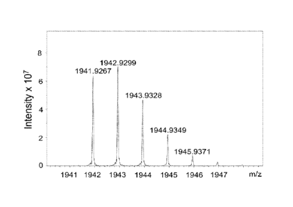
mechanisms for
the antibodies.
[7] Some therapeutic antibodies bind to certain surface antigens on target
cells and render
Fc-mediated functions and other mechanisms on the target cells. The most
important
Fc-mediated mechanisms are antibody-dependent cellular cytotoxicity (ADCC) and
complement-mediated cytolysis (CMC), which both will cause the lysis of the
antibody-bound target cells. Some antibodies binding to certain cell surface
antigens can
induce apoptosis of the bound target cells.
[8] The concept and methodology for preparing antibodies with dual
specificities
germinated more than three decades ago.
In recent year, the advancement in
recombinant antibody engineering methodologies and the drive to develop
improved
medicine has stimulated the development bi-specific antibodies adopting a
large variety of
structural configurations.
[9] For example, the bi-valent or multivalent antibodies may contain two or
more
antigen-binding sites. A number of methods have been reported for preparing
multivalent
antibodies by covalently linking three or four Fab fragments via a connecting
structure. For
example, antibodies have been engineered to express tandem three or four Fab
repeats.
[10] Several methods for producing multivalent antibodies by employing
synthetic
crosslinkers to associate, chemically, different antibodies or binding
fragments have been
disclosed.
One approach involves chemically cross-linking three, four, and more
separately Fab fragments using different linkers. Another method to produce a
construct
with multiple Fabs that are assembled to one-dimensional DNA scaffold was
provided.
Those various multivalent Ab constructs designed for binding to target
molecules differ
among one another in size, half-lives, flexibility in conformation, and
ability to modulate the
immune system. In view of the foregoing, several reports have been made for
preparing
molecular constructs with a fixed number of effector elements or with two or
more different
kinds of functional elements (e.g., at least one targeting element and at
least one effector
element). However, it is often difficult to build a molecular construct with a
particular
combination of the targeting and effector elements either using chemical
synthesis or
2
CA 02996653 2018-02-26
WO 2017/036407
PCT/CN2016/097783
recombinant technology. Accordingly, there exists a need in the related art to
provide
novel molecular platforms to build a more versatile molecule suitable for
covering
applications in a wide range of diseases.
SUMMARY
[11] The following presents a simplified summary of the disclosure in order to
provide a
basic understanding to the reader. This summary is not an extensive overview
of the
disclosure and it does not identify key/critical elements of the present
invention or delineate
the scope of the present invention. Its sole purpose is to present some
concepts disclosed
herein in a simplified form as a prelude to the more detailed description that
is presented
later.
[12] < I > Peptide Core-Based Multi-Arm Linkers for Treating Rejection
Reaction in
Transplantation and Uses thereof
[13] In the first aspect, the present disclosure is directed to a linker unit
for treating
transplantation rejection in a subject. In particular, the linker unit has at
least two different
functional elements linked thereto. For example, the linker unit may have
linked thereto
two different effector elements, one targeting element and one effector
element, or one
effector element and a polyethylene glycol (PEG) chain for prolonging the
circulation time of
the linker unit. The present linker unit is designed to have at least two
different functional
groups such that the functional elements can be linked thereto by reacting
with the
respective functional groups. Accordingly, the present linker unit can serve
as a platform
for preparing a molecular construct with two or more functional elements. As
could be
appreciated, methods for treating a transplant patient using such linker unit
also fall within
the aspect of the present disclosure
[14] According to various embodiments of the present disclosure, the linker
unit comprises a
center core, a plurality of linking arms, a plurality of first elements, and
optionally, a coupling
arm and a second element.
[15] According to various embodiments of the present disclosure, the center
core is a
peptide core having a pre-defined number of amine (-NH2) groups, before being
linked with
the linking arms. For example, the peptide core may have two or more lysine
(K) resides
3
CA 02996653 2018-02-26
WO 2017/036407
PCT/CN2016/097783
having an amine (-NH2) group at the side chain.
[16] In certain embodiments, the peptide core comprises 2 to 15 K resides and
one or more
filler sequences, in which each K residue and a next K residue are separated
by one of the
filler sequences. Each of the filler sequences comprises glycine (G) and
serine (S)
residues. Optionally, the filler sequence consists of 2 to 20 amino acid
residues. In
various embodiments, the filler sequence may have the sequence of GS, GCS,
CSC, or
SEQ ID NOs: 1-16. In certain embodiments of the present disclosure, at least
one of the
filler sequences in one peptide core differs from the remaining filler
sequences of the same
peptide core. According to some embodiments of the present disclosure, the
peptide core
comprises 2 to 15 units of the sequence of G1_5SK; preferably, the peptide
core comprises
the sequence of (GSK)2-15.
[17] According to some other embodiments, the peptide core comprises the
sequence of
(X58-K)2_15, in which Xõ is a PEGylated amino acid having 2 to 12 repeats of
ethylene glycol
(EC) unit.
[18] Each of the linking arms is linked to the amine groups of the center core
via forming an
amide linkage between the amine group and the linking arm. As could be
appreciated, in
the case of a peptide core, the linking arm is linked to the center core by
reacting with the
amine group at the side chain of the K residue. Further, the linking arm
linked to the center
core has a maleimide, an N-hydroxysuccinimidyl (NHS) group, an azide group, an
alkyne
group, a tetrazine group, a cyclooctene group, or a cyclooctyne group at its
free-terminus.
[19] On the other hand, for the peptide core, the amino acid residue at the N-
or C-terminus
of the center core has an azide group or an alkyne group; alternatively or
additionally, the
amino acid residue at the N- or C-terminus of the center core is a cysteine
(C) residue.
[20] According to certain embodiments of the present disclosure, when the
center core is a
a peptide core having a terminal amino acid residue of Cysteine, the present
linker unit
comprises said coupling arm. For peptide cores with terminal the terminal
amino acid
residue of Cysteine, one end of the coupling arm is linked to the Cysteine
residue by
reacting with the thiol group thereof.
4
CA 02996653 2018-02-26
WO 2017/036407
PCT/CN2016/097783
[21] Regarding amino acid residues having the azide group, non-limiting
examples of said
amino acid residues include L-azidohomoalanine (AHA), 4-azido-L-phenylalanine,
4-azido-D-phenylalanine, 3-azido-L-alanine, 3-azido-D-alanine, 4-azido-L-
homoalanine,
4-azido-D-homoalanine, 5-azido-L-ornithine, 5-azido-d-ornithine, 6-azido-L-
lysine, and
6-azido-D-lysine. As to the amino acid residues having the alkyne group,
illustrative
examples thereof include L-homopropargylglycine (L-HPG), D-
homopropargylglycine
(D-HPG), and beta-homopropargylglycine (p-HPG).
[22] When the amino acid residues at the N- or C-terminus of the center core
is the cysteine
residue, the cyclooctene group at the free terminus of the coupling arm may
be, a
trans-cyclooctene (TCO) group, while the cyclooctyne group at the free
terminus of the
coupling arm may be a dibenzocyclooctyne (DBCO), difluorinated cyclooctyne
(DIFO),
bicyclononyne (BCN), or dibenzocyclooctyne (DICO) group. Alternatively, the
tetrazine
group at the free terminus of the coupling arm includes, but is not limited
to,
1,2,3,4-tetrazine, 1,2,3,5-tetrazine, and 1,2,4,5-tetrazine, and derivatives
thereof, such as,
6-methyl tetrazine.
[23] In some embodiments, the linking arm is a PEG chain, preferably having 2
to 20
repeats of EG units. In other embodiments, the coupling linking arm is a PEG
chain,
preferably having 2 to 12 repeats of EG units.
[24] According to various optional embodiments of the present disclosure, the
first element
is an effector element suitable for eliciting an intended effect (e.g., a
therapeutic effect) in a
subject. Alternatively, the first element may be a targeting element for
directing the linker
unit to the site of interest. In preferred embodiments, when the first element
is the effector
element, the second element is the targeting element, and vice versa.
[25] Specifically, the targeting element according to various embodiments of
the present
disclosure is an antibody fragment specific for a human leukocyte antigen
(HLA) allotype
present only on cells of the donor transplant and not on cells of the
recipient, such as the
HLA-A, HLA-B, and HLA-C allotype. Also, the effector element is an
immunosuppressant,
an immune checkpoint protein, or an antibody fragment specific for CD25.
Illustrative
examples of immunosuppressant are inhibitors of mammalian target of rapamycin
(mTOR),
e.g. sirolimus and everolimus. Another set of immunosuppressants are
inhibitors of
5
CA 02996653 2018-02-26
WO 2017/036407
PCT/CN2016/097783
calcineurin, e.g. tacrolimus. Fingolimod and derivatives thereof (e.g.,
fingolimod
phosphate) are also examples of suitable immunosuppressants Immune checkpoint
proteins are those involve in immune checkpoint, such as the extracellular
domain of
cytotoxic T lymphocyte associated protein 4 (CTLA-4, also known as 00151) and
the
extracellular domain of programmed death-ligand 1 (PD-L1, also known as
00274).
[26] In some embodiments, each of the first elements is linked to one of the
linking arms via
forming an amide bound between the linking arm and the first element. In other
embodiments, each of the first elements is linked to one of the linking arms
via
thiol-maleimide reaction, copper catalyzed azide-alkyne cycloaddition (CuAAC)
reaction,
strained-promoted azide-alkyne click chemistry (SPAAC) reaction, or inverse
electron
demand DieIs¨Alder (iEDDA) reaction occurred between the linking arm and the
first
element.
[27] According to some embodiments of the present disclosure, when the
plurality of first
elements are respectively linked to the plurality of linking arms via CuAAC or
SPAAC
reaction, then the amino acid residue at the N- or 0-terminus of the center
core is a cysteine
residue, and the free terminus of the coupling arm is the tetrazine or the
cyclooctene group.
According to other embodiments of the present disclosure, when the plurality
of first
elements are respectively linked to the plurality of linking arms via 'EDDA
reaction, then the
amino acid residue at the N- or 0-terminus of the center core has the azide or
the alkyne
group, or the amino acid residue at the N- or 0-terminus of the center core is
a cysteine
residue, and the free terminus of the coupling arm is the azide, the alkyne,
or the
cyclooctyne group.
[28] In some embodiments, the second element has an azide or alkyne group, so
that it is
linked to the center core or the coupling arm by coupling with the
corresponding alkyne or
azide group of the center core or the coupling arm via CuAAC reaction.
Alternatively, in
other embodiments, the second element having an azide or cyclooctyne group is
linked to
the center core or the coupling arm by coupling with the corresponding
cyclooctyne or azide
group of the center core or the coupling arm via SPAAC reaction. Still
alternatively, in
certain embodiments, the second element having a tetrazine or cyclooctene
group is linked
to the center core or the coupling arm by coupling with the corresponding
cyclooctene or
6
CA 02996653 2018-02-26
WO 2017/036407
PCT/CN2016/097783
tetrazine group of the center core or the coupling arm via iEDDA reaction.
[29] In certain embodiments, the linker unit further comprises an optional
third element that
is different from the first and second elements. In the case where the second
element is
directly linked to the center core, the other terminus (i.e., the free
terminus that is not linked
with the second element) of the center core is optionally a cysteine residue,
which can be
used to introduce an optional third element. Specifically, the thiol group of
the cysteine
residue is reacted with a maleimide group of a PEG chain; and the thus-linked
PEG chain is
designated as the coupling arm, which has a tetrazine group or a cyclooctene
group at its
free terminus. Accordingly, the third element is then linked to the coupling
arm via iEDDA
reaction. Preferably, the third element is an element for improving the
pharmacokinetic
property of the linker unit. One example of the element for improving the
pharmacokinetic
property is a long PEG chain having a molecular weight of about 20,000 to
50,000 Da!tons.
[30] The linker unit according to this aspect of the present disclosure may
find its utility in
clinical medicine for the treatment of transplantation rejection. Accordingly,
the present
disclosure is also directed to a method for suppressing or inhibiting
transplantation rejection
in a subject receiving a donor transplant (e.g., organ, tissue or cells), or
for use in the
manufacture of a medicament for such uses. According to various embodiments of
the
present disclosure, the method for treating the transplantation rejection in a
particular
subject includes the step of administering to the subject in need thereof a
therapeutically
effective amount of the linker unit according to the above-mentioned aspect
and
embodiments of the present disclosure. As could be appreciated, said linker
unit may be
administered in a pharmaceutical formulation, which
comprises a
pharmaceutically-acceptable excipient suitable for the intended or desired
administration
route, in addition to the present linker unit.
[31] < II > Fc-based Molecular Construct for Treating Rejection Reaction in
Transplantation and Uses thereof
[32] In this aspect, the present disclosure is directed to a fragment
crystallizable (Fc)-based
molecular construct that has at least one targeting element and at least one
effector
element linked, directly or indirectly, to a CH2-CH3 domain of an
immunoglobulin.
Targeting and effector elements of the present Fc-based molecular constructs
are
7
CA 02996653 2018-02-26
WO 2017/036407
PCT/CN2016/097783
specifically selected such that these Fc-based molecular constructs are
suitable for use in
suppressing or inhibiting the transplantation rejection in a subject (or
recipient) receiving an
organ, tissue or cell transplantation, or for use in the manufacture of a
medicament for such
uses. As could be appreciated, methods for treating transplantation rejection
using such
Fc-based molecular constructs also fall within the aspect of the present
disclosure.
[33] According to certain embodiments of the present disclosure, the Fc-based
molecular
construct comprises a pair of CH2-CH3 segments of an IgG.Fc, a pair of
effector elements,
and a pair of targeting elements.
[34] According to various embodiments of the present disclosure, the pair of
targeting
elements is an antibody fragment specific for a human leukocyte antigen (HLA)
allotype
present only on cells of the donor transplant and not on cells of the
recipient, such as the
HLA-A, HLA-B, and HLA-C allotype present only on cells of the donor
transplant. Also, the
pair of elements is an immune checkpoint protein, an antibody fragment
specific for CD25,
or a drug bundle comprising an immunosuppressant. Immune checkpoint proteins
are
those involve in immune checkpoint, such as the extracellular domain of
cytotoxic T
lymphocyte associated protein 4 (CTLA-4, also known as CD151) and the
extracellular
domain of programmed death-ligand 1 (PD-L1, also known as CD274). Illustrative
examples of immunosuppressant are inhibitors of mammalian target of rapamycin
(mTOR),
e.g. sirolimus and everolimus. Another set of immunosuppressants are
inhibitors of
calcineurin, e.g. tacrolimus. Fingolimod and derivatives thereof (e.g.,
fingolimod
phosphate) are also examples of suitable immunosuppressants
[35] In the case where the effector element is the immune checkpoint protein,
then the pair
of effector elements is linked to the N-termini of the pair of CH2-CH3
segments, and the pair
of targeting elements is linked to the C-termini of the pair of CH2-CH3
segments, or vice
versa. Alternatively, when the effector element is the drug bundle, then the
pair of effector
elements is linked to the C-termini of the pair of CH2-CH3 segments, and the
pair of
targeting elements is linked to the N-termini of the pair of CH2-CH3 segments.
Still
alternatively, when the effector elements are the antibody fragments, then the
effector
elements is respectively linked to the N-termini of the pair of CH2-CH3
segments, and the
8
CA 02996653 2018-02-26
WO 2017/036407
PCT/CN2016/097783
targeting elements is respectively linked to the C-termini of the pair of CH2-
CH3 segments,
and vice versa.
[36] According to certain embodiments, when the pair of effector elements and
the pair of
targeting elements are both in the form of single-chain variable fragments
(scFvs), then the
pair of targeting elements is linked to the N-termini of the pair of effector
elements in a
tandem or diabody configuration, thereby forming a pair of bispecific scFvs
that are linked to
the N-termini of the pair of CH2-CH3 segments.
[37] In some examples, the pair of the targeting elements takes a Fab
configuration (i.e.,
consisting of the VH-C1-11 domain and the VL-Ck domain); this Fab fragment is
linked to the
N-termini of the first and second heavy chains, so that the Fc-based molecular
construct
adopts an IgG configuration. In these cases, the pair of effector elements is
linked to the
C-termini of the pair of CH2-0H3 segments.
[38] According to some other embodiments of the present disclosure, when the
pair of
effector elements is in the form of an antigen-binding fragment (Fab), and the
pair of
targeting elements is in the form of scFvs, and vice versa; then the Fab and
scFvs are
respectively linked to the N-termini and C-termini of the CH2-CH3 segments, so
that the
molecular construct adopts an extended IgG configuration.
[39] In certain embodiments, the pair of CH2-CH3 segments is derived from
human IgG
heavy chain y4 or human IgG heavy chain yl .
[40] According to some optional embodiments, the effector elements are drug
bundles
based on linker units. Such drug bundles are advantageous at least in that
they can be
manufactured separately before being conjugated to the antibody molecules,
thus avoiding
subjecting drug molecules under harsh chemical conditions for the direct
conjugation with
the antibody molecules. According to various embodiments of the present
disclosure, the
drug bundle comprises a plurality of immunosuppressant molecules. As an
example,
rather than a limitation, these Fc-based molecular constructs are useful in
the treatment of
transplantation rejection.
[41] According to certain embodiments, the present Fc-based molecular
construct further
comprises a peptide extension and a coupling arm. Specifically, the peptide
extension has
9
CA 02996653 2018-02-26
WO 2017/036407
PCT/CN2016/097783
the sequence of (G2_4S)2_8C and is linked to the C-terminus of one of the pair
of CH2-CH3
segments. In such cases, the coupling arm is linked to the C-terminus of the
peptide
extension via thiol-maleimide reaction occurred therebetween. Also, before
being
conjugated with the drug bundle, the free terminus of the coupling arm (that
is, the terminus
that is not linked to the cysteine residue) is modified with an alkyne, azide,
strained alkyne,
or tetrazine group, so that the drug bundle is linked thereto via inverse
electron demand
DieIs-Alder (iEDDA) reaction or the strain-promoted azide-alkyne click
chemistry (SPAAC)
reaction or Copper(I)-catalyzed alkyne-azide cycloaddition (CuAAC) reaction
occurred
therebetween.
[42] According to some optional embodiments, the drug bundle is a linker unit-
based
molecular construct according to the first aspect and embodiments of the
present
disclosure.
[43] Briefly, the center core may be a polypeptide comprising a plurality of
lysine (K)
residues, according to various embodiments of the present disclosure. Each of
the linking
arms has one terminus that is linked to the center core by reacting with the
amine groups at
the side chain of the K residues of the polypeptide core. The linking arm also
carries a
maleimide group at the free terminus thereof, wherein each of the molecules
(e.g.,
immunosuppressant molecules) is linked to the center core through the linking
arm by
reacting with the maleimide group.
[44] In the case where the center core is the polypeptide core, then the amino
acid residue
at the N- or C-terminus of the center core is a cysteine residue or has an
azide group or an
alkyne group.
[45] For polypeptide cores with a terminal amino acid residue having the azide
group or the
alkyne group, the drug bundle may be linked to the peptide extension via the
CuAAC
reaction occurred between said terminal residue and the C-terminus of the
peptide
extension.
[46] Methods for suppressing or inhibiting transplantation rejection in a
subject in need
thereof comprise the step of administering to the subject an effective amount
of the
molecular construct of this aspect.
CA 02996653 2018-02-26
WO 2017/036407
PCT/CN2016/097783
BRIEF DESCRIPTION OF THE DRAWINGS
[47] The present description will be better understood from the following
detailed description
read in light of the accompanying drawings briefly discussed below.
[48] Figure 1A to Figure 1K are schematic diagrams illustrating linker units
according to
certain embodiments of the present disclosure.
[49] Figures 2A to 20 are schematic diagrams illustrating Fc-based molecular
constructs
according to various embodiments of the present disclosure.
[50] Figure 3 is a schematic diagram illustrating a Fc-based molecular
construct according
to some embodiments of the present disclosure.
[51] Figures 4A to 40 are schematic diagrams illustrating Fc-based molecular
constructs
according to various embodiments of the present disclosure.
[52] Figure 5A and 5B are schematic diagrams illustrating Fc-based molecular
constructs
according to various embodiments of the present disclosure.
[53] Figure 6A and Figure 6B respectively show the mass spectrometric analysis
of the
sirolimus-Gly and sirolimus-diGly, according to one working example of the
present
disclosure.
[54] Figure 7A and Figure 7B respectively show the MALDI-TOF analysis of the
azido-PEG3-S-S-conjugated sirolimus-Gly and sirolimus-diGly, according to one
working
example of the present disclosure.
[55] Figure 8 shows the MALDI-TOF analysis of the NHS-PEG5-conjugated
fingolimod.
[56] Figure 9 shows the mass spectrometric analysis of the azido-PEG3-S-S-
conjugated
fingolimod according to one working example of the present disclosure.
[57] Figure 10A and Figure 10B respectively show the mass spectrometric
analysis of the
NHS-PEG4-PEG3-S-S-conjugated sirolimus-Gly and sirolimus-diGly, according to
one
working example of the present disclosure.
11
CA 02996653 2018-02-26
WO 2017/036407 PCT/CN2016/097783
[58] Figure 11 shows the mass spectrometric analysis
of the
NHS-PEG4-PEG3-S-S-conjugated fingolimod, according to one working example of
the
present disclosure.
[59] Figures 12A to 12D respectively show the MALDI-TOF analysis of a drug
bundle
composing of a linker unit with a free TOO functional group and a set of five
sirolimus-Gly,
five fingolimod, ten fingolimod and five fingolimod phosphate molecules,
according to one
working example of the present disclosure.
[60] Figures 13A to 130 respectively show the SDS-PAGE analysis of purified
human
HLA-A1-IgG1.Fc, HLA-A2-IgG1.Fc and PD-1-IgG1.Fc fusion protein, according to
one
working example of the present disclosure.
[61] Figures 14A and 14B respectively show the SDS-PAGE of purified human CTLA-
4 and
PD-L1 proteins; Figure 140 shows the western blot analysis of purified human
CTLA-4; and
Figure 14D shows the ELISA analysis of purified human PD-L1 proteins,
according to one
working example of the present disclosure.
[62] Figures 15A to 15C respectively show the SDS-PAGE, mass spectrometric and
ELISA
analyses of purified scFv of mAb specific for human HLA-Al , according to one
working
example of the present disclosure.
[63] Figures 16A and 16B respectively show the titers of the phages bearing
scFvs specific
for human HLA-A2 and the single colony ELISA analysis of phage-displayed scFvs
specific
for human HLA-A2, according to one working example of the present disclosure.
[64] Figure 17A and Figure 17B respectively show the mass spectrometric and
ELISA
analysis of tetrazine-conjugated scFv specific for human HLA-A1, according to
one working
example of the present disclosure.
[65] Figure 18 show the 10% SDS-PAGE analysis of an effector linker-unit,
composed of a
linker-unit with a free TOO functional group and a set of three three PDL-1
molecules as
effector elements, according to one working example of the present disclosure.
[66] Figure 19 shows the 10% SDS-PAGE analysis of a single linker unit
molecular
construct with one scFv specific for HLA-A1 as targeting element and three PD-
L1
12
CA 02996653 2018-02-26
WO 2017/036407
PCT/CN2016/097783
molecules as an effector element, according to one working example of the
present
disclosure.
[67] Figure 20 and Figure 20B respectively show the mTOR inhibition and T-cell
proliferation assay of sirolimus and sirolimus derivative compounds, according
to one
working example of the present disclosure.
[68] Figure 21A shows the staining analysis of the S1 P1 receptor-expressing
human B cells;
Figure 21B shows transwell migration assay of fingolimod upon the conjugation
to peptide
core through linking arms, according to one working example of the present
disclosure.
[69] Figure 22A shows the SDS-PAGE analysis of purified recombinant 2-chain
(CTLA-4)-IgG1.Fc-(scFv a HLA-A1) fusion protein; Figure 22B and Figure 220
respectively
show the ELISA analysis of of purified recombinant 2-chain (CTLA-4)-IgG1.Fc-
(scFv a
HLA-A1) fusion protein with the scFv specific for CTLA-4 and with human HLA-
A1,
according to one working example of the present disclosure.
[70] Figure 23A shows the SDS-PAGE analysis of purified recombinant 2-chain
(PD-L1)-IgG4.Fc-( scFv a HLA-A1) fusion protein; Figures 23B to 23D
respectively show the
ELISA analysis of purified recombinant 2-chain (PD-L1)-IgG4.Fc-( scFv a HLA-
A1) fusion
protein with the mAb specific for PD-L1, human PD-1, and human HLA-A1,
according to one
working example of the present disclosure.
[71] In accordance with common practice, the various described
features/elements are not
drawn to scale but instead are drawn to best illustrate specific
features/elements relevant to
the present invention. Also, like reference numerals and designations in the
various
drawings are used to indicate like elements/parts, where possible.
DESCRIPTION
[72] The detailed description provided below in connection with the appended
drawings is
intended as a description of the present examples and is not intended to
represent the only
forms in which the present example may be constructed or utilized. The
description sets
forth the functions of the example and the sequence of steps for constructing
and operating
13
CA 02996653 2018-02-26
WO 2017/036407
PCT/CN2016/097783
the example. However, the same or equivalent functions and sequences may be
accomplished by different examples.
[73] For convenience, certain terms employed in the specification, examples
and appended
claims are collected here. Unless otherwise defined herein, scientific and
technical
terminologies employed in the present disclosure shall have the meanings that
are
commonly understood and used by one of ordinary skill in the art.
[74] Unless otherwise required by context, it will be understood that singular
terms shall
include plural forms of the same and plural terms shall include the singular.
Specifically, as
used herein and in the claims, the singular forms "a" and "an" include the
plural reference
unless the context clearly indicated otherwise. Also, as used herein and in
the claims, the
terms at least one" and one or more" have the same meaning and include one,
two, three,
or more. Furthermore, the phrases at least one of A, B, and C", at least one
of A, B, or C"
and at least one of A, B and/or C," as use throughout this specification and
the appended
claims, are intended to cover A alone, B alone, C alone, A and B together, B
and C together,
A and C together, as well as A, B, and C together.
[75] Notwithstanding that the numerical ranges and parameters setting forth
the broad
scope of the invention are approximations, the numerical values set forth in
the specific
examples are reported as precisely as possible. Any numerical value, however,
inherently
contains certain errors necessarily resulting from the standard deviation
found in the
respective testing measurements. Also, as used herein, the term "about"
generally means
within 10%, 5%, 1%, or 0.5% of a given value or range. Alternatively, the term
"about"
means within an acceptable standard error of the mean when considered by one
of ordinary
skill in the art. Other than in the operating/working examples, or unless
otherwise
expressly specified, all of the numerical ranges, amounts, values and
percentages such as
those for quantities of materials, durations of times, temperatures, operating
conditions,
ratios of amounts, and the likes thereof disclosed herein should be understood
as modified
in all instances by the term "about." Accordingly, unless indicated to the
contrary, the
numerical parameters set forth in the present disclosure and attached claims
are
approximations that can vary as desired. At the very least, each numerical
parameter
should at least be construed in light of the number of reported significant
digits and by
14
CA 02996653 2018-02-26
WO 2017/036407
PCT/CN2016/097783
applying ordinary rounding techniques. Ranges can be expressed herein as from
one
endpoint to another endpoint or between two endpoints. All ranges disclosed
herein are
inclusive of the endpoints, unless specified otherwise.
[76] This present disclosure pertains generally to molecular constructs, in
which each
molecular construct comprises a targeting element (T) and an effector element
(E), and
these molecular constructs are sometimes referred to as "T-E molecules", "T-E
pharmaceuticals" or "T-E drugs" in this document.
[77] As used herein, the term "targeting element" refers to the portion of a
molecular
construct that directly or indirectly binds to a target of interest (e.g., a
receptor on a cell
surface or a protein in a tissue) thereby facilitates the transportation of
the present
molecular construct into the interested target. In some example, the targeting
element
may direct the molecular construct to the proximity of the target cell. In
other cases, the
targeting element specifically binds to a molecule present on the target cell
surface or to a
second molecule that specifically binds a molecule present on the cell
surface. In some
cases, the targeting element may be internalized along with the present
molecular construct
once it is bound to the interested target, hence is relocated into the cytosol
of the target cell.
A targeting element may be an antibody or a ligand for a cell surface
receptor, or it may be a
molecule that binds such antibody or ligand, thereby indirectly targeting the
present
molecular construct to the target site (e.g., the surface of the cell of
choice). The
localization of the effector (therapeutic agent) in the diseased site will be
enhanced or
favored with the present molecular constructs as compared to the therapeutic
without a
targeting function. The localization is a matter of degree or relative
proportion; it is not
meant for absolute or total localization of the effector to the diseased site.
[78] According to the present invention, the term "effector element" refers to
the portion of a
molecular construct that elicits a biological activity (e.g., inducing immune
responses,
exerting cytotoxic effects and the like) or other functional activity (e.g.,
recruiting other
hapten tagged therapeutic molecules), once the molecular construct is directed
to its target
site. The "effect" can be therapeutic or diagnostic. The effector elements
encompass
those that bind to cells and/or extracellular immunoregulatory factors. The
effector
element comprises agents such as proteins, nucleic acids, lipids,
carbohydrates,
CA 02996653 2018-02-26
WO 2017/036407
PCT/CN2016/097783
glycopeptides, drug moieties (both small molecule drug and biologics),
compounds,
elements, and isotopes, and fragments thereof.
[79] Although the terms, first, second, third, etc., may be used herein to
describe various
elements, components, regions, and/or sections, these elements (as well as
components,
regions, and/or sections) are not to be limited by these terms. Also, the use
of such ordinal
numbers does not imply a sequence or order unless clearly indicated by the
context.
Rather, these terms are simply used to distinguish one element from another.
Thus, a first
element, discussed below, could be termed a second element without departing
from the
teachings of the exemplary embodiments.
[80] Here, the terms "link," "couple," and "conjugates" are used
interchangeably to refer to
any means of connecting two components either via direct linkage or via
indirect linkage
between two components.
[81] The term "polypeptide" as used herein refers to a polymer having at least
two amino
acid residues. Typically, the polypeptide comprises amino acid residues
ranging in length
from 2 to about 200 residues; preferably, 2 to 50 residues. Where an amino
acid sequence
is provided herein, L-, D-, or beta amino acid versions of the sequence are
also
contemplated. Polypeptides also include amino acid polymers in which one or
more amino
acid residues are an artificial chemical analogue of a corresponding naturally
occurring
amino acid, as well as to naturally occurring amino acid polymers. In
addition, the term
applies to amino acids joined by a peptide linkage or by other, "modified
linkages," e.g.,
where the peptide bond is replaced by an a-ester, a 13-ester, a thioamide,
phosphoramide,
carbomate, hydroxylate, and the like.
[82] In certain embodiments, conservative substitutions of the amino acids
comprising any
of the sequences described herein are contemplated. In various embodiments,
one, two,
three, four, or five different residues are substituted. The term
"conservative substitution"
is used to reflect amino acid substitutions that do not substantially alter
the activity (e.g.,
biological or functional activity and/or specificity) of the molecule.
Typically, conservative
amino acid substitutions involve substitution one amino acid for another amino
acid with
similar chemical properties (e.g., charge or hydrophobicity).
Certain conservative
substitutions include "analog substitutions" where a standard amino acid is
replaced by a
16
CA 02996653 2018-02-26
WO 2017/036407
PCT/CN2016/097783
non-standard (e.g., rare, synthetic, etc.) amino acid differing minimally from
the parental
residue. Amino acid analogs are considered to be derived synthetically from
the standard
amino acids without sufficient change to the structure of the parent, are
isomers, or are
metabolite precursors.
[83] In certain embodiments, polypeptides comprising at least 80%, preferably
at least 85%
or 90%, and more preferably at least 95% or 98% sequence identity with any of
the
sequences described herein are also contemplated.
[84] "Percentage (%) amino acid sequence identity" with respect to the
polypeptide
sequences identified herein is defined as the percentage of polypeptide
residues in a
candidate sequence that are identical with the amino acid residues in the
specific
polypeptide sequence, after aligning the sequences and introducing gaps, if
necessary, to
achieve the maximum percent sequence identity, and not considering any
conservative
substitutions as part of the sequence identity. Alignment for purposes of
determining
percentage sequence identity can be achieved in various ways that are within
the skill in the
art, for instance, using publicly available computer software such as BLAST,
BLAST-2,
ALIGN or Megalign (DNASTAR) software. Those skilled in the art can determine
appropriate parameters for measuring alignment, including any algorithms
needed to
achieve maximal alignment over the full length of the sequences being
compared. For
purposes herein, sequence comparison between two polypeptide sequences was
carried
out by computer program Blastp (protein-protein BLAST) provided online by
Nation Center
for Biotechnology Information (NCB!). The percentage amino acid sequence
identity of a
given polypeptide sequence A to a given polypeptide sequence B (which can
alternatively
be phrased as a given polypeptide sequence A that has a certain % amino acid
sequence
identity to a given polypeptide sequence B) is calculated by the formula as
follows:
¨X x 100 %
where X is the number of amino acid residues scored as identical matches by
the sequence
alignment program BLAST in that program's alignment of A and B, and where Y is
the total
number of amino acid residues in A or B, whichever is shorter.
17
CA 02996653 2018-02-26
WO 2017/036407
PCT/CN2016/097783
[85] The term "PEGylated amino acid" as used herein refers to a polyethylene
glycol (PEG)
chain with one amino group and one carboxyl group. Generally, the PEGylated
amino acid
has the formula of NH2-(CH2CH20)n-COOH. In the present disclosure, the value
of n
ranges from 1 to 20; preferably, ranging from 2 to 12.
[86] As used herein, the term "terminus" with respect to a polypeptide refers
to an amino
acid residue at the N- or C- end of the polypeptide. With regard to a polymer,
the term
"terminus" refers to a constitutional unit of the polymer (e.g., the
polyethylene glycol of the
present disclosure) that is positioned at the end of the polymeric backbone.
In the present
specification and claims, the term "free terminus" is used to mean the
terminal amino acid
residue or constitutional unit is not chemically bound to any other molecular.
[87] The term "antigen" or "Ag" as used herein is defined as a molecule that
elicits an
immune response. This immune response may involve a secretory, humoral, and/or
cellular antigen-specific response. In the present disclosure, the term
"antigen" can be any
of a protein, a polypeptide (including mutants or biologically active
fragments thereof), a
polysaccharide, a glycoprotein, a glycolipid, a nucleic acid, or a combination
thereof.
[88] In the present specification and claims, the term "antibody" is used in
the broadest
sense and covers fully assembled antibodies, antibody fragments that bind with
antigens,
such as antigen-binding fragment (Fab/Fab'), F(ab')2 fragment (having two
antigen-binding
Fab portions linked together by disulfide bonds), variable fragment (Fv),
single chain
variable fragment (scFv), bi-specific single-chain variable fragment (bi-
scFv), nanobodies,
unibodies and diabodies. "Antibody fragments" comprise a portion of an intact
antibody,
preferably the antigen-binding region or variable region of the intact
antibody. Typically, an
"antibody" refers to a protein consisting of one or more polypeptides
substantially encoded
by immunoglobulin genes or fragments of immunoglobulin genes. The well-known
immunoglobulin genes include the kappa, lambda, alpha, gamma, delta, epsilon,
and mu
constant region genes, as well as myriad immunoglobulin variable region genes.
Light
chains are classified as either kappa or lambda. Heavy chains are classified
as gamma,
mu, alpha, delta, or epsilon, which in turn define the immunoglobulin classes,
IgG, IgM, IgA,
IgD, and IgE, respectively. A typical immunoglobulin (antibody) structural
unit is known to
comprise a tetramer. Each tetramer is composed of two identical pairs of
polypeptide
18
CA 02996653 2018-02-26
WO 2017/036407
PCT/CN2016/097783
chains, with each pair having one "light" chain (about 25 kDa) and one "heavy"
chain (about
50-70 kDa). The N-terminus of each chain defines a variable region of about
100 to 110 or
more amino acids primarily responsible for antigen recognition. The terms
variable light
chain (VL) and variable heavy chain (VH) refer to these light and heavy
chains, respectively.
According to embodiments of the present disclosure, the antibody fragment can
be
produced by modifying the nature antibody or by de novo synthesis using
recombinant DNA
methodologies. In certain embodiments of the present disclosure, the antibody
and/or
antibody fragment can be bispecific, and can be in various configurations. For
example,
bispecific antibodies may comprise two different antigen binding sites
(variable regions).
In various embodiments, bispecific antibodies can be produced by hybridoma
technique or
recombinant DNA technique. In certain embodiments, bispecific antibodies have
binding
specificities for at least two different epitopes.
[89] The term "specifically binds" as used herein, refers to the ability of an
antibody or an
antigen-binding fragment thereof, to bind to an antigen with a dissociation
constant (Kd) of
no more than about lx10-6 M, 1 X10⁻⁷ M, I X10-8 M, I X10-9 M, 1)00-10 NA7 1)00-
11 M-7
1)(10-12
M, and/or to bind to an antigen with an affinity that is at least two-folds
greater than its
affinity to a nonspecific antigen.
[90] The term "treatment" as used herein includes preventative (e.g.,
prophylactic), curative
or palliative treatment; and "treating" as used herein also includes
preventative (e.g.,
prophylactic), curative or palliative treatment. In particular, the term
"treating" as used
herein refers to the application or administration of the present molecular
construct or a
pharmaceutical composition comprising the same to a subject, who has a medical
condition
a symptom associated with the medical condition, a disease or disorder
secondary to the
medical condition, or a predisposition toward the medical condition, with the
purpose to
partially or completely alleviate, ameliorate, relieve, delay onset of,
inhibit progression of,
reduce severity of, and/or reduce incidence of one or more symptoms or
features of said
particular disease, disorder, and/or condition. Treatment may be administered
to a subject
who does not exhibit signs of a disease, disorder, and/or condition, and/or to
a subject who
exhibits only early signs of a disease, disorder and/or condition, for the
purpose of
decreasing the risk of developing pathology associated with the disease,
disorder and/or
condition.
19
CA 02996653 2018-02-26
WO 2017/036407
PCT/CN2016/097783
[91] The term "effective amount" as used herein refers to the quantity of the
present
molecular construct that is sufficient to yield a desired therapeutic
response. An effective
amount of an agent is not required to cure a disease or condition but will
provide a treatment
for a disease or condition such that the onset of the disease or condition is
delayed,
hindered or prevented, or the disease or condition symptoms are ameliorated.
The
effective amount may be divided into one, two, or more doses in a suitable
form to be
administered at one, two or more times throughout a designated time period.
The specific
effective or sufficient amount will vary with such factors as particular
condition being treated,
the physical condition of the patient (e.g., the patient's body mass, age, or
gender), the type
of subject being treated, the duration of the treatment, the nature of
concurrent therapy (if
any), and the specific formulations employed and the structure of the
compounds or its
derivatives. Effective amount may be expressed, for example, as the total mass
of active
component (e.g., in grams, milligrams or micrograms) or a ratio of mass of
active
component to body mass, e.g., as milligrams per kilogram (mg/kg).
[92] The terms "application" and "administration" are used interchangeably
herein to mean
the application of a molecular construct or a pharmaceutical composition of
the present
invention to a subject in need of a treatment thereof.
[93] The terms "subject" and "patient" are used interchangeably herein and are
intended to
mean an animal including the human species that is treatable by the molecular
construct,
pharmaceutical composition, and/or method of the present invention. The term
"subject" or
"patient" intended to refer to both the male and female gender unless one
gender is
specifically indicated. Accordingly, the term "subject" or "patient" comprises
any mammal,
which may benefit from the treatment method of the present disclosure.
Examples of a
"subject" or "patient" include, but are not limited to, a human, rat, mouse,
guinea pig,
monkey, pig, goat, cow, horse, dog, cat, bird and fowl. In an exemplary
embodiment, the
patient is a human. The term "mammal" refers to all members of the class
Mammalia,
including humans, primates, domestic and farm animals, such as rabbit, pig,
sheep, and
cattle; as well as zoo, sports or pet animals; and rodents, such as mouse and
rat. The
term "non-human mammal" refers to all members of the class Mammalis except
human.
CA 02996653 2018-02-26
WO 2017/036407
PCT/CN2016/097783
[94] Throughout the present disclosure, the term "transplantation rejection"
refers to the
acute or chronic rejection of cells, tissue or solid organ allografts or
xenografts of, among
the others, pancreatic islets, stem cells, bone marrow, skin, muscle, corneal
tissue,
neuronal tissue, heart, lung, combined heart-lung, kidney, liver, bowel,
pancreas, trachea or
esophagus, or graft-versus-host diseases.
[95] As used herein, the term "donor transplant" refers to a population of
cells, or a tissue or
an organ that is to be moved from one body to another or from a donor site to
another
location on the subject's own body, for the purpose of replacing the
recipient's damaged or
absent tissue or organ.
[96] The present disclosure is based, at least on the construction of the T¨E
pharmaceuticals that can be delivered to target cells, target tissues or
organs at increased
proportions relative to the blood circulation, lymphoid system, and other
cells, tissues or
organs. When this is achieved, the therapeutic effect of the pharmaceuticals
is increased,
while the scope and severity of the side effects and toxicity is decreased. It
is also possible
that a therapeutic effector is administered at a lower dosage in the form of a
T-E molecule,
than in a form without a targeting component. Therefore, the therapeutic
effector can be
administered at lower dosages without losing potency, while lowering side
effects and
toxicity.
[97] Diseases that can benefit from better drug targeting
[98] Drugs used for many diseases can be improved for better efficacy and
safety, if they
can be targeted to the disease sites, i.e., if they can be localized or
partitioned to the
disease sites more favorably than the normal tissues or organs. Certain
antibody drugs,
which target infectious microorganisms or their toxic products, can be
improved, if they are
empowered with the ability to recruit immunocytes, which phagocytose and clear
the
antibody-bound particles. Following are primary examples of diseases, in which
drugs can
be improved if they can be preferentially distributed to the disease sites or
cells or if they
can recruit phagocytic immunocytes.
21
CA 02996653 2018-02-26
WO 2017/036407
PCT/CN2016/097783
[99] Examples of transplantation-related diseases/conditions include, but are
not limited to,
organ transplant rejection (including, chronic, acute, subacute, and
hyperacute rejection)
and graft-versus-host disease (GvHD).
[100] Transplantation is the act of transferring cells, tissues, or organs
from one body to
another or from a donor site to another location of the person's own body. The
malfunction
of an organ system can be corrected with transplantation of an organ from a
donor.
However, the donor transplants (such as the transplanted organ, tissue or
cells), especially
in allografts or xenografts, are recognized as foreign agents by the
recipient's immune
system, thereby causing the rejection of transplanted organs, tissues or
cells.
[101] Although there are many antigens involved in the rejection of
genetically disparate
tissues, those responsible for the most vigorous allograft rejection reactions
are the major
histocompatibility complex (MHC). In humans, the MHC is called the human
leukocyte
antigen (HLA) system and is located on the short arm of chromosome 6, near the
complement genes. The most studied HLA genes are the nine classical MHC genes:
HLA-A, HLA-B, HLA-C, HLA-DPA1, HLA-DPB1, HLA-DQA1, HLA-DQB1, HLA-DRA, and
HLA-DRB1. In humans, the MHC gene cluster is divided into three regions:
classes I, II,
and III. The A, B and C genes belong to MHC class I, whereas the six D genes
belong to
class II.
[102] The MHC expression is codominant, meaning that both set of
inherited alleles are
expressed equally on the cell surface. Furthermore, the set of MHC alleles are
inherited
as haplotypes; hence, a heterozygous individual will have two MHC haplotypes,
one from
the paternal chromosome and the other from maternal chromosome. Each person
carries
two alleles of each of the three class-I genes, (HLA-A, HLA-B and HLA-C), and
hence can
express six different types of MHC-I. In the class-II locus, each person
inherits a pair of
HLA-DP genes (DPA1 and DPB1), a couple of genes HLA-DQ (DQA1 and DQB1), one
gene HLA-DRa (DRA1), and one or more genes HLA-DR6 (DRB1 and DRB3, -4 or -5);
accordingly, one heterozygous individual can inherit six or eight functioning
class-II alleles,
three or more from each parent. The MHC genes are highly polymorphic; many
different
alleles exist in the different individuals inside a population.
22
CA 02996653 2018-02-26
WO 2017/036407
PCT/CN2016/097783
[103] Both MHC class I and MHC class ll proteins play a role in transplant
rejection.
MHC class I are expressed on all nucleated cells; and these class I molecules
are
responsible for presenting antigenic peptides from within the cell (e.g., self-
antigens,
antigens from the intracellular viruses, and tumor-associated antigens) to T
cells having
CD8 receptors, such as alloreactive killer T cells (also known as cytotoxic T
lymphocytes
(CTLs). Once the T cell receptors (TCRs) of CTLs recognize the transplanted
tissue's
MHC class I molecules, the CTLs trigger the target cell's programmed cell
death by
apoptosis. On the other hand, MHC class ll normally occurs only on the
professional
antigen-presenting cells (APCs), such as dendritic cells, activated
macrophages, and B
cells. The MHC class II present extracellular antigens to CD4 T cells. When
memory
helper T cells' CD4 receptors bind to the MHC class II molecules expressed on
the surfaces
of the target cells of the graft tissue, the memory helper T cells' TCRs
recognize their target
antigen, and subsequently produces clones that, as effector cells, secrete
immune signaling
molecules (cytokines) in approximately the cytokine balance that had prevailed
at the
memory helper T cell's priming to memorize the antigen. As the priming event
in this
instance occurred amid inflammation, the immune memory is pro-inflammatory.
[104] Graft-versus-host disease is a medical complication following the
receipt of donor
tissue from a genetically different person. GvHD is commonly associated with
stem cell or
bone marrow transplant but the term also applies to other forms of tissue
graft. Immune
cells (white blood cells) in the donated tissue (the graft) recognize the
recipient (the host) as
"foreign;" and then the transplanted immune cells attack the host's body
cells.
[105] The T-E molecular design of this invention can be applied for
preparing molecular
constructs for preventing the rejection of the donor transplant(s).
[106] In the present molecular constructs, the targeting elements are scFv
of antibodies
specific for an HLA A, B, or C allotype expressed by cells of the donor
transplant and not by
cells of the patient receiving the transplant. Since there are six genes in
the haplotypes of
HLA A, B, and C, it is not difficult to find one gene with different allotypes
between the donor
and the recipient. Many antibodies against HLA allotypes are already
available. For
example, antibodies specific for HLA A2 and B27 are well known. An antibody
specific for
Owl antigen (corresponding allele: 0*01:02) was made. Some antibodies bind to
23
CA 02996653 2018-02-26
WO 2017/036407
PCT/CN2016/097783
determinants shared by several allotypes, for example, one antibody binds to
All and A24
and another one to All, A25, A26, and A66. A panel of antibodies binding to
various HLA
allotypes may be established by isolating HLA A, B, and C allotype-specific B
cells from the
peripheral blood of patients receiving transplants and cloning the VH and VL
sequences of
those B cells by RT-PCR. Similar procedures have been established in preparing
antigen-specific human monoclonal antibodies for various viral antigens. A
molecular
construct with an scFv specific for an HLA allotype can then be chosen for a
patient who has
received a transplant with a particular haplotype.
[107] The effector elements can be chosen from (1) the ectodomain or
extracellular
domain of immune checkpoint proteins, such as CTLA-4 and PD-L1, which can
inhibit
on-going immune activation, (2) scFv of antibodies specific for CD25, which is
expressed by
activated T cells, or (3) small molecular immunosuppressive drugs, sirolimus
(rapamycin),
everolimus, and tacrolimus (FK-506), which have been used broadly for the
prevention of
transplantation rejection. Sirolimus and everolimus, which inhibit mTOR
(mammalian
target of rapamycin), and tacrolimus, which inhibits calcineurin, are powerful
inhibitors of T
cell activity. Fingolimod and derivatives thereof (e.g., fingolimod phosphate)
are also
examples of suitable immunosuppressants. Anti-CD25, fingolimod, sirolimus,
everolimus
and tacrolimus, each have a range of its serious side effects due to their
potent
immunosuppressive effects. It is desirable to shuffle increased proportions of
the drug to
the transplant and decreased proportions in other parts of the body,
especially the blood
and lymphoid system.
[108] Sirolimus (m.w. 914.172 daltons) and tacrolimus (m.w. 804.018
daltons) are
suitable for the present application, because in most applications, sirolimus
or everolimus is
used at approximately 2-10 mg per day and tacrolimus is used at approximately
5-15 mg
per day. The immunosuppressive drugs cyclosporine (m.w. 1,202.61 daltons) and
mycophenolic acid (m.w. 320.34 daltons), which are also used for the
prevention of rejection
of transplants, are not suitable for use herein, because cyclosporin is used
at approximately
150-1,000 mg per day, and mycophenolic acid is used at approximately 800-1,500
mg per
day. For a molecular construct with two scFvs as targeting elements and ten
sirolimus
molecules as effector elements, the weight of the scFv (m.w. 25,000 daltons)
is about 6
times of the weight of sirolimus. Thus, for administering 5 mg of sirolimus,
it requires 30
24
CA 02996653 2018-02-26
WO 2017/036407
PCT/CN2016/097783
mg of scFv, which is feasible. Because the administered sirolimus will be
carried to the
transplant, a less amount will be required than if the drug is administered
without targeting
to the transplant.
[109] Since sirolimus, everolimus, and tacrolimus, act on intracellular
targets of T cells,
they are linked to the multi-arm linker-unit via a reversible bond, which is
cleaved off the
linker by hydrolysis or by cleavage by tissue proteases present in the
targeted tissue.
Since the molecular constructs of the present invention are administered
intravenously, they
can reach the target site in a fast kinetics and hydrolysis en route is not a
major problem.
Sirolimus, everolimus, and tacrolimus molecules have been synthesized de novo
in organic
chemistry laboratories. Various conjugating groups, such as sulfhydryl and
amine groups
can be incorporated to side chains that do not interfere the drug molecules to
inhibit their
targets. Furthermore, it is not a concern that the linkage to the linker-unit
blocks the
activity of fingolimod, sirolimus, everolimus and tacrolimus. The
immunosuppressors
regain activity after they are released. According to embodiments of the
present
disclosure, some T-E molecules in single linker-units or joint linkers
configuration
incorporate scFvs specific for an allogeneic HLA A, B, or C antigen (not
present in the
treated patient) as targeting elements and, sirolimus, everolimus, tacrolimus
or scFv
specific for CD25 as effector elements.
[110] Fingolimod and fingolimod phosphate can provide as a good candidate
for
inhibiting the rejection reaction in transplantation. In clinical trials of
fingolimod for kidney
transplantation, it was not found to be better than other established,
standard care.
However, if increased concentration of fingolimod can be reached in the
transplanted organ,
effective immune suppression against host immune response may be achieved in
the
transplanted organ.
[111] The strategies of targeting of immunosuppressive agents to the
transplanted
organs may be applied to the treatment of graft-versus-host diseases (GvHD).
In patients
who receive stem cells, bone marrow transplants, or even tissues or blood
transfusions, the
immune cells in the transplants recognize the host cells as foreign and mount
immune
response against the host cells, causing severe damages in the liver, the
skin, the mucosa,
the gastrointestinal tracts, and other organs and tissues of the recipient.
CA 02996653 2018-02-26
WO 2017/036407
PCT/CN2016/097783
lmmunosuppressive agents, such as sirolimus, everolimus, tacrolimus,
fingolimod, or
fingolimod phosphate, may be carried to the cells expressing an HLA allele
expressed on
the graft leukocytes. These targeted cells include T cells, which are mainly
responsible for
the cytolytic activities observed in GvHD. When the T cells from the graft are
inhibited, the
GvHD should improve.
[112] PART I Multi-Arm Linkers for Treating Specific Diseases
[113] In various embodiments, the present disclosure provides a multi-arm
linker unit for
treating transplantation rejection in a subject. According to various
embodiments of the
present disclosure, the linker unit comprises a center core, a plurality of
linking arms, a
plurality of first elements, and optionally, a coupling arm and a second
element.
[114] The center core can be a peptide core having a pre-defined number of
amine
(-NH2) groups, before being linked with the linking arms. For example, the
peptide core
may have two or more lysine (K) resides having an amine (-NH2) group at the
side chain.
[115] In the following sections, the structure of the peptide core suitable
for use herein is
disclosed, followed by a description regarding the functional elements
suitable for use to
construct the present multi-arm linker, and the uses of such multi-arm linker.
[116] The first aspect of the present disclosure pertains to a linker unit
that comprises, (1)
a center core that comprises 2-15 lysine (K) residues, and (2) 2-15 linking
arms respectively
linked to the K residues of the center core. The present center core is
characterized in
having or being linked with an azide group, an alkyne group, a tetrazine
group, or a strained
alkyne group at its N- or C-terminus.
[117] In the preparation of the present linker unit, a PEG chain having a
N-hydroxysuccinimidyl (NHS) group at one terminus and a functional group
(e.g., an NHS, a
maleimide, an azide, an alkyne, a tetrazine, or a strained alkyne group) at
the other
terminus is linked to the K residue of the center core by forming an amide
bond between the
NHS group of the PEG chain and the amine group of the K residue. In the
present
disclosure, the PEG chain linked to the K residue is referred to as a linking
arm, which has a
functional group at the free-terminus thereof.
26
CA 02996653 2018-02-26
WO 2017/036407
PCT/CN2016/097783
[118] According to the embodiments of the present disclosure, the center
core is a
polypeptide that has 8-120 amino acid residues in length and comprises 2 to 15
lysine (K)
residues, in which each K residue and the next K residue are separated by a
filler sequence.
[119] According to embodiments of the present disclosure, the filler
sequence comprises
glycine (G) and serine (S) residues; preferably, the filler sequence consists
of 2-15 residues
selected from G, S, and a combination thereof. For example, the filler
sequence can be,
GS,
GGS,
GSG,
GGGS (SEQ ID NO: 1),
GSGS (SEQ ID NO: 2),
GGSG (SEQ ID NO: 3),
GSGGS (SEQ ID NO: 4),
SGGSG (SEQ ID NO: 5),
GGGGS (SEQ ID NO: 6),
GGSGGS (SEQ ID NO: 7),
GGSGGSG (SEQ ID NO: 8),
SGSGGSGS (SEQ ID NO: 9),
GSGGSGSGS (SEQ ID NO: 10),
SGGSGGSGSG (SEQ ID NO: 11),
GGSGGSGGSGS (SEQ ID NO: 12),
SGGSGGSGSGGS (SEQ ID NO: 13),
GGGGSGGSGGGGS (SEQ ID NO: 14),
GGGSGSGSGSGGGS (SEQ ID NO: 15), or
SGSGGGGGSGGSGSG (SEQ ID NO: 16).
[120] The filler sequence placed between two lysine residues may be
variations of
glycine and serine residues in somewhat random sequences and/or lengths.
Longer fillers
may be used for a polypeptide with fewer lysine residues, and shorter fillers
for a
polypeptide with more lysine residues. Hydrophilic amino acid residues, such
as aspartic
acid and histidine, may be inserted into the filler sequences together with
glycine and serine.
27
CA 02996653 2018-02-26
WO 2017/036407
PCT/CN2016/097783
As alternatives for filler sequences made up with glycine and serine residues,
filler
sequences may also be adopted from flexible, soluble loops in common human
serum
proteins, such as albumin and immunoglobulins.
[121] Basically, the filler sequences between lysine residues cover
peptides with glycine
and serine residues. However, they can alternatively be peptides composed of
amino
acids excluding one with amine group in its side chain. Those amino acids are
predominantly, but not necessarily entirely hydrophilic amino acids. The amino
acids are
not necessarily naturally occurring amino acids.
[122] According to certain preferred embodiments of the present disclosure,
the center
core comprises 2-15 units of the sequence of G1_5SK. Alternatively, the
polypeptide
comprises the sequence of (GSK)2_15; that is, the polypeptide comprises at
least two
consecutive units of the sequence of GSK. For example, the present center core
may
comprises the amino acid sequence of the following,
Ac-CGGSGGSGGSKGSGSK (SEQ ID NO: 17),
Ac-CGGSGGSGGSKGSGSKGSK (SEQ ID NO: 18), or
Ac-CGSKGSKGSKGSKGSKGSKGSKGSKGSKGSK (SEQ ID NO: 19),
in which Ac represents the acetyl group.
[123] According to certain embodiments of the present disclosure, the
center core is a
polypeptide that comprises the sequence of (Xõ-K)n, in which Xõ is a PEGylated
amino
acid having 2 to 12 repeats of ethylene glycol (EG) unit, and n is an integral
from 2 to 15.
[124] As would be appreciated, the lysine residue of the present center
core may be
substituted with an amino acid, which side chain contains an amine group. For
example,
an a-amino acid with (CH2-)nNH2 side chain, where n=1-3 or 5; an a-amino acid
with
(CH(OH)-)nCH2-NH2 side chain, where n=1-5; an a-amino acid with
(CH2-CH(OH)-)nCH2-NH2 side chain, where n=1-3; an a-amino acid with
(CH2-CH2-0-)nCH2-NH2 side chain, where n=1-2. These amino acids are not
necessarily
naturally occurring amino acids.
[125] As described above, the present center core is characterized in
having or being
linked with an azide group, an alkyne group, a tetrazine group, or a strained
alkyne group at
28
CA 02996653 2018-02-26
WO 2017/036407
PCT/CN2016/097783
its N- or C-terminus. According to some embodiments of the present disclosure,
the
present center core comprises, at its N- or C-terminus, an amino acid residue
having an
azide group or an alkyne group. The amino acid residue having an azide group
can be,
L-azidohomoalanine (AHA), 4-azido-L-phenylalanine,
4-azido-D-phenylalanine,
3-azido-L-alanine, 3-azido-D-alanine, 4-azido-L-homoalanine, 4-azido-D-
homoalanine,
5-azido-L-ornithine, 5-azido-d-ornithine, 6-azido-L-lysine, or 6-azido-D-
lysine. For
example, the present center core may have the sequence of,
Ac-(GSK)2_7-(G2_4S)1_8-AAH,
Ac-AAH-(SG2-4)1-8-(GSK)2-7,
Ac-AAH-(SG2-4)0-7-(GSK)2_6-(G2_4S)1_8-C,
Ac-C-(SG2-4)0-7-(GSK)2_6-(G2_4S)1_8-AAH,
Ac-K-(Xaa2_12-K)2_4-Xaa2_12-AAH,
Ac-AAH-Xaa2_12-K-(Xaa2_12-K)2-4,
Ac-AAH-Xaa2_12-K-(Xaa2_12-K)1_3-Xaa2_12-C, or
Ac-C-Xaa2_12-K-(Xaa2_12-K)1_3-Xaa2_12-AAH,
in which Xaa is a PEGylated amino acid having specified repeats of EG unit, Ac
represents
the acetyl group, and AAH represents the AHA residue.
[126] Exemplary amino acid having an alkyne group includes, but is not
limited to,
L-homopropargylglycine (L-HPG), D-homopropargylglycine (D-HPG),
or
beta-homopropargylglycine (13-HPG). In this case, the present center core may
have the
sequence of,
Ac-(GSK)2_7-(G2_4S)1_8-GHP,
Ac-G'-(SG2-4)1-8-(GSK)2-7,
Ac-GHP-(SG2-4)o-7-(GSK)2_6-(G2_4S)1_8-C,
Ac-C-(SG2-4)0-7-(GSK)2_6-(G2_4S)1_8-G,
Ac-K-(Xaa2_12-K)2_4-Xaa2_12-GHP,
Ac-G'-Xaa2_12-K-(Xaa2_12-K)2-4,
Ac-G'-Xaa2_12-K-(Xaa2_12-K)1_3-Xaa2_12-C or
Ac-C-Xaa2_12-K-(Xaa2_12-K)1_3-Xaa2_12-GHP,
in which Xaa is a PEGylated amino acid having specified repeats of EG unit, Ac
represents
the acetyl group, and GHP represents the HPG residue.
29
CA 02996653 2018-02-26
WO 2017/036407
PCT/CN2016/097783
[127] It is noted that many of the amino acids containing an azide or
alkyne group in their
side chains and PEGylated amino acids are available commercially in t-boc
(tert-butyloxycarbonyI)- or Fmoc (9-fluorenylmethyloxycarbonyI)-protected
forms, which are
readily applicable in solid-phase peptide synthesis.
[128] According to some working examples of the present disclosure, the
center core
may comprise the sequence of,
Ac-GHPGGSGGSGGSKGSGSK (SEQ ID NO: 20),
Ac-GHPGGSGGSGGSKGSGSKGSK (SEQ ID NO: 21),
Ac-AAHGGSGGSGGSKGSGSKGSK (SEQ ID NO: 22),
Ac-GHPGGSGGSGGSKGSGSKGSGSC (SEQ ID NO: 23),
Ac-C-Xaa2-K-Xaa2-K-Xaa2-K (SEQ ID NO: 24), or
Ac-C-Xaa6-K-Xaa6-K-Xaa6-K-Xaa6-K-Xaa6-K (SEQ ID NO: 25),
in which Xaa is a PEGylated amino acid having specified repeats of EG unit, Ac
represents
the acetyl group, AA" represents the AHA residue, and GHP represents the HPG
residue.
[129] Alternatively, the present center core is linked with a coupling arm,
which has a
functional group (e.g., an azide group, an alkyne group, a tetrazine group, or
a strained
alkyne group) at the free-terminus thereof (that is, the terminus that is not
linked to the
center core). In these cases, the present center core comprises a cysteine
residue at its
N- or C-terminus. To prepare a linker unit linked with a coupling arm, a PEG
chain having
a maleimide group at one terminus and a functional group at the other terminus
is linked to
the cysteine residue of the center core via thiol-maleimide reaction occurred
between the
maleimide group of the PEG chain and the thiol group of the cysteine residue.
In the
present disclosure, the PEG chain linked to the cysteine residue of the center
core is
referred to as the coupling arm, which has a functional group at the free-
terminus thereof.
[130] As would be appreciated, the cysteine residue of the present center
core may be
substituted with an amino acid, which side chain contains a sulfhydryl group.
For example,
an a-amino acid with (CH(OH)-)nCH2-SH side chain, where n=1-5; an a-amino acid
with
(CH2-CH(OH)-)nCH2-SH side chain, where n=1-3; an a-amino acid with
(CH2-CH2-0-)nCH2-SH side chain, where n=1-2. The amino acid is not necessarily
naturally occurring amino acids. The cysteine residue need not be placed at
the N- or
C-terminal of the peptide core. For example, the cysteine residue can be
placed in the
CA 02996653 2018-02-26
WO 2017/036407
PCT/CN2016/097783
middle of the peptide, so that the lysine residues are distributed on two
sides of the cysteine
residue.
[131] Preferably, the coupling arm has a tetrazine group or a strained
alkyne group (e.g.,
a cyclooctene or cyclooctyne group) at the free-terminus thereof. These
coupling arms
have 2-12 EG units. According to the embodiments of the present disclosure,
the tetrazine
group is 1,2,3,4-tetrazine, 1,2,3,5-tetrazine, 1,2,4,5-tetrazine, or
derivatives thereof. The
strained alkyne group may be a cyclooctene or a cyclooctyne group. According
to the
working examples of the present disclosure, the cyclooctene group is a trans-
cyclooctene
(TOO) group; example of cyclooctyne group includes, but is not limited to,
dibenzocyclooctyne (DBCO), difluorinated cyclooctyne (DIFO), bicyclononyne
(BON), and
dibenzocyclooctyne (DIO0). According to some embodiments of the present
disclosure,
the tetrazine group is 6-methyl-tetrazine.
[132] Example of the present center core configured to be linked with the
coupling arm
includes, but is not limited to,
Ac-(GSK)2_7-(G24S)1_8-C-Xaa2_12-tetrazine,
Ac-(GSK)2_7-(G24S)1_8-C-Xaa2_12-strained al kyne,
Ac-K-(Xaa2_12-K)2-4-Xaa2_12-C-Xaa2_12-tetrazine,
Ac-K-(Xaa2_12-K)2-4-Xaa2_12-C-Xaa2_12-strained alkyne,
Tetrazine-Xaa2_12-C(Ac)-(SG2-4)1-8-(GSK)2-7,
Strained alkyne-Xaa2_12-C(Ac)-(5G241-8-(GSK)2-7,
Tetrazine-Xaa2_12-C(Ac)-Xaa2_12-K-(Xaa2_12-K)24., and
Strained alkyne-Xaa2_12-C(Ac)-Xaa2_12-K-(Xaa2_12-K)2-4.
[133] Alternatively, the center core has an azide or alkyne group at one
terminus and a
coupling arm with tetrazine or strained alkyne group at the other terminus.
Examples are
the following:
Ac-AAH-(SG2-4)o-7-(GSK)2_6-(G2_4S)1_8-C-Xaa2_12-tetrazine,
Ac-AAH-(SG2-4)o-7-(GSK)2_6-(G2_4S)1_8-C-Xaa2_12-strained alkyne,
Tetrazine-Xaa2_12-C(Ac)-(5G2-00-7-(GSK)2_6-(G2_45)1_8-AAH,
Strained alkyne-Xaa2_12-C(Ac)-(SG2-4)o-7-(GSK)2_6-(G2_4S)1_8-A,
Ac-AAH-Xaa2_12-K-(Xaa2_12-K)1-3-Xaa2_12-C-Xaa2_12-tetrazine
31
CA 02996653 2018-02-26
WO 2017/036407
PCT/CN2016/097783
AC-AAH-Xaa2_12-K-(Xaa2_12-K)1-3-Xaa2-12-C-Xaa2_12-strained alkyne,
Tetrazine-Xaa2_12-C(Ac)-Xaa2_12-K-(Xaa2_12-K)1_3-Xaa2_12-AAH,
Strained alkyne-Xaa2_12-C(Ac)-Xaa2_12-K-(Xaa2_12-K)1_3-Xaa2_12-AAH
AC-GHP-(S G2-4)0-7-( GS K)26-(G245)1 -8-C -Xaa2_12-tetrazi n e
Ac-GHP-(SG2-4)o-7-(GSK)2_6-(G2_45)1_8-C-Xaa2_12-strained alkyne,
Tetrazine-Xaa2_12-C(Ac)-(5G2-00-7-(GSK)2_6-(G2_45)1_8-GHP,
Strained alkyne-Xaa2_12-C(AC)-(SG2-4)o-7-(GSK)2_6-(G2_4S)1_8-G,
Ac-G'-Xaa2_12-K-(Xaa2_12-K)1-3-Xaa2_12-C-Xaa2_12-tetrazine,
Ac-G'-Xaa2_12-K-(Xaa2_12-K)1-3-Xaa2_12-C-Xaa2_12-strained alkyne,
Tetrazine-Xaa2_12-C(Ac)-Xaa2_12-K-(Xaa2_12-K)1_3-Xaa2_12-GHP, and
Strained alkyne-Xaa2_12-C(Ac)-Xaa2_12-K-(Xaa2_12-K)1_3-Xaa2_12-GHP.
[134] The polypeptide may also be synthesized using recombinant
technology by
expressing designed gene segments in bacterial or mammalian host cells. It is
preferable
to prepare the polypeptide as recombinant proteins if the core has high
numbers of lysine
residues with considerable lengths. As the length of a polypeptide increases,
the number
of errors increases, while the purity and/or the yield of the product
decrease, if solid-phase
synthesis was adopted. To produce a polypeptide in bacterial or mammalian host
cells, a
filler sequence ranges from a few amino acid residues to 10-20 residues may be
placed
between two K residues. Further, since AHA and HPG are not natural amino acids
encoded by the genetic codes, the N-terminal or C-terminal residue for those
recombinant
polypeptides is cysteine. After the recombinant proteins are expressed and
purified, the
terminal cysteine residue is then reacted with short bifunctional cross-
linkers, which have
maleimide group at one end, which reacts with SH group of cysteine residue,
and alkyne,
azide, tetrazine, or strained alkyne at the other end.
[135] The synthesis of a polypeptide using PEGylated amino acids involves
fewer steps
than that with regular amino acids such as glycine and serine resides. In
addition,
PEGylated amino acids with varying lengths (i.e., numbers of repeated ethylene
glycol units)
may be employed, offering flexibility for solubility and spacing between
adjacent amino
groups of lysine residues. Other than PEGylated amino acids, the center cores
may also
be constructed to comprise artificial amino acids, such as D-form amino acids,
homo-amino
acids, N-methyl amino acids, etc. Preferably, the PEGylated amino acids with
varying
32
CA 02996653 2018-02-26
WO 2017/036407
PCT/CN2016/097783
lengths of polyethylene glycol (PEG) are used to construct the center core,
because the
PEG moieties contained in the amino acid molecules provide conformational
flexibility and
adequate spacing between conjugating groups, enhance aqueous solubility, and
are
generally weakly immunogenic. The synthesis of PEGylated amino acid-containing
center
core is similar to the procedures for the synthesis of regular polypeptides.
[136] Optionally, for stability purpose, the present center core has an
acetyl group to
block the amino group at its N-terminus.
[137] As could be appreciated, the number of the linking arms linked to the
center core is
mainly determined by the number of lysine resides comprised in the center
core. Since
there are at least two lysine residues comprised in the present center core,
the present
linker unit may comprise a plurality of linking arms.
[138] Reference is now made to Figure 1A. As illustrated, the linker unit
10A comprises
a center core 11a comprising one HPG (GHP) residue and four lysine (K)
residues
respectively separated by filler sequences (denoted by the dots throughout the
drawings).
The filler sequences between the HPG residue and K residue or between any two
K
residues may comprise the same or different amino acid sequences. In this
example, four
linking arms 20a-20d are linked to the lysine residues by forming an amide
linkage between
the NHS group and the amine group of the lysine residue, respectively. As
could be
appreciated, certain features discussed above regarding the linker unit 10A or
any other
following linker units are common to other linker units disclosed herein, and
hence some or
all of these features are also applicable in the following examples, unless it
is contradictory
to the context of a specific embodiment. However, for the sake of brevity,
these common
features may not be explicitly repeated below.
[139] Figure 1B provides a linker unit 10B according to another embodiment
of the
present disclosure. The center core llb comprises one cysteine (C) residue and
six lysine
(K) residues respectively separated by the filler sequences. In this example,
the linker unit
10B comprises six linking arms 20a-20f that are respectively linked to the
lysine residues.
According to the embodiments of the present disclosure, the linking arm is a
PEG chain
having 2-20 repeats of EG units.
33
CA 02996653 2018-02-26
WO 2017/036407
PCT/CN2016/097783
[140] Unlike the linker unit 10A of Figure 1A, the linker unit 1B further
comprises a
coupling arm 60. As discussed above, a PEG chain having a maleimide group at
one end
and a functional group at the other end is used to form the coupling arm 60.
In this way,
the coupling arm 60 is linked to the cysteine residue of the center core 11b
via
thiol¨maleimide reaction. In this example, the functional group at the free
terminus of the
coupling arm 60 is a tetrazine group 72. According to the embodiments of the
present
disclosure, the coupling arm is a PEG chain having 2-12 repeats of EG units.
[141] When the release of effector elements at the targeted site is
required, a cleavable
bond can be installed in the linking arm. Such a bond is cleaved by
acid/alkaline hydrolysis,
reduction/oxidation, or enzymes. One embodiment of a class of cleavable PEG
chains
that can be used to form the coupling arm is NHS-PEG2_20-S-S-maleimide, where
S-S is a
disulfide bond that can be slowly reduced, while the NHS group is used for
conjugating with
the amine group of the center core, thereby linking the PEG chain onto the
center core.
The maleimide group at the free terminus of the linking arm may be substituted
by an azide,
alkyne, tetrazine, or strained alkyne group.
[142] According to the embodiments of the present disclosure, the linking
arm linked to
the K residue of the center core has a functional group (i.e., a maleimide, an
NHS, an azide,
an alkyne, a tetrazine, or a strained alkyne group) at its free terminus.
Preferably, when
the free terminus of the linking arm is an azide, alkyne, or cyclooctyne
group, then the
amino acid residue at the N- or C-terminus of the center core is a cysteine
residue, and the
free terminus of the coupling arm is a tetrazine or cyclooctene group.
Alternatively, when
the free terminus of the linking arm is a tetrazine group or cyclooctene
group, then the
amino acid residue at the N- or C-terminus of the center core has an azide or
alkyne group,
or the amino acid residue at the N- or C-terminus of the center core is a
cysteine residue,
and the free terminus of the coupling arm is an azide, the alkyne, or the
cyclooctyne group.
[143] As could be appreciated, the preferred linking arms for this
invention are PEG;
however, applicable linking arms and coupling arms are not limited to PEG
chains.
Peptides comprising glycine, serine and other amino acid hydrophilic residues,
and
polysaccharides, and other biocompatible linear polymers, which are modified
to contain
NHS and maleimide groups, can be used.
34
CA 02996653 2018-02-26
WO 2017/036407
PCT/CN2016/097783
[144] The length of the linking arms is important for several
considerations. It should be
long enough to allow flexibility of the linked scFv or other types of
functional elements to
reach targeted antigenic sites on targeted cell surface without steric
constraints; yet not long
enough to cause intra-molecular and inter-molecular tangling of the linking
arms and their
linked scFv fragments or functional elements, or to unnecessarily increase the
size of the
whole molecular construct for hindering tissue penetration. Linking arms that
are too long
may also fail to pull antigen molecules to form compacted clusters, if such
clusters are
required to initiate signal-transducing process for apoptosis or other
cellular effects. The
optimal length of linking arms for different types of combinations of targeted
antigens and
their binding agents may be determined by any skilled artisan in the related
field without
undue experimentation. A linking arm of NHS-(PEG)12-Maleimide (approximately
500
Da!tons) is preferred in a number of molecular construct of this invention. A
fully stretched
(PEG)12 has a length of 40-50 A.
[145] Depending on the functional group (i.e., a maleimide, an NHS, an
azide, an alkyne,
a tetrazine, or a strained alkyne group) present at the free terminus of the
linking arm, it is
feasible to design a functional element (such as, a targeting element, an
effector element, or
an element for improving the pharmacokinetic property) with a corresponding
functional
group, so that the functional element may linked to the free terminus of the
linking arm via
any of the following chemical reactions,
(1) forming an amide bond therebetween: in this case, the linking arm has an
NHS
group at the free terminus, and the functional element has an amine group;
(2) the thiol¨maleimide reaction: in this case, the linking arm has a
maleimide group at
the free terminus, and the functional element has an thiol group;
(3) the Copper(I)-catalyzed alkyne-azide cycloaddition reaction (CuAAC
reaction, or
the "click" reaction for short): one of the free terminus of the linking arm
and the functional
element has an azide group, while the other has an alkyne group; the CuAAC
reaction is
exemplified in Scheme 1;
(4) the inverse electron demand DieIs¨Alder (iEDDA) reaction: one of the free
terminus of the linking arm and the functional element has a tetrazine group,
while the other
has a cyclooctene group; the iEDDA reaction is exemplified in Scheme 2; or
CA 02996653 2018-02-26
WO 2017/036407
PCT/CN2016/097783
(5) the strained-promoted azide-alkyne click chemistry (SPAAC) reaction: one
of the
free terminus of the linking arm and the functional element has an azide
group, while the
other has an cyclooctyne group; the SPAAC reaction is exemplified in Scheme 3.
[146] The CuAAC reaction yields 1,5 di-substituted 1,2,3-triazole. The
reaction
between alkyne and azide is very selective and there are no alkyne and azide
groups in
natural biomolecules. Furthermore, the reaction is quick and pH-insensitive.
It has been
suggested that instead of using copper (I), such as cuprous bromide or cuprous
iodide, for
catalyzing the click reaction, it is better to use a mixture of copper (II)
and a reducing agent,
such as sodium ascorbate to produce copper (I) in situ in the reaction
mixture.
Alternatively, the second element can be linked to the N- or C-terminus of the
present center
core via a copper-free reaction, in which pentamethylcyclopentadienyl
ruthenium chloride
complex is used as the catalyst to catalyze the azide-alkyne cycloaddition.
<<Scheme I CuAAC reaction>>
azide alkyne
R¨N=N=N _______________________________________ R'
copper(I) catalyzed azide-alkyne
cycloaddilion (CuAAC)
1
N¨N
<<Scheme 2 EDDA Reaction>>
36
CA 02996653 2018-02-26
WO 2017/036407
PCT/CN2016/097783
Tetrazine Trans-cyclooctene (TCO)
N¨N a R'
N-=N
inverse electron demand DieIs-Alder
reaction (iEDDA)
R'
/
HN¨N/
<<Scheme 3 SPAAC reaction>>
dibenzocyclooctyl (DBCO)
azide
R¨N=N=N
strained-promoted azide-alkyne click
chemistry reaction (SPAAC)
0
N N
RN
[147] For the sake of illustration, the functional elements linked to the
linking arms are
referred to as the first elements. As could be appreciated, the number of the
first elements
carried by the present linker unit depends on the number of K residues of the
center core
(and thus, the number of the linking arms). Accordingly, one of ordinary skill
in the art may
37
CA 02996653 2018-02-26
WO 2017/036407
PCT/CN2016/097783
adjust the number of the first elements of the linker unit as necessary, for
example, to
achieve the desired targeting or therapeutic effect.
[148] An example of a linker unit 100 having the first elements is
illustrated Figure 1C.
Other than the features discussed hereafter, Figure 1C is quite similar to
Figure 1B. First,
there are five K residues in the center core 11d, and accordingly, five
linking arms 20a-20e
are linked thereto, respectively. Second, the linker unit 100 has five first
elements 30a-30e
linked to each of the linking arms 20a-20e. As discussed below, the optional
tetrazine
group 72 allows for the conjugation with an additional functional element,
another molecular
construct (see, Part II or Part III below).
[149] In order to increase the intended or desired effect (e.g., the
therapeutic effect), the
present linker unit may further comprise a second element in addition to the
first element.
For example, the second element can be either a targeting element or an
effector element.
In optional embodiments of the present disclosure, the first element is an
effector element,
while the second element may be another effector element, which works
additively or
synergistically with or independently of the first element. Still optionally,
the first and
second elements exhibit different properties; for example, the first element
is a targeting
element, and the second element is an effector element, and vice versa.
Alternatively, the
first element is an effector element, and the second element is an element
capable of
improving the pharmacokinetic property of the linker unit, such as solubility,
clearance,
half-life, and bioavailability. The choice of a particular first element
and/or second element
depends on the intended application in which the present linker unit (or multi-
arm linker) is
to be used. Examples of these functional elements are discussed below in Part
I-(iii) of this
specification.
[150] Structurally, the second element is linked to the azide, alkyne,
tetrazine, or strained
alkyne group at the N- or C-terminus of the center core. Specifically, the
second element
may be optionally conjugated with a short PEG chain (preferably having 2-12
repeats of EG
units) and then linked to the N- or C-terminal amino acid residue having an
azide group or
an alkyne group (e.g., AHA residue or HPG residue). Alternatively, the second
element
may be optionally conjugated with the short PEG chain and then linked to the
coupling arm
of the center core.
38
CA 02996653 2018-02-26
WO 2017/036407
PCT/CN2016/097783
[151] According to some embodiments of the present disclosure, the center
core
comprises an amino acid having an azide group (e.g., the AHA residue) at its N-
or
C-terminus; and accordingly, a second element having an alkyne group is linked
to the N- or
C-terminus of the center core via the CuAAC reaction. According to other
embodiments of
the present disclosure, the center core comprises an amino acid having an
alkyne group
(e.g., the HPG residue) at its N- or C-terminus; and a second element having
an azide
group is thus capable of being linked to the N- or C-terminus of the center
core via the
CuAAC reaction.
[152] Figure 1D provides an example of the present linker unit 10D carrying
a plurality of
first elements and one second element. In this example, the center core 11c
comprises
one HPG (GHP) residue and five lysine (K) residues. Five linking arms 20a-20e
are
respectively linked to the five K residues of the center core 11c; and five
first elements
30a-30e are respectively linked to said five linking arms 20a-20e via the
thiol-maleimide
reaction. In addition to the first elements, the linker unit 10D further
comprises one second
element 50 that is linked to one end of a short PEG chain 62. Before being
conjugated
with the center core 11c, the other end of the short PEG chain 62 has an azide
group. In
this way, the azide group may react with the HPG residue that having an alkyne
group via
CuAAC reaction, so that the second element 50 is linked to the center core
11c. The solid
dot 40 depicted in Figure 1D represents the chemical bond resulted from the
CuAAC
reaction occurred between the HPG residue and the azide group.
[153] Alternatively, the second element is linked to the center core via a
coupling arm.
According to certain embodiments of the present disclosure, the coupling arm
has a
tetrazine group, which can be efficiently linked to a second element having a
TCO group via
the iEDDA reaction. According to other embodiments of the present disclosure,
the
coupling arm has a TCO group, which is capable of being linked to a second
element having
a tetrazine group via the iEDDA reaction. In the iEDDA reaction, the strained
cyclooctene
that possess remarkably decreased activation energy in contrast to terminal
alkynes is
employed, and thus eliminates the need of an exogenous catalyst.
[154] Reference is now made to Figure 1E, in which the center core 11d of
the linker unit
10E comprises a terminal cysteine (C) residue and five lysine (K) residues. As
depicted in
39
CA 02996653 2018-02-26
WO 2017/036407
PCT/CN2016/097783
Figure 1E, five linking arms 20a-20e are respectively linked to the five K
residue of the
center core 11d, and then five first elements 30a-30e are respectively linked
to the five
linking arms 20a-20e via thiol-maleimide reactions. The cysteine residue is
linked to the
coupling arm 60, which, before being conjugated with the second element,
comprises a
tetrazine group or a TOO group at its free-terminus. In this example, a second
element 50
linked with a short PEG chain 62 having a corresponding TOO or tetrazine group
can be
linked to the coupling arm 60 via the iEDDA reaction. The ellipse 70 as
depicted in Figure
lE represents the chemical bond resulted from the iEDDA reaction occurred
between the
coupling arm 60 and the short PEG chain 62.
[155] According to other embodiments of the present disclosure, before the
conjugation
with a second element, the coupling arm has an azide group. As such, the
coupling arm
can be linked to the second element having a strained alkyne group (e.g., the
DBCO, DIFO,
BON, or DICO group) at the free-terminus of a short PEG chain via SPAAC
reaction (see,
scheme 3), and vice versa.
[156] Reference is now made to Figure IF, in which the linker unit 1OF has
a structure
similar to the linker unit 10E of Figure 1E, except that the coupling arm 60
comprises an
azide or a strained alkyne group (e.g., the DBCO, DIFO, BON, or DICO group),
instead of
the tetrazine or TOO group. Accordingly, the second element 50 linked with a
short PEG
chain 62 may have a corresponding strained alkyne (e.g., DBCO, DIFO, BON, or
DICO) or
azide group, so that it can be linked to the coupling arm 60 via the SPAAC
reaction. The
diamond 90 as depicted in Figure IF represents the chemical bond resulted from
the
SPAAC reaction occurred between the coupling arm 60 and the short PEG chain
62.
[157] Scheme 4 is an exemplary illustration of the process of preparing
the present linker
unit. In step 1, the center core comprising the amino acid sequence of (GSK)3
and a
L-azidohomoalanine (AHA) residue at the 0-terminus thereof is prepared. In
step 2, three
linking arms are respectively linked to the lysine (K) residues of the center
core via forming
an amide bond between the NHS group and the amine group; the linking arm
linked to the
center core has a maleimide (Mal) group at the free-terminus thereof. In step
3, three
anti-A antigen scFvs (scFv a A) as the first element are respectively linked
to the linking
arms via the thiol-maleimide reaction. Meanwhile, in step 4, one anti-B
antigen scFv (scFv
CA 02996653 2018-02-26
WO 2017/036407
PCT/CN2016/097783
a B) as the second element is linked with a short PEG chain that has 4 repeats
of EG units
and a DBCO group at the free terminus. Finally, in step 5, the second element
is linked to
the AHA residue of the center core via the SPAAC reaction.
<<Scheme 4 Preparation of linker unit linked with two different scFvs via
linking
arm and C-terminal amino acid residue>>
Step I
N-terminal
Ac-(GSK)3-(GGGGS)2-AAH
1-1- NHS
Step 2
Ac-(GSKY,r(GGGGS)12-AAH
Ma! scFv it B
Step 3 soFv ct A Step 4 DBCO-PEG4-Mal
N-terminal
Ac-(GSK)3-(GGGGS)2-AAH DBCO-PEG4-scFv c B
soFv ($. A
SPACC
Step 5
_______________________ Ac-(GSK)3-(GGSG9)2-A = scFv B
sr.E Cr. A
[158] Scheme 5 illustrates another example of the process for preparing
the present
linker unit. In step 1, the center core comprising the amino acid sequence of
(K-Xaa)3 and
a cysteine residue at the C-terminus thereof is prepared. In step 2, a PEG
chain (as the
coupling arm) that has the maleimide (Mal) group at one terminus and a
tetrazine group at
the other terminus is linked to the cysteine residue via the thiol-maleimide
reaction. Then,
in step 3, three linking arm are respectively linked to the lysine (K)
residues of the center
core. Next, three anti-A antigen scFvs (scFv a A) as the first elements are
respectively
linked to the linking arms via the thiol-maleimide reaction as described in
step 4.
Meanwhile, in step 5, one anti-B antigen scFv (scFv a B) as the second element
is linked
41
CA 02996653 2018-02-26
WO 2017/036407
PCT/CN2016/097783
with a short PEG chain that has 3 repeats of EG units and a TOO group at the
free terminus.
Finally, in step 6, the second element is linked to the coupling arm via the
iEDDA reaction.
<<Scheme 5 Preparation of linker unit linked with two different scFvs via
linking
arm and coupling arm>>
Step 'I
N-termina1
Ac-(K.Xaa4)1-C
Step 2 + Mal-PEG4-tetrazine
Ac-(K.X4)3-C-PEG4-tetrazne
+ NFP7
Step 3
Ac-( -Xaa4)3-C-PEC4-tetrazine
ic
soFv a B
+ scFv u A + TCO-PEG3-Mal
Step 4 y Step 5
Ac-(K = Xaa4)3-C-PEG4-tetrazine T sCO-PEG,- cFv a B
scdv a A \/
1EDDA
Step 6 if
Ac-(K.Xaa4)3-C-PEG4-11¨ SCF.V a 2
i
Saki a A
[159] PEGylation is a process, in which a PEG chain is attached or linked
to a molecule
(e.g., a drug or a protein). It is known that PEGylation imparts several
significant
pharmacological advantages over the unmodified form, such as improved
solubility,
increased stability, extended circulating life, and decreased proteolytic
degradation.
According to one embodiment of the present disclosure, the second element is a
PEG chain,
which has a molecular weight of about 20,000 to 50,000 Daltons.
[160] Figure 1G provides an alternative example of the present linker unit
(linker unit
10G), in which five first elements 30 are respectively linked to the lysine
residues via the
42
CA 02996653 2018-02-26
WO 2017/036407
PCT/CN2016/097783
linking arms 20, and the HPG (GHP) residue of the center core 11e is linked
with a PEG
chain 80 via the CuAAC reaction. The solid dot 40 depicted in Figure 1G
represents the
chemical bond resulted from the CuAAC reaction occurred between the HPG
residue and
the PEG chain 80.
[161] Figure 1H provides another example of the present disclosure, in
which the
N-terminus of the center core 11d is a cysteine residue that is linked to a
coupling arm 60.
A PEG chain 80 can be efficiently linked to the coupling arm 60 via the iEDDA
reaction.
The ellipse 70 of the linker unit 10H represents the chemical bond resulted
from the iEDDA
reaction occurred between the coupling arm 60 and the PEG chain 80.
[162] Figure 11 provides an alternative example of the present linker unit,
in which the
linker unit 101 has a structure similar to the linker unit 10G of Figure 1G,
except that the PEG
chain 80 is linked to the coupling arm 60 via the SPAAC reaction. The diamond
90
depicted in Figure 11 represents the chemical bond resulted from the SPAAC
reaction
occurred between the coupling arm 60 and the PEG chain 80.
[163] According to some embodiments of the present disclosure, in addition
to the first
and second elements, the present linker unit further comprises a third
element. In this
case, one of the N- and C-terminus of the center core is an amino acid having
an azide
group or an alkyne group, while the other of the N- and C-terminus of the
center core is a
cysteine residue. The lysine residues of the center core are respectively
linked with the
linking arms, each of which has a maleimide group at its free terminus;
whereas the
cysteine residue of the center core is linked with the coupling arm, which has
a tetrazine
group or a strained alkyne group at its free terminus. As described above, the
first element
is therefore linked to the linking arm via the thiol-maleimide reaction, and
the second
element is linked to the coupling arm via the iEDDA reaction. Further, a third
element is
linked to the terminal amino acid having an azide group or an alkyne group via
the CuAAC
reaction or SPAAC reaction.
[164] Reference is now made to the linker unit 10J of Figure 1J, in
which the center core
11f has an HPG (GHP) residue at the N-terminus thereof and a cysteine residue
at the
C-terminus thereof. The linking arms 20 and the coupling arm 60 are
respectively linked to
the lysine (K) residues and the cysteine (C) residue of the center core 11f.
Further, five first
43
CA 02996653 2018-02-26
WO 2017/036407
PCT/CN2016/097783
elements 30 are respectively linked to the five linking arms 20, the second
element (i.e., the
PEG chain) 80 is linked to the coupling arm 60, and the third element 50 is
linked to the
HPG residue via the short PEG chain 62. The solid dot 40 indicated the
chemical bond
resulted from the CuAAC reaction occurred between the HPG residue and the
short PEG
chain 62; while the ellipse 70 represents the chemical bond resulted from the
iEDDA
reaction occurred between the coupling arm 60 and the PEG chain 80.
[165] Figure 1K provides another embodiment of the present disclosure, in
which the
linker unit 10K has the similar structure with the linker unit 10J of Figure
1J, except that the
short PEG chain 62 is linked with the HPG residue via the SPAAC reaction,
instead of the
iEDDA reaction. The diamond 90 in Figure 1K represents the chemical bond
resulted from
the SPAAC reaction occurred between the short PEG chain 62 and the HPG
residue.
[166] In the preferred embodiments of this disclosure, the linking arms
have a maleimide
group in the free terminus for conjugating with first elements having the
sulfhydryl group via
the thiol-maleimide reaction. Also, there is one cysteine residue or an amino
acid residue
with an azide or alkyne group at a terminus of the peptide core for attaching
a coupling arm
for linking a second element.
[167] It is conceivable for those skilled in the arts that variations may
be made. A
conjugating group, other than maleimide, such as azide, alkyne, tetrazine, or
strained
alkyne may be used for the free terminus of the linking arms, for linking with
first elements
with a CuAAC, iEDDA, or SPAAC reaction. Also the cysteine residue (or an amino
acid
residue with an azide or alkyne group) of the peptide core needs not to be at
the N- or
C-terminus. Furthermore, two or more of such residues may be incorporated in
the
peptide core to attach multiple coupling arms for linking a plural of second
elements.
[168] In the case where the linker unit (or multi-arm linker) comprises
only the first
element but not the second and/or third element(s), the first element is an
effector element
that may elicit a therapeutic effect in a subject. On the other hand, when the
present linker
unit comprises elements in addition to first element(s), then at least one of
the elements is
an effector element, while the other may be another effector element, a
targeting element,
or an element capable of enhancing one or more pharmacokinetic properties of
the linker
unit (e.g., solubility, clearance, half-life, and bioavailability). For
example, the linker unit
44
CA 02996653 2018-02-26
WO 2017/036407
PCT/CN2016/097783
may have two different kinds of effector element, one effector element and one
targeting
element or one pharmacokinetic property-enhancing element, two different kinds
of
targeting elements and one kind of effector element, two different kinds of
effector elements
and one kind of targeting element, or one kind of targeting element, one kind
of effector
element and one element capable of improving the pharmacokinetic property of
the linker
unit.
[169] According to some embodiments of the present disclosure, the
targeting element
is an antibody fragment specific for a human leukocyte antigen (H LA) allotype
present only
on cells of the donor transplant and not on cells of the recipient, such as
the HLA-A, HLA-B,
and HLA-C allotype.
[170] Also, the effector element according to embodiments of the present
disclosure is
an immunosuppressant, an immune checkpoint protein, or an antibody fragment
specific for
CD25. Illustrative examples of immunosuppressant are inhibitors of mammalian
target of
rapamycin (mTOR), e.g. sirolimus and everolimus. Another set of
immunosuppressants
are inhibitors of calcineurin, e.g. tacrolimus. Immune checkpoint proteins are
those involve
in immune checkpoint, such as the extracellular domain of cytotoxic T
lymphocyte
associated protein 4 (CTLA-4, also known as CD151) and the extracellular
domain of
programmed death-ligand 1 (PD-L1, also known as CD274).
[171] The present disclosure also pertains to method for treating
transplantation
rejection in a subject receiving a donor transplant or tissue, or cells using
the suitable
multi-arm linker. Generally, the method comprises the step of administering to
a subject in
need of such treatment an effective amount of the multi-arm linker according
to
embodiments of the present disclosure.
[172] Compared with previously known therapeutic constructs, the present
multi-arm
linker (or linker unit) discussed in Part I is advantageous in two points:
(1) The number of the functional elements may be adjusted in accordance with
the
needs and/or applications. The present linker unit may comprise two elements
(i.e., the
first and second elements) or three elements (i.e., the first, second, and
third elements) in
accordance with the requirements of the application (e.g., the disease being
treated, the
route of administration of the present linker unit, and the binding avidity
and/or affinity of the
CA 02996653 2018-02-26
WO 2017/036407
PCT/CN2016/097783
antibody carried by the present linker unit). For example, when the present
linker unit is
directly delivered into the tissue/organ, one element acting as the effector
element may be
enough, thus would eliminate the need of a second element acting as the
targeting element.
However, when the present linker unit is delivered peripherally (e.g., oral,
enteral, nasal,
topical, transmucosal, intramuscular, intravenous, or intraperitoneal
injection), it may be
necessary for the present linker unit to simultaneously comprise a targeting
element that
specifically targets the present linker unit to the lesion site; and an
effector element that
exhibits a therapeutic effect on the lesion site. For the purpose of
increasing the targeting
or treatment efficacy or increasing the stability of the present linker unit,
a third element (e.g.,
a second targeting element, a second effector element, or a PEG chain) may be
further
included in the present linker unit.
(2) The first element is provided in the form of a bundle. As described above,
the
number of the first element may vary with the number of lysine residue
comprised in the
center core. If the number of lysine residue in the center core ranges from 2
to 15, then at
least two first elements may be comprised in each linker unit. Thus, instead
of providing
one single molecule (e.g., immunosuppressant drug and antibody) as traditional
therapeutic
construct or method may render, the present linker unit is capable of
providing more
functional elements (either as targeting elements or as effector elements) at
one time,
thereby greatly improves the therapeutic effect.
[173] In certain therapeutic applications, it is desirable to have a single
copy of a
targeting or effector element. For example, a single copy of a targeting
element can be
used to avoid unwanted effects due to overly tight binding. This consideration
is relevant,
when the scFv has a relatively high affinity for the targeted antigen and when
the targeted
antigen is a cell surface antigen on normal cells, which are not targeted
diseased cells. In
still another example, it is desirable to have only one copy of long-chain PEG
for enhancing
pharmacokinetic properties. Two or more long PEG chains may cause tangling and
affect
the binding properties of the targeting or effector elements.
[174] PART ll Fc-based Molecular Constructs for Treating
Transplantation
Rejection and Uses thereof
46
CA 02996653 2018-02-26
WO 2017/036407
PCT/CN2016/097783
[175] In the broad sense of the Fc-based configuration, immunoglobulin
antibody can
serve as the base of a targeting or effector element, and its corresponding
effector or
targeting element can be incorporated at the C-terminal of its two heavy y
chains in the form
of scFv domains. For a typical "Fc-based" configuration, two-chain IgG.Fc is
used as the
base of the molecular platform. Each of the polypeptide chain is fused with
one or two
targeting and one or two effector elements, for a total of two to three
elements on each
chain. The T-E molecule with an Fc-based configuration will have a total of
four to six
elements (e.g., scFv, proteins, or drug bundles). Optionally, the Fc portion
of the molecular
constructs also carries Fc-mediated effector functions, ADCC, and/or
complement-mediated
activation. While in certain other applications, such Fc-mediated effector
functions are
avoided.
[176] By selecting the T-E elements of the present Fc-based molecular
construct, the
molecular construct can be used to prevent and/or treat conditions associated
with
transplantation rejection. The present disclosure is also advantageous in
that, in some
embodiments, it utilizes the linker unit proposed in the present disclosure,
which provides a
facile means for controlling the number of the targeting and effector elements
of the present
Fc-based molecular constructs. Depending on the targeting and/or effector
elements
selected, the present Fc-based molecular construct may take different
configurations, which
are discussed below, respectively.
[177] In many molecular constructs of this invention, the preferred
targeting or effector
elements are Fab, Fv, single-chain Fv (scFv), single-domain antibody (sdAb),
or other
antigen-binding fragments of antibodies.
For the scFv, a polypeptide linker with a
sequence of (GGGGS)2_5 is placed between VL and VH, or between VH and VL,
according to
certain preferred embodiments Other sequences of flexible nature and without a
rigid
secondary structure, such as the linking sequences between CHI and CH2 domains
and
CH2 and CH3 domains of some human immunoglobulin isotypes, may also be used.
In
some optional embodiments, a polypeptide linker of (GGGGS)1_3 and a terminal
cysteine
residue is configured at the C-terminal of the scFv or other antibody fragment
or therapeutic
peptide. The sulfhydryl group is for conjugating with a maleimide group at the
end of the
linking arms extending from a linker unit.
47
CA 02996653 2018-02-26
WO 2017/036407
PCT/CN2016/097783
[178] In a first series of Fc-based molecular constructs, the targeting
element can be an
antibody (or a fragment thereof) specific for an HLA allotype, and the elector
element can be
an antibody (or an antibody fragment) specific for CD25. Some illustrative
structures of
this Fc-based molecular construct are discussed below.
[179] Referring to Figure 2A, which is a schematic diagram illustrating an
Fc-based
molecular construct 800A according to certain embodiments of the present
disclosure. As
illustrated, the Fc-based molecular construct 800A comprises two identical CH2-
CH3 chains
810, a first pair of effector elements El (e.g., scFvs specific for CD25)
linked to the
N-termini of the CH2-CH3 chains 810, and a first pair of targeting elements T1
(e.g., scFvs
specific for an HLA allotype) linked to the C-termini of the CH2-CH3 chains
810.
[180] The Fc-based molecular construct 800B illustrated in Figure 2B is
quite similar to
the Fc-based molecular construct 800A of Figure 2A in structure, except that
the two
effector elements El are respectively linked to the C-termini of the CH2-CH3
chains 810,
while the two targeting elements TI are respectively linked to the N-termini
of the CH2-CH3
chains 810.
[181] According to certain embodiments, both the effector elements and
targeting
elements are linked to the N-termini of the CH2-CH3 chains. For example, when
both the
effector element and the targeting element are in the form of single-chain
variable fragments
(scFvs), the effector element and the targeting element may be linked in a
tandem or
diabody configuration, thereby forming a bispecific scFv that is linked to the
N-terminus of
the CH2-CH3 chain. The Fc-based molecular construct 8000 (Figure 20) comprises
an
Fc portion, and each CH2-CH3 chain 810 has a TI-El bispecific scFv linked to
the
N-terminus thereof.
[182] In some examples, the first pair of effector elements or the first
pair of the targeting
elements takes a Fab configuration (i.e., consisting of the VH-CHI domain and
the VL-CK
domain); this Fab fragment is linked to the N-termini of the CH2-CH3 chains,
so that the
Fc-based molecular construct adopts an IgG configuration. In these cases, the
pair of
elements that is not in the Fab configuration may be linked to the 0-termini
of the pair of
0H2-0H3 segments. For example, in the Fc-based molecular construct 800D of
Figure 3,
each of the two targeting elements T1 comprises the VH-CHI domain 820 and the
VL_CK
48
CA 02996653 2018-02-26
WO 2017/036407
PCT/CN2016/097783
domain 825, thereby forming a Fab configuration 830 that is linked to the N-
termini of the
CH2-CH3 chains 810, so that the Fc-based molecular construct 800D adopts the
IgG
configuration. In this case, the pair of effector elements El is linked to the
C-termini of the
pair of CH2-CH3 chains 810.
[183] As described above, the present Fc-based molecular construct may
carry a total of
six elements at most. The additional elements may be a second pair of effector
elements
or a second pair of targeting elements.
[184] In a second series of Fc-based molecular constructs, the targeting
element is an
antibody or a fragment thereof (e.g., an scFv specific for an HLA allotype),
whereas the
effector element is a protein or peptide (e.g., an immune checkpoint protein
or a fragment
thereof).
[185] Referring to Figure 4A, which is a schematic diagram illustrating an
Fc-based
molecular construct 1200A comprises a pair of targeting elements T1 (as scFvs)
linked to
the N-termini of the pair of CH2-CH3 segments 1210, and a pair of effector
elements El (in
the form of therapeutic peptides) linked to the C-termini of the pair of CH2-
CH3 segments
1210. Alternatively, in the Fc-based molecular construct 1200B of Figure 4B,
the pair of
targeting elements T1 (as scFvs) is linked to the C-termini of the pair of CH2-
CH3 segments
1210, whereas the pair of effector elements El (in the form of therapeutic
peptides) is linked
to the N-termini of the pair of CH2-CH3 segments 1210.
[186] In some embodiments, the pair of the targeting elements takes a Fab
configuration
(i.e., consisting of the VH-CHI domain and the VL-CK domain); this Fab
fragment is linked to
the N-termini of the CH2-CH3 chains, so that the Fc-based molecular construct
adopts an
IgG configuration. In these cases, the pair of effector elements may be linked
to the
C-termini of the pair of CH2-CH3 segments.
[187] For example, in the Fc-based molecular construct 12000 of Figure 40,
each of the
two targeting elements T1 comprises the VH-CHI domain 820 and the VL_CK domain
825,
thereby forming a Fab configuration 830 that is linked to the N-termini of the
0H2-0H3
chains 810, so that the Fc-based molecular construct 12000 adopts the IgG
configuration.
49
CA 02996653 2018-02-26
WO 2017/036407
PCT/CN2016/097783
In this case, the pair of effector elements El (a therapeutic peptide) is
linked to the C-termini
of the pair of CH2-CH3 chains 810.
[188] In a third series of Fc-based molecular constructs, the targeting
element can be an
antibody or a fragment thereof, and the elector element can be a drug bundle
comprising a
plurality of immunosuppressant molecules.
[189] In these cases, the Fc-based molecular constructs for treating
diseased cells may
have the configuration of molecular construct 1000A of Figure 5A or molecular
construct
1000B of Figure 5B. As illustrated in Figure 5A, the effector elements El (for
example,
drug bundles) are linked to the C-termini of the pair of CH2-CH3 segments
1010, whereas
the targeting elements T1 (in this case, an scFv) are linked to the N-termini
of the pair of
CH2-CH3 segments 1010. According to alternative embodiments, the molecular
construct
1000B (see, Figure 5B) has a pair of targeting elements T1 that takes the form
of a Fab
1030. Specifically, the Fab 1030 configuration comprises the VH-CHI domain
1020 and
the VL-CK domain 1025, and is linked to the N-termini of the pair of CH2-CH3
segments
1010, so that the Fc-based molecular construct 1000A adopts the IgG
configuration. In
this case, the pair of effector elements El is linked to the C-termini of the
pair of CH2-CH3
chains 1010.
[190] As could be appreciated, the drug bundle (i.e., effector element El)
may be
provided as the linker unit discussed in the present disclosure (see, for
example Figure IA
to Figure 1C). According to the principles and spirits of the present
disclosure, a targeting
construct (comprising the pair of CH2-CH3 segments 1010 and the targeting
elements T1)
and the drug bundles (for use as effector elements El) can be prepared
separately and then
conjugated with each other.
[191] In either series according to embodiments of the present disclosure,
the CH2-CH3
chains are adopted from human immunoglobulins yl or y4. In general, yl is
chosen, when
Fc-mediated functions, such as antibody-dependent cellular cytotoxicity (ADCC)
and
complement-mediated activity (inflammatory activation or target cell lysis),
are desired. In
the case where Fc-mediated functions are avoided, y4 is chosen for
constructing the
present Fc-based molecular constructs.
CA 02996653 2018-02-26
WO 2017/036407
PCT/CN2016/097783
[192] According to embodiments of the present disclosure, the drug bundle
comprises a
center core, a plurality of linking arms, and optionally, a coupling arm. The
center core
may be a polypeptide comprising a plurality of lysine (K) residues, according
to various
embodiments of the present disclosure. Each of the linking arms has one
terminus that is
linked to the center core by reacting with the amine side chain of the K
residues of the
polypeptide core. The linking arm also carries a maleimide group at the free
terminus
thereof, wherein each of the drug molecules is linked to the center core via
connecting
through the linking arm by reacting with the maleimide group. According to
optional
embodiments of the present disclosure, each of the effector elements El is a
drug bundle
with 3-5 immunosuppressant molecules.
[193] In the case where the center core is the polypeptide core, then the
amino acid
residue at the N- or C-terminus of the center core is a cysteine residue or
has an azide
group or an alkyne group. According to certain embodiments, for polypeptide
cores with a
terminal amino acid residue having the azide group, the drug bundle is linked
to the peptide
extension via the SPAAC reaction or CuAAC reaction occurred between said
terminal
residue and the C-terminus of the peptide extension. Alternatively, when the
polypeptide
cores has a terminal amino acid residue with the alkyne group, the drug bundle
is linked to
the peptide extension via the CuAAC reaction occurred between said terminal
residue and
the C-terminus of the peptide extension. Still alternatively, for polypeptide
cores with a
terminal residue that is cysteine, the drug bundle further comprises said
coupling arm.
Specifically, the coupling arm has one terminus linked to the center core by
reacting with the
cysteine residue of the polypeptide core. The coupling arm also carries an
alkyne group,
azide group, tetrazine group, or strained alkyne group at the free terminus
thereof, so that
the drug bundle is linked to the C-terminus of the peptide extension via the
iEDDA reaction
(for coupling arms with the tetrazine or cyclooctene group), SPAAC (for
coupling arms with
the azide or cyclooctyne group) reaction or CuAAC reaction (for coupling arms
with the
alkyne or azide group) occurred therebetween.
[194] According to certain embodiments, the present Fc-based molecular
construct for
treating diseased cells further comprises a pair of peptide extensions 1050
(see, Figures
10A and 10B) respectively having the sequence of (G2_45)2_8C As illustrated,
the pair of
peptide extensions 1050 is linked to the C-termini of the pair of CH2-CH3
segments 1010.
51
CA 02996653 2018-02-26
WO 2017/036407
PCT/CN2016/097783
The cysteine residue at the C-terminus of the peptide extension is linked with
a coupling
arm 1055 via thiol-maleimide reaction occurred therebetween.
Also, before being
conjugated with the effector element El (in this case, a drug bundle), the
free terminus of
the conjugating arm (that is, the terminus that is not linked to the cysteine
residue) is
modified with an alkyne, azide, strained alkyne, or tetrazine group, so that
the drug bundle
is linked thereto via iEDDA reaction (see, Figure 5A), SPAAC (see, Figure 5B),
or CUAAC
(not shown) reaction occurred therebetween.
[195] For example, in Figure 5A, the coupling arm 1040 of the effector
element El (in
this case, a drug bundle) is linked to the CH2-CH3 segment 1010 via iEDDA
reaction. The
ellipse 1045 as depicted in Figure 5A represents the chemical bond resulted
from the
iEDDA reaction occurred between the peptide extension 1050 and the effector
element El.
As could be appreciated, an iEDDA reaction is occurred between a tetrazine
group and a
cyclooctene group, such as a transcyclooctene (TCO) group.
[196] Alternatively, in Figure 5B, the effector element El is linked to the
CH2-CH3
segment 1010 via SPAAC reaction. The diamond 1045 as depicted in Figure 5B
represents the chemical bond resulted from the SPAAC reaction occurred between
the
peptide extension 1050 and the effector element El. Specifically, an SPAAC
reaction is
occurred between an azide group and a strained alkyne group (e.g., a
cyclooctyne group,
including, dibenzocyclooctyne (DBCO), difluorinated cyclooctyne (DIFO),
bicyclononyne
(BCN), and dibenzocyclooctyne (DICO) group).
[197] In a third series of Fc-based molecular constructs, one of the
targeting and effector
elements can be a peptide.
[198] As could be appreciated, the discussions above regarding the Fc
region and drug
bundle of the Fc-based molecular constructs are also applicable here, and
hence, detailed
description regarding the same is omitted herein for the sake of brevity.
[199] According to the embodiments of the present disclosure, there is
ample flexibility in
the numbers of targeting elements and effector elements that can be installed,
allowing
higher targeting specificity and effector activity. The linker units for a
targeting element and
for an effector element can be prepared separately before joining. In
preparing ADCs, the
52
CA 02996653 2018-02-26
WO 2017/036407
PCT/CN2016/097783
bundles of immunosuppressant can be prepared separately without exposing the
antibodies
to harsh chemical conditions. In using this approach, the drug to antibody
ratios (DAR)
can be better controlled than if the drugs are conjugated directly onto
antibody molecules.
The adoption of the Fc-based molecular construct and drug bindle can
accommodate the
preparation of various targeting/effector pharmaceutical molecules. Another
advantage is
that IgG.Fc is not contained in the molecular constructs and can minimize
potential
Fc-mediated effects, such as complement-mediated activation, when such effects
are not
desired.
[200] Now that the basic structural arrangements of the Fc-based molecular
constructs
have been discussed above, certain combinations of particular effector
element(s) and
targeting element(s) are provided below for the illustration purpose.
[201] In constructing Fc-based molecular constructs for preventing and/or
treating
diseases/conditions associated with transplantation rejection in a subject
(recipient)
receiving a donor transplant (e.g., an organ, a tissue, or cells), one may use
an antibody (or
a fragment thereof) specific for an HLA allotype that is present only on cells
of the donor
transplant and not on cells of the recipient as the targeting element.
[202] Regarding the effector element for treating transplantation
rejection, it can be an
immune checkpoint protein, an antibody fragment specific for CD25, or a drug
bundle
comprising as plurality of immunosuppressant molecules. Immune checkpoint
proteins are
those involve in immune checkpoint, such as the extracellular domain of
cytotoxic T
lymphocyte associated protein 4 (CTLA-4, also known as CD151) and the
extracellular
domain of programmed death-ligand 1 (PD-L1, also known as CD274). Illustrative
examples of immunosuppressant are inhibitors of mammalian target of rapamycin
(mTOR),
e.g. sirolimus and everolimus. Another set of immunosuppressants are
inhibitors of
calcineurin, e.g. tacrolimus.
Fingolimod and derivatives thereof (e.g., fingolimod
phosphate) are also examples of suitable immunosuppressants.
[203] The essence of this invention is the rationalization and conception
of the specific
combination or pairing of the targeting and effector elements. The adoption of
Fc-fusion
configuration in the molecular constructs is a preferred embodiment. It is
conceivable for
those skilled in the arts to link the pairs of targeting and effector elements
of this invention
53
CA 02996653 2018-02-26
WO 2017/036407
PCT/CN2016/097783
employing other molecular platforms, such as peptides, proteins (e.g.,
albumin),
polysaccharides, polyethylene glycol, and other types of polymers, which serve
as a
structural base for attaching multiple molecular elements.
[204]
The present disclosure also pertains to method for preventing and/or
treating
diseases/conditions associated with transplantation rejection in a subject
(recipient)
receiving a donor transplant (e.g., an organ, a tissue, or cells). Generally,
the method
comprises the step of administering to a subject in need of such treatment an
effective
amount of the Fc-based molecular construct according to embodiments of the
present
disclosure.
[205] EXPERIMENTAL EXAMPLES
[206] Example 1: Synthesis of peptide 1 (SEQ ID NO: 18), peptide 2 (SEQ ID
NO: 26),
and peptide 3 (SEQ ID NO: 19) as peptide cores, and conjugation of SH group of
cysteine residue with maleimide-PEG3-transcyclooctene (TCO) as a coupling arm
[207] Each of peptides Ito 3 (Chinapeptide Inc., Shanghai, China) was
dissolved in 100
mM sodium phosphate buffer (pH 7.0) containing 50 mM NaCI and 5 mM EDTA at a
final
concentration of 2 mM.
The dissolved peptide was reduced by 1 mM
tris(2-carboxyethyl)phosphine (TCEP) at 25 C for 2 hours. For conjugating the
SH group
of cysteine residue with maleimide-PEG3-TCO (Conju-probe Inc.) to create a
functional
linking group TOO, the peptide and maleimide-PEG3-TCO were mixed at a 1/5
ratio and
incubated at pH 7.0 and 4 C for 18 hours. TOO-conjugated peptides were
purified by
reverse phase HPLC on a Supelco 018 column (250 mm X 10 mm; 5 pm), using a
mobile
phase of acetonitrile and 0.1% trifluoroacetic acid, a linear gradient of 0%
to 100%
acetonitrile over 30 minutes, at a flow rate of 1.0 mL/min and a column
temperature of 25 C.
[208] The present TCO-peptide 1, as illustrated below, had a molecular
weight of
2,078.9 Daltons.
Ac
TCO-PEG3-CGGSGGSGGSKGSGSKGSK
[209] The present TCO-peptide 2, as illustrated below, had a molecular
weight of
54
CA 02996653 2018-02-26
WO 2017/036407
PCT/CN2016/097783
2,020.09 Daltons.
Ac
TCO-PEG3-CGSKGSKGSKGSKGSK
[210] The present TCO-peptide 3, as illustrated below, had a molecular
weight of
3,381.85 Daltons.
Ac
TCO-PEG3-CGSKGSKGSKGSKGSKGSKGSKGSKGSKGSK
[211] Example 2: Synthesis of linker unit by conjugating NHS-PEG12-Mal to
NH2
groups of TCO-peptides 1
[212] Three linking arms of PEG12-maleimide were attached to the peptide
core
TCO-peptide 1. The crosslinker, NHS-PEG12-maleimide (succinimidyl-[(N-
maleimido-
propionamido)-dodecaethyleneglycol] ester, was purchased from Conju-probe Inc.
The
conjugation procedure was performed per the manufacturer's instruction; the
peptide with
lysine residues was dissolved in the conjugation buffer, phosphate buffered
saline (PBS, pH
7.5) at 100 mM. NHS-PEG12-maleimide crosslinker was added to the dissolved
peptide at
a final concentration of 1 mM (10-fold molar excess over 0.1 mM peptide
solution). The
reaction mixtures were incubated for 18 hours at room temperature.
The
maleimide-PEG12-conjugated TCO-peptide 1 was purified by reverse phase HPLC on
a
Supelco 018 column (250 mm X 4.6 mm; 5 pm), using a mobile phase of
acetonitrile and
0.1% trifluoroacetic acid, a linear gradient of 0% to 100% acetonitrile over
30 minutes, at a
flow rate of 1.0 ml/min and a column temperature of 25 C.
[213] As illustrated below, the thus-synthesized maleimide-PEG12-conjugated
TCO-peptide 1 carried one coupling arm with a TOO group and three PEG linking
arms with
maleimide groups; it had a molecular weight of 4,332 Daltons.
CA 02996653 2018-02-26
WO 2017/036407
PCT/CN2016/097783
Mal Mal
LU LU
Ac CL 0_
TCO-PEG3-CGGSGGSGGSKGSGSKGSK
(.5
Mal
[214] Example 3: Synthesis of sirolimus-Gly and sirolimus-diGly molecule
[215] Sirolimus monoglycine (sirolimus-Gly) and sirolimus di-glycine
(sirolimus-diGly)
were designed and prepared under a contractual arrangement with Dr. Jiann-Jyh
Huang's
laboratory at the Department of Applied Chemistry, National Chiayi University,
Chiayi,
Taiwan.
[216] For the synthesis of sirolimus¨Gly (7a) and sirolimus¨diGly (7b),
sirolimus (1)
served as the starting material and was reacted with trimethylsilyl chloride
(TMSCI) using
imidazole as the base to give 28,40-bis-O-TMS sirolimus (see the following
Scheme 6).
The trimethylsilyl group at the 40-0 position of 28,40-bis-O-TMS sirolimus was
selectively
removed by imidazole and imidazole hydrochloride to give 28-0-TMS sirolimus
(2) in 82%
total yield. Esterification of the 40-0H by tritylglycine (3) and
tritylglycylglycine (4) using
DCC as the coupling agent and DMAP as the catalyst in CH2Cl2 gave 28-0-TMS
sirolimus¨GlyTrt (5a) and 28-0-TMS sirolimus¨diGlyTrt (5b) in 75% and >99%
yields,
respectively. The trimethylsilyl group in 5a and 5b was removed under acidic
conditions to
afford sirolimus¨GlyTrt (6a) in 99% yield and sirolimus¨diGlyTrt (6b) in 85%
yield.
Deprotection of the trityl group in 6a and 6b by HOBt in trifluoroethanol gave
the desired
sirolimus¨Gly (7a) and sirolimus¨diGly (7b) in 61% and 27% yields,
respectively.
[217] Reagents and starting materials were used as purchased without
further
purification. Analytical thin-layer chromatography (TLC) was performed on
precoated
plates (silica gel 60 F-254), purchased from Merck Inc. Purification by
column
chromatography was conducted using Merck Reagents Silica Gel 60 (particle size
of
0.063-0.200 mm, 70-230 mesh ASTM). Proton NMR spectra were recorded on an
Agilent
56
CA 02996653 2018-02-26
WO 2017/036407
PCT/CN2016/097783
<<Scheme 6 Synthesis of sirolimus¨Gly (7a) and sirolimus¨diGly (7b)>>
10 0 10 0
H OHO H OHO
1 ..'0Me 0 / N )
I 1. TMSCI, imidazole
____________________________________________ )1.- .,
1 'OMe 0
I Ni--)
I H".
1 2. imidazole, imidazole=HCI __________ I.
IT
I 0 0
- H (82%)
Io 0
- H
. OMe -. OMe
''= 0 OH ''''
= 0 OTMS
_
:
Wõ
28 o 40 OH 28 . o 40
'OH
E OMe = OMe
sirolimus (1) 28-O-
TMS sirolimus (2)
0
HOJ-NHTrt
or 10 o 5a. R = A.NHTrt (75%)
0 H H OHO \
)N 0
HO
rNHTrt
I .''OMe 0 / 1-1`µ1
N H
5b. R = ,-
,,,).NrNHTrt (>99%)
04
I .
1 0
DCC, DMAP, CH2Cl2
I o 0
H
. OMe
''''= 0 OTMS
., ,R
28 o 40 '0
E OMe E 5
10 o 10 0
OHO H O
H HO
., .,
I
HCI 1 'OMe 0 N? HOBt I 'OMe 0
N/¨)
THF I H
1
..
1_,_ CF3CH2OH I H"
I 0 0
OMe
-I 0 0
_
1-7-1
OMe
0 OH ''''' 0 OH
_ _
V, ,R W=
,R
28 . o 40O 28 . o 40O
= OMe E OMe
6 7
O o
6a. R = ,,zz,)-NHTrt (99%) 7a. R = ,-Nh12
(61%)
0H 0
H
NJ_
6b. R = `a2z,).N).rNHTrt (85%) 7b. R = \) If -
NH2 (27%)
o o
57
CA 02996653 2018-02-26
WO 2017/036407
PCT/CN2016/097783
400-MR (400 MHz) spectrometer with CD3OD or DMSO-d6 as solvent. Multiplicities
are
abbreviated as follows: s, singlet; d, doublet; t, triplet; q, quartet; m,
multiplet. ESI-MS mass
spectra were obtained on a Finnigan LCQ mass spectrometer. High-resolution
mass
spectra were obtained by means of an LTQ Orbitrap XL mass spectrometer (Thermo
Fisher
Scientific). Tritylglycine (3) and tritylglycylglycine (4) were prepared
according to reported
procedures (Nogusa et al., 1995).
[218] 28-0-TMS Sirolimus (2). Sirolimus (1, 1.992 g, 2.179 mmol, 1.0 equiv)
and
imidazole (0.1494 g, 2.194 mmol. 1.0 equiv) in CH2Cl2 (109 mL) were slowly
added with
trimethylsilyl chloride (TMSCI, 1.0 M in CH2Cl2, 13.0 mL, 13.0 mmol, 6.0
equiv) in an ice
bath. The reaction mixture was stirred at 0 C and the reaction was monitored
by TLC.
After the reaction was complete (about 10 minutes), the solution was
concentrated under
reduced pressure. The residue was purified by flash column chromatography
using 50%
Et0Ac in hexanes as the eluent to give 28,40-bis-O-TMS sirolimus (2.261 g,
2.136 mmol):
ESI-MS: 1080.62 (M + Na). The obtained 28,40-Bis-O-TMS sirolimus was mixed
with
imidazole (2.250 g, 33.05 mmol, 15 equiv) and imidazole hydrochloride (3.463
g, 33.13
mmol, 16 equiv) in CH2Cl2 (218 mL). The reaction mixture was stirred at room
temperature
and the reaction was monitored by TLC. After the reaction was complete (about
3 hours),
the solution was concentrated under reduced and the residue was purified by
flash column
chromatography using 50% Et0Ac in hexanes as the eluent to give 28-0-TMS
sirolimus (2,
1.772 g, 1.796 mmol) in 82% total yield: ESI-MS: 1008.58 (M + Na).
[219] 28-0-TMS Sirolimus¨GlyTrt (5a). A solution of 2 (0.305 g, 0.309
mmole, 1.0
equiv), tritylglycine (3, 0.592 g, 1.87 mmol, 6.1 equiv), and DMAP (0.065 g,
0.532 mmol, 1.7
equiv) in anhydrous CH2Cl2 (10 mL) was slowly added with DCC (0.386 g, 1.87
mmol, 6.1
equiv) in anhydrous CH2Cl2 (5.0 mL). The reaction mixture was stirred at room
temperature and the reaction was monitored by TLC. After the reaction was
complete
(about 3 hours), the solution was added with H20 (1.0 mL) and the white DBU
precipitate
was filtered. The filtrate was diluted with Et0Ac and washed with saturated
NaHCO3.
The organic layer was dried over Mg504, filtered, and concentrated under
reduced
pressure. The residue was purified by column chromatography using 25% Et0Ac in
hexanes as the eluent to give 5a (300 mg, 0.233 mmol) in 75% yield.
58
CA 02996653 2018-02-26
WO 2017/036407
PCT/CN2016/097783
[220] 28-0-TMS Sirolimus-diGlyTrt (5b). Compound 5b was prepared as the
same
procedure to 5a using 2 (0.305 g, 0.309 mmol, 1.0 equiv), tritylglycylglycine
(4, 0.347 g,
0,927 mmol, 3,0 equiv), DMAP (11 mg, 0.090 mmol, 0.30 equiv), and DCC (0,240
g, 1.16
mmol, 3.8 equiv), The reaction gave 5b (0,535 g, 0.398 mmol) in >99% yield:
HRMS calcd
for C77H107N3Na016Si (M + H)+ 1364.7364, found 1364.7275.
[221] Sirolimus-GlyTrt (6a). Compound 5a (0.193 g, 0.150 mmol, 1.0 equiv)
in THF
(10 mL) was added with H20 (2.0 mL) and 0.10 N HCI (0.50 mL) in an ice bath.
The
reaction mixture was stirred at room temperature for 12 hours. The solution
was added
with NaHCO3 (0.10 M, 1.0 mL) and diluted with Et0Ac. The solution was washed
with
water, dried over MgSO4, and concentrated under reduced pressure. The residue
was
purified by column chromatography using 50% Et0Ac in hexanes as the eluent to
give 6a
(180 mg, 0.148 mmol) in 99% yield. HRMS calcd for C72H97N2014 (M + H)+
1213.6934,
found 1213.6887.
[222] Sirolimus-diGlyTrt (6b). Compound 6b was prepared as the same
procedure to
6a using 5b (0.535 g, 0.398 mmol, 1.0 equiv). The reaction gave 6b (0.431 g,
0.339 mmol)
in 85% yield: HRMS calcd for C74H100N3016 (M + H)+ 1270.7149, found 1270.7054.
[223] Sirolimus-Gly (7a). Compound 6a (0.168 g, 0.138 mmol, 1.0 equiv) was
dissolved in 0.10 M HOBt solution in trifluorothanol (1.0 mL) and the reaction
was monitored
by TLC. After the reaction was complete (-12 h), the solution was added with
H20 (0.10
mL) and diluted with Et0Ac. The solution was washed with saturated Na2CO3,
dried over
MgSO4, filtered, and concentrated under reduced pressure. The residue was
purified by
column chromatography to give 7a (82 mg, 0.084 mmol) in 61% yield: HRMS calcd
for
C63H83N2014 (M + H)+ 971.5839, found 971.5806; 1H NMR (CD30D) 5 6.48-6.36 (m,
2 H),
6.30-6.03 (m, 3 H), 5.47 (s, 1 H), 5.42 (d, 1 H), 5.21 (d, 1 H), 5.06 (d, 1
H), 4.68 (s, 1 H),
4.15 (d, 1 H), 4.08 (d, 1 H), 3.98 (d, 1 H), 3.66 (d, 1 H), 3.56-3.50 (m, 1
H), 3.41 (s, 2 H),
3.36 (s, 3 H), 3.26 (s, 3 H), 3.12 (s, 3 H), 2.84-2.80 (m, 1 H), 2.79-2.76 (m,
1 H), 2.48-2.40
(m, 2 H), 2.35-0.90 (m, 52 H).
[224] Sirolimus-diGly (7b). Compound 7b was prepared as the same procedure
to
7a using 6b (0.431 g, 0.339 mmol, 1.0 equiv). The reaction gave 7b (96 mg,
0.093 mmol)
in 27% yield: 1H NMR (DMSO-d6) 5 6.70-6.53 (m, 1 H), 6.48-6.22 (m, 2 H), 6.23-
6.00 (m, 3
59
CA 02996653 2018-02-26
WO 2017/036407
PCT/CN2016/097783
H), 5.58-5.33 (m, 1 H), 5.29-5.10 (m, 2 H), 4.98-4.84 (m, 2 H), 4.60-4.41 (m,
2 H),
4.14-4.01 (m, 5 H), 3.99-3.46 (m, 2 H), 3.76-3.46 (m, 3 H), 3.21 (s, 3 H),
3.12 (s, 3 H), 3.00
(s, 1 H), 2.94 (s, 3 H), 2.75-2.60 (m, 2 H), 2.39-2.28 (m, 1 H), 2.26-2.16 (m,
1 H),
2.15-1.75 (m, 3 H), 1.68-0.53 (m, 45 H).
[225] Figure 6A shows mass spectrometric analysis of the thus-synthesized
sirolimus-Gly (compound 7a of scheme 6); MS (ESI) calculated for C53H82N201.4
971.22;
found 993.5668, corresponding to [M+Na]. The three isotopic peaks were also
visible in
the MS spectrum at 994.57, 995.574, and 996.58, corresponding to [M+Na+1]+,
[M+Na+2]+,
and [M+Na+3]+.
[226] Figure
6B shows mass spectrometric analysis of the thus-synthesized
sirolimus-diGly (compound 7b of scheme 6); MS (ESI) calculated for C55H85N3015
1028.27;
found 1028.6067, corresponding to [M+H]. The three isotopic peaks were also
visible in
the MS spectrum at 1029.6113, 1030.6152, and 1031.6191, corresponding to
[M+H+1]+,
[M+H+2]+, and [M+H+3]+.
[227]
Example 4: Conjugation of sirolimus-Gly and sirolimus-diGly molecules with
NHS-S-S-PEG3-azido linking arm
[228] In
this example, the NH2 group of the sirolimus-Gly and sirolimus-diGly molecule
was reacted with a hetero-bifunctional cleavable linker, NHS-S-S-PEG3-azido
(Conju-probe
Inc.).
[229]
Briefly, sirolimus-Gly was dissolved in 100% DMSO at a final concentration of
50
mM, while NHS-S-S-PEG3-azido was dissolved in 100% DMSO at a final
concentration of
250 mM. 59.76 pl of the NHS-S-S-PEG3-azido linker solution was added to
149.4p1 of the
dissolved sirolimus-Gly solution at a final concentration of 5 mM (2-fold
molar excess over
2.5 mM sirolimus-Gly solution). Then, 298.8 pl of a buffer solution containing
100 mM
sodium phosphate buffer at pH 7.5 and 2,480 pl of 100% DMSO were added to the
reaction
mixture to reach a total volume of 2,988 pl. The reaction mixture was
incubated for 3 hours
at room temperature, and then the solvent was evaporated under vacuum.
[230] The
product, Azido-PEG3-S-S-conjugated sirolimus-Gly, was dissolved in 65%
acetonitrile; then purified by reverse phase HPLC on a Supelco 018 column (250
mm X
CA 02996653 2018-02-26
WO 2017/036407
PCT/CN2016/097783
10.0 mm; 5 pm), using a mobile phase of acetonitrile and 0.1% trifluoroacetic
acid, a linear
gradient of 50% to 100% acetonitrile over 20 minutes, at a flow rate of 3.0
mL/min and a
column temperature of 45 C.
[231]
Figure 7A shows the mass spectrometric analysis of the thus-synthesized
azido-PEG3-S-S-conjugated sirolimus-Gly, as illustrated below. MS (ESI)
calculated for
C6.4H101N501852 1292.64; found 1314.6463, corresponding to [M+Na]. The three
isotopic
peaks were also visible in the MS spectrum at 1315.65, 1316.6532, and
1317.6559,
corresponding to [M+Na+1]+, [M+Na+2]+, and [M+Na+3]+.
H
azido¨PEG3-S-SN
0,
0
=
0
OH
0 0 0
0 N =
0'
HO
0
_ _
[232]
Similarly, sirolimus-diGly was dissolved in 100% DMSO at a final concentration
of
50 mM, and NHS-S-S-PEG3-azido linker was dissolved in 100% DMSO at a final
concentration of 250 mM. 46.68 pl of the NHS-S-S-PEG3-azido linker solution
was then
added to 116.7 pl of the dissolved sirolimus-diGly solution at a final
concentration of 5 mM
(2-fold molar excess over 2.5 mM sirolimus-diGly solution). Then, 233.4 pl of
a buffer
solution containing 100 mM sodium phosphate buffer at pH 7.5 and 1,937.22 pl
of 100%
DMSO were added to the reaction mixture to reach a total volume of 2,334 pl.
The
reaction mixture was incubated for 3 hours at room temperature, and then the
solvent was
evaporated under vacuum. The product was purified using reverse phase HPLC
following
the protocol described above.
[233]
Figure 7B shows the mass spectrometric analysis of the thus-synthesized
azido-PEG3-S-S-conjugated sirolimus-diGly, as illustrated below. MS (ESI)
calculated for
C66H10.4N501952 1349.69; found 1371.6785, corresponding to [M+Na]. The three
isotopic
61
CA 02996653 2018-02-26
WO 2017/036407
PCT/CN2016/097783
peaks were also visible in the MS spectrum at 1372.6817, 1373.682, and
1374.6828,
corresponding to [M+Na+1]+, [M+Na+2]+, and [M+Na+3]+.
0 H
azido¨PEG3-S-SANNO,.
0
0
nN,(5 0 OH
0 0
0 0:3'=
HO
0
[234] Example 5: Conjugation of fingolimod and fingolimod phosphate
molecule
with NHS-PEG5-NHS cross-linker
[235] Fingolimod was purchased from Biotang Inc. (Lexington, USA) and
fingolimod
phosphate from KM3 Scientific Corporation (New Taipei City, Taiwan). The NH2
group of
fingolimod molecule was reacted with a homo-bifunctional crosslinker, NHS-PEG5-
NHS as
shown in scheme 7.
<<Scheme 7 Conjugation of fingolimod molecule with an NHS-PEG5-NHS
cross-linker>>
OH + NHS-PEG5-NHS
OH
H2N
OH
OH
HN
(1)\
- PEG5-NHS
[236] Briefly, fingolimod was dissolved in 100% DMSO at a final
concentration of 10 mM,
while NHS-PEG5-NHS, a homo-bifunctional crosslinker, was dissolved in 100%
DMSO at a
62
CA 02996653 2018-02-26
WO 2017/036407
PCT/CN2016/097783
final concentration of 250 mM. To activate the NH2 group of fingolimod, 6%
(v/v) of basic
sodium phosphate buffer (pH12.7) was added to the fingolimod solution and then
incubated
for 10 minutes. NHS-PEG5-NHS crosslinker was added to the dissolved fingolimod
solution at a final concentration of 30 mM (3-fold molar excess over 10 mM
fingolimod
solution). The reaction mixture was incubated for 3 hours at room temperature.
[237] Fingolimod phosphate was dissolved in 100% DMSO at a final
concentration of 5
mM, and NHS-PEG5-NHS crosslinker was dissolved in 100% DMSO at a final
concentration
of 250 mM. NHS-PEG5-NHS crosslinker was added to the dissolved fingolimod
phosphate
solution at a final concentration of 15 mM (3-fold molar excess over 5 mM
fingolimod
phosphate solution). The reaction mixture was incubated for 3 hours at room
temperature,
then added 18% (v/v) acid sodium phosphate buffer (decreasing pH value of the
buffer
solution) to quench the reaction. The solvent was evaporated under vacuum.
[238] The NHS-PEG5-conjugated fingolimod and NHS-PEG5-conjugated fingolimod
phosphate were respectively dissolved in 30% acetonitrile, then purified by
reverse phase
HPLC on a Supelco 018 column (250 mm X 4.6 mm; 5 pm), using a mobile phase of
acetonitrile and 0.1% trifluoroacetic acid, a linear gradient of 30% to 100%
acetonitrile over
30 minutes, at a flow rate of 1.0 mL/min and a column temperature of 25 C.
[239] Figure 8 shows that the thus-synthesized NHS-PEG5-conjugated
fingolimod, as
illustrated above in scheme 7, had a molecular weight of 725.41 Da!tons.
[240] The present NHS-PEG5-conjugated fingolimod phsophate, as illustrated
below,
had a molecular weight of 803.3 Da!tons.
OH
/OH
O¨P=0
HN
OH
0)\
PEG5-NHS
[241] Example 6: Conjugation of fingolimod molecule with NHS-S-S-PEG3-
azido
linking arm
63
CA 02996653 2018-02-26
WO 2017/036407
PCT/CN2016/097783
[242] The NH2 group of fingolimod molecule was reacted with a hetero-
bifunctional
cleavable linker, NHS-S-S-PEG3-azido (Conju-probe Inc.), at a 1:3 molar ratio.
The
product, azido-PEG3-S-S-fingolimod was purified by HPLC to remove the excess,
unreacted fingolimod molecules. The procedures for conjugation and
purification were
similar to those described in the preceding example.
[243] Figure 9 shows the mass spectrometric analysis of the thus-
synthesized
azido-PEG3-S-S-conjugated fingolimod, as illustrated below. MS (ESI)
calculated for
C301-153N40652 629.9007; found 651.321, corresponding to [M+Na]. The three
isotopic
peaks were also visible in the MS spectrum at 652.315 and 653.328,
corresponding to
[M+Na+1]+ and [M+Na+2]+.
OH
OH
HN
0
)\S-S-PEG3-azido
[244] Example 7: Conjugation of azido-PEG3-S-S-conjugated sirolimus-Gly
molecule with DBCO-PEG4-NHS crosslinker
[245] Azido-PEG3-S-S-conjugated sirolimus-Gly molecule was dissolved in
100% DMSO
at a final concentration of 10 mM, and DBCO-PEG4-NHS crosslinker was dissolved
in 100%
DMSO at a final concentration of 250 mM. 4 pl of the DBCO-PEG4-NHS crosslinker
solution was added to 50 pl of the azido-PEG3-S-S-conjugated sirolimus-Gly
solution at a
final concentration of 4 mM, so that the final molar ratio of DBCO-PEG4-NHS to
azido-PEG3-S-S-conjugated sirolimus-Gly is 2:1. The reaction mixture was
incubated for 3
hours at room temperature for the SPAAC reaction.
[246] Figure 10A shows the mass spectrometric analysis of the thus-
synthesized
NHS-PEG4-PEG3-S-S-conjugated sirolimus-Gly, as illustrated below (the diamond
symbol
represents a condensation product of SPAAC). MS (ESI) calculated for
C98H140N802852
1942.33; found 1941.9267, corresponding to [M+H]. The four isotopic peaks were
also
visible in the MS spectrum at 1942.9299, 1943.9328, 1944.9349 and 1945.9371,
corresponding to [M+H+1]+, [M+H+2]+, [M+H+3]+, and [M+H+4]+.
64
CA 02996653 2018-02-26
WO 2017/036407
PCT/CN2016/097783
H
NHS-PEG4¨.¨PEG3-S-SN
0
0:20
0 I OH
0 00
HO NO'
0 Cr
[247] Figure 10B shows the mass spectrometric analysis of the thus-
synthesized
NHS-PEG4-PEG3-S-S-conjugated sirolimus-diGly, as illustrated below (the
diamond symbol
represents a condensation product of SPAAC). MS (ESI) calculated for
C100H143N902952
1999.38; found 1998.9514, corresponding to [M+H]. The four isotopic peaks were
also
visible in the MS spectrum at 1999.9546, 2000.9571, 2001.9591 and 2002.9609,
corresponding to [M+H+1]+, [M+H+2]+, [M+H+3]+, and [M+H+4]+.
0 0
NHS-PEG4¨.¨PEG3-S-SANN')L0
H 6
0 I OH
0 00
HO
0 Cr
[248] Example 8: Conjugation of azido-PEG3-S-S-conjugated fingolimod
molecule
with NHS-PEG4-DBCO crosslinker
[249] Azido-PEG3-S-S-conjugated fingolimod molecule was dissolved in 100%
DMSO at
a final concentration of 10 mM, and NHS-PEG4-DBCO crosslinker was dissolved in
100%
DMSO at a final concentration of 250 mM. 5 pl of the NHS-PEG4-DBCO crosslinker
solution was added to 400 pl of the azido-PEG3-S-S-conjugated fingolimod
solution to a
final molar ratio of 1/3.2 (NHS-PEG4-DBCO: azido-PEG3-S-S-conjugated
fingolimod) in 100
mM sodium phosphate buffer at pH 7.5 (final concentration: 10 mM). The
reaction mixture
CA 02996653 2018-02-26
WO 2017/036407
PCT/CN2016/097783
was incubated for 3 hours at room temperature for SPAAC reaction.
[250] Figure 11 shows mass spectrometric analysis of the thus-synthesized
NHS-PEG4-PEG3-S-S-conjugated fingolimod, as illustrated below (the diamond
symbol
represents a condensation product of SPAAC). MS (ESI) calculated for
064H92N701652
1278.5938; found 1278.613, corresponding to [M+H]. The two isotopic peaks were
also
visible in the MS spectrum at 1279.640 and 1280.635, corresponding to [M+H+1]+
and
[M+H+2]+.
OH
OH
HN
0
)\S-S-PEG3¨s¨PEG4-NHS
[251] Example 9: Conjugation of NHS-PEG4-PEG3-S-S-conjugated sirolimus-Gly
to
TCO-peptide 2
[252] TCO-peptide 2 was dissolved in 100 mM sodium phosphate buffer at pH
7.5 to a
final concentration of 20 mM, and NHS-PEG4-PEG3-S-S-conjugated sirolimus-Gly
was
dissolved in 100% DMSO to a final concentration of 10 mM. 1.5 pl of TCO-
peptide 2 and
60 pl of NHS-PEG4-PEG3-S-S-conjugated sirolimus-Gly were mixed at a molar
ratio of 1:20
(TCO-peptide 2: NHS-PEG4-PEG3-S-S-conjugated sirolimus-Gly). Then, 3.5 pl of a
buffer
solution containing 100 mM sodium phosphate buffer at pH 10 and 35 pl of 100%
DMSO
were added to the reaction mixture to reach a total volume of 100 pl, and the
reaction
mixture was incubated for 3 hours at room temperature.
[253] Figure 12A shows that the thus-synthesized drug bundle, as
illustrated below, had
a molecular weight of 11,562 Daltons; it was composed of a linker unit with
one free TOO
functional group and a set of five sirolimus-Gly molecules.
66
CA 02996653 2018-02-26
WO 2017/036407
PCT/CN2016/097783
sirolimus sirolimus sirolimus
(1? (1? ch
co co ch
Lu Lu Lu
Lu Lu Lu
TCO-PEG3-CGSKGS,K GSKGSKGSK
Lu Lu
o_ o_
0 0
0_ 0_
(i)
sirolimus sirolimus
[254] Example 10: Conjugation of NHS-PEG5-conjugated fingolimod molecules
to
TCO-peptide 2 and 3
[255] TCO-peptide 2 was dissolved in 100 mM sodium phosphate buffer at pH
7.5 to a
concentration of 20 mM, and NHS-PEG5-conjugated fingolimod was dissolved in
100%
DMSO to a concentration of 50 mM. TCO-peptide 2 and NHS-PEG5-conjugated
fingolimod were mixed at a molar ratio of 1/42 and incubated for 3 hours at
room
temperature. Additional TCO-peptide 2 was subsequently added to the reaction
solution to
a final molar ratio of 1/13.5 (TCO-peptide 2: NHS PEG5-conjugated fingolimod).
The
mixture was further incubated for 3 hours at room temperature.
[256] Figure 12B shows that the drug bundle of TCO-peptide 2 with
fingolimod had a
molecular weight of 5,069 Daltons. The thus-synthesized drug bundle, as
illustrated below,
was composed of a linker unit with one free TOO functional group and a set of
five
fingolimod molecules.
67
CA 02996653 2018-02-26
WO 2017/036407
PCT/CN2016/097783
fingolimod fingolimod fingolimod
(.61'IL IL)
Ac 0-
TCO-PEG3-CGSKGSKGSKGSKGSK
0 _ 0 _
fingolimod fingolimod
[257] TCO-peptide 3 was dissolved in 100 mM sodium phosphate buffer at pH
7.5 to a
concentration of 10 mM, and NHS-PEG5-conjugated fingolimod was dissolved in
100%
DMSO to a concentration of 50 mM. TCO-peptide 3 and PEG5-NHS-conjugated
fingolimod were mixed at a molar ratio of 1/42 at room temperature overnight.
[258] Figure 120 shows that the drug bundle of TCO-peptide 3 with
fingolimod had a
molecular weight of 9,478.958 Daltons, indicating that 10 fingolimod molecules
were
conjugated to the TCO-peptide 3 linker unit. The present drug bundle, as
illustrated below,
was composed of a linker unit with one free TOO functional group and a set of
ten
fingolimod molecules.
fingolimod fingolimod fingolimod fingolimod fingolimod
U) U(.6 (.5) CD
Lu
Ac D
I
TCO-PEG3-CGSKGSKGSKGSKGSKGSKGSKGSKGSKGSK
0_ 0_ 0_ 0_ Ui
0_
fingolimod fingolimod fingolimod fingolimod fingolimod
[259] Example 11: Conjugation of NHS-PEG5-conjugated fingolimod phosphate
molecules to TCO-peptide 2
[260] TCO-peptide 2 and NHS-PEG5-conjugated fingolimod phosphate were mixed
at a
molar ratio of 1/42 in 100 mM sodium phosphate buffer at pH 7.5 at room
temperature for 3
hours.
68
CA 02996653 2018-02-26
WO 2017/036407
PCT/CN2016/097783
[261] Mass spectrometric analysis shows that the drug bundle of TOO-peptide
2 with
fingolimod phosphate had a molecular weight of 5,379.16 Da!tons (Figure 12D).
The
thus-synthesized drug bundle, as illustrated below, was composed of a linker
unit with one
free TOO functional group and a set of five fingolimod phosphate molecules as
effector
elements.
p-fingolimod p-fingolimod p-fingolimod
Ac 0-
TCO-PEG3-CGSKGSKGSKGSKGSK
LU
0_ 0_
p-fingolimod p-fingolimod
[262] Example 12: Conjugation of NHS-PEG4-PEG3-S-S-conjugated fingolimod
molecules to TCO-peptide2
[263] In this example, five NHS-PEG4-PEG3-S-S-conjugated fingolimod
molecules were
attached to TCO-peptide 2.
The conjugation of NHS-PEG4-PEG3-S-S-conjugated
fingolimod molecules to the NH2 groups of lysine residues of the TCO-peptide 2
was
performed following the protocol set forth in the preceding example. The
identification was
carried out by mass spectrometry MALDI-TOF.
[264] The thus-synthesized drug bundle, as illustrated below, had a
molecular weight of
7,815 Daltons; it was composed of a linker unit with one free TOO functional
group and a
set of five fingolimod molecules.
69
CA 02996653 2018-02-26
WO 2017/036407
PCT/CN2016/097783
fingolimod fingolimod fingolimod
o_ o_ o_
Ac o_ o_
TCO-PEG3-CGSKGS,K GSKGSKGSK
o_ o_
o_ o_
fingolimod fingolimod
[265] Example 13: Production of recombinant human HLA-A1-IgG1.Fc,
HLA-A2-IgG1.Fc and PD-1-IgG1.Fc by Expi293F overexpression system
[266] The gene-encoding sequence was placed in pG1K expression cassette.
The
amino acid sequence of human HLA-A1-IgG1.Fc, HLA-A2-IgG1.Fc and PD-1-IgG1.Fc
are
set forth in SEQ ID NOs: 27 to 29, respectively.
[267] To prepare recombinant proteins using a mammalian expression system,
the
overexpression system based on Expi293FTM cell line was used for
experimentation. The
system employed ExpiFectamineTM 293 transfection kit (Life Technologies,
Carlsbad, USA)
consisting of the Expi293FTM cell line, the cationic lipid-based
ExpiFectamineTM 293
Reagent and ExpiFectamineTM 293 transfection Enhancers 1 and 2, and the
medium, which
was part of the expression system (Gibco, New York, USA).
[268] Expi293F cells were seeded at a density of 2.0 x 10⁶ viable cells/ml
in Expi293F
expression medium and maintained for 18 to 24 hours prior to transfection to
ensure that
the cells were actively dividing at the time of transfection. At the time of
transfection,
7.5x10⁸ cells in 255-ml medium in a 2-liter Erlenmeyer shaker flask were
transfected by
ExpiFectamineTM 293 transfection reagent. The transfected cells were incubated
at 37 C
for 16 to 18 hours post-transfection in an orbital shaker (125 rpm) and
ExpiFectamineTM 293
CA 02996653 2018-02-26
WO 2017/036407
PCT/CN2016/097783
transfection enhancer 1 and enhancer 2 were added to the shaker flask, and
incubated for 5
to 6 days. Culture supernatants were harvested and scFv proteins in the media
were
purified using Protein A affinity chromatography. Figures 13A, 13B and 13C
show
SDS-PAGE analysis results of purified human HLA-A1-IgG1.Fc, HLA-A2-IgG1.Fc and
PD-1-IgG1.Fc fusion protein (indicated by arrow), respectively.
[269] Example 14: Production of recombinant human CTLA-4 and PD-L1 by
Expi293F overexpression system
[270] The sequences of the recombinant human CTLA-4 and PD-L1 are provided
in
SEQ ID NOs: 30 and 31. The two proteins were designed to contain a flexible
linker of
GGGGSGGGGS and a terminal cysteine residue at the C-terminus.
[271] The expression of the constructed gene in Expi293F cells was
performed as in
preceding Examples. The expressed CTLA-4 protein in the media was purified
using
affinity chromatography with immobilized antibody specific for CTLA-4. The
expressed
PD-L1 protein in the media was purified using affinity chromatography with
immobilized
PD-1.
[272] Characterization of the molecular construct was performed with 12%
SDS-PAGE.
The SDA-PAGE results in Figure 14A and 14B show that the purified CTLA-4 and
PD-L1
proteins have a size of about 26 and 32 kDa (indicated by arrow),
respectively, consistent
with the their expected sizes.
[273] Recombinant CTLA-4 protein was analyzed and detected using western
blotting.
Briefly, 50 pl of the purified CTLA-4 protein was electrophoresed on the 12%
SDS-PAGE gel
(lane 2) and electroblotted over to a PDVF membrane.
The protein
(CTLA-4)-IgG1Fc-(scFv a HLA-A1) was used as a positive control (lane 1). After
blocking
with 5% BSA in TBST at room temperature for 1 hour, the diluted scFv specific
for CTLA-4
(1 pg/ml) was added and incubated with the membrane overnight at 4 C with
gentle shaking.
The membrane was rinsed and washed 3 times with TBST. The diluted HRP-
conjugated
protein L (1:5000) was added and incubated with the membrane at room
temperature for 1
hour, and the rinse and wash cycle was repeated with TBST for 3 times. The
membrane
was then incubated with HRP substrate solution for 20 minutes before being
exposed to the
71
CA 02996653 2018-02-26
WO 2017/036407
PCT/CN2016/097783
photographic film.
[274] Figure 140 shows the western blot results indicating that the
recombinant human
CTLA-4 can be specifically bound by the scFv specific for CTLA-4 (indicated by
arrow of
lane 2). The scFv specific for CTLA-4 was prepared in our laboratory described
in PCT
patent application publication No. WO/2016112870.
[275] Binding activity of recombinant PD-L1 protein was assayed with ELISA
using a
96-well plate coated with recombinant PD-L1 protein in 50 pg/ml concentration,
100 pl per
well. After the excess PD-L1 was washed off and the solid phase blocked, 100
pl per well
of PD1-IgG1.Fc at 50 pg/ml was added. The bound PD1-IgG1.Fc was determined by
HRP-conjugated goat anti-human IgG.Fc. 50 pl of TMB substrate was added for
color
development. The reaction was stopped by 50 pl of 1M HCI. Absorbance at 450 nm
was
measured with a plate reader. Each bar represents the mean 0D450 value of
duplicate
samples.
[276] Figure 14D shows the ELISA results indicating that the recombinant
human
PD-L1 specifically bound to recombinant PD1-IgG1.Fc.
[277] Example 15: Production of scFv of mAb specific for HLA-A1 and mAb
specific for CD25 by Expi293F overexpression system
[278] The VL and VH of the scFv specific for human H LA-Al were from
monoclonal
antibody 4-35-7; the VL and VH of the scFv specific for human CD25 were from
monoclonal
antibody dacilizumab. The scFv derived from those antibodies were designed to
contain a
flexible linker of GGGGSGGGGS and a terminal cysteine residue at the C-
terminus. The
cysteine residue provides a sulfhydryl group for conjugation with maleimide
group present
at the free ends of linking arms in various linker units. To produce the scFv
of mAb specific
for human HLA-A1 and mAb specific for human CD25, we used the VL and VH DNA
sequences of the two antibodies with further codon optimization. DNA sequences
encoding VL-(GGGGS)3-VH-(GGGGS)2-C were synthesized. The amino acid sequences
of
the scFv of mAb specific for human HLA-Al , and mAb specific for human CD25
prepared
for the experiments of the invention are set forth in SEQ ID NOs: 32 and 33,
respectively.
[279] The expression of the constructed gene in Expi293F cells was
performed as in
72
CA 02996653 2018-02-26
WO 2017/036407
PCT/CN2016/097783
preceding Examples. Culture supernatants were harvested and scFv proteins in
the media
were purified using Protein L affinity chromatography. Figures 15A, 15B and
15C show 12
% SDS-PAGE, mass spectrometric and ELISA analyses of purified scFv of mAb
specific for
human HLA-Al . The SDA-PAGE results in Figure 15A shows that the purified scFv
of mAb
specific for human HLA-Al has a size of about 25 kDa (indicated by arrow),
consistent with
the their expected sizes. The ELISA result in figure 15C indicates that the
purified scFv of
mAb specific for human HLA-Al bound specifically to human H LA-Al. The scFv
specific
for TNF- a was used as a negative control, which was prepared as described in
PCT patent
application publication No. WO/2016112870.
[280] Example 16: Construction and selection of phage-displayed scFvs
specific
for human HLA-A2
[281] The phage clones carrying human scFv specific for human HLA-A2 were
obtained
through a contractual arrangement with Dr. An-Suei Yang's laboratory at the
Genomics
Research Center, Academia Sinica, Taipei, Taiwan. The framework sequence of
the GH2
scFv library was derived from a human IgG antibody fragment, G6 anti-VEGF Fab
(Protein
Bank Code 2FJG), and cloned into restriction sites Sfil and Notl of phagemid
vector
pCANTAB5E (GE Healthcare), carrying an ampicillin resistance, a lacZ promotor,
a pelB
leader sequence for secretion of scFv fragments into culture supernatants, and
an E-tag
applicable for detection. The VH and VL domains of the scFv template were
diversified
separately based on the oligonucleotide-directed mutagenesis procedure; the
three CDRs
in each of the variable domains were diversified simultaneously. The scFv
library of over
109 clones was used for selections on human HLA-A2-IgG.Fc fusion protein
prepared in the
preceding Example.
[282] Maxisorp 96-well plates (Nunc) coated with recombinant human HLA-A2-
IgG1.Fc
fusion proteins (1 pg/100 pl PBS per well) were used for panning anti-HLA-A2
antibodies.
Briefly, the wells were treated with blocking buffer (5% skim milk in PBST
(phosphate
buffered saline with 0.1% tween-20)) for 1 hour at room temperature.
Recombinant
phages in the blocking buffer diluted to 6x10¹¹ CFU/ml was added to the
antigen-coated
wells for 1 hour with gentle shaking; CFU stands for colony-forming unit. The
wells were
then washed vigorously 10 times with PBST, followed by 6 times with PBS to
remove
73
CA 02996653 2018-02-26
WO 2017/036407
PCT/CN2016/097783
nonspecific binding phages. The bound phages were eluted using 0.1 M
HCl/glycine buffer
at pH 2.2, and the eluted fraction was neutralized immediately by 2 M Tris-
base buffer at pH
9Ø E. coli strain ER2738 (0D600 = ¨0.6) was used for phage infection at 37
C for 30
minutes; non-infected E. coli was eliminated by treating with ampicillin for
30 minutes.
After ampicillin treatment, helper phage M13K07 carrying kanamycin resistance
was added
for another one-hour incubation. The selected phages rescued by helper phage
in the E.
coil culture were amplified under vigorously shaking overnight at 37 C in the
presence of
kanamycin. The amplified phages were precipitated in PEG/NaCI, and then
resuspended
in PBS for the next selection-amplification cycle. A total of three
consecutive panning
rounds were performed on human HLA-A2 by repeating this selection-
amplification
procedure.
[283] Phage-infected ER2738 colonies were enumerated by serial dilution and
phage
titers were calculated, yielding the output titer/ml (CFU/ml) per panning
round. A 2500-fold
increase in phage output titer from 4.0E+05 CFU/well to 1.0E+09 CFU/well was
obtained
after three rounds of panning. The phage output/input titer ratios from each
round are
shown in Figure 16A. For each panning round, the phage output/input titer
ratios are given
on the y-axis. There was clear enrichment of the positive clones over the
three rounds of
panning. The third panning round resulted in a 300-fold increase in the ratios
of phage
output/input titer over the first round, as the binding clones became the
dominant population
in the library.
[284] Example 17: Single colony ELISA analysis of phage-displayed scFvs
specific
for human HLA-A2
[285] E. coli strain ER2738 infected with single-clonal phages each
harboring a selected
scFv gene in its phagemid was grown in the mid-log phase in 2YT broth (16 g/I
tryptone,
10 g/I yeast extract, 5 g/I NaCI, pH 7.0) with 100 pg/ml ampicillin in deep
well at 37 C with
shaking. After the broth reached an 0D600 of 1.0, IPTG was added to a final
concentration of 1 pg/ml. The plates were incubated at 37 C overnight under
rigorously
shaking. Thereafter, the plates were centrifuged at 4,000 g for 15 minutes at
4 C.
[286] For soluble scFv binding test, ELISA was carried out. Briefly, 96-
well Maxisorp
96-well plate (Nunc) was coated with human HLA-A2 (0.5 pg/100 pl PBS per well)
or two
74
CA 02996653 2018-02-26
WO 2017/036407
PCT/CN2016/097783
negative control antigens, human heat shock protein 70 (Hsp 70) and RSV-
IgG1.Fc fusion
protein (prepared by our laboratory), for 18 hours with shaking at 4 C. After
being treated
with 300 pl of blocking buffer for 1 hour, 100 pl of secreted scFv in the
supernatant was
mixed with 100 pl of blocking buffer and then added to the coated plate for
another 1 hour.
Goat anti-E-tag antibody (conjugated with HRP, 1:4000, Cat. No. AB19400,
Abcam) was
added to the plate for 1 hour. TMB substrate (50 pl per well) was added to the
wells and
the absorbance at 450 nm was measured after reactions were stopped by adding
1N HCI
(50 pl per well).
[287] A total of 192 phage clones after the 3rd round of panning were
subjected to the
present analysis. Among them, 12 scFv clones that bound to HLA-A2 with a
differential of
0D450 greater than 10 were further characterized by DNA sequencing of their
encoding
scFv genes. Eight different DNA sequences were identified. Figure 16B shows
the
ELISA result of one scFv clone, 3E10. The amino acid sequence of the scFV
clone 3E10,
which binds to human HLA-A2 with an 0D450 of 1.3, is provided in SEQ ID NO:
34.
[288] Example 18: Preparation of tetrazine-scFv specific for human H LA-A1
[289] The DNA sequence encoding the scFv specific for human HLA-A1 (SEQ ID
NO: 32)
was synthesized and expressed as in the above Examples. For the conjugation
with
Mal-PEG4-tetrazine (Conju-probe, Inc.), the cysteine residue at the C-terminal
end of the
purified scFv of mAb specific for human HLA-A1 was reduced by incubating with
10 pM
TCEP at room temperature for 4 hours with gentle shaking. The buffer of
reduced scFv
proteins were exchanged to sodium phosphate buffer (100 mM sodium phosphate,
pH 7.0,
and 50 mM NaCI) using NAP-10 Sephadex G-25 column. After the reduction
reaction and
buffer exchange, conjugation was conducted overnight at 4 C in a reaction
molar ratio of
10:1 Val-PEG4-tetrazine:[scFv]]. The excess crosslinker was removed by a
desalting
column and the tetrazine-conjugated scFv products were analyzed.
[290] The results of mass spectroscopy MALDI-TOF analysis indicated that
the sample
of tetrazine-conjugated scFv specific for human HLA-Al had a molecular weight
of 27,462
Daltons. The purity of tetrazine-conjugated scFv specific for human HLA-Al was
identified
through Coomassie blue staining of 12% SDS-PAGE. Figure 17A and 17B show,
respectively, the mass spectrometric and ELISA analysis of tetrazine-
conjugated scFv
CA 02996653 2018-02-26
WO 2017/036407
PCT/CN2016/097783
specific for human HLA-Al , in which unmodified scFv specific for human HLA-Al
was used
as a positive control. The ELISA results establish that the tetrazine-
conjugated scFv
specific for human HLA-Al bound to recombinant HLA-Al .
[291] Example 19: Conjugation of three CTLA-4 molecules to three
maleimide-PEG12 linking arms based on TCO-peptide 1
[292] Prior to being conjugated with the TCO-peptide 1 that had three
maleimide-PEG12
linking arms, CTLA-4 was incubated with TCEP at a molar ratio of 2:1
([TCEP]:[protein]) at
room temperature for 4 hours under gentle shaking to keep its C-terminal
cysteine in the
reduced form. Subsequently, the buffer of the reduced CTLA-4 protein was
exchanged to
maleimide-SH coupling reaction buffer (100 mM sodium phosphate, pH 7.0, and 50
mM
NaCI) using an NAP-10 Sephadex G-25 column (GE Healthcare). After the
reduction and
buffer exchange, the conjugation to the TCO-peptide 1 having three maleimide-
PEG12
linking arms was conducted overnight at room temperature at a molar ratio of
1:4
([1inker]:[Protein]).
[293] The reaction mixture was applied to a size exclusion chromatography
column S75.
The PEG12-maleimide-conjugated TCO-peptide 1 conjugated with three CTLA-4
molecules
was separated from the free CTLA-4, free PEG12-maleimide-conjugated TCO-
peptide 1 and
the PEG12-maleimide-conjugated TCO-peptide 1 conjugated with one and two CTLA-
4
molecules by size exclusion chromatography column S75. The purified product,
maleimide-PEG12-conjugated TCO-peptide 1 conjugated with three CTLA-4
molecules, was
concentrated and buffer-exchange into click reaction buffer, 100 mM potassium
phosphate
at pH 7Ø
[294] Illustrated below is the thus-synthesized effector linker unit
that was composed of a
linker unit with one free TOO functional group and a set of three CTLA-4
molecules as
effector elements.
76
CA 02996653 2018-02-26
WO 2017/036407
PCT/CN2016/097783
CTLA-4
TCO
CTLA-4
CTLA-4
[295] Example 20: SDS-PAGE analysis of effector linker unit containing
three
CTLA-4 molecules linked to three maleimide-PEG12 linking arms based on
TCO-peptide 1
[296] The sample of the effector linker unit having three CTLA-4 molecules
linked to the
three maleimide-PEG12 linking arms based on TCO-peptide 1 was analyzed by 8%
SDS-PAGE. The size of the experimental molecular weight was consistent with
the size of
theoretical molecular weight of three CTLA-4 molecules conjugated to TCO-
peptide 1 with
three maleimide-PEG12 linking arms. The SDS-PAGE analysis of the reaction
mixtures of
TCO-peptide 1 with three maleimide-PEG12 linking arms after the conjugation
with CTLA-4
molecules. The product was subjected to 10% SDS-PAGE analysis, and the result
indicated a weak band corresponding to TCO-peptide 1 conjugated with three
CTLA-4
molecules.
[297] Example 21: Preparation of effector linker unit based on TCO-peptide
1 with
three PD-L1 molecules
[298] The conjugation of human PD-L1 to the linker unit and the
purification and analysis
of the product were performed per the protocols set forth in the preceding
Examples.
[299] Figure 18 shows the 10% SDS-PAGE analysis of the reaction mixtures of
TCO-peptide 1 with three maleimide-PEG12 linking arms after the conjugation
with PD-L1
molecules (lane 1). Arrow #1 and #2 were TCO-peptide 1 conjugated with three
and two
PD-L1 molecules, respectively.
[300] The sample of the effector linker unit of three PD-L1 linked to the
three
maleimide-PEG12 linking arms based on TCO-peptide 1 was analyzed by MALDI-TOF.
The median of the experimental molecular weight was consistent with the median
of
77
CA 02996653 2018-02-26
WO 2017/036407
PCT/CN2016/097783
theoretical molecular weight of three PD-L1 conjugated to TCO-peptide 1 with
three
maleimide-PEG12 linking arms. Illustrated below is the thus-synthesized
effector linker unit
that was composed of a linker unit with one free TCO functional group and a
set of three
PD-L1 molecules as effector elements.
PD-L1
TCO
PD-L1
PD-L1
[301] Example 23: Preparation of effector linker unit based on TCO-peptide
1 with
three scFv specific for human CD25 molecule
[302] The conjugation of scFv specific for human 0D25 to the linker unit
and the
purification and analysis of the product were performed per the protocols set
forth in the
preceding Examples.
[303] Illustrated below is the thus-synthesized effector linker unit that
was composed of a
linker unit with one free TCO functional group and a set of three scFvs
specific for the
extracellular domain of human 0D25 as effector elements.
scFv
CD25
TCO
scFv a
CD25
scFv
CD25
[304] Example 22: Preparation of molecular construct with one scFv specific
for
H LA-Al as targeting elements and three CTLA-4 molecules as effector element
[305] In this example, the effector linker unit of the preceding
examples and a
tetrazine-conjugated scFv specific for human HLA-A1 was coupled via a
tetrazine-TCO
78
CA 02996653 2018-02-26
WO 2017/036407
PCT/CN2016/097783
iEDDA reaction. Specifically, the effector linker unit had three CTLA-4
molecules and one
free TOO group.
[306] The procedure for tetrazine-TCO ligation was performed per the
manufacturer's
instructions (Jena Bioscience GmbH, Jena, Germany). Briefly, 100 pl of the
targeting
linker unit (0.3 mg/ml) was added to the solution containing the effector
element at a molar
ratio of 1.2:1 (RetrazineHTC0]). The reaction mixture was incubated for 3 hour
at room
temperature.
[307] In the mass spectrometric analysis, the median of the experimental
molecular
weight was consistent with the median of theoretical molecular weight of the
resultant
joint-linker molecular construct. Illustrated below is the resultant single-
linker molecular
construct with one scFv specific for human HLA-A1 as targeting element and
with three
CTLA-4 molecules as effector elements.
CTLA-4
= scFv a
HLA-Al CTLA-4
CTLA-4
[308] Example 23: Preparation of molecular construct with one scFv specific
for
HLA-Al as targeting elements and three PD-L1 molecules as effector element
[309] In this example, the effector linker unit of the preceding examples
and a
tetrazine-conjugated scFv specific for human HLA-A1 was coupled via a
tetrazine-TCO
iEDDA reaction. Specifically, the effector linker unit had three PD-L1
molecules and one
free TOO group. The procedure for tetrazine-TCO ligation was the same as in
the
preceding Examples.
[310] Illustrated below is the resultant single-linker molecular construct
with one scFv
specific for human HLA-Al as targeting element and with three PD-L1 molecules
as effector
elements.
79
CA 02996653 2018-02-26
WO 2017/036407
PCT/CN2016/097783
PD-L1
scFv a
HLA-A1 PD-L1
PD-L1
[311] Figure 19 shows 10% SDS-PAGE analysis of the reaction mixtures of
resultant
single-linker molecular construct with one scFv specific for human HLA-A1 as
targeting
element and with three PD-L1 molecules as effector elements (lane 2). The lane
1 showed
that the reaction mixtures of TCO-peptide 1 with three maleimide-PEG12 linking
arms after
the conjugation with PD-L1 molecules. Arrow #1 (lane 2) was the single-linker
molecular
construct with one scFv specific for human HLA-A1 as targeting element and
with three
PD-L1 molecules as effector elements. Arrow #2 and #3 were respectively TCO-
peptide 1
conjugated with three and two PD-L1 molecules.
[312] Example 24: Preparation of molecular construct with one scFv specific
for
HLA-A1 as targeting elements and three scFvs specific for human CD25 as
effector
element
[313] In this example, the effector linker unit of the preceding examples
and a
tetrazine-conjugated scFv specific for human HLA-A1 was coupled via a
tetrazine-TCO
iEDDA reaction. Specifically, the effector linker unit had three scFvs
specific for the
extracellular domain of human CD25 and one free TOO group. The procedure for
tetrazine-TCO ligation was the same as in the preceding Examples.
[314] Illustrated below is the resultant joint-linker molecular construct
with one scFv
specific for human HLA-A1 as targeting element and with three scFvs specific
for the
extracellular domain of human 0D25 as effector elements.
CA 02996653 2018-02-26
WO 2017/036407
PCT/CN2016/097783
scFv a
CD25
scFv a scFv a
HLA-Al CD25
scFv a
CD25
[315] Example 25: Preparation of molecular construct with one scFv specific
for
HLA-Al as targeting element and five sirolimus-Gly molecules as effector
elements
[316] In this example, the molecular construct with one scFv specific for
human HLA-A1
and a drug bundle of five sirolimus-Gly molecules was constructed. The
molecular
construct was made by a TCO-tetrazine iEDDA reaction. The procedure for
tetrazine-TCO
ligation was the same as in the preceding Examples.
[317] The MALDI-TOF mass spectrometric analysis showed that the median of
the
experimental molecular weight was consistent with the median of theoretical
molecular
weight of the resultant joint-linker molecular construct. The product, as
illustrated below,
was the molecular construct with one scFv specific for human HLA-Al and one
drug bundle
bearing five sirolimus-Gly molecules.
sirolimus sirolimus
scFv a
HLA-A1 sirolimus
sirolimus sirolimus
[318] Example 26: Preparation of molecular construct with one scFv specific
for
HLA-Al as a targeting element and five fingolimod molecules as effector
elements
[319] In this example, the molecular construct with one scFv specific for
human HLA-Al
and a drug bundle of five fingolimod molecules was constructed. The molecular
construct
was made by a TCO-tetrazine iEDDA reaction. The procedure for tetrazine-TCO
ligation
was the same as in the preceding Examples.
81
CA 02996653 2018-02-26
WO 2017/036407
PCT/CN2016/097783
[320] The median of the experimental molecular weight was consistent with
the median
of theoretical molecular weight of the resultant joint-linker molecular
construct. The
product, as illustrated below, was the molecular construct with one scFv
specific for human
HLA-A1 and one drug bundle bearing five fingolimod molecules.
fingolimod fingolimod
scFv a
HLA-A1 0 fingolimod
fingolimod fingolimod
[321] Example 27: Assay of biological activity of sirolimus upon
conjugation with
NHS-S-S-PEG3-azido linking arm
[322] Mammalian target of rapamycin (mTOR) is a protein kinase that
controls T cell
activation and proliferation. Sirolimus, also known as rapamycin, inhibits
mTOR indirectly
by binding to immunophilin, FK binding protein (FKBP12). The complex of
rapamycin and
FKBP12 then interacts with mTOR and inhibits the function to phosphorylate its
downstream target, p70 S6 Kinase (p70S6K), leading to inhibition of cell
activation and
proliferation.
[323] The syntheses of these modified sirolimus molecules (sirolimus-Gly,
sirolimus-diGly and azido-PEG3-S-S-conjugated sirolimus-Gly) have been shown
in the
preceding examples. To examine the biological activities of these compounds,
western
blot analysis of mTOR/p70S6K signaling pathway and T-cell proliferation assay
were
performed with human Jurkat T cells. T-cell viability and proliferation were
assessed using
alamarBlue cell viability reagent (Invitrogen).
[324] For the western blot analysis of mTOR/p70S6K signaling pathway,
briefly, Jurkat T
cell were seeded into 6-cm cell culture dish in RPMI1640 medium containing 10%
fetal
bovine serum. After 1 hour, cells were co-treated with 10 ng/ml of IL2 and 100
nM of
sirolimus, sirolimus-Gly, azido-PEG3-S-S-conjugated sirolimus-Gly, and
sirolimus-diGly for
24 hours.
[325] The cells were then lysed in gold lysis buffer, containing 30 mM
Tris¨HCI, (pH 7.9),
82
CA 02996653 2018-02-26
WO 2017/036407
PCT/CN2016/097783
mM EGTA, 137 mM NaCI, 15% glycerol, 1% Triton X-100, and 1X protease inhibitor
cocktail. Insoluble material was collected by centrifugation at 14,000xg for
20 minutes at
4 C. The cell lysate samples were separated on 10% SDS-PAGE gels and
transferred to a
PVDF membrane (Millipore). The membrane blots were blocked in PBS containing
5%
5 BSA for 1 hour at room temperature, and incubated with primary antibodies,
anti-phospho-p70S6K antibody (Cell Signaling Technology, Danvers, USA) and
anti-p70S6K antibody (Cell Signaling Technology), overnight at 4 C. After
washing three
times with TBST containing 20 mM Tris¨HCI (pH 7.6), 0.8% (w / v) NaCI and
0.25%
Tween-20, the blots were incubated with goat anti-mouse IgG.Fc antibody
conjugated with
horseradish peroxidase (Millipore). Then the membranes were washed three times
with
TBST, and immunoreacted bands were detected with ECLTM western blotting
detection
reagents (Millipore) and exposed on Fujifilm (Tokyo, Japan). Relative
quantification of ECL
signals on X-ray films were analyzed by using Image J (NIH, Bethesda, MD,
USA).
[326] Figure 20A shows the effect of sirolimus, sirolimus-Gly, sirolimus-
diGly and
azido-PEG3-S-S-conjugated sirolimus-Gly on mTOR protein in Jurkat T cells by
western blot
analysis. As showed in Figure 20A, the phosphorylated level of p7056K was
decreased in
the cells treated with sirolimus and sirolimus derivative compounds, without
changes in the
total p7056K protein level. The result indicates that the mTOR/p70S6K signally
pathway in
the treated T cells was blocked by sirolimus derivative compounds, having a
similar effect
as the unmodified sirolimus.
[327] For T-cell proliferation assay, Jurkat T cells (2*104/well) were
seeded into 96-well
plates in RPMI1640 medium containing 10% fetal bovine serum. After 1 hour,
cells were
treated with or without 10 ng/ml of IL2. After incubating for 24 hours, cells
were then
treated with different concentrations (2 folds dilution from 200 nM) of
sirolimus, sirolimus-Gly,
azido-PEG3-S-S-conjugated sirolimus-Gly, and sirolimus-diGly for 24 hours, 48
hours, and
72 hours. Cells viability was determined by alamarBlue cell viability reagent
kit (Invitrogen),
in accordance with the manufacturer's instruction.
[328] Figure 20B shows the assay results of the biological activity of
sirolimus,
sirolimus-Gly, sirolimus-diGly and azido-PEG3-S-S-conjugated sirolimus-Gly.
The result
indicates that the sirolimus derivative compounds had similar biological
activity to inhibit
83
CA 02996653 2018-02-26
WO 2017/036407
PCT/CN2016/097783
mTOR activity as the unmodified sirolimus.
[329] Example 28: Assay of biological activity of fingolimod upon
conjugation to
peptide core through linking arms
[330] Fingolimod has been used as a functional antagonist of shingosine-1
phosphate
(SIP) receptor-1 (S1P1) function, thereby cells expressing S1P1 receptors that
are
pretreated with fingolimod are rendered unresponsive to subsequent S1P
stimulation
(Pedro J. et al., 2012). Fingolimod's capacity to modulate S1P1 function rely
on its ability to
rapidly internalize S1P1 from a membrane to a cytoplasmic compartment, thus
rendering
cells unable to respond to external S1P signals. Recent data has been shown
that the
phosphorylated form of fingolimod binds to S1P receptors and blocks T and B
lymphocyte
egress and circulation.
[331] The syntheses of these modified fingolimod molecules (NHS-PEG5-
conjugated
fingolimod and the drug bundle with one free TOO functional group and with
five fingolimod
molecules) have been shown in the preceding examples. To examine the
biological
activities of the three compounds, SIP-driven Transwell migration assay was
performed
with human primary B cells isolated from human PBMC (peripheral blood
mononuclear
cells).
[332] In the preparation of human primary B cells, human B cells were
isolated from
human PBMC (peripheral blood mononuclear cells) by B cell isolation kit
(Myltenyi Biotech).
The isolated B cells were seeded and maintained in a 15-cm dish in IMDM medium
supplemented with 10% fetal bovine serum (Gibco) and 20 ng/ml IL2 (Peprotech
Inc.).
[333] Figure 21A shows that staining analysis of the isolated S1P1 receptor-
expressing
human B cells, 2x10⁵ B cells were incubated with 10 pg/ml of anti-S1P1
receptor antibody
(AbD Serotec) in PBS containing 1% BSA on ice for 30 minutes. Cells were
washed and
incubated with FITC-conjugated goat anti-mouse IgG, diluted 1:200 in PBS/BSA,
on ice for
minutes in the dark. The cells were then analyzed by FACS (FACSCanto II, BD
Biosciences).
[334] For chemotaxis assays, 100 pl of the maintained human B cells (4x10⁵
cells) were
transferred into 1.5-ml Eppendorf tube and added fingolimod, fingolimod
phosphate,
84
CA 02996653 2018-02-26
WO 2017/036407
PCT/CN2016/097783
NHS-PEG5-conjugated fingolimod, and the drug bundle with one free TOO
functional group
and with five fingolimod molecules, respectively, at a final concentration of
1 and 10 pM at
37 C for 4 hours. Subsequently, the treated B cells of 100 pl were added to
the upper
chamber of a 6.5 mm Trans-well with 5 pm pore polyester membrane insert
(Corning), and
the lower chamber of the Trans-well had contained 500 pl of IMDM medium with
SIP at a
final concentration of 10 nM. After 3 hours, the migrated cells in the lower
chambers were
collected and further stained with trypan blue and counted by hemocytometer.
For each
measurement, the specific migration was calculated as follows: [(Number of
cells in lower
chamber)/ (Number of cells in lower + upper chamber) x100] - (cell migration
percentage at
0 nM attractant)]. The result of the percentage of specific migrated cells is
shown in Figure
21B.
[335] Figure 21B shows the assay results of the biological activity of
NHS-PEG5-conjugated fingolimod and the drug bundle with one free TOO
functional group
and with five fingolimod molecules. The result indicates that the fingolimod
molecule
conjugated with a linking arm had similar biological activity to block B-cell
migration as the
unmodified fingolimod.
[336] Example 29: Construction of a gene segment encoding 2-chain IgGl.Fc
fusion protein containing CTLA-4 and scFv specific for HLA-Al
[337] Abatacept is a fusion protein composed of the Fc region of the human
IgG1 fused
to the extracellular domain of CTLA-4. The 2-chain IgG.Fc fusion protein was
prepared by
configuring abatacept-(scFv a HLA-A1) in a recombinant chain. The C-terminal
of the
abatecept was fused to the N-terminal of the scFv 4-35-7 specific for human
HLA-Al via a
flexible linker, (GGGGS)2.
[338] The scFv (specific for human HLA-A1) had an orientation of VL-linker-
VH. The VL
and VH in the scFv were connected by a hydrophilic linker, (GGGGS)3. The
sequence of
the recombinant chain in the IgG1.Fc fusion protein molecular construct is
shown as SEQ
ID NO: 35.
[339] Illustrated below is the configuration of the prepared 2-chain
CTLA-4-hIgG1.Fc-(scFv a HLA-A1) molecular construct
CA 02996653 2018-02-26
WO 2017/036407
PCT/CN2016/097783
CTLA-4
I
Ca-12
_________________________________________ \ IgGtFc
CH3
= /
74(4 c= 00\
a ze
[340] Example 30: Expression and purification of recombinant 2-chain
CTLA-4-hIgG1.Fc-(scFv a HLA-A1) fusion protein
[341] In this Example, the gene-encoding sequence was placed in pcDNA3
expression
cassette. Expi293F cells were seeded at a density of 2.0 x 10⁶ viable cells/ml
in Expi293F
expression medium and maintained for 18 to 24 hours prior to transfection to
ensure that
the cells were actively dividing at the time of transfection. At the time of
transfection,
7.5x10⁸ cells in 255-ml medium in a 2-liter Erlenmeyer shaker flask were
transfected by
ExpiFectamineTM 293 transfection reagent. The transfected cells were incubated
at 37 C
for 16 to 18 hours post-transfection in an orbital shaker (125 rpm) and the
cells were added
ExpiFectamineTM 293 transfection enhancer 1 and enhancer 2 to the shaker
flask, and
incubated for 7 days. Culture supernatants were harvested and recombinant 2-
chain
CTLA-4-hIgG1.Fc-(scFv a HLA-A1) fusion protein in the media was purified using
Protein A
chromatography. Following buffer exchange to PBS, the concentration of
CTLA-4-hIgG1.Fc-(scFv a HLA-A1) protein was determined and analyzed by 8%
SDS-PAGE shown in Figure 22A. The Fc-fusion molecular construct was revealed
as the
major band indicated by arrow at about 72 kDa (indicated by arrow), consistent
with the
expected size.
[342] Example 31: ELISA analysis of the binding of recombinant 2-chain
CTLA-4-hIgG1.Fc-(scFv a HLA-A1) fusion protein
86
CA 02996653 2018-02-26
WO 2017/036407
PCT/CN2016/097783
[343] Binding activity of recombinant CTLA-4-hIgG1.Fc-(scFv a HLA-A1) to
was
assayed by ELISA using a 96-well plate coated with recombinant CTLA-4-hIgG1.Fc-
(scFv a
HLA-A1) protein in 5 pg/ml concentration, 100 pl per well.
The (scFv a
endotoxin)-hIgG1.Fc-(scFv a CD32a) prepared by our laboratory is used as a
negative
control.
[344] After treated with 200 pl of blocking buffer for 1 hour, 100 pl of
anti-CTLA-4 scFv
was added to the coated plate for another 1 hour. Then, HRP-conjugated Protein
L
(1:5000) was added to the coated plate for 1 hour. Next, 50 pl of TMB
substrate was
added for color development. The reaction was stopped by 50 pl of 1M HCI.
Absorbance
at 450 nm was measured with a plate reader. Each bar represents the mean 0D450
value
of duplicate samples.
[345] Figure 22B shows ELISA analysis of the present the molecular
construct. The
ELISA results show that CTLA-4-hIgG1.Fc-(scFv a HLA-A1) fusion protein was
bound
specifically by scFv specific for CTLA-4. The scFv specific for CTLA-4 was
prepared in our
laboratory described in PCT patent application publication No. WO/2016112870.
[346] Binding activity of recombinant CTLA-4-hIgG1.Fc-(scFv a HLA-A1) to
was
assayed by ELISA using a 96-well plate coated with recombinant HLA-A1 protein
in 5 pg/ml
concentration, 100 pl per well. The GST protein (a sample from Dr. Kuo I Lin,
Genomics
Research Center, Academia Sinica, Taipei, Taiwan) was used as a negative
control.
[347]
After treated with 200 pl of blocking buffer for 1 hour, 100 pl of recombinant
CTLA-4-hIgG1.Fc-(scFv a HLA-A1) in 5 pg/ml concentration was added to the
coated plate
for another 1 hour. Then, HRP-conjugated goat anti-human IgG.Fc (1:2000) was
added to
the coated plate for 1 hour. Next, 50 pl of TMB substrate was added for color
development.
The reaction was stopped by 50 pl of 1M HCI. Absorbance at 450 nm was measured
with
a plate reader. Each bar represents the mean 0D450 value of duplicate samples.
[348] Figure 220 shows ELISA analysis of the present the molecular
construct. The
ELISA results show that CTLA-4-hIgG1.Fc-(scFv a HLA-A1) fusion protein was
bound
specifically to human HLA-Al .
[349] Example 32: Preparation of recombinant 2-chain (PD-L1)-hIgG4.Fc-(scFv
a
87
CA 02996653 2018-02-26
WO 2017/036407
PCT/CN2016/097783
H LA-A1) fusion protein
[350] The PD-L1-CH2-CH3-scFv (human y4) recombinant chain was configured by
fusing human PD-L1 to the N-terminal of CH2 domain of IgG4.Fc through a
flexible hinge
region, and the scFv 4-35-7 specific for human HLA-A1 was fused to the C-
terminal of CH3
domain through a flexible linker, (GGGGS)3.
[351] The scFvs had an orientation of VL-linker-VH. The VL and VH in the
scFv was
connected by a hydrophilic linker, (GGGGS)3. The sequence of the recombinant
chain in
the IgG4.Fc fusion protein molecular construct is shown as SEQ ID NO: 36.
[352] Characterization of the new construct was performed with SDS-PAGE and
ELISA.
The SDA-PAGE results in Figure 23A shows that the recombinant chain of the new
construct has a size of about 80 kDa (indicated by arrow), consistent with the
expected size.
[353] Binding activity of recombinant (PD-L1)-hIgG4.Fc-(scFv a HLA-A1) was
assayed
by ELISA using a 96-well plate coated with recombinant (PD-L1)-hIgG4.Fc-(scFv
a HLA-A1)
protein in 5 pg/ml concentration, 100 pl per well. After the excess (PD-L1)-
hIgG4.Fc-(scFv
a HLA-A1) was washed off and the solid phase blocked, 100 pl per well of anti-
PD-L1
antibody at 5 pg/ml was added. The bound anti-PD-L1 antibody was determined by
HRP-conjugated goat anti-human IgG.Fc. 50 pl of TMB substrate was added for
color
development. The reaction was stopped by 50 pl of 1M HCI. Absorbance at 450 nm
was
measured with a plate reader. Each bar represents the mean 0D450 value of
duplicate
samples. Figure 23B shows the ELISA result indicates that the mAb specific for
human
PD-L1 (MPDL3280A, a sample from Dr. An Suei Yang, Genomics Research Center,
Academia Sinica, Taipei, Taiwan) specifically bound to (PD-L1)-hIgG4.Fc-(scFv
a HLA-A1).
[354] Binding activity of recombinant (PD-L1)-hIgG4.Fc-(scFv a HLA-A1) was
assayed
by ELISA using a 96-well plate coated with recombinant (PD-L1)-hIgG4.Fc-(scFv
a HLA-A1)
protein in 10 pg/ml concentration, 100 pl per well.
After the excess
(PD-L1)-hIgG4.Fc-(scFv a HLA-A1) was washed off and the solid phase blocked,
100 pl per
well of PD1-IgG1.Fc at 10 pg/ml was added. The bound PD1-IgG1.Fc was
determined by
HRP-conjugated goat anti-human IgG.Fc (1:2000). 50 pl of TMB substrate was
added for
color development. The reaction was stopped by 50 pl of 1M HCI. Absorbance at
450
88
CA 02996653 2018-02-26
WO 2017/036407
PCT/CN2016/097783
nm was measured with a plate reader. Each bar represents the mean 0D450 value
of
duplicate samples.
Figure 230 shows that the recombinant human
(PD-L1)-hIgG4.Fc-(scFv a HLA-A1) specifically bound to recombinant (PD1)-
hIgG1.Fc.
The (reteplase)-hIgG4.Fc-(scFv a fibrin) fusion protein (prepared by our
laboratory) was as
a negative control.
[355] Binding activity of recombinant (PD-L1)-hIgG4.Fc-(scFv a HLA-A1) to
human
HLA-A1 was assayed by ELISA using a 96-well plate coated with recombinant
human
HLA-A1 protein in 10 pg/ml concentration, 100 pl per well. After the excess
HLA-A1
protein was washed off and the solid phase blocked, 100 pl per well of
(PD-L1)-hIgG4.Fc-(scFv a HLA-A1) at 10 pg/ml was added.
The bound
(PD-L1)-hIgG4.Fc-(scFv a HLA-A1) was determined by HRP-conjugated goat anti-
human
IgG.Fc. 50 pl of TMB substrate was added for color development. The reaction
was
stopped by 50 pl of 1M HCI. Absorbance at 450 nm was measured with a plate
reader.
Each bar represents the mean 0D450 value of duplicate samples. Figure 18D
shows that
the recombinant human (PD-L1)-hIgG4.Fc-(scFv a HLA-A1) specifically bound to
recombinant HLA-A1. The GST protein was used as a negative control.
[356] Illustrated below is the configuration of the prepared 2-chain
(PD-L1)-hIgG4.Fc-(scFv a HLA-A1) molecular construct
PD-L1
-\\
)
Ci-12
............................................ IgG4Te
[ c143
/8
\
:s=
20 [357]
Example 33: Construction of a gene segment encoding 2-chain (scFv a
89
CA 02996653 2018-02-26
WO 2017/036407
PCT/CN2016/097783
CD25)-hIgG4.Fc-(scFv a HLA-A1) fusion protein
[358] The VL and VH of the scFv specific for human CD25 were from
monoclonal
antibody dacilizumab. The 2-chain IgG.Fc fusion protein was prepared by
configuring
(scFv a CD25)-CH2-CH3-(scFv a HLA-A1) (human y4) in a recombinant chain. The
C-terminal of the scFv specific for human CD25 was fused to the N-terminal of
CH2 via a
short linker, ASGGS. The scFv specific for HLA-A1 was fused to the C-terminal
of CH3
domain through a flexible linker, (GGGGS)3.
[359] The two scFv had the orientation of VL-linker-VH. The VL and VH in
each of the
two scFv were connected by a hydrophilic linker, (GGGGS)3. The sequence of the
recombinant chain in the IgG4.Fc fusion protein molecular construct is shown
as SEQ ID
NO: 37. The preparation of the Fc. Fusion protein was the same as described in
the
preceding Example.
[360] Illustrated below is the configuration of the prepared 2-chain (scFv
a
CD25)-IgG4.Fc-(scFv a HLA-A1) molecular construct.
7-1
0,0 /
Taos ) 09/
612
\T-1-7.1 IgG4.Fe
V./
[361] It will be understood that the above description of embodiments is
given by way of
example only and that various modifications may be made by those with ordinary
skill in the
art. The above specification, examples, and data provide a complete
description of the
structure and use of exemplary embodiments of the invention.
Although various
CA 02996653 2018-02-26
WO 2017/036407 PCT/CN2016/097783
embodiments of the invention have been described above with a certain degree
of
particularity, or with reference to one or more individual embodiments, those
with ordinary
skill in the art could make numerous alterations to the disclosed embodiments
without
departing from the spirit or scope of this invention.
91