Note: Descriptions are shown in the official language in which they were submitted.
SECONDARY ION MASS SPECTROMETER AND
SECONDARY ION MASS SPECTROMETRIC METHOD
Field of the Invention
[001] The present invention relates to a secondary ion mass spectrometer and
to a method for
the secondary ion mass spectrometric analysis of a sample.
Background of the Invention
[002] A large number of secondary ion mass spectrometers is known in the prior
art. Among
said secondary ion mass spectrometers, the time-of-flight secondary ion mass
spectrometers
(ToF-SIMS) are particularly of interest.
Variant A
[003] A first variant, hereinafter referred to as Variant A, of a ToF-SIMS
mass spectrometer,
uses short primary ion pulses for generating secondary ions in the sample.
[004] In this variant of ToF-SIMS, the sample is bombarded with very short
primary ion pulses
in the nanosecond range and the secondary ions generated are accelerated to
energies in the keV
range. The flight time of the secondary ions is then measured across a
distance of a few tens of
cm up to a few m. The mass of the secondary ions can be determined from the
flight time. The
flight time spectrometers used contain ion-optical elements for efficient
transport of the
secondary ions to the detector and optionally elements for focusing the energy
during the flight
time. Because of the high extraction voltage, a high proportion of the emitted
secondary ions is
also detected. The transmission is generally above 50%. Energy focusing by
means of ion
mirrors (reflectron) or electrostatic sector fields leads to flight times that
are largely independent
1
Date recue/date received 2021-10-28
of the starting energy of the secondary ions. As a result, a high mass
resolution m/dm of a few
1000 to 10,000 can be achieved.
[005] With typical total flight distances in the range of 1 m, the high
acceleration voltages
result in travel times of 1 [is to a few 100 [is of the secondary ions
depending on the mass. This
allows for primary energy pulse frequencies from a few kHz to a few tens of
kHz. At this
frequency, the sample is bombarded with primary ion pulses and the travel time
of the emitted
secondary ions is measured. The mass spectra are typically integrated over
several cycles. The
chemical composition of the sample can be determined from the intensity of the
different
secondary ions.
[006] For the spatially resolved analysis of small sample regions, the primary
ion beam can be
focused to a small beam diameter. By means of a suitable deflection device,
the primary ion
beam can be rasterized over the sample and the mass spectrum can be determined
for a larger
number of points on the sample (pixels). The lateral distribution of the
sample composition can
thus be determined (mapping ToF-SIMS). The high pulse frequency of these time-
of-flight
spectrometers enables a high pixel frequency of the rastering. With typical
pixel numbers of 128
x 128 and 256 x 256 pixels, a distribution image can be measured in a few
seconds.
[007] If the sample is removed by means of the primary ion beam, then the
composition of the
sample can be measured as a function of the depth z (depth profiling). The
combination of
mapping ToF-SIMS with sample removal provides the three-dimensional
composition of a
sample region (3D ToF-SIMS). A 3D measurement with an image stack in the z-
direction of a
few tens to 100 images can be carried out in a few minutes to a few tens of
minutes due to the
high pixel frequency and the short image recording times.
2
Date recue/date received 2021-10-28
[008] The mass resolution of an energy-focusing ToF-SIMS is approximately
10,000 to 16,000.
Since the flight time in these devices depends on the sample height, the mass
resolution is
significantly reduced for rough samples.
[009] Using suitable mass calibration methods, the accuracy of the mass
determination is 5-50
ppm. However, it may be reduced considerably for samples with a severe
topography. The
interpretation of ToF-SIMS spectra can therefore be considerably more
difficult for samples
having a severe surface topography due to the reduced mass resolution and mass
accuracy.
Variant B
[0010] In a second variant, hereinafter referred to as Variant B, DC ion beams
are used to
generate secondary ions.
[0011] Instead of pulsing the primary ion beam, the secondary ion beam can
also be pulsed. A
static secondary ion beam is generated using a DC primary beam.
[0012] The energy of this static ion beam is typically in the range of 100 eV
using pulsed
electrostatic fields, individual ion packets are extracted from this static
secondary ion beam and
accelerated. This can be done by axial or orthogonal acceleration. The
secondary ions are then
accelerated to a few keV and their travel time determined in a time of flight
analyzer. Such a
time-of-flight spectrometer can be operated with frequencies similar to those
in Variant A above.
[0013] Formation of a focusable secondary ion beam at low energies of
approximately 100 eV
requires a reduction of the energy width, which can be produced by the
desorption process and
also by a change in the surface potential in the case of insulators.
3
Date recue/date received 2021-10-28
[0014] This reduction of the energy width can be effected by multipole
transfer in combination
with a gas collision cooling system. The secondary ions are transported using
a multipole with
suitable RF voltages and thermalized in a region with high gas pressures by
gas collision and
collected on the axis of the multipole.
[0015] After gas cooling, the secondary ions can be injected into the pulsing
unit of the time-of-
flight analyzer.
[0016] Typically, a portion of up to about 25-30% of the DC secondary ion beam
can be utilized
for the time-of-flight analysis. This portion decreases at low masses.
[0017] The transit time for a secondary ion from the sample to the pulsed
extraction is about 5 -
ms. The registration of the secondary ions emitted by a sample site takes,
therefore, at least 10
ms. This limits the pixel frequency of such a mapping ToF-SIMS operating in
this manner to a
maximum of 100 Hz.
[0018] Depending on the design and overall flight distance, the mass
resolutions of these devices
are approximately 5,000 to 50,000. Using suitable mass calibration methods, a
mass accuracy of
1-5 ppm is achievable. In contrast to Variant A, the mass resolution and mass
accuracy of these
devices is not affected by a sample roughness.
[0019] The transmission of these time-of-flight mass spectrometers is mass-
dependent and is
below the transmission of Variant A by a factor of 10 to 100.
Other mass spectrometers
[0020] Other types of SIMS devices with high mass resolution are known as
well. For example,
double-focusing magnetic sector fields are also used in conventional SIMS
devices with DC
4
Date recue/date received 2021-10-28
primary ion beams. The mass resolution of these devices can be above 10,000.
However, a
parallel detection of all masses is not possible, but at best the simultaneous
detection of a few
masses. These mass spectrometers are, therefore, not suited for the analysis
of complex organic
solids.
[0021] For high-resolution mass spectrometry, ion trap mass spectrometers can
also be used in
the SIMS.
[0022] A particularly high mass resolution of over 100,000 is achieved by
Fourier Transform Ion
Cyclotron Resonance Mass Spectrometers (FTICR). The secondary ions are here
injected into a
Penning ion trap using a superconducting magnet and stored. After excitation
of the ions, their
orbital period in the magnetic field can be measured with high accuracy and
the mass can be
determined therefrom.
[0023] The mass resolution of these devices depends largely on the measurement
time. The
measurement time for a high-resolution mass spectrum is approx. 0.5 - 5 s. The
mass accuracy is
at 1-5 ppm.
[0024] Other mass analyzers with high mass resolutions are known in mass
spectrometry but
have not yet been used in the SEVIS. For example, OrbitrapTM (Thermo Fischer
Scientific Inc.,
USA) should be mentioned here, which can achieve a mass resolution of over
100,000. The
measurement time for a spectrum with the highest mass resolution is approx.
0.5 to 1 s. Reducing
the measurement time to 0.05 s is possible, but leads to a simultaneous
reduction of the mass
resolution by a factor of about 10.
Date recue/date received 2021-10-28
Primary ion sources
[0025] Various ion sources are known as primary sources for the time-of-flight
secondary ion
mass spectrometry.
Liquid metal ion sources
[0026] For the mapping ToF-SIMS with high lateral resolution, mainly liquid
metal ion sources
(LMIS) are used. Heavy metal clusters such as those emitted by Bismuth LMIS,
for example,
[0027] Bi3 , are particularly suited for organic samples. At beam energies of
some 10 keV, the
DC currents of these ion sources are about 0.1-30 nA with beam diameters of 50
nm up to about
1 um.
[0028] As a result of these high currents, even when generating short ion
pulses in the range of
nanoseconds, LMISs still provide sufficient primary ion intensities for the
ToF-SIMS of Variant
A described above.
[0029] For primary ions such as Bi3 , the secondary ion yields are very high.
However, not only
surface molecules are desorbed when penetrating the high-energy primary ions,
but also the
underlying molecules are destroyed. A high primary ion dose leads to the
complete destruction
of the organic sample material. Depth profiling and 3D analysis of organic
samples is, therefore,
not possible with this ion source.
Gas cluster ion sources
[0030] Gas clusters with a few 100 to a few 1000 atoms at energies of a few
keV to a few 10
keV can be used for the desorption of organic molecules without damage to the
underlying
6
Date recue/date received 2021-10-28
material. Typically, Ar or H20 clusters are ionized from a supersonic jet
using an electron beam
and subsequently accelerated. The gas cluster ion sources (GCIS) typically
achieve DC beam
currents of 1 - 10 nA with beam diameters of a few 10 um. Focusing to a few um
is only possible
with extremely low DC currents of a few pA. Beam diameters below 1 um are not
achievable
according to the current state of the art. The generation of short pulses of a
few ns is difficult due
to the broad mass distribution of cluster ions generated by GCIS. At best,
pulses of 10-20 ns
duration at beam diameters of about 50 um can be achieved. GCIS are,
therefore, not suitable as
primary ion sources for ToF-SIMS in Variant A described above.
Dual-beam-SIMS
[0031] Another variation of the time-of-flight secondary ion mass spectrometry
described above
is accomplished by the use of two different primary ion beams.
[0032] In ToF-SIMS devices of Variant A described above, a dual-beam method is
often used
for depth profiling and 3D analysis. A device for carrying out such a dual
beam method is shown
in Fig. 1.
[0033] Fig. 2 shows the time sequence of an analysis. In this case, the
surface of a sample (1) is
removed for analysis using the ion beam of an analysis ion source (2) as a
primary ion beam. The
analysis ion source (2) provides short ion pulses for the time-of-flight
analysis of the emitted
secondary ions by means of a time-of-flight analyzer (5). After extraction of
the secondary ions
generated by these primary ion beam pulses in an extractor (4), the extraction
voltage is switched
off and the surface is removed using ion beam sputtering from a sputtering ion
source (3). The
removal can either take place during the travel time measurement of the
secondary ions
(interlaced mode, see Fig. 2) or after the end of a analysis cycle (non-
interlaced mode).
7
Date recue/date received 2021-10-28
Furthermore, with the extraction field off, the surface of the sample (1) can
be irradiated with
low-energy electrons to compensate for positive charges on electrically
insulating samples. This
is usually done with electron energies up to max 20 eV.
[0034] By combining the surface analysis by means of primary ions of an LMIS
with a removal
by gas cluster ions of a GCIS, the accumulation of radiation damage can be
avoided. The sample
molecules destroyed by the LMIS are removed by the GCIS. Stable signals can be
obtained from
organic solid samples at suitable relative removal rates of the two ion beams.
The typical ratio of
the removal rate of the GCIS relative to the LMIS is about 10 to 1000,
depending on the sample
material. This means that the majority of the sample material is removed by
the GCIS with the
extraction voltage switched off and therefore does not contribute to the
analysis. Combining
mapping ToF-SIMS analysis with a high-lateral-resolution using LMIS and
removal of
appropriate size and energy gas clusters, 3D analysis of organic solids can be
performed quickly.
[0035] The 3D analysis can be used for the chemical characterization of a wide
variety of
organic solids. Examples include the 3D analysis of organic LEDs (OLEDs),
polymer structures
and biological samples such as tissue and single cells.
Problems of 3D ToF-SIMS
[0036] In the 3D analysis of organic solids with ToF-SIMS devices of Variant
A, the analysis
using LMIS allows a high lateral resolution in the sub-jim and p.m range. At
the same time, the
pixel frequency is very high such that a lateral distribution can be measured
with a high pixel
number and in a short time. For example, the analysis of a 256 x 256 pixel
surface at a typical 10
kHz frequency takes about 6.5 s. A 3D data set with 100 layers in the z
direction can be
measured in approx. 11 min. However, the interpretation of the data is often
very difficult. The
8
Date recue/date received 2021-10-28
mass resolution and mass accuracy of the time-of-flight spectrometers are
generally insufficient
to reliably identify molecules in a mass range from 100 u to a few 100 u. The
initial surface
topography and the change of the topography in the course of the measurements
due to different
removal rates of the different materials in the analyzed volume significantly
influences the time-
of-flight of the secondary ions. The respective shifts of the peak position in
the mass spectrum
can lead to errors in the determination of the mass of a molecule.
Inaccuracies in mass
determination can easily be several hundred ppm. Furthermore, the mass
resolution is reduced
and the numerous interferences of molecular ions and fragment ions make the
detection of
molecules in complex organic matrices considerably more difficult.
[0037] With ToF-SIMS devices of Variant B, the influence of the topography on
mass resolution
and mass accuracy is avoided. Thus, the analysis of 3D data is significantly
simplified, provided
a type of analyzer with a high mass resolution and a high mass accuracy is
used. However, there
are other problems with this type of device. For one, the pixel frequency is
significantly lower
for this device type. For example, with a pixel frequency of 50 Hz, a 3D
analysis with 256 x 256
pixels and 100 layers typically takes more than 36 hours. If a mass
spectrometer with an
extremely high mass resolution and mass accuracy, like an FTICR, is used, the
measurement
time grows to about 76 days at a pixel frequency of 1 Hz.
[0038] On the other hand, the analysis using LMIS with high lateral resolution
causes extreme
damage to the organic samples. The dose of the DC LMIS beam is already many
orders of
magnitude above the damage limit of approximately 1E13 primary ions/cm2 when
recording an
image with a high lateral resolution. A GCIS that avoids this sample damage
could be used
instead of an LMIS. However, this means that no lateral resolution in the sub-
um range can be
9
Date recue/date received 2021-10-28
achieved. With beam diameters of a few p.m, the beam current of the GCIS is
already too low to
allow a sufficient removal rate for a 3D analysis down to a depth of a few
p.m.
Summary of the Invention
[0039] The objective of the present invention is that of providing a mass
spectrometer and a
mass spectrometric method that solves the above-mentioned problems that arise
both in the case
of a ToF-SIMS of Variant A and a ToF-SIMS of Variant B.
[0040] In accordance with one embodiment of the present invention, there is
provided a
secondary ion mass spectrometer having a first primary ion source for
generating a first pulsed
primary ion beam with, compared with the pulse durations of a second pulsed
primary ion beam,
short pulse durations; a second primary ion source for generating the second
pulsed primary ion
beam with pulse durations of 50 ns and up to 5 s; a first time-of-flight
secondary ion mass
spectroscopy, TOF-SIMS, analysis unit for mass spectroscopic analysis of the
secondary ions
generated by the primary ion pulses of the first primary ion source from a
sample, and a second
analysis unit for mass spectroscopic analysis of the secondary ions generated
by the primary ion
pulses of the second primary ion source from a sample.
[0041] In accordance with another embodiment of the present invention, there
is provided a mass
spectrometric analysis method for analyzing a sample, wherein the sample is
bombarded with a
first pulsed primary ion beam with, compared with the pulse durations of a
second pulsed
primary ion beam, short pulse durations, and the secondary ions generated by
the primary ion
pulses are analyzed using a time-of-flight secondary ion mass spectroscopy,
TOF-SIMS, method
with lateral resolution and in that the sample is bombarded by a second
primary ion beam with
Date recue/date received 2021-10-28
pulse durations of 50 ns up to 5 s, and the secondary ions generated by the
second primary ion
beam are analyzed with a mass resolution.
[0042] In the case of ToF-SIMS devices of Variant A, in a dual beam analysis
of the sample, for
example an organic solid, according to the invention a second mass analyzer
that is suitable for
analysis in the DC mode is used in addition to a time-of-flight mass
spectrometer.
Advantageously, this analyzer should have the highest possible mass resolution
and mass
accuracy in order to reliably detect and identify, for example, organic
molecules in complex
mixtures. The aim is therefore advantageously a mass resolution above 10,000
and a mass
accuracy better than 5 ppm.
[0043] With this dual beam method, the lateral distribution is now carried out
with high spatial
resolution by means of a first primary ion beam of a first primary ion source,
for example an
LMIS, and by means of a ToF-SIMS analyzer in the manner described above for
Variant A. In
addition, the secondary ions formed during the removal of the sample (in the z-
direction) by
means of a second ion beam of a second primary ion source, for example a GCIS,
are extracted
and supplied to a second mass analyzer for analysis in the DC mode.
[0044] This combination of a time-of-flight mass analyzer for pulsed secondary
ions and such a
second mass analyzer now produces an additional high-resolution mass spectrum
with high mass
accuracy that is not, or is significantly less, affected by the sample
topography.
[0045] This spectrum is now additionally available for the interpretation of
the ToF-SIMS data
generated by, for example, an LMIS. In a 3D analysis, a high-resolution mass
spectrum can thus
be generated additionally for each layer with the second analyzer. These
additional high-
11
Date recue/date received 2021-10-28
resolution mass spectra for each layer in the z-direction (direction of
removal) allow for
improved identification of the molecules in the 3D data set.
[0046] Depending on the maximum pixel frequency of the second mass analyzer,
the analysis
area can also be subdivided into several fields. Then high-resolution mass
spectra with high mass
accuracy are available for interpretation for each of these fields.
[0047] The solution according to the present invention described above will be
described in
more detail below with reference to some examples. The same or similar
reference signs are used
for the same or similar elements and therefore the description may not be
repeated.
Brief Description of the Drawings
[0048] Fig. 1 illustrates a prior art ToF-SIMS device of Variant A for
carrying out a dual beam
method;
[0049] Fig. 2 illustrates the time sequence of an analysis utilizing a prior
art dual beam method
and the device of Fig. 1;
[0050] Fig. 3 illustrates a dual-beam ToF-SIMS with two analyzers and pulsed
SI beam guide;
[0051] Fig. 4 illustrates a timing diagram with a ToF frequency of 10 kHz with
a cycle time of
100 ps;
[0052] Fig. 5 is a timing diagram illustrating the time relationships for
extraction according to
Example 3 with charge compensation at an analysis frequency of 10 kHz;
[0053] Fig. 6 illustrates a mass spectrometer equipped with a MS/MS device;
12
Date recue/date received 2021-10-28
[0054] Fig. 7 is a schematic presentation of the combination of a dual beam
ToF-SIMS with an
OrbitrapTm mass spectrometer;
[0055] Fig. 8 illustrates the result of a depth profile analysis of an ALED
layer structure using
the dual beam ToF-SIMS method according to prior art;
[0056] Fig. 9 illustrates a depth profile of an OLED layer structure generated
using an
OrbitrapTm mass analyzer according to Fig. 7;
[0057] Fig. 10 illustrates excerpts from the mass spectra measured by means of
a second
Orbitrap mass analyzer;
[0058] Fig. 11 illustrates an MS/MS mass spectrum of the Nbphen parent
molecule measured
and determined with an arrangement according to Fig. 7;
[0059] Fig. 12 illustrates mass spectra of a blue dye on filter paper measured
and determined
with a ToF-SIMS analyzer and a second OrbitrapTm mass analyzer according to
Fig. 7;
[0060] Fig. 12A is a photo of the sample location of a filter paper with a
blue ink spot, the
dashed lines indicate the analysis region for the spectra shown in Figs. 12B
to 12D;
[0061] Fig. 12B illustrates a positive ToF-SIMS mass spectrum in the mass
range of 75 to 700 u
from the region marked in Fig. 12A;
[0062] Fig. 12C illustrates a positive OrbitrapTm mass spectrum in the mass
range of 75 to 700 u
from the region marked in Fig. 12A; and
[0063] Fig. 12D illustrates the superimposition of the spectra from the ToF-
SIMS spectrum of
Fig. 12B and the OrbitrapTm spectrum of Fig. 12C in amass range from 261.05 u
to 261.23 u.
13
Date recue/date received 2021-10-28
Detailed Description
Example 1
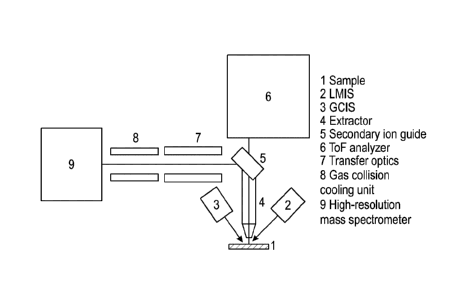
[0064] This example is described using Fig. 3 and Fig. 4. Fig. 3 shows a
diagram of a dual-beam
ToF-SIMS with two analyzers and pulsed S1 beam guide, and Fig. 4 shows a
timing diagram
with a ToF frequency of 10 kHz with a cycle time of 100 [is.
[0065] The sample (1) to be analyzed (see Fig. 3) is bombarded with primary
ion pulses in the ns
range from an LMIS (2) as an analysis beam and the secondary ions produced
therewith are
accelerated to energies in the keV range using an extractor electrode (4). A
pulsed beam guide
(5) allows the generated secondary ions to enter ToF-SIMS analyzer (6) of
Variant A. This
creates a ToF-SIMS spectrum. The lateral distribution on the sample is
measured by scanning the
sample with the focused LMIS primary ion beam.
[0066] Furthermore, the sample is bombarded with a gas cluster beam from a
GCIS (3). This
beam can also be focused and scanned over the sample, but with its own,
possibly with a
different pixel frequency. This beam is also pulsed, however, with very long
ion pulses of a
duration ranging from 10 to a few 100 [is, depending on the chosen cycle time.
The generated
secondary ions are also extracted, however, deflected into a transfer optics
(7) using the beam
guide (5). This transfer optics (7) slows the secondary ions down to a low
energy level and
typically injects them into an RF multipole (8). Collision cooling in which
the secondary ions
reduce their initial energy distribution and are collected on the axis of the
multipole (8) takes
place through a high gas pressure region in the multipole (8). The secondary
ions are then
14
Date recue/date received 2021-10-28
transported with a suitable transfer optics into a high-resolution mass
analyzer (9) suitable for
DC operation and are analyzed there.
[0067] The ToF-SIMS (6) is operated with a frequency of 1 to a maximum of a
few 10 kHz.
Both ion sources are also pulsed at this frequency but with different pulse
durations as indicated
above. The pulsed beam guide directs the secondary ions generated by the LMIS
(2) into the ToF
analyzer (6) and the secondary ions generated by the GCIS (3) into the high-
resolution mass
analyzer (9). Due to the low transport energy and the gas collision cooling, a
large temporal
dispersion of the secondary ions takes place until the mass analyzer (9) is
reached. Therefore, the
secondary ions from a larger number of cycles are combined into a nearly
continuous secondary
ion beam. This secondary ion beam can then be analyzed using the mass
spectrometer (9)
suitable for DC operation. The mass spectrometer (9) then provides mass
spectra with a
significantly lower repetition frequency in the range of about 1-100 Hz.
[0068] The ToF-SIMS of Variant A can also be operated with delayed extraction.
Here, the
desorption of secondary ions by the analysis ion source (2) takes place with
extraction of the
extractor (4) switched off.
[0069] A few ns after desorption, the extraction field is turned on and the
secondary ions are
accelerated to a few keV. Due to the delayed extraction, a high mass
resolution of up to 10,000
can be achieved for primary pulse durations of more than a few ns.
[0070] Various high-resolution mass spectrometers can be used as a mass
analyzer (9).
Preferably and to the extent possible, the mass resolution and mass accuracy
of this additional
mass spectrometer (9) should be significantly higher than those of the ToF
analyzer (6). As mass
Date recue/date received 2021-10-28
spectrometers (9) can be used, for example, orthogonal extraction ToF
analyzers (OTOF),
FTICR or Orbitrae mass spectrometers.
[0071] In this arrangement, the potential of the sample (1) during the
bombardment with primary
ions of the primary ion source (3) must be selected such that, after
acceleration, deceleration, gas
collision cooling and transfer of the secondary ions, their energy is within
the energy window of
the high-resolution mass spectrometer (9). In the mass spectrometers listed
above the energy of
the secondary ions at the entrance should, therefore, advantageously be
typically a few 10-100
eV. This can be achieved with a sample (1) at a corresponding bias voltage of
10 - 100 V
(relative to the ground potential). The acceleration of the secondary ions to
the energies of a few
keV that is typical for the time-of-flight analysis in the ToF analyzer (6) is
then carried out by the
extractor (4) at a respective high voltage potential. Thus, the secondary ion
guide (5) and the ToF
analyzer (6) must be floated to this potential.
Example 2
[0072] The following example shows examples of various operating modes of the
mass
spectrometer described above.
[0073] In one 3D analysis operating mode, the LMIS (2) in combination with the
ToF analyzer
(6) records the lateral distribution of substances in a sample (1) with a
large number of pixels and
at high pixel frequencies. Typical pixel counts are 256 x 256 or 128 x 128.
The spectral or pixel
frequencies are 5 to 20 kHz. As described above, the sample (1) is
additionally bombarded in the
analysis region with primary ions of the GCIS (3) as a sputtering ion source,
thereby achieving
removal and renewal of the sample surface. The secondary ions generated during
the
bombardment with primary ions of the GCIS (3) are supplied to the high-
resolution mass
16
Date recue/date received 2021-10-28
analyzer (9) via the beam guide (5). At the end of the measurement, at least
one spectrum of the
second analyzer (9) generated by the primary ions of GCIS (3) and having a
high resolution with
respect to the mass (m/z ratio) is available for each image with the above
number of pixels. This
spectrum can be combined with the mapping ToF-SIMS data of the analyzer (6)
through
subsequent data processing.
[0074] In particular, the high mass resolution and mass accuracy of this
spectrum of the analyzer
(9) can be used for the interpretation of the ToF-SIMS data of the analyzer
(6). Since this
spectrum is not or hardly affected by the sample height and/or topography of
the sample
surfaces, the information therefrom may be used, for example, for the
subsequent or automated
calibration of the mass scale of the ToF-SIMS spectrum of the analyzer (6).
[0075] In another 3D analysis operating mode, the primary ion beam of the GCIS
(3) is
rasterized and multiple high-resolution mass spectra are generated with the
ion beam of GCIS (3)
from different regions within the analysis area of a sample surface. The
maximum number of
different regions is determined by the ratio of the pixel frequencies of the
two analyzers. If an
image with 256 x 256 pixels and a pixel frequency of 10 kHz is recorded for
example with the
ToF-SIMS analyzer (6) it will take about 6.5 s. If the maximum spectral
frequency of the high-
resolution second analyzer (9) is 10 Hz, then the spectra of 65 different
regions can be recorded
in the same time. These can be divided into 8 x 8 fields in the analysis area.
However, other
divisions into different subregions are, of course, possible.
[0076] For example, selected regions within the analysis area that were
created manually or
automatically beforehand can also be used for dividing the regions. The
regions can also be
derived from the lateral distributions obtained from the ToF-SIMS data.
17
Date recue/date received 2021-10-28
[0077] Subsequent data processing provides various possibilities of linking
the ToF-SIMS data
of the analyzer (6) with the high-resolution spectra of the various subregions
recorded with
analyzer (9). For example, in particular statistical evaluation methods such
as Principal
Component Analysis (PCA) are used for assigning molecular peaks in the high-
resolution
spectrum to distribution images in the ToF-SIMS.
Example 3
[0078] The following example describes further advantageous improvements and
advantageous
additions of the mass spectrometer according to the invention, which can be
used individually or
in combination.
[0079] For the analysis of insulators, the charge of the sample resulting from
the positive
primary ions can be advantageously compensated. This can be done with low-
energy electrons in
an energy range below typically 20 eV. The surface potential stabilizes
automatically due to the
low energy. For the low-energy electrons to reach the sample, the extraction
field for the
secondary ions must be turned off. This requires that the extractor be pulsed.
At the same time,
the sample potential must also be switched to the ground potential. The low-
energy electrons are
always introduced within a cycle after the bombardment with the primary ions.
[0080] Fig. 5 shows a diagram of the time relationships for such an extraction
with charge
compensation at an analysis frequency of 10 kHz (timing diagram). With delayed
extraction, the
timing scheme can be slightly modified. Then, the extraction is turned on only
a few ns after the
arrival of the analysis ion pulse on the sample.
18
Date recue/date received 2021-10-28
[0081] To reduce charging, various raster methods such as line raster, meander
raster or random
raster can be used. The random raster has proven to be particularly
advantageous.
Example 4
[0082] The following example describes different variations of further
advantageous
improvements of the mass spectrometer according to the invention and the mass-
spectrometric
method according to the invention, which can be used individually or in
combination.
[0083] For the identification of molecules, the additional mass spectrometer
can also be
equipped for MS/MS. Fig. 6 shows such an arrangement with an additional MS/MS
device.
Here, a single mass is now transmitted through an upstream mass filter (9).
These so-called
parent molecules are stimulated to dissociate by gas collision in a subsequent
collision cell (10)
(CID collision induced dissociation). The resulting daughter ions are then
examined for mass in
the mass spectrometer (11).
[0084] When using an OTOF or Orbitrap Tm as a high-resolution mass
spectrometer (11)
typically a quadrupole mass filter is optionally switched in as the mass
filter (9) for the MS/MS
operating mode.
[0085] When using ion traps such as FTICR as a high-resolution mass
spectrometer (11), the ion
traps themselves can also be used for MS/MS analyzes.
Additional examples
19
Date recue/date received 2021-10-28
[0086] Fig. 7 is a schematic presentation of this combination of a Dual Beam
ToF-SIMS (6) with
an Orbitrap" mass spectrometer (11) of the type "Q Exactive HF" from Thermo
Fisher
Scientific as an additional analyzer (11) and with a pulsed S1 beam guide (5).
[0087] In this exemplary variant, which is shown in Fig. 7, a "TOF.SIMS 5" of
ION-TOF GmbH
(Munster, Germany) is used as a mass spectrometer (6) with a "Q Exactive HFTM"
together with
an OrbitrapTM mass spectrometer (11) of Thermo Fisher Scientific (USA) as mass
spectrometer
(11) in the manner described above.
[0088] The primary ion source (2) of the analysis beam is a Bi-LMIS and the
primary ion source
(3) used for the removal of the sample is an argon GCIS. In the combination,
the Orbitrap"
mass spectrometer (11) proves to be particularly advantageous because a
significantly higher
mass resolution and mass accuracy is achieved than with a ToF-SIMS. While the
ToF-SIMS (6)
with sub-ns primary ion pulses offers a maximum mass resolution of 16,000, the
Orbitrap" (11)
achieves a mass resolution of up to 240,000. The mass accuracy of the
Orbitrap" (11), at about
1 ppm, is also significantly better than that of the ToF-SIMS (6). Thus, the
Orbitrap (11)
provides the necessary information to positively identify the numerous mass
peaks in the
spatially high-resolution SIMS spectrum.
[0089] The unit in the schematic presentation of Fig. 7 is equipped with a
pulsed liquid metal ion
source (LMIG) (2) and a gas cluster ion source (GCIS) (3). The Orbitrap" mass
analyzer (11) is
preceded by a Quadrupol mass filter (9), which can optionally be activated for
the selection of
the parent molecules for the MS/MS operating mode. A gas collision cell (HCD
cell, higher
energy collisional dissociation) (10) is integrated for the fragmentation.
Here, the selected parent
molecules are fragmented in the MS/MS mode and then transferred into the
Orbitrap" (11) for
mass analysis via a pulsed injector (12).
Date recue/date received 2021-10-28
[0090] Fig. 8 shows the result of a depth profile analysis of an OLED layer
structure using the
dual beam ToF-SIMS method according to the prior art.
[0091] In this example, the surface analysis was carried out using a pulsed Bi
Cluster LMIS. An
argon GCIS was used in the dual beam method 5 at keV for the removal. The
depth profile
shows the depth distribution of the different molecules in the OLED structure.
[0092] The mass resolution in the ToF-SIMS is not sufficient for a separation
of the different
masses. For example, there is a significant superposition of other masses with
the molecules of
mass 774 u, 655 u, 589 u in the region of the first 90 nm. As a result, the
concentrations of these
molecules are not reflected correctly.
[0093] Fig. 9 shows an additional depth profile of an OLED layer structure
that can be generated
using an OrbitrapTm mass analyzer according to Fig. 7.
[0094] In this embodiment according to the invention, the secondary ions
sputtered with the Ar-
GCIS are now extracted and transferred into the OrbitrapTm mass analyzer by
means of the
pulsed beam guide.
[0095] The mass resolution in this additional mass analyzer is between 100,000
and 300,000
depending on the mass (see Fig. 10, explanation below). Due to the high mass
resolutions, the
mass interference can be eliminated. As a result, much higher dynamics and a
low base in the
range up to 90 nm are achieved, for example, for the masses 774 u, 655 u and
589 u. Thus, the
concentrations of these molecules can be determined much better.
[0096] Fig. 10 shows excerpts from the mass spectra measured by means of the
second
OrbitrapTm mass analyzer.
21
Date recue/date received 2021-10-28
[0097] For the different molecular ions analyzed in Fig. 10, very high mass
resolution and mass
accuracy are achieved with this second mass analyzer. Due to the high mass
resolution, there are
no more mass interferences with these masses. For example, the adjacent peak
to the a-NPD
molecule ions can be separated at mass 588.25 u. The high mass accuracy of 0.2
to 2.6 ppm
allows for the reliable identification of the respective molecules. As a
result, according to the
invention, the interpretation of the ToF-SIIVIS data is then significantly
improved as well.
[0098] Fig. 11 shows an MS/MS mass spectrum of the Nbphen parent molecule
measured and
determined with an arrangement according to the invention and Fig. 7.
[0099] The parent molecules generated by the Ar-GCIS 3 are in this example
transmitted
through the quadrupole mass filter 9, fragmented in the HCD cell 10 and then
injected into the
Orbitrae mass analyzer 11 and measured for their masses.
[00100] Fig. 12 shows mass spectra of a blue dye on filter paper measured and
determined with
a ToF-SIIVIS analyzer and a second Orbitrap mass analyzer according to the
invention and
according to Fig. 7.
[00101] Fig. 12A shows a photo of the sample location of a filter paper with a
blue ink spot. The
field of view of the photo is 3 x 3 mm. Dashed lines indicate the analysis
region for the spectra
shown in Figs. 12C to 12D.
[00102] Fig. 12B shows a positive ToF-SIIVIS mass spectrum in the mass range
of 75 to 700 u
from the region marked in Fig. 12A. A Bi3++ primary ion beam from a Bi liquid
metal ion
source with a primary ion energy of 60 keV was used as the pulsed primary ion
beam. Due to the
high sample roughness, the mass resolution and mass accuracy of the ToF-SIIVIS
spectrum is
significantly impaired.
22
Date recue/date received 2021-10-28
[00103] Figure 12C shows a positive OrbitrapTm mass spectrum in the mass range
of 75 to 700 u
from the region marked in Figure 12A. Arn gas clusters (the mean value of n
was about 1500)
from an Ar gas cluster ion source with a primary ion energy of 5 keV were used
as the primary
ion beam. The mass resolution and mass accuracy of the OrbitrapTm mass
analyzer are not
reduced by the sample roughness. The exact mass from the OrbitrapTm spectrum
can now be
used for the subsequent mass calibration of the ToF-SIMS spectrum.
[00104] FIG. 12D shows the superimposition of the spectra from the ToF-SIMS
spectrum of
FIG. 12B and the orbitrapTm spectrum of FIG. 12C in a mass range from 261.05 u
to 261.23 u.
The significant difference in mass resolution is clearly recognizable. Thus,
in the OrbitrapTm
spectrum, the peak at the mass 261.113 u is separated from the main peak at
261.13 u, while in
the ToF-SIMS spectrum both peaks are superimposed.
23
Date recue/date received 2021-10-28