Note: Descriptions are shown in the official language in which they were submitted.
FLUID MANAGEMENT SYSTEM WITH PASS-THROUGH FLUID VOLUME
MEASUREMENT
RELATED APPLICATION
This application is a divisional of Canadian Patent Application No. 2,948,184
filed May 14,2015.
Field or the Invention
[0002] The present invention relates generally to fluid management
systems used
during surgical procedures to provide one or more functions associated with
irrigation,
distention, fluid warming, fluid deficit monitoring, suction, and the like,
and more
particularly relates to a method and apparatus for continuously measuring the
volume of fluid
being returned from the surgical site as it is being delivered to a waste
collection system.
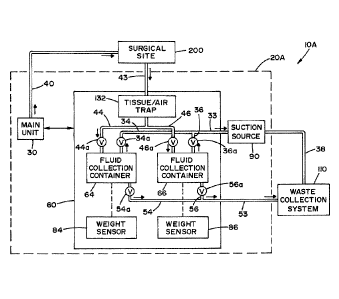
Back2round or the Invention
[0903] A fluid management system (FMS) may be used in connection with a
wide
variety of medical procedures involving one or more fluid delivery functions
including, but
not limited to: fluid irrigation; distention of a body cavity; fluid warming;
fluid deficit
monitoring associated with delivery and return of fluid to/from a surgical
site; and suction.
The medical procedures may be associated with multiple surgical disciplines
including, but
not limited to: gynecologic, urologic, orthopedic, colorectal, and general
surgical procedures.
[0004] During certain medical procedures, patient safety may require
that the amount
of fluid delivered to the surgical site and the amount of fluid returned from
the surgical site
be continuously monitored to determine the "fluid deficit." Accordingly, a FMS
may be
configured to provide a fluid deficit monitoring function to accurately
measure fluid inflow
(to the surgical site) and outflow (from the surgical site), and to calculate
a fluid deficit in
order to monitor a patient's fluid absorption level during a medical procedure
as excess fluid
absorption can result in serious complications. Typically, fluid returning
from the surgical
site is collected in one or more fluid collection containers (e.g.,
canisters). The volume of
fluid collected from the surgical site is typically determined by measuring
weight. A fluid
CA 2997456 2018-03-05
deficit is then calculated by comparing the volume of fluid delivered to the
surgical site with
the volume of fluid returned from the surgical site.
[00051 Canisters are frequently used as fluid collection containers.
When a canister
fills with fluid to a maximum capacity during a medical procedure. it becomes
necessary to
remove the full canister and replace it with a new, empty canister. There are
several
drawbacks to removing and replacing canisters during a medical procedure. In
this regard,
such activity can (i) disrupt the medical procedure by necessitating the
suspension of suction
used to remove fluid from the surgical site, and thereby cause a suspension of
fluid deficit
monitoring; (ii) cause inconvenience to medical personnel, especially in
surgical procedures
involving high fluid volumes, as medical personnel have to physically remove
full canisters
and replace them with new, empty canisters; (iii) potentially introduce errors
into fluid deficit
monitoring calculations due to disruption of the fluid management system
during the canister
replacement process (e.g., bumping or moving), which can adversely affect the
ability of the
fluid management system to accurately weigh the remaining and new canisters;
(iv)
potentially introduce errors into fluid deficit monitoring calculations due to
leaks and spills
caused by detaching tubing used to return fluid from the surgical site from
full canisters and
reattaching such lines to the new, empty canisters, and (v) increase the cost
of a surgical
procedure by requiring that a number of canisters be used during a surgical
procedure which
is commensurate with the amount of fluid used.
[0006] In view of the foregoing, there is a need for a fluid management
system that
incorporates a "pass-through" fluid volume measurement system that
continuously measures
the volume of fluid returning from a surgical site during transit to a waste
collection system
(e.g., a dedicated fluid collection system or a hospital's waste disposal
system) and
eliminates the need to replace full canisters with new, empty canisters during
a medical
procedure.
Summary of the invention
[00071 In accordance with the present invention, there is provided a
fluid
management system comprising: at least one fluid supply container for storing
a fluid to be
delivered to a surgical site; a pump for delivering the fluid from the at
least one fluid supply
container to the surgical site; and a pass-through fluid volume measurement
system for
CA 2997456 2018-03-05
=
3
determining the volume of fluid returned from the surgical site, said pass-
through fluid
volume measurement system comprising: a plurality of fluid collection
containers, wherein
each fluid collection container has (i) a suction input in fluid communication
with a suction
line for drawing a vacuum in the fluid collection container, (ii) a fluid
input in fluid
communication with a fluid return line for receiving fluid returning from the
surgical site,
and (iii) a fluid output in fluid communication with a fluid output line for
evacuating the fluid
collected in the fluid collection container to a waste collection system; one
or more weight
sensors for providing signals indicative of the sensed weight of the fluid
collection
containers; and a plurality of valves moveable between open and closed
positions to control
the flow of fluid through the suction line, the fluid return line and the
fluid output line; a
suction source for providing suction in the suction line to draw a vacuum in
the fluid
collection containers to thereby draw fluid from the surgical site into the
fluid collection
containers, and for providing suction in the fluid output line to draw fluid
collected in the
fluid collection containers into the waste collection system; and a control
unit for receiving
the signals from the one or more weight sensors to monitor a volume of fluid
returned from
the surgical site to the fluid collection containers, and moving the plurality
of valves between
the open and the closed positions to alternately fill one of the fluid
collection containers
while emptying another of the fluid collection containers.
[0008] In accordance with another aspect of the present
invention, there is provided a
fluid management system comprising: at least one fluid supply container for
storing a fluid
to be delivered to a surgical site; a pump for delivering the fluid from the
at least one fluid
supply container to the surgical site; and a pass-through fluid volume
measurement system
for determining the volume of fluid returned from the surgical site, said pass-
through fluid
volume measurement system comprising: a support member for supporting
components of
the pass-through fluid volume measurement system; a flow sensing device
including: a
disposable or single-use fluid measurement tube having an inlet port in fluid
communication
with a fluid return line for receiving fluid returning from the surgical site,
and an outlet port
in fluid communication with a fluid output line for receiving the fluid
exiting the fluid
measurement tube, at least one ultrasonic sensor for providing a signal
indicative of the flow
rate of fluid passing through the fluid measurement tube, and a clamping
mechanism
mounted to the support member, said clamping mechanism for temporarily
mounting the
CA 2997456 2018-03-05
4
fluid measurement tube in a proper orientation between the inlet and outlet
ultrasonic
sensors; a suction source for providing suction in the fluid return line and
fluid output line to
draw the fluid through the fluid measurement tube and subsequently into a
waste collection
system; and a control unit for receiving the signals from the inlet and outlet
sensors to
monitor a volume of fluid returned from the surgical site.
[0009] In accordance with still another aspect of the present invention,
there is
provided a method for continuously measuring a volume of fluid being returned
from a
surgical site as it is being delivered to a waste collection system, said
method comprising:
filling the first fluid collection container, by: opening a valve associated
with a suction line
in fluid communication with a first fluid collection container; opening a
valve associated
with a fluid return line in fluid communication with the first fluid
collection container;
closing a valve associated with a fluid output line in fluid communication
with the first fluid
collection container; closing a valve associated with a suction line in fluid
communication
with a second fluid collection container; and closing a valve associated with
a fluid return
line in fluid communication with the second fluid collection container, upon
filling the first
fluid collection container with fluid to a predetermined volume, emptying the
first fluid
collection container and filling the second fluid collection container, by
closing the valve
associated with a suction line in fluid communication with a first fluid
collection container;
closing the valve associated with a fluid return line in fluid communication
with the first
fluid collection container; opening the valve associated with a fluid output
line in fluid
communication with the first fluid collection container; opening the valve
associated with a
suction line in fluid communication with a second fluid collection container;
opening the
valve associated with a fluid return line in fluid communication with the
second fluid
collection container; and closing a valve associated with a fluid output line
in fluid
communication with the second fluid collection container; and alternately
filling and
emptying the first and second fluid collection containers until a medical
procedure is
completed.
[0010] In accordance with yet another aspect of the present invention,
there is
provided a method for continuously measuring a volume of fluid being returned
from a
surgical site as it is being delivered to a waste collection system, said
method comprising the
steps ofi drawing fluid from the surgical site through a flow sensing device
providing signals
CA 2997456 2018-03-05
5
indicative of a fluid flow rate; monitoring the volume of fluid passing
through the flow
sensing device using the signals indicative of the fluid flow rate; and
passing the fluid from
the flow sensing device to the waste collection system until a medical
procedure is
completed.
[0011] An advantage of the present invention is the provision of a fluid
management
system that continuously measures the volume of fluid returning from a
surgical site during
transit to a waste collection system.
[0012] Another advantage of the present invention is the provision of a
fluid
management system that eliminates the need to replace full fluid collection
containers with
new, empty fluid collection containers during a medical procedure.
[0013] A still further advantage of the present invention is the
provision of a fluid
management system having a stand-alone pass-through fluid volume measurement
system.
[0014] Yet another advantage of the present invention is the provision
of a fluid
management system capable of fluid delivery, suction, fluid
removal/collection, fluid deficit
monitoring, and fluid disposal.
[0015] These and other advantages will become apparent from the
following
description of illustrated embodiments taken together with the accompanying
drawings and
the appended claims
Brief Description of the Drawings
[0016] The invention may take physical form in certain parts and
arrangement of
parts, embodiments of which will be described in detail in the specification
and illustrated in
the accompanying drawings which form a part hereof, and wherein:
[0017] FIG. l is a schematic diagram illustrating an FMS according to a
first
embodiment of the present invention, wherein the FMS includes a fluid
management unit
having a pass-through fluid volume measurement system and an integrated
suction source for
return of fluid from the surgical site and subsequent evacuation of the fluid
returned from the
surgical site to a waste collection system.
[0018] FIG. 2 is a schematic diagram illustrating an FMS according to a
second
embodiment of the present invention, wherein the FMS includes a fluid
management unit
having a pass-through fluid volume measurement system and an integrated
suction source for
CA 2997456 2018-03-05
6
return of fluid from the surgical site and subsequent evacuation of the fluid
returned from the
surgical site to a waste collection system.
[0019] FIG. 3 is a schematic diagram illustrating an FMS according to a
third
embodiment of the present invention, wherein the FMS includes a fluid
management unit
with a pass-through fluid volume measurement system, and an external suction
source for
return of fluid from the surgical site and subsequent evacuation of the fluid
returned from the
surgical site to a waste collection system.
[0020] FIG. 4 is a schematic diagram illustrating an FMS according to a
fourth
embodiment of the present invention, wherein the FMS includes a fluid
management unit
with a pass-through fluid volume measurement system and a suction source of a
waste
collection system, wherein the suction source of the waste collection system
provides suction
for both return of fluid from the surgical site and subsequent evacuation of
the fluid returned
from the surgical site to the waste collection system.
[0021] FIG. 5 is a schematic diagram illustrating a mechanical
configuration of a
pass-through fluid volume measurement system according an embodiment of the
present
invention.
[0022] FIG. 6 illustrates a tubing set used in connection with the FMS
embodiment
shown in FIG. 1.
[0023] FIG. 7 illustrates a tubing set used in connection with the FMS
embodiment
shown in FIG. 2.
[0024] FIG. 8 is a schematic diagram of a pass-through fluid volume
measurement
system according to a first alternative embodiment, wherein suction is
provided by a suction
source external to the measurement system.
[0025] FIG. 9 is a schematic diagram of the pass-through fluid volume
measurement
system according to a second alternative embodiment, wherein suction is
provided by a
suction source integrated in the pass-through fluid volume measurement system.
[0026] FIG. 10 is a schematic diagram of the pass-through fluid volume
measurement
system according to a third alternative embodiment, wherein suction is
provided by a suction
source integrated in the pass-through fluid volume measurement system.
CA 2997456 2018-03-05
7
[0027] FIG. 10A is a schematic diagram of the pass-through fluid volume
measurement system shown in FIG. 10, as modified to include a single
ultrasonic sensor for
sensing fluid flow.
[0028] FIG. 11 illustrates a tubing set used in connection with the pass-
through fluid
volume measurement system shown in FIG. 8.
[0029] FIG. 12 illustrates a tubing set used in connection with the pass-
through fluid
volume measurement system shown in FIG. 9.
[0030] FIG. 13 illustrates a tubing set used in connection with the pass-
through fluid
volume measurement system shown in FIG. 10.
Detailed Description of the Invention
[0031] Referring now to the drawings wherein the showings are for the
purposes of
illustrating embodiments of the invention only and not for the purposes of
limiting same,
FIG. 1 shows a fluid management system (FMS) 10A according to a first
embodiment of the
present invention. FMS 10A is a multifunctional system that supplies fluid to
a surgical site
200, removes fluid from surgical site 200, monitors a fluid deficit, and
disposes of fluid
returned from surgical site 200, as will be described in detail below. It
should be appreciated
that the term -surgical site" as used herein refers not only to the patient's
body where a
surgery is being performed, but also to the general region surrounding the
patient.
[0032] FMS 10A is generally comprised of a fluid management unit 20A
including a
main unit 30, a pass-through fluid volume measurement system 60 and an
integrated suction
source 90. Fluid management unit 20A interfaces with a waste collection system
110, as will
be described below. It should be appreciated that suction source 90 may
alternatively be
arranged as a component of measurement system 60.
[0033] As seen in FIG. 1, a fluid supply line 40 provides a fluid
conduit between
main unit 30 and surgical site 200, a fluid return line 43 provides a fluid
conduit between
measurement system 60 and surgical site 200, a suction line 33 provides a
fluid conduit
between suction source 90 and measurement system 60, a suction line 38
provides a fluid
conduit between suction source 90 and waste collection system 110, and a fluid
output line
53 provides a fluid conduit between measurement system 60 and waste collection
system
CA 2997456 2018-03-05
8
110. Supply line 40, return line 43, suction line 33, suction line 38, and
output line 53 may
take the form of fluid conduits, such as conventional medical grade flexible
plastic tubing.
[0034] Main unit 30 includes a control unit comprised of components such
as a
microprocessor or microcontroller, memory device(s), data storage device(s),
output
device(s) (e.g., LCD screen, touch screen, conventional display device, audio
speaker,
printer, and the like), and input device(s) (e.g., touch screen, keypad,
keyboard, mouse,
mechanical switching devices, and the like). Main unit 30 may also include one
or more
fluid container supports (such as hangers or hooks) for supporting one or more
fluid supply
containers (e.g., fluid bags) that store fluid that is to be delivered to a
surgical site 200,
weight sensors for detecting the weight of fluid in the fluid supply
containers, and a pump for
pressurizing fluid in the fluid supply containers and delivering the fluid to
surgical site 200
via fluid supply line 40. For example, fluid supply line may be connected with
a surgical
instrument to facilitate a surgical procedure. It should be appreciated that
gravity or other
means of fluid pressurization may be substituted for the pump. Main unit 30
may also
include numerous other components for regulating fluid flow, fluid pressure,
fluid
temperature (e.g., a fluid heating apparatus), and the like. The control unit
controls the
supply of fluid delivered to surgical site 200 via fluid supply line 40,
monitors the volume of
fluid supplied to surgical site 200 (via supply line 40), monitors the volume
of fluid returned
from surgical site 200 (via return line 43), and determines a fluid deficit. A
detailed
description of the components and operation of an exemplary fluid management
unit,
including fluid deficit monitoring, is provided in U.S. Patent No. 8,444,592,
issued May 21,
2013.
[0035) Pass-through fluid volume measurement system 60 determines the
volume of
fluid removed from surgical site 200 via fluid return line 43. According to
the illustrated
embodiment, measurement system 60 includes a first fluid collection container
64, a second
fluid collection container 66, and first and second weight sensors 84. 86
respectively
associated with fluid collection containers 64, 66. It is contemplated that
fluid collection
containers 64, 66 may take a variety of forms, including, but not limited to,
disposable or re-
usable rigid hard-shell canisters, rigid hard-shell canisters with disposable
or reusable liners,
disposable pouches or bags having a rigid skeleton, fluid containers
supportable from
mounting brackets or hooks.
CA 2997456 2018-03-05
9
[0036] The end of return line 43 located at surgical site 200 may
include a plurality of
input lines that are combined by a manifold. Each of these input lines may be
located at
different locations at surgical site 200. For example, the input lines may
collect fluid from
the patient, floor suctioning equipment, a fluid collection drape, and
surgical instrument
outflow ports.
[0037] In the embodiment shown in FIG. 1, the end of return line 43
fluidly
connected with measurement system 60 includes a first branch 44 and a second
branch 46 for
fluid communication with fluid inputs (e.g., input tubes) of fluid collection
containers 64 and
66, respectively. Branches 44 and 46 may be joined by a y-connector. Valves
44a, 46a
respectively control fluid flow along first and second branches 44, 46 of
return line 43. In
one embodiment of the present invention, valves 44a, 46a take the form of
pinch valves
operable to open and close the fluid pathway through return line 43. Sections
of tubing
forming suction branches 44, 46 of return line 43 are respectively routed
through the pinch
valves that are controlled by the control unit of main unit 30. Furthermore, a
one-way valve
may be located within return line 43 to prevent backflow of fluid to surgical
site 200.
[0038] Weight sensors 84, 86 may take the form of load cells that
provide signals to
main unit 30 indicative of the measured weight of fluid respectively collected
in fluid
collection containers 64, 66. The control unit of main unit 30 determines the
volume of fluid
collected in fluid collection containers 64, 66 from the measured weight.
[00391 Suction source 90 is fluidly connected with fluid collection
containers 64, 66
(via suction line 33) and waste collection system 110 (via suction line 38).
Waste collection
system 110 is fluidly connected with fluid collection containers 64, 66 (via
output line 53).
Suction source 90 draws a vacuum in fluid collection containers 64, 66 (via
suction line 33)
to return fluid from surgical site 200 to fluid collection containers 64, 66
via return line 43.
Suction source 90 also provides suction in suction line 38 and output line 53
to subsequently
evacuate fluid collected in fluid collection container 64, 66 to waste
collection system 110
via fluid output line 53. In the illustrated embodiment, suction source 90
takes the form of a
vacuum pump.
[0040] Suction line 33 includes a first branch 34 and a second branch 36
for fluid
communication with suction inputs (e.g., a suction tube) of fluid collection
containers 64. 66,
respectively. Branches 34 and 36 may be joined by a y-connector. Valves 34a,
36a
CA 2997456 2018-03-05
10
respectively control suction along first and second branches 34, 36 of suction
line 33. In one
embodiment of the present invention, valves 34a, 36a may take the form of
pinch valves
operable to open and close the fluid pathway through suction line 33. Sections
of tubing
forming suction branches 34, 36 of suction line 33 are respectively routed
through the pinch
valves that are controlled by the control unit of main unit 30. Furthermore, a
hydrophobic
filter may be located within suction line 33 to prevent fluid from being
sucked out of fluid
collection containers 64, 66 through suction line 33. For example, hydrophobic
filters may
be located within branches 34 and 36 of suction line 33.
[0041) Output line 53 includes a first branch 54 and a second branch 56
for fluid
communication with fluid outputs (e.g., a dip tube or bottom suction tube) of
fluid collection
containers 64, 66, respectively. Branches 54 and 56 may be joined by a y-
connector. Valves
54a, 56a respectively control fluid flow along first and second branches 54,
56 of output line
53. In one embodiment of the present invention, valves 54a, 56a may take the
form of pinch
valves operable to open and close the fluid pathway through output line 53.
Sections of
tubing forming suction branches 54, 56 of suction line 53 are respectively
muted through the
pinch valves that are controlled by the control unit of main unit 30.
[0042] As indicated above, return line 43, suction line 33, and output
line 53 take the
form of fluid conduits, such as conventional medical grade flexible plastic
tubing. In one
embodiment of the present invention, the sections of tubing for branches 44,
46 (return line
43): branches 34, 36 (suction line 33); and branches 54, 56 (output line 53)
may each include
an integrated strain relief element that "snaps" into, or otherwise attaches
to, a support
structure (e.g., stand, mounting bracket, frame. etc.) of fluid management
unit 20A. For
example, the strain relief element may be mounted to a support stand 22,
described below
with reference to FIG. 5. It is also contemplated that the sections of tubing
for branches 44,
46 (return line 43); branches 34. 36 (suction line 33); and branches 54, 56
(output line 53)
that connect respectively with fluid input, suction input and fluid output of
fluid collection
containers 64, 66 have an accordion tubing component or section to allow for
relaxed flexing
and extension of the tubing. It should be appreciated that the strain relief
element and
accordion tubing section minimize forces applied to fluid collection
containers 64, 66 as a
result of "pushing and pulling" of the tubing. This minimizes disturbance to
weight
CA 2997456 2018-03-05
11
measurements made by weight sensors 84, 86, and thus provides for greater
accuracy in fluid
deficit monitoring.
[00431 FIG. 1 illustrates pass-through fluid volume measurement system
60 as a
component of fluid management unit 20A. However, as will be described in
detail below,
the pass-through fluid volume measurement system of the present invention may
alternatively be constructed as a stand-alone component that is separate from
a fluid
management unit. In this case, the strain relief element may attach to a
support structure that
independently supports pass-through fluid volume measurement system 60.
[0044] It is contemplated that waste collection system 110 may take a
variety of
different forms, including, but not limited to, a mobile fluid collection
container or cart, a
dedicated stand-alone fluid collection system with integrated suction, or a
hospital's waste
disposal system which may be accessible in the operating room.
[0045] In the illustrated embodiment, a combined tissue/air trap 132 (or
individual
tissue and air traps) is located within return line 43. A tissue trap (or
other similar device)
functions to collect tissue carried by fluid returning from surgical site 200
via return line 43
for subsequent analysis and/or to increase the accuracy of fluid deficit
calculations. In the
absence of a tissue trap, tissue returned from surgical site 200 can increase
the weight of fluid
collection canisters or interfere with fluid flow sensing measurements.
Similarly, an air trap
can increase the accuracy of fluid deficit calculations as air bubbles can
interfere with fluid
flow sensing measurements.
100461 For enhanced safety, it is contemplated that measurement system
60 may also
include one or more fluid level sensors for detecting the fluid level within
fluid collection
containers 64 and 66, and one or more leak sensors for detecting the presence
of a leak in
fluid collection containers 64, 66 or in a tubing connection associated
therewith. A fluid
level sensor determines, independently of the control unit of main unit 30,
whether a fluid
level has reached a predetermined fluid level within fluid collection
containers 64, 66 and
can close one or more of valves 44a, 46a, 34a, and 36a, if necessary. When a
leak sensor
detects the presence of a leak, the leak sensor transmits a signal to the
control unit of main
unit 30. In response to receipt of this signal, the control unit can take
appropriate action,
such as "closing" one or more of valves 44a, 46a. 34a, 36a and providing a
visual and/or
audible indicator to alert a user of a potential problem with measurement
system 60.
CA 2997456 2018-03-05
12
[0047] The operation of FMS 10A will now be described in detail with
reference to
FIG. 1. At the beginning of a surgical procedure, fluid supply containers are
mounted to
main unit 30 and connected with fluid supply line 40 to supply fluid to
surgical site 200. The
volume of fluid supplied to surgical site 200 is monitored by main unit 30. In
addition, two
fluid collection containers 64, 66 are arranged to be independently weighed by
respective
weight sensors 84, 86. Respective strain relief elements are snapped into a
support structure,
and appropriate sections of tubing associated with branches 44, 46 (return
line 43); branches
34, 36 (suction line 33); and branches 54, 56 (output line 53) are routed
through
corresponding valves 44a, 46a; 34a, 36a; and 54a, 56a, which take the form of
pinch valves.
[0048] When a user initiates a procedure using main unit 30 that begins
the flow of
fluid to surgical site 200 via supply line 40, the control unit "zeroes" any
previously stored
weight values and begins recording the weight of each fluid collection
container 64, 66 as
indicated by respective weight sensors 84, 86. Then, valves 34a, 44a
associated with the
suction input and the fluid input of fluid collection container 64 are
"opened" and valve 54a
associated with the fluid output of fluid collection container 64 is "closed."
Furthermore,
valves 36a and 46a associated with the suction input and fluid input of fluid
collection
container 66 are "closed."
[0049] The control unit of main unit 30 monitors the volume of fluid
supplied to the
surgical site 200 and monitors the volume of fluid returned to fluid
collection container 64
via signals received from weight sensor 84. When the fluid volume collected in
fluid
collection container 64 reaches a predetermined volume, the control unit
"closes" valves 34a,
44a respectively associated with the suction input and the fluid input of
fluid collection
container 64, allows the weight sensor reading to stabilize, records the total
weight of fluid
collection container 64, and then "opens" valve 54a associated with the fluid
output of fluid
collection container 64 in order to empty fluid collection container 64 by
evacuating the
collected fluid to waste collection system 110. Simultaneously, the control
unit "opens"
valves 36a and 46a respectively associated with the suction input and the
fluid input of fluid
collection container 66, and -closes" valve 56a associated with the fluid
output of fluid
collection container 66 to begin filling fluid collection container 66 with
the fluid returned
from surgical site 200. In this manner, fluid collection from surgical site
200 and fluid deficit
monitoring continues uninterrupted. The above-described "alternating"
fill/empty process
CA 2997456 2018-03-05
13
(i.e., alternating the filling and emptying of fluid collection containers 64
and 66), is repeated
until the user ends the fluid collection procedure.
[0050] In accordance with the present invention, measurement system 60
is adapted
to measure any amount of fluid returned from surgical site 200 during a
medical procedure,
without the burdensome and costly need to change fluid collection containers.
Furthermore,
the present invention allows fluid management unit 20A to continuously return
fluid from
surgical site 200, and thus allows uninterrupted determination of the fluid
deficit which can
be displayed to a user by visual and/or audible indicators (e.g., alarms) that
may be
appropriate based on the measured or calculated fluid deficit level.
[0051] FIGS. 2-4 illustrate fluid management systems according to
alternative
embodiments of the present invention. In these figures, components similar to
those shown
in FIG. I have been given the same reference numbers.
[0052] In the embodiment shown in FIG. 2, there is shown a FMS 10B
having a fluid
management unit 20B that includes an integrated suction source 95, which
preferably takes
the form of a pump. Suction source 95 provides suction in suction line 33 to
return fluid
from surgical site 200 to fluid collection containers 64, 66 and provides
suction in output line
53 for evacuating fluid collected in fluid collection containers 64, 66 to
waste collection
system 110. It should be appreciated that suction source 95 may also be
directly integrated
into measurement system 60.
[0053] In the embodiment shown in FIG. 3, there is shown a FMS IOC
having a fluid
management unit 20C that does not include an integrated suction source and
therefore fluid
management unit 20C relies upon an external suction source 100 for suction.
External
suction source 100 may take the form of a conventional wall suction unit
(e.g.. vacuum
pump) typically found in hospitals. External suction source 100 provides
suction, via suction
line 33, to return fluid from surgical site 200 to fluid collection containers
64, 66. External
suction source 100 also provides suction via suction line 38b and output line
53 for
evacuating fluid collected in fluid collection containers 64, 66 to waste
collection system
110.
[00541 In the embodiment shown in FIG. 4, there is shown a FMS I OD
having a fluid
management unit 20D and a suction source 120 that is an integrated component
of waste
collection system 110. Suction source 120 may take the form of a conventional
vacuum
CA 2997456 2018-03-05
14
pump. Suction source 120 provides suction in fluid collection container 64,
66, via suction
line 33, to draw fluid from surgical site 200 to fluid collection containers
64, 66. Suction
source 120 also provides suction, via output line 53, for evacuating fluid
collected in fluid
collection containers 64, 66 to waste collection system 110.
[0055] It should be appreciated that the suction sources described
herein (i.e.,
suctions source 90, 95, 100 and 120) may take a variety of forms including,
but not limited
to, a vacuum pump, a peristaltic pump, rotary vane pump, gerotor pump, piston
pump, and
the like.
[0056] In accordance with an embodiment of the present invention, fluid
collection
containers 64, 66 and all tubing associated suction line 33, return line 43
and output line 53
are components of a single-use/disposable tubing set. For example, FIG. 6
illustrates a
tubing set used in connection with FMS 10A (FIG. 1) that includes tubing for
return line 43
(including a plurality of input branches 42), tubing for suction line 33,
tubing for output line
53, and fluid collection containers 64 and 66. A tissue/air trap 132 may also
be located in the
tubing of return line 43. The tubing set may also include additional tubing
for suction line 38
between suction source 90 and waste collection system 110.
[0057] FIG. 7 illustrates a tubing set used in connection with FMS 10B
(FIG. 2) that
includes tubing for return line 43 (including a plurality of input branches
42), tubing for
suction line 33, tubing for output line 53, and fluid collection containers 64
and 66. A
tissue/air trap 132 may also be located in the tubing of return line 43. It
should be
appreciated that in this embodiment, the tubing for output line 53 is arranged
through suction
source 95,
[0058] Tubing sets similar to those illustrated in FIGS. 6 and 7 are
used for the
embodiments of FMS IOC and IOD respectively shown in FIGS. 3 and 4.
[0059] Referring now to FIG. 5, there is shown a schematic diagram
illustrating a
mechanical embodiment of measurement system 60. Main unit 30, suction source
90 and
measurement system 60 are mounted to portable support stand 22 having a pole
23 and a
base 24 with wheels. In an alternative embodiment, main unit 30, suction
source 90 and
measurement system 60 may be mounted to a fixed support structure, such as a
wall.
Support members 74 and 76 (e.g., platform plates) respectively support fluid
collection
containers 64, 66. Weight sensor 84, 86 are mechanically connected with
support members
CA 2997456 2018-03-05
15
74, 76. In
the illustrated embodiment, fluid collection containers 64, 66 are each
independently weighed by respective weight sensors 84, 86. Weight sensors 84,
86 provide
signals to main unit 30 indicative of the respective measured weight of fluid
collected in fluid
collection containers 64, 66. Control unit of main unit 30 determines the
volume of fluid
collected in fluid collection containers 64, 66 from the measured weight.
[00601 It is
contemplated that the pass-through fluid volume measurement system of
the present invention may take alternative forms. Referring now to FIG. 8,
there is shown a
pass-through fluid volume measurement system according to a first alternative
embodiment.
Measurement system 260 replaces the weight sensors and fluid collection
containers of
measurement system 60 with a fluid flow sensing device. In the illustrated
embodiment,
measurement system 260 is comprised of a fluid flow measurement tube 262
having an inlet
port 264 at one end and an outlet port 266 at the opposite end, inlet and
outlet ultrasonic
sensors 274 and 276 (e.g., an ultrasonic transceiver, ultrasonic transmitter,
or ultrasonic
receiver) which are fixed or permanent components of measurement system 260,
and a
clamping mechanism 280 for temporarily mounting or attaching measurement tube
262 in a
proper orientation between inlet and outlet ultrasonic sensors 274, 276. For
example,
clamping mechanism 280 may engage and capture measurement tube 262 with clamp
or grip
members (e.g., C-clamps). It is contemplated that clamping mechanism 280 may
also
include components that cause members supporting sensors 274, 276 to rotate,
pivot, or
move in order to properly position, orient or align sensors 274, 276 relative
to measurement
tube 262 to measure fluid volume in measurement tube 262. For example,
installing a
measurement tube 262 into engagement with clamping mechanism 280 may cause a
spring-
loaded sensor support member to press and hold sensors 274, 276 against the
ends of
measurement tube 262 in proper alignment. Sensors 274 and 276 are used to
determine the
flow rate of fluid passing through measurement tube 262. Sensors 274 and 276
provide
signals, indicative of flow rate of fluid passing through measurement tube
262, to the control
unit of main unit 30 (of a fluid management unit), which in turn determines
the volume of
fluid flowing through measurement tube 262. In the illustrated embodiment,
measurement
tube 262 is part of the single-use or disposable tubing set. Measurement tube
262 may also
be a re-usable fluid tube that can be re-sterilized. Furthermore, sensors 274,
276 and
clamping mechanism 280 may be permanently mounted to a support member (not
shown)
CA 2997456 2018-03-05
16
that supports components of measurement system 260. For example, such support
member
may take the form of a wall, cart, or stand.
[0061] It also contemplated that measurement system 260 may also include
one or
more temperature sensors 290 for sensing the temperature of the fluid in
measurement tube
262. Temperature sensor 290 is properly oriented to measure the temperature of
the fluid
when measurement tube 262 is received into clamping mechanism 280. Temperature
sensor
290 provides fluid temperature information to the control unit of main unit
30, which uses the
temperature information to more accurately determine the fluid flow rate
through
measurement tube 262.
[0062] Furthermore, measurement system 260 may also include an
accumulator in
addition to combined tissue/air trap 132. The accumulator conditions the fluid
prior to
entering measurement tube 262 by absorbing surges or pulsations in the fluid
flow.
[0063] Ultrasonic flowmeters use sound waves to determine the velocity
of a fluid
flowing in a pipe or tube. At "no flow" conditions, the frequencies of an
ultrasonic wave
transmitted into the tube and its reflections from the fluid are the same.
Under flowing
conditions, the frequency of the reflected wave is different due to the
Doppler effect. When
the fluid moves faster, the frequency shift increases linearly. Signals from
the transmitted
wave and its reflections are processed to determine the flow rate. A "transit
time" ultrasonic
flowmeter sends and receives ultrasonic waves between transducers in both the
upstream and
downstream directions in the tube. At "no flow" conditions, it takes the same
time to travel
upstream and downstream between the two transducers. Under flowing conditions,
the
upstream wave will travel slower and take more time than the (faster)
downstream wave.
When the fluid moves faster, the difference between the upstream and
downstream times
increases. Upstream and downstream times are processed to determine the flow
rate.
[0064] For the embodiment of measurement system 260 shown in FIG. 8, an
external
suction source 100 or a suction source 120 internal to a waste collection
system 110A
provides the suction to draw fluid from surgical site 200 through return line
43, measurement
tube 262, and output line 53 to the waste collection system.
[0065] In FIG. 9 a pass-through fluid volume measurement system 260A
includes an
integrated suction source 95 to provide suction for drawing fluid from
surgical site 200
through return line 43, measurement tube 262, and output line 53 to waste
collection system
CA 2997456 2018-03-05
17
110. Suction source 95 takes the form of a pump, wherein suction line 53
extends through
suction source 95.
[0066] In FIG. 10 a pass-through fluid volume measurement system 260B
also
includes an integrated suction source 95 to provide suction for drawing fluid
from surgical
site 200 through return line 43, measurement tube 262, and output line 53 to
waste collection
system 110. Suction source takes the form of a pump, wherein return line 43
extends through
suction source 95.
[0067] It is contemplated in another alternative embodiment that the two
ultrasonic
sensors 274, 276 may be arranged in positions relative to measurement tube 262
that differ
from the positions as depicted in the illustrated figures. For example,
ultrasonic sensors 274,
276 may be located at the top and bottom portions of measurement tube 262.
Furthermore, it
is also contemplated that the measurement system may be configured with only a
single
ultrasonic sensor (e.g., an ultrasonic transceiver) for determining the volume
of fluid flowing
through measurement tube 262. For example, FIG. 10A illustrates a measurement
system
260C that is a modified version of measurement system 260B (FIG. 10), wherein
a single
ultrasonic sensor 278 is substituted for sensors 274, 276. Single ultrasonic
sensor 278 may
take the form of a sensor or transducer that measures the deviation of the
angle of reflected
ultrasound to determine fluid flow rate.
[0068] In accordance with an embodiment of the present invention,
measurement
tube 262, return line 43 and output line 53 are components of a single-
use/disposable tubing
set. For example, FIG. 11 illustrates a tubing set used in connection with
measurement
system 260 (FIG. 8) that includes tubing for return line 43 (including a
plurality of input
branches 42) and tubing for output line 53. A tissue/air trap 132 may also be
located in the
tubing of return line 43. The tubing set may also include additional tubing
for suction line 38
between external suction source 100 and waste collection system 110. It should
be
appreciated that in the embodiment illustrated in FIG. 11, that waste
collection system 110
and external suction source 100 can be replaced with waste collection system
110A having
an internal suction source 120. In this embodiment, tubing for suction line 38
is omitted.
[00691 FIG. 12 illustrates a tubing set used in connection with
measurement system
260A (FIG. 9) that includes tubing for return line 43 (including a plurality
of input branches
42) and tubing for output line 53. A tissue/air trap 132 may also be located
in the tubing of
CA 2997456 2018-03-05
18
return line 43. It should be appreciated that in this embodiment, the tubing
for output line 53
is arranged through suction source 95 that takes the form of a pump.
[0070] FIG. 13 illustrates a tubing set used in connection with
measurement system
260B (FIG. 10) that includes tubing for return line 43 (including a plurality
of input branches
42) and tubing for output line 53. A tissue/air trap 132 may also be located
in the tubing of
return line 43. It should be appreciated that in this embodiment, the tubing
for return line 43
is arranged through suction source 95.
[0071] It should be appreciated that according to an alternative
embodiment of the
present invention, measurement systems 60 and 260 (including alternative
embodiments
260A and 260B) may be configured as stand-alone devices that are physically
separated from
the fluid management unit. In this alternative embodiment, measurement systems
60, 260
may include their own control unit (independent of the control unit of main
unit 30) having a
rnicroprocessor/microcontroller, display unit, and input unit. According to
this embodiment,
the control unit of the measurement system may perform some of the functions
(described
above) that are carried out by the control unit of main unit 30. Furthermore,
measurement
systems 60, 260 may also include a wireless or wired communications interface
for
communicating with main unit 30 of the fluid management unit via a wireless or
wired
communications medium. As a stand-alone device, measurement systems 60, 260
may be
mounted to a portable support structure (e.g., a cart or mobile stand) or
fixed support
structure (e.g., a wall). Furthermore, the strain relief element discussed
above may attach to
the support structure that independently supports stand-alone measurement
systems 60, 260.
[0072] It is contemplated that a variety of modifications and
alterations may be made
to the illustrated embodiments of the present invention without departing from
the spirit and
scope of the present invention. For example, the number of fluid collection
containers and
weight sensors may be greater than the number of fluid collection containers
shown in the
embodiments described above. In one alternative embodiment, a single weight
sensor may
be used to sense the weight of multiple fluid collection containers. Moreover,
it is
contemplated that other suitable means may be substituted for the weight
sensors to detect
the volume of fluid in the fluid collection containers (e.g., means for
counting pump rotations
or height of water column as determined through optical sensing). In addition,
other types of
CA 2997456 2018-03-05
19
tube constricting devices may be substituted for the above-described valves,
including
manually-controllable devices.
[00731 It is further contemplated that the accuracy of fluid deficit
calculations may be
improved by using an opacity meter to provide information indicative of the
composition of
the fluid returned from the surgical site. In this regard, the opacity meter
provides a signal to
the control unit of main unit 30 that can be used to ascertain or estimate the
percentage of
blood that comprises the fluid returned from the surgical site. For example,
an opacity meter
could be used to sense the opacity of the fluid flowing through return line
43, collected in
fluid collection containers 64, 66, flowing through measurement tube 262, or
flowing
through output line 53.
[00741 Other modifications and alterations will occur to others upon
their reading and
understanding of the specification. It is intended that all such modifications
and alterations
be included insofar as they come within the scope of the invention as claimed
or the
equivalents thereof.
CA 2997456 2018-03-05