Note: Descriptions are shown in the official language in which they were submitted.
CA 02997791 2018-03-06
WO 2017/059092
PCT/US2016/054481
ASCO ' LATE FO ULATIONS AND METHODS OF USE
AS CONT ' = ST AGENTS
Related Applications
This application claims the benefit of United States Provisional Patent
Applications
Serial No. 62/234,986, filed September 30, 2015, and Serial No. 62/291,138,
filed February 4,
2016, the disclosures of both of which are incorporated by reference herein in
its entirety.
Field of the Invention
The present invention concerns compositions useful for parenteral
administration of
ascorbate and radiological uses thereof.
Background of the Invention
Magnetic resonance imaging (MRI) produces exquisite renderings of human
anatomy and
pathology at high spatial resolution. To increase diagnostic sensitivity and
specificity for MRI,
such as with imaging for cancer, infection, neurological and cardiovascular
diseases, contrast
material is often administered intravenously before and/or during imaging to
improve signal.
The most common MRI contrast material is based on molecular complexes
containing the
paramagnetic metal gadolinium (Gd). Gd is a heavy metal that is found in
nature only in
combined (salt) form. In water-soluble salts it is highly toxic, but chelated
Gd has reduced
toxicity. In the U.S., all nine MRI contrast agents approved by the Food and
Drug
Administration (FDA) are Gd-based. Gd possesses strong "paramagnetism" that
results in a
locally increased MRI signal on Ti-weighted images. However, Gd-based contrast
agents can
cause a rare but severely debilitating condition called nephrogenic systemic
fibrosis (NSF), a
syndrome involving widespread fibrosis of the skin, joints, eyes, and internal
organs. The World
Health Organization and FDA have issued restrictions on the use of these Gd
agents in patients
with renal insufficiency/failure, with the FDA mandating a "black box" warning
on all
commercial contrast media containing Gd. As a consequence, millions of
patients in the U.S.,
and many more worldwide, are no longer able to receive contrast material for
MRI, severely
limiting detection and characterization for several diseases. Additionally, in
2015 the FDA issued
- 1 -
CA 02997791 2018-03-06
WO 2017/059092
PCT/US2016/054481
a drug safety communication indicating the agency is investigating the risk of
brain deposits
following repeated use of Gd-based contrast agents for MRI due to recent
studies in people and
animals demonstrating that Gd can remain in the brain, even in individuals
with normal kidney
function.
Other paramagnetic complexes, used more rarely either as investigational or as
"off-
label," are usually based on large iron oxide-based nanoparticles developed
and marketed as
intravenous iron replacement therapy (e.g., FERAHEME (ferumoxytol)
injection). The use of
these complexes for MRI is limited, however, by their large molecular size,
which confines these
agents to the subject's blood pool until they are finally cleared by the
reticuloendothelial system
(i.e., macrophages, liver, spleen).
U.S. Patent Application Publication 2014/0154185 to Van Zip et al. discusses
the use of
parenteral glucose to enhance MRI. See also Yadav NN, Xu J, Bar-Shir A, Qin Q,
Chan KW,
Grgac K, Li W, McMahon MT, van Zij1 PC, Natural D-glucose as a biodegradable
MRI contrast
agent for detecting cancer. Magn Reson Med. 2012 Dec;68(6):1764-73; Yadav NN,
Xu J, Bar-
Shir A, Qin Q, Chan KW, Grgac K, Li W, McMahon MT, van Zij1 PC, Natural D-
glucose as a
biodegradable MRI relaxation agent. Magn Reson Med. 2014 Sept;72(3):823-28.
There remains a need for alternative/additional contrast agent compositions
useful for MRI
scanning technologies.
Summary of the Invention
Provided herein are compositions useful in performing magnetic resonance
imaging
(MRI) including ascorbate (Vitamin C) as a contrast agent for the detection
and characterization
of perfusion, metabolism, and oxidative stress in human and non-human tissues,
without the need
for radioactivity or chemical labeling.
In some embodiments, a sterile aqueous composition, which may be suitable for
use as an
MRI contrast agent, is provided, said composition comprising: 100-600 mM
ascorbate; and 100-
600 mM sodium, meglumine, or a combination thereof (e.g., provided as
meglumine ascorbate,
sodium ascorbate, or a combination thereof) (e.g., 100-300 mM ascorbate)
(e.g., wherein said
composition comprises meglumine ascorbate and sodium ascorbate in a molar or
millimolar
(mM) ratio of from 10:90, 20:80, 30:70, or 40:60, up to 90:10, 80:20, 70:30,
or 60:40
(meglumine ascorbate: sodium ascorbate)).
- 2 -
CA 02997791 2018-03-06
WO 2017/059092
PCT/US2016/054481
In some embodiments, the composition has an osmolarity of 200-1400 mOsm/L
(e.g.,
200-1200 mOsm/L).
In some embodiments, the composition further comprises carbonate and/or
phosphate.
In some embodiments, the composition further comprises a reducing and/or a non-
reducing sugar.
In some embodiments, the composition further comprises a stability agent
(e.g., a
chelator such as ethylenediaminetetraacetic acid (EDTA)).
In some embodiments, the composition is provided in unit dosage form.
Also provided is a powder composition comprising: ascorbate; sodium,
meglumine, or a
combination thereof (e.g., sodium ascorbate, meglumine ascorbate, or a
combination thereof);
optionally, carbonate and/or phosphate; and optionally, a reducing or non-
reducing sugar. In
some embodiments, the composition is in unit dosage form. In some embodiments,
the powder
composition, upon addition of a sterile liquid carrier (e.g., water, normal
saline, lactated Ringers,
or other aqueous vehicle suitable for parenteral drug delivery), is suitable
to use in enhancing a
magnetic resonance imaging (MRI) image of a body or body region such as an
organ or organ
region in a subject.
Upon parenteral administration, time-dependent magnetic resonance (MR) signal
changes
are detected in tissues and/or fluids where ascorbate is taken up and/or
passes through. These
MRI signal changes are detectable using routine spin echo or gradient echo-
based T2-weighted
MRI sequences and are quantifiable with T2 mapping. Other, less common
acquisition
techniques sensitive to spin-spin relaxation may also be used to encode MR
signals.
Also provided herein are methods of enhancing an MRI image of a body or body
region
in a subject, such as an organ or organ region, which method includes
parenterally administering
(e.g., intravenous, intraperitoneal, intraarterial, intraosseous, or
intrathecal administration) a
parenteral ascorbate formulation to said subject in an MRI image-enhancing
amount; and then
generating, by MRI of the subject, an image of said body or body region,
whereby the ascorbate
or pharmaceutically acceptable salt thereof enhances the MRI image.
In some embodiments, the MRI image is generated during, or up to 5, 10, 30,
40, 60, 90 or
120 minutes after, or up to 1, 2, 3, or 4 hours after, the parenterally
administering of the
parenteral ascorbate formulation.
- 3 -
CA 02997791 2018-03-06
WO 2017/059092
PCT/US2016/054481
Further provided is the use of an ascorbate foiniulation as taught herein for
carrying out a
method as taught herein, or for the preparation of a medicament or imaging
agent for carrying out
a method as taught herein.
The present invention is explained in greater detail in the drawings herein
and the
specification set forth below. The disclosures of all United States patent
references cited herein
are to be incorporated by reference herein in their entirety.
Brief Description of the Drawings
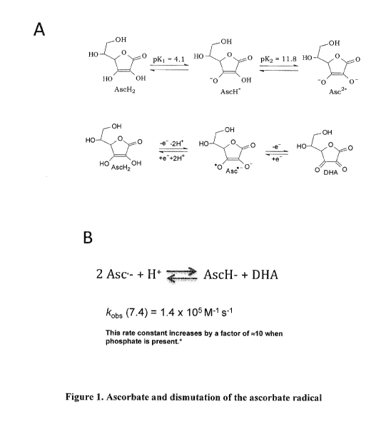
Figure 1. Ascorbate and dismutation of the ascorbate radical. A, Ascorbate is
a di-
acid, however at physiological pH of 7.4, 99% of ascorbate is present as its
mono anion (AscH-).
Ascorbate radical (Asc-) is present at equilibrium (but also at much lower
concentrations) with
oxidized and reduced forms of ascorbate. B, The dismutation of Asc is the
principal route of its
transformation, with a rate constant (kobs) that falls into the "intermediate"
proton exchange rate
on the NMR timescale. This rate constant can increase by a factor of 10 in the
presence of proton
exchange catalysts such as phosphate (Bors W, Buettner GR. (1997) The vitamin
C radical and its
reactions in Vitamin C in Health and Disease, ed. by L. Packer and J. Fuchs,
Marcel Dekker, Inc.,
New York, Chapter 4, pp75-94).
Figure 2. T2 relaxivity (r2 = mArisec-1) of sugars, sugar alcohols, and
ascorbate
Comparisons include both mono and disaccharides. As discussed in the text,
note the diminishing
contrast effect at higher concentration, which is believed to be secondary to
self-association of
like moieties and reduced proton exchange.
Figure 3. In vitro ("phantom") experiments on ascorbate spin-spin relaxation
(T2-
weighted) MRI contrast. A, shows quantitative T2 mapping in 5 phantoms with
progressively
increasing ascorbate concentration. Statistically significant "negative T2
contrast" (signal loss) is
seen as low as 1-5 mM as compared to control (phosphate-buffered saline) with
conventional fast
spin echo (FSE) acquisition. Sensitivity is therefore at the lower end of
expected tissue/cellular
concentrations following pharmacological doses of ascorbate in high uptake
tissues (e.g., tumors
and brain, 10-30 mM). This result also does not take into account any
synergistic effects from
tissue oxidative substrates or physiological exchange catalysts. B, shows the
synergistic effect of
H202 on ascorbate T2 enhancement. H202, which rises to 100-200 micromolar in
brain and
tumors in vivo following parenteral ascorbate, produces a marked synergism on
the T2 contrast
- 4 -
CA 02997791 2018-03-06
WO 2017/059092
PCT/US2016/054481
effect from ascorbic acid. The synergistic effect slowly diminishes over time
in phantoms over 30
min as shown, but will be sustained in vivo as long as H202 is produced
following ascorbate
administration. C, demonstrates the influence of pH on ascorbate's T2 effect,
which is maximized
at neutral/physiological pH (7.0-7.4). This result is consistent with prior
studies on the rate
kinetics of ascorbate disproportionation with its radical and oxidized foim at
equilibrium (Figure
1). D, reveals a marked synergistic effect when ascorbate is salified (salted)
with meglumine (N-
methyl-D-glucamine), an amine sugar derivative of sorbital that is commonly
employed as an
excipient in several FDA-approved drug and contrast formulations.
Figure 4. Comparison of Ascorbate as Na or Meglumine Salt. Solutions are
prepared
with physiological concentrations of PO4 (2 mM) and HCO3- (25 mM) buffers.
Figure 5. Na Ascorbate + Physiological Exchange Catalysts. Each solution is
set at
neutral (pH = 7.0) in deionized water. Concentrations of physiological
exchange catalysts are the
same as the in vivo in serum and extracellular space: PO4 = 2 mM, glucose = 5
mM, and HCO3-
is 25 mM.
Figure 6. Exchange Synergism Between Na Ascorbate/Meglumine and Glucose.
Solutions are compared in the setting of physiological buffers PO4 (2 mM) and
HCO3- (25 mM)
that also contribute as exchange catalysts.
Figure 7. Exchange synergism of Na Asc/Meglumine with sugar alcohols, mono-
and
disaccharides. All solutions are prepared in co-presence of 2 mM PO4 and 25 mM
HCO3-.
Figure 8. Resealed data without control for exchange synergism of Na
Asc/Meglumine with sugar alcohols, mono- and disaccharides. All solutions are
prepared with
2 mM PO4 and 25 mM HCO3-.
Figure 9. In vivo ascorbate T2 contrast changes following high dose parenteral
ascorbate (2g/kg, right IJ i.v. injection.) A, shows a conventional single
slice axial FSE T2WI
image through the midbrain of a normal C57 black mouse, and the two images on
the right
demonstrate a 'first pass' extraction of contrast change during and following
ascorbate
administration i.v. T2 signals in brain tissue are acquired immediately
following, and 10 minutes
after ascorbate administration, then subtracted from the T2 brain signals
acquired before
ascorbate administration. Since ascorbate produces a decrease in signal
intensity, subtraction
from the higher signal pre-dose scan results in a net positive 'map' of flow-
through perfusion
(blood flow) through brain tissue. At 10 minutes, the perfusion effect has
nearly resolved and
- 5 -
CA 02997791 2018-03-06
WO 2017/059092
PCT/US2016/054481
early signal intensity changes related to tissue uptake are beginning to be
observed. B, shows the
signal changes due to tissue uptake of high-dose ascorbate. Color-lute maps of
signal intensity are
not subtracted from the pre-dose scan and therefore show the expected
decreases in T2 signal
over time, maximized between 30-60 min in normal C57 mice.
Figure 10. Ascorbate T2 enhancement in a rodent model of neocortical spreading
depression (SD). In the illustrated experiment, the lower row of images shows
a tiny craniectomy
with gelfoam (red arrow) soaked in a high concentration of potassium chloride
(KC1), which
diffuses locally into the adjacent parietal cortex. The craniectomy site is 1
mm posterior to
bregma, a skull landmark representing the posterior third of the underlying
brain. The above two
rows show T2 images and quantitative color lute T2 maps of signal change in
rodent brain that
are 3 and 4 mm anterior to bregma, that is, distant from the SD induction
site. T2 signal changes
in the anterior slices demonstrate clear T2 asymmetry in the right cerebral
cortex as compared to
the left (again, SD remains confined to the right hemisphere). These marked
cortical signal
changes are consistent with the known hypermetabolic activity that occurs with
SD, as also
observed with "F-FDG PET, and with direct microdialysis and metabolomic
determinations. Of
note, the opposite observation (focally increased T2 signal) is seen directly
under the craniectomy
site itself (row three), consistent with localized edema (increased free
water) at the site of KC!
infusion.
Figures 11A-11B. Perfusion and viability cardiac imaging with parenteral
ascorbate.
Figure 11A depicts the two primary imaging planes, coronal and axial, for rat
heart imaging at
7T. Figure 11B shows transient decrease in T2 signal intensity throughout the
left ventricle with
initial bolus of ascorbate injection i.v.
Figure 12. T2 contrast changes in guinea pigs following i.v. administration of
three
different formulations of ascorbate. Figure 12A, Fast spin echo (FSE) T2
images before and
after 60 min slow infusion of ascorbate show dramatic signal intensity
differences throughout the
brain parenchyma. Figure 12B-C shows and C, normalized signal intensity
changes and
quantitative relaxivity measurements are shown for both guinea cerebral cortex
(Cx) and basal
ganglia (BG) after administration of three different ascorbate formulations.
- 6 -
CA 02997791 2018-03-06
WO 2017/059092
PCT/US2016/054481
Detailed Description of the Preferred Embodiments
The present invention is now described more fully hereinafter with reference
to the
accompanying drawings, in which embodiments of the invention are shown. This
invention may,
however, be embodied in different forms and should not be construed as limited
to the
embodiments set forth herein; rather these embodiments are provided so that
this disclosure will
be thorough and complete and will fully convey the scope of the invention to
those skilled in the
art.
As used herein, the singular forms "a," "an" and "the" are intended to include
plural
forms as well, unless the context clearly indicates otherwise. It will be
further understood that the
terms "comprises" or "comprising" specify the presence of stated features,
integers, steps,
operations, elements, components and/or groups or combinations thereof, but do
not preclude the
presence or addition of one or more other features, integers, steps,
operations, elements,
components and/or groups or combinations thereof.
As used herein, the term "and/or" includes any and all possible combinations
or one or
more of the associated listed items, as well as the lack of combinations when
interpreted in the
alternative ("or").
Unless otherwise defined, all terms (including technical and scientific terms)
used herein
have the same meaning as commonly understood by one of ordinary skill in the
art to which this
invention belongs. It will be further understood that terms, such as those
defined in commonly
used dictionaries, should be interpreted as having a meaning that is
consistent with their meaning
in the context of the specification and claims and should not be interpreted
in an idealized or
overly formal sense unless expressly so defined herein.
Ascorbate. Ascorbate (also known as "ascorbic acid," "L-ascorbic acid" or
"Vitamin C")
is a naturally-occurring organic compound and an essential nutrient, with
important properties as
an antioxidant and co-factor in at least eight enzymatic reactions, including
several collagen
synthesis reactions that, when dysfunctional, result in the most conspicuous
symptoms of scurvy.
Most mammals make ascorbic acid in the liver, where the enzyme L-gulonolactone
oxidase
converts glucose to ascorbic acid. In humans, higher primates, guinea pigs and
most bats,
however, a mutation results in low or absent L-gulonolactone oxidase
expression so that
ascorbate must be consumed in the diet (Lachapelle, M. Y.; Drouin, G. (2010).
"Inactivation
dates of the human and guinea pig vitamin C genes". Genetica 139 (2): 199-
207). In all animal
- 7 -
CA 02997791 2018-03-06
WO 2017/059092
PCT/US2016/054481
species, L-ascorbic acid/ascorbate is the most abundant intracellular
antioxidant, with
intracellular concentrations capable of reaching 10-30 mM in tumors, brain
cells, and some other
tissues. Those tissues that accumulate over 100 times the level in blood
plasma of vitamin C
include the adrenal glands, pituitary, thymus, corpus luteum, and retina.
Those with 10 to 50
times the concentration include brain, spleen, lung, testicle, lymph nodes,
liver, thyroid, small
intestinal mucosa, leukocytes, pancreas, kidney, and salivary glands (Hediger
MA (May 2002).
"New view at C". Nat. Med. 8 (5): 445-6).
Dietary excesses of vitamin C are not absorbed, and excesses in the blood are
rapidly
excreted in the urine. Vitamin C exhibits remarkably low toxicity, with an
LD50 in rats generally
accepted at ¨ 11.9 grams per kilogram of body weight. The mechanism of death
from such doses
(1.2% of body weight, or 0.84 kg for a 70 kg human) is unknown, but may be
mechanical rather
than chemical ("Safety (MSDS) data for ascorbic acid". Oxford University.
October 9, 2005.
Retrieved February 21, 2007). The LD50 in humans is uncertain given the lack
of any accidental
or intentional poisoning death data. The rat LD50 is, therefore, used as a
guide for human toxicity.
At physiological pH, 99% of ascorbate is present as the mono anion (Fig.1A).
The
chemistry and therefore imaging properties of vitamin C are dominated by this
moiety. As a
donor antioxidant, the mono anion donates a hydrogen atom (H or H++ e) to an
oxidizing radical
to produce a resonance-stabilized tricarbonyl ascorbate free radical, Asc*-
(Fig. 1B). The
dismutation reaction (Fig. 1C) of Asc- back to reduced or oxidized ascorbate
is the principal
route of elimination in vitro. This process is supplemented in vivo by enzymes
that aid in
ascorbate recycling (May JM, Qu ZC, Neel DR, Li X (May 2003). "Recycling of
vitamin C from
its oxidized forms by human endothelial cells". Biochim. Biophys. Acta 1640 (2-
3): 153-61).
Dismutation of the radical to either ascorbate or dehydroascorbate occurs via
loss or gain of
hydrogen, which serves as either the electron carrier or the more conventional
cation. Also, the
rate constant of ascorbic radical dismutation is 105-106 so that hydrogen
exchange
accompanying dismutation also occurs at the same rate. On the NMR timescale,
these
"intermediate" exchange rates are optimal for altering 1H spin-spin
relaxation.
Parenteral Formulations of Ascorbate. Ascorbate for parenteral administration
may be
provided in a pharmaceutically acceptable carrier (e.g., sterile water,
endotoxin-free water, or
pyrogen-free water; sterile, endotoxin-free or pyrogen-free saline, etc.) as a
formulation suitable
for parenteral administration. The term "pharmaceutically acceptable" as used
herein means that
- 8 -
CA 02997791 2018-03-06
WO 2017/059092
PCT/US2016/054481
the compound or composition is suitable for administration to a subject to
achieve the treatments
described herein, without unduly deleterious side effects in light of the
severity of the disease and
necessity of the treatment.
The formulations may be presented in unit/dose or multi-dose containers, for
example
sealed ampoules and vials, and may be stored in a dried/powdered/freeze-dried
(lyophilized)
condition requiring only the addition of the sterile liquid carrier, for
example, saline or water-for-
injection immediately prior to use. Extemporaneous injection solutions and
suspensions may be
prepared from sterile powders, granules and tablets. For example, in one
aspect of the present
invention, there is provided an injectable, stable, sterile composition
comprising ascorbate in a
unit dosage form in a sealed container. The ascorbate may be provided in the
form of a
lyophilizate which is capable of being reconstituted with a suitable
phaimaceutically acceptable
carrier to form a liquid composition suitable for injection thereof into a
subject.
Examples of suitable formulations include, but are not limited to, a sterile
aqueous
solution of ascorbic acid in water for injection, containing 10, 20, 30, 40,
or 50, to 80, 90, 100,
150 or 200 mg/mL ascorbate or a salt thereof (e.g., sodium salt, meglumine (N-
methyl-D-
glucamine) salt, combinations thereof, etc.). In some embodiments,
foimulations may include 10,
20, 30, 40, 50, 60, 70, 80, 90, or 100 mM, to 150, 200, 250, 300, 350, 400,
450, 500, 550, 600,
650, 700, 750, or 800 mM ascorbate or a salt thereof (e.g., sodium salt,
meglumine (N-methyl-
D-glucamine) salt, combinations thereof, etc.). For example, formulations may
include from 100
to 700 mM, or from 200 to 650 mM, or from 300 to 600 mM, or from 400 to 550
mM, ascorbate
or a salt thereof. The ascorbate concentration may be adjusted as needed
depending on the route
of administration (e.g., intravenous administration versus direct
administration into a localized
body region or compartment).
In some embodiments, the pH is adjusted to approximately 7 (e.g., pH of from
6.5 to 7.5)
(e.g., with sodium bicarbonate and/or sodium hydroxide).
Formulations suitable for parenteral administration may include a stabilizing
agent.
Example stabilizing agents include chelators such as EDTA (e.g., EDTA
disodium). Formulations
may also include pH buffers such as bicarbonate (HCO3-) and/or phosphate
(PO4).
Formulations according to some embodiments may include a sugar, such as a
reducing or
non-reducing sugar. A "reducing sugar" is an open-chain sugar having a free
aldehyde group or a
free ketone group, which includes all monosaccharides and some disaccharides,
oligosacchrides
- 9 -
CA 02997791 2018-03-06
WO 2017/059092
PCT/US2016/054481
and polysaccharides. Example reducing sugars include, but are not limited to,
glucose, galactose,
glyceraldehyde, fructose, ribose, xylose, lactose, maltose, etc. A "non-
reducing sugar" is a sugar
without a free aldehyde group or free ketone group. Example non-reducing
sugars include, but
are not limited to, sucrose, trehalose, etc.
The spin-spin exchange catalysts that may be used in the ascorbate
formulations as taught
herein may include, but are not limited to, meglumine (N-methyl-D-glucamine),
reducing sugars
(e.g., glucose, galactose, glyceraldehyde, fructose, ribose, xylose, lactose,
maltose, combinations
thereof, etc.), and non-reducing sugars (e.g., sucrose, trehalose,
combinations thereof, etc.).
Formulations suitable for parenteral administration may have an osmolarity in
the range
of from 200 to 1200 or 1400 mOsm/L. In some embodiments, the formulation has
an osmolarity
of from 200, 300, 400, 500 or 600 to 700, 800, 900, 1000, 1100, 1200, 1300, or
1400 mOsm/L.
In some embodiments, the formulation is suitable for injection into an artery
or vein,
and/or into a body region such as an organ or organ region. In some
embodiments, the
formulation is suitable for intravenous infusion. In some embodiments, the
formulation is suitable
for intraarterial infusion. In some embodiments, the formulation is suitable
for intrathecal
infusion.
In some embodiments, the formulation is de-oxygenated. Methods of de-
oxygenation of
aqueous compositions are known, e.g., preparing the formulation under, or
purging with, an inert
gas, such as nitrogen. See, e.g., U.S. Patent Application publication
2014/0048290 to Bodemann.
In some embodiments, the fotinulation is provided as a
dried/powdered/lyophilized
composition of meglumine ascorbate, sodium ascorbate, or a combination of
these salts, with or
without exchange catalysts, chelators, etc., which may be reconstituted in
sterile aqueous media
(e.g., water, normal saline, lactated Ringers, or other accepted aqueous
vehicle used for parenteral
drug delivery) at point of care just prior to administration. Suitable dried
folinulations may
include, but are not limited to, 5, 10, 15, 20, 25, 30, 35, 40, 45, 50, 55,
60, 65, 70, 75, 80, 85, 90,
95 or 100 grams of ascorbate of a salt thereof.
In some embodiments, the formulation is provided in a container suitable for
light-
sensitive liquid compositions, such as an opaque plastic or glass container
(e.g., a high density
polyethylene container, a plastic or glass container coated with black
polyvinyl chloride, etc.),
amber glass, etc. See, e.g., U.S. Patent No. 8,309,191 to Wang et al.; U.S.
Patent Application
publication 2004/0048206 to Miyake et al.
- 10 -
CA 02997791 2018-03-06
WO 2017/059092
PCT/US2016/054481
In some embodiments, the formulation is provided in unit dosage form suitable
for
parenteral administration for MRI imaging. As non-limiting examples, unit
dosage forms
suitable for intravenous administration may be: 1) 0.25 g/min, up to 60 min,
up to 15 grams; 2)
0.5 g/min, up to 60 mm, up to 30 grams; 3) 1.0 g/min, up to 60 min, up to 60
grams; or 4) 1.5
g/min, up to 60 min, up to 90 grams.
Methods of use. As noted above, the parenteral ascorbate compositions as
taught herein
are useful for magnetic resonance imaging (MRI) to provide a contrast agent
for the detection and
characterization of perfusion, metabolism, and/or oxidative stress in human
and non-human
tissues, without the need for radioactivity or chemical labeling.
Ascorbate, especially in the presence of, and co-formulated with, spin-spin
exchange
catalysts (for example, simple sugars, sugar alcohols or amino acids) is a
safe and biodegradable
MRI contrast agent that requires neither the use of metal-based (e.g.,
gadolinium or iron) contrast
material nor ionizing radiation. The technique enables assessment of tissue
perfusion as well as
high-resolution molecular characterization of tissue viability and metabolism
that is analogous to
18F-FDG PET. The latter is possible by virtue of ascorbate's uptake (via
dehydroascorbate) into
cells through the same glucose transport mechanisms that take up 18F-FDG
(i.e., GLUT 1 and 3
transporters) (Rumsey SC, Kwon 0, Xu GW, Burant CF, Simpson I, Levine M (July
1997).
"Glucose transporter isoforms GLUT1 and GLUT3 transport dehydroascorbic acid".
I Biol.
Chem. 272 (30): 18982-9).
"Parenteral administration" as used herein includes, but is not limited to,
intravenous,
subcutaneous, intramuscular, intraperitoneal, intraarterial, intraosseous,
intrathecal or
intraventricular administration, e.g., through injection or infusion. As a non-
limiting example,
intraperitoneal or other parenteral administration may be used where
intravenous (i.v.) access is
difficult for a subject (e.g., low blood pressure), or the route of
administration otherwise would
result in a suitable MRI image.
In some embodiments the MRI is performed during, or up to 5, 10, 30, 40, 60,
90 or 120
minutes after, or up to 1, 2, 3, or 4 hours after, parenterally administering
the ascorbate
composition.
Subjects benefitting from the present invention are, in general, mammalian
subjects,
including both human subjects and animal subjects (e.g., dogs, cats, rabbits,
cattle, horses, etc.),
-11-
CA 02997791 2018-03-06
WO 2017/059092
PCT/US2016/054481
for diagnostic, therapeutic, research or veterinary purposes. Subjects may be
male or female and
may be any age, including neonate, infant, juvenile, adolescent, adult, and
geriatric subjects.
MRI is known, and may be carried out by commercially available equipment, and
by
techniques known in the field. See, e.g., S. Bushong and G. Clarke, Magnetic
Resonance
Imaging: Physical and Biological Principles (Mosby, 4th Ed. 2014). In some
embodiments, the
MRI is perfusion (e.g., blood flow) imaging. In some embodiments, the MRI is
metabolism
imaging. Metabolism imaging may be used as a diagnostic biomarker analogous to
18F FDG PET,
including, but not limited to, identification/characterization of tumors or
dysfunctional tissues
demonstrating hyper- or hypo-metabolism.
"Body or body region" that may be imaged with MRI as taught herein includes
the body
or any region of the body of a subject, such as an organ or organ system, soft
tissue, bone, etc., or
any portion thereof. Examples of body regions include, but are not limited to,
head, neck, thorax,
abdomen, pelvis, limb(s), muscle, fat, other soft tissues, bone, etc. Examples
of organs include,
but are not limited to, adrenal gland, pituitary, thymus, corpus luteum,
retina, brain, spleen, lung,
testicle, lymph nodes, liver, thyroid, small intestinal mucosa, leukocytes,
pancreas, kidney,
salivary gland tissue, heart, etc.
"Enhancing" an MRI image as used herein is inclusive of facilitating the MRI
visualization by enhancing the contrast of structures, tissues or fluids in an
MRI signal.
An "MRI contrast agent" is a substance that can enhance the contrast of
structures, tissues
or fluids within the body during an MRI scan. Examples include, but are not
limited to,
paramagnetic contrast agents such as Gd-containing agents or manganese
chelates, and
superparamagnetic agents such as iron platinum particles. See also U.S. Patent
Application
Publication Nos. 2014/0350193 to Axelsson et al.; and 2014/0234210 to Lin et
al.
Potential applications for ascorbate MRI include several clinical scenarios
where current
medical practice often utilizes PET scanning but where improvements in
methodology using MRI
as an alternative scanning technology will potentially yield further clinical
benefit. These
scenarios include diagnostic studies for cancer, neurological disease (e.g.,
dementia, TBI and
epilepsy) and cardiovascular imaging. Heart studies using Tc99m-labeled agents
(e.g., Tc-99m
sestimibi or "Cardiolite") represent a particularly noteworthy potential
diagnostic application in
need of an alternative approach given the projected contraction of supply of
Tc-99m. Myocardial
perfusion and viability imaging with Tc-99m-related agents is an essential and
widely performed
- 12 -
CA 02997791 2018-03-06
WO 2017/059092
PCT/US2016/054481
procedure, yet to date no commercially feasible solution has been developed to
replace these Tc-
99m-dependent agents.
MR imaging and clinical application of contrast media. Clinical magnetic
resonance
imaging (MRI) generates high-resolution images of the body through the
acquisition of proton
(1H) nuclear magnetic resonsnace (NMR) signals from water and macromolecules
in tissue. For
"Ti-weighted" MR images, signal intensity increases in regions where
longitudinal relaxation rate
(spin lattice relaxation rate, 1/TI) increases. With "T2-weighted" MRI, signal
intensity decreases
when transverse relaxation rate (spin-spin relaxation rate, 1/T2) increases.
Both Ti and T2
weighted images are routinely acquired in virtually all clinical MRI studies.
Intravenous contrast agents are routinely administered in MRI to further
increase 1/T1 or
1/T2, in an effort to better delineate diseased tissue from normal tissue,
improve anatomical
definition, and enhance characterization of physiological or pathological
function. Almost all
currently approved MRI contrast agents are based on chelates of the lanthanide
metal Gd, with a
small subset of angiographic and perfusion studies conducted using iron-oxide
materials (e.g.,
Feraheme) off-label in patients with renal insufficiency/failure. Commercial
Gd-based materials
are used most commonly to increase 1/T1 in diseased tissue, where contrast
material is prone to
accumulate.
For tissue perfusion determinations with MRI, Gd-based agents or iron-oxide
nanoparticles may be used, with acquisition strategies based on either 1/T1 or
1/T2 contrast,
although 1/12 contrast approaches are increasingly favored. Perfusion imaging
is currently used
clinically to characterize tumor aggressiveness, tumor response to therapy,
and tissue viability in
heart, brain and other organs.
Without wishing to be bound by theory, the mechanism of ascorbate signal
change
without paramagnetism, which is also described as "T2-weighted contrast," is
based on
enhancement of the water proton (1H) spin-spin relaxation rate 1/12 (or
reciprocally, spin-spin
relaxation time, 12), as solvent water protons are exchanged with hydroxyl
protons on ascorbate
molecules. The effect of proton exchange on 12 contrast is amplified further
by the dismutation
reaction of the ascorbate radical at physiological pH. Ascorbate oxidation and
ascorbate radical
dismutation are, in turn, driven by the co-presence of oxidizing substrates
such as hydrogen
peroxide (H202) and/or hydrogen ("proton") exchange catalysts.
- 13 -
CA 02997791 2018-03-06
WO 2017/059092
PCT/US2016/054481
Ascorbate transport and excretion. Ascorbic acid is absorbed in the body by
both active
transport and simple diffusion. The two major active transport pathways are
sodium-ascorbate co-
transporters (SVCTs) and hexose transporters (GLUTs). SVCT1 and SVCT2 import
the reduced
form of ascorbate across the plasma membrane (Savini I, Rossi A, Pierro C,
Avigliano L, Catani
MV (April 2008). "SVCT1 and SVCT2: key proteins for vitamin C uptake". Amino
Acids 34 (3):
347-55), whereas GLUT1 and GLUT3 glucose transporters transfer the oxidized
form,
dehydroascorbic acid (Rumsey SC, Kwon 0, Xu GW, Burant CF, Simpson I, Levine M
(July
1997). "Glucose transporter isoforms GLUT1 and GLUT3 transport dehydroascorbic
acid". J.
Biol. Chem. 272 (30): 18982-9). Although dehydroascorbic acid concentrations
are low in plasma
under normal conditions, the oxidized molecule is absorbed at much higher
rates across GLUT1
and GLUT3 than the reduced form is across the SVCTs. When ascorbate
concentrations are
pharmacologically elevated, dehydroascorbate concentration also increases,
enabling marked
absorption where GLUT transporters exist in high copy such as in the brain
(and blood brain
barrier) and tumor cells. Once transported, dehydroascorbic acid is rapidly
reduced back to
ascorbate.
Ascorbate concentrations over the renal re-absorption threshold pass freely
into the urine
and are excreted with a half-life of about 30 minutes. At high dietary doses
(corresponding to
several hundred mg/day in humans) the renal resorption threshold is 1.5 mg/dL
in men and
1.3 mg/dL in women (Oreopoulos DG, Lindeman RD, VanderJagt DJ, Tzamaloukas AH,
Bhagavan HN, Garry PJ (October 1993). "Renal excretion of ascorbic acid:
effect of age and
sex". J Am Coll Nutr 12 (5): 537-42). Ascorbate that is not directly excreted
in the urine or
destroyed by other body metabolism is oxidized by L-ascorbate oxidase and
removed.
Ascorbate is understood to have a pharmacokinetic profile that resembles
vancomycin.
Biodistribution of oral ascorbate is under tight control, with plasma
concentrations rarely
exceeding 200 JAM even at oral doses more than 100 times the recommended daily
allowance
(Levine M, Conry-Cantilena C, Wang Y, Welch RW, Washko PW, Dhariwal KR, Park
JB,
Lazarev A, Graumlich JF, King J, Cantilena LR (April 1996). "Vitamin C
pharmacokinetics in
healthy volunteers: evidence for a recommended dietary allowance". Proc. Natl.
Acad. Sci. U.S.A.
93 (8): 3704-9). Ascorbate administered intravenously, however, bypasses these
tight control
systems, with plasma concentrations of 10 mM or higher achievable. Plasma
concentrations
higher than 10 mM are safely sustained in humans for up to 4 hours with
remarkably low toxicity
- 14-
CA 02997791 2018-03-06
WO 2017/059092
PCT/US2016/054481
(Hoffer LJ., Levine M., Assouline S., Melnychuk D., Padayatty SJ., Rosadiuk
K., Rousseau C.,
Robitaille L., and Miller WH., Jr., Phase I clinical trial of i.v. ascorbic
acid in advanced
malignancy. Ann Oncol 19: 1969-1974, 2008).
The present invention is explained in greater detail in the following non-
limiting
examples.
In Vitro Examples
Ascorbate Enhancement of Spin-Spin Relaxation Rate, 1/T2.
Previous studies have reported on the NMR/MRI contrast effects on T2-weighting
arising
from exchange of bulk water protons with mobile protons of low molecular
weight solutes and
macromolecules (e.g., -NH2, -OH, -SH, -NH). The contrast effect on 1/12 from
this proton
exchange is described as follows:
1 1
= + fcR(Pb, &Di), k, T2b, 7")
T2 1 2a
Bulk water is related to a and exchangeable protons (e.g., from an ascorbate
OH group) to
b. fCR is a closed function with five parameters, derived from Carver and
Richards and refined by
Hill et al. (Carver, J. P.; Richards, R. E. J. General 2-Site Solution For
Chemical Exchange
Produced Dependence Of 12 Upon Carr-Purcell Pulse Separation J. Magn. Reson.
1972, 6, 89-
105; Hills, B. P.; Wright, K. M.; Belton, P. S. N.M.R. studies of water proton
relaxation in
Sephadex bead suspensions Mol. Phys. 1989, 67, 1309-1326). For the hydroxyl
protons of
ascorbate, Pb would be the fraction of exchangeable protons, k is the exchange
rate between
exchangeable protons and water protons, 6cob is the chemical shift between
hydroxyl and bulk
water protons, and T2b is the local spin-spin relaxation time of hydroxyl
protons. T is the inter-
pulse (90 480 ) spacing in the 12-weighted acquisition sequence.
An essential but often neglected parameter influencing proton exchange on T2
contrast is
the role of exchange catalysis (Liepinsh E and Otting, G Proton exchange rates
from amino acid
side chains ¨ implications for image contrast. Magn Reson Med. 1996 35(1): 30-
42). The rate
constant k for proton exchange between OH or NH groups and water can be
described by
- 15 -
CA 02997791 2018-03-06
WO 2017/059092
PCT/US2016/054481
k = ka[H+] + kb[0H]+1kc [catalyst]Y
Ka, Kb and lc denote the exchange rate constants due to catalysis by H+, 01-F
and other exchange
catalysts, respectively. The exponent y is 1 or 2 depending on the mechanism
of a given exchange
catalyst. The rate constants K, and Kb can be calculated in turn by:
1
ka,b = kp _______________________________________
1 + 1OpKDpKA
where KD is the rate constant for diffusion controlled encounter of the proton
donor and acceptor
1010 ma's- ,1,
) and pKD and pKA are the pK values of the proton donor and acceptor. Although
pKH30+ and pKoH_ = 15.7, K, is more challenging to predict because of the
nonlinear dependence
of proton transfer on catalyst concentration. Nonetheless, efficient exchange
catalysis at neutral
pH is attained with at least a moderate difference between pKD - pKA and a
significant
concentration of catalytically active acidic or basic folins of the exchange
catalysts at
physiological pH.
Thus H20, despite its high concentration, is a relatively poor proton donor
and therefore
an inefficient exchange catalyst at physiological pH because the pKA of the
primary species
(H30+ and OH) is 15.7. On the other hand, recognized exchange catalysts in
physiological
conditions include organic phosphates, carbonates (e.g., bicarbonate, HCO3-),
and molecules with
carboxyl and amino groups (Liepinsh E and Otting, G Proton exchange rates from
amino acid
side chains ¨ implications for image contrast. Magn Reson Med. 1996, 35(1): 30-
42).
As shown below, another powerful catalyst not previously recognized is
ascorbate, which
possesses one hydroxyl group having a favorable pKA = 6.75 at the 4 position,
as well as an
equilibrium disproportionation reaction with a pKA = 7Ø Thus, ascorbate has
the potential to not
only 'self-catalyze' but also to be an efficient catalyst of proton exchange
for basic hydroxyl
groups on sugars and other macromolecules.
Figure 2 shows a comparison on T2 enhancement of pure solutions of several
sugars,
sugar alcohols, and ascorbate in deionized water at pH 7. Data are provided
from quantitative 12
mapping at 7T using a RARE FSE protocol with at least 6 different echo times,
at solute
concentrations of 10 and 20 mM. As shown, 12 relaxivity is roughly a function
of the number of
exchangeable OH protons available on each molecule, with disaccharides, as
predicted,
- 16 -
CA 02997791 2018-03-06
WO 2017/059092
PCT/US2016/054481
producing proportionally greater contrast effect than monosaccharides.
Noteworthy is the
nonlinear dependence on solute concentration, with relaxivity enhancement
decreasing as
concentration is increased, a phenomenon that is likely related to self-
association of sugars in
pure solutions. The latter is particularly relevant to observations described
below, where overall
T2 effects are instead synergistically enhanced when ascorbate and sugars are
combined together
at higher total solute concentrations. Formulations combining ascorbate with
mono or
disaccharides provide a means to deliver higher concentrations of both species
in order to
increase T2 contrast effects for MR imaging.
Figure 3A depicts a more detailed demonstration of 12 effects of pure
ascorbate solutions
at different concentrations at neutral pH. Figure 3B reveals the marked
enhancement of the T2
effect when ascorbate is in the presence of only M (i.e., physiological)
concentrations of
hydrogen peroxide (H202), which drives oxidation to dehydroascorbate as well
as ascorbate
radical dismutation. Although H202 is also considered an exchange catalyst in
its own right, the
dramatic effect observed on ascorbate-mediated 1/12 enhancement when H202 is
present at 100-
fold less concentration than ascorbate suggests that proton exchange from
H202¨driven ascorbate
oxidation/dismutation, rather than direct exchange from OH ascorbyl protons,
is an important
contributory mechanism responsible for T2 changes. Further evidence of the
contribution from
dehydroascorbate oxidation/dismutation on proton exchange is depicted in
Figure 3C showing
that the 1/12 enhancement effect is by far the most significant at neutral pH
where the reaction
rate of ascrobate-dehydroascorbate dispropotionation is also greatest.
Data in Figure 3D provide the first suggestion that exchange catalysis between
ascorbate
and an acceptor/donor molecule with an appropriate pK can markedly drive 1/12
enhancement
change. Data here compare solutions of ascorbate (10 mM) as sodium salt and as
meglumine
(aminosugar) salt. Here the T2 contrast effect (T2 relaxation in ms) is
approximately 4 times
greater with meglumine ascorbate as compared to either meglumine or ascorbate
alone in water
at neutral pH.
It was subsequently investigated whether the impressive synergistic effect of
meglumine
with ascorbate was dependent on chemical association with ascorbate as a
salting cation (even
though in theory the two moieties should be fully dissociated in water).
Figure 4 reveals that
proton exchange is actually synergized when the 'salting function' is
performed by Na + cations,
presumably leaving the amine group in addition to the basic OH groups of
meglumine to
- 17-
CA 02997791 2018-03-06
WO 2017/059092
PCT/US2016/054481
participate in exchange catalysis with ascorbate. Note that here control T2
relaxation (ms) values
(T2 = 840 ms) are not shown to better illustrate differences between
experimental groups.
Figure 5 summarizes T2 relaxation data from a series of experiments looking at
the
influence of various physiological exchange catalysts on the contrast effect
from ascorbate. Using
known serum and extracellular concentrations of PO4 = 2 mM, glucose = 5 mM,
and HCO3" of 25
mM, with ascorbate at 10 mM, T2 relaxation of each of these moieties was
examined individually
and in combination. As shown, the 12 relexation effect of ascorbate alone or
with PO4 in water is
modest but in the presence of physiological concentrations of either glucose
or HCO3" is
dramatically increased, with 10 mM ascorbate, (a plasma concentration easily
and safely
achieved with parenteral administration) producing a remarkable 50% change in
T2 relaxation.
The greatest enhancement is seen with ascorbate in the presence of glucose,
HCO3-, and PO4
together at known concentrations in vivo. Thus, by simply administering
ascorbate i.v., the T2
enhancement effect of ascorbate in vivo will be much greater than what might
be expected after
only looking at ascorbate alone in phantom studies without physiological
exchange catalysts
present.
Also predicted from the experiments above is the possibility that formulation
of ascorbate
with other sugars that are not normally present in vivo may further catalyze
the ascorbate contrast
effect. Figure 6, for example, demonstrates additional synergism when
meglumine is added to a
solution of sodium ascorbate at equivalent concentration (20 mM) and into a
background of 2
mM PO4, 25 mM 1-1CO3-. Data show comparison with or without physiological
concentrations (5
mM) of glucose, as well as the effect of meglumine alone added to the
physiological catalysts. As
seen the greatest contrast effect is observed when all moieties are combined.
One implication
therefore is that higher contrast effects may be achievable by combining
different exchange
catalysts with each other, thus limiting the concentration of any one
exogenously administered
species.
Figure 7 summarizes data extending this concept, testing potential synergisms
when Na
ascorbate and meglumine are formulated with other mono and disaccharides and
sugar alcohols.
As shown the contrast effects are dramatic with each potential formulation. In
Figure 8, the
control solution (2 mM PO4 and 25 mM HCO3-) to better illustrate the
differences in contrast
changes between groups. The strongest effect thus observed is when ascorbate
and meglumine
are combined with the common disaccharide sucrose, thus suggesting a promising
candidate
- 18 -
CA 02997791 2018-03-06
WO 2017/059092
PCT/US2016/054481
formulation (i.e., ascorbate/ meglumine/ sucrose) for MRI using only moieties
that may all be
safely administered parenterally.
In vivo Example 1
Normal brain perfusion and metabolic change
Figure 9. In vivo ascorbate T2 contrast changes following high dose parenteral
ascorbate (2g/kg, right IJ i.v. injection.) A, shows a conventional single
slice axial FSE T2WI
image through the midbrain of a normal C57 black mouse, and the two images on
the right
demonstrate a 'first pass' extraction of contrast change during and following
ascorbate
administration i.v. T2 signal in brain tissue immediately following, and 10
minutes after,
ascorbate administration is acquired and then subtracted from the T2 brain
signal acquisition
pre-ascorbate administration. Since ascorbate produces a decrease in signal
intensity, subtraction
from the higher signal pre-dose scan results in a net positive 'map' of flow-
through perfusion
(blood flow) through brain tissue. At 10 minutes, the perfusion effect has
nearly resolved and
early signal intensity changes related to tissue uptake are beginning to be
observed. B, show the
signal changes due to tissue uptake of high-dose ascorbate. Color-lute maps of
signal intensity
are not subtracted from the pre-dose scan and therefore show the expected
decreases in 12 signal
over time, maximized between 30-60 mm in normal C57 mice.
In vivo Example 2
Focal cerebral hypermetabolism in
association with neocortical spreading depression
Figure 10. Ascorbate T2 enhancement in a rodent model of neocortical spreading
depression. Spreading depression (SD) is an experimentally reproducible
pathophysiological
phenomenon of CNS tissues originally described 60 years ago by Loao. After a
focal region of
cortex reaches a critical threshold of ionic perturbation, a massive spreading
wave of cellular
depolarization may begin and spread through gray matter tissue, but remain
confined to the gray
matter zone in which it was induced, not crossing white matter pathways. If
the induction
mechanism (e.g., a local high concentration of applied potassium chloride) is
continuous to the
same region, these waves of SD will recur once every 8-10 minutes and last
over a 2-3 hour
period. Marked changes in brain metabolism accompany SD, and, since no
histologically
- 19 -
CA 02997791 2018-03-06
WO 2017/059092
PCT/US2016/054481
detectable neuronal injury is present after SD, these metabolic changes
parallel metabolic fluxes
in non-ischemic, hyperexcitable brain tissue such as epileptogenic foci.
In the above experiment, the lower row of images shows a tiny craniectomy with
gelfoam (red arrow) soaked in a high concentration of KC1, which diffuses
locally into the
adjacent parietal cortex. The craniectomy site is 1 mm posterior to bregma, a
skull landmark
representing the posterior third of the underlying brain. The above two rows
show T2 images and
quantitiative color lute T2 maps of signal change in rodent brain that are 3
and 4 mm anterior to
bregma, that is, distant from the SD induction site. T2 signal changes in the
anterior slices
demonstrate clear T2 asymmetry in the right cerebral cortex as compared to the
left (again, SD
remains confined to the right hemisphere). These marked cortical signal
changes are consistent
with the known hypermetabolic activity that occurs with SD, as also observed
with 18F-FDG
PET, and with direct microdialysis and metabolomic determinations. Of note,
the opposite
observation (focally increased 12 signal) is seen directly under the
craniectomy site itself (row
three), consistent with localized edema (increased free water) at the site of
KC1 infusion.
In vivo Example 3
Cardiac perfusion and metabolic imaging
Figure 11. Perfusion and viability cardiac imaging with parenteral ascorbate.
A,
depicts the two primary imaging planes, coronal and axial, for rat heart
imaging at 71.
Retrospective gating with respiratory coupling was employed to collect images
at 7T. The
acquisition sequence is moderately 12-weighted and can be further optimized to
enhance the
contrast effect. B, shows transient decrease in 12 signal intensity throughout
the left ventricle
with initial bolus of ascorbate injection i.v. After the initial bolus for
first pass flow or 'perfusion
imaging' quantitative T2 maps using variable flip angles show gradual T2
contrast change in
heart tissue reflecting ascorbate uptake. Only viable, metabolically active
cells will take up
ascorbate.
- 20 -
CA 02997791 2018-03-06
WO 2017/059092
PCT/US2016/054481
Example 4
Table 1: Example parenteral formulations useful for MRI imaging
T2W contrast agent Cation Exchange catalyst Osmolarity
I ascorbate 100-600 mM sodium 100-600 mM*
200-1200 mOsm/L
II ascorbate 100-600 mM meglumine 100-600 meglumine
200-1200 mOsm/L
mM 100-600 mM
III ascorbate 100-600 mM sodium 250-300 mM; meglumine
100- 200-1200 mOsm/L
N-methyl-D-glucamine 300 mM
250-300 mM
IV ascorbate 100-300 mM sodium 100-300 mM meglumine
100- 200-1200 mOsin/L
300 mM
V ascorbate 100-300 mM sodium 100-300 mM reducing
sugars 200-1400 mOsm/L
100-300 mM
VI ascorbate 100-300 mM sodium 100-300 mM non-reducing
200-1400 mOsm/L
sugars 100-300
mM
VII non-reducing sugars'
meglumine 0.0-1.0 200-1400 mOsm/L
0.1-1.0 M
VIII reducing sugarsb 0.1- meglumine
200-1400 mOsm/L
1.0 M 0.0-1.0 M
* sodium may be provided, e.g., as NaOH or NaHCO3_
a non-reducing sugars include, e.g., sucrose, trehalose
reducing sugars include, e.g., glucose, galactose, glyceradledyde, fructose,
ribose,
xylose, lactose, maltose
Example 5
Example preparation of parenteral formulation useful for MRI imaging
Formulation II above is prepared in the following manner: Into 500 mL sterile
water are
added 50 g of ascorbic acid (568 mM) and 55.4 g N-methyl-D-glucamine (568 mM).
Stir until
solution clears. mOsm/L ¨ 1100. pH ¨ 7Ø To promote long-term stability, add
0.025% EDTA
disodum, prepare in de-oxygenated solution under nitrogen blanket and under
light-sensitive
conditions.
In vivo Example 6
-21-
CA 02997791 2018-03-06
WO 2017/059092
PCT/US2016/054481
T2 contrast changes in guinea pigs following
intraveneous administration of three different formulations of ascorbate
We examined T2 contrast changes in whole brains of lightly anesthetized guinea
pigs at
7T. Since guinea pigs share humans' inability to synthesize ascorbate
endogenously, MRI effects
in this model may be more predictive of MRI changes in patients. Ascorbate was
administered
parenterally via femoral or jugular vein access using controlled infusion for
a total dose of 2g/kg
over 60 minutes. MRI was perfoinied for 90 mintues.
Figure 12A shows Fast spin echo (FSE) 12 images before and after 60 min slow
infusion
of ascorbate show dramatic signal intensity differences throughout the brain
parenchyma.
. In Figure 12B and Figure 12C, normalized signal intensity changes and
quantitative
relaxivity measurements are shown for both guinea cerebral cortex (Cx) and
basal ganglia (BG)
after administration of three different ascorbate formulations: (1) 100%
sodium ascorbate; (2)
50% sodium ascorbate and 50% meglumine ascorbate; and 3) 100% meglumine
ascorbate. In
Figure 12B, signal intensity changes are greatest at each time point during
and following
administration of the second formulation (2) consisting of 50% Na AA: 50% Meg
AA, with
observed cortical FSE 12 intensity decreases exceeding 40%. Calculated 12
relaxivity values in
Figure 12C also show a greater than 10% from baseline with foimulation (2),
with maximal
values statistically greater than either formulation (1) or (3). On
conventional FSE 12 weighted
images, signal intensity changes with Meg AA (3) are also noted to be greater
than those
observed with sodium ascorbate (1) at nearly every time point, however T2
relaxivity calculations
do not show statistical differences between these latter two formulations.
The foregoing is illustrative of the present invention, and is not to be
construed as limiting
thereof. The invention is defined by the following claims, with equivalents of
the claims to be
included therein.
- 22 -