Note: Descriptions are shown in the official language in which they were submitted.
CA 02999169 2018-03-19
WO 2016/044740 PCT/US2015/050967
LEFT ATRIAL APPENDAGE OCCLUSION DEVICE DELIVERY SYSTEM
CROSS-REFERENCE TO RELATED APPLICATIONS
This application claims the benefit under 35 U.S.C. 119(e) of U.S.
Provisional Patent
No. 62/052,480, filed September 19, 2014, entitled "LEFT ATRIAL APPENDAGE
OCCLUSION DEVICE DELIVERY SYSTEM," incorporated herein by reference in its
entirety.
TECHNICAL FIELD
The present disclosure relates generally to medical devices and delivery
systems for the
same.
BACKGROUND
The left atrial appendage (LAA) originates from the left wall of the left
atrium. This
fingerlike projection opens to the atrium through an ovoid orifice and extends
2-4 cm long,
pointing towards the apex.
Atrial fibrillation (AF) is the most common arrhythmia (i.e. irregularly timed
contraction)
and oftentimes occurs due to sustained increased left atrial afterload¨leading
to an enlargement
of the left atrium (LA). The presence of AF may establish a positive feedback
loop that furthers
enlargement and increases the probability of thrombus (i.e. clotting)
formation. As the LAA is
not contracting on time, blood stasis occurs in the appendage as the blood
flows into the
appendage but does not flow out in a rhythmic fashion. This leads to blood
clotting in the
appendage, which then becomes a risk as the irregular contraction of the LAA
may force the clot
to travel out of the appendage and into the brain, leading to an ischemic
stroke.
It is believed by researchers that up to 90 percent of the clots found in the
brain come
from the LAA. If AF patients are not treated, their risk of stroke increases
as they age; 15 percent
of all strokes are caused by AF. However, in patients 70 years and older, more
than 20 to 25
percent of strokes are caused by atrial fibrillation.
Current research suggests that occlusion of the left atrial appendage reduces
the risk of
ischemic stroke in atrial fibrillation patients by preventing LAA thrombus
formation from
occurring. It also acts as an alternative therapy to oral anticoagulation
(OAC). Some patients
elect to not take OACs or are ineligible due to side effects.
1
CA 02999169 2018-03-19
WO 2016/044740 PCT/US2015/050967
Delivering a left atrial appendage occlusion device may be challenging due to
varying
LAA morphologies. Thus, flush deployment of an LAA occlusion device may not
easily be
achieved. It is in an objective of this invention to provide a flush
deployment of a LAA occlusion
device through an improved delivery system.
BRIEF SUMMARY OF THE DISCLOSURE
According to various embodiments, the systems and methods herein include an
apparatus
for delivering a medical device to one or more bodily orifices, the apparatus
including: A) a
substantially cylindrical core cannula having a proximal end and a distal end;
B) a locking
member operatively coupled to the distal end of the core cannula; C) one or
more expandable
members coupled to the distal end of the core cannula, wherein the one or more
expandable
members are configured for expanding from a compressed position to an expanded
position; and
D) one or more injection apparatuses inserted into a hollow portion along a
length of the core
cannula, the one or more injection apparatuses defining at least one opening
at a distal end for
transmitting fluid.
In one or more embodiments, the systems and methods herein include a medical
device
delivery system, the delivery system including: A) a substantially cylindrical
core cannula
having a proximal end and a distal end; B) a locking member operatively
coupled to the distal
end of the core cannula; C) one or more expandable members coupled to the
distal end of the
core cannula, wherein the one or more expandable members: i) have a
substantially circular
cross-section; ii) are compressible from an expanded position to a compressed
position; and iii)
remain compressed within a hollow access sheath; D) a substantially
cylindrical injection
apparatus inserted into a hollow portion along a length of the core cannula,
the injection
apparatus defining at least one opening at a distal end for transmitting
fluid; and E) the hollow
access sheath, wherein the hollow access sheath surrounds a portion of the
length of the core
cannula, the locking member, and the one or more expandable members.
In at least one embodiment, the systems and methods herein include a method
for
delivering an implantable medical device, the method including: A) providing a
delivery system
detachably connected to an implantable medical device, the delivery system
including: i) a
substantially cylindrical core cannula having a proximal end and a distal end;
ii) a locking
2
CA 02999169 2018-03-19
WO 2016/044740 PCT/US2015/050967
member operatively coupled to the distal end of the core cannula; iii) one or
more expandable
members coupled to the distal end of the core cannula, wherein the one or more
expandable
members are configured for expanding from a compressed position to an expanded
position; and
iv) at least one injection apparatus inserted into a hollow portion along a
length of the core
cannula, the at least one injection apparatus defining at least one opening at
a distal end for
transmitting fluid; B) threading the delivery system through a hollow access
sheath through a
patient to an area of implant; C) causing the one or more expandable members
to exit the hollow
access sheath and thereby expanding to the expanded position; D) injecting
fluid into the
implantable medical device via the at least one injection apparatus thereby
causing the
implantable medical device to expand; and E) detaching the implantable medical
device from the
delivery system.
BRIEF DESCRIPTION OF THE DRAWINGS
Further features and benefits of the present disclosure will be apparent from
a detailed
description of various embodiments thereof taken in conjunction with the
following drawings,
wherein similar elements are referred to with similar reference numbers, and
wherein:
FIG. 1 shows an exemplary delivery device, according to one embodiment of the
present
disclosure;
FIG. 2 shows the exemplary delivery device of FIG. 1, without the expanding
members,
according to one embodiment of the present disclosure;
FIG. 3 shows exemplary expanding members attached to the core cannula and
fully
expanded, according to one embodiment of the present disclosure;
FIG. 4 shows a proximal or user end of an exemplary delivery system, according
to one
embodiment of the present disclosure;
FIG. 5 shows an exemplary delivery device locking system of an exemplary
delivery
device, according to one embodiment of the present disclosure;
FIG. 6 shows an exemplary occlusion device locking system of an exemplary
occlusion
device, according to one embodiment of the present disclosure;
FIG. 7 shows a cross section of an exemplary delivery device attached to an
exemplary
occlusion device, according to one embodiment of the present disclosure;
3
CA 02999169 2018-03-19
WO 2016/044740 PCT/US2015/050967
FIG. 8 shows a cross section of an exemplary delivery device attached to an
exemplary
occlusion device within an exemplary access sheath, according to one
embodiment of the present
disclosure;
FIG 9 shows a cross section of an exemplary access sheath with an exemplary
delivery
device attached to an exemplary occlusion device within, according to one
embodiment of the
present disclosure;
FIG. 10 shows a cross section of an exemplary access sheath with an exemplary
delivery
device with expanding members attached to an exemplary occlusion device
within, according to
one embodiment of the present disclosure;
FIG. 11 shows a cross section of an exemplary LAA with an exemplary delivery
device
attached to an exemplary occlusion device, according to one embodiment of the
present
disclosure;
FIG. 12 shows a cross section of an exemplary LAA with an exemplary delivery
device
attached to an exemplary occlusion device, according to one embodiment of the
present
disclosure;
FIG. 13 shows a cross section of an exemplary LAA with an exemplary delivery
device
attached to an exemplary occlusion device, according to one embodiment of the
present
disclosure; and
FIG. 14 shows a front view of an alternate embodiment of expanding members for
use
with an exemplary delivery system, according to one embodiment of the present
disclosure.
DETAILED DESCRIPTION
Whether or not a term is capitalized is not considered definitive or limiting
of the
meaning of a term. As used in this document, a capitalized term shall have the
same meaning as
an uncapitalized term, unless the context of the usage specifically indicates
that a more
restrictive meaning for the capitalized term is intended. However, the
capitalization or lack
thereof within the remainder of this document is not intended to be
necessarily limiting unless
the context clearly indicates that such limitation is intended.
For the purpose of promoting an understanding of the principles of the present
disclosure,
reference will now be made to the embodiments illustrated in the figures and
specific language
will be used to describe the same. It will, nevertheless, be understood that
no limitation of the
scope of the disclosure is thereby intended; any alterations and further
modifications of the
described or illustrated embodiments, and any further applications of the
principles of the
4
CA 02999169 2018-03-19
WO 2016/044740 PCT/US2015/050967
disclosure as illustrated therein are contemplated as would normally occur to
one skilled in the
art to which the disclosure relates. All limitations of scope should be
determined in accordance
with and as expressed in the claims.
This application is related to and incorporates by reference herein the
following U.S. and
international patent applications:
U.S. Provisional Patent Appin. No. 61/984,342, entitled "LEFT ATRIAL
APPENDAGE OCCLUSION DEVICE", filed on April 25, 2014; and
PCT Appin. No. PCT/US15/27666, entitled "LEFT ATRIAL APPENDAGE
OCCLUSION DEVICE", filed on April 24, 2015.
The above references are incorporated by reference herein. Any incorporation
by
reference is not intended to give a definitive or limiting meaning of a
particular term. In the
case of a conflict of terms, this document governs.
Overview
The present systems and methods relate to a system of delivering an
implantable,
inflatable device comprising of soft polymeric material(s) with a one-way
sealing system and
insertable fluid for inflation while keeping the device flush to the
surrounding tissue. In various
embodiments, the present systems and methods relate to a system of delivering
an implantable,
inflatable device for occupying body cavities, e.g. the left atrial appendage,
etc.
Exemplary delivery devices such as those disclosed herein may provide several
advantages over previous delivery systems, including giving a user of the
exemplary delivery
device an ability to deploy an implant device inside a body cavity flush with
nearby tissue walls
by one or more expandable members attached to the delivery system, deployed
through, in some
embodiments, a standard transeptal procedure providing a secure and slender
attachment to an
implant device.
Exemplary Device Structure
Turning now to the figures, in the embodiment shown in FIG. 1, an exemplary
delivery
system includes: 1) a core cannula 32, 2) a device locking member 36
operatively connected to a
distal end of the core cannula 32, and 3) an injection apparatus 34 extending
through the core
cannula 32. As will be understood by one of ordinary skill in the art (and as
further discussed
herein), the exemplary delivery system is attached to an exemplary occlusion
device (or another
CA 02999169 2018-03-19
WO 2016/044740 PCT/US2015/050967
device) and threaded through a access sheath into a patient for deployment of
the exemplary
occlusion device.
In various embodiments, the exemplary core cannula 32 is constructed from
polymeric
materials, including polyurethane, latex, Pebax, nylon, PET or silicone.
According to particular
embodiments, the core cannula 32 is hollow, thin-walled, and contains a
central smaller hole at
the proximal end for an injection apparatus or multiple injection apparatuses
(e.g., injection
apparatus 34) to pass through. In particular embodiments, the outer diameter
of the core cannula
32 will allow the transport of an implantable device within (e.g., 8 Fr (2.666
mm), 10 Fr (3.333
mm), 12 Fr (4 mm), etc.). In at least one embodiment, the length of the core
cannula is enough
to reach from an access point on a patient to the delivery destination, such
as the patient's LAA
(e.g., usable length of 75 cm, 85 cm, 95 cm. etc.).
In one or more embodiments, the hollow injection apparatus 34 allows for fluid
to pass
through to inflate a device that may be attached to the device locking member
36. The injection
apparatus 34 may be formed of metallic materials, shape memory materials,
fiber reinforced
materials, and/or polymer materials. In various embodiments, there are one or
more injection
apparatus containing one or more holes or various shapes (e.g., circular,
ovular, etc.) and sizes
(e.g., 0.25 mm, 0.5 mm, 0.75 mm, etc.) per apparatus along the exterior distal
length. In
particular embodiments, the distal end of the one or more injection
apparatuses are rounded or
blunt to reduce the risk of puncturing tissue or a compliant attached device.
In at least one
embodiment, the outer diameter of the injection apparatus 34 will allow the
transport of fluid
(e.g. contrast, sterile water, saline, hydrogels, or non-viscous to semi-
viscous fluids) to an
implantable device (e.g., 1 mm, 1.5 mm, 2 mm, etc.). The length of the
injection apparatus is
enough to reach from a user end of a delivery system (FIG. 4) to an
implantable device at a
delivery destination of an implantable device, such as a patient's LAA (e.g.,
75 cm, 85 cm, 95
cm, etc.). The injection apparatus or injection apparatuses 34 may be of any
suitable cross
sectional shape or shapes, including, but not limited to, substantially
cylindrical, substantially
rectangular, substantially trapezoidal, etc.
In at least one embodiment, the device locking member 36, which may be
constructed
out of metal materials (e.g., aluminum, nitinol, stainless steel, etc.) or
polymeric material(s) (e.g.,
silicone, polyurethane, etc.), is located at the distal end of the core
cannula 32 and allows for
6
CA 02999169 2018-03-19
WO 2016/044740 PCT/US2015/050967
attachment/detachment of a connected device (as further discussed herein). The
device locking
member 36 may be attached to the core cannula 32 through a fastening method
(e.g., adhesives,
screws, welding, etc.) and/or may be at least partially or fully integrally
formed with the core
cannula 32. The device locking member 36 may operate as a twist lock method or
luer lock
method such that the device could untwist from a helical thread or unlock from
an indentation
within the device locking member 36. In the embodiment shown in FIGS. 1 and 2
and further
discussed herein, the device locking member 36 may include one or more
substantially L-shaped
channels terminating at a distal end of the device locking member 36 to allow
for
attachment/detachment of an exemplary occlusion device.
Turning now to FIG. 2, the embodiment shown depicts the exemplary delivery
device of
FIG. 1 with exemplary expanding members 38 operatively connected to the core
cannula 32 at a
distal end. In various embodiments, the expanding members 38 can be attached
to the core
cannula using methods, such as, but not limited to, adhesives or wiring. In
some embodiments,
the expanding members 38 include one or more circular wire-like member that
may be
constructed from Nitinol, stainless steel, cobalt chromium, metal alloy,
polymer, ceramic, shape-
memory materials, or a combination of these materials.
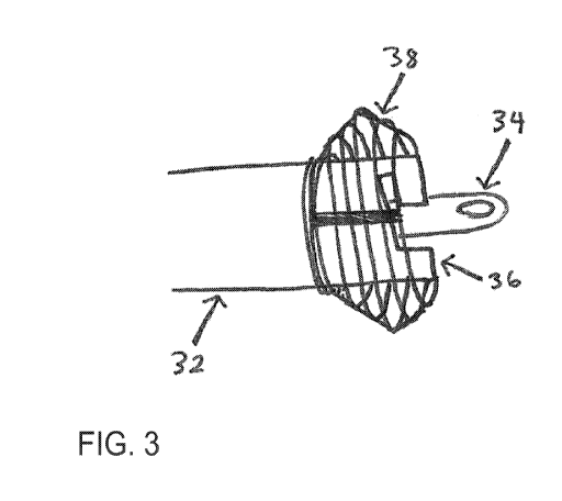
FIG. 3 depicts the exemplary delivery device of FIG. 2, with the expanding
members 38
expanded. In the embodiment shown, the expanding members, when expanded,
generally form
an eggbeater shape. In various embodiments, the expanding members 38 may
generally form
triangular, ovular, and multiangular shapes. In particular embodiments, an
adhering element
may be constructed from various metal alloys or adhesives to keep the core
cannula expanding
members 38 from expanding horizontally
The expanding members 38 may be configured to expand under a number of
suitable
conditions and/or via a number of suitable mechanisms. According to particular
embodiments,
the expanding members 38 expand once exposed inside the body and will
compressed upon
being retrieved through an access sheath (e.g., the expanding members are
compressed at a first
diameter by the diameter of the access sheath and expand upon exiting the
access sheath to a
predetermined second diameter. In various embodiments, the expanding members
38 expand
through a method (e.g., trigger, sensor, feedback system etc.) on the user end
of an exemplary
delivery system as shown in FIG. 4, extending down the core cannula 32, or
remotely.
7
CA 02999169 2018-03-19
WO 2016/044740 PCT/US2015/050967
Turning now to FIG. 4, the embodiment shown depicts a proximal or user end of
an
exemplary delivery system. The embodiment shown includes 1) an injection
apparatus handle
46, 2) a device deployment handle 44, 3) a core cannula 32, 4) a hemostasis
valve 48, 5) an
injection apparatus 34, 6) a delivery catheter 42, and 7) an access sheath 40.
In some
embodiments, the hemostasis valve 48 is located at the proximal end of the
hollow access sheath
40 to prevent backflow of fluid and air penetration into the system. In
various embodiments, the
hemostasis valve 48 can be constructed from materials that include but are not
limited to
polycarbonate, polyethylene, silicone, polyurethane, PVC, EPDM, stainless
steel, combinations
of polymeric materials, or combinations of metal alloys. The hemostasis valve
48 may be
constructed through injection molding, dip molding, or various techniques
known to those skilled
in the art. In various embodiments, the hemostasis valve 48 may be attached to
the access sheath
40 via adhesives, welding, molding techniques, turning, riling operations,
drilling operations, or
bending operations.
In particular embodiments, the injection apparatus handle 46 is located at the
proximal
end of an injection apparatus 34 for maneuvering the length of the injection
apparatus 34 and any
injection fluids that may be passed through the injection apparatus 34. In
particular
embodiments, the delivery system may include multiple injection apparatuses
34. In these
embodiments, there may be one or more injection apparatus handles 46 attached
to one or more
corresponding injection apparatus 34 through adhesives, welding, and/or
molding techniques.
In at least one embodiment, the delivery catheter member 42 travels through
the hollow
access sheath 40 and is attached to the core cannula 32, the device deployment
handle 44, or
body of the core cannula 32 through methods such as adhesives, welding, or
molding techniques.
In particular embodiments, the device deployment handle 44 controls the
detachment of an
implantable device operatively connected to the distal end of the core cannula
32 (not shown in
FIG. 4). In at least one embodiment, the device deployment handle 44 is
located at the proximal
end of the core cannula 32 and is adhered to the core cannula 32 via an
adhesive, by welding,
and/or through molding techniques. In some embodiments, the device deployment
handle 44
and an injection apparatus handle 46 may be constructed from materials that
include but are not
limited to Pebax, polyurethane, Nylon, silicone, polycarbonate, polyethylene,
PVC, EPDM,
stainless steel, combinations of polymeric materials, or combinations of metal
alloys. In further
8
CA 02999169 2018-03-19
WO 2016/044740 PCT/US2015/050967
embodiments, the device deployment handle 44 and an injection apparatus handle
46 may be
integrally formed by molding, casting, 3D printing, or other suitable method.
FIG. 5 depicts an exemplary device locking member (e.g., device locking member
36).
In the embodiment shown, the device locking member 36 is a twist locking
mechanism with an
"L" shaped channel that allows the attachment and detachment of core cannula
32 to an
exemplary implantable device as shown in FIG. 6. The function of an exemplary
device locking
member 36 is controlled by methods such as a trigger, sensor, switch, etc. on
a user (proximal)
end of an exemplary delivery system. In various embodiments, the device
locking member 36
may be in the form of but not limited to a threaded mechanism (e.g., external,
internal), leur lock,
magnet, etc. The device locking member 36 is found at the distal end of core
cannula 32 and may
be attached to the core cannula 32 through a fastening method (e.g.,
adhesives, screws, welding,
etc.) or may be an extension of the core cannula 32.
FIG. 6 depicts an exemplary locking member of an exemplary implantable device
as
discussed herein. Details of exemplary implantable devices are further
discussed in PCT Appin.
No. PCT/US15/27666, entitled "LEFT ATRIAL APPENDAGE OCCLUSION DEVICE",
filed on April 24, 2015, incorporated herein by reference in its entirety. The
embodiment shown
in FIG. 6 includes a cross-shaped locking mechanism that may, for example, be
inserted into the
locking member of the exemplary delivery device in FIG. 5 and twisted to
"lock" the implantable
device to the core cannula for delivery to a particular portion of a patient
(e.g., the patient's left
atrial appendage).
FIGS. 7-10 show various cross sections of an access sheath, catheter, and/or
delivery
device attached to an exemplary implantable device as described herein. In the
embodiments
shown, the delivery device includes a core cannula, an injection apparatus
extending through the
core cannula and into an implantable device, and a device locking member
operatively connected
to the implantable device.
FIG. 7 shows a cross section of the exemplary delivery device attached to the
exemplary
implantable device. FIG. 8 shows a cross section of the access sheath and/or
catheter and the
delivery device and implantable device within. FIG. 9 shows a cross section of
the access sheath
and/or catheter and the exterior of the core cannula and implantable device
(attached via the
exemplary locking member). FIG. 10 shows a cross section of an exemplary
access
9
CA 02999169 2018-03-19
WO 2016/044740 PCT/US2015/050967
sheath/catheter and an exterior of the core cannula and implantable device,
with expandable
members attached. In the embodiment shown, the expandable members are
compressed in a first
position within a catheter and/or access sheath prior to being inserted into a
body, where they
may be expanded upon exiting the catheter and/or access sheath.
The exemplary delivery system shown in FIGS. 7-10 will be navigated from a
user end
access point through the access sheath/catheter to a destination to deploy an
exemplary
implantable device. The distance tolerance between an exemplary delivery
system and access
sheath will be such (e.g., 0.3333 mm, 0.6666 mm, 1 mm, etc.) to allow smooth
navigation of an
exemplary delivery system. An exemplary delivery system can be navigated
within an access
sheath/catheter through bodily fluids such as blood or other fluids such as
saline, sterile water,
etc. or a combination of the aforementioned fluids.
Exemplary Device Use Case
FIGS. 11-13 depict an exemplary delivery system use case. In particular, FIGS.
11-13
depict an exemplary delivery system used for inserting an attached implantable
device into a left
atrial appendage (LAA) within a patient's heart. Exemplary delivery systems
described herein
may be used to deliver an implantable device to occlude the LAA to, for
example, help prevent a
stroke in the patient. Due to a high prevalence of thrombus formation in the
LAA of atrial
fibrillation (AF) patients, occluding the LAA may prevent a majority of
thrombus formation and
thus reduces the risk of ischemic stroke.
Delivering a left atrial appendage closure (LAAC) for a particular patient is
briefly
described, as shown in FIGS. 11-13. Delivery device component numbers used in
FIGS. 1-4 will
be used throughout this description. This exemplary procedure is included to
further promote an
understanding of the exemplary devices and processes disclosed herein and is
not necessarily
intended to be limiting. The exemplary LAAC procedure for the particular
patient may be done
under local or general anesthesia in a catheterization lab using standard
transseptal techniques.
The exemplary procedure may last about one hour and the patient will stay
overnight at a
hospital post implantation in order to monitor any adverse effects.
In particular embodiments, imaging techniques, such as transesophageal
echocardiography (TEE), will be performed to measure the LAA to determine
device size. The
procedure is then performed under appropriate imaging, which may include
fluoroscopic,
CA 02999169 2018-03-19
WO 2016/044740 PCT/US2015/050967
intracardiac echocardiographic (ICE), or TEE guidance. A transseptal needle
attached to a
guidewire (not shown) will then puncture the intraatrial septum, allowing for
the transseptal
access sheath 40 to advance over the transseptal needle and through the
puncture within the
intraatrial septum into the left atrium. The transseptal needle and the
guidewire are then
removed from the system. An adhesive or locking mechanism (not shown) will be
attached to the
transseptal access sheath 40 to prevent movement of the sheath 40. The
transseptal access sheath
40 contains a hemostasis valve 48 to prevent backflow of bodily fluid. A
delivery catheter
embodiment 42 is then advanced through the access sheath 40 to aid in
directing the implantable
device. A core cannula 32 with a proximal or user and distal or device end can
then be advanced
through the access sheath 40, where the distal end of the core cannula 32
includes a portion to
aid in eventual placement of the implantable device. The core cannula 32 is
directed through the
body to the targeted body cavity, for example, the left atrial appendage,
under fluoroscopic, ICE,
or TEE guidance. Once the distal portion of the core cannula 32 exits the
access sheath 40,
expandable members 38 attached to the distal portion of the core cannula 32
will move from a
first position (within the access sheath) to a second, expanded position to
allow the implant
device to expand in a position inside a body cavity, while keeping the device
flush or in the same
plane with nearby tissue.
As discussed herein, the expandable members 38 can be constructed as a wire
from a
self-expanding, super elastic element, such as Nitinol, etc. and can be coated
or enclosed by
chemical or physical means, such as a polymeric, elastomeric material, etc. to
prevent harm
towards neighboring bodily structures. In one embodiment, once the expandable
members 38
have exited the access sheath 40, the expandable members 38 will expand to a
pre-determined
shape, such as a round, ovular, cross, diamond, spring etc. formation. The
expandable members
38 along with the core cannula 32 are advanced towards the opening of the body
cavity until the
expandable members 38 reach the opening and engage with the surrounding
tissue, preventing
the core cannula 32 from advancing further. The expanding members 38 may be
constructed to
take into account varying sizes of the body cavity and can be sized
incrementally (e.g., gradient
of 1 mm, 1.5 mm, 2 mm, etc.) in the form of a spring basket (e.g., as shown in
FIGS. 11 and 12).
The proximal end of the expandable members 38 can be relatively larger
compared the to the
distal portion in order to account for larger LAA orifice diameters. The most
distal portion of the
11
CA 02999169 2018-03-19
WO 2016/044740 PCT/US2015/050967
expandable members 38 may be incrementally (e.g., gradient of 1 mm, 1.5 mm, 2
mm, etc.)
tapered or veer slightly inwards to account for smaller LAA orifice diameters
and to avoid
penetrating surround bodily structures. The position of the core cannula 32
can then be locked at
the proximal end that may include an adhesive, twisting method, or another
locking mechanism
(not shown). As will be understood by one of ordinary skill in the art,
compressed and expanded
diameters of the expandable members may vary based on application. Such as,
for example,
should a patient have a smaller LAA or the surrounding bodily cavity be
smaller than average,
expandable members that have a smaller than average expanded diameter may be
used to prevent
damage to surrounding tissue.
According to particular embodiments, once the expandable members 38, core
cannula 32,
and implantable device are in place, an injection apparatus 34 with a proximal
and distal end is
inserted through the core cannula 32 and travels to the body cavity (as will
be understood by one
of ordinary skill in the art, an injection apparatus 34, in various
embodiments, may be pre-
inserted in the core cannula 32 and the implantable device). In various
embodiments, the
proximal end of an injection apparatus 34 includes an injection apparatus
handle 46 to
manipulate the length and position of an injection apparatus 34 as well as to
reduce the risk of
advancing an injection apparatus 34 through the entirety of the body cavity
during the pre-
insertion period. In particular embodiments, the distal end of the core
cannula 32 is pre-attached
to the implantable device through the device locking member 36, which may
include a twisting
system, luer lock system, etc. In one or more embodiments, the distal end of
an injection
apparatus 34 is pre-inserted or moved inside the implantable device to allow
for fluid ejection.
In particular embodiments, a fluid is injected into an injection apparatus 34
through a syringe
(not shown) until the device has successfully occluded the body cavity (as
discussed in
applications incorporated by reference herein). In at least one embodiment,
upon assessment of
successful occlusion through imaging and final contrast injections, an
injection apparatus 34 is
removed, and the core cannula is detached from the implantable device via the
device locking
member 36 via a twisting method, at the device deployment handle 44 found on
the proximal end
of the core cannula 32. In one embodiment, the device deployment handle 44
further includes
one or more injection apparatus handles 46 that are operatively connected to
an injection
apparatus 34. In particular embodiments, the distal end of the core cannula 32
detaches from the
12
CA 02999169 2018-03-19
WO 2016/044740 PCT/US2015/050967
implantable device and is removed from the patient along with the access
sheath 40. Following
the retrieval of the core cannula 32, attached expandable members 38 are
compressed into the
access sheath 40.
Alternate Embodiments
In a first alternate embodiment, an exemplary device may include expandable
members
of varying locations on the delivery system. In this first alternate
embodiment, a shape memory
structure (such as nitinol or PEEK) can be attached at a proximal end, a
distal end, or other
locations along the length of the exemplary delivery device. Continuing with
this embodiment,
the length of the shape memory structure may range from a portion to the
entire length of the
delivery system. In this alternate embodiment, the shape memory material may
be fabricated
through braiding, laser cutting, or wiring to allow for differentiating
morphologies. As will be
understood by one of ordinary skill in the art, heat treatments may also be
applied to the shape
memory material to yield independent shapes.
Various embodiments of the device herein are depicted as multiple cannulae
with
eggbeater-shaped expandable members, but the expandable members may be in
suitable alternate
shapes, including cylindrical, ovular, cross-shaped, multiangular, or coil-
shaped. Further, in
particular embodiments, the expandable members may be attached to each other
with adhesives
or shape memory material. In some embodiments, the expansion of the expanding
members
could be mechanically manipulated through attached wiring, threading, or a
balloon that can
expand via user control.
As shown in FIG. 14, in some embodiments, an exemplary device may include
features
to further prevent thrombus migration from the LAA. In such embodiments (and
others), the
expandable members may be attached to an occluding fabric made of PTFE, PET,
or various
woven fabrics.
In particular embodiments, the delivery system may have multiple steering
stages,
wherein each stage can move in multiple planes. The steering stages may
integrate multiple
shape memory wires that are heat treated to independently control the wire
configurations and
the consequent steering stages. The steering capability can be controlled by
methods (e.g.,
trigger, switch, etc.) found on the user end of an exemplary delivery system.
This delivery
system with multiple steering stages could be implemented for various
scenarios, including
13
CA 02999169 2018-03-19
WO 2016/044740 PCT/US2015/050967
delivering implantable technologies, angioplasty, valve repair, and drug
therapy. In alternate
embodiments, the point of attachment between the delivery system and device
can vary.
Attachment systems include luer-lock systems, anti-rotation systems,
microscrew mechanisms.
In other embodiments, the location of the attached device to the delivery
system may vary,
including being within the expanding members or located proximally or distally
in relation to the
expanding members. In further alternate embodiments, the delivery system may
not be
detachable from a connected device. In scenarios, such as angioplasty, a
balloon or attached
device may be attached to the delivery system through adhesives, molding, or
welding.
In various embodiments, the distal end of the delivery system may contain a
sensor unit.
In such embodiments, the sensor unit's purpose may include but not limited to
detection of
thrombus, physiological function, sterility, or microbial activity.
In some embodiments, a coating may be applied to the delivery system. The
purpose of
the coating may include but not limited to lubricity, microbial stability,
fluid absorption, and
encrustation reduction. In alternate embodiments, the material of the delivery
system may be
induced with elements, such as tungsten or silver, to increase levels of
radiopacity, microbial
stability, allergy reduction, durability, or sterility.
In particular embodiments, the various tube-like components (e.g., core
cannula, access
sheath, etc.) of the delivery system may be mechanically or chemically
enhanced to increase
flexibility or durability. In a particular embodiment, a coil could be
integrated within the core
cannula through molding or welding techniques for increased functionality.
Continuing with this
embodiment, the tightness or pitch of the wound coil along the length of the
core cannula may
vary. In an alternative embodiment, the coil could also be braided.
In some embodiments, more than one injection apparatus may be inserted into
the
delivery system. This may be applicable in scenarios requiring delivery of
multiple injectable
fluids or fluids that require at least a two-part mixture. Examples of such
injectable materials
include hydrogels, occlusion gels, etc.
In various embodiments, the device attached to the delivery system may be a
balloon. In
alternate embodiments, a device with expandable members (e.g. nitinol) may be
delivered
through the delivery system. Alternatively, in particular embodiments, a
device with woven fiber
(e.g. PET, ePTFE, Dacron) may be deployed through the delivery system.
14
CA 02999169 2018-03-19
WO 2016/044740 PCT/US2015/050967
The delivery system may be used for identification of general soft tissue
concavities or
areas in need of occlusion. In various embodiments, the expandable elements of
the delivery
system allow for precise deployment of an attached device. In some
embodiments, these
characteristics may be appropriate for closure of atrial septal defects (ASDs)
and patent foramen
ovales (PF0s), which are defects found in the atrial septal wall.
Alternatively, the core cannula
elements along with the functionality of the delivery system could be used for
drug delivery to a
blood vessel during angioplasty. In particular embodiments, the delivery
system may be intended
for balloon valvuloplasty to mechanically force the opening of a narrowed
heart valve. In further
embodiments, a drug eluting element may be added to reduce further
calcification.
Conclusion
Accordingly, the reader will see that the aforementioned delivery system can
be used to
easily deploy an implantable, expandable device into a body cavity, such as a
LAA, easy-to-use
system to deploy the device, and allows the device to be flush against the
surrounding tissue.
While the above description contains many specificities, these should not be
construed as
limitations on the scope of any embodiment, but as exemplifications of various
embodiments
thereof Many other ramifications and variations are possible within the
teachings of the various
embodiments. For example, the attachment embodiments may differ compared to
the drawings,
the delivery system may alter in shape, size, and multiple similar devices
could be used for other
applications, etc.
Thus the scope should be determined by the appended claims and their legal
equivalents,
and not by the examples given.