Note: Descriptions are shown in the official language in which they were submitted.
PCT APPLICATION
AFFINITY-OLIGONUCLEOTIDE CONJUGATES AND USES THEREOF
[0001]
BACKGROUND
[0002] Many cell types can be identified and categorized by the abundance of
specific sets of proteins
endogenously expressed and located on their plasma membranes. This phenomenon
enables the study of cells
using a process known as immunophenotyping, in which cells are incubated with
and bound by fluorescently-
labeled antibodies that are specific to known surface proteins of the cells.
Flow cytometry is commonly used
to measure the levels of the surface-bound antibodies for each cell. However,
flow cytometry-based
approaches are limited by the number of fluorophores that can be used
concurrently in the same experiment.
Further, the number of fluorophores that can be used concurrently in the same
experiment using flow
cytometry-based approaches is limited by spectral overlap. Additionally, flow
cytometry is not amenable to
many biologically-relevant assays and subsequent DNA sequencing.
SUMMARY
[0003] Thus, a need exists for methods of characterizing, e.g.,
immunophenotyping, single cells without these
limitations. Unlike flow cytometry-based approaches, the methods described
herein use a sequence readout to
analyze proteins of individual cells and are not limited by the number of
fluorophores that can be used
concurrently in the same experiment or their spectral overlap. Further, the
methods described herein are
amenable to many biologically-relevant assays and subsequent DNA sequencing.
The methods described
herein utilize affinity-oligonucleotide conjugates (e.g., antibody-
oligonucleotide conjugates). The
oligonucleotide of the conjugate comprises an Antigen ID (AID) sequence that
is barcoded to a surface
antigen that the affinity portion of the affinity-oligonucleotide conjugate
specifically binds. Thus, using the
methods described herein, an antigen (e.g., a surface protein) of a single
cell can be analyzed without a need
for fluorophores. For example, a surface protein of a single cell that is
displayed can be identified from the
Antigen ID sequence. One or more of the surface proteins of a single cell can
be used to define the single
cell's identity, characteristics or relevance.
[0004] The affinity-oligonucleotide conjugate of the methods described herein
that can be used to overcome
the problems of slow cell sorting, reduced target yield associated with cell
sorting, limited number of output
streams, and selected bins that do not correspond to a quantified property of
the affinity-oligonucleotide
conjugate, such as affinity. The exemplary affinity-oligonucleotide conjugate
depicted can replace or enhance
sorting with single-cell measurements of tetramer binding in vessels. The
exemplary affinity-oligonucleotide
-1-
Date recue/Date received 2023-02-17
CA 02999888 2018-03-23
WO 2017/053905
PCT/US2016/053598
conjugate depicted can be used in the methods described herein to
simultaneously acquire TCR pair
sequences, clone abundance, and relative tetramer affinities.
[0005] A method of characterizing, e.g., immunophenotyping, cells in vessels
(e.g., emulsion) with affinity-
oligonucleotide conjugates is described herein. In some embodiments, the
method is used to identify cell
subsets in a manner compatible with emulsion-based single cell analysis. In
some embodiments, the method is
used to identify immune cells specific for an antigen in a manner compatible
with emulsion-based single cell
analysis. In some embodiments, prior to cellular analysis, surface protein-
specific antibodies are conjugated to
oligonucleotides. In some embodiments, the oligonucleotides are designed to
contain a sequence motif which
is unique to the target-specificity of the conjugated antibody. The
oligonucleotide can be conjugated to the
affinity portion of the affinity-oligonucleotide conjugate (e.g., an antibody)
covalently or non-covalently (e.g.,
biotin-oligonucleotide to streptavidin-antibody).
[0006] A method can comprise incubating cells in a mixture or a solution with
one or more affinity-
oligonucleotide conjugates. The cells can be washed to remove unbound affinity-
oligonucleotide conjugates.
Cells are then encapsulated in vessels, e.g., an emulsion. The cells can be
present in the vessels at a single cell
per vessel density. Thus, the affinity-oligonucleotide conjugates within a
vessel, e.g., droplet, are bound to the
cell surface, e.g., through a specific antibody-surface protein interaction.
The method can comprise attaching a
vessel-specific DNA sequence (e.g., a unique vessel barcode) to the affinity-
conjugated oligonucleotides.
Additional cellular DNA or mRNA analysis, phenotypic measurements, functional
testing, cell-sorting or
other reactions can be carried out prior to, concurrently with, or after
barcoding the affinity-conjugated
oligonucleotide, (e.g., with a DNA barcode).
[0007] A method can comprise extracting nucleic acids from the emulsion, for
example, subsequent to the
emulsion experimentation. Extracted nucleic acids can be prepared for
sequencing and sequenced (e.g., using
next generation sequencing technology). A method can comprise sequencing
polynucleotide molecules from
the vessels that contain both an Antigen ID sequence and droplet-specific
barcode sequence. The Antigen ID
sequence can define the specific cell surface protein bound by the
oligonucleotide-conjugated antibody. The
Antigen ID sequence can define the specific antibody of the oligonucleotide-
conjugated antibody that binds to
a particular cell surface protein. Thus, the Antigen ID sequence can indicate
which surface protein the
analyzed cell expressed. In a vessel harboring a single cell, all sequences
containing a shared droplet-specific
barcode sequence are associated with a single cell. Therefore, a single cell
can be analyzed as displaying a set
of surface proteins which can be used to define its identity, characteristics
or relevance.
[0008] In one aspect, a method is provided comprising performing a reaction in
a plurality of vessels, the
reaction comprising attaching a vessel barcoded polynucleotide comprising a
vessel barcode sequence to an
oligonucleotide of an affinity-oligonucleotide conjugate bound to a target
antigen of a single cell isolated in a
vessel of a plurality of vessels.
[0009] In one aspect, provided herein is a method comprising, performing a
reaction in a vessel of a plurality
of vessels, the reaction comprising attaching a vessel barcoded
polynucleotide, which comprises a vessel
-2-
CA 02999888 2018-03-23
WO 2017/053905
PCT/US2016/053598
barcode sequence, to an oligonucleotide portion of an affinity-oligonucleotide
conjugate, wherein the affinity-
oligonucleotide conjugate binds to a target antigen expressed by a cell in the
vessel of the plurality of vessels.
100101 In some embodiments, the cell is a single cell contained within the
vessel. In some embodiments, the
vessel comprises two or more vessels of the plurality of vessels. In some
embodiments, the vessel comprises
each vessel of the plurality of vessels. In some embodiments, the reaction
takes place in two or more vessels
of the plurality of vessesl. In some embodiments, the cell in each vessel is
from a same sample. In some
embodiments, the cell in a vessel of a first plurality of vessels of the two
or more pluralities of vessels is from
a same sample as the cell in a vessel in a second plurality of vessels of the
two or more pluralities of vessels.
In some embodiments, the oligonucleotide portion comprises an antigen
identification sequence (AID). In
some embodiments, the AID is barcoded to the target antigen or the affinity
portion of the affinity-
oligonucleotide conjugate.
[0011] In some embodiments, the oligonucleotide further comprises an antigen
identification sequence (AID)
barcoded to the target antigen or the affinity portion of the affinity-
oligonucleotide conjugate. In some
embodiments, the antigen identification sequence (AID) is a known sequence.
[0012] In some embodiments, the vessel barcoded polynucleotide is from a
template vessel barcoded
polynucleotide in the vessel.
[0013] In some embodiments, the method further comprises sequencing the
oligonucleotide or an amplicon
thereof to obtain sequence information.
[0014] In some embodiments, the method further comprises determining a
characteristic of the single cell
based on the sequence information. In some embodiments, the sequence
information comprises the antigen
identification (AID) sequence. In some embodiments, the method further
comprises determining a
characteristic of the single cell based on the sequence information. In some
embodiments, the characteristic is
a phenotype. In some embodiments, the phenotype is an immunophenotype.
[0015] In some embodiments, the method further comprises contacting the
affinity-oligonucleotide conjugate
to a plurality of cells comprising the single cell. In some embodiments, the
contacting is before the single cell
is isolated in the vessel. In some embodiments, the method further comprises
washing the plurality of cells
after the contacting.
[0016] In some embodiments, the vessel does not comprise an affinity-
oligonucleotide conjugate that is not
bound to a target antigen.
[0017] In some embodiments, the method further comprises isolating the single
cell in the vessel. In some
embodiments, the single cell is bound to the affinity-oligonucleotide
conjugate before the isolating.
[0018] In some embodiments, the method further comprises lysing the single
cell. In some embodiments, the
lysing is after the single cell is isolated in the vessel.
[0019] In some embodiments, the plurality of cells is a plurality of unsorted
cells.
[0020] In some embodiments, the vessel barcode sequence of a vessel barcoded
polynucleotide or amplicon
thereof in a first vessel of the plurality of vessels is a different than the
vessel barcode sequence of a vessel
barcoded polynucleotide or amplicon thereof in a second vessel of the
plurality of vessels. In some
-3-
CA 02999888 2018-03-23
WO 2017/053905
PCT/US2016/053598
embodiments, the vessel barcode sequence of each vessel barcoded
polynucleotide or amplicon thereof in a
single vessel of the plurality of vessels comprises a same vessel barcode
sequence. In some embodiments, the
vessel barcode sequence of each vessel barcoded polynucleotide and amplicon
thereof in any single vessel of
the plurality of vessels is unique to the vessel barcode sequence of each
vessel barcoded polynucleotide and
amplicon thereof in any other single vessel of the plurality of vessels.
[0021] In some embodiments, the method further comprises attaching a vessel
barcoded polynucleotide to a
cell polynucleotide from the single cell. In some embodiments, the attaching a
vessel barcoded polynucleotide
to an oligonucleotide of an affinity-oligonucleotide conjugate and the
attaching a vessel barcoded
polynucleotide to a cell polynucleotide from the single cell are performed
simultaneously.
[0022] In some embodiments, the method further comprises amplifying the
oligonucleotide or a complement
thereof. In some embodiments, the method further comprises amplifying the cell
polynucleotide or a
complement thereof. In some embodiments, the amplifying the oligonucleotide or
a complement thereof and
the amplifying the cell polynucleotide or a complement thereof are performed
simultaneously.
[0023] In some embodiments, the vessel barcode sequence of the cell
polynucleotide and the vessel barcode
sequence of the oligonucleotide are the same.
[0024] In some embodiments, the method further comprises pooling
oligonucleotides or amplicons thereof
from two or more vessels of the plurality of vessels. In some embodiments, the
method further comprises
pooling oligonucleotides or amplicons thereof and cell polynucleotides or
amplicons thereof from two or more
vessels of the plurality of vessels. In some embodiments, the pooling is
before sequencing.
[0025] In some embodiments, the affinity-oligonucleotide conjugate comprises a
plurality of different
affinity-oligonucleotide conjugates. In some embodiments, each affinity-
oligonucleotide conjugate of the
plurality of affinity-oligonucleotide conjugates comprises a unique antigen
identification (AID) sequence. In
some embodiments, the oligonucleotide comprises an affinity molecular barcode
(AMB) sequence that is
barcoded to a single affinity-oligonucleotide conjugate molecule of a
plurality of affinity-oligonucleotide
conjugate molecules. In some embodiments, each affinity-oligonucleotide
conjugate molecule of the plurality
of affinity-oligonucleotide conjugate molecules comprises a unique affinity
molecular barcode (AMB)
sequence.
[0026] In some embodiments, the oligonucleotide comprises a fusion sequence
and the attaching comprises
attaching the vessel barcoded polynucleotide to the fusion sequence. In some
embodiments, the
oligonucleotide comprises a primer binding sequence. In some embodiments, the
oligonucleotide comprises a
constant sequence.
[0027] In some embodiments, the method further comprises sequencing the
oligonucleotide, complements
thereof, amplified products thereof, or a combination thereof, thereby
producing oligonucleotide sequence
reads. In some embodiments, the method further comprises comparing one or more
first oligonucleotide
sequence reads to one or more second oligonucleotide sequence reads. In some
embodiments, the method
further comprises analyzing the oligonucleotide sequence reads. In some
embodiments, the method further
comprises analyzing vessel barcode sequences of the oligonucleotide sequence
reads. In some embodiments,
-4-
CA 02999888 2018-03-23
WO 2017/053905
PCT/US2016/053598
the method further comprises analyzing antigen identification (AID) sequences
of the oligonucleotide
sequence reads. In some embodiments, the method further comprises analyzing
affinity molecular barcode
(AMB) sequences of the oligonucleotide sequence reads. In some embodiments,
the analyzing comprises
determining a frequency of one or more vessel barcode sequences, one or more
AID sequences, one or more
affinity molecular barcode (AMB) sequences, or a combination thereof. In some
embodiments, the analyzing
comprises comparing. In some embodiments, the method further comprises
comparing antigen identification
(AID) sequences of oligonucleotide sequence reads to affinity molecular
barcode (AMB) sequences of
oligonucleotide sequence reads.
[0028] In some embodiments, the method further comprises sequencing the cell
polynucleotide, complements
thereof, amplified products thereof, or a combination thereof, thereby
producing cell polynucleotide sequence
reads. In some embodiments, wherein the method further comprises comparing
oligonucleotide sequence
reads to the cell polynucleotide sequence reads. In some embodiments, the
method further comprises
comparing vessel barcode sequences of oligonucleotide sequence reads to vessel
barcode sequences of the cell
polynucleotide sequence reads. In some embodiments, the method further
comprises comparing the cell
polynucleotide sequence reads. In some embodiments, the method further
comprises analyzing vessel barcode
sequences of the cell polynucleotide sequence reads. In some embodiments, the
method further comprises
analyzing molecular barcode sequences of the cell polynucleotide sequence
reads.
[0029] In some embodiments, the method further comprises determining a
characteristic of a cell based on
the analyzing or the comparing. In some embodiments, the method further
comprises selecting an antibody or
TCR based on the oligonucleotide sequence reads. In some embodiments, the
method comprises selecting an
antibody or TCR based on the cell polynucleotide sequence reads.
[0030] In some embodiments, the vessel barcoded polynucleotide attached to the
oligonucleotide and the
vessel barcoded polynucleotide attached to the cell polynucleotide are from a
same template vessel barcoded
polynucleotide in the vessel. In some embodiments, the vessel barcoded
polynucleotide attached to the
oligonucleotide is an amplification product of a template vessel barcoded
polynucleotide.
[0031] In some embodiments, the vessel barcoded polynucleotide attached to the
cell polynucleotide is an
amplification product of the template vessel barcoded polynucleotide.
[0032] In some embodiments, the vessel comprises a solid support. In some
embodiments, the vessel does
not comprise a solid support. In some embodiments, each vessel of the
plurality of vessels comprises a single
cell. In some embodiments, the vessel is a well, an emulsion, or a droplet. In
some embodiments, the template
vessel barcoded polynucleotide is not bound to a solid support. In some
embodiments, the template vessel
barcoded polynucleotide is bound to a solid support.
[0033] In some embodiments, the method further comprises attaching a molecular
barcode sequence of a
molecular barcoded polynucleotide of a plurality of molecular barcoded
polynucleotides to the cell
polynucleotide, wherein the molecular barcode sequence is barcoded to a single
cell polynucleotide molecule
and amplicons thereof.
-5-
CA 02999888 2018-03-23
WO 2017/053905
PCT/US2016/053598
[0034] In some embodiments, the attaching comprises ligating the vessel
polynucleotide to the
oligonucleotide. In some embodiments, the attaching comprises attaching the
vessel polynucleotide to the
oligonucleotide with an enzyme. In some embodiments, the attaching comprises
hybridizing the vessel
polynucleotide to the oligonucleotide. In some embodiments, the attaching
further comprises extending the
oligonucleotide. In some embodiments, the attaching comprises amplifying a
template vessel barcoded
polynucleotide.
[0035] In some embodiments, the oligonucleotide is double stranded. In some
embodiments, the
oligonucleotide is single stranded. In some embodiments, the oligonucleotide
is DNA. In some embodiments,
the oligonucleotide is RNA.
[0036] In some embodiments, the cell polynucleotide comprises a variable
region sequence. In some
embodiments, the method further comprises pairing native chain sequences
containing a variable region
sequence. In some embodiments, the cell polynucleotide is DNA. In some
embodiments, the cell
polynucleotide is RNA. In some embodiments, the RNA is mRNA.
[0037] In some embodiments, the single cell is a B-cell. In some embodiments,
the single cell is a T-cell.
[0038] In some embodiments, the affinity portion of the affinity-
oligonucleotide conjugate binds to an
extracellular antigen of the single cell. In some embodiments, the
extracellular antigen of the single cell is an
antigen specific to an immune cell. In some embodiments, the extracellular
antigen of the single cell is an
antigen specific to a T-cell. In some embodiments, the extracellular antigen
is CD4. In some embodiments, the
extracellular antigen is CD8. In some embodiments, the extracellular antigen
of the single cell is an antigen
specific to a B-cell. In some embodiments, the extracellular antigen is an
immunoglobulin.
[0039] In some embodiments, the affinity portion of the affinity
oligonucleotide conjugate is an antibody or
fragment thereof. In some embodiments, the affinity portion of the affinity
oligonucleotide conjugate is a
peptide. In some embodiments, the affinity portion of the affinity
oligonucleotide conjugate is a protein. In
some embodiments, the affinity portion of the affinity oligonucleotide
conjugate is an aptamer. In some
embodiments, the affinity portion of the affinity oligonucleotide conjugate is
a small molecule. In some
embodiments, the affinity portion of the affinity oligonucleotide conjugate is
a drug. In some embodiments,
the affinity portion of the affinity oligonucleotide conjugate is a cell. In
some embodiments, the cell is an
antigen presenting cell (APC). In some embodiments, the affinity portion of
the affinity oligonucleotide
conjugate comprises a major histocompatibility complex (MHC). In some
embodiments, the MHC is in a
soluble and/or multimeric (e.g., tetrameric) form. In some embodiments, the
MHC is bound to a peptide. In
some embodiments, the peptide is a synthetic peptide. In some embodiments, the
MHC binds to a T-cell
receptor (TCR) and/or a TCR-like binding molecule, such as a TCR-like antibody
or immunoglobulin or
chimeric antigen receptor, e.g., of the single cell.
[0040] In some embodiments, the affinity portion specifically binds to an
antigen-recognizing molecule
and/or immunoreceptor, such as an antibody or immunoglobulin or portion or
fusion thereof, an engineered
immunoreceptor, a chimeric antigen receptor (CAR), or a TCR. In some such
embodiments, the affinity
portion comprises an antigen or epitope or portion thereof recognized by the
antibody or receptor such as the
-6-
CA 02999888 2018-03-23
WO 2017/053905
PCT/US2016/053598
CAR. In some embodiments, the affinity portion comprises an antibody or
antigen-binding fragment thereof
that specifically binds to the immunoreceptor. In some aspects, the antibody
or antigen-binding fragment
thereof specifically binds to a variable and/or antigen-binding portion of the
receptor, such as an idiotope. In
some aspects, the affinity molecule is an anti-idiotypic antibody or fragment
thereof.
[0041] In some embodiments, the affinity portion of the affinity
oligonucleotide conjugate comprises a major
histocompatibility complex (MHC) or functional or binding portion thereof. In
some embodiments, the
affinity portion comprises a multimer of the MHC, optionally a tetramer of the
MHC. In some embodiments,
the MHC is in a soluble form. In some embodiments, the MHC is bound to a
peptide and/or contains a peptide
within a groove of the MHC. In some embodiments, the peptide is a synthetic
peptide. In some embodiments,
the MHC binds to a T-cell receptor (TCR) of the single cell. In some
embodiments, the affinity portion
comprises a peptide that binds to an antibody or a chimeric antigen receptor
(CAR) and/or wherein the target
is an antibody or a CAR. In some embodiments, the affinity portion is or
comprises an antigen or an epitope
specifically recognized by the antibody or the chimeric antigen receptor
and/or comprises an antibody that
specifically binds thereto, optionally an anti-idiotypic antibody that
specifically binds to an antigen binding
portion thereof.
[0042] In one aspect, provided is a composition comprising a plurality of
vessels each comprising a single
cell from a sample comprising a plurality of cells, an affinity-
oligonucleotide conjugate bound to a target
antigen of the single cell, and a vessel barcoded polynucleotide comprising a
vessel barcode sequence. In
some embodiments, the vessel barcoded polynucleotide or a complement thereof
is attached to the
oligonucleotide of the affinity-oligonucleotide conjugate.
[0043] In one aspect, provided is a composition comprising a plurality of
vessels each comprising a single
lysed cell from a sample comprising a plurality of cells, and an affinity-
oligonucleotide conjugate bound to a
target antigen of the single lysed cell; wherein the oligonucleotide of the
affinity-oligonucleotide conjugate
comprises a vessel barcode sequence, and wherein a cell polynucleotide from
the single lysed cell comprises
the same vessel barcode sequence.
[0044] In one aspect, provided herein is a composition comprising a plurality
of vessels, wherein a vessel of
the plurality of vessels comprises a single cell from a sample comprising a
plurality of cells, and a vessel
barcoded polynucleotide comprising a vessel barcode sequence wherein the
vessel further comprises an
affinity-oligonucleotide conjugate that binds to a target antigen of the
single cell, or an oligonucleotide portion
therefrom.
[0045] In some embodiments, a reaction takes place in two or more vessels of
the plurality of vessels. In
some embodiments, the vessel comprises each vessel of the plurality of
vessels. In some embodiments, the
plurality of vessels comprises two or more pluralities of vessels. In some
embodiments, the cell in each vessel
is from a same sample. In some embodiments, the cell in a vessel of a first
plurality of vessels of the two or
more pluralities of vessels is from a same sample as the cell in a vessel in a
second plurality of vessels of the
two or more pluralities of vessels. In some embodiments, the vessel barcoded
polynucleotide or a complement
-7-
CA 02999888 2018-03-23
WO 2017/053905
PCT/US2016/053598
thereof is attached to the oligonucleotide of the affinity-oligonucleotide
conjugate. In some embodiments, the
single cell is lysed.
100461 In one aspect, provided herein is a composition comprising a plurality
of vessels, wherein a vessel of
the plurality of vessels comprises a single lysed cell from a sample
comprising a plurality of cells, and an
affinity-oligonucleotide conjugate comprising an affinity portion that binds
to a target antigen of the single
lysed cell, or an oligonucleotide portion of the affinity-oligonucleotide
conjugate; wherein the oligonucleotide
portion of the affinity-oligonucleotide conjugate comprises a vessel barcode
sequence, and wherein a cell
polynucleotide from the single lysed cell comprises the same vessel barcode
sequence.
100471 In one aspect, provided is a kit, comprising: a first container
comprising a first oligonucleotide
comprising a first antigen identification (AID) sequence, wherein the first
AID sequence is a known sequence;
a second container comprising a second oligonucleotide comprising a second
antigen identification (AID)
sequence, wherein the second AID sequence is a known sequence and is different
than the first AID sequence;
one or more third containers comprising reagents capable of conjugating the
first oligonucleotide to a first
affinity molecule and reagents capable of conjugating the second
oligonucleotide to a second affinity
molecule; a set of instructions describing how to conjugate the first
oligonucleotide to the first affinity
molecule and the second oligonucleotide to the second affinity molecule.
100481 In one aspect, provided is a kit, comprising: a first container
comprising an oligonucleotide
comprising an antigen identification (AID) sequence, wherein the AID sequence
is a known sequence; a
second container comprising reagents capable of conjugating the
oligonucleotide to an affinity molecule; a
third container comprising a plurality of vessel barcoded polynucleotides; and
a set of instructions describing
how to attach a vessel barcoded polynucleotide of the plurality of vessel
barcoded polynucleotides to the
oligonucleotide when conjugated to the affinity molecule.
100491 The methods and compositions disclosed herein can be used for tumor
profiling. For example, the
methods can comprise linking cell phenotypes with an immune repertoire in
patient samples to identify tumor
reactive TCRs. The methods and compositions disclosed herein can be used for
adoptive cell therapy. For
example, the methods can comprise genetic analysis of T cells without sorting.
For example, the methods can
comprise combining T cell clonal information (using TCR) with gene expression
patterns during product
manufacture and treatment. In some embodiments, the methods disclosed herein
may be used to track,
characterize, monitor, and/or assess adoptively transferred cells obtained
from a patient prior to, during the
course of, or after adoptive cell therapy. The methods and compositions
disclosed herein can be used to
identify TCRs against known targets. For example, the methods can comprise
identifying high affinity clones
that may respond highly to antigen, but proliferate poorly. The methods and
compositions disclosed herein can
be used for cell sample multiplexing. For example, an emulsion containing
pooled cell samples contacted to
one or more affinity-oligonucleotide conjugates can be used to identify
original cell samples while processing
multiple samples at the same time.
INCORPORATION BY REFERENCE
-8-
CA 02999888 2018-03-23
WO 2017/053905
PCT/US2016/053598
[0050] All publications, patents, and patent applications mentioned in this
specification are herein
incorporated by reference in their entirety for all purposes, to the same
extent as if each individual publication,
patent, or patent application was specifically and individually indicated to
be incorporated by reference.
BRIEF DESCRIPTION OF THE DRAWINGS
[0051] The novel features described herein are set forth with particularity in
the appended claims. A better
understanding of the features and advantages of the features described herein
will be obtained by reference to
the following detailed description that sets forth illustrative examples, in
which the principles of the features
described herein are utilized, and the accompanying drawings of which:
[0052] FIG. 1 depicts an exemplary schematic of a vessel of the methods
described herein.
[0053] FIG. 2 depicts an exemplary design of oligonucleotide tag conjugated to
an antibody. Each colored
block represents a portion of the complete oligonucleotide sequence. The
fusion sequence is used for
enzymatic attachment of a droplet-specific DNA barcode inside the emulsion
reaction. Only one possible
arrangement of the sequences is shown, although other arrangements are
compatible with the method
described.
[0054] FIG. 3A depicts an exemplary co-capture of immune receptor sequences
with additional mRNA and
protein targets. Surface protein targets are quantified by pre-incubating
cells with DNA-labeled staining
antibodies prior to emulsion sequencing.
[0055] FIG. 3B depicts an exemplary CD4 and CD8 mRNA and protein measurements
on 3,682 droplet
barcode TCR VaVr3 pairs generated from healthy human T-cells.
[0056] FIG. 3C depicts an exemplary concordance between mRNA and protein
measurements (each point is
a droplet barcode linked to a TCR VocVf3 pair).
[0057] FIG. 3D depicts an exemplary table of simultaneous mRNA and protein
detection of CD4 and CD8
from unsorted T-cells in emulsion. From 30,000 input T-cells, 3,682 TCR pairs
were recovered. Frequencies
of TCR pairs called as CD4 + or CD8 + by mRNA vs protein (based on molecular
counting, majority rule) are
shown in a matrix.
[0058] FIG. 3E depicts exemplary results from 45,870 single cell TCR pairs
using an affinity-
oligonucleotide conjugate targeting CD4 and an affinity-oligonucleotide
conjugate targeting CD8.
[0059] FIG. 4 depicts a schematic of an exemplary method using an affinity-
oligonucleotide conjugate
targeting CD4 and an affinity-oligonucleotide conjugate targeting CD8.
[0060] FIG. 5 exemplifies results from a method of single immune cell
barcoding in an emulsion.
[0061] FIG. 5A is an exemplary depiction of two aqueous streams containing
cells and lysisireaction (LR)
mix being passed into oil that produces monodisperse emulsion at over 8
million droplets per hour.
[0062] FIG. 5B is an exemplary depiction showing that cells within the vessels
are lysed and subjected to
molecular- and droplet-specific barcoding in a single reaction.
[0063] FIG. 5C is an exemplary depiction showing that target mRNA is reverse
transcribed and template
switch-tagged with a universal adaptor sequence. Subsequently PCR
amplification occurs of a droplet barcode
template initially diluted to ¨1 molecule per droplet. Amplified barcodes are
appended to template-switched
-9-
CA 02999888 2018-03-23
WO 2017/053905
PCT/US2016/053598
cDNAs by complementary overlap extension. Products are recovered from the
emulsion and purified using a
biotin on the RT primer, before additional library processing steps and high
throughput sequencing.
[0064] FIG. 5D is an exemplary depiction showing that dual barcoding allows
clustering of sequencing reads
into their molecules and droplets of origin, reconstructing the native
receptor chain pairings while minimizing
sequencing errors and amplification biases.
[0065] FIG. 6 exemplifies results from a method of BCR recovery from isolated
healthy B-cells.
[0066] FIG. 6A is an exemplary depiction of droplets in which 3 million B-
cells were passed into an
emulsion at 0.2 cells/droplet resulting in ¨90% of occupied cells containing
single cells.
[0067] FIG. 6B is an exemplary depiction of VHVL pairing precision. After
emulsion barcoding and
sequencing, data was enriched for data from single-cell droplets and VHVL
pairing precision was estimated
using pair consistency among expanded clones.
[0068] FIG. 6C is an exemplary graph of droplet barcode percentage vs. Ig
isotype. Heavy chain isotype
(most abundant isotype within each droplet) and light chain locus usage for
259,368 filtered VHVL pairs are
shown.
[0069] FIG. 6D is an exemplary graph of rank abundance of the 100 most
frequent heavy chain clones in
each of six independent emulsion fractions. 0.05% overall frequency is marked.
[0070] FIG. 6E is an exemplary graph of VH vs VL expression within cells as
estimated by number of
captured mRNAs within each droplet barcode. 5,000 points are shown for each
isotype.
[0071] FIG. 6F is an exemplary graph of VII versus VL mutation correlation for
BCR pairs and density
distributions within each isotype.
[0072] FIG. 7 exemplifies results from a method of HIV broad neutralizing
antibody (bNAb) discovery.
[0073] FIG. 7A is an exemplary graph of heavy chain isotype distribution of
38,620 recovered VHVL pairs
from B-cells from an HIV elite controller were entered into emulsion. A rare
proportion of the IgG chains
aligned well to previously known bNAbs ("PGT-like").
[0074] FIG. 7B is an exemplary depiction of phylogenetic trees of complete VDJ
amino acid sequences of
known bNAbs (black) plus the newly recovered ones (red, labeled with droplet
barcode), with heavy (left) and
light chains (right) plotted separately. Potentially mismatched antibodies
PGT122 and PGT123 are blue.
[0075] FIG. 7C is an exemplary depiction of neutralization activity (IC50,
ug/mL) of 8 newly discovered
PGT-like variants against ten strains of HIV, compared to a control stock of
PGT121.
[0076] FIG. 8 exemplifies results from a method of characterization of TILs
from an ovarian tumor.
[0077] FIG. 8A is an exemplary depiction of droplets in which 400,000 unsorted
dissociated cells from an
ovarian tumor were entered into emulsion and BCR and TCR pairs were
simultaneously recovered by
emulsion barcoding.
[0078] FIG. 8B is an exemplary graph of droplet barcodes vs. receptor chain
combinations showing the
numbers of all VHNL and VaNf3 combinations observed within droplet barcodes
after filtering.
[0079] FIG. 8C is an exemplary graph of droplet barcode percentage vs. heavy
chain isotype distribution of
recovered BCR pairs.
-10-
CA 02999888 2018-03-23
WO 2017/053905
PCT/US2016/053598
[0080] FIG. 8D is an exemplary graph of VH VS VL mutation correlation for BCR
pairs and density
distributions within each isotype.
[0081] FIG. 8E depicts exemplary graphs of the numbers of captured mRNAs for
TCR pairs and BCR pairs
overall (top) and for different isotypes (bottom).
[0082] FIG. 8F depicts exemplary graphs of clonal analysis showing the rank
abundance of the 30 most
frequent BCR heavy chain clones (top) and the 30 most frequent TCR beta chain
clones in each of six
independent emulsion fractions. 1% and 10% overall frequency levels are shown.
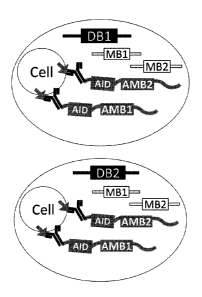
[0083] FIG. 9 exemplifies a method of immunophenotyping using antibody-
oligonucleotide conjugates.
[0084] FIG. 9A depicts an exemplary schematic showing 2 vessels each
containing a single cell bound to an
antibody-oligonucleotide conjugate are depicted. (DB1 ¨ droplet barcode 1; DB2
¨ droplet barcode 2; MB1 ¨
molecular barcode 1; MB2 ¨ molecular barcode 2; AID ¨ antigen ID barcode; AMB1
¨ antibody molecular
barcode 1; AMB2 ¨ antibody molecular barcode 2).
[0085] FIG. 9B depicts an exemplary schematic showing 2 vessels each
containing RNA molecules from a
lysed cell of a vessel from FIG. 9A. The RNA molecules are reverse transcribed
and non-template nucleotides
are added to the end of the cDNA molecule created by the reverse
transcription. Molecular barcodes are
hybridized to the non-template nucleotides added to the end of the cDNA
molecule created by the reverse
transcription.
[0086] FIG. 9C depicts an exemplary schematic showing 2 vessels each
containing a template barcoded
polyriucleotide that is amplified and attached to the cDNA of a vessel from
FIG. 9B via hybridization and the
cDNA is extended (top). The extended cDNA is then amplified (bottom).
[0087] FIG. 9D depicts an exemplary schematic showing that RNA-MB-DB species
with the same
molecular barcode (MB) attached to the same identical RNA sequences is likely
the result of PCR duplication.
RNA-MB-DB species with two different MBs that are attached to the same
identical RNA sequences (RNA1-
MB1-DB and RNA1-MB2-DB) are two independent RNA molecules of origin and not of
PCR duplication.
[0088] FIG. 9E depicts an exemplary schematic showing that DB-AMB-AID species
with the same antibody
molecular barcode (AMB) attached to a sequence with the same droplet barcode
(DB) and antigen ID barcode
(AID) is likely the result of PCR duplication. DB I-AMB1-AID1 and DB1-AMB2-
AID1 species with two
different AMBs attached to sequences with the same droplet barcode (DB) and
antigen ID barcode (AID) are
two independent oligonucleotide molecules from two independent antibody
oligonucleotide conjugate
molecules each with an antibody that specifically binds to the same target
antigen attached to the same single
cell in a vessel, and not of PCR duplication. DBI-AMBn-AID1 and DB1-AMBn-AID2
species with two
different AIDs attached to sequences with the same droplet barcode (DB) and a
same or different antibody
molecular barcodes (AMBs) are two independent oligonucleotide molecules from
two independent antibody
oligonucleotide conjugate molecules attached to the same single cell in a
vessel, wherein one of the antibody
oligonucleotide conjugate molecules has an antibody that specifically binds to
a first target antigen and the
other antibody oligonucleotide conjugate molecule has an antibody that
specifically binds to a second target
antigen.
-11-
CA 02999888 2018-03-23
WO 2017/053905
PCT/US2016/053598
[0089] FIG. 10A depicts a schematic of an exemplary affinity-oligonucleotide
conjugate of the methods
described herein.
[0090] FIG. 10B depicts a schematic of an exemplary affinity-oligonucleotide
conjugate of the methods
described herein.
[0091] FIG. 11A depicts an exemplary graph of binding signal for two exemplary
affinity-oligonucleotide
conjugates of the methods described herein that contain an affinity portion
that binds to a TCR.
[0092] FIG. 12A depicts an exemplary schematic of a T-cell bound to an
exemplary affinity-oligonucleotide
conjugate of the methods described herein.
[0093] FIG. 12B depicts an exemplary schematic of a T-cell in a droplet bound
to an exemplary affinity-
oligonucleotide conjugate of the methods described herein. Nucleic acids in
the droplet are marked with
droplet-identifying sequence and incorporated into a next-generation
sequencing library.
[0094] FIG. 13 depicts an exemplary schematic of an oligonucleotide tag
conjugated to an exemplary
affinity-oligonucleotide conjugate tetramer. The tetramer ID is a short
constant DNA sequence that
corresponds to a tetramer batch and allows multiplexing of different targets,
such as peptide-MHC targets, in
single experiment. The molecular barcode is a degenerate sequence that allows
for molecular counting for
quantification of bound tetramers.
[0095] FIG 14 depicts schematics of an exemplary affinity-oligonucleotide
conjugate generated from DNA-
labeled MHC tetramer reagents. In one embodiment, a Cy5-linked DNA
oligonucleotide is synthesized and
conjugated to streptavidin or neutravidin. In one embodiment, a non-
fluorescent DNA oligonucleotide is
conjugated to an APC-streptavidin. In one embodiment, a mixture of non-
fluorescent DNA oligonucleotide
and streptavidin or neutravidin is conjugated to an activated APC.
[0096] FIG 15 depicts an exemplary method of conjugating an oligonucleotide to
an affinity portion of an
affinity-oligonucleotide conjugate using click-chemistry.
[0097] FIG. 16 depicts a schematic of an exemplary workflow for preparing and
characterizing exemplary
affinity-oligonucleotides.
[0098] FIG. 17 depicts results from an exemplary method described herein using
6 different affinity-
oligonucleotide conjugates targeting CD3, CD19, CD4, CD8, HLA-DR, and CTLA-4.
Each point is a droplet
barcode/single cell. Cell identity was revealed by the type of receptor pair
recovered (TCR = T-cell; Ig = B-
cell).
DETAILED DESCRIPTION
[0099] Several aspects are described below with reference to example
applications for illustration. It should
be understood that numerous specific details, relationships, and methods are
set forth to provide a full
understanding of the features described herein. One having ordinary skill in
the relevant art, however, will
readily recognize that the features described herein can be practiced without
one or more of the specific details
or with other methods. The features described herein are not limited by the
illustrated ordering of acts or
events, as some acts can occur in different orders and/or concurrently with
other acts or events. Furthermore,
-12-
CA 02999888 2018-03-23
WO 2017/053905
PCT/US2016/053598
not all illustrated acts or events are required to implement a methodology in
accordance with the features
described herein.
[00100] The terminology used herein is for the purpose of describing
particular cases only and is not intended
to be limiting. As used herein, the singular forms "a", "an" and "the" are
intended to include the plural forms
as well, unless the context clearly indicates otherwise. Furthermore, to the
extent that the terms "including",
"includes", "having", "has", "with", or variants thereof are used in either
the detailed description and/or the
claims, such terms are intended to be inclusive in a manner similar to the
term "comprising".
[00101] The term "about" or "approximately" can mean within an acceptable
error range for the particular
value as determined by one of ordinary skill in the art, which will depend in
part on how the value is measured
or determined, i.e., the limitations of the measurement system. For example,
"about" can mean within 1 or
more than 1 standard deviation, per the practice in the art. Alternatively,
"about" can mean a range of up to
20%, up to 10%, up to 5%, or up to 1% of a given value. Alternatively,
particularly with respect to biological
systems or processes, the term can mean within an order of magnitude, within 5-
fold, and more preferably
within 2-fold, of a value. Where particular values are described in the
application and claims, unless otherwise
stated the term "about" meaning within an acceptable error range for the
particular value should be assumed.
[00102] It is an object of the invention to provide methods and compositions
for phenotyping single cells (e.g.,
immune cells using affinity-oligonucleotide conjugates (e.g., antibody-
oligonucleotide conjugates) (e.g., in
emulsions).
Definitions
[00103] The term "antibody" herein thus is used in the broadest sense and
includes polyclonal and monoclonal
antibodies, including intact antibodies and functional (antigen-binding)
antibody fragments thereof, including
fragment antigen binding (Fab) fragments, F(ab')2 fragments, Fab' fragments,
Fv fragments, recombinant IgG
(rIgG) fragments, single chain antibody fragments, including single chain
variable fragments (scFv), and
single domain antibodies (e.g., sdAb, sdFv, nanobody) fragments. The term
encompasses genetically
engineered and/or otherwise modified forms of immunoglobulins, such as
intrabodies, peptibodies, chimeric
antibodies, frilly human antibodies, humanized antibodies, and heteroconjugate
antibodies, multispecific, e.g.,
bispecific, antibodies, diabodies, triabodies, and tetrabodies, tandem di-
scFv, tandem tri-scFv. Unless
otherwise stated, the term "antibody" should be understood to encompass
functional antibody fragments
thereof. The term also encompasses intact or full-length antibodies, including
antibodies of any class or sub-
class, including IgG and sub-classes thereof, IgM, IgE, IgA, and IgD.
[00104] The terms "complementarity determining region," and "CDR," synonymous
with "hypervariable
region" or "HVR," are known in the art to refer to non-contiguous sequences of
amino acids within antibody
variable regions, which confer antigen specificity and/or binding affinity. In
general, there are three CDRs in
each heavy chain variable region (CDR-H1, CDR-H2, CDR-H3) and three CDRs in
each light chain variable
region (CDR-LI, CDR-L2, CDR-L3). "Framework regions" and "FR" are known in the
art to refer to the
non-CDR portions of the variable regions of the heavy and light chains. In
general, there are four FRs in each
-13-
CA 02999888 2018-03-23
WO 2017/053905
PCT/US2016/053598
full-length heavy chain variable region (FR-H1, FR-H2, FR-H3, and FR-H4), and
four FRs in each full-length
light chain variable region (FR-L1, FR-L2, FR-L3, and FR-L4).
1001051The precise amino acid sequence boundaries of a given CDR or FR can be
readily determined using
any of a number of well-known schemes, including those described by Kabat et
al. (1991), "Sequences of
Proteins of Immunological Interest," 5th Ed. Public Health Service, National
Institutes of Health, Bethesda,
MD ("Kabat" numbering scheme), Al-Lazikani et al., (1997) JMB 273,927-948
("Chothia" numbering
scheme), MacCallum et al., J. Mol. Biol. 262:732-745 (1996), "Antibody-antigen
interactions: Contact
analysis and binding site topography," J. Mol. Biol. 262, 732-745." ("Contact"
numbering scheme), Lefranc
MP et al., "IMGT unique numbering for immunoglobulin and T cell receptor
variable domains and Ig
superfamily V-like domains," Dev Comp Immunol, 2003 Jan;27(1):55-77 ("IMGT"
numbering scheme), and
Honegger A and Pliickthun A, "Yet another numbering scheme for immunoglobulin
variable domains: an
automatic modeling and analysis tool," J Mol Biol, 2001 Jun 8;309(3):657-70,
("Aho" numbering scheme).
1001061The boundaries of a given CDR or FR may vary depending on the scheme
used for identification. For
example, the Kabat scheme is based structural alignments, while the Chothia
scheme is based on structural
information. Numbering for both the Kabat and Chothia schemes is based upon
the most common antibody
region sequence lengths, with insertions accommodated by insertion letters,
for example, "30a," and deletions
appearing in some antibodies. The two schemes place certain insertions and
deletions ("indels") at different
positions, resulting in differential numbering. The Contact scheme is based on
analysis of complex crystal
structures and is similar in many respects to the Chothia numbering scheme.
1001071Table A, below, lists exemplary position boundaries of CDR-L1, CDR-L2,
CDR-L3 and CDR-H1,
CDR-H2, CDR-H3 as identified by Kabat, Chothia, and Contact schemes,
respectively. For CDR-H1, residue
numbering is listed using both the Kabat and Chothia numbering schemes. FRs
are located between CDRs,
for example, with FR-L1 located between CDR-L1 and CDR-L2, and so forth. It is
noted that because the
shown Kabat numbering scheme places insertions at H35A and H35B, the end of
the Chothia CDR-H1 loop
when numbered using the shown Kabat numbering convention varies between H32
and H34, depending on
the length of the loop.
Table A
CDR Kabat Chothia Contact
CDR-L1 L24--L34 L24--L34 L30¨L36
CDR-L2 L50--L56 L50--L56 L46--L55
CDR-L3 L89--L97 L89--L97 L89--L96
CDR-H1
(Kabat Numbering') H31--H35B H26--H32..34 H30--H35B
CDR-H1
(Chothia Numbering2) H31--H35 H26--H32 H30--H35
-14-
CA 02999888 2018-03-23
WO 2017/053905
PCT/US2016/053598
CDR-H2 H50--H65 H52--H56 H47--H58
CDR-H3 H95--H102 H95--H102 H93--H101
[00108] Thus, unless otherwise specified, a "CDR" or "complementary
determining region," or individual
specified CDRs (e.g., "CDR-H1, CDR-H2), of a given antibody or region thereof,
such as a variable region
thereof, should be understood to encompass a (or the specific) complementary
determining region as defined
by any of the aforementioned schemes. For example, where it is stated that a
particular CDR (e.g., a CDR-
H3) contains the amino acid sequence of a corresponding CDR in a given VH or
VL amino acid sequence, it
is understood that such a CDR has a sequence of the corresponding CDR (e.g.,
CDR-H3) within the variable
region, as defined by any of the aforementioned schemes. In some embodiments,
specified CDR sequences
are specified.
[00109] Likewise, unless otherwise specified, a FR or individual specified
FR(s) (e.g., FR-H1, FR-H2), of a
given antibody or region thereof, such as a variable region thereof, should be
understood to encompass a (or
the specific) framework region as defined by any of the known schemes. In some
instances, the scheme for
identification of a particular CDR, FR, or FRs or CDRs is specified, such as
the CDR as defined by the Kabat,
Chothia, or Contact method. In other cases, the particular amino acid sequence
of a CDR or FR is given.
[00110] The term "variable region" or -variable domain" refers to the domain
of an antibody heavy or light
chain that is involved in binding the antibody to antigen. The variable
domains of the heavy chain and light
chain (VH and VL, respectively) of a native antibody generally have similar
structures, with each domain
comprising four conserved framework regions (FRs) and three CDRs. (See, e.g.,
Kindt et al. Kuby
Immunology, 6th ed., W.H. Freeman and Co., page 91 (2007). A single VH or VL
domain may be sufficient
to confer antigen-binding specificity. Furthermore, antibodies that bind a
particular antigen may be isolated
using a VH or VL domain from an antibody that binds the antigen to screen a
library of complementary VL or
VH domains, respectively. See, e.g., Portolano et al., J. Immunol. 150:880-887
(1993); Clarkson et al., Nature
352:624-628 (1991).
[00111] Among the provided antibodies are antibody fragments. An "antibody
fragment" refers to a molecule
other than an intact antibody that comprises a portion of an intact antibody
that binds the antigen to which the
intact antibody binds. Examples of antibody fragments include but are not
limited to Fv, Fab, Fab', Fab'-SH,
F(ab')2; diabodies; linear antibodies; single-chain antibody molecules (e.g.
scFv); and multispecific antibodies
formed from antibody fragments. In particular embodiments, the antibodies are
single-chain antibody
fragments comprising a variable heavy chain region and/or a variable light
chain region, such as scFvs.
[00112] Unless otherwise stated, the term "TCR" should be understood to
encompass full TCRs as well as
antigen-binding portions or antigen-binding fragments (also called MHC-peptide
binding fragments) thereof.
In some embodiments, the TCR is an intact or full-length TCR. In some
embodiments, the TCR is an antigen-
binding portion that is less than a full-length TCR but that binds to a
specific antigenic peptide bound to (i.e.,
in the context of) an MHC molecule, i.e., an MEC-peptide complex. In some
cases, an antigen-binding
portion or fragment of a TCR can contain only a portion of the structural
domains of a full-length or intact
-15-
CA 02999888 2018-03-23
WO 2017/053905
PCT/US2016/053598
TCR, but yet is able to bind the epitope (e.g., MHC-peptide complex) to which
the full TCR binds. In some
cases, an antigen-binding portion or fragment of a TCR contains the variable
domains of a TCR, such as
variable a chain and variable f3 chain of a TCR, sufficient to form a binding
site for binding to a specific
MHC-peptide complex, such as generally where each chain contains three
complementarity determining
regions. Polypeptides or proteins having a binding domain which is an antigen-
binding domain or is
homologous to an antigen-binding domain are included. Complementarity
determining region (CDR) grafted
antibodies and TCRs and other humanized antibodies and TCRs (including CDR
modifications and
framework region modifications) are also contemplated by these terms. It
should be noted that while reference
may be made only to immunoglobulin chains (e.g., heavy chains and lights
chains), the disclosed invention
can be applied to multiple other different types of paired sequences, e.g., T-
cell receptor chain pairs (TCRa
and TCRI3 chains and TCRy and TCR6 chains), and is not limited to
immunoglobulins.
[00113] The ability of T-cells to recognize antigens associated with various
cancers or infectious organisms is
conferred by its TCR, which is made up of both an alpha (a) chain and a beta
(13) chain or a gamma (y) and a
delta (6) chain. The proteins which make up these chains are encoded by DNA,
which employs a unique
mechanism for generating the tremendous diversity of the TCR. This multi-
subunit immune recognition
receptor associates with the CD3 complex and binds peptides presented by the
MHC class I and II proteins on
the surface of antigen-presenting cells (APCs). Binding of a TCR to the
antigenic peptide on the APC is a
central event in T-cell activation, which occurs at an immunological synapse
at the point of contact between
the T-cell and the APC.
[00114] Each TCR comprises variable complementarity determining regions
(CDRs), as well as framework
regions (FRs). The amino acid sequence of the third complementarity-
determining region (CDR3) loops of the
a and 13 chain variable domains largely determines the sequence diversity of
a13 T-cells arising from
recombination between variable (V(3), diversity (DI3), and joining (43) gene
segments in the 13 chain locus, and
between analogous Va and Ja gene segments in the a chain locus, respectively.
The existence of multiple such
gene segments in the TCR a and f3 chain loci allows for a large number of
distinct CDR3 sequences to be
encoded. Independent addition and deletion of nucleotides at the VI3-1313, DI3-
43, and Va-Ja junctions during
the process of TCR gene rearrangement further increases CDR3 sequence
diversity. In this respect,
immunocompetence is reflected in the diversity of TCRs.
[00115] Immunoglobulins (Igs) expressed by B-cells are in some aspects
proteins consisting of four
polypeptide chains, two heavy chains (IgHs) and two light chains (IgLs),
forming an H2L2structure. Each pair
of IgH and IgL chains contains a hypervariable domain, consisting of a VL and
a VH region, and a constant
domain. The IgH chains of Igs are of several types, II, 6, y, a, and (3. The
diversity of Igs within an individual
is mainly determined by the hypervariable domain. Similar to the TCR, the V
domain of IgH chains is created
by the combinatorial joining of the VH, DH, and JH gene segments. Independent
addition and deletion of
nucleotides at the VH-DH, DH-JH, and VH-JH junctions during the process of Ig
gene rearrangement further
increases hypervariable domain sequence diversity. Here, immunocompetence is
reflected in the diversity of
Igs.
-16-
CA 02999888 2018-03-23
WO 2017/053905
PCT/US2016/053598
[00116] The term "variable region" or "variable domain" refers to the domain
of an antibody heavy or light
chain that is involved in binding the antibody to antigen. The variable
domains of the heavy chain and light
chain (VH and VL, respectively) of a native antibody generally have similar
structures, with each domain
comprising four conserved framework regions (FRs) and three CDRs. (See, e.g.,
Kindt et al. Kuby
Immunology, 6th ed., W.H. Freeman and Co., page 91 (2007). A single VH or VL
domain may be sufficient
to confer antigen-binding specificity. Furthermore, antibodies that bind a
particular antigen may be isolated
using a VH or VL domain from an antibody that binds the antigen to screen a
library of complementary VL or
VH domains, respectively. See, e.g., Portolano et al., J. Immunol. 150:880-887
(1993); Clarkson et al., Nature
352:624-628 (1991).
[00117] An "affinity portion" refers to a portion of the affinity-
oligonucleotide conjugate that interacts with a
target antigen. Exemplary affinity portions include antibodies, peptides,
proteins, aptamers, small molecules,
drugs, cells, MHCs and others.
[001181A "hypervariable region" refers to the amino acid residues of an
antibody or TCR which are
responsible for antigen-binding. The hypervariable region comprises amino acid
residues from a
complementarity determining region or CDR. Framework or FR residues are those
variable domain residues
other than the hypervariable region residues as herein defined.
[00119] Among the provided antibodies are antibody fragments. An "antibody
fragment" refers to a molecule
other than an intact antibody that comprises a portion of an intact antibody
that binds the antigen to which the
intact antibody binds. Examples of antibody fragments include but are not
limited to Fv, Fab, Fab', Fab'-SH,
F(ab')2; diabodies; linear antibodies; single-chain antibody molecules (e.g.
scFv); and multispecific antibodies
formed from antibody fragments. In particular embodiments, the antibodies are
single-chain antibody
fragments comprising a variable heavy chain region and/or a variable light
chain region, such as scFvs.
[00120] Single-domain antibodies are antibody fragments comprising all or a
portion of the heavy chain
variable domain or all or a portion of the light chain variable domain of an
antibody. In certain embodiments,
a single-domain antibody is a human single-domain antibody.
[00121] Antibody fragments can be made by various techniques, including but
not limited to proteolytic
digestion of an intact antibody as well as production by recombinant host
cells. In some embodiments, the
antibodies are recombinantly-produced fragments, such as fragments comprising
arrangements that do not
occur naturally, such as those with two or more antibody regions or chains
joined by synthetic linkers, e.g.,
peptide linkers, and/or that are may not be produced by enzyme digestion of a
naturally-occurring intact
antibody. In some aspects, the antibody fragments are scFvs.
[00122] Also provided are TCR fragments, including antigen-binding fragments.
In some embodiments, the
TCR is an antigen-binding portion thereof, such as a variant of a full-length
TCR not containing the
transmembrane and/or cytoplasmic region(s) thereof, which may be referred to
as a full soluble TCR. In some
embodiments, the TCR is a dimeric TCR (dTCR). In some embodiments, the TCR is
a single-chain TCR
(scTCR), such as a scTCR having a structure as described in PCT patent
publication numbers WO 03/020763,
WO 04/033685, or WO 2011/044186. In certain embodiments, the TCR is a single-
chain TCR fragment
-17-
CA 02999888 2018-03-23
WO 2017/053905
PCT/US2016/053598
comprising an alpha chain variable region linked to a beta chain variable
region, such as a scTv. In some
embodiments, an scTv is also referred to as an scFv
[00123] A single-chain Fv or scFv refers in some aspects to antibody or TCR
fragments that comprise the
variable heavy chain (VH) and variable light chain (VL) domains of an antibody
or the variable alpha or
gamma chain (Va or Vy) and variable beta or delta chain (VP or VS) domains of
a TCR, wherein these
domains are present in a single polypeptide chain. Generally, the Fv
polypeptide further comprises a
polypeptide linker between the VH and VL domains or Va and Vf3 domains or Vy
and Vo domains which
enables the sFy to form the desired structure for antigen binding.
[00124] A diabody refers in some aspects to small antibody and/or TCR
fragments with two antigen-binding
sites, which fragments comprise a VH connected to a VL in the same polypeptide
chain (VH-VL) or a Va
connected to a Vf3 in the same polypeptide chain (Va-V13) or a Vy connected to
a VS in the same polypeptide
chain (Vy-Vo). By using a linker that is too short to allow pairing between
the two domains on the same chain,
the domains are forced to pair with the complementary domains of another chain
and create two antigen-
binding sites. Exemplary diabodies are described more fully in, for example,
EP404097 and W093111161.
[00125] A bispecific antibody or bispecific TCR refers in some aspects to an
antibody or TCR that shows
specificities to two different types of antigens. The terms as used herein
specifically include, without
limitation, antibodies and TCRs which show binding specificity for a target
antigen and to another target that
facilitates delivery to a particular tissue. Similarly, multi-specific
antibodies and TCRs have two or more
binding specificities.
[00126] A linear antibody or "linear TC refers in some aspects to a pair of
tandem Fd segments (e. g. , V H-CHI-
VH-Chl or Va-Cal-Va-Cal) which form a pair of antigen binding regions. Linear
antibodies and TCRs can be
bispecific or monospecific, for example, as described by Zapata et al.,
Protein Eng. 8(10):1057-1062 (1995).
[00127] An antigen-binding domain refers in some aspects to one or more
fragments of an antibody or TCR
that retain the ability to specifically bind to an antigen. Non-limiting
examples of antibody fragments included
within such terms include, but are not limited to, (i) a Fab fragment, a
monovalent fragment consisting of the
VH, CL and CHI domains; (ii) a F(ab')2 fragment, a bivalent fragment
containing two Fab fragments linked
by a disulfide bridge at the hinge region; (iii) a Fd fragment consisting of
the VH and CHI domains; (iv) a Fv
fragment containing the VL and VH domains of a single arm of an antibody,
including scFvs, (v) a dAb
fragment (Ward etal., (1989) Nature 341:544 546), which containing a VH
domain; and (vi) an isolated CDR.
Additionally included in this definition are antibodies comprising a single
heavy chain and a single light chain
or TCRs with a single alpha chain or a single beta chain.
[00128] "F(ab')2" and "Fab" moieties can be produced by treating an Ig with a
protease such as pepsin and
papain, and include antibody fragments generated by digesting immunoglobulin
near the disulfide bonds
existing between the hinge regions in each of the two heavy chains. For
example, papain cleaves IgG
upstream of the disulfide bonds existing between the hinge regions in each of
the two heavy chains to generate
two homologous antibody fragments in which a light chain composed of VL and
CL, and a heavy chain
fragment composed of VH and CHyi (y1 region in the constant region of the
heavy chain) are connected at their
-18-
CA 02999888 2018-03-23
WO 2017/053905
PCT/US2016/053598
C terminal regions through a disulfide bond. Each of these two homologous
antibody fragments is called
'Fab'. Pepsin also cleaves IgG downstream of the disulfide bonds existing
between the hinge regions in each
of the two heavy chains to generate an antibody fragment slightly larger than
the fragment in which the two
above-mentioned 'Fab' are connected at the hinge region. This antibody
fragment is called F('ab')2. The Fab
fragment also contains the constant domain of the light chain and the first
constant domain (CH1) of the heavy
chain. 'Fab' fragments differ from Fab fragments by the addition of a few
residues at the carboxyl terminus of
the heavy chain CH I domain including one or more cysteine(s) from the
antibody hinge region. Fab'-SH is the
designation herein for Fab' in which the cysteine residue(s) of the constant
domains bear a free thiol group.
F(ab')2 antibody fragments originally are produced as pairs of Fab' fragments
which have hinge cysteines
between them.
[00129] Fv refers in some aspects to an antibody or TCR fragment which
contains a complete antigen-
recognition and antigen-binding site. This region consists of a dimer of one
heavy chain and one light chain
variable domain or one TCRa chain and one TCRE3 chain or one TCRy chain and
one TCRS chain in tight,
non-covalent association. It is in this configuration that the three CDRs of
each variable domain interact to
define an antigen-binding site on the surface of the Vii-VL dimer or Va-V13
dimer or Vy-VS dimer.
Collectively, a combination of one or more of the CDRs from each of the VH and
VL chains or Va-V13 chains
or Vy-VS chains confers antigen-binding specificity to the antibody or TCR.
For example, it would be
understood that, for example, the CDRH3 and CDRL3 could be sufficient to
confer antigen-binding
specificity to an antibody or TCR when transferred to VII and VL chains or Va
and VP chains or Vy-VS chains
of a recipient selected antibody, TCR, or antigen-binding fragment thereof and
this combination of CDRs can
be tested for binding, affinity, etc. Even a single variable domain (or half
of an Fv comprising only three
CDRs specific for an antigen) has the ability to recognize and bind antigen,
although likely at a lower affinity
than when combined with a second variable domain. Furthermore, although the
two domains of a Fv fragment
(VL and VH or Va and VI3 or Vy and Vs), are coded for by separate genes, they
can be joined using
recombinant methods by a synthetic linker that enables them to be made as a
single protein chain in which the
VL and VH or Va and Vf3 or Vy and VS chain regions pair to form monovalent
molecules (known as single
chain Fv (scFv); Bird et al. (1988) Science 242:423-426; Huston et al. (1988)
Proc. Natl. Acad. Sci. USA
85:5879-5883; and Osbourn et al. (1998) Nat. Biotechnol. 16:778). Such scFvs
are also intended to be
encompassed within the term "antigen-binding portion" of an antibody. Any VH
and VL sequences of specific
scFv can be linked to an Fc region cDNA or genomic sequences, in order to
generate expression vectors
encoding complete Ig (e.g., IgG) molecules or other isotypes. VH and VL can
also be used in the generation of
Fab, Fv or other fragments of Igs using either protein chemistry or
recombinant DNA technology.
[00130] Antigen-binding polypeptides also include heavy chain dimers such as,
for example, antibodies from
camelids and sharks. Camelid and shark antibodies comprise a homodimeric pair
of two chains of V-like and
C-like domains (neither has a light chain). Since the VH region of a heavy
chain dimer IgG in a camelid does
not have to make hydrophobic interactions with a light chain, the region in
the heavy chain that normally
contacts a light chain is changed to hydrophilic amino acid residues in a
camelid. VH domains of heavy-chain
-19-
CA 02999888 2018-03-23
WO 2017/053905
PCT/US2016/053598
dimer IgGs are called VHH domains. Shark Ig-NARs comprise a homodimer of one
variable domain (termed a
V-NAR domain) and five C-like constant domains (C-NAR domains). In camelids,
the diversity of antibody
repertoire is determined by the CDRs 1, 2, and 3 in the VH or VHE regions. The
CDR3 in the camel VHH region
is characterized by its relatively long length, averaging 16 amino acids
(Muyldermans et al., 1994, Protein
Engineering 7(9): 1129).
[00131] A "humanized" antibody is an antibody in which all or substantially
all CDR amino acid residues
are derived from non-human CDRs and all or substantially all FR amino acid
residues are derived from human
FRs. A humanized antibody optionally may include at least a portion of an
antibody constant region derived
from a human antibody. A "humanized form" of a non-human antibody, refers to a
variant of the non-human
antibody that has undergone humanization, typically to reduce immunogenicity
to humans, while retaining the
specificity and affinity of the parental non-human antibody. In some
embodiments, some FR residues in a
humanized antibody are substituted with corresponding residues from a non-
human antibody (e.g., the
antibody from which the CDR residues are derived), e.g., to restore or improve
antibody specificity or affinity.
[00132] Among the provided antibodies are human antibodies. A "human antibody"
is an antibody with an
amino acid sequence corresponding to that of an antibody produced by a human
or a human cell, or non-
human source that utilizes human antibody repertoires or other human antibody-
encoding sequences,
including human antibody libraries. The term excludes humanized forms of non-
human antibodies
comprising non-human antigen-binding regions, such as those in which all or
substantially all CDRs are non-
human.
[00133] Human antibodies may be prepared by administering an immunogen to a
transgenic animal that has
been modified to produce intact human antibodies or intact antibodies with
human variable regions in
response to antigenic challenge. Such animals typically contain all or a
portion of the human immunoglobulin
loci, which replace the endogenous immunoglobulin loci, or which are present
extrachromosomally or
integrated randomly into the animal's chromosomes. In such transgenic animals,
the endogenous
immunoglobulin loci have generally been inactivated. Human antibodies also may
be derived from human
antibody libraries, including phage display and cell-free libraries,
containing antibody-encoding sequences
derived from a human repertoire.
[00134] Among the provided antibodies are monoclonal antibodies, including
monoclonal antibody
fragments. The term "monoclonal antibody" as used herein refers to an antibody
obtained from or within a
population of substantially homogeneous antibodies, i.e., the individual
antibodies comprising the population
are identical, except for possible variants containing naturally occurring
mutations or arising during
production of a monoclonal antibody preparation, such variants generally being
present in minor amounts. In
contrast to polyclonal antibody preparations, which typically include
different antibodies directed against
different epitopes, each monoclonal antibody of a monoclonal antibody
preparation is directed against a single
epitope on an antigen. The term is not to be construed as requiring production
of the antibody by any
particular method. A monoclonal antibody may be made by a variety of
techniques, including but not limited
-20-
CA 02999888 2018-03-23
WO 2017/053905
PCT/US2016/053598
to generation from a hybridoma, recombinant DNA methods, phage-display and
other antibody display
methods.
[00135] The terms "polypeptide" and "protein" are used interchangeably to
refer to a polymer of amino acid
residues, and are not limited to a minimum length. Polypeptides, including the
provided antibodies and
antibody chains and other peptides, e.g., linkers and binding peptides, may
include amino acid residues
including natural and/or non-natural amino acid residues. The terms also
include post-expression
modifications of the polypeptide, for example, glycosylation, sialylation,
acetylation, phosphorylation, and the
like. In some aspects, the polypeptides may contain modifications with respect
to a native or natural
sequence, as long as the protein maintains the desired activity. These
modifications may be deliberate, as
through site-directed mutagenesis, or may be accidental, such as through
mutations of hosts which produce the
proteins or errors due to PCR amplification.
[00136] A "germline sequence" refers to a genetic sequence from the germline
(the haploid gametes and those
diploid cells from which they are formed). Germline DNA contains multiple gene
segments that encode a
single Ig heavy or light chain, or a single TCRa or TCRI3 chain, or a single
TCRy or TCR S chain. These gene
segments are carried in the germ cells but cannot be transcribed and
translated until they are arranged into
functional genes. During B-cell and T-cell differentiation in the bone marrow,
these gene segments are
randomly shuffled by a dynamic genetic system capable of generating more than
108 specificities. Most of
these gene segments are published and collected by the germline database.
[00137] Affinity refers to the equilibrium constant for the reversible binding
of two agents and is expressed as
KD. Affinity of a binding protein to a ligand such as affinity of an antibody
for an epitope or such as affinity
for a TCR for a MCH-peptide complex can be, for example, from about 100
nanomolar (nM) to about 0.1 n1\4,
from about 100 nM to about 1 picomolar (pM), or from about 100 n1\4 to about 1
femtomolar (fM). The term
"avidity" refers to the resistance of a complex of two or more agents to
dissociation after dilution.
[00138] An epitope refers in some aspects to a portion of an antigen or other
macromolecule capable of
forming a binding interaction with the variable region binding pocket of an
antibody or TCR. Such binding
interactions can be manifested as an intermolecular contact with one or more
amino acid residues of one or
more CDRs. Antigen binding can involve, for example, a CDR3, a CDR3 pair, or
in some instances,
interactions of up to all six CDRs of the VD and VL chains. An epitope can be
a linear peptide sequence (i.e.,
"continuous") or can be composed of noncontiguous amino acid sequences (i.e.,
"conformational" or
"discontinuous"). An antibody or TCR can recognize one or more amino acid
sequences; therefore an epitope
can define more than one distinct amino acid sequence. In some aspects, a TCR
can recognize one or more
amino acid sequences or epitopes in the context of an MEC. Epitopes recognized
by antibodies and TCRs can
be determined by peptide mapping and sequence analysis techniques well known
to one of skill in the art.
Binding interactions are manifested as intermolecular contacts with one or
more amino acid residues of a
CDR.
[00139] In some embodiments, reference to an antibody or TCR with specific
binding refers to a situation in
which an antibody or TCR will not show any significant binding to molecules
other than the antigen
-21-
CA 02999888 2018-03-23
WO 2017/053905
PCT/US2016/053598
containing the epitope recognized by the antibody or TCR. The term is also
applicable where for example, an
antigen binding domain is specific for a particular epitope which is carried
by a number of antigens, in which
case the selected antibody, TCR, or antigen-binding fragment thereof carrying
the antigen binding domain
will be able to bind to the various antigens carrying the epitope. The terms
"preferentially binds" or
"specifically binds" mean that the antibodies, TCRs, or fragments thereof bind
to an epitope with greater
affinity than it binds unrelated amino acid sequences, and, if cross-reactive
to other polypeptides containing
the epitope, are not toxic at the levels at which they are formulated for
administration to human use. In one
aspect, such affinity is at least 1-fold greater, at least 2-fold greater, at
least 3-fold greater, at least 4-fold
greater, at least 5-fold greater, at least 6-fold greater, at least 7-fold
greater, at least 8-fold greater, at least 9-
fold greater, 10-fold greater, at least 20-fold greater, at least 30-fold
greater, at least 40-fold greater, at least
50-fold greater, at least 60-fold greater, at least 70-fold greater, at least
80-fold greater, at least 90-fold
greater, at least 100-fold greater, or at least 1000-fold greater than the
affinity of the antibody, TCR, or
fragment thereof for unrelated amino acid sequences. The term "binding" refers
to a direct association
between two molecules, due to, for example, covalent, electrostatic,
hydrophobic, and ionic and/or hydrogen-
bond interactions under physiological conditions, and includes interactions
such as salt bridges and water
bridges, as well as any other conventional means of binding.
[00140] The term "binding" refers to a direct association between two
molecules, due to, for example,
covalent, electrostatic, hydrophobic, and ionic and/or hydrogen-bond
interactions under physiological
conditions, and includes interactions such as salt bridges and water bridges,
as well as any other conventional
means of binding.
[00141] "Pharmaceutically acceptable" refers to molecular entities and
compositions that are physiologically
tolerable and do not typically produce an allergic or similar untoward
reaction, such as gastric upset, dizziness
and the like, when administered to a human.
[00142] "Prevention" refers to prophylaxis, prevention of onset of symptoms,
prevention of progression of a
disease or disorder associated with excess levels of protein or correlated
with protein activity.
[00143] "Inhibition," "treatment" and "treating" are used interchangeably and
refer to, for example, stasis of
symptoms, prolongation of survival, partial or full amelioration of symptoms,
and partial or full eradication of
a condition, disease or disorder associated with excess levels of protein or
correlated with protein activity. For
example, treatment of cancer includes, but is not limited to, stasis, partial
or total elimination of a cancerous
growth or tumor. Treatment or partial elimination includes, for example, a
fold reduction in growth or tumor
size and/or volume such as about 2-fold, about 3-fold, about 4-fold, about 5-
fold, about 10-fold, about 20-fold,
about 50-fold, or any fold reduction in between. Similarly, treatment or
partial elimination can include a
percent reduction in growth or tumor size and/or volume of about 1%, 2%, 3%,
4%, 5%, 10%, 20%, 30%,
40%, 50%, 60%, 70%, 80%, 90%, 95% or any percentage reduction in between.
[00144] A neutralizing antibody or neutralizing TCR refers in some aspects to
any antibody or TCR that
inhibits replication of a pathogen, such as a virus or bacteria, regardless of
the mechanism by which
neutralization is achieved.
-22-
CA 02999888 2018-03-23
WO 2017/053905
PCT/US2016/053598
[00145] An antibody repertoire or TCR repertoire refers to a collection of
antibodies, TCRs, or fragments
thereof. An antibody repertoire can, for example, be used to select a
particular antibody or screen for a
particular property, such as binding ability, binding specificity, ability of
gastrointestinal transport, stability,
affinity, and the like. The term specifically includes antibody and TCR
libraries, including all forms of
combinatorial libraries, such as, for example, antibody phage display
libraries, including, without limitation,
single-chain Fv (scFv) and Fab antibody phage display libraries from any
source, including naïve, synthetic
and semi-synthetic libraries.
[00146] A "target nucleic acid molecule," "target polynucleotide," "target
polynucleotide molecule," refers to
any nucleic acid of interest.
[00147] A polymerase chain reaction (PCR) refers to an in vitro amplification
reaction of polynucleotide
sequences by the simultaneous primer extension of complementary strands of a
double stranded
polynucleotide. PCR reactions produce copies of a template polynucleotide
flanked by primer binding sites.
The result, with two primers, is an exponential increase in template
polynucleotide copy number of both
strands with each cycle, because with each cycle both strands are replicated.
The polynucleotide duplex has
termini corresponding to the ends of primers used. PCR can comprise one or
more repetitions of denaturing a
template polynucleotide, annealing primers to primer binding sites, and
extending the primers by a DNA or
RNA polymerase in the presence of nucleotides. Particular temperatures,
durations at each step, and rates of
change between steps depend on many factors well-known to those of ordinary
skill in the art. (McPherson et
al., IRL Press, Oxford (1991 and 1995)). For example, in a conventional PCR
using Taq DNA polymerase, a
double stranded template polynucleotide can be denatured at a temperature >90
C, primers can be annealed at
a temperature in the range 50-75 C, and primers can be extended at a
temperature in the range 72-78 C. In
some embodiments, PCR comprises Reverse transcription PCR (RT-PCR), real-time
PCR, nested PCR,
quantitative PCR, multiplexed PCR, or the like. In some embodiments, PCR does
not comprise RT-PCR.
(U.S. Patent Nos. 5,168,038, 5,210,015, 6,174,670, 6,569,627, and 5,925,517;
Mackay etal., Nucleic Acids
Research, 30: 1292-1305 (2002)). RT-PCR comprises a PCR reaction preceded by a
reverse transcription
reaction and a resulting cDNA is amplified, Nested PCR comprises a two-stage
PCR wherein an amplicon of
a first PCR reaction using a first set of primers becomes the sample for a
second PCR reaction using a second
primer set, at least one of which binds to an interior location of an amplicon
of a first PCR reaction.
Multiplexed PCR comprises a PCR reaction, wherein a plurality of
polynucleotide sequences is subjected to
PCR in the same reaction mixture simultaneously. PCR reaction volumes can be
anywhere from 0.2 pL-1000
L. Quantitative PCR comprises a PCR reaction designed to measure an absolute
or relative amount,
abundance, or concentration of one or more sequences in a sample. Quantitative
measurements can include
comparing one or more reference sequences or standards to a polynucleotide
sequence of interest. (Freeman et
al., Biotechniques, 26: 112-126 (1999); Becker-Andre etal., Nucleic Acids
Research, 17: 9437-9447 (1989);
Zimmerman et al., Biotechniques, 21: 268-279 (1996); Diviacco etal., Gene,
122: 3013- 3020 (1992);
Becker-Andre et al., Nucleic Acids Research, 17: 9437-9446 (1989)).
-23-
CA 02999888 2018-03-23
WO 2017/053905
PCT/US2016/053598
[00148] "Nucleotide," "nucleoside," "nucleotide residue," and "nucleoside
residue," as used herein, can mean
a deoxyribonucleotide or ribonucleotide residue, or other similar nucleoside
analogue capable of serving as a
component of a primer suitable for use in an amplification reaction (e.g., PCR
reaction). Such nucleosides and
derivatives thereof can be used as the building blocks of the primers
described herein, except where indicated
otherwise. Nothing in this application is meant to preclude the utilization of
nucleoside derivatives or bases
that have been chemical modified to enhance their stability or usefulness in
an amplification reaction,
provided that the chemical modification does not interfere with their
recognition by a polymerase as
deoxyguanine, deoxycytosine, deoxythymidine, or deoxyadenine, as appropriate.
In some embodiments,
nucleotide analogs can stabilize hybrid formation. In some embodiments,
nucleotide analogs can destabilize
hybrid formation. In some embodiments, nucleotide analogs can enhance
hybridization specificity. In some
embodiments, nucleotide analogs can reduce hybridization specificity.
[00149] A "nucleic acid", or grammatical equivalents, refers to either a
single nucleotide or at least two
nucleotides covalently linked together.
[00150] A "polynucleotide" or grammatical equivalents refers to at least two
nucleotides covalently linked
together. A polynucleotide comprises a molecule containing two or more
nucleotides. A polynucleotide
comprises polymeric form of nucleotides of any length, either ribonucleotides,
deoxyribonucleotides or
peptide nucleic acids (PNAs), that comprise purine and pyrimidine bases, or
other natural, chemically or
biochemically modified, non-natural, or derivatives of nucleotide bases. The
backbone of the polynucleotide
can comprise sugars and phosphate groups, or modified or substituted sugar or
phosphate groups. A
polynucleotide may comprise modified nucleotides, such as methylated
nucleotides and nucleotide analogs.
The sequence of nucleotides may be interrupted by non-nucleotide components. A
polynucleotide can include
other molecules, such as another hybridized polynucleotide. Polynucleotides
include sequences of
deoxyribonucleic acid (DNA), ribonucleic acid (RNA), or both. Non- limiting
examples of polynucleotides
include a gene, a gene fragment, an exon, an intron, intergenic DNA
(including, without limitation,
heterochromatic DNA), messenger RNA (mRNA), transfer RNA, ribosomal RNA,
ribozymes, small
interfering RNA (siRNA), cDNA, recombinant polynucleotides, branched
polynucleotides, plasmids, vectors,
isolated DNA of a sequence, isolated RNA of a sequence, nucleic acid probes,
and primers. Polynucleotides
can be isolated from natural sources, recombinant, or artificially
synthesized.
[00151] Polynucleotides can include nonstandard nucleotides, such as
nucleotide analogs or modified
nucleotides. In some embodiments, nonstandard nucleotides can stabilize hybrid
formation. In some
embodiments, nonstandard nucleotides can destabilize hybrid formation. In some
embodiments, nonstandard
nucleotides can enhance hybridization specificity. In some embodiments,
nonstandard nucleotides can reduce
hybridization specificity. Examples of nonstandard nucleotide modifications
include 2' 0-Me, 2' 0-allyl, 2'
0-propargyl, 2' 0-alkyl, 2' fluoro, 2' arabino, 2' xylo, 2' fluoro arabino,
phosphorothioate,
phosphorodithioate, phosphoroamidates, 2' Amino, 5-alkyl-substituted
pyrimidine, 3' deoxyguanosine, 5-
halo-substituted pyrimidine, alkyl-substituted purine, halo-substituted
purine, bicyclic nucleotides, 2'MOE,
PNA molecules, LNA-molecules, LNA-like molecules, diaminopurine, S2T, 5-
fluorouracil, 5-bromouracil, 5-
-24-
CA 02999888 2018-03-23
WO 2017/053905
PCT/US2016/053598
chlorouracil, 5-iodouracil, hypoxanthine, xantine, 4-acetylcytosine, 5-
(carboxyhydroxylmethyl)uracil, 5-
carboxymethylaminomethy1-2-thiouridine, 5-carboxymethylaminomethyluracil,
dihydrouracil, beta-D-
galactosylqueosine, inosine, N6-isopentenyladenine, 1-methylguanine, 1-
methylinosine, 2,2-dimethylguanine,
2-methyladenine, 2-methylguanine, 3-methylcytosine, 5-methylcytosine, N6-
adenine, 7-methyl guanine, 5-
methylaminomethyluracil, 5-methoxyaminomethy1-2-thiouracil, beta-D-
mannosylqueosine, 5'-
methoxycarboxymethyluracil, 5-methoxyuracil, 2-methylthio-D46-
isopentenyladenine, uracil-5-oxyacetic
acid (v), wybutoxosine, pseudouracil, queosine, 2-thiocytosine, 5- methyl-2-
thiouracil, 2-thiouracil, 4-
thiouracil, 5-methyluracil, uracil-5-oxyacetic acid methylester, uracil-5-oxy
acetic acid (v), 5-methy1-2-
thiouracil, 3-(3-amino-3-N-2- carboxypropyl) uracil, (acp3)w, 2,6-
diaminopurine, and derivatives thereof.
1001521A "subject", "individual", -host" or "patient" refers to a living
organisms such as mammals.
Examples of subjects and hosts include, but are not limited to, horses, cows,
camels, sheep, pigs, goats, dogs,
cats, rabbits, guinea pigs, rats, mice (e.g., humanized mice), gerbils, non-
human primates (e.g., macaques),
humans and the like, non-mammals, including, e.g., non-mammalian vertebrates,
such as birds (e.g., chickens
or ducks) fish (e.g., sharks) or frogs (e.g., Xenopus), and non-mammalian
invertebrates, as well as transgenic
species thereof. In certain aspects, a subject refers to a single organism
(e.g., human). In certain aspects, or a
group of individuals composing a small cohort having either a common immune
factor to study and/or
disease, and/or a cohort of individuals without the disease (e.g.,
negative/normal control) are provided. A
subject from whom samples are obtained can either be inflicted with a disease
and/or disorder (e.g., one or
more allergies, infections, cancers or autoimmune disorders or the like) and
can be compared against a
negative control subject which is not affected by the disease.
[00153] A "kit" refers to a delivery system for delivering materials or
reagents for carrying out a method
disclosed herein. In some embodiments, kits include systems that allow for the
storage, transport, or delivery
of reaction reagents (e.g., probes, enzymes, etc. in the appropriate
containers) and/or supporting materials
(e.g., buffers, written instructions for performing the assay etc.) from one
location to another. For example,
kits include one or more enclosures (e.g., boxes) containing the relevant
reaction reagents and/or supporting
materials. Such contents may be delivered to the intended recipient together
or separately. For example, a first
container may contain an enzyme for use in an assay, while a second container
contains a plurality of primers.
[00154] A polypeptide refers in some aspects to a molecule comprising at least
two amino acids. In some
embodiments, the polypeptide consists of a single peptide. In some
embodiments, a polypeptide comprises
two or more peptides. For example, a polypeptide can comprise at least about
2, 3, 4, 5, 6, 7, 8, 9, 10, 11, 12,
13, 14, 15, 16, 17, 18, 19, 20, 30, 40, 50, 60, 70, 80, 90, 100, 200, 300,
400, 500, 600, 700, 800, 900, or 1000
peptides or amino acids. Examples of polypeptides include, but are not limited
to, amino acid chains, proteins,
peptides, hormones, polypeptide saccharides, lipids, glycolipids,
phospholipids, antibodies, enzymes, kinases,
receptors, transcription factors, and ligands.
[00155] A sample refers in some aspects to a biological, environmental,
medical, subject, or patient sample or
a sample containing a polynucleotide, such as a target polynucleotide.
Affinity-0112onuc1eotide Coniu2ates
-25-
CA 02999888 2018-03-23
WO 2017/053905
PCT/US2016/053598
[00156] An affinity-oligonucleotide conjugate comprises an affinity molecule
portion (e.g., an antibody or
MHC-peptide complex) and an oligonucleotide portion. An antigen identification
sequence of the affinity-
oligonucleotide conjugate's oligonucleotide can be used to identify the one or
more antigens to which the
affinity-oligonucleotide conjugate specifically interacts. In some
embodiments, the oligonucleotide is attached
covalently to the affinity portion of the conjugate. In some embodiments, the
oligonucleotide is attached non-
covalently to the affinity portion of the conjugate.
[00157] In some embodiments, an affinity-oligonucleotide conjugate comprises a
single affinity portion. In
some embodiments, affinity-oligonucleotide conjugates are multivalent affinity-
oligonucleotide conjugates.
For example, multivalent affinity-oligonucleotide conjugates can comprise
antigen-binding domains of at least
two affinity molecules conjugated to one or more oligonucleotide(s). For
example, multivalent affinity-
oligonucleotide conjugates may comprise antigen-binding domains of at least
about 2, 3, 4, 5, 6, 7, 8, 9, 10,
20, 50, 100, 200, 500, or 1,000 affinity molecules conjugated to one or more
oligonucleotides.
[00158] In some embodiments, an affinity-oligonucleotide conjugate comprises a
single oligonucleotide. In
some embodiments, an affinity-oligonucleotide conjugate comprises 2 or more
oligonucleotides. For example,
an affinity-oligonucleotide conjugate can comprise at least about 2, 3, 4, 5,
6, 7, 8, 9, 10, 20, 50, 100, 200,
500, or 1,000 oligonucleotides conjugated to one or more affinity molecules
(e.g., an antibody or MHC-
peptide complex). In some embodiments, an affinity-oligonucleotide conjugate
comprises 2 or more
oligonucleotides containing a same Antigen ID (AID) sequence. For example, an
affinity-oligonucleotide
conjugate can comprise at least about 2, 3, 4, 5, 6, 7, 8, 9, 10, 20, 50, 100,
200, 500, or 1,000 oligonucleotides
containing a same AID sequence
Affinity Portion of Affinity-Oligonueleotide Conjugates
[00159] An affinity portion (or domain) of an affinity-oligonucleotide
conjugate comprises the region,
molecule, domain, portion, fragment, or moiety of an affinity-oligonucleotide
conjugate that binds to a target
antigen. Thus, an affinity portion confers the ability to bind or specifically
bind to a given target antigen, such
as an extracellular domain of a cell-surface protein. In some embodiments, an
affinity portion does not
substantially interact with an antigen of another affinity-oligonucleotide
conjugate comprising a different
Antigen ID sequence. In some embodiments, an affinity portion is a molecule
that can contain a nucleic acid,
or to which an oligonucleotide can be attached, without substantially
abolishing the binding of the affinity
portion to a target antigen.
[00160] An affinity portion of an affinity-oligonucleotide conjugate can be a
nucleic acid molecule or can be
proteinaceous. Affinity portions include, but are not limited to, RNAs, DNAs,
RNA-DNA hybrids, small
molecules (e.g., drugs), aptamers, polypeptides, proteins, antibodies and
fragments thereof, TCRs and
fragments thereof, viruses, virus particles, cells, fragments thereof, and
combinations thereof. (See, e.g.,
Fredriksson et al., (2002) Nat Biotech 20:473-77; Gullberg et al., (2004)
PNAS, 101:8420-24). For example,
an affinity portion can be a single-stranded RNA, a double-stranded RNA, a
single-stranded DNA, a double-
stranded DNA, a DNA or RNA comprising one or more double stranded regions and
one or more single
stranded regions, an RNA-DNA hybrid, a small molecule, an aptamer, a
polypeptide, a protein, an antibody,
-26-
CA 02999888 2018-03-23
WO 2017/053905
PCT/US2016/053598
an antibody fragment, a TCR, a TCR fragment, an MHC, an MHC-peptide complex, a
virus particle, a cell, or
any combination thereof.
[00161] In some embodiments, an affinity portion of an affinity-
oligonucleotide conjugate targets a cell. For
example, an affinity portion of an affinity-oligonucleotide conjugate can
target a T-cell or a B-cell. In some
embodiments, an affinity portion of an affinity-oligonucleotide conjugate
targets a particular cell type or cell
subset. For example, an affinity portion of an affinity-oligonucleotide
conjugate can target a CD4 T-cell or a
CD8 T-cell. For example, an affinity portion of an affinity-oligonucleotide
conjugate can target a T-cell
comprising a TCR that specifically recognizes a particular antigen. For
example, an affinity portion of an
affinity-oligonucleotide conjugate can target a T-cell comprising a TCR that
specifically recognizes a
particular MHC-peptide complex.
[00162] In some embodiments, an affinity portion of an affinity-
oligonucleotide conjugate targets an
extracellular domain of a target of a cell. For example, an affinity portion
of an affinity-oligonucleotide
conjugate can target an extracellular domain of a receptor of a cell, e.g., a
T-cell receptor. For example, an
affinity portion of an affinity-oligonucleotide conjugate can target a
glycosylated region of an extracellular
domain of a receptor of a cell. For example, an affinity portion of an
affinity-oligonucleotide conjugate can
target a ligand binding region of an extracellular domain of a receptor of a
cell. For example, an affinity
portion of an affinity-oligonucleotide conjugate can target a region of an
extracellular domain of a receptor of
a cell that does not bind to a ligand.
Proteins
[00163] In some embodiments, an affinity portion is a polypeptide, a protein,
or any fragment thereof. In some
embodiments, an affinity portion of an affinity-oligonucleotide conjugate is a
protein. In some embodiments,
an affinity portion of an affinity-oligonucleotide conjugate is a peptide. For
example, an affinity portion of an
affinity-oligonucleotide conjugate can be an antibody, such as a binding
domain of an antibody. For example,
an affinity portion of an affinity-oligonucleotide conjugate can be a MHC-
peptide complex. For example, an
affinity portion can be a purified polypeptide, an isolated polypeptide, a
fusion tagged polypeptide, a
polypeptide attached to or spanning the membrane of a cell or a virus or
virion, a cytoplasmic protein, an
intracellular protein, an extracellular protein, a kinase, a phosphatase, an
aromatase, a helicase, a protease, an
oxidoreductase, a reductase, a transferase, a hydrolase, a lyase, an
isomerase, a glycosylase, a extracellular
matrix protein, a ligase, an ion transporter, a channel, a pore, an apoptotic
protein, a cell adhesion protein, a
pathogenic protein, an aberrantly expressed protein, an transcription factor,
a transcription regulator, a
translation protein, a chaperone, a secreted protein, a ligand, a hormone, a
cytokine, a chemokine, a nuclear
protein, a receptor, a transmembrane receptor, a signal transducer, an
antibody, a membrane protein, an
integral membrane protein, a peripheral membrane protein, a cell wall protein,
a globular protein, a fibrous
protein, a glycoprotein, a lipoprotein, a chromosomal protein, any fragment
thereof, or any combination
thereof. In some embodiments, an affinity portion is a heterologous
polypeptide. In some embodiments, an
affinity portion is a protein overexpressed in a cell using molecular
techniques, such as transfection. In some
embodiments, an affinity portion is recombinant polypeptide. For example, an
affinity portion can comprise
-27-
CA 02999888 2018-03-23
WO 2017/053905
PCT/US2016/053598
samples produced in bacterial (e.g., K coli), yeast, mammalian, or insect
cells (e.g., proteins overexpressed by
the organisms). In some embodiments, an affinity portion is a polypeptide
containing a mutation, insertion,
deletion, or polymorphism. In some embodiments, an affinity portion is an
antigen, such as a polypeptide used
to immunize an organism or to generate an immune response in an organism, such
as for antibody production.
Antibodies
[00164] In some embodiments, an affinity portion in an antibody, e.g., a
binding fragment of an antibody. An
antibody can specifically bind to a particular spatial and polar organization
of another molecule. For example,
an antibody can be a purified antibody, an isolated antibody, a fragment of an
antibody, or a fusion tagged
antibody. In some embodiments, an antibody is overexpressed in a cell using
molecular techniques, such as
transfection. In some embodiments, an antibody is a recombinant antibody. An
antibody can specifically bind
to a particular spatial and polar organization of another molecule, such as a
cell surface molecule. An antibody
can be monoclonal, polyclonal, or a recombinant antibody, and can be prepared
by techniques that are well
known in the art such as immunization of a host and collection of sera
(polyclonal) or by preparing continuous
hybrid cell lines and collecting the secreted protein (monoclonal), or by
cloning and expressing nucleotide
sequences, or mutagenized versions thereof, coding at least for the amino acid
sequences required for specific
binding of natural antibodies. In addition, aggregates, polymers, and
conjugates of immunoglobulins or their
fragments can be used where appropriate so long as binding affinity for a
particular molecule is maintained.
Examples of antibody fragments include a Fab fragment, a monovalent fragment
consisting of the VL, VH, CL
and Cm domains; a F(ab)2 fragment, a bivalent fragment comprising two Fab
fragments linked by a disulfide
bridge at the hinge region; an Fd fragment consisting of the VH and CHI
domains; an Fv fragment consisting of
the VL and VH domains of a single arm of an antibody; a single domain antibody
(dAb) fragment (Ward et al.,
(1989) Nature 341 :544-46), which consists of a VH domain; and an isolated CDR
and a single chain Fragment
(scFv) in which the VL and VH regions pair to form monovalent molecules (known
as single chain Fv (scFv);
See, e.g., Bird et al., (1988) Science 242:423-26; and Huston et cd., (1988)
PNAS 85:5879-83). Thus, antibody
fragments include Fab, F(ab)2, scFv, Fv, dAb, and the like. Although the two
domains Vi. and VH are coded
for by separate genes, they can be joined, using recombinant methods, by an
artificial peptide linker that
enables them to be made as a single protein chain. Such single chain
antibodies include one or more antigen
binding moieties. These antibody fragments can be obtained using conventional
techniques known to those of
skill in the art, and the fragments can be screened for utility in the same
manner as are intact antibodies.
Antibodies can be human, humanized, chimeric, isolated, dog, cat, donkey,
sheep, any plant, animal, or
mammal.
MHCs
[00165] The recognition of antigenic structures by the cellular immune system
in some cases is mediated by
surface-expressed major histocompatibility complexes (MHC). Cells, such as
antigen-presenting cells (APCs),
in some aspects process proteins such as antigens into short peptides, which
may be presented in a specific
peptide binding fold of the MHC molecule and in some aspects can thus be
recognized by T-cells. Specific
recognition of the epitope (peptide fragment) by the T-cell receptor (TCR)
generally requires simultaneous
-28-
CA 02999888 2018-03-23
WO 2017/053905
PCT/US2016/053598
interaction with the MHC molecule. A stable multimeric complex can be prepared
with MHC protein
subunits containing a bound peptide. The MHC-antigen complex can form a stable
structure with T-cells
recognizing the complex through their antigen receptor, thereby allowing for
the binding to T-cells that
specifically recognize the antigen. An affinity portion of an affinity-
oligonucleotide conjugate can target a T-
cell. An affinity portion of an affinity-oligonucleotide conjugate can
specifically target a T-cell. An affinity
portion of an affinity-oligonucleotide conjugate can target a T-cell receptor
or TCR-like molecule, such as a
TCR-like CAR. An affinity portion of an affinity-oligonucleotide conjugate can
specifically target a T-cell
receptor. For example, an affinity portion of an affinity-oligonucleotide
conjugate can comprise a MHC
molecule. For example, an affinity portion of an affinity-oligonucleotide
conjugate can comprise a MHC-
peptide complex (MHC-p). An affinity portion of an affinity-oligonucleotide
conjugate can have the formula
(A-B-P)õ, where A is an a-chain of a MHC class I or an MHC class II protein, B
is a 0-chain of a class II
MHC protein or f32microglobulin for a MHC class I protein, and P is a peptide.
In some embodiments, n is 1.
In some embodiments, n is greater than or equal to 2. The MHC protein subunits
can be a soluble form. For
example, soluble MHC protein subunits can be derived from native MHC protein
subunits by deletion of a
transmembrane domain or portion thereof. In some embodiments, MHC protein
subunits do not comprise a
cytoplasmic domain. In some embodiments, MHC protein subunits do not comprise
a transmembrane domain.
[00166] The peptide (P) can be from about 6 to 12 amino acids in length for
complexes with class I MHC
proteins, e.g., about 8 to 10 amino acids. The peptide can be from about 6 to
20 amino acids in length for
complexes with class II MHC proteins, e.g., about 10 to 18 amino acids. The
peptides may have a sequence
derived from a wide variety of proteins. The peptides can be T-cell epitopes.
The epitope sequences from a
number of antigens are known in the art. Alternatively, the epitope sequence
may be empirically determined,
by isolating and sequencing peptides bound to native MHC proteins, by
synthesis of a series of peptides from
the target sequence, then assaying for T-cell reactivity to the different
peptides, or by producing a series of
binding complexes with different peptides and quantitating the T-cell binding.
Preparation of fragments,
identifying sequences, and identifying the minimal sequence is described in
U.S. Pat. No. 5,019,384 and
references cited therein. Peptides may be prepared in a variety of ways.
Conveniently, they can be synthesized
by conventional techniques employing automatic synthesizers, or may be
synthesized manually. Alternatively,
DNA sequences can be prepared which encode the particular peptide and may be
cloned and expressed to
provide the desired peptide. In this instance a methionine may be the first
amino acid. In addition, peptides
may be produced by recombinant methods as a fusion to proteins that are one of
a specific binding pair,
allowing purification of the fusion protein by means of affinity reagents,
followed by proteolytic cleavage,
usually at an engineered site to yield the desired peptide (see, e.g.,
Driscoll et al. (1993) J. MO]. Bio. 232:342-
350). The peptides may also be isolated from natural sources and purified by
known techniques, including, for
example, chromatography on ion exchange materials, separation by size,
immunoaffinity chromatography and
electrophoresis.
[00167] In some embodiments, the a- and 0-subunits are separately produced and
allowed to associate in vitro
to form a stable heteroduplex complex (see, e.g., Altman et al. (1993) or
Garboczi et al. (1992)). In some
-29-
CA 02999888 2018-03-23
WO 2017/053905
PCT/US2016/053598
embodiments, the a- and 0-subunits are expressed together in a single cell. In
some embodiments, a single
molecule having the a- and 0-subunits is used. For example, a single-chain
heterodimer can be created by
fusing together the two subunits using a short peptide linker, e.g., a 15 to
25 amino acid peptide or linker (see,
e.g., Bedzyk et al. (1990) J. Biol. Chem. 265:18615). Soluble heterodimers may
also be produced by isolation
of a native heterodimer and cleavage with a protease, e.g., papain, to produce
a soluble product.
[00168] Soluble subunits can be independently expressed from a DNA construct
encoding a truncated protein.
For expression, the DNA sequences can be inserted into an appropriate
expression vector, where the native
transcriptional initiation region may be employed or an exogenous
transcriptional initiation region, e.g., a
promoter other than the promoter which is associated with the gene in the
normally occurring chromosome.
The promoter may be introduced by recombinant methods in vitro, or as the
result of homologous integration
of the sequence into a chromosome. Transcriptional initiation regions are
known for a wide variety of
expression hosts. The expression hosts may involve prokaryotes or eukaryotes,
particularly E. colt, B. subtilis,
mammalian cells, such as CHO cells, COS cells, monkey kidney cells, lymphoid
cells, human cell lines, and
the like.
[00169] The subunits can be expressed in a suitable host cell, and, if
necessary, solubilized. The two subunits
can be combined with a peptide and allowed to fold in vitro to form a stable
heterodimer complex with
intrachain disulfide bonded domains. The peptide may be included in the
initial folding reaction, or may be
added to the empty heterodimer in a later step. The MHC binding site may be
free of peptides prior to addition
of the peptide. The exception will be those cases where it is desirable to
label the T cells with a natural
peptide-MHC complex, such as those that may be present on the surface of cells
that are a target for
autoimmune attack, etc. The MHC heterodimer will bind to a peptide in the
groove formed by the two
membrane distal domains, either a2 and al for class I, or al and 131 for class
II. Conditions that permit folding
and association of the subunits and peptide are known in the art (see, e.g.,
for Altman etal. (1993) and
Garboczi et al. (1992)). As one example of permissive conditions, roughly
equimolar amounts of solubilized a
and 13 subunits are mixed in a solution of urea. Refolding is initiated by
dilution or dialysis into a buffered
solution without urea. Peptides are loaded into empty class II heterodimers at
about pH 5 to 5.5 for about 1 to
3 days, followed by neutralization, concentration and buffer exchange.
However, it will be readily understood
by one of skill in the art that the specific folding conditions are not
critical for the practice of the invention.
[00170] In some embodiments, a monomeric complex (a-f3-P) can be multimerized.
For example, a multimer
can be formed by binding the monomers to a multivalent entity through specific
attachment sites on the a or 0
subunit. In some embodiments, a multimer is be formed by chemical cross-
linking of the monomers. A
number of reagents capable of cross-linking proteins are known in the art,
including, but not limited to
azidobenzoyl hydrazide, N44-(p-azidosalicylamino)buty1]-3'421-
pyridyldithio]propionamide), bis-
sulfosuccinimidyl suberate, dimethyladipimidate, disuccinimidyltartrate, N-y-
maleimidobutyryloxysuccinimide ester, N-hydroxy sulfosuccinimidy1-4-
azidobenzoate, N-succinimidyl [4-
azidopheny1]-1,3'-dithiopropionate, N-succinimidyl [4-
iodoacetyliaminobenzoate, glutaraldehyde,
formaldehyde and succinimidyl 4[N-maleimidomethyl] cyclohexane-l-carboxylate.
An attachment site for
-30-
CA 02999888 2018-03-23
WO 2017/053905
PCT/US2016/053598
binding to a multivalent entity may be naturally occurring, or may be
introduced through genetic engineering.
The site can be a specific binding pair member or one that is modified to
provide a specific binding pair
member, where the complementary pair has a multiplicity of specific binding
sites. Binding to the
complementary binding member can be a chemical reaction, epitope-receptor
binding or hapten-receptor
binding where a hapten is linked to the subunit chain. In a preferred
embodiment, one of the subunits is fused
to an amino acid sequence providing a recognition site for a modifying enzyme.
The recognition sequence will
usually be fused proximal to the carboxy terminus of one of the subunit to
avoid potential hindrance at the
antigenic peptide binding site. Conveniently, an expression cassette will
include the sequence encoding the
recognition site.
[00171] Modifying enzymes of interest include BirA, various glycosylases,
farnesyl protein transferase,
protein kinases and the like. The subunit may be reacted with the modifying
enzyme at any convenient time,
usually after formation of the monomer. The group introduced by the modifying
enzyme, e.g., biotin, sugar,
phosphate, farnesyl, etc. provides a complementary binding pair member, or a
unique site for further
modification, such as chemical cross-linking, biotinylation, etc. that will
provide a complementary binding
pair member. An alternative strategy is to introduce an unpaired cysteine
residue to the subunit, thereby
introducing a unique and chemically reactive site for binding. The attachment
site may also be a naturally
occurring or introduced epitope, where the multivalent binding partner will be
an antibody, e.g., IgG, IgM, etc.
Any modification will be at a site, e.g., C-terminal proximal, that will not
interfere with binding. Exemplary of
multimer formation is the introduction of the recognition sequence for the
enzyme BirA, which catalyzes
biotinylation of the protein substrate. The monomer with a biotinylated
subunit is then bound to a multivalent
binding partner, e.g., streptavidin or avidin, to which biotin binds with
extremely high affinity. Streptavidin
has a valency of 4, providing a multimer of (a-(3-P)4. The multivalent binding
partner may be free in solution,
or may be attached to an insoluble support. Examples of suitable insoluble
supports include beads, e.g.,
magnetic beads, membranes and micmtiter plates. These are typically made of
glass, plastic (e.g., polystyrene),
polysaccharides, nylon or nitrocellulose. Attachment to an insoluble support
is useful when the binding
complex is to be used for separation of T cells.
Cells
[00172] In some embodiments, an affinity portion of an affinity-
oligonucleotide conjugate is a cell. For
example, an affinity portion can be an intact cell, a cell treated with a
compound (e.g., a drug), a fixed cell, a
lysed cell, or any combination thereof. In some embodiments, an affinity
portion is a single cell. For example,
an affinity portion of an affinity-oligonucleotide conjugate can be a T-cell
or a B-cell. In some embodiments,
an affinity portion is a plurality of cells. In some embodiments, an affinity
portion is a T-cell. In some
embodiments, an affinity portion is B-cell. In some embodiments, an affinity
portion is an antigen presenting
cell (APC). In some embodiments, an affinity portion of an affinity-
oligonucleotide conjugate is a particular
cell type or cell subset. For example, an affinity portion of an affinity-
oligonucleotide conjugate can be a
CD4+T-cell or a CDS+ T-cell. For example, an affinity portion of an affmity-
oligonucleotide conjugate can be
a T-cell comprising a TCR that specifically recognizes a particular antigen.
For example, an affinity portion of
-31-
CA 02999888 2018-03-23
WO 2017/053905
PCT/US2016/053598
an affinity-oligonucleotide conjugate can be a T-cell comprising a TCR that
specifically recognizes a
particular MHC-peptide complex. In some embodiments, an affinity portion is a
cell.
Small Molecules
[00173] In some embodiments, an affinity portion of an affinity-
oligonucleotide conjugate is a small
molecule, such as a drug. For example, a small molecule can be a macrocyclic
molecule, an inhibitor, a drug,
or chemical compound. In some embodiments, a small molecule contains no more
than five hydrogen bond
donors. In some embodiments, a small molecule contains no more than ten
hydrogen bond acceptors. In some
embodiments, a small molecule has a molecular weight of 500 Daltons or less.
In some embodiments, a small
molecule has a molecular weight of from about 180 to 500 Daltons. In some
embodiments, a small molecule
contains an octanol-water partition coefficient lop P of no more than five. In
some embodiments, a small
molecule has a partition coefficient log P of from -0.4 to 5.6. In some
embodiments, a small molecule has a
molar refractivity of from 40 to 130. In some embodiments, a small molecule
contains from about 20 to about
70 atoms. In some embodiments, a small molecule has a polar surface area of
140 Angstroms2 or less.
Nucleic Acids
[00174] In some embodiments, an affinity portion is a polymeric form of
ribonucleotides and/or
deoxyribonucleotides (adenine, guanine, thymine, or cytosine), such as DNA or
RNA (e.g., mRNA). DNA
includes double-stranded DNA found in linear DNA molecules (e.g., restriction
fragments), viruses, plasmids,
and chromosomes. In some embodiments, a polynucleotide affinity portion is
single-stranded, double
stranded, small interfering RNA (siRNA), messenger RNA (mRNA), transfer RNA
(tRNA), a chromosome, a
gene, a noncoding genomic sequence, genomic DNA (e.g., fragmented genomic
DNA), a purified
polynucleotide, an isolated polynucleotide, a hybridized polynucleotide, a
transcription factor binding site,
mitochondrial DNA, ribosomal RNA, a eukaryotic polyriucleotide, a prokaryotic
polynucleotide, a
synthesized polynucleotide, a ligated polynucleotide, a recombinant
polynucleotide, a polynucleotide
containing a nucleic acid analogue, a methylated polynucleotide, a
demethylated polynucleotide, any fragment
thereof, or any combination thereof. In some embodiments, an affinity portion
is a polynucleotide comprising
double stranded region and an end that is not double stranded (e.g., a 5' or
3' overhang region). In some
embodiments, an affinity portion is a recombinant polynucleotide. In some
embodiments, an affinity portion is
a heterologous polynucleotide. For example, an affinity portion can comprise
polynucleotides produced in
bacterial (e.g., E. coli), yeast, mammalian, or insect cells (e.g.,
polynucleotides heterologous to the
organisms). In some embodiments, an affinity portion is a polynucleotide
containing a mutation, insertion,
deletion, or polymorphism.
[00175] In some embodiments, an affinity portion is an aptamer. An aptamer is
an isolated nucleic acid
molecule that binds with high specificity and affinity to a target analyte,
such as a protein. An aptamer is a
three dimensional structure held in certain conformation(s) that provides
chemical contacts to specifically bind
its given target. Although aptamers are nucleic acid based molecules, there is
a fundamental difference
between aptamers and other nucleic acid molecules such as genes and mRNA. In
the latter, the nucleic acid
structure encodes information through its linear base sequence and thus this
sequence is of importance to the
-32-
CA 02999888 2018-03-23
WO 2017/053905
PCT/US2016/053598
function of information storage. In complete contrast, aptamer function, which
is based upon the specific
binding of a target molecule, is not entirely dependent on a conserved linear
base sequence (a non-coding
sequence), but rather a particular secondary/tertiary/quaternary structure.
Any coding potential that an aptamer
may possess is entirely fortuitous and plays no role whatsoever in the binding
of an aptamer to its cognate
target. Aptamers must also be differentiated from the naturally occurring
nucleic acid sequences that bind to
certain proteins. These latter sequences are naturally occurring sequences
embedded within the genome of the
organism that bind to a specialized sub-group of proteins that are involved in
the transcription, translation, and
transportation of naturally occurring nucleic acids (e.g., nucleic acid-
binding proteins). Aptamers on the other
hand are short, isolated, non-naturally occurring nucleic acid molecules.
While aptamers can be identified that
bind nucleic acid-binding proteins, in most cases such aptamers have little or
no sequence identity to the
sequences recognized by the nucleic acid-binding proteins in nature. More
importantly, aptamers can bind
virtually any protein (not just nucleic acid-binding proteins) as well as
almost any target of interest including
small molecules, carbohydrates, peptides, etc. For most targets, even
proteins, a naturally occurring nucleic
acid sequence to which it binds does not exist. For those targets that do have
such a sequence, e.g., nucleic
acid-binding proteins, such sequences will differ from aptamers as a result of
the relatively low binding
affinity used in nature as compared to tightly binding aptamers. Aptamers are
capable of specifically binding
to selected targets and modulating the targets activity or binding
interactions, e.g., through binding, aptamers
may block their target's ability to function. The functional property of
specific binding to a target is an
inherent property an aptamer. A typical aptamer is 6-35 kDa in size (20-100
nucleotides), binds its target with
micromolar to sub-nanomolar affinity, and may discriminate against closely
related targets (e.g., aptamers
may selectively bind related proteins from the same gene family). Aptamers are
capable of using commonly
seen intermolecular interactions such as hydrogen bonding, electrostatic
complementarities, hydrophobic
contacts, and steric exclusion to bind with a specific target. Aptamers have a
number of desirable
characteristics for use as therapeutics and diagnostics including high
specificity and affinity, low
immunogenicity, biological efficacy, and excellent pharmacokinetic properties.
An aptamer can comprise a
molecular stem and loop structure formed from the hybridization of
complementary polynucleotides that are
covalently linked (e.g., a hairpin loop structure). The stem comprises the
hybridized polynucleotides and the
loop is the region that covalently links the two complementary
polynucleotides.
1001761ln some embodiments, an affinity portion is a plurality of affinity
portions, such as a mixture or
library of affinity portions. In some embodiments, an affinity portion is a
plurality of different an affinity
portions. For example, an affinity portion can comprise a plurality of at
least about 2, 3, 4, 5, 6, 7, 8, 9, 10, 15,
20, 30, 40, 50, 60, 70, 80, 90, 100, 200, 500, 1,000, 2,000, 3,000, 4,000,
5,000, 6,000, 7,000, 8,000, 9,000,
10,000, 11,000, 12,000, 13,000, 14,000, 15,000, 16,000, 17,000, 18,000,
19,000, 20,000, 25,000, or 30,000
affinity portions.
Oligonueleotide Portion of Affinity-Oligonueleotide Conjugates
1001771 The oligonucleotide portion of the affinity-oligonucleotide conjugate
is a nucleic acid that is coupled
to the affinity portion of the affinity-oligonucleotide conjugate. In some
embodiments, the oligonucleotide is
-33-
CA 02999888 2018-03-23
WO 2017/053905
PCT/US2016/053598
coupled directly to the affinity portion. In some embodiments, the
oligonucleotide is coupled indirectly to the
affinity portion. In some embodiments, the oligonucleotide is coupled non-
covalently to the affinity portion.
In some embodiments, the oligonucleotide is coupled covalently to the affinity
portion. In some embodiments,
the oligonucleotide is a synthesized oligonucleotide. In preferred
embodiments, an oligonucleotide does not
substantially interact with a target analyte of the affinity portion directly.
1001781 The oligonucleotide coupled to the affinity portion of the affinity-
oligonucleotide conjugate can
comprise one or more barcode sequences. For example, the oligonucleotide
coupled to the affinity portion of
the affinity-oligonucleotide conjugate can comprise an Antigen ID (AID)
sequence and an antigen molecular
barcode (AMB) sequence. An oligonucleotide can comprise an Antigen ID (AID)
sequence, a fusion
sequence, a primer site, a molecular barcode sequence, a constant sequence, or
any combination thereof
1001791 The oligonucleotide may contain a chemical modification to enable
conjugation to the affinity portion
of the affinity-oligonucleotide conjugate (e.g., amine, thiol or biotin).
1001801 An oligonucleotide can comprise a plurality of oligonucleotides. The
plurality oligonucleotides can be
comprised by a plurality of affinity-oligonucleotide conjugates. For example,
an oligonucleotide can comprise
a plurality of at least about 2, 3, 4, 5, 6, 7, 8, 9, 10, 15, 20, 30, 40, 50,
60, 70, 80, 90, 100, 200, 500, 1,000,
2,000, 3,000, 4,000, 5,000, 6,000, 7,000, 8,000, 9,000, 10,000, 11,000,
12,000, 13,000, 14,000, 15,000,
16,000, 17,000, 18,000, 19,000, 20,000, 25,000, or 30,000 oligonucleotides.
For example, a plurality of at
least about 2, 3, 4, 5, 6, 7, 8, 9, 10, 15, 20, 30, 40, 50, 60, 70, 80, 90,
100, 200, 500, 1,000, 2,000, 3,000, 4,000,
5,000, 6,000, 7,000, 8,000, 9,000, 10,000, 11,000, 12,000, 13,000, 14,000,
15,000, 16,000, 17,000, 18,000,
19,000, 20,000, 25,000, or 30,000 oligonucleotides can be comprised by a
plurality of at least about 2, 3, 4, 5,
6, 7, 8, 9, 10, 15, 20, 30, 40, 50, 60, 70, 80, 90, 100, 200, 500, 1,000,
2,000, 3,000, 4,000, 5,000, 6,000, 7,000,
8,000, 9,000, 10,000, 11,000, 12,000, 13,000, 14,000, 15,000, 16,000, 17,000,
18,000, 19,000, 20,000, 25,000,
or 30,000 affinity-oligonucleotide conjugates.
1001811An oligonucleotide can comprise an oligonucleotide barcode sequence, an
oligonucleotide fusion
sequence, an oligonucleotide primer binding sequence, an oligonucleotide
constant sequence, or any
combination thereof
Oligonucleotide antigen ID (AID) sequence
1001821An oligonucleotide can comprise an oligonucleotide antigen barcode
sequence or compliment thereof.
An oligonucleotide antigen barcode can allow for identification of an affinity-
oligonucleotide complex
comprising the oligonucleotide antigen barcode. An oligonucleotide antigen
barcode can allow for
identification of an affinity portion to which the oligonucleotide antigen
barcode is attached. An
oligonucleotide antigen barcode can be used to identify an affinity portion
from a plurality of different affinity
portions that binds to different target analytes. An oligonucleotide antigen
barcode can be barcoded to an
affinity-oligonucleotide complex exclusively. An oligonucleotide antigen
barcode can be barcoded to an
affinity portion exclusively. Thus, an oligonucleotide antigen barcode
sequence can be barcoded to a specific
affinity portion.
-34-
CA 02999888 2018-03-23
WO 2017/053905
PCT/US2016/053598
[00183] An oligonucleotide antigen barcode can be a unique barcode sequence.
For example, any one
oligonucleotide antigen barcode of a plurality of oligonucleotide antigen
barcodes can be a unique barcode
sequence. The number of different antigen barcode sequences theoretically
possible can be directly dependent
on the length of the barcode sequence. For example, if a DNA barcode with
randomly assembled adenine,
thymidine, guanosine and cytidine nucleotides can be used, the theoretical
maximal number of barcode
sequences possible can be 1,048,576 for a length of ten nucleotides, and can
be 1,073,741,824 for a length of
fifteen nucleotides. An oligonucleotide antigen barcode sequence can comprise
a sequence of at least 3, 4, 5,
6, 7, 8, 9, 10, 11, 12, 13, 14, 15, 16, 17, 18, 19, 20, 25, 30, 40, 45, or 50
or more consecutive nucleotides. An
oligonucleotide can comprise two or more oligonucleotide antigen barcode
sequences or compliments thereof.
An oligonucleotide antigen barcode sequence can comprise a randomly assembled
sequence of nucleotides.
An oligonucleotide antigen barcode sequence can be a degenerate sequence. An
oligonucleotide antigen
barcode sequence can be a known sequence. An oligonucleotide antigen barcode
sequence can be a predefined
sequence. In a preferred embodiment, an oligonucleotide antigen barcode
sequence is a known, unique
sequence that is barcoded to an affinity portion to which it is coupled such
that a signal containing the
oligonucleotide antigen barcode (e.g., a sequence read) or compliment thereof
can be used to identify an
affinity portion of a plurality of different affinity portions that interact
with different target analytes.
[00184] For example, the oligonucleotide coupled to the affinity portion of
the affinity-oligonucleotide
conjugate can comprise a barcode that is an Antigen ID (AID) sequence. The AID
sequence can be barcoded
to the affinity portion of the affinity-oligonucleotide conjugate. The AID
sequence can be barcoded to the
antigen that the affinity portion targets. The AID sequence can be used to
identify the affinity portion of the
affinity-oligonucleotide conjugate and/or the antigen that the affinity
portion targets. For example, the AID
sequence can be barcoded to the antibody of an antibody-oligonucleotide
conjugate. For example, the AID
sequence can be barcoded to the antigen that the antibody of an antibody-
oligonucleotide conjugate targets.
For example, the AID sequence can be used to immunophenotype cells. For
example, the AID sequence can
be barcoded to the peptide of an MHC-peptide complex.
[00185] The AID sequence can be unique for each antigen targeted by the
affinity portion of the affinity-
oligonucleotide conjugates. The AID sequence can be unique for the affinity
portion of the affinity-
oligonucleotide conjugates. For example, the AID sequence can be unique for
each antibody that specifically
binds to a different target antigen of a cell. In some embodiments, the AID
sequence is a defined sequence. In
some embodiments, the AID sequence is a known sequence. The AID sequence for
each oligonucleotide can
be determined by sequencing the oligonucleotide or amplification products of
the oligonucleotide, e.g., by
next generation sequencing.
Oligonucleotide molecular barcode sequence
[00186] An oligonucleotide can comprise an oligonucleotide molecular barcode
sequence or compliment
thereof. An oligonucleotide barcode can allow for identification of a molecule
of an affinity-oligonucleotide
complex comprising the oligonucleotide barcode. An oligonucleotide molecular
barcode can be barcoded to a
molecule of an affinity-oligonucleotide complex exclusively. An
oligonucleotide molecular barcode can be
-35-
CA 02999888 2018-03-23
WO 2017/053905
PCT/US2016/053598
barcoded to a molecule of an affinity portion exclusively. Thus, an
oligonucleotide molecular barcode
sequence can be barcoded to a specific molecule of an affinity portion.
[00187] An oligonucleotide molecular barcode can be a unique barcode sequence.
For example, any one
oligonucleotide molecular barcode of a plurality of oligonucleotide molecular
barcodes can be a unique
barcode sequence. The number of different molecular barcode sequences
theoretically possible can be directly
dependent on the length of the barcode sequence. For example, if a DNA barcode
with randomly assembled
adenine, thymidine, guanosine and cytidine nucleotides can be used, the
theoretical maximal number of
barcode sequences possible can be 1,048,576 for a length of ten nucleotides,
and can be 1,073,741,824 for a
length of fifteen nucleotides. An oligonucleotide molecular barcode sequence
can comprise a sequence of at
least 3,4, 5, 6,7, 8,9, 10, 11, 12, 13, 14, 15, 16, 17, 18, 19, 20, 25, 30,
40, 45, or 50 or more consecutive
nucleotides. An oligonucleotide can comprise two or more oligonucleotide
molecular barcode sequences or
compliments thereof An oligonucleotide molecular barcode sequence can comprise
a randomly assembled
sequence of nucleotides. An oligonucleotide molecular barcode sequence can be
a degenerate sequence. An
oligonucleotide molecular barcode sequence can be a known sequence. An
oligonucleotide molecular barcode
sequence can be a predefined sequence. In a preferred embodiment, an
oligonucleotide molecular barcode
sequence is a unique sequence can be used to identify an affinity portion
molecule of a plurality of affinity
portion molecules that interacted with a target analyte.
[00188] For example, the oligonucleotide coupled to the affinity portion of
the affinity-oligonucleotide
conjugate can comprise a barcode that is an antigen molecular barcode (AMB)
sequence. An antigen
molecular barcode (AMB) sequence can be unique for each oligonucleotide
molecule of an affinity-
oligonucleotide conjugate. An AMB sequence can enable the counting of the
number of oligonucleotide
molecules of an affinity-oligonucleotide conjugate that are bound to an
antigen, such as an antigen of an
individual cell in a vessel, e.g., an emulsion droplet. The AMB sequence for
each oligonucleotide can be
determined by sequencing the oligonucleotide or amplification products of the
oligonucleotide, e.g., by next
generation sequencing.
Oligonucleotide fusion sequence
[00189] The oligonucleotide coupled to the affinity portion of the affinity-
oligonucleotide conjugate can
comprise a fusion sequence. The fusion sequence can allow for PCR extension of
a droplet-specific barcode
sequence onto the oligonucleotide of the affinity-oligonucleotide conjugate,
e.g., a cell surface-bound affinity-
oligonucleotide conjugate. The fusion sequence of each oligonucleotide of a
plurality of oligonucleotides can
be identical. The fusion sequence can comprise a sequence that is
complementary to a sequence of a droplet
barcode. In some embodiments, the fusion sequence is located at the end of the
oligonucleotide. In some
embodiments, the fusion sequence at the end of the oligonucleotide is not
directly conjugated to an affinity
portion of the antibody-oligonucleotide conjugate. In some embodiments, the
fusion sequence at the end of the
oligonucleotide comprises a free end.
[00190] The fusion sequence can comprise a region complementary to a region of
a 3' tagging
polynucleotide, such as a polynucleotide comprising a vessel barcode. The
fusion sequence can comprise a
-36-
CA 02999888 2018-03-23
WO 2017/053905
PCT/US2016/053598
region complementary to a complement of region of polynucleotide, such as a
polynucleotide comprising a
vessel barcode. For example, the fusion sequence can comprise a 3' region,
such as a 3' terminal region, that
is complementary to a 3' tagging polynucleotide or complement thereof
containing a barcode, such as a vessel
barcode.
[00191] A 3' tagging polynucleotide can be a polynucleotide used to add
nucleic acids to a 3' end of a target
polynucleotide, such as an oligonucleotide of an affinity-oligonucleotide
conjugate. A 3' tagging
polynucleotide can be a polynucleotide used as a template to add nucleic acids
to a 3' end of a target
polynucleotide, such as an oligonucleotide of an affinity-oligonucleotide
conjugate. A 3' tagging
polynucleotide can be a polynucleotide that hybridizes to a 3' end of a target
polynucleotide, such as an
oligonucleotide of an affinity-oligonucleotide conjugate. A 3' tagging
polynucleotide can be a polynucleotide
that contains a 3' region, such as a 3' terminal region, that hybridizes to a
3' end of a target polynucleotide,
such as an oligonucleotide of an affinity-oligonucleotide conjugate.
[00192] In some embodiments, a 3' tagging polynucleotide is a vessel barcoded
polynucleotide. The vessel
barcode can be added to the oligonucleotide of the affinity oligonucleotide
conjugate. For example, the vessel
barcode can be hybridized to the oligonucleotide of the affinity
oligonucleotide conjugate. A vessel barcoded
polynucleotide can comprise a 3' region, such as a 3' terminal region, that
hybridizes to a 3' end of an
oligonucleotide of an affinity-oligonucleotide conjugate.
[00193] In some embodiments, a 3' tagging polynucleotide is an amplified
product. In some embodiments, a
3' tagging polynucleotide is an amplified product originating from a single
molecule. In some embodiments, a
3' tagging polynucleotide is an amplified product of a vessel barcoded
polynucleotide. In some embodiments,
a 3' tagging polynucleotide is an amplified product originating from a single
vessel barcoded polynucleotide.
The region 5' to the 3' region that hybridizes to a 3' end of an
oligonucleotide of an affinity-oligonucleotide
conjugate can comprise a region complementary to a primer or complement
thereof. The region 5' to the 3'
region that hybridizes to a 3' end of an oligonucleotide of an affinity-
oligonucleotide conjugate can comprise
a region complementary to a primer that can be used to amplify the
oligonucleotide of the affinity-
oligonucleotide conjugate. For example, a primer set comprising a first primer
that is complementary to the
region 5' to the 3' region that hybridizes to a 3' end of an oligonucleotide
of an affinity-oligonucleotide
conjugate or a complement thereof and a second primer that is complementary to
the primer site of the
oligonucleotide of an affinity-oligonucleotide conjugate can be used to
amplify the oligonucleotide of an
affinity-oligonucleotide conjugate.
[00194] The region 5' to the 3' region that hybridizes to a 3' end of an
oligonucleotide of an affinity-
oligonucleotide conjugate can comprise a region complementary to a primer or
complement thereof that was
used to amplify the vessel barcoded polynucleotide.
[00195] An oligonucleotide fusion sequence can be at least about 1, 2, 3, 4,
5, 6, 7, 8, 9, 10, 11, 12, 13, 14, 15,
16, 17, 18, 19, 20, 25, 30, 35, 40, 45, 50, 55, 60, 65, 70, 75, 80, 85, 90,
95, 100, or more consecutive
nucleotides. An oligonucleotide fusion sequence can be a sequence of known
length. An oligonucleotide
fusion sequence can be a known sequence. An oligonucleotide fusion sequence
can be a predefined sequence.
-37-
CA 02999888 2018-03-23
WO 2017/053905
PCT/US2016/053598
An oligonucleotide fusion sequence can be an unknown sequence of known length.
An oligonucleotide fusion
sequence can be a known sequence of known length.
Oligonucleotide constant sequence
[00196] The oligonucleotide coupled to the affinity portion of the affinity-
oligonucleotide conjugate can
comprise a constant sequence. The constant sequence is optional. The constant
sequence of each
oligonucleotide of a plurality of affinity-oligonucleotide conjugates can be
identical.
[00197] An oligonucleotide constant sequence can be used to increase the
length of the oligonucleotide or to
separate one or more of an oligonucleotide barcode, oligonucleotide fusion,
and an oligonucleotide primer
binding site from each other. In some embodiments, an oligonucleotide does not
comprise an oligonucleotide
constant sequence. For example, an oligonucleotide can be coupled to an
affinity portion at an end of the
oligonucleotide comprising an oligonucleotide primer binding site.
[00198] In some embodiments, an oligonucleotide constant sequence is attached
to an affinity portion of an
affinity-oligonucleotide complex. In some embodiments, an oligonucleotide
constant is located upstream of an
oligonucleotide primer binding sequence. For example, an oligonucleotide
constant sequence can be located
5' to an oligonucleotide primer binding sequence. In some embodiments, an
oligonucleotide constant is
located downstream of an oligonucleotide primer binding sequence. For example,
an oligonucleotide constant
sequence can be located 3' to an oligonucleotide primer binding sequence. In
some embodiments, an
oligonucleotide constant is located upstream of an oligonucleotide barcode.
For example, an oligonucleotide
constant sequence can be located 5' to an oligonucleotide barcode. In some
embodiments, an oligonucleotide
constant is located downstream of an oligonucleotide barcode. For example, an
oligonucleotide constant
sequence can be located 3' to an oligonucleotide barcode. In some embodiments,
an oligonucleotide constant
is located upstream of an oligonucleotide fusion sequence. For example, an
oligonucleotide constant sequence
can be located 5' to an oligonucleotide fusion sequence.
[0001] In some embodiments, an oligonucleotide constant sequence is interposed
between an
oligonucleotide primer binding sequence and an affinity portion of an affinity-
oligonucleotide complex. For
example, an oligonucleotide constant sequence can be located 5' to an
oligonucleotide primer binding
sequence and attached to an affinity portion of an oligonucleotide. In some
embodiments, an oligonucleotide
constant sequence is interposed between an oligonucleotide primer binding
sequence and an oligonucleotide
barcode. For example, an oligonucleotide constant sequence can be located 3'
to an oligonucleotide primer
binding sequence and 5' to an oligonucleotide barcode. In some embodiments, an
oligonucleotide constant is
interposed between an oligonucleotide fusion sequence and an oligonucleotide
barcode. For example, an
oligonucleotide constant sequence can be located 3' to an oligonucleotide
barcode and 5' to an
oligonucleotide fusion sequence.
[0002] An oligonucleotide constant sequence can be at least 1, 2, 3, 4, 5, 6,
7, 8, 9, 10, 11, 12, 13, 14, 15, 16,
17, 18, 19, 20, 25, 30, 35, 40, 45, 50, 55, 60, 65, 70, 75, 80, 85, 90, 95,
100, 200, 250, 300, 400, 500 or more
consecutive nucleotides. An oligonucleotide constant sequence can comprise a
nonrandom sequence of
nucleotides. An oligonucleotide constant sequence can be a sequence of known
length. An oligonucleotide
-38-
CA 02999888 2018-03-23
WO 2017/053905
PCT/US2016/053598
constant sequence can be a known sequence. An oligonucleotide constant
sequence can be a predefined
sequence. An oligonucleotide constant sequence can be an unknown sequence of
known length. An
oligonucleotide constant sequence can be a known sequence of known length.
Oligonucleotide primer binding site
[00199] The oligonucleotide coupled to the affinity portion of the affinity-
oligonucleotide conjugate can
comprise a primer site. The primer site can comprise a sequence that is
complementary to a primer, such as an
amplification primer. An oligonucleotide primer binding sequence can be used
as a primer binding site for a
reaction, such as amplification or sequencing. An oligonucleotide primer
binding sequence can be a first
primer binding sequence for a pair of primers used for a reaction, such as
amplification or sequencing. For
example, an oligonucleotide primer binding sequence can be a forward primer
binding site. For example, an
oligonucleotide primer binding site can be a reverse primer binding site. For
example, an oligonucleotide
primer binding site can be a forward primer binding site and a primer binding
sequence of a vessel barcoded
polynucleotide attached to the oligonucleotide can be a reverse primer binding
sequence. In some
embodiments, an oligonucleotide primer binding sequence is a universal primer
binding sequence.
[00200] An oligonucleotide primer binding sequence and a primer binding
sequence of a polynucleotide
attached to the oligonucleotide (e.g., of a vessel barcoded polynucleotide)
can comprise melting temperatures
that differ by no more than 6, 5, 4, 3, 2, or 1 degree Celsius. The nucleotide
sequence of an oligonucleotide
primer binding sequence and a primer binding sequence of a polynucleotide
attached to the oligonucleotide
can differ such that a polynucleotide that hybridizes to the oligonucleotide
primer binding sequence does not
hybridize to the primer binding sequence of the polynucleotide attached to the
oligonucleotide. The nucleotide
sequence of an oligonucleotide primer binding sequence and a primer binding
sequence of a polynucleotide
attached to the oligonucleotide can differ such that a polynucleotide that
hybridizes to the primer binding
sequence of a polynucleotide attached to the oligonucleotide does not
hybridize to the oligonucleotide primer
binding sequence.
Arrangement of oligonucleotide elements
[00201] An oligonucleotide can be arranged in an order such that an
oligonucleotide fusion sequence is
located at one end of the oligonucleotide. An oligonucleotide can be arranged
in an order such that it contains
an oligonucleotide barcode upstream of the oligonucleotide fusion sequence. An
oligonucleotide can be
arranged in an order such that it contains an oligonucleotide primer binding
sequence upstream of the
oligonucleotide barcode. An oligonucleotide can be arranged in an order such
that an oligonucleotide constant
sequence is located upstream or downstream of the oligonucleotide primer
binding sequence. An
oligonucleotide can be arranged in an order such that an oligonucleotide
constant sequence is located
upstream of the oligonucleotide barcode sequence. An oligonucleotide can be
arranged in an order such that
an oligonucleotide constant sequence is located at one end of the
oligonucleotide, for example, an end of the
oligonucleotide that does not contain the oligonucleotide fusion sequence. For
example, an oligonucleotide
can be arranged in an order of the oligonucleotide fusion sequence, the
oligonucleotide barcode sequence, the
oligonucleotide primer binding sequence, and the oligonucleotide constant
sequence. For example, an
-39-
CA 02999888 2018-03-23
WO 2017/053905
PCT/US2016/053598
oligonucleotide can be arranged in an order of the oligonucleotide fusion
sequence, the oligonucleotide
barcode sequence, the oligonucleotide primer binding sequence, and the
oligonucleotide constant sequence
propagating toward from the affinity portion. For example, an oligonucleotide
can be arranged in the order of
the oligonucleotide fusion sequence, the oligonucleotide barcode sequence, the
oligonucleotide primer binding
sequence, and the oligonucleotide constant sequence from the 5' end to the 3'
end or from the 3' end to the 5'
end. For example, an oligonucleotide can comprise a 5' end oligonucleotide
fusion sequence, a unique
oligonucleotide barcode sequence, a reverse oligonucleotide primer binding
sequence, and a 3'
oligonucleotide constant sequence attached to an affinity portion (e.g., via a
primary amine group attached to
the 3'end) in that order. For example, an oligonucleotide attached to an
affinity portion can be arranged,
propagating toward the affinity portion, in the order of the oligonucleotide
fusion sequence, the
oligonucleotide barcode sequence, the oligonucleotide constant sequence, and
the oligonucleotide primer
binding site sequence.
[00202] An oligonucleotide can be arranged in an order such that a fusion
sequence is located at one end of
oligonucleotide. An oligonucleotide can be arranged in an order such that a
fusion sequence is located
downstream of an AID sequence. An oligonucleotide can be arranged in an order
such that a fusion sequence
is located downstream of an AMB sequence. An oligonucleotide can be arranged
in an order such that a fusion
sequence is located downstream of a constant sequence. An oligonucleotide can
be arranged in an order such
that a fusion sequence is located downstream of a primer site.
[00203] An oligonucleotide can be arranged in an order such that a primer site
is located at one end of
oligonucleotide. An oligonucleotide can be arranged in an order such that a
primer site is located upstream of
an AID sequence. An oligonucleotide can be arranged in an order such that a
primer site is located upstream
of an AMB sequence. An oligonucleotide can be arranged in an order such that a
primer site is located
upstream of a constant sequence. An oligonucleotide can be arranged in an
order such that a primer site is
located upstream of a fusion sequence.
[00204] An oligonucleotide can be arranged in an order such that it contains
an AID sequence upstream of the
fusion sequence. An oligonucleotide can be arranged in an order such that it
contains an AID sequence
downstream of the primer site. An AID sequence can be located upstream or
downstream of an AMB
sequence. An AID sequence can be located upstream or downstream of a constant
sequence. An
oligonucleotide can be arranged in an order such that it contains an AID
sequence between a fusion sequence
and a primer site.
[00205] An oligonucleotide can be arranged in an order such that it contains
an AMB sequence upstream of
the fusion sequence. An oligonucleotide can be arranged in an order such that
it contains an AMB sequence
downstream of the primer site. An AMB sequence can be located upstream or
downstream of an AID
sequence. An AMB sequence can be located upstream or downstream of a constant
sequence. An
oligonucleotide can be arranged in an order such that it contains an AMB
sequence between a fusion sequence
and a primer site.
-40-
CA 02999888 2018-03-23
WO 2017/053905
PCT/US2016/053598
[00206] An oligonucleotide can be arranged in an order such that it contains a
constant sequence upstream of
the fusion sequence. An oligonucleotide can be arranged in an order such that
it contains a constant sequence
downstream of the primer site. A constant sequence can be located upstream or
downstream of an AID
sequence. A constant sequence can be located upstream or downstream of an AMB
sequence. An
oligonucleotide can be arranged in an order such that it contains a constant
sequence between a fusion
sequence and a primer site.
[00207] An oligonucleotide can be arranged in an order such that an AMB
sequence and/or an AID sequence
is not located at one end of the oligonucleotide, for example, an end of the
oligonucleotide that contains the
fusion sequence or primer site. For example, an oligonucleotide can be
arranged in an order of the fusion
sequence, the AID sequence, the AMB sequence, and the primer site. For
example, an oligonucleotide can be
arranged in an order of the fusion sequence, the AMB sequence, the AID
sequence, and the primer site.
[00208] For example, an oligonucleotide can be arranged in an order of the
fusion sequence, the AID
sequence, the AMB sequence, the constant sequence, and the primer site. For
example, an oligonucleotide can
be arranged in an order of the fusion sequence, the AMB sequence, the AID
sequence, the constant sequence,
and the primer site. For example, an oligonucleotide can be arranged in an
order of the fusion sequence, the
constant sequence, the AID sequence, the AMB sequence, and the primer site.
For example, an
oligonucleotide can be arranged in an order of the fusion sequence, the
constant sequence, the AMB sequence,
the AID sequence, and the primer site. For example, an oligonucleotide can be
arranged in an order of the
fusion sequence, the AID sequence, the constant sequence, the AMB sequence,
and the primer site. For
example, an oligonucleotide can be arranged in an order of the fusion
sequence, the AMB sequence, the
constant sequence, the AID sequence, and the primer site.
[00209] For example, an oligonucleotide can be arranged in an order of the
fusion sequence, the AID
sequence, the AMB sequence, the constant sequence, and the primer site,
propagating toward the affinity
portion of the affinity-oligonucleotide conjugate. For example, an
oligonucleotide can be arranged in the order
of the fusion sequence, the AID sequence, the AMB sequence, the constant
sequence, and the primer site,
from the 5' end to the 3' end of the oligonucleotide. For example, an
oligonucleotide can comprise a 5' end
fusion sequence, an AID sequence, an AMB sequence, a constant sequence, and a
3' primer site attached to an
affinity portion of the affinity-oligonucleotide conjugate (e.g., via a
primary amine group of an antibody
attached to the 3'end of the oligonucleotide) in that order.
Affinitv-Oligonueleotide Conjugate Preparation
[00210] The affinity-oligonucleotide conjugates employed in the methods and
compositions described herein
may be prepared using any convenient method. An affinity portion can be
coupled directly or indirectly (e.g.,
via a linker) to an oligonucleotide. An affinity portion can be coupled
covalently (e.g., via chemical cross-
linking) or non-covalently (e.g., via streptavidin-biotin) to an
oligonucleotide. The design and preparation of
affinity-oligonucleotide conjugates is widely described in the art, including
various different affinity portions
which may be used, the design of oligonucleotides for proximity ligation
assays, and the coupling of such
oligonucleotides to the affinity portions to form the affinity-oligonucleotide
conjugates. The details and
-41-
CA 02999888 2018-03-23
WO 2017/053905
PCT/US2016/053598
principles described in the art may be applied to the design of the affinity-
oligonucleotide conjugates for use
in the methods of the invention (See, e.g., W02007107743, and U.S. Pat. Nos.
7,306,904 and 6,878,515).
[00211] A direct coupling reaction between an oligonucleotide and an affinity
portion may be utilized, for
example, where each possesses a functional group (e.g., a substituent or
chemical handle) capable of reacting
with a functional group on the other. Functional groups may be present on the
oligonucleotide or affinity
portion, or introduced onto these components (e.g., via oxidation reactions,
reduction reactions, cleavage
reactions and the like). Methods for producing nucleic acid/polypeptide
conjugates have been described (See,
e.g., U.S. Patent No. 5,733,523).
[00212] Functional groups of an antibody or a polypeptide that can be used for
coupling to an oligonucleotide
include, but are not limited to carbohydrates, thiol groups (HS-) of amino
acids, amine groups (H2N-) of
amino acids, and carboxy groups of amino acids. For example, carbohydrate
structures can be oxidized to
aldehydes, and reacted with a H2NNH group containing compound to form the
functional group -C=NH-NH-.
For example, thiol groups can be reacted with a thiol-reactive group to form a
thioether or disulfide. For
example, free thiol groups of proteins may be introduced into proteins by
thiolation or splitting of disulfides in
native cysteine residues. For example, an amino group (e.g., of an amino-
terminus or an omega amino group
of a lysine residue) may be reacted with an electrophilic group (e.g., an
activated carboxy group) to form an
amide group. For example, a carboxy group (e.g., a carboxy-terminus or a
carboxy group of a diacidic alpha
amino acid) may be activated and contacted with an amino group to form an
amide group. Other exemplary
functional groups include, e.g., SPDP, carbodiimide, glutaraldehyde, and the
like.
[00213] In an exemplary embodiment, an oligonucleotide is covalently coupled
to an affinity portion using a
commercial kit ("All-in-One Antibody-Oligonucleotide Conjugation Kit";
Solulink, Inc.). For example, first, a
3'-amino- oligonucleotide can be derivatized with Sulfo-S-4FB. Second, an
affinity portion can be modified
with an S-HyNic group. Third, the HyNic-modified affinity portion can be
reacted with the 4FB -modified
oligonucleotide to yield a bis-arylhydrazone mediated affinity-oligonucleotide
conjugate. Excess 4FB-
modified oligonucleotide can be further removed via a magnetic affinity
matrix. The overall affinity portion
recovery can be at least about 95%, 96%, 97%, 98%, 99%, or 100% free of HyNic-
modified affinity portion
and 4FB-modified oligonucleotide. The bis-arylhydrazone bond can be stable to
both heat (e.g., 94 C) and pH
(e.g., 3-10).
[00214] Where linking groups are employed, such linkers may be chosen to
provide for covalent attachment or
non-covalent attachment of the affinity portion and oligonucleotide through
the linking group. A variety of
suitable linkers are known in the art. In some embodiments, the linker is at
least about 50 or 100 Daltons 100
Daltons. In some embodiments, the linker is at most about 300; 500; 1,000;
10,000, or 100,000 Daltons. A
linker can comprise a functional group at either end with a reactive
functionality capable of bonding to the
oligonucleotide. A linker can comprise a functional group at either end with a
reactive functionality capable of
bonding to the affinity portion. Functional groups may be present on the
oligonucleotide, affinity portion,
and/or linker, or introduced onto these components (e.g., via oxidation
reactions, reduction reactions, cleavage
reactions and the like).
-42-
CA 02999888 2018-03-23
WO 2017/053905
PCT/US2016/053598
[00215] Exemplary linkers include polymers, aliphatic hydrocarbon chains,
unsaturated hydrocarbon chains,
polypeptides, polynucleotides, cyclic linkers, acyclic linkers, carbohydrates,
ethers, polyamines, and others
known in the art. Exemplary functional groups of linkers include nucleophilic
functional groups (e.g., amines,
amino groups hydroxy groups, sulfhydryl groups, amino groups, alcohols,
thiols, and hydrazides),
electrophilic functional groups (e.g., aldehydes, esters, vinyl ketones,
epoxides, isocyanates, and maleimides),
and functional groups capable of cycloaddition reactions, forming disulfide
bonds, or binding to metals. For
example, functional groups of linkers can be primary amines, secondary amines,
hydroxamic acids, N-
hydroxysuccinimidyl esters, N-hydroxysuccinimidyl carbonates,
oxycarbonylimidazoles, nitrophenylesters,
trifluoroethyl esters, glycidyl ethers, vinylsulfones, maleimides,
azidobenzoyl hydrazide, N44-(p-
azidosalicylamino)buty1]-3'-[2'-pyridyldithio]propionamid), bis-
sulfosuccinimidyl suberate,
dimethylaclipimidate, disuccinimidyltartrate, N-
maleimidobutyryloxysuccinimide ester, N-hydroxy
sulfosuccinimidy1-4-azidobenzoate, N-succinimidyl [4-azidopheny1]-1,3'-
dithiopropionate, N-succinimidyl
[4-iodoacetyllaminobenzoate, glutaraldehyde, and succinimidy1-44N-
maleimidomethyl[cyclohexane-l-
carboxylate, 3-(2-pyridyldithio)propionic acid N- hydroxysuccinimide ester
(SPDP), 4-(N-maleimidomethyl)-
cyclohexane-l-carboxylic acid N-hydroxysuccinimide ester (SMCC), and the like.
[002161ln other embodiments, the affinity-oligonucleotide conjugates may be
produced using in vitro
protocols that yield affinity-oligonucleotide conjugates, such as producing
the affinity portion in vitro from
vectors which encode the affinity portion. Examples of such in vitro protocols
of interest include: RepA based
protocols (See, e.g., Fitzgerald et al., Drug Discov Today (2000) 5:253-58 and
W09837186), ribosome
display based protocols (See, e.g., Hanes et al., PNAS (1997) 94:4937-42;
Roberts et al., Curr Opin Chem
Biol (1999) Jun; 3:268-73; Schaffitzel etal., J Immunol Methods (1999) Dec 10;
231:119-35; and
W09854312), etc.
[00217] Techniques for conjugating nucleic acid molecules to antibodies, are
well-known in the art (See, e.g.,
Amon et al, "Monoclonal Antibodies For inuntinotargeting Of Drugs In Cancer
Therapy," in -Monoclonal
Antibodies And Cancer Therapy (Reisfeld etal. eds., Alan R. Liss, Inc., 1985);
Hellstrom et al, "Antibodies
For Drug Delivery," in Controlled Drug Delivery (Robinson et al. eds., Marcel
Deiker, Inc., 2nd ed. 1987);
Thorpe, "Antibody Carriers Of Cytotoxic Agents In Cancer 'Therapy: A Review,"
in Monoclonal Antibodies
'84: Biological And Clinical Applications (Pinchera etal. eds., 1985);
"Analysis, Results, and Future
Prospective of the Therapeutic Use of Radiolabeled Antibody In Cancer
Therapy," in Monoclonal Antibodies
For Cancer Detection And Therapy (Baldwin etal. eds., Academic Press, 1985);
and Thorpe et al, 1982,
inununol. Rev. 62: 119-58. See also, e.g., PCT publication WO 89/12624.) For
example, a nucleic acid
molecule can be co-valently attached to lysines or cysteines on the antibody,
such as through N-
hydroxysuccinimide ester or maleimide functionality respectively.
Target antigens
1002181A target antigen of an affinity portion can be a nucleic acid molecule
or can be proteinaceous, such as
a target protein or peptide. A target antigen may be a compound or composition
that is present on a cell in a
sample. In some embodiments, a target antigen can be a compound or composition
capable of eliciting a cell-
-43-
CA 02999888 2018-03-23
WO 2017/053905
PCT/US2016/053598
mediated immune response (that is, an adaptive immune response), particularly
in a mammal, such as a human.
In some embodiments, a target antigen can be recognized by a T cell in the
context of the MHC molecule.
Target antigens include, but is not limited to, cells, tissue extracts, tissue
or cell lysates, proteins, individually
or as a mixture, a plurality of proteins, peptides, mixtures of peptides,
lipids, carbohydrates, sugars, and the
like. A target antigen can be characteristic of a disease, such as an
infectious disease, an autoinimune disease,
or a cancer. A target antigen can be, for example, a viral antigen, a
bacterial antigen, a cancer antigen, etc.
1002191In some embodiments, a target antigen is a viral antigen. Viral
antigens include, for example, a viral
coat protein, an influenza viral antigen, an HIV antigen, a Hepatitis B
antigen, or a Hepatitis C antigen.
1002201ln some embodiments, a target antigen is a cancer antigen (e.g.,
protein, peptide, lipid, carbohydrate,
etc.) that is solely or predominantly expressed or over-expressed by a tumor
cell or cancer cell, such that
the antigen is associated with the tumor or cancer. A cancer antigen may be a
cancer antigen of only one type
of cancer or tumor, such that the cancer antigen is associated with or
characteristic of only one type of cancer
or tumor. Alternatively, a cancer antigen may be a cancer antigen (e.g., may
be characteristic) of more than
one type of cancer or tumor. For example, a cancer antigen may be expressed by
both breast and prostate
cancer cells and not expressed at all by normal, non-tumor, or non-cancer
cells, or expressed only minimally.
A cancer antigen may a melanoma cancer antigen or a breast cancer antigen.
Exemplary cancer antigens
include those of the group consisting of gp100. MART-1, NY-ESO-1, a member of
the MAGE family of
proteins, e.g., MAGE-A 1, mesothelin, Tyrosinase, l'RP-1, l'RP-2, PMSA, Her-2,
and p53.
[002211A target antigen can be naturally, artificially, synthetically, or
recombinantly produced. Thus, a
target antigen can be a synthetic, recombinant, isolated, and/or purified
protein, polypeptide, or peptide.
Methods of making or obtaining such antigens are known in the art. For
example, suitable methods of de novo
synthesizing polypeptides and proteins (e.g., antigenic polypeptides and
proteins) are described in Chan et al.,
Fmoc Solid Phase Peptide Synthesis, Oxford University Press. Oxford, United
Kingdom, 2005; Peptide and
Protein Drug Analysis, ed. Reid, R., Marcel Dekker, Inc., 2000; Epitope
Mapping, ed. Westwood et al.,
Oxford University Press. Oxford. United Kingdom, 2000; and U.S. Pat. No.
5,449,752. Also, polypeptides and
proteins (e.g., antigenic polypeptides and proteins) can be recombinantly
produced using nucleic acids which
encode the polypeptide or protein using standard recombinant methods. See, for
instance, Sambrook et al.,
Molecular Cloning: A Laboratory Manual, 3rd ed. Cold Spring Harbor Press, Cold
Spring Harbor, N.Y. 2001;
and Ausubel et al., Current Protocols in Molecular Biology. Greene Publishing
Associates and John Wiley &
Sons, NY, 1994. The nucleotide sequences of many antigens are known in the art
and are available from the
GenBank database of the National Center for Biotechnology Information (NCBI)
website. Further,
an antigen can be isolated and/or purified from a source, such as a plant, a
bacteritun, an insect, a mammal,
e.g., a rat, a human, etc. Methods of isolation and purification are well-
known in the art.
1002221An antigen can be a free antigen, e.g., unbound antigenic peptide
(e.g., a free peptide), or can be a
bound antigen, e.g., an MHC-peptide tetramer or an antigenic peptide presented
by a carrier cell which was
pulsed with the peptide.
-44-
CA 02999888 2018-03-23
WO 2017/053905
PCT/US2016/053598
[00223] In some embodiments, a target analyte is a membrane bound protein. In
one embodiment, the
membrane bound protein is CD4, a classical type I membrane protein with a
single transmembrane (TM)
domain. (Carr et al., (1989) 1 Biol. chem. 264:21286-95). In another
embodiment, the membrane bound
protein is GPR77, a multi-spanning, G-protein coupled receptor (GPCR) membrane
protein. (Cain & Monk,
(2002) 1 Biol. Chem. 277:7165-69).
[00224] Additional exemplary membrane bound proteins include, but are not
limited to, GPCIts (e.g.,
adrenergic receptors, angiotensin receptors, cholecystokinin receptors,
muscarinic acetylcholine receptors,
neurotensin receptors, galanin receptors, dopamine receptors, opioid
receptors, erotonin receptors,
somatostatin receptors, etc.), ion channels (e.g., nicotinic acetylcholine
receptors, sodium channels, potassium
channels, etc.), receptor tyrosine kinases, receptor serine/threonine kinases,
receptor guanylate cyclases,
growth factor and hormone receptors (e.g., epidermal growth factor (EGF)
receptor), and others. Mutant or
modified variants of membrane-bound proteins may also be used. For example,
some single or multiple point
mutations of GPCRs retain function and are involved in disease (See, e.g.,
Stadel et at., (1997) Trends in
Pharmacological Review 18:430-37).
Sin2le Cell Characterization, Cell Polvnucleotide Barcodin2, and Chain Pairing
[00225] The methods described herein can comprise characterizing cells
utilizing affinity-oligonucleotide
conjugates. A plurality of cells can be contacted to one or more affinity-
oligonucleotide conjugates. Cells can
be washed to remove unbound conjugates. Cells can be isolated in vessels as
single cells. Affinity-
oligonucleotide conjugates bound to isolated cells can be modified to contain
a vessel barcode sequence, such
as by attaching a vessel barcoded polynucleotide to the oligonucleotide of the
conjugates.
[002261A polynucleotide harboring a vessel barcode can also be introduced
during formation of the vessels.
These vessel barcoded polynucleotides can carry degenerate barcodes such that
each oligonucleotide
containing a vessel barcode contains a unique identity code corresponding to
the vessel they are in.
[00227] Oligonucleotides can be amplified and amplified products of the
reaction can be recovered from the
vessels. Amplified products can be PCR enriched to add next-generation
sequencing (NGS) tags. The library
can be sequenced using a high throughput sequencing platform followed by
analysis of vessel barcode
sequences and/or AID sequences and/or AMB sequences. Because each single cell
is isolated in its respective
vessel, for each vessel barcode observed twice, the amplified oligonucleotide
products sequenced originated
from the same vessel and therefore from a unique single cell. Because each AID
of an oligonucleotide is
barcoded to the affinity-portion of the affinity-oligonucleotide conjugate to
which it is attached and each
single cell is isolated in its respective vessel, for each AID observed for
sequences containing the same vessel
barcode, the amplified oligonucleotide products sequenced originated from a
particular affinity-
oligonucleotide conjugate bound to a single cell in the same vessel. For each
different AMB, individually,
observed among a set of sequences all containing the same vessel barcode, the
amplified oligonucleotide
products having the AMB in the set sequenced originated from a different (as
compared to the other individual
AMBs) single oligonucleotide portion of a single affinity-oligonucleotide
conjugate molecule bound to a cell
in the same vessel, e.g., in cases in which single-cell vessels are used, bout
to the single cell in the vessel. For
-45-
CA 02999888 2018-03-23
WO 2017/053905
PCT/US2016/053598
each single AMB observed, all amplified oligonucleotide products with
sequences containing that same vessel
barcodeoriginated from an oligonucleotide portion of a single (the same)
affinity-oligonucleotide conjugate
molecule (e.g., representing PCR duplicates or amplicons).. Thus, each
oligonucleotide observed with a given
combination of a specific AMB and a specific vessel barcode indicates a single
molecule of affinity-
oligonucleotide conjugate bound to a single cell; thus, detection of a given
number (e.g., 2, 3, 4, or more) of
multiple oligonucleotides sequenced with such AMB/vessel barcode combination
is indicative of the number
(e.g., the 2, 3, 4, or more) of affinity-oligonucleotide conjugates bound to a
single cell, e.g., within the cell
population or sample assayed. Thus, such number can be indicative of the
number of molecules on the cell to
which the given affinity portion of the affinity-oligonucleotide conjugate is
designed to bind, e.g., number of
copies expressed on or in the single cell.
[00228] In some embodiments, the methods described herein further comprise
barcoding polynucleotides
derived from cells and/or polynucleotides in the vessel that are distinct from
the affinity-oligonucleotide
conjugates and distinct from portions or copies thereof For example, single
cells encapsulated in vessels that
are or were bound to affinity-oligonucleotide conjugates can be lysed and
further polynucleotides, such as
polynucleotides from or within the single cell can be barcoded. In some
embodiments, an oligonucleotide
portion of an affinity-oligonucleotide conjugate in the vessel, e.g., one that
is or was bound to a single cell in
the vessel, and/or copy or amplified product thereof, can be barcoded with a
vessel barcode sequence. In some
embodiments, one or more cell polynucleotides from the single cell can be
barcoded with the same vessel
barcoded sequence; such additional one or more cell polynucleotides in some
embodiments are further
barcoded with molecular barcodes.
[00229] T-cell receptor chain pairs and antibody immunoglobulin chain pairs
are both types of immune
receptors. In some embodiments, the cell polynucleotide is an antibody
immunoglobulin chain (or portion)-
encoding polynucleotide; in some embodiments, it is or comprises a TCR (or
portion)-encoding
polynucleotide. In some embodiments, the antigen bound by the affinity-
oligonucleotide conjugate is a TCR
or antibody or chain or portion thereof. In one aspect, the methods described
herein further comprise
generating polynucleotide libraries for high-throughput sequencing and
diagnostics. In one aspect, the
methods described herein further comprise developing human derived library
panels for antibody and/or TCR
discovery from patient or cohorts with specific common attributes. The
disclosed invention can be applied to
multiple different types of paired variable sequences, e.g., T-cell receptor
chain pairs and antibody
immunoglobulin chain pairs, together with single cell characterization using
affinity-oligonucleotide
conjugates. For example, polynucleotides complementary to cell
polynucleotides, such as heavy and/or light
chain, e.g., VH and/or VI_ antibody chains and/or alpha and/or beta and/or
gamma and/or delta chains, e.g.,
Va/VI3 and Vy/VS T-cell receptor (TCR) chains (such as those derived from
framework portions thereof), can
be introduced during formation of (or included within) the vessels. A
polynucleotide harboring a vessel
barcode can also be introduced during formation of (or included within) a
vessel. These vessel barcoded
polynucleotides can carry degenerate barcodes such that each cell
polynucleotide containing a vessel barcode
contains a unique identity code corresponding to the vessel it is in during
the reaction(s). Thus in some such
-46-
CA 02999888 2018-03-23
WO 2017/053905
PCT/US2016/053598
embodiments, a plurality of polynucleotides with the same unique identity code
are deemed to have originated
from the same vessel and in some aspects thus from a single cell. A plurality
of polynucleotides harboring a
molecular barcode can also be introduced during formation of or included in
the vessels. These molecular
barcoded polynucleotides can carry degenerate barcodes such that each cell
polynucleotide molecule
containing a molecular barcode contains a unique identity code corresponding
to a single cell polynucleotide
molecule from which they came. The millions of single immune cells can be
lysed inside the emulsion and
cell transcripts, such as VII and VL and/or Va/VI3 and/or V7N6 chain
transcripts, can be reverse transcribed or
copied using primers, followed by tagging with a vessel barcode and a
molecular barcode, and PCR
amplification of the barcoded polynucleotides. Each VH and VL and/or VaN13
and/or V7/V6 chain stemming
from a single immune cell (e.g., a B-cell or T-cell) can be virtually linked
to each other with the same vessel
barcode identity.
[00230] The VII and VL and/or VaN13 and/or V7N6 chains can then be recovered
from the vessels and PCR
enriched in order to add next-generation sequencing (NGS) tags. The library
can be sequenced using a high
throughput sequencing platform followed by analysis of repertoire diversity,
antibody frequency, CDR3
characterization, somatic hype rmutation phylogeny analysis, etc. A database
of correctly matched VII and VL
and/or VaN13 and/or V7N6 pairs can be generated by deconvoluting the vessel
and molecular barcode
sequences. Because each single immune cell are isolated in their respective
vessel, for each vessel barcode
observed twice, the transcripts sequenced originated from the same emulsion
droplets and therefore from a
unique single cell. For each different molecular barcode observed, for
sequences containing the same vessel
barcode, the transcripts sequenced originated from a different transcript
molecule from a single cell. For each
same molecular barcode observed, for sequences containing the same vessel
barcode, the transcripts
sequenced originated from a same transcript molecule from a single cell (e.g.,
PCR duplicates).
[00231] In parallel to the sequencing, a library of VII and VL and/or Va/V13
and/or V7N6 chains recovered
from the vessels can be cloned into antibody expression vectors and co-
transfected for yeast display screening.
Cloning this identical library pool is the preferred method compared to
splitting a biological sample at the
beginning, as some rare immune cells would only be captured in one, or the
other assay. The library of human
derived VH and VL and/or Vu and V13 and/or Vy and V6 chains can be expressed
regardless of correct or
incorrect pair matching as with classic display assays. Yeast display can then
be performed against one or
more antigen targets to enrich for potential antibody candidates.
[00232] Positive candidate antibodies emerging from display technologies, such
as a yeast display, can be
sequenced and queried against the barcode database of matched pairs. Each
yeast displayed VH and/or Vu
and/or V7 chain can be matched back to its respective VL or VI3 or V6 chain,
respectively, and each yeast
displayed VL and/or VI3 and/or V6 chain can be matched back to its respective
VH or Vu or V7 chain,
respectively. These correctly paired candidates can be gene synthesized and
expressed in mammalian cell
lines and functionally validated against the target of interest. These
candidates can be fully human antibodies
and/or TCRs.
Samples
-47-
CA 02999888 2018-03-23
WO 2017/053905
PCT/US2016/053598
[00233] In some embodiments, Any sample containing polynucleotides can be used
in the methods described
herein. Any sample containing a cell generally can be used in the methods
described herein. For example, a
sample can be a biological sample from a subject or from a sample derived
therefrom containing RNA or
DNA. The polynucleotides can be extracted from the biological sample, or the
sample can be directly
subjected to the methods without extraction or purification of the
polynucleotides. The sample can be
extracted or isolated DNA or RNA. A sample can also be total RNA or DNA
extracted from a biological
specimen, a cDNA library, viral, or genomic DNA. In one embodiment,
polynucleotides are isolated from a
biological sample containing a variety of other components, such as proteins,
lipids and non-template nucleic
acids. Nucleic acid template molecules can be obtained from any cellular
material, obtained from an animal,
plant, bacterium, fungus, or any other cellular organism. In certain
embodiments, the polynucleotides are
obtained from a single cell. Polynucleotides can be obtained directly from an
organism or from a biological
sample obtained from an organism. Any tissue or body fluid specimen may be
used as a source for nucleic
acid for use in the invention. Polynucleotides can also be isolated from
cultured cells, such as a primary cell
culture or a cell line. The cells or tissues from which template nucleic acids
are obtained can be infected with
a virus or other intracellular pathogen.
[002341ln certain embodiments, antibody or TCR-producing immune cells can be
isolated from the blood or
other biological samples of a subject or host, such as a human or other
animal, such as a human or other
animal that has been immunized or that is suffering from an infection, cancer,
an autoimmune condition, or
any other diseases to identify a pathogen-, tumor-, and/or disease specific
antibody or TCR of potential
clinical significance. For example, the human may be diagnosed with a disease,
be exhibiting symptoms of a
disease, not be diagnosed with a disease, or not be exhibiting symptoms of a
disease. For example, the human
may be one that was exposed to and/or who can make useful antibodies or TCRs
against an infectious agent
(e.g., viruses, bacteria, parasites, prions, etc), antigen, or disease. For
example, the animal may be one that
was exposed to and/or who can make useful antibodies or TCRs against an
infectious agent (e.g., viruses,
bacteria, parasites, prions, etc), antigen, or disease. Certain immune cells
from immunized hosts make
antibodies or TCRs to one or more target antigens in question and/or one or
more unknown antigens. In the
present invention the lymphocyte pool can be enriched for the desired immune
cells by any suitable method,
such as screening and sorting the cells using fluorescence-activated cell
sorting (FACS), magnetic activated
cell sorting (MACS), panning or other screening method to generate a plurality
of immune cells from a
sample, such as an immune cell library, before antibody chains are sequenced,
antibodies are made, or an
expression library is/are made. In contrast to prior art enrichment methods,
which provide only a few subsets
of immune cells expressing different antibodies, and therefore only a few
naturally occurring combinations of
variable domains, the immune cell library of the present invention contains at
least 2 subsets of or individual
immune cells expressing different antibodies or TCRs. For example, the immune
cell library of the present
invention can contain at least 5, 10, 100, 250, 500, 750, 1000, 2500, 5000,
10000, 25000, 50000, 75000,
10000, 250000, 500000, 750000, 1000000, 2500000, 5000000, 7500000, or 10000000
subsets of or individual
-48-
CA 02999888 2018-03-23
WO 2017/053905
PCT/US2016/053598
immune cells expressing different antibodies or TCRs. The methods of the
present invention maximize
immune cell recovery, and afford very high diversity.
1002351 T cells can be obtained from a number of sources, including peripheral
blood mononuclear cells, bone
marrow, thymus, tissue biopsy, tumor, lymph node tissue, gut associated
lymphoid tissue, mucosa associated
lymphoid tissue, spleen tissue, or any other lymphoid tissue, and tumors. T
cells can be obtained from T
cell lines and from autologous or allogeneic sources. T cells may be obtained
from a single individual or a
population of individuals, for example, a population of individual who all
suffer from the same disease, such
as, a cancer or an infectious disease. In some embodiments, cells from the
circulating blood of an individual
are obtained by apheresis or leukapheresis. The apheresis product typically
contains lymphocytes, including T
cells, monocytes, granulocytes, B cells, other nucleated while blood cells,
red blood cells, and platelets. In one
embodiment, the cells collected by apheresis or leukapheresis may be washed to
remove the plasma fraction
and to place the cells in an appropriate buffer or media for subsequent
processing steps. In one embodiment of
the invention, the cells are washed with phosphate buffered saline (PBS). In
an alternative embodiment, the
wash solution lacks calcium and may lack magnesium or may lack many if not all
divalent cations. As those
of ordinary skill in the art would readily appreciate a washing step may be
accomplished by methods known
to those in the art, such as by using a semi-automated "flow-through"
centrifuge. After washing, the cells may
be resuspended in a variety of biocompatible buffers, such as, for example.
Ca++/Mg++ free PBS.
Alternatively, the undesirable components of the apheresis sample may be
removed and the cells directly
resuspended in culture media. In other embodiments, T cells are isolated from
peripheral blood lymphocytes
by lysing the red blood cells and by centrifugation through a PERCOLLTM
gradient. A specific subpopulation
of T cells, such as CD28', CD4', CD8H, CD45RAH, and CD45RO'T cells, can be
further isolated by positive
or negative selection techniques. For example, CD3', CD28+ T cells can be
positively selected using
CD3/CD28 conjugated magnetic beads (e.g., DYNABEADSO M-450 CD3/CD28 T Cell
Expander). In some
embodiments, enrichment of a T cell population by negative selection can be
accomplished with a
combination of antibodies directed to surface markers unique to the negatively
selected cells. One such
method is cell sorting and/or selection via negative magnetic immunoadherence
or flow cytometry that uses a
cocktail of monoclonal antibodies directed to cell surface markers present on
the cells negatively selected. For
example, to enrich for CD4+ cells by negative selection, a monoclonal antibody
cocktail typically includes
antibodies to CD14, CD20, CD11b, CD16, HLA-DR, and CD8. Another method for
preparing T cells for
stimulation is to freeze the cells after the washing step, which does not
require the monocyte-removal step.
Wishing not to be bound by theory, the freeze and subsequent thaw step
provides a more uniform product by
removing granulocytes and, to some extent, monocytes in the cell population.
After the washing step that
removes plasma and platelets, the cells may be suspended in a freezing
solution. While many freezing
solutions and parameters are known in the art and will be useful in this
context, one method involves using
PBS containing 20% DMSO and 8% human serum albumin (HSA), or other suitable
cell freezing media. This
is then diluted 1:1 with media so that the final concentration of DMSO and HSA
are 10% and 4%,
-49-
CA 02999888 2018-03-23
WO 2017/053905
PCT/US2016/053598
respectively. The cells are then frozen to -80 C. at a rate of 1 C per
minute and stored in the vapor phase of a
liquid nitrogen storage tank.
[00236] In some embodiments, immune cells from non-immunized human or non-
human donors are utilized.
The naive repertoire of an animal (the repertoire before antigen challenge)
provides the animal with antibodies
or TCRs that can bind with moderate affinity (KA of about 1x10⁻⁶ to 1x10⁻⁷ M)
to essentially any non-self-
molecule. The sequence diversity of antibody or TCR binding sites is not
encoded directly in the germline but
is assembled in a combinatorial manner from V gene segments. Immunizations
trigger any immune cell
making a VII-VL or Va-VI3 or V7-Vo combination that binds the immunogen to
proliferate (clonal expansion)
and to secrete the corresponding antibody as noted above. However, the use of
spleen cells and/or immune
cells or other peripheral blood lymphocytes (PBLs) from an unimmunized subject
can provide a better
representation of the possible antibody or TCR repertoire, and also permits
the construction of a subsequent
B-cell or T-cell antibody or TCR library using any animal species.
[00237] In some cases, in order to obtain sufficient nucleic acid for testing,
a blood volume of at least 0.001,
0.005, 0.01, 0.05, 0.1, 0.5, 1, 2, 3, 4, 5, 10, 20, 25, 30, 35, 40, 45, or 50
mL is drawn.
[00238] In some cases, the starting material is peripheral blood. The
peripheral blood cells can be enriched for
a particular cell type (e.g., mononuclear cells; red blood cells; CD4+ cells;
CD8+ cells; immune cells; T cells,
NK cells, or the like). The peripheral blood cells can also be selectively
depleted of a particular cell type (e.g.,
mononuclear cells; red blood cells; CD4+ cells; CD8+ cells; immune cells; T
cells, NK cells, or the like).
[00239] In some cases, the starting material can be a tissue sample comprising
a solid tissue, with non-limiting
examples including brain, liver, lung, kidney, prostate, ovary, spleen, lymph
node (including tonsil), thyroid,
pancreas, heart, skeletal muscle, intestine, larynx, esophagus, and stomach.
In other cases, the starting material
can be cells containing nucleic acids, immune cells, and in particular B-cells
or T-cells. In some cases, the
starting material can be a sample containing nucleic acids, from any organism,
from which genetic material
can be obtained. In some cases, a sample is a fluid, e.g., blood, saliva,
lymph, or urine.
[00240] A sample can be taken from a subject with a condition. In some cases,
the subject from whom a
sample is taken can be a patient, for example, a cancer patient or a patient
suspected of having cancer. The
subject can be a mammal, e.g., a human, and can be male or female. In some
cases, the female is pregnant.
The sample can be a tumor biopsy. The biopsy can be performed by, for example,
a health care provider,
including a physician, physician assistant, nurse, veterinarian, dentist,
chiropractor, paramedic, dermatologist,
oncologist, gastroenterologist, or surgeon.
[00241] In some cases, non-nucleic acid materials can be removed from the
starting material using enzymatic
treatments (such as protease digestion).
[00242] In some cases, blood can be collected into an apparatus containing a
magnesium chelator including
but not limited to EDTA, and is stored at 4 C. Optionally, a calcium
chelator, including but not limited to
EGTA, can be added. In another case, a cell lysis inhibitor is added to the
blood including but not limited to
formaldehyde, formaldehyde derivatives, formalin, glutaraldehyde,
glutaraldehyde derivatives, a protein
cross-linker, a nucleic acid cross-linker, a protein and nucleic acid cross-
linker, primary amine reactive
-50-
CA 02999888 2018-03-23
WO 2017/053905
PCT/US2016/053598
crosslinkers, sulfhydryl reactive crosslinkers, sulfhydryl addition or
disulfide reduction, carbohydrate reactive
crosslinkers, carboxyl reactive crosslinkers, photoreactive crosslinkers, or
cleavable crosslinkers.
[00243] In some cases when the extracted material comprises single-stranded
RNA, double-stranded RNA, or
DNA-RNA hybrid, these molecules can be converted to double-stranded DNA using
techniques known in the
field. For example, reverse transcriptase can be employed to synthesize DNA
from RNA molecules. In some
cases, conversion of RNA to DNA can require a prior ligation step, to ligate a
linker fragment to the RNA,
thereby peiiiiitting use of universal primers to initiate reverse
transcription. In other cases, the poly-A tail of
an mRNA molecule, for example, can be used to initiate reverse transcription.
Following conversion to DNA,
the methods detailed herein can be used, in some cases, to further capture,
select, tag, or isolate a desired
sequence.
[00244] Nucleic acid molecules include deoxyribonucleic acid (DNA) and/or
ribonucleic acid (RNA). Nucleic
acid molecules can be synthetic or derived from naturally occurring sources.
In one embodiment, nucleic acid
molecules are isolated from a biological sample containing a variety of other
components, such as proteins,
lipids and non-template nucleic acids. Nucleic acid template molecules can be
obtained from any cellular
material, obtained from an animal, plant, bacterium, fungus, or any other
cellular organism. In certain
embodiments, the nucleic acid molecules are obtained from a single cell.
Biological samples for use in the
present invention include viral particles or preparations. Nucleic acid
molecules can be obtained directly from
an organism or from a biological sample obtained from an organism, e.g., from
blood, urine, cerebrospinal
fluid, seminal fluid, saliva, sputum, stool and tissue. Any tissue or body
fluid specimen may be used as a
source for nucleic acid for use in the invention. Nucleic acid molecules can
also be isolated from cultured cells,
such as a primary cell culture or a cell line. The cells or tissues from which
template nucleic acids are obtained
can be infected with a virus or other intracellular pathogen.
[00245] A sample can also be total RNA extracted from a biological specimen, a
cDNA library, viral, or
genomic DNA. In certain embodiments, the nucleic acid molecules are bound as
to other target molecules
such as proteins, enzymes, substrates, antibodies, binding agents, beads,
small molecules, peptides, or any
other molecule Generally, nucleic acid can be extracted from a biological
sample by a variety of techniques
such as those described by Sambrook and Russell, Molecular Cloning: A
Laboratory Manual, Third Edition,
Cold Spring Harbor, N.Y. (2001). Nucleic acid molecules may be single-
stranded, double-stranded, or double-
stranded with single-stranded regions (for example, stem- and loop-
structures).
[00246] Methods of DNA extraction are well-known in the art. A classical DNA
isolation protocol is based on
extraction using organic solvents such as a mixture of phenol and chloroform,
followed by precipitation with
ethanol (J. Sambrook et al., "Molecular Cloning: A Laboratory Manual," 1989,
2nd Ed., Cold Spring Harbour
Laboratory Press: New York, N.Y.). Other methods include: salting out DNA
extraction (P. Sunnucks et al.,
Genetics, 1996, 144: 747-756; S. M. Aljanabi etal., Nucl. Acids Res. 1997, 25:
4692-4693),
trimethylammonium bromide salts DNA extraction (S. Gustincich etal.,
BioTechniques, 1991, 11: 298-302)
and guanidinium thiocyanate DNA extraction (J. B. W. Hammond et al.,
Biochemistry, 1996, 240: 298-300).
A variety of kits are commercially available for extracting DNA from
biological samples (e.g., BD
-51-
CA 02999888 2018-03-23
WO 2017/053905
PCT/US2016/053598
Biosciences Clontech (Palo Alto, CA): Epicentre Technologies (Madison, WI);
Gentra Systems, Inc.
(Minneapolis, MN); MicroProbe Corp. (Bothell, WA); Organon Teknika (Durham,
NC); and Qiagen Inc.
(Valencia, CA)).
[00247] Methods of RNA extraction are also well known in the art (e.g., J.
Sambrook et al., "Molecular
Cloning: A Laboratory Manual" 1989, 211d Ed., Cold Spring Harbour Laboratory
Press: New York) and kits
for RNA extraction from bodily fluids are commercially available (e.g.,
Ambion, Inc. (Austin, TX);
Amersham Biosciences (Piscataway, NJ); BD Biosciences Clontech (Palo Alto,
CA); BioRad Laboratories
(Hercules, CA); Dynal Biotech Inc. (Lake Success, NY); Epicentre Technologies
(Madison, WI); Gentra
Systems, Inc. (Minneapolis, MN); GIBCO BRL (Gaithersburg, MD); Invitrogen Life
Technologies (Carlsbad,
CA); MicroProbe Corp. (Bothell, WA); Organon Teknika (Durham, NC); Promega,
Inc. (Madison, WI); and
Qiagen Inc. (Valencia, CA)).
[00248] One or more samples can be from one or more sources. One or more of
samples may be from two or
more sources. One or more of samples may be from one or more subjects. One or
more of samples may be
from two or more subjects. One or more of samples may be from the same
subject. One or more subjects may
be from the same species. One or more subjects may be from different species.
The one or more subjects may
be healthy. The one or more subjects may be affected by a disease, disorder or
condition.
[002491ln some embodiments, a sample is a fluid, such as blood, saliva, lymph,
urine, cerebrospinal fluid,
seminal fluid, sputum, stool, or tissue homogenates.
[002501A sample can be taken from a subject with a condition. In some
embodiments, the subject from whom
a sample is taken can be a patient, for example, a cancer patient or a patient
suspected of having cancer. The
subject can be a mammal, e.g., a human, and can be male or female. In some
embodiments, the female is
pregnant. The sample can be a tumor biopsy. The biopsy can be performed by,
for example, a health care
provider, including a physician, physician assistant, nurse, veterinarian,
dentist, chiropractor, paramedic,
dermatologist, oncologist, gastroenterologist, or surgeon.
[002511ln some embodiments, the polynucleotides are bound to other target
molecules such as proteins,
enzymes, substrates, antibodies, binding agents, beads, small molecules,
peptides, or any other molecule. In
some embodiments, the polynucleotides are not bound to a solid support.
Nucleic acids can be extracted from
a biological sample by a variety of techniques (Sambrook etal., Molecular
Cloning: A Laboratory Manual,
Third Edition, Cold Spring Harbor, N.Y. (2001)).
[00252] In some embodiments, the sample is saliva. In some embodiments, the
sample is whole blood. In
some embodiments, in order to obtain sufficient amount of polynucleotides for
testing, a blood volume of at
least about 0.001, 0.005, 0.01, 0.05, 0.1, 0.5, 1, 2, 3, 4, 5, 10, 20, 25, 30,
35, 40, 45, or 50 mL is drawn. In
some embodiments, blood can be collected into an apparatus containing a
magnesium chelator including but
not limited to EDTA, and is stored at 4 C. Optionally, a calcium chelator,
including but not limited to EGTA,
can be added.
[002531ln some embodiments, a cell lysis inhibitor is added to the blood
including but not limited to
formaldehyde, formaldehyde derivatives, formalin, glutaraldehyde,
glutaraldehyde derivatives, a protein
-52-
CA 02999888 2018-03-23
WO 2017/053905
PCT/US2016/053598
cross-linker, a nucleic acid cross-linker, a protein and nucleic acid cross-
linker, primary amine reactive
crosslinkers, sulfhydryl reactive crosslinkers, sulfhydryl addition or
disulfide reduction, carbohydrate reactive
crosslinkers, carboxyl reactive crosslinkers, photoreactive crosslinkers, or
cleavable crosslinkers. In some
embodiments, non-nucleic acid materials can be removed from the starting
material using enzymatic
treatments (such as protease digestion).
10025411A plurality of samples may comprise at least 2, 3, 4, 5, 10, 20, 30,
40, 50, 60, 70, 80, 90 or 100 or
more samples. The plurality of samples may comprise at least about 100, 200,
300, 400, 500, 600, 700, 800,
900 or 1000 or more samples. The plurality of samples may comprise at least
about 1000, 2000, 3000, 4000,
5000, 6000, 7000, 8000 samples, 9000, or 10,000 samples, or 100,000 samples,
or 1,000,000 or more samples.
The plurality of samples may comprise at least about 10,000 samples.
[00255] The one or more polynucleotides in a first sample may be different
from one or more polynucleotides
in a second sample. The one or more polynucleotides in a first sample may be
different from one or more
polynucleotides in a plurality of samples. One or more polynucleotides in a
sample can comprise at least about
80%, 85%, 90%, 95%, 96%, 97%, 98%, 99%, or 100% sequence identity. In some
embodiments, one or more
polynucleotides in a sample can differ by less than about 100, 90, 80, 70, 60,
50, 40, 30, 25, 20, 25, 10, 9, 8, 7,
6, 5, 4, 3, 2, or 1 nucleotide or base pair. A plurality of polynucleotides in
one or more samples of the plurality
of samples can comprise two or more identical sequences. At least about 1%,
2%, 3%, 4%, 5%, 6%, 7%, 8%,
9%, 10%, 15%, 20%, 25%, 30%, 35%, 40%, 45%, 50%, 55%, 60%, 65%, 70%, 75%, 80%,
85%, 90%, 95%,
97%, 98%, 99% or 100% of the total polynucleotides in one or more of the
plurality of samples can comprise
the same sequence. A plurality of polynucleotides in one or more samples of
the plurality of samples may
comprise at least two different sequences. At least about 5%, 10 %, 15%, 20%,
25%, 30%, 35%, 40%, 45%,
50%, 55%, 60%, 65%, 70%, 75%, 80%, 81%, 82%, 83%, 84%, 85%, 86%, 87%, 88%,
89%, 90%, 91%, 92%,
93%, 94%, 95%, 96%, 97%, 98%, 99%, or 100% of the total polynucleotides in one
or more of the plurality of
samples may comprise at least two different sequences. In some embodiments,
one or more polynucleotides
are variants of each other. For example, one or more polynucleotides may
contain single nucleotide
polymorphisms or other types of mutations. In another example, one or more
polynucleotides are splice
variants.
[00256] A first sample may comprise one or more cells and the second sample
may comprise one or more
cells. The one or more cells of the first sample may be of the same cell type
as the one or more cells of the
second sample. The one or more cells of the first sample may be of a different
cell type as one or more
different cells of the plurality of samples.
1002571 The plurality of samples may be obtained concurrently. A plurality of
samples can be obtained at the
same time. The plurality of samples can be obtained sequentially. A plurality
of samples can be obtained over
a course of years, e.g., 100 years, 10 years, 5 years, 4 years, 3 years, 2
years or 1 year of obtaining one or
more different samples. One or more samples can be obtained within about one
year of obtaining one or more
different samples. One or more samples can be obtained within 12 months, 11
months, 10 months, 9 months, 8
months, 7 months, 6 months, 4 months, 3 months, 2 months or 1 month of
obtaining one or more different
-53-
CA 02999888 2018-03-23
WO 2017/053905
PCT/US2016/053598
samples. One or more samples can be obtained within 30 days, 28 days, 26 days,
24 days, 21 days, 20 days,
18 days, 17 days, 16 days, 15 days, 14 days, 13 days, 12 days, 11 days, 10
days, 9 days, 8 days, 7 days, 6
days, 5 days, 4 days, 3 days, 2 days or 1 day of obtaining one or more
different samples. One or more samples
can be obtained within about 24 hours, 22 hours, 20 hours, 18 hours, 16 hours,
14 hours, 12 hours, 10 hours, 8
hours, 6 hours, 4 hours, 2 hours or 1 hour of obtaining one or more different
samples. One or more samples
can be obtained within about 60 seconds, 45 seconds, 30 seconds, 20 seconds,
10 seconds, 5 seconds, 2
seconds or 1 second of obtaining one or more different samples. One or more
samples can be obtained within
less than one second of obtaining one or more different samples.
[00258] The different polynucleotides of a sample can be present in the sample
at different concentrations or
amounts (e.g., different number of molecules). For example, the concentration
or amount of one
polynucleotide can be greater than the concentration or amount of another
polynucleotide in the sample. In
some embodiments, the concentration or amount of at least one polynucleotide
in the sample is at least about
1.5, 2, 3, 4, 5, 6, 7, 8, 9, 10, 11, 12, 13, 14, 15, 20, 25, 30, 35, 40, 45,
50, 60, 70, 80, 90, 100, 200, 300, 400,
500, 600, 700, 800, 900, 1000, or more times greater than the concentration or
amount of at least one other
polynucleotide in the sample. In another example, the concentration or amount
of one polynucleotide is less
than the concentration or amount of another polynucleotide in the sample. The
concentration or amount of at
least one polynucleotide in the sample may be at least about 1.5, 2, 3, 4, 5,
6, 7, 8, 9, 10, 11, 12, 13, 14, 15, 20,
25, 30, 35, 40, 45, 50, 60, 70, 80, 90, 100, 200, 300, 400, 500, 600, 700,
800, 900, 1000, or more times less
than the concentration or amount of at least one other polynucleotide in the
sample.
[002591ln some embodiments, two or more samples may contain different amounts
or concentrations of the
polyriucleotides. In some embodiments, the concentration or amount of one
polynucleotide in one sample may
be greater than the concentration or amount of the same polynucleotide in a
different sample. For example, a
blood sample might contain a higher amount of a particular polynucleotide than
a urine sample. Alternatively,
a single sample can divided into two or more subsamples. The subsamples may
contain different amounts or
concentrations of the same polynucleotide. The concentration or amount of at
least one polynucleotide in one
sample may be at least about 1.5, 2, 3, 4, 5, 6, 7, 8, 9, 10, 11, 12, 13, 14,
15, 20, 25, 30, 35, 40, 45, 50, 60, 70,
80, 90, 100, 200, 300, 400, 500, 600, 700, 800, 900, 1000, or more times
greater than the concentration or
amount of the same polynucleotide in another sample. Alternatively, the
concentration or amount of one
polynucleotide in one sample may be less than the concentration or amount of
the same polynucleotide in a
different sample. For example, the concentration or amount of at least one
polynucleotide in one sample may
be at least about 1.5, 2, 3, 4, 5, 6, 7, 8, 9, 10, 11, 12, 13, 14, 15, 20, 25,
30, 35, 40, 45, 50, 60, 70, 80, 90, 100,
200, 300, 400, 500, 600, 700, 800, 900, 1000, or more times less than the
concentration or amount of the same
polynucleotide in another sample.
Tar2et Polynucleotides
[00260] In some cases, methods provided herein are directed to amplification
and sequencing of a target
polynucleotide molecule, such as a polynucleotide molecule from a cell, an
oligonucleotide of an affinity-
oligonucleotide conjugate, or amplicons thereof. In some cases, methods
provided herein are directed to
-54-
CA 02999888 2018-03-23
WO 2017/053905
PCT/US2016/053598
amplification and sequencing of at least one region of a target polynucleotide
molecule. In some cases,
methods provided herein are directed to amplification and sequencing of at
least one target polynucleotide
molecule. In one aspect, target polynucleotides are oligonucleotides of
affinity-oligonucleotide conjugates. In
one aspect, target polynucleotides are RNA. In some embodiments, target RNA
polynucleotides are mRNA.
In some embodiments, target RNA polynucleotides are polyadenylated. In some
embodiments, the RNA
polynucleotides are not polyadenylated. In some embodiments, the target
polynucleotides are DNA
polynucleotides. For example, target polynucleotides include cDNA. The DNA
polynucleotides may be
genomic DNA. The DNA polynucleotides may comprise exons, introns, untranslated
regions, or any
combination thereof.
[00261] In one aspect, target polynucleotides are genomic nucleic acids. DNA
derived from the genetic
material in the chromosomes of a particular organism can be genomic DNA. In
some embodiments, target
polynucleotides include sequences comprising variable regions of an antibody
or TCR produced by an
immune cell. In some embodiments, target polynucleotides include sequences
comprising a variable region of
a heavy chain of an antibody produced by an immune cell. In some embodiments,
target polynucleotides
include sequences comprising a variable region of a light chain of an antibody
produced by an immune cell. In
some embodiments, target polynucleotides include sequences comprising a
variable region of an alpha chain
of a TCR produced by an immune cell. In some embodiments, target
polynucleotides include sequences
comprising a variable region of a beta chain of a TCR produced by an immune
cell. In some embodiments,
target polynucleotides include sequences comprising a variable region of a
gamma chain of a TCR produced
by an immune cell. In some embodiments, target polynucleotides include
sequences comprising a variable
region of a delta chain of a TCR produced by an immune cell. For example,
target polynucleotides may
include a polynucleotide template used to generate products of a reverse
transcription reaction or primer
extension reaction, and also include the reverse transcription reaction or
primer extension reaction products
themselves. For example, target polynucleotides include polynucleotides of
interest that can be subjected to a
reverse transcription reaction or a primer extension reaction.
[002621ln some embodiments, target polynucleotides include sequences
comprising AIDs of oligonucleotides
of affinity-oligonucleotide conjugates. In some embodiments, target
polynucleotides include sequences
comprising AMDs of oligonucleotides of affinity-oligonucleotide conjugates.
For example, target
polynucleotides include RNA or DNA. For example, target polynucleotides
include synthesized
oligonucleotides. For example, target polynucleotides include oligonucleotides
containing an AID and/or an
AMB.
[00263] Target polynucleotides can be obtained from virtually any source and
can be prepared using methods
known in the art. For example, target polynucleotides can be directly isolated
without amplification using
methods known in the art, including without limitation extracting a fragment
of genomic DNA or mRNA from
an organism or a cell (e.g., an immune cell) to obtain target polynucleotides.
A target polynucleotide can also
encompass cDNA generated from RNA (such as mRNA) through reverse transcription-
PCR. In some cases, a
target polynucleotide is an RNA molecule. In some cases, a target
polynucleotide is an mRNA molecule, or a
-55-
CA 02999888 2018-03-23
WO 2017/053905
PCT/US2016/053598
cDNA produced from the mRNA molecule. In some cases, a target polynucleotide
is an mRNA molecule, or
cDNA molecule produced from the mRNA molecule, from a single immune cell. In
some cases, target
polynucleotides are mRNA molecules, or cDNA molecules produced from the mRNA
molecules, from
individual immune cells. In some cases, target polynucleotides are mRNA
molecules encoding an antibody
sequence from a single immune cell. In some cases, target polynucleotides are
mRNA molecules encoding
heavy chain antibody sequences from individual immune cells. In some cases,
target polynucleotides are
mRNA molecules encoding a heavy chain antibody sequence from a single immune
cell. In some cases, target
polynucleotides are mRNA molecules encoding light chain antibody sequences
from individual immune cells.
In some cases, target polynucleotides are mRNA molecules encoding a light
chain antibody sequence from a
single immune cell. In some cases, target polynucleotides are mRNA molecules
encoding antibody variable
sequences from individual immune cells. In some cases, target polynucleotides
are mRNA molecules
encoding a variable antibody sequence from a single immune cell. In some
cases, target polynucleotides are
mRNA molecules encoding variable light chain antibody sequences from
individual immune cells. In some
cases, target polynucleotides are mRNA molecules encoding a variable light
chain antibody sequence from a
single immune cell. In some cases, target polynucleotides are mRNA molecules
encoding variable heavy
chain antibody sequences from individual immune cells. In some cases, target
polynucleotides are mRNA
molecules encoding a variable heavy chain antibody sequence from a single
immune cell. In some cases, a
target polynucleotide can be a cell-free nucleic acid, e.g., DNA or RNA. In
some cases, target polynucleotides
are mRNA molecules encoding variable alpha, beta, gamma, and/or delta chain
TCR sequences from
individual immune cells.
1002641 The methods described herein can be used to generate a library of
polynucleotides from one or more
target polynucleotides for sequencing. In some embodiments, libraries can be
generated from two or more
regions of a target polynucleotide. In some embodiments, methods libraries can
be generated from two or
more target polynucleotides. In some embodiments, target polynucleotides are
genomic nucleic acids or DNA
derived from chromosomes. In some embodiments, target polynucleotides include
sequences comprising a
variant, such as a polymorphism or mutation. In some embodiments, target
polynucleotides include DNA and
not RNA. In some embodiments, target polynucleotides include RNA and not DNA.
In some embodiments,
target polynucleotides include DNA and RNA. In some embodiments, a target
polynucleotide is a single
stranded polynucleotide. In some embodiments, a target polynucleotide is a
double stranded polynucleotide.
In some embodiments, a target polynucleotide is a single strand of a double
stranded polynucleotide.
1002651 Target polynucleotides can be synthesized or obtained from any
biological sample and prepared using
methods known in the art. In some embodiments, target polynucleotides are
directly isolated without
amplification. Methods for direct isolation are known in the art. Non-limiting
examples include extracting
genomic DNA or mRNA from a biological sample, organism or, cell. In some
embodiments, one or more
target polynucleotides are purified from a biological sample. In some
embodiments, a target polynucleotide is
not purified from the biological sample in which it is contained. In some
embodiments, a target polynucleotide
is isolated from a biological sample. In some embodiments, a target
polynucleotide is not isolated from the
-56-
CA 02999888 2018-03-23
WO 2017/053905
PCT/US2016/053598
biological sample in which it is contained. In some embodiments, a target
polynucleotide can be a cell-free
nucleic acid. In some embodiments, a target polynucleotide can be a fragmented
nucleic acid. In some
embodiments, a target polynucleotide can be a transcribed nucleic acid. In
some embodiments, a target
polynucleotide is a modified polynucleotide. In some embodiments, a target
polynucleotide is a non-modified
polynucleotide.
[00266] In some embodiments, a target polynucleotide is an oligonucleotide
from an affinity-oligonucleotide
conjugate. In some embodiments, a plurality of target polynucleotides
comprises a plurality of
oligonucleotides from a plurality of affinity-oligonucleotide conjugates. In
some embodiments, a plurality of
target polynucleotides comprises 2, 3, 4, 5, 6, 7, 8, 9, 10, 20, 30, 40, 50,
60, 70, 80, 90, 100, 150, 200, 250,
300, 350, 400, 450, 500, 550, 600, 650, 700, 750, 800, 850, 900, or 1000 or
more oligonucleotides from a
plurality of affinity-oligonucleotide conjugates. In some embodiments, a
plurality of target polynucleotides
comprises a plurality of oligonucleotides from 2, 3, 4, 5, 6, 7, 8, 9, 10, 20,
30, 40, 50, 60, 70, 80, 90, 100, 150,
200, 250, 300, 350, 400, 450, 500, 550, 600, 650, 700, 750, 800, 850, 900, or
1000 or more affinity-
oligonucleotide conjugates. In some embodiments, a plurality of target
polynucleotides comprises 2, 3, 4, 5, 6,
7, 8, 9, 10, 20, 30, 40, 50, 60, 70, 80, 90, 100, 150, 200, 250, 300, 350,
400, 450, 500, 550, 600, 650, 700, 750,
800, 850, 900, or 1000 or more oligonucleotides from 2, 3, 4, 5, 6, 7, 8, 9,
10, 20, 30, 40, 50, 60, 70, 80, 90,
100, 150, 200, 250, 300, 350, 400, 450, 500, 550, 600, 650, 700, 750, 800,
850, 900, or 1000 or more affinity-
oligonucleotide conjugates.
[00267] In some embodiments, a target polynucleotide comprises an AID
sequence. In some embodiments, a
plurality of target polynucleotides comprises 2, 3, 4, 5, 6, 7, 8, 9, 10, 20,
30, 40, 50, 60, 70, 80, 90, 100, 150,
200, 250, 300, 350, 400, 450, 500, 550, 600, 650, 700, 750, 800, 850, 900, or
1000 or more AID sequences. In
some embodiments, a target polynucleotide comprises an AMB sequence. In some
embodiments, a plurality
of target polynucleotides comprises at least 10, 20, 30, 40, 50, 60, 70, 80,
90, 100, 150, 200, 250, 300, 350,
400, 450, 500, 550, 600, 650, 700, 750, 800, 850, 900, 1000, 1500, 2000, 3000,
4000, 5000, 6000, 7000, 8000,
9000, 10,000, 11,000, 12,000, 13,000, 14,000, 15,000, 16,000, 17,000, 18,000,
19,000, 20,000, 25,000,
30,000, 35,000, 40,000, 45,000, 50,000, 60,000, 70,000, 80,000, 90,000,
100,000, 200,000, 300,000, 400,000,
500,000, 600,000, 700,000, 800,000, 900,000, 1x10⁶, 2x10⁶, 3x10⁶, 4x10⁶,
5x10⁶, 6x10⁶, 7x10⁶, 8x10⁶, 9x10⁶,
1x10⁷, 2x10⁷, 3x10⁷, 4x10⁷, 5x10⁷, 6x10⁷, 7x10⁷, 8x10⁷, 9x10⁷, 1x10⁸, 2x10⁸,
3x10⁸, 4x10⁸, 5x10⁸, 6x10⁸,
7x10⁸, 8x10⁸, 9x10⁸, 1x10⁹, 2x10⁹, 3x10⁹, 4x10⁹, 5x10⁹, 6x10⁹, 7x10⁹, 8x10⁹,
9x10⁹, lx101 , 2x101 , 3x101 ,
4x101 , 5x101 , 6x101 , 7x101 , 8x101 , 9x101 , lx1011, 2x10¹¹, 3x10¹¹,
4x10¹¹, 5x10¹¹, 6x10¹¹, 7x10¹¹, 8x10¹¹,
9x10¹¹, lx1012, 2x10¹², 3x10¹², 4x10¹², 5x10¹², 6x10¹², 7x10¹², 8x10¹², or
9x10'2 or more AMB sequences.
[00268] In some embodiments, a target polynucleotide is a polynucleotide from
a single cell. In some
embodiments, target polynucleotides are from individual cells. In some
embodiments, a target polynucleotide
is a polynucleotide from a sample containing a plurality of cells.
[00269] In some embodiments, a target polynucleotide encodes a biomarker
sequence. In some embodiments,
a target polynucleotide encodes two or more biomarker sequences. In some
embodiments, a plurality of target
polynucleotides encodes a biomarker sequence. In some embodiments, a plurality
of target polynucleotides
-57-
CA 02999888 2018-03-23
WO 2017/053905
PCT/US2016/053598
encodes two or more biomarker sequences. In some embodiments, a plurality of
target polynucleotides
encodes 3, 4, 5, 6, 7, 8, 9, 10, 20, 30, 40, 50, 60, 70, 80, 90, or 100 or
more biomarker sequences.
[00270] In some embodiments, a plurality of target polynucleotides comprises a
panel of oligonucleotide
sequences. In some embodiments, a plurality of target polynucleotides
comprises a panel of immunoglobulin
sequences. In some embodiments, a plurality of target polynucleotides
comprises a panel of TCR sequences.
For example, a panel of immunoglobulin sequences can be VII and/or VL
sequences. In some embodiments, a
panel of immunoglobulin or TCR sequences contains 1, 2, 3, 4, 5, 6, 7, 8, 9,
or 10 immunoglobulin or TCR
sequences. In some embodiments, a panel of immunoglobulin or TCR sequences
contains at least about 10,
20, 30, 40, 50, 60, 70, 80, 90, 100, 150, 200, 250, 300, 350, 400, 450, 500,
550, 600, 650, 700, 750, 800, 850,
900, 1000, 1500, 2000, 3000, 4000, 5000, 6000, 7000, 8000, 9000, 10,000,
11,000, 12,000, 13,000, 14,000,
15,000, 16,000, 17,000, 18,000, 19,000, 20,000, 25,000, 30,000, 35,000,
40,000, 45,000, 50,000, 60,000,
70,000, 80,000, 90,000, 100,000, 200,000, 300,000, 400,000, 500,000, 600,000,
700,000, 800,000, 900,000,
1x10⁶, 2x10⁶, 3x10⁶, 4x10⁶, 5x10⁶, 6x10⁶, 7x10⁶, 8x10⁶, 9x10⁶, 1x10⁷, 2x10⁷,
3x10⁷, 4x10⁷, 5x10⁷, 6x10⁷,
7x10⁷, 8x10⁷, 9x10⁷, 1x10⁸, 2x10⁸, 3x10⁸, 4x10⁸, 5x10⁸, 6x10⁸, 7x10⁸, 8x10⁸,
9x10⁸, 1x10⁹, 2x10⁹, 3x10⁹,
4x10 9, 5X 1 09, 6X 1 09, 7X 1 09, 8X 1 09, 9x10⁹, lx101 , 2x10¹⁰, 3x101 ,
4x101 , 5x101 , 6x10¹⁰, 7x101 , 8x101 ,
9x101 , lx1011, 2x10¹¹, 3x10¹¹, 4x10¹¹, 5x10¹¹, 6x10¹¹, 7x10¹¹, 8x10¹¹,
9x10¹¹, lx1012, 2x10¹², 3x10¹², 4x10¹²,
5x10¹², 6x10¹², 7x10¹², 8x10¹², or 9x 1 012 immunoglobulin or TCR sequences.
In some embodiments, a panel
of immunoglobulin or TCR sequences contains at most about 10, 20, 30, 40, 50,
60, 70, 80, 90, 100, 150, 200,
250, 300, 350, 400, 450, 500, 550, 600, 650, 700, 750, 800, 850, 900, 1000,
1500, 2000, 3000, 4000, 5000,
6000, 7000, 8000, 9000, 10,000, 11,000, 12,000, 13,000, 14,000, 15,000,
16,000, 17,000, 18,000, 19,000,
20,000, 25,000, 30,000, 35,000, 40,000, 45,000, 50,000, 60,000, 70,000,
80,000, 90,000, 100,000, 200,000,
300,000, 400,000, 500,000, 600,000, 700,000, 800,000, 900,000, 1x10⁶, 2x10⁶,
3x10⁶, 4x10⁶, 5x10⁶, 6x10⁶,
7x10⁶, 8x10⁶, 9x10⁶, 1x10⁷, 2x10⁷, 3x10⁷, 4x10⁷, 5x10⁷, 6x10⁷, 7x10⁷, 8x10⁷,
9x10⁷, 1x10⁸, 2x10⁸, 3x10⁸,
4x10⁸, 5x10⁸, 6x10⁸, 7x10⁸, 8x10⁸, 9x10⁸, 1x10⁹, 2x10⁹, 3x10⁹, 4x10⁹, 5x10⁹,
6x10⁹, 7x10⁹, 8x10⁹, 9x10⁹,
lx101 , 2x101 , 3x101 , 4x101 , 5x101 , 6x101 , 7x101 , 8x101 , 9x101 ,
lx1011, 2x10¹¹, 3x10¹¹, 4x10¹¹, 5x10¹¹,
6x10¹¹, 7x10¹¹, 8x10¹¹, 9x10¹¹, lx1012, 2x10¹², 3x10¹², 4x10¹², 5x10¹²,
6x10¹², 7x10¹², 8x10¹², or 9x10¹²
immunoglobulin or TCR sequences. In some embodiments, a panel of
immunoglobulin or TCR sequences
contains from about 10-20, 10-30, 10-40, 10-30, 10-40, 10-50, 10-60, 10-70, 10-
80, 10-90, 10-100, 50-60, 50-
70, 50-80, 50-90, 50-100, 100-200, 100-300, 100-400, 100-300, 100-400, 100-
500, 100-600, 100-700, 100-
800, 100-900, 100-1000, 500-600, 500-700, 500-800, 500-900, 500-1000, 1000-
2000, 1000-3000, 1000-4000,
1000-3000, 1000-4000, 1000-5000, 1000-6000, 1000-7000, 1000-8000, 1000-9000,
1000-10000, 5000-6000,
5000-7000, 5000-8000, 5000-9000, 5000-10000, 1-1x10⁵, 1-2x10⁵, 1-3x10⁵, 1-
4x10⁵, 1-5x10⁵, 1-6x10⁵, 1-
7x10⁵, 1-8x10⁵, 9x10⁵, 1-1x10⁶, 1-2x10⁶, 1-3x10⁶, 1-4x10⁶, 1-5x10⁶, 1-6x10⁶, 1-
7x10⁶, 1-8x10⁶, 9x10⁶, 1x10⁷,
1-2x10⁷, 1-3x10⁷, 1-4x10⁷, 1-5x10⁷, 1-6x10⁷, 1-7x10⁷, 1-8x10⁷, 1-9x10⁷, 1-
1x10⁸, 1-2x10⁸, 1-3x10⁸, 1-4x10⁸,
1-5x10⁸, 1-6x10⁸, 1-7x10⁸, 1-8x10⁸, 1-9x10⁸, 1-1x10⁹, 1-2x10⁹, 1-3x10⁹, 1-
4x10⁹, 1-5x10⁹, 1-6x10⁹, 1-7x10⁹,
1-8x10⁹, 1-9x10⁹, 1-1x101 , 1-2x101 , 1-3x101 , 1-4x101 , 1-5x101 , 1-6x101 ,
1-7x101 , 1-8x101 , 1-9x101 , 1-
-58-
CA 02999888 2018-03-23
WO 2017/053905
PCT/US2016/053598
lx1011, 1-2x10¹¹, 1-3x10¹¹, 1-4x10¹¹, 1-5x10¹¹, 1-6x10¹¹, 1-7x10¹¹, 1-8x10¹¹,
1-9x10¹¹, 1-1x10¹², 1-2x10¹², 1-
3x10¹², 1-4x10¹², 1-5x10¹², 1-6x10¹², 1-7x10¹², 1-8x10¹², or 1-9x10¹²
immunoglobulin or TCR sequences.
[00271] In some embodiments, a target polynucleotide is about 10, 20, 30, 40,
50, 60, 70, 80, 90, 100, 150,
200, 250, 300, 350, 400, 450, 500, 550, 600, 650, 700, 750, 800, 850, 900,
1000, 1500, 2000, 3000, 4000,
5000, 6000, 7000, 8000, 9000, 10,000, 11,000, 12,000, 13,000, 14,000, 15,000,
16,000, 17,000, 18,000,
19,000, or 20,000 bases or base-pairs in length. In some embodiments, a target
polynucleotide is at least about
10, 20, 30, 40, 50, 60, 70, 80, 90, 100, 150, 200, 250, 300, 350, 400, 450,
500, 550, 600, 650, 700, 750, 800,
850, 900, 1000, 1500, 2000, 3000, 4000, 5000, 6000, 7000, 8000, 9000, 10,000,
11,000, 12,000, 13,000,
14,000, 15,000, 16,000, 17,000, 18,000, 19,000, or 20,000 bases or base-pairs
in length. In some
embodiments, a target polynucleotide is at most about 10, 20, 30, 40, 50, 60,
70, 80, 90, 100, 150, 200, 250,
300, 350, 400, 450, 500, 550, 600, 650, 700, 750, 800, 850, 900, 1000, 1500,
2000, 3000, 4000, 5000, 6000,
7000, 8000, 9000, 10,000, 11,000, 12,000, 13,000, 14,000, 15,000, 16,000,
17,000, 18,000, 19,000, or 20,000
bases or base-pairs in length. In some embodiments, a target polynucleotide is
from about 10-20, 10-30, 10-
40, 10-30, 10-40, 10-50, 10-60, 10-70, 10-80, 10-90, 10-100, 50-60, 50-70, 50-
80, 50-90, 50-100, 100-200,
100-300, 100-400, 100-300, 100-400, 100-500, 100-600, 100-700, 100-800, 100-
900, 100-1000, 500-600,
500-700, 500-800, 500-900, 500-1000, 1000-2000, 1000-3000, 1000-4000, 1000-
3000, 1000-4000, 1000-
5000, 1000-6000, 1000-7000, 1000-8000, 1000-9000, 1000-10000, 5000-6000, 5000-
7000, 5000-8000, 5000-
9000, or 5000-10000 bases or base-pairs in length. In some embodiments, the
average length of the target
polynucleotides, or fragments thereof, can be less than about 100, 200, 300,
400, 500, or 800 base pairs, or
less than about 5, 10, 20, 30, 40, 50, 60, 70, 80, 90, 100, 110, 120, 130,
140, 150, 160, 170, 180, 190, or 200
nucleotides, or less than about 1, 2, 5, 10, 20, 30, 40, 50, 60, 70, 80, 90,
100 kilobases. In some embodiments,
a target sequence from a relative short template, such as a sample containing
a target polynucleotide, is about
40, 45, 50, 55, 60, 65, 70, 75, 80, 85, 90, 95, or 100 bases. In certain
embodiments, sequencing data are
aligned against known or expected sequences using a database containing
sequences or immunoglobulin or
TCR sequences associated with a disease or condition.
[002721ln some embodiments, a method further comprises determining a germ line
sequence of the first cell
polynucleotide, the second cell polynucleotide, or both wherein the first cell
polynucleotide comprises an IgH
or VH sequence, and wherein the second cell polynucleotide comprises an IgL or
VL sequence, or any
combination thereof. In some embodiments, a method further comprises
determining a variance of the
sequence of the IgL IgH, VH, VL, or any combination thereof from a sequence of
those of the germ line. In
some embodiments, a method further comprises determining at least one of the
total number of unique IgH
sequences; the total number of unique IgL sequences; the total number of
unique IgH and IgL sequences; the
total number of unique paired IgL and IgH sequences; the frequency of an IgH
sequence, or an IgL sequence;
or the frequency of a combination of an IgH sequence and an IgL sequence
against one or more others.
1002731ln some embodiments, a method further comprises determining a germ line
sequence of the first cell
polynucleotide, the second cell polynucleotide, or both wherein the first cell
polynucleotide comprises a
TCRa or Va sequence, and wherein the second cell polynucleotide comprises
TCRI3 or VI3 sequence, or any
-59-
CA 02999888 2018-03-23
WO 2017/053905
PCT/US2016/053598
combination thereof. In some embodiments, a method further comprises
determining a variance of the
sequence of the TCRa, TCRO, Va, vp, or any combination thereof from a sequence
of those of the germ line.
[00274] In some embodiments, a method further comprises determining at least
one of the total number of
unique TCRa, sequences; the total number of unique TCR O sequences; the total
number of unique TCRa, and
TCRI3 sequences; the total number of unique paired TCRI3 and TCRa, sequences;
the frequency of a TCRa
sequence, or a TCRf3 sequence; or the frequency of a combination of a TCRa
sequence and a TCRi3 sequence
against one or more others. In some embodiments, a method further comprises
determining a germ line
sequence of the first cell polynucleotide, the second cell polynucleotide, or
both wherein the first cell
polynucleotide comprises a TCRy or Vy sequence, and wherein the second cell
polynucleotide comprises
TCR S or VS sequence, or any combination thereof In some embodiments, a method
further comprises
determining a variance of the sequence of the TCRy, TCR6, Vy, VS, or any
combination thereof from a
sequence of those of the germ line. In some embodiments, a method further
comprises determining at least
one of the total number of unique TCRy, sequences; the total number of unique
TCRS sequences; the total
number of unique TCRy, and TCR S sequences; the total number of unique paired
TCRS and TCRy,
sequences; the frequency of a TCRy sequence, or a TCR S sequence; or the
frequency of a combination of a
TCRy sequence and a TCRS sequence against one or more others. In some
embodiments, a method further
comprises determining at least one of the total number of sequences from a
first gene; the total number of
sequences from a second gene; the total number of unique sequences from a
first gene; the total number of
unique sequences from a second gene; or the frequency of a sequence from a
first gene, or a sequence from a
second gene.
[00275] In some embodiments, a method further comprises selecting an antibody
or TCR based on a total
quantity of one or more pairs of individually paired IgL and IgH sequences, or
TCRa and TCRI3 sequences, or
TCRy and TCR 6 sequences, and a variance from a germ line. In some
embodiments, a method further
comprises selecting an antibody or TCR based on one or more IgL or IgH
sequences, TCRa and TCRI3
sequences, or TCRy and TCR 6 sequences, and a variance from a germ line. In
some embodiments, a method
further comprises selecting an antibody or TCR based on one or more of
sequence patterns, variance analysis,
dynamics, or frequency. In some embodiments, a method further comprises
selecting an antibody or TCR
based on frequency.
Cloning and Expression of Antibodies and TCRs
[00276] "Antibody expression library" or "TCR expression library" or
"expression library" as used herein can
refer to a collection of molecules (i.e. two or more molecules) at either the
nucleic acid or protein level. Thus,
this term can refer to a collection of expression vectors which encode a
plurality of antibody or TCR
molecules (i.e. at the nucleic acid level) or can refer to a collection of
antibody or TCR molecules after they
have been expressed in an appropriate expression system (i.e. at the protein
level). Alternatively the
expression vectors/expression library may be contained in suitable host cells
in which they can be expressed.
The antibody molecules which are encoded or expressed in the expression
libraries of the invention can be in
any appropriate format, e.g., may be whole antibody or TCR molecules or may be
antibody or TCR
-60-
CA 02999888 2018-03-23
WO 2017/053905
PCT/US2016/053598
fragments, e.g., single chain antibodies (e.g., scFv antibodies), Fv
antibodies, Fab' antibodies, (Fab')2
fragments, diabodies, etc. The terms -encoding" and -coding for" as is nucleic
acid sequence
"encoding"/"coding for" or a DNA coding sequence of or a nucleotide sequence
"encoding"/"coding for" a
particular enzyme, as well as other synonymous terms, refer to a DNA sequence
which is transcribed and
translated into an enzyme when placed under the control of appropriate
regulatory sequences. A "promotor
sequence" is a DNA regulatory region capable of binding RNA polymerase in a
cell and initiating
transcription of a downstream (3' direction) coding sequence. The promoter is
part of the DNA sequence. This
sequence region has a start codon at its 3' terminus. The promoter sequence
includes the minimum number of
bases with elements necessary to initiate transcription at levels detectable
above background. However, after
the RNA polymerase binds the sequence and transcription is initiated at the
start codon (3' terminus with a
promoter), transcription proceeds downstream in the 3' direction. Within the
promotor sequence will be found
a transcription initiation site (conveniently defined by mapping with nuclease
Si) as well as protein binding
domains (consensus sequences) responsible for the binding of RNA polymerase.
[00277] Antibody or TCR molecules identified by, derived from, selected from,
or obtainable from the
antibody or TCR expression libraries of the invention form a yet further
aspect of the invention. Again these
antibody or TCR molecules may be proteins or nucleic acids encoding antibody
or TCR molecules, which
nucleic acids may in turn be incorporated into an appropriate expression
vector and/or be contained in a
suitable host cell.
[00278] The cDNA pool can be subjected to a PCR reaction with polynucleotides
that hybridize to a constant
region of the heavy chain of antibody genes and polynucleotides that hybridize
to the 5' end of the NTH or Vu
or Vy chain region of antibody or TCR genes. The cDNA pool can be subjected to
a PCR reaction with
polynucleotides that hybridize to a constant region of the heavy chain or
alpha or gamma chain of antibody or
TCR genes and polynucleotides that hybridize to region 5' to the 5' end of the
VH or Vu or Vy chain region of
a barcoded polynucleotide comprising an antibody or TCR sequence. A PCR
reaction is can also set up for the
amplification of the VL or VI3 or V6 chain pool of e.g., kappa and lambda
classes. The cDNA pool can be
subjected to a PCR reaction with polynucleotides that hybridize to a constant
region of the light chain of
antibody genes and polynucleotides that hybridize to the 5' end of the VL or
Vf3 or V6 chain region of
antibody or TCR genes. The cDNA pool can be subjected to a PCR reaction with
polynucleotides that
hybridize to a constant region of the light chain of antibody genes and
polynucleotides that hybridize to region
5' to the 5' end of the VL or V13 or V6 chain region of a barcoded
polynucleotide comprising an antibody or
TCR sequence. Such oligonucleotides or primers may be designed based on known
and publicly available
immunoglobulin or TCR gene sequence database information.
[00279] In some embodiments, VH and VL or Vu and NT or Vy and V6 sequences can
be conveniently
obtained from a library of VH and VL or Vu and V13 or V7 and V6 sequences
produced by PCR amplification
using one or more primers that are not specific for heavy or light chain genes
and, in particular, for one or
both the terminal regions of the VH and VL or Vu and vo or Vy and V6
polynucleotides. In some
embodiments, VH and VL sequences can be conveniently obtained from a library
of VH and VL or Vu and VP
-61-
CA 02999888 2018-03-23
WO 2017/053905
PCT/US2016/053598
or Vy and V6 sequences produced by PCR amplification using primers specific to
a region of the vessel
barcoded polynucleotide. In some embodiments, VII and VL sequences can be
conveniently obtained from a
library of VH and VL or Va and Vf3 or Vy and V6 sequences produced by PCR
amplification using C-gene
family-specific primers or C-gene-specific primers. In some embodiments. VH
and VL sequences can be
conveniently obtained from a library of VII and VL or Va and Vf3 or Vy and V6
sequences produced by PCR
amplification using a primer set with a first primer specific to a region of
the vessel barcoded polynucleotide
and a second primer or plurality of second primers that are C-gene family-
specific primers or C-gene-specific
primers. In some embodiments, VH and VL or Va and Vf3 or Vy and V6 sequences
can be conveniently
obtained from a library of VII and VL or Va and Vf3 or Vy and V6 sequences
produced by PCR amplification
using a primer set with a first primer specific to a region of the vessel
barcoded polynucleotide and a second
primer specific to a universal sequence.
[00280] In some embodiments, upon reverse transcription, the resulting cDNA
sequences may be amplified by
PCR using one or more primers specific for immunoglobulin genes and, in
particular, for one or both the
terminal regions of the VH and VL or Va and vp or Vy and V6 polynucleotides.
In some embodiments, VH and
VL sequences can be obtained from a library of VII and VL or Va and V13 or Vy
and V6 sequences produced by
PCR amplification using V-gene family-specific primers or V-gene-specific
primers (Nicholls et al., J.
Immunol. Meth., 1993, 165:81; W093/12227) or are designed according to
standard art-known methods based
on available sequence information. (The VH and VL or Va and V13 or Vy and V6
sequences can be ligated,
usually with an intervening spacer sequence (e.g., encoding an in-frame
flexible peptide spacer), forming a
cassette encoding a single-chain antibody). V region sequences can be
conveniently cloned as cDNAs or PCR
amplification products for immunoglobulin-express sing cells. The VH and VL or
Va and vp or Vy and V6
regions are sequenced, optionally, in the methods described herein and
particularly after certain steps as noted
(e.g., after single cell PCR; after mammalian or other cell surface display,
after FACS screening, and the like).
Sequencing can be used, among other reasons, to verify that the level of
diversity is at an acceptable level.
Sequencing can include high-throughput sequencing, deep sequencing (in which
the same gene is sequenced
from a plurality of individual samples to identify differences in the
sequences), or combinations of the two.
[00281] In some embodiments, it is unnecessary to physically link the natural
VH and VL or Va and Vf3 or Vy
and V6 combinations using the methods described herein. In some embodiments,
cDNAs, barcoded
polynucleotides, or PCR amplified barcoded cDNAs are not physically linked. In
some embodiments, cDNAs,
barcoded polynucleotides, or PCR amplified barcoded cDNAs are not physically
linked in the same reaction
or vessel.
[00282] In some embodiments, the natural VH and VL or Va and V13 or Vy and V6
combinations are physically
linked, using, in addition to the cDNA primers, one primer or plurality of
primers for the 5' end of the VH or
Va or Vy gene and another primer or plurality of primers for the 5' end of the
VL or V13 or V6 gene. These
primers also contain complementary tails of extra sequence, to allow the self-
assembly of the VH and VL or
Va and Vf3 or Vy and V6 genes. After PCR amplification and linking, the chance
of getting mixed products, in
other words, mixed variable regions, is minimal because the amplification and
linking reactions were
-62-
CA 02999888 2018-03-23
WO 2017/053905
PCT/US2016/053598
performed within each cell. The risk of mixing can be further decreased by
utilizing bulky reagents such as
digoxigenin labeled nucleotides to further ensure that V region cDNA pairs do
not leave the cellular
compal Intent and intermix, but remain within the cell for PCR
amplification and linking. The amplified
sequences are linked by hybridization of complementary terminal sequences.
After linking, sequences may be
recovered from cells for use in further method steps described herein. For
example, the recovered DNA can be
PCR amplified using terminal primers, if necessary, and cloned into vectors
which may be plasmids, phages,
cosmids, phagemids, viral vectors or combinations thereof as detailed below.
Convenient restriction enzyme
sites may be incorporated into the hybridized sequences to facilitate cloning.
These vectors may also be saved
as a library of linked variable regions for later use.
[00283] To provide additional VH and VL or Va and V13 or Vy and Vo
combinations, an expression system can
be chosen. For example, bacteriophage expression systems allow for the random
recombination of heavy- and
light-chain sequences. Other suitable expression systems are known to those
skilled in the art.
[00284] It should be noted that in the case of VH and VL or Va and V13 or Vy
and VS sequences derived from
nonhumans, in some embodiments, it can be preferable to chimerize these
sequences with a fully human Fc.
As used herein "chimerized" refers to an immunoglobulin or TCR, wherein the
heavy and light chain variable
regions or Vu and V13 or Vy and VS regions are not of human origin and wherein
the constant regions of the
heavy and light chains or Vu and V13 or Vy and V5 chains are of human origin.
This is affected by amplifying
and cloning the variable domains into a human Fc. The human Fc can be part of
the vector, or in a separate
molecule, and library of Fc's could also be used. In a preferred embodiment
the chimerized molecules grown
in mammalian cells such as CHO cells, screened with FACS twice to enrich the
cell population for cells
expressing the antibody of interest. The chimerized antibodies or TCRs are
characterized, by either
sequencing followed by functional characterization, or direct functional
characterization or kinetics. Growth,
screening and characterization are described in detail below.
[00285] It is important to note that the above described PCR reactions are
described for cloning the antibodies
in the IgG fofill. These are preferred as they are generally associated with a
more mature immune response
and generally exhibit higher affinity than IgM antibodies, thereby making them
more desirable for certain
therapeutic and diagnostic applications. Clearly, however, polynucleotides can
be designed which will allow
the cloning of one or more of the other forms of immunoglobulin molecules,
e.g., IgM, IgA, IgE and IgD if
desired or appropriate.
[00286] After an antibody or TCR has been identified and the appropriate
populations of cells have been
isolated at an appropriate time and optionally enriched as described above,
the antibody or TCR expression
libraries need not be generated immediately, providing the genetic material
contained in the cells can be kept
intact thereby enabling the library to be made at a later date. Thus, for
example the cells, a cell lysate, or
nucleic acid, e.g., RNA or DNA derived therefrom, can be stored until a later
date by appropriate methods,
e.g., by freezing, and the expression libraries generated at a later date when
desired.
[00287] Once the library of expression vectors has been generated, the encoded
antibody molecules can then
be expressed in an appropriate expression system and screened using
appropriate techniques which are well
-63-
CA 02999888 2018-03-23
WO 2017/053905
PCT/US2016/053598
known and documented in the art. Thus the above defined method of the
invention may comprise the further
steps of expressing the library of expression vectors in an appropriate
expression system and screening the
expressed library for antibodies with desired properties.
[00288] As indicated herein, polynucleotides prepared by the methods of the
disclosure which comprise a
polynucleotide encoding antibody or TCR sequences can include, but are not
limited to, those encoding the
amino acid sequence of an antibody or TCR fragment, by itself, the noncoding
sequence for the entire
antibody or TCR or a portion thereof, the coding sequence for an antibody or
TCR, fragment or portion, as
well as additional sequences, such as the coding sequence of at least one
signal leader or fusion peptide, with
or without the aforementioned additional coding sequences, such as at least
one intron, together with
additional, non-coding sequences, including but not limited to, non-coding 5'
and 3' sequences, such as the
transcribed, nontranslated sequences that play a role in transcription, mRNA
processing, including splicing
and polyadenylation signals (for example--ribosome binding and stability of
mRNA); an additional coding
sequence that codes for additional amino acids, such as those that provide
additional functionalities. Thus, the
sequence encoding an antibody can be fused to a marker sequence, such as a
sequence encoding a peptide that
facilitates purification of the fused antibody or TCR comprising an antibody
or TCR fragment or portion.
[00289] The primary PCR products can then optionally be subjected to a
secondary PCR reaction with new
polynucleotide sets that hybridize to the 5' and 3' ends of the antibody or
TCR variable domains VH, VL kappa
and VL lambda or Va and Vf3 or Vy and V6 (as appropriate depending on whether
the primary PCR reaction
with which the new polynucleotide sets are used was designed to amplify
portions of the heavy or light chain
antibody genes or Va or V13 TCR genes or Vy or V6 TCR genes). These
polynucleotides advantageously
include DNA sequences specific for a defined set of restriction enzymes (i.e.
restriction enzyme sites) for
subsequent cloning. The selected restriction enzymes must be selected so as
not to cut within human antibody
or TCR V-gene segments. Such polynucleotides may be designed based on known
and publicly available
immunoglobulin or TCR gene sequence and restriction enzyme database
information. However, preferred
restriction enzyme sites to be included are NcoI, Hind III, MluI and NotI. The
products of such secondary
PCR reactions are repertoires of various V-heavy, V-light kappa and V-light
lambda antibody
fragments/domains. This type of secondary PCR reaction is therefore generally
carried out when the
expression library format of interest is a scFv or Fv format, wherein only the
VH and VL or Va and VP or Vy
and V6 domains of an antibody or TCR are present.
[00290] PCR products can also be subjected to a PCR reaction with new primer
sets that hybridize to the 5'
and 3' ends of the barcoded polynucleotides. These polynucleotides can
advantageously include DNA
sequences specific for a defined set of restriction enzymes (i.e. restriction
enzyme sites) for subsequent
cloning. The selected restriction enzymes must be selected so as not to cut
within human antibody or TCR V-
gene segments. Such polynucleotides may be designed based on known and
publicly available
immunoglobulin or TCR gene sequence and restriction enzyme database
information. However, preferred
restriction enzyme sites to be included are NcoI, Hind III, MluI and Not!. The
products of such secondary
-64-
CA 02999888 2018-03-23
WO 2017/053905
PCT/US2016/053598
PCR reactions are repertoires of various VH. VL kappa and VL lambda antibody
fragments/domains or Va and
vo or Vy and VS TCR fragments/domains.
[00291] Heavy or light chain or Va or Vf3 chain or Vy or VS chain Fv or Fab
fragments, or single-chain
antibodies or TCRs may also be used with this system. A heavy or light chain
or Va or Vf3 chain or Vy or VS
chain can be mutagenized followed by the addition of the complementary chain
to the solution. The two
chains are then allowed to combine and form a functional antibody fragment.
Addition of random non-specific
light or heavy chain or Va or Vf3 chain or Vy or Vo chain sequences allows for
the production of a
combinatorial system to generate a library of diverse members.
[00292] Libraries of such repertoires of cloned fragments comprising the
variable heavy chain or Va chain or
Vy chain regions, or fragments thereof, and/or variable light chain or Vf3
chain or VS chain regions, or
fragments thereof, of antibody or TCR genes derived from the B- or T-
lymphocytes of immuno-challenged
hosts as defined herein form further aspects of the invention. These libraries
comprising cloned variable
regions may optionally be inserted into expression vectors to form expression
libraries.
[00293] In some embodiments, the PCR reactions can be set up so as to retain
all or part of the constant
regions of the various antibody or TCR chains contained in the isolated immune
cell population. This is
desirable when the expression library format is a Fab format, wherein the
heavy or alpha or gamma chain
component comprises VH or Va or Vy and CH or Ca or Cy domains and the light
chain or V13 chain or VS
chain component comprises VL or V13 or VS chain and CL or Cf3 or CS domains.
Again, libraries of such
cloned fragments comprising all or part of the constant regions of antibody or
TCR chains form further
aspects of the invention.
[00294] These nucleic acids can conveniently comprise sequences in addition to
a polynucleotide of the
present invention. For example, a multi-cloning site comprising one or more
endonuclease restriction sites can
be inserted into the nucleic acid to aid in isolation of the polynucleotide.
Also, translatable sequences can be
inserted to aid in the isolation of the translated polynucleotide of the
present invention. For example, a hexa-
histidine marker sequence provides a convenient means to purify the proteins
of the present invention. The
nucleic acid of the present invention, excluding the coding sequence, is
optionally a vector, adapter, or linker
for cloning and/or expression of a polynucleotide of the present invention.
[00295] Additional sequences can be added to such cloning and/or expression
sequences to optimize their
function in cloning and/or expression, to aid in isolation of the
polynucleotide, or to improve the introduction
of the polynucleotide into a cell. Use of cloning vectors, expression vectors,
adapters, and linkers is well
known in the art. (See, e.g., Ausubel, supra; or Sambrook, supra).
[00296] The libraries disclosed herein may be used in a variety of
applications. As used herein, a library
comprises a plurality of molecules. In some embodiments, a library comprises a
plurality of polynucleotides.
In some embodiments, a library comprises a plurality of primers. In some
embodiments, a library comprises a
plurality of sequence reads from one or more polynucleotides, amplicons, or
amplicon sets. A library can be
stored and used multiple times to generate samples for analysis. Some
applications include, for example,
genotyping polymorphisms, studying RNA processing, and selecting clonal
representatives to do sequencing
-65-
CA 02999888 2018-03-23
WO 2017/053905
PCT/US2016/053598
according to the methods provided herein. Libraries comprising a plurality of
polynucleotides, such as primers
or libraries for sequencing or amplification, can be generated, wherein a
plurality of polynucleotides
comprises at least about 2, 3, 4, 5, 6, 7, 8, 9, 10, 11, 12, 13, 14, 15, 20,
25, 30, 35, 40, 45, 50, 60, 70, 80, 90,
100, 200, 300, 400, 500, 600, 700, 800, 900, 1000, 1500, 2000, 3000, 4000,
5000, 6000, 7000, 8000, 9000,
10,000, 11,000, 12,000, 13,000, 14,000, 15,000, 16,000, 17,000, 18,000,
19,000, 20,000, 30,000, 40,000,
50,000, 60,000, 70,000, 80,000, 90,000, 100,000, 200,000, 300,000, 400,000,
500,000, 600,000, 700,000,
800,000, 900,000, 1,000,000, 50,000,000, 100,000,000 or more molecular
barcodes or vessel barcodes. In
some embodiments, libraries of polynucleotides comprise a plurality of at
least about 2, 3, 4, 5, 6, 7, 8, 9, 10,
11, 12, 13, 14, 15, 20, 25, 30, 35, 40, 45, 50, 60, 70, 80, 90, 100, 200, 300,
400, 500, 600, 700, 800, 900, 1000,
1500, 2000, 3000, 4000, 5000, 6000, 7000, 8000, 9000, 10,000, 11,000, 12,000,
13,000, 14,000, 15,000,
16,000, 17,000, 18,000, 19,000, 20,000, 30,000, 40,000, 50,000, 60,000,
70,000, 80,000, 90,000, 100,000,
200,000, 300,000, 400,000, 500,000, 600,000, 700,000, 800,000, 900,000,
1,000,000, 50,000,000,
100,000,000 or more unique polynucleotides, wherein each unique polynucleotide
comprises one or more
molecular barcodes and vessel barcodes.
Barcodes
[002971A molecular barcode, such as an antigen molecular barcode, comprises
information that is unique to a
single molecule, such as a single oligonucleotide of an affinity-
oligonucleotide complex or a polynucleotide
molecule from a single cell or from a single vessel. A vessel barcode
comprises information that is unique to
polynucleotides from a single cell or present in a single vessel, compared to
polynucleotides from a different
single cell or present in a different single vessel. In some embodiments the
unique information comprises a
unique sequence of nucleotides. For example, the sequence of the molecular
barcode or a vessel barcode can
be determined by determining the identity and order of the unique or random
sequence of nucleotides
comprising the molecular barcode or a vessel barcode. In some embodiments the
unique information cannot
be used to identify the sequence of a target polynucleotide. For example, a
molecular barcode may be attached
to one target polynucleotide, but the molecular barcode cannot be used to
determine the target polynucleotide
to which it is attached. In some embodiments the unique information is not a
known sequence linked to the
identity of the sequence of a target polynucleotide. For example, a vessel
barcode may be attached to one or
more target polynucleotides, but the vessel barcode cannot be used to
determine which of the one or more
target polynucleotides to which it is attached. In some embodiments, the
unique information comprises a
random sequence of nucleotides. In some embodiments the unique information
comprises one or more unique
sequences of nucleotides on a polynucleotide. In some embodiments the unique
information comprises a
degenerate nucleotide sequence or degenerate barcode. A degenerate barcode can
comprise a variable
nucleotide base composition or sequence. For example, a degenerate bar code
can be a random sequence. In
some embodiments, a complement sequence of a molecular barcode or a vessel
barcode is also a molecular
barcode or a vessel barcode sequence.
1002981A barcode can comprise any length of nucleotides. For example, any of
the barcodes described herein
can have a length within a range of from 2 to 36 nucleotides, 4 to 36
nucleotides, or from 6 to 30 nucleotides,
-66-
CA 02999888 2018-03-23
WO 2017/053905
PCT/US2016/053598
or from 8 to 20 nucleotides, 2 to 20 nucleotides, 4 to 20 nucleotides, or from
6 to 20 nucleotides. In certain
aspects, the melting temperatures of barcodes within a set are within 10 C of
one another, within 5 C of one
another, or within 2 C of one another. In certain aspects, the melting
temperatures of barcodes within a set
are not within 10 C of one another, within 5 C of one another, or within 2
C of one another. In some
aspects, barcodes are members of a minimally cross-hybridizing set. For
example, the nucleotide sequence of
each member of such a set can be sufficiently different from that of every
other member of the set such that no
member can form a stable duplex with the complement of any other member under
stringent hybridization
conditions. In some embodiments, the nucleotide sequence of each member of a
minimally cross-hybridizing
set differs from those of every other member by at least two nucleotides.
Barcode technologies are described
in Winzeler et al. (1999) Science 285:901; Brenner (2000) Genome Bio1.1:1
Kumar et al. (2001) Nature Rev.
2:302; Giaever et al. (2004) Proc. Natl. Acad. Sci. USA 101:793; Eason et al.
(2004) Proc. Natl. Acad. Sci.
USA 101:11046; and Brenner (2004) Genome Biol. 5:240.
[00299] For example a barcode can comprise at least about 2, 3, 4, 5, 6, 7, 8,
9, 10, 11, 12, 13, 14, 15, 16, 17,
18, 19, 20, 21, 22, 23, 24, 25, 26, 27, 28, 29, 30, 31, 32, 33, 34, 35, 36,
37, 38, 39, 40, 41, 42, 43, 44,45, 46,
47, 48, 49, 50, 60, 70, 80, 90, 100, 200, 500, or 1000 nucleotides. For
example a barcode can comprise at
most about 5, 6, 7, 8, 9, 10, 11, 12, 13, 14, 15, 16, 17, 18, 19, 20, 21, 22,
23, 24, 25, 26, 27, 28, 29, 30, 31, 32,
33, 34, 35, 36, 37, 38, 39, 40, 41, 42, 43, 44, 45, 46, 47, 48, 49, 50, 60,
70, 80, 90, 100, 200, 500, or 1000
nucleotides. In some embodiments, a barcode has a particular length of
nucleotides. For example, a barcode
can be about 2, 3, 4, 5, 6, 7, 8, 9, 10, 11, 12, 13, 14, 15, 16, 17, 18, 19,
20, 21, 22, 23, 24, 25, 26, 27, 28, 29,
30, 31, 32, 33, 34, 35, 36, 37, 38, 39, 40, 41, 42, 43, 44, 45, 46, 47, 48,
49, 50, 60, 70, 80, 90, 100, 200, 500,
or 1000 nucleotides in length.
[003001ln some embodiments, each barcode in a plurality of barcodes has at
least about 2 nucleotides. For
example, each barcode in a plurality of barcodes can be at least about 2, 3,
4, 5, 6, 7, 8, 9, 10, 11, 12, 13, 14,
15, 16, 17, 18, 19, 20, 21, 22, 23, 24, 25, 26, 27, 28, 29, 30, 31, 32, 33,
34, 35, 36, 37, 38, 39, 40, 41,42, 43,
44, 45, 46, 47, 48, 49, 50, 60, 70, 80, 90, 100, 200, 500, or 1000 nucleotides
in length. In some embodiments,
each barcode in a plurality of barcodes has at most about 1000 nucleotides.
For example, each barcode in a
plurality of barcodes can be at most about 5, 6, 7, 8, 9, 10, 11, 12, 13, 14,
15, 16, 17, 18, 19, 20, 21, 22, 23, 24,
25, 26, 27, 28, 29, 30, 31, 32, 33, 34, 35, 36, 37, 38, 39, 40, 41, 42, 43,
44, 45, 46, 47, 48, 49, 50, 60, 70, 80,
90, 100, 200, 500, or 1000 nucleotides in length.
1003011 The number of molecular barcodes can be in excess of the total number
of molecules to be labeled in
a plurality of vessels. The number of vessel barcodes can be in excess of the
total number of molecules to be
labeled in a plurality of vessels. For example, the number of molecular
barcodes or vessel barcodes can be at
least about 2, 3, 4, 5, 6, 7, 8, 9, 10, 15, 20, 30, 40, 50, 60, 70, 80, 90, or
100 times greater than the total
number of molecules to be labeled in a plurality of vessels.
[00302] The number of different molecular barcodes can be in excess of the
total number of molecules to be
labeled in a plurality of vessels. In some embodiments, the number of
different molecular barcodes is at least
-67-
CA 02999888 2018-03-23
WO 2017/053905
PCT/US2016/053598
about 1, 1.5, 2, 2.5, 3, 3.5,4, 4.5, 5, 6, 7, 8, 9, 10, 15, 20, 30, 40, 50,
60, 70, 80, 90, or 100 times greater than
the total number of molecules to be labeled in a plurality of vessels.
[00303] The number of different molecular barcodes in a single vessel can be
in excess of the number of
different molecules to be labeled in the single vessel. In some embodiments,
the number of different
molecular barcodes in a single vessel is at least about 1, 1.5, 2, 2.5, 3,
3.5, 4, 4.5, 5, 6, 7, 8, 9, 10, 15, 20, 30,
40, 50, 60, 70, 80, 90, or 100 times greater than the number of different
molecules to be labeled in the single
vessel.
[00304] The number of different vessel barcodes can be less than the total
number of molecules to be labeled
in a plurality of vessels. In some embodiments, the number of different vessel
barcodes is at least about 1, 1.5,
2, 2.5, 3, 3.5, 4, 4.5, 5, 6, 7, 8, 9, 10, 15, 20, 30, 40, 50, 60, 70, 80, 90,
or 100 times less than the total number
of molecules to be labeled in a plurality of vessels.
[00305] The number of amplified product molecules from a vessel barcoded
polynucleotide molecule in a
single vessel can be in excess of the number of different molecules to be
labeled in the single vessel. In some
embodiments, the number of amplified product molecules from a vessel barcoded
polynucleotide molecule in
a single vessel is at least about 1, 1.5, 2, 2.5, 3, 3.5, 4, 4.5, 5, 6, 7, 8,
9, 10, 15, 20, 30, 40, 50, 60, 70, 80, 90, or
100 times greater than the number of different molecules to be labeled in the
single vessel.
[00306] The number of vessel barcoded polynucleotide molecules in a single
vessel can be less than the
number of different molecules to be labeled in the single vessel. In some
embodiments, the number of vessel
barcoded polynucleotide molecules in a single vessel is at least about 1, 1.5,
2, 2.5, 3, 3.5, 4, 4.5, 5, 6, 7, 8, 9,
10, 15, 20, 30, 40, 50, 60, 70, 80, 90, or 100 times less than the number of
different molecules to be labeled in
the single vessel.
[00307] The number of vessel barcoded polynucleotide molecules in a single
vessel can be one molecule. The
number of unamplified vessel barcoded polynucleotide molecules in a single
vessel can be one molecule.
1003081 In some embodiments, at least about 1%, 2%, 3%, 4%, 5%, 6%, 7%, 8%,
9%, 10%, 15%, 20%, 25%,
30%, 35%, 40%, 45%, 50%, 55%, 60%, 65%, 70%, 75%, 80%, 85%, 90%, 95%, 97%, or
100% of the
different molecular barcodes have the same concentration. In some embodiments,
at least about 1%, 2%, 3%,
4%, 5%, -0//0, 7%, 8%, 9%, 10%, 15%, 20%, 25%, 30%, 35%, 40%, 45%, 50%, 55%,
60%, 65%, 70%, 75%,
80%, 85%, 90%, 95%, 97%, or 100% of the different vessel barcodes have the
same concentration.
1003091ln some embodiments, at least about 1%, 2%, 3%, 4%, 5%, 6%, 7%, 8%, 9%,
10%, 15%, 20%, 25%,
30%, 35%, 40%, 45%, 50%, 55%, 60%, 65%, 70%, 75%, 80%, 85%, 90%, 95%, 97%, or
100% of the
different molecular barcodes have a different concentration. In some
embodiments, at least about 1%, 2%,
3%, 4%, 5%, 6%, 7%, 8%, 9%, 10%, 15%, 20%, 25%, 30%, 35%, 40%, 45%, 50%, 55%,
60%, 65%, 70%,
75%, 80%, 85%, 90%, 95%, 97%, or 100% of the different vessel barcodes have a
different concentration.
1003101 The molecular barcodes or vessel barcodes in a population of molecular
barcodes or vessel barcodes
can have at least 10, 15, 20, 25, 30, 35, 40, 45, 50, 60, 70, 80, 90, 100,
200, 300, 400, 500, 600, 700, 800, 900,
1000 or more different sequences. For example, the molecular barcodes or
vessel barcodes in a population can
have at least 2,000, 3,000, 4,000, 5,000, 6,000, 7,000, 8,000, 9,000, 10,000,
15,000, 20,000, 25,000, 30,000,
-68-
CA 02999888 2018-03-23
WO 2017/053905
PCT/US2016/053598
35,000, 40,000, 45,000, 50,000, 60,000, 70,000, 80,000, 90,000, 100,000,
200,000, 300,000, 400,000,
500,000, 600,000, 700,000, 800,000, 900,000, 1,000,000 or more different
sequences. Thus, a plurality of
molecular barcodes or vessel barcodes can be used to generate at least 10, 15,
20, 25, 30, 35, 40, 45, 50, 60,
70, 80, 90, 100, 200, 300, 400, 500, 600, 700, 800, 900, 1000 or more
different sequences from one or more
polynucleotides, such as target polynucleotides. For example, a plurality of
molecular barcodes or vessel
barcodes can be used to generate at least 2,000, 3,000, 4,000, 5,000, 6,000,
7,000, 8,000, 9,000, 10,000,
15,000, 20,000, 25,000, 30,000, 35,000, 40,000, 45,000, 50,000, 60,000,
70,000, 80,000, 90,000, 100,000,
200,000, 300,000, 400,000, 500,000, 600,000, 700,000, 800,000, 900,000, 1x10⁶,
2x10⁶, 3x10⁶, 4x10⁶, 5x10⁶,
6x10⁶, 7x10⁶, 8x10⁶, 9x10⁶, 1x10⁷, 2x10⁷, 3x10⁷, 4x10⁷, 5x10⁷, 6x10⁷, 7x10⁷,
8x10⁷, 9x10⁷, 1x10⁸, 2x10⁸,
3x10⁸, 4x10⁸, 5x10⁸, 6x10⁸, 7x10⁸, 8x10⁸, 9x10⁸, 1x10⁹, 2x10⁹, 3x10⁹, 4x10⁹,
5x10⁹, 6x10⁹, 7x10⁹, 8x10⁹,
9x10⁹, lx101 , 2x101 , 3x101 , 4x101 , 5x101 , 6x101 , 7x101 , 8x101 , 9x101 ,
lx1011, 2x10¹¹, 3x10¹¹, 4x10¹¹,
5x10¹¹, 6x10¹¹, 7x10¹¹, 8x10¹¹, 9x10¹¹, lx1012, 2x10¹², 3x10¹², 4x10¹²,
5x10¹², 6x10¹², 7x10¹², 8x10¹², 9x10¹²
or more different sequences from one or more polynucleotides, such as target
polynucleotides. For example, a
plurality of molecular barcodes or vessel barcodes can be used to generate at
least about 10, 15, 20, 25, 30, 35,
40, 45, 50, 60, 70, 80, 90, 100, 200, 300, 400, 500, 600, 700, 800, 900, 1000,
2000, 3000, 4000, 5000, 6000,
7000, 8000, 9000, 10,000, 15,000, 20,000, 25,000, 30,000, 35,000, 40,000,
45,000, 50,000, 60,000, 70,000,
80,000, 90,000, 100,000, 200,000, 300,000, 400,000, 500,000, 600,000, 700,000,
800,000, 900,000, 1x10⁶,
2x10⁶, 3x10⁶, 4x10⁶, 5x10⁶, 6x10⁶, 7x10⁶, 8x10⁶, 9x10⁶, 1x10⁷, 2x10⁷, 3x10⁷,
4x10⁷, 5x10⁷, 6x10⁷, 7x10⁷,
8x10⁷, 9x10⁷, 1x10⁸, 2x10⁸, 3x10⁸, 4x10⁸, 5x10⁸, 6x10⁸, 7x10⁸, 8x10⁸, 9x10⁸,
1x10⁹, 2x10⁹, 3x10⁹, 4x10⁹,
5x10⁹, 6x10⁹, 7x10⁹, 8x10⁹, 9x10⁹, lx101 , 2x101 , 3x101 , 4x101 , 5x101 ,
6x101 , 7x101 , 8x101 , 9x101 ,
lx1011, 2x10¹¹, 3x10¹¹, 4x10¹¹, 5x10", 6x10¹¹, 7x10¹¹, 8x10¹¹, 9x10¹¹, lx1012,
2x10¹², 3x10¹², 4x10¹², 5x10¹²,
6x10¹², 7x10¹², 8x10¹², 9x10'2 or more different sequences from at least about
10, 15, 20, 25, 30, 35, 40, 45,
50, 60, 70, 80, 90, 100, 200, 300, 400, 500, 600, 700, 800, 900, 1000, 2000,
3000, 4000, 5000, 6000, 7000,
8000, 9000, 10,000, 15,000, 20,000, 25,000, 30,000, 35,000, 40,000, 45,000,
50,000, 60,000, 70,000, 80,000,
90,000, 100,000, 200,000, 300,000, 400,000, 500,000, 600,000, 700,000,
800,000, 900,000, 1x10⁶, 2x10⁶,
3x10⁶, 4x10⁶, 5x10⁶, 6x10⁶, 7x10⁶, 8x10⁶, 9x10⁶, 1x10⁷, 2x10⁷, 3x10⁷, 4x10⁷,
5x10⁷, 6x10⁷, 7x10⁷, 8x10⁷,
9x10⁷, 1x10⁸, 2x10⁸, 3x10⁸, 4x10⁸, 5x10⁸, 6x10⁸, 7x10⁸, 8x10⁸, 9x10⁸, 1x10⁹,
2x10⁹, 3x10⁹, 4x10⁹, 5x10⁹,
6x10⁹, 7x10⁹, 8x10⁹, 9x10⁹, lx101 , 2x101 , 3x101 , 4x101 , 5x101 , 6x101 ,
7x101 , 8x101 , 9x101 , lx1011,
2x10", 3x10¹¹, 4x10¹¹, 5x10", 6x10", 7x10¹¹, 8x10", 9x10", lx1012, 2x10¹²,
3x10¹², 4x10¹², 5x10¹², 6x10¹²,
7x10¹², 8x10¹², 9x10'2 or more target polynucleotides.
1003111In some embodiments, one or more molecular barcodes are used to group
or bin sequences. In some
embodiments, one or more molecular barcodes are used to group or bin
sequences, wherein the sequences in
each bin contain the same molecular barcode. In some embodiments, one or more
molecular barcodes or
vessel barcodes are used to group or bin sequences, wherein the sequences in
each bin comprise an amplicon
set. In some embodiments, one or more molecular barcodes are used to group or
bin sequences, wherein the
sequences in each bin comprise a plurality of sequences wherein the
polynucleotides from which the plurality
-69-
CA 02999888 2018-03-23
WO 2017/053905
PCT/US2016/053598
of sequences were generated were derived from the same polynucleotide molecule
in an amplification
reaction.
[00312] In some embodiments, one or more vessel barcodes are used to group or
bin sequences. In some
embodiments, one or more vessel barcodes are used to group or bin sequences,
wherein the sequences in each
bin contain the same vessel barcode. In some embodiments, one or more vessel
barcodes are used to group or
bin sequences, wherein the sequences in each bin comprise one or more amplicon
sets. In some embodiments,
one or more vessel barcodes are used to group or bin sequences, wherein the
sequences in each bin comprise a
plurality of sequences wherein the polynucleotides from which the plurality of
sequences were generated were
derived from the polynucleotides from a single vessel or single cell.
[003131ln some embodiments, one or more AID sequences are used to group or bin
sequences. In some
embodiments, one or more AID sequences are used to group or bin sequences,
wherein the sequences in each
bin contain the same AID sequence. In some embodiments, one or more AID
sequences are used to group or
bin sequences, wherein the sequences in each bin comprise one or more amplicon
sets. In some embodiments,
one or more AID sequences are used to group or bin sequences, wherein the
sequences in each bin comprise a
plurality of sequences wherein the polynucleotides from which the plurality of
sequences were generated were
derived from the polynucleotides from a single vessel or single cell.
[003141ln some embodiments, one or more AMB sequences are used to group or bin
sequences. In some
embodiments, one or more AMB sequences are used to group or bin sequences,
wherein the sequences in each
bin contain the same AMB sequence. In some embodiments, one or more AMB
sequences are used to group
or bin sequences, wherein the sequences in each bin comprise one or more
amplicon sets. In some
embodiments, one or more AMB sequences are used to group or bin sequences,
wherein the sequences in each
bin comprise a plurality of sequences wherein the polynucleotides from which
the plurality of sequences were
generated were derived from the polynucleotides from a single vessel or single
cell.
[003151ln some embodiments, one or more AID sequences and AMB sequences are
used to group or bin
sequences. In some embodiments, one or more AID sequences and AMB sequences
are used to group or bin
sequences, wherein the sequences in each bin contain the same AID sequence. In
some embodiments, one or
more AID sequences and AMB sequences are used to group or bin sequences,
wherein the sequences in each
bin contain the same AID sequence and a different AMB sequence. In some
embodiments, one or more AID
sequences and AMB sequences are used to group or bin sequences, wherein the
sequences in each bin contain
the same AID sequence and the same AMB sequence. In some embodiments, one or
more AID sequences and
AMB sequences are used to group or bin sequences, wherein the sequences in
each bin comprise one or more
amplicon sets. In some embodiments, one or more AID sequences and AMB
sequences are used to group or
bin sequences, wherein the sequences in each bin comprise a plurality of
sequences wherein the
polynucleotides from which the plurality of sequences were generated were
derived from the same
polynucleotide in an amplification reaction and from the same single cell or
vessel.
1003161In some embodiments, one or more vessel barcodes and AMB sequences are
used to group or bin
sequences. In some embodiments, one or more vessel barcodes and AMB sequences
are used to group or bin
-70-
CA 02999888 2018-03-23
WO 2017/053905
PCT/US2016/053598
sequences, wherein the sequences in each bin contain the same vessel barcode.
In some embodiments, one or
more vessel barcodes and AMB sequences are used to group or bin sequences,
wherein the sequences in each
bin contain the same vessel barcode and a different AMB sequence. In some
embodiments, one or more vessel
barcodes and AMB sequences are used to group or bin sequences, wherein the
sequences in each bin contain
the same vessel barcode and the same AMB sequence. In some embodiments, one or
more vessel barcodes
and AMB sequences are used to group or bin sequences, wherein the sequences in
each bin comprise one or
more amplicon sets. In some embodiments, one or more vessel barcodes and AMB
sequences are used to
group or bin sequences, wherein the sequences in each bin comprise a plurality
of sequences wherein the
polynucleotides from which the plurality of sequences were generated were
derived from the same
polynucleotide in an amplification reaction and from the same single cell or
vessel.
1003171 In some embodiments, one or more AID sequences and vessel barcodes are
used to group or bin
sequences. In some embodiments, one or more AID sequences and vessel barcodes
are used to group or bin
sequences, wherein the sequences in each bin contain the same AID sequence. In
some embodiments, one or
more AID sequences and vessel barcodes are used to group or bin sequences,
wherein the sequences in each
bin contain the same AID sequence and a same vessel barcode. In some
embodiments, one or more AID
sequences and vessel barcodes are used to group or bin sequences, wherein the
sequences in each bin
comprise one or more amplicon sets. In some embodiments, one or more AID
sequences and vessel barcodes
are used to group or bin sequences, wherein the sequences in each bin comprise
a plurality of sequences
wherein the polynucleotides from which the plurality of sequences were
generated were derived from the
same polynucleotide in an amplification reaction and from the same single cell
or vessel.
100318] In some embodiments, one or more molecular barcodes and vessel
barcodes are used to group or bin
sequences. In some embodiments, one or more molecular barcodes and vessel
barcodes are used to group or
bin sequences, wherein the sequences in each bin contain the same molecular
barcode and same vessel
barcode. In some embodiments, one or more molecular barcodes and vessel
barcodes are used to group or bin
sequences, wherein the sequences in each bin comprise one or more amplicon
sets. In some embodiments, one
or more molecular barcodes and vessel barcodes are used to group or bin
sequences, wherein the sequences in
each bin comprise a plurality of sequences wherein the polynucleotides from
which the plurality of sequences
were generated were derived from the same polynucleotide in an amplification
reaction and from the same
single cell or vessel. In some embodiments, one or more molecular barcodes and
vessel barcodes are not used
to align sequences.
1003191 In some embodiments, one or more molecular barcodes are not used to
align sequences. In some
embodiments, one or more molecular barcodes are used to align sequences. In
some embodiments, one or
more molecular barcodes are used to group or bin sequences, and a target
specific region is used to align
sequences. In some embodiments, one or more vessel barcodes are not used to
align sequences. In some
embodiments, one or more vessel barcodes are used to align sequences. In some
embodiments, one or more
vessel barcodes are used to group or bin sequences, and a target specific
region is used to align sequences. In
some embodiments, one or more molecular barcodes and vessel barcodes are used
to align sequences. In some
-71-
CA 02999888 2018-03-23
WO 2017/053905
PCT/US2016/053598
embodiments, one or more molecular barcodes and vessel barcodes are used to
group or bin sequences, and a
target specific region is used to align sequences.
[00320] In some embodiments, the aligned sequences contain the same AID. In
some embodiments, the
aligned sequences contain the same AMB. In some embodiments, the aligned
sequences contain the same AID
and AMB. In some embodiments, the aligned sequences contain the same AID and
vessel barcode. In some
embodiments, the aligned sequences contain the same AMB and vessel barcode. In
some embodiments, the
aligned sequences contain the same molecular barcode. In some embodiments, the
aligned sequences contain
the same vessel barcode. In some embodiments, the aligned sequences contain
the same molecular barcode
and vessel barcode. In some embodiments, one or more molecular barcodes or
vessel barcodes are used align
sequences, wherein the aligned sequences comprise two or more sequences from
an amplicon set. In some
embodiments, one or more molecular barcodes or vessel barcodes are used to
align sequences, wherein the
aligned sequences comprise a plurality of sequences wherein the
polynucleotides from which the plurality of
sequences were generated were derived from the same polynucleotide molecule in
an amplification reaction.
In some embodiments, one or more molecular barcodes or vessel barcodes are
used to align sequences,
wherein the aligned sequences comprise a plurality of sequences wherein the
polynucleotides from which the
plurality of sequences were generated were derived from a single cell or
single vessel.
Droplet Generation
[00321] Splitting a sample of a plurality of cells into small reaction
volumes, coupled with molecular and
vessel barcoding of polynucleotides from, or derived from, an individual cell
from the plurality of cells can
enable high throughput sequencing of a repertoire of sequences, such as
biomarker sequences.
[00322] Splitting a sample of a plurality of cells into small reaction
volumes, coupled with molecular and
vessel barcoding of polynucleotides from, or derived from, an individual cell
from the plurality of cells can
enable high throughput sequencing of a repertoire of sequences, such as
sequences representing a percentage
of the transcriptome of an organism. For example, a repertoire of sequences
can comprise a plurality of
sequences representing at least about 0.00001%, 0.00005%, 0.00010%, 0.00050%,
0.001%, 0.005%, 0.01%,
0.05%, 0.1%, 0.5%, 1%, 2%, 2.5%, 3%, 3.5%, 4%, 4.5%, 5%, 6%, 7%, 8%, 9%, 10%,
15%, 20%, 30%, 35%,
40%, 45, 50%, 55%, 60%, 65%, 70%, 75%, 80%, 85%, 90%, 95%, 98%, 99%, or 100%
of the transcriptome
of an organism.
[00323] Splitting a sample of immune cells into small reaction volumes,
coupled with molecular and vessel
barcoding of polynucleotides from, or derived from, an individual immune cell
from the plurality of immune
cells can enable high throughput sequencing of a repertoire of heavy and light
chain sequences. These
methods can also allow for pairing of the heavy and light chains after
sequencing based on the barcoded
sequences. Splitting a sample into small reaction volumes as described herein
can also enable the use of
reduced amounts of reagents, thereby lowering the material cost of the
analysis.
[00324] In some cases, the reverse transcription reaction and/or the
amplification reaction (e.g., PCR) are
carried out in droplets, such as in droplet digital PCR. In certain aspects,
the invention provides fluidic
compartments to contain all or a portion of a target material. In some
embodiments, a compartment is droplet.
-72-
CA 02999888 2018-03-23
WO 2017/053905
PCT/US2016/053598
While reference is made to "droplets" throughout the specification, that term
is used interchangeably with
fluid compartment and fluid partition unless otherwise indicated. Except where
indicated otherwise, "droplet"
is used for convenience and any fluid partition or compartment may be used.
The droplets used herein can
include emulsion compositions (or mixtures of two or more immiscible fluids),
such as described in US Patent
No. 7,622,280. The droplets can be generated by devices described in
WO/2010/036352. The term emulsion,
as used herein, can refer to a mixture of immiscible liquids (such as oil and
water). Oil-phase and/or water-in-
oil emulsions allow for the compartmentalization of reaction mixtures within
aqueous droplets. The emulsions
can comprise aqueous droplets within a continuous oil phase. The emulsions
provided herein can be oil-in-
water emulsions, wherein the droplets are oil droplets within a continuous
aqueous phase. The droplets
provided herein are designed to prevent mixing between compartments, with each
compartment protecting its
contents from evaporation and coalescing with the contents of other
compartments.
1003251The mixtures or emulsions described herein can be stable or unstable.
The emulsions can be relatively
stable and have minimal coalescence. Coalescence occurs when small droplets
combine to form progressively
larger ones. In some cases, less than 0.00001%, 0.00005%, 0.00010%, 0.00050%,
0.001%, 0.005%, 0.01%,
0.05%, 0.1%, 0.5%, 1%, 2%, 2.5%, 3%, 3.5%, 4%, 4.5%, 5%, 6%, 7%, 8%, 9%, or
10% of droplets generated
from a droplet generator coalesce with other droplets. The emulsions can also
have limited flocculation, a
process by which the dispersed phase comes out of suspension in flakes.
1003261Droplets can be generated having an average diameter of about, less
than about, or more than about,
or at least about 0.001, 0.01, 0.05, 0.1, 1, 5, 10, 20, 30, 40, 50, 60, 70,
80, 100, 120, 130, 140, 150, 160, 180,
200, 300, 400, or 500 microns. Droplets can have an average diameter of about
0.001 to about 500, about 0.01
to about 500, about 0.1 to about 500, about 0.1 to about 100, about 0.01 to
about 100, or about 1 to about 100
microns. Microfluidic methods of producing emulsion droplets using
microchannel cross-flow focusing or
physical agitation are known to produce either monodisperse or polydisperse
emulsions. The droplets can be
monodisperse droplets. The droplets can be generated such that the size of the
droplets does not vary by more
than plus or minus 5% of the average size of the droplets. In some cases, the
droplets are generated such that
the size of the droplets does not vary by more than plus or minus 2% of the
average size of the droplets. A
droplet generator can generate a population of droplets from a single sample,
wherein none of the droplets
vary in size by more than plus or minus about 0.1%, 0.5%, 1%, 1.5%, 2%, 2.5%,
3%, 3.5%, 4%, 4.5%, 5%,
5.5%, 6%, 6.5%, 7%, 7.5%, 8%, 8.5%, 9%, 9.5%, or 10% of the average size of
the total population of
droplets.
1003271 Higher mechanical stability can be useful for microfluidic
manipulations and higher-shear fluidic
processing (e.g., in microfluidic capillaries or through 90 degree turns, such
as valves, in fluidic path). Pre-
and post-thermally treated droplets or capsules can be mechanically stable to
standard pipet manipulations and
centrifugation.
1003281A droplet can be formed by flowing an oil phase through an aqueous
sample. The aqueous phase can
comprise a buffered solution and reagents for performing an amplification
reaction, including cells,
nucleotides, nucleotide analogues, molecular barcoded polynucleotides, vessel
barcoded polynucleotides
-73-
CA 02999888 2018-03-23
WO 2017/053905
PCT/US2016/053598
primers, template nucleic acids, and enzymes, such as a DNA polymerase, RNA
polymerase, and/or reverse
transcriptase.
1003291 The aqueous phase can comprise a buffered solution and reagents for
performing an amplification
reaction with or without a solid surface, such as a bead. The buffered
solution can comprise about, more than
about, or less than about 1, 5, 10, 15, 20, 30, 50, 100, or 200 mM Tris. In
some cases, the concentration of
potassium chloride can be about, more than about, or less than about 10, 20,
30, 40, 50, 60, 80, 100, 200 mM.
The buffered solution can comprise about 15 mM Tris and 50 mM KC1. The
nucleotides can comprise
deoxyribonucleotide triphosphate molecules, including dATP, dCTP, dGTP, and
dTTP, in concentrations of
about, more than about, or less than about 50, 100, 200, 300, 400, 500, 600,
or 700pim each. In some cases
dUTP is added within the aqueous phase to a concentration of about, more than
about, or less than about 50,
100, 200, 300, 400, 500, 600, or 700, 800, 900, or 1000 gm. In some cases,
magnesium chloride or
magnesium acetate (MgCl2) is added to the aqueous phase at a concentration of
about, more than about, or less
than about 1.0, 2.0, 3.0, 4.0, or 5.0 mM. The concentration of MgCl2 can be
about 3.2 mM. In some cases,
magnesium acetate or magnesium is used. In some cases, magnesium sulfate is
used.
1003301A non-specific blocking agent such as BSA or gelatin from bovine skin
can be used, wherein the
gelatin or BSA is present in a concentration range of approximately 0.1-0.9%
w/v. Other possible blocking
agents can include betalactoglobulin, casein, dry milk, or other common
blocking agents. In some cases,
preferred concentrations of BSA and gelatin are about 0.1% w/v.
1003311Primers for amplification within the aqueous phase can have a
concentration of about, more than
about, or less than about 0.05, 0.1, 0.2, 0.3, 0.4, 0.5, 0.6, 0.7, 0.8, 0.9,
1.0, 1.2, 1.5, 1.7, or 2.0gm. Primer
concentration within the aqueous phase can be about 0.05 to about 2, about 0.1
to about 1.0, about 0.2 to about
1.0, about 0.3 to about 1.0, about 0.4 to about 1.0, or about 0.5 to about
1.0gm. The concentration of primers
can be about 0.5gm. Amenable ranges for target nucleic acid concentrations in
PCR include, but are not
limited to between about 1 pg and about 500 ng.
1003321ln some cases, the aqueous phase can also comprise additives including,
but not limited to, non-
specific background/blocking nucleic acids (e.g., salmon sperm DNA),
biopreservatives (e.g., sodium azide),
PCR enhancers (e.g., Betaine, Trehalose, etc.), and inhibitors (e.g., RNAse
inhibitors). Other additives can
include, e.g., dimethyl sulfoxide (DMSO), glycerol, betaine (mono)hydrate (NNN-
trimethylglycine =
[caroxy-methyl] trimethylammonium), trehalose, 7-Deaza-2'-deoxyguanosine
triphosphate (dC7GTP or 7-
deaza-2'-dGTP), BSA (bovine serum albumin), formamide (methanamide),
tetramethylammonium chloride
(TMAC), other tetraalkylammonium derivatives (e.g., tetraethyammonium chloride
(TEA-C1) and
tetrapropylammonium chloride (TPrA-C1), non-ionic detergent (e.g., Triton X-
100, Tween 20, Nonidet P-40
(NP-40)), or PREXCEL-Q. In some cases, the aqueous phase can comprise 0, 1, 2,
3, 4, 5, 6, 7, 8, 9, or 10
different additives. In other cases, the aqueous phase can comprise at least
0, 1, 2, 3, 4, 5, 6, 7, 8, 9, or 10
different additives.
1003331In some cases, a non-ionic Ethylene Oxide/Propylene Oxide block
copolymer can be added to the
aqueous phase in a concentration of about 0.1%, 0.2%, 0.3%, 0.4%, 0.5%, 0.6%,
0.7%, 0.8%, 0.9%, or 1.0%.
-74-
CA 02999888 2018-03-23
WO 2017/053905
PCT/US2016/053598
Common biosurfactants include non-ionic surfactants such as Pluronic F-68,
Tetronics, and Zonyl FSN.
Pluronic F-68 can be present at a concentration of about 0.5% w/v.
[00334] In some cases magnesium sulfate can be substituted for magnesium
chloride, at similar
concentrations. A wide range of common, commercial PCR buffers from varied
vendors can be substituted for
the buffered solution.
[00335] The emulsion can be formulated to produce highly monodisperse droplets
having a liquid-like
interfacial film that can be converted by heating into microcapsules having a
solid-like interfacial film; such
microcapsules can behave as bioreactors able to retain their contents through
a reaction process such as PCR
amplification. The conversion to microcapsule form can occur upon heating. For
example, such conversion
can occur at a temperature of greater than about 50 C, 60 C, 70 C, 80 C,
90 C, or 95 C. In some cases this
heating occurs using a thermocycler. During the heating process, a fluid or
mineral oil overlay can be used to
prevent evaporation. Excess continuous phase oil can or cannot be removed
prior to heating. The
biocompatible capsules can be resistant to coalescence and/or flocculation
across a wide range of thermal and
mechanical processing. Following conversion, the capsules can be stored at
about, more than about, or less
than about 3 C, 4 C, 5 C, 6 C, 7 C, 8 C, 9 C 10 C, 15 C, 20 C, 25
C, 30 C, 35 C, or 40 C. These
capsules can be useful in biomedical applications, such as stable, digitized
encapsulation of macromolecules,
particularly aqueous biological fluids containing a mix of nucleic acids or
protein, or both together; drug and
vaccine delivery; biomolecular libraries; clinical imaging applications, and
others.
[00336] The microcapsules can contain one or more polynucleotides and can
resist coalescence, particularly at
high temperatures. Accordingly, PCR amplification reactions can occur at a
very high density (e.g., number of
reactions per unit volume). In some cases, greater than 100,000, 500,000,
1,000,000, 1,500,000, 2,000,000,
2,500,000, 5,000,000, or 10,000,000 separate reactions can occur per ml. In
some cases, the reactions occur in
a single well, e.g., a well of a microtiter plate, without inter-mixing
between reaction volumes. The
microcapsules can also contain other components necessary to enable a reverse
transcription, primer
extension, and/or PCR reaction to occur, e.g., primers, probes, dNTPs, DNA or
RNA polymerases, etc. These
capsules exhibit resistance to coalescence and flocculation across a wide
range of thernial and mechanical
processing.
[00337] In some cases, the amplifying step is carried out by performing
digital PCR, such as microfluidic-
based digital PCR or droplet digital PCR.
[00338] Droplets can be generated using microfluidic systems or devices. As
used herein, the "micro-" prefix
(for example, as "microchannel" or "microfluidic"), generally refers to
elements or articles having widths or
diameters of less than about 1 mm, and less than about 100 microns
(micrometers) in some cases. In some
cases, the element or article includes a channel through which a fluid can
flow. Additionally, "microfluidic",
as used herein, refers to a device, apparatus or system that includes at least
one microscale channel.
[00339] Microfluidic systems and devices have been described in a variety of
contexts, typically in the context
of miniaturized laboratory (e.g., clinical) analysis. Other uses have been
described as well. For example,
International Patent Application Publication Nos. WO 01/89788; WO 2006/040551;
WO 2006/040554; WO
-75-
CA 02999888 2018-03-23
WO 2017/053905
PCT/US2016/053598
2004/002627; WO 2008/063227; WO 2004/091763; WO 2005/021151; WO 2006/096571;
WO 2007/089541;
WO 2007/081385 and WO 2008/063227.
[00340] A droplet generally includes an amount of a first sample fluid in a
second carrier fluid. Any technique
known in the art for forming droplets may be used with methods of the
invention. An exemplary method
involves flowing a stream of the sample fluid containing the target material
(e.g., immune cell) such that it
intersects two opposing streams of flowing carrier fluid. The carrier fluid is
immiscible with the sample fluid.
Intersection of the sample fluid with the two opposing streams of flowing
carrier fluid results in partitioning of
the sample fluid into individual sample droplets containing the target
material.
[00341] The carrier fluid may be any fluid that is immiscible with the sample
fluid. An exemplary carrier fluid
is oil. In certain embodiments, the carrier fluid includes a surfactant.
[00342] The same method may be applied to create individual droplets that
contain other reagents such as
reagents for an amplification reaction such as a polymerase chain reaction
(PCR), or a non-PCR based
amplification reaction such as multi-strand displacement amplification, or
other methods known to one of
ordinary skill in the art. Suitable reagents for conducting PCR-based
amplification reactions are known to
those of ordinary skill in the art and include, but are not limited to, DNA
polymerases, forward and reverse
primers, deoxynucleotide triphosphates (dNTPs), and one or more buffers.
[003431ln certain embodiments, fluidic compartments are formed by providing a
first fluid partition (e.g., a
droplet) comprising a target material (e.g., an immune cell and/or a solid
support such as a bead) and a second
fluid (e.g., as a fluid stream or within droplets). The first and second
fluids are merged to form a droplet.
Merging can be accomplished by application of an electric field to the two
fluids. In certain embodiments, the
second fluid contains reagents for conducting an amplification reaction, such
as a polymerase chain reaction
or a amplification reaction.
[003441ln certain aspects, the invention provides a method of making a library
of uniquely barcoded heavy
and light chain antibody sequences and/or alpha and beta chain TCR sequences
and/or gamma and delta chain
TCR sequences including obtaining a plurality of nucleic acid constructs in
which each construct includes a
unique N-mer and a functional N-mer. The functional N-mer can be a random N-
mer, a PCR primer, a
universal primer, an antibody, a sticky end, or any other sequence. The method
can include making M sets of
a number N of fluid compartments each containing one or more copies of a
unique construct. The method can
create barcode libraries of higher complexity by adding an additional
construct to each compartment in a set,
and repeating that for each set to produce N xM compartments each containing a
unique pair of constructs.
The pairs can be hybridized or ligated to produce new constructs. In each
construct in a barcode library, each
unique N-mer can be adapted for identification by sequencing, probe
hybridization, other methods, or a
combination of methods.
Droplet Libraries
[00345] In general, a droplet library is made up of a number of library
elements that are pooled together in a
single collection. Libraries may vary in complexity from a single library
element to 1x10¹⁵ library elements or
more. Each library element is one or more given components at a fixed
concentration. The element may be,
-76-
CA 02999888 2018-03-23
WO 2017/053905
PCT/US2016/053598
but is not limited to, cells, beads, amino acids, proteins, polypeptides,
nucleic acids, polynucleotides or small
molecule chemical compounds. The element may contain an identifier such as a
molecular barcode, a vessel
barcode, or both.
[00346] A cell library element can include, but is not limited to, hybridomas,
B-cells, T-cells, primary cells,
cultured cell lines, cancer cells, stem cells, or any other cell type.
Cellular library elements are prepared by
encapsulating a number of cells from one to tens of thousands in individual
droplets. The number of cells
encapsulated is usually given by Poisson statistics from the number density of
cells and volume of the droplet.
However, in some cases the number deviates from Poisson statistics as
described in Edd et at., Lab Chip,
8(8):1262-1264, 2008. The discreet nature of cells allows for libraries to be
prepared in mass with a plurality
of cell variants, such as immune cells producing one antibody or TCR each, all
present in a single starting
media and then that media is broken up into individual droplet capsules that
contain at most one cell. The cells
within the individual droplets capsules are then lysed, heavy chain and light
chain polynucleotides and/or
alpha and beta chain polynucleotides and/or gamma and delta chain
polynucleotides from the lysed cells are
barcoded with molecular barcodes and vessel barcodes and amplified and then
combined or pooled to form a
library consisting of heavy and light chain and/or alpha and beta chain and/or
gamma and delta chain library
elements.
[00347] A bead based library element contains one or more beads, and may also
contain other reagents, such
as antibodies, enzymes or other proteins. In the case where all library
elements contain different types of
beads, but the same surrounding media, the library elements can all be
prepared from a single starting fluid or
have a variety of starting fluids. In the case of cellular libraries prepared
in mass from a collection of variants,
the library elements will be prepared from a variety of starting fluids. It is
desirable to have exactly one cell
per droplet with only a few droplets containing more than one cell when
starting with a plurality of cells. In
some cases, variations from Poisson statistics can be achieved to provide an
enhanced loading of droplets such
that there are more droplets with exactly one cell per droplet and few
exceptions of empty droplets or droplets
containing more than one cell.
[00348] In some embodiments, it is desirable to have exactly one vessel
barcoded polynucleotide per droplet
with only a few droplets containing more than one vessel barcoded
polynucleotide when starting with a
plurality of vessel barcoded polynucleotide. In some cases, variations from
Poisson statistics can be achieved
to provide an enhanced loading of droplets such that there are more droplets
with exactly one vessel barcoded
polynucleotide per droplet and few exceptions of empty droplets or droplets
containing more than one vessel
barcoded polynucleotide.
[00349] Examples of droplet libraries are collections of droplets that have
different contents, ranging from
beads, cells, small molecules, DNA, primers, antibodies, and barcoded
polynucleotides. The droplets range in
size from roughly 0.5 micron to 500 micron in diameter, which corresponds to
about 1 picoliter to 1 nanoliter.
However, droplets can be as small as 5 microns and as large as 500 microns.
Preferably, the droplets are at
less than 100 microns, about 1 micron to about 100 microns in diameter. The
most preferred size is about 20
-77-
CA 02999888 2018-03-23
WO 2017/053905
PCT/US2016/053598
to 40 microns in diameter (10 to 100 picoliters). The preferred properties
examined of droplet libraries include
osmotic pressure balance, uniform size, and size ranges.
[00350] The droplets comprised within the droplet library provided by the
instant invention are preferably
uniform in size. That is, the diameter of any droplet within the library will
vary less than 5%, 4%, 3%, 2%, 1%
or 0.5% when compared to the diameter of other droplets within the same
library. The uniform size of the
droplets in the library may be critical to maintain the stability and
integrity of the droplets and also may be
essential for the subsequent use of the droplets within the library for the
various biological and chemical
assays described herein.
[00351] The invention provides a droplet library comprising a plurality of
aqueous droplets within an
immiscible fluid, wherein each droplet is preferably substantially uniform in
size and comprises a different
library element. The invention provides a method for forming the droplet
library comprising providing a
single aqueous fluid comprising different library elements, encapsulating each
library element into an aqueous
droplet within an immiscible fluid.
[00352] In certain embodiments, different types of elements (e.g., cells or
beads), are pooled in a single source
contained in the same medium. After the initial pooling, the elements are then
encapsulated in droplets to
generate a library of droplets wherein each droplet with a different type of
bead or cell is a different library
element. The dilution of the initial solution enables the encapsulation
process. In some embodiments, the
droplets formed will either contain a single element or will not contain
anything, i.e., be empty. In other
embodiments, the droplets formed will contain multiple copies of a library
element. The elements being
encapsulated are generally variants of a type. In one example, elements are
immune cells of a blood sample,
and each immune cell is encapsulated to amplify and barcode the antibody
sequences of the nucleotides in the
immune cells.
[00353] For example, in one type of emulsion library, there are library
elements that have different particles,
i.e., cells or barcoded polynucleotides in a different medium and are
encapsulated prior to pooling. In one
example, a specified number of library elements, i.e., n number of different
cells or barcoded polynucleotides,
is contained within different mediums. Each of the library elements are
separately emulsified and pooled, at
which point each of the n number of pooled different library elements are
combined and pooled into a single
pool. The resultant pool contains a plurality of water-in-oil emulsion
droplets each containing a different type
of particle.
[00354] In some embodiments, the droplets formed will either contain a single
library element or will not
contain anything, i.e., be empty. In other embodiments, the droplets formed
will contain multiple copies of a
library element. The contents of the beads follow a Poisson distribution,
where there is a discrete probability
distribution that expresses the probability of a number of events occurring in
a fixed period of time if these
events occur with a known average rate and independently of the time since the
last event. The oils and
surfactants used to create the libraries prevent the exchange of the contents
of the library between droplets
Primers
-78-
CA 02999888 2018-03-23
WO 2017/053905
PCT/US2016/053598
[00355] Generally, one or more pairs of primers can be used in a amplification
reaction; one primer of a
primer pair can be a forward primer and one primer of a primer pair can be a
reverse primer.
[00356] In some cases, a first pair of primers can be used in the
amplification reaction; one primer of the first
pair can be a forward primer complementary to a sequence of a first target
polynucleotide molecule and one
primer of the first pair can be reverse primer can be complementary to a
second sequence of the first target
polynucleotide molecule, and a first target locus can reside between the first
sequence and the second
sequence. In some embodiments, the first target locus comprises a VH or Va or
Vy sequence. In some
embodiments, the first target locus comprises an AID sequence and/or an AMB
sequence.
[00357] In some cases, a second pair of primers can be used in the
amplification reaction; one primer of the
second pair can be a forward primer complementary to a first sequence of a
second target polynucleotide
molecule and one primer of the second pair can be a reverse primer
complementary to a second sequence of
the second target polynucleotide molecule, and a second target locus can
reside between the first sequence and
the second sequence. In some embodiments, the second target locus comprises a
Vi. or vp or VS sequence.
[00358] In some cases, a third pair of primers can be used in the
amplification reaction; one primer of the third
pair can be a forward primer complementary to a first sequence of a third
target polynucleotide molecule and
one primer of the third pair can be a reverse primer complementary to a second
sequence of the third target
polynucleotide molecule, and a third target locus can reside between the first
sequence and the second
sequence. In some embodiments, the third target locus comprises a barcode,
such as a molecular barcode or
vessel barcode.
[00359] The length of the forward primer and the reverse primer can depend on
the sequence of the target
polynucleotide and the target locus. For example, the length and/or TM of the
forward primer and reverse
primer can be optimized. In some case, a primer can be about, more than about,
or less than about 10, 11, 12,
13, 14, 15, 16, 17, 18, 19, 20, 21, 22, 23, 24, 25, 26, 27, 28, 29, 30, 31,
32, 33, 34, 35, 36, 37, 38, 39, 40, 41,
42, 43, 44, 45, 46, 47, 48, 49, 50, 51, 52, 53, 54, 55, 56, 57, 58, 59, or 60
nucleotides in length. In some cases,
a primer is about 15 to about 20, about 15 to about 25, about 15 to about 30,
about 15 to about 40, about 15 to
about 45, about 15 to about 50, about 15 to about 55, about 15 to about 60,
about 20 to about 25, about 20 to
about 30, about 20 to about 35, about 20 to about 40, about 20 to about 45,
about 20 to about 50, about 20 to
about 55, or about 20 to about 60 nucleotides in length.
[00360] A primer can be a single-stranded DNA prior to binding a template
polynucleotide. In some cases, the
primer initially comprises double-stranded sequence. The appropriate length of
a primer can depend on the
intended use of the primer but can range from about 6 to about 50 nucleotides,
or from about 15 to about 35
nucleotides. Short primer molecules can generally require cooler temperatures
to form sufficiently stable
hybrid complexes with a template. In some embodiments, a primer need not
reflect the exact sequence of the
template nucleic acid, but can be sufficiently complementary to hybridize with
a template. In some cases, a
primer can be partially double-stranded before binding to a template
polynucleotide. A primer with double-
stranded sequence can have a hairpin loop of about, more than about, or less
than about 3, 4, 5, 6, 7, 8, 9, 10,
11, 12, 13, 14, 15, 16, 17, 18, 19, or 20 bases. A double stranded portion of
a primer can be about, more than
-79-
CA 02999888 2018-03-23
WO 2017/053905
PCT/US2016/053598
about, less than about, or at least about 3, 4, 5, 6, 7, 8, 9, 10, 11, 12, 13,
14, 15, 16, 17, 18, 19, 20, 21, 22, 23,
24, 25, 26, 27, 28, 29, 30, 31, 32, 33, 34, 35, 36, 37, 38, 39, 40, 41, 42,
43, 44, 45, 46, 47, 48, 49, or 50 base-
pairs. The design of suitable primers for the amplification of a given target
sequence is well known in the art.
[00361] Primers can incorporate additional features that allow for the
detection or immobilization of the
primer but do not alter a basic property of the primer (e.g., acting as a
point of initiation of DNA synthesis).
For example, primers can contain an additional nucleic acid sequence at the 5'
end which does not hybridize
to a target nucleic acid, but which facilitates cloning or further
amplification, or sequencing of an amplified
product. For example, the additional sequence can comprise a primer binding
site, such as a universal primer
binding site. A region of the primer which is sufficiently complementary to a
template to hybridize can be
referred to herein as a hybridizing region.
[00362] In another case, a primer utilized in methods and compositions
described herein can comprise one or
more universal nucleosides. Non-limiting examples of universal nucleosides are
5-nitroindole and inosine, as
described in U.S. Appl, Pub. Nos. 2009/0325169 and 2010/0167353.
[00363] Primers can be designed according to known parameters for avoiding
secondary structures and self-
hybridization. Different primer pairs can anneal and melt at about the same
temperatures, for example, within
1 C, 2 C, 3 C, 4 C, 5 C, 6 C, 7 C, 8 C, 9 C or 10 C of another primer pair.
In some cases, greater
than 1, 2, 3, 4, 5, 6, 7, 8, 9, 10, 15, 20, 25, 30, 35, 40, 45, 50, 100, 200,
500, 1000, 5000, 10,000 or more
primers are initially used. Such primers can hybridize to target
polynucleotides described herein.
[00364] Primers can be prepared by a variety of methods including but not
limited to cloning of appropriate
sequences and direct chemical synthesis using methods well known in the art
(Narang et al., Methods
Enzymol. 68:90 (1979); Brown et al., Methods Enzymol. 68:109 (1979)). Primers
can also be obtained from
commercial sources. The primers can have an identical melting temperature. The
primers can have non-
identical melting temperatures. The lengths of the primers can be extended or
shortened at the 5' end or the 3'
end to produce primers with desired melting temperatures. One of the primers
of a primer pair can be longer
than the other primer. The 3' annealing lengths of the primers, within a
primer pair, can differ. Also, the
annealing position of each primer pair can be designed such that the sequence
and length of the primer pairs
yield the desired melting temperature. An equation for determining the melting
temperature of primers smaller
than 25 base pairs is the Wallace Rule (Tm=2(A+T)+4(G+C)). Computer programs
can also be used to design
primers. The Tm (melting or annealing temperature) of each primer can be
calculated using software
programs. The annealing temperature of the primers can be recalculated and
increased after any cycle of
amplification, including but not limited to cycle 1, 2, 3, 4, 5, cycles 6-10,
cycles 10-15, cycles 15-20, cycles
20-25, cycles 25-30, cycles 30-35, or cycles 35-40. After the initial cycles
of amplification, the 5' half of the
primers can be incorporated into the products from each loci of interest; thus
the Tm can be recalculated based
on both the sequences of the 5' half and the 3' half of each primer.
[00365] A primer site includes the area of the template to which a primer
hybridizes. In some embodiments,
primers are capable of acting as a point of initiation for template-directed
nucleic acid synthesis. For example,
primers can initiate template-directed nucleic acid synthesis when four
different nucleotides and a
-80-
CA 02999888 2018-03-23
WO 2017/053905
PCT/US2016/053598
polymerization agent or enzyme, such as DNA or RNA polyrnerase or reverse
transcriptase. A primer pair
includes 2 primers: a first primer with a 5' upstream region that hybridizes
with a 5' end of a template
sequence, and a second primer with a 3' downstream region that hybridizes with
the complement of the 3' end
of the template sequence. A primer set includes two or more primers: a first
primer or first plurality of
primers with a 5' upstream region that hybridizes with a 5' end of a template
sequence or plurality of template
sequences, and a second primer or second plurality of primers with a 3'
downstream region that hybridizes
with the complement of the 3' end of the template sequence or plurality of
template sequences. In some
embodiments, a primer comprises a target specific sequence. In some
embodiments, a primer comprises a
sample barcode sequence. In some embodiments, a primer comprises a universal
priming sequence. In some
embodiments, a primer comprises a PCR priming sequence. In some embodiments, a
primer comprises a PCR
priming sequence used to initiate amplification of a polynucleotide.
(Dieffenbach, PCR Primer: A Laboratory
Manual, 2nd Edition (Cold Spring Harbor Press, New York (2003)). The universal
primer binding site or
sequence allows the attachment of a universal primer to a polynucleotide
and/or amplicon. Universal primers
are well known in the art and include, but are not limited to, -47F (M13F),
alfaMF, A0X3', A0X5', BGHr,
CMV-30, CMV-50, CVMf, LACrmt, lambda gti OF, lambda gt1OR, lambda gtilF,
lambda gt11R, M13 rev,
M13Forward(-20), MI3Reverse, male, p 10SEQPpQE, pA-120, pet4, pGAP Forward,
pGLRVpr3, pGLpr2R,
pKLAC14, pQEFS, pQERS, pucUl, pucU2, reversA, seqIREStam, seqIRESzpet, seqori,
seqPCR, seqpIRES-,
seqpIRES+, seqpSecTag, seqpSecTag+, seqretro+PSI, 5P6, T3-prom, T7-prom, and
T7-tenriInv. As used
herein, attach can refer to both or either covalent interactions and
noncovalent interactions. Attachment of the
universal primer to the universal primer binding site may be used for
amplification, detection, and/or
sequencing of the polynucleotide and/or amplicon. The universal primer binding
site may comprise at least
about 1, 2, 3,4, 5, 6, 7, 8, 9, 10, 11, 12, 13, 14, 15, 16, 17, 18, 19, 20,
30, 40, 50, 60, 70, 80, 90, 100, 200, 300,
400, 500, 600, 700, 800, 900, or 1000 nucleotides or base pairs. In another
example, the universal primer
binding site comprises at least about 1500, 2000, 2500, 3000, 3500, 4000,
4500, 5000, 5500, 6000, 6500,
7000, 7500, 8000, 8500, 9000, 9500, or 10000 nucleotides or base pairs. In
some embodiments, the universal
primer binding site comprises 1-10, 10-20, 10-30 or 10100 nucleotides or base
pairs. In some embodiments,
the universal primer binding site comprises from about 1-90, 1-80, 1-70, 1-60,
1-50, 1-40, 1-30, 1-20, 1-10, 2-
90, 2-80, 2-70, 2-60, 2-50, 2-40, 2-30, 2-20, 2-10, 1-900, 1-800, 1-700, 1-
600, 1-500, 1-400, 1-300, 1-200, 1-
100, 2-900, 2-800, 2-700, 2-600, 2-500, 2-400, 2-300, 2-200, 2-100, 5-90, 5-
80, 5-70, 5-60, 5-50, 5-40, 5-30,
5-20, 5-10, 10-90, 10-80, 10-70, 10-60, 10-50, 10-40, 10-30, 10-20, 10-10, 5-
900, 5-800, 5-700, 5-600, 5-500,
5-400, 5-300, 5-200, 5-100, 10-900, 10-800, 10-700, 10-600, 10-500, 10-400, 10-
300, 10-200, 10-100, 25-
900, 25-800, 25-700, 25-600, 25-500, 25-400, 25-300, 25-200, 25-100, 100-1000,
100-900, 100-800, 100-700,
100-600, 100-500, 100-400, 100-300, 100-200, 200-1000, 200-900, 200-800, 200-
700, 200-600, 200-500,
200-400, 200-300, 300-1000, 300-900, 300-800, 300-700, 300-600, 300-500, 300-
400, 400-1000, 400-900,
400-800, 400-700, 400-600, 400-500, 500-1000, 500-900, 500-800, 500-700, 500-
600, 600-1000, 600-900,
600-800, 600-700, 700-1000, 700-900, 700-800, 800-1000, 800-900, or 900-1000
nucleotides or base pairs.
-81-
CA 02999888 2018-03-23
WO 2017/053905
PCT/US2016/053598
1003661 Primers can have a length compatible with its use in synthesis of
primer extension products. A primer
can be a polynucleotide that is 8 to 200 nucleotides in length. The length of
a primer can depend on the
sequence of the template polynucleotide and the template locus. For example,
the length and/or melting
temperature (Tm) of a primer or primer set can be optimized. In some case, a
primer can be about, more than
about, or less than about 10, 11, 12, 13, 14, 15, 16, 17, 18, 19, 20, 21, 22,
23, 24, 25, 26, 27, 28, 29, 30, 31, 32,
33, 34, 35, 36, 37, 38, 39, 40, 41, 42, 43, 44, 45, 46, 47, 48, 49, 50, 51,
52, 53, 54, 55, 56, 57, 58, 59, or 60
nucleotides in length. In some embodiments, primers are about 8-100
nucleotides in length, for example, 10-
75, 15-60, 15-40, 18-30, 20-40, 21-50, 22-45, 25-40, 7-9, 12-15, 15-20, 15-25,
15-30, 15-45, 15-50, 15-55,
15-60, 20-25, 20-30, 20-35, 20-45, 20-50, 20-55, or 20-60 nucleotides in
length and any length there between.
In some embodiments, primers are at most about 10, 12, 15, 20, 21, 22, 23, 24,
25, 26, 27, 28, 29, 30, 35, 40,
45, 50, 55, 60, 65, 70, 75, 80, 85, 90, 95 or 100 nucleotides in length.
1003671Generally, one or more pairs of primers can be used in an exponential
amplification reaction; one
primer of a primer pair can be a forward primer and one primer of a primer
pair can be a reverse primer. In
some embodiments, a first pair of primers can be used in the exponential
amplification reaction; one primer of
the first pair can be a forward primer complementary to a sequence of a first
template polynucleotide molecule
and one primer of the first pair can be a reverse primer complementary to a
second sequence of the first
template polynucleotide molecule, and a first template locus can reside
between the first sequence and the
second sequence. In some embodiments, a second pair of primers can be used in
the amplification reaction;
one primer of the second pair can be a forward primer complementary to a first
sequence of a second target
polynucleotide molecule and one primer of the second pair can be a reverse
primer complementary to a
second sequence of the second target polynucleotide molecule, and a second
target locus can reside between
the first sequence and the second sequence. In some embodiments, the second
target locus comprises a
variable light chain antibody sequence. In some embodiments, a third pair of
primers can be used in the
amplification reaction; one primer of the third pair can be a forward primer
complementary to a first sequence
of a third template polynucleotide molecule and one primer of the third pair
can be a reverse primer
complementary to a second sequence of the third template polynucleotide
molecule, and a third template locus
can reside between the first sequence and the second sequence.
1003681 The one or more primers can anneal to at least a portion of a
plurality of template polynucleotides.
The one or more primers can anneal to the 3' end and/or 5' end of the
plurality of template polynucleotides.
The one or more primers can anneal to an internal region of the plurality of
template polynucleotides. The
internal region can be at least about 10, 11, 12, 13, 14, 15, 16, 17, 18, 19,
20, 21, 22, 23, 24, 25, 26, 27, 28, 29,
30, 31, 32, 33, 34, 35, 36, 37, 38, 39, 40, 41, 42, 43, 44, 45, 46, 47, 48,
49, 50, 100, 150, 200, 220, 230, 240,
250, 260, 270, 280, 290, 300, 310, 320, 330, 340, 350, 360, 370, 380, 390,
400, 410, 420, 430, 440, 450, 460,
470, 480, 490, 500, 510, 520, 530, 540, 550, 560, 570, 580, 590, 600, 650,
700, 750, 800, 850, 900 or 1000
nucleotides from the 3' ends or 5' ends the plurality of template
polynucleotides. The one or more primers can
comprise a fixed panel of primers. The one or more primers can comprise at
least one or more custom primers.
The one or more primers can comprise at least one or more control primers. The
one or more primers can
-82-
CA 02999888 2018-03-23
WO 2017/053905
PCT/US2016/053598
comprise at least one or more housekeeping gene primers. The one or more
primers can comprise a universal
primer. The universal primer can anneal to a universal primer binding site. In
some embodiments, the one or
more custom primers anneal to an SBC, a target specific region, complements
thereof, or any combination
thereof. The one or more primers can comprise a universal primer. The one or
more primers primer can be
designed to amplify or perform primer extension, reverse transcription, linear
extension, non-exponential
amplification, exponential amplification, PCR, or any other amplification
method of one or more target or
template polynucleotides
[00369] The target specific region can comprise at least about 1, 2, 3, 4, 5,
6, 7, 8, 9, 10, 11, 12, 13, 14, 15, 16,
17, 18, 19, 20, 21, 22, 23, 24, 25, 26, 27, 28, 29, 30, 31, 32, 33, 34, 35,
36, 37, 38, 39, 40, 41, 42, 43, 44, 45,
46, 47, 48, 49, 50, 100, 150, 200, 220, 230, 240, 250, 260, 270, 280, 290,
300, 310, 320, 330, 340, 350, 360,
370, 380, 390, 400, 410, 420, 430, 440, 450, 460, 470, 480, 490, 500, 510,
520, 530, 540, 550, 560, 570, 580,
590, 600, 650, 700, 750, 800, 850, 900 or 1000 nucleotides or base pairs. In
another example, the target
specific region comprises at least about 1500, 2000, 2500, 3000, 3500, 4000,
4500, 5000, 5500, 6000, 6500,
7000, 7500, 8000, 8500, 9000, 9500, or 10000 nucleotides or base pairs. in
some embodiments, the target
specific region comprises from about 5-10, 10-15, 10-20, 10-30, 15-30, 10-75,
15-60, 15-40, 18-30, 20-40, 21-
50, 22-45, 25-40, 7-9, 12-15, 15-20, 15-25, 15-30, 15-45, 15-50, 15-55, 15-60,
20-25, 20-30, 20-35, 20-45,
20-50, 20-55, 20-60, 2-900, 2-800, 2-700, 2-600, 2-500, 2-400, 2-300, 2-200, 2-
100, 25-900, 25-800, 25-700,
25-600, 25-500, 25-400, 25-300, 25-200, 25-100, 100-1000, 100-900, 100-800,
100-700, 100-600, 100-500,
100-400, 100-300, 100-200, 200-1000, 200-900, 200-800, 200-700, 200-600, 200-
500, 200-400, 200-300,
300-1000, 300-900, 300-800, 300-700, 300-600, 300-500, 300-400, 400-1000, 400-
900, 400-800, 400-700,
400-600, 400-500, 500-1000, 500-900, 500-800, 500-700, 500-600, 600-1000, 600-
900, 600-800, 600-700,
700-1000, 700-900, 700-800, 800-1000, 800-900, or 900-1000 nucleotides or base
pairs.
[00370] Primers can be designed according to known parameters for avoiding
secondary structures and self-
hybridization. In some embodiments, different primer pairs can anneal and melt
at about the same
temperatures, for example, within 1 C, 2 C, 3 C, 4 C, 5 C, 6 C, 7 C, 8
C, 9 C or 10 C of another
primer pair. In some embodiments, one or more primers in a plurality of
primers can anneal and melt at about
the same temperatures, for example, within 1, 2, 3, 4, 5, 6, 7, 8, 9 or 10 C
of another primer in the plurality of
primers. In some embodiments, one or more primers in a plurality can anneal
and melt at different
temperatures than another primer in the plurality of primers.
[003711A plurality of primers for one or more steps of the methods described
herein can comprise a plurality
of primers comprising about, at most about, or at least about 1, 2, 3, 4, 5,
6, 7, 8, 9, 10, 11, 12, 13, 14, 15, 16,
17, 18, 19, 20, 30, 40, 50, 60, 70, 80, 90, 100, 200, 300, 400, 500, 600, 700,
800, 900, 1000, 1500, 2000, 3000,
4000, 5000, 6000, 7000, 8000, 9000, 10,000, 11,000, 12,000, 13,000, 14,000,
15,000, 16,000, 17,000, 18,000,
19,000, 20,000, 30,000, 40,000, 50,000, 60,000, 70,000, 80,000, 90,000,
100,000, 200,000, 300,000, 400,000,
500,000, 600,000, 700,000, 800,000, 900,000, 1,000,000, 50,000,000,
100,000,000 different primers. For
example, each primer in a plurality of primers can comprise a different target
or template specific region or
sequence.
-83-
CA 02999888 2018-03-23
WO 2017/053905
PCT/US2016/053598
Reverse Transcription
[00372] In some cases, the target polynucleotides are prepared from an RNA by
reverse transcription. In some
cases, the target polynucleotides are prepared from a DNA by primer extension,
such as using a polymerase.
[00373] The methods described herein can be used in coupled reverse
transcription-PCR (reverse
transcription-PCR). For example, reverse transcription and PCR can be carried
out in two distinct steps. First a
cDNA copy of the sample mRNA can be synthesized using either a polynucleotide
dT primer, a sequence
specific primer, a universal primer, or any primer described herein.
[00374] Reverse transcription and PCR can be carried out in a single closed
vessel reaction. For example,
three primers can be employed, one for reverse transcription and two for PCR.
The primer for reverse
transcription can bind to the mRNA 3' to the position of the PCR amplicon.
Although not essential, the
reverse transcription primer can include RNA residues or modified analogs such
as 2'-0-methyl RNA bases,
which will not form a substrate for RNase H when hybridized to the mRNA.
[00375] The temperature to carry out the reverse transcription reaction
depends on the reverse transcriptase
being used. In some cases, a thermostable reverse transcriptase is used and
the reverse transcription reaction is
carried out at about 37 C to about 75 C, at about 37 C to about 50 C, at
about 37 C to about 55 C, at about
37 C to about 60 C, at about 55 C to about 75 C, at about 55 C to about
60 C, at about 37 C, or at about
60 C. In some cases, a reverse transcriptase that transfers 3 or more non-
template terminal nucleotides to an
end of the transcribed product is used.
[003761A reverse transcription reaction and the PCR reaction described herein
can be carried out in various
formats known in the art, such as in tubes, microtiter plates, microfluidic
devices, or, preferably, droplets.
[003771A reverse transcription reaction can be carried out in volumes ranging
from 5 pi, to 100 IAL, or in 10
viL to 20 viL reaction volumes. In droplets, reaction volumes can range from 1
pL to 100 nL, or 10 pL to 1 nL.
In some cases, the reverse transcription reaction is carried out in a droplet
having a volume that is about or
less than 1 nL. In some cases, a PCR reaction is in a droplet having a
reaction volume ranges from 1 pL to 100
nL preferably 10 pL to 1 nL. In some cases, the PCR reaction is carried out in
a droplet having a volume that
is about or less than 1 nL. In some cases, a reverse transcription reaction
and a PCR reaction are carried out in
the same droplet having a reaction volume ranges from 1 pL to 100 nL or 10 pL
to 1 nL. In some cases, the
reverse transcription reaction and the PCR reaction are carried out in a
droplet having a volume that is about
or less than 1 nL or a volume that is about or less than 1 pL. In some cases,
a reverse transcription reaction
and a PCR reaction are carried out in a different droplet. In some cases, a
reverse transcription reaction and a
PCR reaction are carried out in a plurality of droplets each having a reaction
volume ranges from 1 pL to 100
nL or 10 pL to 1 nL. In some cases, the reverse transcription reaction and the
PCR reaction are carried out in a
plurality of droplets each having a volume that is about or less than 1 nL.
[003781ln some cases, a first PCR reaction is in a first droplet having a
reaction volume ranges from 1 pL to
100 nL preferably 10 pL to 1 nL and a second PCR reaction is in a second
droplet having a reaction volume
ranges from 1 pL to 100 nL preferably 10 pL to 1 nL. In some cases, a first
PCR reaction is in a first droplet
-84-
CA 02999888 2018-03-23
WO 2017/053905
PCT/US2016/053598
having a volume that is about or less than 1 nL, and a second PCR reaction is
in a second droplet having a
volume that is about or less than 1 nL.
[00379] In some cases, a first PCR reaction and a second PCR reaction are
carried out in a plurality of droplets
each having a reaction volume ranges from 1 pL to 100 nL or 10 pL to 1 nL. In
some cases, a first PCR
reaction and a second PCR reaction are carried out in a plurality of droplets
each having a volume that is
about or less than 1 nL.
[00380] Target polynucleotides, such as RNA, can be reverse transcribed into
cDNA using one or more
reverse transcription primers. The one or more reverse transcription primers
can comprise a region
complementary to a region of the RNA, such as a constant region (e.g., a heavy
or light chain constant region
or a poly-A tail of mRNA). In some embodiments, the reverse transcription
primers can comprise a first
reverse transcription primer with a region complementary to a constant region
of a first RNA, and a second
reverse transcription primer with a region complementary to a constant region
of a second RNA. In some
embodiments, the reverse transcription primers can comprise a first reverse
transcription primer with a region
complementary to a constant region of a first RNA, and one or more reverse
transcription primers with a
region complementary to a constant region of one or more RNAs, respectively.
[00381] In some embodiments, reverse transcription primers do not comprise a
barcode.
[00382] Reverse transcription primers can further comprise a region that is
not complementary to a region of
the RNA. In some embodiments, the region that is not complementary to a region
of the RNA is 5' to a region
of the primers that is complementary to the RNA. In some embodiments, the
region that is not complementary
to a region of the RNA is 3' to a region of the primers that is complementary
to the RNA. In some
embodiments, the region that is not complementary to a region of the RNA is a
5' overhang region. In some
embodiments, the region that is not complementary to a region of the RNA
comprises a priming site for
amplification and/or a sequencing reaction. Using the one or more primers
described herein, the RNA
molecules are reverse transcribed using suitable reagents known in the art.
[00383] After performing the reverse transcription reactions of the RNA
molecules, the resulting cDNA
molecules can be barcoded with a molecular barcode and a vessel barcode and
amplified by one or more PCR
reactions, such as a first and/or a second PCR reaction. The first and/or
second PCR reaction can utilize a pair
of primers or a plurality of primer pairs. The first and/or second PCR
reaction can utilize a plurality of
forward/reverse primers and a reverse primer. The first and/or second PCR
reaction can utilize a plurality of
forward/reverse primers and a forward primer. A first and/or second primer of
a plurality of forward/reverse
primers can be a forward/reverse primer containing a region complementary to
the cDNA molecules or
barcoded cDNA molecules. A first and/or second primer of a plurality of
forward/reverse primers can be a
forward/reverse primer containing a region complementary to the barcoded cDNA
molecules.
[00384] In some embodiments, a plurality of forward/reverse primers comprises
one or more forward/reverse
primers wherein each of the forward/reverse primers in the plurality of
forward/reverse primers comprises a
region complementary to one or more upstream or downstream regions to a V
segment of the cDNAs or
barcoded cDNAs. For example, a plurality of forward/reverse primers comprises
a forward/reverse primer
-85-
CA 02999888 2018-03-23
WO 2017/053905
PCT/US2016/053598
comprising a region complementary to a upstream or downstream region to a V
segment of the cDNAs or
barcoded cDNAs and one or more other forward/reverse primers comprising a
region complementary to one
or more other upstream or downstream regions to a V segment of the cDNAs or
barcoded cDNAs. For
example, a plurality of forward/reverse primers comprises a first and/or
second forward/reverse primer
comprising a region complementary to a first and/or second upstream or
downstream region to a V segment of
the cDNAs or barcoded cDNAs and a second forward/reverse primer comprising a
region complementary to a
second upstream or downstream region to a V segment of the cDNAs or barcoded
cDNAs. For example, a
plurality of forward/reverse primers comprises a first and/or second
forward/reverse primer comprising a
region complementary to a first and/or second upstream or downstream region to
a V segment of the cDNAs
or barcoded cDNAs, a second forward/reverse primer comprising a region
complementary to a second
upstream or downstream region to a V segment of the cDNAs or barcoded cDNAs,
and a third
forward/reverse primer comprising a region complementary to a third upstream
or downstream region to a V
segment of the cDNAs or barcoded cDNAs, etc. The primers in the plurality of
forward/reverse primers can
be used to anneal to all possible upstream or downstream regions of all V
segments expressed by the cells,
such as immune B-cells or T-cells, in the sample.
[00385] In some embodiments, a plurality of forward/reverse primers comprises
one or more forward/reverse
primers wherein each of the forward/reverse primers in the plurality of
forward/reverse primers comprises a
region complementary to one or more upstream or downstream regions to a C
segment of the cDNAs or
barcoded cDNAs. For example, a plurality of forward/reverse primers comprises
a forward/reverse primer
comprising a region complementary to a upstream or downstream region to a C
segment of the cDNAs or
barcoded cDNAs and one or more other forward/reverse primers comprising a
region complementary to one
or more other upstream or downstream regions to a C segment of the cDNAs or
barcoded cDNAs. For
example, a plurality of forward/reverse primers comprises a first and/or
second forward/reverse primer
comprising a region complementary to a first and/or second upstream or
downstream region to a C segment of
the cDNAs or barcoded cDNAs and a second forward/reverse primer comprising a
region complementary to a
second upstream or downstream region to a C segment of the cDNAs or barcoded
cDNAs. For example, a
plurality of forward/reverse primers comprises a first and/or second
forward/reverse primer comprising a
region complementary to a first and/or second upstream or downstream region to
a C segment of the cDNAs
or barcoded cDNAs, a second forward/reverse primer comprising a region
complementary to a second
upstream or downstream region to a C segment of the cDNAs or barcoded cDNAs,
and a third
forward/reverse primer comprising a region complementary to a third upstream
or downstream region to a C
segment of the cDNAs or barcoded cDNAs, etc. The primers in the plurality of
forward/reverse primers can
be used to anneal to all possible upstream or downstream regions of all C
segments expressed by the cells,
such as immune B-cells or T-cells, in the sample.
[00386] In some embodiments, a plurality of forward/reverse primers comprises
one or more forward/reverse
primers wherein each of the forward/reverse primers in the plurality of
forward/reverse primers comprises a
region complementary to one or more upstream or downstream regions to a
molecular barcode of the
-86-
CA 02999888 2018-03-23
WO 2017/053905
PCT/US2016/053598
barcoded cDNAs. For example, a plurality of forward/reverse primers comprises
a forward/reverse primer
comprising a region complementary to a upstream or downstream region to a
molecular barcode of the
barcoded cDNAs and one or more other forward/reverse primers comprising a
region complementary to one
or more other upstream or downstream regions to a molecular barcode of the
barcoded cDNAs. For example,
a plurality of forward/reverse primers comprises a first and/or second
forward/reverse primer comprising a
region complementary to a first and/or second upstream or downstream region to
a molecular barcode of the
barcoded cDNAs and a second forward/reverse primer comprising a region
complementary to a second
upstream or downstream region to a molecular barcode of the barcoded cDNAs.
For example, a plurality of
forward/reverse primers comprises a first and/or second forward/reverse primer
comprising a region
complementary to a first and/or second upstream or downstream region to a
molecular barcode of the
barcoded cDNAs, a second forward/reverse primer comprising a region
complementary to a second upstream
or downstream region to a molecular barcode of the barcoded cDNAs, and a third
forward/reverse primer
comprising a region complementary to a third upstream or downstream region to
a molecular barcode of the
barcoded cDNAs, etc. The plurality of forward/reverse primers can be used to
anneal to all possible upstream
or downstream regions of all molecular barcodes expressed by the cells, such
as immune B-cells or T-cells, in
the sample.
[00387] In some embodiments, a plurality of forward/reverse primers comprises
one or more forward/reverse
primers wherein each of the forward/reverse primers in the plurality of
forward/reverse primers comprises a
region complementary to one or more upstream or downstream regions to a vessel
barcode of the barcoded
cDNAs. For example, a plurality of forward/reverse primers comprises a
forward/reverse primer comprising a
region complementary to a upstream or downstream region to a vessel barcode of
the barcoded cDNAs and
one or more other forward/reverse primers comprising a region complementary to
one or more other upstream
or downstream regions to a vessel barcode of the barcoded cDNAs. For example,
a plurality of
forward/reverse primers comprises a first and/or second forward/reverse primer
comprising a region
complementary to a first and/or second upstream or downstream region to a
vessel barcode of the barcoded
cDNAs and a second forward/reverse primer comprising a region complementary to
a second upstream or
downstream region to a vessel barcode of the barcoded cDNAs. For example, a
plurality of forward/reverse
primers comprises a first and/or second forward/reverse primer comprising a
region complementary to a first
and/or second upstream or downstream region to a vessel barcode of the
barcoded cDNAs, a second
forward/reverse primer comprising a region complementary to a second upstream
or downstream region to a
vessel barcode of the barcoded cDNAs, and a third forward/reverse primer
comprising a region
complementary to a third upstream or downstream region to a vessel barcode of
the barcoded cDNAs, etc. The
primers in the plurality of forward/reverse primers can be used to anneal to
all possible upstream or
downstream regions of all vessel barcodes expressed by the cells, such as
immune B-cells or T-cells, in the
sample.
[00388] The forward/reverse primers in the plurality of forward/reverse
primers further comprise a region that
is not complementary to a region of the RNA. In some embodiments, the region
that is not complementary to
-87-
CA 02999888 2018-03-23
WO 2017/053905
PCT/US2016/053598
a region of the RNA is 5' to a region of the forward/reverse primers that is
complementary to the RNA (i.e. a
upstream or downstream regions of a V segment). In some embodiments, the
region that is not complementary
to a region of the RNA is 3' to a region of the forward/reverse primers that
is complementary to the RNA. In
some embodiments, the region that is not complementary to a region of the RNA
is a 5' overhang region. In
some embodiments, the region that is not complementary to a region of the RNA
comprises a priming site for
amplification and/or a second sequencing reaction. In some embodiments, the
region that is not
complementary to a region of the RNA comprises a priming site for
amplification and/or a third sequencing
reaction. In some embodiments, the region that is not complementary to a
region of the RNA comprises a
priming site for a second and a third sequencing reaction. In some
embodiments, the sequence of the priming
site for the second and the third sequencing reaction are the same. Using the
one or more forward/reverse
primers and a reverse primer as described herein, the cDNA molecules are
amplified using suitable reagents
known in the art. In some embodiments, a region is complementary to a region
of the RNA, such as the
constant region or a poly-A tail of mRNA.
[00389] In some embodiments, a plurality of forward/reverse primers comprises
one or more forward/reverse
primers wherein each of the forward/reverse primers in the plurality of
forward/reverse primers comprises a
region complementary to one or more upstream or downstream regions to a vessel
barcode of the barcoded
oligonucleotides of affinity-oligonucleotide conjugates. In some embodiments,
a plurality of forward/reverse
primers comprises one or more forward/reverse primers wherein each of the
forward/reverse primers in the
plurality of forward/reverse primers comprises a region complementary to one
or more upstream or
downstream regions to an AID of the barcoded oligonucleotides of affinity-
oligonucleotide conjugates. In
some embodiments, a plurality of forward/reverse primers comprises one or more
forward/reverse primers
wherein each of the forward/reverse primers in the plurality of
forward/reverse primers comprises a region
complementary to one or more upstream or downstream regions to an AMB of the
barcoded oligonucleotides
of affinity-oligonucleotide conjugates. For example, a plurality of
forward/reverse primers comprises a
forward/reverse primer comprising a region complementary to a upstream or
downstream region to a vessel
barcode, AID, and/or AMB of the barcoded oligonucleotides of affinity-
oligonucleotide conjugates and one or
more other forward/reverse primers comprising a region complementary to one or
more other upstream or
downstream regions to a vessel barcode, AID, and/or AMB of the barcoded
oligonucleotides of affinity-
oligonucleotide conjugates. For example, a plurality of forward/reverse
primers comprises a first and/or
second forward/reverse primer comprising a region complementary to a first
and/or second upstream or
downstream region to a vessel barcode, AID, and/or AMB of the barcoded
oligonucleotides of affinity-
oligonucleotide conjugates and a second forward/reverse primer comprising a
region complementary to a
second upstream or downstream region to a vessel barcode, AID, and/or AMB of
the barcoded
oligonucleotides of affinity-oligonucleotide conjugates. For example, a
plurality of forward/reverse primers
comprises a first and/or second forward/reverse primer comprising a region
complementary to a first and/or
second upstream or downstream region to a vessel barcode, AID, and/or AMB of
the barcoded
oligonucleotides of affinity-oligonucleotide conjugates, a second
forward/reverse primer comprising a region
-88-
CA 02999888 2018-03-23
WO 2017/053905
PCT/US2016/053598
complementary to a second upstream or downstream region to a vessel barcode,
AID, and/or AMB of the
barcoded oligonucleotides of affinity-oligonucleotide conjugates, and a third
forward/reverse primer
comprising a region complementary to a third upstream or downstream region to
a vessel barcode, AID,
and/or AMB of the barcoded oligonucleotides of affinity-oligonucleotide
conjugates, etc.
Amplification
[00390] After a vessel barcode has been added to a target polynucleotide, the
target polynucleotide can be
amplified. For example, after a vessel barcode has been added to an
oligonucleotide of an affinity-
oligonucleotide conjugate, the oligonucleotide can be amplified. For example,
after a vessel barcode has been
added to cell polynucleotide, the vessel barcoded cell polynucleotide can be
amplified.
[00391] An amplification reaction can comprise one or more additives. In some
cases, the one or more
additives are dimethyl sulfoxide (DMSO), glycerol, betaine (mono)hydrate (A/AN-
trimethylglycine =
[caroxy-methyl] trimethylammonium), trehalose, 7-Deaza-2'-deoxyguanosine
triphosphate (dC7GTP or 7-
deaza-2'-dGTP), BSA (bovine serum albumin), formamide (methanamide),
tetramethylammonium chloride
(TMAC), other tetraalkylammonium derivatives (e.g., tetraethyammonium chloride
(TEA-C1) and
tetrapropylammonium chloride (TPrA-C1), non-ionic detergent (e.g., Triton X-
100, Tween 20, Nonidet P-40
(NP-40)), or PREXCEL-Q. In some cases, an amplification reaction comprises 0,
1, 2, 3, 4, 5, 6, 7, 8, 9, or 10
different additives. In other cases, an amplification reaction comprises at
least 0, 1, 2, 3, 4, 5, 6, 7, 8, 9, or 10
different additives.
[00392] Thermocycling reactions can be performed on samples contained in
reaction volumes (e.g., droplets).
Droplets can be polydisperse or preferably monodisperse, generated through
agitation, sonication or
microfluidically through a T-channel junction or other means by those familiar
with the art. Densities can
exceed 20,000 droplets/40u1 (1 nL droplets), 200,000 droplets/40u1 (100 pL
droplets). The droplets can remain
intact during thermocycling. Droplets can remain intact during thermocycling
at densities of greater than
about 10,000 droplets/ L, 100,000 droplets/vtt, 200,000 droplets/pL, 300,000
droplets/ L, 400,000
droplets4tL, 500,000 droplets/p.L, 600,000 droplets/pt, 700,000 droplets4tL,
800,000 droplets/pL, 900,000
droplets4tL or 1,000,000 droplets/pt. In other cases, two or more droplets do
not coalesce during
thermocycling. In other cases, greater than 100 or greater than 1,000 droplets
do not coalesce during
thermocycling.
[00393] Any DNA polymerase that catalyzes primer extension can be used,
including but not limited to E. coli
DNA polymerase, Klenow fragment of E. coli DNA polymerase 1, T7 DNA
polymerase, T4 DNA
polymerase, Taq polymerase, Pfu DNA polymerase, Vent DNA polymerase,
bacteriophage 29, REDTaqTm,
Genomic DNA polymerase, or sequenase. In some cases, a thermostable DNA
polymerase is used. A hot start
PCR can also be performed wherein the reaction is heated to 95 C for two
minutes prior to addition of the
polymerase or the polymerase can be kept inactive until the first heating step
in cycle 1. Hot start PCR can be
used to minimize nonspecific amplification. Any number of PCR cycles can be
used to amplify the DNA, e.g.,
about, more than about, or less than about 1, 2, 3, 4, 5, 6, 7, 8, 9, 10, 11,
12, 13, 14, 15, 16, 17, 18, 19, 20, 21,
22, 23, 24, 25, 26, 27, 28, 29, 30, 31, 32, 33, 34, 35, 36, 37, 38, 39, 40,
41, 42, 43, 44 or 45 cycles. The
-89-
CA 02999888 2018-03-23
WO 2017/053905
PCT/US2016/053598
number of amplification cycles can be about 1-45, 10-45, 20-45, 30-45, 35-45,
10-40, 10-30, 10-25, 10-20,
10-15, 20-35, 25-35, 30-35, or 35-40.
[00394] Amplification of target nucleic acids can be performed by any means
known in the art. Target nucleic
acids can be amplified by polymerase chain reaction (PCR) or isothermal DNA
amplification. Examples of
PCR techniques that can be used include, but are not limited to, quantitative
PCR, quantitative fluorescent
PCR (QF-PCR), multiplex fluorescent PCR (MF-PCR), real time PCR (reverse
transcription-PCR), single cell
PCR, restriction fragment length polymorphism PCR (PCR-RFLP), PCR-RFLP/reverse
transcription-PCR-
RFLP, hot start PCR, nested PCR, in situ polony PCR, in situ rolling circle
amplification (RCA), digital PCR
(dPCR), droplet digital PCR (ddPCR), bridge PCR, picoliter PCR and emulsion
PCR. Other suitable
amplification methods include the ligase chain reaction (LCR), transcription
amplification, molecular
inversion probe (MIP) PCR, self-sustained sequence replication, selective
amplification of target
polynucleotide sequences, consensus sequence primed polymerase chain reaction
(CP-PCR), arbitrarily
primed polymerase chain reaction (AP-PCR), degenerate polynucleotide-primed
PCR (DOP-PCR) and nucleic
acid based sequence amplification (NABSA). Other amplification methods that
can be used herein include
those described in U.S. Pat. Nos. 5,242,794; 5,494,810; 4,988,617; and
6,582,938, as well as include Q beta
replicase mediated RNA amplification. Amplification can be isothermal
amplification, e.g., isothermal linear
amplification.
[00395] In some embodiments, amplification does not occur on a solid support.
In some embodiments,
amplification does not occur on a solid support in a droplet. In some
embodiments, amplification does occur
on a solid support when the amplification is not in a droplet.
Sequencing
[00396] After performing one or more of the methods or method steps described
herein, a library of
polynucleotides generated can be sequenced.
[00397] Sequencing can be performed by any sequencing method known in the art.
In some embodiments,
sequencing can be performed in high throughput. Suitable next generation
sequencing technologies include
the 454 Life Sciences platform (Roche, Branford, CT) (Margulies et al.,
Nature, 437, 376-380 (2005));
111umina's Genome Analyzer, GoldenGate Methylation Assay, or Infinium
Methylation Assays, i.e., Infinium
HumanMethylation 27K BeadArray or VeraCode GoldenGate methylation array
(Illumina, San Diego, CA;
Bibkova etal., Genome Res. 16, 383-393 (2006); and U.S. Patent Nos. 6,306,597,
7,598,035, 7,232,656), or
DNA Sequencing by Ligation, SOLiD System (Applied Biosystems/Life
Technologies; U.S. Patent Nos.
6,797,470, 7,083,917, 7,166,434, 7,320,865, 7,332,285, 7,364,858, and
7,429,453); or the Helicos True Single
Molecule DNA sequencing technology (Harris etal., Science, 320, 106-109
(2008); and U.S. Patent Nos.
7,037,687, 7,645,596, 7,169,560, and7,769,400), the single molecule, real-time
(SMRTTm) technology of
Pacific Biosciences, and sequencing (Soni etal., Clin. Chem. 53, 1996-2001
(2007)). These systems allow
multiplexed parallel sequencing of many polynucleotides isolated from a sample
(Dear, Brief Funct. Genomic
Proteomic, 1(4), 397-416 (2003) and McCaughan etal., J. Pathol., 220, 297-306
(2010)). In some
embodiments, polynucleotides are sequenced by sequencing by ligation of dye-
modified probes,
-90-
CA 02999888 2018-03-23
WO 2017/053905
PCT/US2016/053598
pyrosequencing, or single-molecule sequencing. Determining the sequence of a
polynucleotide may be
performed by sequencing methods such as HelioscopeTm single molecule
sequencing, Nanopore DNA
sequencing, Lynx Therapeutics' Massively Parallel Signature Sequencing (MPSS),
454 pyrosequencing,
Single Molecule real time (RNAP) sequencing, Illumina (Solexa) sequencing,
SOLiD sequencing, Ion
Torrenem, Ion semiconductor sequencing, Single Molecule SMRTTm sequencing,
Polony sequencing, DNA
nanoball sequencing, and VisiGen Biotechnologies approach. Alternatively,
determining the sequence of
polynucleotides may use sequencing platforms, including, but not limited to,
Genome Analyzer Hx, HiSeq,
and MiSeq offered by Illumina, Single Molecule Real Time (SMRT") technology,
such as the PacBio RS
system offered by Pacific Biosciences (California) and the Solexa Sequencer,
True Single Molecule
Sequencing (tSMS") technology such as the HeliScopeTM Sequencer offered by
Helicos Inc. (Cambridge,
MA). Sequencing can comprise MiSeq sequencing. Sequencing can comprise HiSeq
sequencing. In some
embodiments, determining the sequence of a polynucleotide comprises paired-end
sequencing, nanopore
sequencing, high-throughput sequencing, shotgun sequencing, dye-terminator
sequencing, multiple-primer
DNA sequencing, primer walking, Sanger dideoxy sequencing, Maxim-Gilbert
sequencing, pyrosequencing,
true single molecule sequencing, or any combination thereof. Alternatively,
the sequence of a polynucleotide
can be determined by electron microscopy or a chemical-sensitive field effect
transistor (chemFET) array.
1003981 A method can further comprise sequencing one or more polynucleotides
in the library. A method can
further comprise aligning one or more polynucleotide sequences, sequence
reads, amplicon sequences, or
amplicon set sequences in the library to each other.
1003991 Aligning can comprise comparing a test sequence, such as a sequence
read, to one or more other test
sequences, reference sequences, or a combination thereof. In some embodiments,
aligning can be used to
determine a consensus sequence from a plurality of sequences or aligned
sequences. In some embodiments,
aligning comprises determining a consensus sequence from a plurality of
sequences that each has an identical
molecular barcode or vessel barcode. In some embodiments, the length of a
sequence aligned for comparison
purposes is at least 30%, at least 40%, at least 50%, at least 60%, at least
70%, at least 80%, at least 90%, or at
least 95%, of the length of a reference sequence. The actual comparison of the
two or more sequences can be
accomplished by well-known methods, for example, using a mathematical
algorithm. A non-limiting example
of such a mathematical algorithm is described in Karlin, S. and Altschul, S.,
Proc. Natl. Acad. Sci. USA, 90-
5873-5877 (1993). Such an algorithm is incorporated into the NBLAST and XBLAST
programs (version 2.0),
as described in Altschul, S. et at., Nucleic Acids Res., 25:3389-3402 (1997).
When utilizing BLAST and
Gapped BLAST programs, any relevant parameters of the respective programs
(e.g., NBLAST) can be used.
For example, parameters for sequence comparison can be set at score= 100, word
length= 12, or can be varied
(e.g., W=5 or W=20). Other examples include the algorithm of Myers and Miller,
CABIOS (1989),
ADVANCE, ADAM, BLAT, and FASTA. In some embodiments, the percent identity
between two amino
acid sequences can be accomplished using, for example, the GAP program in the
GCG software package
(Accelrys, Cambridge, UK).
-91-
CA 02999888 2018-03-23
WO 2017/053905
PCT/US2016/053598
[00400] Sequencing can comprise sequencing at least about 10, 20, 30, 40, 50,
60, 70, 80, 90, 100 or more
nucleotides or base pairs of the polynucleotides. In some embodiments,
sequencing comprises sequencing at
least about 200, 300, 400, 500, 600, 700, 800, 900, 1000, or more nucleotides
or base pairs of the
polynucleotides. In other instances, sequencing comprises sequencing at least
about 1500, 2000, 3000, 4000,
5000, 6000, 7000, 8000, 9000, 10,000, or more nucleotides or base pairs of the
polynucleotides.
[00401] Sequencing can comprise at least about 200, 300, 400, 500, 600, 700,
800, 900, 1000 or more
sequencing reads per run. As used herein, a sequence read comprises a sequence
of nucleotides determined
from a sequence or stream of data generated by a sequencing technique. In some
embodiments, sequencing
comprises sequencing at least about 1500, 2000, 3000, 4000, 5000, 6000, 7000,
8000, 9000, 10,000, or more
sequencing reads per run. Sequencing can comprise more than, less than, or
equal to about 1,000,000,000
sequencing reads per run. Sequencing can comprise more than, less than, or
equal to about 200,000,000 reads
per run.
Enzymes
[00402] The methods and kits disclosed herein may comprise one or more
enzymes. Examples of enzymes
include, but are not limited to ligases, reverse transcriptases, polymerases,
and restriction nucleases.
[00403] In some embodiments, attachment of an adaptor to polynucleotides
comprises the use of one or more
ligases. Examples of ligases include, but are not limited to, DNA ligases such
as DNA ligase I, DNA ligase
III, DNA ligase IV, and T4 DNA ligase, and RNA ligases such as T4 RNA ligase I
and T4 RNA ligase II.
[00404] The methods and kits disclosed herein may further comprise the use of
one or more reverse
transcriptases. In some embodiments, the reverse transcriptase is a HIV-1
reverse transcriptase, M-MLV
reverse transcriptase, AMV reverse transcriptase, and telomerase reverse
transcriptase. In some embodiments,
the reverse transcriptase is M-MLV reverse transcriptase.
[00405] In some embodiments, the methods and kits disclosed herein comprise
the use of one or more
proteases.
[00406] In some embodiments, the methods and kits disclosed herein comprise
the use of one or more
polymerases. Examples of polymerases include, but are not limited to, DNA
polymerases and RNA
polymerases. In some embodiments, the DNA polymerase is a DNA polymerase I,
DNA polymerase II, DNA
polymerase III holoenzyme, and DNA polymerase IV. Commercially available DNA
polymerases include, but
are not limited to, Bst 2.0 DNA Polymerase, Bst 2.0 WarmStartTM DNA
Polymerase, Bst DNA Polymerase,
Sulfolobus DNA Polymerase IV, Taq DNA Polymerase, 9 NTMm DNA Polymerase, Deep
VentRTM (exo-)
DNA Polymerase, Deep VentRTM DNA Polymerase, Hemo KlenTaqm, LongAmp Taq DNA
Polymerase,
OneTaq0 DNA Polymerase, Phusion DNA Polymerase, Q5TM High-Fidelity DNA
Polymerase,
TherminatorTm y DNA Polymerase, TherminatorTm DNA Polymerase, TherminatorTm II
DNA Polymerase,
TherminatorTm III DNA Polymerase, VentR DNA Polymerase, VentRO (exo-) DNA
Polymerase, Bsu DNA
Polymerase, phi29 DNA Polymerase, T4 DNA Polymerase, T7 DNA Polymerase,
Terminal Transferase,
Titanium Taq Polymerase, KAPA Taq DNA Polymerase and KAPA Taq Hot Start DNA
Polymerase.
-92-
CA 02999888 2018-03-23
WO 2017/053905
PCT/US2016/053598
[00407] In some embodiments, the polymerase is an RNA polymerases such as RNA
polymerase I, RNA
polymerase II, RNA polymerase III, E. coli Poly(A) polymerase, phi6 RNA
polymerase (RdRP), Poly(U)
polymerase, SP6 RNA polymerase, and T7 RNA polymerase.
Additional Reagents
[00408] The methods and kits disclosed herein may comprise the use of one or
more reagents. Examples of
reagents include, but are not limited to, PCR reagents, ligation reagents,
reverse transcription reagents,
enzyme reagents, hybridization reagents, sample preparation reagents, affinity
capture reagents, solid supports
such as beads, and reagents for nucleic acid purification and/or isolation.
[00409] In other embodiments, the methods, kits, and compositions disclosed
herein may comprise a support.
In some embodiments, the methods, kits, and compositions disclosed herein do
not comprise a support.
Typically, a solid support comprises one or more materials comprising one or
more rigid or semi-rigid
surfaces. In some embodiments, the support is a non-solid support. The support
or substrate may comprise a
membrane, paper, plastic, coated surface, flat surface, glass, slide, chip, or
any combination thereof. In some
embodiments, one or more surfaces of a support are substantially flat,
although in some embodiments it may
be desirable to physically separate synthesis regions for different compounds
with, for example, wells, raised
regions, pins, etched trenches, or the like. In some embodiments, solid
supports comprise beads, resins, gels,
microspheres, or other geometric configurations. Alternatively, solid supports
can comprises silica chips,
microparticles, nanoparticles, plates, and arrays. The solid support can
comprise the use of beads that self-
assemble in microwells. For example, the solid support comprises Illumina's
BeadArray Technology.
Alternatively, the solid support comprises Abbott Molecular's Bead Array
technology, and Applied
Microarray's FlexiPlexim system. In other instances, the solid support is a
plate. Examples of plates include,
but are not limited to, MSD multi-array plates, MSD Multi-Spot plates,
microplate, ProteOn microplate,
AlphaPlate, DELFIA plate, IsoPlate, and LumaPlate. In some embodiments, a
support can comprise a
plurality of beads. In some embodiments, a support can comprise an array. In
some embodiments, a support
can comprise a glass slide. Methods, substrates, and techniques applicable to
polymers (U.S. Patent Nos.
5,744,305, 5,143,854, 5,242,974, 5,252,743, 5,324,633, 5,384,261, 5,405,783,
5,424,186, 5,451,683,
5,482,867, 5,491,074, 5,527,681, 5,550,215, 5,571,639, 5,578,832, 5,593,839,
5,599,695, 5,624,711,
5,631,734, 5,795,716, 5,831,070, 5,837,832, 5,856,101, 5,858,659, 5,936,324,
5,968,740, 5,974,164,
5,981,185, 5,981,956, 6,025,601, 6,033,860, 6,040,193, 6,090,555, 6,136,269,
6,269,846 and 6,428,752; US
Patent Pub. Nos. 20090149340, 20080038559, 20050074787; and in PCT Publication
Nos. WO 00/58516,
WO 99/36760, and WO 01/58593). The attachment of the polynucleotides to a
support may comprise amine-
thiol crosslinking, maleimide crosslinking, N-hydroxysuccinimide or N-
hydroxysulfosuccinimide, Zenon or
Site Click. Attaching the labeled nucleic acids to the support may comprise
attaching biotin to the plurality of
polynucleotides and coating the one or more beads with streptavidin. In some
embodiments, the solid support
is a bead. Examples of beads include, but are not limited to, streptavidin
beads, agarose beads, magnetic
beads, Dynabeacls , MACS microbeads, antibody conjugated beads (e.g., anti-
immunoglobulin microbead),
protein A conjugated beads, protein G conjugated beads, protein A/G conjugated
beads, protein L conjugated
-93-
CA 02999888 2018-03-23
WO 2017/053905
PCT/US2016/053598
beads, polynucleotide dT conjugated beads, silica beads, silica-like beads,
anti-biotin microbead, anti-
fluorochrome microbead., and BcMagTm Carboxy-Terminated Magnetic Beads. The
diameter of the beads may
be about 5 p.m, 10 p.m, 20 gm, 25 pm, 30 p.m, 35 p.m, 40 p.m, 45 p.m or 50
p.m. The solid support may be an
array or microarray. The solid support may comprise discrete regions. The
solid support may be an array, e.g.,
an addressable array.
[00410] A solid support can comprise virtually any insoluble or solid
material, and often a solid support
composition is selected that is insoluble in water. For example, a solid
support can comprise or consist
essentially of silica gel, glass (e.g., controlled-pore glass (CPG)), nylon,
Sephadex , Sepharose , cellulose, a
metal surface (e.g., steel, gold, silver, aluminum, silicon and copper), a
magnetic material, a plastic material
(e.g., polyethylene, polypropylene, polyamide, polyester, polyvinylidene
difluoride (PVDF)) and the like.
Examples of beads for use according to the embodiments can include an affinity
moiety that allows the bead
to interact with a nucleic acid molecule. A solid phase (e.g., a bead) can
comprise a member of a binding pair
(e.g., avidin, streptavidin or derivative thereof). For instance, the bead may
be a streptavidin-coated bead and
a nucleic acid molecule for immobilization on the bead can include a biotin
moiety. In some cases, each
polynucleotide molecule can include two affinity moieties, such as biotin, to
further stabilize the
polynucleotide. Beads can include additional features for use in immobilizing
nucleic acids or that can be used
in a downstream screening or selection processes. For example, the bead may
include an affinity portion, a
fluorescent label or a fluorescent quencher. In some cases, the bead can be
magnetic. In some instances, the
solid support is a bead. Examples of beads include, but are not limited to,
streptavidin beads, agarose beads,
magnetic beads, Dynabeads0, MACS microbeads, antibody conjugated beads (e.g.,
anti-immunoglobulin
microbead), protein A conjugated beads, protein G conjugated beads, protein
A/G conjugated beads, protein L
conjugated beads, polynucleotide-dT conjugated beads, silica beads, silica-
like beads, anti-biotin microbead,
anti-fluoro chrome microbead, and BcMagTm Carboxy-Terminated Magnetic Beads.
Beads or particles may be
swellable (e.g., polymeric beads such as Wang resin) or non-swellable (e.g.,
CPG). In some embodiments a
solid phase is substantially hydrophilic. In some embodiments a solid phase
(e.g., a bead) is substantially
hydrophobic. In some embodiments a solid phase comprises a member of a binding
pair (e.g., avidin,
streptavidin or derivative thereof) and is substantially hydrophobic or
substantially hydrophilic. In some
embodiments, a solid phase comprises a member of a binding pair (e.g., avidin,
streptavidin or derivative
thereof) and has a binding capacity greater than about 1350 picomoles of free
capture agent (e.g., free biotin)
per mg solid support. In some embodiments the binding capacity of solid phase
comprising a member of a
binding pair is greater than 800, 900, 1000, 1100, 1200, 1250, 1300, 1350,
1400, 1450, 1500, 1600, 1800,
2000 picomoles of free capture agent per mg solid support. Other examples of
beads that are suitable for the
invention are gold colloids or beads such as polystyrene beads or silica
beads. Substantially any bead radii
may be used. Examples of beads may include beads having a radius ranging from
150 nanometers to 10
microns. Other sizes may also be used.
[00411] The methods and kits disclosed herein may comprise the use of one or
more buffers. Examples of
buffers include, but are not limited to, wash buffers, ligation buffers,
hybridization buffers, amplification
-94-
CA 02999888 2018-03-23
WO 2017/053905
PCT/US2016/053598
buffers, and reverse transcription buffers. In some embodiments, the
hybridization buffer is a commercially
available buffer, such as TMAC Hyb solution, SSPE hybridization solution, and
ECONOTm hybridization
buffer. The buffers disclosed herein may comprise one or more detergents.
[00412] The methods and kits disclosed herein may comprise the use of one or
more carriers. Carriers may
enhance or improve the efficiency of one or more reactions disclosed herein
(e.g., ligation reaction, reverse
transcription, amplification, hybridization). Carriers may decrease or prevent
non-specific loss of the
molecules or any products thereof (e.g., a polynucleotide and/or amplicon).
For example, the carrier may
decrease non-specific loss of a polynucleotide through absorption to surfaces.
The carrier may decrease the
affinity of a polynucleotide to a surface or substrate (e.g., container,
Eppendorf tube, pipet tip). Alternatively,
the carrier may increase the affinity of a polynucleotide to a surface or
substrate (e.g., bead, array, glass, slide,
chip). Carriers may protect the polynucleotide from degradation. For example,
carriers may protect an RNA
molecule from ribonucleases. Alternatively, carriers may protect a DNA
molecule from a DNase. Examples of
carriers include, but are not limited to, polynucleotides such as DNA and/or
RNA, or polypeptides. Examples
of DNA carriers include plasmids, vectors, polyadenylated DNA, and DNA
polynucleotides. Examples of
RNA carriers include polyadenylated RNA, phage RNA, phage MS2 RNA, E.coli RNA,
yeast RNA, yeast
tRNA, mammalian RNA, mammalian tRNA, short polyadenylated synthetic
ribonucleotides and RNA
polynucleotides. The RNA carrier may be a polyadenylated RNA. Alternatively,
the RNA carrier may be a
non-polyadenylated RNA. In some embodiments, the carrier is from a bacteria,
yeast, or virus. For example,
the carrier may be a polynucleotide or a polypeptide derived from a bacteria,
yeast or virus. For example, the
carrier is a protein from Bacillus subtilis. In another example, the carrier
is a polynucleotide from Escherichia
coli. Alternatively, the carrier is a polynucleotide or peptide from a mammal
(e.g., human, mouse, goat, rat,
cow, sheep, pig, dog, or rabbit), avian, amphibian, or reptile.
[00413] The methods and kits disclosed herein may comprise the use of one or
more control agents. Control
agents may include control polynucleotides, inactive enzymes, non-specific
competitors. Alternatively, the
control agents comprise bright hybridization, bright probe controls, nucleic
acid templates, spike-in controls,
PCR amplification controls. The PCR amplification controls may be positive
controls. In other instances, the
PCR amplification controls are negative controls. The nucleic acid template
controls may be of known
concentrations. The control agents may comprise one or more labels.
[00414] Spike-in controls may be templates that are added to a reaction or
sample. For example, a spike-in
template may be added to an amplification reaction. The spike-in template may
be added to the amplification
reaction any time after the first amplification cycle. In some embodiments,
the spike-in template is added to an
amplification reaction after cycle number 2, 3, 4, 5, 6, 7, 8, 9, 10, 11, 12,
13, 14, 15, 20, 25, 30, 35, 40, 45, or
50. The spike-in template may be added to the amplification reaction any time
before the last amplification
cycle. The spike-in template may comprise one or more nucleotides or nucleic
acid base pairs. The spike-in
template may comprise DNA, RNA, or any combination thereof. The spike-in
template may comprise one or
more labels.
-95-
CA 02999888 2018-03-23
WO 2017/053905
PCT/US2016/053598
1004151 Disclosed herein are molecules, materials, compositions, and
components that can be used for, can be
used in conjunction with, can be used in preparation for, or are products of
methods and compositions
disclosed herein. It is understood that when combinations, subsets,
interactions, groups, etc. of these materials
are disclosed and while specific reference of each various individual and
collective combinations and
permutation of these molecules and compounds cannot be explicitly disclosed,
each is specifically
contemplated and described herein. For example, if a nucleotide or nucleic
acid is disclosed and discussed and
a number of modifications that can be made to a number of molecules including
the nucleotide or nucleic acid
are discussed, each and every combination and permutation of nucleotide or
nucleic acid and the
modifications that are possible are specifically contemplated unless
specifically indicated to the contrary. This
concept applies to all aspects of this application including, but not limited
to, steps in methods of making and
using the disclosed methods and compositions. Thus, if there are a variety of
additional steps that can be
performed it is understood that each of these additional steps can be
performed with any specific embodiment
or combination of embodiments of the disclosed methods, and that each such
combination is specifically
contemplated and should be considered disclosed.
1004161While some embodiments described herein have been shown and described
herein, such embodiments
are provided by way of example only. Numerous variations, changes, and
substitutions will now occur to
those skilled in the art without departing from the disclosure provided
herein. It should be understood that
various alternatives to the embodiments described herein can be employed in
practicing the methods described
herein.
1004171 Unless otherwise explained, all technical and scientific terms used
herein have the same meaning as
commonly understood by one of ordinary skill in the art to which this
disclosure belongs. The following
references contain embodiments of the methods and compositions that can be
used herein: The Merck Manual
of Diagnosis and Therapy, 18th Edition, published by Merck Research
Laboratories, 2006 (ISBN 0-9119102);
Benjamin Lewin, Genes IX, published by Jones & Bartlett Publishing, 2007 (ISBN-
13: 9780763740634);
Kendrew et al. (eds.), The Encyclopedia of Mol. Biology, published by
Blackwell Science Ltd., 1994 (ISBN
0-632-02182-9); and Robert A. Meyers (ed.), Mol. Biology and Biotechnology: a
Comprehensive Desk
Reference, published by VCH Publishers, Inc., 1995 (ISBN 1-56081-569-8).
1004181 Standard procedures of the present disclosure are described, e.g., in
Maniatis et al., Molecular
Cloning: A Laboratory Manual, Cold Spring Harbor Laboratory Press, Cold Spring
Harbor, N.Y., USA
(1982); Sambrook et al., Molecular Cloning: A Laboratory Manual (2 ed.), Cold
Spring Harbor Laboratory
Press, Cold Spring Harbor, N.Y., USA (1989); Davis et al., Basic Methods in
Molecular Biology, Elsevier
Science Publishing, Inc., New York, USA (1986); or Methods in Enzymology:
Guide to Molecular Cloning
Techniques Vol. 152, S. L. Berger and A. R. Kimmerl (eds.), Academic Press
Inc., San Diego, USA (1987)).
Current Protocols in Molecular Biology (CPMB) (Fred M. Ausubel, et al. ed.,
John Wiley and Sons, Inc.),
Current Protocols in Protein Science (CPPS) (John E. Coligan, et al., ed.,
John Wiley and Sons, Inc.), Current
Protocols in Immunology (CPI) (John E. Coligan, et al., ed. John Wiley and
Sons, Inc.), Current Protocols in
Cell Biology (CPCB) (Juan S. Bonifacino et al. ed., John Wiley and Sons,
Inc.), Culture of Animal Cells: A
-96-
CA 02999888 2018-03-23
WO 2017/053905
PCT/US2016/053598
Manual of Basic Technique by R. Ian Freshney, Publisher: Wiley-Liss; 5th
edition (2005), and Animal Cell
Culture Methods (Methods in Cell Biology, Vol. 57, Jennie P. Mather and David
Barnes editors, Academic
Press, 1st edition, 1998).
EXAMPLES
Example 1 ¨ Immunotyping of single T-lymphocytes in a population of T-cells
[00419] The immunophenotyping methods described herein were validated by
analyzing both CD4 and CD8
mRNA and surface protein expression in human T lymphocytes (FIG. 1).
Canonically, mature T-cells are
expected to be either the CD4 subtype or the CD8 subtype. A CD4 subtype should
express CD4 mRNA and
CD4 protein together, but should express either CD8 mRNA or CD8 protein. A CD8
subtype should express
CD8 mRNA and CD8 protein together, but should express either CD4 mRNA or CD4
protein.
[00420[30,000 T-cells were incubated with both a CD4-specific antibody-
oligonucleotide comprising a CD4
Antigen ID sequence and a CD8-specific antibody-oligonucleotide comprising a
CD8 Antigen ID sequence.
The CD4, CD8 and TCR mRNA content of the T-cells were also analyzed in a
single-cell emulsion
experiment. The T-cell receptor alpha, T-cell receptor beta, CD4 and CD8 mRNA
were reverse transcribed to
cDNA and a droplet-specific barcode sequence was fused to both the cDNA and
antibody-conjugated
oligonucleotides through a polymerase chain reaction. This DNA was extracted
from the emulsion and
analyzed with next generation sequencing.
[00421] Almost all droplet-barcodes (related to a single cell) were associated
with either the CD4-specific
Antigen ID or the CD8-specific Antigen ID. Substantial agreement was found
between the mRNA and
surface protein-based assignments (FIG. 3).
Example 2
[00422] Sequencing of the immune repertoire has wide applications in basic
immunology, autoimmunity,
infectious disease and oncology. While many studies have investigated BCR and
TCR diversity in circulating
blood, there is growing interest in the immune receptors of TILs, whose
functions are highly relevant to
cancer growth or regression yet variable and often uncharacterized. A critical
step towards a better
understanding of TILs will be recovery and functional characterization of
their BCRs and TCRs, since this
may allow the identification of new tumor-associated antigens. Tumor antigens
are critically required to
develop cancer vaccines, understand the role of checkpoint inhibitors, and
advance chimeric antigen receptor
T-cell (CAR-T) therapy in solid tumors. However, despite decades of technical
progress in immune
sequencing, no study has recovered full-length, natively paired BCRs (heavy
and light chains) and TCRs
(alpha and beta chains) from a heterogeneous sample such as a tumor without in
vitro culture or cell sorting,
steps that restrict and bias the observed repertoire. The technical challenge
is particularly high since primary
uncultured immune cells can contain 100-fold less receptor RNA than in vitro
stimulated cells. To allow
comprehensive analysis of natively paired BCRs and TCRs from complex
heterogeneous samples a
microfluidic emulsion-based method was developed for parallel isolation and
DNA barcoding of large
numbers of single cells. Up to a million cells per hour are isolated in
individual ¨65 picoliter emulsion
droplets. Within the droplets cells are lysed, target mRNA is reverse
transcribed with target-specific primers
-97-
CA 02999888 2018-03-23
WO 2017/053905
PCT/US2016/053598
and a two-step DNA barcoding process attaches both molecule-specific and
droplet-specific barcodes to the
cDNAs. After subsequent recovery and next generation sequencing, the dual
barcoding strategy allows
clustering of sequence reads into both their molecules and cells of origin.
This allows extensive correction of
errors and amplification biases, clone counting at both the mRNA and cellular
levels, heavy chain isotype
determination, and importantly, recovery of full-length, natively paired V(D)J
sequences of BCR and TCRs
simultaneously at extremely high throughput.
[00423] Tumor-infiltrating lymphocytes (TILs) are critical to anti-cancer
immune responses, but are
challenging to study due to their unpredictable abundance, phenotypes and
function. A method for deep TIL
characterization without the need for cell-sorting, stimulation or culture was
developed by the inventors. The
emulsion-based, single-cell barcoding method captures natively paired B-cell
and T-cell receptor (BCR and
TCR) sequences from lymphocytes among millions of input cells. In contrast to
previous approaches the
recovered variable regions are full-length and can be accompanied by
additional mRNA and protein targets.
The method was validated with 3 million B-cells from healthy human blood and
350,000 B-cells from an HIV
patient before processing 400,000 unsorted cells from an ovarian
adenocarcinoma, recovering paired BCRs
and TCRs from over 11,000 TILs. Our results represent the deepest sampling of
any paired BCR or TCR
repertoire as well as the first demonstration of simultaneous RNA sequencing
and protein measurements from
single-cells in emulsion.
Example 3 ¨ Large scale recovery of B-cell VEIVL pairs from a healthy blood
sample
[00424] The technology was initially developed and pairing capability and
throughput was assessed with 3
million B-cells isolated by negative bead enrichment from peripheral blood of
a healthy volunteer. The
emulsion was split across six separate fractions which were processed in
parallel and not remixed prior to
sequencing. The emulsion was loaded at 0.2 cells per droplet, giving a Poisson
expectation that ¨90% of
occupied droplets contain single cells, which was consistent with emulsion
droplet observations (FIG. 5A).
After emulsion breaking and additional library processing steps paired-end
325+300bp sequencing was
performed with Illumina MiSeq. To process the sequencing data, the droplet and
molecular barcodes were
used together to collect PCR replicate reads from each original mRNA molecule,
and determined a consensus
for each mRNA keeping only mRNA sequences built from at least two reads.
Forward and reverse reads were
stitched to generate full-length products comprising the 5' UTR, complete
V(D)J sequence, and constant
region sufficient for isotype determination. Rearranged immunoglobulin heavy
and light chain sequences were
annotated with IMGT High-VQuest and/or IgBLAST.
[00425] The resulting dataset contained 324,988 droplet barcodes that were
associated with at least one heavy
chain (VH) and one light chain (VL) mRNA, with 229,869 distinct VH clonal
lineages present as estimated by
heavy chain clustering analysis. Since this raw set includes data from multi-
cell as well as single-cell droplets,
data from single-cell droplets was enriched by filtering out droplet barcodes
linked to non-unanimous heavy
or light chain V(D)J sequences (FIG. 5B). This step is made possible by the
high diversity of typical immune
repertoires, in which VH or VL mRNAs from two random cells will almost never
match. The resulting
enriched dataset comprised 259,368 VHVL droplet barcodes and contained 182,745
VH clonal lineages,
-98-
CA 02999888 2018-03-23
WO 2017/053905
PCT/US2016/053598
representing comfortably the deepest sampling of a paired immune repertoire to
date. Precision of the VHVL
pairings by identifying incidences of clonal expansion was directly estimated,
since clonally related cells
should show consistency in their VHVL pairings. 2,604 VH clones were
identified that were observed in more
than one emulsion fraction with high confidence to be clonally expanded cells.
The consistency of VL
sequences paired with these VH clones across fractions was very high and
indicated a pairing precision of
96.1%, allowing high confidence in the entire filtered dataset of 259,368 VHVL
pairs. The cross-fraction VH
and VL sequences were invariably associated with different droplet and
molecular barcodes in each fraction
and thus did not represent library cross- contamination. The analysis may
underestimate pairing precision
since some B-cells are known to express multiple light chains. 75.0% of the
259,368 filtered VHVL droplet
barcodes or "VHVL pairs" contained IgM and/or IgD (FIG. 5C), which were
frequently observed together as
expected given the typical IgM'IgEo phenotype of naive B-cells. Lower but
substantial fractions of IgA
(18.3%) and IgG (6.6%) VHVL pairs were also found. All VH isotypes were paired
with either Igic or IgX in a
¨3:2 ratio. Among the 182,745 VII clonal lineages clone expansion was assessed
in two ways: the number of
droplet barcodes associated with a clone and observation of the clone across
emulsion fractions. Clones seen
in multiple droplet barcodes could reflect clonal expansion or multi-barcode
droplets, which are expected in
¨37% of droplets given the initial = 1 Poisson dispersal of barcodes into
droplets. However, any clone
represented by > 8 droplet barcodes is likely to be genuinely expanded
(Poisson probability in a single droplet
<10-6). While overall 6.0% of clones were seen in more than one fraction, for
the clones seen in more than 8
droplet barcodes (0.7% overall), 99% of them were seen in more than one
fraction. The 100 most frequent
clones (30-137 droplet barcodes each, FIG. 5D) were all seen in at least five
of six fractions. A combination of
barcode counting and independent fraction analysis thus allows detection of
rare expanded lineages amongst a
vast background of non-expanded clones. Notably however, even the most
abundant expanded clone was
present at less than one cell in a thousand, exemplifying the huge diversity
of human peripheral immune
repertoires.
1004261The number of captured mRNAs of each VH and VL chain within pairs as an
estimate of expression
level (FIG. 5E). Generally less than ten heavy chain (mean 2.0) and light
chain (mean 4.0) mRNAs were
captured per droplet barcode, a small population of droplet barcodes with
dozens to hundreds of captured
heavy and light chain mRNAs per cell was observed, almost exclusively from IgG
and IgA expressing cells.
Interestingly the degree of VH and VL mutation within pairs was strongly
correlated both within each isotype
(e.g., VH VS VL for IgG) and between isotypes (e.g., IgG vs IgM) (FIG. 2F).
Furthermore, IgG and IgA pairs
were almost all substantially mutated in both their VH and VL chains, whereas
IgM and IgD pairs mostly
showed little VH or VL mutation. These results are consistent with the
mechanism of B-cell activation leading
to class-switching from IgM and IgD to IgG or IgA, increased immunoglobulin
expression and somatic
hypermutation that affects both heavy and light chain loci in the cell. In
addition to this observation that
highly mutated VH chains tend to be paired with highly mutated VL chains, this
method is capable of
generating large numbers of full-length, natively paired BCRs from resting
human B-cell repertoires.
Example 4 - Recovery of known low frequency VHVL pairs from an HIV elite
controller.
-99-
CA 02999888 2018-03-23
WO 2017/053905
PCT/US2016/053598
[00427] As a further validation of the pairing sensitivity and accuracy of the
assay, a sample was processed
where several rare (< 1 cell in 10,000) native VHVL pairings are already and
publically known. Peripheral B-
cells from an HIV elite controller patient were obtained whose memory B cells
have been mined heavily in
recent years for antibodies displaying HIV neutralization activity. 350,000 B-
cells were processed to generate
a total of 38,620 filtered VHVL pairs. Interestingly, this individual showed a
greater proportion of IgG than the
previous healthy sample (FIG. 3A) or typical healthy peripheral B-cell
repertoires. VH sequences from this
dataset were compared to all reported broadly neutralizing antibodies (bNAbs)
from this individual including
PGT121 and found eight close or identical VH sequences, indicating that this
family of bNAbs represents less
than 0.03% of circulating B-cells. Crucially, all light chains paired to these
heavy chains were of the expected
and similarly rare bNAb lineage, displaying the same IgX-V3-21/J3
rearrangement and hallmark triple codon
insertion as previously reported, supporting the high accuracy and sensitivity
of our method. Furthermore, on
a phylogenetic tree of all known and newly generated PGT121-like VHVL pairs
from this individual (FIG. 3B),
the VII and VL trees show strikingly similar topology with paired VH and VL
sequences occupying mirror-like
positions, likely reflecting shared phylogenetic history. The variant pairs
discovered here fit well with this
rule. Interestingly, two published antibodies PGT122 and PGT123 appear as
exceptions; support for these two
pairings was not found, but instead PGT122VH:PGT123VL-like, and
PGT123VH:PGT122VL-like pairs were
found, addressing the unverified pairing in the original report. DNA encoding
the complete V(D)J regions of 8
novel PGT-like VHVL pairs were synthesized, expressed the antibodies as full
IgG and tested their ability to
neutralize multiple pseudostrains of HIV (FIG. 6C). The antibodies expressed
well and all showed strong
neutralizing activity against the virus, demonstrating the utility of our
approach in rapidly generating natively
paired functional antibody variants from a relevant biological sample.
Example 5 -B-cell and T-cell receptor pairs from tumor infiltrating
lymphocytes
[00428] Having validated emulsion barcoding for high throughput recovery of
paired receptors, immune
receptors were recovered directly from a tumor. A protease-dissociated
resected ovarian adenocarcinoma
sample was taken and entered 400,000 unsorted cells into emulsion. CD3/CD19
staining of a separate aliquot
of the sample suggested substantial numbers of infiltrating B (-5%) and T
cells (-20%) among the material.
Single cell dispersal in the emulsion was similar to purified cells albeit
with some limited clumping visible,
and extensive variation in cell size and shape within the droplets as expected
given the cell type heterogeneity
of the sample (FIG. 7A).
[00429] Primers targeting the constant regions of T-cell receptor alpha and
beta chains together were used
with the BCR primers used previously, and following sequencing and stringent
filtering recovered thousands
of droplet barcodes linked to BCR or TCR products. To assess single cell
precision all possible combinations
of the four target loci (VH, VL, Va, Vfl) within droplet barcodes were counted
(FIG. 7B). The vast majority
(97.9%) of droplet barcodes with more than one target chain contained
biologically expected pairings of BCR
VHVL or TCR Va ATO with only 2.1% containing mixed BCR-TCR combinations. Since
barcoding of
products is unbiased with respect to target chain, this result allows a high
degree of confidence in the resulting
6,056 BCR VHVL and 5,217 TCR VaVfl pairs. The BCRs showed striking dominance
of IgG (>80%)
-100-
CA 02999888 2018-03-23
WO 2017/053905
PCT/US2016/053598
compared to other isotypes (FIG. 7C), although all were present (IgE < 0.05%
only). Kappa and lambda light
chains were present in similar ratios to the peripheral blood datasets.
[00430] Similarly to peripheral blood a correlation was observed between BCR
isotype and mutation level of
both VII and VL chains, with IgG and IgA pairs showing greater VH and VL
mutation than IgD and IgM, and a
general correlation of mutation between VH and VL within each isotype (FIG.
7D). Interestingly, while IgD,
IgM and IgA pairs showed very similar mutational distributions between the
tumor and peripheral blood
datasets (FIG. 5F), the tumor IgG fraction also contained a substantial
proportion of little-to non-mutated
sequences that was not observed in the peripheral blood. For TCRs, and for
BCRs containing IgD, similar
numbers of captured mRNAs were observed per droplet barcode to the BCR results
from peripheral blood
(FIG. 7E, mostly <10 per droplet barcode). In stark contrast, the tumor-
derived IgM, IgA and IgG pairs
showed a 10 to 100-fold increased average expression level with hundreds or
thousands of target mRNAs
captured in many of the droplet barcodes. The diversity of the captured TIL-
TCR and BCR repertoires was
then assessed (FIG. 7F). Among the 5,217 total TCR pairs 2,423 distinct TCR
beta clones were observed.
Seven clones were present at a frequency >1% with the top clone representing
16.9% of all droplet barcodes.
Among the 6,056 total BCR pairs 1,518 distinct heavy chain clones were
observed, with 15 clones at >1%
frequency but none >5%. While this represents substantially more restricted
diversity than the healthy
peripheral BCR repertoire (where no clone was present in greater than 0.06%
frequency), the presence of so
many class-switched, mutated and highly expressed clones in the tumor sample
demonstrates the necessity of
a deep and sensitive sampling approach for TIL characterization. These method
allows rapid retrieval of large
numbers of TIL immune receptor pairs, from both B and T cells simultaneously,
without the need for prior
sorting or exogenous activation of defined TIL populations.
Example 6 -Capture of additional phenotypic markers of interest
[00431] Pairing of receptor chains by droplet barcoding potentially allows
capture of additional targets besides
immune receptors. To investigate this possibility healthy T-cells into CD4 +
and CD8 + populations were
separated by magnetic bead enrichment and entered 20,000 cells of each type
into separate emulsion runs,
with primers targeting TCR alpha and beta chains and CD4 and CD8 mRNAs. After
sequencing, 47.0% of
3,861 droplet barcodes containing TCR Vu and V13 ("TCR pairs") from CD4 +
isolated cells were linked to
CD4 mRNA, while only 0.3% were linked to CD8 mRNA. Conversely, 50.6% of 2,235
TCR pairs from CD8'
isolated cells were associated with CD8 mRNA, while only 0.6% were linked to
CD4 mRNA. This
demonstrates the high specificity but limited sensitivity of an mRNA-based
approach to cell phenotyping,
similar to a previous report. In contrast, proteins such as cell surface
receptors are usually present in far higher
numbers (1,000-100,000 per cell) than their coding mRNAs, potentially making
them easier to detect as well
as being potentially more directly relevant to cell phenotype. To measure
target protein levels on each cell
custom oligonucleotide DNA labels were conjugated to anti-human CD4 and CD8
antibodies, and incubated
the labeled antibodies with an unseparated mixture of CD4 + and CD8 + T-cells
before entry of 30,000 cells into
an emulsion (FIG. 8A). The DNA labels carry antibody-specific sequence tags as
well as molecular barcodes
and sequence complementarity to the amplified droplet barcodes, allowing
emulsion droplet barcoding and
-101-
CA 02999888 2018-03-23
WO 2017/053905
PCT/US2016/053598
molecular counting similarly to that done for mRNAs. The DNA labels were
targeted as well as TCR, CD4
and CD8 mRNAs simultaneously. After sequencing and filtering 3,682 droplet
barcodes were identified with
high confidence TCR VaVf3 pairs. Consistent with the previous experiment,
roughly half (52%) of the TCR
pairs could be assigned CD4 or CD8 status based on mRNA (FIG. 8B). However,
over 95% of droplet
barcodes could be assigned CD4 or CD8 based on protein status, with average
molecular counts per droplet
considerably higher for CD4/8 proteins (mean 20.5) than CD4/8 mRNAs (mean
1.0). Concordance between
mRNA and protein signals was high (FIG. 8C): 96.0% of droplets given both mRNA
and protein calls were in
agreement. In some rare instances, both CD4 and CD8 proteins were detected,
likely a result of droplets that
contained two or more cells. Emulsion barcoding allows, for the first time,
direct linking of single cell
immune receptors to mRNA and protein markers of interest, all at high
throughput. Application of this
approach to TILs with an expanded, immune-oncology relevant marker set such as
anti-PD-1 and anti-CTLA-
4 is warranted.
Example 7 - Human Samples
[00432] The blood sample for healthy repertoire validation was collected under
the approval of the Personal
Genome Project. PBMCs for the HIV bNAb experiment were obtained from donor 17,
an HIV-1 infected
donor from the IAVI Protocol G cohort. All human HIV samples were collected
with written informed
consent under clinical protocols approved by the Republic of Rwanda National
Ethics Committee, the Emory
University Institutional Review Board, the University of Zambia Research
Ethics Committee, the Charing
Cross Research Ethics Committee, the UVRI Science and Ethics Committee, the
University of New South
Wales Research Ethics Committee. St. Vincent's Hospital and Eastern Sydney
Area Health Service, Kenyatta
National Hospital Ethics and Research Committee, University of Cape Town
Research Ethics Committee, the
International Institutional Review Board, the Mahidol University Ethics
Committee, the Walter Reed Army
Institute of Research (WRAIR) Institutional Review Board, and the Ivory Coast
Comite "National d'Ethique
des Sciences de la Vie et de la Sante" (CNESVS). Cryopreserved, dissociated
resected ovarian
adenocarcinoma from a single donor was obtained from Conversant Biologics with
written informed consent
under an IRB approved protocol.
Example 8 - Cell preparation
[00433] For the study of 3 million healthy B-cells, 50 mL blood was drawn into
Vacutainer CPT Cell
Preparation Tubes with sodium heparin (BD), centrifuged for 20 min at 1800 x
g, washed twice in cell
preparation buffer (lx PBS supplemented with 2% fetal bovine serum and 2 mM
EDTA), using spins at 200
g to remove platelets, and the resulting PBMCs were cryopreserved in RPMI-1640
medium (Life
Technologies) + 20% fetal bovine serum + 10% DMSO at -80 C until needed.
Prior to emulsion generation,
PBMCs were thawed, washed twice in cell preparation buffer and counted. B-
cells were isolated using a
negative selection-based human B-cell enrichment kit (Stem Cell Technologies)
according to the
manufacturer's instructions. Cells were passed through a 20 pim cell strainer
and diluted to 6.2 x 10⁶ cells/mL
(3-million B-cell experiment) or 3.1 x 10⁶ cells/mL (PGT-donor and ovarian
tumor experiments) in cell
preparation buffer.
-102-
CA 02999888 2018-03-23
WO 2017/053905
PCT/US2016/053598
Example 9 - Immune receptor barcoding in emulsion
[00434] The emulsion generation platform consisted of three Mitos P-Pumps
(Dolomite Microfluidics) driven
by a single air compressor, each with a Mitos Flow Rate sensor, to allow
computer-controlled flow of two
aqueous phases and one fluorophilic oil continuous phase into a
fluorophilically-coated quartz Dolomite Small
2-Reagent chip. One aqueous input channel contained the cells at the required
density to produce the desired
cells-per-droplet occupancy level, while the second aqueous channel contained
lysis and reaction mix,
consisting of AbPairrm reaction buffer and oligonucleotides
(www.abvitro.com/catalog AV2070-1S and
AV2080-1S), 5 imits/pL MuMLV-based reverse transcriptase (Thermo Scientific)
and 0.1 units/pL Herculase
II PCR polymerase. A 100 ML Hamilton Microliter syringe was used to overload a
100-4, internal diameter
PEEK tubing sample loop in two injections of ¨1001AL each of LR mix. A 100 ML
Hamilton Gastight syringe
was used to load ¨110 L of the cell suspension into a ¨100 ML, 0.2 mm internal
diameter FEP tubing loop.
The emulsion was formed by focused flow jetting of the aqueous phases at
identical flow rates through the 2-
reagent chip with simultaneous oil flow from the two oil channels in the chip.
The emulsion leaving the chip
exit channel was dripped into 0.2 mL PCR strip tubes (Eppendorf) on a cold
block, after which excess oil was
removed by pipetting from the bottom of the tube, 40 L of overlay solution was
added (25 mM Na-EDTA,
pH 8.0) and tubes were transferred to a standard thermocycler for the
transcript tagging reaction. During a 45
min reverse transcription (RT) step, RNA is reverse transcribed at 42 C with
target-specific RT primers, with
template-switch-based addition of a universal adaptor sequence containing a
randomized molecular barcode.
Following RT, emulsions were subjected to 40 cycles of thermocycling (each
cycle: 82 C for 10 sec, 65 C
for 25 sec) to perform PCR amplification of the droplet barcode templates,
which were diluted in the initial
lysis and reaction mix to 30,000 cp/pL, generating a concentration in the
final mixture of 15,000 cp/111, or ¨1
per ¨65 pl droplet. One end of the droplet barcode comprises the Illumina read
2 ("P7") primer site, whereas
the other end matches the common sequence of the universal adaptor
oligonucleotide. Therefore, during PCR,
template-switched cDNAs can anneal to amplified droplet barcode strands and
become spliced by overlap
extension to produce full-length products containing target, molecular barcode
and droplet barcode sequences.
Example 10 - Emulsion breaking, cleanup, downstream PCRs, pooling and
sequencing
[00435] After thermocycling, the overlay solution was removed by pipetting and
40 ML emulsion breaking
solution (1:1 FC-40:perfluorooctanol) were added together with 15 ML lysate
clearing solution (12.5 viL
Qiagen Protease, 2.5 ML 0.5 M Na-EDTA, pH 8.0). After inverting 10 times to
break the emulsion, the
mixture was incubated for 15 minutes at 50 C and 3 minutes at 95 C to
inactivate the protease. After
centrifugation at 15,000 x g for 1 min to isolate the aqueous phase, the
recovered material was rigorously
purified to remove oligonucleotides, reagents and excess droplet barcode PCR
products. Since full length
products contain biotin due to 5' biotinylation of the RT primer, they can be
efficiently separated from excess
droplet barcode PCR products by cleanup on streptavidin beads, thus minimizing
downstream PCR
recombination artifacts, a common problem in extension-by-overlap approaches.
First products were purified
using AMPure XP beads (Agencourt) using manufacturer's instructions at a 1:1
ratio, followed by cleanup
using streptavidin beads (New England Biolabs) also using manufacturer's
instructions, followed by elution in
-103-
CA 02999888 2018-03-23
WO 2017/053905
PCT/US2016/053598
deionized water at 95 C, followed by a second cleanup with AMPure XP beads at
a 1:1 ratio. Products were
then entered into a target enrichment PCR in which primers specific to the
constant regions of the B- or T-cell
receptor targets were used together with a primer specific to the universal
end of the droplet barcode
sequence. This reverse primer also contained a six-base index barcode for
multiplexed sequencing on the
MiSeq instrument according the manufacturer's instructions. Thus, only full-
length, droplet-barcoded target
sequences are amplified in this step. All targets were first amplified
together for seven cycles of 98 C 10
seconds; 64 C 20 seconds; 72 C 15 seconds, using Q5 Hot Start polymerase
(New England Biolabs) under
manufacturer-recommended conditions, including a 2 minute 98 C polymerase
activation step at the
beginning of the reaction. This was followed by AMPure XP cleanup at a 1.5:1
beads:PCR ratio. A second
seven-cycle targeting each chain (VH, VL, Va, VD) separately was then
performed, using the same
thermocycling conditions as before, followed by AMPure XP cleanup. A final PCR
with the same
thermocycling conditions and 5-15 cycles (depending on yield as judged by
qPCR) to add the full-length
Illumina sequencing adaptors and generate enough material for Tape Station
D1000 (Agilent) quantification
was then performed. Libraries were then pooled and sequenced on the V3 2 x
300bp MiSeq platform
(Illumina).
Example 11 - Modifications to the MiSeq platform
1004361Reconstruction of the complete variable V(D)J region of BCR or TCR
requires stitching the two
paired-end Illumina reads. To improve this process the forward read of the 2 x
300bp kit was extended to
325bp. 10% phiX spike-in was used to alleviate issues of limited library
diversity, since immune receptor
libraries have limited diversity in the constant region primer sites.
Example 12 - Overview of bioinformatics processing of reads
1004371Illumina MiSeq reads were processed using custom pipelines built around
the pRESTO package
(version 0.4) to generate full length consensus sequences for mRNA molecules
from each droplet, annotated
with IgBLAST and/or IMGT/HighV-QUEST, and further aligned, filtered, and
processed with custom scripts
and the Change-0 package to generate statistics.
Example 13 - Read processing and annotation, Isotype assignment.
1004381Raw read processing, V(D)J annotation and clonal assignment was
performed with custom pipelines
utilizing the pRESTO and Change-0 packages. Briefly, raw Illumina paired-end
325+300 bp reads were
quality-controlled, primer-trimmed, and droplet- specific (DB) and molecule-
specific barcodes (MB)
identified via frizzy matching of primer sites. Together, DB and MB uniquely
specify a molecule of origin,
and this unique molecular identifier (UMI) was used to group agreeing PCR
replicate reads (minimum of two)
hailing from the same molecule to generate a consensus for each mRNA sequence.
Isotype-specific priming
was confirmed by fuzzy matching of known isotype-specific constant regions
within primer-trimmed
sequences. V(D)J germline segments and rearrangement structure was determined
using IgBLAST and
confirmed with IMGT/HighV-QUEST where appropriate, parsed by Change-0 and
custom scripts.
1004391 Clones were assigned via single-linkage clustering within groups of
functional V(D)J sequences
having matching IGHV gene, IGHJ gene, and junction length as implemented in
Change-O. For the 3 million
-104-
CA 02999888 2018-03-23
WO 2017/053905
PCT/US2016/053598
circulating cell dataset, a weighted intraclonal distance of 4.0, using a
symmetrized transition/transversion
model was used as the nearest-neighbor distance cutoff within clones.
Example 14 - Droplet immune receptor inclusion filtering and pairing fidelity
calculation
[00440] Precision of B-cell sequence recovery from droplets can be assessed in
two ways with this barcoding
method: using intra-droplet mRNA sequence agreement, and via cross- fraction
pairing agreement. Within
each droplet, multiple mRNAs are captured per locus; expressed V(D)J sequences
from one cell should agree.
The presence of more than one productive VDJ and one productive VJ sequence
per droplet is flagged
bioinformatically as putative immune receptor inclusion or multi-cell
occupancy, using a cutoff of 2%
sequence diversity (mean painvise nucleotide differences pi55 <0.02 ) of
multiple aligned V(D)J segments to
define sequence agreement. Heavy and light chain consensus sequences were
built for each allelically
excluded droplet, and were used for clone definition and cross-fraction
pairing analysis. For the 3 million
circulating B-cell dataset, each Vu lineage is associated with one (in the
ideal case) of >20,000 light chain
clones in the dataset. Among 259,368 immune-locus-excluded droplets with VHVL
pairs, 10,870 VDJ heavy
locus rearrangement clusters were present in at least two of six physically
separated emulsion fractions. These
clusters represent either expanded lineages or independent but similar
rearrangement of the same VJ exons.
Where a VDJ rearrangement is paired with a consistent VJ rearrangement across
two replicates, both
experiments independently produced a true positive (33,157 of 35,922 possible
pairwise comparisons for
2,604 clones with rarer rearrangements). Thus, the precision for each
replicate is 96.1% (0.923^0.5).
Example 15 - HIV bNAb candidate sequence discovery
1004411New natively paired broadly-neutralizing antibodies (BNAbs) to HIV were
discovered by mining our
38,620 VHVI, pairs for similarity to known bNAb HCDR3s, VDJ sequences and
Donor 17 lineages culled
from the literature using tblastx, MUSCLE, and PhyML, followed by manual
inspection of phylogenetic trees
of full V(D)J amino acid sequence to select antibody candidates interspersing
with known bNAb sequences.
Example 16 - HIV bNAb protein expression and purification
[00442] Antibody sequences were synthesized and cloned into previously
described heavy and light chain
vectors. Heavy and light chain plasmids were co-transfected (1:1 ratio) in 293
FreeStyle cells using 293fectin
(Invitrogen) according to the manufacturer's protocol. Antibody supernatants
were harvested four days
following transfection and purified by protein-A affinity chromatography.
Purified antibodies were buffer
exchanged into PBS before use in further assays.
Example 17 - Pseudovirus production and neutralization assays
[00443] Pseudoviruses were generated by transfection of 293T cells with an HIV-
1 Env expressing plasmid
and an Env-deficient genomic backbone plasmid (pSG3AEnv). Pseudoviruses were
harvested 72 hr post-
transfection for use in neutralization assays. Neutralizing activity was
assessed using a single round of
replication pseudovirus assay and TZM-Bl target cells. Briefly, TZM-Bl cells
were seeded in a 96 well flat
bottom plate. To this plate was added pseudovirus, which was preincubated with
serial dilutions of antibody
for 1 hr at 37 C. Luciferase reporter gene expression was quantified 72 hr
after infection upon lysis and
-105-
CA 02999888 2018-03-23
WO 2017/053905
PCT/US2016/053598
addition of Bright-Glo Luciferase substrate (Promega). To determine IC50
values, dose- response curves were
fit by nonlinear regression.
Example 18 - Ovarian tumor target chain identification
[00444] After simultaneous BCR and TCR capture from ovarian dissociated tumor
tissue in emulsion, reads
were filtered using molecular and droplet barcodes as previously described,
but then looked for the presence
of each of the four possible target chain types (BCR VH, BCR VL, TCR Va, TCR
v13). Target chains were
retained if they were supported by at least two mRNAs, each with at least two
sequencing reads. All droplet
barcodes containing only BCR VHVL or TCR VaVf3 pairs were analyzed further.
BCR heavy chain and TCR
beta chain clones were called based on distinct CDR3 amino acid sequences.
Example 19 - Protein detection in emulsion through DNA-labelled antibody
staining
[00445] Single-stranded, 200 bp DNA oligonucleotides were designed to contain
unique 5 bp antigen ID
sequences and were modified with a 5' amino group (Integrated DNA
Technologies). Mouse monoclonal,
anti-human CD4 (BioLegend, #300516) and CD8a (BioLegend, #301018) antibodies
were conjugated to
DNA oligonucleotide tags using the Thunder-Link kit (Innova Biosciences)
according to manufacturer's
protocol. For cell labeling prior to emulsion, two million negatively selected
T-cells from peripheral blood
were diluted in 400 cell buffer + 2 mM EDTA + 0.05% sodium azide. Single
stranded salmon sperm DNA
was added to cells to a final concentration of 200 pg/inL and cells were
rotated at room temperature for five
minutes. A mixture of CD4 and CD8a DNA- labeled antibodies (each to a final
concentration of 5 nM) was
added to the cells and incubated at room temperature for 30 minutes. Cells
were washed three times with cell
Buffer + 2 mM EDTA + 0.05% sodium azide + 200 [tg/mL single stranded salmon
sperm DNA. Cells were
resuspended in cell buffer + 0.05% sodium azide prior to entry into emulsion
analysis. 30,000 cells were used
for emulsion sequencing.
-106-