Note: Descriptions are shown in the official language in which they were submitted.
CA 03000048 2018-03-27
WO 2017/072210
PCT/EP2016/075884
ANTI-VARIANT FC-REGION ANTIBODIES AND METHODS OF USE
FIELD OF THE INVENTION
The present invention relates to antibodies against variant Fc-regions (anti-
variant
Fc-region antibodies) which specifically bind to variant Fc-regions while not
binding to the corresponding wild-type Fc-region. Also reported herein are
methods for their production and uses thereof
BACKGROUND
Since the development of the first monoclonal antibodies by Koehler and
Milstein
in 1974 a lot of efforts have been dedicated to the development of antibodies
which
are appropriate for therapy in humans. The first monoclonal antibodies which
became available had been developed in mice and rats. These antibodies when
used
for therapy of a human being caused unwanted side effects due to anti-rodent
antibodies. A lot of efforts have been dedicated to the reduction or even
elimination
of such unwanted side effects.
In the past years an ever growing number of human monoclonal antibodies or
humanized monoclonal antibodies have reached the market. Well-known examples
include for example Herceptin0 and MabThera0 from Hoffinann-La Roche, Basel.
A quite significant number of human or humanized monoclonal antibodies is
under
investigation and needs to be studied in experimental animals, before entry
into
human can be considered for the first trial purposes.
Important criteria like bio-availability and antibody clearance just to
mention two
of them have to be studied by the aid of experimental animals. Many of these
studies require the quantification of the therapeutic antibody in the
background of
the host's own antibodies. In most cases mammals are used as experimental
animals. Toxicology often is first assessed in rodents like mice or rats. In
the more
advanced stages of drug development, especially before entry of the drug into
human beings, even monkeys have to be included into such pre-clinical studies.
Mammals usually have between about 10 to about 30 milligram of immunoglobulin
per ml in the circulation.
CA 03000048 2018-03-27
WO 2017/072210
PCT/EP2016/075884
- 2 -
Therapeutic monoclonal antibodies typically have to be tested with serum
levels
ranging from about between 1 nanogram per ml to about 100 microgram per ml.
The therapeutic antibody thus has to be detected against a background of host
antibodies which is in an excess of about 100-fold to 10 million-fold. The
detection
of a human or humanized therapeutic antibody in the background of host
immunoglobulin represents quite a significant task to the pharmacologist. In
addition it will be appreciated that different therapeutic antibodies may
require
different reagents and assay formats. The detection of a human or humanized
antibody becomes more and more difficult the closer the therapeutic antibody
is
related to wild-type human antibodies.
Presently, the enzyme linked immunosorbent sandwich assay (ELISA) bridging
assay (Figure 1A) represents the state of the art assay format for
immunogenicity
testing due to its high throughput and sensitivity and its easy applicability
to
different projects (Mikulskis, A., et al., J. Immunol. Meth. 365 (2011) 38-
49).
However, reliability of this assay is challenged by both the interference due
to
oligomeric target leading to false positive results (Bautista, A.C., et al.,
Bioanal. 2
(2010) 721-731; Mire-Sluis, A.R., et al., J. Immunol. Meth. 289 (2004) 1-16
(2004); Weeraratne, D.K., et al., J. Immunol. Meth. 396 (2013) 44-55; Zhong,
Z.D., et al., J. Immunol. Meth. 355 (2010) 21-28) and the presence of high
drug
concentrations in clinical samples that competes with labelled drug molecules
and
thus prevents ADAs from generating signals, thereby leading to false negative
results (Mire-Sluis, A.R., et al., J. Immunol. Meth. 289 (2004) 1-16 (2004);
Geng,
D., et al., J. Pharm. Biomed. Anal. 39 (2005) 364-375). In particular, the
detection
of ADA bound in drug immune-complexes is significantly restricted in
traditional
bridging assays (Mire-Sluis, A.R., et al., J. Immunol. Meth. 289 (2004) 1-16
(2004); Geng, D., et al., J. Pharm. Biomed. Anal. 39 (2005) 364-375).
SUMMARY
Since bridging assays for detection of anti-drug antibodies (ADAs) are often
hampered by oligomeric targets and high drug concentrations, improved
approaches are required. For therapeutic antibodies lacking Fc effector
functions,
e.g. by introduction of a Pro329Gly (PG) substitution within the Fc-region, a
drug-
and target-tolerant immune complex assay is reported herein, employing a
capture
antibody specific for the substitution within the Fc-region, e.g. an anti-PG
antibody, and a human soluble Fcy receptor for detection. The assay as
reported
herein has increased drug an oligomeric target tolerance compared to the
CA 03000048 2018-03-27
WO 2017/072210
PCT/EP2016/075884
-3 -
conventional bridging assay (Wessels, U., et al., Bioanalysis 8 (2016) 2135-
2145).
Even in the presence of high drug concentrations this method allows the
determination of anti-drug antibodies because the human soluble Fcgamma
receptor, such as e.g. the human soluble FcyRI, specifically binds to wild-
type (wt)
IgG but no to Fc-region modified IgG.
In combination with a bridging assay a detailed ADA characterization of
clinical
samples is now possible, because both assays differentially recognize ADA Ig-
subtypes. With the assay as reported herein conventional bridging assay are
complemented for in-depth characterization of individual ADA-responses against
Fc-region-modified therapeutic antibodies.
One aspect as reported herein is an assay for the determination of the
presence
and/or amount of anti-drug antibodies in a (serum containing) sample
comprising
the following steps:
- incubating the sample with an antibody specifically binding to an
antibody lacking Fc effector function (by introduction of one or more
substitution(s) within the Fc-region) to capture the antibody lacking Fc
effector function from the sample (including free and ADA complexed
antibody),
- detecting the captured antibody by incubating the captured antibody
with human soluble FcyRI,
and determining the presence and/or amount of anti-drug antibody in the
sample.
One aspect as reported herein is a method for the in vitro determination of
the
presence and/or the amount of a binding partner, which can be specifically
bound
by a first binding specificity of a multispecific binder, wherein binding
partner
bound to the multispecific binder is depleted prior to the detection of the
binding
partner by incubating the sample with a monospecific binder specifically
binding to
a second binding specificity of the multispecific binder, comprising the
following
steps:
- incubating a
sample comprising binding partner and multispecific
binder with an monospecific binder that specifically binds to a second
CA 03000048 2018-03-27
WO 2017/072210
PCT/EP2016/075884
- 4 -
binding specificity of the multispecific binder which is different from
the first binding specificity,
- depleting the monospecific binder-multispecific binder-complex from
the sample prior to the determination of the presence or the amount of
free binding partner, and
- determining the amount of the binding partner in the multispecific
binder-depleted sample with a method as reported in the previous
aspect.
One aspect as reported herein is an isolated antibody that specifically binds
to an
Fc-region comprising at positions 253, 310 and 435 each the amino acid residue
alanine (numbering according to Kabat EU index) comprising (a) a HVR-H1
comprising the amino acid sequence of SEQ ID NO: 09 or 10; (b) a HVR-H2
comprising the amino acid sequence of SEQ ID NO: 12, 13 or 14; (c) a HVR-H3
comprising the amino acid sequence of SEQ ID NO: 16, 17 or 18; (d) a HVR-L1
comprising the amino acid sequence of SEQ ID NO: 23 or 24; (e) a HVR-L2
comprising the amino acid sequence of SEQ ID NO: 26; and (f) a HVR-L3
comprising the amino acid sequence of SEQ ID NO: 28, 29 or 30.
This antibody is denoted as anti-AAA antibody in the following.
One aspect as reported herein is an isolated antibody that that specifically
binds to
an Fc-region comprising at position 329 the amino acid residue glycine (and
optionally at positions 234 and 235 each the amino acid residue alanine)
(numbering according to Kabat EU index) comprising (a) a HVR-H1 comprising
the amino acid sequence of SEQ ID NO: 20; (b) a HVR-H2 comprising the amino
acid sequence of SEQ ID NO: 21; (c) a HVR-H3 comprising the amino acid
sequence of SEQ ID NO: 22; (d) a HVR-L1 comprising the amino acid sequence of
SEQ ID NO: 32; (e) a HVR-L2 comprising the amino acid sequence of SEQ ID
NO: 34; and (f) a HVR-L3 comprising the amino acid sequence of SEQ ID NO: 35.
This antibody is denoted as anti-PG antibody in the following.
In one embodiment the antibody is a monoclonal antibody.
In one embodiment the antibody is a human, humanized, or chimeric antibody.
CA 03000048 2018-03-27
WO 2017/072210
PCT/EP2016/075884
-5 -
In one embodiment the antibody is an antibody fragment that specifically binds
to
the respective mutated Fc-region.
One aspect as reported herein is an isolated nucleic acid encoding an antibody
as
reported herein.
One aspect as reported herein is a host cell comprising the nucleic acid as
reported
herein.
In one embodiment the host cell is a eukaryotic cell. In one embodiment the
eukaryotic cell is a mammalian cell. In one preferred embodiment the mammalian
cell is a CHO cell or a HEK cell.
One aspect is a method of producing an antibody comprising culturing the host
cell
as reported herein so that the antibody is produced.
In one embodiment comprises the method the steps of cultivating the cell as
reported herein comprising the nucleic acid encoding the antibody as reported
herein and recovering the antibody from the cell or the cultivation medium.
One aspect as reported herein is a conjugate comprising the antibody as
reported
herein conjugated to a detectable label.
One aspect as reported herein is the use of an antibody as reported herein in
an
immunoassay either as capture antibody or as tracer antibody for the
determination
of a therapeutic antibody of the IgG1 or IgG4 subclass comprising the mutation
P329G or the mutations P329G/L234A/L235A or the mutations
1253A/H310A/H435A in the Fc-region (in a sample) (numbering according to
Kabat EU index).
One aspect as reported herein is the use of two different antibodies as
reported
herein in an immunoassay as capture antibody and as tracer antibody for the
determination of a therapeutic antibody of the IgG1 or IgG4 subclass
comprising
the mutations 1253A/H310A/H435A in a sample whereby the capture antibody and
the tracer antibody differ in their HVR sequences (numbering according to
Kabat
EU index).
One aspect as reported herein is the use of an antibody as reported herein in
an
immunoassay either as capture antibody or as tracer antibody for the
determination
of anti-drug antibodies against a therapeutic antibody of the IgG1 or IgG4
subclass
CA 03000048 2018-03-27
WO 2017/072210
PCT/EP2016/075884
- 6 -
wherein the Fc-region of the therapeutic antibody comprises the mutation P329G
or
the mutations P329G/L234A/L235A or the mutations 1253A/H310A/H435A (in a
sample) (numbering according to Kabat EU index).
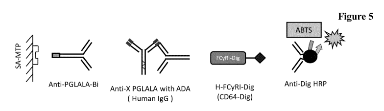
BRIEF DESCRIPTION OF THE FIGURES
Figure 1 Scheme of an immunoassay using the antibody as reported herein
as capture reagent.
Figure 2 Scheme of an immunoassay using the antibody as reported
herein
as tracer reagent.
Figure 3 Scheme of an immunoassay using the antibody as reported
herein
as capture and as tracer reagent.
Figure 4 Scheme of an immunoassay using the antibody as reported
herein
as standard.
Figure 5 Scheme of an immunoassay using the antibody as reported
herein
as capture antibody and a soluble Fcgamma receptor as tracer
molecule.
Figure 6 Scheme of an immunoassay using the antibody as reported
herein
as capture antibody and an anti-target antibody as tracer antibody.
Figure 7 Exemplary time course of ADA occurrence in 4 patients of
a
clinical trial: patients had received biweekly (A,C) or weekly
administrations (B) of the therapeutic antibody (10 mg each
dose), and blood samples were collected daily before and after
each dose (C2, C3: second and third treatment cycle). All samples
were tested for anti-drug antibodies (ADAs) by both the
conventional bridging (light grey bars) and the hsFcyRI-PG assay
(black bars). Blood drug concentrations (ng/ml) are provided
(bottom curve), and samples assessed as ADA negative (-) or
positive (+) are indicated above the corresponding bars.
DETAILED DESCRIPTION OF EMBODIMENTS OF THE INVENTION
I. DEFINITIONS
As used herein, the amino acid positions of all constant regions and domains
of the
heavy and light chain are numbered according to the Kabat numbering system
described in Kabat, et al., Sequences of Proteins of Immunological Interest,
5th ed.,
Public Health Service, National Institutes of Health, Bethesda, MD (1991) and
is
CA 03000048 2018-03-27
WO 2017/072210
PCT/EP2016/075884
- 7 -
referred to as "numbering according to Kabat" herein. Specifically the Kabat
numbering system (see pages 647-660) of Kabat, et al., Sequences of Proteins
of
Immunological Interest, 5th ed., Public Health Service, National Institutes of
Health, Bethesda, MD (1991) is used for the light chain constant domain CL of
kappa and lambda isotype and the Kabat EU index numbering system (see pages
661-723) is used for the constant heavy chain domains (CH1, Hinge, CH2 and
CH3).
An "acceptor human framework" for the purposes herein is a framework
comprising the amino acid sequence of a light chain variable domain (VL)
framework or a heavy chain variable domain (VH) framework derived from a
human immunoglobulin framework or a human consensus framework, as defined
below. An acceptor human framework "derived from" a human immunoglobulin
framework or a human consensus framework may comprise the same amino acid
sequence thereof, or it may contain amino acid sequence changes. In some
embodiments, the number of amino acid changes are 10 or less, 9 or less, 8 or
less,
7 or less, 6 or less, 5 or less, 4 or less, 3 or less, or 2 or less. In some
embodiments,
the VL acceptor human framework is identical in sequence to the VL human
immunoglobulin framework sequence or human consensus framework sequence.
"Affinity" refers to the strength of the sum total of non-covalent
interactions
between a single binding site of a molecule (e.g., an antibody) and its
binding
partner (e.g., an antigen). Unless indicated otherwise, as used herein,
"binding
affinity" refers to intrinsic binding affinity which reflects a 1:1
interaction between
members of a binding pair (e.g., antibody and antigen). The affinity of a
molecule
X for its partner Y can generally be represented by the dissociation constant
(kd).
Affinity can be measured by common methods known in the art, including those
described herein.
An "affinity matured" antibody refers to an antibody with one or more
alterations
in one or more hypervariable regions (HVRs), compared to a parent antibody
which does not possess such alterations, such alterations resulting in an
improvement in the affinity of the antibody for antigen.
The term "alteration" denotes the mutation, addition, or deletion of one or
more
amino acid residues in a parent amino acid sequence, e.g. of an antibody or
fusion
polypeptide comprising at least an FcRn binding portion of an Fc-region, to
obtain
a variant antibody or fusion polypeptide.
CA 03000048 2018-03-27
WO 2017/072210
PCT/EP2016/075884
- 8 -
The term "amino acid mutation" denotes a modification in the amino acid
sequence
of a parent amino acid sequence. Exemplary modifications include amino acid
substitutions, insertions, and/or deletions. In one embodiment the amino acid
mutation is a substitution. The term "amino acid mutations at the position"
denotes
the substitution or deletion of the specified residue, or the insertion of at
least one
amino acid residue adjacent the specified residue. The term "insertion
adjacent to a
specified residue" denotes the insertion within one to two residues thereof
The
insertion may be N-terminal or C-terminal to the specified residue.
The term "amino acid substitution" denotes the replacement of at least one
amino
acid residue in a predetermined parent amino acid sequence with a different
"replacement" amino acid residue. The replacement residue or residues may be a
"naturally occurring amino acid residue" (i.e. encoded by the genetic code)
and
selected from the group consisting of: alanine (Ala); arginine (Arg);
asparagine
(Asn); aspartic acid (Asp); cysteine (Cys); glutamine (Gin); glutamic acid
(Glu);
glycine (Gly); histidine (His); isoleucine (Ile): leucine (Leu); lysine (Lys);
methionine (Met); phenylalanine (Phe); proline (Pro); serine (Ser); threonine
(Thr);
tryptophan (Trp); tyrosine (Tyr); and valine (Val). In one embodiment the
replacement residue is not cysteine. Substitution with one or more non-
naturally
occurring amino acid residues is also encompassed by the definition of an
amino
acid substitution herein. A "non-naturally occurring amino acid residue"
denotes a
residue, other than those naturally occurring amino acid residues listed
above,
which is able to covalently bind adjacent amino acid residues(s) in a
polypeptide
chain. Examples of non-naturally occurring amino acid residues include
norleucine,
ornithine, norvaline, homoserine, aib and other amino acid residue analogues
such
as those described in Ellman, et al., Meth. Enzym. 202 (1991) 301-336. To
generate such non-naturally occurring amino acid residues, the procedures of
Noren, et al. (Science 244 (1989) 182) and/or Ellman, et al. (supra) can be
used.
Briefly, these procedures involve chemically activating a suppressor tRNA with
a
non-naturally occurring amino acid residue followed by in vitro transcription
and
translation of the RNA. Non-naturally occurring amino acids can also be
incorporated into peptides via chemical peptide synthesis and subsequent
fusion of
these peptides with recombinantly produced polypeptides, such as antibodies or
antibody fragments.
The term "amino acid insertion" denotes the incorporation of at least one
additional
amino acid residue into a predetermined parent amino acid sequence. While the
CA 03000048 2018-03-27
WO 2017/072210
PCT/EP2016/075884
- 9 -
insertion will usually consist of the insertion of one or two amino acid
residues, the
present application contemplates larger "peptide insertions", e.g. insertion
of about
three to about five or even up to about ten amino acid residues. The inserted
residue(s) may be naturally occurring or non-naturally occurring as defined
above.
The term "amino acid deletion" denotes the removal of at least one amino acid
residue at a predetermined position in an amino acid sequence.
Within this application whenever an amino acid alteration is mentioned it is a
deliberated amino acid alteration and not a random amino acid modification.
The terms "anti-variant (human) Fc-region antibody" and "an antibody that
specifically binds to a variant (human) Fc-region" refer to an antibody that
is
capable of binding a variant (human) Fc-region with sufficient affinity such
that the
antibody is useful as a diagnostic agent in targeting a variant (human) Fc-
region. In
one embodiment, the extent of binding of an anti-variant (human) Fc-region
antibody to the corresponding wild-type (human) Fc-region is less than about
10 %
of the binding of the antibody to the variant (human) Fc-region. This can be
determined e.g. using Surface Plasmon Resonance. In certain embodiments, an
antibody that specifically binds to a variant (human) Fc-region has a
dissociation
constant (KD) of 10-8 M or less, e.g. from 10-8 M to 10-13 M, e.g., from 10-9
M to 10-
13
M).
The term "antibody" herein is used in the broadest sense and encompasses
various
antibody structures, including but not limited to monoclonal antibodies,
polyclonal
antibodies, multispecific antibodies (e.g., bispecific antibodies), and
antibody
fragments so long as they exhibit the desired antigen-binding activity.
An "antibody fragment" refers to a molecule other than an intact antibody that
comprises a portion of an intact antibody that binds the antigen to which the
intact
antibody binds. Examples of antibody fragments include but are not limited to
Fv,
Fab, Fab', Fab'-SH, F(ab')2; diabodies; linear antibodies; single-chain
antibody
molecules (e.g. scFv); and multispecific antibodies formed from antibody
fragments.
The term "binding to" denotes the binding of a first entity to a second
entity, such
as e.g. of an antibody to its antigen. This binding can be determined using,
for
example, a BIAcore0 assay (GE Healthcare, Uppsala, Sweden).
CA 03000048 2018-03-27
WO 2017/072210
PCT/EP2016/075884
- 10 -
For example, in one possible embodiment of the BIAcore0 assay the antigen is
bound to a surface and binding of the antibody is measured by surface plasmon
resonance (SPR). The affinity of the binding is defined by the terms ka
(association
constant: rate constant for the association to form a complex), kd
(dissociation
constant; rate constant for the dissociation of the complex), and KD (kd/ka).
Alternatively, the binding signal of a SPR sensorgram can be compared directly
to
the response signal of a reference, with respect to the resonance signal
height and
the dissociation behaviors.
The term "CH2 domain" denotes the part of an antibody heavy chain polypeptide
that extends approximately from EU position 231 to EU position 340 (EU
numbering system according to Kabat). The CH2 domain is unique in that it is
not
closely paired with another domain. Rather, two N-linked branched carbohydrate
chains are interposed between the two CH2 domains of an intact native Fc-
region.
It has been speculated that the carbohydrate may provide a substitute for the
domain-domain pairing and help stabilize the CH2 domain. Burton, Mol. Immunol.
22 (1985) 161-206.
The term "CH3 domain" denotes the part of an antibody heavy chain polypeptide
that extends approximately from EU position 341 to EU position 446.
The term "chimeric" antibody refers to an antibody in which a portion of the
heavy
and/or light chain is derived from a particular source or species, while the
remainder of the heavy and/or light chain is derived from a different source
or
species.
The "class" of an antibody refers to the type of constant domain or constant
region
possessed by its heavy chain. There are five major classes of antibodies: IgA,
IgD,
IgE, IgG, and IgM, and several of these may be further divided into subclasses
(isotypes), e.g., IgGi, IgG2, IgG3, Igai, IgAi, and IgA2. The heavy chain
constant
domains that correspond to the different classes of immunoglobulins are called
a,
8, e, 7, and , respectively.
The term "complement-dependent cytotoxicity (CDC)" refers to lysis of cells
induced by the antibody as reported herein in the presence of complement. CDC
is
found if the antibody induces lysis of 20 % or more of the target cells at a
concentration of 30 g/ml. Binding to the complement factor Clq can be
measured
in an ELISA. In such an assay in principle an ELISA plate is coated with
CA 03000048 2018-03-27
WO 2017/072210
PCT/EP2016/075884
- 11 -
concentration ranges of the antibody, to which purified human C 1 q or human
serum is added. C 1 q binding is detected by an antibody directed against C 1
q
followed by a peroxidase-labeled conjugate. Detection of binding (maximal
binding Bmax) is measured as optical density at 405 nm (0D405) for peroxidase
substrate ABTSO (2,2'-azino-di-[3-ethylbenzthiazoline-6-sulfonate (6)]).
"Effector functions" refer to those biological activities attributable to the
Fc-region
of an antibody, which vary with the antibody class. Examples of antibody
effector
functions include: C 1 q binding and complement dependent cytotoxicity (CDC);
Fc
receptor binding; antibody-dependent cell-mediated cytotoxicity (ADCC);
phagocytosis; down regulation of cell surface receptors (e.g. B cell
receptor); and B
cell activation.
Fc receptor binding dependent effector functions can be mediated by the
interaction
of the Fc-region of an antibody with Fc receptors (FcRs), which are
specialized cell
surface receptors on hematopoietic cells. Fc receptors belong to the
immunoglobulin superfamily, and have been shown to mediate both the removal of
antibody-coated pathogens by phagocytosis of immune complexes, and the lysis
of
erythrocytes and various other cellular targets (e.g. tumor cells) coated with
the
corresponding antibody, via antibody dependent cell mediated cytotoxicity
(ADCC) (see e.g. Van de Winkel, J.G. and Anderson, C.L., J. Leukoc. Biol. 49
(1991) 511-524). FcRs are defined by their specificity for immunoglobulin
isotypes: Fc receptors for IgG antibodies are referred to as FcyR. Fc receptor
binding is described e.g. in Ravetch, J.V. and Kinet, J.P., Annu. Rev.
Immunol. 9
(1991) 457-492; Capel, P.J., et al., Immunomethods 4 (1994) 25-34; de Haas,
M.,
et al., J. Lab. Clin. Med. 126 (1995) 330-341; Gessner, J.E., et al., Ann.
Hematol.
76 (1998) 231-248.
Cross-linking of receptors for the Fc-region of IgG antibodies (FcyR) triggers
a
wide variety of effector functions including phagocytosis, antibody-dependent
cellular cytotoxicity, and release of inflammatory mediators, as well as
immune
complex clearance and regulation of antibody production. In humans, three
classes
of FcyR have been characterized, which are:
¨ FcyRI (CD64) binds monomeric IgG with high affinity and is expressed on
macrophages, monocytes, neutrophils and eosinophils. Modification in the Fc-
region IgG at least at one of the amino acid residues E233-G236, P238, D265,
N297, A327 and P329 (numbering according to EU index of Kabat) reduce
CA 03000048 2018-03-27
WO 2017/072210
PCT/EP2016/075884
- 12 -
binding to FcyRI. IgG2 residues at positions 233-236, substituted into IgG1
and IgG4, reduced binding to FcyRI by 103-fold and eliminated the human
monocyte response to antibody-sensitized red blood cells (Armour, K.L., et
al.,
Eur. J. Immunol. 29 (1999) 2613-2624).
¨ FcyRII (CD32) binds complexed IgG with medium to low affinity and is
widely expressed. This receptor can be divided into two sub-types, FcyRIIA
and FcyRIIB. FcyRIIA is found on many cells involved in killing (e.g.
macrophages, monocytes, neutrophils) and seems able to activate the killing
process. FcyRIIB seems to play a role in inhibitory processes and is found on
B-cells, macrophages and on mast cells and eosinophils. On B-cells it seems to
function to suppress further immunoglobulin production and isotype switching
to, for example, the IgE class. On macrophages, FcyRIIB acts to inhibit
phagocytosis as mediated through FcyRIIA. On eosinophils and mast cells the
B-form may help to suppress activation of these cells through IgE binding to
its
separate receptor. Reduced binding for FcyRIIA is found e.g. for antibodies
comprising an IgG Fc-region with mutations at least at one of the amino acid
residues E233-G236, P238, D265, N297, A327, P329, D270, Q295, A327,
R292, and K414 (numbering according to EU index of Kabat).
¨ FcyRIII (CD16) binds IgG with medium to low affinity and exists as two
types.
FcyRIIIA is found on NK cells, macrophages, eosinophils and some monocytes
and T cells and mediates ADCC. FcyRIIIB is highly expressed on neutrophils.
Reduced binding to FcyRIIIA is found e.g. for antibodies comprising an IgG
Fc-region with mutation at least at one of the amino acid residues E233-G236,
P238, D265, N297, A327, P329, D270, Q295, A327, S239, E269, E293, Y296,
V303, A327, K338 and D376 (numbering according to EU index of Kabat).
Mapping of the binding sites on human IgG1 for Fc receptors, the above
mentioned
mutation sites and methods for measuring binding to FcyRI and FcyRIIA are
described in Shields, R.L., et al. J. Biol. Chem. 276 (2001) 6591-6604.
The term "Fc receptor" as used herein refers to activation receptors
characterized
by the presence of a cytoplasmatic ITAM sequence associated with the receptor
(see e.g. Ravetch, J.V. and Bolland, S., Annu. Rev. Immunol. 19 (2001) 275-
290).
Such receptors are FcyRI, FcyRIIA and FcyRIIIA. The term "no binding of FcyR"
denotes that at an antibody concentration of 10 g/ml the binding of an
antibody as
CA 03000048 2018-03-27
WO 2017/072210
PCT/EP2016/075884
- 13 -
reported herein to NK cells is 10 % or less of the binding found for anti-
OX4OL
antibody LC.001 as reported in WO 2006/029879.
While IgG4 shows reduced FcR binding, antibodies of other IgG subclasses show
strong binding. However Pro238, Asp265, Asp270, Asn297 (loss of Fc
carbohydrate), Pro329 and 234, 235, 236 and 237 11e253, Ser254, Lys288 ,
Thr307,
G1n311, Asn434, and His435 are residues which provide if altered also reduce
FcR
binding (Shields, R.L., et al. J. Biol. Chem. 276 (2001) 6591-6604; Lund, J.,
et al.,
FASEB J. 9 (1995) 115-119; Morgan, A., et al., Immunology 86 (1995) 319-324;
and EP 0 307 434). In one embodiment the antibody as reported herein is of
IgG1
or IgG2 subclass and comprises the mutation PVA236, GLPSS331, and/or
L234A/L235A. In one embodiment the antibody as reported herein is of IgG4
subclass and comprises the mutation L235E. In one embodiment the antibody
further comprises the mutation 5228P.
The term "Fc-region" herein is used to define a C-terminal region of an
immunoglobulin heavy chain that contains at least a portion of the constant
region.
The term includes native sequence Fc-regions and variant Fc-regions. In one
embodiment, a human IgG heavy chain Fc-region extends from Cys226, or from
Pro230, to the carboxyl-terminus of the heavy chain. However, the C-terminal
lysine (Lys447) of the Fc-region may or may not be present. Unless otherwise
specified herein, numbering of amino acid residues in the Fc-region or
constant
region is according to the EU numbering system, also called the EU index, as
described in Kabat, E.A. et al., Sequences of Proteins of Immunological
Interest,
5th ed., Public Health Service, National Institutes of Health, Bethesda, MD
(1991),
NIH Publication 91-3242.
The antibodies as reported herein comprise as Fc-region, in one embodiment an
Fc-
region derived from human origin. In one embodiment the Fc-region comprises
all
parts of the human constant region. The Fc-region of an antibody is directly
involved in complement activation, C 1 q binding, C3 activation and Fc
receptor
binding. While the influence of an antibody on the complement system is
dependent on certain conditions, binding to Clq is caused by defined binding
sites
in the Fc-region. Such binding sites are known in the state of the art and
described
e.g. by Lukas, T.J., et al., J. Immunol. 127 (1981) 2555-2560; Brunhouse, R.,
and
Cebra, J.J., Mol. Immunol. 16 (1979) 907-917; Burton, D.R., et al., Nature 288
(1980) 338-344; Thommesen, J.E., et al., Mol. Immunol. 37 (2000) 995-1004;
Idusogie, E.E., et al., J. Immunol. 164 (2000) 4178-4184; Hezareh, M., et al.,
J.
CA 03000048 2018-03-27
WO 2017/072210
PCT/EP2016/075884
- 14 -
Virol. 75 (2001) 12161-12168; Morgan, A., et al., Immunology 86 (1995) 319-
324;
and EP 0 307 434. Such binding sites are e.g. L234, L235, D270, N297, E318,
K320, K322, P331 and P329 (numbering according to EU index of Kabat; Unless
otherwise specified herein, numbering of amino acid residues in the Fc-region
or
constant region is according to the EU numbering system, also called the EU
index,
as described in Kabat, E.A. et al., Sequences of Proteins of Immunological
Interest,
5th ed., Public Health Service, National Institutes of Health, Bethesda, MD
(1991),
NIH Publication 91-3242). Antibodies of subclass IgGl, IgG2 and IgG3 usually
show complement activation, C 1 q binding and C3 activation, whereas IgG4 do
not
activate the complement system, do not bind C 1 q and do not activate C3. An
"Fc-
region of an antibody" is a term well known to the skilled artisan and defined
on
the basis of papain cleavage of antibodies. In one embodiment the Fc-region is
a
human Fc-region. In one embodiment the Fc-region is of the human IgG4 subclass
comprising the mutations S228P and/or L235E (numbering according to EU index
of Kabat). In one embodiment the Fc-region is of the human IgG1 subclass
comprising the mutations L234A and L235A (numbering according to EU index of
Kabat).
"Framework" or "FR" refers to variable domain residues other than
hypervariable
region (HVR) residues. The FR of a variable domain generally consists of four
FR
domains: FR1, FR2, FR3, and FR4. Accordingly, the HVR and FR sequences
generally appear in the following sequence in VH (or VL): FR1-H1(L1)-FR2-
H2(L2)-FR3 -H3 (L3)-FR4 .
The terms "full length antibody", "intact antibody", and "whole antibody" are
used
herein interchangeably to refer to an antibody having a structure
substantially
similar to a native antibody structure or having heavy chains that contain an
Fc-
region as defined herein.
The terms "host cell", "host cell line", and "host cell culture" are used
interchangeably and refer to cells into which exogenous nucleic acid has been
introduced, including the progeny of such cells. Host cells include
"transformants"
and "transformed cells," which include the primary transformed cell and
progeny
derived therefrom without regard to the number of passages. Progeny may not be
completely identical in nucleic acid content to a parent cell, but may contain
mutations. Mutant progeny that have the same function or biological activity
as
screened or selected for in the originally transformed cell are included
herein.
CA 03000048 2018-03-27
WO 2017/072210
PCT/EP2016/075884
- 15 -
A "human consensus framework" is a framework which represents the most
commonly occurring amino acid residues in a selection of human immunoglobulin
VL or VH framework sequences. Generally, the selection of human
immunoglobulin VL or VH sequences is from a subgroup of variable domain
sequences. Generally, the subgroup of sequences is a subgroup as in Kabat,
E.A. et
al., Sequences of Proteins of Immunological Interest, 5th ed., Bethesda MD
(1991),
NIH Publication 91-3242, Vols. 1-3. In one embodiment, for the VL, the
subgroup
is subgroup kappa I as in Kabat et al., supra. In one embodiment, for the VH,
the
subgroup is subgroup III as in Kabat et al., supra.
A "humanized" antibody refers to a chimeric antibody comprising amino acid
residues from non-human HVRs and amino acid residues from human FRs. In
certain embodiments, a humanized antibody will comprise substantially all of
at
least one, and typically two, variable domains, in which all or substantially
all of
the HVRs (e.g., CDRs) correspond to those of a non-human antibody, and all or
substantially all of the FRs correspond to those of a human antibody. A
humanized
antibody optionally may comprise at least a portion of an antibody constant
region
derived from a human antibody. A "humanized form" of an antibody, e.g., a non-
human antibody, refers to an antibody that has undergone humanization.
The term "hypervariable region" or "HVR", as used herein, refers to each of
the
regions of an antibody variable domain comprising the amino acid residue
stretches
which are hypervariable in sequence ("complementarity determining regions" or
"CDRs") and/or form structurally defined loops ("hypervariable loops"), and/or
contain the antigen-contacting residues ("antigen contacts"). Generally,
antibodies
comprise six HVRs; three in the VH (H1, H2, H3), and three in the VL (L1, L2,
L3).
HVRs include
(a) hypervariable loops occurring at amino acid residues 26-32 (L1), 50-52
(L2), 91-96 (L3), 26-32 (H1), 53-55 (H2), and 96-101 (H3) (Chothia, C.
and Lesk, A.M., J. Mol. Biol. 196 (1987) 901-917);
(b) CDRs occurring at amino acid residues 24-34 ( L1), 50-56 (L2), 89-97 (L3),
31-35b (H1), 50-65 (H2), and 95-102 (H3) (Kabat, E.A. et al., Sequences of
Proteins of Immunological Interest, 5th ed. Public Health Service, National
Institutes of Health, Bethesda, MD (1991), NIH Publication 91-3242.);
CA 03000048 2018-03-27
WO 2017/072210
PCT/EP2016/075884
- 16 -
(c) antigen contacts occurring at amino acid residues 27c-36 (L1), 46-55 (L2),
89-96 (L3), 30-35b (H1), 47-58 (H2), and 93-101 (H3) (MacCallum et al. J.
Mol. Biol. 262: 732-745 (1996)); and
(d) combinations of (a), (b), and/or (c), including amino acid residues 46-56
(L2), 47-56 (L2), 48-56 (L2), 49-56 (L2), 26-35 (H1), 26-35b (H1), 49-65
(H2), 93-102 (H3), and 94-102 (H3).
Unless otherwise indicated, HVR residues and other residues in the variable
domain (e.g., FR residues) are numbered herein according to Kabat et al.,
supra.
The term "hinge region" denotes the part of an antibody heavy chain
polypeptide
that joins in a wild-type antibody heavy chain the CH1 domain and the CH2
domain, e. g. from about position 216 to about position 230 according to the
EU
number system of Kabat, or from about position 226 to about position 230
according to the EU number system of Kabat. The hinge regions of other IgG
subclasses can be determined by aligning with the hinge-region cysteine
residues of
the IgG1 subclass sequence.
The hinge region is normally a dimeric molecule consisting of two polypeptides
with identical amino acid sequence. The hinge region generally comprises about
25
amino acid residues and is flexible allowing the antigen binding regions to
move
independently. The hinge region can be subdivided into three domains: the
upper,
the middle, and the lower hinge domain (see e.g. Roux, et al., J. Immunol. 161
(1998) 4083).
In one embodiment the hinge region has the amino acid sequence
DKTHTCPX4CP, wherein X4 is either S or P. In one embodiment the hinge region
has the amino acid sequence HTCPX4CP, wherein X4 is either S or P. In one
embodiment the hinge region has the amino acid sequence CPX4CP, wherein X4 is
either S or P.
The term "lower hinge region" of an Fc-region denotes the stretch of amino
acid
residues immediately C-terminal to the hinge region, i.e. residues 233 to 239
of the
Fc-region according to the EU numbering of Kabat.
An "individual" or "subject" is a mammal. Mammals include, but are not limited
to, domesticated animals (e.g. cows, sheep, cats, dogs, and horses), primates
(e.g.,
CA 03000048 2018-03-27
WO 2017/072210
PCT/EP2016/075884
- 17 -
humans and non-human primates such as monkeys), rabbits, and rodents (e.g.,
mice
and rats). In certain embodiments, the individual or subject is a human.
An "isolated" antibody is one which has been separated from a component of its
natural environment. In some embodiments, an antibody is purified to greater
than
95% or 99% purity as determined by, for example, electrophoretic (e.g., SDS-
PAGE, isoelectric focusing (IEF), capillary electrophoresis) or
chromatographic
(e.g., ion exchange or reverse phase HPLC). For review of methods for
assessment
of antibody purity, see, e.g., Flatman, S. et al., J. Chromatogr. B 848 (2007)
79-87.
An "isolated" nucleic acid refers to a nucleic acid molecule that has been
separated
from a component of its natural environment. An isolated nucleic acid includes
a
nucleic acid molecule contained in cells that ordinarily contain the nucleic
acid
molecule, but the nucleic acid molecule is present extrachromosomally or at a
chromosomal location that is different from its natural chromosomal location.
"Isolated nucleic acid encoding an anti-variant (human) Fc-region antibody"
refers
to one or more nucleic acid molecules encoding antibody heavy and light chains
(or
fragments thereof), including such nucleic acid molecule(s) in a single vector
or
separate vectors, and such nucleic acid molecule(s) present at one or more
locations
in a host cell.
The term "monoclonal antibody" as used herein refers to an antibody obtained
from
a population of substantially homogeneous antibodies, i.e., the individual
antibodies comprising the population are identical and/or bind the same
epitope,
except for possible variant antibodies, e.g., containing naturally occurring
mutations or arising during production of a monoclonal antibody preparation,
such
variants generally being present in minor amounts. In contrast to polyclonal
antibody preparations, which typically include different antibodies directed
against
different determinants (epitopes), each monoclonal antibody of a monoclonal
antibody preparation is directed against a single determinant on an antigen.
Thus,
the modifier "monoclonal" indicates the character of the antibody as being
obtained
from a substantially homogeneous population of antibodies, and is not to be
construed as requiring production of the antibody by any particular method.
For
example, the monoclonal antibodies to be used in accordance with the present
invention may be made by a variety of techniques, including but not limited to
the
hybridoma method, recombinant DNA methods, phage-display methods, and
methods utilizing transgenic animals containing all or part of the human
CA 03000048 2018-03-27
WO 2017/072210
PCT/EP2016/075884
- 18 -
immunoglobulin loci, such methods and other exemplary methods for making
monoclonal antibodies being described herein.
"Native antibodies" refer to naturally occurring immunoglobulin molecules with
varying structures. For example, native IgG antibodies are heterotetrameric
glycoproteins of about 150,000 daltons, composed of two identical light chains
and
two identical heavy chains that are disulfide-bonded. From N- to C-terminus,
each
heavy chain has a variable region (VH), also called a variable heavy domain or
a
heavy chain variable domain, followed by three constant domains (CH1, CH2, and
CH3), whereby between the first and the second constant domain a hinge region
is
located. Similarly, from N- to C-terminus, each light chain has a variable
region
(VL), also called a variable light domain or a light chain variable domain,
followed
by a constant light (CL) domain. The light chain of an antibody may be
assigned to
one of two types, called kappa (x) and lambda (4 based on the amino acid
sequence of its constant domain.
"Percent (%) amino acid sequence identity" with respect to a reference
polypeptide
sequence is defined as the percentage of amino acid residues in a candidate
sequence that are identical with the amino acid residues in the reference
polypeptide sequence, after aligning the sequences and introducing gaps, if
necessary, to achieve the maximum percent sequence identity, and not
considering
any conservative substitutions as part of the sequence identity. Alignment for
purposes of determining percent amino acid sequence identity can be achieved
in
various ways that are within the skill in the art, for instance, using
publicly
available computer software such as BLAST, BLAST-2, ALIGN or Megalign
(DNASTAR) software. Those skilled in the art can determine appropriate
parameters for aligning sequences, including any algorithms needed to achieve
maximal alignment over the full length of the sequences being compared. For
purposes herein, however, % amino acid sequence identity values are generated
using the sequence comparison computer program ALIGN-2. The ALIGN-2
sequence comparison computer program was authored by Genentech, Inc., and the
source code has been filed with user documentation in the U.S. Copyright
Office,
Washington D.C., 20559, where it is registered under U.S. Copyright
Registration
No. TXU510087. The ALIGN-2 program is publicly available from Genentech,
Inc., South San Francisco, California, or may be compiled from the source
code.
The ALIGN-2 program should be compiled for use on a UNIX operating system,
CA 03000048 2018-03-27
WO 2017/072210
PCT/EP2016/075884
- 19 -
including digital UNIX V4.0D. All sequence comparison parameters are set by
the
ALIGN-2 program and do not vary.
In situations where ALIGN-2 is employed for amino acid sequence comparisons,
the % amino acid sequence identity of a given amino acid sequence A to, with,
or
against a given amino acid sequence B (which can alternatively be phrased as a
given amino acid sequence A that has or comprises a certain % amino acid
sequence identity to, with, or against a given amino acid sequence B) is
calculated
as follows:
100 times the fraction X/Y
where X is the number of amino acid residues scored as identical matches by
the
sequence alignment program ALIGN-2 in that program's alignment of A and B,
and where Y is the total number of amino acid residues in B. It will be
appreciated
that where the length of amino acid sequence A is not equal to the length of
amino
acid sequence B, the % amino acid sequence identity of A to B will not equal
the %
amino acid sequence identity of B to A. Unless specifically stated otherwise,
all %
amino acid sequence identity values used herein are obtained as described in
the
immediately preceding paragraph using the ALIGN-2 computer program.
The terms "native Fc-region" and "wild-type Fc-region" as used herein, refers
to
any native or wild-type Fc-region from any vertebrate source, including
mammals
such as primates (e.g. humans) and rodents (e.g., mice and rats), unless
otherwise
indicated.
The term "variant (human) Fc-region" denotes an amino acid sequence which
differs from that of a "native" or "wild-type" (human) Fc-region amino acid
sequence by virtue of at least one "amino acid alteration/mutation". In one
embodiment the variant Fc-region has at least one amino acid mutation compared
to a native Fc-region, e.g. from about one to about ten amino acid mutations,
and in
one embodiment from about one to about five amino acid mutations in a native
Fc-
region. In one embodiment the (variant) Fc-region has at least about 80 %
homology with a wild-type Fc-region, and in one embodiment the variant Fc-
region has least about 90 % homology, in one embodiment the variant Fc-region
has at least about 95 % homology.
The variant Fc-regions as reported herein are defined by the amino acid
alterations
that are contained. Thus, for example, the term P329G denotes a variant Fc-
region
CA 03000048 2018-03-27
WO 2017/072210
PCT/EP2016/075884
- 20 -
with the mutation of proline to glycine at amino acid position 329 relative to
the
parent (wild-type) Fc-region. The identity of the wild-type amino acid may be
unspecified, in which case the aforementioned variant is referred to as 329G.
The
alteration can be an addition, deletion, or mutation. The term "mutation"
denotes a
change to naturally occurring amino acids as well as a change to non-naturally
occurring amino acids (see e.g. US 6,586,207, WO 98/48032, WO 03/073238, US
2004/0214988, WO 2005/35727, WO 2005/74524, Chin, J.W., et al., J. Am. Chem.
Soc. 124 (2002) 9026-9027; Chin, J.W. and Schultz, P.G., ChemBioChem 11
(2002) 1135-1137; Chin, J.W., et al., PICAS United States of America 99 (2002)
11020-11024;Wang, L. and Schultz, P.G., Chem. (2002) 1-10).
The term "wild-type Fc-region" denotes an amino acid sequence identical to the
amino acid sequence of an Fc-region found in nature. Wild-type human Fc-
regions
include a native human IgG1 Fc-region (non-A and A allotypes), native human
IgG2 Fc-region, native human IgG3 Fc-region, and native human IgG4 Fc-region
as well as naturally occurring variants thereof.
The term "therapeutic antibody" relates to any antibody preparation which is
intended for use in a human being. Preferably such therapeutic antibody will
be a
monoclonal antibody. Further preferred such monoclonal antibody will be
obtained
from a great ape or be a human monoclonal antibody. Preferably, it will be a
human monoclonal antibody. Also preferred such therapeutic monoclonal antibody
will be a humanized monoclonal antibody.
The term "variable region" or "variable domain" refers to the domain of an
antibody heavy or light chain that is involved in binding the antibody to
antigen.
The variable domains of the heavy chain and light chain (VH and VL,
respectively)
of a native antibody generally have similar structures, with each domain
comprising four conserved framework regions (FRs) and three hypervariable
regions (HVRs). (See, e.g., Kindt, T.J. et al. Kuby Immunology, 6th ed., W.H.
Freeman and Co., N.Y. (2007), page 91) A single VH or VL domain may be
sufficient to confer antigen-binding specificity. Furthermore, antibodies that
bind a
particular antigen may be isolated using a VH or VL domain from an antibody
that
binds the antigen to screen a library of complementary VL or VH domains,
respectively (see, e.g., Portolano, S., et al., J. Immunol. 150 (1993) 880-
887;
Clackson, T., et al., Nature 352 (1991) 624-628).
CA 03000048 2018-03-27
WO 2017/072210
PCT/EP2016/075884
- 21 -
The term "vector", as used herein, refers to a nucleic acid molecule capable
of
propagating another nucleic acid to which it is linked. The term includes the
vector
as a self-replicating nucleic acid structure as well as the vector
incorporated into
the genome of a host cell into which it has been introduced. Certain vectors
are
capable of directing the expression of nucleic acids to which they are
operatively
linked. Such vectors are referred to herein as "expression vectors".
II. COMPOSITIONS AND METHODS
In one aspect, the invention is based, in part, on the finding that antibodies
specifically binding to a variant (human) Fc-region can be provided that do
not
substantially bind to the corresponding wild-type (human) Fc-region. In
certain
embodiments, antibodies that specifically bind to a human Fc-region with the
mutations P329G or L234A, L235A, P329G or I253A, H310A, H435A are
provided (numbering according to EU index of Kabat). Antibodies of the
invention
are useful, e.g., for the determination of the respective therapeutic antibody
in a
sample or for the determination of anti-drug antibodies (ADA) against a
therapeutic
antibody.
A. Exemplary Anti-Fc-region Antibodies
As described earlier for preclinical studies in cynomolgus or mice
(Stubenrauch,
K., et al., J. Pharm. Biomed. Anal. 52 (2010) 249-254; Moore, G.L., et al.,
MAbs 2
(2010) 181-189; Stubenrauch, K., et al., Anal. Biochem. 430 (2012) 193-199;
Carrasco-Triguero, M., et al., J. Immunol. Res. 2016 2618575 (2016)), drug
tolerance can be improved by generic or universal assay formats that detect
complexes of drug and ADA (anti-drug antibody). Likewise, detection of drug-
ADA complexes (drug = therapeutic antibody administered beforehand) by the
assay as reported herein, e.g. the hsFcyRI-PG assay, requires no labeled drug
antibody as capture or detection reagent. It has been found that this assay
setup lead
to improved drug tolerance. This has been confirmed in spiking experiments.
The drug tolerance factor is the ratio between the amount of positively
detectable
positive control and the amount of drug present in the sample. For the
bridging
assay, a drug tolerance factor of 6 (500 ng/mL positive control could be
detected in
the presence of up to 3 iug/mL drug) whereas for the hsFcyRI-PG assay a drug
tolerance factor of 833 (30 ng/ml control ADA were found positive in the
presence
of up to 25 ug/m1 drug) was determined.
CA 03000048 2018-03-27
WO 2017/072210
PCT/EP2016/075884
- 22 -
With the assay as reported herein and a standard bridging assay the time
course of
ADA occurrence was determined in clinical samples from four patients (Figure
7).
These datasets show in an exemplary way the differences of both assays
regarding
Drug tolerance, sensitivity and Ig isotype specificity.
In patient A (Table below; Figure 7A), both assays show early ADA positivity
at
120h for the bridging assay and 168h for the hsFcyRI-PG assay. The ADA signals
at 168h for both assays are comparable and significant with approximately 0.5
OD.
The corresponding drug level in the samples is very low (about 2 ng/ml). At
the
next measured time point, 24h after second dosing (C2 24h), the drug level is
much
higher (about 2430 ng/mL). This influences the bridging assay, resulting in a
major
signal drop below the corresponding cut point. The hsFcyRI-PG assay in
contrast
is not affected by the drug and the ADA signal even rises from 168h to C2 24h.
Similar signal drops due to rising drug levels for the bridging assay were
observed
in other patient samples (see Table below). The signals of the hsFcyRI-PG
assay
were not affected in any of the patients, confirming the improved drug
tolerance of
this assay.
Table: Assessment of ADA formation in 8 patients by the conventional bridging
and the hsFcyRI-PG assay as reported herein.
,tnts
Patient A .. Pa t E Patient F
I t2
E
- 8 -a r = BP
- _
S S I IS- if =
u-
- co I co 2 co CCI
2411
72h 7
-
-
96h - 18 - - + 1
120h - + 7 - - - +
168h + >
_ + -
C2 16 h + + +
C3 pre + 4- - Oih. j- .Q
CA 03000048 2018-03-27
WO 2017/072210
PCT/EP2016/075884
- 23 -
Anc=,,4 rtattontS
t
w
korEq.elkqe, Lg,
m lb,- co So I Slic la 05 cff
24h 385 18DO Dosing
721) 0
96h
C2 Pre b Q
, .
C2 9 + .2 = = ,Q
C3 pte _
C3 24h
C3 96h
Figure 7B shows the time course of patient B (Table above). After initial
positivity
at 96h, the bridging assay shows very low signals over the next 6 time points
with 4
signals even below cut-point, resulting in negative classification of these
samples.
This may be due to rising drug levels for time-points C2 24h and C3 24 h, but
even
with drug levels below limit of quantification (C3 pre) samples show ADA
signals
below cut point in the bridging assay.
The hsFcyRI-PG assay also shows early positivity at C2 24h. But, this patient
stays
positive with significant signals during the whole time course. This indicates
a
better sensitivity for the hsFcyRI-PG assay compared to the bridging assay.
Patient
A (Table above; Figure 7A) also shows much higher signals for very late time
points in the hsFcyRI-PG assay compared to the bridging assay. One patient was
negative in the bridging assay over the whole time course and was detected
positive
in the hsFcyRI-PG assay for the last 2 time points.
Also considering the different dilution factors for both assays with 1 to 50
diluted
samples in the hsFcyRI-PG assay and 1 to 10 diluted samples in the bridging
assay,
the hsFcyRI-PG assay is more sensitive, especially for late ADA responses,
even
leading to a qualitative difference in ADA positivity in some study samples.
In contrast, the bridging assay has a much lower cut-point than the hsFcyRI-PG
assay and for early ADA responses the bridging assay shows higher signals than
the hsFcyRI-PG assay. For some patients, the standard bridging assay showed
earlier ADA positivity and/or higher signal intensities for early ADA
responses
than did the hsFcyRI-PG assay. This can be observed for example for patient C
CA 03000048 2018-03-27
WO 2017/072210
PCT/EP2016/075884
- 24 -
(Table above; Figure 7C). While the bridging assay shows positivity for all
samples
from 96h on, the hsFcyRI-PG assay only show positive ADA positivity for the
last
2 time points. Since the hsFcyRI-PG assay exclusively detects IgG but no IgM
(Fridman, W.H., FASEB J. 5 (1991) 2684-2690), the first peak of ADA positivity
observed by the bridging assay might very likely reflect an IgM response,
because
IgM is usually the first antibody class to appear in response to an initial
antigen
exposure (Stewart, J.J., et al., Autoimmunity 44 (2011) 294-303). This pattern
of
higher ADA responses in the bridging assay in early samples, indicating IgM
responses could be observed in 3/8 patients. Patient D (Table above) was
especially
critical, since it showed an early ADA response for the bridging assay with a
transient progression. The hsFcyRI-PG assay showed only weak ADA positivity
with a similar transient progression, indicating a mixed IgM, IgG response
with a
predominant proportion of IgM. Especially for patients with high levels of
rheumatoid factor, IgM can be a problem for bridging ADA assays, resulting in
false positive results (Stubenrauch, K., et al., Clin. Ther. 32 (2010) 1597-
1609).
Combination of both assay formats allows for interpretation regarding ADA
responses, which would not have been possible with just one assay.
The assay as reported herein and the standard bridging assay differ in the
principles
of ADA capturing and detection. In the bridging assay, ADAs are both captured
and detected by the differentially labelled drug molecules. As a result,
oligomeric
target can generate false positive results, and drug tolerance is usually low
(Bautista, A.C., et al., Bioanal. 2 (2010) 721-731; Mire-Sluis, A.R., et al.,
J.
Immunol. Meth. 289 (2004) 1-16 (2004); Weeraratne, D.K., et al., J. Immunol.
Meth. 396 (2013) 44-55; Zhong, Z.D., et al., J. Immunol. Meth. 355 (2010) 21-
28).
However, the bridging assay is able to detect ADA of various Ig-subtypes
including IgM and is applicable to all kinds of therapeutic antibodies (Mire-
Sluis,
A.R., et al., J. Immunol. Meth. 289 (2004) 1-16 (2004); Geng, D., et al., J.
Pharm.
Biomed. Anal. 39 (2005) 364-375). In the hsFcyRI-PG assay, complexes of ADA
and therapeutic mAb are detected, independent of the binding region of the
therapeutic antibody. This assay is not affected by oligomeric target, and
drug
tolerance is much better than in the standard bridging format. The assay does
not
recognize Ig-subtypes other than IgG due to the FcyRI-based antibody
detection.
Also, the FcyRI-based assay is especially suited for therapeutic antibodies
bearing
the PG modification within the Fc-region. For this group of therapeutic
antibodies,
the hsFcyRI-PG assay represents a generic approach and can easily be applied.
CA 03000048 2018-03-27
WO 2017/072210
PCT/EP2016/075884
- 25 -
Also, for the hsFcyRI-PG assay, there is no need to use the therapeutic
antibody
(drug) in labelled form. Especially regarding the rising complexity of
therapeutic
antibodies (e.g. multivalent antibodies or antibody-drug conjugates),
labelling can
be difficult.
To summarize, the assay as reported herein offers the possibility for robust
and
sensitive detection of ADA against Fc-region modified therapeutic antibodies.
In
combination with the standard bridging assay, it can be used to characterize
an
immune response in more detail, for example by classifying an assay signal as
IgG
based.
The assay as reported herein is a generic approach and is applicable for all
therapeutic antibodies, e.g. those with a Pro329Gly substitution, with
prevented
FcyR binding. The hsFcyRI-PG assay detects drug-ADA complexes and is based
on two specific assay reagents, (i) a bi-labelled antibody against the Fc-
region
modification (e.g. the P329G modified therapeutic antibody) and (ii) dig-
labelled
hsFcyRI that specifically detects human IgG1 but no Fc-region modified Ig. In
comparison to the conventional bridging assay, drug tolerance and sensitivity
to
late immune responses are markedly improved in the hsFcyRI-PG assay. Since
non-IgG related signals are not detected by the hsFcyRI-based assay but by the
conventional bridging assay, the combination of both assays allows a more
detailed
and robust assessment of immunogenicity, including an early differentiation
between IgG and IgM responses.
The assay as reported herein is applicable for both clinical and preclinical
testing.
The Fc region of both human and cynomolgus monkey IgG have a high sequence-
homology (Jacobsen, F.W., et al., J. Immunol. 186 (2011) 341-349), and hsFcyRI-
based detection reagent has been demonstrated to recognize ADAs against
therapeutic mAbs in cynomolgus monkey (Wessels, U., et al., Bioanalysis 8
(2016)
2135-2145). This fact eliminates the requirement of different assays in the
analysis
of animal and human samples. Moreover, several other Fc modifications than the
PG-substitution have been identified that affect the affinity of therapeutic
antibodies to both Fc receptors and complement and, as consequence, alter
their
functional profile (Moore, G.L., et al., MAbs 2 (2010) 181-189; Richards,
JØ, et
al., Mol. Cancer. Ther. 7 (2008) 2517-2527; Lazar, G.A., et al., Proc. Natl.
Acad.
Sci. USA 103 (2010) 4005-4010; Schlothauer, T., et al., Prot. Eng. Des. Sel.
29
(2016) 457-466).
CA 03000048 2018-03-27
WO 2017/072210
PCT/EP2016/075884
- 26 -
The principle of the assay as reported herein can be transferred to a wide
range of
Fc-region mutations that allow the generation of specific antibodies.
In the examples presented here, samples were pre-incubated in assay buffer
containing the therapeutic antibody, but pre-existing drug-ADA complexes can
be
equally detected by the same protocol without prior spiking of the drug. The
detection of ADA bound in drug immune complexes appears to be particularly
important because many adverse effects related to ADA formation result from
the
formation of ADA immune complexes (Krishna, M. and Nadler, S.G., Front.
Immunol. 7 (2016) 21).
To summarize, the combination of the conventional bridging assay with the
method
as reported herein helps to characterize the immunogenicity profile of
therapeutic
mAbs with suppressed or altered Fc effector function. This approach might be
of
utmost importance if high levels of soluble oligomeric targets occur, and high
drug
concentrations are present in preclinical and clinical samples.
Thus, one aspect as reported herein is a method for the determination of the
presence and/or amount of anti-drug antibodies in as sample (of a patient that
had
been administered the drug antibody) comprising the following steps:
- incubating the sample with an antibody specifically binding to an
antibody lacking Fc effector function (by introduction of one or more
substitution(s) within the Fc-region) to capture the antibody lacking Fc
effector function from the sample (including free and ADA complexed
antibody),
- detecting the captured antibody by incubating the captured antibody
with human soluble FcyRI,
and determining the presence and/or amount of anti-drug antibody in the
sample.
In one embodiment the method comprises the following steps:
- incubating the sample with an anti-PG antibody as reported herein
(specifically binding to a drug antibody that has the P329G substitution
in the Fc-region and is of human IgG1 subclass) to capture the drug
CA 03000048 2018-03-27
WO 2017/072210
PCT/EP2016/075884
- 27 -
antibody from the sample (including free and ADA complexed drug
antibody),
-
detecting the captured drug antibody by incubating the captured drug
antibody with human soluble FcyRI,
and determining the presence and/or amount of anti-drug antibody in the
sample by determining the presence and/or amount of bound human
soluble FcyRI.
In one embodiment the anti-PG antibody is immobilized to a solid phase.
In one embodiment the anti-PG antibody comprises (a) a HVR-H1 comprising the
amino acid sequence of SEQ ID NO: 20; (b) a HVR-H2 comprising the amino acid
sequence of SEQ ID NO: 21; (c) a HVR-H3 comprising the amino acid sequence
of SEQ ID NO: 22; (d) a HVR-L1 comprising the amino acid sequence of SEQ ID
NO: 32; (e) a HVR-L2 comprising the amino acid sequence of SEQ ID NO: 34;
and (f) a HVR-L3 comprising the amino acid sequence of SEQ ID NO: 35.
The antibodies as reported herein have the following sequences:
antibody VH VL HVR- HVR- HVR- HVR- HVR- HVR-
H1 H2 H3 Ll L2 L3
1.1.3 01 02 09 12 16 23 26 28
1.3.17 03 04 10 13 17 24 26 29
1.7.24 07 08 10 14 18 24 26 30
1.6.22 05 06 20 21 22 32 34 35
In one aspect, the invention provides isolated antibodies that specifically
bind to a
variant (human) Fc-region.
In certain embodiments, an anti-variant (human) Fc-region antibody as reported
herein (anti-AAA antibody)
= specifically binds to an epitope on the variant (human) Fc-region
comprising the amino acid residue (A)253, (A)310 and (A)435
(numbering according to Kabat EU index),
= specifically binds to a variant (human) Fc-region that has an alanine
amino acid residue at positions 253, 310 and 435 (numbering
according to Kabat EU index),
CA 03000048 2018-03-27
WO 2017/072210
PCT/EP2016/075884
- 28 -
= does not (specifically) bind to the wild-type (human) Fc-region that
has an isoleucine amino acid residue at position 253 and a histidine
amino acid residue at position 310 and a histidine amino acid
residue at position 435 (numbering according to Kabat EU index),
= does not (specifically) bind to the (human) Fc-region that has an
isoleucine amino acid residue at position 253 and a histidine amino
acid residue at position 310 and a histidine amino acid residue at
position 435 and a glycine amino acid residue at position 329 and
an alanine amino acid residue at position 234 and an alanine amino
acid residue at position 235 (numbering according to Kabat), and
= does not (specifically) bind to the variant (human) Fc-region that
has an isoleucine amino acid residue at position 253 and a histidine
amino acid residue at position 310 and a histidine amino acid
residue at position 435 and a proline amino acid residue at position
329 and a leucine amino acid residue at position 234 and a leucine
amino acid residue at position 235 (numbering according to Kabat).
The term "does not (specifically) bind to" denotes that in an assay in which
the
binding is determined the results obtained is not significantly different from
the
result obtained with a sample not comprising the antibody in question, i.e. a
blank
sample or a buffer sample.
In one specific embodiment the variant (human) Fc-region is an Fc-region of
the
human IgG1 or IgG4 subclass with the mutations I253A, H310A and H435A
(numbering according to Kabat EU index).
In one aspect, the invention provides an anti-Fc-region antibody that
specifically
binds to an Fc-region comprising at positions 253, 310 and 435 (numbering
according to Kabat EU index) each the amino acid residue alanine comprising at
least one, two, three, four, five, or six HVRs selected from (a) a HVR-H1
comprising the amino acid sequence of SEQ ID NO: 09 or 10; (b) a HVR-H2
comprising the amino acid sequence of SEQ ID NO: 12, 13 or 14; (c) a HVR-H3
comprising the amino acid sequence of SEQ ID NO: 16, 17 or 18; (d) a HVR-L1
comprising the amino acid sequence of SEQ ID NO: 23 or 24; (e) aHVR-L2
comprising the amino acid sequence of SEQ ID NO: 26; and (f) a HVR-L3
comprising the amino acid sequence of SEQ ID NO: 28, 29 or 30.
CA 03000048 2018-03-27
WO 2017/072210
PCT/EP2016/075884
- 29 -
In another aspect, an antibody of the invention comprises (a) a VH domain
comprising (i) a HVR-H1 comprising the amino acid sequence of SEQ ID NO: 09
or 10, (ii) a HVR-H2 comprising the amino acid sequence of SEQ ID NO: 12 or 13
or 14, and (iii) a HVR-H3 comprising an amino acid sequence selected from SEQ
ID NO: 16, 17 or 18; and (b) a VL domain comprising (i) a HVR-L1 comprising
the amino acid sequence of SEQ ID NO: 23 or 24, (ii) HVR-L2 comprising the
amino acid sequence of SEQ ID NO: 26, and (c) HVR-L3 comprising the amino
acid sequence of SEQ ID NO: 28, 29 or 30.
In another aspect, the invention provides an antibody comprising (a) a HVR-H1
comprising the amino acid sequence of SEQ ID NO: 09; (b) a HVR-H2 comprising
the amino acid sequence of SEQ ID NO: 12; (c) a HVR-H3 comprising the amino
acid sequence of SEQ ID NO 16; (d) a HVR-L1 comprising the amino acid
sequence of SEQ ID NO: 23; (e) a HVR-L2 comprising the amino acid sequence of
SEQ ID NO: 26; and (f) HVR-L3 comprising an amino acid sequence selected
from SEQ ID NO: 28.
In another aspect, the invention provides an antibody comprising (a) a HVR-H1
comprising the amino acid sequence of SEQ ID NO: 10; (b) a HVR-H2 comprising
the amino acid sequence of SEQ ID NO: 13; (c) a HVR-H3 comprising the amino
acid sequence of SEQ ID NO 17; (d) a HVR-L1 comprising the amino acid
sequence of SEQ ID NO: 24; (e) a HVR-L2 comprising the amino acid sequence of
SEQ ID NO: 26; and (f) HVR-L3 comprising an amino acid sequence selected
from SEQ ID NO: 29.
In another aspect, the invention provides an antibody comprising (a) a HVR-H1
comprising the amino acid sequence of SEQ ID NO: 10; (b) a HVR-H2 comprising
the amino acid sequence of SEQ ID NO: 14; (c) a HVR-H3 comprising the amino
acid sequence of SEQ ID NO 18; (d) a HVR-L1 comprising the amino acid
sequence of SEQ ID NO: 24; (e) a HVR-L2 comprising the amino acid sequence of
SEQ ID NO: 26; and (f) HVR-L3 comprising an amino acid sequence selected
from SEQ ID NO: 30.
In certain embodiments, any one or more amino acids of an anti-variant (human)
Fc-region antibody as provided above are substituted at the following HVR
positions:
- in HVR-H1 (SEQ ID NO: 11): position 5;
- in HVR-H2 (SEQ ID NO: 15): positions 3, 7, 8, 11, 12;
CA 03000048 2018-03-27
WO 2017/072210
PCT/EP2016/075884
- 30 -
- in HVR-H3 (SEQ ID NO: 19): positions 2, 10;
- in HVR-L1 (SEQ ID NO: 25): positions 3, 14;
- in HVR-L2 (SEQ ID NO: 27): positions 4; and
- in HVR-L3 (SEQ ID NO: 31): positions 1, 6.
In certain embodiments, the substitutions are conservative substitutions, as
provided herein. In certain embodiments, any one or more of the following
amino
acid residues (any one either alone or in combination independently of each
other)
may be present in any combination:
- in HVR-H1 (SEQ ID NO: 11): at position 5 a neutral hydrophilic amino
acid residue selected from the group of amino acid residues consisting of
S, T, N, and Q;
- in HVR-H2 (SEQ ID NO: 15): at position 3 a neutral hydrophilic or acidic
amino acid residue selected from the group of amino acid residues
consisting of S, T, N, Q, D and E, at position 7 a neutral hydrophilic or
basic amino acid residue selected from the group of amino acid residues
consisting of S, T, N, Q, H, K, and R, at position 8 a neutral hydrophilic
amino acid residue or a residue that influence chain orientation selected
from the group of amino acid residues consisting of S, T, N, Q, G, and P,
at position 11 a neutral hydrophilic or aromatic amino acid residue or a
residue that influence chain orientation selected from the group of amino
acid residues consisting of S, T, N, Q, G, P, W, Y, and F, at position 12 a
neutral hydrophilic amino acid residue or a residue that influence chain
orientation selected from the group of amino acid residues consisting of S,
T, N, Q, G, and P;
- in HVR-H3 (SEQ ID NO: 19): at position 2 a hydrophobic or aromatic
amino acid residue selected from the group of amino acid residues
consisting of M, A, V, L, I, W, Y, and F, at position 10 a neutral
hydrophilic or aromatic amino acid residue selected from the group of
amino acid residues consisting of S, T, N, Q, W, Y, and F;
- in HVR-L1 (SEQ ID NO: 25): at position 3 a neutral hydrophilic amino
acid residue selected from the group of amino acid residues consisting of
S, T, N, and Q, at position 14 a neutral hydrophilic or acidic amino acid
CA 03000048 2018-03-27
WO 2017/072210
PCT/EP2016/075884
- 31 -
residue selected from the group of amino acid residues consisting of S, T,
N, Q, D, and E;
- in HVR-L2 (SEQ ID NO: 27): at position 4 an acidic or basic amino acid
residue selected from the group of amino acid residues consisting of E, D,
H, K, and R; and
- in HVR-L3 (SEQ ID NO: 31): at position 1 a hydrophobic amino acid
residue selected from the group of amino acid residues consisting of M, A,
V, L, and I, at position 6 a neutral hydrophilic or acidic amino acid residue
selected from the group of amino acid residues consisting of S, T, N, Q, D,
and E.
All possible combinations of the above substitutions are encompassed by the
consensus sequences of SEQ ID NO: 11, 15, 19, 25, 27, and 31.
In any of the above embodiments, an anti-variant (human) Fc-region antibody is
humanized. In one embodiment, an anti-variant (human) Fc-region antibody
comprises HVRs as in any of the above embodiments, and further comprises an
acceptor human framework, e.g. a human immunoglobulin framework or a human
consensus framework.
In another embodiment the humanized antibody comprises a heavy chain variable
domain amino acid sequenced derived from SEQ ID NO: 01 and a light chain
variable domain amino acid sequence derived from SEQ ID NO: 02, and the
humanized antibody has the same binding specificity as a chimeric or murine
antibody that contains as heavy chain variable domain the amino acid sequence
of
SEQ ID NO: 01 and as light chain variable domain the amino acid sequence of
SEQ ID NO: 02.
In another embodiment the humanized antibody comprises a heavy chain variable
domain amino acid sequenced derived from SEQ ID NO: 03 and a light chain
variable domain amino acid sequence derived from SEQ ID NO: 04, and the
humanized antibody has the same binding specificity as a chimeric or murine
antibody that contains as heavy chain variable domain the amino acid sequence
of
SEQ ID NO: 03 and as light chain variable domain the amino acid sequence of
SEQ ID NO: 04.
CA 03000048 2018-03-27
WO 2017/072210
PCT/EP2016/075884
- 32 -
In another embodiment the humanized antibody comprises a heavy chain variable
domain amino acid sequenced derived from SEQ ID NO: 07 and a light chain
variable domain amino acid sequence derived from SEQ ID NO: 08, and the
humanized antibody has the same binding specificity as a chimeric or murine
antibody that contains as heavy chain variable domain the amino acid sequence
of
SEQ ID NO: 07 and as light chain variable domain the amino acid sequence of
SEQ ID NO: 08.
In another aspect, an anti-variant (human) Fc-region antibody comprises a
heavy
chain variable domain (VH) sequence having at least 90%, 91%, 92%, 93%, 94%,
95%, 96%, 97%, 98%, 99%, or 100% sequence identity to the amino acid sequence
of any one of SEQ ID NO: 01, 03 and 07. In certain embodiments, a VH sequence
having at least 90%, 91%, 92%, 93%, 94%, 95%, 96%, 97%, 98%, or 99% identity
contains substitutions (e.g., conservative substitutions), insertions, or
deletions
relative to the reference sequence, but an anti-variant (human) Fc-region
antibody
comprising that sequence retains the ability to bind to the variant (human) Fc-
region. In certain embodiments, a total of 1 to 10 amino acids have been
substituted, inserted and/or deleted in any one of SEQ ID NO: 01, 03 and 07.
In
certain embodiments, substitutions, insertions, or deletions occur in regions
outside
the HVRs (i.e., in the FRs). Optionally, the anti-variant (human) Fc-region
antibody comprises the VH sequence as in any one of SEQ ID NO: 01, 03 and 07,
including post-translational modifications of that sequence.
In another aspect, an anti-variant (human) Fc-region antibody is provided,
wherein
the antibody comprises a light chain variable domain (VL) having at least 90%,
91%, 92%, 93%, 94%, 95%, 96%, 97%, 98%, 99%, or 100% sequence identity to
the amino acid sequence of any one of SEQ ID NO: 02, 04 or 08. In certain
embodiments, a VL sequence having at least 90%, 91%, 92%, 93%, 94%, 95%,
96%, 97%, 98%, or 99% identity contains substitutions (e.g., conservative
substitutions), insertions, or deletions relative to the reference sequence,
but an
anti-variant (human) Fc-region antibody comprising that sequence retains the
ability to bind to the variant (human) Fc-region. In certain embodiments, a
total of
1 to 10 amino acids have been substituted, inserted and/or deleted in SEQ ID
NO:
02, 04 or 08. In certain embodiments, the substitutions, insertions, or
deletions
occur in regions outside the HVRs (i.e., in the FR). Optionally, the anti-
variant
(human) Fc-region antibody comprises the VL sequence of SEQ ID NO: 02, 04 or
08, including post-translational modifications of that sequence.
CA 03000048 2018-03-27
WO 2017/072210
PCT/EP2016/075884
- 33 -
In another aspect, an anti-variant (human) Fc-region antibody is provided,
wherein
the antibody comprises a VH as in any of the embodiments provided above, and a
VL as in any of the embodiments provided above. In one embodiment, the
antibody
comprises (i) the VH and VL sequences in SEQ ID NO: 01 and SEQ ID NO: 02, or
(ii) the VH and VL sequences in SEQ ID NO: 03 and SEQ ID NO: 04,
respectively, or (iii) the VH and VL sequences in SEQ ID NO: 07 and SEQ ID NO:
08, including post-translational modifications of those sequences.
In certain embodiments, an anti-variant (human) Fc-region antibody as reported
herein (anti-PG antibody)
= specifically binds to an epitope on the variant (human) Fc-region
comprising the amino acid residue(s (A)234, (A)235 and) (G)329
(numbering according to Kabat EU index),
= specifically binds to a variant (human) Fc-region that has (an
alanine amino acid residue at position 234 and 235, and) a glycine
amino acid residue at position 329 (numbering according to Kabat
EU index),
= specifically binds to a variant (human) Fc-region that has an alanine
amino acid residue at position 234 and 235, a glycine amino acid
residue at position 329 and an isoleucine amino acid residue at
position 253 and a histidine amino acid residue at position 310 and
a histidine amino acid residue at position 435 (numbering according
to Kabat),
= specifically binds to a variant (human) Fc-region that has an alanine
amino acid residue at position 234, 235, 253, 310 and 435, and a
glycine amino acid residue at position 329 (numbering according to
Kabat)
= does not (specifically) bind to the wild-type (human) Fc-region that
has (a leucine amino acid residue at position 234 and 235, and )a
proline amino acid residue at position 329 (numbering according to
Kabat),
CA 03000048 2018-03-27
WO 2017/072210
PCT/EP2016/075884
- 34 -
= does not (specifically) bind to the wild-type (human) Fc-region that
has a leucine amino acid residue at position 234 and 235, and a
proline amino acid residue at position 329 and an isoleucine amino
acid residue at position 253 and a histidine amino acid residue at
position 310 and a histidine amino acid residue at position 435
(numbering according to Kabat), and
= does not (specifically) bind to the variant (human) Fc-region that
has a leucine amino acid residue at position 234 and 235, and a
proline amino acid residue at position 329 and an alanine amino
acid residue at position 253 and an alanine amino acid residue at
position 310 and an alanine amino acid residue at position 435
(numbering according to Kabat).
As the immunization performed for the generation of the anti-PG antibody was
performed with human IgG1 bearing the P329G, L234A and L235A Fc-region
substitutions, it was expected to obtain an antibody specifically binding to
these
amino acid residues. Surprisingly, the antibody obtained specifically binds to
human IgG1 and Fc-region fragments only having the P239G mutation
independently of the presence or absence of the L234A and L235A mutation,
whereas human wt-IgG1 and human IgG1 with the mutations L234A and L235A
were not bound. Thus, the anti-PG antibody as reported herein specific for the
single P329G-substitution in the Fc-region of human IgGl.
In one specific embodiment the variant (human) Fc-region is an Fc-region of
the
human IgG1 or IgG4 subclass with the mutation P329G (numbering according to
Kabat EU index).
In one aspect, the invention provides an anti-Fc-region antibody that
specifically
binds to an Fc-region comprising at position 329 the amino acid residue
glycine
(and optionally at positions 234 and 235 the amino acid residue alanine)
(numbering according to Kabat EU index) comprising at least one, two, three,
four,
five, or six HVRs selected from (a) a HVR-H1 comprising the amino acid
sequence
of SEQ ID NO: 20; (b) a HVR-H2 comprising the amino acid sequence of SEQ ID
NO: 21; (c) a HVR-H3 comprising the amino acid sequence of SEQ ID NO: 22; (d)
a HVR-L1 comprising the amino acid sequence of SEQ ID NO: 32; (e) a HVR-L2
CA 03000048 2018-03-27
WO 2017/072210
PCT/EP2016/075884
- 35 -
comprising the amino acid sequence of SEQ ID NO: 34; and (f) a HVR-L3
comprising the amino acid sequence of SEQ ID NO: 35.
In another aspect, an antibody of the invention comprises (a) a VH domain
comprising (i) a HVR-H1 comprising the amino acid sequence of SEQ ID NO: 20,
(ii) a HVR-H2 comprising the amino acid sequence of SEQ ID NO: 21, and (iii) a
HVR-H3 comprising an amino acid sequence selected from SEQ ID NO: 22; and
(b) a VL domain comprising (i) a HVR-L1 comprising the amino acid sequence of
SEQ ID NO: 32, (ii) HVR-L2 comprising the amino acid sequence of SEQ ID NO:
34, and (c) HVR-L3 comprising the amino acid sequence of SEQ ID NO: 35.
In another aspect, the invention provides an antibody comprising (a) a HVR-H1
comprising the amino acid sequence of SEQ ID NO: 20; (b) a HVR-H2 comprising
the amino acid sequence of SEQ ID NO: 21; (c) a HVR-H3 comprising the amino
acid sequence of SEQ ID NO 22; (d) a HVR-L1 comprising the amino acid
sequence of SEQ ID NO: 32; (e) a HVR-L2 comprising the amino acid sequence of
SEQ ID NO: 34; and (f) HVR-L3 comprising an amino acid sequence selected
from SEQ ID NO: 35.
In certain embodiments, any one or more amino acids of an anti-variant (human)
Fc-region antibody as provided above are substituted at the following HVR
positions:
- in HVR-L1 (SEQ ID NO: 33): position 9.
In certain embodiments, the substitutions are conservative substitutions, as
provided herein. In certain embodiments, any one or more of the following
amino
acid residues (any one either alone or in combination independently of each
other)
may be present in any combination:
- in HVR-L1 (SEQ ID NO: 33): at position 9 a neutral hydrophilic amino
acid residue or a residue that influence chain orientation selected from the
group of amino acid residues consisting of S, T, N, Q, G and P.
All possible combinations of the above substitutions are encompassed by the
consensus sequence of SEQ ID NO: 33.
In any of the above embodiments, an anti-variant (human) Fc-region antibody is
humanized. In one embodiment, an anti- variant (human) Fc-region antibody
comprises HVRs as in any of the above embodiments, and further comprises an
CA 03000048 2018-03-27
WO 2017/072210
PCT/EP2016/075884
- 36 -
acceptor human framework, e.g. a human immunoglobulin framework or a human
consensus framework.
In another embodiment the humanized antibody comprises a heavy chain variable
domain amino acid sequenced derived from SEQ ID NO: 05 and a light chain
variable domain amino acid sequence derived from SEQ ID NO: 06, and the
humanized antibody has the same binding specificity as a chimeric or murine
antibody that contains as heavy chain variable domain the amino acid sequence
of
SEQ ID NO: 05 and as light chain variable domain the amino acid sequence of
SEQ ID NO: 06.
In another aspect, an anti-variant (human) Fc-region antibody comprises a
heavy
chain variable domain (VH) sequence having at least 90%, 91%, 92%, 93%, 94%,
95%, 96%, 97%, 98%, 99%, or 100% sequence identity to the amino acid sequence
of SEQ ID NO: 05. In certain embodiments, a VH sequence having at least 90%,
91%, 92%, 93%, 94%, 95%, 96%, 97%, 98%, or 99% identity contains
substitutions (e.g., conservative substitutions), insertions, or deletions
relative to
the reference sequence, but an anti-variant (human) Fc-region antibody
comprising
that sequence retains the ability to bind to the variant (human) Fc-region. In
certain
embodiments, a total of 1 to 10 amino acids have been substituted, inserted
and/or
deleted in SEQ ID NO: 05. In certain embodiments, substitutions, insertions,
or
deletions occur in regions outside the HVRs (i.e., in the FRs). Optionally,
the anti-
variant (human) Fc-region antibody comprises the VH sequence as in SEQ ID NO:
05, including post-translational modifications of that sequence.
In another aspect, an anti-variant (human) Fc-region antibody is provided,
wherein
the antibody comprises a light chain variable domain (VL) having at least 90%,
91%, 92%, 93%, 94%, 95%, 96%, 97%, 98%, 99%, or 100% sequence identity to
the amino acid sequence of SEQ ID NO: 06. In certain embodiments, a VL
sequence having at least 90%, 91%, 92%, 93%, 94%, 95%, 96%, 97%, 98%, or
99% identity contains substitutions (e.g., conservative substitutions),
insertions, or
deletions relative to the reference sequence, but an anti-variant (human) Fc-
region
antibody comprising that sequence retains the ability to bind to the variant
(human)
Fc-region. In certain embodiments, a total of 1 to 10 amino acids have been
substituted, inserted and/or deleted in SEQ ID NO: 06. In certain embodiments,
the
substitutions, insertions, or deletions occur in regions outside the HVRs
(i.e., in the
FRs). Optionally, the anti-variant (human) Fc-region antibody comprises the VL
CA 03000048 2018-03-27
WO 2017/072210
PCT/EP2016/075884
- 37 -
sequence of SEQ ID NO: 06, including post-translational modifications of that
sequence.
In another aspect, an anti-variant (human) Fc-region antibody is provided,
wherein
the antibody comprises a VH as in any of the embodiments provided above, and a
VL as in any of the embodiments provided above. In one embodiment, the
antibody
comprises the VH and VL sequences in SEQ ID NO: 05 and SEQ ID NO: 06,
including post-translational modifications of those sequences.
In a further aspect of the invention, an anti-variant (human) Fc-region
antibody
according to any of the above embodiments is a monoclonal antibody, including
a
chimeric, humanized or human antibody. In one embodiment, an anti-variant
(human) Fc-region antibody is an antibody fragment, e.g., an Fv, Fab, Fab',
scFv,
diabody, or F(ab')2 fragment. In another embodiment, the antibody is a full
length
antibody, e.g., an intact antibody of the human IgG1 subclass or other
antibody
class or isotype as defined herein.
In a further aspect, an anti-variant (human) Fc-region antibody according to
any of
the above embodiments may incorporate any of the features, singly or in
combination, as described in Sections 1-4 below:
1. Antibody Fragments
In certain embodiments, an antibody provided herein is an antibody fragment.
Antibody fragments include, but are not limited to, Fab, Fab', Fab'-SH,
F(ab')2, Fv,
and scFv fragments, and other fragments described below. For a review of
certain
antibody fragments, see Hudson, P.J. et al., Nat. Med. 9 (2003) 129-134. For a
review of scFv fragments, see, e.g., Plueckthun, A., In; The Pharmacology of
Monoclonal Antibodies, Vol. 113, Rosenburg and Moore (eds.), Springer-Verlag,
New York (1994), pp. 269-315; see also WO 93/16185; US 5,571,894 and
US 5,587,458. For discussion of Fab and F(ab')2 fragments comprising salvage
receptor binding epitope residues and having increased in vivo half-life, see
US 5,869,046.
Diabodies are antibody fragments with two antigen-binding sites that may be
bivalent or bispecific. See, for example, EP 0 404 097; WO 1993/01161; Hudson,
P.J. et al., Nat. Med. 9 (2003) 129-134; and Holliger, P. et al., Proc. Natl.
Acad.
Sci. USA 90 (1993) 6444-6448. Triabodies and tetrabodies are also described in
Hudson, P.J., et al., Nat. Med. 9 (20039 129-134).
CA 03000048 2018-03-27
WO 2017/072210
PCT/EP2016/075884
- 38 -
Single-domain antibodies are antibody fragments comprising all or a portion of
the
heavy chain variable domain or all or a portion of the light chain variable
domain
of an antibody. In certain embodiments, a single-domain antibody is a human
single-domain antibody (Domantis, Inc., Waltham, MA; see, e.g., US 6,248,516).
Antibody fragments can be made by various techniques, including but not
limited
to proteolytic digestion of an intact antibody as well as production by
recombinant
host cells (e.g. E. coli or phage), as described herein.
2. Chimeric and Humanized Antibodies
In certain embodiments, an antibody provided herein is a chimeric antibody.
Certain chimeric antibodies are described, e.g., in US 4,816,567; and
Morrison,
S.L. et al., Proc. Natl. Acad. Sci. USA 81 (1984) 6851-6855). In one example,
a
chimeric antibody comprises a non-human variable region (e.g., a variable
region
derived from a mouse, rat, hamster, rabbit, or non-human primate, such as a
monkey) and a human constant region. In a further example, a chimeric antibody
is
a "class switched" antibody in which the class or subclass has been changed
from
that of the parent antibody. Chimeric antibodies include antigen-binding
fragments
thereof
In certain embodiments, a chimeric antibody is a humanized antibody.
Typically, a
non-human antibody is humanized to reduce immunogenicity to humans, while
retaining the specificity and affinity of the parental non-human antibody.
Generally, a humanized antibody comprises one or more variable domains in
which
HVRs, e.g., CDRs, (or portions thereof) are derived from a non-human antibody,
and FRs (or portions thereof) are derived from human antibody sequences. A
humanized antibody optionally will also comprise at least a portion of a human
constant region. In some embodiments, some FR residues in a humanized antibody
are substituted with corresponding residues from a non-human antibody (e.g.,
the
antibody from which the HVR residues are derived), e.g., to restore or improve
antibody specificity or affinity.
Humanized antibodies and methods of making them are reviewed, e.g., in
Almagro, J.C. and Fransson, J., Front. Biosci. 13 (2008) 1619-1633, and are
further
described, e.g., in Riechmann, I. et al., Nature 332 (1988) 323-329; Queen, C.
et
al., Proc. Natl. Acad. Sci. USA 86 (1989) 10029-10033; US 5, 821,337,
US 7,527,791, US 6,982,321, and US 7,087,409; Kashmiri, S.V. et al., Methods
36
CA 03000048 2018-03-27
WO 2017/072210
PCT/EP2016/075884
- 39 -
(2005) 25-34 (describing specificity determining region (SDR) grafting);
Padlan,
E.A., Mol. Immunol. 28 (1991) 489-498 (describing "resurfacing"); Dall'Acqua,
W.F. et al., Methods 36 (2005) 43-60 (describing "FR shuffling"); and Osbourn,
J.
et al., Methods 36 (2005) 61-68 and Klimka, A. et al., Br. J. Cancer 83 (2000)
252-
260 (describing the "guided selection" approach to FR shuffling).
Human framework regions that may be used for humanization include but are not
limited to: framework regions selected using the "best-fit" method (see, e.g.,
Sims,
M.J. et al., J. Immunol. 151 (1993) 2296-2308; framework regions derived from
the consensus sequence of human antibodies of a particular subgroup of light
or
heavy chain variable regions (see, e.g., Carter, P. et al., Proc. Natl. Acad.
Sci. USA
89 (1992) 4285-4289; and Presta, L.G. et al., J. Immunol. 151 (1993) 2623-
2632);
human mature (somatically mutated) framework regions or human germline
framework regions (see, e.g., Almagro, J.C. and Fransson, J., Front. Biosci.
13
(2008) 1619-1633); and framework regions derived from screening FR libraries
(see, e.g., Baca, M. et al., J. Biol. Chem. 272 (1997) 10678-10684 and Rosok,
M.J.
et al., J. Biol. Chem. 271 (19969 22611-22618).
3. Multispecific Antibodies
In certain embodiments, an antibody provided herein is a multispecific
antibody,
e.g. a bispecific antibody. Multispecific antibodies are monoclonal antibodies
that
have binding specificities for at least two different sites. In certain
embodiments,
one of the binding specificities is for the variant (human) Fc-region and the
other is
for any other antigen. In certain embodiments, bispecific antibodies may bind
to
two different epitopes of the variant (human) Fc-region. Bispecific antibodies
can
be prepared as full length antibodies or antibody fragments.
Techniques for making multispecific antibodies include, but are not limited
to,
recombinant co-expression of two immunoglobulin heavy chain-light chain pairs
having different specificities (see Milstein, C. and Cuello, A.C., Nature 305
(1983)
537-540, WO 93/08829, and Traunecker, A. et al., EMBO J. 10 (1991) 3655-
3659), and "knob-in-hole" engineering (see, e.g., US 5,731,168). Multi-
specific
antibodies may also be made by engineering electrostatic steering effects for
making antibody Fc-heterodimeric molecules (WO 2009/089004); cross-linking
two or more antibodies or fragments (see, e.g., US 4,676,980, and Brennan, M.
et
al., Science 229 (1985) 81-83); using leucine zippers to produce bi-specific
antibodies (see, e.g., Kostelny, S.A. et al., J. Immunol. 148 (1992) 1547-
1553;
CA 03000048 2018-03-27
WO 2017/072210
PCT/EP2016/075884
- 40 -
using "diabody" technology for making bispecific antibody fragments (see,
e.g.,
Holliger, P. et al., Proc. Natl. Acad. Sci. USA 90 (1993) 6444-6448); and
using
single-chain Fv (sFv) dimers (see, e.g. Gruber, M et al., J. Immunol. 152
(1994)
5368-5374); and preparing trispecific antibodies as described, e.g., in Tutt,
A. et
al., J. Immunol. 147 (1991) 60-69).
Engineered antibodies with three or more functional antigen binding sites,
including "Octopus antibodies," are also included herein (see, e.g.
US 2006/0025576).
The antibody or fragment herein also includes a "Dual Acting Fab" or "DAF"
comprising an antigen binding site that binds to the variant Fc-region as well
as
another, different antigen (see, US 2008/0069820, for example).
The antibody or fragment herein also includes multispecific antibodies
described in
WO 2009/080251, WO 2009/080252, WO 2009/080253, WO 2009/080254,
W02010/112193, W02010/115589, W02010/136172, W02010/145792, and
W02010/145793.
Several approaches for CH3-modifications in order to support
heterodimerization
have been described, for example in WO 96/27011, WO 98/050431, EP 1870459,
WO 2007/110205, WO 2007/147901, WO 2009/089004, WO 2010/129304,
W02011/90754, WO 2011/143545, WO 2012/058768, WO 2013/157954,
WO 2013/096291, which are herein included by reference. Typically, in the
approaches known in the art, the CH3 domain of the first heavy chain and the
CH3
domain of the second heavy chain are both engineered in a complementary manner
so that the heavy chain comprising one engineered CH3 domain can no longer
homodimerize with another heavy chain of the same structure (e.g. a CH3-
engineered first heavy chain can no longer homodimerize with another CH3-
engineered first heavy chain; and a CH3-engineered second heavy chain can no
longer homodimerize with another CH3-engineered second heavy chain). Thereby
the heavy chain comprising one engineered CH3 domain is forced to
heterodimerize with another heavy chain comprising the CH3 domain, which is
engineered in a complementary manner. For this embodiment, the CH3 domain of
the first heavy chain and the CH3 domain of the second heavy chain are
engineered
in a complementary manner by amino acid substitutions, such that the first
heavy
chain and the second heavy chain are forced to heterodimerize, whereas the
first
CA 03000048 2018-03-27
WO 2017/072210
PCT/EP2016/075884
- 41 -
heavy chain and the second heavy chain can no longer homodimerize (e.g. for
steric reasons).
The different approaches for supporting heavy chain heterodimerization known
in
the art, that were cited and included above, are contemplated as different
alternatives used in providing a multispecific antibody as reported herein,
which
comprises a "non-crossed Fab region" derived from a first antibody, which
specifically binds to a first antigen, and a "crossed Fab region" derived from
a
second antibody, which specifically binds to a second antigen, in combination
with
the particular amino acid substitutions described above.
The CH3 domains of the multispecific antibody as reported herein can be
altered
by the "knob-into-holes" technology which is described in detail with several
examples in e.g. WO 96/027011, Ridgway, J.B., et al., Protein Eng. 9 (1996)
617-
621; and Merchant, A.M., et al., Nat. Biotechnol. 16 (1998) 677-681. In this
method the interaction surfaces of the two CH3 domains are altered to increase
the
heterodimerization of both heavy chains containing these two CH3 domains. Each
of the two CH3 domains (of the two heavy chains) can be the "knob", while the
other is the "hole". The introduction of a disulfide bridge further stabilizes
the
heterodimers (Merchant, A.M., et al., Nature Biotech. 16 (1998) 677-681;
Atwell,
S., et al., J. Mol. Biol. 270 (1997) 26-35) and increases the yield.
In one preferred embodiment the multispecific antibody as reported herein
comprises a T366W mutation in the CH3 domain of the "knobs chain" and T366S,
L368A, Y407V mutations in the CH3 domain of the "hole-chain" (numbering
according to Kabat EU index). An additional interchain disulfide bridge
between
the CH3 domains can also be used (Merchant, A.M., et al., Nature Biotech. 16
(1998) 677-681) e.g. by introducing a Y349C mutation into the CH3 domain of
the
"knobs chain" and a E356C mutation or a S354C mutation into the CH3 domain of
the "hole chain". Thus in a another preferred embodiment, the multispecific
antibody as reported herein comprises the Y349C and T366W mutations in one of
the two CH3 domains and the E356C, T366S, L368A and Y407V mutations in the
other of the two CH3 domains or the multispecific antibody as reported herein
comprises the Y349C and T366W mutations in one of the two CH3 domains and
the 5354C, T3665, L368A and Y407V mutations in the other of the two CH3
domains (the additional Y349C mutation in one CH3 domain and the additional
E356C or 5354C mutation in the other CH3 domain forming a interchain disulfide
bridge) (numbering according to Kabat EU index).
CA 03000048 2018-03-27
WO 2017/072210
PCT/EP2016/075884
- 42 -
In one embodiment of all aspects and embodiments as reported herein the
multispecific antibody is a bispecific antibody or a trispecific antibody. In
one
preferred embodiment the multispecific antibody is a bispecific antibody.
In one embodiment of all aspects as reported herein, the antibody is a
bivalent or
trivalent antibody. In one embodiment the antibody is a bivalent antibody.
In one embodiment of all aspects as reported herein, the multispecific
antibody has
a constant domain structure of an IgG type antibody. In one further embodiment
of
all aspects as reported herein, the multispecific antibody is characterized in
that
said multispecific antibody is of human subclass IgGl, or of human subclass
IgG1
with the mutations L234A and L235A. In one further embodiment of all aspects
as
reported herein, the multispecific antibody is characterized in that said
multispecific antibody is of human subclass IgG2. In one further embodiment of
all
aspects as reported herein, the multispecific antibody is characterized in
that said
multispecific antibody is of human subclass IgG3. In one further embodiment of
all
aspects as reported herein, the multispecific antibody is characterized in
that said
multispecific antibody is of human subclass IgG4 or, of human subclass IgG4
with
the additional mutation S228P. In one further embodiment of all aspects as
reported
herein, the multispecific antibody is characterized in that said multispecific
antibody is of human subclass IgG1 or human subclass IgG4. In one further
embodiment of all aspects as reported herein, the multispecific antibody is
characterized in that said multispecific antibody is of human subclass IgG1
with
the mutations L234A and L235A (numbering according to Kabat EU index). In one
further embodiment of all aspects as reported herein, the multispecific
antibody is
characterized in that said multispecific antibody is of human subclass IgG1
with
the mutations L234A, L235A and P329G (numbering according to Kabat EU
index). In one further embodiment of all aspects as reported herein, the
multispecific antibody is characterized in that said multispecific antibody is
of
human subclass IgG4 with the mutations S228P and L235E (numbering according
to Kabat EU index). In one further embodiment of all aspects as reported
herein,
the multispecific antibody is characterized in that said multispecific
antibody is of
human subclass IgG4 with the mutations S228P, L235E and P329G (numbering
according to Kabat EU index).
CA 03000048 2018-03-27
WO 2017/072210 PCT/EP2016/075884
- 43 -
4. Antibody Variants
In certain embodiments, amino acid sequence variants of the antibodies
provided
herein are contemplated. For example, it may be desirable to improve the
binding
affinity and/or other biological properties of the antibody. Amino acid
sequence
variants of an antibody may be prepared by introducing appropriate
modifications
into the nucleotide sequence encoding the antibody, or by peptide synthesis.
Such
modifications include, for example, deletions from, and/or insertions into
and/or
substitutions of residues within the amino acid sequences of the antibody. Any
combination of deletion, insertion, and substitution can be made to arrive at
the
final construct, provided that the final construct possesses the desired
characteristics, e.g., antigen-binding.
a) Substitution, Insertion, and Deletion Variants
In certain embodiments, antibody variants having one or more amino acid
substitutions are provided. Sites of interest for substitutional mutagenesis
include
the HVRs and FRs. Conservative substitutions are shown in Table 1 under the
heading of "preferred substitutions". More substantial changes are provided in
Table 1 under the heading of "exemplary substitutions," and as further
described
below in reference to amino acid side chain classes. Amino acid substitutions
may
be introduced into an antibody of interest and the products screened for a
desired
activity, e.g., retained/improved antigen binding, decreased immunogenicity,
or
improved ADCC or CDC.
TABLE 1
Original Exemplary Preferred
Residue Substitutions Substitutions
Ala (A) Val; Leu; Ile Val
Arg (R) Lys; Gln; Asn Lys
Asn (N) Gln; His; Asp, Lys; Arg Gln
Asp (D) Glu; Asn Glu
Cys (C) Ser; Ala S er
Gln (Q) Asn; Glu Asn
Glu (E) Asp; Gln Asp
Gly (G) Ala Ala
CA 03000048 2018-03-27
WO 2017/072210 PCT/EP2016/075884
- 44 -
Original Exemplary Preferred
Residue Substitutions Substitutions
His (H) Asn; Gln; Lys; Arg Arg
Ile (I) Leu; Val; Met; Ala; Phe; Norleucine Leu
Leu (L) Norleucine; Ile; Val; Met; Ala; Phe Ile
Lys (K) Arg; Gln; Asn Arg
Met (M) Leu; Phe; Ile Leu
Phe (F) Trp; Leu; Val; Ile; Ala; Tyr Tyr
Pro (P) Ala Ala
Ser (S) Thr Thr
Thr (T) Val; Ser Ser
Trp (W) Tyr; Phe Tyr
Tyr (Y) Trp; Phe; Thr; Ser Phe
Val (V) Ile; Leu; Met; Phe; Ala; Norleucine Leu
Amino acids may be grouped according to common side-chain properties:
(1) hydrophobic: Norleucine, Met, Ala, Val, Leu, Ile;
(2) neutral hydrophilic: Cys, Ser, Thr, Asn, Gln;
(3) acidic: Asp, Glu;
(4) basic: His, Lys, Arg;
(5) residues that influence chain orientation: Gly, Pro;
(6) aromatic: Trp, Tyr, Phe.
Non-conservative substitutions will entail exchanging a member of one of these
classes for another class.
One type of substitutional variant involves substituting one or more
hypervariable
region residues of a parent antibody (e.g. a humanized or human antibody).
Generally, the resulting variant(s) selected for further study will have
modifications
(e.g., improvements) in certain biological properties (e.g., increased
affinity,
reduced immunogenicity) relative to the parent antibody and/or will have
substantially retained certain biological properties of the parent antibody.
An
exemplary substitutional variant is an affinity matured antibody, which may be
conveniently generated, e.g., using phage display-based affinity maturation
techniques such as those described herein. Briefly, one or more HVR residues
are
CA 03000048 2018-03-27
WO 2017/072210
PCT/EP2016/075884
- 45 -
mutated and the variant antibodies displayed on phage and screened for a
particular
biological activity (e.g. binding affinity).
Alterations (e.g., substitutions) may be made in HVRs, e.g., to improve
antibody
affinity. Such alterations may be made in HVR "hotspots," i.e., residues
encoded
by codons that undergo mutation at high frequency during the somatic
maturation
process (see, e.g., Chowdhury, P.S., Methods Mol. Biol. 207 (2008) 179-196),
and/or residues that contact antigen, with the resulting variant VH or VL
being
tested for binding affinity. Affinity maturation by constructing and
reselecting from
secondary libraries has been described, e.g., in Hoogenboom, H.R. et al. in
Methods in Molecular Biology 178 (2002) 1-37. In some embodiments of affinity
maturation, diversity is introduced into the variable genes chosen for
maturation by
any of a variety of methods (e.g., error-prone PCR, chain shuffling, or
oligonucleotide-directed mutagenesis). A secondary library is then created.
The
library is then screened to identify any antibody variants with the desired
affinity.
Another method to introduce diversity involves HVR-directed approaches, in
which several HVR residues (e.g., 4-6 residues at a time) are randomized. HVR
residues involved in antigen binding may be specifically identified, e.g.,
using
alanine scanning mutagenesis or modeling. CDR-H3 and CDR-L3 in particular are
often targeted.
In certain embodiments, substitutions, insertions, or deletions may occur
within one
or more HVRs so long as such alterations do not substantially reduce the
ability of
the antibody to bind antigen. For example, conservative alterations (e.g.,
conservative substitutions as provided herein) that do not substantially
reduce
binding affinity may be made in HVRs. Such alterations may, for example, be
outside of antigen contacting residues in the HVRs. In certain embodiments of
the
variant VH and VL sequences provided above, each HVR either is unaltered, or
contains no more than one, two or three amino acid substitutions.
A useful method for identification of residues or regions of an antibody that
may be
targeted for mutagenesis is called "alanine scanning mutagenesis" as described
by
Cunningham, B.C. and Wells, J.A., Science 244 (1989) 1081-1085. In this
method,
a residue or group of target residues (e.g., charged residues such as arg,
asp, his,
lys, and glu) are identified and replaced by a neutral or negatively charged
amino
acid (e.g., alanine or polyalanine) to determine whether the interaction of
the
antibody with antigen is affected. Further substitutions may be introduced at
the
amino acid locations demonstrating functional sensitivity to the initial
CA 03000048 2018-03-27
WO 2017/072210
PCT/EP2016/075884
- 46 -
substitutions. Alternatively, or additionally, a crystal structure of an
antigen-
antibody complex to identify contact points between the antibody and antigen.
Such contact residues and neighboring residues may be targeted or eliminated
as
candidates for substitution. Variants may be screened to determine whether
they
contain the desired properties.
Amino acid sequence insertions include amino- and/or carboxyl-terminal fusions
ranging in length from one residue to polypeptides containing a hundred or
more
residues, as well as intrasequence insertions of single or multiple amino acid
residues. Examples of terminal insertions include an antibody with an N-
terminal
methionyl residue. Other insertional variants of the antibody molecule include
the
fusion to the N- or C-terminus of the antibody to an enzyme (e.g. for ADEPT)
or a
polypeptide which increases the serum half-life of the antibody.
b) Glycosylation variants
In certain embodiments, an antibody provided herein is altered to increase or
decrease the extent to which the antibody is glycosylated. Addition or
deletion of
glycosylation sites to an antibody may be conveniently accomplished by
altering
the amino acid sequence such that one or more glycosylation sites is created
or
removed.
Where the antibody comprises an Fc-region, the carbohydrate attached thereto
may
be altered. Native antibodies produced by mammalian cells typically comprise a
branched, biantennary oligosaccharide that is generally attached by an N-
linkage to
Asn297 of the CH2 domain of the Fc-region. See, e.g., Wright, A. and Morrison,
S.L., TIBTECH 15 (1997) 26-32. The oligosaccharide may include various
carbohydrates, e.g., mannose, N-acetyl glucosamine (G1cNAc), galactose, and
sialic acid, as well as a fucose attached to a GlcNAc in the "stem" of the
biantennary oligosaccharide structure. In some embodiments, modifications of
the
oligosaccharide in an antibody of the invention may be made in order to create
antibody variants with certain improved properties.
In one embodiment, antibody variants are provided having a carbohydrate
structure
that lacks fucose attached (directly or indirectly) to an Fc-region. For
example, the
amount of fucose in such antibody may be from 1% to 80%, from 1% to 65%, from
5% to 65% or from 20% to 40%. The amount of fucose is determined by
calculating the average amount of fucose within the sugar chain at Asn297,
relative
CA 03000048 2018-03-27
WO 2017/072210
PCT/EP2016/075884
- 47 -
to the sum of all glycostructures attached to Asn 297 (e. g. complex, hybrid
and
high mannose structures) as measured by MALDI-TOF mass spectrometry, as
described in WO 2008/077546, for example. Asn297 refers to the asparagine
residue located at about position 297 in the Fc-region (EU numbering of Fc-
region
residues); however, Asn297 may also be located about 3 amino acids upstream
or
downstream of position 297, i.e., between positions 294 and 300, due to minor
sequence variations in antibodies. Such fucosylation variants may have
improved
ADCC function. See, e.g., US 2003/0157108; US 2004/0093621. Examples of
publications related to "defucosylated" or "fucose-deficient" antibody
variants
include: US 2003/0157108; WO 2000/61739; WO 2001/29246; US 2003/0115614;
US 2002/0164328; US 2004/0093621; US 2004/0132140; US 2004/0110704;
US 2004/0110282; US 2004/0109865; WO 2003/085119; WO 2003/084570;
WO 2005/035586; WO 2005/035778; WO 2005/053742; WO 2002/031140;
Okazaki, A. et al., J. Mol. Biol. 336 (2004) 1239-1249; Yamane-Ohnuki, N. et
al.,
Biotech. Bioeng. 87 (2004) 614-622. Examples of cell lines capable of
producing
defucosylated antibodies include Lec13 CHO cells deficient in protein
fucosylation
(Ripka, J. et al., Arch. Biochem. Biophys. 249 (1986) 533-545; US
2003/0157108;
and WO 2004/056312, especially at Example 11), and knockout cell lines, such
as
alpha-1,6-fucosyltransferase gene, FUT8, knockout CHO cells (see, e.g., Yamane-
Ohnuki, N. et al., Biotech. Bioeng. 87 (2004) 614-622; Kanda, Y. et al.,
Biotechnol. Bioeng. 94 (2006) 680-688; WO 2003/085107).
Antibodies variants are further provided with bisected oligosaccharides, e.g.,
in
which a biantennary oligosaccharide attached to the Fc-region of the antibody
is
bisected by GlcNAc. Such antibody variants may have reduced fucosylation
and/or
improved ADCC function. Examples of such antibody variants are described,
e.g.,
in WO 2003/011878; US 6,602,684; and US 2005/0123546. Antibody variants with
at least one galactose residue in the oligosaccharide attached to the Fc-
region are
also provided. Such antibody variants may have improved CDC function. Such
antibody variants are described, e.g., in WO 1997/30087; WO 1998/58964; and
WO 1999/22764.
c) Fc-region variants
In certain embodiments, one or more amino acid modifications may be introduced
into the Fc-region of an antibody provided herein, thereby generating an Fc-
region
variant. The Fc-region variant may comprise a human Fc-region sequence (e.g.,
a
CA 03000048 2018-03-27
WO 2017/072210
PCT/EP2016/075884
- 48 -
human IgG1 , IgG2, IgG3 or IgG4 Fc-region) comprising an amino acid
modification (e.g. a substitution) at one or more amino acid positions.
In certain embodiments, the invention contemplates an antibody variant that
possesses some but not all effector functions, which make it a desirable
candidate
for applications in which the half-life of the antibody in vivo is important
yet
certain effector functions (such as complement and ADCC) are unnecessary or
deleterious. In vitro and/or in vivo cytotoxicity assays can be conducted to
confirm
the reduction/depletion of CDC and/or ADCC activities. For example, Fc
receptor
(FcR) binding assays can be conducted to ensure that the antibody lacks FcyR
binding (hence likely lacking ADCC activity), but retains FcRn binding
ability.
The primary cells for mediating ADCC, NK cells, express Fc(RIII only, whereas
monocytes express FcyRI, FcyRII and FcyRIII. FcR expression on hematopoietic
cells is summarized in Table 3 on page 464 of Ravetch, J.V. and Kinet, J.P.,
Annu.
Rev. Immunol. 9 (1991) 457-492. Non-limiting examples of in vitro assays to
assess ADCC activity of a molecule of interest is described in US 5,500,362
(see,
e.g. Hellstrom, I. et al., Proc. Natl. Acad. Sci. USA 83 (1986) 7059-7063; and
Hellstrom, I. et al., Proc. Natl. Acad. Sci. USA 82 (1985) 1499-1502);
US 5,821,337 (see Bruggemann, M. et al., J. Exp. Med. 166 (1987) 1351-1361).
Alternatively, non-radioactive assays methods may be employed (see, for
example,
ACTITm non-radioactive cytotoxicity assay for flow cytometry (CellTechnology,
Inc. Mountain View, CA; and CytoTox 96 non-radioactive cytotoxicity assay
(Promega, Madison, WI). Useful effector cells for such assays include
peripheral
blood mononuclear cells (PBMC) and Natural Killer (NK) cells. Alternatively,
or
additionally, ADCC activity of the molecule of interest may be assessed in
vivo,
e.g., in an animal model such as that disclosed in Clynes, R. et al., Proc.
Natl.
Acad. Sci. USA 95 (1998) 652-656. Clq binding assays may also be carried out
to
confirm that the antibody is unable to bind Clq and hence lacks CDC activity
(see,
e.g., Clq and C3c binding ELISA in WO 2006/029879 and WO 2005/100402). To
assess complement activation, a CDC assay may be performed (see, for example,
Gazzano-Santoro, H. et al., J. Immunol. Methods 202 (1996) 163-171; Cragg,
M.S.
et al., Blood 101 (2003) 1045-1052; and Cragg, M.S. and M.J. Glennie, Blood
103
(2004) 2738-2743). FcRn binding and in vivo clearance/half-life determinations
can
also be performed using methods known in the art (see, e.g., Petkova, S.B. et
al.,
Int. Immunol. 18 (2006: 1759-1769).
CA 03000048 2018-03-27
WO 2017/072210
PCT/EP2016/075884
- 49 -
Antibodies with reduced effector function include those with substitution of
one or
more of Fc-region residues 238, 265, 269, 270, 297, 327 and 329 (US
6,737,056).
Such Fc mutants include Fc mutants with substitutions at two or more of amino
acid positions 265, 269, 270, 297 and 327, including the so-called "DANA" Fc
mutant with substitution of residues 265 and 297 to alanine (US 7,332,581).
Certain antibody variants with improved or diminished binding to FcRs are
described. (See, e.g., US 6,737,056; WO 2004/056312, and Shields, R.L. et al.,
J.
Biol. Chem. 276 (2001) 6591-6604)
In certain embodiments, an antibody variant comprises an Fc-region with one or
more amino acid substitutions which improve ADCC, e.g., substitutions at
positions 298, 333, and/or 334 of the Fc-region (EU numbering of residues).
In some embodiments, alterations are made in the Fc-region that result in
altered
(i.e., either improved or diminished) C 1 q binding and/or Complement
Dependent
Cytotoxicity (CDC), e.g., as described in US 6,194,551, WO 99/51642, and
Idusogie, E.E. et al., J. Immunol. 164 (2000) 4178-4184.
Antibodies with increased half-lives and improved binding to the neonatal Fc
receptor (FcRn), which is responsible for the transfer of maternal IgGs to the
fetus
(Guyer, R.L. et al., J. Immunol. 117 (1976) 587-593, and Kim, J.K. et al., J.
Immunol. 24 (1994) 2429-2434), are described in US 2005/0014934. Those
antibodies comprise an Fc-region with one or more substitutions therein which
improve binding of the Fc-region to FcRn. Such Fc variants include those with
substitutions at one or more of Fc-region residues: 238, 256, 265, 272, 286,
303,
305, 307, 311, 312, 317, 340, 356, 360, 362, 376, 378, 380, 382, 413, 424 or
434,
e.g., substitution of Fc-region residue 434 (US 7,371,826).
See also Duncan, A.R. and Winter, G., Nature 322 (1988) 738-740; US 5,648,260;
US 5,624,821; and WO 94/29351 concerning other examples of Fc-region variants.
In one embodiment of all aspects the antibody comprises (all positions
according to
EU index of Kabat) with the proviso that the antibody does not/cannot bind to
itself
i) a homodimeric Fc-region of the human IgG1 subclass optionally with
the mutations P329G, L234A and L235A, or
ii) a homodimeric Fc-region of the human IgG4 subclass optionally with
the mutations P329G, 5228P and L235E, or
CA 03000048 2018-03-27
WO 2017/072210
PCT/EP2016/075884
- 50 -
iii) a homodimeric Fc-region of the human IgG1 subclass optionally with
the mutations P329G, L234A, L235A, I253A, H310A, and H435A, or
optionally with the mutations P329G, L234A, L235A, H310A,
H433A, and Y436A, or
iv) a heterodimeric Fc-region whereof
a) one Fc-region polypeptide comprises the mutation T366W, and
the other Fc-region polypeptide comprises the mutations T366S,
L368A and Y407V, or
b) one Fc-region polypeptide comprises the mutations T366W and
Y349C, and the other Fc-region polypeptide comprises the
mutations T366S, L368A, Y407V, and S354C, or
c) one Fc-region polypeptide comprises the mutations T366W and
5354C, and the other Fc-region polypeptide comprises the
mutations T3665, L368A, Y407V and Y349C,
Or
v) a heterodimeric Fc-region of the human IgG1 subclass whereof both
Fc-region polypeptides comprise the mutations P329G, L234A and
L235A and
a) one Fc-region polypeptide comprises the mutation T366W, and
the other Fc-region polypeptide comprises the mutations T3665,
L368A and Y407V, or
b) one Fc-region polypeptide comprises the mutations T366W and
Y349C, and the other Fc-region polypeptide comprises the
mutations T3665, L368A, Y407V, and 5354C, or
c) one Fc-region
polypeptide comprises the mutations T366W and
5354C, and the other Fc-region polypeptide comprises the
mutations T3665, L368A, Y407V and Y349C,
Or
vi) a
heterodimeric Fc-region of the human IgG4 subclass whereof both
Fc-region polypeptides comprise the mutations P329G, 5228P and
L235E and
a) one Fc-region polypeptide comprises the mutation T366W, and
the other Fc-region polypeptide comprises the mutations T3665,
L368A and Y407V, or
b) one Fc-region polypeptide comprises the mutations T366W and
Y349C, and the other Fc-region polypeptide comprises the
mutations T3665, L368A, Y407V, and 5354C, or
CA 03000048 2018-03-27
WO 2017/072210
PCT/EP2016/075884
- 51 -
c) one
Fc-region polypeptide comprises the mutations T366W and
S354C, and the other Fc-region polypeptide comprises the
mutations T366S, L368A, Y407V and Y349C,
Or
vii) a combination of one of i), ii), and iii) with one of vi), v) and vi).
In one embodiment of all aspects as reported herein, an antibody comprising a
heavy chain including a CH3 domain as specified herein, comprises an
additional
C-terminal glycine-lysine dipeptide (G446 and K447, numbering according to
Kabat EU index). In one embodiment of all aspects as reported herein, an
antibody
comprising a heavy chain including a CH3 domain, as specified herein,
comprises
an additional C-terminal glycine residue (G446, numbering according to Kabat
EU
index).
The antibody as reported herein is in one embodiment characterized by being of
human subclass IgG1 with mutations PVA236, L234A/L235A, and/or GLPSS331
(numbering according to EU index of Kabat), or of subclass IgG4. In a further
embodiment, the antibody is characterized by being of any IgG class, in one
embodiment being IgG1 or IgG4, containing at least one mutation in E233, L234,
L235, G236, D270, N297, E318, K320, K322, A327, A330, P331 and/or P329
(numbering according to EU index of Kabat). It is further in one embodiment
that
the antibody of IgG4 subclass contains the mutation S228P, or the mutations
5228P
and L235E (Angal, S., et al., Mol. Immunol. 30 (1993) 105-108) (numbering
according to EU index of Kabat).
The C-terminus of the heavy chain of the antibody as reported herein can be a
complete C-terminus ending with the amino acid residues PGK. The C-terminus of
the heavy chain can be a shortened C-terminus in which one or two of the C-
terminal amino acid residues have been removed. In one preferred embodiment
the
C-terminus of the heavy chain is a shortened C-terminus ending PG.
d) Cysteine engineered antibody variants
In certain embodiments, it may be desirable to create cysteine engineered
antibodies, e.g., "thioMAbs," in which one or more residues of an antibody are
substituted with cysteine residues. In particular embodiments, the substituted
residues occur at accessible sites of the antibody. By substituting those
residues
with cysteine, reactive thiol groups are thereby positioned at accessible
sites of the
CA 03000048 2018-03-27
WO 2017/072210
PCT/EP2016/075884
- 52 -
antibody and may be used to conjugate the antibody to other moieties, such as
drug
moieties or linker-drug moieties, to create an immunoconjugate, as described
further herein. In certain embodiments, any one or more of the following
residues
may be substituted with cysteine: V205 (Kabat numbering) of the light chain;
A118
(EU numbering) of the heavy chain; and S400 (EU numbering) of the heavy chain
Fc-region. Cysteine engineered antibodies may be generated as described, e.g.,
in
US 7,521,541.
e) Antibody Derivatives
In certain embodiments, an antibody provided herein may be further modified to
contain additional non-proteinaceous moieties that are known in the art and
readily
available. The moieties suitable for derivatization of the antibody include
but are
not limited to water soluble polymers. Non-limiting examples of water soluble
polymers include, but are not limited to, polyethylene glycol (PEG),
copolymers of
ethylene glycol/propylene glycol, carboxymethylcellulose, dextran, polyvinyl
alcohol, polyvinyl pyrrolidone, poly-1, 3-dioxolane, poly-1,3,6-trioxane,
ethylene/maleic anhydride copolymer, polyaminoacids (either homopolymers or
random copolymers), and dextran or poly(n-vinyl pyrrolidone)polyethylene
glycol,
propropylene glycol homopolymers, prolypropylene oxide/ethylene oxide co-
polymers, polyoxyethylated polyols (e.g., glycerol), polyvinyl alcohol, and
mixtures thereof Polyethylene glycol propionaldehyde may have advantages in
manufacturing due to its stability in water. The polymer may be of any
molecular
weight, and may be branched or unbranched. The number of polymers attached to
the antibody may vary, and if more than one polymer is attached, they can be
the
same or different molecules. In general, the number and/or type of polymers
used
for derivatization can be determined based on considerations including, but
not
limited to, the particular properties or functions of the antibody to be
improved,
whether the antibody derivative will be used in a therapy under defined
conditions,
etc.
In another embodiment, conjugates of an antibody and non-proteinaceous moiety
that may be selectively heated by exposure to radiation are provided. In one
embodiment, the non-proteinaceous moiety is a carbon nanotube (Kam, N.W. et
al.,
Proc. Natl. Acad. Sci. USA 102 (2005) 11600-11605). The radiation may be of
any
wavelength, and includes, but is not limited to, wavelengths that do not harm
ordinary cells, but which heat the non-proteinaceous moiety to a temperature
at
which cells proximal to the antibody-non-proteinaceous moiety are killed.
CA 03000048 2018-03-27
WO 2017/072210
PCT/EP2016/075884
- 53 -
B. Recombinant Methods and Compositions
Antibodies may be produced using recombinant methods and compositions, e.g.,
as
described in US 4,816,567. In one embodiment, isolated nucleic acid encoding
an
anti-variant (human) Fc-region antibody described herein is provided. Such
nucleic
acid may encode an amino acid sequence comprising the VL and/or an amino acid
sequence comprising the VH of the antibody (e.g., the light and/or heavy
chains of
the antibody). In a further embodiment, one or more vectors (e.g., expression
vectors) comprising such nucleic acid are provided. In a further embodiment, a
host
cell comprising such nucleic acid is provided. In one such embodiment, a host
cell
comprises (e.g., has been transformed with): (1) a vector comprising a nucleic
acid
that encodes an amino acid sequence comprising the VL of the antibody and an
amino acid sequence comprising the VH of the antibody, or (2) a first vector
comprising a nucleic acid that encodes an amino acid sequence comprising the
VL
of the antibody and a second vector comprising a nucleic acid that encodes an
amino acid sequence comprising the VH of the antibody. In one embodiment, the
host cell is eukaryotic, e.g. a Chinese Hamster Ovary (CHO) cell or lymphoid
cell
(e.g., YO, NSO, Sp20 cell). In one embodiment, a method of making an anti-
variant
(human) Fc-region antibody is provided, wherein the method comprises culturing
a
host cell comprising a nucleic acid encoding the antibody, as provided above,
under
conditions suitable for expression of the antibody, and optionally recovering
the
antibody from the host cell (or host cell culture medium).
For recombinant production of an anti-variant (human) Fc-region antibody,
nucleic
acid encoding an antibody, e.g., as described above, is isolated and inserted
into
one or more vectors for further cloning and/or expression in a host cell. Such
nucleic acid may be readily isolated and sequenced using conventional
procedures
(e.g., by using oligonucleotide probes that are capable of binding
specifically to
genes encoding the heavy and light chains of the antibody).
Suitable host cells for cloning or expression of antibody-encoding vectors
include
prokaryotic or eukaryotic cells described herein. For example, antibodies may
be
produced in bacteria, in particular when glycosylation and Fc effector
function are
not needed. For expression of antibody fragments and polypeptides in bacteria,
see,
e.g., US 5,648,237, US 5,789,199, and US 5,840,523. (See also Charlton, K.A.,
In:
Methods in Molecular Biology, Vol. 248, Lo, B.K.C. (ed.), Humana Press,
Totowa,
NJ (2003), pp. 245-254, describing expression of antibody fragments in E.
coli.)
CA 03000048 2018-03-27
WO 2017/072210
PCT/EP2016/075884
- 54 -
After expression, the antibody may be isolated from the bacterial cell paste
in a
soluble fraction and can be further purified.
In addition to prokaryotes, eukaryotic microbes such as filamentous fungi or
yeast
are suitable cloning or expression hosts for antibody-encoding vectors,
including
fungi and yeast strains whose glycosylation pathways have been "humanized,"
resulting in the production of an antibody with a partially or fully human
glycosylation pattern. See Gerngross, T.U., Nat. Biotech. 22 (2004) 1409-1414;
and Li, H. et al., Nat. Biotech. 24 (2006) 210-215.
Suitable host cells for the expression of glycosylated antibody are also
derived
from multicellular organisms (invertebrates and vertebrates). Examples of
invertebrate cells include plant and insect cells. Numerous baculoviral
strains have
been identified which may be used in conjunction with insect cells,
particularly for
transfection of Spodoptera frugiperda cells.
Plant cell cultures can also be utilized as hosts. See, e.g., US 5,959,177,
US 6,040,498, US 6,420,548, US 7,125,978, and US 6,417,429 (describing
PLANTIBODIES TM technology for producing antibodies in transgenic plants).
Vertebrate cells may also be used as hosts. For example, mammalian cell lines
that
are adapted to grow in suspension may be useful. Other examples of useful
mammalian host cell lines are monkey kidney CV1 line transformed by 5V40
(COS-7); human embryonic kidney line (293 or 293 cells as described, e.g., in
Graham, F.L. et al., J. Gen Virol. 36 (1977) 59-74); baby hamster kidney cells
(BHK); mouse sertoli cells (TM4 cells as described, e.g., in Mather, J.P.,
Biol.
Reprod. 23 (1980) 243-252); monkey kidney cells (CV1); African green monkey
kidney cells (VERO-76); human cervical carcinoma cells (HELA); canine kidney
cells (MDCK; buffalo rat liver cells (BRL 3A); human lung cells (W138); human
liver cells (Hep G2); mouse mammary tumor (MMT 060562); TRI cells, as
described, e.g., in Mather, J.P. et al., Annals N.Y. Acad. Sci. 383 (1982) 44-
68;
MRC 5 cells; and F54 cells. Other useful mammalian host cell lines include
Chinese hamster ovary (CHO) cells, including DHFR- CHO cells (Urlaub, G. et
al.,
Proc. Natl. Acad. Sci. USA 77 (1980) 4216-4220); and myeloma cell lines such
as
YO, NSO and 5p2/0. For a review of certain mammalian host cell lines suitable
for
antibody production, see, e.g., Yazaki, P. and Wu, A.M., Methods in Molecular
Biology, Vol. 248, Lo, B.K.C. (ed.), Humana Press, Totowa, NJ (2004), pp. 255-
268.
CA 03000048 2018-03-27
WO 2017/072210
PCT/EP2016/075884
- 55 -
C. Assays
Anti-variant (human) Fc-region antibodies provided herein may be used in
various
assays known in the art.
The antibodies as reported herein are especially useful if a therapeutic
antibody
comprising the respective mutations in the Fc-region or an anti-drug antibody
against such a therapeutic antibody has to be detected, e.g. in a sample.
In one embodiment the therapeutic antibody comprises
i) the mutations P329G or P329G, L234A and L235A, and/or
ii) the mutations I253A, H310A and H435A.
In one embodiment the antibody comprising the respective mutations is an
antibody comprising
i) the mutations P329G or P329G, L234A and L235A, and/or
ii) the mutations I253A, H310A and H435A.
One aspect as reported herein is the use of an antibody as reported herein in
an
(antigen bridging) immunoassay either as capture antibody or as tracer
antibody for
the determination of a therapeutic antibody comprising the respective
mutations in
the Fc-region (i.e. an antibody comprising the respective mutations in the Fc-
region) (in a sample). The respective other reagent required for detection or
for
capture can be any of the antigen of the therapeutic antibody, a second
antibody
that specifically binds to the therapeutic antibody or a moiety conjugated
thereto
but not interfering with the binding of the antibody used (i.e. both
antibodies can
bind to the therapeutic antibody simultaneously), an anti-idiotypic antibody
for the
therapeutic antibody, or a human Fcgamma receptor I or an Fc-region-binding
fragment thereof, respectively, which has been derivatized, immobilized or
labelled
accordingly.
This assay is applicable to any non-human serum if the antibody as reported
herein
is used as a tracer antibody. In one embodiment the use is for the
determination in a
serum sample of a non-human experimental animal.
This assay is applicable to any serum (including human serum) if the antibody
as
reported herein is used as a capture antibody.
CA 03000048 2018-03-27
WO 2017/072210
PCT/EP2016/075884
- 56 -
Such an assay has a lower limit of quantification of below 100 pg/ml, e.g. of
40-80
pg/ml in 10 % cynomolgus serum (assay concentration).
One aspect as reported herein is the use of one or more antibodies as reported
herein in an antigen bridging immunoassay as capture antibody and as tracer
antibody for the determination of a therapeutic antibody comprising the
respective
mutations in the Fc-region (in a sample).
This assay is applicable to any serum (including human serum). In one
preferred
embodiment two different antibodies as reported herein are used as capture
antibody and as tracer antibody.
One aspect as reported herein is the use of an antibody as reported herein in
an
antigen bridging immunoassay as calibration standard.
One aspect as reported herein is the use of an antibody as reported herein in
an
immunoassay for the determination of an anti-drug antibody against a
therapeutic
antibody whereby the therapeutic antibody comprises the respective mutations
in
the Fc-region (in a sample).
This assay is applicable to any serum (including human serum) if the antibody
as
reported herein is used as a capture antibody.
One aspect as reported herein is a method for detecting a therapeutic antibody
of
human IgG1 or IgG4 subclass comprising the respective mutations in the Fc-
region
(in a sample) comprising the steps of
a) optionally providing a sample to be analyzed,
b) incubating the sample with an antibody as reported herein or an Fc-
region
binding fragment thereof,
c) optionally incubating the sample with a reagent for the selective
detection
of the therapeutic antibody, and
d) correlating the complex formed in (b) or (c) to the presence of the
therapeutic antibody and thereby detecting the therapeutic antibody.
In one embodiment the antibody as reported herein is used as a capture
antibody.
The capturing antibody is in one embodiment immobilized to a solid surface.
This
solid surface is in one embodiment (the wall or the bottom of) a well of a
multi-
well plate. Thus, in one embodiment the method comprises the following steps:
CA 03000048 2018-03-27
WO 2017/072210
PCT/EP2016/075884
- 57 -
a) incubating the sample with an antibody as reported herein or an Fc-
region
binding fragment thereof immobilized on a solid surface to form an
immobilized complex comprising the therapeutic antibody,
b) optionally washing the solid surface (to remove unbound substances),
c) incubating the immobilized complex with a detection reagent selectively
binding to the therapeutic antibody,
and thereby detecting the therapeutic antibody in a sample.
In one embodiment the antibody as reported herein is used as a tracer
antibody. For
capturing a suitable capture reagent is in one embodiment immobilized to a
solid
surface. This solid surface is in one embodiment (the wall or the bottom of) a
well
of a multi-well plate. Thus, in one embodiment the method comprises the
following
steps:
a) incubating the sample with a capture reagent immobilized on a
solid surface
to form an immobilized complex comprising the therapeutic antibody,
b) optionally washing the solid surface (to remove unbound substances),
c) incubating the immobilized complex with an antibody as reported herein
or
an Fc-region binding fragment thereof conjugated to a detectable label to
form a further complex,
d) incubating the complex formed in c) with a detection reagent selectively
binding to the detectable label,
and thereby detecting the therapeutic antibody in a sample.
One aspect as reported herein is a method for determining a therapeutic
antibody of
human IgG1 or IgG4 subclass comprising the respective mutations in the Fc-
region
in a sample using an antigen bridging immunoassay comprising a capture
antibody
and a tracer antibody, characterized in that the capture antibody and the
tracer
antibody are both independently selected from antibodies as reported herein or
an
Fc-region binding fragment thereof
In one embodiment the sample is obtained from an experimental animal selected
from the members of the families of marmosets and tamarins, old world monkeys,
dwarf and mouse lemurs, gibbons and lesser apes, true lemurs, as well as
crossings
thereof or from a human. In one embodiment the sample is obtained from a
rhesus-
monkey, or a marmoset monkey, or a baboon monkey, or a cynomolgus monkey,
or a human. In one embodiment the experimental animal is a macaca or macaque
monkey. In one embodiment the sample is obtained from a cynomolgus monkey or
a rhesus-monkey or a human.
CA 03000048 2018-03-27
WO 2017/072210
PCT/EP2016/075884
- 58 -
One aspect as reported herein is the use of an antibody as reported herein or
an Fc-
region binding fragment thereof which is specifically binding to a therapeutic
antibody of human IgG1 or IgG4 subclass comprising the respective mutations in
the Fc-region for determining the concentration of total, active, ADA-bound,
or
antigen-bound therapeutic antibody (in a sample).
One aspect as reported herein is an antibody composition comprising a mixture
of
the antibodies as reported herein and/or Fc-region binding fragments thereof.
One aspect as reported herein is the use of an antibody composition as
reported
herein in a method as reported herein.
In one embodiment the immunoassay is a sandwich immunoassay. In another
embodiment the conjugation of the antibody to its conjugation partner is
performed
by chemically binding via N-terminal and/or 8-amino groups (lysine), 8-amino
groups of different lysines, carboxy-, sulfhydryl-, hydroxyl- and/or phenolic
functional groups of the amino acid backbone of the antibody and/or sugar
alcohol
groups of the carbohydrate structure of the antibody. In one embodiment the
capture antibody is immobilized via a specific binding pair. In one embodiment
the
capture antibody is conjugated to biotin and immobilization is performed via
immobilized avidin or streptavidin. In one embodiment the tracer antibody is
conjugated to the detectable label via a specific binding pair. In one
embodiment
the tracer antibody is conjugated to digoxygenin and linking to the detectable
label
is performed via an antibody against digoxygenin. In one embodiment the
therapeutic antibody is a human or a humanized antibody. In one embodiment the
human or humanized antibody is a monoclonal antibody. In one embodiment the
total therapeutic antibody is detected, in another embodiment the active
therapeutic
antibody is detected, and in a further embodiment the therapeutic antibody is
detected which is bound to its antigen.
One aspect as reported herein is a method of detecting a therapeutic antibody
(in a
sample) comprising the steps of:
a) incubating a sample with an antibody as reported herein or an Fc-region
binding fragment thereof that has been immobilized on a solid surface to
form a complex,
b) incubating said sample with a reagent appropriate for the selective
detection of total, active or antigen-bound therapeutic antibody, and
CA 03000048 2018-03-27
WO 2017/072210
PCT/EP2016/075884
- 59 -
c) correlating the complex formed in (b) to the concentration of said
therapeutic antibody (in the sample),
and thereby detecting the therapeutic antibody.
One aspect as reported herein is an anti-drug antibody immunoassay for the
determination of the presence of an anti-drug antibody against an Fc-receptor
binding suppressed human or humanized drug antibody (i.e. an antibody
comprising the respective mutations in the Fc-region) (in a sample) comprising
the
following steps in the following order:
a) incubating a sample comprising mammalian blood serum with an antibody
as reported herein or an Fc-region binding fragment thereof that has been
immobilized on a solid surface to form a complex,
b) incubating the complex of a) with full length human Fcgamma receptor I
or an Fc-region binding fragment thereof so that a complex between the
anti-drug antibody against the Fc-receptor binding suppressed human or
humanized drug antibody present in the sample and the human Fcgamma
receptor I or the Fc-region binding fragment thereof forms, whereby the
full length human Fcgamma receptor I or the Fc-region binding fragment
thereof is conjugated to a detectable label, and
c) determining the complex formed in the previous step by the detectable
label.
One aspect as reported herein is an anti-drug antibody immunoassay for the
determination of the presence of an anti-drug antibody against an Fc-receptor
binding suppressed human or humanized drug antibody (i.e. an antibody
comprising the respective mutations in the Fc-region) (in a sample) comprising
the
following steps in the following order:
a) incubating a sample comprising mammalian blood serum with full length
human Fcgamma receptor I or an Fc-region binding fragment thereof that
has been immobilized on a solid surface to form a complex,
b) incubating the complex of a) with an antibody as reported herein or an
Fc-
region binding fragment thereof so that a complex between the anti-drug
antibody against the Fc-receptor binding suppressed human or humanized
drug antibody present in the sample and the antibody as reported herein
forms, whereby the antibody as reported herein is conjugated to a
detectable label,
CA 03000048 2018-03-27
WO 2017/072210
PCT/EP2016/075884
- 60 -
c)
determining the complex formed in the previous step by the detectable
label.
One aspect as reported herein is an anti-drug antibody immunoassay for the
determination of the presence of an anti-drug antibody against an Fc-receptor
binding suppressed human or humanized drug antibody (i.e. an antibody
comprising the respective mutations in the Fc-region) (in a sample) comprising
the
following steps in the following order:
a) incubating a solid phase on which the Fc-receptor binding suppressed
human or humanized drug antibody has been immobilized with a
sample comprising mammalian blood serum (so that a solid-phase-
bound drug antibody-anti-drug antibody complex is formed),
b) incubating the solid phase (to which the drug antibody-anti-drug
antibody complex formed in step a) is bound) with an antibody as
reported herein or an Fc-region binding fragment thereof conjugated to
a detectable label, and
c) determining the formation of a solid-phase-bound complex in step b)
by determining the presence of the detectable label and thereby
determining the presence of an anti-drug antibody against an Fc-
receptor binding suppressed human or humanized drug antibody in the
sample.
One aspect as reported herein is an anti-drug antibody immunoassay for the
determination of the presence of an anti-drug antibody against an Fc-receptor
binding suppressed human or humanized drug antibody in a sample comprising the
following steps in the following order:
a) incubating a
solid phase on which the FAB of an Fc-receptor binding
suppressed human or humanized drug antibody has been immobilized
with a sample comprising mammalian blood serum (so that a solid-
phase-bound FAB-anti-drug antibody complex is formed),
b) incubating the solid phase (to which the FAB-anti-drug antibody
complex formed in step a) is bound) with an antibody as reported
herein or an Fc-region binding fragment thereof conjugated to a
detectable label, and
c) determining the formation of a solid-phase bound complex in step b) by
determining the presence of the detectable label and thereby
CA 03000048 2018-03-27
WO 2017/072210
PCT/EP2016/075884
- 61 -
determining the presence of an anti-drug antibody against an Fc-
receptor binding suppressed human.
One aspect as reported herein is an anti-drug antibody immunoassay for the
determination of the presence of an anti-drug antibody against an Fc-receptor
binding suppressed human or humanized drug antibody (i.e. an antibody
comprising the respective mutations in the Fc-region) (in a sample) comprising
the
following steps in the following order:
a) adding (excess) drug antibody to the sample (to transfer (any) anti-drug
antibody present in the sample in a drug-antibody-anti-drug antibody
complex), wherein the sample comprises mammalian blood serum,
b) incubating a solid phase on which the antigen to which the Fc-receptor
binding suppressed human or humanized drug antibody specifically
binds has been immobilized with the sample obtained in step a) (so that
a solid-phase-bound antigen-drug antibody-anti-drug antibody complex
is formed),
c) incubating the solid phase (to which the antigen-drug antibody-anti-
drug antibody complex formed in step b) is bound) with an antibody as
reported herein or an Fc-region binding fragment thereof conjugated to
a detectable label, and
d) determining the formation of a solid-phase-bound complex in step c) by
determining the presence of the detectable label and thereby
determining the presence of an anti-drug antibody against an Fc-
receptor binding suppressed human or humanized drug antibody in the
sample.
One aspect as reported herein is an anti-drug antibody immunoassay for the
determination of the presence of an anti-drug antibody against an Fc-receptor
binding suppressed human or humanized drug antibody (i.e. an antibody
comprising the respective mutations in the Fc-region) (in a sample) comprising
the
following steps in the following order:
a) adding (excess)
drug antibody to the sample (to transfer (any) anti-drug
antibody present in the sample in a drug-antibody-anti-drug antibody
complex), wherein the sample comprises mammalian blood serum,
b) incubating a solid phase on which an antibody as reported herein or
an
Fc-region binding fragment thereof has been immobilized with the
CA 03000048 2018-03-27
WO 2017/072210
PCT/EP2016/075884
- 62 -
sample obtained in step a) (so that a solid-phase-bound Fc-region
antibody-drug antibody-anti-drug antibody complex is formed),
c) incubating the solid phase (to which the Fc-region antibody-drug
antibody-anti-drug antibody complex formed in step b) is bound) with
the antigen of the drug antibody, whereby the antigen is conjugated to a
detectable label, and
d) determining the formation of a solid-phase-bound complex in step c) by
determining the presence of the detectable label and thereby
determining the presence of an anti-drug antibody against an Fc-
receptor binding suppressed human or humanized drug antibody in the
sample.
One aspect as reported herein is an anti-drug antibody immunoassay for the
determination of the presence of an anti-drug antibody against an Fc-receptor
binding suppressed human or humanized drug antibody (i.e. an antibody
comprising the respective mutations in the Fc-region) (in a sample) comprising
the
following steps in the following order:
a) adding (excess) drug antibody to the sample (to transfer (any) anti-
drug
antibody present in the sample in a drug-antibody-anti-drug antibody
complex), wherein the sample comprises mammalian blood serum,
b) incubating a solid phase on which an anti-drug antibody against the
drug antibody has been immobilized with the sample obtained in step a)
(so that a solid-phase-bound anti-drug antibody-drug antibody-anti-
drug antibody complex is formed),
c) incubating the solid phase (to which the anti-drug antibody-drug
antibody-anti-drug antibody complex formed in step b) is bound) with
an antibody as reported herein or an Fc-region binding fragment thereof
conjugated to a detectable label, and
d) determining the formation of a solid-phase-bound complex in step c) by
determining the presence of the detectable label and thereby
determining the presence of an anti-drug antibody against an Fc-
receptor binding suppressed human or humanized drug antibody in the
sample.
One aspect as reported herein is a method for the determination of the
presence of
an anti-drug antibody against an Fc-receptor binding suppressed human or
CA 03000048 2018-03-27
WO 2017/072210
PCT/EP2016/075884
- 63 -
humanized drug antibody (i.e. an antibody comprising the respective mutations
in
the Fc-region) (in a sample) comprising the following steps in the following
order:
- incubating a sample comprising mammalian blood serum with an
antibody as reported herein or an Fc-region binding fragment thereof so
that a complex between the anti-drug antibody against the Fc-receptor
binding suppressed human or humanized drug antibody present in the
sample and the antibody as reported herein or the Fc-region binding
fragment thereof forms, whereby the antibody as reported herein or the
Fc-region binding fragment thereof is conjugated to a detectable label,
and
- determining the complex formed in the previous step by the detectable
label.
One aspect as reported herein is the use of an antibody as reported herein in
an
(antigen bridging) immunoassay either as capture antibody for the
determination of
a therapeutic antibody comprising the respective mutations in the Fc-region
(i.e. an
antibody comprising the respective mutations in the Fc-region) complexed with
an
anti-drug antibody (in a sample). The respective other reagent required for
detection of the complex can be a human Fcgamma receptor I or an Fc-region-
binding fragment thereof, respectively, which has been derivatized,
immobilized or
labelled accordingly.
This assay is applicable to any human and non-human serum if an antibody as
reported herein is used as a capture antibody. In one embodiment the use is
for the
determination in a serum sample of a non-human experimental animal.
Such an assay has a lower limit of quantification of below 100 pg/ml, e.g. of
40-80
pg/ml in 10 % cynomolgus serum (assay concentration).
One aspect as reported herein is the use of an antibody as reported herein or
an Fc-
region binding fragment thereof for the determination of the presence or
amount of
an anti-drug antibody against an Fc-receptor binding suppressed human or
humanized drug antibody (i.e. an antibody comprising the respective mutations
in
the Fc-region) (in a sample) comprising mammalian blood serum.
In one embodiment of all aspects as reported herein each incubating step is
followed by the following step:
- washing the solid phase to remove unbound compounds.
CA 03000048 2018-03-27
WO 2017/072210
PCT/EP2016/075884
- 64 -
In one embodiment of all aspects as reported herein the assay is for the
determination of the presence and the amount of an anti-drug antibody against
an
Fc-receptor binding suppressed human or humanized drug antibody (i.e. an
antibody comprising the respective mutations in the Fc-region) (in a sample)
and
comprises as final steps:
- determining the formation of a solid-phase-bound complex in the
previous step by determining the presence of the detectable label and
determining the amount of an anti-drug antibody against an Fc-receptor
binding suppressed human or humanized drug antibody in the sample
by correlating the amount of the determined label with the amount of
the anti-drug antibody using a standard curve.
In one embodiment of all aspects as reported herein the Fc-receptor binding
suppressed human or humanized drug antibody is of the human IgG1 or IgG4
subclass.
In one embodiment of all aspects as reported herein the Fc-receptor binding
suppressed human or humanized drug antibody is of the human IgG1 subclass and
has the mutations L234A, L235A and P329G in both Fc-region polypeptides, or
the
Fc-receptor binding suppressed human or humanized drug antibody is of the
human
IgG4 subclass and has the mutations S228P, L235E and P329G in both Fc-region
polypeptides, or the Fc-receptor binding suppressed human or humanized drug
antibody is of the human IgG1 subclass and has the mutations I253A, H310A,
H435A and P329G in both Fc-region polypeptides, or the Fc-receptor binding
suppressed human or humanized drug antibody is of the human IgG4 subclass and
has the mutations I253A, H310A, H435A and P329G in both Fc-region
polypeptides (numbering according to the EU numbering system according to
Kabat).
In one embodiment of all aspects the Fc-receptor binding suppressed human or
humanized drug antibody is a bispecific antibody, or a trispecific antibody,
or a
tetraspecific antibody, or a pentaspecific antibody, or a hexaspecific
antibody. In
one embodiment the Fc-receptor binding suppressed human or humanized drug
antibody is a bispecific antibody.
In one embodiment of all aspects as reported herein the mammalian blood serum
is
human blood serum or cynomolgus blood serum or mouse blood serum.
CA 03000048 2018-03-27
WO 2017/072210
PCT/EP2016/075884
- 65 -
In one embodiment of all aspects as reported herein the mammalian blood serum
has been obtained from a mammal to which the Fc-receptor binding suppressed
human or humanized drug antibody had been administered. In one embodiment the
sample is obtained at least 2 days after the first administration of the
antibody to
the mammal.
In one embodiment of all aspects as reported herein the anti-drug antibody
against
an effector function suppressed human or humanized drug antibody is of the IgG
class.
In one embodiment of all aspects as reported herein the presence and/or amount
of
the label is determined using an enzyme linked color reaction, surface plasmon
resonance, electrochemiluminescense, or radioimmunoassay.
In one embodiment of all aspects as reported herein the immunoassay and/or the
method and/or the use is an in vitro immunoassay and/or an in vitro method
and/or
an in vitro use.
In one embodiment of all aspects as reported herein the solid phase is
conjugated to
a first member of a binding pair and the compound to be immobilized on the
solid
phase is conjugated to the second member of a binding pair.
Such a binding pair (first member/second member) is in one embodiment selected
from streptavidin or avidin/biotin, antibody/antigen (see, for example,
Hermanson,
G.T., et al., Bioconjugate Techniques, Academic Press (1996)),
lectin/polysaccharide, steroid/steroid binding protein, hormone/hormone
receptor,
enzyme/substrate, IgG/Protein A and/or G, etc.
In one embodiment the second binding partner is bound (immobilized) via a
specific binding pair. Such a binding pair (first component/second component)
is in
one embodiment selected from streptavidin or avidin/biotin, antibody/antigen
(see,
for example, Hermanson, G.T., et al., Bioconjugate Techniques, Academic Press
(1996)), lectin/polysaccharide, steroid/steroid binding protein,
hormone/hormone
receptor, enzyme/substrate, IgG/Protein A and/or G, etc. In one embodiment the
second binding partner is conjugated to biotin and immobilization is performed
via
immobilized avidin or streptavidin.
In one preferred embodiment the first member of a binding pair is streptavidin
and
the second member of a binding pair is biotin.
CA 03000048 2018-03-27
WO 2017/072210
PCT/EP2016/075884
- 66 -
In one embodiment the solid phase is conjugated to streptavidin and the
compound
to be immobilized on the solid phase is biotinylated.
In one embodiment the solid phase is a streptavidin coated paramagnetic bead
or a
streptavidin coated sepharose bead or a streptavidin coated well of a multi-
well-
plate.
In one embodiment the compound to be conjugated to the solid phase is a
mixture
comprising at least two compounds that differ in the site at which they are
conjugated to biotin and thereby thereafter immobilized on the solid phase.
In one embodiment the compound to be immobilized on the solid phase is
conjugated to the second member of the binding pair by chemically binding via
N-
terminal and/or 8-amino groups (lysine), 8-amino groups of different lysins,
carboxy-, sulfhydryl-, hydroxyl- and/or phenolic functional groups of the
amino
acid backbone of the polypeptide and/or sugar alcohol groups of the
carbohydrate
structure of the polypeptide.
Such conjugation via different amino groups can be performed by acylation of a
part of the 8-amino groups with chemical protecting agents, e.g. by
citraconylation,
in a first step. In a second step conjugation is performed via the remaining
amino
groups. Subsequently citraconylation is removed and the binding partner is
immobilized on the solid phase via remaining free amino groups, i.e. the
binding
partner obtained is immobilized on the solid phase via amino groups that have
not
been protected by citraconylation. Suitable chemical protecting agents form
bonds
at unprotected side chain amines and are less stable than and different from
those
bonds at the N-terminus. Many such chemical protecting agents are known (see
for
example EP 0 651 761). In one embodiment the chemical protecting agents
include
cyclic dicarboxylic acid anhydrides like maleic or citraconylic acid
anhydride.
One aspect as reported herein is a method for the (immunological)
determination of
the amount of a multispecific binder (in a sample) comprising the step of:
- determining the amount of a complex formed between
i) an anti-idiotypic antibody that specifically binds to a first binding
specificity of the multispecific binder, and
ii) the multispecific binder
CA 03000048 2018-03-27
WO 2017/072210
PCT/EP2016/075884
- 67 -
by incubating the complex with an antibody as reported herein or an
Fc-region binding fragment thereof, and
thereby determining the amount of the multispecific binder in the sample.
In one embodiment the anti-idiotypic antibody that specifically binds to a
first
binding specificity of the multispecific binder is conjugated to a solid
phase.
In one embodiment the antibody as reported herein of the Fc-region binding
fragment thereof is conjugated to a detectable label.
In one embodiment the sample comprises (human) serum or (human) plasma,
and/or is a cell lysate, and/or comprises one or more antigens of the
multispecific
binder. In one embodiment the sample is (human) serum or (human) plasma.
In one embodiment the multispecific binder is selected from an antibody, a
fusion
polypeptide comprising an antibody or antibody fragment and a non-antibody
polypeptide, a fusion polypeptide comprising an antibody or antibody fragment
and
a soluble receptor, or a fusion polypeptide comprising an antibody or antibody
fragment and a peptidic binding molecule.
In one embodiment the multispecific binder is an antibody. In one embodiment
the
antibody is a bispecific antibody, or a trispecific antibody, or a
tetraspecific
antibody, or a pentaspecific antibody, or a hexaspecific antibody. In one
embodiment the antibody is a bispecific antibody.
In one embodiment the binding specificity is a binding site or a pair of an
antibody
heavy chain variable domain and an antibody light chain variable domain.
In one embodiment the anti-idiotypic antibody that specifically binds to a
first
binding specificity of the multispecific binder is biotinylated and the solid
phase is
streptavidin coated. In one embodiment the solid phase is a streptavidin
coated
paramagnetic bead or a streptavidin coated sepharose bead.
In one embodiment the antibody as reported herein or an Fc-region binding
fragment thereof is digoxigenylated.
In one embodiment the method comprises the step of
- determining the amount of a complex formed between
CA 03000048 2018-03-27
WO 2017/072210
PCT/EP2016/075884
- 68 -
i) an antibody as reported herein or an Fc-region binding fragment
thereof,
ii) the multispecific binder, and
iii) an anti-idiotypic antibody that specifically binds to a binding
specificity of the multispecific binder and that comprises a
detectable label,
by determination the detectable label in the formed complex.
In one embodiment the conjugation of an anti-idiotypic antibody to its
conjugation
partner is performed by chemically binding via N-terminal and/or 8-amino
groups
(lysine), 8-amino groups of different lysins, carboxy-, sulfhydryl-, hydroxyl-
and/or
phenolic functional groups of the amino acid backbone of the drug antibody
and/or
sugar alcohol groups of the carbohydrate structure of the drug antibody.
In one embodiment the anti-idiotypic antibody is a mixture comprising the anti-
idiotypic antibody conjugated via at least two different amino groups to the
solid
phase. Such coupling via different amino groups can be performed by acylation
of
a part of the 8-amino groups with chemical protecting agents, e.g. by
citraconylation, in a first step. In a second step conjugation is performed
via the
remaining amino groups. Subsequently citraconylation is removed and the
antibody
is conjugated to the solid phase via remaining free amino groups, i.e. the
antibody
obtained is conjugated to the solid phase via amino groups that have not been
protected by citraconylation. Suitable chemical protecting agents form bonds
at
unprotected side chain amines and are less stable than and different from
those
bonds at the N-terminus. Many such chemical protecting agents are known (see
for
example EP 0 651 761). In one embodiment the chemical protecting agents
include
cyclic dicarboxylic acid anhydrides like maleic or citraconylic acid
anhydride.
In one embodiment the antibody as reported herein or an Fc-region binding
fragment thereof is conjugated (immobilized) via a specific binding pair. Such
a
binding pair (first component/second component) is in one embodiment selected
from streptavidin or avidin/biotin, antibody/antigen (see, for example,
Hermanson,
G.T., et al., Bioconjugate Techniques, Academic Press (1996),
lectin/polysaccharide, steroid/steroid binding protein, hormone/hormone
receptor,
enzyme/substrate, IgG/Protein A and/or G, etc. In one embodiment the anti-
CA 03000048 2018-03-27
WO 2017/072210
PCT/EP2016/075884
- 69 -
idiotypic antibody is conjugated to biotin and immobilization is performed via
immobilized avidin or streptavidin.
One aspect as reported herein is an in vitro method for the determination of
the
presence and/or the amount of an (first) antigen of a bispecific antibody (in
a
sample), whereby the antigen to be detected can be specifically bound by a
first
binding specificity of the bispecific antibody, and whereby the antigen is
complexed to the bispecific antibody (antigen-bispecific antibody-complex),
comprising the step of:
- incubating a sample comprising the antigen and the bispecific antibody
with an antibody as reported herein or an Fc-region binding fragment
thereof, which is conjugated to a solid phase.
In one embodiment the method comprises the steps of:
- incubating a sample comprising the antigen and the bispecific antibody
with an antibody as reported herein or an Fc-region binding fragment
thereof, which is conjugated to a solid phase, and
- detecting the complex of antigen-bispecific antibody-anti-Fc-region
antibody and thereby determining the presence and/or the amount of the
antigen of a bispecific antibody.
In one embodiment the method comprises the steps of:
- incubating a sample comprising the antigen and the bispecific antibody
with an antibody as reported herein or an Fc-region binding fragment
thereof, which is conjugated to a solid phase, and
- incubating the complex formed in the first step with an antibody that
specifically binds to the antigen at an epitope different from the epitope
bound by the bispecific antibody and thereby determining the presence
and/or the amount of the antigen of a bispecific antibody in a sample.
In one embodiment the method is for the determination of the presence and/or
the
amount of an antigen of a bispecific antibody, which is complexed to the
bispecific
antibody.
In one embodiment the method comprises the following steps:
- providing a sample comprising the antigen and the bispecific antibody,
wherein at least 90 % of the antigen is complexed by the bispecific
antibody,
CA 03000048 2018-03-27
WO 2017/072210
PCT/EP2016/075884
- 70 -
- incubating a sample comprising the antigen and the bispecific antibody
with an antibody as reported herein or an Fc-region binding fragment
thereof, which is conjugated to a solid phase, and
- incubating the complex formed in the first step with an antibody that
specifically binds to the antigen at an epitope different from the epitope
bound by the bispecific antibody and thereby determining the presence
and/or the amount of the antigen of a bispecific antibody in a sample.
In one embodiment the method comprises the following steps:
- incubating a sample comprising the antigen and the bispecific antibody
with an amount of the bispecific antibody to provide a sample wherein at
least 90 % of the antigen is complexed by the bispecific antibody,
- incubating the sample comprising the antigen complexed by the bispecific
antibody with an antibody as reported herein or an Fc-region binding
fragment thereof, which is conjugated to a solid phase, and
- incubating the complex formed in the previous step with an antibody that
specifically binds to the antigen at an epitope different from the epitope
bound by the bispecific antibody and thereby determining the presence
and/or the amount of the antigen of a bispecific antibody in a sample.
In one embodiment at least 95 % of the antigen is complexed by the bispecific
antibody. In one embodiment at least 98 % of the antigen is complexed by the
bispecific antibody.
One aspect as reported herein is an in vitro method for the determination of
the
amount of antibody-bound (first) antigen of a bispecific antibody (in a
sample),
whereby the antigen can be specifically bound by a first binding specificity
of the
bispecific antibody, comprising the steps of:
- incubating a first aliquot of the sample comprising the antigen and the
bispecific antibody with an amount of the bispecific antibody to provide a
sample wherein at least 90 % of the antigen is complexed by the bispecific
antibody,
- incubating the sample comprising the antigen complexed by the bispecific
antibody with an antibody as reported herein or an Fc-region binding
fragment thereof, which is conjugated to a solid phase, and
- incubating the complex formed in the previous step with an antibody that
specifically binds to the antigen at an epitope different from the epitope
bound by the bispecific antibody and thereby determining the presence
CA 03000048 2018-03-27
WO 2017/072210
PCT/EP2016/075884
- 71 -
and/or the amount of the antigen of a bispecific antibody in a sample and
thereby determining the total amount of the antigen present in the sample,
- incubating a second aliquot of the sample comprising the antigen and the
bispecific antibody with an antibody as reported herein or an Fc-region
binding fragment thereof, which is conjugated to a solid phase, and
- incubating the formed complex with an antibody that specifically binds to
the antigen at an epitope different from the epitope bound by the bispecific
antibody and thereby determining the amount of the free antigen of a
bispecific antibody present in the sample, and
- determining the amount of antibody-bound antigen of a bispecific
antibody by the difference between the total amount of the antigen present
in the sample and the amount of free antigen present in the sample.
One aspect as reported herein is a method for the in vitro determination of
the
presence and/or amount of a binding partner (antigen, target, analyte), which
can be
specifically bound by a first binding specificity of a multispecific binder,
wherein
the fraction of binding partner bound to the multispecific binder present in a
sample
is depleted prior to the detection of the binding partner by incubating the
sample
with an antibody as reported herein or an Fc-region binding fragment thereof
In one embodiment the binding partner to be detected is non-complexed binding
partner or free binding partner.
One aspect as reported herein is an in vitro method for the determination of
the
presence and/or the amount of a (first) binding partner of a multispecific
binder,
whereby the binding partner can be specifically bound by a first binding
specificity
of the multispecific binder, comprising the step of:
- incubating a sample comprising (first) binding partner and multispecific
binder with an antibody as reported herein or an Fc-region binding
fragment thereof
In one embodiment the method comprises the steps of:
- incubating a sample comprising (first) binding partner and multispecific
binder with an antibody as reported herein or an Fc-region binding
fragment thereof, and
- determining the amount of the (free first) binding partner in the
multispecific binder-depleted sample.
CA 03000048 2018-03-27
WO 2017/072210
PCT/EP2016/075884
- 72 -
In one embodiment the method comprises the step of:
- incubating a sample comprising (first) binding partner and multispecific
binder with an antibody as reported herein or an Fc-region binding
fragment thereof,
- depleting the Fc-region antibody-multispecific binder-complex from the
sample prior to the determination of the presence or the amount of free
binding partner, and
- determining the amount of the (free first) binding partner in the
multispecific binder-depleted sample.
By the incubation with an antibody as reported herein or an Fc-region binding
fragment thereof the multispecific binder is removed/depleted from the sample.
Concomitantly also (first) binding partner-multispecific binder-complexes are
removed from the sample.
In one embodiment the multispecific binder is selected from an antibody, a
fusion
polypeptide comprising an antibody or antibody fragment and non-antibody
polypeptide, a fusion polypeptide comprising an antibody or antibody fragment
and
a soluble receptor, or a fusion polypeptide comprising an antibody or antibody
fragment and a peptidic binding molecule.
In one embodiment the multispecific binder is an antibody. In one embodiment
the
antibody is a bispecific antibody, or a trispecific antibody, or a
tetraspecific
antibody, or a pentaspecific antibody, or a hexaspecific antibody. In one
embodiment the antibody is a bispecific antibody.
In one embodiment the binding specificity is a binding site or a pair of an
antibody
heavy chain variable domain and an antibody light chain variable domain.
In one embodiment the antibody as reported herein or an Fc-region binding
fragment thereof is conjugated to a solid phase.
In one embodiment the second binding partner is biotinylated and the solid
phase is
streptavidin coated. In one embodiment the solid phase is a streptavidin
coated
paramagnetic bead or a streptavidin coated sepharose bead.
One aspect as reported herein is a method for the immunological determination
of
the presence and/or amount of a binding partner of a multispecific binder in a
sample using an immunoassay, wherein the multispecific binder is depleted from
CA 03000048 2018-03-27
WO 2017/072210
PCT/EP2016/075884
- 73 -
the sample prior to the determination of the binding partner by incubation
with an
antibody as reported herein or an Fc-region binding fragment thereof and
removal
of the formed complexes.
In one embodiment of all respective aspects as reported herein the binding
partner
is the free binding partner, i.e. binding partner that is not bound or
complexed by
the multispecific binder.
In one embodiment the antibody as reported herein or an Fc-region binding
fragment thereof is a biotinylated second binding partner and is conjugated to
a
solid phase via streptavidin.
In one embodiment of the methods as reported herein the antibody as reported
herein or the Fc-region binding fragment thereof is a mixture comprising at
least
two antibodies as reported herein and/or an Fc-region binding fragments
thereof
that differ in the site at which they are conjugated to the solid phase. In
one
embodiment the site is the amino acid position of the amino acid sequence of
the
antibody as reported herein or an Fc-region binding fragment thereof
In one embodiment the first binding partner is a polypeptide.
In one embodiment the second binding partner is a polypeptide.
In one embodiment the conjugation is performed by chemically binding via N-
terminal and/or 8-amino groups (lysine), 8-amino groups of different lysins,
carboxy-, sulfhydryl-, hydroxyl- and/or phenolic functional groups of the
amino
acid backbone of the polypeptide and/or sugar alcohol groups of the
carbohydrate
structure of the polypeptide.
Coupling via different amino groups can be performed by acylation of a part of
the
8-amino groups with chemical protecting agents, e.g. by citraconylation, in a
first
step. In a second step conjugation is performed via the remaining amino
groups.
Subsequently citraconylation is removed and the binding partner is conjugated
to
the solid phase via remaining free amino groups, i.e. the binding partner
obtained is
conjugated to the solid phase via amino groups that have not been protected by
citraconylation. Suitable chemical protecting agents form bonds at unprotected
side
chain amines and are less stable than and different from those bonds at the N-
terminus. Many such chemical protecting agents are known (see for example
CA 03000048 2018-03-27
WO 2017/072210
PCT/EP2016/075884
- 74 -
EP 0 651 761). In one embodiment the chemical protecting agents include cyclic
dicarboxylic acid anhydrides like maleic or citraconylic acid anhydride.
In one embodiment the antibody as reported herein or an Fc-region binding
fragment thereof is conjugated to the solid phase by passive adsorption.
Passive
adsorption is, e. g., described by Butler, J.E., in "Solid Phases in
Immunoassay"
(1996) 205-225 and Diamandis, E.P., and Christopoulos, T.K. (Editors), in
"Immunoassay" (1996) Academic Press (San Diego).
In one embodiment the antibody as reported herein or an Fc-region binding
fragment thereof is conjugated (immobilized) via a specific binding pair. Such
a
binding pair (first component/second component) is in one embodiment selected
from streptavidin or avidin/biotin, antibody/antigen (see, for example,
Hermanson,
G.T., et al., Bioconjugate Techniques, Academic Press (1996)),
lectin/polysaccharide, steroid/steroid binding protein, hormone/hormone
receptor,
enzyme/substrate, IgG/Protein A and/or G, etc. In one embodiment the second
binding partner is conjugated to biotin and immobilization is performed via
immobilized avidin or streptavidin.
In one embodiment the method comprises the following steps:
- incubating a multispecific antibody-depleted sample with a capture
antibody that specifically binds to the (first) antigen to form a capture
antibody-(first) antigen complex, and
- correlating the formed capture antibody-(first) antigen complex to the
amount of the (first) antigen in the sample.
In one embodiment the method comprises the following steps:
- incubating a multispecific antibody-depleted sample with a capture
antibody that specifically binds to the (first) antigen to form a capture
antibody-(first) antigen complex,
- incubating the capture antibody-(first) antigen complex with a tracer
antibody, whereby the capture antibody and the tracer antibody bind to
non-overlapping epitope on the (first) antigen, and
- correlating the formed capture antibody-(first) antigen-tracer antibody
complex to the amount of the antigen in the sample.
In one embodiment the method comprises the following steps:
CA 03000048 2018-03-27
WO 2017/072210
PCT/EP2016/075884
- 75 -
- incubating a multispecific antibody-depleted sample with a capture
antibody that specifically binds to the (first) antigen to form a capture
antibody-(first) antigen complex,
- incubating the capture antibody-(first) antigen complex with a tracer
antibody, whereby the capture antibody and the tracer antibody bind to
non-overlapping epitope on the (first) antigen,
- incubating the capture antibody-(first) antigen-tracer antibody complex
with a detection antibody comprising a detectable label, whereby the
detection antibody specifically binds to the tracer antibody at an epitope
outside the variable domains of the tracer antibody, and
- correlating the formed capture antibody-(first) antigen-tracer antibody
complex to the amount of the (first) antigen in the sample.
In one embodiment the multispecific antibody is a bispecific antibody that has
a
first binding specificity that specifically binds to a first antigen or first
epitope on
an antigen and that has a second binding specificity that specifically binds
to a
second antigen or to a second epitope on the antigen.
In one embodiment the first antigen and the second antigen are the same
antigen
and the first binding specificity binds to a first epitope on the antigen and
the
second binding specificity binds to a second epitope on the antigen whereby
the
second epitope is a non-overlapping epitope to the first epitope and the
binding of
the first binding specificity does not interfere with the binding of the
second
binding specificity.
In one embodiment the method comprises the step of:
- depleting the formed complex from the sample prior to the determination
of the presence or the amount of the (first) antigen.
One aspect as reported herein is an in vitro method for the determination of
the
presence and/or the amount of an (first) antigen of a multispecific antibody
(in a
sample), whereby the antigen to be detected can be specifically bound by a
first
binding specificity of the multispecific antibody, comprising the step of:
- incubating a sample comprising the (first) antigen with a complex of
bispecific antibody and an antibody as reported herein or an Fc-region
binding fragment thereof.
CA 03000048 2018-03-27
WO 2017/072210
PCT/EP2016/075884
- 76 -
In one embodiment the second antigen is a labeled second antigen. In one
embodiment the antibody as reported herein or an Fc-region binding fragment
thereof is immobilized via a specific binding pair to a solid phase. In one
embodiment the specific binding pair is biotin and streptavidin.
In one embodiment the method comprises as second step:
- incubating the complex formed in the first step with an antibody that
specifically binds to the first antigen at an epitope different from the
epitope bound by the bispecific antibody.
One aspect as reported herein is an in vitro method for the determination of
the
presence and/or the amount of an (first) antigen of a bispecific antibody (in
a
sample) complexed to the bispecific antibody (first antigen-bispecific
antibody-
complex), whereby the antigen to be detected can be specifically bound by a
first
binding specificity of the bispecific antibody, comprising the step of:
- incubating a sample comprising the (first) antigen and the bispecific
antibody with an antibody as reported herein or an Fc-region binding
fragment thereof, which is conjugated to a solid phase.
In one embodiment the method comprises as second step:
- incubating the complex formed in the first step with an antibody that
specifically binds to the first antigen at an epitope different from the
epitope bound by the bispecific antibody and thereby determining the
amount of a (first) antigen of a bispecific antibody complexed to the
bispecific antibody (first antigen-bispecific antibody-complex) in a
sample.
In one embodiment the method comprises the step of:
- depleting the formed complex from the sample prior to the determination
of the presence or the amount of the (first) antigen.
One aspect as reported herein is an in vitro method for the determination of
the
presence and/or amount of an antigen of a multispecific antibody (in a
sample),
whereby the antigen to be detected can be specifically bound by a first
binding
specificity of the multispecific antibody, comprising the step of:
- incubating a sample comprising the multispecific antibody, multispecific
antibody bound antigen and free antigen with an antibody as reported
herein or an Fc-region binding fragment thereof.
CA 03000048 2018-03-27
WO 2017/072210
PCT/EP2016/075884
- 77 -
In one embodiment the method comprises the steps of:
- incubating a sample comprising antigen and multispecific antibody with
an antibody as reported herein or an Fc-region binding fragment thereof,
and
- determining
the amount of the antigen in the multispecific antibody-
depleted sample.
In one embodiment the method comprises the step of:
- incubating a sample comprising antigen and multispecific antibody an
antibody as reported herein or an Fc-region binding fragment thereof,
- depleting the anti-Fc-region antibody-multispecific antibody-complex
from the sample prior to the determination of the presence or the amount
of free antigen, and
- determining the amount of the antigen in the multispecific antibody-
depleted sample.
In one embodiment the sample comprises multispecific antibody, free antigen
and
multispecific antibody-antigen complexes and the detection is of free antigen
of the
multispecific antibody.
In one embodiment the antibody as reported herein or an Fc-region binding
fragment thereof is conjugated to a paramagnetic bead.
In one embodiment the antibody as reported herein or an Fc-region binding
fragment thereof is conjugated to a solid phase.
In one embodiment the antibody as reported herein or an Fc-region binding
fragment thereof is biotinylated and the solid phase is streptavidin coated.
In one
embodiment the solid phase is a streptavidin coated paramagnetic bead or a
streptavidin coated sepharose bead.
In one embodiment the binding specificity is a binding site. In one embodiment
the
binding site is a pair of an antibody heavy chain variable domain and an
antibody
light chain variable domain.
In one embodiment the method comprises the following steps:
- incubating a sample comprising the multispecific antibody, multispecific
antibody-bound antigen and free antigen with an antibody as reported
herein or an Fc-region binding fragment thereof, and
CA 03000048 2018-03-27
WO 2017/072210
PCT/EP2016/075884
- 78 -
- removing the anti-Fc-region antibody-multispecific antibody complex
from the sample.
In one embodiment the anti-Fc-region antibody-multispecific antibody complex
is
a mixture of anti-Fc-region antibody-multispecific antibody complex and anti-
Fc-
region antibody-multispecific antibody-antigen complex.
In one embodiment the method comprises the following steps:
- incubating a sample comprising antigen and multispecific antibody with
an antibody as reported herein or an Fc-region binding fragment thereof to
form an anti-Fc-region antibody-multispecific antibody complex,
- removing the anti-Fc-region antibody-multispecific antibody complex
from the sample, and
- determining the amount of the antigen in the multispecific-antibody
depleted sample.
In one embodiment the determining of the amount of the antigen comprises the
following steps:
- incubating a multispecific antibody-depleted sample with a capture
antibody that specifically binds to the antigen to form a capture antibody-
antigen complex, and
- correlating the formed capture antibody-antigen complex to the amount of
the antigen in the sample.
In one embodiment the determining of the amount of the antigen comprises the
following steps:
- incubating a multispecific antibody-depleted sample with a capture
antibody that specifically binds to the antigen to form a capture antibody-
antigen complex,
- incubating the capture antibody-antigen complex with a tracer antibody,
whereby the capture antibody and the tracer antibody bind to non-
overlapping epitope on the antigen, and
- correlating the formed capture antibody-antigen-tracer antibody complex
to the amount of the antigen in the sample.
In one embodiment the determining of the amount of the antigen comprises the
following steps:
CA 03000048 2018-03-27
WO 2017/072210
PCT/EP2016/075884
- 79 -
- incubating a multispecific antibody-depleted sample with a capture
antibody that specifically binds to the antigen to form a capture antibody-
antigen complex,
- incubating the capture antibody-antigen complex with a tracer antibody,
whereby the capture antibody and the tracer antibody bind to non-
overlapping epitope on the antigen,
- incubating the capture antibody-antigen-tracer antibody complex with a
detection antibody comprising a detectable label, whereby the detection
antibody specifically binds to the tracer antibody at an epitope outside the
variable domains of the tracer antibody, and
- correlating the formed capture antibody-antigen-tracer antibody complex
to the amount of the antigen in the sample.
In one embodiment the multispecific antibody is a bispecific antibody that has
a
first binding specificity that specifically binds to a first antigen or first
epitope on
an antigen and that has a second binding specificity that specifically binds
to a
second antigen or to a second epitope on the antigen.
One aspect as reported herein is the use of an antibody as reported herein or
an Fc-
region binding fragment thereof for the depletion of antigen bound to the
second
binding specificity of the multispecific antibody from a sample.
The term "therapeutic antibody" denotes an antibody which is tested or has
been
tested in clinical studies for approval as human therapeutic and which can be
administered to an individual for the treatment of a disease. In one
embodiment the
therapeutic antibody is a monoclonal antibody. In a further embodiment the
therapeutic antibody is obtained from a great ape or an animal transformed
with a
human antibody locus, or is a human monoclonal antibody, or is a humanized
monoclonal antibody. In one embodiment the therapeutic antibody is a human
monoclonal antibody. In one embodiment the therapeutic antibody is a humanized
monoclonal antibody. Therapeutic antibodies are being used widely for the
treatment of various diseases such as oncological diseases (e.g. hematological
and
solid malignancies including non-Hodgkin's lymphoma, breast cancer, and
colorectal cancer), immunological diseases, central nervous diseases, vascular
diseases, or infectious diseases. Such antibodies are, for instance,
antibodies
against CD19, CD20, CD22, HLA-DR, CD33, CD52, EGFR, G250, GD3, HER2,
PSMA, CD56, VEGF, VEGF2, CEA, Levis Y antigen, IL-6 receptor (IL6R), or
IGF-1 receptor (IGF1R).
CA 03000048 2018-03-27
WO 2017/072210
PCT/EP2016/075884
- 80 -
The term "epitope" denotes a protein determinant capable of specifically
binding to
an antibody. Epitopes usually consist of chemically active surface groupings
of
molecules such as amino acids or sugar side chains and usually epitopes have
specific three dimensional structural characteristics, as well as specific
charge
characteristics. Conformational and non-conformational epitopes are
distinguished
in that the binding to the former but not the latter is lost in the presence
of
denaturing solvents.
The principles of different immunoassays are described, for example, by Hage,
D.S. (Anal. Chem. 71 (1999) 294R-304R). Lu, B., et al. (Analyst 121 (1996) 29R-
32R) report the orientated immobilization of antibodies for the use in
immunoassays. Avidin-biotin-mediated immunoassays are reported, for example,
by Wilchek, M., and Bayer, E.A., in Methods Enzymol. 184 (1990) 467-469.
Polypeptides and monoclonal antibodies and their constant domains contain a
number of reactive amino acid side chains for conjugating to a member of a
binding pair, such as a polypeptide/protein, a polymer (e.g. PEG, cellulose or
polystyrol), or an enzyme. Chemical reactive groups of amino acids are, for
example, amino groups (lysins, alpha-amino groups), thiol groups (cystins,
cysteines, and methionins), carboxylic acid groups (aspartic acids, glutamic
acids),
and sugar-alcoholic groups. Such methods are e.g. described by Aslam M., and
Dent, A., in "Bioconjugation", MacMillan Ref Ltd. 1999, pages 50-100.
One of the most common reactive groups of polypeptides and antibodies is the
aliphatic 8-amine of the amino acid lysine. In general, nearly all
polypeptides and
antibodies contain abundant lysine. Lysine amines are reasonably good
nucleophiles above pH 8.0 (pKa = 9.18) and therefore react easily and cleanly
with
a variety of reagents to form stable bonds. Amine-reactive reagents react
primarily
with lysins and the a-amino groups of proteins. Reactive esters, particularly
N-
hydroxy-succinimide (NHS) esters, are among the most commonly employed
reagents for modification of amine groups. The optimum pH for reaction in an
aqueous environment is pH 8.0 to 9Ø Isothiocyanates are amine-modification
reagents and form thiourea bonds with proteins. They react with protein amines
in
aqueous solution (optimally at pH 9.0 to 9.5). Aldehydes react under mild
aqueous
conditions with aliphatic and aromatic amines, hydrazines, and hydrazides to
form
an imine intermediate (Schiffs base). A Schiffs base can be selectively
reduced
with mild or strong reducing agents (such as sodium borohydride or sodium
cyanoborohydride) to derive a stable alkyl amine bond. Other reagents that
have
CA 03000048 2018-03-27
WO 2017/072210
PCT/EP2016/075884
- 81 -
been used to modify amines are acid anhydrides. For example,
diethylenetriaminepentaacetic anhydride (DTPA) is a bifunctional chelating
agent
that contains two amine-reactive anhydride groups. It can react with N-
terminal and
8-amine groups of amino acids to form amide linkages. The anhydride rings open
to create multivalent, metal-chelating arms able to bind tightly to metals in
a
coordination complex.
Another common reactive group in polypeptides and antibodies is the thiol
residue
from the sulfur-containing amino acid cystine and its reduction product
cysteine (or
half cystine). Cysteine contains a free thiol group, which is more
nucleophilic than
amines and is generally the most reactive functional group in a protein.
Thiols are
generally reactive at neutral pH, and therefore can be coupled to other
molecules
selectively in the presence of amines. Since free sulfhydryl groups are
relatively
reactive, proteins with these groups often exist with them in their oxidized
form as
disulfide groups or disulfide bonds. In such proteins, reduction of the
disulfide
bonds with a reagent such as dithiothreitol (DTT) is required to generate the
reactive free thiol. Thiol-reactive reagents are those that will couple to
thiol groups
on polypeptides, forming thioether-coupled products. These reagents react
rapidly
at slight acidic to neutral pH and therefore can be reacted selectively in the
presence of amine groups. The literature reports the use of several thiolating
crosslinking reagents such as Traut's reagent (2-iminothiolane), succinimidyl
(acetylthio) acetate (SATA), and sulfosuccinimidyl 6-[3-(2-pyridyldithio)
propionamido] hexanoate (Sulfo-LC-SPDP) to provide efficient ways of
introducing multiple sulfhydryl groups via reactive amino groups. Haloacetyl
derivatives, e.g. iodoacetamides, form thioether bonds and are also reagents
for
thiol modification. Further useful reagents are maleimides. The reaction of
maleimides with thiol-reactive reagents is essentially the same as with
iodoacetamides. Maleimides react rapidly at slight acidic to neutral pH.
Another common reactive group in polypeptides and antibodies are carboxylic
acids. Polypeptides and antibodies contain carboxylic acid groups at the C-
terminal
position and within the side chains of aspartic acid and glutamic acid. The
relatively low reactivity of carboxylic acids in water usually makes it
difficult to
use these groups to selectively modify polypeptides and antibodies. When this
is
done, the carboxylic acid group is usually converted to a reactive ester by
the use of
a water-soluble carbodiimide and reacted with a nucleophilic reagent such as
an
amine, hydrazide, or hydrazine. The amine-containing reagent should be weakly
CA 03000048 2018-03-27
WO 2017/072210
PCT/EP2016/075884
- 82 -
basic in order to react selectively with the activated carboxylic acid in the
presence
of the more highly basic 8-amines of lysine to form a stable amide bond.
Protein
crosslinking can occur when the pH is raised above 8Ø
Sodium periodate can be used to oxidize the alcohol part of a sugar within a
carbohydrate moiety attached to an antibody to an aldehyde. Each aldehyde
group
can be reacted with an amine, hydrazide, or hydrazine as described for
carboxylic
acids. Since the carbohydrate moiety is predominantly found on the
crystallizable
fragment (Fc) region of an antibody, conjugation can be achieved through site-
directed modification of the carbohydrate away from the antigen-binding site.
A
Schiff s base intermediate is formed, which can be reduced to an alkyl amine
through the reduction of the intermediate with sodium cyanoborohydride (mild
and
selective) or sodium borohydride (strong) water-soluble reducing agents.
The term "sample" includes, but is not limited to, any quantity of a substance
from
a living thing or formerly living thing. Such living things include, but are
not
limited to, humans, mice, monkeys, rats, rabbits, and other animals. In one
embedment the sample is obtained from a monkey, especially a cynomolgus
monkey, or a rabbit, or a mouse, or rat, or a human. Such substances include,
but
are not limited to, in one embodiment whole blood or serum from an individual,
which are the most widely used sources of sample in clinical routine.
The term "solid phase" denotes a non-fluid substance, and includes particles
(including microparticles and beads) made from materials such as polymer,
metal
(paramagnetic, ferromagnetic particles), glass, and ceramic; gel substances
such as
silica, alumina, and polymer gels; capillaries, which may be made of polymer,
metal, glass, and/or ceramic; zeolites and other porous substances;
electrodes;
microtiter plates; solid strips; and cuvettes, tubes or other spectrometer
sample
containers. A solid phase component is distinguished from inert solid surfaces
in
that a "solid phase" contains at least one moiety on its surface, which is
intended to
interact with a substance in a sample. A solid phase may be a stationary
component, such as a tube, strip, cuvette or microtiter plate, or may be non-
stationary components, such as beads and microparticles. A variety of
microparticles that allow either non-covalent or covalent attachment of
proteins and
other substances may be used. Such particles include polymer particles such as
polystyrene and poly (methylmethacrylate); gold particles such as gold
nanoparticles and gold colloids; and ceramic particles such as silica, glass,
and
CA 03000048 2018-03-27
WO 2017/072210
PCT/EP2016/075884
- 83 -
metal oxide particles. See for example Martin, C.R., et al., Analytical
Chemistry-
News & Features, 70 (1998) 322A-327A, or Butler, J.E., Methods 22 (2000) 4-23.
From chromogens (fluorescent or luminescent groups and dyes), enzymes, NMR-
active groups, metal particles, or haptens, such as digoxygenin, the
detectable label
is selected in one embodiment. The detectable label can also be a
photoactivatable
crosslinking group, e.g. an azido or an azirine group. Metal chelates which
can be
detected by electrochemiluminescense are also in one embodiment signal-
emitting
groups, with particular preference being given to ruthenium chelates, e.g. a
ruthenium (bispyridy1)32 chelate. Suitable ruthenium labeling groups are
described, for example, in EP 0 580 979, WO 90/05301, WO 90/11511, and WO
92/14138.
Some compounds as used in the immunoassay and method as reported herein are
conjugated to a member of a binding pair. The conjugation is in one embodiment
performed by chemical binding via N-terminal and/or 8-amino groups (lysine), 8-
amino groups of different lysins, carboxy-, sulfhydryl-, hydroxyl- and/or
phenolic
functional groups of the amino acid backbone of the compound and/or sugar
alcohol groups of the carbohydrate structure of the compound. The conjugated
compound is in one embodiment a mixture of at least two compounds conjugated
to a member of a binding pair, wherein the at least two compounds in the
mixture
differ in the site at which they are conjugated to the member of the binding
pair.
For example, the mixture may comprise a conjugation via an amino acid of the
amino acid backbone and a conjugation via a sugar alcohol group of a
carbohydrate. Also, for example, the mixture may comprise compounds conjugated
to the member of a binding pair via different amino acid residues of the amino
acid
backbone. The expression "different amino acid residue" denotes either two
different kinds of amino acids, such as e.g. lysine and aspartic acid, or
tyrosine and
glutamic acid, or two amino acid residues of the amino acid backbone differing
in
their position in the amino acid sequence of the compound. In the latter case
the
amino acid can be of the same kind or of different kind. The expression
"differ in
the site" denotes a difference either in the kind of site, e.g. amino acid or
sugar
alcohol group, or in the number of the amino acid of the amino acid backbone,
e.g.
at which the compound is conjugated to the member of the binding pair.
The term "anti-idiotypic antibody" denotes an antibody, which specifically
binds to
a binding specificity such as a binding site of a parent antibody, i.e. which
is
directed e.g. against an antigen binding site of a parent antibody. In one
CA 03000048 2018-03-27
WO 2017/072210
PCT/EP2016/075884
- 84 -
embodiment the anti-idiotypic antibody specifically binds to one or more of
the
CDRs of the parent antibody. In one embodiment the parent antibody is a
therapeutic antibody. In one embodiment the parent antibody is a multispecific
antibody. In one embodiment the parent antibody is a bispecific antibody.
The term "total" therapeutic antibody refers to any antibody detected
irrespective
of whether the antibody is active (i.e., still reactive with its antigen),
inactive,
and/or antigen-bound.
The term "active" therapeutic antibody relates to the therapeutic antibody
present
in an experimental animal that still is capable of binding its antigen. Such
antibodies, e.g., have not bound its antigen or any other molecule at its
antigen
binding site.
The term "antigen-bound" therapeutic antibody is used to indicate the
therapeutic
antibody as present in the circulation of an experimental animal that is bound
to its
antigen.
Total, active or antigen-bound therapeutic antibody as defined above can be
directly detected in a method according to the present invention.
In addition, it is also possible to indirectly assess any "inactive"
therapeutic
antibody. Such inactive therapeutic antibody may, e.g., be a therapeutic
antibody
bound to its antigen, the therapeutic antibody bound to a cross-reactive
antigen, or
the therapeutic antibody blocked by an auto antibody against the therapeutic
antibody. As the skilled artisan will appreciate, it is possible by aid of the
present
disclosure to assess the fraction of inactive antibody. In case the total
antibody
amounts to more than the sum of active antibody and antigen-bound antibody, an
additional fraction of antibody comprising the inactive antibody not bound to
its
corresponding antigen will be present.
In one preferred embodiment total therapeutic antibody is detected in a
sandwich
type immunoassay, wherein an antibody as reported herein or an Fc-region
binding
fragment thereof is used at both sides of such sandwich assay. The antibody
used at
one side of such sandwich is bound or capable of binding to a solid phase
(often
referred to as capture antibody), whereas the antibody at the other side of
such
sandwich is labeled in such a manner that direct or indirect detection is
facilitated
(so-called detection antibody). The amount of detection antibody bound in such
CA 03000048 2018-03-27
WO 2017/072210
PCT/EP2016/075884
- 85 -
sandwich assay procedure is directly correlated to the amount of therapeutic
antibody in the sample investigated.
In the art (e.g. US 2003/0068664) assay systems are known, which allow for the
detection of active therapeutic antibodies. Such systems require the binding
of the
antigen to a solid phase, binding of the therapeutic antibody to this bound
antigen
and detection of the therapeutic antibody bound via the antigen to the solid
phase.
Detection of active therapeutic antibody in a sample may be achieved by
convenient state of the art procedures. However, the detection of total
therapeutic
antibody or of the fraction of therapeutic antibody bound to its antigen is
rather
complicated and requires quite different assay set-ups and especially requires
tailor-made reagents for each of the different assays. With the antibodies as
reported herein it is possible to assess the fraction of active therapeutic
antibody,
total therapeutic antibody, or antigen-bound therapeutic antibody in test
systems
which are analogues to each other. By its very nature this kind of comparative
assessment of total, active, or antigen-bound therapeutic antibody should have
big
advantages once quantitative comparisons are made in between these various
fractions of therapeutic antibody.
A sandwich type assay format can (also) be set up to detect the active
therapeutic
antibody. In one preferred embodiment an antibody as reported herein or an Fc-
region binding fragment thereof is used as a capture antibody and the
detection side
of such sandwich assay either makes use of the antigen in a labeled form or
after
binding of the antigen makes use of a second antibody not binding to or
competing
with the epitope recognized by the therapeutic antibody, wherein said second
antibody is specifically detectable and/or is labeled in such a manner that
direct or
indirect detection is facilitated.
The antigen-bound therapeutic antibody preferably is detected in a sandwich
type
assay format again preferably using an antibody as reported herein or an Fc-
region
binding fragment thereof as a capture reagent. In the detection preferably a
second
antibody is used binding to the antigen at an epitope which does not compete
with
the epitope of the therapeutic antibody. Said second antibody preferably is
labeled
in such a manner that direct or indirect detection is facilitated.
For direct detection the labeling group can be selected from any known
detectable
marker groups, such as dyes, luminescent labeling groups such as
CA 03000048 2018-03-27
WO 2017/072210
PCT/EP2016/075884
- 86 -
chemiluminescent groups, e.g. acridinium esters or dioxetanes, or fluorescent
dyes,
e.g. fluorescein, coumarin, rhodamine, oxazine, resorufm, cyanine and
derivatives
thereof Other examples of labeling groups are luminescent metal complexes,
such
as ruthenium or europium complexes, enzymes, e.g. as used for ELISA or for
CEDIA (Cloned Enzyme Donor Immunoassay, e.g. EP-A-0 061 888), and
radioisotopes.
Indirect detection systems comprise, for example, that the detection reagent,
e.g.,
the detection antibody is labeled with a first partner of a bioaffine binding
pair.
Examples of suitable binding pairs are hapten or antigen/antibody, biotin or
biotin
analogues such as aminobiotin, iminobiotin or desthiobiotin/avidin or
streptavidin,
sugar/lectin, nucleic acid or nucleic acid analogue/complementary nucleic
acid, and
receptor/ligand, e.g., steroid hormone receptor/steroid hormone. Preferred
first
binding pair members comprise hapten, antigen and hormone. Especially
preferred
are haptens like digoxin and biotin and analogues thereof The second partner
of
such binding pair, e.g. an antibody, streptavidin, etc., usually is labeled to
allow for
direct detection, e.g., by the labels as mentioned above.
Immunoassays are well known to the skilled artisan. Methods for carrying out
such
assays as well as practical applications and procedures are summarized in
related
textbooks. Examples of related textbooks are Tijssen, P., Preparation of
enzyme-
antibody or other enzyme-macromolecule conjugates (in: "Practice and theory of
enzyme immunoassays" (1990), pp. 221-278, Eds. R.H. Burdon and v. P.H.
Knippenberg, Elsevier, Amsterdam) and various volumes of "Methods in
Enzymology" (Eds. S.P. Colowick, N.O. Caplan, Academic Press), dealing with
immunological detection methods, especially volumes 70, 73, 74, 84, 92 and
121.
In all the above immunological detection methods reagent conditions are chosen
which allow for binding of the reagents employed, e.g. for binding of an
antibody
to its corresponding antigen. The skilled artisan refers to the result of such
binding
event by using the term complex. The complex formed in an assay method
according to the present invention is correlated by state of the art
procedures to the
corresponding concentration of said therapeutic antibody. Depending on the
detection reagent employed this correlating step will result in the
concentration of
total, active or antigen-bound therapeutic antibody.
As the skilled artisan will appreciate the methods according to the present
invention will not only reveal the concentrations of total, antigen-bound,
active or
CA 03000048 2018-03-27
WO 2017/072210
PCT/EP2016/075884
- 87 -
even inactive therapeutic antibody. Due to the preferred use of one and the
same
reagent, an antibody as reported herein or an Fc-region binding fragment
thereof, in
the different assays the values obtained can be easily compared to each other
and
even ratios thereof assessed. In a further preferred embodiment the present
invention relates to the ratio of active to total therapeutic antibody. This
ratio may
well serve as an indicator for the efficacy of a therapeutic antibody.
D. Methods and Compositions for Diagnostics and Detection
In certain embodiments, any of the anti-variant (human) Fc-region antibodies
provided herein is useful for detecting the presence of a therapeutic antibody
comprising a variant Fc-region with the mutation(s) P329G(/L234A/L235A) or the
mutations 1253A/H310A/H435A in a biological sample. The term "detecting" as
used herein encompasses quantitative or qualitative detection. In certain
embodiments, a biological sample comprises a biological fluid, such as e.g.
blood
serum.
In one embodiment, an anti-variant (human) Fc-region antibody for use in a
method of diagnosis or detection is provided. In a further aspect, a method of
detecting the presence of a therapeutic antibody comprising a variant Fc-
region
with the mutation P329G or the mutations 1253A/H310A/H435A in a biological
sample is provided. In certain embodiments, the method comprises contacting
the
biological sample with an anti-variant (human) Fc-region antibody as described
herein under conditions permissive for binding of the anti-variant (human) Fc-
region antibody to a therapeutic antibody comprising a variant Fc-region with
the
mutation P329G or the mutations 1253A/H310A/H435A, and detecting whether a
complex is formed between the anti-variant (human) Fc-region antibody and the
therapeutic antibody comprising a variant Fc-region with the mutation P329G or
the mutations 1253A/H310A/H435A. Such method may be an in vitro or in vivo
method.
In certain embodiments, labeled anti-variant (human) Fc-region antibodies are
provided. Labels include, but are not limited to, labels or moieties that are
detected
directly (such as fluorescent, chromophoric, electron-dense, chemiluminescent,
and
radioactive labels), as well as moieties, such as enzymes or ligands, that are
detected indirectly, e.g., through an enzymatic reaction or molecular
interaction.
Exemplary labels include, but are not limited to, the radioisotopes 32P5 14C5
12515 3H5
and 13
115 fluorophores such as rare earth chelates or fluorescein and its
derivatives,
CA 03000048 2018-03-27
WO 2017/072210
PCT/EP2016/075884
- 88 -
rhodamine and its derivatives, dansyl, umbelliferone, luceriferases, e.g.,
firefly
luciferase and bacterial luciferase (US 4,737,456),
luciferin, 2,3-
dihydrophthalazinediones, horseradish peroxidase (HRP), alkaline phosphatase,
0-
galactosidase, glucoamylase, lysozyme, saccharide oxidases, e.g., glucose
oxidase,
galactose oxidase, and glucose-6-phosphate dehydrogenase, heterocyclic
oxidases
such as uricase and xanthine oxidase, coupled with an enzyme that employs
hydrogen peroxide to oxidize a dye precursor such as HRP, lactoperoxidase, or
microperoxidase, biotin/avidin, spin labels, bacteriophage labels, stable free
radicals, and the like.
III. EXAMPLES
The following are examples of methods and compositions of the invention. It is
understood that various other embodiments may be practiced, given the general
description provided above.
Example 1
Materials and Methods
General information regarding the nucleotide sequences of human
immunoglobulins light and heavy chains is given in: Kabat, E.A., et al.,
Sequences
of Proteins of Immunological Interest, 5th ed., Public Health Service,
National
Institutes of Health, Bethesda, MD (1991). Amino acids of antibody chains are
numbered and referred to according to numbering according to Kabat (Kabat,
E.A.,
et al., Sequences of Proteins of Immunological Interest, 5th ed., Public
Health
Service, National Institutes of Health, Bethesda, MD (1991)).
Recombinant DNA techniques
Standard methods can be used to manipulate DNA as described in Sambrook, J. et
al., Molecular Cloning: A laboratory manual; Cold Spring Harbor Laboratory
Press, Cold Spring Harbor, New York, 1989. The molecular biological reagents
are
used according to the manufacturer's instructions.
Gene synthesis
Desired gene segments can be prepared from oligonucleotides made by chemical
synthesis. The long gene segments, which can be flanked by singular
restriction
endonuclease cleavage sites, can be assembled by annealing and ligating
CA 03000048 2018-03-27
WO 2017/072210
PCT/EP2016/075884
- 89 -
oligonucleotides including PCR amplification and subsequently cloned via the
indicated restriction sites. The DNA sequences of the subcloned gene fragments
can be confirmed by DNA sequencing.
DNA sequence determination
DNA sequences can be determined by double strand sequencing.
DNA and protein sequence analysis and sequence data management
The GCG's (Genetics Computer Group, Madison, Wisconsin) software package
version 10.2 and Infomax's Vector NT1 Advance suite version 8.0 can be used
for
sequence creation, mapping, analysis, annotation and illustration.
Expression vectors
For the expression of antibodies, expression plasmids for transient expression
(e.g.
in HEK293 cells) based either on a cDNA organization with or without a CMV-
intron A promoter or on a genomic organization with a CMV promoter can be
applied.
Beside the antibody expression cassette the vector may contain:
- an origin of replication which allows replication of this plasmid in E.
coli, and
- a 13-lactamase gene which confers ampicillin resistance in E. coli.
The transcription unit of the antibody gene may be composed of the following
elements:
- unique restriction site(s) at the 5' end
- the immediate early enhancer and promoter from the human
cytomegalovirus,
- the intron A sequence in the case of cDNA organization,
- a 5 '-untranslated region derived from a human antibody gene,
- an immunoglobulin heavy chain signal sequence,
- the respective antibody chain encoding nucleic acid either as cDNA or
with genomic exon-intron organization,
- a 3' untranslated region with a polyadenylation signal sequence, and
- unique restriction site(s) at the 3' end.
CA 03000048 2018-03-27
WO 2017/072210
PCT/EP2016/075884
- 90 -
The fusion genes encoding the antibody chains can be generated by PCR and/or
gene synthesis and assembled by known recombinant methods and techniques by
connection of the according nucleic acid segments e.g. using unique
restriction
sites in the respective vectors. The subcloned nucleic acid sequences can be
verified by DNA sequencing. For transient transfections larger quantities of
the
plasmids can be prepared by plasmid preparation from transformed E. coli
cultures.
Cell culture techniques
Standard cell culture techniques as described in Current Protocols in Cell
Biology
(2000), Bonifacino, J.S., Dasso, M., Harford, J.B., Lippincott-Schwartz, J.
and
Yamada, K.M. (eds.), John Wiley & Sons, Inc., can be used.
Transient transfections in HEK293 system
Antibodies can be produced by transient expression. Therefore a transfection
with
the respective plasmids using the HEK293 system (Invitrogen) according to the
manufacturer's instruction can be done. Briefly, HEK293 cells (Invitrogen)
growing in suspension either in a shake flask or in a stirred fermenter in
serum-free
FreeStyleTM 293 expression medium (Invitrogen) can be transfected with a mix
of
the respective expression plasmids and 293fectinTM or fectin (Invitrogen). For
2 L
shake flask (Corning) HEK293 cells can be seeded at a density of 1.0*106
cells/mL
in 600 mL and incubated at 120 rpm, 8% CO2. On the next day the cells can be
transfected at a cell density of approx. 1.5*106 cells/mL with approx. 42 mL
of a
mixture of A) 20 mL Opti-MEM medium (Invitrogen) comprising 600 iLig total
plasmid DNA (1 g/mL) and B) 20 ml Opti-MEM medium supplemented with
1.2 mL 293 fectin or fectin (2 1/mL). According to the glucose consumption
glucose solution can be added during the course of the fermentation. The
supernatant containing the secreted antibody is generally harvested after 5-10
days
and antibodies can be either directly purified from the supernatant or the
supernatant is frozen and stored.
Protein determination
The protein concentration of purified antibodies and derivatives can be
determined
by determining the optical density (OD) at 280 nm, using the molar extinction
coefficient calculated on the basis of the amino acid sequence according to
Pace, et
al., Protein Science 4 (1995) 2411-1423.
CA 03000048 2018-03-27
WO 2017/072210
PCT/EP2016/075884
- 91 -
Antibody concentration determination in supernatants
The concentration of antibodies and derivatives in cell culture supernatants
can be
estimated by immunoprecipitation with protein A agarose-beads (Roche
Diagnostics GmbH, Mannheim, Germany). Therefore, 60 L protein A Agarose
beads can be washed three times in TBS-NP40 (50 mM Tris buffer, pH 7.5,
supplemented with 150 mM NaC1 and 1% Nonidet-P40). Subsequently, 1-15 mL
cell culture supernatant can be applied to the protein A Agarose beads pre-
equilibrated in TBS-NP40. After incubation for at 1 hour at room temperature
the
beads can be washed on an Ultrafree-MC-filter column (Amicon) once with 0.5 mL
TBS-NP40, twice with 0.5 mL 2x phosphate buffered saline (2xPBS, Roche
Diagnostics GmbH, Mannheim, Germany) and briefly four times with 0.5 mL
100 mM Na-citrate buffer (pH 5.0). Bound antibody can be eluted by addition of
35 1 NuPAGEO LDS sample buffer (Invitrogen). Half of the sample can be
combined with NuPAGEO sample reducing agent or left unreduced, respectively,
and heated for 10 min at 70 C. Consequently, 5-30 1 can be applied to a 4-
12%
NuPAGEO Bis-Tris SDS-PAGE gel (Invitrogen) (with MOPS buffer for non-
reduced SDS-PAGE and MES buffer with NuPAGEO antioxidant running buffer
additive (Invitrogen) for reduced SDS-PAGE) and stained with Coomassie Blue.
The concentration of the antibodies in cell culture supernatants can be
quantitatively measured by affinity HPLC chromatography. Briefly, cell culture
supernatants containing antibodies that bind to protein A can be applied to an
Applied Biosystems Poros A/20 column in 200 mM KH2PO4, 100 mM sodium
citrate, pH 7.4 and eluted with 200 mM NaC1, 100 mM citric acid, pH 2.5 on an
Agilent HPLC 1100 system. The eluted antibody can be quantified by UV
absorbance and integration of peak areas. A purified standard IgG1 antibody
served
as a standard.
Alternatively, the concentration of antibodies and derivatives in cell culture
supernatants can be measured by Sandwich-IgG-ELISA. Briefly, StreptaWell High
Bind Streptavidin A-96 well microtiter plates (Roche Diagnostics GmbH,
Mannheim, Germany) can be coated with 100 L/well biotinylated anti-human IgG
capture molecule F(a1302-anti-human Fcgamma antibody-BI (Dianova) at 0.1
,g/mL for 1 hour at room temperature or alternatively overnight at 4 C and
subsequently washed three times with 200 L/well PBS, 0.05% Tween (PBST,
Sigma). Thereafter, 100 L/well of a dilution series in PBS (Sigma) of the
respective antibody containing cell culture supernatants can be added to the
wells
CA 03000048 2018-03-27
WO 2017/072210
PCT/EP2016/075884
- 92 -
and incubated for 1-2 hour on a shaker at room temperature. The wells can be
washed three times with 200 4/we11 PBST and bound antibody was detected with
100 1 F(ab`)2-anti-human Fcgamma antibody-POD (Dianova) at 0.1 g/mL as the
detection antibody by incubation for 1-2 hours on a shaker at room
temperature.
Unbound detection antibody can be removed by washing three times with 200
4/we11 PBST. The bound detection antibody can be detected by addition of 100
iut ABTS/well followed by incubation. Determination of absorbance was
performed on a Tecan Fluor Spectrometer at a measurement wavelength of 405 nm
(reference wavelength 492 nm).
Preparative antibody purification
Antibodies can be purified from filtered cell culture supernatants referring
to
standard protocols. In brief, antibodies can be applied to a protein A
Sepharose
column (GE Healthcare) and washed with PBS. Elution of antibodies can be
achieved at pH 2.8 followed by immediate neutralization. Aggregated protein
can
be separated from monomeric antibodies by size exclusion chromatography
(Superdex 200, GE Healthcare) in PBS or in 20 mM Histidine buffer comprising
150 mM NaC1 (pH 6.0). Monomeric antibody fractions can be pooled, concentrated
(if required) using e.g., a MILLIPORE Amicon Ultra (30 MWCO) centrifugal
concentrator, frozen and stored at -20 C or -80 C. Part of the samples can be
provided for subsequent protein analytics and analytical characterization e.g.
by
SDS-PAGE, size exclusion chromatography (SEC) or mass spectrometry.
SDS-PAGE
The NuPAGEO Pre-Cast gel system (Invitrogen) can be used according to the
manufacturer's instruction. In particular, 10% or 4-12% NuPAGEO Novex0 Bis-
TRIS Pre-Cast gels (pH 6.4) and a NuPAGEO MES (reduced gels, with
NuPAGEO antioxidant running buffer additive) or MOPS (non-reduced gels)
running buffer can be used.
CE-SDS
Purity and antibody integrity can be analyzed by CE-SDS using microfluidic
Labchip technology (PerkinElmer, USA). Therefore, 5 1 of antibody solution
can
be prepared for CE-SDS analysis using the HT Protein Express Reagent Kit
according manufacturer's instructions and analyzed on LabChip GXII system
using
a HT Protein Express Chip. Data can be analyzed using LabChip GX Software.
CA 03000048 2018-03-27
WO 2017/072210
PCT/EP2016/075884
- 93 -
Analytical size exclusion chromatography
Size exclusion chromatography (SEC) for the determination of the aggregation
and
oligomeric state of antibodies can be performed by HPLC chromatography.
Briefly, protein A purified antibodies can be applied to a Tosoh TSKgel
G3000SW
column in 300 mM NaC1, 50 mM KH2PO4/K2HPO4 buffer (pH 7.5) on an Dionex
Ultimate system (Thermo Fischer Scientific), or to a Superdex 200 column (GE
Healthcare) in 2 x PBS on a Dionex HPLC-System. The eluted antibody can be
quantified by UV absorbance and integration of peak areas. BioRad Gel
Filtration
Standard 151-1901 served as a standard.
Mass spectrometry
The antibodies can be deglycosylated with N-Glycosidase F in a phosphate or
Tris
buffer at 37 C for up to 17 h at a protein concentration of 1 mg/ml. The
limited
LysC (Roche Diagnostics GmbH, Mannheim, Germany) digestions can be
performed with 100 iug deglycosylated antibody in a Tris buffer (pH 8) at room
temperature for 120 hours, or at 37 C for 40 min, respectively. Prior to mass
spectrometry the samples can be desalted via HPLC on a Sephadex G25 column
(GE Healthcare). The total mass was determined via ESI-MS on a maXis 4G UHR-
QTOF MS system (Bruker Daltonik) equipped with a TriVersa NanoMate source
(Advion).
Production of immuno globulin
Hybridoma cell lines are inoculated at initial cell densities (live cells)
between
1.0 x 10⁵ and 2.2 x 10⁵ cells per ml in RPMI 1640 supplemented with 10% FCS,
and commonly used supplements and expanded in a T-flask (Celline, IBS) for a
period of approximately three weeks. Purification of the antibodies from the
culture
supernatants are done according to standard protein chemical methods, e.g. as
those
reported in Bruck, C., et al., Methods Enzymol. 121 (1986) 587- 596.
Bridging ADA assay
All steps of this one-step ELISA bridging assay were performed at room
temperature (RT), and samples and quality controls were analyzed in the
presence
of 10% HPS (human pooled serum). Samples were diluted 1:10 in low cross
buffer containing biotinylated capture and digoxigenylated detection
antibodies
(=therapeutic antibody) (1.0 ug/m1 each) and incubated overnight at RT and
with
CA 03000048 2018-03-27
WO 2017/072210
PCT/EP2016/075884
- 94 -
shaking at 450 rpm. Samples (100 1) were then transferred to a SA-coated MTP.
After lh incubation at RT (450 rpm), wells were washed three-times (300 1
washing buffer each). After addition of 100 1 polyclonal anti-Dig-S-Fab-HRP
conjugate (25 mU/m1) and 1 h incubation, the plate was washed again (three-
times
with 300 1 washing buffer). Finally, 100 1 ABTS substrate per well was
added,
and color reaction was photometrically assessed at 405 nm (reference
wavelength
490 nm). Samples were measured in duplicates and averaged. Measurements were
accepted as valid if the precision of duplicates was < 20% of the coefficient
of
variation (CV). The screening cut-point was determined according to Shankar et
al.
(Shankar, G., et al., J. Pharm. Biomed. Anal. 48 (2008) 1267-1281) by
analyzing
34 individual blank serum samples, 17 each of males and females, in
duplicates.
Samples assessed as ADA positive during screening, were confirmed for
specificity
by re-testing in the presence of unlabeled detection antibody (therapeutic
antibody)
(333 ng/ml), which was added to diluted samples before overnight incubation.
hsFcyRI-PG assay
All steps of the procedure were performed at RT. Samples were tested at a
final
dilution of 1:50, and all samples and quality controls were analyzed in the
presence
of 2% HPS and liug/mL therapeutic antibody. First, 100 1/we11 bi-labelled
anti-PG
antibody (2 1.1g/m1) was bound to a SA-coated MTP. After 1 h incubation (450
rpm), wells were washed three times in 300 1 washing buffer (all following
incubation and washing steps were performed likewise). Then, quality standards
and samples that had been pre-incubated in low cross buffer containing 1
ug/m1
therapeutic antibody were added at a volume of 100 1/we11. After incubation
and
washing, dig-labelled hsFcyRI (soluble human FcyRI) (0.5 ug/m1) at 100
1/we11,
and, after additional incubation and washing, 100 1/we11 polyclonal anti-Dig-
S-
Fab-HRP conjugate (50 mU/m1) were added. After incubation and washing as
described above, 100 1/we11 ABTS substrate was added. Absorption was measured
at 405 nm wavelength (reference wavelength 490 nm). Samples were analyzed in
duplicates and averaged. The cut-point of this assay was evaluated by analysis
of
25 individual blank serum samples of either sex in duplicates. Calculation of
the
cut-point was performed using a non-parametric approach according to Shankar
et
al. (Shankar, G., et al., J. Pharm. Biomed. Anal. 48 (2008) 1267-1281).
CA 03000048 2018-03-27
WO 2017/072210
PCT/EP2016/075884
- 95 -
Example 2
The antibodies as reported herein as capture antibody
Biotinylated anti-PGLALA Fc-region antibody or biotinylated anti-AAA Fc-region
antibody, respectively, was bound to the wells of a streptavidin-coated multi-
well
plate (SA-MTP) to produce a capture plate. Excess of unbound antibody was
removed by washing. Sample/standard antibodies spiked in human and
cynomolgus monkey serum (10 % final concentration) was added to wells of the
SA-MTP multi-well plate coated with the capture plate and incubated for 1 hour
at
room temperature. After washing, the wells were incubated with digoxigenylated
anti-human kappa antibody M1.7.10 (see e.g. WO 2011/048043, incorporated
herein by reference). After washing the bound digoxigenylated anti-human kappa
antibody complex was incubated with a horseradish peroxidase (HRP) labelled
anti-digoxigenin antibody. After another washing step, an ABTS solution was
added to the wells and incubated. The product of the color reaction was
measured
by Elisa reader at 405 nm wavelength (reference wavelength: 490 nm).
Absorbance
values of each sample or standard were determined in triplicates.
The following Table shows the extinction values determined for an anti-
VEGF/ANG2 antibody with the mutations P329G, L234A, L235A, I253A, H310A,
and H435A in serum with the anti-variant (human) Fc-region antibody M1.3.17
(SEQ ID NO: 03 and 04) as reported herein as capture antibody.
anti-VEGF/ANG2 antibody signal
[ng/mL] [OD405nm]
10 2.2255
5 1.6010
2.5 0.9865
1.25 0.5730
0.625 0.3140
0.3125 0.1815
0.15625 0.1075
0 0.0740
The following Table shows the extinction values determined for antibodies of
different specificity with different mutations in the Fc-region using
different
antibodies as reported herein as capture and detection antibodies.
CA 03000048 2018-03-27
WO 2017/072210
PCT/EP2016/075884
- 96 -
assay B: capture antibody: M1.6.22-Bi/M1.7.24-Bi/M1.3.17-Bi
tracer antibody: 1.7.10-Dig
assay C: capture antibody: M1.6.22-Bi/M1.7.24-Bi/M1.3.17-Bi
tracer compound: FcyRI-Dig
M1.6.22 = anti-AAA variant Fc-region antibody
M1.7.10 = anti-IgG1 kappa antibody
M1.7.24 = anti-PGLALA variant Fc-region antibody
M1.3.17 = anti-PGLALA variant Fc-region antibody
samples:
1) anti-VEGF/ANG2 antibody (I gG1 subclass with mutations
P329G/L234A/L235A/I253A/H310A/H435A)
2) anti-VEGF/ANG2 antibody (IgG1 subclass with mutations
P329G/L234A/L235A)
3) anti-IGF-1R antibody (IgG1 subclass with mutations
I253A/H310A/H435A)
4) anti-P-Selectin antibody (IgG4 subclass with mutations S228P/L235E)
5) anti-VEGF/ANG2 antibody (wild-type IgG1 subclass)
assay capture tracer antibody concentration signal
[ng/mL] [OD405nm]
B M1.6.22-Bi M1.7.10-Dig 1 100 0.052
50 0.050
0.044
13 0.041
6 0.039
3 0.039
2 0.041
0 0.047
B M1.6.22-Bi M1.7.10-Dig 2 100 0.210
50 0.127
25 0.092
13 0.066
6 0.053
3 0.044
2 0.041
0 0.038
B M1.6.22-Bi M1.7.10-Dig 3 100 2.898
50 2.900
25 2.894
13 2.849
CA 03000048 2018-03-27
WO 2017/072210
PCT/EP2016/075884
- 97 -
assay capture tracer antibody concentration signal
ing/mL] [OD405nm]
6 2.734
3 2.259
2 1.482
0 0.037
B M1.6.22-Bi M1.7.10-Dig 4 100 0.051
50 0.047
25 0.048
13 0.042
6 0.040
3 0.039
2 0.032
0 0.038
B M1.6.22-Bi M1.7.10-Dig 5 100 0.061
50 0.049
25 0.047
13 0.046
6 0.039
3 0.037
2 0.036
0 0.037
B M1.7.24-Bi M1.7.10-Dig 1 100 0.057
50 0.060
25 0.047
13 0.053
6 0.053
3 0.040
2 0.049
0 0.049
B M1.7.24-Bi M1.7.10-Dig 2 100 3.235
50 3.167
25 3.159
13 3.148
6 2.981
3 2.513
2 1.730
0 0.046
B M1.7.24-Bi M1.7.10-Dig 3 100 0.098
50 0.064
25 0.048
13 0.043
6 0.039
3 0.046
2 0.047
0 0.040
CA 03000048 2018-03-27
WO 2017/072210
PCT/EP2016/075884
- 98 -
assay capture tracer antibody concentration signal
ing/mL] [OD405nm]
B M1.7.24-Bi M1.7.10-Dig 4 100 0.050
50 0.043
25 0.040
13 0.037
6 0.037
3 0.031
2 0.033
0 0.035
B M1.7.24-Bi M1.7.10-Dig 5 100 0.047
50 0.040
25 0.041
13 0.040
6 0.038
3 0.033
2 0.034
0 0.039
B M1.3.17-Bi M1.7.10-Dig 1 100 0.035
50 0.035
25 0.035
13 0.034
6 0.035
3 0.036
2 0.036
0 0.035
B M1.3.17-Bi M1.7.10-Dig 2 100 0.216
50 0.215
25 0.213
13 0.217
6 0.196
3 0.165
2 0.125
0 0.038
B M1.3.17-Bi M1.7.10-Dig 3 100 0.050
50 0.048
25 0.046
13 0.044
6 0.045
3 0.047
2 0.047
0 0.045
B M1.3.17-Bi M1.7.10-Dig 4 100 0.048
50 0.049
25 0.047
13 0.047
CA 03000048 2018-03-27
WO 2017/072210
PCT/EP2016/075884
- 99 -
assay capture tracer antibody concentration signal
ing/mL] [OD405nm]
6 0.048
3 0.050
2 0.049
0 0.049
B M1.3.17-Bi M1.7.10-Dig 5 100 0.043
50 0.042
25 0.043
13 0.042
6 0.046
3 0.044
2 0.043
0 0.043
C M1.6.22-Bi FcyRI-Dig 1 100 0.079
50 0.077
25 0.081
13 0.064
6 0.060
3 0.066
2 0.077
0 0.077
C M1.6.22-Bi FcyRI-Dig 2 100 0.076
50 0.057
25 0.063
13 0.061
6 0.060
3 0.068
2 0.072
0 0.064
C M1.6.22-Bi FcyRI-Dig 3 100 2.096
50 1.059
25 0.401
13 0.175
6 0.104
3 0.083
2 0.082
0 0.069
C M1.6.22-Bi FcyRI-Dig 4 100 0.069
50 0.054
25 0.060
13 0.052
6 0.051
3 0.050
2 0.067
0 0.077
CA 03000048 2018-03-27
WO 2017/072210
PCT/EP2016/075884
- 100 -
assay capture tracer antibody concentration signal
ing/mL] [OD405nm]
C M1.6.22-Bi FcyRI-Dig 5 100 0.076
50 0.067
25 0.066
13 0.070
6 0.064
3 0.069
2 0.077
0 0.069
C M1.7.24-Bi FcyRI-Dig 1 100 0.050
50 0.084
25 0.082
13 0.085
6 0.079
3 0.094
2 0.085
0 0.076
C M1.7.24-Bi FcyRI-Dig 2 100 0.082
50 0.094
25 0.083
13 0.060
6 0.060
3 0.057
2 0.093
0 0.092
C M1.7.24-Bi FcyRI-Dig 3 100 0.110
50 0.095
25 0.073
13 0.058
6 0.069
3 0.077
2 0.074
0 0.086
C M1.7.24-Bi FcyRI-Dig 4 100 0.080
50 0.066
25 0.070
13 0.066
6 0.053
3 0.053
2 0.056
0 0.076
C M1.7.24-Bi FcyRI-Dig 5 100 0.073
50 0.066
25 0.063
13 0.057
CA 03000048 2018-03-27
WO 2017/072210
PCT/EP2016/075884
- 101 -
assay capture tracer antibody concentration signal
ing/mL] [OD405nm]
6 0.057
3 0.053
2 0.058
0 0.073
C M1.3.17-Bi FcyRI-Dig 1 100 1.194
50 1.167
25 1.074
13 1.137
6 1.171
3 1.161
2 1.171
0 1.176
C M1.3.17-Bi FcyRI-Dig 2 100 1.222
50 1.211
25 1.214
13 1.226
6 1.215
3 1.222
2 1.221
0 1.233
C M1.3.17-Bi FcyRI-Dig 3 100 1.204
50 1.214
25 1.203
13 1.213
6 1.212
3 1.210
2 1.260
0 1.217
C M1.3.17-Bi FcyRI-Dig 4 100 1.170
50 1.153
25 1.166
13 1.161
6 1.175
3 1.188
2 1.191
0 1.189
C M1.3.17-Bi FcyRI-Dig 5 100 1.183
50 1.161
25 1.163
13 1.166
6 1.173
3 1.187
2 1.190
0 1.197
CA 03000048 2018-03-27
WO 2017/072210
PCT/EP2016/075884
- 102 -
assay D: capture antibody: M1.7.24-Bi/M1.3.17-Bi
tracer antibody: 1.7.10-Dig/M1.19.31-Dig
M1.7.10 = anti-IgG1 kappa antibody
M1.19.31 = anti-IgG1 kappa antibody
M1.7.24 = anti-PGLALA variant Fc-region antibody
M1.3.17 = anti-PGLALA variant Fc-region antibody
samples:
6) anti-Dig antibody (IgG1 subclass with mutations
P329G/L234A/L235A)
assay capture tracer signal signal signal
buffer 10% 10% human
[OD405nm] cynomolgus serum
serum [OD405nm]
[OD405nm]
D M1.3.17-Bi M1.7.10-Dig 0.053
2.116 2.656
0.038 1.177 2.550
0.037 0.485 2.508
0.032 0.194 2.482
0.031 0.096 2.489
0.032 0.065 2.529
0.029 0.052 2.533
0.031 0.044 2.514
D M1.3.17-Bi M1.19.31-Dig 0.050
1.874 2.583
0.041 0.872 2.137
0.040 0.357 1.843
0.037 0.162 1.728
0.036 0.105 1.706
0.033 0.082 1.671
0.033 0.075 1.706
0.036 0.077 1.802
D M1.7.24-Bi M1.7.10-Dig 0.062
2.367 2.738
0.046 1.413 2.609
0.039 0.575 2.484
0.034 0.223 2.457
0.031 0.094 2.407
0.031 0.050 2.396
0.034 0.041 2.438
0.033 0.031 2.452
D M1.7.24-Bi M1.19.31-Dig 0.054
2.254 2.505
0.051 1.168 2.044
0.045 0.438 1.605
CA 03000048 2018-03-27
WO 2017/072210
PCT/EP2016/075884
- 103 -
assay capture tracer signal signal signal
buffer 10% 10% human
[OD405nm] cynomolgus serum
serum [OD405nm]
[OD405nm]
0.037 0.173 1.427
0.033 0.078 1.335
0.033 0.049 1.362
0.033 0.039 1.386
0.033 0.035 1.423
Example 3
The antibodies as reported herein as tracer antibody
Biotinylated anti-human kappa antibody M1.7.10 (see e.g. WO 2011/048043) was
bound to the wells of a streptavidin-coated multi-well plate (SA-MTP) to
produce a
capture plate. Excess of unbound antibody was removed by washing.
Sample/standard antibodies spiked in human and cynomolgus monkey serum (10
% final concentration) was added to wells of the SA-MTP multi-well plate
coated
with the capture plate and incubated for 1 hour at room temperature. After
washing,
the wells were incubated with digoxigenylated anti-PGLALA Fc-region antibody
or anti-AAA Fc-region antibody, respectively. After washing the bound
digoxigenylated anti-human kappa antibody complex was incubated with a
horseradish peroxidase (HRP) labelled anti-digoxigenin antibody. After another
washing step, an ABTS solution was added to the wells. The product of the
color
reaction was measured by Elisa reader at 405 nm wavelength (reference
wavelength: 490 nm). Absorbance values of each sample or standard were
determined in triplicates.
The following Table shows the extinction values determined for an anti-
VEGF/ANG2 antibody with the mutations P329G, L234A, L235A, I253A, H310A,
and H435A in serum with the anti-variant (human) Fc-region antibody M1.7.24
(SEQ ID NO: 07 and 08) as reported herein as tracer antibody.
CA 03000048 2018-03-27
WO 2017/072210
PCT/EP2016/075884
- 104 -
anti-VEGF/ANG2 antibody signal
[ng/mL] [OD405nm]
2.7565
5 2.1725
2.5 1.437
1.25 0.8465
0.625 0.468
0.3125 0.261
0.15625 0.1475
0.078125 0.096
0.0390625 0.072
0.01953125 0.057
The following Table shows the extinction values determined for antibodies of
different specificity with different mutations in the Fc-region using
different
antibodies as reported herein as capture and detection antibodies.
assay A: capture antibody: M1.7.10-Bi
5 tracer antibody: M1.6.22-Dig/M1.7.24-Dig/M1.3.17-Dig
M1.6.22 = anti-AAA variant Fc-region antibody
M1.7.10 = anti-IgG1 kappa antibody
M1.7.24 = anti-PGLALA variant Fc-region antibody
M1.3.17 = anti-PGLALA variant Fc-region antibody
10 samples:
1) anti-VEGF/ANG2 antibody (IgG1 subclass with mutations
P329G/L234A/L235A/I253A/H310A/H435A)
2) anti-VEGF/ANG2 antibody (IgG1 subclass with mutations
P329G/L234A/L235A)
3) anti-IGF-1R antibody (IgG1 subclass with mutations
I253A/H310A/H435A)
4) anti-P-Selectin antibody (IgG4 subclass with mutations S228P/L235E)
5) anti-VEGF/ANG2 antibody (wild-type IgG1 subclass)
CA 03000048 2018-03-27
WO 2017/072210
PCT/EP2016/075884
- 105 -
assay capture tracer antibody concentration signal
ing/mL] [OD405nm]
A M1.7.10-Bi M1.6.22-Dig 1 100 0.059
50 0.060
25 0.061
13 0.061
6 0.057
3 0.057
2 0.055
0 0.058
A M1.7.10-Bi M1.6.22-Dig 2 100 0.076
50 0.065
25 0.060
13 0.051
6 0.044
3 0.039
2 0.047
0 0.048
A M1.7.10-Bi M1.6.22-Dig 3 100 2.889
50 2.740
25 2.509
13 1.738
6 0.858
3 0.414
2 0.199
0 0.046
A M1.7.10-Bi M1.6.22-Dig 4 100 0.057
50 0.052
25 0.057
13 0.050
6 0.041
3 0.037
2 0.040
0 0.043
A M1.7.10-Bi M1.6.22-Dig 5 100 0.056
50 0.045
25 0.053
13 0.045
6 0.046
3 0.039
2 0.047
0 0.042
A M1.7.10-Bi M1.7.24-Dig 1 100 0.053
50 0.051
25 0.047
13 0.047
CA 03000048 2018-03-27
WO 2017/072210
PCT/EP2016/075884
- 106 -
assay capture tracer antibody concentration signal
ing/mL] [OD405nm]
6 0.045
3 0.046
2 0.042
0 0.050
A M1.7.10-Bi M1.7.24-Dig 2 100 2.652
50 2.604
25 2.606
13 2.516
6 2.239
3 1.730
2 1.134
0 0.043
A M1.7.10-Bi M1.7.24-Dig 3 100 0.060
50 0.048
25 0.047
13 0.044
6 0.042
3 0.045
2 0.048
0 0.049
A M1.7.10-Bi M1.7.24-Dig 4 100 0.046
50 0.049
25 0.048
13 0.046
6 0.045
3 0.040
2 0.036
0 0.045
A M1.7.10-Bi M1.7.24-Dig 5 100 0.042
50 0.048
25 0.039
13 0.042
6 0.042
3 0.038
2 0.038
0 0.041
A M1.7.10-Bi M1.3.17-Dig 1 100 0.043
50 0.043
25 0.040
13 0.040
6 0.042
3 0.038
2 0.043
0 0.044
CA 03000048 2018-03-27
WO 2017/072210
PCT/EP2016/075884
- 107 -
assay capture tracer antibody concentration signal
ing/mL]
[OD405nm]
A M1.7.10-Bi M1.3.17-Dig 2 100 3.044
50 2.955
25 2.932
13 2.698
6 1.985
3 1.215
2 0.669
0 0.042
A M1.7.10-Bi M1.3.17-Dig 3 100 0.047
50 0.044
25 0.043
13 0.040
6 0.038
3 0.036
2 0.042
0 0.040
A M1.7.10-Bi M1.3.17-Dig 4 100 0.040
50 0.037
25 0.037
13 0.038
6 0.036
3 0.033
2 0.034
0 0.036
A M1.7.10-Bi M1.3.17-Dig 5 100 0.042
50 0.041
25 0.037
13 0.038
6 0.036
3 0.033
2 0.034
0 0.036
The following Table shows the extinction values determined for an cytokine (IL-
2)
antibody conjugate with the mutations P329G, L234A, L235A, I253A, H310A, and
H435A in serum with an anti-1L2 antibody as capture antibody and the anti-
variant
(human) Fc-region antibody M1.7.24 (SEQ ID NO: 07 and 08) as reported herein
as tracer antibody.
CA 03000048 2018-03-27
WO 2017/072210
PCT/EP2016/075884
- 108 -
cytokine antibody conjugate signal
ing/mL] [OD405nm]
2500 2.346
1250 1.459
625 0.811
312.5 0.4505
156.25 0.253
78.125 0.1525
39.0625 0.1005
0 0.0475
Example 4
The antibodies as reported herein as capture antibody as well as as tracer
antibody
Biotinylated anti-PGLALA Fc-region antibody or anti-AAA Fc-region antibody,
respectively, was bound to the wells of a streptavidin-coated multi-well plate
(SA-
MTP) to produce a capture plate. Excess of unbound antibody was removed by
washing. Sample/standard antibodies spiked in human and cynomolgus monkey
serum (10 % final concentration) was added to wells of the SA-MTP multi-well
plate coated with the capture plate and incubated for 1 hour at room
temperature.
After washing, the wells were incubated with digoxigenylated anti-PG Fc-region
antibody or anti-AAA Fc-region antibody, respectively. After washing the bound
digoxigenylated anti-human kappa antibody complex was incubated with a
horseradish peroxidase (HRP) labelled anti-digoxigenin antibody. After another
washing step, an ABTS solution was added to the wells. The product of the
color
reaction was measured by Elisa reader at 405 nm wavelength (reference
wavelength: 490 nm). Absorbance values of each sample or standard were
determined in triplicates.
The following Table shows the extinction values determined for an anti-
VEGF/ANG2 antibody with the mutations P329G, L234A, L235A, I253A, H310A,
and H435A in serum with the anti-variant (human) Fc-region antibody M1.3.17
(SEQ ID NO: 03 and 04) as reported herein as capture antibody and the anti-
variant
(human) Fc-region antibody M1.7.24 (SEQ ID NO: 07 and 08) as reported herein
as tracer antibody.
CA 03000048 2018-03-27
WO 2017/072210
PCT/EP2016/075884
- 109 -
anti-VEGF/ANG2 antibody signal
[ng/mL] [OD405nm]
2500 2.7700
1250 1.8810
625 1.1345
312.5 0.6580
156.25 0.4185
78.125 0.3015
39.0625 0.2325
0 0.1755
Example 5
The antibodies as reported herein as calibration standard in an anti-drug
antibody assay
A dilution series of the anti-PG Fc-region antibody or anti-AAA Fc-region
antibody, respectively, was prepared as standard curve.
Biotinylated anti-VEGF/ANG2 antibody with the mutations P329G, L234A,
L235A, I253A, H310A, and H435A and digoxigenylated anti-VEGF/ANG2
antibody with the mutations P329G, L234A, L235A, I253A, H310A, and H435A
were pre-incubated with the diluted sample or standards overnight at room
temperature. After pre-incubation, the samples were transferred to a
streptavidin-
coated multi-well plate and incubated for 1 hour at room temperature. Excess
of
unbound antibody was removed by washing. After a washing step the bound
digoxigenylated complexes comprising biotinylated and digoxigenylated anti-
VEGF/ANG2 antibody with the mutations P329G, L234A, L235A, I253A, H310A,
and H435A as well as the anti-PG Fc-region antibody M1.3.17 (SEQ ID NO: 03
and 04) or anti-drug antibody, respectively, were detected with an horseradish
peroxidase (HRP) labelled anti-digoxigenin-antibody. After a washing step and
upon incubation with the respective substrate the HRP present in the formed
complex catalyzes the conversion of ABTS into a colored product. The signal
was
measured by Elisa reader at 405 nm wavelength (reference wavelength: 490 nm).
Absorbance values of each serum sample were determined in triplicates.
The following Table shows the extinction values determined for an anti-
VEGF/ANG2 antibody with the mutations P329G, L234A, L235A, I253A, H310A,
and H435A in serum as capture antibody (biotinylated) as well as as tracer
CA 03000048 2018-03-27
WO 2017/072210
PCT/EP2016/075884
- 110 -
antibody (digoxigenylated) with the anti-variant (human) Fc-region antibody
M1.3.17 (SEQ ID NO: 03 and 04) as reported herein as standard antibody.
anti-PGLALA antibody M1.3.17 signal
[ng/mL] [OD405nm]
250 0.150
125 0.085
62.5 0.063
31.25 0.054
15.625 0.050
7.8125 0.047
3.90625 0.045
0 0.046
Example 6
Use of the antibodies as reported herein to deplete target antibodies from
samples
depletion concentration concentration recovery
agent before depletion after depletion
hag/mL] hag/mL] i%i
1000.00 0.0169 0.002%
<C 30.00 0.0080 0.027%
100.00 0.0173 0.017%
1-4
3.00 0.0023 0.078%
40 -5 =
10.00 0.0017 0.017%
0.30 0.0002 0.065%
1.00 0.0007 0.067%
depletion concentration concentration recovery
agent before depletion after depletion
hag/mL] hag/mL] i%i
1000.00 0.0163 0.002%
30.00 0.0054 0.018%
4:1
.*
100.00 0.0143 0.014%
fl
= 3.00 0.0023 0.008%
10.00 0.0005 0.005%
0.30 0.0001 0.039%
1.00 0.0003 0.031%
CA 03000048 2018-03-27
WO 2017/072210
PCT/EP2016/075884
- 111 -
Example 7
The antibodies as reported herein in the detection of PGLALAAAA antibodies
Biotinylated anti-PGLALA Fc-region antibody was bound to the wells of a
streptavidin-coated multi-well plate (SA-MTP) to produce a capture plate.
Excess
of unbound antibody was removed by washing. Sample/standard antibodies spiked
in human and cynomolgus monkey serum (10 % final concentration) was added to
wells of the SA-MTP multi-well plate coated with the capture plate and
incubated
for 1 hour at room temperature. After washing, the wells were incubated with
digoxigenylated anti-human kappa antibody M1.7.10 (see e.g. WO 2011/048043,
incorporated herein by reference). After washing the bound digoxigenylated
anti-
human kappa antibody complex was incubated with a horseradish peroxidase
(HRP) labelled anti-digoxigenin antibody. After another washing step, an ABTS
solution was added to the wells and incubated. The product of the color
reaction
was measured by Elisa reader at 405 nm wavelength (reference wavelength: 490
nm). Absorbance values of each sample or standard were determined in
triplicates.
assay E: capture antibody: M1.7.24-Bi/M1.3.17-Bi
tracer antibody: 1.7.10-Dig
M1.7.10 = anti-IgG1 kappa antibody
M1.7.24 = anti-PGLALA variant Fc-region antibody
M1.3.17 = anti-PGLALA variant Fc-region antibody
samples:
1) anti-VEGF/ANG2 antibody (I gG1 subclass with mutations
P329G/L234A/L235A/I253A/H310A/H435A)
2) anti-VEGF/ANG2 antibody (I gG1 subclass with mutations
P329G/L234A/L235A)
assay capture tracer analyte concentration signal
[ng/mL] [OD405nm]
E M1.7.24-Bi M1.7.10-Dig 1) 100 2.312
50 1.942
25 1.553
12.5 1.057
6.25 0.640
3.124 0.363
1.5625 0.208
CA 03000048 2018-03-27
WO 2017/072210
PCT/EP2016/075884
- 112 -
assay capture tracer analyte concentration signal
ing/mL] [OD405nm]
0 0.025
E M1.7.24-Bi M1.7.10-Dig 2)
100 2.217
50 1.796
25 1.390
12.5 0.884
6.25 0.528
3.124 0.298
1.5625 0.172
0 0.028
E M1.3.17-Bi M1.7.10-Dig 1)
100 2.287
50 1.951
25 1.602
12.5 1.093
6.25 0.677
3.124 0.383
1.5625 0.224
0 0.027
E M1.3.17-Bi M1.7.10-Dig 2)
100 2.269
50 1.890
25 1.476
12.5 0.960
6.25 0.577
3.124 0.316
1.5625 0.187
0 0.027
Example 8
Measurement of cynomolgus study samples - comparison of the anti-drug
assay according to the invention and conventional bridging anti-drug antibody
assay
Bridging format anti-drug antibody assay
In a first step biotinylated effector function silent therapeutic antibody,
sample
from a human study using the effector function silent therapeutic antibody, as
well
as digoxigenylated effector function silent therapeutic antibody were pre-
incubated
overnight at room temperature (RT) on a microtiter plate (MTP) shaker (500
rpm, 1
g/ml final capture and tracer concentration; 0-100 ng/ml sample
concentration). In
a second step pre-incubated samples were transferred to a streptavidin coated
MTP
(SA-MTP). The excess of unbound complex was removed by washing three times
with 300 iut buffer each. After washing the complex-bound digoxigenylated
CA 03000048 2018-03-27
WO 2017/072210
PCT/EP2016/075884
- 113 -
effector function silent therapeutic antibody was detected with a horseradish
peroxidase conjugated anti-digoxigenin antibody (incubation for 1 hour at room
temperature, 500 rpm shaking). After a further washing step (three times 300
iut
buffer) ABTS substrate was added. The signal was measured by ELISA reader at
405 nm wavelength (reference wavelength: 490 nm). Absorbance values of each
serum sample were determined in triplicates.
Sample dilution 1 to 10
Cut Point Approx. 0.06
Drug Tolerance Low
IgM Detection Yes
Immune complex assay format
Biotinylated anti-PGLALA antibody was bound to streptavidin-coated microtiter
plates (SA-MTP) in the first step. Excess of unbound antibody was removed by
washing. Samples comprising a complex of effector function silent therapeutic
antibody and anti-drug antibody from a study in human serum was added to the
wells and incubated for one hour. After washing, the wells were incubated with
digoxigenylated human FcyRI. After washing the bound digoxigenylated human
FcyRI was detected with a horseradish peroxidase (HRP) conjugated anti-
digoxigenin antibody. After a further washing step, ABTS substrate was added.
The signal was measured by ELISA reader at 405 nm wavelength (reference
wavelength: 490 nm). Absorbance values of each serum sample were determined in
duplicates.
Sample dilution 1 to 50
Cut Point Approx. 0.18
Drug Tolerance high
IgM Detection No
CA 03000048 2018-03-27
WO 2017/072210
PCT/EP2016/075884
- 114 -
Study sample analysis results:
subject time point drug level bridging immune
cycle/hours ing/m1] assay complex
assay
1 Cl 24 h 1620 - -
Cl 72 h 42.6 - -
Cl 96 h 6.09 + -
Cl 120 h 2.5 + -
Cl 168 h 2.11 + -
C2 24h 1310 + -
C2 168 h b.l.q. + +
C3 pre b.l.q. + +
2 Cl 24 h 2350 - -
Cl 72 h 94.4 - -
Cl 96 h 11.1 + -
Cl 120h b.l.q. + +
Cl 168 h b.l.q. + +
C2 24h 1010 + +
C2 168 h b.l.q. + +
C3 pre b.l.q. + +
3 Cl 24 h 1130 - -
Cl 72 h 11.6 - -
Cl 96 h 5.46 - -
Cl 120 h 1.79 + -
Cl 168 h b.l.q. + +
C2 24h 1870 - -
C2 168 h b.l.q. + +
C3 pre 455 + +
4 Cl 24 h 6250 - -
Cl 96 h 218 - -
Cl 120 h 26.5 + -
Cl 168 h 2.01 + +
C2 24h 2430 - +
C2 168 h b.l.q. + +
C3 pre b.l.q. + +
Cl 24 h 1800 - -
Cl 72 h 69.6 - -
Cl 96 h 5.83 - -
C2 pre b.l.q. + +
C2 24 h 547 + +
C2 96 h b.l.q. + +
C3 pre b.l.q. + +
C324 h 13.6 + +
C3 96 h b.l.q. + +
6 Cl 24 h 2820 - -
CA 03000048 2018-03-27
WO 2017/072210
PCT/EP2016/075884
- 115 -
subject time point drug level bridging immune
cycle/hours ing/m1] assay complex
assay
Cl 72 h 262 - -
Cl 96 h 2.62 + -
C2 pre b.l.q. + +
C2 24 h 263 - +
C2 96 h b.l.q. + +
C3 pre b.l.q. + +
7 Cl 24 h 885 - -
Cl 72 h 3.41 - -
Cl 96 h 4.22 + -
C2 pre 1.25 + +
C2 24 h 144 - +
C2 96 h b.l.q. - +
C3 pre b.l.q. + +
C3 24 h 176 - +
C3 96 h 0.764 - +
8 Cl 24 h 212 - -
Cl 96 h 3.5 - -
Cl 120 h 2.57 - -
Cl 168 h 1.99 - -
C2 pre b.l.q. - -
C2 24h 545 - -
C2 168 h b.l.q. - +
C3 pre b.l.q. - +
9 Cl 24 h 722 - -
Cl 72 h 61.5 - -
Cl 96 h 6.73 - -
C2 pre b.l.q. + +
C2 96h 9.61 + +
C3 pre 1.44 + -
C3 96 h 8.13 + -
Cl 24 h 1210 - +
Cl 96 h 5.82 - +
Cl 120h b.l.q. + +
Cl 168 h b.l.q. + +
C2 pre 73.8 + +
C2 168 h b.l.q. + +
C3 pre b.l.q. + +
CA 03000048 2018-03-27
WO 2017/072210
PCT/EP2016/075884
- 116 -
Example 9
The antibodies as reported herein as capture reagent for Drug-target complex
detection to allow differentiation of target bound and total drug
0.5 iLig/mL biotinylated anti-PG antibody was bound to the wells of a
streptavidin-
coated multi-well plate (SA-MTP) to produce a capture plate. Excess of unbound
antibody was removed by washing (3 times with 300 L/well). 100 L/well
sample/standard antibodies were added to wells of the SA-MTP multi-well plate
coated with the capture antibody and incubated for 1 hour at room temperature.
Samples include anti target X antibodies (I) with PG(LALA) modification and
target X, both free and bound. The anti-target antibody (I) will be bound to
that
plate.
After washing (3 times with 300 L/well), the wells were incubated with
100 L/well of 0.5 iLig/mL digoxigenylated anti-target antibody (II).
Anti-target antibodies (I) and (II) are able to bind target X simultaneously.
After washing (3 times with 300 L/well) the bound digoxigenylated anti-target
antibody (II) was incubated with a 100 L/well of 50 mU/mL horseradish
peroxidase (HRP) labelled anti-digoxigenin antibody. After another washing
step,
100 L/well of an ABTS solution was added to the wells. The product of the
color
reaction was measured by Elisa reader at 405 nm wavelength (reference
wavelength: 490 nm). Absorbance values of each sample or standard were
determined in triplicates.
Only complexes of drug and target will generate a signal in the assay (bound
drug).
It is also possible to pre-incubate the sample with an excess of target to
convert all
available drug molecules to target bound drug before measurement in the assay
to
get total drug.
For a scheme of this assay see Figure 6.
Although the foregoing invention has been described in some detail by way of
illustration and example for purposes of clarity of understanding, the
descriptions
and examples should not be construed as limiting the scope of the invention.
The
CA 03000048 2018-03-27
WO 2017/072210
PCT/EP2016/075884
- 117 -
disclosures of all patent and scientific literature cited herein are expressly
incorporated in their entirety by reference.