Note: Descriptions are shown in the official language in which they were submitted.
COMPUTER-IMPLEMENTED COMPOSITE TISSUE IMAGE WITH
REAL-TIME ADJUSTABLE INTERFACE
FIELD
This specification relates, among other things, to devices, systems, and
methods for
manipulation and/or analysis of digitized images of tissue samples. This
specification also relates to computer-implemented devices, systems and
methods
for viewing a composite image of two or more images from the same or different
patients, animals, or other specimens, such as viewing a tissue sample as a
composite image of two or more serial tissue section samples. The
specification
also relates to computer-implemented devices, systems and methods for
comparing
tissue specimens from the same or different patients, animals, or other
specimens
as a composite image having a real-time adjustable interface.
BACKGROUND
Digital Pathology refers to the management and interpretation of pathology
information in a digital environment. Scanning devices are used to image
slides of
tissue sections, which may be stained, such that digital slides, e.g., whole
slide
images are generated. Digital Pathology software enables digital slides to be
stored
in a computer memory device, viewed on a computer monitor, and analyzed for
pathology information. However, there are a number of impediments to the
widespread adoption of Digital Pathology and the promise of its various
benefits,
such as imaging performance, scalability and management.
While certain novel features are shown and described below, some or all of
which
may be pointed out in the claims, the devices, systems and methods of this
disclosure are not intended to be limited to the details specified, since a
person of
ordinary skill in the relevant art will understand that various omissions,
modifications, substitutions and changes in the forms and details of the
illustrated
embodiments and in their operation may be made without departing in any way
Date Recue/Date Received 2021-09-16
CA 03000050 2018-03-27
WO 2017/076865
PCT/EP2016/076351
from the spirit of the disclosure. No feature described herein is critical or
essential
unless it is expressly stated as being "critical" or "essential."
SUMMARY
The present disclosure provides devices, systems and methods for the
manipulation
and/or analysis of digitized images of tissue samples. For example, in some
embodiments, the present disclosure provides computer-implemented devices,
systems and methods for visualizing a tissue sample as a composite digital
image
of two or more digital images. In further embodiments each composite digital
image is generated from a glass slide using a different imaging mode (for
example
brightfield microscopy and fluorescent microscopy), or a glass slide in which
a
tissue section was prepared using a different stain (for example HE, IHC
stains), or
both, as compared to another of the two or more digital images.
In some embodiments, the process involves creating a single output digital
image
display of a tissue sample, which is a composite of two or more digital image
files
of adjacent tissue sections of the sample. In further embodiments, the two or
more
digital image files are from two to five digital image files. In some
embodiments
the two or more digital image files are two digital image files. In some
embodiments, the digital image display is interactive, including an adjustable
interface. In yet further embodiments, the digital image display comprises two
or
more regions, wherein each region has a size that can be modified by
interaction
with the display, and at least two of the regions show image data from a
different
adjacent tissue section. In yet further embodiments, interacting with the
display to
modify the single output digital image results in executing a local image
registration process to match tissue structure along an interface between the
modified regions. In some embodiments, the composite image is displayed in a
side-by-side curtain view. In some embodiments, the composite image is
displayed
in an overlaid flashlight view.
In some embodiments, the disclosure provides an image analysis system
including:
1) a processor, a memory containing instructions for execution by the
processor,
which if executed results in one or more of: a display of a digital composite
image
of a tissue section, wherein the composite image comprises two or more regions
and each region ends at an adjustable boundary and is derived from image data
from a different slide in a set of slides of adjacent tissue sections; a
modification in
size, shape or both of one or more regions; and, a modification in number of
regions; 2) a client user interface for triggering the processor to execute
the
2
instructions; and, 3) a monitor which can display the client user interface,
the one
or more images of adjacent tissue sections, the results and combinations
thereof. In
some embodiments, the instructions for modifying the size, shape or both of a
region are triggered by a user interacting with the display to move the
adjustable
boundary. In further embodiments, the instructions for modifying the size,
shape
or both of a region comprise a local image registration process.
In some embodiments, the disclosure provides a computer program product for
visualizing a tissue sample, comprising: a tangible computer readable storage
medium having a computer readable program code embedded therein, the computer
readable program code is configured to: 1) produce and display an image of the
tissue sample, which is a composite of one or more digital images in a set of
digital
images of adjacent tissue sections from the sample, wherein each of the one or
more digital images comprises a proportion of the composite image and each
digital image in the set of digital images of adjacent tissue sections is
obtained
using a different stain, a different imaging mode, or both; and, b) modify the
proportion of one or more of the digital images in the composite image. In
some
embodiments, producing the composite image of the tissue sample, and/or
modifying the proportion of one or more digital images in the composite image,
comprises implementing local image registration at a boundary between digital
images in the composite image.
Registration here refers to an image analysis step to align digital images of
tissue
sections. The goal of this step is to provide aligned digital images such that
parts
of tissue that were spatially close in a tissue block before creating tissue
sections
are close in the aligned digital images taken from sections from this tissue
block.
Many methods to register and align digital images are known to one skilled in
the
art, with one example implementation being described in the publication Sarkar
A.,
Quan Yuan, and Chukka Srinivas, "A robust method for inter-marker whole slide
registration of digital pathology images using lines based features," in
Biomedical
Imaging (ISB1), 2014 IEEE 11th International Symposium on, pp. 762-765. IEEE,
2014. The present
disclosure refers to two different kinds of registration. A global
registration
method aligns images by imposing a constraint that provides for identifying
tissue
sections that were close in the tissue block, and generating a whole image
that
reflects the closeness of the tissue sections in corresponding digital images.
A
local registration method finds a solution where this constraint is fulfilled
best for a
region of interest in the images. In some embodiments, the area of interest
for local
3
Date Recue/Date Received 2021-09-16
CA 03000050 2018-03-27
WO 2017/076865
PCT/EP2016/076351
registration is an interface on a display that separates image data from two
or more
tissue sections presented next to each other.
In some embodiments, the disclosure provides a method for digitally viewing a
tissue sample, which includes: 1) selecting a first image from a set of
digital
images of adjacent tissue sections, wherein each image is produced from a
slide
obtained using a different stain, a different imaging mode, or both; 2)
selecting one
or more second images from the set; 3) instructing a computer processor to
execute
instructions resulting in stacking and aligning the selected first image and
one or
more second images to form an aligned layer of images, if the images have not
been registered before; it may further involve adjusting the position and
orientation
of one or more displayed images; 4) instructing the computer processor to
execute
instructions resulting in revealing a portion of one or more of the second
images
resulting in displaying a composite image of the tissue sample. In some
embodiments, the method further comprises instructing the computer processor
to
execute instructions resulting in modifying the revealed portion of the one or
more
images. In some embodiments, instructing modifying involves moving a boundary
displayed between adjacent images; and executing modification instructions
involves executing a local image registration process to match tissue
structure
along a boundary between adjacent images.
While the disclosure provides certain specific embodiments, the invention is
not
limited to those embodiments. A person of ordinary skill will appreciate from
the
description herein that modifications can be made to the described embodiments
and therefore that the specification is broader in scope than the described
embodiments. All examples are therefore non-limiting.
BRIEF DESCRIPTION OF THE DRAWINGS
Figure 1 is a
perspective, pictorial representation of an embodiment of a
medical imaging workstation system in which the devices, systems and methods
according to this disclosure may be implemented.
Figure 2 is a
network diagram illustrating an embodiment of a networked
system in which the devices, systems and methods according to this disclosure
may
be implemented.
Figure 3 is a
screenshot of a home screen comprised of interactive menu bars
and windows, which home screen may be part of a windowed graphical client user
4
CA 03000050 2018-03-27
WO 2017/076865
PCT/EP2016/076351
interface associated with an embodiment of an image analysis program in
accordance with this disclosure.
Figure 4 is
another screenshot of the home screen of FIG. 3 in which all
slides from one tissue block are selected and a menu option, "Register
Slides", is
highlighted.
Figure 5 is
another screenshot of the home screen of FIG. 3 with yet another
menu option, "Viewer", highlighted.
Figure 6 is a
flow diagram of an embodiment of a wholeslide viewer after the
fused-view mode has been launched.
Figure 7 is a screenshot of
an embodiment of the fused-view module GUI in
which two or more digital slides may be viewed in a curtain-view composite
image, and which may be launched from the home screen of FIG. 3.
Figure 8 is
another screenshot of the fused-view module GUI of FIG. 7 after
the boundary between different digital images has been moved.
Figure 9 is another
screenshot of the screen of FIG. 8 after the boundary
between different digital images has been moved again.
Figure 10 is a
screenshot another embodiment of the fused-view module GUI
in which two or more digital slides may be viewed in a curtain-view composite
image, and which may be launched from the home screen of FIG. 3.
Figure 11 is a screenshot of
an embodiment of the fused-view module GUI in
which two or more digital slides may be viewed in a flashlight composite
image,
and which may be launched from the home screen of FIG. 3.
Figure 12 is
another screenshot of the fused-view module GUI of FIG. 11 after
the boundary between different digital images has been moved.
Figure 13 is another
screenshot of the fused-view module GUI of FIG. 11 after
the boundary between different digital images has been moved.
Figure 14 is a
flow diagram illustrating an embodiment of a method carried
out by an image analysis software program in accordance with this disclosure.
5
CA 03000050 2018-03-27
WO 2017/076865
PCT/EP2016/076351
DETAILED DESCRIPTION
Detailed descriptions of one or more embodiments are provided herein. It is to
be
understood, however, that the devices, systems and methods according to this
disclosure may be embodied in various forms. Therefore, specific details
disclosed
herein are not to be interpreted as limiting, but rather as a representative
basis for
the claims and for teaching one skilled in the art to employ the present
devices,
systems and methods in any appropriate manner.
Unless defined otherwise, all technical and scientific terms used herein have
the
same meaning as is commonly understood by one of ordinary skill in the art to
which this disclosure belongs. In the event that there is a plurality of
definitions
for a term herein, those in this section prevail unless stated otherwise.
Where ever the phrase "for example," "such as," "including" and the like are
used
herein, the phrase "and without limitation" is understood to follow unless
explicitly
stated otherwise. Similarly "an example," "exemplary" and the like are
understood
to be non-limiting.
The term "substantially" allows for deviations from the descriptor that don't
negatively impact the intended purpose. Descriptive terms are understood to be
modified by the term "substantially" even if the word "substantially" is not
explicitly recited.
The term "about" is meant to account for variations due to experimental error.
All
measurements or numbers are implicitly understood to be modified by the word
about, even if the measurement or number is not explicitly modified by the
word
about.
The terms "comprising" and "including" and "having" and "involving" and the
like
are used interchangeably and have the same meaning. Similarly, "comprises",
"includes," "has," and "involves") and the like are used interchangeably and
have
the same meaning. Specifically, each of the terms is defined consistent with
the
common United States patent law definition of "comprising" and is therefore
interpreted to be an open term meaning "at least the following," and is also
interpreted not to exclude additional features, limitations, aspects, etc.
Thus, for
example, "a device having components a, b, and c" means that the device
includes
at least components a, b and c. Similarly, the phrase: "a method involving
steps a,
b, and c" means that the method includes at least steps a, b, and c.
6
CA 03000050 2018-03-27
WO 2017/076865
PCT/EP2016/076351
Where ever the terms "a" or "an" are used, "one or more" is understood unless
explicitly stated otherwise or such interpretation is nonsensical in context.
The terms "align" and "register" and all of their forms (for example,
"aligning" and
"registering") are used in the alternative and mean the same thing when used
in
connection with the term "image." For example, the phrases "aligned images"
and
"registered images" are used in the alternative to describe digital images,
which
have undergone an image registration process (for example a coarse
registration
and/or a fine registration process).
"Adjacent tissue sections of a tissue sample" refers to sections of a tissue
sample,
which have been prepared for use on a slide. In the context of this
disclosure,
adjacent tissue sections means only that the tissue section was taken from the
same
sample; it does not mean that the tissue sections necessarily abut one
another.
Thus, for example, if the disclosure refers to fusing together two digital
images,
one derived from a first tissue section and another derived from an adjacent
tissue
section or one derived from an adjacent tissue section and another derived
from a
second adjacent tissue section, only means that the two sections are derived
from
the same tissue sample, but not necessarily that the two sections abut one
another in
the tissue sample.
As is understood in the art, a digital image file comprises data (image data).
Accordingly, references to digital images are also references to image data.
For
example, reference to a set of digital images implicitly discloses/refers to a
set of
image data comprising one or more image data files.
The analysis of pathology slides for diagnosis, therapy decision, and follow-
up
generally assesses the presence, shape, intensity and other features of the
staining
response of biological structures like cells, glands, tumors, etc. In many
cases,
information visible on one slide is not sufficient for a task at hand, and
adjacent
sections of tissue can be stained with different assays to interrogate
multiple
properties from the tissue. The information available from multiple slides
stained
with multiple assays is typically not used fully in an analysis of consecutive
tissue
sections because it is difficult and tedious for an observer to find, view,
and analyze
matching regions of tissue. In conventional microscopy, only one slide can be
viewed at a time, and no tools are readily available to navigate to the same
region
and position on the tissue on different slides.
This disclosure relates to Digital Pathology, including devices, systems and
methods for fused-view computer-implemented imaging. In some embodiments,
7
CA 03000050 2018-03-27
WO 2017/076865
PCT/EP2016/076351
the devices, systems and methods are implemented on a stand-alone workstation
(which may include a modem for access to the interne . In some embodiments,
the
devices, systems and methods may be implemented over a computer network.
Two different assays and analysis problems for pathology data are used to
exemplify and provide context to the fused-view digital imaging devices,
systems
and methods disclosed herein. In one embodiment, for the assessment of the
immune response to a tumor, immune cells are stained with an IHC assay. For
example, an assay comprising an anti-CD3 primary antibody stains T-lymphocytes
and facilitates assessment of the occurrence of T-lymphocytes in a tumor,
peritumoral stroma, or intratumoral stroma. For a more comprehensive analysis,
one can assess if and how may such immune cells have infiltrated the tumor or
surrounding structures. Typically, the immune cells arc visible with the IHC
stain,
whereas tumor, intratumoral stroma, lymphatic vessels, etc. are better visible
on
slides stained with Hematoxylin and Eosin (H&E). Accordingly, one example
involves fusion of a digital image derived from a slide prepared using a CD3-
IHC
stained tissue section with a digital image derived from a slide prepared
using an
H&E stained adjacent tissue section. In a second embodiment, for the
assessment
of the immune response to a tumor, PD-Li -positive immune cells are stained
using
a PD-L1/IHC assay. Because tumor cells can also carry the PD-Li antibody, and
it
is desirable to also determine if the PD-Li stained cell is a tumor cell or an
immune
cell, an H&E stain assay is used on an adjacent tissue section. Thus, the
second
example, involves fusion of a digital image derived from a slide prepared
using a
PD-L1/IHC stained tissue section with a digital image derived from a slide
prepared using an H&E stained adjacent tissue section. However, although both
examples involve one of CD3 and PD-Li IHC staining combined with H&E
staining, and both involve fusion of two slides, the disclosure encompasses
fusion
of digital images derived from multiple slides stained with different IHC and
special stains assays, which may or may not also be combined with a digital
image
derived from an H&E stained slide.
Whether implemented on a stand-alone workstation or over a network, the
systems
according to this disclosure may include at least some of the following
hardware
components: a computer comprising an output device for displaying images
and/or
results such as a monitor and one or more input devices such as a keyboard and
mouse or trackball for interacting with software programs, and a processor for
executing the software programs. The systems may also include a storage device
for storing sets of digital image files, wherein each set includes one or more
whole
slide images of adjacent tissue sections of the same tissue of a single
patient. Each
8
CA 03000050 2018-03-27
WO 2017/076865
PCT/EP2016/076351
digital image file in a set may be generated from a glass slide using a
different
imaging mode (for example brightfield microscopy and fluorescent microscopy),
or
a glass slide in which a tissue section was prepared using a different stain
(for
example HE, IHC stains), or both, as compared to another digital image file in
the
set. The storage device can be part of the computer itself or it can be a
separate
device such as a network-accessible storage device. The systems may also
include
a scanner for producing the digital image files from glass slides.
In certain embodiments within the scope of this disclosure, a biological
specimen
(which may or may not be a tissue specimen) is placed on a substrate, which
may
or may not be a glass or microscope slide. In certain embodiments within the
scope of this disclosure, the biological specimens (e.g., tissue specimens)
that are
imaged and compared may not originate from the same section or block of a
patient. In certain embodiments within the scope of this disclosure, the
digital
images that are registered and available for use in accordance with methods
within
the scope of this disclosure may be images of non-adjacent tissue sections
from a
single patient. In certain embodiments within the scope of this disclosure,
the
digital images that are registered and available for use in accordance with
methods
within the scope of this disclosure may be images of biological specimens from
different patients. In certain embodiments within the scope of this
disclosure, the
system may be used to compare tissue specimens from the same or different
patients, animals, or other specimens. One example for such an embodiment
would
allow to view and compare normal and diseased tissue or tissue at different
stages
of a disease process side by side while matching of the underlying anatomy of
the
viewed tissue blocks.
Whether implemented on a stand-alone workstation or over a network, the
systems
may also include the following software components: an image analysis program
comprising an image registration module and a fused-view imaging module (which
itself may include an optional local registration module, as well as an
optional
global registration module (having a coarse registration module and/or a fine
registration module).
The fused-view imaging module, when executed by the processor, results in an
image of a tissue sample, which is a composite of two or more selected digital
images, which have been registered, wherein each of the selected digital
images is
derived from a different adjacent tissue section of a tissue sample. In other
words,
different regions of the composite image show image data from different
digital
adjacent tissue section slide images. The fused-view imaging module further
9
enables a user to determine and manipulate which regions on the composite
image
contain data from which of the selected digital images using a client user
interface.
In one possible embodiment, two or more registered fields of view (F0Vs) of
the
same size as the displayed combined image are obtained as regions from the two
or
more digital tissue images. The selected regions can contain a subset of the
image
information, or they can be the digital images in their entirety. Regions are
assigned to the display area to show image content from the first, the second,
or
any other of the FOV images. The assignment of these regions to the display
area
can be interactively modified by a user using input devices and by interacting
with
the displayed image. In some of the embodiments, the display is updated in
real-
time whenever the user changes the assignment of regions. For example, in some
embodiments, the client user interface is implemented in a "curtain view"
wherein
a slider is provided at the boundary of adjacent regions; the slider enables a
user to
increase the proportion of image data provided to the composite image from one
of
the selected images while simultaneously decreasing the proportion of image
data
provided to the composite image from the abutting digital image. In another or
further example, the client user interface is implemented in a "flashlight
view"
wherein a portion of the digital image is "illuminated" in, for example, a
disc
shape, to provide image data from a secondary, selected digital within the
disc
shape and leaving image data from the main, selected digital image outside the
disc
shape. For this embodiment, the region assigned to a second image in the
combined display is disk-shaped, and the position and size of this disk can be
adjusted by a user of the system. However the region is not limited to being
disc-
shaped but could be any other desired shape such as rectangular or square. The
illuminated area may be enlarged, decreased, and/or moved around the image to
select which portion of the image is derived from a secondary, selected slide
rather
than the main, selected slide. Also for this example, user interaction
modifies the
respective assignment of the disk-shaped region in the displayed area.
The "global" registration module, when executed by the processor, results in
aligning at least two digital images in a set of digital images of adjacent
tissue
sections thereby creating a set of aligned digital images. Registration can be
accomplished by any means known in the art, for example as described in PCT
App. No. PCT/EP2014/054781, filed March 12, 2014, entitled "Whole Slide Image
Registration and Cross-Image Annotation Devices, Systems and Methods.
The optional "local" registration process within the fused-view imaging
module,
operates on a fused-view image to align two or more digital images, which make
Date Recue/Date Received 2021-09-16
CA 03000050 2018-03-27
WO 2017/076865
PCT/EP2016/076351
up the fused-view image, along an interface between the two or more digital
images when the interface is adjusted. For example, in embodiments wherein a
slider denoting the interface between adjacent images is moved to expand the
view
of one image at the expense of another image, the overlay between the two
images
may be imperfect due to the fact that the images are derived from adjacent
sections
and may not be identical. Local registration is intended to minimize or reduce
visual artifacts at the boundary of the overlaid region, for example, by
matching
tissue structure along the boundary region. For the curtain view, the
flashlight
view, as well as for other embodiments of this invention, interface regions
are
defined as those areas in the displayed image where the assignment of regions
to
the display changes. The process of local registration modifies the position
and/or
orientation of one or more of the displayed FOVs such that the image content
from
two or more FOVs displayed in this interface region is as similar as possible.
Local registration can be implemented by determining the image transformation,
for example translation and rotation of one of the images that maximizes a
quantitative measure of image similarity in the interface area. For example,
the
correlation of two or more FOV images in the interface region can be used as
similarity measure. It will be evident to one skilled in the art that many
different
types of image transformations and many different measures of image similarity
can be used to achieve this task.
In some embodiments, the computer-implemented methods also comprise: a
computer-implemented "global" registration process for aligning at least two
digital images from the same tissue block, section, or sample of a single
patient
resulting in a set of aligned digital images, wherein each digital image in
the set
may be derived from an image obtained using a different stain, a different
imaging
mode, or both as compared to the other digital images in the set. In some
embodiments of this invention, the global image registration step is used to
select
FOVs from different tissue images that show the same tissue region, while
local
registration is applied to the FOV images to improve the similarity of
displayed
tissue in the interface regions of the combined display.
Although examples described herein refer to digital imaging of slides prepared
using certain staining or imaging methods, the specification is not limited to
those
staining or imaging methods but encompasses digital imaging of all possible
slide
preparations. Further, although the examples described herein describe a
particular
image as the main image (for example an H&E image) and another image is a
secondary image (e.g. an IHC stain), the specification is also not limited to
those
contexts. For example, the IHC stain may be primary and the H&E secondary in
11
CA 03000050 2018-03-27
WO 2017/076865
PCT/EP2016/076351
certain embodiments. Also, although the examples describe "curtain" and
"flashlight" views, again the specification is not limited to those imaging
modes
but encompasses all possible methods of analyzing fused view images by viewing
image data from one digital image in one region of the composite image and
viewing image data from one or more additional digital images in other regions
of
the slide. In other words, the specification is directed generically at
visualizing
information derived from any two or more slides of a tissue sample in a single
composite image of the tissue sample rather than the typical side-by-side
viewing
of two images of the tissue sample, offering functionality for the analysis of
tissue
stained with multiple assays on multiple slides, that is not available or is
different
than the typical side-by-side viewing approach.
Referring now to the Figures, wherein like reference numerals refer to like
parts
throughout, FIG. 1 is a perspective, pictorial representation of an embodiment
of a
medical imaging workstation system 10 in which the devices, systems and
methods
according to this disclosure may be implemented. As shown, the medical imaging
workstation system 10 includes a computer 20 having a housing for hardware
components 30 such as a processor ("CPU") (not shown), a storage device (not
shown), a graphics processor unit ("GPU") (not shown), and optionally a modem
(not shown); a first output device, which in the illustrated example is a
monitor 40;
a first user input device, which in the illustrated example is a keyboard 50;
and, a
second user input device, which in the illustrated example is a pointing
device for
interacting with the display such as a track ball or mouse 60. As is known in
the
art, although the computer 20, hardware component 30, monitor 40, and user
input
devices 50, 60 are illustrated as separate components, they may be integrated
in
fewer parts such as they may all be integrated in the form of a laptop
computer.
The medical imaging workstation system 10 may also include additional
peripherals such as a third input device, which in the illustrated example is
a slide
scanner 70, a second output device, which in the illustrated example is a
printer 80,
a back-up power supply 90, and external storage devices (not shown), among
other
devices which are known to be associated with computer-implemented medical
imaging systems. In some embodiments, the medical imaging workstation system
10 may include more than one monitor 40 for ease of simultaneous viewing of
multiple digital tissue images on multiple screens. As a person of skill
appreciates,
the specific components may change as technology changes. For example, a
peripheral pointing device may not be necessary if the screen is responsive to
a
user's finger, or voice commands.
12
CA 03000050 2018-03-27
WO 2017/076865
PCT/EP2016/076351
The medical imaging workstation system 10 also includes software components
such as an image analysis program comprising a fused-view imaging module
including a local registration module, and optionally a global registration
module.
The software components may be one or more files, which are stored on the
storage
device (for example the software components may be stored on an internal hard
drive) and/or the software components may be stored on a memory disc such as a
DVD, CD or memory card, which can be accessed by the processor when the
memory disc is inserted into the housing 30 through a memory-disc receiving
port
25.
The CPU is operatively connected to the various peripherals and hardware
components, including the storage device and the GPU. The storage device may
temporarily or permanently store sets of digital images, which may be imported
into the system, for example by a scanning device. The sets of digital images
include one or more digital images of adjacent tissue sections of a single
patient,
wherein each image can be obtained using a different stain/label/marker, a
different
imaging mode, or both as compared to another image. The GPU processes
instructions from an image display program and image analysis program (which
may be combined in a single program). When executed, for example by the GPU,
the image display program may provide a windowed graphical user interface
("GUI") on the monitor 40 with multiple windows such that a user may interact
with the GUI to provide instructions resulting in a processor, such as for
example
the CPU, executing one or more aspects of the image analysis program, and/or
may
result in displaying one or more of the stored digital images on one or more
of the
monitors 40, either in their native (originally-scanned) format or as modified
by the
image analysis program. As previously mentioned, the image analysis program
may comprise a registration module and fused-view imaging module. When
executed, for example by the CPU, the registration module results in aligning
a
least two of the stored digital images, even stored digital images that are
obtained
using different stains, different imaging modes, or both creating a set of
aligned
images. When executed, for example by the CPU, the fused-view imaging module
results in displaying an image of a tissue sample, which is comprised of two
or
more digital images derived from slides of adjacent tissue sections and which
image can be modified to change which portions of the composite image arc
derived from one digital image versus another.
FIG. 14 illustrates a system 900, for example, an imaging system for image
analysis in accordance with an exemplary embodiment of the present subject
invention. System 900 comprises a source 901 for generating a multi-channel
13
image or multi-channel image data (for example, an RGB image or RGB image
data and/or a multispectral image or multispectral image data). For instance,
source 901 may be or include a fluorescence microscope, camera, optical,
scanner,
CCD, or imaging system that generates a fluorescent image, or a bright-field
microscope, camera, optical scanner, or imaging system generating an RGB
image,
multispectral image, and/or RGB or multispectral image data. Examples of
imaging systems can be, for example, any fluorescent or a brightfield
microscope
with spectral filter wheel or a whole slide scanner. Source
901 is in
communication with a memory 903, which includes a plurality of processing
modules or logical operations that are executed by processor 905 coupled to
computer interface 907. For instance, a sample, such as a biological specimen,
may be mounted on a slide or other substrate or device for purposes of imaging
by
a microscope, camera, scanner, CCD, or other optical system coupled to memory
903, with analysis of images of the specimen being performed by processor 905
executing one or more of the plurality of modules stored on memory 903 in
accordance with the present disclosure. The analysis may be for purposes of
identification and study of the specimen. For instance, a biological or
pathological
system may study the specimen for biological information, such as the presence
of
proteins, protein fragments or other markers indicative of cancer or other
disease,
or for other purposes such as genomic DNA detection, messenger RNA detection,
protein detection, detection of viruses, detection of genes, or other.
The specimen, for example, a tissue specimen or cytology specimen may be
stained
by means of application of one or more different stains that may contain one
or
more different quantum dots, fluorophore(s), or other stains. For example, in
a
fluorescent slide, the different stains may correspond to different quantum
dots
and/or fluorophores. The fluorophores may comprise one or more nano-
crystalline
semiconductor fluorophores (e.g., quantum dots), each producing a peak
luminescent response in a different range of wavelengths. Quantum dots are
well
known, and may be commercially available from Invitrogen Corp., Evident
Technologies, and others. For example, the specimen may be treated with
several
different quantum dots, which respectively produce a peak luminescent response
at
565, 585, 605, and 655 nm. One or more of the fluorophores applied to the
specimen may be organic fluorophores 14 (e.g., DAPI, Texas Red), which are
well
known in the art, and are described in at least commonly-owned and assigned
U.S.
Patent 8,290,236.
Moreover, a typical specimen is processed utilizing a staining/assay
platform, which may be automated, that applies a stain, for example, a stain
14
Date Recue/Date Received 2021-09-16
CA 03000050 2018-03-27
WO 2017/076865
PCT/EP2016/076351
containing quantum dots and/or organic fluorophores to the specimen. There are
a
variety of commercial products on the market suitable for use as the
staining/assay
platform.
After preliminary tissue processing and staining, one or more digital images
of the
specimen may be captured at source 901 via, for instance, a scanner, CCD array
spectral camera, or other imaging system that is used for imaging a slide
containing
a sample of a material, and generate a digital image of the sample on the
slide. (In
accordance with the present invention, fused images comprise portions of at
least
two different slides.) The slide containing the sample is subjected to a light
source
for illuminating the specimen at wavelengths intended to produce a luminescent
response from the stain applied to the specimen. In the case of quantum dots,
the
light source may be a broad spectrum light source. Alternatively, the light
source
may comprise a narrow band light source such as a laser. An RGB brightfield
image may also be captured. The imaging system may include, for example, a
digital camera, a microscope or other optical system having one or more
objective
lenses, and light sources, as well as a set of spectral filters. Other
techniques for
capturing images at different wavelengths may be used. Camera platforms
suitable
for imaging stained biological specimens are known in the art and commercially
available from companies such as Zeiss, Canon, Applied Spectral Imaging, and
others, and such platforms are readily adaptable for use in the system,
methods and
apparatus of this subject disclosure. The image may be supplied to memory, or
storage device 903, either via a wireless or wireline connection, for example,
a
cable connection between the source 901 and computer 907, via a computer
network, or using any other medium that is commonly used to transfer digital
information between computers. The image may also be supplied over the network
to a network server or database for storage and later retrieval by computer
907.
Besides processor 905 and memory 903, computer 907 also includes user input
and
output devices such as a keyboard, mouse, stylus, and a display / touchscreen.
As
will be explained in the following discussion, processor 905 executes modules
stored on memory 903, performing analysis of the image, of the image or image
data derived from such images, quantitative analysis, and display of
quantitative /
graphical results to a user operating computer 907.
Modules stored on memory 903 may include an image registration module 902 and
a fused-view imaging module 904 as described above and further herein.
However,
the operations performed by these modules are not limited to those described
herein, and the sequence, arrangement, and total number of modules may vary,
with the presently described embodiment being solely for example purposes. The
CA 03000050 2018-03-27
WO 2017/076865
PCT/EP2016/076351
software modules may be accessed via a client user interface, for example, a
user
interface associated with computer 907.
Once the program is launched, a user may select a digital image 921 for
analysis.
In the embodiment described herein, the user selects images from a series of
images of adjacent serial sections, wherein, for example, at least one of the
sections
has been stained with H&E and other sections have been stained with one or
more
different IHC stains. However, the invention is not limited to fuse-view
imaging of
adjacent serial sections. For example, in other embodiments a viewer could
select
from images of tissue specimens from the same or different patients, animals,
or
other specimens in order to view and compare, for example, normal and diseased
tissue or tissue at different stages of a disease process.
Through the client interface, a user may then invoke the image registration
module
902, which when executed for example by the CPU or processor 905, aligns the
selected images, for example by matching of the underlying anatomy of the
viewed
tissue blocks.
Through the client interface, a user may also invoke the fused-view imaging
module 904, which when executed, for example by the CPU or processor 905,
generates a fused image of selected images 921 which were registered by the
image
registration module. For example, the fused-view imaging module 904 may
generate a composite image of the selected registered slides in a side-by-side
"curtain" view (including one or more curtains) and/or in a "flashlight" view
wherein one or more portions of one or more secondary slides appear to replace
one or more portions within a primary slide.
FIG. 2 is a network diagram illustrating an embodiment of a networked system
in
which the devices, systems and methods according to this disclosure may be
implemented. As shown, the system 200 includes a database server 210 and a
network-accessible storage device 215, each of which is connected to a network
220. The storage device 215 stores sets of digital images, wherein each set
includes one or more digital images of adjacent tissue sections of a single
patient.
Each image in a set may be obtained by using a different stain, a different
imaging
mode or both as compared to another image in a set. One or more client
computers
230, which may have associated input and output devices such as a keyboard
232,
mouse (not shown) and printer (not shown) are also connected to the network
220
by any means known in the art (for example a dedicated connection, a DSL or
cable modem, a wireless intern& connection, a dial-up modem or the like). The
16
CA 03000050 2018-03-27
WO 2017/076865
PCT/EP2016/076351
client computer 230 includes a web browser, which is used to access the
digital
images in the stored device 215. In exemplary embodiments of the present
invention, cloud storage may be utilized for storing the digital images.
The client computer 230 includes at least one processor configured to execute
instructions relating to an image analysis program. The image analysis program
may be downloaded to the client computer 230 from the server 210. The image
analysis program may include an image viewer module, which provides a client
user interface such that when executed, the image viewer module may provide a
windowed GUI (and may include multiple windows) that enables a user to provide
instructions resulting in the processor executing one or more aspects of the
image
analysis program and/or may result in displaying one or more of the stored
digital
images, either in their originally-scanned format or as modified by the image
analysis program. If the stored images have not previously been registered (or
if a
user desires to use a second or different registration methodology), the image
analysis program may optionally enable a user to select images for
registration
from a set of images obtained from a tissue section of a single patient, but
wherein
at least some of the images in the set may have been made using a different
stain,
or a different mode or both as compared to other images in the set. The image
analysis program also enables a user to view a digital image of the tissue
sample
that is a composite of two or more digital images in the set of digital
images. In
some embodiments, the system 200 also includes a scanner 240 for scanning
whole
slides 250 and producing the digital images, which are stored in the storage
device
215.
As a person of skill understands, implementing the image analysis program in
the
context of a computerized network enables certain activities that may
otherwise be
limited by stand-alone work stations. For example, pathologists who are not co-
located, and indeed may be remote from one another, may collaborate in
analyzing
images, or the right pathologist may be reached at the right time, independent
of
location.
FIGS. 1 and 2 illustrate certain elements, which may be present in one or more
computer system or network topologies. A person of skill understands that
computer systems and networks in which devices and systems according to this
disclosure may be implemented may encompass other computer system and
network topologies, and may include more or less elements in those other
computer
system and network topologies. In other words, the embodiments of FIGS. 1 and
2
17
CA 03000050 2018-03-27
WO 2017/076865
PCT/EP2016/076351
are not limiting. For example, in some embodiments, cloud storage may be used
for storing the digital images.
Accordingly, an exemplary embodiment of a computer system for use in
accordance with the present disclosure may include any number of computer
platforms or multiple types of computer platforms, such as workstations,
personal
computers, servers, hand-held devices, multi-processor systems, microprocessor-
based or programmable consumer electronics, network PCs, minicomputers,
mainframe computers or any other present or future computer.
An exemplary embodiment may also be practiced in distributed computing
environments where tasks are performed by local and/or remote processing
devices
that are connected (by, for example, hardwired connections, wireless
connections,
or a combination thereof), in a communications network. In a distributed
computing environment, program modules may be located in both local and remote
computer storage media including memory storage devices. It will, however, be
appreciated by one of ordinary skill in the art that the aforementioned
computer
platforms as described herein are specifically configured to perform the
specialized
operations of the described invention and are not considered general purpose
computers.
Computers typically include known components, such as a processor, an
operating
system, system memory, memory storage devices, input-output controllers, input-
output devices, and display devices. It will also be understood by those of
ordinary
skill in the relevant art that there are many possible configurations and
components
of a computer and may also include cache memory, a data backup unit, and many
other devices.
Examples of input devices include a keyboard, cursor control devices (e.g., a
mouse), a microphone, a scanner, and so forth.
Examples of output devices include a display device (e.g., a monitor or
projector),
speakers, a printer, a network card, and so forth. Display devices may include
display devices that provide visual information, this information typically
may be
logically and/or physically organized as an array of pixels.
An interface controller may also be included that may comprise any of a
variety of
known or future software programs for providing input and output interfaces.
For
example, interfaces may include what are generally referred to as "Graphical
User
Interfaces" (often referred to as GUI's) that provide one or more graphical
18
CA 03000050 2018-03-27
WO 2017/076865
PCT/EP2016/076351
representations to a user. Interfaces are typically enabled to accept user
inputs
using means of selection or input known to those of ordinary skill in the
related art.
The interface may also be a touch screen device.
In the same or alternative embodiments, applications on a computer may employ
an
interface that includes what are referred to as "command line interfaces"
(often
referred to as CU's). CU's typically provide a text based interaction between
an
application and a user. Typically, command line interfaces present output and
receive input as lines of text through display devices. For example, some
implementations may include what are referred to as a "shell" such as Unix
Shells known to those of ordinary skill in the related art, or Microsoft
Windows
Powershell that employs object-oriented type programming architectures such as
the Microsoft .NET framework. Those of ordinary skill in the related art will
appreciate that interfaces may include one or more GUI's, CU's or a
combination
thereof.
A processor may include a commercially available processor such as a Celeron,
Core, or Pentium processor made by Intel Corporation, a SPARC processor made
by Sun Microsystems, an Athlon, Sempron, Phenom, or Opteron processor made
by AMD Corporation, or it may be one of other processors that are or will
become
available. Some embodiments of a processor may include what is referred to as
multi-core processor and/or be enabled to employ parallel processing
technology in
a single or multi-core configuration. For example, a multi-core architecture
typically comprises two or more processor "execution cores". In the present
example, each execution core may perform as an independent processor that
enables parallel execution of multiple threads. In addition, those of ordinary
skill
in the related will appreciate that a processor may be configured in what
is generally referred to as 32 or 64 bit architectures, or other architectural
configurations now known or that may be developed in the future.
A processor typically executes an operating system, which may be, for example,
a
Windows-type operating system from the Microsoft Corporation; the Mac OS X
operating system from Apple Computer Corp.; a Unix or Linux-type operating
system available from many vendors or what is referred to as an open source;
another or a future operating system; or some combination thereof. An
operating system interfaces with firmware and hardware in a well-known manner,
and facilitates the processor in coordinating and executing the functions of
various
computer programs that may be written in a variety of programming
languages. An operating system, typically in cooperation with a
19
CA 03000050 2018-03-27
WO 2017/076865
PCT/EP2016/076351
processor, coordinates and executes functions of the other components of a
computer. An operating system also provides scheduling, input-output control,
file
and data management, memory management, and communication control and
related services, all in accordance with known techniques.
System memory may include any of a variety of known or future memory storage
devices that can be used to store the desired information and that can be
accessed
by a computer. Computer-readable storage media may include volatile and non-
volatile, removable and non-removable media implemented in any method or
technology for storage of information such as computer readable instructions,
data
structures, program modules, or other data. Examples include any commonly
available random access memory (RAM), read-only memory (ROM), electronically
erasable programmable read-only memory (EEF'ROM), digital versatile disks
(DVD), magnetic medium, such as a resident hard disk or tape, an optical
medium
such as a read and write compact disc, or other memory storage device. Memory
storage devices may include any of a variety of known or future devices,
including
a compact disk drive, a tape drive, a removable hard disk drive, USB or flash
drive,
or a diskette drive. Such types of memory storage devices typically read from,
and/or write to, a program storage medium such as, respectively, a compact
disk, magnetic tape, removable hard disk, USB or flash drive, or floppy
diskette.
Any of these program storage media, or others now in use or that may later be
developed, may be considered a computer program product.
As will be appreciated, these program storage media typically store a computer
software program and/or data. Computer software programs, also called computer
control logic, typically are stored in system memory and/or the program
storage
device used in conjunction with memory storage device. In some embodiments, a
computer program product is described comprising a computer usable
medium having control logic (computer software program, including program
code) stored therein. The control logic, when executed by a processor, causes
the
processor to perform functions described herein. In other embodiments, some
functions are implemented primarily in hardware using, for example, a hardware
state machine. Implementation of the hardware state machine so as to perform
the
functions described herein will be apparent to those skilled in the relevant
arts.
Input-output controllers could include any of a variety of known devices for
accepting and processing information from a user, whether a human or a
machine,
whether local or remote. Such devices include, for example, modem cards,
wireless cards, network interface cards, sound cards, or other types of
controllers
CA 03000050 2018-03-27
WO 2017/076865
PCT/EP2016/076351
for any of a variety of known input devices. Output controllers could include
controllers for any of a variety of known display devices for presenting
information
to a user, whether a human or a machine, whether local or remote.
In the presently described embodiment, the functional elements of a computer
communicate with each other via a system bus. Some embodiments of a computer
may communicate with some functional elements using network or other types of
remote communications. As will be evident to those skilled in the relevant
art, an
instrument control and/or a data processing application, if implemented in
software, may be loaded into and executed from system memory and/or a memory
storage device. All or portions of the instrument control and/or data
processing
applications may also reside in a read-only memory or similar device of the
memory storage device, such devices not requiring that the instrument control
and/or data processing applications first be loaded through input-output
controllers. It will be understood by those skilled in the relevant art that
the
instrument control and/or data processing applications, or portions of it, may
be loaded by a processor, in a known manner into system memory, or cache
memory, or both, as advantageous for execution.
Additionally, an internet client may include an application enabled to access
a
remote service on another computer using a network and may for instance
comprise
what are generally referred to as "Web Browsers". In the present example, some
commonly employed web browsers include Microsoft Internet Explorer available
from Microsoft Corporation, Mozilla Firefox from the Mozilla Corporation,
Safari
from Apple Computer Corp., Google Chrome from the Google Corporation, or
other type of web browser currently known in the art or to be developed in the
future. Also, in the same or other embodiments an internet client may include,
or
could be an element of, specialized software applications enabled to access
remote information via a network such as a data processing application for
biological applications.
A network may include one or more of the many various types of networks well
known to those of ordinary skill in the art. For example, a network may
include a
local or wide area network that may employ what is commonly referred to as a
TCP/IP protocol suite to communicate. A network may include a network
comprising a worldwide system of interconnected computer networks that is
commonly referred to as the internet, or could also include various intranet
architectures. Those of ordinary skill in the related arts will also
appreciate that
some users in networked environments may prefer to employ what are generally
21
CA 03000050 2018-03-27
WO 2017/076865
PCT/EP2016/076351
referred to as "firewalls" (also sometimes referred to as Packet Filters, or
Border
Protection Devices) to control information traffic to and from hardware and/or
software systems.
FIGS. 3 - 5 together illustrate an embodiment of the client user interface for
interacting with the processor to view images in a fused view format. In the
illustrated embodiment, the client user interface is implemented over two
basic
tools based on which the fused-view imaging framework is presented. A first
software tool (FIG. 3) presents a list of available tissue images, typically
grouped
together by the tissue block from which the tissue section was obtained.
Slides can
be selected in this list and actions like a global registration or a viewing
of the
slides can be triggered. For example, as shown in in FIG. 4, all slides from
one
tissue block (i.e., the Block Id is the same for each image) are selected and
a menu
option, "Register Slides", is highlighted and/or selected. As another example,
and
as shown in FIG. 5, certain registered slides from a tissue block are selected
and a
different menu option, "Viewer", is highlighted and/or selected. A second
software
tool (FIGS. 7-13) provides the viewing functionality further disclosed in this
disclosure. The functionalities provided in this disclosure, however, can be
integrated with other annotation/viewer GUI tools and other image
analysis/management tools. For example, any whole slide viewer may be used to
implement the products and methods of the disclosure including Ventana
ImageViewer, Ventana VIRTUOSO Viewer and Ventana VECTOR viewer.
Also illustrated in FIG. 7, for example, the Viewer GUI includes a "Viewer"
window 326 having a menu bar and a number of icons to facilitate a user's
interaction with the displayed image (such as zoom buttons 340 and pan button
350). Included among the viewing tools are fused-view visualization buttons
such
as curtain-view mode 360 and flashlight mode 370.
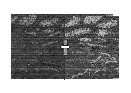
In curtain-view, as shown in FIG. 7, the selected slides appear stacked one on
the
other and a slider element can be dragged to determine what portion of the
composite image is derived from one image versus another; that is, the slider
button is moved to reveal a secondary, underlying image providing the
appearance
of a "curtain view" fused visualization. In the illustrated embodiment the
digital
image on the left side of the screen is derived from a slide of a tissue
section
stained with H&E, and the digital image on the right is derived from a slide
of an
adjacent tissue section stained with a IHC assay, for example, a duplex IHC
assay.
22
CA 03000050 2018-03-27
WO 2017/076865
PCT/EP2016/076351
FIGS. 7-9 illustrate the appearance of the composite image as a result of
moving
the slider button from left to right. As can be seen, shifting the slider
button shifts
the proportion of each image's contribution to the composite image. In some
embodiments, local registration adjusts at least one of the images so that the
image
data from the different slides is as similar as possible in the boundary area
right and
left of the curtain. Other viewer functionalities may also be used, including
pan,
zoom and creation of annotations.
As shown in FIG. 10, curtain view is not limited to a composite image of only
two
digital images of adjacent tissue sections and left-right movement of the
slider
button. In the example of FIG. 10, curtain view is applied to five slides and
includes four slider buttons providing top to bottom, bottom to top, left to
right and
right to left control. In the illustrated curtain-view embodiment
visualization, the
central image is derived from an H&E stained slide with image data from
digital
images derived from IHC stained slides of adjacent tissue sections made
visible (or
invisible) with a respective curtain view (slider button) control.
Fusion viewing is not limited to the curtain view implementation but
encompasses
any manner in which a composite image may be visualized and/or manipulated. As
an example, FIG. 11 illustrates another embodiment for fused viewing of two or
more registered digital images of adjacent tissue sections of a sample:
"flashlight"
or "spotlight" fusion viewing wherein IHC data is overlaid on H&E data. Here,
a
user may select any region of a secondary image, which appears as if stacked
under
the primary (top) image, to replace corresponding image data of the primary
image.
In some implementations, the region from the secondary slide, which is fused-
into
the primary slide, is disc-shaped as if a flashlight or spotlight is shining
on the
replaced region. However the disclosure is not limited to disc-shapes. A user
may
further interact with the computer through the client user interface to
increase or
decrease the size of the region or modify the shape of the region. And, as
shown in
FGIS. 11-13, the user can shift the location of the region, for example by
moving
the mouse pointer (or any other interactive means such as voice control). Here
again local registration may be used at the boundary between regions to adjust
alignment between the regions.
Although the example of FIGS. 11-13 is of IHC data overlaid on H&E data, a
user
may also select which image is primary (top) and which is secondary
(stacked/overlaid). Thus, for example, H&E data may be overlaid on IHC data.
The ordering selection (that is, which image is assigned as primary and the
layering
of secondary, underlying images) can be made by the user at the time the
images
23
CA 03000050 2018-03-27
WO 2017/076865
PCT/EP2016/076351
used for fused viewing are chosen. Alternatively, or in addition, the order of
stacking can change during viewing, for example by reshuffling the stack (e.g.
by
instructing the top image to move to the back of the stack).
Further, although the example of FIGS. 11-13 illustrate only one fused
(spotlight)
region, in some embodiments there can be multiple fused regions. Each of the
regions may provide image data from a single secondary slide, or else some or
all
of the regions may provide image data from different secondary slides.
FIG. 14 is a flow diagram illustrating an implementation of a method carried
out by
an embodiment of an image analysis software program in accordance with this
disclosure. The image analysis software program enables a user to instruct the
processor to view two or more images in fused view mode. In some embodiments,
if the chosen images are not aligned, the program may also enable a user to
register
selected digital images (e.g. digital images of scanned slides of tissue
sections,
including whole slide images, partial slide images, or portions of whole or
part
slide images).
As shown in FIG. 6, the method 600 begins at the start block 602. At block
604, a
set of image data or digital images is acquired (e.g. scanned or selected from
the
database or received from a source) for manipulation. Each set of image data
includes image data corresponding to, for example, a tissue section from a set
of
adjacent tissue sections of a single patient. Each image may be derived from
tissue
sections that are differently stained, or that are digitized using a different
imaging
mode, or both, as compared to another image. In some embodiments, the digital
images are produced by scanning slides (e.g. microscope glass slides) prepared
from adjacent tissue sections.
At block 606, the user assigns an image among the selected images as the
primary
(i.e. top) image. If there is only one secondary image, then the procedure
continues
to block 612. Otherwise, at block 610, the user assigns an order to the
secondary
images.
Passing to block 612, if the selected images have previously been aligned,
then the
process proceeds to block 616. Otherwise, the images are aligned at block 614
using any image registration method. At block 616, the selected, and
registered
(aligned), images are displayed on a common grid, with the images overlaid in
a
single image and displayed on a monitor (or on several monitors). At block
618,
the client user selects the fused view visualization tool such as curtain
view,
multiple curtain view, flashlight view, multiple flashlight view, resulting in
the
24
CA 03000050 2018-03-27
WO 2017/076865
PCT/EP2016/076351
image being displayed as a composite image (with interactive features such as
a
slider button or spotlight), wherein information from the primary and one or
more
secondary digital images is combined such that different regions of the
composite
image show image data from different digital images, for example as described
above in connection with curtain-view, multiple curtain-view, flashlight view
and
multiple flashlight view modes.
At block 620, the client user can interact with the display to manipulate the
composite image to increase or decrease the proportion of image data being
contributed by the primary and one or more secondary digital images. For
example, in curtain view this may be accomplished by increasing the size of
one
curtain at the expense of another. As another example, in flashlight view this
can
be accomplished by one or more of increasing or decreasing the size of the
spotlight and/or increasing or decreasing the number of spotlights.
In some embodiments, local registration occurs during this block 620 at the
interface of two digital images when the interactive element (such as the
slider or
the spotlight) is activated (e.g., moved) to modify the composite image. Local
registration is used to reduce alignment issues, which may occur at the
boundary of
two images that aren't identical but rather derive from adjacent tissue
sections.
A client user may then decide either to end the procedure (proceed to block
622), or
may decide to modify the composite image by modifying the selection of primary
and secondary images. This can be done either by reordering the existing stack
(to
identify a new image as primary for example), or by selecting new (registered)
images from the database and adding them to the stack or replacing existing
images
in the stack, partially or entirely, with the newly selected images. Again, if
the
images have been aligned, a new composite image is displayed, which can be
manipulated using any of the fusion viewing tools. Or else, the images are
first
aligned, then displayed as a composite image ready for manipulation.
Certain embodiments have been described but a person of skill understands that
still other embodiments are encompassed by this disclosure. It will be
appreciated
by those skilled in the art that changes could be made to the embodiments
described above without departing from the broad inventive concepts thereof
For
example, the disclosure is not limited to the fusion of two slides; multiple
slides
can be stained with different IHC and special stains assays, for example, and
fused
together for display. As another example, an H&E stained slide can be used
with
the IHC or other stained slides, but is not necessary. It is understood,
therefore, that
CA 03000050 2018-03-27
WO 2017/076865
PCT/EP2016/076351
this disclosure and the inventive concepts are not limited to the particular
embodiments disclosed, but are intended to cover modifications within the
spirit
and scope of the inventive concepts including as defined in the appended
claims.
Accordingly, the foregoing description of various embodiments does not
necessarily imply exclusion. For example,
"some" embodiments or "other"
embodiments may include all or part of "some", "other," "further," and
"certain"
embodiments within the scope of this invention.
26