Note: Descriptions are shown in the official language in which they were submitted.
CA 03000167 2018-03-27
WO 2017/058771 PCT/US2016/053927
ANTIBODIES THAT BIND HUMAN CANNABINOID 1 (CB1) RECEPTOR
FIELD OF THE INVENTION
[0001] The present invention relates to antibodies and antigen-binding
fragments thereof that
bind to cannabinoid receptor 1 (CB1) receptor, and methods of using such
antibodies and
antigen-binding fragments.
DESCRIPTION OF THE TEXT FILE SUBMITTED ELECTRONICALLY
[0002] The contents of the text file submitted electronically herewith are
incorporated herein
by reference in their entirety: A computer readable format copy of the
Sequence Listing
(filename: 15-343-PRO Seq_List 2015-09-30 ST25, date recorded: September 30,
2015, file
size 803 KB).
BACKGROUND
[0003] Cannabinoid 1 (CBI) receptor is a member of the G protein-coupled
receptor (GPCR)
superfamily. The CB1 receptor is expressed in the central nervous system
(CNS), lungs, liver,
adipose tissue and kidneys, and has been implicated in many human diseases
including obesity,
diabetes, fibrosis, liver diseases, cardiovascular disease, cancer, pain, MS
spasticity, and
glaucoma, among others. More specifically, CB1 receptor has been shown to
exhibit detrimental
activity in, for example, obesity, diabetes, fibrosis, liver diseases,
cardiovascular disease and
cancer; and has been shown to exhibit beneficial activity in pain, MS
spasticity and glaucoma,
among others.
[0004] There is a need in the art for new CB1 receptor antagonists and
agonists for
therapeutic purposes as well as selective binders for diagnostic/imaging
purposes. In particular, a
CB1 receptor-targeting compound that lacks the capacity for CNS penetration
would be desirable
to reduce potential CNS-mediated side effects of CB1 receptor modulation,
highlighted by the
psychiatric adverse events associated with the CB1 inverse agonist rimonabant.
1
CA 03000167 2018-03-27
WO 2017/058771 PCT/US2016/053927
SUMMARY OF THE INVENTION
[0005] In one aspect, the present invention provides antibodies and antigen-
binding
fragments thereof that bind to cannabinoid 1 receptor (also referred to herein
as "CBI receptor"
or "CB1"). In some embodiments, the CB1 receptor is a human CB1 receptor. In
some
embodiments, the antibody or fragment thereof recognizes one or more
extracellular epitopes on
the CB1 receptor. In some embodiments, the CB1 receptor binding antibodies and
fragments
thereof provided herein are functional antibodies or antigen binding fragments
thereof In some
embodiments, the CB1 receptor binding antibodies or fragments thereof inhibit
or increase CB1
receptor signaling activity. In some embodiments, the CB1 receptor binding
antibodies or
fragments thereof are antagonistic antibodies, in that they inhibit CB1
receptor signaling activity.
In some embodiments, the CB1 receptor binding antibodies or fragments thereof
are agonistic
antibodies, in that they enhance CB1 receptor signaling activity. In some
embodiments, the CB1
receptor binding antibodies or fragments thereof are modulators of CB1
receptor signaling
activity or are allosteric modulators of CB1 receptor signaling activity. In
some embodiments the
CB1 receptor binding antibodies or fragments thereof are selective binders
without agonist or
antagonist activity. In some embodiments, the CB1 receptor binding antibodies
or fragments
thereof are selective binders without agonist or antagonist activity that are
useful for diagnostic
and/or imaging purposes.
[0006] The isolated antibodies or antigen binding fragments thereof, in
some embodiments,
are at least as potent as small molecule CB1 receptor modulators such as, for
example, AM6545,
AM251, or rimonabant. In some embodiments, the antibodies or fragments thereof
have CB1
receptor inhibiting or activating activity that is at least 2 fold, at least 3
fold, at least 4 fold, at
least 5 fold, at least 10 fold, or at least 20 fold more potent relative to
the small molecules
AM6545, AM251, or rimonabant. In some embodiments, the isolated antibodies or
antigen
binding fragments thereof inhibit CB1 receptor agonist-mediated signal
transduction. In some
embodiments, the inhibition of CB1 receptor agonist-mediated signal
transduction is measured
by determining intracellular cAMP levels and/or downstream ERK
phosphorylation.
2
CA 03000167 2018-03-27
WO 2017/058771 PCT/US2016/053927
[0007] In some embodiments, the isolated antibodies and antigen-binding
fragments thereof
have the advantage of reduced or absent brain penetration. In some
embodiments, the brain
penetration of the isolated antibodies and antigen-binding fragments thereof
exhibit reduced
brain penetration relative to small molecule CB1 receptor agonists or
antagonists (e.g., AM6545,
AM251, or rimonabant). In some embodiments, the CB1 receptor binding
antibodies and
fragments thereof provided herein provide a therapeutic benefit with reduced
central nervous
system side effects relative to a small molecule CB1 receptor agonist or
antagonist. CNS side
effects associated with small molecule CB1 receptor antagonist rimonabant
include anxiety,
depression, agitation, eating disorders, irritability, aggression, and
insomnia (Moreira, 2009, Rev
Bras Psiquiatr., 31(2):145-53).
[0008] In some embodiments, the isolated antibodies and antigen-binding
fragments thereof
provided herein are generated from hybridoma cell lines. In other embodiments,
the isolated
antibodies and antigen-binding fragments thereof provided herein are generated
from phage
display libraries.
[0009] In some embodiments, the isolated antibodies and antigen-binding
fragments thereof
provided herein have an affinity for native human CB1 receptor that is at
least nM range. For
example, in some embodiments, the affinity for CB1 receptor is about 1 ILIM or
less, or about 750
nM or less, or about 500 nM or less, or about 250nM or less, or about 100 nM
or less, or about
75 nM or less, or about 50 nM or less, or about 25 nM or less, or about 10 nM
or less, or about 1
nM or less. In other embodiments, the isolated antibodies and antigen-binding
fragments
disclosed herein have an affinity for CB1 receptor of about 15 nM or less. In
some embodiments,
the isolated antibodies and antigen-binding fragments thereof have an affinity
for human CB1
receptor that is from about 0.01 nM to about 500 nM, about 0.02 nM to about
250 nM, about
0.02 to about 200 nM, about 0.05 to about 100 nM, about 0.05 to about 50 nM.
[0010] The isolated antibodies and antigen binding fragments thereof of the
present
invention may be derived from any species including, but not limited to,
mouse, rat, rabbit,
hamster, guinea pig, primate, llama or human. In some embodiments, the
isolated antibodies and
antigen binding fragments thereof are murine antibodies. In other embodiments,
the isolated
antibodies and antigen binding fragments thereof are chimeric antibodies. In
still other
3
CA 03000167 2018-03-27
WO 2017/058771 PCT/US2016/053927
embodiments, the isolated antibodies and antigen binding fragments thereof are
humanized
antibodies. In some embodiments, the isolated antibodies and antigen binding
fragments thereof
are fully human antibodies.
[0011] In one embodiment, the isolated antibodies and antigen binding
fragments thereof are
humanized or chimeric P1C4 antibodies, as described herein. In one embodiment,
the humanized
P1C4 antibodies are selected from the group consisting of P1C4-HO, P1C4-H2,
and P1C4-H4, as
described herein. In one embodiment, the isolated antibodies and antigen
binding fragments
thereof comprise Fc modifications that result in reduced, impaired, or
eliminated antibody
effector function. In a further embodiment, the isolated antibodies and
antigen binding fragments
thereof are selected from the group consisting of P1C4-HO-IgG2-4 Hybrid, P1C4-
HO-
IgG2A330S/P331S, P1C4-HO-IgG4S228P, P1C4-H2-IgG2-4 Hybrid, P1C4-H2-
IgG2A330S/P331S, P1C4-H2-IgG4S228P, P1C4-H4-IgG2-4 Hybrid, P1C4-H4-
IgG2A330S/P331S, P1C4-H4-IgG4S228P.
[0012] In one embodiment, the isolated antibody or antigen binding fragment
thereof
comprises a heavy chain variable region comprising a nucleic acid sequence
selected from the
group consisting of SEQ ID NOs. 1, 9, 17, 25, 33, 41, 49, and 57. In another
embodiment, the
isolated antibody or antigen binding fragment thereof comprises a heavy chain
variable region
comprising an amino acid sequence selected from the group consisting of SEQ ID
NOs. 2, 10,
18, 26, 34, 42, 50, and 58. In another embodiment, the isolated antibody or
antigen binding
fragment thereof comprises a heavy chain constant region comprising a nucleic
acid sequence
selected from the group consisting of SEQ ID NOs. 3, 11, 19, 27, 35, 43, 51,
and 59. In another
embodiment, the isolated antibody or antigen binding fragment thereof
comprises a heavy chain
constant region comprising an amino acid sequence selected from the group
consisting of SEQ
ID NOs. 4, 12, 20, 28, 36, 44, 52, and 60. In another embodiment, the isolated
antibody or
antigen binding fragment thereof comprises a light chain variable region
comprising a nucleic
acid sequence selected from the group consisting of SEQ ID NOs 5, 13, 21, 29,
37, 45, 53, and
61. In another embodiment, the isolated antibody or antigen binding fragment
thereof comprises
a light chain variable region comprising an amino acid sequence selected from
the group
consisting of SEQ ID NOs. 6, 14, 22, 30, 38, 46, 54, and 62. In another
embodiment, the isolated
4
CA 03000167 2018-03-27
WO 2017/058771 PCT/US2016/053927
antibody or antigen binding fragment thereof comprises a light chain constant
region comprising
an amino acid sequence selected from the group consisting of SEQ ID NOs. 7,
15, 23, 31, 39, 47,
55, and 63. In another embodiment, the isolated antibody or antigen binding
fragment thereof
comprises a light chain constant region comprising an amino acid sequence
selected from the
group consisting of SEQ ID NOs. 8, 16, 24, 32, 40, 48, 56, and 64.
[0013] In one embodiment, the isolated antibody or antigen binding fragment
thereof
comprises a heavy chain variable region comprising a nucleic acid sequence
according to SEQ
ID NO: 1; a heavy chain constant region comprising a nucleic acid sequence
according to SEQ
ID NO: 3; a light chain variable region comprising a nucleic acid sequence
according to SEQ ID
NO: 5; and a light chain constant region comprising a nucleic acid sequence
according to SEQ
ID NO: 7.
[0014] In one embodiment, the isolated antibody or antigen binding fragment
thereof
comprises a heavy chain variable region comprising an amino acid sequence
according to SEQ
ID NO: 2; a heavy chain constant region comprising an amino acid sequence
according to SEQ
ID NO: 4; a light chain variable region comprising an amino acid sequence
according to SEQ ID
NO: 6; and a light chain constant region comprising an amino acid sequence
according to SEQ
ID NO: 8.
[0015] In one embodiment, the isolated antibody or antigen binding fragment
thereof
comprises a heavy chain variable region comprising a nucleic acid sequence
according to SEQ
ID NO: 9; a heavy chain constant region comprising a nucleic acid sequence
according to SEQ
ID NO: 11; a light chain variable region comprising a nucleic acid sequence
according to SEQ
ID NO: 13; and a light chain constant region comprising a nucleic acid
sequence according to
SEQ ID NO: 15.
[0016] In one embodiment, the isolated antibody or antigen binding fragment
thereof
comprises a heavy chain variable region comprising an amino acid sequence
according to SEQ
ID NO: 10; a heavy chain constant region comprising an amino acid sequence
according to SEQ
ID NO: 12; a light chain variable region comprising an amino acid sequence
according to SEQ
ID NO: 14; and a light chain constant region comprising an amino acid sequence
according to
SEQ ID NO: 16.
CA 03000167 2018-03-27
WO 2017/058771 PCT/US2016/053927
[0017] In one embodiment, the isolated antibody or antigen binding fragment
thereof
comprises a heavy chain variable region comprising a nucleic acid sequence
according to SEQ
ID NO: 17; a heavy chain constant region comprising a nucleic acid sequence
according to SEQ
ID NO: 19; a light chain variable region comprising a nucleic acid sequence
according to SEQ
ID NO: 21; and a light chain constant region comprising a nucleic acid
sequence according to
SEQ ID NO: 23.
[0018] In one embodiment, the isolated antibody or antigen binding fragment
thereof
comprises a heavy chain variable region comprising an amino acid sequence
according to SEQ
ID NO: 18; a heavy chain constant region comprising an amino acid sequence
according to SEQ
ID NO: 20; a light chain variable region comprising an amino acid sequence
according to SEQ
ID NO: 22; and a light chain constant region comprising an amino acid sequence
according to
SEQ ID NO: 24.
[0019] In one embodiment, the isolated antibody or antigen binding fragment
thereof
comprises a heavy chain variable region comprising a nucleic acid sequence
according to SEQ
ID NO: 25; a heavy chain constant region comprising a nucleic acid sequence
according to SEQ
ID NO: 27; a light chain variable region comprising a nucleic acid sequence
according to SEQ
ID NO: 29; and a light chain constant region comprising a nucleic acid
sequence according to
SEQ ID NO: 31.
[0020] In one embodiment, the isolated antibody or antigen binding fragment
thereof
comprises a heavy chain variable region comprising an amino acid sequence
according to SEQ
ID NO: 26; a heavy chain constant region comprising an amino acid sequence
according to SEQ
ID NO: 28; a light chain variable region comprising an amino acid sequence
according to SEQ
ID NO: 30; and a light chain constant region comprising an amino acid sequence
according to
SEQ ID NO: 32.
[0021] In one embodiment, the isolated antibody or antigen binding fragment
thereof
comprises a heavy chain variable region comprising a nucleic acid sequence
according to SEQ
ID NO: 33; a heavy chain constant region comprising a nucleic acid sequence
according to SEQ
ID NO: 35; a light chain variable region comprising a nucleic acid sequence
according to SEQ
6
CA 03000167 2018-03-27
WO 2017/058771 PCT/US2016/053927
ID NO: 37; and a light chain constant region comprising a nucleic acid
sequence according to
SEQ ID NO: 39.
[0022] In one embodiment, the isolated antibody or antigen binding fragment
thereof
comprises a heavy chain variable region comprising an amino acid sequence
according to SEQ
ID NO: 34; a heavy chain constant region comprising an amino acid sequence
according to SEQ
ID NO: 36; a light chain variable region comprising an amino acid sequence
according to SEQ
ID NO: 38; and a light chain constant region comprising an amino acid sequence
according to
SEQ ID NO: 40.
[0023] In one embodiment, the isolated antibody or antigen binding fragment
thereof
comprises a heavy chain variable region comprising a nucleic acid sequence
according to SEQ
ID NO: 41; a heavy chain constant region comprising a nucleic acid sequence
according to SEQ
ID NO: 43; a light chain variable region comprising a nucleic acid sequence
according to SEQ
ID NO: 45; and a light chain constant region comprising a nucleic acid
sequence according to
SEQ ID NO: 47.
[0024] In one embodiment, the isolated antibody or antigen binding fragment
thereof
comprises a heavy chain variable region comprising an amino acid sequence
according to SEQ
ID NO: 42; a heavy chain constant region comprising an amino acid sequence
according to SEQ
ID NO: 44; a light chain variable region comprising an amino acid sequence
according to SEQ
ID NO: 46; and a light chain constant region comprising an amino acid sequence
according to
SEQ ID NO: 48.
[0025] In one embodiment, the isolated antibody or antigen binding fragment
thereof
comprises a heavy chain variable region comprising a nucleic acid sequence
according to SEQ
ID NO: 49; a heavy chain constant region comprising a nucleic acid sequence
according to SEQ
ID NO: 51; a light chain variable region comprising a nucleic acid sequence
according to SEQ
ID NO: 53; and a light chain constant region comprising a nucleic acid
sequence according to
SEQ ID NO: 55.
[0026] In one embodiment, the isolated antibody or antigen binding fragment
thereof
comprises a heavy chain variable region comprising an amino acid sequence
according to SEQ
ID NO: 50; a heavy chain constant region comprising an amino acid sequence
according to SEQ
7
CA 03000167 2018-03-27
WO 2017/058771 PCT/US2016/053927
ID NO: 52; a light chain variable region comprising an amino acid sequence
according to SEQ
ID NO: 54; and a light chain constant region comprising an amino acid sequence
according to
SEQ ID NO: 56.
[0027] In one embodiment, the isolated antibody or antigen binding fragment
thereof
comprises a heavy chain variable region comprising a nucleic acid sequence
according to SEQ
ID NO: 57; a heavy chain constant region comprising a nucleic acid sequence
according to SEQ
ID NO: 59; a light chain variable region comprising a nucleic acid sequence
according to SEQ
ID NO: 61; and a light chain constant region comprising a nucleic acid
sequence according to
SEQ ID NO: 63.
[0028] In one embodiment, the isolated antibody or antigen binding fragment
thereof
comprises a heavy chain variable region comprising an amino acid sequence
according to SEQ
ID NO: 58; a heavy chain constant region comprising an amino acid sequence
according to SEQ
ID NO: 60; a light chain variable region comprising an amino acid sequence
according to SEQ
ID NO: 62; and a light chain constant region comprising an amino acid sequence
according to
SEQ ID NO: 64.
[0029] In one embodiment, the invention provides an isolated antibody or
fragment thereof
that comprises a nucleic acid sequence or an amino acid sequence that is at
least 65%, at least
70%, at least 75%, at least 80%, at least 85%, at least 90%, at least 95%, or
at least 99% identical
to an amino acid sequence selected from the group consisting of SEQ ID NOs. 1-
351.
[0030] In one embodiment, the isolated antibody or antigen binding fragment
thereof
comprises heavy chain complementary determining regions (CDRs) independently
selected from
the CDRs present in the heavy chain variable regions according to SEQ ID NOs:
2, 10, 18, and
26. In another embodiment, the isolated antibody or antigen binding fragment
thereof comprises
light chain CDRs independently selected from the CDRs present in the light
chain variable
regions according to SEQ ID NOs: 6, 14, 22, and 30.
[0031] In one embodiment, the isolated antibody or antigen binding fragment
thereof is a
humanized antibody comprising heavy chain complementary determining regions
(CDRs)
independently selected from the CDRs present in the heavy chain variable
regions according to
SEQ ID NOs: 2, 10, 18, and 26. In another embodiment, the isolated antibody or
antigen binding
8
CA 03000167 2018-03-27
WO 2017/058771 PCT/US2016/053927
fragment thereof is a humanized antibody comprising light chain complementary
determining
regions (CDRs) independently selected from the CDRs present in the light chain
variable regions
according to SEQ ID NOs: 6, 14, 22, and 30.
[0032] In one embodiment, the heavy chain variable region comprises,
consists essentially
of, or consists of an amino acid sequence selected from the group consisting
of SEQ ID NOs:
339-341. In one embodiment, the isolated antibody or antigen binding fragment
thereof
comprises a heavy chain variable region amino acid sequence that is at least
65%, at least 70%,
at least 75%, at least 80%, at least 85%, at least 90%, at least 95%, at least
96%, at least 97%, at
least 98% or at least 99% identical to an amino acid sequence selected from
the group consisting
of SEQ ID NOs: 339-341. In a further embodiment, the heavy chain variable
region comprises,
consists essentially of, or consists of an amino acid sequence selected from
the group consisting
of SEQ ID NOs: 339-341.
[0033] In another embodiment, the isolated antibody or antigen binding
fragment thereof
comprises a heavy chain amino acid sequence that is least 65%, at least 70%,
at least 75%, at
least 80%, at least 85%, at least 90%, at least 95%, at least 96%, at least
97%, at least 98% or at
least 99% identical to an amino acid sequence selected from the group
consisting of SEQ ID
NOs: 343-351. In a further embodiment, the heavy chain comprises, consists
essentially of, or
consists of an amino acid sequence selected from the group consisting of SEQ
ID NOs:343-351.
[0034] In another embodiment, the isolated antibody or antigen binding
fragment thereof
comprises a light chain variable region amino acid sequence that is least 65%,
at least 70%, at
least 75%, at least 80%, at least 85%, at least 90%, at least 95%, at least
96%, at least 97%, at
least 98% or at least 99% identical to an amino acid sequence according to SEQ
ID NO: 337. In
a further embodiment, the heavy chain variable region comprises, consists
essentially of, or
consists of an amino acid sequence according to SEQ ID NO: 337. In another
embodiment, the
isolated antibody or antigen binding fragment thereof comprises a light chain
amino acid
sequence that is least 65%, at least 70%, at least 75%, at least 80%, at least
85%, at least 90%, at
least 95%, at least 96%, at least 97%, at least 98% or at least 99% identical
to an amino acid
sequence according to SEQ ID NO: 338. In a further embodiment, the light chain
comprises,
consists essentially of, or consists of an amino acid sequence according to
SEQ ID NO: 338.
9
CA 03000167 2018-03-27
WO 2017/058771 PCT/US2016/053927
[0035] In one embodiment, the invention provides a humanized isolated
antibody or antigen
binding fragment thereof that binds CBI. In a further embodiment, the isolated
antibody or
antigen binding fragment thereof comprises a light chain variable region
according to SEQ ID
NO: 337 and a heavy chain variable region according to SEQ ID NO: 339. In
another
embodiment, the isolated antibody or antigen binding fragment thereof
comprises a light chain
variable region according to SEQ ID NO: 337 and a heavy chain variable region
according to
SEQ ID NO: 340. In another embodiment, the isolated antibody or antigen
binding fragment
thereof comprises a light chain variable region according to SEQ ID NO: 337
and a heavy chain
variable region according to SEQ ID NO: 341. In another embodiment, the
isolated antibody or
antigen binding fragment thereof comprises a full light chain according to SEQ
ID NO: 338 and
a full heavy chain according to a sequence selected from the group consisting
of SEQ ID NOs:
343-351.
[0036] In other embodiments, the isolated antibody or antigen binding
fragment thereof
comprises a light chain sequence according to SEQ ID NO: 829, 830, 831, or
832. In some
embodiments, the isolated antibody or antigen binding fragments comprise a
light chain
sequence according to SEQ ID NO: 829, 830, 831, or 832, and a heavy chain
sequence according
to SEQ ID NO: 437.
[0037] In one embodiment, the isolated antibody or fragment thereof
comprises a heavy
chain CDR1 sequence having at least 80%, at least 85%, at least 90%, at least
95% at least 96%,
at least 97%, at least 98%, or at least 99% homology to the amino acid
sequence of SEQ ID NO:
352 (YYWMN). In another embodiment, the isolated antibody or fragment thereof
comprises a
heavy chain CDR2 sequence having at least 80%, at least 85%, at least 90%, at
least 95% at least
96%, at least 97%, at least 98%, or at least 99% homology to the amino acid
sequence of SEQ ID
NO: 353 (QIYPGDGETKY). In another embodiment, the isolated antibody or
fragment thereof
comprises a heavy chain CDR3 sequence having at least 80%, at least 85%, at
least 90%, at least
95% at least 96%, at least 97%, at least 98%, or at least 99% homology to the
amino acid
sequence of SEQ ID NO: 354 (SHGNYLPY). In another embodiment, the isolated
antibody or
fragment thereof comprises a light chain CDR1 sequence having at least 80%, at
least 85%, at
least 90%, at least 95% at least 96%, at least 97%, at least 98%, or at least
99% homology to the
CA 03000167 2018-03-27
WO 2017/058771 PCT/US2016/053927
amino acid sequence of SEQ ID NO: 355 (SSYLH). In another embodiment, the
isolated
antibody or fragment thereof comprises a light chain CDR2 sequence having at
least 80%, at
least 85%, at least 90%, at least 95% at least 96%, at least 97%, at least
98%, or at least 99%
homology to the amino acid sequence of SEQ ID NO: 356 (STSNLAS). In another
embodiment,
the isolated antibody or fragment thereof comprises a light chain CDR3
sequence having at least
80%, at least 85%, at least 90%, at least 95% at least 96%, at least 97%, at
least 98%, or at least
99% homology to the amino acid sequence of SEQ ID NO: 357 (HQYHRSPPTF).
[0038] In one embodiment, the isolated antibody or antigen binding fragment
thereof
comprises a heavy chain CDR1, CDR2, and CDR3 comprising amino acid sequences
according
to SEQ ID NOs: 352, 353, and 354, respectively. In another embodiment, the
isolated antibody
or antigen binding fragment thereof comprises a light chain CDR1, CDR2, and
CDR3
comprising amino acid sequences according to SEQ ID NOs: 355, 356, and 357,
respectively. In
a further embodiment, the isolated antibody or antigen binding fragment
thereof comprises a
heavy chain CDR1, heavy chain CDR2, heavy chain CDR3, light chain CDR1, light
chain
CDR2, and light chain CDR3 comprising amino acid sequences according to SEQ ID
NOs: 352,
353, 354, 355, 356, and 357, respectively. In a still further embodiment, the
isolated antibody or
fragment thereof is chimeric or humanized.
[0039] In still another embodiment, the isolated antibody or antigen
binding fragment thereof
comprises a light chain CDR1 comprising amino acid sequence according to SEQ
ID NO: 833.
In further embodiments, the isolated antibody or antigen binding fragment
thereof comprises a
light chain CDR2 selected from a group comprising amino acid sequence
according to SEQ ID
NOs: 694, 834 and 835. In still further embodiments, the isolated antibody or
antigen binding
fragment thereof comprises a light chain CDR3 selected from a group comprising
amino acid
sequence according to SEQ ID NOs: 779 and 836.
[0040] In an embodiment, the isolated antibody or antigen binding fragment
thereof
comprises a light chain CDR1 comprising amino acid sequence according to SEQ
ID NO: 833, a
light chain CDR2 comprising amino acid sequence according to SEQ ID NO: 694,
and a light
chain CDR3 comprising amino acid sequence according to SEQ ID NO: 836.
11
CA 03000167 2018-03-27
WO 2017/058771 PCT/US2016/053927
[0041] In another embodiment, the isolated antibody or antigen binding
fragment thereof
comprises a light chain CDR1 comprising amino acid sequence according to SEQ
ID NO: 833, a
light chain CDR2 comprising amino acid sequence according to SEQ ID NO: 835,
and a light
chain CDR3 comprising amino acid sequence according to SEQ ID NO: 836.
[0042] I yet another embodiment, the isolated antibody or antigen binding
fragment thereof
comprises a light chain CDR1 comprising amino acid sequence according to SEQ
ID NO: 833, a
light chain CDR2 comprising amino acid sequence according to SEQ ID NO: 694,
and a light
chain CDR3 comprising amino acid sequence according to SEQ ID NO: 357.
[0043] In still another embodiment, the isolated antibody or antigen
binding fragment thereof
comprises a light chain CDR1 comprising amino acid sequence according to SEQ
ID NO: 833, a
light chain CDR2 comprising amino acid sequence according to SEQ ID NO: 834,
and a light
chain CDR3 comprising amino acid sequence according to SEQ ID NO: 779.
[0044] In still another embodiment, disclosed herein is an isolated
antibody or antigen
binding fragment thereof that binds to cannabinoid receptor 1 (CB1), wherein
the antibody or
antigen binding fragment thereof comprises a light chain CDR1 comprising an
amino acid
sequence according to SEQ ID NOs: 355 or 833; a light chain CDR2 comprising an
amino acid
sequence according to SEQ ID NOs: 356, 694, 834, or 835; and a light chain
CDR3 comprising
an amino acid sequence according to SEQ ID NOs: 357, 779, or 836.
[0045] The person of skill in the art will understand that the heavy and
light chain CDRs of
the antibodies provided herein may be independently selected, or mixed and
matched, to form an
antibody or binding fragment thereof comprising any light chain CDR1, CDR2,
and CDR3; and
any heavy chain CDR1, CDR2, and CDR3 from the antibodies provided herein. The
skilled
person will further understand that the heavy and light chain variable regions
of the antibodies
provided herein may be independently selected, or mixed and matched, to form
an antibody or
binding fragment comprising any heavy and light chain from the antibodies
provided herein.
[0046] In one embodiment, the antibody or antigen binding fragment thereof
provided herein
is a chimeric antibody or fragment containing heavy and light chain CDRs
selected from the
CDRs provided herein, or conservative variants of the CDRs provided herein. In
another
embodiment, the antibody or antigen binding fragment thereof provided herein
is a humanized
12
CA 03000167 2018-03-27
WO 2017/058771 PCT/US2016/053927
antibody or fragment containing heavy and light chain CDRs selected from the
CDRs provided
herein, or conservative variants of the CDRs provided herein. In one
embodiment, the antibody
or antigen binding fragment thereof provided herein comprises a light chain
and/or a heavy chain
comprising a sequence provided herein, or a conservative variant thereof In
one embodiment,
the conservative variants have at least 80%, at least 85%, at least 90%, at
least 95%, at least
96%, at least 97%, at least 98% or at least 99% homology to the reference
sequence provided
herein. In one embodiment, the conservative variants comprise 1, 2, 3, 4, 5,
6, 7, 8, 9, 10, or
more amino acid substitutions, insertions, or deletions.
[0047] In some embodiments, the isolated antibody or antigen binding
fragment thereof
binds CB1 and exhibits reduced effector function. In one embodiment, the
isolated antibody or
antigen binding fragment thereof binds CB1 and comprises one or more Fc region
modifications.
In a further embodiment, the antibody or antigen binding fragment thereof
binds CB1 and
comprises an amino acid sequence comprising one or more mutations in the Fc
region. In a
further embodiment, the isolated antibody or antigen binding fragment thereof
has a mutation at
position 228 and/or 330 and/or 331. In another embodiment, the isolated
antibody or antigen
binding fragment thereof has a mutation at position 228 of the Fc region,
wherein the Fc region
is of the IgG4 isotype. In a further embodiment, the mutation is S228P. In
another embodiment,
the isolated antibody or antigen binding fragment thereof has a mutation at
position 330 and/or
position 331. In a further embodiment, the isolated antibody or antigen
binding fragment thereof
has a mutation at position 330 and/or 331, wherein the Fc region is of the
IgG2 isotype. In a
further embodiment, the isolated antibody or antigen binding fragment thereof
has the following
mutations in the Fc region: A330S and P331S. In another embodiment, the
isolated antibody or
antigen binding fragment thereof comprises an Fc region that is a hybrid Fc
region. For example,
in one embodiment, the Fc region is a hybrid IgG2/IgG4 Fc region, wherein the
CH1 and hinge
regions are derived from IgG2, and the CH2 and CH3 regions are derived from
IgG4.
[0048] Thus, in one embodiment, the antibody or antigen binding fragment
thereof provided
herein is a chimeric or humanized antibody or fragment containing heavy and
light chain CDRs
selected from the CDRs provided herein, or conservative variants of the CDRs
provided herein,
wherein the isolated antibody or fragment thereof comprises an Fc region
comprising
13
CA 03000167 2018-03-27
WO 2017/058771 PCT/US2016/053927
modifications that alter antibody effector functions. For example, in one
embodiment, the
isolated antibody or antigen binding fragment thereof comprises light and
heavy chain CDRs
according to SEQ ID NOs: 352-357, or conservative variants thereof, and
further comprises an
IgG2-IgG4 hybrid Fc region, an IgG2 Fc region comprising amino acid mutations
at positions
330 and 331 (e.g., A330S and P331S), or an IgG4 Fc region comprising an amino
acid mutation
at position 228 (e.g., S228P).
[0049] In one embodiment, the present invention provides an isolated
antibody or antigen
binding fragment thereof that binds to CB1, wherein the antibody or fragment
has a binding
affinity Kd for CB1 receptor of about 70 nM or less, about 60nM or less, about
50nM or less,
about 40nM or less, about 30nM or less, about 25nM or less, about 20nM or
less, about 15nM or
less, about lOnM or less, about 8nM or less, about 6nM or less, about 5 nM or
less, about 4nM or
less, about 3nM or less, about 2nM or less, or about 1nM or less. In one
embodiment, present
invention provides an isolated antibody or fragment thereof that binds to CB1,
wherein the
antibody or fragment has a binding affinity Kd for CB1 receptor in the range
of about 1nM to
about 100nM, about 2nM to about 75nM, about 3nM to about 50nM, about 4nM to
about lOnM,
or has a binding affinity Kd for CB2 receptor that is about 50nM, or about
40nM, or about
30nM, or about 20nM, or about lOnM, or about 5nM, or about 4nM, or about 3nM
or about
2nM, or about 1nM.
[0050] In one embodiment, the present invention provides an isolated
antibody or antigen
binding fragment thereof that is at least 2 fold, at least 3 fold, at least 4
fold, at least 5 fold, at
least 6 fold, at least 7 fold, at least 8 fold, at least 9 fold, at least 10
fold, at least 11 fold, at least
12 fold, at least 13 fold, at least 14 fold, or at least 15 fold more potent
than the small molecule
rimonabant, wherein the potency of the antibody or fragment or rimonabant is
measured by
inhibition of CB1 receptor antagonist-mediated signal transduction in a cAMP
assay. In a further
embodiment, the isolated antibody or antigen binding fragment thereof is
humanized.
[0051] In one embodiment, the present invention provides an isolated
humanized antibody or
antigen binding fragment thereof that binds to CB1, wherein the antibody or
fragment exhibits
greater binding affinity and/or greater potency than a corresponding non-
humanized or chimeric
antibody, wherein the humanized antibody or fragment and the corresponding non-
humanized or
14
CA 03000167 2018-03-27
WO 2017/058771 PCT/US2016/053927
chimeric antibody comprise the same heavy and light chain CDRs. For example,
in one
embodiment, the present invention provides a humanized antibody or fragment
thereof
comprising heavy chain CDR1, CDR2, and CDR3 and light chain CDR1, CDR2, and
CDR3
according to SEQ ID NOs: 352, 353, 354, 355, 356, and 357, respectively,
wherein the
humanized antibody exhibits greater binding affinity for CB1 receptor and/or
greater potency
with respect to inhibition of CB1 receptor agonist. In one embodiment, the
humanized
antibodies and fragments provided herein exhibit at least 50% greater, at
least 100% greater, at
least 2 fold greater, at least 3 fold greater, at least 4 fold greater, at
least 5 fold greater, or at least
fold greater potency relative to the corresponding non-humanized or chimeric
antibody. In a
further embodiment, the potency is measured by inhibition of CB1- cAMP
production.
[0052] Potency of CB1 receptor antibodies provided herein may be measured
by any method
known in the art. For example, in one embodiment, potency of the antibodies
and fragments
provided herein is measured by intracellular cAMP levels or ERK
phosphorylation. For example,
potency may be measured by the level of inhibition cAMP production in a cAMP
functional
assay (Cisbio) or inhibition of WIN55,212-induced ERK phosphorylation in a
Western blot.
[0053] In some embodiments, the present invention provides an antibody or
antigen binding
fragment thereof that is capable of competing with the antibody or antigen
binding fragments
thereof disclosed herein for binding to CB1 receptor. In some other
embodiments, the present
invention provides an antibody or antigen binding fragment thereof that is
capable of specifically
binding to essentially the same epitope on CB1 receptor as the antibodies or
antigen binding
fragments disclosed herein. Such antibodies can be identified using routine
competition binding
assays. In certain embodiments, competition is measured by ELISA, flow
cytometry, or surface
plasmon resonance (SPR) assay.
[0054] In some embodiments, the antibodies and fragments thereof are
conjugated to one or
more agents selected from the group including an additional therapeutic agent,
a cytotoxic agent,
an immunoadhesion molecule, and an imaging agent. In some embodiments, the
imaging agent
is selected from the group consisting of a radiolabel, an enzyme, a
fluorescent label, a
luminescent label, a bioluminescent label, a magnetic label, and biotin.
CA 03000167 2018-03-27
WO 2017/058771 PCT/US2016/053927
[0055] In one aspect, methods are provided for modulating the signaling
activity of CB1
receptor comprising contacting a cell expressing CB1 receptor with the
antibody or fragment
thereof disclosed herein. In some embodiments, the methods provided result in
inhibition of the
activity of CB1 receptor signaling. In some embodiments, the methods provided
result in
increased activity of CB1 receptor signaling. In some embodiments, the
modulation of CB1
receptor signaling activity is indirect, such as though an allosteric
modulator. In some
embodiments, the modulation of CB1 receptor signaling activity is biased for
Galpha i/o
mediated signaling versus beta arrestin mediated signaling.
[0056] In one aspect, methods for treating a disease or disorder responsive
to antagonism or
agonism of CB1 receptor in a subject in need thereof are provided. In some
embodiments, the
methods comprise administering to the subject an anti-CB1 receptor antibody or
antigen binding
fragment thereof as disclosed herein. In one embodiment, the subject is a
mammal. In a further
embodiment, the subject is a human. In some embodiments, the disease or
disorder is obesity,
diabetes, dyslipidemia, metabolic diseases, fibrosis, non-alcoholic
steatohepatitis (NASH), liver
disease, primary biliary cirrhosis, renal disease, kidney fibrosis, chronic
kidney disease,
osteoporosis, atherosclerosis, cardiovascular disease, cancer, an inflammatory
disease, pain, MS
spasticity, and ocular diseases, including glaucoma. In some embodiments, the
disease or
disorder is, for example, obesity, diabetes, fibrosis, liver disease,
cardiovascular disease, or
cancer, and the method provided results in inhibition of the activity of CB1
receptor. In some
embodiments, the disease or disorder is, for example, pain or glaucoma, and
the method provided
results in activation or increase of CB1 receptor activity.
[0057] In one aspect, a method for detecting CB1 receptor in a cell,
tissue, or subject is
provided, the method comprising contacting a cell with a CB1 receptor binding
antibody or
antigen binding fragment provided herein. In one embodiment, the cell is
present in a subject. In
another embodiment, the cell is present in a human subject. In another
embodiment, the CB1
receptor expression level on cells is correlated with a disease state. Thus,
in one aspect, the
present invention provides methods of using antibodies and fragments thereof
that specifically
bind to CB1 receptor as tools for the diagnosis and/or prognosis of human
diseases. In one
embodiment, the present invention provides methods for imaging CB1 receptor
comprising the
16
CA 03000167 2018-03-27
WO 2017/058771 PCT/US2016/053927
use of the CB1 receptor antibodies and fragments disclosed herein. In one
embodiment, the
method for detecting CB1 receptor is achieved with a CB1 receptor antibody or
fragment thereof
disclosed herein that selectively binds CB1 receptor. In a further embodiment,
the selective CB1
receptor antibody or fragment thereof does not exhibit agonistic or
antagonistic activity. In a
further embodiment, the selective CB1 receptor antibody or fragment thereof
does not
internalize. In one embodiment, the present invention provides diagnostic and
imaging methods
comprising the use of a CB1 receptor antibody or fragment that is conjugated
to an imaging
agent such as, for example, a radiolabel, an enzyme, a fluorescent label, a
luminescent label, a
bioluminescent label, a magnetic label, or biotin.
[0058] In one embodiment, the invention provides a host cell expressing an
isolated antibody
or fragment thereof that specifically binds to CB1 receptor. In another
embodiment, a method
for making an antibody or fragment thereof that specifically binds to CB1
receptor is provided,
the method comprising immunizing mammals with purified CB1 receptor or an
antigenic
fragment thereof, CB1/lipid complexes, CB1 receptor iCAPS, and/or CB1 receptor
DNA. In a
further embodiment, the immunized mammals are mice. In another embodiment, the
mammals
are immunized one, two, three, four, five, or more times with purified CB1 or
an antigenic
fragment thereof, CB1/lipid complex, CB1 receptor iCAPS, and/or CB1 receptor
DNA prior to
harvesting cells from the immunized mammals. In a further embodiment, the
antibody or
fragment thereof that specifically binds to CB1 receptor is generated from a
hybridoma cell line
comprising cells derived from the immunized mammals. In another embodiment,
the antibody or
fragment thereof that specifically binds to CB1 receptor is generated from a
phage display
library. In a further embodiment, the phage display library is derived from
cells isolated from the
immunized mammals. In a further embodiment, the phage display library is
derived from naïve
human immunoglobulin sequences.
BRIEF DESCRIPTION OF THE FIGURES
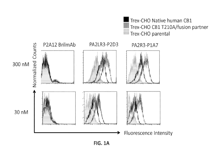
[0059] Figure lA is a set of histograms showing binding of P2Al2 Bril mAb
(left column),
PA2LR3-P2D3 (middle column), or PA2R3-P1A7 (right column) at 300 nM (top row)
or 30 nM
(bottom row) to Trex-CHO Native human CB1 cell line (native CB1 receptor
expressing; dark
17
CA 03000167 2018-03-27
WO 2017/058771 PCT/US2016/053927
gray lines), Trex-CHO CB1 T210A/fusion partner (overexpressed CBI; medium gray
lines), or
Trex-CHO parental cell line (no CB1 receptor expression; light gray lines).
Figure 1B is a set of
histograms showing binding of PA2R3-P1F1 (left column), PA2LR3-P2E5 (middle
column), or
PA2LR3-P3B10 (right column) at 300nM (top row) or 30nM (bottom row) to Trex-
CHO Native
CB1 cell line (native human CB1 receptor expressing; dark gray lines), Trex-
CHO CB1
T210A/fusion partner (overexpressed CBI; medium gray lines), or Trex-CHO
parental cell line
(no CB1 receptor expression; light gray lines). Figure 1C is a set of
histograms showing
binding of PA2LR3-P3B8 (left column) or PA2LR3-P1H4 (right column) at 300 nM
(top row) or
30 nM (bottom row) to Trex-CHO Native human CB1 cell line (native CB1 receptor
expressing;
dark gray lines), Trex-CHO CB1 T210A/fusion partner (overexpressed CBI; medium
gray
lines), or Trex-CHO parental cell line (no CB1 receptor expression; light gray
lines). Figure 1D
is a set of histograms showing binding of PA2LR3-P4B1 (left column), PA2LR3-
P4B5 (middle
column), or PA2LR3-P4C6 (right column) at 300nM (top row) or 30nM (bottom row)
to Trex-
CHO Native human CB1 cell line (native CB1 receptor expressing; dark gray
lines), Trex-CHO
CB1 T210A/fusion partner (overexpressed CB1; medium gray lines), or Trex-CHO
parental cell
line (no CB1 receptor expression; light gray lines). Figure 1E is a set of
histograms showing
binding of PA2LR3-P4G10 (left column) or PA2LR3-P6D7 (right column) at 300nM
(top row)
or 30nM (bottom row) to Trex-CHO Native human CB1 cell line (native CB1
receptor
expressing; dark gray lines), Trex-CHO CB1 T210A/fusion partner (overexpressed
CB1;
medium gray lines), or Trex-CHO parental cell line (no CB1 receptor
expression; light gray
lines). Figure 1F is a set of histograms showing binding of PA2LR3-P1G6 (left
column),
PA2LR3-P1H4 (middle column), or PA2LR3-P2B8 (right column) to Trex-CHO
parental cell
line (no CB1 receptor expression; top row), Trex-CHO CB1 T210A/fusion partner
cell line
(overexpressed CB1; middle row), or Trex-CHO A156 cell line (native human CB1
receptor
expressing; bottom row).
[0060] Figure 2 shows the selectivity of two of the CB1 receptor
antibodies, PA13R3-P1C4
and 36E12B6C2, for binding to cells expressing CB1. Figure 2A (showing PA13R3-
P1C4
binding) and Figure 2B (showing 36E12B6C2 binding) show that both antibodies
bound to
A156 (native human CB1 receptor expressing) and, to an even greater extent,
A56 (over-
18
CA 03000167 2018-03-27
WO 2017/058771 PCT/US2016/053927
expresses CB1 receptor modified by T210A mutation and ICL3 replacement with
fusion partner
fusion partner) but did not exhibit binding to non-CB1 receptor expressing CHO
cells, CB2
expression cell line, or 5HT2b expression cell line. Expression of CB2 (Figure
2C) and 5HT2b
(Figure 2D) was confirmed in CB2 expression and 5HT2b expression cell lines,
respectively.
[0061] Figure 3 shows the results of a competition assay to determine if
36E12B6C2 and
P1C4 bind to similar epitopes. Trex CHO A156 native human CB1 cells were
incubated with
competitor IgGs (PA13R3-P1C4 IgG or Fab and 36E12B6C2) followed by different
concentrations of staining IgGs (300nM or 75nM of P1C4, Figure 3A; 80nM or
25nM of
36E12B6C2, Figure 3B).
[0062] Figure 4 shows the results of the cAMP functional antagonist assay.
Antibodies
36E12B2H8 (Figure 4A) and PA13R3-P1C4 (Figure 4B) exhibited antagonistic
activity that was
equipotent (36E12B2H8) or more potent (PA13R3-P1C4) relative to positive
control small
molecule CB1 receptor inhibitors AM251 (Figure 4C), SR141716A (rimonabant)
(Figure 4D),
and AM6545 (Figure 4E). P2Al2 and hybridoma IgG isotype were used as negative
controls
(Figures 4F and 4G).
[0063] Figure 5A is a set of Western blots showing phosphorylated ERK
(pERK) and total
ERK in Trex-CHO native human CB1 receptor cells following CB1 receptor
expression and
treatment with control IgG, positive control small molecule AM6545, or phage
derived mAb
PA13R3-P1C4 or PA13R3-P1E4, followed by treatment with 100nm of CB1 receptor
agonist
WIN55,212. The Western blots shown are from 10 minutes following WIN55,212
activation
(left panels) or 15 minutes following WIN55,212 activation (right panels).
Figure 5B is a set of
Western blots showing pERK and total ERK in Trex-CHO native human CB1 receptor
cells
following CB1 receptor expression and treatment with control IgGl, control
IgG2, WIN55,212,
AM6545, or hybridoma-derived mAb 36E12B2E5, 36E12B6C2, or 36E12B2F2, followed
by
WINS 5 ,212 activation.
[0064] Figure 6A shows the results of the cAMP functional assay performed
in the absence
of CP55940 to assess potential agonist activity of PA2LR3-P2D3, PA2LR3-P4B1,
PA2LR3-
P6B12, and PA2LR3-P6G6, relative to controls depicted in Figure 6B: CP55940
(positive
control), P2Al2 (negative control), or PA2R3-P1A7 (negative control). Figure
6C shows the
19
CA 03000167 2018-03-27
WO 2017/058771 PCT/US2016/053927
results of the cAMP functional assay performed in the presence of CP55940 to
assess potential
allosteric modulator activity of PA2LR3-P2D3, PA2LR3-P4B1, PA2LR3-P6B12, and
PA2LR3-
P6G6, relative to controls depicted in Figure 6D: CP55940 alone (positive
control), P2Al2
(negative control), or PA2R3-P1A7 (negative control).
[0065] Figure 7 shows the results of a cAMP assay conducted to assess the
inverse agonist
or neutral antagonist activity of PA13R3-P1C4 and 36E12B6C2. AM6545 and
SR141716A were
used as positive controls for neutral antagonist and inverse agonist
respectively.
[0066] Figure 8A shows an iCAPS ELISA binding assay assessing 36E12B6C2 Fab
(top left
panel) or IgG (top right panel), or P1F7 Fab (bottom left panel) or Fab
(bottom right panel) to
rBril-0918, empty iCAPS, iCAPS that do not express CB1 receptor (h13h iCAPS),
or iCAPS that
express human CB1 receptor (A138 iCAPS and A139 iCAPS). Figure 8B shows an
iCAPS
ELISA binding assay assessing PA13R3-P1C4 Fab (top left panel) or IgG (top
right panel), or
P1F7 Fab (bottom left panel) or Fab (bottom right panel) to rBril-0918, empty
iCAPS, iCAPS
that do not express CB1 receptor (h13h iCAPS), or iCAPS that express human CB1
receptor
(A138 iCAPS and A139 iCAPS).
[0067] Figures 9A and 9B show CB1 receptor internalization following
various treatments.
Figure 9A shows that CB1 antibodies do not block WIN55,212 induced receptor
internalization.
The top row of histograms in Figure 9A shows surface expression of CB1
following treatment
with WIN55,212 or control, or pre-treatment with CB1 specific neutral
antagonist AM6545
followed by WIN55,212. The middle and bottom rows of histograms in Figure 9A
show surface
expression of CB1 following pre-treatment with CB1 antibodies PA2LR3-P3A8,
PA2LR3-P3F8,
PA2LR3-P5B11, PA2LR3-P5E7, PA2LR3-P6B12, PA2LR3-P6G7, PA3R3-P4D5, PA2LR3-
P4B1, PA2LR3-P4B5, PA2LR3-P4C6, and PA2LR3-P4G10, or negative control P2Al2
followed by treatment with WIN55,212. Figure 9B shows that CB1 antibodies
alone do not
induce CB1 receptor internalization. The top row of histograms in Figure 9B
show surface
expression of CB1 following treatment with WINS 5,212 or control, or pre-
treatment with CB1
specific neutral antagonist AM6545 followed by WIN55,212. The middle and
bottom rows of
histograms in Figure 9B show surface expression of CB1 following treatment
with CB1
antibodies PA2LR3-P3A8, PA2LR3-P3F8, PA2LR3-P5B11, PA2LR3-P5E7, PA2LR3-P6B12,
CA 03000167 2018-03-27
WO 2017/058771 PCT/US2016/053927
PA2LR3-P6G7, PA3R3-P4D5, PA2LR3-P4B1, PA2LR3-P4B5, PA2LR3-P4C6, and PA2LR3-
P4G10, or negative control P2Al2
[0068] Figure 10 shows the results of the cAMP functional antagonist assay
for humanized
versus chimeric P1C4 antibodies. Humanized antibody P1C4-HO exhibited
antagonistic activity
that was similar to the chimeric P1C4 antibody. Humanized antibodies P1C4-H4
and PIC4-H2
exhibited antagonistic activity that was more potent relative to the chimeric
P1C4 antibody or to
positive control small molecule CB1 receptor inhibitor rimonabant.
[0069] Figure 11 shows the binding affinity, cross-reactivity, and
specificity of humanized
P1 C4 antibodies as measured by flow cytometry. Humanized P1C4 antibodies P1C4-
H2 and
PIC4-H4 exhibited superior binding affinity to both native human CB1 cells
(Figure 11A, top
panel) and to overexpressed CB1 cells (Figure 11A, bottom panel) relative to
the P1C4 chimeric
antibody. None of the chimeric or humanized antibodies bound to mouse cells
expression mouse
CB1, TRex-CHO parental cells, or TRex-CHO cells expressing human CB2 (Figure
11B).
[0070] Figure 12 shows the affinity of unlabeled P4B5 antibody versus
Vivotag 680 XL-
labeled P4B5 antibody to CB1 on cells.
[0071] Figure 13A shows the detection of labeled P4B5 antibody in the
heart, lungs, liver,
kidneys, stomach, intestines, and bladder at time points Oh, lh, 5h, 24h, 48h,
72h, 96h, and 144h
(13A.1); and the detection of labeled P4B5 antibody in the brain at timepoints
Oh, lh, 5h, 24h,
48h, 72h, 96h, and 144h (13A.2). Figure 13B shows the detection of labeled
antibody in the
brain including both tissue and blood (left panel) versus detection of labeled
antibody in the
brain with the blood signal subtracted (i.e., brain tissue only; right panel).
[0072] Figure 14 shows the SEC profiles and SDS-PAGE analyses for PA13R3-
P1C4
humanized variants expressed in 293 and CHO-K1 cells. Figure 14A shows the SEC
profile
(top) and SDS-PAGE (bottom) analyses for one of the 293 FreeStyle batches.
Figure 14B
shows the SEC profile (top) and SDS-PAGE (bottom) analyses for one of the CHO-
K1 batches.
[0073] Figure 15 shows cAMP functional assays for PA13R3-P1C4 humanized
variants
compared with parental chimeric PA13R3-P1C4 and P2Al2 mAb, a non-GPCR
targeting mAb
negative control antibody of IgG1 isotype.
21
CA 03000167 2018-03-27
WO 2017/058771 PCT/US2016/053927
[0074] Figure 16 shows a comparison of the activity of humanized variants
P1C4-h2-IgG2
and P1C4-h2-IgG4 with rimonabant, AM6545 and the P2Al2-IgG1 negative control
antibody in
1.5 ilM forskolin stimulated TRex-CHO CB1 cells (Figure 16A) as well as 5
i..1M forskolin
stimulated TRex-CHO CB1 cells (Figure 16B).
[0075] Figure 17 shows the effect of increasing P1C4-h2-IgG4 concentrations
on CP55,940
(Figure 17A and WIN55,212 (Figure 17C). Schild plots for each treatment are
also shown
(Figures 17B and 17D).
[0076] Figures 18A and 18B show Western blot ERK activation assays
measuring the
ability of PA13R3-P1C4 humanized variants to block WIN55,212 mediated ERK
activation.
[0077] Figure 19A shows a flow-cytometry based CB1 receptor internalization
study under
various induction conditions, in the absence of inhibitor (Panel A), or the
presence of rimonabant
(Panel B) or PA13R3-P1C4 humanized variants (Panels C-G). Figure 19B shows the
same CB1
receptor internalization assay investigating the effect of P1C4-h2 cloned into
the different human
Fc frameworks IgG2 and IgG4.
[0078] Figure 20, A-D show flow cytometry data measuring binding of
humanized PA13R3-
P1C4 antibody variants to TRex-CHO cells stably transfected with tetracycline
inducible human
CB1. Figure 20, E-M show binding selectivity and cross-reactivity of humanized
PA13R3-
P1C4 variants to human CB1 versus human CB2 and mouse CB1.
[0079] Figure 21 shows flow cytometry data measuring the binding of
antibodies (indicated
at top) to cells expressing various CB1 constructs (indicated at left).
[0080] Figure 22 shows antibody mediated cytotoxicity and complement
dependent
cytotoxicity of humanized P1C4 variants P1C4-h2-IgG2 and P1C4-h2-IgG4 in Daudi
cells.
Figures 22A-C show the effect of P 14C variants in antibody mediated toxicity
assays. Figure
22D shows the effect of Pl4C variants in complement dependent cytotoxicity
assays.
[0081] Figure 23 shows Western Blot analysis assessing recognition of
denatured CB1
protein by the indicated P1C4 primary antibodies or control antibodies, and
anti-human (Figure
23A) or anti-mouse (Figure 23B) secondary antibodies. Purified human IgG with
human
secondary (Figure 23A, Lane 1), and mouse primary with anti-rabbit secondary
(Figure 23B,
Lane 11) are presented as negative controls.
22
CA 03000167 2018-03-27
WO 2017/058771 PCT/US2016/053927
[0082] Figure 24 shows the results of flow cytometry binding experiments
(Figure 24A),
and inhibition of cAMP production (Figure 24B), by chimeric and humanized P1C4
Fab
antibody fragments incubated with cells expressing CB1 receptor.
[0083] Figure 25 shows positive CB1-specifc staining in macrophage,
hepatocytes, and
hepatic myofibroblasts in early NASH (left panel), NASH fibrosis (middle
panel) and late
fibrosis (right panel) samples.
[0084] Figure 26 shows that no staining was observed with isotype
controlled irrelevant
antibodies in cells derived from either normal (middle panel) or NASH fibrosis
(right panel)
cells.
[0085] Figure 27 shows no CB1 specific staining in normal tissues.
[0086] Figure 28 shows RT-PCR expression data measuring Pro-collagen Al
(I), in primary
hepatic stellate cells treated with PBS, non-functional control antibody, and
P1C4-h2 antibodies.
[0087] Figure 29 shows RT-PCR expression data measuring TGFI3 expression
levels in
primary hepatic stellate cells treated with the indicated antibodies,
concentrations, and controls.
[0088] Figure 30 shows RT-PCR expression data measuring TIMP1 expression
levels in
primary hepatic stellate cells treated with the indicated antibodies,
concentrations, and controls.
[0089] Figure 31 shows RT-PCR expression data measuring a-SMA expression
levels in
primary hepatic stellate cells treated with the indicated antibodies,
concentrations, and controls.
DETAILED DESCRIPTION
[0090] In one aspect, the present invention provides antigen binding
proteins such as
antibodies and antigen-binding fragments thereof that bind selectively to
human cannabinoid 1
(CBI) receptor. The antibodies and fragments thereof are functional antibodies
that agonize or
antagonize CB1 receptor, or are selectively recognizing CB1 without agonist or
antagonist
activity.
[0091] As used herein, the term "antibody" refers to binding proteins
having at least one
antigen-binding domain and includes monoclonal antibodies fragments and/or
variants thereof
including recombinant polypeptides, fusion proteins, and immunoconjugates.
Thus, the terms
"antibody," "antibody fragment," and "antibody variant" are used
interchangeably herein.
23
CA 03000167 2018-03-27
WO 2017/058771 PCT/US2016/053927
Examples of antibody fragments of the invention include, but are not limited
to, the Fab
fragment, consisting of VL, VH, CL and CHI domains; the Fc fragment,
consisting of the VH
and CHI domains; the Fv fragment consisting of the VL and VH; the dAb fragment
consisting of
a VH domain; isolated CDR regions; F(ab')2 a bivalent fragment comprising two
linked Fab
fragments; and single chain Fv molecules (scFv). The CB1 receptor binding
antibodies provided
herein may be generated from any species including, but not limited to, mouse,
rat, rabbit,
primate, llama and human. The CB1 receptor binding antibodies may be chimeric,
humanized, or
fully human antibodies.
[0092] As used herein, the term "derived" when used to refer to a molecule
or polypeptide
relative to a reference antibody or other binding protein, means a molecule or
polypeptide that is
capable of binding with specificity to the same epitope as the reference
antibody or other binding
protein.
[0093] The use of the singular includes the plural unless specifically
stated otherwise. The
word "a" or "an" means "at least one" unless specifically stated otherwise.
The use of "or"
means "and/or" unless stated otherwise. The meaning of the phrase "at least
one" is equivalent
to the meaning of the phrase "one or more." Furthermore, the use of the term
"including," as
well as other forms, such as "includes" and "included," is not limiting. Also,
terms such as
"element" or "component" encompass both elements or components comprising one
unit and
elements or components comprising more than one unit unless specifically
stated otherwise.
[0094] The antibodies and antigen-binding fragments thereof disclosed
herein are specific for
cannabinoid 1 (CB1) receptor. By "specific for" is meant that the antibodies
and fragments
thereof bind CB1 receptor with greater affinity (i.e., a lower binding
affinity Kd value) than any
other target. Thus, antibodies and fragments thereof that are selective for
CB1 receptor bind CB1
receptor with greater affinity (i.e., a lower binding affinity Kd value)than
any other cannabinoid
receptor or any other GPCR or any other target. The antibodies and fragments
or variants
thereof may have a binding affinity Kd value for CB1 receptor in the range of
about 0.01 nM to
about 500 nM, about 0.02 nM to about 250 nM, about 0.02 to about 200 nM, about
0.05 to about
100 nM, about 0.05 to about 50 nM. The antibodies and fragments thereof may
have a binding
affinity Kd value for CB1 receptor of about 500 nM, about 250 nM, about 200
nM, about 150
24
CA 03000167 2018-03-27
WO 2017/058771 PCT/US2016/053927
nM, about 100 nM, about 75 nM, about 50 nM, about 25 nM, about 10 nM, about 5
nM, about 1
nM, about 500 pM, about 250pM, about 100pM, about 50pM, or about 1 OpM. The
antibodies
and fragments thereof may have a binding affinity Kd value for CB1 receptor of
about 100nM or
less, about 75nM or less, about 50nM or less, about 1 OnM or less, about 1nM
or less, about
500pM or less, or about 100pM or less.
[0095] As used herein, the term "agonist" refers to a compound that
enhances the signaling
activity of another compound or receptor site.
[0096] As used herein, the term "antagonist" refers to a compound that
inhibits, diminishes
or prevents the signaling activity of another compound at a receptor site and
more generally refer
to a compound that diminishes or prevents the activation and/or the signaling
activity of a
receptor.
[0097] An "allosteric modulator" is a compound that indirectly modulates
the agonistic
effects of another compound. For example, an allosteric modulator may
indirectly modulate the
agonistic effect of a receptor agonist by inducing a conformational change
within the protein
structure. Allosteric modulators may be positive (amplify the agonistic effect
of the agonist
compound) or negative (diminish the effect of the agonist compound)
modulators.
[0098] As used herein, the terms "treatment" or "treating" refers to both
therapeutic
treatment and prophylactic or preventive measures. A subject in need of
treatment is a subject
that already has the disease or disorder as well as those that may develop the
disease or disorder
and in whom the object is to prevent, delay, or diminish the disease or
disorder. The methods of
"treatment" disclosed herein employ administration to a subject, an antibody
or antigen binding
fragment disclosed herein, for example, a subject having a CB1-associated
disease or disorder
(e.g., a fibrotic disease) or predisposed to having such a disease or
disorder, in order to prevent,
cure, delay, reduce the severity of, or ameliorate one or more symptoms of the
disease or
disorder or recurring disease or disorder, or in order to prolong the survival
of a subject beyond
that expected in the absence of such treatment. As used herein, the term
"subject" denotes a
mammal, such as a rodent, a feline, a canine, and a primate. Preferably a
subject according to the
invention is a human.
CA 03000167 2018-03-27
WO 2017/058771 PCT/US2016/053927
[0099] A "therapeutically effective amount," as used herein, refers to the
amount of a
compound or composition that is necessary to provide a therapeutic and/or
preventative benefit
to the subject. A therapeutically effective amount will vary depending upon
the subject and
disease condition being treated, the weight and age of the subject, the
severity of the disease
condition, the manner of administration and the like, which can readily be
determined by one of
ordinary skill in the art. The dosages for administration can range from, for
example, about 1 ng
to about 10,000 mg, about 1 ug to about 5,000 mg, about 1 mg to about 1,000
mg, about 10 mg
to about 100 mg, of an antibody or antigen binding fragment thereof, disclosed
herein. Dosage
regiments may be adjusted to provide the optimum therapeutic response. An
effective amount is
also one in which any toxic or detrimental effects (i.e., side effects) of an
antibody or antigen
binding fragment thereof are minimized or outweighed by the beneficial
effects.
I. Modified Anti-CB1 Antibodies
[0100] In certain embodiments, anti-CB1 receptor antibodies disclosed
herein may comprise
one or more modifications. Modified forms of anti-CB1 receptor antibodies
disclosed herein can
be made using any techniques known in the art. Non-exhaustive examples of
modified anti-CB1
receptor antibodies are disclosed in U.S. Application No. 14/774,582, filed
September 10, 2015,
International Application No. PCT/US15/23108, filed March 27, 2015,
International Application
No. PCT/CN2014/074199, filed March 27, 2014, and International Application No.
PCT/CN2014/081797, filed July 8, 2014, the disclosures of each of which are
incorporated
herein by reference in their entireties.
[0101] In some embodiments, the anti-CB1 receptor antibodies and fragments
thereof are
conjugates further comprising an agent selected from the group including an
additional
therapeutic agent, a cytotoxic agent, an immunoadhesion molecule, and an
imaging agent. In
some embodiments, the imaging agent is selected from the group consisting of a
radiolabel, an
enzyme, a fluorescent label, a luminescent label, a bioluminescent label, a
magnetic label, and
biotin. In some embodiments, the imaging agent is a radiolabel selected from
the group
14C, 35s, 64cu, "zr, "'In,, 90y, 99Tc, "'In,
1251, 1311, 171u, 166-m,
consisting of: 3H,
n and 153Sm. In some
embodiments, the therapeutic agent or cytotoxic agent is selected from the
group including an
immunosuppressive agent, an immuno-stimulatory agent, an anti-metabolite, an
alkylating agent,
26
CA 03000167 2018-03-27
WO 2017/058771 PCT/US2016/053927
an antibiotic, a growth factor, a cytokine, an anti-angiogenic agent, an anti-
mitotic agent, an
anthracycline, a toxin, and an apoptotic agent.
[0102] In one embodiment, the isolated anti-CB1 receptor antibody or
antigen binding
fragment disclosed herein is conjugated to a CB1 antagonist. Non-limiting
examples of known
CB1 antagonists include rimonabant, taranabant, VD60, Isis-414930 Antisense
CB1, JD5037,
AM6545, and TM38837. In one embodiment, the isolated anti-CB1 receptor
antibody or antigen
binding fragment disclosed herein is conjugated to rimonabant. In one
embodiment, the isolated
antibody or antigen binding fragment thereof that is conjugated to the
cytotoxic agent is a CB1
receptor agonist. In another embodiment, the isolated antibody or antigen
binding fragment that
is conjugated to the cytotoxic agent is a CB1 receptor neutral binder that
allows receptor
internalization to occur.
[0103] In another aspect, the isolated anti-CB1 receptor antibody or
antigen binding
fragment disclosed herein is conjugated to a chemotherapeutic agent. A
"chemotherapeutic
agent" is a chemical compound useful in the treatment of cancer. Examples of
chemotherapeutic
agents include alkylating agents such as thiotepa and CYTOXANO
cyclosphosphamide; alkyl
sulfonates such as busulfan, improsulfan and piposulfan; aziridines such as
benzodopa,
carboquone, meturedopa, and uredopa; ethylenimines and methylamelamines
including
altretamine, triethylenemelamine, triethylenephosphoramide,
triethylenethiophosphoramide and
trimethylolomelamine; acetogenins (especially bullatacin and bullatacinone); a
camptothecin
(including the synthetic analogue topotecan); bryostatin; callystatin; CC-1065
(including its
adozelesin, carzelesin and bizelesin synthetic analogues); cryptophycins
(particularly
cryptophycin 1 and cryptophycin 8); dolastatin; duocarmycin (including the
synthetic analogues,
KW-2189 and CB1-TM1); eleutherobin; pancratistatin; a sarcodictyin;
spongistatin; nitrogen
mustards such as chlorambucil, chlornaphazine, cholophosphamide, estramustine,
ifosfamide,
mechlorethamine, mechlorethamine oxide hydrochloride, melphalan, novembichin,
phenesterine,
prednimustine, trofosfamide, uracil mustard; nitrosureas such as carmustine,
chlorozotocin,
fotemustine, lomustine, nimustine, and ranimustine; antibiotics such as the
enediyne antibiotics
(e. g., calicheamicin, especially calicheamicin gamma 11 and calicheamicin
omega 11 (see, e.g.,
Agnew, Chem Intl. Ed. Engl., 33: 183-186 (1994)); dynemicin, including
dynemicin A;
27
CA 03000167 2018-03-27
WO 2017/058771 PCT/US2016/053927
bisphosphonates, such as clodronate; an esperamicin; as well as
neocarzinostatin chromophore
and related chromoprotein enediyne antibiotic chromophores), aclacinomysins,
actinomycin,
authramycin, azaserine, bleomycins, cactinomycin, carabicin, carminomycin,
carzinophilin,
chromomycinis, dactinomycin, daunorubicin, detorubicin, 6-diazo-5-oxo-L-
norleucine,
ADRIAMYCINO doxorubicin (including morpholino-doxorubicin, cyanomorpholino-
doxorubicin, 2-pyrrolino-doxorubicin and deoxydoxorubicin), epirubicin,
esorubicin, idarubicin,
marcellomycin, mitomycins such as mitomycin C, mycophenolic acid, nogalamycin,
olivomycins, peplomycin, potfiromycin, puromycin, quelamycin, rodorubicin,
streptonigrin,
streptozocin, tubercidin, ubenimex, zinostatin, zorubicin; anti-metabolites
such as methotrexate
and 5-fluorouracil (5-FU); folic acid analogues such as denopterin,
methotrexate, pteropterin,
trimetrexate; purine analogs such as fludarabine, 6-mercaptopurine,
thiamiprine, thioguanine;
pyrimidine analogs such as ancitabine, azacitidine, 6-azauridine, carmofur,
cytarabine,
dideoxyuridine, doxifluridine, enocitabine, floxuridine; androgens such as
calusterone,
dromostanolone propionate, epitiostanol, mepitiostane, testolactone; anti-
adrenals such as
aminoglutethimide, mitotane, trilostane; folic acid replenisher such as
frolinic acid; aceglatone;
aldophosphamide glycoside; aminolevulinic acid; eniluracil; amsacrine;
bestrabucil; bisantrene;
edatraxate; defofamine; demecolcine; diaziquone; elfornithine; elliptinium
acetate; an
epothilone; etoglucid; gallium nitrate; hydroxyurea; lentinan; lonidainine;
maytansinoids such as
maytansine and ansamitocins; mitoguazone; mitoxantrone; mopidanmol;
nitraerine; pentostatin;
phenamet; pirarubicin; losoxantrone; podophyllinic acid; 2-ethylhydrazide;
procarbazine; PSKO
polysaccharide complex (JHS Natural Products, Eugene, Oreg.); razoxane;
rhizoxin; sizofiran;
spirogermanium; tenuazonic acid; triaziquone; 2,2',22"-trichlorotriethylamine;
trichothecenes
(especially T-2 toxin, verracurin A, roridin A and anguidine); urethan;
vindesine; dacarbazine;
mannomustine; mitobronitol; mitolactol; pipobroman; gacytosine; arabinoside
("Ara-C");
cyclophosphamide; thiotepa; taxoids, e.g., TAXOLO paclitaxel (Bristol-Myers
Squibb
Oncology, Princeton, N.J.), ABRAXANETM Cremophor-free, albumin-engineered
nanoparticle
formulation of paclitaxel (American Pharmaceutical Partners, Schaumberg,
Illinois), and
TAXOTEREO doxetaxel (Rhone-Poulenc Rorer, Antony, France); chloranbucil;
GEMZARO
gemcitabine; 6-thioguanine; mercaptopurine; methotrexate; platinum analogs
such as cisplatin
28
CA 03000167 2018-03-27
WO 2017/058771 PCT/US2016/053927
and carboplatin; vinblastine; platinum; etoposide (VP-16); ifosfamide;
mitoxantrone; vincristine;
NAVELBINEO vinorelbine; novantrone; teniposide; edatrexate; daunomycin;
aminopterin;
xeloda; ibandronate; CPT-11; topoisomerase inhibitor RFS 2000;
difluorometlhylornithine
(DMF0); retinoids such as retinoic acid; capecitabine; and pharmaceutically
acceptable salts,
acids or derivatives of any of the above. Also included in the definition are
proteasome
inhibitors such as bortezomib (Velcade), BCL-2 inhibitors, IAP antagonists
(e.g. Smac
mimics/xIAP and cIAP inhibitors such as certain peptides, pyridine compounds
such as (S)-N-
{ 6-b enzo [1,3] dioxo1-5 -y1-145 -(4-fluoro-b enzoy1)-pyridin-3 -ylmethy1]-2-
oxo-1,2-dihydro-
pyridin-3 -y1} -2-methylamino-propionamide, xIAP antisense), HDAC inhibitors
(HDACI) and
kinase inhibitors (Sorafenib). In one embodiment, the isolated antibody or
antigen binding
fragment that is conjugated to the cytotoxic agent is a CB1 receptor agonist.
[0104] In some embodiments, the binding protein is conjugated directly to
the agent. In other
embodiments, the binding protein is conjugated to the agent via a linker.
Suitable linkers include,
but are not limited to, amino acid and polypeptide linkers disclosed herein.
Linkers may be
cleavable or non-cleavable.
[0105] In certain embodiments, the antibodies and fragments thereof are
bispecific or bi-
functional antibodies. The term "bispecific antibodies" refers to molecules
which combine the
antigen-binding sites of two antibodies within a single molecule. Thus, a
bispecific antibody is
able to bind two different antigens simultaneously. A bispecific antibody
typically is an artificial
hybrid antibody having two different heavy chain/light chain pairs and two
different binding
sites or epitopes. Bispecific antibodies can be monoclonal, preferably human
or humanized,
antibodies that have binding specificities for at least two different
antigens.
[0106] In one embodiment, the bispecific antibody and/or fragment thereof
has binding
specificities directed towards CB1 and a second target antigen. In certain
embodiments, the
bispecific antibody and/or fragment thereof has binding specificities directed
toward CB1 and
TGF-13, 2-AG, PDGF-13, IL-6, anandamide (AEA), or LOXL-2.
[0107] The antibodies and fragments disclosed herein may bind to one or
more target
antigens selected from the group consisting of carbonic anhydrase IX, alpha-
fetoprotein, a-
actinin-4, A3, antigen specific for A33 antibody, ART-4, B7, Ba 733, BAGE,
BrE3-antigen,
29
CA 03000167 2018-03-27
WO 2017/058771 PCT/US2016/053927
CA125, CAMEL, CAP-1, CASP-8/m, CCCL19, CCCL21, CD1, CD1a, CD2, CD3, CD4, CD5,
CD8, CD11A, CD14, CD15, CD16, CD18, CD19, CD20, CD21, CD22, CD23, CD25, CD29,
CD30, CD32b, CD33, CD37, CD38, CD40, CD4OL, CD45, CD46, CD52, CD54, CD55,
CD59,
CD64, CD66a-e, CD67, CD70, CD74, CD79a, CD80, CD83, CD95, CD126, CD132, CD133,
CD138, CD147, CD154, CDC27, CDK-4/m, CDKN2A, CXCR4, colon-specific antigen-p
(CSAp), CEA (CEACAM5), CEACAM1, CEACAM6, c-met, DAM, EGFR, EGFRvIII, EGP-1,
EGP-2, ELF2-M, Ep-CAM, Flt-1, Flt-3, folate receptor, G250 antigen, GAGE,
gp100, GROB,
HLA-DR, HM1.24, human chorionic gonadotropin (HCG) and its subunits, HER2/neu,
HMGB-
1, hypoxia inducible factor (HIF-1), HSP70-2M, HST-2, Ia, IGF-1R, IFN-y, IFN-
a, IFN-13, IL-2,
IL-4R, IL-6R, IL-13R, IL-15R, IL-17R, IL-18R, IL-6, IL-8, IL-12, IL-15, IL-17,
IL-18, IL-23,
IL-25, insulin-like growth factor-1 (IGF-1), KC4-antigen, KS-1-antigen, KS1-4,
Le-Y,
LDR/FUT, macrophage migration inhibitory factor (MIF), MAGE, MAGE-3, MART-1,
MART-
2, NY-ESO-1, TRAG-3, mCRP, MCP-1, MIP-1A, MIP-1B, MIF, MUC1, MUC2, MUC3,
MUC4, MUC5, MUM-1/2, MUM-3, NCA66, NCA95, NCA90, antigen specific for PAM-4
antibody, placental growth factor, p53, PLAGL2, prostatic acid phosphatase,
PSA, PRAME,
PSMA, PIGF, IGF, IGF-1R, IL-6, RS5, RANTES, T101, SAGE, S100, survivin,
survivin-2B,
TAC, TAG-72, tenascin, TRAIL receptors, TNF-a, Tn antigen, Thomson-
Friedenreich antigens,
tumor necrosis antigens, VEGFR, ED-B fibronectin, WT-1, 17-1A-antigen,
complement factors
C3, C3a, C3b, C5a, C5, an angiogenesis marker, bc1-2, bc1-6, Kras, cMET, an
oncogene marker
and an oncogene product (see, e.g., Sensi et al., Clin Cancer Res 2006,
12:5023-32; Parmiani et
al., J Immunol 2007, 178:1975-79; Novellino et al. Cancer Immunol Immunother
2005, 54:187-
207).
[0108] Methods for making bispecific antibodies are well known.
Traditionally, the
recombinant production of bispecific antibodies is based on the co-expression
of two
immunoglobulin heavy chain/light chain pairs, where the two heavy chains have
different
specificities (Milstein et al., Nature 305:537 (1983)). Because of the random
assortment of
immunoglobulin heavy and light chains, the hybridomas (quadromas) produce a
potential
mixture of ten different antibody molecules, of which only one has the correct
bispecific
structure. The purification of the correct molecule is usually accomplished by
affinity
CA 03000167 2018-03-27
WO 2017/058771 PCT/US2016/053927
chromatography steps. Similar procedures are disclosed in WO 93/08829 and in
Traunecker et
al., EMBO J 10:3655 (1991). Other methods for making bispecific antibodies are
provided in,
for example, Kufer et al., Trends Biotech 22:238-244, 2004.
[0109] Antibody variable domains with the desired binding specificities can
be fused to
immunoglobulin constant domain sequences. The fusion preferably is with an
immunoglobulin
heavy chain constant domain, comprising at least part of the hinge, CH2, and
CH3 regions. It may
have the first heavy chain constant region (CH1) containing the site necessary
for light chain
binding present in at least one of the fusions. DNAs encoding the
immunoglobulin heavy chain
fusions and, if desired, the immunoglobulin light chain, are inserted into
separate expression
vectors, and are co-transformed into a suitable host organism. For further
details of generating
bispecific antibodies see, for example Suresh et al., Meth Enzym 121:210
(1986). A variety of
recombinant methods have been developed for efficient production of bispecific
antibodies, both
as antibody fragments (Carter et al. (1995), J. Hematotherapy 4: 463-470;
Pluckthun et al. (1997)
Immunotechology 3: 83-105; Todorovska et al. (2001) J. Immunol. Methods 248:
47-66) and full
length IgG formats (Carter (2001) J. Immunol. Methods 248: 7-15).
[0110] Unless otherwise stated, the practice of the present invention
employs conventional
molecular biology, cell biology, biochemistry, and immunology techniques that
are well known
in the art and described, for example, in Methods in Molecular Biology, Humana
Press;
Molecular Cloning: A Laboratory Manual, second edition (Sambrook et al.,
1989), Current
Protocols in Immunology (J. E. Coligan et al., eds., 1991); Immunobiology (C.
A. Janeway and
P. Travers, 1997); Antibodies (P. Finch, 1997); Antibodies: a practical
approach (D. Catty., ed.,
IRL Press, 1988-1989); Monoclonal antibodies: a practical approach (P.
Shepherd and C. Dean,
eds., Oxford University Press, 2000); Phage display: a laboratory manual (C.
Barbas III et al,
Cold Spring Harbor Laboratory Press, 2001); and Using antibodies: a laboratory
manual (E.
Harlow and D. Lane (Cold Spring Harbor Laboratory Press, 1999).
[0111] In one aspect, the invention provides methods for producing the
antibodies and
fragments or variants described herein comprising immunizing mice with a CB1
receptor
immunogen such as, for example, CB1 receptor DNA, CB1 receptor protein, or
CB1/lipid
complex. In some embodiments, mice are immunized 1, 2, 3, 4, 5, or more times.
In some
31
CA 03000167 2018-03-27
WO 2017/058771 PCT/US2016/053927
embodiments, mice are immunized with CB1 receptor DNA and the CB1 receptor
response is
boosted by further immunization with CB1 receptor DNA and/or purified CB1
receptor protein,
membranes comprising CB1 receptor protein, or CB1 receptor iCAPS. In some
embodiments,
cells from immunized mice are used to generate hybridoma and phage libraries.
[0112] In some embodiments, CB1 receptor antibodies are generated by
recovering B cells
from immunized mice and generating CB1 receptor antibody-producing hybridoma
cells. The
generation of hybridomas is a technique well known in the art and involves
fusion of antibody-
producing cells with myeloma or other immortalized cells to generate the
immortalized antibody-
producing hybridoma cell line (see, for example, Kohler and Milstein, 1975,
Nature, 256:495).
Monoclonal antibodies produced by the hybridoma cells can be isolated from the
supernatant by
conventional means of immunoglobulin purification such as precipitation,
chromatography,
ultrafiltration, centrifugation, gel electrophoresis, and/or any other method
known in the art.
Supernatants or isolated monoclonal antibodies may be tested for binding to
CB1 receptor by
assessing binding to CB1 receptor membranes relative to naïve membranes. For
example,
supernatants or isolated monoclonal antibodies may be tested for binding to
CB1 receptor in an
ELISA.
[0113] Another aspect of the invention provides methods of producing the
antibodies and
fragments or variants described herein comprising the use of a phage library.
Methods for
recombinantly generating antibodies via phage display technology are known in
the art (see, for
example, Winter et al., Annu. Rev. Immunol. 12:433-455 (1994), McCafferty et
al., Nature 348:
552-553 (1990), and Clackson et al. Nature 352:624 (1991)). In some
embodiments, spleens
from immunized mice are used to isolate an array of anti-CB1 receptor
antibodies and form a
random combinatorial library of variable genes derived from those antibodies.
In some
embodiments, rather than utilizing the cells from immunized mice to generate
the phage display
library, the library is generated from variable heavy and light chain genes of
human primary
blood lymphocytes.
[0114] In some embodiments, the phage library is panned for CB1 receptor
binding phage in
at least 3 rounds of panning, and phage binders are subsequently screened for
specific binding to
32
CA 03000167 2018-03-27
WO 2017/058771 PCT/US2016/053927
CB1 receptor by ELISA. Specific binders may then be selected and converted
into full
antibodies.
[0115] In some embodiments, the antibodies and fragments provided herein
are chimeric
antibodies or humanized antibodies. Methods for generating chimeric and
humanized antibodies
are well known in the art and summarized, for example, in Lo, Benny, K. C.,
editor, in Antibody
Engineering: Methods and Protocols, volume 248, Humana Press, New Jersey,
2004.
[0116] A "chimeric antibody" is an antibody having at least a portion of
the heavy chain
variable region and at least a portion of the light chain variable region
derived from one species;
and at least a portion of a constant region derived from another species. For
example, in one
embodiment, a chimeric antibody may comprise murine variable regions and a
human constant
region. A "humanized antibody" is an antibody containing complementarity
determining regions
(CDRs) that are derived from a non-human antibody; and framework regions as
well as constant
regions that are derived from a human antibody.
[0117] As used herein, the term "CDR" or "complementarity determining
region" means the
noncontiguous antigen combining sites found within the variable region of both
heavy and light
chain polypeptides. These particular regions have been described by Kabat et
al., J. Biol. Chem.
252, 6609-6616 (1977) and Kabat et al., Sequences of protein of immunological
interest. (1991),
and by Chothia et al., J. Mol. Biol. 196:901-917 (1987) and by MacCallum et
al., J. Mol. Biol.
262:732-745 (1996) where the definitions include overlapping or subsets of
amino acid residues
when compared against each other. The Kabat definition is based on sequence
variability. The
IMGT unique numbering for all IG and TR V-regions of all species relies on the
high
conservation of the structure of the variable region (Lefranc, Mp et al., Dev
comp. Immunol.
27:55-77, 2003). IMGT numbering, set up after aligning more than 5,000
sequences takes into
account and combines the definition of the framework and CDRs. The Clothia
definition is
based on the location of the structural loop regions. The Contact definition
(MacCallum et al.) is
based on an analysis of the complex crystal structures and antibody-antigen
interactions. The
amino acid residues which encompass the CDRs as defined by each of the above
cited references
are set forth for comparison. In one embodiment disclosed herein, the term
"CDR" is a CDR as
33
CA 03000167 2018-03-27
WO 2017/058771 PCT/US2016/053927
defined by the Kabat definition. In another embodiment disclosed herein, the
CDR is a CDR as
defined by IMGT.
[0118] The CDRs generally are of importance for epitope recognition and
antibody binding.
However, changes may be made to residues that comprise the CDRs without
interfering with the
ability of the antibody to recognize and to bind the cognate epitope. For
example, changes that
do not impact epitope recognition, yet increase the binding affinity of the
antibody for the
epitope, may be made. Several studies have surveyed the effects of introducing
one or more
amino acid changes at various positions in the sequence of an antibody, based
on the knowledge
of the primary antibody sequence, on the properties thereof, such as binding
and level of
expression (Yang et al., 1995, J Mol Biol 254:392-403; Rader et al., 1998,
Proc Natl Acad Sci
USA 95:8910-8915; and Vaughan et al., 1998, Nature Biotechnology 16, 535-539).
[0119] Thus, equivalents of an antibody of interest can be generated by
changing the
sequences of the heavy and light chain genes in the CDR1, CDR2 or CDR3, or
framework
regions, using methods such as oligonucleotide-mediated site-directed
mutagenesis, cassette
mutagenesis, error-prone PCR, DNA shuffling or mutator-strains of E. coli
(Vaughan et al.,
1998, Nat Biotech 16:535-539; and Adey et al., 1996, Chap. 16, pp. 277-291, in
Phage Display
of Peptides and Proteins, eds. Kay et al., Academic Press). The methods of
changing the nucleic
acid sequence of the primary antibody can result in antibodies with improved
affinity (Gram et
al., 1992, Proc Natl Acad Sci USA 89:3576-3580; Boder et al., 2000, Proc Natl
Acad Sci USA
97:10701-10705; Davies & Riechmann, 1996, Immunotech 2:169-179; Thompson et
al., 1996, J
Mol Biol 256:77-88; Short et al., 2002, J Biol Chem 277:16365-16370; and
Furukawa et al.,
2001, J Biol Chem 276:27622-27628).
[0120] For example, the CB1 antibodies provided herein may comprise CDRs
derived from
one or more murine antibodies and human framework and constant regions. Thus,
in one
embodiment, the humanized antibody provided herein binds to the same epitope
on CB1 as the
murine antibody from which the antibody's CDRs are derived. Exemplary
humanized antibodies
are provided herein. Additional humanized CB1 antibodies comprising the heavy
and light chain
CDRs provided herein, or variants thereof, may be generated using any human
framework
sequence, and are also encompassed in the present invention. In one
embodiment, framework
34
CA 03000167 2018-03-27
WO 2017/058771 PCT/US2016/053927
sequences suitable for use in the present invention include those framework
sequences that are
structurally similar to the framework sequences provided herein. In some
embodiments, human
frameworks were selected based on homology between the parent antibody and the
human
germline VH and VK genes. Selected frameworks, in some embodiments, had the
highest
homology with the parent antibody VH and VK genes and also were predicted,
based on
computer modeling or other means, to support the CDR structure predicted to be
presented by
the parent antibody.
[0121] Further modifications in the framework regions may be made to
improve the
properties of the antibodies provided herein. Such further framework
modifications may include
chemical modifications; point mutations to reduce immunogenicity or remove T
cell epitopes; or
back mutation to the residue in the original germline sequence. In one
embodiment of the present
invention, the humanized antibodies and fragments thereof comprise a human
framework and
grafted CDRs provided herein, without further modifications to the variable
region. Humanized
antibodies that do not comprise a human framework backmutation are herein
termed HO (e.g.,
P1C4-H0). In another embodiment of the present invention, the humanized
antibodies and
fragments thereof comprise a human framework and grafted CDRs provided herein,
wherein the
amino acid at position 27 and/or and 28 of the heavy chain framework region 1
is backmutated.
In a further embodiment, the amino acid at position 27 is backmutated from Gly
(G) to Tyr (Y);
and the amino acid at position 28 is backmutated from Thr (T) to Glu (E).
Humanized antibodies
having such mutations at positions 27 and 28 are herein described as "H2" or
"H2 (YE)" (e.g.,
P1C4-H2 or P1C4-H2 (YE)). In another embodiment of the present invention, the
humanized
antibodies and fragments thereof comprise a human framework and grafted CDRs
provided
herein, wherein the amino acid at position 27 and/or and 28 of the heavy chain
framework region
1 and the amino acid at position 60 and/or 61 of the heavy chain framework
region 3 is
backmutated. In a further embodiment, the amino acid at position 27 is
backmutated from Gly
(G) to Tyr (Y); the amino acid at position 28 is backmutated from Thr (T) to
Glu (E); the amino
acid at position 60 is backmutated from Ala (A) to Asn (N); and the amino acid
at position 61 is
backmutated from Gln (Q) to Gly (G). Humanized antibodies having such
mutations at positions
27, 28, 60, and 61 are herein described as "H4" or "H4 (YENG)" (e.g., P1C4-H4
or P1C4-H4
CA 03000167 2018-03-27
WO 2017/058771 PCT/US2016/053927
(YENG)). In one embodiment of the present invention, the antibodies and
antigen binding
fragments thereof comprise framework modifications such as backmutations in
the light chain.
For example, in one embodiment, the antibodies comprise a mutation at position
45 and/or 47 of
the light chain framework region 2. In a further embodiment, the amino acid at
position 45 is
mutated from Arg (R) to Lys (K) and the amino acid at position 47 is mutated
from Leu (L) to
Trp (W). The present invention also encompasses humanized antibodies that bind
to CB1 and
comprise framework modifications corresponding to the exemplary modifications
described
herein with respect to any suitable framework sequence, as well as other
framework
modifications that otherwise improve the properties of the antibodies. The CB1
antibodies and
fragments thereof disclosed herein may be of an IgG1 , IgG2, IgG3, or IgG4
isotype, or any
combination thereof The term "isotype" refers to the antibody class encoded by
the heavy chain
constant region genes. In addition, the heavy chain constant region may be
derived from any
species including, but not limited to, mouse, rat, rabbit, hamster, guinea
pig, primate, llama or
human. For example, in one embodiment, the CB1 antibodies and fragments
thereof of the
present invention comprise a human IgG1 Fc constant region. In another
embodiment, the CB1
antibodies and fragments thereof comprise a human IgG2, human IgG4, or hybrid
IgG2-IgG4 Fc
constant region.
II. Effector Functions and Fc Modifications
[0122] In some embodiments, present invention provides CB1 antibodies
comprising variant
Fc regions. The Fc region of an antibody is the portion of the antibody that
binds to Fcy receptors
(FcyRs) and the complement molecule C 1 q. The Fc region plays a role in
mediating antibody
effector functions. "Effector functions," as used herein in connection with
antibody Fc, refers to
antibody functions such as, for example, C 1 q binding; complement dependent
cytotoxicity
(CDC); Fc receptor binding; antibody-dependent cell-mediated cytotoxicity
(ADCC);
phagocytosis; opsonization; transcytosis; and down-regulation of cell surface
receptors (e.g. B
cell receptor). Such effector functions generally require the Fc region to be
combined with a
binding domain (e.g. an antibody variable domain) and can be assessed using
various assays
known in the art for evaluating such antibody effector functions. Variant Fc
regions are Fc
36
CA 03000167 2018-03-27
WO 2017/058771 PCT/US2016/053927
regions that comprise modifications that alter effector functions. In some
embodiments, the CB1
antibodies provided herein comprise Fc region modifications that reduce,
impair, or eliminate
one or more effector functions. For example, in one embodiment, the antibodies
and fragments
thereof disclosed herein bind CB1 and exhibit reduced, impaired, or absent C 1
q binding and/or
CDC and/or ADCC. Fc modifications may be amino acid insertions, deletions, or
substitutions,
or may be chemical modifications. For example, Fc region modifications may be
made to
increase or decrease complement binding; to increase or decrease antibody-
dependent cellular
cytoxicity; or to modify glycosylation. Various Fc modifications are known in
the art and have
been described, for example, in Labrijin et al., Nature Biotech 27(8):767-71
(2009); Idusogie, et
al. J Immunol 2000; Greenwood et al Eur J Immunol 23:1098-104 (1993); Mueller
et al . Mol
Immunol 1997; 34:441-52; and Rother et al Nature Biotechnol 2007; 25:1256-64.
Any of the Fc
modifications known in the art may be applied to the exemplary CB1 antibodies
disclosed herein
to alter effector function. Moreover, various therapeutic antibodies have been
engineered to have
Fc region modifications to alter effector function. Such therapeutic
antibodies are known in the
art and include, for example, alemtuzumab, benralizumab, bevacizumab,
bimekizumab,
cantuzumab, codrituzumab, dalotuzumab, efalizumab, elotuzumab, enavatuzumab,
enokizumab,
etrolizumab, farletuzumab, ficlatuzumab, imgatuzumab, itolizumab,
lifastuzumab, ligelizumab,
lodelcizumab, lorvotuzumab, mogamulizumab, motavizumab, obinutuzumab,
ocaratuzumab,
omalizumab, parsatuzumab, pateclizumab, perakizumab, pertuzumab, pidilizumab,
quilizumab,
rontalizumab, sofituzumab, solanezumab, suvizumab, teplizumab, tildrakizumab,
tocilizumab,
trastuzumab, trastuzumab emtansine, tregalizumab, vedolizumab, vorsetuzumab,
vorsetuzumab
mafodotin, yttrium (90 Y) clivatuzumab tetraxetan, anrukinzumab, dacetuzumab,
daclizumab,
etaracizumab, milatuzumab, ozanezumab, pinatuzumab vedotin, polatuzumab
vedotin,
tigatuzumab, veltuzumab, abituzumab, bococizumab, demcizumab, gevokizumab,
ponezumab,
ralpancizumab, romosozumab, tanezumab, blosozumab, concizumab, crenezumab,
ibalizumab,
ixekizumab, lebrikizumab, olokizumab, pembrolizumab, simtuzumab, ulocuplumab,
vatelizumab, and samalizumab (see, e.g., SEQ ID NOs: 358-432). Any of the Fc
modifications
known in the art may be applied to the CB1 receptor antibodies provided herein
to alter effector
function, antibody half life, or other antibody properties.
37
CA 03000167 2018-03-27
WO 2017/058771 PCT/US2016/053927
[0123] In one embodiment, the CB1 antibody exhibits reduced effector
function. In a further
embodiment, the CB1 antibody comprises an IgG4 Fc region having a mutation at
position 228.
In a further embodiment, the amino acid at position 228 is mutated from serine
(S) to proline (P)
(i.e., S228P). In another embodiment, the CB1 antibody exhibits reduced
effector function and
comprises an IgG2 Fc region having a mutation at position 330 and/or 331. In a
further
embodiment, the amino acid at position 330 is mutated from alanine (A) to
serine (S), and/or the
amino acid at position 331 is mutated from proline (P) to serine (S). In a
further embodiment, the
CB1 antibody comprises an IgG2 Fc domain having both A330S and P331S
mutations. In
another embodiment, the CB1 antibody comprises an IgG2/IgG4 hybrid Fc region.
For example,
in one embodiment, the CB1 antibody comprises a CH1 and hinge region derived
from IgG2,
and a CH2 and CH3 region derived from IgG4.
[0124] Conformation Antigen Presenting System (iCAPS). iCAPS enable the
purified,
isolated, conformationally correct presentation of functional GPCRs. Purified
GPCRs are
stabilized in lipid bilayers surrounded by a belt protein.
[0125] In one embodiment, the invention provides an isolated nucleic acid
encoding any one
of the antibodies and antigen binding fragments or variants thereof disclosed
herein. In some
embodiments, a vector comprising the isolated nucleic acid is provided. In
some embodiments, a
host cell transformed with the vector is provided. In some embodiments, the
host cell is a
prokaryotic cell. In further embodiments, the host cell is Escherichia coli.
In some
embodiments, the host cell is a eukaryotic cell. In further embodiments, the
eukaryotic cell is
selected from the group consisting of protist cell, animal cell, plant cell
and fungal cell. In some
embodiments, the host cell is a mammalian cell including, but not limited to,
293, COS, NSO,
and CHO and; or a fungal cell such as Saccharomyces cerevisiae; or an insect
cell such as 5f9.
One embodiment of the invention provides methods of producing the antibodies
and fragments
or variants described herein comprising culturing any one of the host cells
also herein in a culture
medium under conditions sufficient to produce the binding protein.
[0126] Exemplary CB1 receptor binding antibodies of the invention are
provided below in
Table 1. Additional exemplary CB1 receptor binding antibodies of the invention
are defined by
SEQ ID NO. and provided in the sequence listing as shown in Table 2. Sequences
for the
38
CA 03000167 2018-03-27
WO 2017/058771 PCT/US2016/053927
exemplary humanized CB1 receptor binding antibodies of the invention are
provided in Table 3
and Table 35.
Table 1. Nucleic acid and amino acid sequences of heavy chain variable regions
and light
chain variable regions of exemplary CB1 receptor binding antibodies
Sequence SEQ ID
NameSequence
description NO
PA13R3- Heavy chain 1 GAGGTCCAGCTGCAGCAGTCTGGGGCTGAGCTGGTG
P1C4 (HC) AGGCCTGGGGTCTCAGTGAAGATTTCCTGCAAGGCTT
(chimeric) variable CTGGCTATGAATTCAGTTACTACTGGATGAACTGGGT
region GAAGCAGAGGCCTGGACAGGGTCTTGAGTGGATTGG
ACAGATTTATCCTGGAGATGGTGAAACTAAGTACAA
nucleic acid
TGGAAAGTTCAAGGGTAAAGCCACACTGACTGCAGA
sequence
CAAATCCTCCAACACAGCCTATATGCAGCTCAGCAG
CCTAACATCTGAGGACTCTGCGGTCTATTTCTGTGCA
AGATCCCATGGTAACTACCTTCCTTACTGGGGCCAAG
GGACTCTGGTCACTGTCTCTGCA
HC variable 2 EVQLQQSGAELVRPGVSVKISCKASGYEFSYYWMNWV
region KQRPGQGLEWIGQIYPGDGETKYNGKFKGKATLTADK
amino acid SSNTAYMQLSSLTSEDSAVYFCARSHGNYLPYWGQGT
sequence LVTVSA
HC constant 3 GCCTCCACCAAGGGCCCATCGGTCTTCCCCCTGGCAC
region CCTCCTCCAAGAGCACCTCTGGGGGCACAGCGGCCC
nucleic acid TGGGCTGCCTGGTCAAGGACTACTTCCCCGAACCGGT
sequence GACGGTGTCATGGAACTCAGGCGCCCTGACCAGCGG
CGTGCACACCTTCCCGGCTGTCCTACAGTCCTCAGGA
CTCTACTCCCTCAGCAGCGTGGTGACCGTGCCCTCCA
GCAGCTTGGGCACCCAGACCTACATCTGCAACGTGA
ATCACAAGCCCAGCAACACCAAGGTGGACAAGAAA
GTTGAGCCCAAATCTTGTGACAAAACTCACACATGC
CCACCGTGCCCAGCACCTGAACTCCTGGGGGGACCG
TCAGTCTTCCTCTTCCCCCCAAAACCCAAGGACACCC
TCATGATCTCCCGGACCCCTGAGGTCACATGCGTGGT
GGTGGACGTGAGCCACGAAGACCCTGAGGTCAAGTT
CAACTGGTACGTGGACGGCGTGGAGGTGCATAATGC
CAAGACAAAGCCGCGGGAGGAGCAGTACAACAGCA
CGTACCGTGTGGTCAGCGTCCTCACCGTCCTGCACCA
GGACTGGCTGAATGGCAAGGAGTACAAGTGCAAGGT
CTCCAACAAAGCCCTCCCAGCCCCCATCGAGAAAAC
CATCTCCAAAGCCAAAGGGCAGCCCCGAGAACCACA
GGTGTACACCCTGCCCCCATCCCGGGATGAGCTGAC
CAAGAACCAGGTCAGCCTGACCTGCCTGGTCAAAGG
CTTCTATCCCAGCGACATCGCCGTGGAGTGGGAGAG
39
CA 03000167 2018-03-27
WO 2017/058771 PCT/US2016/053927
CAATGGGCAGCCGGAGAACAACTACAAGACCACGCC
TCCCGTGCTGGACTCCGACGGCTCCTTCTTCCTCTAC
AGCAAGCTCACCGTGGACAAGAGCAGGTGGCAGCAG
GGGAACGTCTTCTCATGCTCCGTGATGCATGAGGCTC
TGCACAACCACTACACGCAGAAGAGCCTCTCCCTGT
CTCCGGGTAAATGA
HC constant 4 AS TKGP SVFPLAP S SKS T S GGTAAL GCLVKDYFPEPVTV
region S WN S GALT S GVHTFPAVLQ SS GLYSLS SVVTVP SS SLGT
amino acid QTYICNVNHKPSNTKVDKKVEPKSCDKTHTCPPCPAPE
sequence LLGGP SVFLFPPKPKDTLMISRTPEVTCVVVDV SHEDPE
VKFNWYVDGVEVHNAKTKPREEQYNSTYRVVSVLTV
LHQDWLNGKEYKCKVSNKALPAPIEKTISKAKGQPREP
QVYTLPPSRDELTKNQVSLTCLVKGFYPSDIAVEWESN
GQPENNYKTTPPVLDSDGSFFLYSKLTVDKSRWQQGN
VF SC SVMHEALHNHYT QKSL S L SP GK
Light chain 5 GATATTGTTCTCACCCAGTCTCCAGCAATCATGTCTG
(LC) CATCTCTAGGGGAACGGGTCACCATGACCTGCACTG
variable CCAGCTCAAGTGTAAGTTCCAGTTACTTGCACTGGTA
region CCAGCAGAAGCCAGGATCCTCCCCCAAACTCTGGAT
nucleic acid TTATAGCACATCCAACCTGGCTTCTGGAGTCCCAGCT
CGCTTCAGTGGCAGTGGGTCTGGGACCTCTTACTCTC
sequence
TCACAATCAGCAGCATGGAGGCTGAAGATGCTGCCA
CTTATTACTGCCACCAGTATCATCGTTCCCCACCCAC
GTTCGGTGCTGGGACCAAGCTGGAGCTGAAA
LC variable 6 DIVLTQ SPAIMSASLGERVTMTCTASS SVS SSYLHWYQ
region QKPGSSPKLWIYSTSNLASGVPARFSGSGSGTSYSLTISS
amino acid MEAEDAATYYCHQYHRSPPTFGAGTKLELK
sequence
LC constant 7 CGAACTGTGGCTGCACCATCTGTCTTCATCTTCC
region CGCCATCTGATGAGCAGTTGAAATCTGGAACTG
nucleic acid CCTCTGTTGTGTGCCTGCTGAATAACTTCTATCC
sequence CAGAGAGGCCAAAGTACAGTGGAAGGTGGATA
ACGCCCTCCAATCGGGTAACTCCCAGGAGAGTG
TCACAGAGCAGGACAGCAAGGACAGCACCTACA
GCCTCAGCAGCACCCTGACGCTGAGCAAAGCAG
AC TACGAGAAACACAAAGTCTACGC CT GCGAAG
TCACCCATCAGGGCCTGAGCTTGCCCGTCACAA
AGAGCTTCAACAGGGGAGAGTGTTAA
LC constant 8 RTVAAPSVFIFPPSDEQLKSGTASVVCLLNNFYPRE
region AKVQWKVDNALQSGNSQESVTEQDSKDSTYSLSS
amino acid TLTLSKADYEKHKVYACEVTHQGLSLPVTKSFNR
sequence GEC
CA 03000167 2018-03-27
WO 2017/058771 PCT/US2016/053927
36E12B6 Heavy chain 9 CAGGTTCAGCTGCAGCAGTCTGGGGCTGAGCTGGTG
C2 (HC) AGGCCTGGGTCCTCAGTGAAGATTTCCTGCAAGGCTT
(chimeric) variable CTGGCTATGAATTCAGTTACTACTGGATGAACTGGGT
region GAAGCAGAGGCCTGGACAGGGTCTTCAGTGGATTGG
ACAGATTTATCCTGGAGATGGTGATACTAACTACAAT
nucleic acid
GGAAAGTTCAAGGGTAAAGCCACACTGACTGCAGAC
sequence
AAATCCTCCAGCACAGCCTACATGCACCTCACCAGC
CTAACGTCTGAGGACTCTGCGGTCTATTTCTGTGCAA
GATCGGGGGGTAACCCCTTTGCTTTCTGGGGCCAAG
GGACTCTGGTCACTGTCTCTGCA
HC variable 10 QVQLQQSGAELVRPGSSVKISCKASGYEFSYYWMNWV
region KQRPGQGLQWIGQIYPGDGDTNYNGKFKGKATLTADK
amino acid SSSTAYMHLTSLTSEDSAVYFCARSGGNPFAFWGQGTL
sequence VTVSA
HC constant 11 GCCTCCACCAAGGGCCCATCGGTCTTCCCCCTGGCAC
region CCTCCTCCAAGAGCACCTCTGGGGGCACAGCGGCCC
nucleic acid TGGGCTGCCTGGTCAAGGACTACTTCCCCGAACCGGT
sequence GACGGTGTCATGGAACTCAGGCGCCCTGACCAGCGG
CGTGCACACCTTCCCGGCTGTCCTACAGTCCTCAGGA
CTCTACTCCCTCAGCAGCGTGGTGACCGTGCCCTCCA
GCAGCTTGGGCACCCAGACCTACATCTGCAACGTGA
ATCACAAGCCCAGCAACACCAAGGTGGACAAGAAA
GTTGAGCCCAAATCTTGTGACAAAACTCACACATGC
CCACCGTGCCCAGCACCTGAACTCCTGGGGGGACCG
TCAGTCTTCCTCTTCCCCCCAAAACCCAAGGACACCC
TCATGATCTCCCGGACCCCTGAGGTCACATGCGTGGT
GGTGGACGTGAGCCACGAAGACCCTGAGGTCAAGTT
CAACTGGTACGTGGACGGCGTGGAGGTGCATAATGC
CAAGACAAAGCCGCGGGAGGAGCAGTACAACAGCA
CGTACCGTGTGGTCAGCGTCCTCACCGTCCTGCACCA
GGACTGGCTGAATGGCAAGGAGTACAAGTGCAAGGT
CTCCAACAAAGCCCTCCCAGCCCCCATCGAGAAAAC
CATCTCCAAAGCCAAAGGGCAGCCCCGAGAACCACA
GGTGTACACCCTGCCCCCATCCCGGGATGAGCTGAC
CAAGAACCAGGTCAGCCTGACCTGCCTGGTCAAAGG
CTTCTATCCCAGCGACATCGCCGTGGAGTGGGAGAG
CAATGGGCAGCCGGAGAACAACTACAAGACCACGCC
TCCCGTGCTGGACTCCGACGGCTCCTTCTTCCTCTAC
AGCAAGCTCACCGTGGACAAGAGCAGGTGGCAGCAG
GGGAACGTCTTCTCATGCTCCGTGATGCATGAGGCTC
TGCACAACCACTACACGCAGAAGAGCCTCTCCCTGT
CTCCGGGTAAATGA
HC constant 12 ASTKGPSVFPLAPSSKSTSGGTAALGCLVKDYFPEPVTV
region SWNSGALTSGVHTFPAVLQ SS GLYSLS SVVTVP SS SLGT
amino acid QTYICNVNHKPSNTKVDKKVEPKSCDKTHTCPPCPAPE
sequence LLGGPSVFLFPPKPKDTLMISRTPEVTCVVVDVSHEDPE
41
CA 03000167 2018-03-27
WO 2017/058771 PCT/US2016/053927
VKFNWYVDGVEVHNAKTKPREEQYNSTYRVVSVLTV
LHQDWLNGKEYKCKVSNKALPAPIEKTISKAKGQPREP
QVYTLPPSRDELTKNQVSLTCLVKGFYPSDIAVEWESN
GQPENNYKTTPPVLDSDGSFFLY SKLTVDKSRWQQGN
VF SC SVMHEALHNHYT QKSL S L SP GK
Light chain 13 GATATCCAGATGACACAGACTTCATCCTCCCTGTCTG
(LC) CCTCTCTGGGAGACAGAGTCACCTTCAGTTGCAGGG
variable CAAGTCAGGACATTAGCAATTATTTAAACTGGTATCA
GCAGAAACCAGATGGAACTGTTAAACTCCTGATCTA
region
CTACACATCAAGATTACACTCAGGAGTCACATCAAG
nucleic acid
GTTCCGTGGCAGTGGGTCTGGAACAGATTATTCTCTC
sequence
ACCATTAGCAACCTGGAGCAAGAAGACGTTGCCACT
TACTTTTGCCAACAGGGTCATACGCTTCCGTGGTCGT
TCGGTGGAGGCACCAAGCTGGAAATCAAA
LC variable 14 DIQMTQTS S SLSASLGDRVTFSCRAS QDISNYLNWYQQ
region KPDGTVKLLIYYTSRLHSGVTSRFRGSGSGTDYSLTISN
amino acid LEQEDVATYFCQQGHTLPWSFGGGTKLEIK
sequence
LC constant 15 CGTACGGTGGCTGCACCATCTGTCTTCATCTTCCCGC
region CATCTGATGAGCAGTTGAAATCTGGAACTGCCTCTGT
nucleic acid TGTGTGCCTGCTGAATAACTTCTATCCCAGAGAGGCC
sequence AAAGTACAGTGGAAGGTGGATAACGCCCTCCAATCG
GGTAACTCCCAGGAGAGTGTCACAGAGCAGGACAGC
AAGGACAGCACCTACAGCCTCAGCAGCACCCTGACG
CTGAGCAAAGCAGACTACGAGAAACACAAAGTCTAC
GCCTGCGAAGTCACCCATCAGGGCCTGAGCTCGCCC
GTCACAAAGAGCTTCAACAGGGGAGAGTGTTAG
LC constant 16 RTVAAPSVFIFPPSDEQLKSGTASVVCLLNNFYPREAKV
region QWKVDNALQ SGNS QESVTEQDSKDSTYSLS STLTLSKA
amino acid DYEKHKVYACEVTHQGLSSPVTKSFNRGEC
sequence
36E12B6 Heavy chain 17 CAGGTTCAGCTGCAGCAGTCTGGGGCTGAGCTGGTG
C2 (HC) AGGCCTGGGTCCTCAGTGAAGATTTCCTGCAAGGCTT
(mouse) variable CTGGCTATGAATTCAGTTACTACTGGATGAACTGGGT
region GAAGCAGAGGCCTGGACAGGGTCTTCAGTGGATTGG
ACAGATTTATCCTGGAGATGGTGATACTAACTACAAT
nucleic acid
GGAAAGTTCAAGGGTAAAGCCACACTGACTGCAGAC
sequence
AAATCCTCCAGCACAGCCTACATGCACCTCACCAGC
CTAACGTCTGAGGACTCTGCGGTCTATTTCTGTGCAA
GATCGGGGGGTAACCCCTTTGCTTTCTGGGGCCAAG
GGACTCTGGTCACTGTCTCTGCA
HC variable 18 QVQLQQ SGAELVRPGS SVKISCKASGYEF SYYWMNWV
region KQRPGQGLQWIGQIYPGDGDTNYNGKFKGKATLTADK
amino acid S S S TAYMHLT S LT SED SAVYFCARS GGNPFAFWGQ GTL
sequence VTVSA
42
CA 03000167 2018-03-27
WO 2017/058771 PCT/US2016/053927
HC constant 19 GCTAAAACAACAGCCCCATCGGTCTATCCACTGGCC
region CCTGTGTGTGGAGATACAACTGGCTCCTCGGTGACTC
nucleic acid TAGGATGCCTGGTCAAGGGTTATTTCCCTGAGCCAGT
sequence GACCTTGACCTGGAACTCTGGATCCCTGTCCAGTGGT
GTGCACACCTTCCCAGCTGTCCTGCAGTCTGACCTCT
ACACCCTCAGCAGCTCAGTGACTGTAACCTCGAGCA
CCTGGCCCAGCCAGTCCATCACCTGCAATGTGGCCCA
CCCGGCAAGCAGCACCAAGGTGGACAAGAAAATTGA
GCCCAGAGGGCCCACAATCAAGCCCTGTCCTCCATG
CAAATGCCCAGCACCTAACCTCTTGGGTGGACCATCC
GTCTTCATCTTCCCTCCAAAGATCAAGGATGTACTCA
TGATCTCCCTGAGCCCCATAGTCACATGTGTGGTGGT
GGATGTGAGCGAGGATGACCCAGATGTCCAGATCAG
CTGGTTTGTGAACAACGTGGAAGTACACACAGCTCA
GACACAAACCCATAGAGAGGATTACAACAGTACTCT
CCGGGTGGTCAGTGCCCTCCCCATCCAGCACCAGGA
CTGGATGAGTGGCAAGGAGTTCAAATGCAAGGTCAA
CAACAAAGACCTCCCAGCGCCCATCGAGAGAACCAT
CTCAAAACCCAAAGGGTCAGTAAGAGCTCCACAGGT
ATATGTCTTGCCTCCACCAGAAGAAGAGATGACTAA
GAAACAGGTCACTCTGACCTGCATGGTCACAGACTT
CATGCCTGAAGACATTTACGTGGAGTGGACCAACAA
CGGGAAAACAGAGCTAAACTACAAGAACACTGAACC
AGTCCTGGACTCTGATGGTTCTTACTTCATGTACAGC
AAGCTGAGAGTGGAAAAGAAGAACTGGGTGGAAAG
AAATAGCTACTCCTGTTCAGTGGTCCACGAGGGTCTG
CACAATCACCACACGACTAAGAGCTTCTCCCGGACT
CCGGGTAAATGA
HC constant 20 AKTTAPSVYPLAPVCGDTTGSSVTLGCLVKGYFPEPVT
region LTWNSGSLSSGVHTFPAVLQSDLYTLSSSVTVTSSTWPS
amino acid QSITCNVAHPASSTKVDKKIEPRGPTIKPCPPCKCPAPNL
sequence LGGPSVFIFPPKIKDVLMISLSPIVTCVVVDVSEDDPDVQ
ISWFVNNVEVHTAQTQTHREDYNSTLRVVSALPIQHQD
WMSGKEFKCKVNNKDLPAPIERTISKPKGSVRAPQVYV
LPPPEEEMTKKQVTLTCMVTDFMPEDIYVEWTNNGKT
ELNYKNTEPVLDSDGSYFMYSKLRVEKKNWVERNSYS
CSVVHEGLHNHHTTKSFSRTPGK
Light chain 21 GATATCCAGATGACACAGACTTCATCCTCCCTGTCTG
(LC) CCTCTCTGGGAGACAGAGTCACCTTCAGTTGCAGGG
variable CAAGTCAGGACATTAGCAATTATTTAAACTGGTATCA
region GCAGAAACCAGATGGAACTGTTAAACTCCTGATCTA
CTACACATCAAGATTACACTCAGGAGTCACATCAAG
nucleic acid
GTTCCGTGGCAGTGGGTCTGGAACAGATTATTCTCTC
sequence
ACCATTAGCAACCTGGAGCAAGAAGACGTTGCCACT
TACTTTTGCCAACAGGGTCATACGCTTCCGTGGTCGT
TCGGTGGAGGCACCAAGCTGGAAATCAAA
LC variable 22 DIQMTQTSSSLSASLGDRVTFSCRASQDISNYLNWYQQ
43
CA 03000167 2018-03-27
WO 2017/058771 PCT/US2016/053927
region KPDGTVKLLIYYTSRLHSGVTSRFRGSGSGTDYSLTISN
amino acid LEQEDVATYFCQQGHTLPWSFGGGTKLEIK
sequence
LC constant 23 CGGGCAGATGCTGCACCAACTGTATCCATCTTCCCAC
region CATCCAGTGAGCAGTTAACATCTGGAGGTGCCTCAG
nucleic acid TC GTGTGCTTCTTGAACAACTT CTAC CC CAAAGACAT
sequence CAATGTCAAGTGGAAGATTGATGGCAGTGAACGACA
AAATGGCGTCCTGAACAGTTGGACTGATCAGGACAG
CAAAGACAGCACCTACAGCATGAGCAGCACCCTCAC
GTTGACCAAGGACGAGTATGAACGACATAACAGCTA
TACCTGTGAGGCCACTCACAAGACATCAACTTCACCC
ATTGTCAAGAGCTTCAACAGGAATGAGTGTTAG
LC constant 24 RADAAPTVSIFPP SSEQLTSGGASVVCFLNNFYPKDINV
region KWKIDGSERQNGVLNSWTDQDSKDSTYSMS STLTLTK
amino acid DEYERHNSYTCEATHKTSTSPIVKSFNRNEC
sequence
PA2LR3- Heavy chain 25 GAGGTTCAGCTGCAGCAGTCTGGGGCTGAACTGGTG
P4B5 (HC) AGGCCTGGGTCCTCAGTGAAGATTTCCTGCAAGGCTT
(chimeric) variable CTGGCTATGCATTCAGTTATTACTGGATGAACTGGGT
region GAAGCAGAGGCCTGGACAGGGTCTTGAGTGGATTGG
ACAGATTTATCCTGGAGATGGTGATACTAACTACAGT
nucleic acid
GGAAGGTTCAAGGGTAAAGCCACACTGACTGCAGAC
sequence
AAATCCTCCAGCACAGCCTACATTCAGCTCAGCAGC
CTAACATCTGAGGACTCTGCGGTCTATTTCTGTGCAA
GATCGCACGGTAACTATTTTCCTTACTGGGGCCAAGG
GACTCTGGTCACTGTCTCTGCA
HC variable 26 EVQLQ QS GAELVRPGS SVKISCKASGYAFSYYWMNWV
region KQRPGQGLEWIGQIYPGDGDTNYSGRFKGKATLTADK
amino acid S SSTAYIQLS SLT S ED SAVYF CARSH GNYFPYWGQ GTL
sequence VTVSA
HC constant 27 GCCTCCACCAAGGGCCCATCGGTCTTCCCCCTGGCAC
region CCTCCTCCAAGAGCACCTCTGGGGGCACAGCGGCCC
nucleic acid TGGGCTGCCTGGTCAAGGACTACTTCCCCGAACCGGT
sequence GACGGTGTCATGGAACTCAGGCGCCCTGACCAGCGG
CGTGCACACCTTCCCGGCTGTCCTACAGTCCTCAGGA
CTCTACTCCCTCAGCAGCGTGGTGACCGTGCCCTCCA
GCAGCTTGGGCACCCAGACCTACATCTGCAACGTGA
ATCACAAGCCCAGCAACACCAAGGTGGACAAGAAA
GTTGAGCCCAAATCTTGTGACAAAACTCACACATGC
CCACCGTGCCCAGCACCTGAACTCCTGGGGGGACCG
TCAGTCTT CCTCTTC CC CC CAAAACC CAAGGACAC CC
TCATGATCTCCCGGACCCCTGAGGTCACATGCGTGGT
GGTGGACGTGAGCCACGAAGACCCTGAGGTCAAGTT
CAACTGGTACGTGGACGGCGTGGAGGTGCATAATGC
CAAGACAAAGCCGCGGGAGGAGCAGTACAACAGCA
44
CA 03000167 2018-03-27
WO 2017/058771 PCT/US2016/053927
CGTACCGTGTGGTCAGCGTCCTCACCGTCCTGCACCA
GGACTGGCTGAATGGCAAGGAGTACAAGTGCAAGGT
CTCCAACAAAGCCCTCCCAGCCCCCATCGAGAAAAC
CATCTCCAAAGCCAAAGGGCAGCCCCGAGAACCACA
GGTGTACACCCTGCCCCCATCCCGGGATGAGCTGAC
CAAGAACCAGGTCAGCCTGACCTGCCTGGTCAAAGG
CTTCTATCCCAGCGACATCGCCGTGGAGTGGGAGAG
CAATGGGCAGCCGGAGAACAACTACAAGACCACGCC
TCCCGTGCTGGACTCCGACGGCTCCTTCTTCCTCTAC
AGCAAGCTCACCGTGGACAAGAGCAGGTGGCAGCAG
GGGAACGTCTTCTCATGCTCCGTGATGCATGAGGCTC
TGCACAACCACTACACGCAGAAGAGCCTCTCCCTGT
CTCCGGGTAAATGA
HC constant 28 AS TKGP SVFPLAP S SKS T S GGTAAL GCLVKDYFPEPVTV
region S WN S GALT S GVHTFPAVLQ SS GLYSLS SVVTVP SS SLGT
amino acid QTYICNVNHKPSNTKVDKKVEPKSCDKTHTCPPCPAPE
sequence LLGGP SVFLFPPKPKDTLMISRTPEVTCVVVDV SHEDPE
VKFNWYVDGVEVHNAKTKPREEQYNSTYRVVSVLTV
LHQDWLNGKEYKCKVSNKALPAPIEKTISKAKGQPREP
QVYTLPPSRDELTKNQVSLTCLVKGFYPSDIAVEWESN
GQPENNYKTTPPVLDSDGSFFLYSKLTVDKSRWQQGN
VF SC SVMHEALHNHYT QKSL S L SP GK
Light chain 29 GACATTGTTCTCAACCAGTCTCCAGCAATCATGTCTG
(LC) CATCTCTAGGGGAACGGGTCACCATGACCTGCACTG
variable CCAGCTCAAGTGTAAGTTCCAGTTACTTGCACTGGTA
region CCAGCAGAAGCCAGGATCCTCCCCCAAACTCTGGAT
TTATAGCACATCCAACCTGGCTTCTGGAGTCCCAGCT
nucleic acid
CGCTTCAGTGGCAGTGGGTCTGGGACCTCTTACTCTC
sequence
TCACAATCAGCAGCATGGAGGCTGAAGATGCTGCCA
CTTATTACTGCCACCAGTATCATCGTTCCCCGCTCAC
GTTCGGTGCTGGGACCAAACTGGAAATAAAA
LC variable 30 DIVLNQ SPAIM SA SLGERVTMTCTAS SSVSS SYLHWYQ
region QKPGSSPKLWIYSTSNLASGVPARFSGSGSGTSYSLTISS
amino acid MEAEDAATYYCHQYHRSPLTFGAGTKLEIK
sequence
LC constant 31 CGAACTGTGGCTGCACCATCTGTCTTCATCTTCC
region CGCCATCTGATGAGCAGTTGAAATCTGGAACTG
nucleic acid CCTCTGTTGTGTGCCTGCTGAATAACTTCTATCC
sequence CAGAGAGGCCAAAGTACAGTGGAAGGTGGATA
ACGCCCTCCAATCGGGTAACTCCCAGGAGAGTG
TCACAGAGCAGGACAGCAAGGACAGCACCTACA
GCCTCAGCAGCACCCTGACGCTGAGCAAAGCAG
ACTACGAGAAACACAAAGTCTACGCCTGCGAAG
TCACCCATCAGGGCCTGAGCTTGCCCGTCACAA
AGAGCTTCAACAGGGGAGAGTGTTAA
CA 03000167 2018-03-27
WO 2017/058771 PCT/US2016/053927
LC constant 32 RTVAAPSVFIFPPSDEQLKSGTASVVCLLNNFYPRE
region AKVQWKVDNALQSGNSQESVTEQDSKD STYSLS S
amino acid TLTLSKADYEKHKVYACEVTHQGLSLPVTKSFNR
sequence GEC
PA2LR3- Heavy chain 33 GAGGTTCAGCTGCAGCAGTCTGGGGCTGAGCTGGTG
P2D3 (HC) AGGCCTGGGTCCTCAGTGAAGATTTCCTGCAAGGCTT
(chimeric) variable CTGGCTATGCATTCAGTTACTACTGGATGAACTGGGT
region GAAGCAGAGGCCTGGACAGGGTCTTGAGTGGATTGG
nucleic acid ACAGATTTATCCTGGAGATGGTGATACTAACTACAAT
GGAAAGTTCAAGGGTAAAGCCACACTGACTGCAGAC
sequence
AAATCCTCCAGTACAGCCTACATGCAGCTCAGCAGC
CTAACATCTGAGGACTCTGCGGTCTATTTCTGTGCAA
GATCGCACGGTAGCTATTTTGCTTACTGGGGCCAAGG
GACTCTGGTCACTGTCTCTGCA
HC variable 34 EVQLQ QS GAELVRPGS SVKISCKAS GYAFSYYWMNWV
region KQRPGQGLEWIGQIYPGDGDTNYNGKFKGKATLTADK
amino acid S SSTAYMQLSSLTSEDSAVYFCARSHGSYFAYWGQGTL
sequence VTVSA
HC constant 35 GCCTCCACCAAGGGCCCATCGGTCTTCCCCCTGGCAC
region CCTCCTCCAAGAGCACCTCTGGGGGCACAGCGGCCC
nucleic acid TGGGCTGCCTGGTCAAGGACTACTTCCCCGAACCGGT
sequence GACGGTGTCATGGAACTCAGGCGCCCTGACCAGCGG
CGTGCACACCTTCCCGGCTGTCCTACAGTCCTCAGGA
CTCTACTCCCTCAGCAGCGTGGTGACCGTGCCCTCCA
GCAGCTTGGGCACCCAGACCTACATCTGCAACGTGA
ATCACAAGCCCAGCAACACCAAGGTGGACAAGAAA
GTTGAGCCCAAATCTTGTGACAAAACTCACACATGC
CCACCGTGCCCAGCACCTGAACTCCTGGGGGGACCG
TCAGTCTTCCTCTTCCCCCCAAAACCCAAGGACACCC
TCATGATCTCCCGGACCCCTGAGGTCACATGCGTGGT
GGTGGACGTGAGCCACGAAGACCCTGAGGTCAAGTT
CAACTGGTACGTGGACGGCGTGGAGGTGCATAATGC
CAAGACAAAGCCGCGGGAGGAGCAGTACAACAGCA
CGTACCGTGTGGTCAGCGTCCTCACCGTCCTGCACCA
GGACTGGCTGAATGGCAAGGAGTACAAGTGCAAGGT
CTCCAACAAAGCCCTCCCAGCCCCCATCGAGAAAAC
CATCTCCAAAGCCAAAGGGCAGCCCCGAGAACCACA
GGTGTACACCCTGCCCCCATCCCGGGATGAGCTGAC
CAAGAACCAGGTCAGCCTGACCTGCCTGGTCAAAGG
CTTCTATCCCAGCGACATCGCCGTGGAGTGGGAGAG
CAATGGGCAGCCGGAGAACAACTACAAGACCACGCC
TCCCGTGCTGGACTCCGACGGCTCCTTCTTCCTCTAC
AGCAAGCTCACCGTGGACAAGAGCAGGTGGCAGCAG
GGGAACGTCTTCTCATGCTCCGTGATGCATGAGGCTC
46
CA 03000167 2018-03-27
WO 2017/058771 PCT/US2016/053927
TGCACAACCACTACACGCAGAAGAGCCTCTCCCTGT
CTCCGGGTAAATGA
HC constant 3 6 AS TKGP SVFPLAP S SKS T S GGTAAL
GCLVKDYFPEPVTV
region S WN S GALT S GVHTFPAVLQ SS GLYSLS SVVTVP SS
SLGT
amino acid QTYICNVNHKPSNTKVDKKVEPKSCDKTHTCPPCPAPE
sequence LLGGP SVFLFPPKPKDTLMISRTPEVTCVVVDV SHEDPE
VKFNWYVDGVEVHNAKTKPREEQYNSTYRVVSVLTV
LHQDWLNGKEYKCKVSNKALPAPIEKTISKAKGQPREP
QVYTLPPSRDELTKNQVSLTCLVKGFYPSDIAVEWESN
GQPENNYKTTPPVLDSDGSFFLY SKLTVDKSRWQQGN
VF SC SVMHEALHNHYT QKSL S L SP GK
Light chain 37 GATATTGAGCTGGCCCAATCTCCAGCTTCTTTGGCTG
(LC) TGTCTCTAGGGCAGAGGGCCACCATATCCTGCAGAG
variable CCAGTGAAAGTGTTGATAGTTATGGCAATAGTTTTAT
region GCACTGGTACCAGCAGAAACCAGGACAGCCACCCAA
ACTCCTCATCTATCTTGCATCCAACCTAGAATCTGGG
nucleic acid
GTCCCTGCCAGGTTCAGCGGCAGTGGGTCTAGGGCA
sequence
GACTTCACCCTCACCATTGATCCTGTGGAGGCTGATG
ATGCTGCAACCTATTACTGTCTACAATATGCTAGTTC
TCCTCCTACGTTCGGTGCTGGGACCAAACTGGAAATA
AAA
LC variable 38 DIELAQSPASLAVSLGQRATISCRASESVDSYGNSFMH
region WYQQKPGQPPKLLIYLASNLESGVPARFSGSGSRADFT
amino acid LTIDPVEADDAATYYCLQYAS SPPTFGAGTKLEIK
sequence
LC constant 39 CGAACTGTGGCTGCACCATCTGTCTTCATCTTCCCGC
region CATCTGATGAGCAGTTGAAATCTGGAACTGCCTCTGT
nucleic acid TGTGTGCCTGCTGAATAACTTCTATCCCAGAGAGGCC
sequence AAAGTACAGTGGAAGGTGGATAACGCCCTCCAATCG
GGTAACTCCCAGGAGAGTGTCACAGAGCAGGACAGC
AAGGACAGCACCTACAGCCTCAGCAGCACCCTGACG
CTGAGCAAAGCAGACTACGAGAAACACAAAGTCTAC
GCCTGCGAAGTCACCCATCAGGGCCTGAGCTTGCCC
GTCACAAAGAGCTTCAACAGGGGAGAGTGTTAA
LC constant 40 RTVAAPSVFIFPPSDEQLKSGTASVVCLLNNFYPREAKV
region QWKVDNALQ SGNS QESVTEQD SKD S TY SL S STLTLSKA
amino acid DYEKHKVYACEVTHQGLSLPVTKSFNRGEC
sequence
PA2LR3 - Heavy chain 41 GAG GTCCAGCTTCAGCAATCTGGG GCTGAGCTGGTG
P4B1 (HC) AGGCCTGGGTCCTCAGTGAAGATTTCCTGCAAGGCTT
(chimeric) variable CTGGCTTTGCATTCAGTAACTACTGGATGAACTGGGT
region GAAGCAGAGGCCTGGACAGGGTCTTGAGTGGATTGG
ACAGATTTATCCTGGAGATGGTGATACTAACTTCAAT
nucleic acid
GGAAAGTTCAAGGGTAGAGCCATACTGACTGCAGAC
sequence
ATATCCTCCAACACAGCCTACATGCAGCTCAGCAGC
47
CA 03000167 2018-03-27
WO 2017/058771 PCT/US2016/053927
CTAACATCTGAGGACTCTGCGGTCTATTTCTGTGCAA
GATCGCACGGTAACTATTTTCCTTACTGGGGCCAAGG
GACTCTGGTCACTGTCTCTGCA
HC variable 42 EVQLQQ SGAELVRPGS SVKISCKASGFAFSNYWMNWV
region KQRPGQ GLEWIGQIYPGDGDTNFNGKFKGRAILTADIS S
amino acid NTAYMQLS SLT S ED SAVYF CARS HGNYFPYWGQ GTLV
sequence TVSA
HC constant 43 GCCTCCACCAAGGGCCCATCGGTCTTCCCCCTGGCAC
region CCTCCTCCAAGAGCACCTCTGGGGGCACAGCGGCCC
nucleic acid TGGGCTGCCTGGTCAAGGACTACTTCCCCGAACCGGT
sequence GACGGTGTCATGGAACTCAGGCGCCCTGACCAGCGG
CGTGCACACCTTCCCGGCTGTCCTACAGTCCTCAGGA
CTCTACTCCCTCAGCAGCGTGGTGACCGTGCCCTCCA
GCAGCTTGGGCACCCAGACCTACATCTGCAACGTGA
ATCACAAGCCCAGCAACACCAAGGTGGACAAGAAA
GTTGAGCCCAAATCTTGTGACAAAACTCACACATGC
CCACCGTGCCCAGCACCTGAACTCCTGGGGGGACCG
TCAGTCTTCCTCTTCCCCCCAAAACCCAAGGACACCC
TCATGATCTCCCGGACCCCTGAGGTCACATGCGTGGT
GGTGGACGTGAGCCACGAAGACCCTGAGGTCAAGTT
CAACTGGTACGTGGACGGCGTGGAGGTGCATAATGC
CAAGACAAAGCCGCGGGAGGAGCAGTACAACAGCA
CGTACCGTGTGGTCAGCGTCCTCACCGTCCTGCACCA
GGACTGGCTGAATGGCAAGGAGTACAAGTGCAAGGT
CTCCAACAAAGCCCTCCCAGCCCCCATCGAGAAAAC
CATCTCCAAAGCCAAAGGGCAGCCCCGAGAACCACA
GGTGTACACCCTGCCCCCATCCCGGGATGAGCTGAC
CAAGAACCAGGTCAGCCTGACCTGCCTGGTCAAAGG
CTTCTATCCCAGCGACATCGCCGTGGAGTGGGAGAG
CAATGGGCAGCCGGAGAACAACTACAAGACCACGCC
TCCCGTGCTGGACTCCGACGGCTCCTTCTTCCTCTAC
AGCAAGCTCACCGTGGACAAGAGCAGGTGGCAGCAG
GGGAACGTCTTCTCATGCTCCGTGATGCATGAGGCTC
TGCACAACCACTACACGCAGAAGAGCCTCTCCCTGT
CTCCGGGTAAATGA
HC constant 44 ASTKGPSVFPLAPSSKSTSGGTAALGCLVKDYFPEPVTV
region S WN S GALT S GVHTFPAVLQ SSGLYSLS SVVTVP SS SLGT
amino acid QTYICNVNHKPSNTKVDKKVEPKSCDKTHTCPPCPAPE
sequence LLGGP SVFLFPPKPKDTLMISRTPEVTCVVVDVSHEDPE
VKFNWYVDGVEVHNAKTKPREEQYNSTYRVVSVLTV
LHQDWLNGKEYKCKVSNKALPAPIEKTISKAKGQPREP
QVYTLPPSRDELTKNQVSLTCLVKGFYPSDIAVEWESN
GQPENNYKTTPPVLDSDGSFFLY SKLTVDKSRWQQGN
VF SC SVMHEALHNHYT QKSL S L SP GK
Light chain 45 CAAATTGTGTTGACACAGTCTCCAGCAATCATGTCTG
(LC) CATCTCTAGGGGAACGGGTCACCATGACCTGCACTG
48
CA 03000167 2018-03-27
WO 2017/058771 PCT/US2016/053927
variable CCAGCTCAAGTGTAAGTTCCAGTTACTTGCACTGGTA
region CCAGCAGAAGCCAGGATCCTCCCCCAAACTCTGGAT
nucleic acid TTATAGCACATCCAACCTGGCTTCTGGAGTCCCAGCT
sequence CGCTTCAGTGGCAGTGGGTCTGGGACCTCTTACTCTC
TCACAATCAGCAGCATGGAGGCTGAAGATGCTGCCA
CTTATTACTGCCACCAGTATCATCGTTCCCCGCTCAC
GTTCGGTGCTGGGACCAAGCTGGAGCTGAAA
LC variable 46 QIVLTQ SPAIMSASLGERVTMTCTASS SVS SSYLHWYQ
region QKP GS SPKLWIYSTSNLAS GVPARFS GSGS GTSYSLTIS S
amino acid MEAEDAATYYCHQYHRSPLTFGAGTKLELK
sequence
LC constant 47 CGAACTGTGGCTGCACCATCTGTCTTCATCTTCCCGC
region CATCTGATGAGCAGTTGAAATCTGGAACTGCCTCTGT
nucleic acid TGTGTGCCTGCTGAATAACTTCTATCCCAGAGAGGCC
sequence AAAGTACAGTGGAAGGTGGATAACGCCCTCCAATCG
GGTAACTCCCAGGAGAGTGTCACAGAGCAGGACAGC
AAGGACAGCACCTACAGCCTCAGCAGCACCCTGACG
CTGAGCAAAGCAGACTACGAGAAACACAAAGTCTAC
GCCTGCGAAGTCACCCATCAGGGCCTGAGCTTGCCC
GTCACAAAGAGCTTCAACAGGGGAGAGTGTTAA
LC constant 48 RTVAAPSVFIFPPSDEQLKSGTASVVCLLNNFYPREAKV
region QWKVDNALQ SGNSQESVTEQDSKDSTYSLS STLTLSKA
amino acid DYEKHKVYACEVTHQGLSLPVTKSFNRGEC
sequence
PA2LR3- Heavy chain 49 GAGGTCCAGCTTCAGCAATCTGGGGCTGAGCTGGTG
P6B12 (HC) AGGCCTGGGTCCTCAGTGAAGATTTCCTGCAAGGCTT
(chimeric) variable CTGGCTATGCATTCAGTTACTACTGGATGAACTGGGT
region GAAGCAGAGGCCTGGACAGGGTCTTGAGTGGATTGG
ACAGATTTATCCTGGAGATGGTGATACTAACTACAAT
nucleic acid
GGAAAGTTCAAGGGTAAAGCCACACTGACTGCAGAC
sequence
AAATCCTCCAGTACAGCCTACATGCAGCTCAGCAGC
CTAACATCTGAGGACTCTGCGGTCTATTTCTGTGCAA
GATCGCACGGTAACTATTTTGCTTACTGGGGCCAAGG
GACTCTGGTCACTGTCTCTGCA
HC variable 50 EVQLQ QSGAELVRPGS SVKISCKASGYAFSYYWMNWV
region KQRPGQGLEWIGQIYPGDGDTNYNGKFKGKATLTADK
amino acid S S S TAYMQ L S SLT SED SAVYFCARS HGNYFAYWGQ GT
sequence LVTVSA
HC constant 51 GCCTCCACCAAGGGCCCATCGGTCTTCCCCCTGGCAC
region CCTCCTCCAAGAGCACCTCTGGGGGCACAGCGGCCC
nucleic acid TGGGCTGCCTGGTCAAGGACTACTTCCCCGAACCGGT
sequence GACGGTGTCATGGAACTCAGGCGCCCTGACCAGCGG
CGTGCACACCTTCCCGGCTGTCCTACAGTCCTCAGGA
CTCTACTCCCTCAGCAGCGTGGTGACCGTGCCCTCCA
GCAGCTTGGGCACCCAGACCTACATCTGCAACGTGA
49
CA 03000167 2018-03-27
WO 2017/058771 PCT/US2016/053927
ATCACAAGCCCAGCAACACCAAGGTGGACAAGAAA
GTTGAGCCCAAATCTTGTGACAAAACTCACACATGC
CCACCGTGCCCAGCACCTGAACTCCTGGGGGGACCG
TCAGTCTTCCTCTTCCCCCCAAAACCCAAGGACACCC
TCATGATCTCCCGGACCCCTGAGGTCACATGCGTGGT
GGTGGACGTGAGCCACGAAGACCCTGAGGTCAAGTT
CAACTGGTACGTGGACGGCGTGGAGGTGCATAATGC
CAAGACAAAGCCGCGGGAGGAGCAGTACAACAGCA
CGTACCGTGTGGTCAGCGTCCTCACCGTCCTGCACCA
GGACTGGCTGAATGGCAAGGAGTACAAGTGCAAGGT
CTCCAACAAAGCCCTCCCAGCCCCCATCGAGAAAAC
CATCTCCAAAGCCAAAGGGCAGCCCCGAGAACCACA
GGTGTACACCCTGCCCCCATCCCGGGATGAGCTGAC
CAAGAACCAGGTCAGCCTGACCTGCCTGGTCAAAGG
CTTCTATCCCAGCGACATCGCCGTGGAGTGGGAGAG
CAATGGGCAGCCGGAGAACAACTACAAGACCACGCC
TCCCGTGCTGGACTCCGACGGCTCCTTCTTCCTCTAC
AGCAAGCTCACCGTGGACAAGAGCAGGTGGCAGCAG
GGGAACGTCTTCTCATGCTCCGTGATGCATGAGGCTC
TGCACAACCACTACACGCAGAAGAGCCTCTCCCTGT
CTCCGGGTAAATGA
HC constant 52 AS TKGP SVFPLAP S SKS T S GGTAAL GCLVKDYFPEPVTV
region S WN S GALT S GVHTFPAVLQ SS GLYSLS SVVTVP SS SLGT
amino acid QTYICNVNHKPSNTKVDKKVEPKSCDKTHTCPPCPAPE
sequence LLGGP SVFLFPPKPKDTLMISRTPEVTCVVVDV SHEDPE
VKFNWYVDGVEVHNAKTKPREEQYNSTYRVVSVLTV
LHQDWLNGKEYKCKVSNKALPAPIEKTISKAKGQPREP
QVYTLPPSRDELTKNQVSLTCLVKGFYPSDIAVEWESN
GQPENNYKTTPPVLDSDGSFFLYSKLTVDKSRWQQGN
VF SC SVMHEALHNHYT QKSL S L SP GK
Light chain 53 CAAATTGTACTCACCCAGTCTCCAGCAATCATGTCTG
(LC) CATCTCTAGGGGAACGGGTCACCATGACCTGCACTG
variable CCAGCTCAAGTGTAAGTTCCAGTTACTTGCACTGGTA
region CCAGCAGAAGCCAGGATCCTCCCCCAAACTCTGGAT
n ucleic acid TTATAGCACATCCAACCTGGCTTCTGGAGTCCCAGCT
CGCTTCAGTGGCAGTGGGTCTGGGACCTCTTACTCTC
sequence
TCACAATCAGCAGCATGGAGGCTGAAGATGCTGCCA
CTTATTACTGCCACCAGTATCATCGTTCCCCCCTCGC
GTTCGGTGCTGGGACCAAGCTGGAGCTGAAA
LC variable 54 QIVLTQ SPAIMSASLGERVTMTCTASS SVS SSYLHWYQ
region QKPGSSPKLWIYSTSNLASGVPARFSGSGSGTSYSLTISS
amino acid MEAEDAATYYCHQYHRSPLAFGAGTKLELK
sequence
LC constant 55 CGAACTGTGGCTGCACCATCTGTCTTCATCTTCCCGC
region CATCTGATGAGCAGTTGAAATCTGGAACTGCCTCTGT
nucleic acid TGTGTGCCTGCTGAATAACTTCTATCCCAGAGAGGCC
CA 03000167 2018-03-27
WO 2017/058771 PCT/US2016/053927
sequence AAAGTACAGTGGAAGGTGGATAACGCCCTCCAATCG
GGTAACTCCCAGGAGAGTGTCACAGAGCAGGACAGC
AAGGACAGCACCTACAGCCTCAGCAGCACCCTGACG
CTGAGCAAAGCAGACTACGAGAAACACAAAGTCTAC
GCCTGCGAAGTCACCCATCAGGGCCTGAGCTTGCCC
GTCACAAAGAGCTTCAACAGGGGAGAGTGTTAA
LC constant 56 RTVAAPSVFIFPPSDEQLKSGTASVVCLLNNFYPREAKV
region QWKVDNALQ SGNS QESVTEQDSKDSTYSLS STLTLSKA
amino acid DYEKHKVYACEVTHQGLSLPVTKSFNRGEC
sequence
PA2LR3- Heavy chain 57 GAG GTTCAGCTTCAGCAATCTGGGGCTGAGCT GGTG
P6G7 (HC) AGGCCTGGGTCCTCAGTGAAGATTTCCTGCAAGGCTT
(chimeric) variable CTGGCTTTGCATTCAGTAACTACTGGATGAACTGGGT
region GAAGCAGAGGCCCGGACAGGGTCTTGAGTGGATTGG
ACAGATTTATCCTGGAGATGGTGATACTAACTTCAAT
nucleic acid
GGAAAGTTCAAGGGTAGAGCCATACTGACTGCAGAC
sequence
ATATCCTCCAACACAGCCTACATGCAGCTCAGCAGC
CTAACATCTGAGGACTCTGCGGTCTATTTCTGTGCAA
GATCGCACGGTAACTATTTTCCTTACTGGGGCCAAGG
GACTCTGGTCACTGTCTCTGCA
HC variable 58 EVQLQQ SGAELVRPGS SVKISCKASGFAFSNYWMNWV
region KQRPGQ GLEWIGQIYPGDGDTNFNGKFKGRAILTADIS S
amino acid NTAYMQLS SLT S ED SAVYF CARS HGNYFPYWGQ GTLV
sequence TVSA
HC constant 59 GCCTCCACCAAGGGCCCATCGGTCTTCCCCCTGGCAC
region CCTCCTCCAAGAGCACCTCTGGGGGCACAGCGGCCC
nucleic acid TGGGCTGCCTGGTCAAGGACTACTTCCCCGAACCGGT
sequence GACGGTGTCATGGAACTCAGGCGCCCTGACCAGCGG
CGTGCACACCTTCCCGGCTGTCCTACAGTCCTCAGGA
CTCTACTCCCTCAGCAGCGTGGTGACCGTGCCCTCCA
GCAGCTTGGGCACCCAGACCTACATCTGCAACGTGA
ATCACAAGCCCAGCAACACCAAGGTGGACAAGAAA
GTTGAGCCCAAATCTTGTGACAAAACTCACACATGC
CCACCGTGCCCAGCACCTGAACTCCTGGGGGGACCG
TCAGTCTTCCTCTTCCCCCCAAAACCCAAGGACACCC
TCATGATCTCCCGGACCCCTGAGGTCACATGCGTGGT
GGTGGACGTGAGCCACGAAGACCCTGAGGTCAAGTT
CAACTGGTACGTGGACGGCGTGGAGGTGCATAATGC
CAAGACAAAGCCGCGGGAGGAGCAGTACAACAGCA
CGTACCGTGTGGTCAGCGTCCTCACCGTCCTGCACCA
GGACTGGCTGAATGGCAAGGAGTACAAGTGCAAGGT
CTCCAACAAAGCCCTCCCAGCCCCCATCGAGAAAAC
CATCTCCAAAGCCAAAGGGCAGCCCCGAGAACCACA
GGTGTACACCCTGCCCCCATCCCGGGATGAGCTGAC
CAAGAACCAGGTCAGCCTGACCTGCCTGGTCAAAGG
CTTCTATCCCAGCGACATCGCCGTGGAGTGGGAGAG
51
CA 03000167 2018-03-27
WO 2017/058771 PCT/US2016/053927
CAATGGGCAGCCGGAGAACAACTACAAGACCACGCC
TCCCGTGCTGGACTCCGACGGCTCCTTCTTCCTCTAC
AGCAAGCTCACCGTGGACAAGAGCAGGTGGCAGCAG
GGGAACGTCTTCTCATGCTCCGTGATGCATGAGGCTC
TGCACAACCACTACACGCAGAAGAGCCTCTCCCTGT
CTCCGGGTAAATGA
HC constant 60 ASTKGPSVFPLAPSSKSTSGGTAALGCLVKDYFPEPVTV
region SWNSGALTSGVHTFPAVLQ SS GLYSLS SVVTVP SS SLGT
amino acid QTYICNVNHKPSNTKVDKKVEPKSCDKTHTCPPCPAPE
sequence LLGGPSVFLFPPKPKDTLMISRTPEVTCVVVDVSHEDPE
VKFNWYVDGVEVHNAKTKPREEQYNSTYRVVSVLTV
LHQDWLNGKEYKCKVSNKALPAPIEKTISKAKGQPREP
QVYTLPPSRDELTKNQVSLTCLVKGFYPSDIAVEWESN
GQPENNYKTTPPVLDSDGSFFLYSKLTVDKSRWQQGN
VFSCSVMHEALHNHYTQKSLSLSPGK
Light chain 61 GATATTGTGCTAACTCAGTCTCCAGCAATCATGTCCG
(LC) CATCTCTAGGGGAACGGGTCACCATGACCTGCACTG
variable CCAGCTCAAGTGTAAGTTCCAGTTACTTACACTGGTA
region CCAGCAGAAGCCAGGATCCTCCCCCAAACTCTGGAT
TTATAGCACCTCCAACCTGGCTTCTGGAGTCCCAGCT
nucleic acid
CGCTTCAGTGGCAGTGGGTCTGGGACCTCTTACTCTC
sequence
TCACAATCAGCAGCATGGAGGCTGAAGATGCTGCCA
CTTATTACTGCCACCAGCATCATCGTTCCCCACCCAC
GTTCGGTGCTGGGACCAAGCTGGAGCTGAAA
LC variable 62 DIVLTQSPAIMSASLGERVTMTCTASSSVSSSYLHWYQ
region QKPGSSPKLWIYSTSNLASGVPARFSGSGSGTSYSLTISS
amino acid MEAEDAATYYCHQHHRSPPTFGAGTKLELK
sequence
LC constant 63 CGAACTGTGGCTGCACCATCTGTCTTCATCTTCCCGC
region CATCTGATGAGCAGTTGAAATCTGGAACTGCCTCTGT
nucleic acid TGTGTGCCTGCTGAATAACTTCTATCCCAGAGAGGCC
sequence AAAGTACAGTGGAAGGTGGATAACGCCCTCCAATCG
GGTAACTCCCAGGAGAGTGTCACAGAGCAGGACAGC
AAGGACAGCACCTACAGCCTCAGCAGCACCCTGACG
CTGAGCAAAGCAGACTACGAGAAACACAAAGTCTAC
GCCTGCGAAGTCACCCATCAGGGCCTGAGCTTGCCC
GTCACAAAGAGCTTCAACAGGGGAGAGTGTTAA
LC constant 64 RTVAAPSVFIFPPSDEQLKSGTASVVCLLNNFYPREAKV
region QWKVDNALQSGNSQESVTEQDSKDSTYSLSSTLTLSKA
amino acid DYEKHKVYACEVTHQGLSLPVTKSFNRGEC
sequence
Table 2. SEQ ID NOs for additional CI31 receptor binding antibodies
52
CA 03000167 2018-03-27
WO 2017/058771 PCT/US2016/053927
Name Sequence description
SEQ ID
NO
PA2LR3-P1G6 (chimeric) Heavy chain (HC) variable nucleic acid 65
HC variable region amino acid 66
HC constant region nucleic acid 67
HC constant region amino acid 68
Light chain (LC) variable region nucleic acid 69
LC variable region amino acid 70
LC constant region nucleic acid 71
LC constant region amino acid 72
PA2LR3-P1H4 (chimeric) Heavy chain (HC) variable nucleic acid 73
HC variable region amino acid 74
HC constant region nucleic acid 75
HC constant region amino acid 76
Light chain (LC) variable region nucleic acid 77
LC variable region amino acid 78
LC constant region nucleic acid 79
LC constant region amino acid 80
PA2LR3-P2B8 (chimeric) Heavy chain (HC) variable nucleic acid 81
HC variable region amino acid 82
HC constant region nucleic acid 83
HC constant region amino acid 84
Light chain (LC) variable region nucleic acid 85
LC variable region amino acid 86
LC constant region nucleic acid 87
LC constant region amino acid 88
PA2LR3-P2E5 (chimeric) Heavy chain (HC) variable nucleic acid 89
HC variable region amino acid 90
HC constant region nucleic acid 91
HC constant region amino acid 92
Light chain (LC) variable region nucleic acid 93
LC variable region amino acid 94
LC constant region nucleic acid 95
LC constant region amino acid 96
PA2LR3-P3A8 (chimeric) Heavy chain (HC) variable nucleic acid 97
HC variable region amino acid 98
HC constant region nucleic acid 99
HC constant region amino acid 100
Light chain (LC) variable region nucleic acid 101
LC variable region amino acid 102
LC constant region nucleic acid 103
LC constant region amino acid 104
53
CA 03000167 2018-03-27
WO 2017/058771
PCT/US2016/053927
PA2LR3-P3B10 (chimeric) Heavy chain (HC) variable nucleic acid 105
HC variable region amino acid 106
HC constant region nucleic acid 107
HC constant region amino acid 108
Light chain (LC) variable region nucleic acid 109
LC variable region amino acid 110
LC constant region nucleic acid 111
LC constant region amino acid 112
PA2LR3-P3B8 (chimeric) Heavy chain (HC) variable nucleic acid 113
HC variable region amino acid 114
HC constant region nucleic acid 115
HC constant region amino acid 116
Light chain (LC) variable region nucleic acid 117
LC variable region amino acid 118
LC constant region nucleic acid 119
LC constant region amino acid 120
PA2LR3-P3F8 (chimeric) Heavy chain (HC) variable nucleic acid 121
HC variable region amino acid 122
HC constant region nucleic acid 123
HC constant region amino acid 124
Light chain (LC) variable region nucleic acid 125
LC variable region amino acid 126
LC constant region nucleic acid 127
LC constant region amino acid 128
PA2LR3-P4C6 (chimeric) Heavy chain (HC) variable nucleic acid 129
HC variable region amino acid 130
HC constant region nucleic acid 131
HC constant region amino acid 132
Light chain (LC) variable region nucleic acid 133
LC variable region amino acid 134
LC constant region nucleic acid 135
LC constant region amino acid 136
PA2LR3-P4G10 (chimeric) Heavy chain (HC) variable nucleic acid 137
HC variable region amino acid 138
HC constant region nucleic acid 139
HC constant region amino acid 140
Light chain (LC) variable region nucleic acid 141
LC variable region amino acid 142
LC constant region nucleic acid 143
LC constant region amino acid 144
PA2LR3-P5E7 (chimeric) Heavy chain (HC) variable nucleic acid 145
HC variable region amino acid 146
54
CA 03000167 2018-03-27
WO 2017/058771
PCT/US2016/053927
HC constant region nucleic acid 147
HC constant region amino acid 148
Light chain (LC) variable region nucleic acid 149
LC variable region amino acid 150
LC constant region nucleic acid 151
LC constant region amino acid 152
PA2LR3-P6D7 (chimeric) Heavy chain (HC) variable nucleic acid 153
HC variable region amino acid 154
HC constant region nucleic acid 155
HC constant region amino acid 156
Light chain (LC) variable region nucleic acid 157
LC variable region amino acid 158
LC constant region nucleic acid 159
LC constant region amino acid 160
PA2R3-P1A7 (chimeric) Heavy chain (HC) variable nucleic acid 161
HC variable region amino acid 162
HC constant region nucleic acid 163
HC constant region amino acid 164
Light chain (LC) variable region nucleic acid 165
LC variable region amino acid 166
LC constant region nucleic acid 167
LC constant region amino acid 168
PA2R3-P1F1 (chimeric) Heavy chain (HC) variable nucleic acid 169
HC variable region amino acid 170
HC constant region nucleic acid 171
HC constant region amino acid 172
Light chain (LC) variable region nucleic acid 173
LC variable region amino acid 174
LC constant region nucleic acid 175
LC constant region amino acid 176
PA13R3-P3A7 (chimeric) Heavy chain (HC) variable nucleic acid 177
HC variable region amino acid 178
HC constant region nucleic acid 179
HC constant region amino acid 180
Light chain (LC) variable region nucleic acid 181
LC variable region amino acid 182
LC constant region nucleic acid 183
LC constant region amino acid 184
PA13R3-P3C3 (chimeric) Heavy chain (HC) variable nucleic acid 185
HC variable region amino acid 186
HC constant region nucleic acid 187
HC constant region amino acid 188
CA 03000167 2018-03-27
WO 2017/058771
PCT/US2016/053927
Light chain (LC) variable region nucleic acid 189
LC variable region amino acid 190
LC constant region nucleic acid 191
LC constant region amino acid 192
PA13R3-P3D10 (chimeric) Heavy chain (HC) variable nucleic acid 193
HC variable region amino acid 194
HC constant region nucleic acid 195
HC constant region amino acid 196
Light chain (LC) variable region nucleic acid 197
LC variable region amino acid 198
LC constant region nucleic acid 199
LC constant region amino acid 200
PA13R3-P3D11 (chimeric) Heavy chain (HC) variable nucleic acid 201
HC variable region amino acid 202
HC constant region nucleic acid 203
HC constant region amino acid 204
Light chain (LC) variable region nucleic acid 205
LC variable region amino acid 206
LC constant region nucleic acid 207
LC constant region amino acid 208
PA13R3-P3F6 (chimeric) Heavy chain (HC) variable nucleic acid 209
HC variable region amino acid 210
HC constant region nucleic acid 211
HC constant region amino acid 212
Light chain (LC) variable region nucleic acid 213
LC variable region amino acid 214
LC constant region nucleic acid 215
LC constant region amino acid 216
PA13R3-P4C4 (chimeric) Heavy chain (HC) variable nucleic acid 217
HC variable region amino acid 218
HC constant region nucleic acid 219
HC constant region amino acid 220
Light chain (LC) variable region nucleic acid 221
LC variable region amino acid 222
LC constant region nucleic acid 223
LC constant region amino acid 224
PA13R3-P4F8 (chimeric) Heavy chain (HC) variable nucleic acid 225
HC variable region amino acid 226
HC constant region nucleic acid 227
HC constant region amino acid 228
Light chain (LC) variable region nucleic acid 229
LC variable region amino acid 230
56
CA 03000167 2018-03-27
WO 2017/058771
PCT/US2016/053927
LC constant region nucleic acid 231
LC constant region amino acid 232
PA13R3-P4G11 (chimeric) Heavy chain (HC) variable nucleic acid 233
HC variable region amino acid 234
HC constant region nucleic acid 235
HC constant region amino acid 236
Light chain (LC) variable region nucleic acid 237
LC variable region amino acid 238
LC constant region nucleic acid 239
LC constant region amino acid 240
PA13R3-P4H10 (chimeric) Heavy chain (HC) variable nucleic acid 241
HC variable region amino acid 242
HC constant region nucleic acid 243
HC constant region amino acid 244
Light chain (LC) variable region nucleic acid 245
LC variable region amino acid 246
LC constant region nucleic acid 247
LC constant region amino acid 248
PA15R3-P3A6 (chimeric) Heavy chain (HC) variable nucleic acid 249
HC variable region amino acid 250
HC constant region nucleic acid 251
HC constant region amino acid 252
Light chain (LC) variable region nucleic acid 253
LC variable region amino acid 254
LC constant region nucleic acid 255
LC constant region amino acid 256
PA15R3-P3A7(chimeric) Heavy chain (HC) variable nucleic acid 257
HC variable region amino acid 258
HC constant region nucleic acid 259
HC constant region amino acid 260
Light chain (LC) variable region nucleic acid 261
LC variable region amino acid 262
LC constant region nucleic acid 263
LC constant region amino acid 264
PA15R3-P3C9 (chimeric) Heavy chain (HC) variable nucleic acid 265
HC variable region amino acid 266
HC constant region nucleic acid 267
HC constant region amino acid 268
Light chain (LC) variable region nucleic acid 269
LC variable region amino acid 270
LC constant region nucleic acid 271
LC constant region amino acid 272
57
CA 03000167 2018-03-27
WO 2017/058771
PCT/US2016/053927
PA16R3-P2G6 (chimeric) Heavy chain (HC) variable nucleic acid 273
HC variable region amino acid 274
HC constant region nucleic acid 275
HC constant region amino acid 276
Light chain (LC) variable region nucleic acid 277
LC variable region amino acid 278
LC constant region nucleic acid 279
LC constant region amino acid 280
PA16R3-P1A6 (chimeric) Heavy chain (HC) variable nucleic acid 281
HC variable region amino acid 282
HC constant region nucleic acid 283
HC constant region amino acid 284
Light chain (LC) variable region nucleic acid 285
LC variable region amino acid 286
LC constant region nucleic acid 287
LC constant region amino acid 288
PA16R3-P1B5 (chimeric) Heavy chain (HC) variable nucleic acid 289
HC variable region amino acid 290
HC constant region nucleic acid 291
HC constant region amino acid 292
Light chain (LC) variable region nucleic acid 293
LC variable region amino acid 294
LC constant region nucleic acid 295
LC constant region amino acid 296
PA16R3-P1E5 (chimeric) Heavy chain (HC) variable nucleic acid 297
HC variable region amino acid 298
HC constant region nucleic acid 299
HC constant region amino acid 300
Light chain (LC) variable region nucleic acid 301
LC variable region amino acid 302
LC constant region nucleic acid 303
LC constant region amino acid 304
PA16R3-P1H5 (chimeric) Heavy chain (HC) variable nucleic acid 305
HC variable region amino acid 306
HC constant region nucleic acid 307
HC constant region amino acid 308
Light chain (LC) variable region nucleic acid 309
LC variable region amino acid 310
LC constant region nucleic acid 311
LC constant region amino acid 312
PA18R3-P1D8 (chimeric) Heavy chain (HC) variable nucleic acid 313
HC variable region amino acid 314
58
CA 03000167 2018-03-27
WO 2017/058771
PCT/US2016/053927
HC constant region nucleic acid 315
HC constant region amino acid 316
Light chain (LC) variable region nucleic acid 317
LC variable region amino acid 318
LC constant region nucleic acid 319
LC constant region amino acid 320
PA18R3-P1E5 (chimeric) Heavy chain (HC) variable nucleic acid 321
HC variable region amino acid 322
HC constant region nucleic acid 323
HC constant region amino acid 324
Light chain (LC) variable region nucleic acid 325
LC variable region amino acid 326
LC constant region nucleic acid 327
LC constant region amino acid 328
PA18R3-P1H5 (chimeric) Heavy chain (HC) variable nucleic acid 329
HC variable region amino acid 330
HC constant region nucleic acid 331
HC constant region amino acid 332
Light chain (LC) variable region nucleic acid 333
LC variable region amino acid 334
LC constant region nucleic acid 335
LC constant region amino acid 336
Table 3.Sequences of exemplary humanized antibodies
Name/ SEQ Sequence
sequence ID
description NO
Humanized 3 3 7 EIVLTQSPATLSLSPGERATLSCRASQSVSS SYLHWYQQKPGQAPRLLIYS
P1C4 light T SNLASGIPARFS GSGS GTDFTLTI SRLEPEDFAVYYCHQYHRS PPTFGQ GT
chain KVEIK
variable
region
Humanized 3 3 8 EIVLTQSPATLSLSPGERATLSCRASQSVSS SYLHWYQQKPGQAPRLLIYS
P 1 C4 full T SNLASGIPARFS GSGS GTDFTLTI SRLEPEDFAVYYCHQYHRS PPTFGQ GT
light chain KVEIKRTVAAP SVFIFPP SDEQLKSGTASVVCLLNNFYPREAKVQWKVDN
ALQSGNSQESVTEQDSKDSTYSLSSTLTLSKADYEKHKVYACEVTHQGLS
SPVTKSFNRGEC
Humanized 339 QVQLVQ SGAEVKKPGS SVKVSCKASGGTFSYYWMNWVRQAPGQGLEW
P 1 C4-HO MGQIYPGDGETKYAQKFQGRVTITADKSTSTAYMELS SLRSEDTAVYYC
heavy chain ARSHGNYLPYWGQGTLVTVSS
variable
59
CA 03000167 2018-03-27
WO 2017/058771 PCT/US2016/053927
region
Humanized 340 QVQLVQ SGAEVKKPGS SVKVSCKASGYEFSYYWMNWVRQAPGQ GLEW
P1C4-H2 MGQIYPGDGETKYAQKFQGRVTITADKSTSTAYMELS SLRSEDTAVYYC
heavy chain ARSHGNYLPYWGQGTLVTVSS
variable
region
Humanized 341 QVQLVQ SGAEVKKPGS SVKVSCKASGYEFSYYWMNWVRQAPGQ GLEW
P1C4-H4 MGQIYPGDGETKYNGKFQGRVTITADKSTSTAYMELS SLRSEDTAVYYC
heavy chain ARSHGNYLPYWGQGTLVTVSS
variable
region
Humanized 342 ASTKGP SVFPLAP S SKS T S GGTAALGCLVKDYFPEPVTV SWNS GALT S GV
P1C4 heavy HTFPAVLQ S SGLYSLSSVVTVPS SSLGTQTYICNVNHKP SNTKVDKKVEPK
chain S CDKTHTCPP CPAPELLGGP SVFLFPPKPKDTLMI SRTPEVTCVVVDV SHE
constant DPEVKFNWYVDGVEVHNAKTKPREEQYNSTYRVVSVLTVLHQDWLNG
region KEYKCKVSNKALPAPIEKTISKAKGQPREPQVYTLPP SRDELTKNQVSLTC
LVKGFYP SDIAVEWESNGQPENNYKTTPPVLD SD G SFFLY SKLTVDKSRW
QQGNVF SC SVMHEALHNHYTQKSL SL SP GK
Humanized 343 QVQLVQ SGAEVKKPGS SVKVSCKASGGTFSYYWMNWVRQAPGQ GLEW
P1C4 HO MGQIYPGDGETKYAQKFQGRVTITADKSTSTAYMELS SLRSEDTAVYYC
IgG2-4 ARSHGNYLPYWGQGTLVTVSSASTKGPSVFPLAPCSRSTSESTAALGCLV
Hybrid KDYFPEPVTV SWN S GALT S GVHTFPAVLQ S SGLYSLSSVVTVPS SNFGTQ
TYTCNVDHKP SNTKVDKTVERKCCVECPPCPAPEFLGGP SVFLFPPKPKD
TLMISRTPEVTCVVVDVS QEDPEVQFNWYVDGVEVHNAKTKPREEQFNS
TYRVVSVLTVLHQDWLNGKEYKCKVSNKGLP S SIEKTISKAKGQPREP QV
YTLPP S QEEMTKN QV SLTCLVKGFYP SDIAVEWE SNGQPENNYKTTPPVL
DSDGSFFLYSRLTVDKSRWQEGNVFSCSVMHEALHNHYTQKSLSLSLGK
Humanized 344 QVQLVQ SGAEVKKPGS SVKVSCKASGGTFSYYWMNWVRQAPGQ GLEW
P1C4 HO MGQIYPGDGETKYAQKFQGRVTITADKSTSTAYMELS SLRSEDTAVYYC
IgG2A330S/ ARSHGNYLPYWGQGTLVTVSSASTKGPSVFPLAPCSRSTSESTAALGCLV
P3315 KDYFPEPVTV SWN S GALT S GVHTFPAVLQ S SGLYSLSSVVTVPS SNFGTQ
TYTCNVDHKP SNTKVDKTVERKCCVECPPCPAPPVAGP SVFLFPPKPKDT
LMISRTPEVTCVVVDVSHEDPEVQFNWYVDGVEVHNAKTKPREEQFNST
FRVVSVLTVVHQDWLNGKEYKCKVSNKGLP SSIEKTISKTKGQPREPQVY
TLPP SREEMTKNQVSLTCLVKGFYPSDIAVEWESNGQPENNYKTTPPMLD
SDGSFFLYSKLTVDKSRWQQGNVFSCSVMHEALHNHYTQKSLSLSPGK
Humanized 345 QVQLVQ SGAEVKKPGS SVKVSCKASGGTFSYYWMNWVRQAPGQ GLEW
P1C4 HO MGQIYPGDGETKYAQKFQGRVTITADKSTSTAYMELS SLRSEDTAVYYC
IgG4S228P ARSHGNYLPYWGQGTLVTVSSASTKGPSVFPLAPCSRSTSESTAALGCLV
KDYFPEPVTV SWNS GALT S GVHTFPAVLQ S SGLYSLSSVVTVPS SSLGTKT
CA 03000167 2018-03-27
WO 2017/058771 PCT/US2016/053927
YTCNVDHKP SNTKVDKRVESKYGPPCPPCPAPEFLGGPSVFLFPPKPKDTL
MISRTPEVTCVVVDVS QEDPEVQFNWYVDGVEVHNAKTKPREEQFNSTY
RVVSVLTVLHQDWLNGKEYKCKVSNKGLPS SIEKTISKAKGQPREPQVYT
LPP SQEEMTKNQVSLTCLVKGFYP SDIAVEWESNGQPENNYKTTPPVLDS
DGSFFLYSRLTVDKSRWQEGNVFSCSVMHEALHNHYTQKSLSLSLGK
Humanized 346 QVQLVQ SGAEVKKPGS SVKVSCKASGYEFSYYWMNWVRQAPGQ GLEW
P1C4 H2 MGQIYPGDGETKYAQKFQGRVTITADKSTSTAYMELS SLRSEDTAVYYC
IgG2-4 ARSHGNYLPYWGQGTLVTVSSASTKGPSVFPLAPCSRSTSESTAALGCLV
Hybrid KDYFPEPVTV SWN S GALT S GVHTFPAVLQ S SGLYSLSSVVTVPS SNFGTQ
TYTCNVDHKP SNTKVDKTVERKCCVECPPCPAPEFLGGP SVFLFPPKPKD
TLMISRTPEVTCVVVDVS QEDPEVQFNWYVDGVEVHNAKTKPREEQFNS
TYRVVSVLTVLHQDWLNGKEYKCKVSNKGLP S SIEKTISKAKGQPREP QV
YTLPP S QEEMTKN QV SLTCLVKGFYP SDIAVEWE SNGQPENNYKTTPPVL
DSDGSFFLYSRLTVDKSRWQEGNVFSCSVMHEALHNHYTQKSLSLSLGK
Humanized 347 QVQLVQ SGAEVKKPGS SVKVSCKASGYEFSYYWMNWVRQAPGQ GLEW
P1C4 H2 MGQIYPGDGETKYAQKFQGRVTITADKSTSTAYMELS SLRSEDTAVYYC
IgG2A330S/ ARSHGNYLPYWGQGTLVTVSSASTKGPSVFPLAPCSRSTSESTAALGCLV
P3315 KDYFPEPVTV SWN S GALT S GVHTFPAVLQ S SGLYSLSSVVTVPS SNFGTQ
TYTCNVDHKP SNTKVDKTVERKCCVECPPCPAPPVAGP SVFLFPPKPKDT
LMISRTPEVTCVVVDVSHEDPEVQFNWYVDGVEVHNAKTKPREEQFNST
FRVVSVLTVVHQDWLNGKEYKCKVSNKGLP SSIEKTISKTKGQPREPQVY
TLPP SREEMTKNQVSLTCLVKGFYPSDIAVEWESNGQPENNYKTTPPMLD
SDGSFFLYSKLTVDKSRWQQGNVFSCSVMHEALHNHYTQKSLSLSPGK
Humanized 348 QVQLVQ SGAEVKKPGS SVKVSCKASGYEFSYYWMNWVRQAPGQ GLEW
P1C4 H2 MGQIYPGDGETKYAQKFQGRVTITADKSTSTAYMELS SLRSEDTAVYYC
IgG4S228P ARSHGNYLPYWGQGTLVTVSSASTKGPSVFPLAPCSRSTSESTAALGCLV
KDYFPEPVTV SWNS GALT S GVHTFPAVLQ S SGLYSLSSVVTVPS SSLGTKT
YTCNVDHKP SNTKVDKRVESKYGPPCPPCPAPEFLGGPSVFLFPPKPKDTL
MISRTPEVTCVVVDVS QEDPEVQFNWYVDGVEVHNAKTKPREEQFNSTY
RVVSVLTVLHQDWLNGKEYKCKVSNKGLPS SIEKTISKAKGQPREPQVYT
LPP SQEEMTKNQVSLTCLVKGFYP SDIAVEWESNGQPENNYKTTPPVLDS
DGSFFLYSRLTVDKSRWQEGNVFSCSVMHEALHNHYTQKSLSLSLGK
Humanized 349 QVQLVQ SGAEVKKPGS SVKVSCKASGYEFSYYWMNWVRQAPGQ GLEW
P1C4 H4 MGQIYPGDGETKYNGKFQGRVTITADKSTSTAYMELS SLRSEDTAVYYC
IgG2-4 ARSHGNYLPYWGQGTLVTVSSASTKGPSVFPLAPCSRSTSESTAALGCLV
Hybrid KDYFPEPVTV SWN S GALT S GVHTFPAVLQ S SGLYSLSSVVTVPS SNFGTQ
TYTCNVDHKP SNTKVDKTVERKCCVECPPCPAPEFLGGP SVFLFPPKPKD
TLMISRTPEVTCVVVDVS QEDPEVQFNWYVDGVEVHNAKTKPREEQFNS
TYRVVSVLTVLHQDWLNGKEYKCKVSNKGLP S SIEKTISKAKGQPREP QV
YTLPP S QEEMTKN QV SLTCLVKGFYP SDIAVEWE SNGQPENNYKTTPPVL
DSDGSFFLYSRLTVDKSRWQEGNVFSCSVMHEALHNHYTQKSLSLSLGK
61
CA 03000167 2018-03-27
WO 2017/058771 PCT/US2016/053927
Humanized 350 QVQLVQ SGAEVKKPGS SVKVSCKASGYEFSYYWMNWVRQAPGQ GLEW
P1C4 H4 MGQIYPGDGETKYNGKFQGRVTITADKSTSTAYMELS SLRSEDTAVYYC
IgG2A330S/ ARSHGNYLPYWGQGTLVTVSSASTKGPSVFPLAPCSRSTSESTAALGCLV
P3315 KDYFPEPVTV SWN S GALT S GVHTFPAVLQ S SGLYSLSSVVTVPS SNFGTQ
TYTCNVDHKPSNTKVDKTVERKCCVECPPCPAPPVAGPSVFLFPPKPKDT
LMISRTPEVTCVVVDVSHEDPEVQFNWYVDGVEVHNAKTKPREEQFNST
FRVVSVLTVVHQDWLNGKEYKCKVSNKGLP SSIEKTISKTKGQPREPQVY
TLPP SREEMTKNQVSLTCLVKGFYPSDIAVEWESNGQPENNYKTTPPMLD
SDGSFFLYSKLTVDKSRWQQGNVFSCSVMHEALHNHYTQKSLSLSPGK
Humanized 351 QVQLVQ SGAEVKKPGS SVKVSCKASGYEFSYYWMNWVRQAPGQ GLEW
P1C4 H4 MGQIYPGDGETKYNGKFQGRVTITADKSTSTAYMELS SLRSEDTAVYYC
IgG4S228P ARSHGNYLPYWGQGTLVTVSSASTKGPSVFPLAPCSRSTSESTAALGCLV
KDYFPEPVTV SWNS GALT S GVHTFPAVLQ S SGLYSLSSVVTVPS SSLGTKT
YTCNVDHKPSNTKVDKRVESKYGPPCPPCPAPEFLGGPSVFLFPPKPKDTL
MISRTPEVTCVVVDVSQEDPEVQFNWYVDGVEVHNAKTKPREEQFNSTY
RVVSVLTVLHQDWLNGKEYKCKVSNKGLPS SIEKTISKAKGQPREPQVYT
LPP SQEEMTKNQVSLTCLVKGFYP SDIAVEWESNGQPENNYKTTPPVLDS
DGSFFLYSRLTVDKSRWQEGNVFSCSVMHEALHNHYTQKSLSLSLGK
[0127] In some embodiments, the anti-CB1 receptor antibodies provided
herein comprise
the sequences provided herein or conservative variants thereof "Conservative
variants," as used
herein, include conservative amino acid substitutions, insertions, or
deletions. The person of skill
in the art will recognize that a conservative amino acid substitution is a
substitution of one amino
acid with another amino acid that has a similar structural or chemical
properties, such as, for
example, a similar side chain; and a conservative amino acid substitution,
insertion or deletion
results in a sequence that retains the biological activity of the reference
sequence. Exemplary
conservative substitutions are described in the art, for example, in Watson et
al., Molecular
Biology of the Gene, The Bengamin/Cummings Publication Company, 4th Ed.
(1987).
III. Methods of Treating CB1-Associated Disorders
[0128] The antibodies and antigen binding fragments thereof disclosed
herein can be
administered to a human subject for therapeutic purposes. In some embodiments,
methods of
treatment comprising administering the antibodies and binding fragments or
variants thereof
disclosed herein to a subject.
62
CA 03000167 2018-03-27
WO 2017/058771 PCT/US2016/053927
[0129] In certain embodiments, methods are provided for treatment of
diseases wherein the
peripheral CB1 receptors are preferentially targeted. "Peripheral CB1
receptors", as defined
herein, are those CB1 receptors that are not localized to the brain or central
nervous system (e.g.
peripherally restricted CB1 receptors). In contrast, the term "global CB1
receptors" refers to CB1
receptors anywhere in the body, including the brain and CNS.
[0130] In one embodiment, the isolated antibodies and antigen binding
fragments thereof are
useful in the treatment of various diseases or disorders such as, for example,
obesity, diabetes,
dyslipidemia, fibrosis, non-alcoholic steatohepatitis (NASH), liver diseases,
primary biliary
cirrhosis, cardiovascular disease, cancer, pain, multiple sclerosis (MS)
spasticity, glaucoma,
inflammatory diseases, nephropathies, osteoporosis, metabolic disorders,
psychiatric disorders,
neurological disorders, neurodegenerative disorders, reproductive disorders,
renal disease,
kidney fibrosis, chronic kidney disease, atherosclerosis, cancer, and skin
disorders, among
others.
[0131] CB1 receptor signaling has been shown to exhibit detrimental
activity in, for
example, obesity, diabetes, fibrosis, liver diseases, cardiovascular disease,
and cancer. (Kunos et
al., 2009, Trends Pharmacol Sci 30:1-7.) In one aspect, the anti-CB1
antibodies, or fragments
thereof, disclosed herein are useful for antagonizing CB1 activity.
Accordingly, in another
aspect, the invention provides methods for treating CB1-associated diseases or
disorders by
administering to a subject in need of thereof a pharmaceutical composition
comprising one or
more anti-CB1 antibodies, or antigen binding fragments thereof disclosed
herein. In some
embodiments, the antagonistic CB1 receptor antibodies and fragments thereof
provided herein
provide a beneficial effect when used as a treatment for, or for prevention
of, obesity, diabetes,
fibrosis, liver diseases, cardiovascular diseases, addictions such as nicotine
addiction, or cancers.
[0132] Nonalcoholic steatohepatitis (NASH), also known as nonalcoholic
fatty liver disease
(NAFLD), refers to the accumulation of hepatic steatosis not due to excess
alcohol consumption.
NASH is a liver disease characterized by inflammation of the liver with
concurrent fat
accumulation. NASH is also frequently found in people with diabetes and
obesity and is related
to metabolic syndrome. NASH is the progressive form of the relatively benign
non-alcoholic
fatty liver disease, for it can slowly worsen causing fibrosis accumulation in
the liver, which
63
CA 03000167 2018-03-27
WO 2017/058771 PCT/US2016/053927
leads to cirrhosis (reviewed in Smith, et al., 2011, Crit Rev Clin Lab Sci.,
48(3):97-113).
Currently, no specific therapies for NASH exist.
[0133] In one aspect, the anti-CB1 antibodies or fragments thereof
disclosed herein are used
in the treatment, prevention, detection, or study of fibrosis. Several studies
in mouse models have
confirmed the role of CB1 receptor in fibrosis, including liver fibrosis.
(See, e.g., Wei et al.,
2014, Exp. Biol. Med. 239(2):183-192; Tam et al., 2010, J. Clin. Invest.
120(8):2953-66; Wan et
al., 2014, Cell Metabolism, 19(6):900-1; Takano et al., 2014, Synapse, 68:89-
97). Peripheral
CB1 has been implicated in several mechanisms contributing to NASH and liver
fibrosis,
including steatosis (fatty liver), inflammation, and liver injury (reviewed by
Mallat et al., 2013, J
Hepatology, 59(4):891-896). CB1 has been demonstrated to be up-regulated in
activated human
hepatic stellate cells (HSC), which mediate fibrosis by transitioning into
myofibroblasts.
(Teixeira-Clerc et al., 2006, Nature Med., 12(6):671-76). CB1 has also been
implicated in
diabetic nephropathy. Lin et al., 2014 J. Mol. Med. 92(7):779-92.)
[0134] Studies in hepatocyte-specific and global CB1-knockout mice have
implicated a
major role of CB1 in peripheral cell type (hepatocytes) relevant to several
metabolic diseases and
disorders. In a mouse model of diet-induced obesity, both global CB1 knockout
(CB1-/-) and
hepatocyte-specific CB1 knockout (LCB1-/-) demonstrated reduced steatosis
(fatty liver) and
increased liver function, thus demonstrating a role of CB1 in peripheral cell
type (hepatocytes)
relevant to non-alcoholic steatohepatitis (NASH), diabetes, and metabolic
syndrome disease
pathologies. (Osei-Hyiaman et al., 2008, J. Clin. Invest., 118(9):3160-3169;
Liu et al., 2012,
Gastroenterology, 142:1218-1228). Selective knockdown of CB1 using a
macrophage-specific
CB1 knockdown siRNA (CB1R-GeRPs) prevents progressive hyperglycemia and
decline in
plasma insulin and C-peptide in Zucker diabetic fatty (ZDF) rats, which are a
common model for
T2D insulin resistance, hyperglycemia and beta cell failure. (Jourdan et al.,
2013, Nature Med.
19(9):1132-1140) In a mouse model of alcohol-induced liver steatosis, both
global CB1
knockout (CB1-/-) and hepatocyte-specific CB1 knockout (LCB1-/-) have reduced
steatosis and
increased liver function, thus demonstrating a role of CB1 in peripheral cell
type (hepatocytes)
relevant to steatosis disease pathology. (Jeong, et al., 2008, Cell
Metabolism, 7:227-235). Lipid
accumulation was shown to be reduced in epididymal white adipose cell lines
generated from
64
CA 03000167 2018-03-27
WO 2017/058771 PCT/US2016/053927
CB1 knockout mice relative to wild-type control (Wagner et al., 2011,
Nutrition and Diabetes,
1:e16).
[0135]
Studies in different models of disease in mouse have shown that peripherally-
restricted CB1 receptor small molecule antagonists can effectively inhibit
liver fibrosis
progression. (See, e.g., Wei et al., 2014, Exp. Biol. Med. 239(2):183-192; Tam
et al., 2010, J.
Clin. Invest. 120(8):2953-66; Wan et al., 2014, Cell Metabolism, 19(6):900-1;
Takano et al.,
2014, Synapse, 68:89-97.) Non-limiting examples of known CB1 antagonists
include
rimonabant, taranabant , VD60, Isis-414930 Antisense CB1, JD5037, AM6545, and
TM38837.
CB1 antagonists such as rimonabant have been shown to inhibit cell
proliferation and down-
regulate pro-fibrotic gene expression in primary human hepatic stellate cells
(HSC), which
mediate fibrosis by transitioning into myofibroblasts (Patsenker et al., 2011,
Mol Med.,17(11-
12):1285-1294). In the CC14-induced liver fibrosis mouse model, CB1 antagonist
VD60 (3,4,22-
3 -demethoxycarbony1-3 -hydro xylmethy1-4-de acetyl-vindo line
3 ,4-thiono carbonate) was
demonstrated to inhibit production of pro-fibrotic gene expression (alpha
collagen) and
proliferation in activated hepatic stellate cells (HSC line LX-2), while
selective CB1 agonist
ACEA (N-(2-chloroethyl)-5Z,8Z,11Z,14Z-eicosatetraenamide) prevented this
effect (Wei Y. et
al., 2014, Exp. Biol. Med. 239(2):183-192). CB1 antagonist JD5037 has been
shown to reverse
endocannabinoid-induced inhibition of insulin signaling. (Cinar et al., 2014,
Hepatology,
59(1)143-153). CB1 blockade using rimonabant reverses inflammation-induced
impairment of
glucose uptake in adipocytes isolated from high-fat diet rats (Miranville et
al, 2010, Obesity 18:
2247-2254).
[0136]
Human studies also link peripheral CB1 receptors to disease etiology and
progression.
For example, up-regulation of CB1 in liver of NASH and HCV patients correlated
with severity
of liver steatosis and fibrosis. (Auguet et al., 2014, BioMed Res. Intl. Vol.
2014, Article ID
502542). In addition, chronic CB1 agonism (via cannabis use) correlated with
increased severity
of liver steatosis and fibrosis in HCV patients. (Van der Poorten et al.,
2010, PlosOne 5, e12841)
Furthermore, it has been shown that CB1 blockade in obese patients improves
liver steatosis.
(Despres et al., 2009, Arterioscler Thromb Vasc Biol. 29:416-423).
CA 03000167 2018-03-27
WO 2017/058771 PCT/US2016/053927
[0137] It will also be recognized that the effect of CB1 antagonism differs
depending on the
location of the receptor. For instance, it is known that the effects of CB1
antagonism are tissue
specific, as beneficial cardiometabolic effects of rimonabant observed in
patients are independent
of weight loss. (Pi-Sunyeret al, 2006, J Am Coll Cardio. 147: 362A).
Furthermore, it is known
that rimonabant improves glycemic control in type 2 diabetes patients, (see,
e.g., Hollander et al.,
2010, Diabetes Care. 33(3):605-7) but that this effect is accompanied by
significant psychiatric
side effects imparted by CB1 receptors located in the CNS. (Kunos et al, 2009,
Trends
Pharmacol Sci 30:1-7; Moreira et al., 2009, Rev Bras Psiquiatr. 31(2):145-53;
Pacher et al,
2013, FEBS J. 280(9): 1918-1943.) The CB1 receptor antagonist rimonabant was
shown to
improve the profile of several metabolic risk factors (including adiponectin
levels) in overweignt
patients according to the Rimonabant in Obesity¨Lipids (RIO-Lipids) study.
(See, e.g., Despres
et al., 2005, N Engl J Med, 353:2121-2134).
[0138] CB1 receptor signaling has been shown to exhibit beneficial activity
in and pain, MS
spasticity, and glaucoma, among others. (Pacher et al., 2013, FEBS J.
280(9):1918-1943). In
some embodiments, the agonistic CB1 receptor antibodies and fragments thereof
provided herein
provide a beneficial effect when used as a treatment for, or for prevention
of, pain, MS spasticity,
or glaucoma. CB1 agonists have been demonstrated to activate liver fatty acid
synthesis,
gluconeogenesis, and other metabolic pathways. (See, e.g., Osei-Hyiaman et al,
2005, J. Clin.
Invest., 115(5):1298-1305; Chanda et al., 2012, JBC, 287(45):38041-38049).
[0139] Multiple Sclerosis (MS) spasticity refers to feelings of stifthess
and a wide range of
involuntary muscle spasms (sustained muscle contractions or sudden movements).
Spasticity is
one of the more common symptoms of MS, and can vary in degree from mild
tightness to
painful, uncontrollable spasms of extremities. Left untreated, spasticity can
lead to serious
complications, including contractures (frozen or immobilized joints) and
pressure sores. Current
treatment options for MS spasticity include baclofen, tizanidine, diazepam,
dantrolene, phenol,
among others. CB1 receptors have been shown to mediate control of spasticity
in a mouse
model of MS. (Pryce et al., 2007, Br J Pharmacol. 150(4): 519-525).
[0140] Activation of CB1 receptors produces analgesic effects in several
experimental pain
models, including visceral pain arising from the gastrointestinal tract. CB1
agonists such as
66
CA 03000167 2018-03-27
WO 2017/058771 PCT/US2016/053927
WIN55,212-2 and SAB-378 have also been shown to inhibit pain-related responses
to repetitive
noxious stimuli (Brusberg et al., 2009, J. Neuroscience, 29(5):1554-1564;
Talwar et al., 2011,
CNS Neurol Disord Drug Targets. 10(5):536-44.)
[0141] One skilled in the art would be able, by routine experimentation, to
determine what an
effective, non-toxic amount of antibody (or additional therapeutic agent)
would be for the
purpose of treating a CB1-associated disease or disorder. For example, a
therapeutically active
amount of a polypeptide may vary according to factors such as the disease
stage (e.g., stage I
versus stage IV), age, sex, medical complications (e.g., immunosuppressed
conditions or
diseases) and weight of the subject, and the ability of the antibody to elicit
a desired response in
the subject. The dosage regimen may be adjusted to provide the optimum
therapeutic response.
For example, several divided doses may be administered daily, or the dose may
be proportionally
reduced as indicated by the exigencies of the therapeutic situation.
Generally, however, an
effective dosage is expected to be in the range of about 0.05 to 100
milligrams per kilogram body
weight per day and in an embodiment from about 0.5 to 10, milligrams per
kilogram body weight
per day.
[0142] In some embodiments, the anti-CB1 antibodies and fragments disclosed
herein are
used in methods utilizing a combination therapy wherein human antibodies are
administered to a
subject with another therapeutic agent, such as one or more additional
antibodies that bind other
targets (e.g., antibodies that bind other cytokines or that bind cell surface
molecules). Because
signaling pathway redundancies can result in lack of response to a single
antibody, diverse
strategies to use combination therapy with antibodies that bind to different
epitopes or different
antigens on the same target cell have been proposed. Combinations such as anti-
CD20 and anti-
CD22 (Stein et al., Clin Cancer Res 2004, 10:2868-2878), anti-CD20 and anti-
HLA-DR (Tobin
et al., Leuk Lymphoma 2007, 48:944-956), anti-CD20 and anti-TRAIL-R1
(Maddipatla et al.,
Clin Cancer Res 2007, 13:4556-4564), anti-IGF-1R and anti-EGFR (Goetsche et
al., Int J Cancer
2005, 113:316-328), anti-IGF-1R and anti-VEGF (Shang et al., Mol Cancer Ther
2008, 7:2599-
2608), or trastuzumab and pertuzumab that target different regions of human
EGFR2 (Nahta et
al., Cancer Res 2004, 64:2343-2346) have been evaluated preclinically, showing
enhanced or
synergistic antitumor activity in vitro and in vivo. Such combination
therapies may
67
CA 03000167 2018-03-27
WO 2017/058771 PCT/US2016/053927
advantageously utilize lower dosages of the administered therapeutic agents,
thus avoiding
possible toxicities or complications associated with the various
monotherapies.
[0143] The antibodies and fragments disclosed herein can be administered in
combination
with any desired therapeutic agent. In certain embodiments, the antibodies and
fragments
disclosed herein are administered in combination with, for example, a LOXL2
antibody, TGFI3
antibody, nintedanib, tyrosine kinase inhibitor, PPAR agonist, Farnesoid X
receptor (FXR)
agonist, glucagon-like peptide 1 receptor agonist, or caspase inhibitor.
IV. Pharmaceutical Compositions
[0144] In another aspect, the invention provides pharmaceutical
compositions comprising an
anti-CB1 antibody, or fragment thereof
[0145] Methods of preparing and administering antibodies, or fragments
thereof, disclosed
herein to a subject are well known to or are readily determined by those
skilled in the art. The
route of administration of the antibodies, or fragments thereof, disclosed
herein may be oral,
parenteral, by inhalation or topical. The term parenteral as used herein
includes intravenous,
intraarterial, intraperitoneal, intramuscular, subcutaneous, rectal or vaginal
administration. The
intravenous, intraarterial, subcutaneous and intramuscular forms of parenteral
administration can
be used in certain embodiments. While all these forms of administration are
clearly contemplated
as being within the scope disclosed herein, a form for administration would be
a solution for
injection, in particular for intravenous or intraarterial injection or drip.
Usually, a suitable
pharmaceutical composition for injection may comprise a buffer (e.g. acetate,
phosphate or
citrate buffer), a surfactant (e.g. polysorbate), optionally a stabilizer
agent (e.g. human albumin),
etc. However, in other methods compatible with the teachings herein, the
polypeptides can be
delivered directly to the site of the adverse cellular population thereby
increasing the exposure of
the diseased tissue to the therapeutic agent.
[0146] Preparations for parenteral administration include sterile aqueous
or non-aqueous
solutions, suspensions, and emulsions. Examples of non-aqueous solvents are
propylene glycol,
polyethylene glycol, vegetable oils such as olive oil, and injectable organic
esters such as ethyl
oleate. Aqueous carriers include water, alcoholic/aqueous solutions, emulsions
or suspensions,
68
CA 03000167 2018-03-27
WO 2017/058771 PCT/US2016/053927
including saline and buffered media. In the subject invention,
pharmaceutically acceptable
carriers include, but are not limited to, 0.01-0.1M (e.g. 0.05M) phosphate
buffer or 0.8% saline.
Other common parenteral vehicles include sodium phosphate solutions, Ringer's
dextrose,
dextrose and sodium chloride, lactated Ringer's, or fixed oils. Intravenous
vehicles include fluid
and nutrient replenishers, electrolyte replenishers, such as those based on
Ringer's dextrose, and
the like. Preservatives and other additives may also be present such as for
example,
antimicrobials, antioxidants, chelating agents, and inert gases and the like.
More particularly,
pharmaceutical compositions suitable for injectable use include sterile
aqueous solutions (where
water soluble) or dispersions and sterile powders for the extemporaneous
preparation of sterile
injectable solutions or dispersions. In such cases, the composition must be
sterile and should be
fluid to the extent that easy syringability exists. It should be stable under
the conditions of
manufacture and storage and will in an embodiment be preserved against the
contaminating
action of microorganisms, such as bacteria and fungi. The carrier can be a
solvent or dispersion
medium containing, for example, water, ethanol, polyol (e.g., glycerol,
propylene glycol, and
liquid polyethylene glycol, and the like), and suitable mixtures thereof. The
proper fluidity can
be maintained, for example, by the use of a coating such as lecithin, by the
maintenance of the
required particle size in the case of dispersion and by the use of
surfactants. Prevention of the
action of microorganisms can be achieved by various antibacterial and
antifungal agents, for
example, parabens, chlorobutanol, phenol, ascorbic acid, thimerosal and the
like. In certain
embodiments, isotonic agents are included, for example, sugars, polyalcohols,
such as mannitol,
sorbitol, or sodium chloride in the composition. Prolonged absorption of the
injectable
compositions can be brought about by including in the composition an agent
which delays
absorption, for example, aluminum monostearate and gelatin.
[0147] In any case, sterile injectable solutions can be prepared by
incorporating an active
compound (e.g., an antibody by itself or in combination with other active
agents) in the required
amount in an appropriate solvent with one or a combination of ingredients
enumerated herein, as
required, followed by filtered sterilization. Generally, dispersions are
prepared by incorporating
the active compound into a sterile vehicle, which contains a basic dispersion
medium and the
required other ingredients from those enumerated above. In the case of sterile
powders for the
69
CA 03000167 2018-03-27
WO 2017/058771 PCT/US2016/053927
preparation of sterile injectable solutions, the methods of preparation can be
vacuum drying and
freeze-drying, which yields a powder of an active ingredient plus any
additional desired
ingredient from a previously sterile-filtered solution thereof. The
preparations for injections are
processed, filled into containers such as ampoules, bags, bottles, syringes or
vials, and sealed
under aseptic conditions according to methods known in the art. Further, the
preparations may be
packaged and sold in the form of a kit such as those described in co-pending
U.S. Ser. No.
09/259,337 and U.S. Ser. No. 09/259,338 each of which is incorporated herein
by reference.
Such articles of manufacture will in an embodiment have labels or package
inserts indicating that
the associated compositions are useful for treating a subject suffering from,
or predisposed to
autoimmune or neoplastic disorders.
[0148] Effective doses of the stabilized antibodies, or fragments thereof,
disclosed herein, for
the treatment of the above described conditions vary depending upon many
different factors,
including means of administration, target site, physiological state of the
patient, whether the
patient is human or an animal, other medications administered, and whether
treatment is
prophylactic or therapeutic. Usually, the patient is a human, but non-human
mammals including
transgenic mammals can also be treated. Treatment dosages may be titrated
using routine
methods known to those of skill in the art to optimize safety and efficacy.
[0149] For passive immunization with an antibody disclosed herein, the
dosage may range,
e.g., from about 0.0001 to 100 mg/kg, and more usually 0.01 to 5 mg/kg (e.g.,
0.02 mg/kg, 0.25
mg/kg, 0.5 mg/kg, 0.75 mg/kg, 1 mg/kg, 2 mg/kg, etc.), of the host body
weight. For example
dosages can be 1 mg/kg body weight or 10 mg/kg body weight or within the range
of 1-10
mg/kg, or in particular embodiments at least 1 mg/kg. Doses intermediate in
the above ranges are
also intended to be within the scope disclosed herein.
[0150] Subjects can be administered such doses daily, on alternative days,
weekly or
according to any other schedule determined by empirical analysis. An exemplary
treatment
entails administration in multiple dosages over a prolonged period, for
example, of at least six
months. Additional exemplary treatment regimens entail administration once per
every two
weeks or once a month or once every 3 to 6 months. Exemplary dosage schedules
include 1-10
mg/kg or 15 mg/kg on consecutive days, 30 mg/kg on alternate days or 60 mg/kg
weekly. In
CA 03000167 2018-03-27
WO 2017/058771 PCT/US2016/053927
some methods, two or more monoclonal antibodies with different binding
specificities are
administered simultaneously, in which case the dosage of each antibody
administered may fall
within the ranges indicated.
[0151] Antibodies or fragments thereof, disclosed herein can be
administered on multiple
occasions. Intervals between single dosages can be, e.g., daily, weekly,
monthly or yearly.
Intervals can also be irregular as indicated by measuring blood levels of
polypeptide or target
molecule in the patient. In some methods, dosage is adjusted to achieve a
certain plasma
antibody or toxin concentration, e.g., 1-1000 ug/ml or 25-300 ug/ml.
Alternatively, antibodies, or
fragments thereof, can be administered as a sustained release formulation, in
which case less
frequent administration is required. Dosage and frequency vary depending on
the half-life of the
antibody in the patient. In general, humanized antibodies show the longest
half-life, followed by
chimeric antibodies and nonhuman antibodies. In one embodiment, the
antibodies, or fragments
thereof, disclosed herein can be administered in unconjugated form. In another
embodiment, the
antibodies disclosed herein can be administered multiple times in conjugated
form. In still
another embodiment, the antibodies, or fragments thereof, disclosed herein can
be administered
in unconjugated form, then in conjugated form, or vice versa.
[0152] The dosage and frequency of administration can vary depending on
whether the
treatment is prophylactic or therapeutic. In prophylactic applications,
compositions containing
the present antibodies or a cocktail thereof are administered to a patient not
already in the disease
state to enhance the patient's resistance. Such an amount is defined to be a
"prophylactic
effective dose." In this use, the precise amounts again depend upon the
patient's state of health
and general immunity, but generally range from 0.1 to 25 mg per dose,
especially 0.5 to 2.5 mg
per dose. A relatively low dosage is administered at relatively infrequent
intervals over a long
period of time. Some patients continue to receive treatment for the rest of
their lives.
[0153] In therapeutic applications, a relatively high dosage (e.g., from
about 1 to 400 mg/kg
of antibody per dose, with dosages of from 5 to 25 mg being more commonly used
for
radioimmunoconjugates and higher doses for cytotoxin-drug conjugated
molecules) at relatively
short intervals is sometimes required until progression of the disease is
reduced or terminated,
71
CA 03000167 2018-03-27
WO 2017/058771 PCT/US2016/053927
and, in particular embodiments, until the patient shows partial or complete
amelioration of
symptoms of disease. Thereafter, the patient can be administered a
prophylactic regime.
[0154] In one embodiment, a subject can be treated with a nucleic acid
molecule encoding a
polypeptide disclosed herein (e.g., in a vector). Doses for nucleic acids
encoding polypeptides
range from about 10 ng to 1 g, 100 ng to 100 mg, 1 ug to 10 mg, or 30-300 ug
DNA per patient.
Doses for infectious viral vectors vary from 10-100, or more, virions per
dose.
[0155] Therapeutic agents can be administered by parenteral, topical,
intravenous, oral,
subcutaneous, intraarterial, intracranial, intraperitoneal, intranasal or
intramuscular means for
prophylactic or therapeutic treatment. Intramuscular injection or intravenous
infusion can be
used for administration of an antibody disclosed herein . In some methods,
therapeutic
antibodies, or fragments thereof, are injected directly into the cranium. In
some methods,
antibodies, or fragments thereof, are administered as a sustained release
composition or device,
such as a MedipadTM device.
[0156] Agents disclosed herein can optionally be administered in
combination with other
agents that are effective in treating the disorder or condition in need of
treatment (e.g.,
prophylactic or therapeutic). Additional agents are those which are art
recognized and are
routinely administered for a particular disorder.
[0157] While a great deal of clinical experience has been gained with 1311
and 90Y, other
radiolabels are known in the art and have been used for similar purposes.
Still other radioisotopes
are used for imaging. For example, additional radioisotopes which are
compatible with the scope
of the instant invention include, but are not limited to, 1231, 1251, 32P,
57Co, 64Cu, 67Cu, 77Br,
81Rb, 81Kr, 87Sr, 113In, 127Cs, 129Cs, 1321, 197Hg, 203Pb, 206Bi, 177Lu,
186Re, 212Pb,
212Bi, 47Sc, 105Rh, 109Pd, 1535m, 188Re, 199Au, 225Ac, 211A 213Bi. In this
respect alpha,
gamma and beta emitters are all compatible with in the instant invention.
Further, in view of the
instant disclosure it is submitted that one skilled in the art could readily
determine which
radionuclides are compatible with a selected course of treatment without undue
experimentation.
To this end, additional radionuclides which have already been used in clinical
diagnosis include
1251, 1231, 99Tc, 43K, 52Fe, 67Ga, 68Ga, as well as 111In. Antibodies have
also been labeled
with a variety of radionuclides for potential use in targeted immunotherapy
(Peirersz et al.
72
CA 03000167 2018-03-27
WO 2017/058771 PCT/US2016/053927
Immunol. Cell Biol. 65: 111, 1987). These radionuclides include 188Re and
186Re as well as
199Au and 67Cu to a lesser extent. U.S. Patent No. 5,460,785 provides
additional data regarding
such radioisotopes and is incorporated herein by reference.
[0158] As previously discussed, the antibodies, or fragments thereof,
disclosed herein can be
administered in a pharmaceutically effective amount for the in vivo treatment
of mammalian
disorders. In this regard, it will be appreciated that the disclosed
antibodies, or fragments thereof,
will be formulated so as to facilitate administration and promote stability of
the active agent. In
certain embodiments, pharmaceutical compositions in accordance with the
present invention
comprise a pharmaceutically acceptable, non-toxic, sterile carrier such as
physiological saline,
non-toxic buffers, preservatives and the like. For the purposes of the instant
application, a
pharmaceutically effective amount of an antibody disclosed herein, conjugated
or unconjugated
to a therapeutic agent, shall be held to mean an amount sufficient to achieve
effective binding to
a target and to achieve a benefit, e.g., to ameliorate symptoms of a disease
or disorder or to
detect a substance or a cell. In the case of tumor cells, the polypeptide will
in certain
embodiments be capable of interacting with selected immunoreactive antigens on
neoplastic or
immunoreactive cells and provide for an increase in the death of those cells.
Of course, the
pharmaceutical compositions disclosed herein may be administered in single or
multiple doses
to provide for a pharmaceutically effective amount of the polypeptide.
[0159] In keeping with the scope of the present disclosure, the antibodies
disclosed herein
may be administered to a human or other animal in accordance with the
aforementioned methods
of treatment in an amount sufficient to produce a therapeutic or prophylactic
effect. The
polypeptides disclosed herein can be administered to such human or other
animal in a
conventional dosage form prepared by combining the antibody disclosed herein
with a
conventional pharmaceutically acceptable carrier or diluent according to known
techniques. It
will be recognized by one of skill in the art that the form and character of
the pharmaceutically
acceptable carrier or diluent is dictated by the amount of active ingredient
with which it is to be
combined, the route of administration and other well-known variables. Those
skilled in the art
will further appreciate that a cocktail comprising one or more species of
polypeptides according
to the present invention may prove to be particularly effective.
73
CA 03000167 2018-03-27
WO 2017/058771 PCT/US2016/053927
[0160] Also disclosed herein is a method of treating a condition caused by
increased
expression of CB1 or increased sensitivity to CB1 comprising administering to
a patient or other
subject orally, parenterally by a solution for injection, by inhalation, or
topically a
pharmaceutically effective amount of a CB1 antibody.
[0161] Also disclosed herein is the use of a pharmaceutically effective
amount of a CB1
antibody for the manufacture of a medicament for treating a condition caused
by increased
expression of CB1 or increased sensitivity to CB1 comprising administering to
a patient or other
subject orally, parenterally by a solution for injection, by inhalation, or
topically.
[0162] In some embodiments, the disclosed isolated antibodies and antigen
binding
fragments thereof have the advantage of minimal brain penetration. In some
embodiments, the
isolated antibodies and fragments thereof exhibit high selectivity for CB1
receptor and do not
penetrate the blood brain barrier, or exhibit reduced penetration of the blood
brain barrier relative
to small molecule CB1 receptor compounds, so that CNS side effects are
minimized. In further
embodiments, the isolated antibodies and fragments thereof do not penetrate
the blood brain
barrier, or exhibit reduced penetration of the blood brain barrier relative to
small molecule CB1
receptor compounds such as rimonabant, following intravenous injection.
[0163] In one embodiment, the antibodies and binding fragments or variants
thereof
disclosed herein may be administered to the subject by at least one route
selected from
parenteral, subcutaneous, intramuscular, intravenous, intrarticular,
intrabronchial,
intraabdominal, intracapsular, intracartilaginous, intracavitary, intracelial,
intracerebellar,
intracerebroventricular, intracolic, intracervical, intragastric,
intrahepatic, intramyocardial,
intraosteal, intrapelvic, intrapericardiac, intraperitoneal, intrapleural,
intraprostatic,
intrapulmonary, intrarectal, intrarenal, intraretinal, intraspinal,
intrasynovial, intrathoracic,
intratympanic, intrauterine, intravesical, intravitreal, bolus,
subconjunctival, vaginal, rectal,
buccal, sublingual, intranasal, and transdermal.
[0164] The present invention provides isolated antibodies and antigen
binding fragments
thereof, and nucleic acids encoding such antibodies and fragments, as well as
compositions
comprising such isolated antibodies, fragments, and nucleic acids. The present
invention further
provides pharmaceutical compositions comprising the isolated antibodies or
fragments thereof,
74
CA 03000167 2018-03-27
WO 2017/058771 PCT/US2016/053927
or nucleic acids encoding such antibodies or fragments, and further comprising
one or more
pharmaceutically acceptable carrier. Pharmaceutically acceptable carriers
include, for example,
excipients, diluents, encapsulating materials, fillers, buffers, or other
agents.
DESCRIPTION OF PARTICULAR ASPECTS AND EMBODIMENTS
[0165] The different aspects disclosed herein and their embodiments can be
combined with
each other. In addition, any of the aspects and their embodiments described
above can be
combined with any of the particular aspects and embodiments described herein
below.
[0166] Some particular aspects and embodiments that further serve to
illustrate the present
invention are provided in the following:
[0167] 1. An isolated antibody or antigen binding fragment thereof that
binds to
cannabinoid 1 (CBI) receptor, wherein the antibody or fragment has a binding
affinity Kd for
CB1 receptor of about liuM or less.
[0168] 2. The isolated antibody or antigen binding fragment of embodiment
1, wherein the
antibody or fragment has a binding affinity Kd for CB1 receptor of about 100nM
or less.
[0169] 3. The isolated antibody or antigen binding fragment of embodiment
1, wherein the
antibody or fragment has a binding affinity Kd for CB1 receptor of about lOnM
or less.
[0170] 4. The isolated antibody or antigen binding fragment of embodiment
1, wherein the
antibody or fragment has a binding affinity Kd for CB1 receptor of about 1nM
or less.
[0171] 5. The isolated antibody or antigen binding fragment of embodiment
1, wherein the
antibody or fragment binds to an extracellular epitope on CB1 receptor.
[0172] 6. The isolated antibody or antigen binding fragment of embodiment
1, wherein the
antibody or fragment binds to human CB1 receptor.
[0173] 7. The isolated antibody or antigen binding fragment of embodiment
1, wherein the
antibody or fragment inhibits CB1 receptor signaling activity.
[0174] 8. The isolated antibody or antigen binding fragment of embodiment
7, wherein the
antibody or fragment has CB1 receptor signaling inhibiting activity that is at
least equivalent in
CA 03000167 2018-03-27
WO 2017/058771 PCT/US2016/053927
potency relative to small molecule rimonabant, wherein the potency is measured
by inhibition of
CB1 receptor agonist-mediated signal transduction in a cAMP assay.
[0175] 9. The isolated antibody or antigen binding fragment of embodiment
7, wherein the
antibody or fragment has CB1 receptor signaling inhibiting activity that is at
least 3 fold more
potent relative to small molecule rimonabant, wherein the potency is measured
by inhibition of
CB1 receptor agonist-mediated signal transduction in a cAMP assay.
[0176] 10. The isolated antibody or antigen binding fragment of embodiment
7, wherein the
antibody or fragment has CB1 receptor signaling inhibiting activity that is at
least equivalent in
potency relative to small molecule rimonabant, wherein the potency is measured
by the
inhibition of CB1 receptor agonist-mediated ERK phosphorylation.
[0177] 11. The isolated antibody or antigen binding fragment of embodiment
7, wherein the
antibody or fragment has CB1 receptor signaling inhibiting activity that is at
least 3 fold more
potent relative to small molecule rimonabant, wherein the potency is measured
by the inhibition
of CB1 receptor agonist-mediated ERK phosphorylation.
[0178] 12. The isolated antibody or antigen binding fragment of embodiment
1, wherein the
antibody or fragment activates or enhances CB1 receptor activity.
[0179] 13. The isolated antibody or antigen binding fragment of embodiment
1, wherein the
antibody or fragment is an allosteric modulator of CB1 receptor.
[0180] 14. The isolated antibody or antigen binding fragment of embodiment
1, wherein the
antibody or fragment is an inverse agonist of CB1 receptor.
[0181] 15. The isolated antibody or antigen binding fragment of embodiment
1, wherein the
antibody or fragment is murine.
[0182] 16. The isolated antibody or antigen binding fragment of embodiment
1, wherein the
antibody or fragment is chimeric.
[0183] 17. The isolated antibody or antigen binding fragment of embodiment
1, wherein the
antibody or fragment is humanized.
[0184] 18. The isolated antibody or antigen binding fragment of embodiment
1, wherein the
antibody or fragment selectively binds CB1.
76
CA 03000167 2018-03-27
WO 2017/058771 PCT/US2016/053927
[0185] 19. The isolated antibody or antigen binding fragment of embodiment
1, wherein the
antibody is conjugated to an agent, for example, an additional therapeutic
agent, a cytotoxic
agent, an immunoadhesion molecule, or an imaging agent.
[0186] 20. The isolated antibody or antigen binding fragment of embodiment
19, wherein the
agent is an additional therapeutic agent, a cytotoxic agent, an immunoadhesion
molecule, or an
imaging agent.
[0187] 21. The isolated antibody or antigen binding fragment of embodiment
20, wherein the
therapeutic agent is rimonabant.
[0188] 22. The isolated antibody or antigen binding fragment of embodiment
20, wherein the
imaging agent is selected from the group consisting of a radiolabel, an
enzyme, a fluorescent
label, a luminescent label, a bioluminescent label, a magnetic label, and
biotin.
[0189] 23. The isolated antibody or antigen binding fragment of embodiment
18, wherein the
antibody does not have agonistic or antagonistic activity.
[0190] 24. An antibody or antigen binding fragment thereof that is capable
of competing for
binding to CB1 receptor with the antibody or antigen binding fragment
according to embodiment
1.
[0191] 25. An antibody or antigen binding fragment thereof that is capable
of specifically
binding to essentially the same epitope on CB1 receptor as the antibody or
antigen binding
fragment according to embodiment 1.
[0192] 26. The isolated antibody or antigen binding fragment of embodiment
1, wherein the
antibody or antigen binding fragment thereof comprises: a heavy chain variable
region
comprising an amino acid sequence selected from the group consisting of SEQ ID
NOs: 2, 10,
18, and 26; and a light chain variable region comprising an amino acid
sequence selected from
the group consisting of SEQ ID NOs: 6, 14, 22, and 30.
[0193] 27. The isolated antibody or antigen binding fragment of embodiment
1, wherein the
antibody or fragment comprises a heavy chain constant region comprising an
amino acid
sequence selected from the group consisting of SEQ ID NOs: 4, 12, 20, and 28,
and a light chain
constant region selected from the group consisting of SEQ ID NOs: 8, 16, 24,
and 32.
77
CA 03000167 2018-03-27
WO 2017/058771 PCT/US2016/053927
[0194] 28. The isolated antibody or antigen binding fragment of embodiment
1, wherein the
antibody or fragment thereof comprises a heavy chain variable region
comprising an amino acid
sequence according to SEQ ID NO: 2; a heavy chain constant region comprising
an amino acid
sequence according to SEQ ID NO: 4; a light chain variable region comprising
an amino acid
sequence according to SEQ ID NO: 6, and a light chain constant region
comprising an amino
acid sequence according to SEQ ID NO: 8.
[0195] 29. The isolated antibody or antigen binding fragment of embodiment
1, wherein the
antibody or fragment thereof comprises a heavy chain variable region
comprising an amino acid
sequence according to SEQ ID NO: 10; a heavy chain constant region comprising
an amino acid
sequence according to SEQ ID NO: 12; a light chain variable region comprising
an amino acid
sequence according to SEQ ID NO: 14, and a light chain constant region
comprising an amino
acid sequence according to SEQ ID NO: 16.
[0196] 30. The isolated antibody or antigen binding fragment of embodiment
1, wherein the
antibody or fragment thereof comprises a heavy chain variable region
comprising an amino acid
sequence according to SEQ ID NO: 18; a heavy chain constant region comprising
an amino acid
sequence according to SEQ ID NO: 20; a light chain variable region comprising
an amino acid
sequence according to SEQ ID NO: 22, and a light chain constant region
comprising an amino
acid sequence according to SEQ ID NO: 24.
[0197] 31. The isolated antibody or antigen binding fragment of embodiment
1, wherein the
antibody or fragment thereof comprises a heavy chain variable region comprises
an amino acid
sequence according to SEQ ID NO: 26; a heavy chain constant region according
to SEQ ID NO:
28; a light chain variable region according to SEQ ID NO: 30, and a light
chain constant region
according to SEQ ID NO: 32.
[0198] 32. A method of antagonizing CB1, the method comprising contacting a
cell
expressing CB1 receptor with an antibody or binding fragment according to
embodiment 1.
[0199] 33. A method of agonizing CB1, the method comprising contacting a
cell expressing
CB1 receptor with an antibody or binding fragment according to embodiment 1.
78
CA 03000167 2018-03-27
WO 2017/058771 PCT/US2016/053927
[0200] 34. A method of treating a disease or disorder responsive to
antagonism or agonism
of CB1 receptor in a subject in need thereof, the method comprising
administering to the subject
an antibody or antigen binding fragment according to embodiment 1.
[0201] 35. The method of embodiment 34, wherein the subject is a human.
[0202] 36. A method for detecting CB1, comprising contacting a cell with an
antibody or
antigen binding fragment according to embodiment 1.
[0203] 37. The method of embodiment 34, wherein the disease or disorder is
selected from
the group consisting of obesity, diabetes, dyslipidemia, metabolic diseases,
fibrosis, non-
alcoholic steatohepatitis (NASH), liver disease, primary biliary cirrhosis,
renal disease, kidney
fibrosis, chronic kidney disease, osteoporosis, atherosclerosis,
cardiovascular disease, cancer,
and inflammatory disease.
[0204] 38. The method of embodiment 34, wherein the disease or disorder is
selected from
the group consisting of pain, multiple sclerosis spasticity and glaucoma.
[0205] 39. The method of embodiment 37, wherein the disease or disorder is
fibrosis.
[0206] 40. The method of embodiment 37, wherein the antibody or antigen
binding fragment
antagonizes CB1.
[0207] 41. The method of embodiment 38, wherein the antibody or antigen
binding fragment
agonizes CB1.
[0208] 42. A method for diagnosing a disease or disorder associated with
CB1, the method
comprising contacting a cell with an antibody or antigen binding fragment
according to
embodiment 1.
[0209] 43. A method for determining the prognosis for a subject diagnosed
with a disease or
disorder associated with CB1, the method comprising measuring CB1 expression
by contacting a
cell with an antibody or fragment thereof according to embodiment 1.
[0210] 44. The method of embodiment 42-43, wherein the disease or disorder
is selected
from the group consisting of obesity, diabetes, dyslipidemia, metabolic
diseases, fibrosis,
NASH, liver disease, primary biliary cirrhosis, renal disease, kidney
fibrosis, chronic kidney
disease, osteoporosis, atherosclerosis, cardiovascular disease, cancer, an
inflammatory disease,
pain, MS spasticity, and ocular diseases, including glaucoma.
79
CA 03000167 2018-03-27
WO 2017/058771 PCT/US2016/053927
[0211] 45. The method of embodiment 44, wherein the disease or disorder is
fibrosis.
[0212] 46. The method of embodiment 36, wherein the isolated antibody or
antigen binding
fragment thereof is conjugated to an imaging agent.
[0213] 47. The method of embodiment 46, wherein the imaging agent is
selected from the
group consisting of a radiolabel, an enzyme, a fluorescent label, a
luminescent label, a
bioluminescent label, a magnetic label, and biotin.
[0214] 48. The method of embodiment 42-43, wherein the cell is present in a
subject.
[0215] 49. The method of embodiment 48, wherein the subject is a human.
[0216] 50. A host cell expressing the isolated antibody or fragment
according to embodiment
1.
[0217] 51. A method of making an antibody or fragment thereof that
specifically binds to
CB1, the method comprising immunizing mammals with purified CB1 receptor or an
antigenic
fragment thereof, CB1/lipid complexes, and/or CB1 receptor DNA.
[0218] 52. The method of embodiment 51, wherein the antibody or fragment
thereof is
generated from a hybridoma cell line comprising cells derived from the
immunized mammals.
[0219] 53. The method of embodiment 51, wherein the antibody or fragment
thereof is
generated from a phage library.
[0220] 54. A method of making an antibody or fragment thereof that
specifically binds to
CB1, the method comprising generating a phage library comprising variable
heavy and light
chain regions from human primary blood lymphocytes and panning the phage
library for CB1
receptor binding.
[0221] 55. An isolated antibody or antigen binding fragment thereof that
binds to
cannabinoid receptor 1 (CB1), wherein the antibody or antigen binding fragment
thereof
comprises a heavy chain CDR1, CDR2, and CDR3 according to SEQ ID NOs: 352,
353, and
354, respectively.
[0222] 56. An isolated antibody or antigen binding fragment thereof that
binds to
cannabinoid receptor 1 (CB1), wherein the antibody or antigen binding fragment
thereof
comprises a light chain CDR1, CDR2, and CDR3 according to SEQ ID NOs: 355,
356, and 357,
respectively.
CA 03000167 2018-03-27
WO 2017/058771 PCT/US2016/053927
[0223] 57. An isolated antibody or antigen binding fragment thereof that
binds to
cannabinoid receptor 1 (CB1), wherein the antibody or antigen binding fragment
thereof
comprises a heavy chain CDR1, CDR2, and CDR3 sequence having at least 80%, at
least 85%,
at least 90%, at least 95% at least 96%, at least 97%, at least 98%, or at
least 99% homology to
the amino acid sequence of SEQ ID NOs: 352, 353, and 354, respectively.
[0224] 58. An isolated antibody or antigen binding fragment thereof that
binds to
cannabinoid receptor 1 (CB1), wherein the antibody or antigen binding fragment
thereof
comprises a light chain CDR1, CDR2, and CDR3 sequence having at least 80%, at
least 85%, at
least 90%, at least 95% at least 96%, at least 97%, at least 98%, or at least
99% homology to the
amino acid sequence of SEQ ID NOs: 355, 356, and 357, respectively.
[0225] 60. An antibody or antigen binding fragment thereof that
specifically binds to the
same epitope as the antibody or antigen binding fragment according to any one
of embodiments
listed or disclosed herein.
[0226] 61. An isolated antibody or antigen binding fragment thereof that
binds to
cannabinoid receptor 1 (CB1), wherein the antibody or antigen binding fragment
thereof
comprises: a heavy chain CDR1 comprising an amino acid sequence selected from
the group
consisting of SEQ ID NOs: 443-463; a heavy chain CDR2 comprising an amino acid
sequence
selected from the group consisting of SEQ ID NOs: 464-577, and a heavy chain
CDR3
comprising an amino acid sequence selected from the group consisting of SEQ ID
NOs: 578-
625.
[0227] 61. An isolated antibody or antigen binding fragment thereof that
binds to
cannabinoid receptor 1 (CB1), wherein the antibody or antigen binding fragment
thereof
comprises: a light chain CDR1 comprising an amino acid sequence selected from
the group
consisting of SEQ ID NOs: 626-661; a light chain CDR2 comprising an amino acid
sequence
selected from the group consisting of SEQ ID NOs: 662-742, and a light chain
CDR3 comprising
an amino acid sequence selected from the group consisting of SEQ ID NOs: 742-
824.
[0228] 62. The isolated antibody or fragment thereof according to
embodiment 1, wherein
the antibody or antigen binding fragment thereof is a humanized antibody.
81
CA 03000167 2018-03-27
WO 2017/058771 PCT/US2016/053927
[0229] 63. The isolated antibody or antigen binding fragment thereof as
disclosed herein,
wherein the antibody or antigen binding fragment thereof comprises a human
IgG1 Fc region.
[0230] 64. The isolated antibody or antigen binding fragment thereof of any
of the preceding
embodiments, wherein the antibody or antigen binding fragment thereof
comprises a modified Fc
region.
[0231] 65. The isolated antibody or antigen binding fragment of embodiment
64, wherein the
antibody or antigen binding fragment thereof comprises an Fc region selected
from the group
consisting of an IgG2/IgG4 hybrid, an IgG2 comprising A330S and P331S
mutations, and an
IgG4 comprising an S228P mutation.
[0232] 66. An isolated antibody or antigen binding fragment thereof that
binds to
cannabinoid receptor 1 (CB1), wherein the antibody or antigen binding fragment
thereof
comprises a heavy chain variable region amino acid sequence selected from the
group consisting
of SEQ ID NOs: 339-341, and, optionally, a light chain variable region
according to SEQ ID
NO: 337.
[0233] 67. The isolated antibody or antigen binding fragment of embodiment
66, wherein the
antibody or antigen binding fragment thereof comprises a heavy chain constant
region according
to SEQ ID NO: 342.
[0234] 68. The isolated antibody or antigen binding fragment of embodiment
66, wherein the
antibody or antigen binding fragment thereof comprises a heavy chain amino
acid sequence
selected from the group consisting of SEQ ID NOs: 343-351, and a light chain
amino acid
sequence according to SEQ ID NO: 338.
[0235] 69. An isolated antibody or antigen binding fragment thereof
comprising a heavy
chain variable region comprising SEQ ID NO: 341 and a heavy chain constant
region comprising
SEQ ID NO: 433, SEQ ID NO: 434, or SEQ ID NO: 435.
[0236] 70. An isolated antibody or antigen binding fragment thereof
comprising a heavy
chain variable region comprising SEQ ID NO: 340 and a heavy chain constant
region comprising
SEQ ID NO: 433, SEQ ID NO: 434, or SEQ ID NO: 435.
[0237] 71. An isolated antibody or fragment thereof that comprises a
nucleic acid sequence
or an amino acid sequence that is at least 65%, at least 70%, at least 75%, at
least 80%, at least
82
CA 03000167 2018-03-27
WO 2017/058771 PCT/US2016/053927
85%, at least 90%, at least 95%, or at least 99% identical to an amino acid
sequence selected
from the group consisting of SEQ ID NOs. 1-351 and SEQ ID NOs. 436-824.
[0238] 72. An isolated humanized antibody or antigen binding fragment
thereof that binds to
CB1, wherein the antibody or fragment has a binding affinity Kd for CB1
receptor of about 100
nM or less.
[0239] 73. The isolated humanized antibody or antigen binding fragment of
embodiment 72,
wherein the antibody or fragment has a binding affinity Kd for CB1 receptor of
about 5 nM or
less.
[0240] 74. An isolated humanized antibody or antigen binding fragment
thereof that binds to
CB1, wherein the antibody or fragment has CB1 receptor inhibiting activity
that is at least 10
fold more potent relative to small molecule rimonabant, wherein the potency is
measured by the
inhibition of CB1 receptor agonist-mediated signal transduction in a cAMP
assay.
[0241] 75. The isolated humanized antibody or antigen binding fragment of
embodiment 74,
wherein the antibody or fragment has CB1 receptor inhibiting activity that is
at least 5 fold more
potent relative to small molecule rimonabant, wherein the potency is measured
by the inhibition
of CB1 receptor agonist-mediated signal transduction in a cAMP assay.
[0242] 76. The isolated humanized antibody or antigen binding fragment of
embodiment 74,
wherein the antibody or fragment has CB1 receptor inhibiting activity that is
at least equivalent
in potency relative to small molecule rimonabant , wherein the potency is
measured by the
inhibition of CB1 receptor agonist-mediated signal transduction in a cAMP
assay.
[0243] 77. An isolated humanized antibody or antigen binding fragment
thereof that binds to
CB1, wherein the antibody or fragment exhibits greater potency than a
corresponding non-
humanized or chimeric antibody, wherein the humanized antibody or fragment and
the
corresponding non-humanized or chimeric antibody comprise the same heavy and
light chain
CDRs, and wherein the potency is measured by the inhibition of CB1 receptor
agonist-mediated
signal transduction in a cAMP assay.
[0244] 78. The isolated humanized antibody or antigen binding fragment of
embodiment 77,
wherein the humanized antibody or fragment has CB1 receptor inhibiting
activity that is at least
83
CA 03000167 2018-03-27
WO 2017/058771 PCT/US2016/053927
2 fold more potent relative to the corresponding non-humanized or chimeric
antibody or
fragment.
[0245] 79. The isolated humanized antibody or antigen binding fragment of
embodiment 78,
wherein the humanized antibody or fragment has CB1 receptor inhibiting
activity that is at least
3 fold more potent relative to the corresponding non-humanized or chimeric
antibody or
fragment.
[0246] 80. The isolated humanized antibody or antigen binding fragment of
embodiment 79,
wherein the humanized antibody or fragment has CB1 receptor inhibiting
activity that is at least
fold more potent relative to the corresponding non-humanized or chimeric
antibody or
fragment.
[0247] 81. The isolated humanized antibody or antigen binding fragment
disclosed herein,
wherein the antibody or fragment exhibits reduced or absent brain penetration.
[0248] 82. The isolated humanized antibody or antigen binding fragment of
embodiment 81,
wherein the brain penetration of the antibody or fragment exhibits reduced
brain penetration
relative to a small molecule CB1 receptor agonist or antagonist.
[0249] 83. The isolated humanized antibody or antigen binding fragment of
embodiment 81,
wherein the antibody or fragment exhibits reduced central nervous system (CNS)
side effects
relative to a small molecule CB1 receptor agonist or antagonist.
[0250] 84. The isolated humanized antibody or antigen binding fragment of
embodiment 83,
wherein the small molecule CB1 receptor agonist or antagonist is AM6545,
AM251, taranabant,
or rimonabant.
[0251] 85. A method of treating a disease or disorder responsive to
antagonism or agonism
of CB1 receptor in a subject in need thereof, the method comprising
administering to the subject
an antibody or antigen binding fragment thereof according to any one of
embodiments 55-84.
[0252] 86. The method of embodiment 85, wherein the subject is a human.
[0253] 87. The method of embodiment 85, wherein the disease or disorder is
selected from
the group consisting of obesity, diabetes, dyslipidemia, metabolic diseases,
fibrosis, NASH,
liver disease, primary biliary cirrhosis, renal disease, kidney fibrosis,
chronic kidney disease,
84
CA 03000167 2018-03-27
WO 2017/058771 PCT/US2016/053927
osteoporosis, atherosclerosis, cardiovascular disease, cancer, an inflammatory
disease, pain, MS
spasticity, and ocular diseases, including glaucoma.
[0254] 88. The method of embodiment 85, wherein the antibody or antigen
binding fragment
exhibits reduced or absent brain penetration.
[0255] Although the foregoing invention has been described in some detail
by way of
illustration and example for purposes of clarity of understanding, it will be
readily apparent to
one of ordinary skill in the art in light of the teachings of this invention
that certain changes and
modifications may be made thereto without departing from the spirit or scope
of the appended
claims. The following examples are provided by way of illustration only and
not by way of
limitation. Those of skill in the art will readily recognize a variety of non-
critical parameters that
could be changed or modified to yield essentially similar results.
EXAMPLES
Example 1. Mouse immunization for generation of CB1 receptor antibodies
[0256] The cDNA sequence and primers for cloning of human CB1 receptor were
based on
pubmed NCBI reference sequence NM 001160258. CB1 receptor was expressed by
transient
expression with lipofectamine in 293 cells or by generation of stable cell
lines. For stable cell
line generation, pcDNA4/TO native full length human CB1 receptor construct was
transfected
into tetracycline inducible system Trex-CHO and Trex-293 cells. Cells were
cultured under the
antibiotics zeocin and blasticidine. Clones that expressed CB1 upon
tetracycline induction were
identified by FACS staining with anti-CB1 receptor antibody from R&D (Clone
368302).
Membranes were prepared and either used for immunization directly, or
following solubilization
of membrane proteins in detergent followed by talon purification. Purified CB1
receptor was
stabilized in a lipid bilayer. The CB1/lipid complex was designated CB1
receptor iCAPS.
[0257] Balb/c mice were immunized for 2 rounds with CB1 receptor DNA
followed by
boosts with CB1 receptor membranes or CB1 receptor iCAPS. Blood samples were
taken pre-
and post-immunizations and serum was tested for binding to CB1 receptor
expressing
membranes versus naïve membranes in ELISAs. Once mouse sera showed a positive
signal on
CA 03000167 2018-03-27
WO 2017/058771 PCT/US2016/053927
CB1 receptor membranes versus naïve membranes, mice were sacrificed and
spleens were
removed for hybridoma and phage library generation.
Example 2. Recovery of lymphocytes, B cell isolation, fusion, and selection of
CB1 receptor
binding hybridomas
[0258] Spleens from immunized mice were removed and single cell suspensions
generated.
To generate single cell suspensions, spleens were transferred to a screen
placed on top of a 50
mL conical centrifuge tube, and the plunger of a 3 mL syringe was used to
grind the cells out of
the spleen. Red blood cells were lysed with ice cold ACK buffer for 10mins,
5m1 DMEM was
added and the cells were centrifuged. This step was repeated once, and cells
were resuspended to
a concentration of 2x10⁷/m1 in DMEM medium.
[0259] Myeloma cell recovery and preparation: SP2/0 cells were seeded at a
density of
approximately 5 x 10⁴ cells/mL and passed every 2 days. Before the cell
fusion, parental
myeloma cells were harvested by centrifuging in a 50 mL conical centrifuge
tube at room
temperature (RT) at 500 x g for 10 minutes. Cells were washed 3 times by
adding 30 mL of
serum free medium, and repeated the centrifugation. Supernatant was removed by
pipetting and
the cell pellet was resuspended in 25 mL of Medium and adjusted to a
concentration of 2 x
107/m1 viable cells.
[0260] Cell Fusion: Myeloma cells and spleen cells were mixed at a ratio of
1:5. The cell
mixture was spun down at 1000 rpm for 5 mins, and then the supernatant
discarded to obtain the
cell pellet. Cell mixture was washed twice with fusion working buffer and
centrifuged to obtain
the cell pellet. The cell pellet was gently resuspended with fusion buffer to
a final cell
concentration of 8x10⁷/ml. Cell mixtures were added into the electrode bath
and electrofusion
performed. The cells were allowed to rest for 5 mins after cell fusion, washed
lx with DMEM
medium, and pre-heated HAT medium was added into the fusion cells to a final
concentration of
0.5 x10⁶ B cells /ml. Then 100 1 cell suspension (5x10⁴ B cells) was added
into each well of a 96
well plate. The average fusion efficiency is 1 hybridoma/1x104 B cells, so the
protocol aims for 5
hybridomas in each well.
[0261] Hybridoma cell culture. Fused hybridoma were cultured in cell
culture incubator at
5% CO2, 37 C. The hybridoma growth condition was checked daily. The colonies
become
86
CA 03000167 2018-03-27
WO 2017/058771 PCT/US2016/053927
visible after 5 days normally. Medium was changed with fresh DMEM medium at
day 7 before
positive screening.
[0262] Positive screening: after 7 - 9 days of cell fusion, when the colony
became bigger,
100 L of supernatant from each hybridoma well was transferred to a separate
well on a new 96-
well plate and analyzed using ELISA with CB1 protein.
Example 3. Generation and screening of phage libraries
[0263] Total RNA extraction: Spleen tissue was harvested in RNAlater. A
small piece of
frozen tissue (-30mg) was homogenized in a mortar cooled with liquid N2 and
ground into a fine
powder. TRIzol0 Reagent was added with vigorous shaking by hand for 30
seconds. lmL
solution was transferred to 1.5 microfuge tubes at room temperature 2-3min and
let stand in
room temperature for a few minutes. To the mixture, 0.2 ml chloroform per lmL
solution was
added and shaken vigorously by hand for 30 seconds. The mixture was incubated
at room
temperature for 5 min and then centrifuged at 12,000 xg for 20 min at 4 C. The
aqueous phase
was removed and transferred to a new tube. An equal volume of isopropyl
alcohol was added to
the tube and mixed for 30 seconds. After incubating at room temperature for 5
min, the mixture
was centrifuged at 12,000 g for 15min at 4 C. The pellet was washed by adding
500 IA 75%
ethanol and centrifugation at 12,000 g for 15min at 4 C. The resulting total
RNA pellet was air
dried and dissolved with 50 IA RNase-free water per 1 g of RNA expected. OD
was measured at
260 nm and 280 nm of a 1:10 dilution of the RNA sample.
[0264] cDNA prep: First-Strand cDNA Synthesis was done with a commercial
kit
(Invitrogen, Cat. No: 18080-051), briefly, 20 g total RNA was mixed with 5 M
oligo(dT)20
and 1 mM dNTP in DEPC-treated water in 40 1 and incubated at 65 C for 5 min,
then 80 IA of
RT buffer with 5 mM MgC12, 10 M DTT, 16 unit of RNaseOUT and 80 unit of
Superscript III
reverse transcriptase was added. The resulting mix was incubated at 50 C for
50min, and heat
inactivated, before 4 IA of RNase H was added to remove residue RNA. The cDNA
was used for
subsequent library construction.
[0265] Chimeric Fab Library was constructed as follows: The variable
regions of heavy
chain or light chain were amplified by heavy chain or light chain-specific
primers representing
87
CA 03000167 2018-03-27
WO 2017/058771 PCT/US2016/053927
multiple germline families described in Barbas et al, using the mouse cDNA
template prepared
above. The human heavy chain and light chain constant region, Chl and CL1,
respectively, were
amplified from an existing clone pCOM3xTT. The heavy chain variable region and
constant
region were connected together by overlapping PCR. The resulting heavy chain
and light chain
were connected by overlapping PCR again to obtain the chimeric Fab DNA
fragment, which was
cloned into a modified pCOM3x vector as SfiI fragment insert by ligation. The
ligated library
DNA was cleaned and transformed into SS320 high efficiency competent cell. The
number of
total unique transformants obtained was at least 5x10⁷.
[0266] For panning of phage libraries, a phage library from immunized mouse
was
subtracted twice in Maxisorp immunotubes (Thermo Scientific) coated with empty
iCAPS, then
subtracted twice with Dynabeads MyOne Streptavidin T1 (Life Technologies)
coated with
biotinylated Bril protein. All subtraction steps lasted 30 minutes. Meanwhile,
biotinylated CB1
receptor iCAPS were coated on Dynabeads MyOne Strepavidin T1 (Life
Technologies). The
subtracted phage pool and CB1 iCAPS on beads were then mixed, along with un-
biotinylated
Bril protein and empty iCAPS for competition, and incubated for one hour at
room temperature
with rotation. Beads were separated from the binding mixture with magnet and
washed multiple
times to eliminate unbound phage (5 times in Round 1, 10 times each in Round 2
and 3). Bound
phage was eluted twice with Glycine buffer pH2.2 for 10 minutes each. The
eluates were
combined, neutralized with Tris-HC1 pH8.0, and used to infect TG1 cells to
produce phage for
the next round of panning. After 3 rounds of panning, single colonies were
picked and screened
by monoclonal phage ELISA.
[0267] For screening of phage binders, single colonies of TG1 infected with
panning output
were picked into 96-well plates containing 2YT/carbenicilliniglucose medium
and shaken
overnight at 30C. The next day, a small volume of saturated culture was
transferred to fresh
2YT/carbenicilliniglucose medium and shaken at 37C until OD600nm reached 0.6-
0.7. Then the
culture was infected with K07 helper phage. The infected TG1 cells were spun
down, re-
suspended in 2YT/carbenicillinikanamycin medium and shaken at 30C overnight.
Meanwhile,
Maxisorp 96-well plates (Thermo Scientific) were coated with streptavidin
(Wako) at 4C
overnight. The third day, biotinylated antigens were captured on streptavidin
coated plates,
88
CA 03000167 2018-03-27
WO 2017/058771 PCT/US2016/053927
which were then blocked with 3% non-fat milk in PBST. TG1 cells were spun down
again.
Phage containing supernatants were blocked in 3% non-fat milk and then loaded
to the ELISA
plates and incubated for one hour at room temperature. After three rounds of
washing by PBST,
HRP mouse anti-M13 antibody (GE Healthcare) diluted 1:2000 in 3% non-fat milk
in PBST was
added to the plates and incubated for another hour at room temperature. The
plates were washed
three times with PBST, then developed with TMB (Biopanda). HC1 was added to
the plates to
stop the reaction. Absorbance at 450nm was read on Emax precision microplate
reader
(Molecular Devices). Clones that bound to iCAPS specifically were picked for
further
characterization and sequencing.
[0268] E. coli colonies harboring plasmids that produce Fab displayed on
phage were
recovered. Plasmid DNA was extracted and sequenced to obtain the Fab DNA
information.
Specific primers were designed to amplify the V region of heavy chain, the PCR
product was
cloned into pTT5-HCV3, a modified version of pTT5 expression vector,
previously treated with
ApaI and SacI restriction enzymes, in front of the human heavy constant region
by seamless
cloning. Specific primers were also designed to amplify the entire light chain
region of Fab
fragment. The resulting PCR fragment was cloned into pTT5-1L2-LCC, a modified
pTT5 vector,
treated with EcoRI and NotI by seamless cloning. The resulting 2 plasmids were
sequence-
verified.
Example 4. Hybridoma sequencing
[0269] Hybridoma cells (1x10⁷) were harvested and total RNA was extracted
using Tri
Reagent as described above for spleen tissue. cDNA was prepared using
SuperScript III kit
according to the manufacturer's instruction, described above. The resulting
cDNA product was
used as template for PCR with primers VhRevU and VhForU, the resulting 300 bp
PCR product
was cleaned up using a PCR clean-up kit and sequenced with the same primer.
PCR reaction
was also performed with light chain V-region specific primer VkRev7 and VkFor
(for variable
region only) or KappaFor primers (for entire kappa light chain). Sequencing
reactions were
performed on cleaned PCR product to obtain DNA sequence.
89
CA 03000167 2018-03-27
WO 2017/058771 PCT/US2016/053927
Example 5. Expression and analysis of IgG
[0270] IgG expression: Two pTT5-based plasmids, one containing the Heavy
chain and the
other containing the light chain DNA, were co-transfected into HEK293F cells
for IgG
expression. 24 hours prior to transfection, 293F cells were diluted to the
density of 8X10⁵
cells/ml. On the day of transfection, cells were maintained at 1.1-1.3X10⁶
cells/ml. One ug of
plasmid DNA was used for transfection of lml cell suspension culture. 8Oug of
DNA were
diluted into 4m1 of fresh 293F freestyle medium. 24Oug of the transfection
reagent
Polyethylenimine (PEI) were diluted into a final volume of 4m1 293F freestyle
medium. After 3
minutes incubation, 4m1 DNA were mixed thoroughly with 4m1 PEI. The 8m1 of DNA
and PEI
mixture were incubated for 15 minutes at room temperature and slowly added
into 80m1 of 293F
cells suspension culture. Cells were incubated at an orbital shaking platform
at a speed of
13Orpm at 37 C with 5% CO2 and harvested in 4 days.
[0271] IgG purification: 0.4 ml bed volume of Protein A were placed into a
1 mL column
and washed with 10 mL of dH20 and 10m1 of pH 8.0 PBS. Transfected 293F cells
suspensions
were spun down at 4000 rpm for 45 minutes at 4 C. The pellets were discarded
and the
supernatant was adjusted to pH 8.0 on ice and loaded into the prepared Protein
A column. When
the supernatant loading was finished, the column was washed with 5m1 of pH 8.0
PBS and eluted
with 4m1 of 0.1 M Na Citrate-HC1 pH3.5. The elution containing IgGs was
neutralized with 200
[L1 pH 8.8 1.5M Tris-HC1 buffer and concentrated with a 30kD 4m1 concentrator.
4.5ml of PBS
were filled up the concentrator and spun down. Finally, IgGs were exchanged
and stored into
PBS. The IgGs were detected by 0D280 and the purity was determined by SDS-PAGE
gel and
SEC.
[0272] The concentrations, volumes, and yields of PA13R3-P1C4 achieved in
four different
experiments are shown in Table 4. The concentrations, volumes, and amounts of
various clones
are shown below in Table 5.
Table 4. PA13R3-P1C4 expression
Concentration Volume Amount Yield
Name
(mg/ml) 1110 (11g) (mg/L)
PA13R3-P1C4 0.3 200 60 0.75
CA 03000167 2018-03-27
WO 2017/058771 PCT/US2016/053927
PA13R3-P1C4 1.01 500 505 3.2
PA13R3-P1C4 0.49 500 245 1.5
PA13R3-P1C4 1.25 1250 1562.5 3.9
Table 5. Expression of clones
Clone Concentration Volume (1t1) Amount (Itg)
(mg/ml)
PA13R3-P1C4 0.3 200 60
(functional)
PA2LR3-P2D3 0.31 250 77.5
0.18 200 36
PA2LR3-P1G6 1.99 350 696.5
PA2LR3-P3B10 0.23 600 138
0.64 200 128
PA2R3-P1A7 1.93 400 772
3.1 300 930
0.06 250 15
1.07 1200 1284
PA2LR3-P1H4 1.94 300 582
PA2LR3-P4B1 1.62 250 405
0.98 200 196
PA2LR3-P4B5 0.8 200 160
0.85 450 382.5
0.61 200 122
1.68 600 1008
PA2LR3-P4G10 1.32 250 330
PA2LR3-P4C6 1.63 250 407.5
0.56 250 140
PA2LR3-P3B8 4.13 250 1032.5
PA2LR3-P2B8 1.01 550 555.5
1.78 500 890
1.1 500 550
3.3 900 2970
PA2LR3-P2E5 0.47 550 258.5
PA2R3-P1F1 0.8 500 400
PA2LR3-P3A8 1.65 200 330
PA2LR3-P3F8 1.97 150 295.5
PA2LR3-P5E7 0.74 250 185
PA2LR3-P6B12 2.45 200 490
PA2LR3-P6G7 0.92 200 184
91
CA 03000167 2018-03-27
WO 2017/058771 PCT/US2016/053927
Example 6. Binding of IgG to CBI iCAPS by ELISA
[0273] Purified IgG was tested for binding to purified CB1 receptor (CBI
Conformation
Antigen Presenting System (iCAPS)) by ELISA. Non-biotinylated antigens (e.g.
empty iCAPS)
were coated directly on Maxisorp plates. The primary antibodies were purified
IgGs with 1:3
serial dilutions, incubated on the plate for 1 hour. After 3 rounds of washing
with PBST,
secondary antibodies HRP goat anti-mouse IgG (Abmart) or HRP goat anti-human
IgG (Sigma),
depending on the species of the IgGs were added and incubated for another
hour. The plates
were washed three times with PBST, then developed with TMB (Biopanda). HC1 was
added to
the plates to stop the reaction. Absorbance at 450 nm was read on Emax
precision microplate
reader (Molecular Devices). Binding data is summarized in Table 6.
Table 6. Binding to CBI iCAPS
Clone A139 EC50 (nM) A138 EC50 (nM)
PA2LR3-P1G6 35 81
PA2LR3-P1H4 33.52 21.04
PA2LR3-P2B8 1.8 6.7
PA2LR3-P2D3 2.6 4.9
PA2LR3-P2E5 3.2 10
PA2LR3-P3B10 0.78 1.4
PA2LR3-P3B8 71
PA2LR3-P4B1 1.406 1.277
PA2LR3-P4B5 0.24 0.24
PA2LR3-P4C6 0.4 0.4
PA2LR3-P4G10 1.354 1.189
PA2R3-P1A7 3.5 7.5
PA2R3-P1F1 1.1 1.3
PA13R3-P1C4 0.175 0.1719
Example 7. Binding of IgG by FACS
[0274] TRex CHO parental cells, TRex CHO A56 over-expressed CB1 (CBI
T210A/fusion
partner), and Native human CB1 TRex CHO A156 were harvested from flasks. 100
[L1 of 1x10⁶
92
CA 03000167 2018-03-27
WO 2017/058771 PCT/US2016/053927
cells/ml of cells were incubated with primary antibody IgGs. Secondary
Antibody PE conjugated
anti-human and anti-mouse was diluted in 1:200 folds. Anti-Human Fab FITC was
diluted in
1:32 folds. Cells were washed with 200 ul of FACS buffer twice and transferred
to BD 5m1
Falcon tube and analyzed by flow cytometry. Binding of purified IgG was
initially tested at
concentrations of 30 nM and 300 nM. A number of binders were identified as
shown in Figure
1A-1F.
[0275] To further assess TRex CHO parental cells, TRex CHO A56
overexpressed CB1,
Native human CB1 TRex CHO A156, 5HT2B, Mouse CB1 and human CB2 were used to
examine the specificity of IgG binding. 1000 of lx106cells/m1 of cells were
incubated with
primary antibody IgGs in 3-fold serial dilutions starting from 1 uM to 0.5 nM
for 30 minutes on
ice. After being washed with 2000 of FACS buffer twice, cells were incubated
with secondary
antibody for 30 minutes on ice. Cells were washed with 200 [L1 of FACS buffer
twice and
transferred to BD Falcon 5m1 tube and analyzed by FACS.
[0276] Santa Cruz anti-CB1 rabbit polyclonal antibody and secondary
antibody FITC
conjugated anti-rabbit were used to detect the expression of mouse CB1. R&D
mouse
monoclonal anti-CB2 and human IgG P2C2 were used to confirm the expression of
CB2 and
5HT2B respectively. Both anti-mouse and anti-human secondary antibodies were
PE-conjugated.
[0277] For selected binders, full binding curves were generated on CB1
receptor by testing a
range of concentrations. Three-fold serial dilutions from 1 ILLM to 0.1 ILLM
were prepared.
Selectivity was determined by measuring binding to cells expressing 5HT2B or
CB2, relative to
binding to A156 (native human CB1 receptor expressing) or A56 (overexpressed -
CB1 receptor
with the T210A modification and ICL3 replacement with fusion partner) by flow
cytometry. As
shown in Figure 2C and Figure 2D, the expression of CB2 and 5HT2B was
confirmed using
mouse monoclonal anti-CB2 and P2C2 human IgG to confirm CB2 and 5HT2B
expression,
respectively. PE-conjugated anti-mouse (for detection of anti-CB2) and PE
conjugated anti-
human antibodies were used to detect CB2 and 5HT2B, respectively. Antibodies
PA13R3-P1C4
and 36E12B6C2 bound selectively to CB1, as shown in Figure 2A and Figure 2B.
In addition,
Table 7 shows the concentrations and disassociation constants (Kd) for each of
several batches
of PA13R3-P1C4 and 36E12B6C2.
93
CA 03000167 2018-03-27
WO 2017/058771 PCT/US2016/053927
[0278] The results of the study showed that PA13R3-P1C4 IgG and Fab and
36E12B6C2
bind to both A56 and A156 but not parental TRex CHO and they do not have cross-
activity with
5ht2b, human CB2 or Mouse CB1.
Table 7. Flow cytometry results for various batches of antibodies
IgG Concentration Cell line Kd
PA13R3-P1C4 1.01mg/m1 A156 40.54 nM
PA13R3-P1C4 1.01mg/m1 A156 171 nM
PA13R3-P1C4 0.49mg/m1 A156 187 nM
PA13R3-P1C4 1.25mg/m1 A156 72.6 nM
36E12B2E5 4mg/m1 A156 37.4 nM
36E12B2H8 7.05mg/m1 A156 25.89 nM
36E12B6C2 4.85mg/m1 A156 63.95 nM
36E12B6F2 5.57mg/m1 A156 61.87 nM
36E12B6C2 5.78mg/m1 A156 151.1 nM
36E12B6C2 5.9mg/m1 A156 25.97 nM
36E12B6C2 5.28mg/m1 A156 27.66 nM
PA13R3-P1C4 1.01mg/m1 A56 27.3 nM
PA13R3-P1C4 1.25mg/m1 A56 50.59 nM
36E12B2E5 4mg/m1 A56 30.95 nM
36E12B2H8 7.05mg/m1 A56 20.34 nM
36E12B6C2 4.85mg/m1 A56 29.32 nM
36E12B6F2 5.57mg/m1 A56 23.91 nM
36E12B6C2 5.78mg/m1 A56 69.42 nM
36E12B6C2 5.9mg/m1 A56 60.24 nM
36E12B6C2 5.28mg/m1 A56 51.94 nM
Example 8. Competition Assay
[0279] TRex CHO A156 Native human CB1 cells were used to test whether
36E12B6C2 and
P1C4 bind to similar epitopes. Concentrations at EC80 and EC50 of P1C4 and
36E12B6C2 were
used for staining. Excess of PA13R3-P1C4 IgG, Fab and 36E12B6C2 were used for
competition.
100[il of 1x10⁶ cells/ml of A156 cells were incubated with competitor IgGs for
30 minutes on ice
and then staining IgGs were added into the mixture with 30 minutes incubation
on ice. After
94
CA 03000167 2018-03-27
WO 2017/058771 PCT/US2016/053927
being washed with 200[L1 of FACS buffer twice, cells were incubated with
secondary antibody
for 30 minutes on ice. PE conjugated anti-human and anti-mouse was diluted in
1:200 folds.
Cells were washed with 200[L1 of FACS buffer twice and transferred to BD
Falcon 5m1 tube and
analyzed by FACS.
[0280] The results of the study showed that PA13R3-P1C4 Fab and IgG
competed with
36E12B6C2 for CB1 binding, suggesting PA13R3-P1C4 and 36E12B6C2 bind to
overlapping
epitopes (Figure 3A and B). The competitor 36E12B6C2 brought up from 100nM to
500nM
could also compete with PA13R3-P1C4 for binding to CB1.
Example 9. cAMP functional assay
[0281] A cAMP functional assay was performed to measure the antagonism of
the
antibodies. The cAMP functional assay (Cisbio) was performed on white 384-well
low volume
plate (Greiner). 8000 cells/well of stably expressed CB1 TRex CHO cells were
seeded to the
plate followed by incubating antagonist at various concentrations (ranging
from 10 ilM to 0 ilM)
at room temperature for 10 min. 5 ilM of forskolin (Sigma Aldrich) and 9 ilM
of the cannabinoid
CP55940 (Sigma Aldrich) were added to the cell stimulation mixture to and
incubated for 30 min
at room temperature to activate CB1. After 30 min incubation, 5 iut of cAMP-d2
(1:39 dilution
with conjugate and lysis buffer provided by Cisbio) and 5 iut of anti-cAMP
cryptate (1:9 dilution
with conjugate and lysis buffer provided by Cisbio) were added to the cell
stimulation and
incubated for an hour. FRET signal was detected with Envision multilabel plate
reader (Perkin
Elmer) at anti-cAMP cryptate excitation at 620 nm and emission at 665 nm. Data
analysis was
performed using GraphPad Prism.
[0282] As shown in Figure 4, two antibodies, 36E12B2H8 (hybridoma) and
PA13R3-P1C4
(phage derived) exhibited antagonistic activity equipotent (36E12B2H8) or more
potent
(PA13R3-PIC4) than the small molecule positive controls (inverse CB1 receptor
agonists
5R141716A (rimonabant) and AM251, and neutral antagonist AM6545) with IC50
values of 350
28 nM and 90 13 nM, respectively.
Example 10: ERK activation assay
CA 03000167 2018-03-27
WO 2017/058771 PCT/US2016/053927
[0283] To further confirm antagonist activity of mAbs, ERK activation as
part of the CB1
receptor signaling pathway was assessed. Two days before the experiment, Trex-
CHO CB1
receptor cells were seeded at 500,000 cells/well into 6-well plates. 1 ilg/mL
tetracycline was
used to induce CB1 receptor expression after 24 hours. Cells were serum
starved for at least two
hours before the experiment. Purified IgGs at 300 nM were added to the culture
media, after 30
minutes, cells were stimulated with CB1 receptor agonist WIN55,212 (100 nM)
for 10 and 15
minutes. Cell lysates were harvested and the level of ERK activation was
determined by western
blot. Anti-ERK and Anti-phospho-specific ERK antibodies were obtained from
Cell Signaling
Inc.
[0284] Treatment with CB1 receptor agonist WIN55,212 induced ERK activation
as
demonstrated by the increase in phosphorylated ERK signal. Total ERK was used
as western blot
loading control to show equal loading of the samples. As shown in Figure 5A,
phage-derived
antibody PA13R3-P1C4 (300 nM) but not control IgG (irrelevant binder) or
PA13R3-P1E4,
blocked WIN55,212 (100 nM) induced ERK phosphorylation. As shown in Figure 5B,
hybridoma-derived antibodies 36E12B2E5, 36E12B6C2, and 36E12B6F2, but not
control IgG,
blocked WIN55,212 induced ERK phosphorylation. AM6545 (neutral antagonist) was
used as
positive control as shown in both Figure 5A and 5B.
Example 11. cAMP functional assays
[0285] The cAMP agonist functional assay (Cisbio) was performed on white
384-well low
volume plate (Greiner). 8000 cells/well of stably expressed CB1 TRex CHO cells
were seeded to
the plate followed by incubating agonist at various concentrations (ranging
from 1.5 ilM to 0
ilM) at room temperature for 10 min. To test for agonist activities (Figure 6A
and 6B), 5 ilM of
forskolin (Sigma Aldrich) was added to the cell stimulation mixture and
incubated for 30 min at
room temperature. To assess for positive allosteric modulator activity (Figure
6C and 6D), 5
ilM of forskolin (Sigma Aldrich) and 1 ilM of CP55940 were added to the cell
stimulation
mixture and incubated for 30 min at room temperature.
[0286] After 30 min incubation, 5 iut of cAMP-d2 (1:39 dilution with
conjugate and lysis
buffer provided by Cisbio) and 5 iut of anti-cAMP cryptate (1:9 dilution with
conjugate and lysis
96
CA 03000167 2018-03-27
WO 2017/058771 PCT/US2016/053927
buffer provided by Cisbio) were added to the cell stimulation and incubate for
an hour. FRET
signal was detected with Envision multilabel plate reader (Perkin Elmer) when
anti-cAMP
cryptate excitation at 620 nm and emission at 665 nm. Data analysis was
performed using
GraphPad Prism software.
[0287] Results from the cAMP agonist screening identified four potential
agonist IgGs
including PA2LR3-P2D3, PA2LR3-P4B1, PA2LR3-P6G7 and PA2LR3-P6B12. In
particular,
PA2LR3-P2D3, PA2LR3-P4B1, and PA2LR3-P6G7 exhibited EC50 > 300 nM, as shown in
Figure 6A and 6B. Further, PA2LR3-P6B12 is a potential allosteric modulator,
with an EC50 of
about 1000nM in the presence of CP55940, as shown in Figure 6C. Positive and
negative
controls are shown in Figure 6B and 6D.
[0288] A cAMP assay was also performed to further characterize PA13R3-P1C4
and
36E12B6C2. The cAMP functional assay (Cisbio) was performed on white 384-well
low volume
plates (Greiner). 8000 cells/well of stably expressing CB1 TRex-CHO cells were
seeded to the
plate followed by incubating antagonists, including AM6545, SR141716A, PA12R3-
P1C4 and
36E12B6C2, at concentrations ranging from 3 ilM to 0 ilM for 10 minutes at
room temperature.
After 10 minutes incubation, 5 ilM of forskolin (Sigma Aldrich) were added to
the cells
stimulation mixture and incubated for 30 min at room temperature. To quantify
for the cAMP
production, 5 iut of cAMP-d2 and 5 iut of anti-cAMP cryptate were added to the
cells
stimulation and incubated for an hour. FRET signal was detected with EnVision
multilabel plate
reader (Perkin Elmer) when anti-cAMP cryptate excitation at 620 nm and
emission at 665 nm.
Data analysis was performed using GraphPad Prism software. The results of the
study are shown
in Figure 7. Previously, AM6545 and SR141716A have been characterized as
neutral antagonist
and inverse agonist, respectively. The results from the cAMP functional assays
indicated that
PA12R3-P1C4 and 36E12B6C2 have similar inhibition patterns as 5R141716A,
suggesting that
PA12R3-P1C4 and 36E12B6C2 are inverse agonists.
Example 12. iCAPS ELISA binding assay
[0289] An ELISA binding assay was carried out to assess binding of CB1
receptor IgG or
Fab antibodies to iCAPS expressing CB1 (A138 containing ICL3 native sequence
replacement
97
CA 03000167 2018-03-27
WO 2017/058771 PCT/US2016/053927
with protein 1 sequence and A139 containing ICL3 native sequence replacement
with BRIL),
iCAPS expressing 5HT2B (h13h), empty iCAPS, or rBril-0918. IgG and Fab
molecules tested
were 36E12B6C2 IgG, 36E12B6C2 Fab, PA13R3-P1C4 IgG, and PA13R3-P1C4 Fab,
compared
to negative control BRIL binder P1F7 IgG and P1F7 Fab. For IgG antibodies, the
secondary
antibody used to detect binding was anti-mouse IgG-HRP; for Fabs, the
secondary antibody used
to detect binding was anti-human IgG-HRP. The results of the study are shown
in Figures 8A
and 8B and below in Table 8. For both A138 iCAPS and A139 iCAPS binding,
36E12B6C2
Fab yielded higher EC50 values relative to 36E12B6C2 IgG. In contrast, PA13R3-
P1C4 IgG and
Fab yielded approximately equivalent EC50 values for binding to both A139 and
A138. None of
the CB1 antibodies or Fabs exhibited binding to rBril-0918, empty iCAPs, or
iCAPS expressing
5HT2B. The control mAb P1F7 recognizes BRIL, and hence shows binding to A139
containing
BRIL fusion in ICL3, but not to A138 lacking BRIL.
Table8. EC50 values in 36E12B6C2 and PA13R3-P1C4 IgG and Fab ELISA
EC50 A139 A138
36E12B6C2 Fab 0.8054 1.017
36E12B6C2 IgG 0.19 0.2011
PA13R3-P1C4 Fab 0.27 0.23
PA13R3-P1C4 IgG 0.17 0.17
Example 13. CB1 receptor internalization study
[0290] The impact of CB1 antibody on WIN55,212 (CB1 specific agonist)
induced CB1
internalization was examined by flow cytometry. 5 x 10⁵ cells/well stably
expressing CB1 TRex-
CHO cells were seeded in a 6-well plate. Tetracycline (1 lg/m1) was added to
culture medium
for 24 hours to induce CB1 expression. On the day of the experiment, cells
were serum starved
for 2 hours. Cells were then pre-incubated with CB1 antibody (300 nM), AM6545
(CBI neutral
antagonist) and negative control (BRIL binder) for half an hour. CB1 agonist
(1 ilM WIN55,212)
was then added to the culture media for 1 hour to induce receptor
internalization. Surface
expression of CB1 was stained with anti-CB1 N-terminus mouse monoclonal
antibody from
R&D and the mean fluorescence intensity (MFI) was determined using flow
cytometry. The
98
CA 03000167 2018-03-27
WO 2017/058771 PCT/US2016/053927
results of the study are shown in Figure 9A. Treatment with CB1 agonist
WIN55,212 showed a
reduction in MFI compared to control suggesting internalization of CB1 (Figure
9A, top row of
histograms). Pre-treatment with CB1 specific neutral antagonist AM6545 blocked
WIN55,212
induced CB1 receptor internalization (Figure 9A, top row of histograms). Pre-
treatment of CB1
antibodies (300 nM) did not affect WIN55,212 induced CB1 receptor
internalization (Figure 9A,
middle and bottom rows of histograms).
[0291] The impact of CB1 antibody on receptor internalization was also
investigated. After 2
hours serum starvation, 300 nM CB1 antibodies, P2Al2 negative control (BRIL
binder) and CB1
agonist WIN55,212 were added to the culture media for 1 hour. Cells were
harvested and stained
with anti-CB1 N-terminus mouse monoclonal antibody (R&D). The results of the
study are
shown in Figure 9B. Again, WIN55212 induced CB1 receptor internalization and
blocked by
pre-treatment with CB1 neutral antagonist AM6545 (Figure 9B, top row of
histograms). Surface
expression of CB1 was not affected by CB1 antibodies suggesting CB1 antibodies
did not induce
receptor internalization (Figure 9B, middle and bottom rows of histograms).
Example 14. Potency of humanized CB1 antibodies
[0292] Humanized P1C4 antibodies were generated and tested for potency,
specificity, and
affinity. To generate the humanized P1C4 antibodies, human frameworks were
selected based on
homology between P1C4 and human germline VH and VK genes. The selected
frameworks had
the highest homology with the PIC4 VH and VK regions and were selected based
on computer
modeling as being able to support the CDR structure predicted to be presented
by PIC4.
The following humanized antibodies were generated: (1) P1C4-H0 ¨ IgG; (2) P1C4-
H2 (YE) ¨
IgG (comprising G27Y and T28E mutations in the heavy chain variable region);
and (3) P1C4-
H4 (YENG)¨ IgG (comprising G27Y, T28E, A6ON, an Q61G mutations in the heavy
chain
variable region)
[0293] A cAMP assay was performed in order to determine the potency of
chimeric
PA13R3-P1C4 and humanized PA13R3-P1C4 antibodies. The cAMP functional assay
(Cisbio)
was performed on white 384-well low volume plates (Greiner). 8,000 cells/well
of stably
expressing native human CB1 TRex-CHO cells were seeded to the plate, followed
by incubating
99
CA 03000167 2018-03-27
WO 2017/058771 PCT/US2016/053927
Rimonab ant (SR141716A), PA12R3-P1C4 chimeric, PA12R3-P1C4 HO (no back
mutation),
PA12R3-P1C4 H2 (YE), PA12R3-P1C4 H4 (YENG) and P2Al2 (negative control), at
concentrations ranging from 1 ilM to 0 ilM for 10 minutes at room temperature.
After 10
minutes, 5 ilM of forskolin (Sigma Aldrich) was added to the stimulation
mixture and incubated
for 30 min at room temperature. To quantify cAMP production, 5 iut of cAMP-d2
and 5 iut of
anti-cAMP cryptate were added and incubated for one hour. A FRET signal was
detected with
EnVision multilabel plate reader (Perkin Elmer) with anti-cAMP cryptate
excitation at 620 nm
and emission at 665 nm. Data analysis was performed using GraphPad Prism
software.
[0294] The results of the study are provided in Figure 10 and below in
Table 9. The cAMP
functional assay indicated that humanized P1C4-H2 and P1C4-H4 have an IC50 of
21 nM and
17 nM, respectively. Thus, of the antibodies tested, the humanized antibodies
H1C4-H2 and
PIC4-H4 exhibited potency even greater than the corresponding chimeric
antibody, as measured
by inhibition of cAMP.
Table 9. 1050 for chimeric and humanized CB1 antibodies
PA13R3- P1C4-H0 P1C4-H2 P1C4-H4 Rimonabant
P1C4 No mutation (YE) (YENG)
chimeric
1C50 (nM) 93 146 21 17 415
[0295] The binding affinity, cross-reactivity and specificity of humanized
P1C4 antibodies
were determined by flow cytometry with TRex CHO parental cells, TRex CHO A56
overexpressed CB1 cells (T210A/fusion partner), native human CB1 TRex CHO A156
cells,
Trex parental (no CB1) cells, mouse CB1 cells, and human CB2 stable cells. 100
i.il of
lx106cells/m1 of cells were incubated with PA13R3-P1C4 chimeric, humanized
P1C4-HO (no
mutation), P1C4-H2 (YE), P1C4-H4 (YENG) or P2Al2 (control) IgGs in 3-fold
serial dilutions
starting from 300 nM to 0.5nM for 30 minutes on ice. After being washed with
200 [il of FACS
buffer twice, cells were incubated with PE conjugated anti-human secondary
antibody (Southern
Biotech) for 30 minutes on ice. Cells were washed with 200 1..L1 of FACS
buffer twice and
transferred to BD Falcon 5m1 tube and analyzed by flow cytometry (BD
FACScalibur).
100
CA 03000167 2018-03-27
WO 2017/058771 PCT/US2016/053927
Table 10. Binding affinity and cross-reactivity of humanized P1C4 antibodies
Kd (nM) TRexCHO A56 TRexCHO A156 TRexCHO TRexCHO TRexCHO
CB1 Native human parental
Mouse CB1 Human CB2
T210A/fusion CB1
partner
PA13R3- 10.5 25 No
binding No binding No binding
P1C4
chimeric
P1C4-HO 4.5 25 No
binding No binding No binding
No
mutation
P1C4-H2 4.2 9.4 No
binding No binding No binding
(YE)
P1C4-H4 4.0 9.6 No
binding No binding No binding
(YENG)
P2Al2 No binding No binding No
binding No binding No binding
(control)
[0296] The results of the study are shown in Figure 11 and above in Table
10. The
humanized CB1 antibodies bound to A56 cells and A156 cells. Humanized
antibodies P1C4-H2
and PIC4-H4 both bound to TRex CHO A56 overexpressed CB1 cells as well as to
native human
CB1 A156 cells with higher affinity relative to the chimeric P1C4 antibody
(Figure 11A). In
addition, none of the antibodies tested exhibited cross-reactivity with mouse
CB1 or with human
CB2 (Figure 11B).
Example 15. Reduced Effector Function CB1 Antibodies
[0297] CB1 antagonist antibodies designed to exhibit reduced effector
function are
constructed and tested. CB1 antagonist antibodies having one or more of the
following Fc
modifications are generated: (1) IgG4 constant region with a serine to proline
mutation at
position 228 (S228P); (2) IgG2 constant region with an alanine to serine
mutation at position 330
(A330S) and a proline to serine mutation at position 331 (P331S); and (3)
IgG2/IgG4 hybrid
constant region.
101
CA 03000167 2018-03-27
WO 2017/058771 PCT/US2016/053927
[0298] The resulting humanized CB1 antibodies having 0, 2, or 4
backmutations in the
framework regions are provided as SEQ ID NOs: 343-351. The antibodies are
tested for the
extent of binding to Fcy receptors and complement Clq by ELISA. The resulting
CB1 antibodies
are also tested for their abilities to activate primary human immune cells in
vitro. Specifically,
the CB1 antibodies having one or more Fc modification are tested for
activation of immune cells,
for example, by assessing their crosslinking capacity or the ability of the
antibodies to induce
expression of activation markers. The results of the study show that the CB1
antagonist
antibodies have reduced FcyR binding and/or reduced C 1 q binding and/or
reduced immune cell
activation relative to the corresponding CB1 antagonist antibody that does not
contain the
constant region modification.
Example 16. Biodistribution Study
[0299] A study was conducted to determine the biodistribution of CB1
antibody P4B5 in
vivo in mice. Antibody P4B5 was labeled with Vivotag 680 XL (Perkin Elmer),
and hairless
mice (n=4 per group) were injected IV with 5 mg/kg or 25 mg/kg of labeled
antibody. Whole
body imaging was conducted using fluorescence mediated tomography (FMT) at the
following
timepoints: 0 hours (Oh), 1 h, 5h, 24h, 48h, 72h, 96h, and 144h to measure
fluorescence in various
tissues. Labeled P4B5 exhibited similar binding affinity to CB1 cells relative
to unlabeled P4B5,
as shown in Figure 12. The EC50 for unlabeled P4B5 was 60.5 nM, and the EC50
for P4B5
antibody labeled with Vivotag 680 XL was 57.8 nM.
[0300] The results of the study are shown in Figures 13A and 13B. Labeled
antibody was
detected throughout the timecourse, as shown in Figure 13A.1 and A.2, which
provides the data
from a representative mouse that received the higher antibody dose (25 mg/kg).
However, when
the background signal from blood was subtracted, anti-CB1 antibody was not
detectable in the
brain (Figure 13B), indicating that the antibody did not penetrate the blood-
brain barrier after IV
inj ection.
Example 17. Expression and analysis of IgG for PA13R3-P1C4 humanized variants
102
CA 03000167 2018-03-27
WO 2017/058771 PCT/US2016/053927
[0301] Among the different human IgG subclasses, the Fc regions of IgG2 and
IgG4
subclasses bind poorly to effector molecules, such as activating FcyRs or
complement lq (Clq),
resulting in lower effector function activity. In order to minimize the
activation of immune
effector function humanized lead series antibodies P1C4-H2 and P1C4-H4 were
cloned into 3
human Fc framework variants, IgG2, IgG4, and a hybrid between IgG2 and IgG4,
for further
characterization.
[0302] The variable region of the heavy chain of humanized P1C4-H2 (SEQ ID
NO: 340),the
variable region of the heavy chain of humanized P1C4-H4 (SEQ ID NO: 341), and
the light
chain of humanized P1C4 (SEQ ID NO: 338) are shown below in Table 11. Bold
residues are
back mutations and the underlined residues denote CDR regions. To make the Fc
variants in
different IgG families, three heavy chain constant region sequences were used.
The sequence of
the IgG2 heavy chain constant region (SEQ ID NO: 433), IgG4 heavy chain
constant region
(SEQ ID NO: 434), hybrid IgG2/4 heavy chain constant region (SEQ ID NO: 435)
are shown
below in Table 11.
Table 11. Design of Fc variants
Antibody/ Sequence SEQ ID
Fragment NO:
Name or
Sequence
Description
Humanized 340
P1C4-H2 QVQLVQSGAEVKKPGSSVKVSCKASGYEFSYYWMNWVRQAPGQGL
heavy chain EWMGQIYPGDGETKYAQKFQGRVTITADKSTSTAYMELSSLRSEDTA
variable VYYCARSHGNYLPYWGQGTLVTVSS
region
Humanized QVQLVQSGAEVKKPGSSVKVSCKASGYEFSYYWMNWVRQAPGQGL 341
P1C4-H4 EWMGQIYPGDGETKYNGKFQGRVTITADKSTSTAYMELSSLRSEDTA
heavy chain VYYCARSHGNYLPYWGQGTLVTVSS
variable
region
Humanized EIVLTQSPATLSLSPGERATLSCRASQSVS SSYLHWYQQKPGQAPRLLI 338
P1C4 full YSTSNLASGIPARFSGSGSGTDFTLTISRLEPEDFAVYYCHQYHRSPPT
light chain FGQGTKVEIKRTVAAPSVFIFPPSDEQLKSGTASVVCLLNNFYPREAK
VQWKVDNALQSGNSQESVTEQDSKDSTYSLSSTLTLSKADYEKHKV
YACEVTHQGLSSPVTKSFNRGEC
103
CA 03000167 2018-03-27
WO 2017/058771
PCT/US2016/053927
IgG2 heavy ASTKGPSVFPLAPCSRSTSESTAALGCLVKDYFPEPVTVSWNSGALTS 433
chain GVHTFPAVLQSSGLYSLSSVVTVPSSNFGTQTYTCNVDHKPSNTKVD
constant KTVERKCCVECPPCPAPPVAGPSVFLFPPKPKDTLMISRTPEVTCVVV
region DVSHEDPEVQFNWYVDGVEVHNAKTKPREEQFNSTFRVVSVLTVVH
QDWLNGKEYKCKVSNKGLPSSIEKTISKTKGQPREPQVYTLPPSREEM
TKNQVSLTCLVKGFYPSDIAVEWESNGQPENNYKTTPPMLDSDGSFF
LYSKLTVDKSRWQQGNVFSCSVMHEALHNHYTQKSLSLSPGK
IgG4 heavy ASTKGPSVFPLAPCSRSTSESTAALGCLVKDYFPEPVTVSWNSGALTS 434
chain GVHTFPAVLQSSGLYSLSSVVTVPSSSLGTKTYTCNVDHKPSNTKVD
constant KRVESKYGPPCPPCPAPEFLGGP SVFLFPPKPKDTLMISRTPEVTCVVV
region DVS QEDPEVQFNWYVDGVEVHNAKTKPREEQFNSTYRVVSVLTVLH
QDWLNGKEYKCKVSNKGLPSSIEKTISKAKGQPREPQVYTLPPSQEE
MTKNQVSLTCLVKGFYPSDIAVEWESNGQPENNYKTTPPVLDSDGSF
FLYSRLTVDKSRWQEGNVFSCSVMHEALHNHYTQKSLSLSLGK
Hybrid ASTKGPSVFPLAPCSRSTSESTAALGCLVKDYFPEPVTVSWNSGALTS 435
IgG2/4 GVHTFPAVLQSSGLYSLSSVVTVPSSNFGTQTYTCNVDHKPSNTKVD
heavy chain KTVERKCCVECPPCPAPEFLGGPSVFLFPPKPKDTLMISRTPEVTCVVV
constant DVS QEDPEVQFNWYVDGVEVHNAKTKPREEQFNSTYRVVSVLTVLH
region QDWLNGKEYKCKVSNKGLPSSIEKTISKAKGQPREPQVYTLPPSQEE
MTKNQVSLTCLVKGFYPSDIAVEWESNGQPENNYKTTPPVLDSDGSF
FLYSRLTVDKSRWQEGNVFSCSVMHEALHNHYTQKSLSLSLGK
P1C4-H2- QVQLVQSGAEVKKPGSSVKVSCKASGYEFSYYWMNWVRQAPGQGL 436
IgG2 EWMGQIYPGDGETKYAQKFQGRVTITADKSTSTAYMELSSLRSEDTA
VYYCARSHGNYLPYWGQGTLVTVSSASTKGPSVFPLAPCSRSTSEST
AALGCLVKDYFPEPVTVSWNSGALTSGVHTFPAVLQSSGLYSLSSVV
TVPSSNFGTQTYTCNVDHKPSNTKVDKTVERKCCVECPPCPAPPVAG
PSVFLFPPKPKDTLMISRTPEVTCVVVDVSHEDPEVQFNWYVDGVEV
HNAKTKPREEQFNSTFRVVSVLTVVHQDWLNGKEYKCKVSNKGLPS
SIEKTISKTKGQPREPQVYTLPPSREEMTKNQVSLTCLVKGFYPSDIAV
EWESNGQPENNYKTTPPMLDSDGSFFLYSKLTVDKSRWQQGNVFSCS
VMHEALHNHYTQKSLSLSPGK
P1C4-H2- QVQLVQSGAEVKKPGSSVKVSCKASGYEFSYYWMNWVRQAPGQGL 437
IgG4 EWMGQIYPGDGETKYAQKFQGRVTITADKSTSTAYMELSSLRSEDTA
VYYCARSHGNYLPYWGQGTLVTVSSASTKGPSVFPLAPCSRSTSEST
AALGCLVKDYFPEPVTVSWNSGALTSGVHTFPAVLQSSGLYSLSSVV
TVP SS SLGTKTYTCNVDHKP SNTKVDKRVESKYGPPCPPCPAPEFLGG
PSVFLFPPKPKDTLMISRTPEVTCVVVDVSQEDPEVQFNWYVDGVEV
HNAKTKPREEQFNSTYRVVSVLTVLHQDWLNGKEYKCKVSNKGLPS
SIEKTISKAKGQPREPQVYTLPPS QEEMTKNQVSLTCLVKGFYP SDIAV
EWESNGQPENNYKTTPPVLDSDGSFFLYSRLTVDKSRWQEGNVFSCS
VMHEALHNHYTQKSLSLSLGK
104
CA 03000167 2018-03-27
WO 2017/058771
PCT/US2016/053927
P1C4-H2- QVQLVQ SGAEVKKPGS SVKVSCKASGYEFSYYWMNWVRQAPGQGL 438
IgG2/4 EWMGQIYPGDGETKYAQKFQGRVTITADKSTSTAYMELS SLRSED TA
VYYCARSHGNYLPYWGQGTLVTVSSASTKGP SVFPLAPCSRSTSEST
AALGCLVKDYFPEPVTV SWNS GALT S GVHTFPAVLQ SS GLYSLSSVV
TVP SSNFGTQTYTCNVDHKP SNTKVDKTVERKCCVECPPCPAPEFLG
GP SVFLFPPKPKDTLMISRTPEVTCVVVDVSQEDPEVQFNWYVDGVE
VHNAKTKPREEQFNSTYRVVSVLTVLHQDWLNGKEYKCKVSNKGLP
S SIEKTISKAKGQPREPQVYTLPP SQEEMTKNQVSLTCLVKGFYP SDIA
VEWESNGQPENNYKTTPPVLD SD G SFFLY SRLTVDKSRWQEGNVF SC
SVMHEALHNHYTQKSLSLSLGK
P1C4-H4- QVQLVQ SGAEVKKPGS SVKVSCKASGYEFSYYWMNWVRQAPGQGL 439
IgG2 EWMGQIYPGDGETKYNGKFQGRVTITADKSTSTAYMELS SLRSEDTA
VYYCARSHGNYLPYWGQGTLVTVSSASTKGP SVFPLAPCSRSTSEST
AALGCLVKDYFPEPVTV SWNS GALT S GVHTFPAVLQ SS GLYSLSSVV
TVP SSNFGTQTYTCNVDHKP SNTKVDKTVERKCCVECPPCPAPPVAG
P SVFLFPPKPKDTLMISRTPEVTCVVVDVSHEDPEVQFNWYVDGVEV
HNAKTKPREEQFNSTFRVVSVLTVVHQDWLNGKEYKCKVSNKGLP S
SIEKTISKTKGQPREPQVYTLPPSREEMTKNQVSLTCLVKGFYPSDIAV
EWE SNGQPENNYKTTPPMLD SDGSFFLY SKLTVDKSRWQQGNVF SC S
VMHEALHNHYTQKSL SLSPGK
P1C4-H4- QVQLVQ SGAEVKKPGS SVKVSCKASGYEFSYYWMNWVRQAPGQGL 440
IgG4 EWMGQIYPGDGETKYNGKFQGRVTITADKSTSTAYMELS SLRSEDTA
VYYCARSHGNYLPYWGQGTLVTVSSASTKGP SVFPLAPCSRSTSEST
AALGCLVKDYFPEPVTV SWNS GALT S GVHTFPAVLQ SS GLYSLSSVV
TVP SS SLGTKTYTCNVDHKP SNTKVDKRVESKYGPPCPPCPAPEFLGG
P SVFLFPPKPKDTLMI SRTPEVTCVVVDV S QEDPEVQFNWYVD GVEV
HNAKTKPREEQFNSTYRVVSVLTVLHQDWLNGKEYKCKVSNKGLPS
SIEKTISKAKGQPREPQVYTLPPSQEEMTKNQVSLTCLVKGFYP SDIAV
EWE SNGQPENNYKTTPPVLD SD G SFFLY SRLTVDKSRWQEGNVF S CS
VMHEALHNHYTQKSL SLSLGK
P1C4-H4- QVQLVQ SGAEVKKPGS SVKVSCKASGYEFSYYWMNWVRQAPGQGL 441
IgG2/4 EWMGQIYPGDGETKYNGKFQGRVTITADKSTSTAYMELS SLRSEDTA
VYYCARSHGNYLPYWGQGTLVTVSSASTKGP SVFPLAPCSRSTSEST
AALGCLVKDYFPEPVTV SWNS GALT S GVHTFPAVLQ SS GLYSLSSVV
TVP SSNFGTQTYTCNVDHKP SNTKVDKTVERKCCVECPPCPAPEFLG
GP SVFLFPPKPKDTLMISRTPEVTCVVVDVSQEDPEVQFNWYVDGVE
VHNAKTKPREEQFNSTYRVVSVLTVLHQDWLNGKEYKCKVSNKGLP
S SIEKTISKAKGQPREPQVYTLPP SQEEMTKNQVSLTCLVKGFYP SDIA
VEWESNGQPENNYKTTPPVLD SD G SFFLY SRLTVDKSRWQEGNVF SC
SVMHEALHNHYTQKSLSLSLGK
P1C4-Lc EIVLTQ SPATL SL SP GERATL S CRAS Q SVS SSYLHWYQQKPGQAPRLLI 442
humanized YSTSNLASGIPARF SGS GS GTDFTLTISRLEPEDFAVYYCHQYHRSPPT
FGQGTKVEIKRTVAAP SVFIFPP SDEQLKSGTASVVCLLNNFYPREAK
VQWKVDNALQ SGNSQESVTEQDSKDSTYSLS STLTLSKADYEKHKV
YACEVTHQGLS SPVTKSFNRGEC
105
CA 03000167 2018-03-27
WO 2017/058771 PCT/US2016/053927
[0303] The antibody variants were expressed and purified in 293FreeStyle,
CHO-S, and
CHO-K1 cells in different batches on different dates.
[0304] For 293 FreeStyle expression separate pTT5 plasmids encoding heavy
and light chain
sequences were co-transfected into FreeStyle HEK293F cells for expression of
full IgG
antibodies. Cells were cultured at 37 C with 5% CO2 in FreeStyle 293
Expression Medium.
Twenty four hours prior to transfection, cells were diluted to a density of 8
x 105 cells/mL. To
prepare the transfection solution, 80 [tg DNA (40 [tg light chain + 40 [tg
heavy chain) and 240 ug
polyethylenimine (PEI) were diluted into 8 mL Freestyle 293F medium, mixed
thoroughly and
filtered through a 0.2 um syringe-top filter into a 50 mL conical tube, then
incubated for 15
minutes at 22 C. Eight mL of transfection solution were slowly added to 80 mL
293F cell
culture (diluted in FreeStyle 293 Expression Medium to a density of 1.1 - 1.3
x 106 cells/mL),
which were then incubated at 37 C with 5% CO2 with rotation at 130 rpm for 4
days.
Supernatants from transfected cell cultures were harvested by centrifugation
at 4000 rpm for 45
minutes at 4 C, adjusted to pH 8.0 with 0.1 M NaOH and held on ice until
protein purification.
[0305] For CHO-S expression separate pTT5 plasmids encoding heavy and light
chain
sequences were co-transfected into CHO-S cells for expression of full IgG
antibodies. Cells were
cultured in CD-CHO medium at 37 C with 5% CO2. Twenty four hours prior to
transfection,
cells were diluted to a density of 0.6-0.7 x 10⁶ cells/mL in CD-CHO medium. On
the day of
transfection, cells were diluted to a density of 1.1-1.3 x 10⁶ cells/mL in CD-
CHO medium in 250
mL shake flasks. To each 250 mL shake flask, 80 mL of cells were added. Eighty
[tg of plasmid
DNA (40 ug light chain + 40 [tg heavy chain) was diluted into a final volume
of 4 mL CD-CHO
medium and filtered through a 0.2 um syringe-top filter into a 50 mL conical
tube. In a separate
50 mL conical tube, 80 uL of FreeStyle Max reagent were diluted into a final
volume of 4 mL
CD-CHO medium. These 2 mixtures were incubated at 22 C for 3 minutes before
they were
combined, mixed and incubated for an additional 15 minutes at 22 C. The 8 mL
DNA/transfection reagent mixture was slowly added to the 80 mL cell culture in
the 250 mL
flask. This brought the final density of cells to ¨1.0 x 10⁶ cells/mL. The
culture flask was then
incubated on an orbital shaking platform at 37 C with 5% CO2 at a speed of
133 rpm for 6 days.
The culture supernatants were then harvested by centrifugation (Allegra X-15R,
Beckman) at
106
CA 03000167 2018-03-27
WO 2017/058771 PCT/US2016/053927
4000xg for 40 minutes at 4 C, adjusted to pH 8.0 with 0.1 M NaOH and held on
ice until protein
purification.
[0306] IgG purification from 293 FreeStyle and CHO-S was performed as
follows:
supernatants were loaded onto Protein A columns (0.4 mL bed volume) pre-
equilibrated with
PBS pH 7.4 and allowed to flow through by gravity. Columns were washed with 5
mL of PBS
pH 7.4 and protein was eluted with 4 mL of 0.1 M Na Citrate-HC1 pH 3.5. The
eluent was
neutralized with 200 1AL 1.5 M Tris-HC1 buffer pH 8.8, concentrated and buffer-
exchanged with
PBS pH 7.4 using an Amicon 30 kDa 4 mL concentrator (Millipore), according to
the
manufacturer's instructions, to a final volume of approximately 0.5 - 1 mL.
Protein
concentrations were determined by absorbance at 280flm and the purity was
determined by SDS-
PAGE and SEC.
[0307] Protein expression and purification in CHO-K1 was done with
proprietary methods at
a contract research organization (CRO). Briefly, CHO-K1 cells were used for
transfection. IgG
was purified with MabSelectTM SuReTM beads and the wash steps used Dulbecco's
PBS (Lonza
BE17-512Q). IgG was eluted with 0.1M Glycine pH 3.5
[0308] For protein QC, 3[Lg of antibody was used for each test, SDS-PAGE
and SEC
analysis. The QC passage criteria were purity > 90% in SDS PAGE and monomeric
peak > 90%
in SEC. For purified IgG protein which passed the QC tests, protein was
aliquoted into screw
caps tubes at 100 [iL/tube, with concentration of ¨5 mg/mL. The aliquots were
flash frozen in
liquid nitrogen and stored at -80 C.
The protein yields in 293FreeStyle ranged from low mg/L to 53mg/L. The yields
in CHO-S cells
were low at about 1 mg/L. The yields in CHO-K1 cells ranged from 198 mg/L to
350 mg/L.
SDS-PAGE showed intact protein running at about 150 kDa under non-reducing
conditions and
2 bands representing heavy chain and light chain with no visible degradation
or aggregation. The
SEC profiles and SDS-PAGE analyses for one of the 293 FreeStyle batches are
shown in Figure
14A and one of the CHO-K1 batches in Figure 14B, as examples. Protein
purification data for
293FreeStyle and CHO-S batches are summarized in Table 12.
Table 12. PA13R3-P1C4 mAb Protein Purification Summary
107
CA 03000167 2018-03-27
WO 2017/058771 PCT/US2016/053927
Protein Culture Monomer
Volume Amount Yield
Sample conc. Vol. by SEC
(uL) (ug) (mg/L)
(mg/mL) (mL) (%)
P1C4-H2-
IgG2 2.10 11500 24150'00 600.00 40.25 >90
P1C4-H2-
1470.00
IgG2 2.94 500 160.00 9.19 >90
P1C4-H2-
IgG4 2.24 14400 32256'00 600.00
53.76 >90
P1C4-H2-
1110.00
IgG4 2.22 500 160.00 6.94 >90
P1C4-H2-
69.00
IgG4 (CHO-S) 0.28 250 80.00 0.86 >90
P1C4-H2-
IgG2/4 2.18 12500 27250'00 600.00
45.42 >90
P1C4-H2-
606.00
IgG2/4 1.01 600 160.00 3.79 >90
P1C4-H4-
16056.00
IgG2 2.23 7200 600.00 26.76 >90
P1C4-H4-
2572.50
IgG2 3.43 750 160.00 16.08 >90
P1C4-H4-
IgG4 2.39 10700 25573'00 600.00
42.62 >90
P1C4-H4-
2725.00
IgG4 5.45 500 160.00 17.03 >90
P1C4-H4-
68.75
IgG4 (CHO-S) 0.28 250 80.00 0.86 >90
P1C4-H4-
IgG2/4 2.02 9700 19594'00 600.00 32.66
>90
P1C4-H4-
2355.00
IgG2/4 3.14 750 160.00 14.72 >90
Example 18. cAMP functional assays for PA13R3-P1C4 humanized variants
[0309] A commercially available kit (Cisbio) based on a competitive
immunoassay format
using cryptate-labeled anti-cAMP antibody and d2-labeled cAMP was used to
characterize
PA13R3-P1C4 humanized variant antibodies by measuring changes in intracellular
cAMP levels
in TRex-CHO cells stably expressing CB1. Cell numbers, forskolin concentration
and CP55,940
concentration were optimized according to the manufacturer's instructions. The
cAMP
antagonist functional assay was performed in white 384-well low volume plates
(Greiner). TRex-
CHO cells expressing human CB1 were seeded at a density of eight thousand
cells/well in serum
108
CA 03000167 2018-03-27
WO 2017/058771 PCT/US2016/053927
free Ham's F12 media followed by incubating mAb or control compound at various
concentrations at 22 C for 10 minutes. Five ilM forskolin (at EC20, Sigma
Aldrich) and 9 nM
CP55,940 (at EC80, Sigma Aldrich) were subsequently added to the cells and
incubated for 30
minutes at 22 C to enhance adenylyl cyclase activity and activate CB1
signaling, respectively.
Five iut cAMP-d2 (1:39 dilution with conjugate and lysis buffer provided by
Cisbio) and 5 iut
anti-cAMP cryptate (1:9 dilution with conjugate and lysis buffer provided by
Cisbio) were then
added to the cells and incubated for 1 hour at 22 C. FRET signal was detected
with Envision
multilabel plate reader (Perkin Elmer) at anti-cAMP cryptate excitation at 620
nm and emission
at 665 nm. Data analysis was performed using GraphPad Prism.
[0310] Activities of humanized PA13R3-P1C4 antibodies in this assay were
compared with
parental chimeric PA13R3-P1C4, rimonabant, a small molecule inverse agonist of
CB1, and
P2Al2 mAb, a non-GPCR targeting mAb negative control antibody of IgG1 isotype.
PA13R3-
P1C4 mAb and its humanized variants dose-dependently inhibited CP55,940-
induced reduction
in intracellular cAMP levels while negative control P2Al2 mAb did not have any
effects (Figure
15). The mean IC5Os SD of PA13R3-P1C4 humanized variants are listed in Table
13.
Table 13. Summary of IC50 of humanized P1C4 variants determined by cAMP
antagonist assay
cAMP 1050 (nM) cAMP 1050 (nM) cAMP 1050 (nM) cAMP 1050 (nM)
Mean SD [n] Mean of Batch 1 Mean of Batch 2 CHO-S [n]
[n] [n]
Chimeric P1C4- 138 21 [6] # 120 11 [3] 155 8
[3] N/A
IgG
P1C4-h0-IgG1 195 96 [5] # 158 57 [4] 343 [1]
N/A
P1C4-h2-IgG1 41 4 [3]"'" 41 4 [3] N/A
N/A
P1C4-h2-IgG2 84 13 [6] "'" 77 13 [3] 91 11
[3] N/A
P1C4-h2-IgG4 61 13 [7] "'" 54 8 [3] 63 17
[3] 72 [1]
P1C4-h2-IgG2/4 79 13 [6] "' M' 74 14 [3] 85 13
[3] N/A
P1C4-h4-IgG1 42 7 [6] "'" 40 6 [3] 44 7
[3] N/A
P1C4-h4-IgG2 81 19 [6] "'" 82 28 [3] 80 13
[3] N/A
P1C4-h4-IgG4 54 9 [7] "'" 54 12 [3] 54 11
[3] 51 [1]
P1C4-h4-IgG2/4 80 17 [6] "' M' 69 9 [3] 90 17
[3] N/A
109
CA 03000167 2018-03-27
WO 2017/058771 PCT/US2016/053927
Rimonabant 417 82 [3]* N/A N/A N/A
* p < 0.05, **p < 0.005; and compared to rimonabant, # p < 0.05, #4 p < 0.02.
[0311] Humanized P1C4-h2 and h4 variants were more potent (1.6 ¨ 3.3 fold)
than chimeric
PA13R3-P1C4 mAb in a cAMP antagonist assay (p<0.005). P1C4-h2 and P1C4-h4
humanized
variants had comparable potency, while among the different isotypes of
humanized PA13R3-
P1C4 variants, a trend of higher potency of the IgG1 and IgG4 variants versus
the IgG2 and
IgG2/4 variants was observed.
[0312] We further characterized the mechanism of PA13R3-P1C4 humanized
variant
antibody antagonism, in particular whether such antibodies behave as CB1
inverse agonists or
neutral antagonists. Rimonabant (SR141716A), a known CB1 inverse agonist, and
AM6545, a
known CB1 neutral antagonist, were used as reference compounds.
[0313] To characterize whether PA13R3-P1C4 humanized variant antibodies
acts as an
inverse agonist or neutral antagonist, a cAMP assay was performed in the
absence of exogenous
agonist. Forskolin, a nonspecific adenylyl cyclase activator, was added to
assay media to elevate
basal cAMP levels to within the limits of detection. The cAMP agonist
functional assay (Cisbio)
was performed on white 384-well low volume plates (Greiner). Eight thousand
cells/well in
serum free Ham's F12 media of human CB1 expressing TRex-CHO cells were seeded
to the
plate followed by incubating mAb or control compounds at various
concentrations at 22 C for
minutes. Five ilM forskolin (Sigma Aldrich) was added to the cells and
incubated for 30
minutes at 22 C. After a 30 minute incubation at 22 C, 5 iut cAMP-d2 (1:39
dilution with
conjugate and lysis buffer provided by Cisbio) and 5 iut anti-cAMP cryptate
(1:9 dilution with
conjugate and lysis buffer provided by Cisbio) was added to the cells and
incubated for 1 hour.
FRET signal was detected with the Envision multilabel plate reader (Perkin
Elmer) at anti-cAMP
cryptate excitation at 620 nm and emission at 665 nm. Data analysis was
performed using
GraphPad Prism.
[0314] We compared the activity of humanized variants P1C4-h2-IgG2 and P1C4-
h2-IgG4
with rimonabant, AM6545 and the P2Al2-IgG1 negative control antibody in 1.5
ilM forskolin
stimulated TRex-CHO CB1 cells (Figure 16A) as well as 5 ilM forskolin
stimulated TRex-CHO
110
CA 03000167 2018-03-27
WO 2017/058771 PCT/US2016/053927
CB1 cells (Figure 16B). The results show that P1C4-h2-IgGl, P1C4-h2-IgG2, P1C4-
h2-IgG4,
and rimonabant dose-dependently increased cAMP levels, which are indicative of
an inverse
agonist mechanism. In contrast, the CB1 neutral antagonist AM6545 and the
P2Al2-IgG1
negative control antibody did not affect cAMP levels.
[0315] Schild plot analysis was performed to determine the equilibrium
dissociation constant
(KB), which is the measure of the binding affinity of the antagonist for its
receptor independent
of the nature and concentration of agonist used. Dose response curves of CB1
agonists
CP55,940 and WIN55,212 in cAMP HTRF antagonist assays were determined in the
presence of
various concentrations of P1C4-h2-IgG4.
[0316] Figures 17A-D show the effect of increasing P1C4-h2-IgG4
concentrations on
CP55,940 and WIN55,212 induced CB1 activity by cAMP assay (respectively). The
dose ratio
(R) was calculated based on EC50 of CP55,940 or WIN55,212 by which
concentration of CB1
agonists (CP55,940 or WIN55,212) needs to be increased by to obtain the same
response in the
presence of P1C4-h2-IgG4 as was obtained in its absence. Tables 14 and 15
below shows the
Schild slope and equilibrium dissociation constant (KB) measured from 4
different experiments.
Table 14. Schild slope and equilibrium dissociation constant CP55,940
CP55,940 Experiment 1 Experiment 2 Experiment 3 Experiment 4
Schild Slope 1.75 1.755 1.32 1.44
KB (nM) 48 15 26 28
Table 15. Schild slope and equilibrium dissociation constant WIN55,212
WIN55,212 Experiment 1 Experiment 2 Experiment 3 Experiment 4
Schild Slope 1.4 2.1 1.5 1.6
KB (nM) 21 17 22.4 28
111
CA 03000167 2018-03-27
WO 2017/058771 PCT/US2016/053927
Example 19. ERK activation assay for PA13R3-P1C4 humanized variants
[0317] We showed in Example 10 and Figure 5A that parental antibody PA13R3-
P1C4
blocks WIN55,212 induced ERK activation. To confirm that PA13R3-P1C4 humanized
variants
also block WIN55,212 induced ERK activation we tested the ability of these
antibodies to inhibit
WIN55,212 induced ERK phosphorylation.
[0318] Two days before the experiment, Trex-CHO CB1 receptor-expressing
cells were
seeded at 500,000 cells/well into 6-well plates. 1 ilg/mL tetracycline was
used to induce CB1
receptor expression after 24 hours. Cells were serum starved for at least two
hours before the
experiment. Purified IgGs at 300 nM were added to the culture media, after 30
minutes, cells
were stimulated with CB1 receptor agonist WIN55,212 (100 nM) for 10 and 15
minutes. Cell
lysates were harvested and the level of ERK activation was determined by
western blot. Anti-
ERK and Anti-phospho-specific ERK antibodies were obtained from Cell Signaling
Inc.
[0319] As shown in Figure 18A treatment with CB1 agonist WIN55,212
increased the level
of phosphorylated ERK suggesting that the CB1 receptor signals through the ERK
pathway. Pre-
treatment with CB1-specific antagonist rimonabant inhibited the WIN55,212-
induced ERK
activation. Similar to rimonabant, pre-treatment with anti-CB1 antibodies P1C4-
h2-IgG1 and
P1C4-h4-IgG1 inhibited WIN55,212 induced ERK activation (Figure 18A). P1C4-h0-
IgG1 did
not inhibit WIN55,212 induced ERK activation. This is consistent with the cAMP
antagonist
assay, which showed that P1C4-h0-IgG1 is less potent than other humanized P1C4
variants and
the lack of effect on blocking WIN55,212 induced ERK activation was
hypothesized.
[0320] The effect of P1C4-h2 in IgG2 and IgG4 frameworks on WIN55,212
activated ERK
pathway was also examined. Similar to chimeric PA13R3-P1C4 and humanized P1C4-
h2-IgGl,
pre-treatment with CB1 antibodies P1C4-h2-IgG2 and P1C4-h2-IgG4 blocked
WIN55,212
induced ERK phosphorylation (Figure 18B). Non-GPCR targeting mAb P2Al2 did not
block
WINS 5,212 induced ERK activation. These results indicate that these Fc
frameworks do not
affect the antagonist characteristics of PA13R3-P1C4.
Example 20. CB1 receptor internalization study for PA13R3-P1C4 humanized
variants
112
CA 03000167 2018-03-27
WO 2017/058771 PCT/US2016/053927
[0321] Flow cytometry was used to characterize the activity of PA13R3-P1C4
and
humanized variant antibodies in a CB1 receptor internalization assay in the
presence or absence
of agonist or antagonist compounds.
[0322] On the day of the experiment, cells were serum starved for 2 hours.
Cells were then
pre-incubated with CB1 antibody (300 nM), AM6545 (CBI neutral antagonist) and
negative
control (BRIL binder) for half an hour. CB1 agonist (1 uM WIN55,212) was then
added to the
culture media for 1 hour to induce receptor internalization. Surface
expression of CB1 was
stained with anti-CB1 N-terminus mouse monoclonal antibody from R&D and the
mean
fluorescence intensity (MFI) was determined using flow cytometry (Guava).
[0323] CB1 expression in TRex-293 cells was induced by tetracycline as
evident by the
increase in surface staining using a mouse monoclonal antibody targeting the
CB1 N-terminus
(Figure 19A, Panel A, dotted trace). Treatment with tetracycline and CB1
agonist WIN55,212
reduced surface staining indicating the loss of CB1 on cell surface through
internalization
(Figure 19A, Panel A, dashed trace). Pre-treatment with CB1-specific
antagonist rimonabant
(Figure 19A, Panel B, solid black trace) inhibited the agonist-induced
reduction in cell surface
CB1 staining. Similar to rimonabant, pre-treatment with anti-CB1 antibodies
(PA13R3-P1C4
(Figure 19A, Panel D, solid black trace), P1C4-h2-IgG1 (Figure 19A, Panel F,
solid black
trace) and P1C4-h4-IgG1 (Figure 19A, Panel G, solid black trace)) inhibited
WIN55,212
induced CB1 receptor internalization. P1C4-h0-IgG1 (Fig. 19A, Panel E, solid
black trace) and
negative control antibody P2Al2-IgG1 (Figure 19A, Panel C, solid black trace)
did not inhibit
WIN55,212 induced CB1 internalization. Consistent with the cAMP antagonist and
ERK
activation assays, P1C4-h0-IgG1 is less potent than other humanized P1C4
variants and the lack
of effect on blocking WINS 5,212 induced receptor internalization may be due
to the high off-rate
of P1C4-h0-IgGl.
[0324] Among the different human IgG subclasses , the Fc regions of IgG2
and IgG4
subclasses bind poorly to effector molecules, such as activating FcyRs and to
complement 1 q
(Clq), resulting in lower effector function activity. As such, P1C4-h2 was
cloned into human Fc
frameworks IgG2 and IgG4 as for our therapeutic applications activation of
immune effector
functions is undesired (discussed in Example 17). Humanized variants P1C4-h2-
IgGl, P1C4-h4-
113
CA 03000167 2018-03-27
WO 2017/058771 PCT/US2016/053927
IgGl, P1C4-h2-IgG2 and P1C4-h2-IgG4 were then assayed for blockade of CB1
receptor
internalization. As above, CB1 expression in TRex-293 cells was induced by
tetracycline as was
evident by the increase in surface staining using a mouse monoclonal antibody
targeting the CB1
N-terminus (Figure 19B, Panel A, dotted trace). Treatment with tetracycline
and CB1 agonist
WIN55,212 reduced surface staining indicating the loss of CB1 on cell surface
through
internalization (Figure 19B, Panel A, dashed trace). Pre-treatment with CB1-
specific
antagonist rimonabant (Figure 19B, Panel B, solid black trace) inhibited the
agonist-induced
reduction in cell surface CB1 staining. Similar to rimonabant, pre-treatment
with anti-CB1
antibodies (PA13R3-P1C4 (Figure 19B, Panel D, solid black trace), P1C4-h2-IgG1
(Figure
19B Panel F, solid black trace) and P1C4-h4-IgG1 (Figure 19B, Panel G, solid
black trace))
inhibited WIN55,212 induced CB1 receptor internalization. P1C4-h0-IgG1 (Figure
19B, Panel
E, solid black trace) and negative control antibody P2Al2-IgG1 (Figure 19B,
Panel C, solid
black trace) did not inhibit WIN55,212 induced CB1 internalization.
Example 21. Binding of humanized PA13R3-P1C4 antibody variants by flow
cytometry
[0325] The binding affinity of PA13R3-P1C4 humanized variants was
determined by flow
cytometry using TRex CHO cells stably transfected with the CB1. TRex CHO
parental cells,
and TRex-CHO cells stably transfected with tetracycline inducible human CB1,
human CB2 or
mouse CB1 expression constructs were harvested. To determine binding of test
antibodies, one
hundred microliters of 1 x 10⁶ cells/mL of cells were incubated with test
antibodies with a range
of concentrations between 1 i..1M and 1.3 nM for 30 minutes on ice. Cells were
then centrifuged
at 4 C at 1600 rpm for 3 minutes; supernatant was aspirated and cells were
washed with 200 ilL
FACS buffer. The washing procedure was repeated twice. After the final wash,
cells were re-
suspended in FACS buffer containing PE-conjugated anti-human Fc secondary
antibody (1:200
dilutions) and incubated at 4 C for 30 minutes. Cells were washed with 200 ilL
FACS buffer
twice and analyzed by flow cytometry (Guava). Data analysis and measurement of
binding
affinity (KD) was performed using GraphPad Prism software. The mean
dissociation constant
(KD) of PA13R3-P1C4 humanized variants was determined by averaging at least 4
different
experiments using at least 2 different batches of protein (Figure 20A-D and
Table 16).
114
CA 03000167 2018-03-27
WO 2017/058771 PCT/US2016/053927
Table 16. Mean dissociation constant (K ) of PA13R3-P1C4 humanized variants
Binding Affinity Binding Affinity K (nM)
K (nM)
Mean SD [n] Mean of Batch Mean of Batch Mean of Batch
1[n] 2[n] 3[n]
PA13R3-P1C4 103 18 [7] 92[3] 116[3] 96[1]
P1C4-h0-IgG1 24 6 [7] *** 23 [4] 24 [3] 24 [3]
P1C4-h2-IgG1 41 18 [4] ** 41 [4] N/A N/A
P1C4-h2-IgG2 78 24 [6] 74 [3] 77 [3] N/A
P1C4-h2-IgG4 57 11 [7] *** 59 [3] 59 [3] 43 [1]
P1C4-h2-IgG2/4 82 24 [6] 82 [3] 82 [3] N/A
P1C4-h4-IgG1 37 12 [7] *** 37 [4] 37 [3] N/A
P1C4-h4-IgG2 65 26 [6] * 65 [3] 65 [3] N/A
P1C4-h4-IgG4 49 15 [7] *** 51 [3] 49 [3] 49 [1]
P1C4-h4-IgG2/4 75 41 [6] 69 [3] 74 [3] N/A
* p < 0.05, **p < 0.01, ***p<0.001.
[0326] Humanized variants except h2-IgG2, h2-IgG2/4 and h4-IgG2/4 showed
statistically
significant increased binding affinity to human CB1 relative to the parental
chimeric antibody
PA13R3-P1C4. The mean dissociation constant for each species was determined
using at least
two different protein preparations (except P1C4-h2-IgG1 which has only one
protein
preparation). Dissociation constants of each species were comparable between
different protein
preparations (Table 16). The binding affinities of P1C4-h2 and P1C4-h4
variants were similar.
However, a slight trend, although not statistically significant, of higher
binding affinities of IgG1
variants compared to IgG2, IgG4 and IgG2/4 variants was observed.
[0327] P1C4-h0-IgG1 has the lowest apparent dissociation constant measured
(KD 24 nM).
However, the maximum FACS signal (mean fluorescence intensity) of P1C4-h0-IgG1
did not
reach as high as the other P1C4 variants. This may indicate a higher off-rate
of P1C4-h0-IgGl.
115
CA 03000167 2018-03-27
WO 2017/058771 PCT/US2016/053927
[0328] Binding selectivity and cross-reactivity of PA13R3-P1C4 and its
humanized variants
was also characterized by flow cytometry using TRex CHO cells stably
transfected with the
GPCRs of interest. Specifically, binding selectivity for CB1 over CB2 was
determined. Cross
reactivity to mouse CB1 was also determined. For detecting expression of mouse
CB1, 100 ilL
of 1 x 10⁶ cells/mL of cells were incubated with anti-CB1 rabbit polyclonal
antibody (Santa
Cruz) at 1 lg/100 ilL on ice for 30 min. Mouse monoclonal anti-CB2 antibody
from R&D
Systems was used to confirm the expression of CB2. 100 ilL of 1 x 10⁶ cells/mL
of cells were
incubated with anti-CB2 antibody at 0.5 lg/100 ilL on ice for 30 minutes.
After incubation, cells
were then centrifuged at 4 C at 1600 rpm for 3 minutes; supernatant was
aspirated and cells were
washed with 200 ilL FACS buffer. The procedure was repeated twice. After the
final wash, cells
were re-suspended in FACS buffer containing FITC-conjugated anti-rabbit
secondary antibody
(1:200 dilutions) for mouse CB1 detection and PE-conjugated anti-mouse IgG
secondary
antibody (1:200 dilutions) for CB2 expression and incubated at 4 C for 30
minutes. After 30 a
minute incubation with secondary antibody, cells were then centrifuged at 4 C
at 1600 rpm for 3
minutes; supernatant was aspirated to remove excess secondary antibodies.
Cells were washed
with 200 ilL FACS buffer twice and analyzed by flow cytometry (Guava). Binding
of purified
IgG full concentration curves ranging from 1 [iM to 1.3 nM was determined.
Data analysis and
measurement of binding affinity (KD) was performed using GraphPad Prism
software.
[0329] Binding selectivity and cross-reactivity of humanized PA13R3-P1C4
variants to
human CB1 versus human CB2 and mouse CB1 are shown in Figures 20E-M. Similar
to
PA13R3-P1C4, the humanized variants bound selectively to human CB1 vs human
CB2. No
substantial binding to mouse CB1 was observed at concentrations up to 1 ilM,
indicating lack of
cross-reactivity with this species despite high amino acid identity between
human and mouse
CB1 in the extracellular domains (97% identity).
Example 22. Swapping of ELC2 effect on FACS binding
[0330] To test the effect of ECL2 mutations to P1C4's ability to bind to
CB1 expressed on
cell surface, we built a CB1 cell expression construct by site-directed
mutagenesis. Briefly, 2
oligos, Apollo ECL2 h2m F (CTGCAATCTGTTTGCTCAGACATTTTCCCACTCAT
116
CA 03000167 2018-03-27
WO 2017/058771 PCT/US2016/053927
TGATGAAACCTACCT) (SEQ ID NO: 826) and Apollo ECL2 h2m R (GGAAAATGTCT
GAGCAAACAGATTGCAGTTTCTTGCAGTTCCAGCCCAGG) (SEQ ID NO: 827) were
used as primers in a PCR reaction using pcDNA4TO-human CB1 as template. The
reaction
introduced EK and HL mutations in ECL2, making the human CB1 ECL2 sequence
identical to murine CB1 ECL2. The 50 nt, PCR reaction mixture contained 104,
5xPCR buffer,
24, dNTPs (10mM each), 0.254, each of forward and reverse primers (10004
stock), 50 ng of
template DNA, 14, DMSO, and 14, Phusion polymerase (NEB). The PCR cycles were
95 C
for 30 seconds, 55 C 1 minute, 72 C 7 minutes, and repeated for 16 times.
After the PCR
reaction was finished, 14, DpnI (20U/nL) was added into PCR product. The PCR
product was
incubated at 37 C for 1 hour before 1 nt, it was transformed into Dh5a E.
coli. The resulting
transformants were plated and single colonies were sequenced to verify the
mutation. The
resulting construct was named as pcDNA4TO-human/mouse ECL2 swapped CB1-IRES-
GFP.
[0331] To confirm the sites E-->K and H-->L in ECL2 were critical for
PA13R3-P1C4 CB1
binding, transiently transfected TRex-CHO cells with human CB1, mouse CB1 or
human/mouse
ECL2 swapped expression constructs were used to investigate the binding of
P1C4-h4-IgG1 by
flow cytometry.
[0332] TRex-CHO cells were grown in Ham's F12 culture media supplemented
with 10%
fetal bovine serum, 1% penicillin and streptomycin and 10 ng/ml blasticidine.
The day before
transfection, cells were passaged and seed at 0.5 x 10⁶ cell in 2 ml culture
media per well in a 6-
well plate. On the day of transfection, cells were transfected with pcDNA4TO-
human CB1,
pcDNA4TO-mouse CB1-IRES-GFP, pcDNA4TO-human/mouse ECL2 swapped CB1-IRES-
GFP or pcDNA4TO-GFP negative control using Lipofectamine 2000 following Life
Technologies' instructions. The day after transfection, cells were treated
with 1 ng/ml
tetracycline for 24 hours to induce CB1 expression. On the day of experiment,
media were
aspirated; cells were washed with DPBS once and incubated with cell
dissociation buffer for 5
minutes. Cells were collected and centrifuged at 1600 rpm for 3 minutes.
Supernatants were
aspirated and cells were washed with DPBS. Cell counts were determined using
Bio-Rad T10
cell counter. Cells were centrifuged at 1600 rpm for 3 minutes; supernatant
was aspirated to
remove DPBS and resuspended at 1 x 10⁶ cells/mL FACS buffer (3% FBS in DPBS,
0.09%
117
CA 03000167 2018-03-27
WO 2017/058771 PCT/US2016/053927
sodium azide). To determine binding of PA13R3-P1C4 mAb to human CB1, mouse
CB1,
human/mouse ECL2 swapped CB1 and control cells, 100 uL of 1 x 10⁶ cells/mL of
cells were
incubated with P1C4-h4-IgGl, R&D CB1 mAb or P2Al2 for 30 minutes on ice.
Secondary PE-
conjugated anti-human mAb or PE-conjugated anti-mouse mAb was diluted 1:200
fold in FACS
buffer. Cells only stained with secondary antibody were used as negative
control. After
incubation with secondary antibody, cells were washed with 200 [L1 of FACS
buffer twice and
analyzed by flow cytometry (Guava).
[0333] As shown in Figure 21, P1C4-h4-IgGlbound to human CB1, but not mouse
CB1 or
human/mouse ECL2 swapped CBI. The only difference between human CB1 and
human/mouse
ECL2 swapped CB1 are at the ECL2 residues EK and HL. The flow cytometry
binding
results indicated that ECL2 are critical for P1C4 binding. R&D CB1 monoclonal
antibody was
used as positive control showing the expression of CBI. P2Al2 and pcDNA4TO-GFP
were used
as staining control and empty vector control respectively.
Example 23. ADCC and CDC Effector Function Analysis
[0334] To confirm the presumed absence of effector function of humanized
P1C4 variants
P1C4-h2-IgG2 and P1C4-h2-IgG4 antibody dependent cell mediated cytotoxicity
(ADCC) and
complement dependent cytotoxicity (CDC) assays were performed using Daudi
cells.
[0335] For ADCC assay, Daudi target cell concentration was adjusted to 12.5
x 104 cells/mL
with ADCC medium. 80 iut of cell suspension (1 x 10⁴ viable cells) was added
to each well of a
round-bottom 96-well plate. Twenty iut of antibody, serially diluted in ADCC
medium, was
dispensed to each well in triplicate. The final concentrations of Rituximab
and Anti-hel-hIgG1
were: 0.128 ng/mL, 0.64 ng/mL, 3.2 ng/mL, 16 ng/mL, 80 ng/mL, 0.4 g/mL, 2
g/mL; the final
concentration of Anti-hel-hIgG2, Anti-hel-hIgG4, P1C4-h2-IgG2 and P1C4-h2-IgG4
were: 3.2
ng/mL, 16 ng/mL, 80 ng/mL, 0.4 g/mL, 2 g/mL, 10 g/mL, 50 g/mL. The plate
was
incubated at 22-25 C for 30 minutes. PBMC concentration was adjusted with
ADCC medium so
that by adding 1004 of PBMC cells to the target cells, the Effector: Target
cell ratio were 25:1
and 50:1, respectively. The plate was centrifuged at 250 x g for 4 minutes and
then incubated at
37 C. 45 minutes prior to harvesting the supernatant, 20 iut of lysis
solution from the CytoTox
118
CA 03000167 2018-03-27
WO 2017/058771 PCT/US2016/053927
96 kit (Promega) was added to maximum lysis control wells. After a 5 hour
incubation, the plate
was centrifuged at 250 x g for 4 minutes. 50 L of supernatant from each well
was transfer to a
clean flat-bottom 96 well assay plate. 50 L of substrate from CytoTox 96 kit
(Promega) was
added to each well and mixed for 30 seconds. The plate was incubated at room
temperature for
30 minutes before 50 L of stop solution was added to each well and mixed for
30 seconds. The
absorbance was read at 490 nm. For data analysis: the % lysis is calculated
using GraphPad
Prism 5.0 to generate the IC50, as follows:
(Experimental release) ¨ (Target + PBMC)
% Lysis = x 100%
(Max lysis) ¨ Ave (Target only)
[0336] Rituximab induced ADCC effect in Daudi cells with the IC50 of 1.19
ng/mL at 50:1
E/T ratio, of 0.92 ng/mL at 25:1 E/T ratio. As shown in Figure 22A-C the P1C4
Fc variants
antibodies had no ADCC effect at 50:1 and 25:1 E/T ratio in Daudi cells.
[0337] For CDC assay Daudi target cells were harvested and cell
concentration was adjusted
to 80 x 104 cells/mL with cell culture medium To a flat-bottom 96-well white
plate, 25 L/well
of cells was added. Then, 12.5 L/well of P1C4, Rituximab, Anti-HEL-hIgGl, and
Anti-HEL-
hIgG2, serially diluted in ADCC medium, were added to each well in duplicate.
The final
concentrations of Rituximab and Anti-hel-hIgG1 were: 0.128 ng/mL, 0.64 ng/mL,
3.2 ng/mL, 16
ng/mL, 80 ng/mL, 0.4 g/mL, 2 g/mL; the final concentration of Anti-hel-
hIgG2, Anti-hel-
hIgG4, P1C4-H2-IgG2 and P1C4-H2-IgG4 were: 3.2 ng/mL, 16 ng/mL, 80 ng/mL, 0.4
g/mL, 2
g/mL, 10 g/mL, 50 g/mL The plates were incubated in the hood for 15 minutes.
Human
complement, diluted with ADCC medium, was added to the plates containing cells
at 12.5
L/well to reach a final concentration of 10%. For the maximum lysis wells, 5
L of lysis
solution was added. The final volume of all the wells was adjusted to 50 lut
with ADCC
medium. The plates were incubated at 37 C for 2 hours before 50 L/well of
CTG solution was
added to the cells. The plates were shaken on a microplate shaker for 2
minutes at a speed of 200
and then incubated at room temperature for 10 minutes. The luminescence signal
was red with
119
CA 03000167 2018-03-27
WO 2017/058771 PCT/US2016/053927
Envision. For data analysis, % cytotoxicity and IC50 was calculated using
GraphPad Prism 5.0 as
follows:
Experimental - Ave (cell+complement)
%Cytotoxicity ¨ x 100%
Ave (Maxi lysis) ¨ Ave (cell only)
[0338] The CDC effect of antibodies was tested in Daudi cells, cell lysis
of Daudi by PBMC
was accessed by cell titer Glo assay, the final concentration of complement
was 10%. Rituximab
induced CDC effect in Daudi cells with IC50 of 399 ng/mL. As shown in Figure
22D the P1C4
Fc variants antibodies had no CDC effect in Daudi cells.
Example 24. Recognition of denatured CB1 protein by P1C4 antibodies
[0339] A study was performed to examine whether PA13R3-P1C4 and its
humanized variant
antibodies recognize epitope(s) on denatured CB1 protein (His tagged, N/C-
termini truncated
CB1) by western blot analysis. Purified human CB1 protein (750 ng per lane)
was mixed with
SDS reducing buffer containing beta-mercaptoethanol (Double Helix). The
denatured CB1
recombinant protein was loaded onto 12% reducing SDS-PAGE gels and protein was
separated
at 120V for one hour, then electro-transferred to PVDF membranes (pre-soaked
in methanol) at
300 mA for 70 minutes. Membranes were blocked with 5% NFDM/PBS-T for one hour
at 22 C
followed by immune-blotting with test antibodies (2 ilg/mL) in 5% NFDM/PBS-T
overnight at 4
C. Commercial mouse anti-His antibody and mouse anti-CB1 antibody (R&D
Systems) were
used as positive controls, and rabbit anti-CB1 antibody (Cayman), which
recognizes a C-terminal
region that was deleted from the recombinant CB1 protein used in this study,
served as a
negative control.
[0340] After overnight incubation, membranes were washed with 0.5% Tween-20
PBS
(PBS-T) three times at 22 C. Secondary antibodies AP-conjugated anti-human
IgG (1:5000) or
anti-mouse IgG or anti-rabbit IgG in 5% NFDM/PBS-T were added to respective
membranes
120
CA 03000167 2018-03-27
WO 2017/058771 PCT/US2016/053927
and incubated for 1 hour at 22 C. Membranes were washed three times with PBS-
T each for 5
minutes. After the final wash, signal was developed by incubating with
NBT/BCIP substrate
solution at 22 C and the reaction was stopped by washing the membrane under
running water.
[0341] Western blot results showed that positive control antibodies were
able to detect
denatured CB1 protein with the correct apparent molecular weight of 63 kDa
(Figure 23B,
Lanes 9 and 10). No band was detected by the negative control C-terminal
specific antibody
(Figure 23B, Lane 11), by PA13R3-P1C4 (Figure 23A, Lane 2) or by humanized
variant
antibodies (Figure 23A, Lanes 3 to 6). Anti-human Fc secondary antibody used
in the
experiment was able to detect purified human IgG (Figure 23A, Lane 1). The
results indicate
that PA13R3-P1C4 and humanized variant antibodies could not recognize
denatured and
linearized CB1 protein. Together with the flow cytometry experiments, these
results confirm that
PA13R3-P1C4 and humanized variant antibodies recognize conformational but not
linear
epitopes.
Example 25. Chimeric and humanized P1C4 Fab cAMP and FACS binding
[0342] To determine PA13R3-P1C4 Fab binding affinity, full binding curves
were generated
on CB1 receptor by testing a range of concentrations using TRex-CHO cells
stably transfected
with tetracycline inducible CB1 expression construct by flow cytometry. Three-
fold serial
dilutions from 3 iuM to 0.1 iuM were prepared. FITC-conjugated anti-human
antibody was used
to detect PA13R3-P1C4 Fab. PA13R3-P1C4 Fab dose dependently bound TRex-CHO CB1
cells
(Figure 24A and Table 17).
[0343] A cAMP functional assay was performed to measure the antagonism of
P1C4 Fab.
The cAMP functional assay (Cisbio) was performed on white 384-well low volume
plate
(Greiner). 8000 cells/well of stably expressed CB1 TRex CHO cells were seeded
to the plate
followed by incubating P1C4 Fab at varies concentrations at room temperature
for 10 minutes. 5
ilM of forskolin (Sigma Aldrich) and 9 nM of the cannabinoid CP55940 (Sigma
Aldrich) were
added to the cell stimulation mixture to and incubated for 30 minutes at room
temperature to
activate CB1. After the 30 minutes incubation, 5 iut of cAMP-d2 (1:39 dilution
with conjugate
and lysis buffer provided by Cisbio) and 5 iut of anti-cAMP cryptate (1:9
dilution with conjugate
121
CA 03000167 2018-03-27
WO 2017/058771
PCT/US2016/053927
and lysis buffer provided by Cisbio) were added to the cell stimulation and
incubated for an
hour. FRET signal was detected with Envision multilabel plate reader (Perkin
Elmer) at anti-
cAMP cryptate excitation at 620 nm and emission at 665 nm. Data analysis was
performed using
GraphPad Prism. The results are shown in Figure 24B and Table 17. The mean
SD of IC50s
and dissociation constants (Kd) shown were measured from at least 2 different
experiments.
Table 16. 1050 and disassociation constants of PA13R3-P1C4 Fab
Fab Cell line Kd by flow cytometry 1050
cAMP assay
Chimeric P1C4 Human CB1 130 9 nM 427
121 nM
P1C4-h2 Human CB1 14 0.4 nM 52 22 nM
P1C4-h4 Human CB1 16.2 2.3 nM 43 5 nM
Example 26. Biophysical Characterization of P1C4 Humanized Variants
[0344] We characterize the stability and solubility of PA13R3-P1C4
humanized Fc variants
by conducting tests to measure stability under low and high pH and to test
maximum solubility.
We also characterized the stability of these molecules under accelerated
conditions, in human
and non-human primate serum, after multiple freeze/thaw cycles and under
conditions of pH
shift.
[0345] In order to characterize the pH stability of the P1C4 humanized
variants 200 ilL of
each antibody (-5 mg/mL) and PBS control were prepared and introduced into a
Pur-A-Lyzer
Maxi 12000 Dialysis cassette (Sigma, Cat# PURX12015-1KT). Proteins were
dialyzed against
2L of pH 3 buffer (0.1 M acetic acid adjusted to pH 3 by NaOH) or pH 9 buffer
(0.2 M glycine
adjusted to pH 9 by NaOH) respectively at 4 C overnight. Protein samples were
recovered from
dialysis cassettes into pre-weighted empty Eppendorf tubes and checked for
visible precipitation.
The sample volume was measured by weight. To confirm the success of dialysis,
3 ilL of
dialyzed sample was taken and checked by pH paper. Fifty ilL samples were
removed for SEC
analysis. The remaining samples were kept in a 40 C incubator for 48 hours.
After that, samples
122
CA 03000167 2018-03-27
WO 2017/058771 PCT/US2016/053927
were checked for visible precipitation again. Six ilL of 48 hour-incubated
sample was injected
into TSK G3000SWXL SEC column and the protein concentration calculated by SEC
peak area
(after 40 C incubation). To determine protein recovery rate, the following
formula was used:
Before Dialysis (Vol. x Conc.)/ After Dialysis (Vol. x Conc.) x 100%.
[0346] No precipitation of the 4 IgGs was observed after dialysis to pH 3
and pH 9 buffers
followed by a 48 hour incubation at 40 C. For all four IgGs, recovery rates
were about 71-83%.
Low recovery rate was likely due to sample remaining in dialysis cassette.
Monomer IgGs
calculated by SEC profile were more than 99% for all 4 dialyzed IgGs. IgGs in
pH 3 buffer after
a 48 hour incubation at 40 C showed wider SEC peaks, suggesting higher
heterogeneity of IgGs
after long incubation at pH 3 buffer. The cAMP and CB1-expressing cell-binding
activity was
measure according Examples 11 and 7 and the results are summarized in Tables
18 and 19,
respectively. The results showed that under pH 3, the cAMP activity of P1C4-h2-
IgG4, P1C4-
h4-IgG2 and P1C4-h4-IgG4 decreased, as did P1C4-h4-IgG2 under pH 9.
Table 18. pH stability of P1C4 humanized variants: solubility
sampk7: 0:,:,:oi calc, olhovj cA0w vw (ut..1; conc. imenli.' Ptet.mred Vd
.:.:0 Ze;-:mri rate % V;,:-,ornet SEC M
:="=.i. C4-.H2- 02 3 i 4,5 1 21s6 3,79 in
77.86 >69
PI C.4- =i2. ?,(::4 , 3 5,2 2C0 . 4136 164
71. a >99
3 4.6
. 2.=M -.3.S
IT/ . y4 07 .,
PI C.4.?i4. *G4 3 a5 ,
2=X '1;'...,--=
; 4.. ::. ,IM i=;i: '-:$2
,!-:,:i.:3
- 9 5..2 2e,2 4.6 162 86.56 *9.
I 241 4.26 179 8.1.&1
fr99
t
133 K.S2 >,:--
:,=:)
_Ssmp.e. .T.qi :.3i.ai Conc.. .:.'o0 Otir43i ',/e3
(BO Cm:, (.1,,gfrrq ! ...Reso.e.r1).4:4 i:.:.i. M?:e,,,,e.r.i z-itt?? %
M.,,omet SEC (.'.
PI C.4-,H2- ?,.=.(::2 3 4,6 1 2()3 3.'39 ,
3 5..2 2a1 4..06 1 1114 71, Ki ,-
fii.$
1C4- i-WV2.2 3
3..6, 177
F,1;:4-*CA 3 5..5 20,2 2.99 262. , 76. 6!=:,
.,. .-:'
L) ¶-;
4.23 1 1-n . .
'''i S.2 2ffl 4.6 1
1.82 aal,K-
,.i.
.4.
Pi C4-S4-VA
$' 55 2M 4.77 , 188 81.52 ,
>$5
123
CA 03000167 2018-03-27
WO 2017/058771 PCT/US2016/053927
Table 19. pH stability of P1C4 humanized variants: cAMP activity
IC50 (nM) h2-IgG2 h2-IgG4 h4-IgG2 h4-IgG4
Mean + SD (Evitra) 69 16 58 5 50 17 61 23
Acceptable range (50-150%) 34.5 ¨ 103.5 29 - 87 25-75
30.5 ¨ 91.5
pH3 83 104 115 139
pH9 87 85 97 64
[0347] The solubility of P1C4 humanized variants was characterized by
concentrating 400
L IgG (-5 mg/mL), by centrifugal filtration (Amicon Ultra-0.5mL 30K) at 14000
x g at 4 C
down to ¨100 L. 200 L more IgG was added into centrifugal filters and
concentrated at 14000
x g at 4 C down to ¨100 L. After that another 200 L IgG was added into
centrifugal filters
and concentrate at 14000 x g at 4 C down to ¨100 L (from total 800 L to 100
4). The
concentrated protein was inspected for visible precipitation by pipetting up
and down. The
concentration was then continued at 14000 x g at 4 C until the volume was
less than 50 L. The
centrifugal filters were reversed and set in pre-weighted empty tube. The
centrifuge filters were
spun down at 1000 x g for 5 minutes at 4 C to collect the concentrated
sample. The tubes were
weighed to obtain sample volumes. If precipitation was visible, the tubes were
spun down at
14000 x g for 10 minutes at 4 C. Supernatants were removed into new 1.5 mL
Eppendorf tubes
and incubate at room temperature for 24 hours. Afterwards, precipitation was
spun down by
centrifugation at 14000 x g for 10 minutes at 4 C and supernatants were moved
into new 1.5 mL
Eppendorf tubes. For SEC characterization, 6 L supernatant of concentrated
sample was
injected into TSK G3000SWXL SEC column and the protein concentration
calculated by SEC
peak area. The protein recovery rate was calculated as: Before Conc. (Vol. x
Conc.)/ After Conc.
(Vol. x Conc.) x 100 %.
[0348] Four Fc variants were concentrated to the protein concentration
higher than 85
mg/mL. The protein recovery rates were more than 99%. Slight visible
precipitation of P1C4-h2-
IgG4 at 93.9 mg/mL before and after 24 hour incubation at room temperature was
observed. No
124
CA 03000167 2018-03-27
WO 2017/058771 PCT/US2016/053927
visible precipitation for other 3 IgGs at concentration higher than 85 mg/mL
after 24 hour
incubation at room temperature was observed. Monomer IgGs calculated by SEC
profile are
more than 96% for all 4 concentrated IgGs compared to the starting level at
¨99%. The data are
summarized in Table 20.
Table 20. Solubility of P1C4 humanized variants
Originiai Conc. Originiai Vol Recovery rate Monomer
SEC
Sample mg/mL (oL Conc. (memt..) Conc. Vol WI)
() ) (%)
P1C4-1-12-102 4.6 800 85.6 43.6 >99 96,84
PlC4-142-IgG4 5.2 800 93.9 44.5 .>99 97.39
PiC44-14-1g(32 4.6 800 87.34 42.2 >99 97.05
P1C4-H4-1gG4 5.5 800 116.51 38.7 >99 96.38
1
[0349] The accelerated stability of P1C4 humanized variants was also
assessed. In a 1.5 mL
Eppendorf tube, 100 uL protein sample (-5mg/m1) was placed. The tube was
sealed with
Parafilm. Two tubes were set up, one was kept at 4 C and the other was kept
at 40 C for 33
days. An aliquot of 20 ul was removed to check visible precipitation and for
SEC analysis. For
SEC analysis, 6 uL sample was injected into TSK G3000SWXL SEC column and the
protein
concentration was measured by SEC peak area. The protein recovery rate was
calculated as
following: Before Incubation (Vol. x Conc.)/ After Incubation (Vol. Conc.) x
100 %.
[0350] No precipitation was observed after a 33 day incubation at 4 C and
40 C. Monomer
IgGs calculated by SEC profile were more than 98% for all 4 IgGs at both 4 C
and 40 C. The
recovery rate calculated by SEC profile was more than 96% for all 4 IgGs at
both 4 C and 40 C
(Table 21). No change in potency of P1C4 Fc variant was observed after 33 days
at 4 C and 40
C, except P1C4-h4-IgG2, which showed IC50 outside of the referenced range
indicating there is
a slight reduce in its potency. (Table 22).
125
CA 03000167 2018-03-27
WO 2017/058771 PCT/US2016/053927
Table 21. Accelerated stability: Solubility
ilcubatton 1 Originial Conc.
Sampie Temp (0 days 1 (mg/mL} Conc. f rnemL) Recovery rate %
Monomer SEC (%)
,
P1C4-H2-102 4 33 1 4.6 4.66 >99 >99
:
PIC4-H2-IgG4 4 33 5.2 5.23 >99 >99
:
PIC4-H4-1g,G2 4 33 ' 4.6 4.71 , >99 >99
4
P1C4-H4-104 4 33 1 5.5 5.47: >99 >99
PIC4-H2-IgG2 40 33 i 4.6 4.66 , >99 >99
PIC4-H2-1e,G4 40 33 , 5,2 1 5.23 >99 93
:
P1C4-H4-ig52 . 40 33 --- 1 ------- 4.6 .31 4.45 -- 9 .95
>99
-4.-- ¨ ---- --1- -H -1- -
_ P.104-1-14-IgG4 . 40 _ 33 5 5. --- 1 5.28 96.00
.t.., ¨ -------- ¨ >99
Table 22. Accelerated stability: Potency
IC50 (nM) h2-IgG2 h2-IgG4 h4-IgG2 h4-IgG4
Mean + SD (Evitra) 69 16 58 5 50 17 61 23
Acceptable range (50-150%) 34.5 ¨ 103.5 29 - 87 25-75
30.5 ¨ 91.5
At 4 C for 33 days 75 55 69 62
At 40 C for 33 days 65 68 88 71
[0351] Serum stability of P1C4 humanized variants was also characterized.
Human serum
was obtained from Sigma. Non-Human Primate serum was collected by Crown
Bioscience. In an
Eppendorf tube, 950 !IL serum was mixed with 50 i,IL IgG at final conc. 250
i.tg/m1 (-1.67 ilM)
and incubate at 37 C. Samples of 200 i,IL were taken at time points 0, 24,
48, and 72 hours for
flow cytometry binding assay using CB1-expressing cells. The starting
concentration for IgG
tested was 500 nM and the samples were serial diluted 3 fold for flow
cytometry assays to
determine binding KD. The results, listed in Table 23 and 24, showed no
changes in affinity after
24 hours incubation with human or NHP serum at 37 C. No significant change in
KD was
observed for the samples except P1C4-h2-IgG2, which showed a decrease of CB1-
expressing
cell-binding affinity after 48 hours incubation with human and NHP sera. In
addition, P1C4-h4-
IgG2 also showed reduced CBI- expressing cell-binding affinity after 48 hours.
126
CA 03000167 2018-03-27
WO 2017/058771 PCT/US2016/053927
Table 23. Human serum stability of P1C4 humanized variants, 24 hours
Kd (nM) in human sera h2-IgG2 h2-IgG4 h4-IgG2 h4-IgG4
Mean + SD (Evitra) 69 16 58 5 50 17 61 23
Acceptable range (50-150%) 14.5 ¨ 43.5 17 - 51 15.5 ¨ 46.5
15 - 45
0 hours 21 16 23 16
24 hours 21 11 17 12
48 hours 56 19 31 12
72 hours 38 20 11 15
Table 24. NHP serum stability of P1C4 humanized variants
Kd (nM) in NHP h2-IgG2 h2-IgG4 h4-IgG2 h4-IgG4
sera
Mean + SD (Evitra) 69 16 58 5 50 17 61 23
Acceptable range 14.5¨ 17 - 51 15.5¨ 15 - 45
(50-150%) 43.5 46.5
0 hours 21 19 20 20
24 hours 15 12 16 13
48 hours 57 37 48 28
72 hours 41 29 33 26
[0352] The freeze/thaw stability of P1C4 humanized variants was
characterized as follows.
A 100 i.11_, aliquot from frozen stocks of each humanized P1C4 Fc variants was
thawed in a 22 C
water bath, then rapid frozen by liquid nitrogen. The frozen sample was kept
at -80 C for at least
127
CA 03000167 2018-03-27
WO 2017/058771 PCT/US2016/053927
20 minutes before it was thawed in a 22 C water bath again. The samples went
through 10 such
freeze/thaw cycles. Visual inspection was used to check for precipitation. A
20 pL aliquot was
removed from the sample for SEC analysis at freeze/thaw cycle 1, 5, and 10.
For SEC
characterization, 6 pL of incubated sample was injected into TSK G3000SWXL SEC
column.
The protein concentration was measured by SEC peak area. The protein recovery
rate was
calculated by using formula: Before F/T (Conc.)/ After F/T (Conc.) x 100 %.
[0353] The results showed that after 10 freeze/thaw cycles monomer IgGs
were higher than
97% for all 4 IgG variants tested. The protein recovery rate was more than 96%
for all IgGs.
P1C4-H2-IG4 showed slight turbidity after 1st freeze thaw cycle, but no
significant precipitation
or decrease of protein concentration was observed. The results are summarized
in Tables 25-28.
The protein samples recovered after 5 freeze/thaw cycles were also tested for
function using the
cAMP antagonist assay, and the calculated IC50 results showed no significant
changes in potency
with the numbers falling within the normal range (Table 29).
Table 25. Freeze / thaw stability of P1C4-H2-IgG2
Sample F/T
Cycles Originial Conc. (mg/m14 Conc. (mg/mL) Recovery rate % Monomer SEC (%)
P1C4-H2-IgG2 0 4.6 4.60 >99 >99
P1C4-H2-IgG2 1 4.6 4.66 >99 >99
P1C4-H2-IgG2 5 4.6 4.57 >99 >99
P1C4-H2-IgG2 10 4.6 4.58 >99 >99
Table 26. Freeze / thaw stability of P1C4-H2-IgG4
San* Cydes
C1/4.;;VMat: Com, Or0=0L) Conc. (r0gfint3, Recovery rate Monomer (%).1
PIC,44f2.qC4 5..2 >99 >99
5.2 5,21 ...... >99
P1C4.-H2-gG4- 5..2 5,18 :>99 98,49
Pl,C4-H24.- 10 5.2 5.01 9,5,41 9714
Table 27. Freeze / thaw stability of P1C4-H4-IgG2
Sample F/T
Cycles Originial Conc. (mg/m14 Conc. (mg/mL) Recovery rate % Monomer SEC (%)
P1C4-H4-IgG2 0 4.6 4.60 >99 >99
P1C4-H4-IgG2 1 4.6 4.60 >99 >99
P1C4-H4-IgG2 5 4.6 4.58 >99 >99
P1C4-H4-IgG2 10 4.6 4.52 98.35 >99
128
CA 03000167 2018-03-27
WO 2017/058771 PCT/US2016/053927
Table 28. Freeze / thaw stability of P1C4-H4-IgG4
Sample F/T Cycles Origi n i al Conc. ( mg/m L) Conc. ( mg/m L) Recovery
rate % Monomer SEC (%)
P1C4-H4-IgG4 0 5.5 5.50 >99 >99
P1C4-H4-IgG4 1 5.5 5.46 >99 >99
P1C4-H4-IgG4 5 5.5 5.53 >99 98.60
P1C4-H4-IgG4 10 5.5 5.37 97.64 98.05
Table 29. Freeze / thaw stability of humanized variants: cAMP activity after 5
cycles
IC50 (nM) h2-IgG2 h2-IgG4 h4-IgG2 h4-IgG4
Mean + SD (Evitra) 69 16 58 5 50 17 61 23
Acceptable range 34.5 ¨ 29 - 87 25-75 30.5 ¨
(50-150%) 103.5 91.5
F/T cycles 38 28 43 32
[0354] To examine stability of 4 P1C4 Fc variants under pH shift, 0.2 mL
MabSelect resin
was packed into 10 mL column and equilibrated with 10 mL DPBS pH 7.4. 150 ilL
IgG sample
(¨ 5 mg/mL) was loaded onto column. The column was washed with 1.6 mL DPBS.
The IgG
was then eluted with 1.6 mL sodium citrate (pH ¨3.5). The concentration of the
protein was
measured. The protein was then concentrated to ¨3 mg/mL for functional assay.
No visible
precipitation was observed. These samples were spun down at 14000 x g for 10
minutes at 4 C.
The supernatant was transferred to a new eppendorf tube and the protein
concentration was
measured. This sample was also checked for aggregation by SEC and functionally
tested by
cAMP antagonist assay. The SEC profiles showed that monomer IgG percentage is
more than
95% after the pH is shifted to pH 3.5. The cAMP antagonist results are listed
in Table 30 and
showed that P1C4-h2-IgG4, P1C4-h4-IgG2 and P1C4-h4-IgG4 were stable at pH 3.5,
with the
ICsos measured falling within the acceptable range. P1C4-h2-IgG2 behaved
differently at pH 3.5,
with a lower ICso value compared to the acceptable range. This result is
similar to that in the pH
stability test.
129
CA 03000167 2018-03-27
WO 2017/058771 PCT/US2016/053927
Table 30. Stability of P1C4 humanized variants under pH shift
IC 50 (nM) P1C4-h2-IgG2 P1C4-h2-IgG4 P1C4-h4-IgG2 P1C4-h4-IgG4
Mean + SD 69 16 58 5 50 17 61 23
(Evitra)
Acceptable range 34.5 ¨ 103.5 29 - 87 25-75 30.5 ¨ 91.5
pH3.5 34 59 55 43.4
Example 27. CDR Mutagenesis of PA13R3-P1C4
[0355]
To investigate the role of each amino acid position on each CDR, site-directed
saturation mutagenesis was conducted on every CDR position of chimeric P1C4
Fab. Mutagenic
primers containing NNS or a specific codon were synthesized, dissolved in Tris-
EDTA buffer
and diluted to 10 [iM working stock. Twenty five ilL PCR reactions were set up
in 96-well plates
using high fidelity DNA polymerase. The resulting PCR products were treated
with 0.8 1AL DpnI
(20 U/4) in each well 37 C for 5 hours. The DpnI-treated PCR product (2 ilL)
was transformed
into 30 i.11 E. coli Dh5a competent cells. DNA was isolated from the
transformants by miniprep
and sequenced to identify desired mutations. Plasmid DNA from desired clones
was used to
transform E. coli BL21 (CodonPlus) competent cells. Single colonies were used
for Fab protein
expression.
[0356]
For Fab expression, colonies were picked into 96-well plate containing 100
i.11 SB
medium and cultured overnight. On the next day, 10 ilL from each well was used
to inoculate
500 ilL ZYM medium with 50 i.tg/mL Kanamycin in deep well 96 well plate. The
plate was
sealed with breathable plate sealer and shaken 36 hours at 25 C in shaker at
45Orpm.
[0357]
To prepare samples for ELISA, the deep well 96-well plate was centrifuged at
3000
rpm in the Beckman table-top for 20 minutes (-2050 rcf) at 4 C. 100 ilL
supernatant was
transferred from the expression plate into a dilution plate containing 200 ilL
PBS, pH 7.4 and
mixed well.
130
CA 03000167 2018-03-27
WO 2017/058771 PCT/US2016/053927
[0358] Fab expression was measured by ELISA. 96-well half-well ELISA plates
(Corning,
3690) were directly coated with 2 i.tg/mL anti-his antibody (Sigma H1029
2mg/m1) at 50 ill/well
overnight at 4 C. The plate was then washed 6 times with 150 ilL PBST. Each
well was then
blocked with 175 4/we11 of 3% milk/PBS, pH 7.4 for 1 hour at 22 C. The plate
was then
washed 6 times with PBST before 25 4/we11 Fab containing-culture medium
diluted 3 times in
PBS, pH 7.4 was added to each well and incubated for 1 hour at 22 C. The
plate was again
washed 6 times with PBST before 50 ill/well of HRP labeled anti-human Fab
antibody (0293
Sigma) was added at 1:10000 dilution in 3% milk and incubated at 22 C for 1
hour. To develop
the ELISA the plate was washed 6 times with PBST and 50 ilL / well of TMB
substrate were
added. The reaction was stopped by adding 50 ilL / well 1 N HC1 and the 0D450
was read on the
BioTek reader.
[0359] For ELISA measuring iCAPS-binding, 96-well half-well ELISA plates
were coated
with 2 i.tg/mL, 25 lL/well of streptavidin in PBS, pH 7.4 overnight at 4 C.
The plate was then
washed 6 times with 150 ilL PBST with rotation in between. Then 25 4/we11 of
5ug/mL CB1
iCAPS was added to the plate for 1 hour incubation. After washing 6 times with
PBST, each well
was blocked with 175 4/we11 of 3% milk/PBS pH 7.4 for 1 hour at 22 C. The
plate was then
washed 6 times with PBST before 25 4/we11 Fab containing-culture medium
diluted 3 times by
PBS pH 7.4 was added to each well and incubated for 1 hour at 22 C. The plate
was again
washed 6 times with PBST before 50 ill/well anti-human Fab antibody (0293
Sigma) was added
at 1:10000 dilution by 3% milk and incubated at 22 C for 1 hour. To develop
the plate, the plate
was washed 6 times with PBST, and 50 ilL / well of TMB substrate were added.
The reaction
was stopped by adding 50 ilL / well 1 N HC1 and the 0D450 was read on the
BioTek reader.
[0360] Each clone was assayed in triplicate. To calculate the relative
binding to iCAPS
normalized by Fab expression, the ELISA signal for iCAPS binding was divided
by Fab
expression ELISA data, and compared to the data from parental clone. Allowable
changes were
defined as clones that retained at least 50% specific iCAPS binding activity
compared to the
parental clones. These allowable changes are summarized in Table 31 and Table
32. The CDR
sequences of the indicated mutations can be found in Table 34.
131
CA 03000167 2018-03-27
WO 2017/058771
PCT/US2016/053927
Table 31. Allowable mutations PA13R3-P1C4 Heavy Chain CDRs
CDR Position Allowable change (s)
HCDRI Y1 H, W
Y2 F, I, K, N
W3 A, F
M4 A, E, F, L, N, Q, T, V
N5 I, K, L, S, W
HCDR2 Q1 D, E
12 A,D,E,F,G,H,K,L,N,Q,R,S,T,W,Y
Y3 F
P4 A,F,H,K
G5 A,C,D,E,F,H,I,K,L,M,Q,R,S,T,V,W,Y
D6 I,L,M,N,P,Q,V,W,Y
G7 A,D,E,F,H,I,K,L,M,N,P,Q,R,S,T,V,Y
E8 A,D,M,Q,V,Y
T9 A,D,E,F,G,H,I,K,Q,R,S,T,W,Y
K10 D,E,H,I,L,M,N,P,Q,R,S,T,V,W,Y
Yll C,D,E,F,G,H,I,L,N,P,Q,R,T,W
HCDR3 SI N,T,Y
Y5 A,C,F,H,N,S
L6 D,E,F,G,H,I,K,M,N,Q,S,W,Y
P7 A,E,F,G,H,KL,Q,R,S,V,W,Y
Y8 A,D,E,F,G,H,I,K,L,M,R,S,V
132
CA 03000167 2018-03-27
WO 2017/058771 PCT/US2016/053927
Table 32. Allowable mutations PA13R3-P1C4 Light Chain CDRs
CDR Position Allowable change (s)
LCDR1 S1 A,D,F,L,M,R,T,V,W,Y
S2 A,D,E,F,G,H,K,M,N,P,Q,V,W
Y3 F
L4 F,H,I,K,N,P,Q,R,S,T,W,Y
LCDR2 S1 A,H,N,R
T2 A,D,F,G,H,L,M,S,V,W,Y
S3 D,F,H,I,K,LN,Q,Y
N4 D,E,F,G,H,I,K,Q,R,S,V
L5 D,E,F,G,H,I,K,M,N,P,Q,V,W,Y
A6 D,F,G,I,K,Q,R,S,V,W
S7 A,D,F,G,H,K,L,R,T,V,W
G8 A,F,I,N,P,R,S,T,V,Y
LCDR3 H1 A,C,D,E,F,I,K,L,N,R,S,T,V,W,Y
Q2 A,C,E,G,N,S,T,V
Y3 A,F,G,H,Q,W
H4 A,E,G,K,L,N,Q,S,T,V,W
R5 A,C,D,E,F,I,L,M,N,Q,V,W,Y
S6 A,C,E,F,G,I,M,P,R,T,V,W,Y
P7 K,W,Y
P8 D,H
T9 D,F,G,I,L,M,N,Q,R,S,V,Y
Example 28. Immuno-staining of CB1 in liver tissue samples by P1C4-h2-IgG4
antibody
conjugated to HRP.
[0361] P1C4-h2-IgG4 antibody was labelled with the Lightning-Link HRP
conjugation kit
from (Innova Bioscience, 701-0010) according to the manufacturer's
instructions. Slides of
Parafilm treated human liver samples were treated with Clearene Solvent (Leica
Biosystems) for
minutes and then with decreasing concentration ethanol to a final 50%
concentration. The
slides were then placed into methanol/hydrogen peroxide for 15 minutes after
which they were
briefly washed in PBS. Slides were then treated with citric saline (Vector, H-
3300) with heating
for antigen retrieval before being placed in pre-warmed of PBS (350m1) and
Trypsin (2.45m1) in
a 37 C water bath for 20 minutes. After washing with PBS, the slides were
blocked with casein
133
CA 03000167 2018-03-27
WO 2017/058771 PCT/US2016/053927
(Vector, SP-5020) for 1 hour at room temperature. After blocking 1:100 diluted
HRP conjugated
P1C4-h2-IgG4 or isotype control antibody was added and incubated overnight at
4 C. The slides
were then washed with PBS, and treated with 3 drops of Vector ABC Tertiary
(Vector, PK-7100)
at 22 C for 45 minutes. PBS was used to wash the slides for 3 times and DAB
mix (Vector, SK-
4100) was added to the slides for 5-10 minutes before they were again washed
briefly with PBS.
Following this, the slides were counter stained with Meyers Haematoxylin (TCS
Biosciences,
H5315) for 1 minute followed by treatment in water, increasing concentration
of ethanol (50% to
100%), 2 rounds of Clearene Solvent before they were mounted in Pertex (Leica
Biosystems).
[0362] The results showed positive CB1-specific staining in macrophage,
hepatocytes, and
hepatic myofibroblasts in early NASH (Figure 25, left panel), NASH fibrosis
(Figure 25,
middle panel) and late fibrosis (Figure 25, right panel) samples. No staining
was observed with
isotype controlled irrelevant antibodies (Figure 26) or normal tissue samples
(Figure 27).
Example 29. Measuring effects of anti-CB1 antibody on genetic markers of
fibrosis in
primary human hepatic stellate cells
[0363] Primary hepatic stellate cells (HSCs) were isolated from liver
tissue obtained from 3
healthy donors. After 2-3 passages in DMEM + 10% FBS, the cells were activated
on plastic and
placed in medium with 0.5% serum overnight. The cells were then treated for 6
or 24 hours with
rimonabant (a CB1 antagonist), P1C4-h2-IgG4 and non-functional control
antibodies at various
concentrations. Inhibition of pro-fibrotic gene signatures, including a-SMA,
Pro-collagen Al (I),
TIMP1, and TGFI3, were measured by RT-PCR, and the data were plotted.
[0364] The results showed that there was a significant decrease in Pro-
collagen Al(I)
expression when the HSCs were treated with P1C4-h2 antibodies, but not with
non-functional
control and PBS (Figure 28). There was also a significant decrease in TGFI3
(Figure 29) and
TIMP1 (Figure 30) expression compared to PBS and non-functional antibody
controls. In
addition, the decrease in a-SMA expression was also significant in cells
treated with P1C4-IgG1
or IgG4 compared to that in PBS or non-functional binder treated cells (Figure
31).
Example 30: Quantification of anti-CB1 antibody in cynomolgus monkey cerebral
spinal
fluid (CSF)
134
CA 03000167 2018-03-27
WO 2017/058771
PCT/US2016/053927
[0365] Cynomolgus monkeys (2 male and 2 female/group) were treated with
P1C4-h2-IgG4
according to the treatment scheme in Table 33, and CSF was collected at the
indicated time
points. P1C4-h2-IgG4 was quantified by ELISA coating 96 well plates with an
anti-ID antibody
at 1 ug/mL followed by the addition of CSF and detection with an HRP-
conjugated anti-IgG
antibody (Abcam) and color reagent.
[0366] As shown in Table 33, the antibody was only detected at very low
levels if at all. In
the highest dose group, less than 0.1% of the injected dose is detectable in
the CSF indicating
that antibody exposure of CNS is very low.
Table 33. Quantification of anti-CB1 antibody in cynomolgus monkey CSF
Group Timepoint Concentration (ng/ml)
0.3 mg/kg IV Pre-dose BLQ BLQ BLQ BLQ
2 h post 1st dose (d 1) BLQ BLQ BLQ BLQ
2 h post 2nd dose (d 8) BLQ BLQ 35 BLQ
2 h post 3rd dose (d 15) BLQ BLQ BLQ BLQ
3 mg/kg SC Pre-dose BLQ BLQ BLQ BLQ
2 h post et (d 1), 2nd (d 8) BLQ BLQ BLQ BLQ
and 3rd dose (d 15)
3 mg/kg IV Pre-dose BLQ BLQ BLQ BLQ
2 h post et dose (d 1) BLQ BLQ 37 BLQ
2 h post 2nd dose (d 8) 15 BLQ BLQ BLQ
2 h post 3rd dose (d 15) 21 BLQ BLQ BLQ
40 mg/kg IV Pre-dose BLQ BLQ NA NA
9 h post-dose 86 136* NA NA
BLQ=below limit of quantification
Example 31. Measuring effects of anti-CB1 antibody on metabolic and
cardiovascular
factors in cynomolgus monkeys
[0367] The RIO program demonstrated the cardiometabolic effects of
rimonabant treatment
using several factors. See, e.g., Pi-Sunyeret al., 2006, J Am Coll Cardio,
147:362A. As such,
the effect of the anti-CB1 antibodies disclosed herein is also evaluated for
effects on similar
cardiometabolic factors.
135
CA 03000167 2018-03-27
WO 2017/058771 PCT/US2016/053927
[0368] Obese cynomolgus and rhesus monkeys are treated with the anti-CB1
antibody P1C4-
h2-IgG4 at 3 mg/kg or 0.3 mg/kg with weekly dosing s.c. For negative controls,
a set of primates
are injected with either (1) pharmaceutical carrier only; or (2) a control
antibody known not to
bind CB1. Effects on primate food intake, body weights, insulin sensitivity,
triglyceride levels,
and other cardiovascular risk factors are observed.
Primates treated with the anti-CB1 antibody P1C4-h2-IgG4 at 3 mg/kg or 0.3
mg/kg are shown
to exhibit reduced triglyceride levels and other cardiovascular risk factors.
Primates treated with
the anti-CB1 antibody P1C4-h2-IgG4 at 3 mg/kg or 0.3 mg/kg are also shown to
exhibit
improved insulin sensitivity. Primates injected with either control antibody
or carrier-only are
observed without any improvement in these factors.
[0369] Numerous modifications and variations in the invention as set forth
in the above
illustrative examples are expected to occur to those skilled in the art.
Consequently only such
limitations as appear in the appended claims should be placed on the
invention. All references
cited herein are incorporated herein by reference in their entireties for all
purposes.
Table 34. Sequences of allowable mutations within PA13R3-P1C4 CDRs
Source Sequence Sequence Mutation SEQ ID NO
Description
Heavy chain CDR1 YYWMN 352
sequence
HYWMN Y14H 443
IYWMN Y14I 444
WYWMN Y14W 445
YFWMN Y24F 446
YKWMN Y24K 447
YNWMN Y24N 448
YYAMN W3 4A 449
YYFMN W3 4F 450
YYWAN M44A 451
YYWEN M44E 452
YYWFN M44F 453
YYWLN M44L 454
YYWNN M44N 455
YYWQN M44Q 456
YYWTN M44T 457
YYWVN M44V 458
YYWMI N54I 459
YYWMK N54K 460
136
CA 03000167 2018-03-27
WO 2017/058771
PCT/US2016/053927
Source Sequence Sequence Mutation SEQ ID NO
Description
YYWML N5 41_, 461
YYWMS N5 4 S 462
YYWMW N5 4W 463
Heavy chain CDR2 QIYPGDGETKY 353
sequence
DIYPGDGETKY Q1 4D 464
EIYPGDGETKY Q1 4E 465
QAYPGDGETKY 12 4A 466
QDYPGDGETKY 12 4D 467
QEYPGDGETKY 12 4E 468
QFYPGDGETKY 12 4F 469
QGYPGDGETKY 12 4G 470
QHYPGDGETKY 12 4H 471
QKYPGDGETKY 12 4K 472
QLYPGDGETKY 12 4 L 473
QNYPGDGETKY 12 4N 474
QQYPGDGETKY 12 4Q 475
QRYPGDGETKY 12 4R 476
QSYPGDGETKY 12 4 S 477
QTYPGDGETKY 12 4T 478
QWYPGDGETKY 12 4W 479
QYYPGDGETKY 12 4Y 480
QIFPGDGETKY Y3 ->F 481
QIYAGDGETKY P4 4A 482
QIYFGDGETKY P4 4F 483
QIYHGDGETKY P4 4H 484
QIYKGDGETKY P4 4K 485
QIYPADGETKY G5 4A 486
QIYPCDGETKY G5 4C 487
QIYPDDGETKY G5 4D 488
QIYPEDGETKY G5 4E 489
QIYPFDGETKY G5 4F 490
QIYPHDGETKY G5 4H 491
QIYPIDGETKY G5 4 I 492
QIYPKDGETKY G5 4K 493
QIYPLDGETKY G5 4 L 494
QIYPMDGETKY G5 4M 495
QIYPQDGETKY G5 4Q 496
QIYPRDGETKY G5 4R 497
QIYPSDGETKY G5 4 S 498
QIYPTDGETKY G5 4T 499
QIYPVDGETKY G5 4V 500
QIYPWDGETKY G5 4W 501
QIYPYDGETKY G5 4Y 502
QIYPGIGETKY D6 4 I 503
QIYPGLGETKY D6 4 L 504
QIYPGMGETKY D6 4M 505
QIYPGNGETKY D6 4N 506
QIYPGPGETKY D6 4P 507
137
CA 03000167 2018-03-27
WO 2017/058771
PCT/US2016/053927
Source Sequence Sequence Mutation SEQ ID NO
Description
QIYPGQGETKY D64 Q 508
QIYPGVGETKY D6 4V 509
QIYPGWGETKY D64 W 510
QIYPGYGETKY D6 4 Y 511
QIYPGDAETKY G7 4 A 512
QIYPGDDETKY G7 4 D 513
QIYPGDEETKY G7 4 E 514
QIYPGDFETKY G7 4 F 515
QIYPGDHETKY G7 4 H 516
QIYPGDIETKY G7 4 I 517
QIYPGDKETKY G7 4 K 518
QIYPGDLETKY G7 4 L 519
QIYPGDMETKY G7 4 M 520
QIYPGDNETKY G7 4N 521
QIYPGDPETKY G7 4 P 522
QIYPGDQETKY G74 Q 523
QIYPGDRETKY G7 4 R 524
QIYPGDSETKY G74 S 525
QIYPGDTETKY G74 T 526
QIYPGDVETKY G7 4 V 527
QIYPGDYETKY G7 4 Y 528
QIYPGDGATKY E8 4 A 529
QIYPGDGDTKY E8 4 D 530
QIYPGDGMTKY E8 4 M 531
QIYPGDGQTKY E8 4 Q 532
QIYPGDGVTKY E8 4 V 533
QIYPGDGYTKY E8 4 Y 534
QIYPGDGEAKY T9 4A 535
QIYPGDGEDKY T9 4D 536
QIYPGDGEEKY T9 4E 537
QIYPGDGEFKY T9 4F 538
QIYPGDGEGKY T9 4G 539
QIYPGDGEHKY T9 4H 540
QIYPGDGEIKY T9 4 I 541
QIYPGDGEKKY T9 4K 542
QIYPGDGEQKY T9 4Q 543
QIYPGDGERKY T9 4R 544
QIYPGDGESKY T9 4 S 545
QIYPGDGETKY T9 4T 546
QIYPGDGEVKY T9 4W 547
QIYPGDGEYKY T9 4Y 548
QIYPGDGETDY K10 4 D 549
QIYPGDGETEY K10 4 E 550
QIYPGDGETHY K10 4 H 551
QIYPGDGETIY K10 4 I 552
QIYPGDGETLY K10 4 L 553
QIYPGDGETMY K10 4 M 554
QIYPGDGETNY K10 4N 555
QIYPGDGETPY K10 4 P 556
138
CA 03000167 2018-03-27
WO 2017/058771
PCT/US2016/053927
Source Sequence Sequence Mutation SEQ ID NO
Description
QIYPGDGETQY K104 Q 557
QIYPGDGETRY K104 R 558
QIYPGDGETSY K104 S 559
QIYPGDGETTY K104 T 560
QIYPGDGETVY K104 V 561
QIYPGDGETWY K104 W 562
QIYPGDGETYY K104 Y 563
QIYPGDGETKC Yll 4 C 564
QIYPGDGETKD Yll 4 D 565
QIYPGDGETKE Yll 4 E 566
QIYPGDGETKF Yll 4 F 567
QIYPGDGETKG Yll 4 G 568
QIYPGDGETKH Yll 4 H 569
QIYPGDGETKI Yll 4 I 570
QIYPGDGETKL Yll 4 L 571
QIYPGDGETKN Y11 -*N 572
QIYPGDGETKP Yll 4 P 573
QIYPGDGETKQ Yll 4 Q 574
QIYPGDGETKR Yll 4 R 575
QIYPGDGETKT Yll 4 T 576
QIYPGDGETKW Yll 4 W 577
Heavy chain CDR3 SHGNYLPY 354
sequence
NHGNYLPY S1 4N 578
THGNYLPY S14 T 579
YHGNYLPY S1 4Y 580
SHGNALPY Y5 4A 581
SHGNCLPY Y5 4C 582
SHGNFLPY Y54F 583
SHGNHLPY Y5 4H 584
SHGNNLPY Y5 4N 585
SHGNSLPY Y54 S 586
SHGNYDPY L64D 587
SHGNYEPY L64E 588
SHGNYFPY L64F 589
SHGNYGPY L64G 590
SHGNYHPY L64H 591
SHGNYIPY L64I 592
SHGNYKPY L64K 593
SHGNYMPY L64M 594
SHGNYNPY L64N 595
SHGNYQPY L64 Q 596
SHGNYSPY L64 S 597
SHGNYWPY L64W 598
SHGNYYPY L64Y 599
SHGNYLAY P74A 600
SHGNYLEY P74E 601
SHGNYLFY P74F 602
SHGNYLGY P74G 603
139
CA 03000167 2018-03-27
WO 2017/058771
PCT/US2016/053927
Source Sequence Sequence Mutation SEQ ID NO
Description
SHGNYLHY P74H 604
SHGNYLKY P74K 605
SHGNYLLY P74L 606
SHGNYLQY P74Q 607
SHGNYLRY P74R 608
SHGNYLSY P74S 609
SHGNYLVY P74V 610
SHGNYLWY P74W 611
SHGNYLYY P74Y 612
SHGNYLPA Y84A 613
SHGNYLPD Y84D 614
SHGNYLPE Y84E 615
SHGNYLPF Y84F 616
SHGNYLPG Y84G 617
SHGNYLPH Y84H 618
SHGNYLPI Y84I 619
SHGNYLPK Y84K 620
SHGNYLPL Y84L 621
SHGNYLPM Y84M 622
SHGNYLPR Y84R 623
SHGNYLPS Y84S 624
SHGNYLPV Y84V 625
Light chain CDR1 SSYLH 355
sequence
ASYLH S14A 626
DSYLH S14D 627
FSYLH S14F 628
LSYLH S14L 629
MSYLH S14M 630
RSYLH S14R 631
TSYLH S14T 632
VSYLH S14V 633
WSYLH S14W 634
YSYLH S14Y 635
SAYLH 524A 636
SDYLH 524D 637
SEYLH 524E 638
SFYLH 524F 639
SGYLH 524G 640
SHYLH 524H 641
SKYLH 524K 642
SMYLH 524M 643
SNYLH 524N 644
SPYLH 524P 645
SQYLH 524Q 646
SRYLH 524R 833
SVYLH 524V 647
SWYLH 524W 648
SSFLH Y34F 649
140
CA 03000167 2018-03-27
WO 2017/058771
PCT/US2016/053927
Source Sequence Sequence Mutation SEQ ID NO
Description
SSYFH L44F 650
SSYHH L44H 651
SSYIH L44I 652
SSYKH L44K 653
SSYNH L44N 654
SSYPH L44P 655
SSYQH L44Q 656
SSYRH L44R 657
SSYSH L44S 658
SSYTH L44T 659
SSYWH L44W 660
SSYYH L44Y 661
Light chain CDR2 STSNLAS 356
sequence
ATSNLAS S14A 662
GTSNLAS S14G 834
HTSNLAS S1 4H 663
NTSNLAS S14N 664
RTSNLAS S14R 665
SASNLAS T24A 666
SDSNLAS T24D 667
SFSNLAS T24F 668
SGSNLAS T24G 669
SHSNLAS T24H 670
SLSNLAS T24L 671
SMSNLAS T24M 672
SSSNLAS T24S 673
SVSNLAS T24V 674
SWSNLAS T24W 675
SYSNLAS T24Y 676
STDNLAS 534D 677
STFNLAS 534F 678
STHNLAS 534H 679
STINLAS 5341 680
STKNLAS 534K 681
STLNLAS 534L 682
STNNLAS 534N 683
STQNLAS 534Q 684
STYNLAS 534Y 685
STSDLAS N44D 686
STSELAS N44E 687
STSFLAS N44F 688
STSGLAS N44G 689
STSHLAS N44H 690
STSILAS N44I 691
STSKLAS N44K 692
STSQLAS N44Q 693
STSRLAS N44R 694
STSSLAS N44S 695
141
CA 03000167 2018-03-27
WO 2017/058771
PCT/US2016/053927
Source Sequence Sequence Mutation SEQ ID NO
Description
STSVLAS N44V 696
STSNDAS L5 4D 697
STSNEAS L5 4E 698
STSNFAS L5 4F 699
STSNGAS L5 4G 700
STSNHAS L5 4H 701
STSNIAS L5 4I 702
STSNKAS L5 4K 703
STSNMAS L5 4M 704
STSNNAS L5 4N 705
STSNPAS L5 4P 706
STSNQAS L5 4Q 707
STSNRAS L5 4R 835
STSNVAS L5 4V 708
STSNWAS L5 4W 709
STSNYAS L5 4Y 710
STSNLDS A64D 711
STSNLFS A64F 712
STSNLGS A64G 713
STSNLIS A64I 714
STSNLKS A64K 715
STSNLQS A64Q 716
STSNLRS A64R 717
STSNLSS A64 S 718
STSNLVS A64V 719
STSNLWS A64W 720
STSNLAA 574A 721
STSNLAD 574D 722
STSNLAF 574F 723
STSNLAG 574G 724
STSNLAH 574H 725
STSNLAK 574K 726
STSNLAL 574L 727
STSNLAR 574R 728
STSNLAT 574T 729
STSNLAV 574V 730
STSNLAW 574W 731
STSNLASG 732
STSNLASA G84A 733
STSNLASF G84F 734
STSNLASI G84I 735
STSNLASN G84N 736
STSNLASP G84P 737
STSNLASR G84R 738
STSNLASS G84 S 739
STSNLAST G84T 740
STSNLASV G84V 741
STSNLASY G84Y 742
Light chain CDR3 HQYHRSPPTF 357
142
CA 03000167 2018-03-27
WO 2017/058771
PCT/US2016/053927
Source Sequence Sequence Mutation SEQ ID NO
Description
sequence
AQYHRSPPTF H1 4A 743
CQYHRSPPTF H14C 744
DQYHRSPPTF H1 4D 745
EQYHRSPPTF H1 4E 746
FQYHRSPPTF H1 4F 747
IQYHRSPPTF H1 4I 748
KQYHRSPPTF H1 4K 749
LQYHRSPPTF H1 4L 750
NQYHRSPPTF H1 4N 751
QQYHRSPPTF H1 4 Q 836
RQYHRSPPTF H14R 752
SQYHRSPPTF H14 S 753
TQYHRSPPTF H1 4 T 754
VQYHRSPPTF H1 4V 755
WQYHRSPPTF H1 4 W 756
YQYHRSPPTF H1 4Y 757
HAYHRSPPTF Q2 4A 758
HCYHRSPPTF Q2 4C 759
HEYHRSPPTF Q2 4E 760
HGYHRSPPTF Q2 4G 761
HNYHRSPPTF Q2 4N 762
HSYHRSPPTF Q24 S 763
HTYHRSPPTF Q2 4T 764
HVYHRSPPTF Q2 4V 765
HQAHRSPPTF Y3 4A 766
HQFHRSPPTF Y3 4F 767
HQGHRSPPTF Y3 4G 768
HQHHRSPPTF Y3 4H 769
HQQHRSPPTF Y3 4Q 770
HQWHRSPPTF Y3 4W 771
HQYARSPPTF H4 4A 772
HQYERSPPTF H4 4E 773
HQYGRSPPTF H4 4G 774
HQYKRSPPTF H4 4K 775
HQYLRSPPTF H4 4L 776
HQYNRSPPTF H4 4N 777
HQYQRSPPTF H4 4Q 778
HQYSRSPPTF H44 S 779
HQYTRSPPTF H4 4T 780
HQYVRSPPTF H4 4V 781
HQYWRSPPTF H4 4W 782
HQYHASPPTF R5 4A 783
HQYHCSPPTF R5 4C 784
HQYHDSPPTF R5 4D 785
HQYHESPPTF R5 4E 786
HQYHFSPPTF R5 4F 787
HQYHISPPTF R5 4I 788
HQYHLSPPTF R5 4L 789
143
CA 03000167 2018-03-27
WO 2017/058771 PCT/US2016/053927
Source Sequence Sequence Mutation SEQ ID NO
Description
HQYHMSPPTF R5 4M 790
HQYHNSPPTF R5 4N 791
HQYHQSPPTF R5 4Q 792
HQYHVSPPTF R5 4V 793
HQYHWSPPTF R5 4W 794
HQYHYSPPTF R5 4Y 795
HQYHRAPPTF 564A 796
HQYHRCPPTF 564C 797
HQYHREPPTF 564E 798
HQYHRFPPTF 564F 799
HQYHRGPPTF 564G 800
HQYHRIPPTF 5641 801
HQYHRMPPTF S 64 M 802
HQYHRPPPTF 564P 803
HQYHRRPPTF 564R 804
HQYHRTPPTF 564T 805
HQYHRVPPTF 564V 806
HQYHRWPPTF 56 4 W 807
HQYHRYPPTF 564Y 808
HQYHRSKPTF P7 4K 809
HQYHRSWPTF P74 W 810
HQYHRSYPTF P7 4Y 811
HQYHRSPDTF P84D 812
HQYHRSPHTF P84H 813
HQYHRSPPDF T9 4D 814
HQYHRSPPFF T9 4F 815
HQYHRSPPGF T9 4G 816
HQYHRSPPIF T9 4I 817
HQYHRSPPLF T9 4L 818
HQYHRSPPMF T9 4M 819
HQYHRSPPNF T9 4N 820
HQYHRSPPQF T9 4Q 821
HQYHRSPPRF T9 4R 822
HQYHRSPPSF T94 S 823
HQYHRSPPVF T9 4V 824
HQYHRSPPYF T9 4Y 825
Example 32. Affinity Maturation of anti-CB1 antibody P1C4-h2-IgG4
[0370] In order to obtain more potent variants of P1C4-h2-IgG4 an affinity
maturation
campaign was carried out by constructing three large yeast display libraries
containing
randomized CDR residues and using magnetic-activated cell sorting (MACS) and
FACS to select
improved binders. For additional details see, for example, Wang et al., 2011,
J Biol Chem.
286(51): 44218-44233. The design of the libraries was based on an analysis of
the variability of
144
CA 03000167 2018-03-27
WO 2017/058771
PCT/US2016/053927
CDR residues in a large database of natural human antibodies of the same
germline families as
P1C4-h2-IgG4 (VH1 family member 1-69 and Vk3 family member 3-20). The CDR
residues
that were selected for randomization are underlined below in Table 35.
Table 35. Residues randomized in P1C4 affinity maturation libraries
Antibody Name Sequence
SEQ ID
/Sequence NO:
Description
P 1 C4 -H2 -IgG4 QVQLVQSGAEVKKPGS SVKVSCKASGYEF SYYWMNWVRQAPGQGLE 437
WMGQIYPGDGETKYAQKFQGRVTITADKST STAYMELS SLRSEDTAVY
YCARSHGNYLPYWGQGTLVTVS SASTKGP SVFPLAPC SRSTSESTAALG
CLVKDYFPEPVTVSWNSGALT SGVHTFPAVLQ S SGLYSLS SVVTVPS S S
LGTKTYTCNVDHKPSNTKVDKRVE SKYGPPCPPCPAPEFLGGP SVFLFP
PKPKDTLMISRTPEVTCVVVDVSQEDPEVQFNWYVDGVEVHNAKTKPR
EEQFNSTYRVVSVLTVLHQDWLNGKEYKCKVSNKGLPS SIEKTISKAKG
QPREPQVYTLPP SQEEMTKNQVS LTCLVKGFYP SDIAVEWE SNGQPENN
YKTTPPVLDSDGSFFLYSRLTVDKSRWQEGNVF SC SVMHEALHNHYTQ
KSLSLSLGK
P 1 C4 -LC- EIVLTQSPATLSLSPGERATLSCRASQSVS S SYLHWYQQKPGQAPRLLIY 828
humanized ST SNLASGIPARF SGSGSGTDF TLTISRLEPEDFAVYYCHQYHRSPPTFGQ
GTKVEIKRTVAAP SVFIFPP SDEQLKSGTASVVCLLNNFYPREAKVQWK
VDNALQ SGNS QE SVTEQD S KD STY SLS STLTLSKADYEKHKVYACEVT
HQGLS SPVTKSFNRGEC
[0371] The three yeast display libraries were designed to carry mutations
in (1) the light
chain only, (2) the heavy chain only, or (3) in both the light and heavy
chains. The theoretical
diversity of each library was 6.5 x 10³, 1.4 x 10⁵ and 9.0 x 10⁸ and the
actual size of each library
was 1.5 x 10⁵, 5.0 x 10⁷ and 2.3 x 10⁹ respectively.
[0372] After several rounds of MACS and FACS selection, with progressively
lower
concentrations of CB1 iCAPS, four clones were identified that showed
potentially higher affinity
for CB1. All four clones had changes only in the light chain CDRs, and carried
unchanged
parental heavy chains. The sequences of the novel light chains are shown below
in Table 36 with
the altered residues underlined.
145
CA 03000167 2018-03-27
WO 2017/058771 PCT/US2016/053927
Table 36. Sequences of Affinity Matured P1C4 Clones
Antibody Sequence SEQ ID
Name/ NO:
Sequence
Description
RY-LC-B12 EIVLTQSPATLSLSPGERATLSCRASQSVSSRYLHWYQQKPGQAP 829
FACS3 RLLIYSTSRLASGIPARFSGSGSGTDFTLTISRLEPEDFAVYYCQQY
HRSPPTFGQGTKVEIKRTVAAPSVFIFPPSDEQLKSGTASVVCLLN
NFYPREAKVQWKVDNALQSGNSQESVTEQDSKDSTYSLSSTLTL
SKADYEKHKVYACEVTHQGLSSPVTKSFNRGEC
RY-LC-A10 EIVLTQSPATLSLSPGERATLSCRASQSVSSRYLHWYQQKPGQAP 830
FACS5 RLLIYSTSNRASGIPARFSGSGSGTDFTLTISRLEPEDFAVYYCQQY
HRSPPTFGQGTKVEIKRTVAAPSVFIFPPSDEQLKSGTASVVCLLN
NFYPREAKVQWKVDNALQSGNSQESVTEQDSKDSTYSLSSTLTL
SKADYEKHKVYACEVTHQGLSSPVTKSFNRGEC
RY-HL-LC- EIVLTQSPATLSLSPGERATLSCRASQSVSSRYLHWYQQKPGQAP 831
C12 RLLIYSTSRLASGIPARFSGSGSGTDFTLTISRLEPEDFAVYYCHQY
FACS4 HRSPPTFGQGTKVEIKRTVAAPSVFIFPPSDEQLKSGTASVVCLLN
NFYPREAKVQWKVDNALQSGNSQESVTEQDSKDSTYSLSSTLTL
SKADYEKHKVYACEVTHQGLSSPVTKSFNRGEC
RY-HL-LC- EIVLTQSPATLSLSPGERATLSCRASQSVSSRYLHWYQQKPGQAP 832
Al2 RLLIYGTSNLASGIPARFSGSGSGTDFTLTISRLEPEDFAVYYCHQY
FACS4 SRSPPTFGQGTKVEIKRTVAAPSVFIFPPSDEQLKSGTASVVCLLN
NFYPREAKVQWKVDNALQSGNSQESVTEQDSKDSTYSLSSTLTL
SKADYEKHKVYACEVTHQGLSSPVTKSFNRGEC
[0373] The individual residues found to be altered in the light chains of
the affinity matured
P1C4 variants are summarized below in Table 37.
Table 37. Summary of Amino Acid Changes in Affinity Matured Clones
Source Sequence Description Sequence Mutation SEQ ID NO
Light chain CDR1 sequence SSYLH 355
SRYLH 52->R 833
Light chain CDR2 sequence STSNLAS 356
GTSNLAS S1->G 834
STSRLAS N4->R 694
ST SNRAS L5->R 835
Light chain CDR3 sequence HQYHRSPPTF 357
QQYHRSPPTF H1->Q 836
HQYSRSPPTF H4->S 779
[0374] The four affinity matured P1C4 variants that were identified via the
yeast display
selections were then reformatted from the yeast display vector to full length
IgG4 (bearing a
146
CA 03000167 2018-03-27
WO 2017/058771 PCT/US2016/053927
hinge mutation at S228P) and tested for improved binding affinity and
antagonist function. Each
antibody was expressed in 293 cells and purified by Protein A affinity
chromatography. Parental
P1C4-h2-IgG4 was also expressed and purified in an identical manner to ensure
a comparable
control was available.
[0375]
Improvement in binding affinity of the four affinity matured P1C4 variants was
assessed by flow cytometry on TRex-CHO cells expressing full length native
human CB1, with
TRex-CHO parental cells serving as the negative control. Briefly, 100[L1 of
lx106cells/m1 of cells
were incubated with P1C4 affinity matured variants and parental P1C4 IgGs in 3-
fold serial
dilutions starting from 104 to 1.3nM for 30 minutes on ice. After being washed
with 200 1AL of
FACS buffer twice, cells were incubated with secondary antibody for 30 minutes
on ice. Cells
were washed with 200 1AL of FACS buffer twice and transferred to BD Falcon 5
mL tube and
analyzed by FACS. Data analysis and measurement of binding affinity (KD) was
performed
using GraphPad Prism software. The results are summarized in Tables 38.
Binding affinity was
enhanced approximately 2-3 fold for all four clones in comparison to parental
P1C4-h2-IgG4.
Table 38. Summary of dissociation constants of P1C4 affinity matured clones
Clone Exp 1 K Exp 2 K Mean K
(nM) (nM) (nM)
RY-LC- 8.75 8.89 8.82
B12 FACS3
RY-LC- N/A 12.41 12.41
A10 FACS5
RY-HL-LC- 14.36 13.90 14.13
C12 FACS4
RY-HL-LC- 11.51 9.19 10.35
Al2 FACS4
Parental P1C4-h2- N/A 24.14 24.14
IgG4
[0376]
Functional activity of the affinity matured P1C4 variants was quantified by
cAMP
antagonist assay using a commercially available cAMP kit based on a
competitive immunoassay
using cryptate-labeled anti-cAMP antibody and d2-labeled cAMP (Cisbio). Eight
thousand
147
CA 03000167 2018-03-27
WO 2017/058771 PCT/US2016/053927
cells/well of human CB1 expressing TRex-CHO cells were seeded to a white 384-
well plate
followed by incubating mAb or control compound at various concentrations at 22
C for 10
minutes. Five uM forskolin (Sigma Aldrich) and 9 nM CP55,940 (Sigma Aldrich)
were added to
the cells and incubated for 30 minutes at 22 C to activate CB1 signaling.
After 30 minutes
incubation at 22 C, 5 ut, cAMP-d2 and 5 ut, anti-cAMP cryptate were added to
the cells and
incubated for 1 hour. FRET signal was detected with Envision multilabel plate
reader (Perkin
Elmer) at anti-cAMP cryptate excitation at 620 nm and emission at 665 nm. Data
analysis was
performed using GraphPad Prism. The results are summarized in Table 39.
Similar to the
enhancement observed for binding affinity, functional potency was enhanced up
to 2-3 fold in
some affinity matured clones (RY-HL-LC-Al2 FACS4 and RY-LC-B12 FACS3) in
comparison to parental P1C4-h2-IgG4.
Table 39. Summary of ICso of P1C4 affinity matured clones
Clone Exp 1 IC50 Exp 2 IC50 Exp 3 IC50 Mean IC50 SD
(nM) (nM) (nM) (nM)
RY-LC- 40.9 32.9 39.7 38 4.3
B12 FACS3
RY-LC- 120.2 62.6 48.1 77 38
A10 FACS5
RY-HL-LC- 76.76 52.5 43.1 57 17.3
C12 FACS4
RY-HL-LC- 48 33.9 42.4 41 7.1
Al2 FACS4
Parental P1C4-h2- 115.7 74.1 77.8 89 23
IgG4
148