Note: Descriptions are shown in the official language in which they were submitted.
CA 03000506 2018-03-28
WO 2017/066622 PCT/US2016/057115
Methods of Genomic Evaluation in Livestock
REFERENCE TO RELATED APPLICATIONS
This Application claims priority from United States Provisional Patent
Application No.
62/242,828 filed on October 16, 2015, and United States Provisional Patent
Application No.
62/249,018 filed on October 30, 2015.
BACKGROUND OF THE INVENTION
When producing future generations of animals of the highest genetic merit or
elite genomic
value, a critical selection of potential breeding animals must be made. Only
germplasm from the
most elite animals can be harvested and used at the genetic nucleus level.
Germplasm can include
but is not exclusive to gametes such as sperm and oocytes, but also embryos,
fetuses, neonates and
somatic cells or tissues from living animals.
To that end, genomic testing in the livestock industry has become a valuable
tool in
evaluating young animals and in increasing genetic progress by increasing the
accuracy of
selection and decreasing the generation interval. Typically, young animals are
genomically tested
shortly after birth or as young adults, therefore requiring that significant
resources be devoted to
supporting the mother during fetal gestation even though the genetic merit of
the offspring is
unknown.
Embryo transfer is a procedure that follows fertilization (either in vitro or
in vivo) and
involves the transfer of one or more embryos, from a test tube or the
biological mother, to a
recipient animal for gestation and birth. Embryo transfer is another tool for
increasing genetic
progress, since it increases selection intensity by allowing the use of a
smaller number of elite
1
CA 03000506 2018-03-28
WO 2017/066622 PCT/US2016/057115
females as mothers of many offspring and may also decrease the generation
interval in the case
where female egg donors are made to ovulate sooner than they normally would be
able to give
birth. In the livestock industry, the major expense portion of any embryo
transfer program is the
cost and maintenance of recipient animals into which the embryos are placed
for gestation, which
may limit its application.
Cloning is yet another tool that can be used to increase genetic progress by
increasing the
accuracy of selection. See Bousquet and Blondin, "Potential Uses of Cloning in
Breeding
Schemes: Dairy Cattle," Cloning and Stem Cells, vol. 6, no. 2, abstract
(2004). Cloning can also
be used to speed up genetic dissemination of genes from animals of
exceptionally high genetic
merit to the commercial population. Id. The applicability of cloning has to
date been limited,
however, due to the lag time before a cloned animal can participate in a
breeding program. Id. at
193.
Accordingly, there is a need to increase genetic progress and/or genetic
dissemination by
increasing and improving the use of genomic testing, embryo transfer and
cloning in the livestock
industry, as well as to reduce the costs and maintenance associated with
maintaining recipient
animals used in embryo transfer.
SUMMARY OF THE INVENTION
Certain embodiments of the invention encompass a method of determining a
genomic
estimated breeding value (GEBV) of a non-human mammalian fetus comprising
removing
amniotic fluid from an amniotic sac containing a viable, non-human mammalian
fetus; isolating
one or more amniocytes from the amniotic fluid; extracting DNA from the one or
more
amniocytes; genotyping the DNA to obtain a genotype for the fetus; and
determining a GEBV of
the fetus based on the genotype. In certain embodiments, the invention further
comprises one or
2
CA 03000506 2018-03-28
WO 2017/066622 PCT/US2016/057115
more of the following steps: birthing the viable, non-human mammalian fetus;
amplifying the
DNA; culturing the one or more amniocytes; and creating a clone from the one
or more amniocytes
using nuclear transfer. In some embodiments of the invention, amniotic fluid
is removed or
extracted between day 30 and day 90 of gestation of the fetus.
In certain embodiments, the amniocytes for use in the invention are amniotic
fluid-derived
mesenchymal stem cells. In a specific embodiment, DNA is extracted from ten or
fewer such cells.
A certain aspect of the invention contemplates that the DNA is genotyped using
a BovineSNP50
vi BeadChip, Bovine SNP v2 BeadChip, Bovine 3K BeadChip, Bovide LD BeadChip,
Bovine
HD BeadChip, Geneseekg Genomic ProfilerTM LD BeadChip or Geneseekg Genomic
ProfilerTM
HD BeadChip. An additional embodiment of the invention further comprises
verifying parentage
of the fetus based on the genotype.
In other embodiments of the invention, the GEBV is used to determine Genomic
Predicted
Transmitting Ability (GPTA). In a further embodiments, GEBVs are used in
calculating the
Genomic Total Performance Index (GTPIg), which is a genomic selection index
used in dairy
animals. In yet a further embodiment of the invention, it is contemplated that
GEBVs and/or
GPTAs are estimated or determined for one or more traits, including but not
limited to the
following: protein; feed efficiency; dairy form; feet and legs composite;
somatic cell score;
daughter calving ease; fat; udder composite; productive life; fertility index;
and daughter stillbirth.
In certain embodiments of the invention, feed efficiency is equal to dollar
value of milk produced
less feed costs for extra milk and less extra maintenance costs. In further
embodiments, the fertility
index is a function of heifer conception rate, cow conception rate and
daughter pregnancy rate.
Other embodiments of the invention encompass a method of determining a GEBV or
GPTA of a non-human mammalian fetus comprising extracting DNA from a first
fetal amniocyte;
3
CA 03000506 2018-03-28
WO 2017/066622 PCT/US2016/057115
genotyping the DNA to obtain a genotype for the fetus; and determining a GEBV
of the fetus based
on the genotype. In another embodiment, the method further comprises the step
of isolating the
first fetal amniocyte from amniotic fluid, or the step of cloning the fetus
using a second fetal
amniocyte. In some embodiments of the invention, the first amniocyte or the
second amniocyte
comprises an amniotic fluid-derived mesenchymal stem cell.
In certain embodiments, the invention also encompasses a method of increasing
the genetic
progress in a non-human mammalian line, herd or genetic nucleus, comprising
extracting DNA
from a first amniocyte derived from a fetus from the line, herd or genetic
nucleus; genotyping the
DNA to obtain a genotype for the fetus; determining a GEBV or a GPTA of the
fetus based on the
genotype; selecting the fetus as a parent for the line or herd based on the
GEBV or the GPTA; and
cloning the fetus to produce a clone. In a further embodiment, the step of
cloning the fetus
comprises using a second amniocyte derived from the fetus. In yet another
embodiment, the first
amniocyte or the second amniocyte comprises an amniotic fluid-derived
mesenchymal stem cell.
In yet another embodiment, the method further comprises the steps of
fertilizing an egg from the
clone with sperm from a male in the line or herd to produce an embryo; and
transferring the embryo
into a female recipient for gestation. In certain embodiments, the sperm is
sex-sorted sperm of
which at least 60 % bear an X-chromosome.
Another embodiment of the invention encompasses a method of increasing genetic
progress in a population of non-human mammals comprising extracting DNA from
one or more
amniocytes derived from a fetus from the population; genotyping the DNA to
obtain a genotype
for the fetus; selecting the fetus as a parent for the population based on the
genotype; and cloning
the fetus to produce a clone. In a further embodiment, the step of cloning the
fetus comprises using
an amniocyte derived from the fetus. In another embodiment, the one or more
amniocytes
4
CA 03000506 2018-03-28
WO 2017/066622 PCT/US2016/057115
comprise amniotic fluid-derived mesenchymal stem cells. In yet a further
embodiment, the
aforementioned method further comprises the step of determining a GEBV or a
GPTA of the fetus
based on the genotype. In a specific embodiment of this method, the genotype
is an SNP genotype.
The aforementioned method may also comprise the additional steps of
fertilizing an oocyte from
the clone with sperm from a male in the population to produce an embryo; and
transferring the
embryo into a female recipient for gestation. Finally, in a further
embodiment, the sperm is sex-
sorted sperm of which at least 60 % bear an X-chromosome.
The invention also encompasses a method of genetic dissemination comprising
extracting
DNA from one or more amniocytes derived from a fetus; genotyping the DNA to
obtain a genotype
for the fetus; and selecting the fetus as a donor of oocytes for use in IVF
based on the genotype.
This method may further comprise the steps of collecting one or more oocytes
from the donor; and
fertilizing the one or more oocytes with sex-sorted sperm to produce one or
more female embryos.
In a yet a further embodiment, the method may also comprise the step of
transferring the one or
more female embryos into one or more recipient females. In certain
embodiments, the one or more
recipient females comprise production animals. This method may also further
comprise the steps
of producing one or more heifers or cows from the one or more female embryos;
and producing
milk from the one or more heifers or cows. Finally, in another embodiment,
this method may
further comprise the step of determining a GEBV or a GPTA of the fetus based
on the genotype,
and in an even more specific embodiment, the genotype is an SNP genotype.
Embodiments of the invention encompass numerous species of non-human mammals,
and
the invention should be understood not to be limited to the species of non-
human mammals
described by the specific examples within this application. Rather the
specific examples within
this application are intended to be illustrative of the varied and numerous
species of non-human
CA 03000506 2018-03-28
WO 2017/066622 PCT/US2016/057115
mammals to which the methods of the invention may be applied. Embodiments of
the invention,
for example, encompass animals having commercial value for meat or dairy
production such as
swine, ovine, bovine, equine, deer, elk, buffalo, or the like (naturally the
mammals used for meat
or dairy production may vary from culture to culture). They also encompass
various domesticated
non-human mammalian species such as canines and felines, as well as primates,
including but not
limited to chimpanzees, and gorillas, as well as whales, dolphins and other
marine mammals.
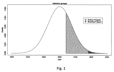
BRIEF DESCRIPTION OF THE DRAWINGS
Figure 1 shows a distribution of EBVs for a population of selection
candidates, including
EBVs for animals selected for a breeding program to produce sires and EBVs for
animals selected
for an embryo production program.
DETAILED DESCRIPTION OF THE INVENTION
The present invention is a novel method encompassing embryo transfer,
obtaining an
embryonic and/or fetal cell sample from amniotic fluid during gestation,
extracting DNA from the
cell sample, performing a genomic analysis of the extracted DNA and then
cloning the
embryo/fetus. In certain embodiments of the invention, the decision to clone
an embryo or fetus
is based on its genomic analysis, including but not limited to its genomic
estimated breeding value
with respect to one or more traits.
Certain embodiments of the invention can be used to select against production
of animals
of inferior or detrimental genetic and/or genomic value, while selecting for
the production of the
most productive elite genotypes, with the highest call rates, available in a
genetic nucleus system.
Accordingly, certain embodiments of the invention utilize genomic tools,
extensive genetic and
genomic evaluation for production, health, fertility and other physiological
traits based on analysis
of single nucleotide polymorphism (SNP) data from historical reference
information, then combine
6
CA 03000506 2018-03-28
WO 2017/066622 PCT/US2016/057115
breeding genotypes in a molecular and biotechnology-based breeding program to
maximize
genetic progress in a line, herd or genetic nucleus. Embryos are created in
vivo and in vitro from
elite females and bulls to produce offspring with the potential for the
highest genetic merit. These
embryos are transferred into a highly screened and selected group of
recipients maintained on
recipient farms. The surrogate females carrying high genetic and/or genomic
value pregnancy are
monitored during pregnancy, verified for fetal sex and then placed into
rotation for amniocentesis-
based genetic diagnosis. After organogenesis is complete and fetal growth is
underway, fluid and
cell aspiration from the fetal amnion is performed. These fluids are collected
in a novel aspirate
collection system and brought into the laboratory to be placed into cell
culture. Aspirate and cells
are analyzed by cellular assays and/or genomic approaches, the cells are
continued in culture to
confluence, passage, cryopreservation or productive use. After genetic and
genomic evaluations,
genetic information can be used to determine the developmental fate and
production direction of
any developmentally competent pregnancy. In certain embodiments, selected
genetic and genomic
based genotypes are placed into a component somatic cloning system to
propagate the most elite
lines of genotypes. Breeders of non-human mammalian species are focused on
increasing the rate
of genetic progress in a line, herd or genetic nucleus, as well as on
increasing the rate of genetic
dissemination of superior genotypes. In furtherance of these goals, tools such
as genomic testing,
embryo transfer and cloning are being developed and utilized by breeders at
various stages of
animal production.
Embryo transfer is extensively used in the modern livestock industry. As noted
above, the
major expense portion of any embryo transfer program is the cost and
maintenance of the recipient
animals. Typically, however, these costs are offset by the value of the
resulting animal, and
generally, the higher the genetic merit of the resulting animal, the higher
its commercial value.
7
CA 03000506 2018-03-28
WO 2017/066622 PCT/US2016/057115
Accordingly, embryo transfer programs place an emphasis on the production of
high genetic merit
animals.
One aspect of the instant invention allows a breeder to ascertain the genetic
merit of a fetus
early in gestation. Terminating the pregnancies of low genetic merit fetuses
then allows a breeder
to either reduce the number of recipient animals needed in their embryo
transfer program, or
alternatively, to increase the number of high genetic merit fetuses that can
be produced using a
given number of recipients over a given period of time. In another embodiment,
after ascertaining
the genetic merit of a fetus, a breeder may decide to maintain the pregnancy
but replace the
recipient carrying the fetus with a new recipient; and in yet a further
embodiment the new recipient
is carrying a fetus.
Another aspect of the instant invention allows a breeder to clone high genetic
merit fetuses
early in gestation and without harming the fetus. Specifically, fetal cells or
tissue obtained for
ascertaining genetic merit (via amniocentesis, for example) are used to
produce clones via somatic
cell nuclear transfer. In contrast to the invention, clones in the livestock
industry are typically
created from somatic cells obtained from young adult animals, and if derived
from an in vitro
embryo or fetus, the embryo or fetus is generally discarded or severely
compromised after such a
procedure. Additionally, even without being subjected to biopsy procedures,
embryos created by
in vitro fertilization (IVF) have a significantly lower survival rate than
their conventional, in vivo
counterparts. Accordingly, use of the instant invention raises the probability
that the costs
associated with genomic testing will be recouped since genomic testing is
performed after the
embryo has established a successful gestation in the recipient.
8
CA 03000506 2018-03-28
WO 2017/066622 PCT/US2016/057115
Embryo production in vivo and in vitro
In certain embodiments of the invention, embryos may be produced in vivo by
traditional
methods for synchronized supernumerary follicle production, artificial
insemination (AI) and
scheduled non-surgical transvaginal catheterized intrauterine embryo recovery.
In other aspects
of the invention, in vitro produced embryos may be produced in the laboratory
by non-typical
harvest of oocytes, IVF and embryo culture methodologies. In peripubertal
heifers, prophase I
immature cumulus oocyte complexes (COCs) are recovered from live standing
females by using
ultrasound guided transvaginal oocyte recovery (TVOR) system, also referred to
as ovum pickup
(OPU). In prepubertal heifers, ultrasound guided laparoscopic OPU is employed
for COC
recovery. When immature COCs are brought into the laboratory, they are placed
into typical in
vitro maturation (IVM) culture system where the most developmentally capable
oocytes undergo
spontaneous and programmed meiosis. After an overnight culture period, those
oocytes that
progress through meiosis I (and accordingly shed their second polar body
progressing to metaphase
of the second meiotic division) and are morphologically normal (including an
intact plasma
membrane) are used in IVF. Mature oocytes from individual females are placed
into traditional
IVF drops and mated to specific sires, using highly screened and accurate
sperm capacitation
treatments and sperm concentration per oocyte fertilized. Zygotes (day 1) are
placed into
traditional co-culture system and cultured to uterine stages of development by
day 7-8 of culture.
Embryos are typically transported to a recipient heifer farm where they are
non-surgically
transferred. Prior to transfer, embryos may be biopsied or sampled for genetic
screening and/or
genomic evaluation. Within certain specific stages of embryo development,
embryos can be
dismantled and used in embryo multiplication procedures and/or cryopreserved
for later use.
Embryos destined for transfer to synchronized surrogate females are
transported to the farm in
9
CA 03000506 2018-03-28
WO 2017/066622 PCT/US2016/057115
culture and non-surgically transferred by traditional methods. In certain
embodiments, the
invention contemplates that recipient females are regularly checked by
veterinarians and ongoing
pregnancies are monitored on a regular and scheduled basis via transrectal
real time
ultrasonography.
Embryo Transfer
Although not necessarily required, certain embodiments of the invention
encompass
embryo transfer. Specifically, in some embodiments, fetal cell samples are
obtained from amniotic
fluid of a recipient animal into which an embryo has been placed via embryo
transfer. In other
embodiments of the invention, embryo transfer is used to transfer a cloned
embryo into a recipient.
Any method known in the art may be used to transfer an embryo into a
recipient, including any
known surgical or non-surgical method. In alternative embodiments, fetal cell
samples are
obtained from fetuses that are conceived and that gestate entirely in vivo.
The following surgical and non-surgical methods of embryo transfer are
provided by way
of non-limiting example only.
In cattle, an embryo can be transferred via mid-line abdominal incision, or a
flank incision,
to a recipient under general anesthesia. Recipients are placed in squeeze
chutes that give access
to either flank. The corpus luteum is located by rectal palpation and the
flank ipsilateral to the
corpus luteum is clipped, washed with soap and water, and sterilized with
iodine and alcohol.
About 60 ml of 2 percent procaine is given along the line of the planned
incision. A skin incision
is made about 15 cm long, high on the flank, just anterior to the hip. Muscle
layers are separated,
and the peritoneum is cut. The surgeon inserts a hand and forearm into the
incision, locates the
ovary, generally about 25 cm posterior to the incision, and visualizes or
palpates the corpus luteum.
The uterine horn is exteriorized by grasping and stretching with the thumb and
forefinger the broad
CA 03000506 2018-03-28
WO 2017/066622 PCT/US2016/057115
ligament of the uterus, which is located medial to the uterine horn. A
puncture wound is made
with a blunted needle through the wall of the cranial one-third of the exposed
uterine horn. Using
about 0.1 ml of medium in a small glass pipette (<1.5 mm outside diameter),
the embryo is drawn
up from the storage container. The pipette is then inserted into the lumen of
the uterus, and the
embryo is expelled. The incision is then closed, using two layers of sutures.
Alternatively, a non-surgical method may be used to transfer an embryo in
cattle. First, it
is necessary to palpate ovaries in order to select the side of ovulation,
since pregnancy rates are
lowered if embryos are transferred to the uterine horn contralateral to the
corpus luteum.
Recipients should be rejected if no corpus luteum is present or pathology of
the reproductive tract
is noted. The next step is to pass the embryo transfer device, e.g., a
standard Cassou inseminating
gun, through the cervix. The third step of non-surgical transfer is to insert
the tip of the instrument
into the desired uterine horn ipsilateral to the corpus luteum. The final step
of the procedure is to
transfer the embryo from a container, such as a straw, into the desired
uterine horn using the
transfer device.
Collection of Amniotic Fluid
Certain embodiments of the invention encompass methods of collecting amniotic
fluid.
Once amniotic fluid is collected, a further aspect includes isolating fetal
cells from the amniotic
fluid and performing genomic analysis on DNA extracted from the fetal cells.
Any method known
in the art for collection of amniotic fluid may be used in the invention,
including but not limited to
trans-vaginal/trans-uterine collection using either ultrasound guided or
manual puncture
techniques. Additionally, amniotic fluid may be collected at any time during
gestation in a mother
or embryo transfer recipient, including from day 45 through parturition, or
between day 1 to day
10, day 20 to day 30, 30 to day 280, day 40 to day 100, day 50 to day 80, day
60 to day 70, day 70
11
CA 03000506 2018-03-28
WO 2017/066622 PCT/US2016/057115
to day 80, day 80 to day 90, day 90 to day 100, day 100 to day 120, day 70 to
day 90, day 75 to
day 80, day 75 to day 90, day 70 to day 85, or day 120 to day 280, of
gestation,.
By way of example, the following collection procedure may be used in the
invention. One
skilled in the art will know that variations on this method exist and that
this method should not be
construed to limit the functionality or scope of the current invention. This
method is illustrative
only.
Obtain a bovine mother, or recipient, with a fetus on day 65 to day 250 of
gestation.
Administer standard caudal epidural anesthesia with 2% lidocaine. Raise the
animals
approximately 40 cm at the front using a platform in order to place the
reproductive tract back
towards the pelvis. Clean and disinfect the vulva region and inside of the
vaginal vaults several
times with iodine. Trans-rectally retract the uterus with the opposite hand
and juxtapose the
pregnant horn against the vaginal wall. Insert an ultrasound-transducer
covered with a sterile
sleeve into the vaginal vault with the aid of light lubrication approximately
to the level of the
cervix. Aspirate the fetal fluid by intra-vaginal placement of a needle (0 =
1.3 mm, 68 cm length)
installed within the body of the ultrasound-transducer and connected to a
vacuum-tube blood
collection assembly. Ultrasound scanner may be equipped with a 5.0 MHz convex
type transducer
approximately 1.6 cm wide and 58 cm long. Advance the needle through the
vaginal and uterine
walls by sharply moving the vacuum tube over a distance of about 3 to 4 cm. If
the syringe plunger
meets resistance, reposition the needle and take another aspirate. Transfer
the aspirate was to a
sterile 10 ml test tube, placed on ice, and submit for DNA analysis. Confirm
successful needle
placement by direct observation of ultrasonography and fetal fluid swirling
within the vacuum
tube. Fetal viability may be assessed between 7 to 10 days after the
aspiration procedure. Imaging
of either independent fetal movement or heart beat may be taken as proof of
viability.
12
CA 03000506 2018-03-28
WO 2017/066622 PCT/US2016/057115
Another collection method in pregnant cattle encompasses the use of ultrasound-
guided
transvaginal oocyte recovery (TVOR) equipment, specialized fluid recovery
tubing, and adapted
filter collection system. In this example, in all cattle destined for
amniocentesis, pregnancy is
confirmed and fetal sex determined by transrectal ultrasonography at specific
periods after embryo
transfer, implantation and the completion of organogenesis. By day 45-100, or
more specifically
day 75-80, of the first trimester of gestation, ultrasound-guided transvaginal
oocyte recovery
equipment is adapted and used to visualize the entire fetus and amniotic
vesicle in a uterine horn
during aspiration. Prior to collection, the heifers are restrained in stocks
and sedated prior to
performing amniocentesis. The veterinary staff performing amniocentesis use
complete sterile
procedures, including powder free nitrile gloved hands and ethanol
sterilization of equipment. To
ensure that the area is free of contamination before insertion of the
transducer, the rectum is
emptied of feces, and under epidural anesthesia the vulva and rectal area of
the cow are thoroughly
cleaned and scrubbed. The disinfection step is completed by rinsing the vulva
and rectal area with
Betadine solution and the rinsing and spraying the cleaned area with 70%
ethanol. The TVOR
equipment is cleaned and sterilized with ethanol immediately prior to its
introduction into the
vagina and is fitted with a sterile stainless steel single-needle guide. The
TVOR equipment is
advanced into the vagina, positioned to the left or the right of the cervical
os and by means of
manipulation per rectum, the pregnant uterine horn is positioned against the
probe, avoiding
interposition of other tissue in the proposed needle path. The exact location
of the amniotic sac is
determined by the recognition of fetal body parts, the allantoamniotic and
allantochorionic
membranes and the uterine wall. When a non-echogenic area representing
amniotic fluid is seen
on the monitor screen, a sterile needle with a stylette is inserted within the
needle guide and
advanced penetrating through the vaginal wall, uterus and subsequent fetal
membranes. As soon
13
CA 03000506 2018-03-28
WO 2017/066622 PCT/US2016/057115
as the tip of the needle is seen to have entered the fetal fluid compartment,
the stylette is withdrawn
from the needle and the needle is placed inside the amnion of the fetus. An
initial 5-10 ml of fetal
fluid is aspirated into the tubing and flushed out of the tubing system to
reduce or eliminate
maternal contamination. An amniocentesis filter is attached to the tubing and
an additional 30-40
ml of amniotic fluid is aspirated. During the fluid collection, the pregnant
uterine horn is held in
the same position, and the exact location of the tip of the needle is
guaranteed by its visualization
on the ultrasound screen. When samples from more than 1 heifer are collected
on the same day,
the needle-guide is replaced by a sterile one, and the transducer is
thoroughly cleaned and
disinfected before being used on the next animal. After collection of amniotic
fluid is completed
in an animal, the collected fluid in the filter system is placed on ice and
transported back to the
cell culture laboratory.
Isolating Amniocytes from Amniotic Fluid
The term "amniocytes" as used herein, refers to cells obtained from amniotic
fluid, as well
as to cells cultured from cells obtained from amniotic fluid. Amniocytes,
including fetal
fibroblasts and amniotic fluid-derived mesenchymal stem cells (AFMSCs), used
in the present
invention may be obtained from, e.g., amniotic fluid from amniocentesis
performed for fetal
karyotyping, or amniotic fluid obtained at term. For purposes of the
invention, amniocytes may
be isolated from the amniotic fluid by any method known in the art, e.g., by
centrifugation followed
by removal of the supernatant.
Amniocyte Cell Culture
One aspect of the invention encompasses culturing isolated amniocytes.
Cultured
amniocytes can in turn be used in various applications, including genotyping
and for producing
clones. By way of example, the following culturing procedure may be used in
certain
14
CA 03000506 2018-03-28
WO 2017/066622 PCT/US2016/057115
embodiments of the invention. One skilled in the art will know that variations
on this method exist
and that this method should not be construed to limit the functionality or
scope of the current
invention. This method is illustrative only.
Amniocytes are centrifuged (200 g, 10 min) at room temperature and the pellet
is gently
resuspended in Chang medium. Cells are plated into 100 mm gelatinized Petri
dishes and left
undisturbed. Media is changed every 3-4 days. After 2 weeks in culture, they
are trypsinized to
disperse cells and allow their growth in a monolayer. Amniocytes are cultured
at 37 C in a
humidified 5% CO2 atmosphere. Cells are passaged at a ratio 1:4 every 5 days
until they reach
80% confluence. For subsequent passages, the media is aspirated, washed with
PBS, detached
with 0.05% trypsine for 5 min at 37 C.
Isolation and Culture of Amniotic Fluid-Derived Mesenchymal Stem Cells
In certain embodiments of the invention, a two-stage culture method may be
used to isolate,
culture, and enrich amniotic fluid-derived mesenchymal stem cells (AFMSCs)
from amniotic fluid
obtained by amniocentesis. Mammalian mesenchymal stem cells are presumptively
multipotent
cells that have the potential to differentiate into multiple lineages
including bone, cartilage, muscle,
tendon, ligament fat and a variety of other connective tissues.
Morphologically, mesenchymal
stem cells in their undifferentiated state are spindle shaped and resemble
fibroblasts.
Mesenchymal stem cells have been identified mostly in bone marrow, but have
also been found in
both adult and fetal peripheral blood, fetal liver, fetal spleen, placenta and
in term umbilical cord
blood. Significantly, mesenchymal stem cells can be found in mammalian
amniotic fluid. Under
specific culture conditions, mammalian AFMSCs have been induced to
differentiate into
adipocytes, osteocytes and neuronal cells.
CA 03000506 2018-03-28
WO 2017/066622 PCT/US2016/057115
The two-stage culture protocol comprises a first stage of culturing
amniocytes, and a
second stage of culturing mesenchymal stem cells. The method begins by setting
up primary
cultures using cytogenetic laboratory amniocytes culture protocol. Non-
adhering amniotic fluid
cells in the supernatant medium are collected. For culturing mesenchymal stem
cells, the non-
adhering cells are centrifuged and then plated in a culture flask with an
alpha-modified Minimum
Essential Medium supplemented with fetal bovine serum. For mesenchymal stem
cell growth, the
culture is incubated with humidified CO2.
By way of example, the following specific culturing procedure may be used in
certain
embodiments of the invention. One skilled in the art will know that variations
on this method exist
and that this method should not be construed to limit the functionality or
scope of the current
invention. This method is illustrative only.
For culturing amniocytes, set up four primary in situ cultures in 35 mm tissue
culture-grade
dishes using Chang medium (Irvine Scientific, Santa Ana, Calif). Collect non-
adhering amniotic
fluid cells in the supernatant medium on the 5th day after the primary
amniocytes culture and keep
them until a completion of fetal chromosome analysis.
For culturing mesenchymal stem cells, centrifuge the tube containing the non-
adhering
cells, then plate them in 5-15 ml of alpha-modified Minimum Essential Medium
(a-MEM)
supplemented with 10-20% fetal bovine serum (FBS) and 1-20 ng/ml b-FGF in a
25cm2 culture
flask and incubate at 37 C with 5% humidified CO2 for mesenchymal stem cell
growth.
Flow cytometry, RT-PCR, and immunocytochemistry may be used to analyze the
phenotypic characteristics of the cultured mesenchymal stem cells. Von Kossa,
Oil Red 0 and
TuJ-1 stainings may be used to assess the differentiation potentials of the
mesenchymal stem cells.
16
CA 03000506 2018-03-28
WO 2017/066622 PCT/US2016/057115
The following additional culture method is presented by way of example only.
The
invention contemplates sterile technique, including being gloved with non-
powder nitrile gloves
to process amniotic fluid. In certain embodiments of the invention, the entire
process is performed
in a cell culture laminar flow biosafety cabinet and only food grade ethanol
is used in washing
gloved hands whenever needed or possible.
Fluid and amniocytes are aspirated by pipette into 15 ml conical tubes. The
collection filter
is rinsed with culture medium to remove any adhered cells and repeated as
necessary to remove a
maximal amount of amniocytes from the filter. The conical tubes are
centrifuged until a cell pellet
is formed, supernatant is aspirated, and cells are resuspended in cell culture
medium. The cell
suspension is thoroughly mixed and pipetted into culture wells and/or dishes.
Cell cultures are
placed into a cell culture incubator and cultured at 38.7C in 5% CO2/air for 5
days undisturbed.
On day 5 of culture, cell culture dishes are removed from culture and cell
culture medium and any
floating cells are aspirated and placed into 15 ml centrifuge tube. The
remaining cells plated on
the original cell culture dishes, primarily fetal fibroblasts and AFMSCs are
fed with fresh culture
medium and placed back into cell culture incubators and cultured until 80-90%
confluent. After
reaching confluency, the cells are lifted for passage and/or cryopreservation.
The aspirated
floating amniocytes can be started in amniocyte-specific cell culture or used
in fetal diagnostic
testing and/or genomic testing and profiling. Both original plated fetal
fibroblast cultures and
original floating amniocyte cell cultures can be cultured for indefinite
passaging and
cryopreservation. Cryopreserved fetal fibroblasts and/or amniocytes can be
warmed and passaged
or used in cloning procedures.
17
CA 03000506 2018-03-28
WO 2017/066622 PCT/US2016/057115
DNA Extraction and Amplification
Another aspect of the invention encompasses genotyping amniocytes.
Specifically, once
fetal fibroblasts or mesenchymal stem cells have been isolated from the
amniotic fluid, their DNA
may be extracted and used for genotyping. In a specific embodiment, the DNA of
cultured fetal
fibroblasts or mesenchymal stem cells can be used for genotyping. Fetal
fibroblast, or
mesenchymal stem cell, DNA may first be extracted and then amplified (via PCR)
so that there is
a sufficient amount of DNA for genotyping. Alternatively, in some embodiments
of the invention,
DNA may be extracted directly from amniocytes, including fibroblasts and
mesenchymal stem
cells, found in amniotic fluid. The invention encompasses embodiments in which
the amount of
DNA extracted is very low, ranging from 1ng/ 1 to 10 ng/ 1 (based on double
strand DNA assays).
Visualization using 1% agarose gels has shown the extracted DNA in some
examples to be large,
>23000 MW with little fragmented DNA.
For genomic analysis, approximately 1-200 ng of double stranded DNA should be
extracted per sample DNA at concentration per sample of 1-50 ng/ul. In certain
embodiments of
the invention, only 1ng/ 1 to 10 ng/ 1 of DNA are necessary for genomic
analysis. In a particular
embodiment, less than 15 ng of DNA total is necessary for genomic analysis. In
some
embodiments of the invention, the DNA is used in genotyping for parental
verification and
genomic evaluation. The genomic evaluation for production, health, fertility
and other
physiological traits utilized in certain embodiments of the invention is based
on analysis of SNP
data from historical reference population data determined by genome-wide
association studies
(GWAS). This evaluation of fetal cells also allows for rapid generation
modeling by allowing pre-
selection of fetus as a parent for the next generation of matings. The
remaining cells in culture
18
CA 03000506 2018-03-28
WO 2017/066622 PCT/US2016/057115
remain in cell culture for passage and eventual harvest and cryopreservation
for later diagnostic,
cytogenetic and biological productive use such as cloning.
By way of example, the following DNA extraction and amplification procedure
may be
used in certain embodiments of the invention. One skilled in the art will know
that variations on
this method exist and that this method should not be construed to limit the
functionality or scope
of the current invention. This method is illustrative only.
Fibroblast DNA is extracted from the contents of a 25-cm2 culture bottle by
the salting-out
procedure, with minor modifications (Miller et al., 1988; Biase et al., 2002).
Fifty nanograms of
genomic DNA is used in 25 tL of PCR mix (1 U Taq polymerase, 100 i.tM dNTP, 1
mM MgC12,
pmol of each primer) and amplified 36 times using the following conditions: 93
C for 3 min,
93 C for 40 s, 58 C for 40 seconds, 72 C for 40 seconds, and 72 C for 5 min.
The primers are
designed to amplify a 410-bp fragment of the NeoR gene (sense: 5'-GAG-GCT-ATT-
CGG-CTA-
TGA-CTG-3' and anti-sense: 5' -TCG-ACA-AGA-CCG-GCT-TCC-ATC-3') and a 262-bp
fragment of bovine satellite I DNA (Gaillard et al., 1981) (sense: 5'-AGG-TCG-
CGA-GAT-TGG-
TCG-CTA-GGT-CAT-GCA-3' and anti-sense: 5 '-AAG-ACC-TCG-AGA-GAC-CCT-CTT-
CAA-CAC-GT-3').
In certain embodiments of the invention, DNA from amniocytes and mesenchymal
stem
cells can be extracted using the Purelink Genomic Kit Cat # K1820-00
(Invitrogen). In further
embodiments, once the DNA is extracted, it can be put through a whole genome
amplification
protocol using the Illustra Genomiphi V2 DNA amplification kit (GE
Lifesciences), which uses
the phi29 DNA polymerase to amplify the genome.
In other embodiments of the invention the following DNA extraction procedure
is
employed.
19
CA 03000506 2018-03-28
WO 2017/066622 PCT/US2016/057115
Cells exposed to culture media often contain fetal calf serum. Due to high
levels of DNase
commonly found in fetal calf serum and the presence of cations that could
catalyze the hydrolytic
cleavage of phosphodiester linkage in DNA, an equal volume of a solution
containing Tris-EDTA
is added to the harvested cells to chelate cations essential for DNase
activity. After adding the
Tris-EDTA, the cell suspension is then stored in 1.5 ml microcentrifuge tubes
at 4 C until required
for DNA extraction.
The 1.5 ml tubes containing cell suspension are spun at >10000 x g in a
microcentrifuge
for 45 seconds to pellet cells. The suspension solution is pipetted off
carefully so as to not remove
pelleted cells. Approximately 50 IA of suspension solution is left in the
tube. The tubes are then
vortexed for 10 seconds to resuspend the cell pellets. 300 IA of Tissue and
Cell Lysis Solution
(Epicentre; Madison Wisconsin) containing 1 IA of Proteinase K (Epicentre;
Madison Wisconsin)
are then added to each tube and mixed. The tubes are then incubated at 65 C
for 30 minutes while
making sure to vortex at 15 minutes. The samples are then cooled to 37 C.
Afterwards 1 IA of 5
mg/ 1 RNase A (Epicentre; Madison Wisconsin) is added to to each sample and
then mixed. The
samples are then incubated at 37 C for 30 minutes. The samples are then placed
in a 4 C cooler
for 5 minutes. 175 IA of MPC Protein Precipitation Reagent (Epicentre; Madison
Wisconsin) are
then added to each sample, and the samples are then vortexed vigorously for 10-
15 seconds. The
samples are then centrifuged in order to pellet debris for 8 minutes at >10000
x g. The supernatant
is then transferred to a clean microcentrifuge tube. 600 IA of cold (-20 C)
isopropanol is added to
the supernatant. Each tube is then inverted 30-40 times. The DNA is then
pelleted by
centrifugation for 8 minutes in a microcentrifuge at >10000 x g. The
isopropanol is poured off
without dislodging DNA pellet. The pellet is rinsed once with 70% ethanol and
then the ethanol
is carefully poured off so as not to disturb the DNA pellet. The residual
ethanol is then removed
CA 03000506 2018-03-28
WO 2017/066622 PCT/US2016/057115
with a pipet, and the DNA pellet is allowed to air dry in the microcentrifuge
tube. Once dried,
resuspend the DNA pellet in 2011.1 Tris-EDTA.
Genotyping DNA
In one aspect of the invention, extracted and/or amplified DNA from amniocytes
and
mesenchymal stem cells may be genotyped using SNP arrays or chips, which are
readily available
for various species of animals from companies such as Illumina and Affymetrix.
For purposes of
the invention, the term "genotyping" includes, but is not limited to,
obtaining SNP and/or copy
number variation (CNV) data from DNA. For purposes of the invention, the term
"genotype"
includes, but is not limited to, SNP and/or copy number variation (CNV) data
obtained from DNA.
Low density and high density chips are contemplated for use with the
invention, including SNP
arrays comprising from 3,000 to 800,000 SNPs. By way of example, a "50K" SNP
chip measures
approximately 50,000 SNPs and is commonly used in the livestock industry to
establish genetic
merit or genomic estimated breeding values (GEBVs). In certain embodiments of
the invention,
any of the following SNP chips may be used: BovineSNP50 vi BeadChip
(Illumina), Bovine SNP
v2 BeadChip (Illumina), Bovine 3K BeadChip (Illumina), Bovide LD BeadChip
(Illumina),
Bovine HD BeadChip (Illumina), Geneseekg Genomic ProfilerTM LD BeadChip, or
Geneseekg
Genomic ProfilerTM HD BeadChip.
Determining GEBVs from SNP Data
The basis, and algorithm, for using SNPs in determining GEBVs is found in
Meuwissen et
at., "Prediction of total genetic value using genome-wide dense marker maps,"
Genetics 157, 1819
1829 (2001), which is incorporated by reference herein in its entirety.
Implementation of genomic
data in predictions for desirable traits is found in Van Raden, "Efficient
Methods to Compute
21
CA 03000506 2018-03-28
WO 2017/066622 PCT/US2016/057115
Genomic Predictions," Dairy Science 91, 4414 4423 (2008), which is
incorporated by reference
herein in its entirety.
Livestock in the United States are often ranked using selection indexes that
incorporate
data related to various commercially important traits. With the advent of
genomic testing, genomic
data is now commonly used to predict these traits. To calculate an animal's
score for a genomic
selection index, one must first calculate the animal's GEBVs for each trait in
the index, which can
be accomplished using the teachings in Meuwissen et at. and VanRaden, above.
Next, one
determines the economic weight for each trait in the index. Finally, to
determine the animal's
score for the selection index, multiply each trait's GEBV by its economic
weight and then sum all
of these values together.
A genomic index commonly used in the United States for dairy cattle is the
Genomic Total
Performance Index (GTPIg), which is comprised of the following traits:
protein; feed efficiency;
dairy form; feet and legs composite; somatic cell score; daughter calving
ease; fat; udder
composite; productive life; fertility index; and daughter stillbirth. In
certain embodiments, feed
efficiency is equal to the dollar value of milk produced less feed costs for
extra milk and less extra
maintenance costs, and the fertility index is a function of heifer conception
rate, cow conception
rate and daughter pregnancy rate. In other embodiments of the invention, GEBV
is used to
determine Genomic Predicted Transmitting Ability (GPTA).
By way of example, in addition to determining a GEBV for a trait, the presence
or absence
of any of the following diseases and/or traits can be detected using SNP data
or genomic data:
Demetz syndrome; white heifer disease; Weaver syndrome (haplotype BHW);
haplotype HIM;
haplotype HH1; lethal brachygnathia trisomy syndrome; haplotype HHO; bovine
hereditary
cardiomyopathy; bovine dilated cardiomyopathy; neuronal ceroid lipofuscinosis;
bovine
22
CA 03000506 2018-03-28
WO 2017/066622 PCT/US2016/057115
chondrodysplastic dwarfism; notched ears/nicked ears; idiopathic epilepsy;
bilateral convergent
strabismus with exophthalmos; haplotype BHP; haplotype HHP; haplotype JHP;
neuropathic
hydrocephalus/water head; congenital hypotrichosis and anodontia
defect/ectodermal dysplasia;
ichthyosis fetalis; lethal trait A46/bovine hereditary zinc deficiency; Marfan
Syndrome; double
muscling; multiple ocular defects; bovine ocular squamous cell carcinoma; pink
tooth; posterior
paralysis/hind-limb paralysis; haplotype BHM; bovine spongiform
encephalopathy/mad cow
disease; mule foot disease (haplotype HEIM); myophosphorylase deficiency;
dropsy; black/red
coat color (haplotype HBR; haplotype HEIR); BAND3 deficiency; Charolais
ataxia; bovine spinal
dysmyelination (haplotype BHD); Dun coat color in Dexter cattle; bovine
familial convulsions
and ataxia; bulldog calf; simmental hereditary thrombopathy; GHRD; renal
tubular dysplasia
(RTD)/chronic interstitial nephritis; Hereford white face; haplotype HHC;
developmental
duplications; black kidney; cardiomyopathy/Japanese black cattle; crooked tail
syndrome;
congenital pseudomyotonia; bovine hereditary arthrogyposis multiplex
congentia; belted;
Syndrome d'Hypoplasie Generalisee Capreoliforme; fawn calf syndrome; bovine
neonatal
pancytopenia; rat-tail syndrome; cheilognathoschisis; German White Fleckvieh
syndrome;
haplotype JH1; paunch calf syndrome; acorn calf disease/congenital joint
laxity and dwarfism;
haplotype HH2; haplotype HH3; haplotype HH4; Holstein bull-dog dwarfism;
haplotype AHl;
haplotype HH5; haplotype JH2; and lethal arthrogyposis syndrome.
Cloning
An additional aspect of the invention encompasses cloning embryos and/or
fetuses that
have been genomically evaluated using the techniques disclosed herein. Cloning
is generally
understood to be the creation of a living animal/organism that is essentially
genetically identical
to the unit or individual from which it was produced. In those embodiments of
the invention that
23
CA 03000506 2018-03-28
WO 2017/066622 PCT/US2016/057115
encompass cloned embryos and/or fetuses, any method by which an animal can be
cloned that is
known in the art can be utilized. Thus, it is contemplated that cloned embryos
and cloned fetuses
are produced by any conventional method, for instance including the cloning
techniques described
herein, as well as those described in international patent application
PCT/US01/41561. In one
aspect of the invention, a basis for cloning an embryo or a fetus is its
genomic merit. In a further
aspect, the embryo or fetus's genetic merit is determined by genomic analysis
as disclosed herein.
Cloning of embryos by nuclear transfer has been developed in several species.
This
technique generally involves the transfer of a cell nucleus (obtained from a
donor cell) into an
enucleated cell, for instance, a metaphase II oocyte. This oocyte has the
ability to incorporate the
transferred nucleus and support development of a new embryo (Prather et al.,
Biol. Reprod 41:414-
418, 1989; Campbell et at., Nature 380:64-66, 1996; Wilmut et at., Nature
385:810-813, 1997).
Morphological indications of this re-programming are the dispersion of
nucleoli (Szollosi et at., J.
Cell Sci. 91:603-613, 1988) and swelling of the transferred nucleus (Czolowska
et at., 1984; Stice
and Robl, Biol. Reprod 39:657-664, 1988; Prather et at., J. Exp. Zool. 225:355-
358, 1990; Collas
and Robl. Biol. Reprod 45:455-465, 1991). The most conclusive evidence that
the oocyte
cytoplasm has the ability to re-program is the birth of offspring from nuclear
transplant embryos
in several species, including sheep (Smith and Wilmut, Biol. Reprod. 40:1027
1035, 1989;
Campbell et at., Nature 380:64-66, 1996; Wells et at., Biol. Reprod. 57:385-
393, 1997), cattle
(Wells et al., Biol. Reprod. 60:996-1005, 1999; Kato et al., Science 282:2095-
2098, 1998; Prather
et al., Biol. Reprod. 37:859-866, 1987; Bondioli et al., Theriogenology 33:165-
174, 1990), pigs
(Prather et al., Biol. Reprod. 41:414-418, 1989) and rabbits (Stice and Robl,
Biol. Reprod. 39:657-
664, 1988).
24
CA 03000506 2018-03-28
WO 2017/066622 PCT/US2016/057115
Cloning by nuclear transfer entails removing the nucleus from the recipient
oocyte and
isolating a nucleus from a donor cell. The donor nucleus is then joined to the
recipient oocyte and
electrically induced cell fusion is used to introduce the nuclei from the
donor embryo cell into a
recipient cell. In certain embodiments, the embryo cloning process follows a
basic five step
procedure as follows: (1) selecting a proper recipient embryo or oocyte for
nuclear transfer; (2)
enucleating, i.e., removing the nuclear material from the recipient oocyte;
(3) introducing the
membrane-bounded nucleus of the donor cell to the enucleated recipient oocyte;
(4) orienting the
donor membrane-bounded nucleus and the recipient oocyte for cell fusion; and
(5) fusing the
membrane surrounding the donor nucleus to the membrane of the recipient oocyte
and activating
the recipient oocyte by dielectrophoresis.
In certain embodiments of the invention, the oocyte used as the recipient cell
is a cell that
develops from an oogonium and, following meiosis, becomes a mature ovum. In
certain
embodiments relating to bovines, metaphase II stage oocytes, can be matured
either in vivo or in
vitro. In some embodiments, mature metaphase II oocytes may be collected
surgically from either
nonsuperovulated or superovulated cows or heifers 35 to 48 hours past the
onset of estrus or past
an injection of human Chorionic Gonadotropin (hCG) or similar hormone.
Alternatively, in other
embodiments, immature oocytes may be recovered by aspiration from ovarian
follicles obtained
from slaughtered cows or heifers and then may be matured in vitro by
appropriate hormonal
treatment and culturing.
In certain embodiments of the invention, micromanipulation of cells may
performed using
a cell holding pipette, having an outer diameter of about 120 micrometers and
an inner diameter
of approximately 25 to 35 micrometers, and a beveled, sharpened enucleation
and transfer pipette
having an outer diameter of approximately 25 to 35 micrometers. Mature oocytes
may be first
CA 03000506 2018-03-28
WO 2017/066622 PCT/US2016/057115
treated with cytochalasin B at about 7.5 micrograms per milliliter, or an
effectively similar
microtubal inhibitor at a concentration sufficient to allow the enucleation
and transfer pipette to
be inserted through the zona pellucida to allow for removal of a portion of
the cytoplasm without,
at any point, actually rupturing the plasma membrane. The mature oocyte can be
held in place by
mild suction by the cell holding pipette. The enucleation and transfer pipette
can then be inserted
through the zona pellucida of the oocyte at the point of either the metaphase
II bulge or adjacent
to the first polar body, i.e., in a location intended to be adjacent to the
metaphase chromosomes.
The pipette does not penetrate the plasma membrane. Aspiration applied through
the pipette draws
a cellular bulge into the pipette which includes, in the case of the metaphase
II bulge, the entire
bulge and surrounding cytoplasm, or, in the case of the first polar body, the
polar body plus the
surrounding cytoplasm. This process is intended to draw all the metaphase
chromosomes into the
pipette. As the pipette is withdrawn, with suction maintained, the plasma
membrane is stretched
and then seals to itself leaving a competent plasma membrane on the enucleated
oocyte.
In some embodiments of the invention, the donor cells may be treated with
cytochalasin B,
or may not be, depending on the size of the transfer pipette. The transfer
pipette carrying the
aspirated membrane-bounded nucleus can be inserted through the zona pellucida
of the recipient
enucleated oocyte, and the membrane-bounded nucleus can then deposited under
the zona
pellucida with its membrane abutting the plasma membrane of the recipient
oocyte.
In some embodiments of the invention, fusion of the membrane-bounded nucleus
to the
enucleated recipient oocyte and simultaneous activation of the recipient
oocyte may be carried out
by a single dielectrophoresis step using commercially available electrofusion
equipment. Prior to
electrofusing the donor embryo nucleus and enucleated recipient oocyte
together, it is necessary
to orient the cell membranes in the electric field. The term "orientation" as
used herein is defined
26
CA 03000506 2018-03-28
WO 2017/066622 PCT/US2016/057115
as the placement of the two cells such that the plane of contact of the two
membranes, i.e., the
plasma membrane of the body carrying the donor nucleus and the plasma membrane
of the
recipient oocyte, which will become fused together, is perpendicular to the
electrical field. It has
been found that random orientation results in a marked reduction in the
successful fusion rate. If
cells are oriented such that the fusion membranes are parallel, or at
approximately a 450 angle, to
the electrical field, the rate of successful fusion will decrease. The
alignment may be done
electrically or mechanically. If the size of the two cells is not greatly
disproportionate, a small
alignment alternating-current voltage (5 volts per millimeter at 1000 KHz) for
a short time (10
seconds) will cause the cells to reorient with their membranes apposed.
Repeated pulses may be
needed. If the cells vary greatly in size, mechanical manipulation may be
required to properly
orient the membranes.
In some embodiments of the invention, the insertion of a membrane-bounded
nucleus into
an enucleated bovine oocyte may be conducted by a dielectrophoretic method of
cell fusion, using
a DC current and using a non-conductive, i.e., non-ionic, cell fusion medium
such as a mannitol
solution or Zimmerman cell fusion medium. The fusion phenomenon is the result
of cell
membrane breakdown and pore formation between properly oriented opposing
cells. The pores,
or small channels, created between the two cells are thermodynamically
unstable because of the
high surface curvature of the channels and the associated high tension in the
membrane. This
instability causes the channels to merge and enlarge until the membranes form
a single cell.
The embryonic single-celled clones produced as described herein preferably are
cultured,
either in vivo or in vitro, to the morula or blastula stage. For example, the
clones may be cultured
in sheep oviducts or in a suitable culture medium. The embryos then may be
transferred into the
uteri of cattle, or other suitable animals, at a suitable stage of estrus. The
procedures for embryo
27
CA 03000506 2018-03-28
WO 2017/066622 PCT/US2016/057115
transfer are commonly known and practiced in the embryo transfer field. A
percentage of these
embryo transfers will initiate pregnancies in the maternal surrogates. Live
calves born of these
pregnancies will be genetically identical where the donor cells were from a
single embryo or a
clone thereof.
In one embodiment of the invention, cloning can be performed in one step using
the nucleus
of a somatic cell, such as a fetal fibroblast, or a stem cell, such as a
mesenchymal stem cell. The
somatic cell or stem cell is fused with an enucleated oocyte. After culture,
many of the fused
couplets (or cybrids) develop into morulae that can be implanted in recipients
for gestation.
In a further embodiment, two or more cycles of cloning can be carried out in
order to
increase the efficiency of production of cloned animals. Two-step cloning, for
example, involves
a first cloning cycle (e.g., by nuclear transfer) using a donor cell, growing
the resultant cybrid in
vitro and/or in vivo to produce a clonal fetus, then using a fetal cell from
the clonal fetus for a
second round of cloning (e.g., also by nuclear transfer). In one example, a
fibroblast is fused with
an enucleated oocyte and cultured to about the morula stage. The viable
morulae resulting from
this procedure are transferred to recipients. Most of these first-cycle
pregnancies can be allowed
to attempt to reach term, for instance for use as an internal experimental
control. After the embryo
has developed into a fetus (generally for a sufficient amount of time to
display differentiation into
tissues and organs), at least one and up to several of these first-cycle
fetuses are removed surgically
to provide tissue for the production of tissue cultures. By way of example,
cattle fetuses can
generally be used after they have reached a gestational age of at least 30
days; in specific
embodiments, cattle fetuses can be sacrificed at about 45 days gestational
age. Alternatively,
instead of sacrificing the fetus, amniocytes can be removed from the recipient
via amniocentesis
as described herein. Any fetal tissue or cells can serve to produce cell
cultures. In representative
28
CA 03000506 2018-03-28
WO 2017/066622 PCT/US2016/057115
embodiments, fetal cell cultures are produced from fetal fibroblasts, gonadal
cells, mesenchymal
stem cells or cells from the genital ridge. The fetal cell cultures are
propagated and samples
preserved (e.g., frozen) for future use. In certain embodiments, fetal tissue
is used directly for the
second round of cloning (without an intervening storage stage, and in some
instances without
development of an in vitro cell culture).
The fetal cell cultures (e.g., fibroblast cultures) can be used as nuclear
donors for the second
cloning cycle. In this second cycle (the second "step" of two-step cloning),
fetal cultured cells are
fused with enucleated oocytes to produce second-generation morulae. These
morulae are
transferred to recipients and the resulting pregnancies allowed to go to term
to produce live
progeny. Pregnancies resulting from the transfer of fetal-origin, second-
generation cloned-
embryos are allowed to mature for the full gestation period and result in the
delivery of live calves.
In certain embodiments, both the donor cell and the oocyte must be activated.
An activated
(e.g., non-quiescent) donor cell is a cell that is in actively dividing (e.g.,
not in a resting stage of
mitosis). In particular, an activated donor cell is one that is engaged in the
mitotic cell cycle, such
as GI phase, S phase or G2/M phase. The mitotic cell cycle has the following
phases, Gl, S, G2
and M. The G2/M phase refers to the transitional phase between the G2 phase
and M phase. The
commitment event in the cell cycle, called START (or restriction point), takes
place during the GI
phase. "START" as used herein refers to late GI stage of the cell cycle prior
to the commitment
of a cell proceeding through the cell cycle. The decision as to whether the
cell will undergo another
cell cycle is made at START. Once the cell has passed through START, it passes
through the
remainder of the GI phase (i.e., the pre-DNA synthesis stage). The S phase is
the DNA synthesis
stage, which is followed by the G2 phase, the stage between synthesis and
mitosis. Mitosis takes
place during the M phase. If prior to START, the cell does not undergo another
cell cycle, the cell
29
CA 03000506 2018-03-28
WO 2017/066622 PCT/US2016/057115
becomes arrested. In addition, a cell can be induced to exit the cell cycle
and become quiescent or
inactive. A "quiescent" or "inactive" cell, is referred to as a cell in GO
phase. A quiescent cell is
one that is not in any of the above-mentioned phases of tile cell cycle.
Preferably, the invention
utilizes a donor cell is a cell in the G1 phase of the mitotic cell cycle.
In certain embodiments of the invention, the donor cells are synchronized.
Using donor
cells at certain phases of the cell cycle, for example, G1 phase, allows for
synchronization of the
donor cells. One can synchronize the donor cells by depriving (e.g., reducing)
the donor cells of
a sufficient amount of nutrients in the media that allows them to divide. Once
the donor cells have
stopped dividing, then the donor cells are exposed to media (serum) containing
a sufficient amount
of nutrients to allow them to being dividing (e.g., mitosis). The donor cells
begin mitosis
substantially at the same time, and are therefore, synchronous. For example,
the donor cells are
deprived of a sufficient concentration of serum by placing the cells in 0.5%
Fetal Bovine Serum
(FBS) for about a week. Thereafter, the cells are placed in about 10% FBS and
they will begin
dividing at about the same time. They will enter the G1 phase about the same
time, and are
therefore, ready for the cloning process.
Methods of determining which phase of the cell cycle a cell is in are known to
those skilled
in the art, for example, U.S. Pat. No. 5,843,705 to DiTullio et al., Campbell,
K. H. S., et al., Embryo
Transfer Newsletter, vol. 14(1):12-16 (1996), Campbell, K. H. S., et al.,
Nature, 380:64-66 (1996),
Cibelli, J. B., et al., Science, 280:1256-1258 (1998), Yong, Z. and L.
Yuqiang, Biol. of Reprod.,
58:266-269 (1998) and Wilmut, I., et al., Nature, 385:810-813 (1997). For
example, as described
below, various markers are present at different stages of the cell cycle. Such
markers can include
cyclines D 1, 2, 3 and proliferating cell nuclear antigen (PCNA) for Gl, and
BrDu to detect DNA
synthetic activity. In addition, cells can be induced to enter the GO stage by
culturing the cells on
CA 03000506 2018-03-28
WO 2017/066622 PCT/US2016/057115
a serum-deprived medium. Alternatively, cells in GO stage can be induced to
enter into the cell
cycle, that is, at G1 stage by serum activation (e.g., exposing the cells to
serum after the cells have
been deprived of a certain amount of serum).
In certain embodiments, the genome of the donor cell can be the naturally
occurring
genome, for example, for the production of cloned animals, or the genome can
be genetically
altered to comprise a transgenic sequence, for example, for the production of
transgenic cloned
animals.
In some embodiments of the invention, the oocytes used in the present
invention are
activated oocytes. Activated oocytes are those that are in a dividing stage of
meiotic cell division,
and include metaphase I, anaphase I, anaphase II, and preferably, telophase
II. Oocytes in
metaphase II are considered to be in a resting state. The oocytes can be in
the resting stage of
metaphase II, and then activated, using methods described herein. The stage
that the oocyte is in
can be identified by visual inspection of the oocyte under a sufficient
magnification. Oocytes that
are in telophase II are identified, for example, by the presence of a
protrusion of the plasma
membrane of a second polar body. Methods for identifying the stage of meiotic
cell division are
known in the art.
Oocytes are generally activated by increasing their exposure to calcium
levels, in certain
embodiments. Increasing levels of calcium, e.g., by between about 10% and
about 60% above the
baseline levels, induces activation or meiotic cell division of the oocyte.
Baseline levels are those
levels of calcium found in an inactive oocyte. Rising levels of calcium,
coupled with decreasing
levels of phosphorylation further facilitates activation of the oocyte.
Several methods exist that
allow for activation of the oocyte. In particular, a calcium ionophore (e.g.,
ionomycin) is an agent
that increases the permeability of the oocyte's membrane and allows calcium to
enter into the
31
CA 03000506 2018-03-28
WO 2017/066622 PCT/US2016/057115
oocyte. As the free calcium concentration in the cell increases during
exposure to the ionophore,
the oocyte is activated following reduction in MPF (maturation promoting
factor) activity. Such
methods of activation are described in U.S. Pat. No. 5,496,720. Ethanol has a
similar affect. Prior
to or following enucleation, an oocyte in metaphase II can be activated with
ethanol according to
the ethanol activation treatment as described in Presicce and Yang, Mol.
Reprod. Dev., 37.61-68
(1994); and Bordignon & Smith, Mol. Reprod. Dev., 49:29-36 (1998). Exposure of
calcium to the
oocyte also occurs through electrical stimulation. The electrical stimulation
allows increasing
levels of calcium to enter the oocyte.
As contemplated herein, oocytes can be obtained from a donor animal during
that animal's
reproductive cycle. For example, oocytes can be aspirated from follicles of
ovaries at given times
during the reproductive cycle (exogenous hormone-stimulated or non-
stimulated). Also at given
times following ovulation, a significant percentage of the oocytes, for
example, are in telophase.
Additionally, oocytes can be obtained and then induced to mature in vitro to
arrested metaphase II
stage. Arrested metaphase II oocytes, produced in vivo or in vitro can then be
induced in vitro to
enter telophase. Thus, oocytes in telophase can readily be obtained for use in
certain embodiments
of the present invention. In particular, oocytes can be collected from a
female animal following
super ovulations. Oocytes can be recovered surgically by flushing the oocytes
from the oviduct
of a female donor. Methods of inducing super ovulations in, for example, goats
and the collection
of the oocytes are described herein.
In certain embodiments of the invention, the cell stage of the activated
oocytes correlates
to the stage of the cell cycle of the activated donor cell. This correlation
between the meiotic stage
of the oocyte and the mitotic stage of the donor cell is also referred to
herein as "synchronization."
For example, an oocyte in telophase fused with the genome of a donor cell in
G1 prior to START,
32
CA 03000506 2018-03-28
WO 2017/066622 PCT/US2016/057115
provides a synchronization between the oocyte and the donor nuclei in the
absence of premature
chromatin condensation (PCC) and nuclear envelope breakdown (NEBD).
In some embodiments, invention utilizes an oocyte that is enucleated. As
contemplated
herein, an enucleated oocyte is one that is devoid of the genome, or one that
is "functionally
enucleated." A functionally enucleated oocyte contains a genome that is non-
functional, e.g.,
cannot replicate or synthesize DNA. See, for example, Bordignon, V. and L. C.
Smith, Molec.
Reprod. Dev., 49:29-36 (1998). Preferably, the genome of the oocyte is removed
from the oocyte.
A genome can be functionally enucleated from the oocyte by irradiation, by x-
ray irradiation, by
laser irradiation, by physically removing the genome, or by chemical means.
Other known
methods of enucleation can be used with the present invention to enucleate the
oocyte.
The oocyte can also be rendered functionally inactive by, for example,
irradiating the
endogenous nuclear material in the oocyte. Methods of using irradiation are
known to those in the
art and are described, for example, in Bradshaw et al., Molecul. Reprod. Dev.,
41:503-512 (1995).
To physically remove the genome of an oocyte, one can insert a micropipette or
needle into
the zona pellicuda of the oocyte to remove nuclear material from the oocyte.
In one example,
telophase oocytes which have two polar bodies can be enucleated with a
micropipette or needle by
removing the second polar body in surrounding cytoplasm. Specifically, oocytes
in telophase stage
of meiosis can be enucleated at any point from the presence of a protrusion in
the plasma
membrane from the second polar body up to the formation of the second polar
body itself Thus,
as used herein, oocytes which demonstrate a protrusion in the plasma membrane,
usually with a
spindle abutted to it, up to extrusion of a second polar body are considered
to be oocytes in
telophase.
33
CA 03000506 2018-03-28
WO 2017/066622 PCT/US2016/057115
The oocyte can be rendered functionally inactive also by chemical methods.
Methods of
chemically inactivating the DNA are known to those of skill in the art. For
example, chemical
inactivation can be performed using the ctopsoide-cycloheximide method as
described in Fulka
and Moore, Molecul. Reprod. Dev., 34:427-430 (1993). Certain embodiments of
the present
invention contemplate enucleating the genome of an oocyte by treating the
oocyte with a
compound that will induce the oocyte genome (e.g., nuclear chromatin) to
segregate into the polar
bodies during meiotic maturation thereby leaving the oocyte devoid of a
functional genome, and
resulting in the formation of a recipient cytoplast for use in nuclear
transfer procedures. Examples
of agents that will effect such differential segregation include agents that
will disrupt 1)
cytoskeletal structures including, but not limited to, Taxo1 (e.g.,
paclitaxel), demecolcine,
phalloidin, colchicine, nocodozole, and 2) metabolism including, but not
limited to, cycloheximide
and tunicamycin. In addition, exposure of oocytes to other agents or
conditions (e.g. increased or
decreased temperature, pH, osmolality) that preferentially induce the skewed
segregation of the
oocyte genome so as to be extruded from the confines of the oocyte (e.g., in
polar bodies) also are
included in the preferred method. See, for example, methods to include changes
in the cytoskeleton
and metabolism of cells, methods that are known to those in the art Andreau,
J. M. and Timasheff,
S. N., Proc. Nat. Acad. Sci. 79:6753 (1982), Obrig, T. G., et al., J. Biol.
Chem. 246:174 (1971),
Duskin, D. and Mahoney, W. C., J. Biol. Chem. 257:3105 (1982), Scialli, A. R.,
et al., Teratogen,
Carcinogen, Mutagen 14:23 (1994), Nishiyarna, I and Fujii, T., Exp. Cell Res.
198:214 (1992),
Small, J. V., et al.,J. Cell Sci. 89:21 (1988), Lee, J. C., et al., Biochem.
19:6209 (1980).
Combination of the activated, enucleated oocyle and the genome from the
activated donor
cell can occur a variety of ways to form the nuclear transfer embryo. A genome
of an activated
donor cell can be injected into the activated oocyte by employing a
microinjector (i.e., micropipette
34
CA 03000506 2018-03-28
WO 2017/066622 PCT/US2016/057115
or needle). The nuclear genome of the activated donor cell, for example, a
somatic cell, is extracted
using a micropipette or needle. Once extracted, the donor's nuclear genome can
then be placed
into the activated oocyte by inserting the micropipette, or needle, into the
oocyte and releasing the
nuclear genome of the donor's cell. McGrath, J. and D. Solter, Science,
226:1317-1319 (1984).
In certain embodiments, the present invention includes combining the genome of
an
activated donor cell with an activated oocyte by fusion e.g., electrofusion,
viral fusion, liposomal
fusion, biochemical reagent fusion (e.g., phytoheniaglutinin (PHA) protein),
or chemical fusion
(e.g., polyethylene glycol (PEG) or ethanol). The nucleus of the donor cell
can be deposited
within the zona pelliduca which contains the oocyte. The steps of fusing the
nucleus with the
oocyte can then be performed by applying an electric field which will also
result in a second
activation of the oocyte. The telophase oocytes used are already activated,
hence any activation
subsequent to or simultaneous with the introduction of genome from a somatic
cell would be
considered a second activation event. With respect to electrofusion, chambers,
such as the BTX
200 Embryomanipulation System for carrying out electrofusion are commercially
available from
for example BTX , San Diego. The combination of the genome of the activated
donor cell with
the activated oocyle results in a nuclear transfer embryo.
A nuclear transfer embryo of the present invention is then transferred into a
recipient
animal female and allowed to develop or gestate into a cloned animal.
Conditions suitable for
gestation are those conditions that allow for the embryo to develop and mature
into a fetus, and
eventually into a live animal. For example, the nuclear transfer embryo can be
transferred via the
fimbria into the oviductal lumen of each recipient animal female. In addition,
methods of
transferring an embryo to a recipient known to those skilled in the art and
are described in Ebert
et al., Bio/Technology, 12:699 (1994). The nuclear transfer embryo can be
maintained in a culture
CA 03000506 2018-03-28
WO 2017/066622 PCT/US2016/057115
system until at least first cleavage (2-cell stage) up to the blastocyst
stage, preferably the embryos
are transferred at the 2-cell or 4-cell stage. Various culture media for
embryo development are
known to those skilled in the art. For example, the nuclear transfer embryo
can be co-cultured
with oviductal epithelial cell monolayer derived from the type of animal to be
provided by the
practitioner.
Another aspect of the present invention includes methods for enucleating an
activated
oocyte comprising contacting the oocyte with a compound that destabilizes
(e.g., disrupts or
disassociates) the meiotic spindle apparatus. Disruption of the meiotic
spindle apparatus results
in disruption of microtubules, chromosomes and centrioles. Such a compound
renders the nucleus
non-frictional. Examples of such compounds are cochicine, pactiltaxel,
nocodazole and
preferably, demecolcine.
This aspect of the invention can be used for enucleation in combination with
the methods
described herein. For example, an activated oocyte can be prepared for nuclear
transfer by
activating the oocyte (e.g., exposing the oocyte to ethanol or an ionophore),
and then subjecting
the activated oocyte to a compound that destabilizes the meiotic spindles
(e.g., demecolcine). Once
the activated oocyte is prepared, then it can be combined with genome from an
activated donor
cell to result in a nuclear transfer embryo.
The following cloning procedure is provided by way of example only.
Cumulus Oocyte Complexes (COCs). COCs contain immature oocytes that are in
prophase
of the first meiotic division. They can be obtained from ovaries collected
from killed animals at
an abattoir, or they can be obtained in vivo by real time ultrasound guided
transvaginal oocyte
recovery (TVOR), also known as ovum pickup (OPU). OPU-derived COCs can be
produced from
random or scheduled regular OPUs in conjunction with developing follicular
waves on the ovaries.
36
CA 03000506 2018-03-28
WO 2017/066622 PCT/US2016/057115
Alternatively, scheduled OPUs can be performed on hormone-stimulated donor
females with a
regular schedule. All COCs regardless of their source are placed into in vitro
maturation (IVM).
Cytoplast Formation. After the completion of in vitro maturation (IVM) of
COCs, COCs
are processed for enucleation which entails the removal of chromatin
(metaphase plate) from
mature oocytes. At least 20 h after IVM, COCs are placed into pH stabile TL-
Hepes with lmg/m1
hyaluronidase where they are mixed and gently pipetted to remove their cumulus
investments.
After oocytes are free of cumulus cells, they are evaluated under
stereomicroscopy for their
morphology, the presence of a perivitelline space with an extruded first polar
body, and the
integrity of the cytoplasm is determined. Oocytes with a normal zona
pellucida, a distinct
perivitelline space with normal polar body formation, and a homogenous
cytoplasm are
subjectively considered mature oocytes (MOs). All MOs are incubated in a
microfilament
inhibitor such as cytochalasin-b to effectively depolymerize filamentous actin
and relax the plasma
membrane of the MO. MOs are incubated with a ultraviolet (UV) light activated
DNA-specific
fluorochrome Hoechst 33342 to illuminate the metaphase chromosomes under
fluorescence
microscopy and enable their removal via micromanipulation. Under low
incandescent lighting
and controlled UV light when needed on an inverted compound microscope,
special beveled
needles are used to pierce through the zona pellucida and into but not
piercing the plasma
membrane of the MO, just under the area of the fluorescing metaphase
chromosomes. Chromatin
is gently aspirated out of the MO with as little cytoplasm as possible as a
plasma membrane
enclosed karyoplast, effectively leaving the former mature oocyte as a
rendered and enucleated
cytoplast devoid of all chromatin. These enucleations continue until all MOs
have been
manipulated into plasma membrane intact cytoplasts.
37
CA 03000506 2018-03-28
WO 2017/066622 PCT/US2016/057115
Preparation of frozen somatic cells. Using aseptic cell culture technique,
thaw a cryovial
of specific genotype somatic cells in a 37C water bath for 1 minute, 1-2 days
prior to cloning,
depending on how the cell line grows in vitro. Using aseptic cell recovery
technique in a laminar
flow cell culture hood, transfer the warmed contents of the cryovial into a
15m1 centrifuge tube.
Add 10m1 of cell culture medium (DMEM; DMEM cell culture medium containing
Glutamine,
Penicillin-Streptomycin and 20% Fetal Bovine Serum) to the centrifuge tube,
gently mixing by
swirling as medium drops are added. Centrifuge the tube of cells at 200 x rpm
for 5-10 minutes.
Cells are cultured in a 4-well Nunc tissue culture plate and 100 mm cell
culture plate. In the 4-
well Nunc plate, add 0.5 ml of DMEM into each well and 2 ml of DMEM into the
center well. In
the 100 mm cell culture dish, add 12 ml DMEM into the dish. After completion
of centrifugation,
remove supernatant without disturbing the pellet. The pellet is gently
resuspended in 0.5 ml of
culture medium. After mixing, 50 11.1 of cell suspension is added into each of
the first two Nunc
wells, 25 11.1 to the third well and 15 11.1 to the fourth well. The remainder
of cells in suspension is
placed into the 100 mm dish. All cell cultures are placed into the incubator
and cultured at 38.7C
in 5% CO2 and air. On the day of use in cloning, these cells are lifted out of
cell culture by protease
treatment and free and dissociated cells are placed into an organized culture
dish for use in somatic
cell nuclear transfer cloning.
Clone Reconstruction. After all MOs are enucleated, cytoplasts are prepared
for clone
reconstruction. Clone reconstruction is the process by which a somatic cell is
placed inside the
zona pellucida of a cytoplast, later fused to a cytoplast by electrical pulse
fusion, after which the
reconstructed clones are processed for activation of reprogramming of the
somatic cell and the
activation of an maternally driven development and eventual activation of a
figurative embryonic
genome. Specifically, when holding a cytoplast in a plane where the needle
incision for
38
CA 03000506 2018-03-28
WO 2017/066622 PCT/US2016/057115
enucleation is in a good focal plane, the enucleation tip picks up a somatic
cell and goes through
the actual incision from enucleation in the zona pellucida. The somatic cell
is then placed next to
the plasma membrane. All reconstructions are serially completed.
Oocyte Activation. After clone reconstruction is complete, all reconstructed
cytoplasts are
placed into an electrofusion chamber containing a conductive sugar alcohol
based fusion medium.
When the reconstructed cytoplasts are aligned uniformly within the chamber,
the cytoplasts are
treated with a direct current pulse of 100 volts for 40 sec. After
electrofusion, cytoplasts are
washed and cultured allowing the cybrids to complete the fusion process. It
generally takes 15-30
minutes for somatic cells to fuse to cytoplasts and chromatin to be
incorporated into the cytoplasm.
After the fusion process is complete, cybrids are placed into culture medium
containing ionomycin,
a calcium ionophore molecule used to induce the parthenogenetic activation of
a mature oocyte
and cause a fertilization-like increase in intracellular calcium. Ionomycin
induces oocyte second
messenger systems that activate the turn on of the maternal genome and induce
cortical granule
release outside the plasma membrane. This process is not unlike what happens
to the oocyte upon
sperm fusion and activation of the maternal genome at the onset of
fertilization of the mature
oocyte. To enhance the efficiency and completion of parthenogenetic activation
after ionomycin
treatment, cloned embryos are incubated for about 5 hours in a protein
synthesis inhibitor
cycloheximide (CHX) which induces the resumption of meiosis processes by
inactivation of
maturation-promoting factor (MPF) and mitogen-activated protein kinase (MAPK)
activity (Tian
et al., 2002). Bovine oocytes generally require several hours of CHX treatment
after ionomycin-
induced activation for proper release from meiotic metaphase arrest and
complete activation. It is
also during this time that the somatic chromatin is reorganized and
reprogrammed for embryo
development.
39
CA 03000506 2018-03-28
WO 2017/066622 PCT/US2016/057115
Cloned Embryo Culture. All intact reconstructed cloned embryos are placed into
long term
culture in bovine specific embryo culture medium supplemented with bovine
serum albumin. On
day 5, embryos with greater than 8 cells and showing signs of early compaction
are supplemented
with 10% FBS. On day 6-8, advanced blastocyst stage cloned embryos are packed
in transport
medium and driven to a recipient farm facility where they are non-surgically
transferred into
surrogate heifer recipients.
Recipient Heifer Management. Cloned embryos destined for transfer to
synchronized
surrogate females are transported to the farm in culture tubes and non-
surgically transferred by
traditional methods into specific recipients. Recipient females are regularly
checked by
veterinarians and ongoing pregnancies are monitored on a regular and scheduled
basis via
transrectal real time ultrasonography on a monthly basis through term of
pregnancy. All females
carrying cloned calves are placed into a gestation and pregnancy maintenance
protocol which
concludes in scheduled caesarian section and intensive care for the live
offspring.
The following additional example of cloning is provided by way of example
only.
Oocyte Enucleation. In vivo matured oocytes are collected from donor females.
Oocytes
with attached cumulus cells or devoid of polar bodies are discarded. Cumulus-
free oocytes are
divided into two groups: oocytes with only one polar body evident (metaphase
II stage) and the
activated telophase II protocol (oocytes with one polar body and evidence of
an extruding second
polar body). Oocytes in telophase II are cultured in M199+10% FBS for 3 to 4
hours. Oocytes
that are activated during this period, as evidenced by a first polar body and
a partially extruded
second polar body, are grouped as culture induced, calcium activated telophase
II oocytes
(Telophase II-Ca+2) and enucleated. Oocytes that have not activated are
incubated for 5 minutes
in PBS containing 7% ethanol prior to enucleation. Metaphase II stage oocytes
(one polar body)
CA 03000506 2018-03-28
WO 2017/066622 PCT/US2016/057115
are enucleated with a 25-30 micron glass pipette by aspirating the first polar
body and adjacent
cytoplasm surrounding the polar body (approximately 30% of the cytoplasm)
presumably
containing metaphase plate.
Telophase stage oocytes are prepared by two procedures. Oocytes are initially
incubated
in phosphate buffered saline (PBS, Ca+2/Mg+2 free) supplemented with 5% FBS
for 15 minutes
and Cultured in M 199+10% FBS at 38 C. for approximately three hours until
the telophase
spindle configuration or the extrusion of the second polar body is reached.
All the oocytes that
respond to the sequential culture under differential extracellular calcium
concentration treatment
are separated and grouped as Telophase II-Ca2+. The other oocytes that do not
respond are further
incubated in 7% ethanol in M199+10% FBS for 5-7 minutes (Telophase II-ETOH)
and cultured
in M199+10% FBS for 2 to 4 hours. Oocytes are then cultured in M199+10%/ FBS
containing 5
1.tg/m1 of cytochalasin-B for 10-15 minutes at 38 C. Oocytes are enucleated
with a 30 micron
(OD) glass pipette by aspirating the first polar body and approximately 30% of
the adjacent
cytoplasm containing the metaphase II or about 10% of the cytoplasm containing
the telophase II
spindle. After enucleation the oocytes are immediately reconstructed.
Embryo Reconstruction. Somatic cells are harvested on day 7 by trypsinizing
(0.025%
trypsin/0.5 mM EDTA) (Sigma) for 7 minutes. Single cells are resuspended in
equilibrated
M199+10% FBS supplemented with 2 mM L-glutamine, penicillin/streptomycin. The
donor cell
injection is carried out in the same medium as for enucleation. Donor cells
are graded into small,
medium and large before selection for injection to enucleated cytoplasts.
Small single cells (10-
15 micron) are selected with a 20-30 micron diameter glass pipette. The
pipette is introduced
through the same slit of the zona made during enucleation and donor cells are
injected between the
41
CA 03000506 2018-03-28
WO 2017/066622 PCT/US2016/057115
zone pellucida and the ooplasmic membrane. The reconstructed embryos are
incubated in MI99
30-60 minutes before fusion and activation.
Fusion and Activation. All reconstructed embryos (ethanol pretreatment or not)
are washed
in fusion buffer (0.3 M mannitol, 0.05 mM CaC12, 0.1 mM MgSO4-, 9 mM K2HPO4,
0.1 mM
glutathione, 0.1 mg/ml BSA in distilled water) for 3 minutes before
electrofusion. Fusion and
activation are carried out at room temperature, in a chamber with two
stainless steel electrodes 200
microns apart (BTX 200 Embryomanipulation System, BTX -Genetronics, San
Diego, Calif)
filled with fusion buffer. Reconstructed embryos are placed with a pipette in
groups of 3-4 and
manually aligned so the cytoplasmic membrane of the recipient oocytes and
donor CFF155-92-6
cells are parallel to the electrodes. Cell fusion and activation are
simultaneously induced 32-42
hours post GnRH injection with an initial alignment/holding pulse of 5-10 V AC
for 7 seconds,
followed by a fusion pulse of 1.4 to 1.8 KV/cm DC for 70 microseconds using an
Electrocell
Manipulator and Enhancer 400 (BTX -Genetronics). Embryos are washed in fusion
medium for
3 minutes, then they are transferred to MI99 containing 5 [tg/m1 cytochalasin-
B (Sigma) and 10%
FBS and incubated for 1 hour. Embryos are removed from M199/cytochalasin-B
medium and co-
cultured in 50 microliter drops of MI99 plus 10% FBS with goat oviductal
epithelial cells overlaid
with paraffin oil. Embryo cultures are maintained in a humidified 39 C
incubator with 5% CO2
for 48 hours before transfer of the embryos to recipient females.
Increasing Genetic Progress in a Genetic Nucleus, Line or Herd Using Clones
Generated from
Amniocytes
Certain aspects of the invention encompass a method of increasing genetic
progress in a
genetic nucleus, line or herd by using clones generated from amniocytes.
Within a genetic nucleus,
(or line or herd), once selected, parents that produce the next generation are
mated with one
42
CA 03000506 2018-03-28
WO 2017/066622 PCT/US2016/057115
another, while avoiding matings between closely related individuals, with the
goal of increasing
the genetic merit of the next generation. An increase in the genetic merit of
the next generation
constitutes genetic progress. An increase in genetic merit, in this context,
means that for a given
trait or set of traits, the individuals in the successive generation will
express the desired trait or set
of traits more strongly than their parents. With respect to undesirable
traits, an increase in genetic
merit means the individuals in the successive generation will express the
trait or set of traits less
strongly than their parents.
Genetic change, including desirable genetic change (i.e., genetic progress per
year), ("dG")
can be measured as the difference between the average genetic level of all
progeny born in one
year and all progeny born the following year. The difference is the result of
selected parents having
higher genetic merit than the average genetic merit of all the selection
candidates (the animals
available for selection as parents of the next generation). In ideal
conditions, this depends upon
the heritability (h2) of the trait and the difference between the average
performance of selected
parents and that of selection candidates. The heritability of a trait (h2) is
the proportion of
observable differences (phenotypic variance, a2p) in a trait between
individuals within a population
that is due to additive genetic (A), as opposed to environmental (E),
differences (h2= a2A/a2p -
G2v(G2A G2E\
)) The difference between the average performance of selected parents and that
of
all selection candidates (of which the selected parents are a subset) is also
known as the selection
differential.
The genetic progress per year is the result of genetic superiority of selected
males and of
selected females. This is expressed in the following equation:
dG = (R114 * i)males + (RIH*Ofemales}*GF1/(Lmales + Lfemales),
43
CA 03000506 2018-03-28
WO 2017/066622 PCT/US2016/057115
Where, R = the accuracy of selection, i = the selection intensity, ax =
genetic variation and L =
generation interval, for male or female parents.
H = breeding goal that combines genetic merit (g) of the traits (1 to m) that
need to be produced
weighted by the economic values (v) of the traits (H=vigi + v2g2 + .........
+ \Ting.). The economic
value is positive if selection is for larger phenotypic values and negative if
selection is for smaller
phenotypic values.
I = an index that combines all the trait information on the individual and its
relatives and is the
best estimate of the value of H for the individual.
In a large population, the selection intensity depends upon how many animals
are tested
and how many are selected¨the lower the proportion selected the higher the
selection intensity
and the larger the genetic progress, all else being equal. Thus, in order to
maximize genetic
progress, one should rank all tested animals based on the GEBV, for example,
and then select the
minimum number of top males and females required to maintain the line, breed
and/or herd size
and to avoid inbreeding problems. This ensures that the average GEBV of
selected animals is
substantially higher than the average GEBV of all animals tested. In
particular through the use of
artificial insemination (AI), one needs to select fewer males than females and
the selection
intensity for males is higher than for females.
The generation interval for males (or females) is the average age of male
parents (or female
parents) when progeny are born. The annual rate of genetic progress depends on
the generation
interval and on the superiority of the parent's GEBVs compared to that of the
selection candidates.
In general, males contribute more to the genetic progress per year than the
females.
44
CA 03000506 2018-03-28
WO 2017/066622 PCT/US2016/057115
"Line" as used herein refers to animals having a common origin and similar
identifying
characteristics. "Genetic nucleus" as used herein refers to one or more
populations of male and
female animals used to generate selection candidates in a breeding program.
"Breeding program"
as used herein refers to a system for making genetic progress in a population
of animals.
The invention encompasses a method in which GEBVs for a genetic nucleus, line
or herd
are obtained from DNA extracted from amniocytes rather than from tissue
samples obtained from
adults. The method generally encompasses the steps of extracting DNA from a
first amniocyte
derived from a fetus from the genetic nucleus, line or herd; genotyping the
DNA to obtain a
genotype for the fetus; determining a GEBV of the fetus based on the genotype;
selecting the fetus
as a parent for the genetic nucleus, line or herd based on the GEBV; and
cloning the fetus to
produce a clone. As demonstrated in Example 3 below, the use of amniocentesis
to obtain
amniocytes for genomic evaluation independently results in an increase in
selection candidates in
the genetic nucleus, line or herd and thereby increases selection intensity
and genetic progress.
This is because fetuses having low genomic scores can be aborted prior to
birth, allowing recipient
females to be recycled sooner thereby yielding additional candidates.
Furthermore, the use of
cloning independently results in a decrease in the number of selected animals
and thereby increases
selection intensity and genetic progress. This is because multiple copies of a
single female parent
with a superior genomic score can be used to produce all, or a larger portion,
of the required
number of replacement heifers for the next generation (as opposed having to
select multiple
different females in order to produce a sufficient number of replacements).
Once produced, cloned females can be used as parents for the next generation
using OPU
and IVF, including superovulation. Thereafter, the above steps can be
repeated, i.e., embryos
resulting from IVF, once transferred into recipients, can be genomically
evaluated using their
CA 03000506 2018-03-28
WO 2017/066622 PCT/US2016/057115
amniocytes and a determination can be made whether they will be parents and
thus cloned, or
alternatively, aborted.
In certain aspects of this embodiment, it is contemplated that IVF is
performed using sex-
sorted sperm. The term "sex-sorted sperm" includes a sperm sample that has
been processed to
skew the ratio of X-bearing chromosome sperm to Y-bearing chromosome sperm. As
contemplated herein, "sex sorted sperm" can be created either by separating X-
and Y-bearing
sperm from one another via, for example, well known techniques using flow
cytometry, or
alternatively, by killing or otherwise incapacitating sperm bearing the
undesired sex chromosome
via, for example, laser ablation. In certain embodiments, at least 60 %, 70%,
80%, 90%, 98% or
99%, of sperm in a sex-sorted sperm sample, bear an X-chromosome. In other
embodiments, at
least 60 %, 70%, 80%, 90%, 98% or 99%, of sperm in a sex-sorted sperm sample,
bear a Y-
chromosome.
Another embodiment of the invention that makes use of the high testing
capacity achieved
using amniocentesis encompasses increasing the number of selected animals and
then grouping
the selected animals into two different categories: one group of animals is
used in a breeding
program for generating AT sires, and the other group of animals become oocyte
donors for the in
vitro production of commercial dairy embryos that are intended for transfer
into females on
production farms. In a specific embodiment, the breeding program generates
selection candidates
for both the breeding program and the embryo program. In a further embodiment
of the invention,
the animals selected for the breeding program comprise animals having higher
GEBVs or GPTAs
than those animals selected for the embryo program. In a more specific
embodiment, the animals
selected for the breeding program comprise the top I% of selection candidates
in terms of GEBVs
or GPTAs. In a further embodiment, the animals selected for the embryo program
comprise the
46
CA 03000506 2018-03-28
WO 2017/066622 PCT/US2016/057115
next 29% of selection candidates in terms of GEBVs or GPTAs. These relative
percentages can
be adjusted upwards or downwards depending on the needs of each program. In a
specific
embodiment, the animals selected for the breeding program comprise the top 2%,
3%, 4%, 5%,
6%, 7%, 8%, 9 % or 10% of selection candidates in terms of GEBVs or GPTAs.
Females selected
for the embryo program are superovulated and their oocytes collected using any
methods known
in the art. These oocytes are then used to produce female embryos via IVF
using sex-sorted sperm,
and then the embryos are transferred into female animals at the commercial
dairy farm level to
subsequently become production animals. As shown in Example 4, below, this
embodiment of
the invention is able to produce commercial dairy cows/production animals that
exceed the average
EBV (or GEBV or GPTA) of the selection candidates. In a specific embodiment of
the invention,
the selection candidates comprise offspring of parents in a genetic nucleus,
line or herd. For
purposes of the invention, the term "production animal" comprises an animal
that produces, or has
produced, milk for commercial sale.
EXAMPLE 1 ¨ CLONING USING CULTURED MESENCHYMAL STEM CELLS
Step 1. Production of donor embryo via IVF. Prophase I immature COCs were
recovered
from a peripubertal Holstein heifer using a TVOR system. The immature COCs
were brought into
the laboratory and placed into an IVM culture system. After an overnight
culture period, oocytes
that progressed through meiosis I and were morphologically normal, were used
in IVF. The
mature oocytes were placed into IVF drops and fertilized with a specific
concentration of
capacitated sperm from a Holstein bull. Zygotes (day 1) were placed into
traditional co-culture
system and cultured to uterine stages of development by day 7-8 of culture. An
embryo was
transported to a recipient heifer farm where it was non-surgically transferred
into a synchronized
47
CA 03000506 2018-03-28
WO 2017/066622 PCT/US2016/057115
recipient female. The pregnancy was monitored on a regular and scheduled basis
via transrectal
real time ultrasonography.
Step 2. Amniocentesis to obtain amniocytes. On day 76 of the pregnancy,
amniocentesis
was performed on the recipient female. The female was restrained in stocks and
sedated prior to
performing the amniocentesis. The recipient's rectum was emptied of feces, and
under epidural
anesthesia, the vulva and rectal area of the recipient was cleaned and
scrubbed. The disinfection
step was completed by rinsing the vulva and rectal area with Betadine solution
and then rinsing
and spraying the cleaned area with 70% ethanol. TVOR equipment was cleaned and
sterilized
with ethanol immediately prior to its introduction into the vagina and was
fitted with a sterile
stainless steel single-needle guide. The TVOR equipment was advanced into the
vagina,
positioned to the left or the right of the cervical os and by means of
manipulation per rectum, the
pregnant uterine horn was positioned against the probe, avoiding interposition
of other tissue in
the proposed needle path. The exact location of the amniotic sac was
determined by the
recognition of fetal body parts, the allantoamniotic and allantochorionic
membranes and the
uterine wall. When a non-echogenic area representing amniotic fluid was seen
on the monitor
screen, a sterile needle with a stylette was inserted within the needle guide
and advanced
penetrating through the vaginal wall, uterus and subsequent fetal membranes.
As soon as the tip
of the needle was seen to have entered the fetal fluid compartment, the
stylette was withdrawn
from the needle and the needle was placed inside the amnion of the fetus. An
initial 5-10 ml of
fetal fluid was aspirated into the tubing and flushed out of the tubing system
to reduce or eliminate
maternal contamination. An amniocentesis filter was attached to the tubing and
an additional 30-
40 ml of Amniotic fluid was aspirated. During the fluid collection, the
pregnant uterine horn was
held in the same position, and the exact location of the tip of the needle was
guaranteed by its
48
CA 03000506 2018-03-28
WO 2017/066622 PCT/US2016/057115
visualization on the ultrasound screen. The collected fluid in the filter
system was placed on ice
and transported back to the cell culture laboratory.
Step 3. Processing amniocentesis fluid. Under sterile conditions, the
collected fluid and
amniocytes were aspirated by pipette into 15 ml conical tubes. The collection
filter was rinsed
with culture medium to remove any adhered cells and repeated as necessary to
remove a maximal
amount of amniocytes from the filter. The conical tubes were then centrifuged
until a cell pellet
was formed. The supernatant was aspirated, and the cells resuspended in cell
culture medium.
The cell suspension was thoroughly mixed and pipetted into culture dishes. The
cell cultures were
placed into a cell culture incubator and cultured at 38.7C in 5% CO2/air for 5
days undisturbed.
On day 5 of culture, the cell culture dishes were removed from culture and
cell culture medium
and any floating cells (mesenchymal stem cells) were aspirated and placed into
15 ml centrifuge
tubes. The aspirated floating mesenchymal stem cells were started in a
separate cell culture. The
remaining cells (fibroblasts) were fed with fresh culture medium and placed
back into cell culture
incubators and cultured until 80-90% confluent. After reaching confluency (day
20), the
fibroblasts were lifted for cryopreservation.
Step 4. DNA extraction from cultured fibroblasts and genomic analysis. The
frozen
fibroblasts were transported to the facility for DNA extraction and genomic
analysis. After
thawing, an equal volume of a solution containing Tris-EDTA was added. The
cell suspension
was then stored in 1.5 ml microcentrifuge tubes at 4 C until required for DNA
extraction.
The 1.5 ml tubes containing cell suspension were spun at >10000 x gin a
microcentrifuge
for 45 seconds to pellet the cells. The suspension solution was pipetted off
carefully so as to not
remove the pelleted cells. Approximately 5011.1 of suspension solution was
left in each tube. The
tubes were then vortexed for 10 seconds to resuspend the cell pellets. 300
11.1 of Tissue and Cell
49
CA 03000506 2018-03-28
WO 2017/066622 PCT/US2016/057115
Lysis Solution (Epicentre; Madison Wisconsin; Catalog # MTC096H) containing 1
11.1 of
Proteinase K (Epicentre; Madison Wisconsin; at 50 ug/ 1; Catalog #MPRK092) was
then added
to each tube and mixed. The tubes were incubated at 65 C for 30 minutes and
vortexed at 15
minutes. The samples were cooled to 37 C. Afterwards 1 11.1 of 5 mg/ .1 RNase
A (Epicentre;
Madison Wisconsin; at 5 mg/ml; Catalog # MPRK092) was added to each sample and
then mixed.
The samples were then incubated at 37 C for 30 minutes. The samples were then
placed in a 4 C
cooler for 5 minutes. 175 11.1 of MPC Protein Precipitation Reagent
(Epicentre; Madison
Wisconsin; Catalog # MMP095H) was added to each sample, and the samples
vortexed vigorously
for 10-15 seconds. The samples were centrifuged in order to pellet debris for
8 minutes at >10000
x g. The supernatant was transferred to a clean microcentrifuge tube. 600 11.1
of cold (-20 C)
isopropanol was added to the supernatant. Each tube was then inverted 30-40
times. The DNA
was pelleted by centrifugation for 8 minutes in a microcentrifuge at >10000 x
g. The isopropanol
was poured off without dislodging the DNA pellet. The pellet was rinsed once
with 70% ethanol
and then the ethanol was carefully poured off so as not to disturb the DNA
pellet. The residual
ethanol was removed with a pipet, and the DNA pellet was allowed to air dry in
the microcentrifuge
tube. Once dried, the DNA pellet was resuspended in 20 11.1 Tris-EDTA. The
extraction yielded
less than 10 ng/ .1 double stranded DNA.
The extracted DNA was then analyzed using an Illumina bovine SNP BeadChip. The
data
generated by the SNP BeadChip was used to confirm parentage of the donor
embryo and yielded
a GTPI score of 2451.
Step 5. IVM of COCs used in cloning. COCs were obtained from slaughterhouse
donors
and placed into an IVM culture system. After the completion of IVM, the COCs
were processed
for enucleation. 20 h after IVM, the COCs were placed into pH stabile TL-Hepes
with lmg/m1
CA 03000506 2018-03-28
WO 2017/066622 PCT/US2016/057115
Hyaluronidase, where they were mixed and gently pipetted to remove their
cumulus investments.
After oocytes were free of cumulus cells, they were evaluated under
stereomicroscopy for their
morphology, the presence of a perivitelline space with an extruded first polar
body, and the
integrity of the cytoplasm. Oocytes with a normal zona pellucida, a distinct
perivitelline space
with normal polar body formation, and a homogenous cytoplasm were considered
MOs. MOs
were incubated in a microfilament inhibitor and Hoechst 33342. Under low
incandescent lighting
and controlled UV light on an inverted compound microscope, a beveled needle
was used to pierce
through the zona pellucida and into the plasma membrane of each MO just under
the area of
fluorescing metaphase chromosomes. Chromatin was successfully aspirated out of
MOs to render
enucleated cytoplasts.
Step 6. Preparation of mesenchymal stem cells for cloning. Cultured
mesenchymal stem
cells from the donor embryo were lifted from culture and placed in 15 ml
centrifuge tubes. 10m1
of cell culture medium DMEM was added dropwise to each tube while swirling.
The tubes were
centrifuged at 200 x rpm for 5-10 minutes. Cells were cultured in a 4-well
Nunc tissue culture
plate and 100 mm cell culture plate. In the 4-well Nunc plate, 0.5 ml of DMEM
was added into
each well and 2 ml of DMEM was added into the center well. In the 100 mm cell
culture dish, 12
ml of DMEM was added into the dish. After completion of centrifugation, the
supernatant was
removed without disturbing the pellet. The pellet was gently resuspended in
0.5 ml of culture
medium. After mixing, 50 11.1 of cell suspension was added into each of the
first two Nunc wells,
25 11.1 to the third well and 15 11.1 to the fourth well. The remainder of
cells in suspension was
placed into the 100 mm dish. All cell cultures were placed into the incubator
and cultured at
38.7 C in 5% CO2 and air. On the day of use in cloning, these cells were
lifted out of cell culture
51
CA 03000506 2018-03-28
WO 2017/066622 PCT/US2016/057115
by protease treatment and free and dissociated cells were placed into an
organized culture dish for
use in somatic cell nuclear transfer.
Step 7. Clone reconstruction. Cytoplasts were prepared for clone
reconstruction. While
holding each cytoplast in a plane where the needle incision for enucleation
was in a good focal
plane, an enucleation tip was used to pick up a mesenchymal stem cell and then
go through the
actual incision from enucleation in the zona pellucida. The mesenchymal stem
cell was then placed
next to the plasma membrane in each cytoplast. Reconstructions were serially
completed.
Step 8. Oocyte activation. After clone reconstruction was completed,
reconstructed
cytoplasts were placed into an electrofusion chamber containing a conductive
sugar alcohol based
fusion medium. When the reconstructed cytoplasts were aligned uniformly within
the chamber,
the cytoplasts were treated with a direct current pulse of 100 volts for 40
sec. After electrofusion,
cytoplasts were washed and cultured allowing the cybrids to complete the
fusion process. After
the fusion process was completed, cybrids were placed into culture medium
containing ionomycin.
Thereafter, the cloned embryos were incubated for approximately 5 hours in
CHX.
Step 9. Cloned embryo culture. All intact reconstructed cloned embryos were
placed into
long term culture in bovine specific embryo culture medium supplemented with
bovine serum
albumin. On day 5, embryos with greater than 8 cells and showing signs of
early compaction were
supplemented with 10% FB S. On day 6-8, advanced blastocyst stage cloned
embryos were packed
in transport medium and driven to a recipient farm facility where they were
non-surgically
transferred into surrogate heifer recipients.
Step 10. Recipient Heifer Management and Birth. Cloned embryos were
transported to
the farm in culture tubes and non-surgically transferred by traditional
methods into specific
synchronized female recipients. Recipient females were regularly checked by
veterinarians and
52
CA 03000506 2018-03-28
WO 2017/066622 PCT/US2016/057115
ongoing pregnancies were monitored on a regular and scheduled basis via
transrectal real time
ultrasonography on a monthly basis through term of pregnancy. A successful
pregnancy resulted
in the birth of a cloned calf. A genomic analysis from a tissue sample
obtained from the calf
confirmed that the calf was a clone of the donor embryo.
EXAMPLE 2 ¨ CLONING USING CULTURED FIBROBLASTS
The materials and methods employed in Example 1 were used to obtain cloned
embryos
from a second embryo donor, with the following exceptions: 1) DNA extraction
and genomic
analysis (as described in Step 4, above) were performed using mesenchymal stem
cells obtained
on day 5 of culture (as obtained in Step 3, above); and 2) the cloned embryos
were created using
cryopreserved fibroblasts (as obtained in Step 3, above) instead of
mesenchymal stem cells.
Additionally, each cryovial of fibroblasts was thawed in a 37 C water bath for
1 minute, 1-2 days
prior to cloning, transferred into a 15m1 centrifuge tube, and then processed
in accordance with
Step 5, above.
EXAMPLE 3 ¨ CLONING OF AMNIOCYTES TO INCREASE GENETIC PROGRESS
In the following example, the effects of amniocentesis and cloning on genetic
progress in
a herd, line or genetic nucleus were evaluated using the following parameters
and assumptions.
Parameters
= o-p = Phenotypic standard deviation
= h2 = Heritability
= o-A = I/T2 * o-p (Additive genetic/genomic standard deviation)
= p = Proportion of selected animals
= r = Accuracy of selection
= z = Quantil
= i = zlp (Intensity of selection)
= AG = i *AllT2 * o-p *r
53
CA 03000506 2018-03-28
WO 2017/066622 PCT/US2016/057115
Assumptions
# additive genomic standard deviation
sA =76
# Capacity for recipients
N = 6000
# number of selected individuals
Nsel = 150
# cloning - this gives the number of clones per female
Nclones = 10
# gestation length in days
GL = 285
# Gestation day at Amniocentesis
AD =74
# per spot in the barn: how many days of the year is an animal not pregnant?
# Days to pregnancy for recipient
DP =32
# Days from taking sample to genomic test results (GTPI)
gs0 = 21
# Accuracy genomic test results
r = 0.8
Scenarios
Genetic progress of the herd, line or genetic nucleus under four scenarios was
determined.
Genomic evaluation (GTPI) (which results in an increase in the accuracy of
selection), is
performed in all four scenarios. However, in scenarios 1 and 3, genomic
evaluation is conducted
using post-birth tissue samples, while in scenarios 2 and 4, genomic
evaluation is conducted using
amniocytes obtained from amniocentesis. Additionally, in scenarios 1 and 2, no
cloning was
performed, while in scenarios 3 and 4 cloning was performed. A summary of the
four scenarios
is as follows.
1. No amniocentesis; no cloning.
54
CA 03000506 2018-03-28
WO 2017/066622
PCT/US2016/057115
2. Amniocentesis; no cloning
3. No amniocentesis; cloning using post-birth tissue sample.
4. Amniocentesis; cloning using amniocytes obtained from amniocentesis.
Calculation of genetic progress per generation for the scenarios
# function that computes deltaG given the parameters above
deltaG = function(N, Nsel, r, sA) {
p = Nsel / N
i = dnorm(qnorm(1-p)) / p
G = i * sA *r
return(list(G = G, N = N))
Glist <- list()
GClist - list()
Glist[[1]] = deltaG(N = N * (365 / (GL + DP + gs0)),
Nsel = Nsel,
r = r,
sA = sA)
Glist[[2]] = deltaG(N = N * (365 / (AD + DP + gs0)),
Nsel = Nsel,
r = r,
sA = sA)
# and with clones
GClist[[1]] = deltaG(N = N * (365 / (GL + DP + gs0)),
Nsel = Nsel / Nclones,
r = r,
sA = sA)
GClist[[2]] = deltaG(N = N * (365 / (AD + DP + gs0)),
Nsel = Nsel / Nclones,
r = r,
sA = sA)
CA 03000506 2018-03-28
WO 2017/066622 PCT/US2016/057115
Table 1. Results
Scenario Amnio Cloning Tested.Animals delta.G
1 No No 6479 179.87
2 Yes No 17244 206.12
3 No Yes 6479 237.66
4 Yes Yes 17244 258.82
Results:
The use of amniocentesis to obtain amniocytes for genomic evaluation
independently
results in an increase in selection candidates and thereby increases selection
intensity. This is
because fetuses having low genomic scores can be aborted prior to birth,
allowing recipient
females to be recycled sooner thereby yielding additional candidates.
Furthermore, the use of
cloning independently results in a decrease in the number of selected animals
and thereby increases
selection intensity. This is because multiple copies of a single female with a
superior genomic
score can be used to produce all, or a larger portion, of the required number
of replacement heifers
for the next generation (as opposed having to select multiple different
females in order to produce
a sufficient number of replacements). An increase in selection intensity
results in an increase in
genetic progress, all else being equal.
The use of amniocentesis and cloning together (scenario 4) resulted in the
largest increase
in genetic progress. See Table 1. The use of cloning alone (scenario 3) was
superior to use of
amniocentesis alone (scenario 2). The lowest genetic progress was obtained
when using neither
amniocentesis nor cloning (scenario 1).
EXAMPLE 4 ¨ USE OF IVF AND EMBRYO TRANSFER TO INCREASE GENETIC MERIT
OF PRODUCTION ANIMALS
The high number of individuals that can be tested through the methods
described in
Example 3, above, increases selection intensity when assuming the number of
selected animals per
56
CA 03000506 2018-03-28
WO 2017/066622 PCT/US2016/057115
generation to be constant. Another approach of making use of that high testing
capacity is to
increase the number of selected animals, but group them into two different
categories: one group
is used in a breeding program for generating AT sires (breeding program = BP).
The other group
of animals become oocyte donors for the in vitro production of commercial
dairy embryos that are
intended for transfer into females on commercial/production farms (embryo
program = EP).
Assumed parameters
Number of animals tested through using amniocentesis: 17,244
Average EBV of selection candidates: 2,600
Additive genetic standard deviation (GA): 76
Number of selected animals for breeding program: 150
Number of selected animals for embryo program: 5,000
Outcome
Figure 1 shows the range over the distribution of breeding values across all
selection
candidates. The animals for the breeding program constitute the top 1% of
selection candidates in
terms of EBV, while those for the embryo program make up the next 29% of
selection candidates
in terms of EBV. This leads to two truncation points of the distribution. The
first one defines the
lower bound for the EP animals (2,640.15) and the second, the upper EP and
lower BP bound
(2,780.74). The resulting average EBVs in the two selection groups are
2,684.76 and 2,806.12 for
the EP and BP group, respectively. The use of amniocentesis in conjunction
with an embryo
production program for commercial dairy farms is therefore able to deliver
commercial dairy cows
that exceed the average EBV of the selection candidates (given the assumed
parameters and
general concept of the program). Any selected EP donor is assumed to deliver
200 offspring
57
CA 03000506 2018-03-28
WO 2017/066622 PCT/US2016/057115
through an intensive IVF program. The 5,000 EP animals in this example will
therefore be able
to generate 1,000,000 commercial dairy cows.
Although the foregoing invention has been described in some detail, one of
ordinary skill
in the art will understand that certain changes and modifications may be
practiced within the scope
of the claims.
58