Note: Descriptions are shown in the official language in which they were submitted.
CA 03001011 2018-04-04
WO 2017/062451
PCT/US2016/055492
GENETICALLY-MODIFIED CELLS COMPRISING A MODIFIED HUMAN T
CELL RECEPTOR ALPHA CONSTANT REGION GENE
CROSS REFERENCE TO RELATED APPLICATIONS
This application claims priority to U.S. Provisional Application No.
62/297,426,
entitled "Genetically-Modified Cells Comprising a Modified Human T Cell
Receptor Alpha
Constant Region Gene," filed February 19, 2016, and U.S. Provisional
Application No.
62/237,394, entitled "Genetically-Modified Cells Comprising a Modified Human T
Cell
Receptor Alpha Constant Region Gene," filed October 5, 2015, the disclosures
of which are
hereby incorporated by reference in their entireties.
FIELD OF THE INVENTION
The invention relates to the fields of oncology, cancer immunotherapy,
molecular
biology and recombinant nucleic acid technology. In particular, the invention
relates to a
genetically-modified cell comprising in its genome a modified human T cell
receptor alpha
constant region gene, wherein the cell has reduced cell-surface expression of
the endogenous
T cell receptor. The invention further relates to methods for producing such a
genetically-
modified cell, and to methods of using such a cell for treating a disease,
including cancer, in a
subject.
REFERENCE TO A SEQUENCE LISTING SUBMITTED AS
A TEXT FILE VIA EFS-WEB
The instant application contains a Sequence Listing which has been submitted
in
ASCII format via EFS-Web and is hereby incorporated by reference in its
entirety. Said
ASCII copy, created on October 3, 2016, is named 2000706_00180W01.txt, and is
264,046
bytes in size.
BACKGROUND OF THE INVENTION
T cell adoptive immunotherapy is a promising approach for cancer treatment.
This
strategy utilizes isolated human T cells that have been genetically-modified
to enhance their
specificity for a specific tumor associated antigen. Genetic modification may
involve the
expression of a chimeric antigen receptor or an exogenous T cell receptor to
graft antigen
specificity onto the T cell. By contrast to exogenous T cell receptors,
chimeric antigen
1
CA 03001011 2018-04-04
WO 2017/062451
PCT/US2016/055492
receptors derive their specificity from the variable domains of a monoclonal
antibody. Thus,
T cells expressing chimeric antigen receptors (CAR T cells) induce tumor
immunoreactivity
in a major histocompatibility complex non-restricted manner. To date, T cell
adoptive
immunotherapy has been utilized as a clinical therapy for a number of cancers,
including B
cell malignancies (e.g., acute lymphoblastic leukemia (ALL), B cell non-
Hodgkin lymphoma
(NHL), and chronic lymphocytic leukemia), multiple myeloma, neuroblastoma,
glioblastoma,
advanced gliomas, ovarian cancer, mesothelioma, melanoma, and pancreatic
cancer.
Despite its potential usefulness as a cancer treatment, adoptive immunotherapy
with
CAR T cells has been limited, in part, by expression of the endogenous T cell
receptor on the
cell surface. CART cells expressing an endogenous T cell receptor may
recognize major and
minor histocompatibility antigens following administration to an allogeneic
patient, which
can lead to the development of graft-versus-host-disease (GVHD). As a result,
clinical trials
have largely focused on the use of autologous CART cells, wherein a patient's
T cells are
isolated, genetically-modified to incorporate a chimeric antigen receptor, and
then re-infused
into the same patient. An autologous approach provides immune tolerance to the
administered CAR T cells; however, this approach is constrained by both the
time and
expense necessary to produce patient-specific CART cells after a patient's
cancer has been
diagnosed.
Thus, it would be advantageous to develop "off the shelf' CAR T cells,
prepared
using T cells from a third party donor, that have reduced expression of the
endogenous T cell
receptor and do not initiate GVHD upon administration. Such products could be
generated
and validated in advance of diagnosis, and could be made available to patients
as soon as
necessary. Therefore, a need exists for the development of allogeneic CART
cells that lack
an endogenous T cell receptor in order to prevent the occurrence of GVHD.
Genetic modification of genomic DNA can be performed using site-specific, rare-
cutting endonucleases that are engineered to recognize DNA sequences in the
locus of
interest. Methods for producing engineered, site-specific endonucleases are
known in the art.
For example, zinc-finger nucleases (ZFNs) can be engineered to recognize and
cut pre-
determined sites in a genome. ZFNs are chimeric proteins comprising a zinc
finger DNA-
binding domain fused to the nuclease domain of the FokI restriction enzyme.
The zinc finger
domain can be redesigned through rational or experimental means to produce a
protein that
binds to a pre-determined DNA sequence ¨18 basepairs in length. By fusing this
engineered
protein domain to the FokI nuclease, it is possible to target DNA breaks with
genome-level
2
CA 03001011 2018-04-04
WO 2017/062451
PCT/US2016/055492
specificity. ZFNs have been used extensively to target gene addition, removal,
and
substitution in a wide range of eukaryotic organisms (reviewed in Durai et al.
(2005), Nucleic
Acids Res 33, 5978). Likewise, TAL-effector nucleases (TALENs) can be
generated to
cleave specific sites in genomic DNA. Like a ZFN, a TALEN comprises an
engineered, site-
specific DNA-binding domain fused to the FokI nuclease domain (reviewed in Mak
et al.
(2013), Curr Opin Struct Biol. 23:93-9). In this case, however, the DNA
binding domain
comprises a tandem array of TAL-effector domains, each of which specifically
recognizes a
single DNA basepair. A limitation that ZFNs and TALENs have for the practice
of the
current invention is that they are heterodimeric, so that the production of a
single functional
nuclease in a cell requires co-expression of two protein monomers.
Compact TALENs have an alternative endonuclease architecture that avoids the
need
for dimerization (Beurdeley et al. (2013), Nat Commun. 4:1762). A Compact
TALEN
comprises an engineered, site-specific TAL-effector DNA-binding domain fused
to the
nuclease domain from the I-TevI homing endonuclease. Unlike FokI, I-TevI does
not need to
dimerize to produce a double-strand DNA break so a Compact TALEN is functional
as a
monomer.
Engineered endonucleases based on the CRISPR/Cas9 system are also know in the
art
(Ran et al. (2013), Nat Protoc. 8:2281-2308; Mali et al. (2013), Nat Methods
10:957-63). A
CRISPR endonuclease comprises two components: (1) a caspase effector nuclease,
typically
microbial Cas9; and (2) a short "guide RNA" comprising a ¨20 nucleotide
targeting sequence
that directs the nuclease to a location of interest in the genome. By
expressing multiple guide
RNAs in the same cell, each having a different targeting sequence, it is
possible to target
DNA breaks simultaneously to multiple sites in the genome. Thus, CRISPR/Cas9
nucleases
are suitable for the present invention. The primary drawback of the
CRISPR/Cas9 system is
its reported high frequency of off-target DNA breaks, which could limit the
utility of the
system for treating human patients (Fu et al. (2013), Nat Biotechnol. 31:822-
6).
Homing endonucleases are a group of naturally-occurring nucleases that
recognize
15-40 base-pair cleavage sites commonly found in the genomes of plants and
fungi. They are
frequently associated with parasitic DNA elements, such as group 1 self-
splicing introns and
inteins. They naturally promote homologous recombination or gene insertion at
specific
locations in the host genome by producing a double-stranded break in the
chromosome,
which recruits the cellular DNA-repair machinery (Stoddard (2006), Q. Rev.
Biophys. 38: 49-
95). Homing endonucleases are commonly grouped into four families: the
LAGLIDADG
3
CA 03001011 2018-04-04
WO 2017/062451
PCT/US2016/055492
(SEQ ID NO:7) family, the GIY-YIG family, the His-Cys box family and the HNH
family.
These families are characterized by structural motifs, which affect catalytic
activity and
recognition sequence. For instance, members of the LAGLIDADG (SEQ ID NO:7)
family
are characterized by having either one or two copies of the conserved
LAGLIDADG (SEQ
ID NO:7) motif (see Chevalier etal. (2001), Nucleic Acids Res. 29(18): 3757-
3774). The
LAGLIDADG (SEQ ID NO:7) homing endonucleases with a single copy of the
LAGLIDADG (SEQ ID NO:7) motif form homodimers, whereas members with two copies
of the LAGLIDADG (SEQ ID NO:7) motif are found as monomers.
I-CreI (SEQ ID NO: 6) is a member of the LAGLIDADG (SEQ ID NO:7) family of
homing endonucleases that recognizes and cuts a 22 basepair recognition
sequence in the
chloroplast chromosome of the algae Chlamydomonas reinhardtii. Genetic
selection
techniques have been used to modify the wild-type I-CreI cleavage site
preference (Sussman
etal. (2004), 1 Mol. Biol. 342: 31-41; Chames etal. (2005), Nucleic Acids Res.
33: e178;
Seligman etal. (2002), Nucleic Acids Res. 30: 3870-9, Arnould etal. (2006), 1
Mol. Biol.
355: 443-58). More recently, a method of rationally-designing mono-LAGLIDADG
(SEQ
ID NO:7) homing endonucleases was described that is capable of comprehensively
redesigning I-CreI and other homing endonucleases to target widely-divergent
DNA sites,
including sites in mammalian, yeast, plant, bacterial, and viral genomes (WO
2007/047859).
As first described in WO 2009/059195, I-CreI and its engineered derivatives
are
normally dimeric but can be fused into a single polypeptide using a short
peptide linker that
joins the C-terminus of a first subunit to the N-terminus of a second subunit
(Li etal. (2009),
Nucleic Acids Res. 37:1650-62; Grizot etal. (2009), Nucleic Acids Res. 37:5405-
19). Thus, a
functional "single-chain" meganuclease can be expressed from a single
transcript.
The use of engineered meganucleases for cleaving DNA targets in the human T
cell
receptor alpha constant region was previously disclosed in International
Publication
WO 2014/191527. The '527 publication discloses variants of the I-OnuI
meganuclease that
are engineered to target a recognition sequence (SEQ ID NO:3 of the '527
publication) within
exon 1 of the TCR alpha constant region gene. Although the '527 publication
discusses that
a chimeric antigen receptor can be expressed in TCR knockout cells, the
authors do not
disclose the insertion of the chimeric antigen receptor coding sequence into
the meganuclease
cleavage site in the TCR alpha constant region gene.
The use of other nucleases and mechanisms for disrupting expression of the
endogenous TCR have also been disclosed. For example, the use of zinc finger
nucleases for
4
CA 03001011 2018-04-04
WO 2017/062451
PCT/US2016/055492
disrupting TCR genes in human T cells was described by U.S. Patent No.
8,95,828 and by
U.S. Patent Application Publication No. U52014/034902. U.S. Publication No.
U52014/0301990 describes the use of zinc finger nucleases and transcription-
activator like
effector nucleases (TALENs), and a CRISPR/Cas system with an engineered single
guide
RNA for targeting TCR genes in an isolated T cell. U.S. Patent Application
Publication No.
US2012/0321667 discloses the use of small-hairpin RNAs that target nucleic
acids encoding
specific TCRs and/or CD3 chains in T cells.
However, the present invention improves upon the teachings of the prior art.
The
present inventors are the first to teach genetically-modified cells that
comprise an exogenous
polynucleotide sequence (e.g.õ a chimeric antigen receptor or exogenous TCR
coding
sequence) inserted into the human TCR alpha constant region gene, which
simultaneously
disrupts expression of the endogenous T cell receptor at the cell surface.
Further, the prior art
does not teach the meganucleases or the recognition sequences described
herein, or their use
for producing such genetically-modified cells.
SUMMARY OF THE INVENTION
The present invention provides a genetically-modified cell comprising in its
genome a
modified T cell receptor (TCR) alpha constant region gene. Such a cell is a
genetically-
modified human T cell, or a genetically-modified cell derived from a human T
cell. Further,
such a cell has reduced cell-surface expression of the endogenous TCR when
compared to an
unmodified control cell. The present invention also provides a method for
producing the
genetically-modified cell. The present invention further provides a method of
immunotherapy for treating cancer by administering the genetically-modified
cell.
Thus, in one aspect, the invention provides a genetically-modified cell
comprising in
its genome a modified human TCR alpha constant region gene, wherein the
modified human
TCR alpha constant region gene comprises from 5' to 3': (a) a 5' region of the
human TCR
alpha constant region gene; (b) an exogenous polynucleotide; and (c) a 3'
region of the human
TCR alpha constant region gene. The genetically-modified cell is a genetically-
modified
human T cell or a genetically-modified cell derived from a human T cell.
Further, the
genetically-modified cell has reduced cell-surface expression of the
endogenous TCR when
compared to an unmodified control cell.
5
CA 03001011 2018-04-04
WO 2017/062451
PCT/US2016/055492
In one embodiment, the exogenous polynucleotide comprises a nucleic acid
sequence
encoding a chimeric antigen receptor, wherein the chimeric antigen receptor
comprises an
extracellular ligand-binding domain and one or more intracellular signaling
domains.
In one such embodiment, the chimeric antigen receptor comprises an
extracellular
ligand-binding domain having at least 80%, at least 85%, at least 90%, at
least 95%, or up to
100% sequence identity to SEQ ID NO:112, wherein the extracellular ligand-
binding domain
binds to CD19.
In another such embodiment, the chimeric antigen receptor comprises an
intracellular
cytoplasmic signaling domain having at least 80%, at least 85%, at least 90%,
at least 95%, or
up to 100% sequence identity to SEQ ID NO:113.
In another such embodiment, the chimeric antigen receptor comprises an
intracellular
co-stimulatory signaling domain having at least 80%, at least 85%, at least
90%, at least 95%,
or up to 100% sequence identity to SEQ ID NO:114.
In another such embodiment, the chimeric antigen receptor further comprises a
signal
peptide. In some embodiments, the signal peptide can have at least 80%, at
least 85%, at
least 90%, at least 95%, or up to 100% sequence identity to SEQ ID NO:115.
In another such embodiment, the chimeric antigen receptor further comprises a
hinge
domain. In some embodiments, the hinge domain has at least 80%, at least 85%,
at least
90%, at least 95%, or up to 100% sequence identity to SEQ ID NO:116.
In another such embodiment, the chimeric antigen receptor further comprises a
transmembrane domain. In some embodiments, the transmembrane domain has at
least 80%,
at least 85%, at least 90%, at least 95%, or up to 100% sequence identity to
SEQ ID NO:117.
In another such embodiment, the chimeric antigen receptor has at least 80%, at
least
85%, at least 90%, at least 95%, or up to 100% sequence identity to SEQ ID
NO:111.
In another embodiment, the exogenous polynucleotide comprises a promoter
sequence
that drives expression of the exogenous polynucleotide. In one such
embodiment, the
promoter sequence has at least 80%, at least 85%, at least 90%, at least 95%,
or up to 100%
sequence identity to SEQ ID NO:118.
In another embodiment, the nucleic acid sequence of the exogenous
polynucleotide
has at least 80%, at least 85%, at least 90%, at least 95%, or up to 100%
sequence identity to
SEQ ID NO:119.
In another embodiment, the exogenous polynucleotide is inserted into the TCR
gene
at a position within a recognition sequence comprising SEQ ID NO:3. In one
such
6
CA 03001011 2018-04-04
WO 2017/062451
PCT/US2016/055492
embodiment, the modified human TCR alpha constant region gene comprises a
nucleic acid
sequence having at least 80%, at least 85%, at least 90%, at least 95%, or up
to 100%
sequence identity to SEQ ID NO:120.
In another embodiment, the exogenous polynucleotide is inserted into the TCR
alpha
constant region gene at a position within a recognition sequence comprising
SEQ ID NO:4.
In one such embodiment, the modified human TCR alpha constant region gene
comprises a
nucleic acid sequence having at least 80%, at least 85%, at least 90%, at
least 95%, or up to
100% sequence identity to SEQ ID NO:121.
In another embodiment, the exogenous polynucleotide is inserted into the TCR
alpha
constant region gene at a position within a recognition sequence comprising
SEQ ID NO:5.
In one such embodiment, the modified human TCR alpha constant region gene
comprises a
nucleic acid sequence having at least 80%, at least 85%, at least 90%, at
least 95%, or up to
100% sequence identity to SEQ ID NO:122.
In another aspect, the invention provides a pharmaceutical composition
comprising a
genetically-modified cell, as described herein, and a pharmaceutically
acceptable carrier.
In another aspect, the invention provides a genetically-modified cell, as
described
herein, for use as a medicament. The invention further provides the use of a
genetically-
modified cell, as described herein, in the manufacture of a medicament for
treating a disease
in a subject in need thereof In one such aspect, the medicament is useful in
the treatment of
cancer. In some embodiments, the treatment of cancer is immunotherapy.
In another aspect, the invention provides a method for producing a genetically-
modified cell comprising a modified human TCR alpha constant region gene, the
method
comprising: (a) introducing into a cell: (i) a first nucleic acid sequence
encoding an
engineered nuclease; or (ii) an engineered nuclease protein; wherein the
engineered nuclease
produces a cleavage site at a recognition sequence within the human TCR alpha
constant
region gene; and (b) introducing into the cell a second nucleic acid sequence
comprising an
exogenous polynucleotide. In such a method, the cell is a human T cell or is
derived from a
human T cell. Additionally, the sequence of the exogenous polynucleotide is
inserted into the
human TCR alpha constant region gene at the cleavage site. Further, the
genetically-
modified cell has reduced cell-surface expression of the endogenous TCR when
compared to
an unmodified control cell.
7
CA 03001011 2018-04-04
WO 2017/062451
PCT/US2016/055492
In various embodiments of the method, the first nucleic acid sequence or the
engineered nuclease protein can be introduced into the cell prior to
introducing the second
nucleic acid, or subsequent to introducing the second nucleic acid.
In one embodiment of the method, the second nucleic acid sequence comprises
from
5' to 3': (a) a 5' homology arm that is homologous to the 5' upstream sequence
flanking the
cleavage site; (b) the exogenous polynucleotide; and (c) a 3' homology arm
that is
homologous to the 3' downstream sequence flanking the cleavage site. In such
an
embodiment, the sequence of the exogenous polynucleotide is inserted into the
human TCR
alpha constant region gene at the cleavage site by homologous recombination.
In another embodiment of the method, the second nucleic acid lacks substantial
homology to the cleavage site, and the sequence of the exogenous
polynucleotide is inserted
into the human TCR alpha constant region gene by non-homologous end-joining.
In another embodiment of the method, the exogenous polynucleotide comprises a
nucleic acid sequence encoding a chimeric antigen receptor.
In another embodiment of the method, the exogenous polynucleotide comprises a
first
promoter sequence that drives expression of the exogenous polynucleotide.
In another embodiment of the method, the first nucleic acid encoding the
engineered
nuclease is introduced into the cell using an mRNA. In some embodiments, the
mRNA can
be a polycistronic mRNA comprising a coding sequence for at least one
engineered nuclease
described herein and a coding sequence for at least one additional protein
(e.g., a second
nuclease). In particular embodiments, a polycistronic mRNA can encode two or
more
engineered nucleases described herein that target different recognition
sequences within the
same gene (e.g., the T cell receptor alpha constant region gene). In other
embodiments, a
polycistronic mRNA can encode an engineered nuclease described herein and a
second
nuclease that recognizes and cleaves a different recognition sequence within
the same gene
(e.g., the T cell receptor alpha constant region gene) or, alternatively,
recognizes and cleaves
a different recognition sequence within another gene of interest in the
genome. In such
embodiments, genetically-modified cells produced using such polycistronic mRNA
can have
multiple genes knocked out simultaneously. In additional embodiments, a
polycistronic
mRNA can encode at least one engineered nuclease described herein and one
additional
protein that is beneficial to the cell, improves efficiency of insertion of an
exogenous
sequence of interest into a cleavage site, and/or is beneficial in the
treatment of a disease.
8
CA 03001011 2018-04-04
WO 2017/062451
PCT/US2016/055492
In another embodiment of the method, at least the second nucleic acid sequence
is
introduced into the cell by contacting the cell with a viral vector comprising
the second
nucleic acid sequence. In some embodiments, both the first nucleic acid
sequence and the
second nucleic acid sequence are introduced by contacting the cell with a
single viral vector
comprising both the first nucleic acid sequence and the second nucleic acid
sequence.
Alternatively, the cell can be contacted with a first viral vector comprising
the first nucleic
acid sequence and a second viral vector comprising the second nucleic acid
sequence.
In such an embodiment of the method, wherein the second nucleic acid sequence
is
introduced by a viral vector, the second nucleic acid can further comprise a
second promoter
sequence positioned 5' upstream of the 5' homology arm or, alternatively,
positioned 3'
downstream of the 3' homology arm. In embodiments where the second promoter is
positioned 3' downstream of the 3' homology arm, the promoter may be inverted.
In another particular embodiment of the method, at least the second nucleic
acid
sequence is introduced into the cell by contacting the cell with a recombinant
adeno-
associated virus (AAV) vector comprising the second nucleic acid sequence. In
some
embodiments, both the first nucleic acid sequence and the second nucleic acid
sequence are
introduced by contacting the cell with a single recombinant AAV comprising
both the first
nucleic acid sequence and the second nucleic acid sequence. Alternatively, the
cell can be
contacted with a first recombinant AAV comprising the first nucleic acid
sequence and a
second recombinant AAV comprising the second nucleic acid sequence.
In such an embodiment of the method, wherein the second nucleic acid sequence
is
introduced by a recombinant AAV vector, the second nucleic acid can further
comprise a
second promoter sequence positioned 5' upstream of the 5' homology arm or,
alternatively,
positioned 3' downstream of the 3' homology arm. In embodiments where the
second
promoter is positioned 3' downstream of the 3' homology arm, the promoter may
be inverted.
In another such embodiment of the method, the recombinant AAV vector is a self-
complementary AAV vector.
In another such embodiment of the method, the recombinant AAV vector can have
any serotype. In a particular embodiment of the method, the recombinant AAV
vector has a
serotype of AAV2. In another particular embodiment of the method, the
recombinant AAV
vector has a serotype of AAV6.
In another embodiment of the method, at least the second nucleic acid sequence
is
introduced into the cell using a single-stranded DNA template.
9
CA 03001011 2018-04-04
WO 2017/062451
PCT/US2016/055492
In a particular embodiment of the method, the first nucleic acid sequence
encoding a
engineered nuclease described herein is introduced into the cell by an mRNA,
and the second
nucleic acid sequence comprising an exogenous polynucleotide is introduced
into the cell
using a viral vector, preferably a recombinant AAV vector, wherein the cell is
a human T
cell, and wherein the sequence of interest encodes a chimeric antigen
receptor. In such an
embodiment, the method produces a genetically-modified T cell comprising a
chimeric
antigen receptor and reduced cell-surface expression of the endogenous T cell
receptor when
compared to a control cell.
In another embodiment of the method, the engineered nuclease is a recombinant
meganuclease, a recombinant zinc-finger nuclease (ZFN), a recombinant
transcription
activator-like effector nuclease (TALEN), a CRISPR/Cas nuclease, or a megaTAL
nuclease.
In a particular embodiment of the method, the engineered nuclease is a
recombinant
meganuclease.
In such an embodiment of the method, the recombinant meganuclease recognizes
and
cleaves a recognition sequence within residues 93-208 of the human T cell
receptor alpha
constant region (SEQ ID NO:1). Such a recombinant meganuclease comprises a
first subunit
and a second subunit, wherein the first subunit binds to a first recognition
half-site of the
recognition sequence and comprises a first hypervariable (HVR1) region, and
wherein the
second subunit binds to a second recognition half-site of the recognition
sequence and
comprises a second hypervariable (HVR2) region.
In one such embodiment of the method, the recognition sequence comprises SEQ
ID
NO:3 (i.e., the TRC 1-2 recognition sequence).
In another such embodiment of the method, the first meganuclease subunit
comprises
an amino acid sequence having at least 80%, at least 85%, at least 90%, or at
least 95%
sequence identity to residues 198-344 of any one of SEQ ID NOs:8-18 or
residues 7-153 of
any one of SEQ ID NOs:19-27, and the second meganuclease subunit comprises an
amino
acid sequence having at least 80%, at least 85%, at least 90%, or at least 95%
sequence
identity to residues 7-153 of any one of SEQ ID NOs:8-18 or residues 198-344
of any one of
SEQ ID NOs:19-27.
In another such embodiment of the method, the HVR1 region comprises Y at a
position corresponding to: (a) position 215 of any one of SEQ ID NOs:8-18; or
(b) position
24 of any one of SEQ ID NOs:19-27. In another such embodiment, the HVR1 region
comprises G at a position corresponding to: (a) position 233 of any one of SEQ
ID NOs:8-18;
CA 03001011 2018-04-04
WO 2017/062451
PCT/US2016/055492
or (b) position 42 of any one of SEQ ID NOs:19-27. In another such embodiment,
the HVR1
region comprises one or more of Y and G at positions corresponding to (a)
positions 215 and
233, respectively, of any one of SEQ ID NOs:8-18; or (b) positions 24 and 42,
respectively,
of any one of SEQ ID NOs:19-27.
In another such embodiment of the method, the HVR2 region comprises T at a
position corresponding to: (a) position 26 of any one of SEQ ID NOs:8-18; or
(b) position
217 of any one of SEQ ID NOs:19-27. In another such embodiment, the HVR2
region
comprises F or Y at a position corresponding to: (a) position 28 of any one of
SEQ ID NOs:8-
18; or (b) position 219 of any one of SEQ ID NOs:19-27. In another such
embodiment, the
HVR2 region comprises F at a position corresponding to: (a) position 38 of any
one of SEQ
ID NOs:8-18; or (b) position 229 of any one of SEQ ID NOs:19-27. In another
such
embodiment, the HVR2 region comprises S at a position corresponding to: (a)
position 44 of
any one of SEQ ID NOs:8-18; or (b) position 235 of any one of SEQ ID NOs:19-
27. In
another such embodiment, the HVR2 region comprises F or Y at a position
corresponding to:
(a) position 46 of any one of SEQ ID NOs:8-18; or (b) position 237 of any one
of SEQ ID
NOs:19-27. In another such embodiment, the HVR2 region comprises one or more
of T, F or
Y, F, S, and F or Y, and Rat positions corresponding to: (a) positions 26, 28,
38, 44, and 46,
respectively, of any one of SEQ ID NOs:8-18; or (b) positions 217, 219, 229,
235, and 237,
respectively, of any one of SEQ ID NOs:19-27.
In another such embodiment of the method, the HVR1 region comprises residues
215-
270 of any one of SEQ ID NOs:8-18 or residues 24-79 of any one of SEQ ID
NOs:19-27. In
another such embodiment, the HVR2 region comprises residues 24-79 of any one
of SEQ ID
NOs:8-18 or residues 215-270 of any one of SEQ ID NOs:19-27.
In another such embodiment of the method, the first meganuclease subunit
comprises
residues 198-344 of any one of SEQ ID NOs:8-18 or residues 7-153 of any one of
SEQ ID
NOs:19-27. In another such embodiment, the second meganuclease subunit
comprises
residues 7-153 of any one of SEQ ID NOs:8-18 or residues 198-344 of any one of
SEQ ID
NOs : 19-27.
In another such embodiment of the method, the recombinant meganuclease is a
single-chain meganuclease comprising a linker, wherein the linker covalently
joins the first
subunit and the second subunit.
In another such embodiment of the method, the recombinant meganuclease
comprises
the amino acid sequence of any one of SEQ ID NOs:8-27.
11
CA 03001011 2018-04-04
WO 2017/062451
PCT/US2016/055492
In a further embodiment of the method, the recognition sequence comprises SEQ
ID
NO :4 (i.e., the TRC 3-4 recognition sequence).
In one such embodiment of the method, the first meganuclease subunit comprises
an
amino acid sequence having at least 80%, at least 85%, at least 90%, or at
least 95% sequence
identity to residues 7-153 of SEQ ID NO:28 or 29, and the second meganuclease
subunit
comprises an amino acid sequence having at least 80%, at least 85%, at least
90%, or at least
95% sequence identity to residues 198-344 of SEQ ID NO:28 or 29.
In another such embodiment of the method, the HVR1 region comprises Y at a
position corresponding to position 24 of SEQ ID NO:28 or 29. In another such
embodiment,
the HVR1 region comprises T at a position corresponding to position 26 of SEQ
ID NO:30 or
31. In another such embodiment, the HVR1 region comprises Y at a position
corresponding
to position 46 of SEQ ID NO:28 or 29. In another such embodiment, the HVR1
region
comprises one or more of Y, T, and Y at positions corresponding to positions
24, 26, and 46,
respectively, of SEQ ID NO:28 or 29.
In another such embodiment of the method, the HVR2 region comprises H at a
position corresponding to position 215 of SEQ ID NO:28 or 29. In another such
embodiment, the HVR2 region comprises T at a position corresponding to
position 266 of
SEQ ID NO:28 or 29. In another such embodiment, the HVR2 region comprises C at
a
position corresponding to position 268 of SEQ ID NO:28 or 29. In another such
embodiment, the HVR2 region comprises one or more of H, T, and C at positions
corresponding to positions 215, 266, and 268 of SEQ ID NOs:28 or 29.
In another such embodiment of the method, the HVR1 region comprises residues
24-
79 of SEQ ID NO:28 or 29. In another such embodiment, the HVR2 region
comprises
residues 215-270 of SEQ ID NO:28 or 29.
In another such embodiment of the method, the first meganuclease subunit
comprises
residues 7-153 of SEQ ID NO:28 or 29. In another such embodiment, the second
meganuclease subunit comprises residues 198-344 of SEQ ID NO:28 or 29.
In another such embodiment of the method, the recombinant meganuclease is a
single-chain meganuclease comprising a linker, wherein the linker covalently
joins the first
subunit and the second subunit.
In another such embodiment of the method, the recombinant meganuclease
comprises
the amino acid sequence of SEQ ID NO:28 or 29.
12
CA 03001011 2018-04-04
WO 2017/062451
PCT/US2016/055492
In a further embodiment of the method, the recognition sequence comprises SEQ
ID
NO:5 (i.e., the TRC 7-8 recognition sequence).
In one such embodiment of the method, the first meganuclease subunit comprises
an
amino acid sequence having at least 80%, at least 85%, at least 90%, or at
least 95% sequence
identity to residues 7-153 of SEQ ID NO:30 or residues 198-344 of SEQ ID NO:31
or 32,
and the second meganuclease subunit comprises an amino acid sequence having at
least 80%,
at least 85%, at least 90%, or at least 95% sequence identity to residues 198-
344 of SEQ ID
NO:30 or residues 7-153 of SEQ ID NO:31 or 32.
In another such embodiment of the method, the HVR1 region comprises Y at a
position corresponding to: (a) position 24 of SEQ ID NO:30; or (b) position
215 of SEQ ID
NO:31 or 32.
In another such embodiment of the method, the HVR2 region comprises Y or W at
a
position corresponding to: (a) position 215 of SEQ ID NO:30; or (b) position
24 of SEQ ID
NO:31 or 32. In another such embodiment, the HVR2 region comprises M, L, or W
at a
position corresponding to: (a) position 231 of SEQ ID NO:30; or (b) position
40 of SEQ ID
NO:31 or 32. In another such embodiment, the HVR2 region comprises Y at a
position
corresponding to: (a) position 237 of SEQ ID NO:30; or (b) position 46 of SEQ
ID NO:31 or
32. In another such embodiment, the HVR2 region comprises one or more of Y or
W, M, L,
or W, and Y at positions corresponding to: (a) positions 215, 231, and 237,
respectively, of
SEQ ID NO:30; or (b) positions 24, 40, and 46, respectively, of SEQ ID NO:31
or 32.
In another such embodiment of the method, the HVR1 region comprises residues
24-
79 of SEQ ID NO:30 or residues 215-270 of SEQ ID NO:31 or 32. In another such
embodiment, the HVR2 region comprises residues 215-270 of SEQ ID NO:30 or
residues 24-
79 of SEQ ID NO:31 or 32.
In another such embodiment of the method, the first meganuclease subunit
comprises
residues 7-153 of SEQ ID NO:30 or residues 198-344 of SEQ ID NO:31 or 32. In
another
such embodiment, the second meganuclease subunit comprises residues 198-344 of
SEQ ID
NO:30 or residues 7-153 of SEQ ID NO:31 or 32.
In another such embodiment of the method, the recombinant meganuclease is a
single-chain meganuclease comprising a linker, wherein the linker covalently
joins the first
subunit and the second subunit.
In another such embodiment of the method, the recombinant meganuclease
comprises
the amino acid sequence of any one of SEQ ID NOs:30-32.
13
CA 03001011 2018-04-04
WO 2017/062451
PCT/US2016/055492
In another aspect, the invention provides a method of immunotherapy for
treating
cancer in a subject in need thereof In some embodiments, the method comprises
administering to the subject a pharmaceutical composition comprising a
genetically-modified
cell, as described herein, and a pharmaceutically acceptable carrier. In some
embodiments,
the method comprises administering to the subject a pharmaceutical composition
comprising
a genetically-modified cell produced according to the methods described
herein, and a
pharmaceutically acceptable carrier.
In another embodiment of the method, the cancer to be treated is selected from
the
group consisting of a cancer of B-cell origin, breast cancer, gastric cancer,
neuroblastoma,
osteosarcoma, lung cancer, melanoma, prostate cancer, colon cancer, renal cell
carcinoma,
ovarian cancer, rhabdomyo sarcoma, leukemia, and Hodgkin's lymphoma.
In another embodiment of the method, the cancer of B-cell origin is selected
from the
group consisting of B-lineage acute lymphoblastic leukemia, B-cell chronic
lymphocytic
leukemia, and B-cell non-Hodgkin's lymphoma.
In some embodiments, the CAR comprises an extracellular antigen-binding
domain.
In some embodiments, the extracellular ligand-binding domain or moiety can be
in the form
of a single-chain variable fragment (scFv) derived from a monoclonal antibody,
which
provides specificity for a particular epitope or antigen (e.g., an epitope or
antigen
preferentially present on the surface of a cell, such as a cancer cell or
other disease-causing
cell or particle). The scFy can be attached via a linker sequence. The
extracellular ligand-
binding domain can be specific for any antigen or epitope of interest. In some
embodiments,
the scFy can be humanized. The extracellular domain of a chimeric antigen
receptor can also
comprise an autoantigen (see, Payne etal. (2016), Science 353(6295): 179-184),
which can
be recognized by autoantigen-specific B cell receptors on B lymphocytes, thus
directing T
cells to specifically target and kill autoreactive B lymphocytes in antibody-
mediated
autoimmune diseases. Such CARS can be referred to as chimeric autoantibody
receptors
(CAARs), and their use is encompassed by the invention.
The foregoing and other aspects and embodiments of the present invention can
be
more fully understood by reference to the following detailed description and
claims. Certain
features of the invention, which are, for clarity, described in the context of
separate
embodiments, may also be provided in combination in a single embodiment. All
combinations of the embodiments are specifically embraced by the present
invention and are
disclosed herein just as if each and every combination was individually and
explicitly
14
CA 03001011 2018-04-04
WO 2017/062451
PCT/US2016/055492
disclosed. Conversely, various features of the invention, which are, for
brevity, described in
the context of a single embodiment, may also be provided separately or in any
suitable sub-
combination. All sub-combinations of features listed in the embodiments are
also
specifically embraced by the present invention and are disclosed herein just
as if each and
every such sub-combination was individually and explicitly disclosed herein.
Embodiments
of each aspect of the present invention disclosed herein apply to each other
aspect of the
invention mutatis mutandis.
BRIEF DESCRIPTION OF THE FIGURES
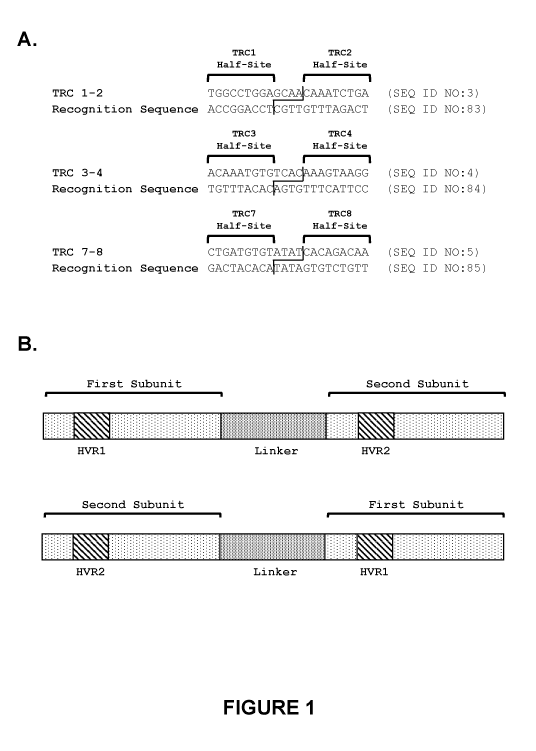
Figure 1. TRC recognition sequences in the human TRC alpha constant region
gene.
A) Each recognition sequence targeted by a recombinant meganuclease of the
invention
comprises two recognition half-sites. Each recognition half-site comprises 9
base pairs,
separated by a 4 base pair central sequence. The TRC 1-2 recognition sequence
(SEQ ID
NO:3) spans nucleotides 187-208 of the human T cell alpha constant region (SEQ
ID NO:1),
and comprises two recognition half-sites referred to as TRC1 and TRC2. The TRC
3-4
recognition sequence (SEQ ID NO:4) spans nucleotides 93-114 of the human T
cell alpha
constant region (SEQ ID NO:1), and comprises two recognition half-sites
referred to as
TRC3 and TRC4. The TRC 7-8 recognition sequence (SEQ ID NO:5) spans
nucleotides 118-
139 of the human T cell alpha constant region (SEQ ID NO:1), and comprises two
recognition half-sites referred to as TRC7 and TRC8. B) The recombinant
meganucleases of
the invention comprise two subunits, wherein the first subunit comprising the
HVR1 region
binds to a first recognition half-site (e.g., TRC1, TRC3, or TRC7) and the
second subunit
comprising the HVR2 region binds to a second recognition half-site (e.g.,
TRC2, TRC4, or
TRC8). In embodiments where the recombinant meganuclease is a single-chain
meganuclease, the first subunit comprising the HVR1 region can be positioned
as either the
N-terminal or C-terminal subunit. Likewise, the second subunit comprising the
HVR2 region
can be positioned as either the N-terminal or C-terminal subunit.
Figure 2A-B. Amino acid alignment of TRC1-binding subunits. A-B) Some
recombinant meganucleases encompassed by the invention comprise one subunit
that binds
the 9 base pair TRC1 recognition half-site of SEQ ID NO:3. Amino acid sequence
alignments are provided for the TRC1-binding subunits (SEQ ID NOs:33-52) of
the
recombinant meganucleases set forth in SEQ ID NOs:8-27. As shown, the TRC1-
binding
subunit of SEQ ID NOs:8-18 comprises residues 198-344, whereas the TRC1-
binding subunit
CA 03001011 2018-04-04
WO 2017/062451
PCT/US2016/055492
of SEQ ID NOs:19-27 comprises residues 7-153. Each TRC1-binding subunit
comprises a
56 amino acid hypervariable region as indicated. Variable residues within the
hypervariable
region are shaded, with the most frequent amino acids at each position further
highlighted;
the most prevalent residues are bolded, whereas the second most prevalent are
bolded and
italicized. Residues outside of the hypervariable region are identical in each
subunit, with the
exception of a Q or E residue at position 80 or position 271 (see, U.S. Patent
No. 8,021,867).
All TRC1-binding subunits provided in Figure 2 share at least 90% sequence
identity to the
TRC1-binding subunit (residues 198-344) of the TRC 1-2x.87 EE meganuclease
(SEQ ID
NO:33). Residue numbers shown are those of SEQ ID NOs:8-27.
Figure 3A-B. Amino acid alignment of TRC2-binding subunits. A-B) Some
recombinant meganucleases encompassed by the invention comprise one subunit
that binds
the 9 base pair TRC2 recognition half-site of SEQ ID NO:3. Amino acid sequence
alignments are provided for the TRC2-binding subunits (SEQ ID NOs:58-77) of
the
recombinant meganucleases set forth in SEQ ID NOs:8-27. As shown, the TRC2-
binding
subunit of SEQ ID NOs:8-18 comprises residues 7-153, whereas the TRC2-binding
subunit
of SEQ ID NOs:19-27 comprises residues 198-344. Each TRC2-binding subunit
comprises a
56 amino acid hypervariable region as indicated. Variable residues within the
hypervariable
region are shaded, with the most frequent amino acids at each position further
highlighted;
the most prevalent residues are bolded, whereas the second most prevalent are
bolded and
italicized. Residues outside of the hypervariable region are identical in each
subunit, with the
exceptions of a Q or E residue at position 80 or position 271 (see, U.S.
Patent No. 8,021,867),
and an R residue at position 330 of meganucleases TRC 1-2x.87 EE, TRC 1-2x.87
QE, TRC
1-2x.87 EQ, TRC 1-2x.87, and TRC 1-2x.163 (shaded grey and underlined). All
TRC2-
binding subunits provided in Figure 3 share at least 90% sequence identity to
the TRC2-
binding subunit (residues 7-153) of the TRC 1-2x.87 EE meganuclease (SEQ ID
NO:58).
Residue numbers shown are those of SEQ ID NOs:8-27.
Figure 4. Amino acid alignment of TRC3-binding subunits. Some recombinant
meganucleases encompassed by the invention comprise one subunit that binds the
9 base pair
TRC3 recognition half-site of SEQ ID NO:4. Amino acid sequence alignments are
provided
for the TRC3-binding subunits (SEQ ID NOs:53 and 54) of the recombinant
meganucleases
set forth in SEQ ID NOs:28 and 29. As shown, the TRC3-binding subunit of SEQ
ID
NOs:28 and 29 comprises residues 7-153. Each TRC3-binding subunit comprises a
56 amino
acid hypervariable region as indicated. Variable residues within the
hypervariable region are
16
CA 03001011 2018-04-04
WO 2017/062451
PCT/US2016/055492
shaded. Residues outside of the hypervariable region are identical in each
subunit, with the
exceptions of a Q or E residue at position 80 (see, U.S. Patent No.
8,021,867). The TRC3-
binding subunits of the TRC 3-4x.3 and TRC 3-4x.19 meganucleases share 97%
sequence
identity. Residue numbers shown are those of SEQ ID NOs:28 and 29.
Figure 5. Amino acid alignment of TRC4-binding subunits. Some recombinant
meganucleases encompassed by the invention comprise one subunit that binds the
9 base pair
TRC4 recognition half-site of SEQ ID NO:4. Amino acid sequence alignments are
provided
for the TRC4-binding subunits (SEQ ID NOs:78 and 79) of the recombinant
meganucleases
set forth in SEQ ID NOs:28 and 29. As shown, the TRC4-binding subunit of SEQ
ID
NOs:28 and 29 comprises residues 198-344. Each TRC4-binding subunit comprises
a 56
amino acid hypervariable region as indicated. Variable residues within the
hypervariable
region are shaded. Residues outside of the hypervariable region are identical
in each subunit,
with the exceptions of a Q or E residue at position 80 (see, U.S. Patent No.
8,021,867). The
TRC4-binding subunits of the TRC 3-4x.3 and TRC 3-4x.19 meganucleases share
97%
sequence identity. Residue numbers shown are those of SEQ ID NOs:28 and 29.
Figure 6A-B. Amino acid alignment of TRC7-binding subunits. A-B) Some
recombinant meganucleases encompassed by the invention comprise one subunit
that binds
the 9 base pair TRC7 recognition half-site of SEQ ID NO:5. Amino acid sequence
alignments are provided for the TRC7-binding subunits (SEQ ID NOs:55-57) of
the
recombinant meganucleases set forth in SEQ ID NOs:30-32. As shown, the TRC7-
binding
subunit of SEQ ID NO:30 comprises residues 7-153, whereas the TRC7-binding
subunit of
SEQ ID NOs:31 and 32 comprises residues 198-344. Each TRC7-binding subunit
comprises
a 56 amino acid hypervariable region as indicated. Variable residues within
the
hypervariable region are shaded, with the most frequent amino acids at each
position further
highlighted; the most prevalent residues are bolded, whereas the second most
prevalent are
bolded and italicized. Residues outside of the hypervariable region are
identical in each
subunit, with the exception of a Q or E residue at position 80 or position 271
(see, U.S. Patent
No. 8,021,867). All TRC7-binding subunits provided in Figure 6 share at least
90% sequence
identity to the TRC7-binding subunit (residues 7-153) of the TRC 7-8x.7
meganuclease (SEQ
ID NO:55). Residue numbers shown are those of SEQ ID NOs:30-32.
Figure 7A-B. Amino acid alignment of TRC8-binding subunits. A-B) Some
recombinant meganucleases encompassed by the invention comprise one subunit
that binds
the 9 base pair TRC8 recognition half-site of SEQ ID NO:5. Amino acid sequence
17
CA 03001011 2018-04-04
WO 2017/062451
PCT/US2016/055492
alignments are provided for the TRC8-binding subunits (SEQ ID NOs:80-82) of
the
recombinant meganucleases set forth in SEQ ID NOs:30-32. As shown, the TRC8-
binding
subunit of SEQ ID NO:30 comprises residues 198-344, whereas the TRC8-binding
subunit of
SEQ ID NOs:31 and 32 comprises residues 7-153. Each TRC8-binding subunit
comprises a
56 amino acid hypervariable region as indicated. Variable residues within the
hypervariable
region are shaded, with the most frequent amino acids at each position further
highlighted;
the most prevalent residues are bolded, whereas the second most prevalent are
bolded and
italicized. Residues outside of the hypervariable region are identical in each
subunit, with the
exception of a Q or E residue at position 80 or position 271 (see, U.S. Patent
No. 8,021,867).
All TRC 8-binding subunits provided in Figure 7 share at least 90% sequence
identity to the
TRC8-binding subunit (residues 198-344) of the TRC 7-8x.7 meganuclease (SEQ ID
NO:80).
Residue numbers shown are those of SEQ ID NOs:30-32.
Figure 8. Schematic of reporter assay in CHO cells for evaluating recombinant
meganucleases targeting recognition sequences found in the T cell receptor
alpha constant
region (SEQ ID NO:1). For the recombinant meganucleases described herein, a
CHO cell
line was produced in which a reporter cassette was integrated stably into the
genome of the
cell. The reporter cassette comprised, in 5' to 3' order: an 5V40 Early
Promoter; the 5' 2/3 of
the GFP gene; the recognition sequence for an engineered meganuclease of the
invention
(e.g., the TRC 1-2 recognition sequence, the TRC 3-4 recognition sequence, or
the TRC 7-8
recognition sequence); the recognition sequence for the CHO-23/24 meganuclease
(WO/2012/167192); and the 3' 2/3 of the GFP gene. Cells stably transfected
with this
cassette did not express GFP in the absence of a DNA break-inducing agent.
Meganucleases
were introduced by transduction of plasmid DNA or mRNA encoding each
meganuclease.
When a DNA break was induced at either of the meganuclease recognition
sequences, the
duplicated regions of the GFP gene recombined with one another to produce a
functional
GFP gene. The percentage of GFP-expressing cells could then be determined by
flow
cytometry as an indirect measure of the frequency of genome cleavage by the
meganucleases.
Figure 9. Efficiency of recombinant meganucleases for recognizing and cleaving
recognition sequences in the human T cell receptor alpha constant region (SEQ
ID NO:1) in a
CHO cell reporter assay. Each of the recombinant meganucleases set forth in
SEQ ID
NOs:8-32 were engineered to target the TRC 1-2 recognition sequence (SEQ ID
NO:3), the
TRC 3-4 recognition sequence (SEQ ID NO:4), or the TRC 7-8 recognition
sequence (SEQ
ID NO:5), and were screened for efficacy in the CHO cell reporter assay. The
results shown
18
CA 03001011 2018-04-04
WO 2017/062451
PCT/US2016/055492
provide the percentage of GFP-expressing cells observed in each assay, which
indicates the
efficacy of each meganuclease for cleaving a TRC target recognition sequence
or the CHO-
23/24 recognition sequence. A negative control (RHO 1-2 bs) was further
included in each
assay. A)-C) Meganucleases targeting the TRC 1-2 recognition sequence. D)
Meganucleases
targeting the TRC 3-4 recognition sequence. E)-F) Meganucleases targeting the
TRC 7-8
recognition sequence. G) Variants of the TRC 1-2x.87 meganuclease, wherein the
Q at
position 271 is substituted with E (TRC 1-2x.87 QE), the Q at position 80 is
substituted with
E (TRC 1-2x.87 EQ), or the Q at position 80 and the Q at position 271 are both
substituted
with E (TRC 1-2x.87 EE).
Figure 10. Time course of recombinant meganuclease efficacy in CHO cell
reporter
assay. The TRC 1-2x.87 QE, TRC 1-2x.87 EQ, and TRC 1-2x.87 EE meganucleases
were
evaluated in the CHO reporter assay, with the percentage of GFP-expressing
cells determined
1, 4, 6, 8, and 12 days after introduction of meganuclease-encoding mRNA into
the CHO
reporter cells.
Figure 11. Analysis of Jurkat cell genomic DNA following transfection with TRC
1-2
meganucleases. At 72 hours post-transfection with mRNA encoding TRC 1-2
meganucleases, genomic DNA was harvested and a T7 endonuclease assay was
performed to
estimate genetic modification at the endogenous TRC 1-2 recognition sequence.
Figure 12. Dose-response of TRC 1-2 meganuclease expression in Jurkat cells on
genetic modification at the endogenous TRC 1-2 recognition sequence. Jurkat
cells were
transfected with either 3[Ig or liag of a given TRC 1-2 meganuclease mRNA. At
96 hours,
genomic DNA was analyzed using a T7 endonuclease assay.
Figure 13. Cleavage of TRC 1-2 recognition sequence in human T cells. A) CD3+
T
cells were stimulated with anti-CD3 and anti-CD28 antibodies for 3 days, then
electroporated
with mRNA encoding the TRC 1-2x.87 EE meganuclease. Genomic DNA was harvested
at 3
days and 7 days post-transfection, and analyzed using a T7 endonuclease assay.
B) To
determine whether mutations at the endogenous TRC 1-2 recognition sequence
were
sufficient to eliminate surface expression of the T cell receptor, cells were
analyzed by flow
cytometry using an anti-CD3 antibody. Control cells (transfected with water)
and TRC 1-
2x.87 EE-transfected cells were analyzed at day 3 and day 7 post-transfection,
and the
percentage of CD3-positive and CD3-negative T cells was determined.
19
CA 03001011 2018-04-04
WO 2017/062451
PCT/US2016/055492
Figure 14. Nucleic acid sequences of representative deletions that were
observed at
the TRC 1-2 recognition sequence in human T cells following expression of TRC
1-2
meganucleases.
Figure 15. Diagram illustrating sequence elements of recombinant AAV vectors
and
their use in combination with an engineered nuclease to insert an exogenous
nucleic acid
sequence into the endogenous TCR alpha constant region gene.
Figure 16. Map of plasmid used to produce the AAV405 vector.
Figure 17. Map of plasmid used to produce the AAV406 vector.
Figure 18. Determining the timing of meganuclease mRNA transfection and
recombinant AAV transduction to enhance AAV transduction efficiency. Human
CD3+ T
cells were electroporated with mRNA encoding the TRC 1-2x.87 EE meganuclease
and at 2,
4, or 8 hours post-transfection, cells were transduced with a recombinant AAV
vector
encoding GFP (GFP-AAV). T cells were analyzed by flow cytometry for GFP
expression at
72 hours post-transduction to determine transduction efficiency.
Figure 19. Analyzing human T cells for insertion of an exogenous nucleic acid
sequence using recombinant AAV vectors. CD3+ T cells transfected with TRC 1-
2x.87 EE
mRNA and subsequently transduced (2 hours post-transfection) with AAV405 or
AAV406.
Transduction-only controls were mock transfected (with water) and transduced
with either
AAV405 or AAV406. Meganuclease-only controls were transfected with TRC 1-2x.87
EE
and then mock transduced (with water) at 2 hours post-transfection. Genomic
DNA was
harvested from T cells and the TRC 1-2 locus was amplified by PCR using
primers that
recognized sequences beyond the region of homology in the AAV vectors. PCR
primers
outside of the homology regions only allowed for amplification of the T cell
genome, not
from the AAV vectors. PCR products were purified and digested with EagI. PCR
products
were then analyzed for cleavage.
Figure 20. Characterization of EagI insertion into the TRC 1-2 recognition
sequence
of human T cells using AAV405. A) Undigested PCR product generated from
previous
experiments was cloned into a pCR-blunt vector. Colony PCR was performed using
M13
forward and reverse primers and a portion of PCR products from cells
transfected with TRC
1-2x.87 EE and AAV405 was analyzed by gel electrophoresis. Analysis shows a
mix of full-
length PCR products (approximately 1600 bp), smaller inserts, and empty
plasmids
(approximately 300 bp). B) In parallel, another portion of PCR products were
digested with
EagI to determine the percent of clones that contain the EagI recognition site
inserted in the
CA 03001011 2018-04-04
WO 2017/062451
PCT/US2016/055492
TRC 1-2 recognition sequence. PCR products cleaved with EagI generated
expected
fragments of approximately 700 and 800 bp.
Figure 21. Characterization of EagI insertion into the TRC 1-2 recognition
sequence
of human T cells using AAV406. A) Undigested PCR product generated from
previous
experiments was cloned into a pCR-blunt vector. Colony PCR was performed using
M13
forward and reverse primers and a portion of PCR products from cells
transfected with TRC
1-2x.87 EE and AAV406 was analyzed by gel electrophoresis. Analysis shows a
mix of full-
length PCR products (approximately 1600 bp), smaller inserts, and empty
plasmids
(approximately 300 bp). B) In parallel, another portion of PCR products were
digested with
EagI to determine the percent of clones that contain the EagI recognition site
inserted in the
TRC 1-2 recognition sequence. PCR products cleaved with EagI generated
expected
fragments of approximately 700 and 800 bp.
Figure 22. A) Nucleic acid sequences of representative deletions and
insertions (i.e.,
indels) that were observed at the TRC 1-2 recognition sequence in human T
cells following
expression of TRC 1-2 meganucleases. B) Nucleic acid sequence of the TRC 1-2
recognition
sequence confirming insertion of the exogenous nucleic acid sequence
comprising the EagI
restriction site.
Figure 23. Enhancement of recombinant AAV transduction efficiency.
Transduction
efficiency was further analyzed by optimizing the timing of meganuclease mRNA
transfection and subsequent AAV transduction. Human CD3+ T cells were
electroporated
with mRNA encoding the TRC 1-2x.87 EE meganuclease and subsequently transduced
with
GFP-AAV immediately after transfection or 2 hours post-transfection.
Additionally, non-
stimulated resting T cells were transduced with GFP-AAV. Mock transduced cells
were also
analyzed. At 72 hours post-transduction, cells were analyzed by flow cytometry
for GFP
expression to determine AAV transduction efficiency.
Figure 24. Map of plasmid used to produce the AAV-CAR100 (AAV408) vector.
Figure 25. Map of plasmid used to produce the AAV-CAR763 (AAV412) vector.
Figure 26. Insertion of chimeric antigen receptor coding sequence at TRC 1-2
recognition site in human T cells. A PCR-based assay was developed to
determine whether
the AAV412 HDR template was utilized to repair double-strand breaks at the TRC
1-2
recognition sequence.
Figure 27. Insertion of chimeric antigen receptor coding sequence at TRC 1-2
recognition site in human T cells. A PCR-based assay was developed to
determine whether
21
CA 03001011 2018-04-04
WO 2017/062451
PCT/US2016/055492
the AAV408 HDR template was utilized to repair double-strand breaks at the TRC
1-2
recognition sequence. A) PCR products generated using a primer pair that only
amplifies a
product on the 5' end of the TRC 1-2 recognition sequence locus if the CAR
gene has been
inserted into that locus. B) PCR products generated using a primer pair that
only amplifies a
product on the 3' end of the TRC 1-2 recognition sequence locus if the CAR
gene has been
inserted into that locus.
Figure 28. Digital PCR. A) Schematic of a digital PCR assay developed to
quantitatively determine insertion efficiency of the chimeric antigen receptor
coding
sequence into the TRC 1-2 recognition site in human T cells. B) Results of
digital PCR on
genomic DNA from human T cells electroporated with a TRC 1-2x.87EE
meganuclease
mRNA and/or increasing amounts of AAV408.
Figure 29. Cell-surface expression of CD19 chimeric antigen receptor on human
T
cells. The expression level of the anti-CD19 chimeric antigen receptor was
determined in
cells that had the CAR gene inserted into the TRC 1-2 recognition sequence
using AAV408
as the HDR template. Cell-surface expression was analyzed by flow cytometry.
A) Cells
that were mock electroporated and mock transduced (MOI ¨ 0), and cells that
were mock
electroporated and transduced with increasing amounts of AAV408. B) Cells that
were
electroporated with TRC 1-2x.87EE and mock transduced (MOI ¨ 0), and cells
that were
electroporated with TRC 1-2x.87EE and transduced with increasing amounts of
AAV408.
Figure 30. Map of plasmid used to produce the AAV421 vector.
Figure 31. Map of plasmid used to produce the AAV422 vector.
Figure 32. Insertion of chimeric antigen receptor coding sequence. PCR methods
were used to determine if the chimeric antigen receptor coding sequence
introduced by
AAV421 or AAV422 inserted at the TRC 1-2 recognition site cleaved by the TRC 1-
2x.87EE
meganuclease. A) Analysis of insertion following transduction with AAV421. B)
Analysis
of insertion following transduction with AAV422.
Figure 33. Cell-surface expression of CD19 chimeric antigen receptor on human
T
cells. The expression level of the anti-CD19 chimeric antigen receptor was
determined in
cells that had the CAR gene inserted into the TRC 1-2 recognition sequence
using AAV421
as the HDR template. Cell-surface expression was analyzed by flow cytometry.
A) Cells
that were mock electroporated and mock transduced (MOI ¨ 0), and cells that
were mock
electroporated and transduced with increasing amounts of AAV421. B) Cells that
were
22
CA 03001011 2018-04-04
WO 2017/062451
PCT/US2016/055492
electroporated with TRC 1-2x.87EE and mock transduced (MOI ¨ 0), and cells
that were
electroporated with TRC 1-2x.87EE and transduced with increasing amounts of
AAV421.
Figure 34. Expansion of human T cells expressing a cell-surface chimeric
antigen
receptor. Methods were determined for preferentially expanding and enriching a
CD37CAR
T cell population following electroporation with mRNA for the TRC 1-2x.87EE
meganuclease and transduction with AAV421. A) Supplementation with IL-7 (10
ng/mL)
and IL-15 (10 ng/mL). B) Supplementation with IL-7 (10 ng/mL) and IL-15 (10
ng/mL), and
incubation with mitomycin C-inactivated IM-9 cells. C) Supplementation with IL-
7 (10
ng/mL) and IL-15 (10 ng/mL), and two incubations with mitomycin C-inactivated
IM-9 cells.
Figure 35. Cell-surface expression of CD19 chimeric antigen receptor on human
T
cells. The expression level of the anti-CD19 chimeric antigen receptor was
determined in
cells that had the CAR gene inserted into the TRC 1-2 recognition sequence
using AAV422
as the HDR template. Cell-surface expression was analyzed by flow cytometry.
A) Cells
that were mock electroporated and mock transduced (MOI ¨ 0), and cells that
were mock
electroporated and transduced with increasing amounts of AAV422. B) Cells that
were
electroporated with TRC 1-2x.87EE and mock transduced (MOI ¨ 0), and cells
that were
electroporated with TRC 1-2x.87EE and transduced with increasing amounts of
AAV422.
Figure 36. Expansion of human T cells expressing a cell-surface chimeric
antigen
receptor. Methods were determined for preferentially expanding and enriching a
CD3-/CAR
T cell population following electroporation with mRNA for the TRC 1-2x.87EE
meganuclease and transduction with AAV422. A) Supplementation with IL-7 (10
ng/mL)
and IL-15 (10 ng/mL). B) Supplementation with IL-7 (10 ng/mL) and IL-15 (10
ng/mL), and
incubation with mitomycin C-inactivated IM-9 cells. C) Supplementation with IL-
7 (10
ng/mL) and IL-15 (10 ng/mL), and two incubations with mitomycin C-inactivated
IM-9 cells.
Figure 37. Meganuclease knockout efficiency using single-strand AAV.
Experiments
were conducted to examine the knockout efficiency of two meganucleases in
human T cells
when simultaneously transduced with a single-stranded AAV vector. A) Cells
electroporated
with mRNA for TRC 1-2x.87EE and transduced with increasing amounts of the
single-
stranded AAV412. B) Cells electroporated with mRNA for a meganuclease
targeting the
beta-2 microglobulin gene and transduced with increasing amounts of the single-
stranded
AAV412. C) Cells electroporated with mRNA for TRC 1-2x.87EE and transduced
with
increasing amounts of the single-stranded AAV422.
23
CA 03001011 2018-04-04
WO 2017/062451
PCT/US2016/055492
Figure 38. Functional activity of anti-CD19 CART cells. A) IFN-gamma ELISPOT
assay, in which either CD19+ Raji cells or CD19- U937 cells were the target
population. B)
Cell killing assay in which luciferase-labeled CD19+ Raji cells were the
target.
Figure 39. Expression of chimeric antigen receptors following transduction
with
linearized DNA donor templates. These experiments generated plasmids that
contain an anti-
CD19 CAR gene flanked by homology arms that are homologous to the TRC 1-2
recognition
sequence locus. Different promoters were used in some plasmids, and homology
arms were
either "short" (200 bp on the 5' homology arm and 180 bp on the 3' homology
arm) or "long"
(985 bp on the 5' homology arm and 763 bp on the 3' homology arm). CAR donor
plasmids
were linearized at a restriction site in the vector backbone and gel purified.
A) Background
CD37CAR+ staining. B) Cells electroporated with TRC 1-2x.87EE mRNA alone. C)
Cells
co-electroporated with TRC 1-2x.87EE mRNA and a long homology arm vector with
an
EFla core promoter with an HTLV enhancer. D) Cells co-electroporated with TRC
1-
2x.87EE mRNA and a short homology arm vector with EFla core promoter (with no
enhancer). E) Cells electroporated with a long homology arm vector with an
EFla core
promoter with an HTLV enhancer in the absence of TRC 1-2x.87EE mRNA. F) Cells
electroporated with a short homology arm vector with EF la core promoter (with
no
enhancer) in the absence of TRC 1-2x.87EE mRNA. G) Cells electroporated with a
long
homology arm construct that contains an MIND promoter driving expression of
the CAR and
an intron in the 5' end of the CAR gene, as well as TRC 1-2x.87EE mRNA. H)
Cells
electroporated with a long homology arm construct that contains an MIND
promoter driving
expression of the CAR and no intron, as well as TRC 1-2x.87EE mRNA. I) Cells
electroporated with a short homology arm plasmid with the MIND promoter and no
intron, as
well as TRC 1-2x.87EE mRNA. J) Cells electroporated with a long homology arm
construct
that contains an MIND promoter driving expression of the CAR and an intron in
the 5' end of
the CAR gene, but no TRC 1-2x.87EE mRNA. K) Cells electroporated with a long
homology arm construct that contains an MIND promoter driving expression of
the CAR and
no intron, but no TRC 1-2x.87EE mRNA. L) Cells electroporated with a short
homology arm
plasmid with the MIND promoter and no intron, but no TRC 1-2x.87EE mRNA. M)
Cells
electroporated with a short homology arm construct that contained a JeT
promoter, as well as
TRC 1-2x.87EE mRNA. N) Cells electroporated with a long homology arm construct
that
contained a CMV promoter, as well as TRC 1-2x.87EE mRNA. 0) Cells
electroporated with
a short homology arm construct that contained a JeT promoter, but no TRC 1-
2x.87EE
24
CA 03001011 2018-04-04
WO 2017/062451
PCT/US2016/055492
mRNA. P) Cells electroporated with a long homology arm construct that
contained a CMV
promoter, but no TRC 1-2x.87EE mRNA.
Figure 40. PCR analysis to determine whether the chimeric antigen receptor
coding
region delivered by linearized DNA constructs was inserted into the TRC 1-2
recognition
sequence in human T cells.
Figure 41. Map of plasmid used to produce the AAV423 vector.
Figure 42. Cell-surface expression of CD19 chimeric antigen receptor on human
T
cells. The expression level of the anti-CD19 chimeric antigen receptor was
determined in
cells that had the CAR gene inserted into the TRC 1-2 recognition sequence
using AAV423
as the HDR template. Cell-surface expression was analyzed by flow cytometry.
A) Cells
that were mock electroporated and mock transduced (MOI ¨ 0), and cells that
were mock
electroporated and transduced with increasing amounts of AAV423. B) Cells that
were
electroporated with TRC 1-2x.87EE and mock transduced (MOI ¨ 0), and cells
that were
electroporated with TRC 1-2x.87EE and transduced with increasing amounts of
AAV423.
Figure 43. Insertion of chimeric antigen receptor coding sequence. PCR methods
were used to determine if the chimeric antigen receptor coding sequence
introduced by
AAV423 inserted at the TRC 1-2 recognition site cleaved by the TRC 1-2x.87EE
meganuclease.
Figure 44. Phenotype analysis of anti-CD19 CART cells. A) Activated T cells
were
electroporated with TRC 1-2x.87 EE mRNA, then transduced with an AAV6 vector
comprising an anti-CD19 CAR expression cassette driven by a JeT promoter and
flanked by
homology arms. Following 5 days of culture with IL-2 (10 ng/mL), cells were
analyzed for
cell-surface CD3 and anti-CD19 CAR expression by flow cytometry. B) CD3- cells
were
enriched by depleting CD3 + cells using anti-CD3 magnetic beads. Depleted
cells were then
cultured for 3 days in IL-15 (10 ng/mL) and IL-21 (10 ng/mL) and re-analyzed
for cell-
surface expression of CD3 and anti-CD19 CAR. C) The purified population of CD3-
CD19-
CAR T cells was analyzed by flow cytometry to determine the percentage of
cells that were
CD4+ and CD8 . D) The purified population of CD3- CD19-CAR T cells was further
analyzed by flow cytometry to determine whether they were central memory T
cells,
transitional memory T cells, or effector memory T cells by staining for CD62L
and CD45RO.
Figure 45. Raji disseminated lymphoma model. Raji cells stably expressing
firefly
luciferase (ffLuc)44 were injected i.v. into 5-6 week old female NSG mice on
Day 1, at a dose
of 2.0 x 10⁵ cells per mouse. On Day 4 mice were injected i.v. with PBS or PBS
containing
CA 03001011 2018-04-04
WO 2017/062451
PCT/US2016/055492
gene edited control TCR KO T cells prepared from the same healthy donor PBMC
or PBS
containing the indicated doses of CAR T cells prepared from the same donor. On
the
indicated days, live mice were injected i.p. with Luciferin substrate
(150mg/kg in saline),
anesthetized, and Luciferase activity measured after 7 minutes using IVIS
SpectrumCTO
(Perkin Elmer, Waltham, MA). Data was analyzed and exported using Living Image
software 4.5.1 (Perkin Elmer, Waltham, MA). Luminescence signal intensity is
represented
by radiance in p/sec/cm2/sr.
BRIEF DESCRIPTION OF THE SEQUENCES
SEQ ID NO: 1 sets forth the nucleotide sequence of the human T cell receptor
alpha
constant region gene (NCBI Gene ID NO. 28755).
SEQ ID NO: 2 sets forth the amino acid sequence encoded by the human T cell
receptor alpha constant region.
SEQ ID NO: 3 sets forth the amino acid sequence of the TRC 1-2 recognition
sequence.
SEQ ID NO: 4 sets forth the nucleotide sequence of the TRC 3-4 recognition
sequence.
SEQ ID NO: 5 sets forth the nucleotide sequence of the TRC 7-8 recognition
sequence.
SEQ ID NO: 6 sets forth the amino acid sequence of I-CreI.
SEQ ID NO: 7 sets forth the amino acid sequence of the LAGLIDADG motif
SEQ ID NO: 8 sets forth the amino acid sequence of the TRC 1-2x.87 EE
meganuclease.
SEQ ID NO: 9 sets forth the amino acid sequence of the TRC 1-2x.87 QE
meganuclease.
SEQ ID NO: 10 sets forth the amino acid sequence of the TRC 1-2x.87 EQ
meganuclease.
SEQ ID NO: 11 sets forth the amino acid sequence of the TRC 1-2x.87
meganuclease.
SEQ ID NO: 12 sets forth the amino acid sequence of the TRC 1-2x.6
meganuclease.
SEQ ID NO: 13 sets forth the amino acid sequence of the TRC 1-2x.20
meganuclease.
SEQ ID NO: 14 sets forth the amino acid sequence of the TRC 1-2x.55
meganuclease.
SEQ ID NO: 15 sets forth the amino acid sequence of the TRC 1-2x.60
meganuclease.
26
CA 03001011 2018-04-04
WO 2017/062451
PCT/US2016/055492
SEQ ID NO: 16 sets forth the amino acid sequence of the TRC 1-2x.105
meganuclease.
SEQ ID NO: 17 sets forth the amino acid sequence of the TRC 1-2x.163
meganuclease.
SEQ ID NO: 18 sets forth the amino acid sequence of the TRC 1-2x.113_3
meganuclease.
SEQ ID NO: 19 sets forth the amino acid sequence of the TRC 1-2x.5
meganuclease.
SEQ ID NO: 20 sets forth the amino acid sequence of the TRC 1-2x.8
meganuclease.
SEQ ID NO: 21 sets forth the amino acid sequence of the TRC 1-2x.25
meganuclease.
SEQ ID NO: 22 sets forth the amino acid sequence of the TRC 1-2x.72
meganuclease.
SEQ ID NO: 23 sets forth the amino acid sequence of the TRC 1-2x.80
meganuclease.
SEQ ID NO: 24 sets forth the amino acid sequence of the TRC 1-2x.84
meganuclease.
SEQ ID NO: 25 sets forth the amino acid sequence of the TRC 1-2x.120
meganuclease.
SEQ ID NO: 26 sets forth the amino acid sequence of the TRC 1-2x.113_1
meganuclease.
SEQ ID NO: 27 sets forth the amino acid sequence of the TRC 1-2x.113_2
meganuclease.
SEQ ID NO: 28 sets forth the amino acid sequence of the TRC 3-4x.3
meganuclease.
SEQ ID NO: 29 sets forth the amino acid sequence of the TRC 3-4x.19
meganuclease.
SEQ ID NO: 30 sets forth the amino acid sequence of the TRC 7-8x.7
meganuclease.
SEQ ID NO: 31 sets forth the amino acid sequence of the TRC 7-8x.9
meganuclease.
SEQ ID NO: 32 sets forth the amino acid sequence of the TRC 7-8x.14
meganuclease.
SEQ ID NO: 33 sets forth residues 198-344 of the TRC 1-2x.87 EE meganuclease.
SEQ ID NO: 34 sets forth residues 198-344 of the TRC 1-2x.87 QE meganuclease.
SEQ ID NO: 35 sets forth residues 198-344 of the TRC 1-2x.87 EQ meganuclease.
SEQ ID NO: 36 sets forth residues 198-344 of the TRC 1-2x.87 meganuclease.
SEQ ID NO: 37 sets forth residues 198-344 of the TRC 1-2x.6 meganuclease.
SEQ ID NO: 38 sets forth residues 198-344 of the TRC 1-2x.20 meganuclease.
SEQ ID NO: 39 sets forth residues 198-344 of the TRC 1-2x.55 meganuclease.
SEQ ID NO: 40 sets forth residues 198-344 of the TRC 1-2x.60 meganuclease.
SEQ ID NO: 41 sets forth residues 198-344 of the TRC 1-2x.105 meganuclease.
SEQ ID NO: 42 sets forth residues 198-344 of the TRC 1-2x.163 meganuclease.
27
CA 03001011 2018-04-04
WO 2017/062451
PCT/US2016/055492
SEQ ID NO: 43 sets forth residues 198-344 of the TRC 1-2x.113_3 meganuclease.
SEQ ID NO: 44 sets forth residues 7-153 of the TRC 1-2x.5 meganuclease.
SEQ ID NO: 45 sets forth residues 7-153 of the TRC 1-2x.8 meganuclease.
SEQ ID NO: 46 sets forth residues 7-153 of the TRC 1-2x.25 meganuclease.
SEQ ID NO: 47 sets forth residues 7-153 of the TRC 1-2x.72 meganuclease.
SEQ ID NO: 48 sets forth residues 7-153 of the TRC 1-2x.80 meganuclease.
SEQ ID NO: 49 sets forth residues 7-153 of the TRC 1-2x.84 meganuclease.
SEQ ID NO: 50 sets forth residues 7-153 of the TRC 1-2x.120 meganuclease.
SEQ ID NO: 51 sets forth residues 7-153 of the TRC 1-2x.113_1 meganuclease.
SEQ ID NO: 52 sets forth residues 7-153 of the TRC 1-2x.113_2 meganuclease.
SEQ ID NO: 53 sets forth residues 7-153 of the TRC 3-4x.3 meganuclease.
SEQ ID NO: 54 sets forth residues 7-153 of the TRC 3-4x.19 meganuclease.
SEQ ID NO: 55 sets forth residues 7-153 of the TRC 7-8x.7 meganuclease.
SEQ ID NO: 56 sets forth residues 198-344 of the TRC 7-8x.9 meganuclease.
SEQ ID NO: 57 sets forth residues 198-344 of the TRC 7-8x.14 meganuclease.
SEQ ID NO: 58 sets forth residues 7-153 of the TRC 1-2x.87 EE meganuclease.
SEQ ID NO: 59 sets forth residues 7-153 of the TRC 1-2x.87 QE meganuclease.
SEQ ID NO: 60 sets forth residues 7-153 of the TRC 1-2x.87 EQ meganuclease.
SEQ ID NO: 61 sets forth residues 7-153 of the TRC 1-2x.87 meganuclease.
SEQ ID NO: 62 sets forth residues 7-153 of the TRC 1-2x.6 meganuclease.
SEQ ID NO: 63 sets forth residues 7-153 of the TRC 1-2x.20 meganuclease.
SEQ ID NO: 64 sets forth residues 7-153 of the TRC 1-2x.55 meganuclease.
SEQ ID NO: 65 sets forth residues 7-153 of the TRC 1-2x.60 meganuclease.
SEQ ID NO: 66 sets forth residues 7-153 of the TRC 1-2x.105 meganuclease.
SEQ ID NO: 67 sets forth residues 7-153 of the TRC 1-2x.163 meganuclease.
SEQ ID NO: 68 sets forth residues 7-153 of the TRC 1-2x.113_3 meganuclease.
SEQ ID NO: 69 sets forth residues 198-344 of the TRC 1-2x.5 meganuclease.
SEQ ID NO: 70 sets forth residues 198-344 of the TRC 1-2x.8 meganuclease.
SEQ ID NO: 71 sets forth residues 198-344 of the TRC 1-2x.25 meganuclease.
SEQ ID NO: 72 sets forth residues 198-344 of the TRC 1-2x.72 meganuclease.
SEQ ID NO: 73 sets forth residues 198-344 of the TRC 1-2x.80 meganuclease.
SEQ ID NO: 74 sets forth residues 198-344 of the TRC 1-2x.84 meganuclease.
SEQ ID NO: 75 sets forth residues 198-344 of the TRC 1-2x.120 meganuclease.
28
CA 03001011 2018-04-04
WO 2017/062451
PCT/US2016/055492
SEQ ID NO: 76 sets forth residues 198-344 of the TRC 1-2x.113_1 meganuclease.
SEQ ID NO: 77 sets forth residues 198-344 of the TRC 1-2x.113_2 meganuclease.
SEQ ID NO: 78 sets forth residues 198-344 of the TRC 3-4x.3 meganuclease.
SEQ ID NO: 79 sets forth residues 198-344 of the TRC 3-4x.19 meganuclease.
SEQ ID NO: 80 sets forth residues 198-344 of the TRC 7-8x.7 meganuclease.
SEQ ID NO: 81 sets forth residues 7-153 of the TRC 7-8x.9 meganuclease.
SEQ ID NO: 82 sets forth residues 7-153 of the TRC 7-8x.14 meganuclease.
SEQ ID NO: 83 sets forth the nucleotide sequence of the antisense strand of
the TRC
1-2 recognition sequence.
SEQ ID NO: 84 sets forth the nucleotide sequence of the antisense strand of
the TRC
3-4 recognition sequence.
SEQ ID NO: 85 sets forth the nucleotide sequence of the antisense strand of
the TRC
7-8 recognition sequence.
SEQ ID NO: 86 sets forth nucleotides 162-233 of SEQ ID NO:l.
SEQ ID NO: 87 sets forth nucleotides 162-233 of SEQ ID NO:1 comprising a
deletion resulting from cleavage and NHEJ.
SEQ ID NO: 88 sets forth nucleotides 162-233 of SEQ ID NO:1 comprising a
deletion resulting from cleavage and NHEJ.
SEQ ID NO: 89 sets forth nucleotides 162-233 of SEQ ID NO:1 comprising a
deletion resulting from cleavage and NHEJ.
SEQ ID NO: 90 sets forth nucleotides 162-233 of SEQ ID NO:1 comprising a
deletion resulting from cleavage and NHEJ.
SEQ ID NO: 91 sets forth nucleotides 162-233 of SEQ ID NO:1 comprising a
deletion resulting from cleavage and NHEJ.
SEQ ID NO: 92 sets forth nucleotides 162-233 of SEQ ID NO:1 comprising a
deletion resulting from cleavage and NHEJ.
SEQ ID NO: 93 sets forth nucleotides 162-233 of SEQ ID NO:1 comprising a
deletion resulting from cleavage and NHEJ.
SEQ ID NO: 94 sets forth nucleotides 162-233 of SEQ ID NO:1 comprising an
insertion resulting from cleavage and NHEJ.
SEQ ID NO: 95 sets forth nucleotides 162-233 of SEQ ID NO:1 comprising an
insertion resulting from cleavage and NHEJ.
29
CA 03001011 2018-04-04
WO 2017/062451
PCT/US2016/055492
SEQ ID NO: 96 sets forth nucleotides 162-233 of SEQ ID NO:1 comprising a
deletion resulting from cleavage and NHEJ.
SEQ ID NO: 97 sets forth nucleotides 162-233 of SEQ ID NO:1 comprising a
deletion resulting from cleavage and NHEJ.
SEQ ID NO: 98 sets forth nucleotides 162-233 of SEQ ID NO:1 comprising a
deletion resulting from cleavage and NHEJ.
SEQ ID NO: 99 sets forth nucleotides 162-233 of SEQ ID NO:1 comprising a
deletion resulting from cleavage and NHEJ.
SEQ ID NO: 100 sets forth nucleotides 162-233 of SEQ ID NO:1 comprising a
deletion resulting from cleavage and NHEJ.
SEQ ID NO: 101 sets forth nucleotides 162-233 of SEQ ID NO:1 comprising a
deletion resulting from cleavage and NHEJ.
SEQ ID NO: 102 sets forth nucleotides 162-233 of SEQ ID NO:1 comprising a
deletion resulting from cleavage and NHEJ.
SEQ ID NO: 103 sets forth nucleotides 162-233 of SEQ ID NO:1 comprising a
deletion resulting from cleavage and NHEJ.
SEQ ID NO: 104 sets forth nucleotides 162-233 of SEQ ID NO:1 comprising a
deletion resulting from cleavage and NHEJ.
SEQ ID NO: 105 sets forth nucleotides 181-214 of SEQ ID NO: 1.
SEQ ID NO: 106 sets forth nucleotides 181-214 of SEQ ID NO:1 comprising an
exogenous nucleic acid sequence inserted via homologous recombination.
SEQ ID NO: 107 sets forth the nucleotide sequence of a plasmid used to
generate the
AAV405 vector.
SEQ ID NO: 108 sets forth the nucleotide sequence of a plasmid used to
generate the
AAV406 vector.
SEQ ID NO: 109 sets forth the nucleotide sequence of a plasmid used to
generate the
AAV-CAR100 (AAV408) vector.
SEQ ID NO: 110 sets forth the nucleotide sequence of a plasmid used to
generate the
AAV-CAR763 (AAV412) vector.
SEQ ID NO: 111 sets forth the amino acid sequence of an anti-CD19 chimeric
antigen
receptor.
SEQ ID NO: 112 sets forth the amino acid sequence of an anti-CD19
extracellular
ligand-binding domain.
CA 03001011 2018-04-04
WO 2017/062451
PCT/US2016/055492
SEQ ID NO: 113 sets forth the amino acid sequence of a chimeric antigen
receptor
intracellular cytoplasmic signaling domain.
SEQ ID NO: 114 sets forth the amino acid sequence of a chimeric antigen
receptor
intracellular co-stimulatory domain.
SEQ ID NO: 115 sets forth the amino acid sequence of a chimeric antigen
receptor
signal peptide domain.
SEQ ID NO: 116 sets forth the amino acid sequence of a chimeric antigen
receptor
hinge region.
SEQ ID NO: 117 sets forth the amino acid sequence of a chimeric antigen
receptor
transmembrane domain.
SEQ ID NO: 118 sets forth the nucleotide sequence of an EF-1 alpha core
promoter.
SEQ ID NO: 119 sets forth the nucleotide sequence of an exogenous
polynucleotide
insert.
SEQ ID NO: 120 sets forth the nucleotide sequence of the human TCR alpha
constant
region gene comprising an exogenous nucleic acid sequence inserted within the
TRC 1-2
recognition sequence.
SEQ ID NO: 121 sets forth the nucleotide sequence of the human TCR alpha
constant
region gene comprising an exogenous nucleic acid sequence inserted within the
TRC 3-4
recognition sequence.
SEQ ID NO: 122 sets forth the nucleotide sequence of the human TCR alpha
constant
region gene comprising an exogenous nucleic acid sequence inserted within the
TRC 7-8
recognition sequence.
SEQ ID NO: 123 sets forth the nucleic acid sequence of a plasmid used to
generate
the AAV421 vector.
SEQ ID NO: 124 sets forth the nucleic acid sequence of a plasmid used to
generate
the AAV422 vector.
SEQ ID NO: 125 sets forth the nucleic acid sequence of a plasmid used to
generate
the AAV423 vector.
DETAILED DESCRIPTION OF THE INVENTION
1.1 References and Definitions
The patent and scientific literature referred to herein establishes knowledge
that is
available to those of skill in the art. The issued US patents, allowed
applications, published
31
CA 03001011 2018-04-04
WO 2017/062451
PCT/US2016/055492
foreign applications, and references, including GenBank database sequences,
which are cited
herein are hereby incorporated by reference to the same extent as if each was
specifically and
individually indicated to be incorporated by reference.
The present invention can be embodied in different forms and should not be
construed
as limited to the embodiments set forth herein. Rather, these embodiments are
provided so
that this disclosure will be thorough and complete, and will fully convey the
scope of the
invention to those skilled in the art. For example, features illustrated with
respect to one
embodiment can be incorporated into other embodiments, and features
illustrated with respect
to a particular embodiment can be deleted from that embodiment. In addition,
numerous
variations and additions to the embodiments suggested herein will be apparent
to those
skilled in the art in light of the instant disclosure, which do not depart
from the instant
invention.
Unless otherwise defined, all technical and scientific terms used herein have
the same
meaning as commonly understood by one of ordinary skill in the art to which
this invention
belongs. The terminology used in the description of the invention herein is
for the purpose of
describing particular embodiments only and is not intended to be limiting of
the invention.
All publications, patent applications, patents, and other references mentioned
herein
are incorporated by reference herein in their entirety.
As used herein, "a," "an," or "the" can mean one or more than one. For
example, "a"
cell can mean a single cell or a multiplicity of cells.
As used herein, unless specifically indicated otherwise, the word "or" is used
in the
inclusive sense of "and/or" and not the exclusive sense of "either/or."
As used herein, the term "meganuclease" refers to an endonuclease that binds
double-
stranded DNA at a recognition sequence that is greater than 12 base pairs.
Preferably, the
recognition sequence for a meganuclease of the invention is 22 base pairs. A
meganuclease
can be an endonuclease that is derived from I-CreI, and can refer to an
engineered variant of
I-CreI that has been modified relative to natural I-CreI with respect to, for
example, DNA-
binding specificity, DNA cleavage activity, DNA-binding affinity, or
dimerization properties.
Methods for producing such modified variants of I-CreI are known in the art
(e.g.,
WO 2007/047859). A meganuclease as used herein binds to double-stranded DNA as
a
heterodimer or as a "single-chain meganuclease" in which a pair of DNA-binding
domains
are joined into a single polypeptide using a peptide linker. The term "homing
endonuclease"
is synonymous with the term "meganuclease." Meganucleases of the invention are
32
CA 03001011 2018-04-04
WO 2017/062451
PCT/US2016/055492
substantially non-toxic when expressed in cells, particularly in human T
cells, such that cells
can be transfected and maintained at 37 C without observing deleterious
effects on cell
viability or significant reductions in meganuclease cleavage activity when
measured using the
methods described herein.
As used herein, the term "single-chain meganuclease" refers to a polypeptide
comprising a pair of nuclease subunits joined by a linker. A single-chain
meganuclease has
the organization: N-terminal subunit ¨ Linker ¨ C-terminal subunit. The two
meganuclease
subunits will generally be non-identical in amino acid sequence and will
recognize non-
identical DNA sequences. Thus, single-chain meganucleases typically cleave
pseudo-
palindromic or non-palindromic recognition sequences. A single-chain
meganuclease may be
referred to as a "single-chain heterodimer" or "single-chain heterodimeric
meganuclease"
although it is not, in fact, dimeric. For clarity, unless otherwise specified,
the term
"meganuclease" can refer to a dimeric or single-chain meganuclease.
As used herein, the term "linker" refers to an exogenous peptide sequence used
to join
two meganuclease subunits into a single polypeptide. A linker may have a
sequence that is
found in natural proteins, or may be an artificial sequence that is not found
in any natural
protein. A linker may be flexible and lacking in secondary structure or may
have a propensity
to form a specific three-dimensional structure under physiological conditions.
A linker can
include, without limitation, those encompassed by U.S. Patent No. 8,445,251.
In some
embodiments, a linker may have an amino acid sequence comprising residues 154-
195 of any
one of SE() ID NOs:8-32.
As used herein, the term "TALEN" refers to an endonuclease comprising a DNA-
binding domain comprising 16-22 TAL domain repeats fused to any portion of the
FokI
nuclease domain.
As used herein, the term "Compact TALEN" refers to an endonuclease comprising
a
DNA-binding domain with 16-22 TAL domain repeats fused in any orientation to
any
catalytically active portion of nuclease domain of the I-TevI homing
endonuclease.
As used herein, the term "CRISPR" refers to a caspase-based endonuclease
comprising a caspase, such as Cas9, and a guide RNA that directs DNA cleavage
of the
caspase by hybridizing to a recognition site in the genomic DNA.
As used herein, the term "megaTAL" refers to a single-chain nuclease
comprising a
transcription activator-like effector (TALE) DNA binding domain with an
engineered,
sequence-specific homing endonuclease.
33
CA 03001011 2018-04-04
WO 2017/062451
PCT/US2016/055492
As used herein, with respect to a protein, the term "recombinant" means having
an
altered amino acid sequence as a result of the application of genetic
engineering techniques to
nucleic acids that encode the protein, and cells or organisms that express the
protein. With
respect to a nucleic acid, the term "recombinant" means having an altered
nucleic acid
sequence as a result of the application of genetic engineering techniques.
Genetic
engineering techniques include, but are not limited to, PCR and DNA cloning
technologies;
transfection, transformation and other gene transfer technologies; homologous
recombination; site-directed mutagenesis; and gene fusion. In accordance with
this
definition, a protein having an amino acid sequence identical to a naturally-
occurring protein,
but produced by cloning and expression in a heterologous host, is not
considered
recombinant.
As used herein, the term "wild-type" refers to the most common naturally
occurring
allele (i.e., polynucleotide sequence) in the allele population of the same
type of gene,
wherein a polypeptide encoded by the wild-type allele has its original
functions. The term
"wild-type" also refers a polypeptide encoded by a wild-type allele. Wild-type
alleles (i.e.,
polynucleotides) and polypeptides are distinguishable from mutant or variant
alleles and
polypeptides, which comprise one or more mutations and/or substitutions
relative to the wild-
type sequence(s). Whereas a wild-type allele or polypeptide can confer a
normal phenotype
in an organism, a mutant or variant allele or polypeptide can, in some
instances, confer an
altered phenotype. Wild-type nucleases are distinguishable from recombinant or
non-
naturally-occurring nucleases.
As used herein with respect to recombinant proteins, the term "modification"
means
any insertion, deletion or substitution of an amino acid residue in the
recombinant sequence
relative to a reference sequence (e.g., a wild-type or a native sequence).
As used herein, the term "recognition sequence" refers to a DNA sequence that
is
bound and cleaved by an endonuclease. In the case of a meganuclease, a
recognition
sequence comprises a pair of inverted, 9 basepair "half sites" that are
separated by four
basepairs. In the case of a single-chain meganuclease, the N-terminal domain
of the protein
contacts a first half-site and the C-terminal domain of the protein contacts a
second half-site.
Cleavage by a meganuclease produces four basepair 3' "overhangs". "Overhangs",
or "sticky
ends" are short, single-stranded DNA segments that can be produced by
endonuclease
cleavage of a double-stranded DNA sequence. In the case of meganucleases and
single-chain
meganucleases derived from I-CreI, the overhang comprises bases 10-13 of the
22 basepair
34
CA 03001011 2018-04-04
WO 2017/062451
PCT/US2016/055492
recognition sequence. In the case of a Compact TALEN, the recognition sequence
comprises
a first CNNNGN sequence that is recognized by the I-TevI domain, followed by a
non-
specific spacer 4-16 basepairs in length, followed by a second sequence 16-22
bp in length
that is recognized by the TAL-effector domain (this sequence typically has a
5' T base).
Cleavage by a Compact TALEN produces two basepair 3' overhangs. In the case of
a
CRISPR, the recognition sequence is the sequence, typically 16-24 basepairs,
to which the
guide RNA binds to direct Cas9 cleavage. Cleavage by a CRISPR produced blunt
ends.
As used herein, the term "target site" or "target sequence" refers to a region
of the
chromosomal DNA of a cell comprising a recognition sequence for a nuclease.
As used herein, the term "DNA-binding affinity" or "binding affinity" means
the
tendency of a meganuclease to non-covalently associate with a reference DNA
molecule
(e.g., a recognition sequence or an arbitrary sequence). Binding affinity is
measured by a
dissociation constant, Kd . As used herein, a nuclease has "altered" binding
affinity if the
Kd of the nuclease for a reference recognition sequence is increased or
decreased by a
statistically significant percent change relative to a reference nuclease.
As used herein, the term "homologous recombination" or "HR" refers to the
natural,
cellular process in which a double-stranded DNA-break is repaired using a
homologous DNA
sequence as the repair template (see, e.g., Cahill etal. (2006), Front.
Biosci. 11:1958-1976).
The homologous DNA sequence may be an endogenous chromosomal sequence or an
exogenous nucleic acid that was delivered to the cell.
As used herein, the term "non-homologous end-joining" or "NHEJ" refers to the
natural, cellular process in which a double-stranded DNA-break is repaired by
the direct
joining of two non-homologous DNA segments (see, e.g., Cahill etal. (2006),
Front. Biosci.
11:1958-1976). DNA repair by non-homologous end-joining is error-prone and
frequently
results in the untemplated addition or deletion of DNA sequences at the site
of repair. In
some instances, cleavage at a target recognition sequence results in NHEJ at a
target
recognition site. Nuclease-induced cleavage of a target site in the coding
sequence of a gene
followed by DNA repair by NHEJ can introduce mutations into the coding
sequence, such as
frameshift mutations, that disrupt gene function. Thus, engineered nucleases
can be used to
effectively knock-out a gene in a population of cells.
As used herein, a "chimeric antigen receptor" or "CAR" refers to an engineered
receptor that confers or grafts specificity for an antigen onto an immune
effector cell (e.g., a
human T cell). A chimeric antigen receptor typically comprises an
extracellular ligand-
CA 03001011 2018-04-04
WO 2017/062451
PCT/US2016/055492
binding domain or moiety and an intracellular domain that comprises one or
more stimulatory
domains that transduce the signals necessary for T cell activation. In some
embodiments, the
extracellular ligand-binding domain or moiety can be in the form of single-
chain variable
fragments (scFvs) derived from a monoclonal antibody, which provide
specificity for a
particular epitope or antigen (e.g., an epitope or antigen preferentially
present on the surface
of a cancer cell or other disease-causing cell or particle). The extracellular
ligand-binding
domain can be specific for any antigen or epitope of interest. In a particular
embodiment, the
ligand-binding domain is specific for CD19.
The extracellular domain of a chimeric antigen receptor can also comprise an
autoantigen (see, Payne etal. (2016), Science 353 (6295): 179-184), that can
be recognized
by autoantigen-specific B cell receptors on B lymphocytes, thus directing T
cells to
specifically target and kill autoreactive B lymphocytes in antibody-mediated
autoimmune
diseases. Such CARS can be referred to as chimeric autoantibody receptors
(CAARs), and
their use is encompassed by the invention.
The scFvs can be attached via a linker sequence. The intracellular stimulatory
domain
can include one or more cytoplasmic signaling domains that transmit an
activation signal to
the immune effector cell following antigen binding. Such cytoplasmic signaling
domains can
include, without limitation, CD3-zeta. The intracellular stimulatory domain
can also include
one or more intracellular co-stimulatory domains that transmit a proliferative
and/or cell-
survival signal after ligand binding. Such intracellular co-stimulatory
domains can include,
without limitation, a CD28 domain, a 4-1BB domain, an 0X40 domain, or a
combination
thereof A chimeric antigen receptor can further include additional structural
elements,
including a transmembrane domain that is attached to the extracellular ligand-
binding domain
via a hinge or spacer sequence.
As used herein, an "exogenous T cell receptor" or "exogenous TCR" refers to a
TCR
whose sequence is introduced into the genome of an immune effector cell (e.g.,
a human T
cell) that may or may not endogenously express the TCR. Expression of an
exogenous TCR
on an immune effector cell can confer specificity for a specific epitope or
antigen (e.g., an
epitope or antigen preferentially present on the surface of a cancer cell or
other disease-
causing cell or particle). Such exogenous T cell receptors can comprise alpha
and beta chains
or, alternatively, may comprise gamma and delta chains. Exogenous TCRs useful
in the
invention may have specificity to any antigen or epitope of interest.
36
CA 03001011 2018-04-04
WO 2017/062451
PCT/US2016/055492
As used herein, the term "reduced expression" refers to any reduction in the
expression of the endogenous T cell receptor at the cell surface of a
genetically-modified cell
when compared to a control cell. The term reduced can also refer to a
reduction in the
percentage of cells in a population of cells that express an endogenous
polypeptide (i.e., an
endogenous T cell receptor) at the cell surface when compared to a population
of control
cells. Such a reduction may be up to 5%, 10%, 20%, 30%, 40%, 50%, 60%, 70%,
80%, 90%,
95%, or up to 100%. Accordingly, the term "reduced" encompasses both a partial
knockdown and a complete knockdown of the endogenous T cell receptor.
As used herein with respect to both amino acid sequences and nucleic acid
sequences,
the terms "percent identity," "sequence identity," "percentage similarity,"
"sequence
similarity" and the like refer to a measure of the degree of similarity of two
sequences based
upon an alignment of the sequences that maximizes similarity between aligned
amino acid
residues or nucleotides, and that is a function of the number of identical or
similar residues or
nucleotides, the number of total residues or nucleotides, and the presence and
length of gaps
in the sequence alignment. A variety of algorithms and computer programs are
available for
determining sequence similarity using standard parameters. As used herein,
sequence
similarity is measured using the BLASTp program for amino acid sequences and
the
BLASTn program for nucleic acid sequences, both of which are available through
the
National Center for Biotechnology Information (www.ncbi.nlm.nih.gov/), and are
described
in, for example, Altschul etal. (1990), 1 Mol. Biol. 215:403-410; Gish and
States
(1993), Nature Genet. 3:266-272; Madden etal. (1996), Meth. Enzymol.266:131-
141;
Altschul etal. (1997), Nucleic Acids Res. 25:33 89-3402); Zhang etal. (2000),
1 Comput.
Biol. 7(1-2):203-14. As used herein, percent similarity of two amino acid
sequences is the
score based upon the following parameters for the BLASTp algorithm: word
size=3; gap
opening penalty=-11; gap extension penalty=-1; and scoring matrix=BLOSUM62. As
used
herein, percent similarity of two nucleic acid sequences is the score based
upon the following
parameters for the BLASTn algorithm: word size=11; gap opening penalty=-5; gap
extension
penalty=-2; match reward=1; and mismatch penalty=-3.
As used herein with respect to modifications of two proteins or amino acid
sequences,
the term "corresponding to" is used to indicate that a specified modification
in the first
protein is a substitution of the same amino acid residue as in the
modification in the second
protein, and that the amino acid position of the modification in the first
proteins corresponds
to or aligns with the amino acid position of the modification in the second
protein when the
37
CA 03001011 2018-04-04
WO 2017/062451
PCT/US2016/055492
two proteins are subjected to standard sequence alignments (e.g., using the
BLASTp
program). Thus, the modification of residue "X" to amino acid "A" in the first
protein will
correspond to the modification of residue "Y" to amino acid "A" in the second
protein if
residues X and Y correspond to each other in a sequence alignment, and despite
the fact that
X and Y may be different numbers.
As used herein, the term "recognition half-site," "recognition sequence half-
site," or
simply "half-site" means a nucleic acid sequence in a double-stranded DNA
molecule that is
recognized by a monomer of a homodimeric or heterodimeric meganuclease, or by
one
subunit of a single-chain meganuclease.
As used herein, the term "hypervariable region" refers to a localized sequence
within
a meganuclease monomer or subunit that comprises amino acids with relatively
high
variability. A hypervariable region can comprise about 50-60 contiguous
residues, about 53-
57 contiguous residues, or preferably about 56 residues. In some embodiments,
the residues
of a hypervariable region may correspond to positions 24-79 or positions 215-
270 of any one
of SEQ ID NOs:8-32. A hypervariable region can comprise one or more residues
that contact
DNA bases in a recognition sequence and can be modified to alter base
preference of the
monomer or subunit. A hypervariable region can also comprise one or more
residues that
bind to the DNA backbone when the meganuclease associates with a double-
stranded DNA
recognition sequence. Such residues can be modified to alter the binding
affinity of the
meganuclease for the DNA backbone and the target recognition sequence. In
different
embodiments of the invention, a hypervariable region may comprise between 1-20
residues
that exhibit variability and can be modified to influence base preference
and/or DNA-binding
affinity. In particular embodiments, a hypervariable region comprises between
about 15-18
residues that exhibit variability and can be modified to influence base
preference and/or
DNA-binding affinity. In some embodiments, variable residues within a
hypervariable
region correspond to one or more of positions 24, 26, 28, 29, 30, 32, 33, 38,
40, 42, 44, 46,
66, 68, 70, 72, 73, 75, and 77 of any one of SEQ ID NOs:8-32. In other
embodiments,
variable residues within a hypervariable region correspond to one or more of
positions 215,
217, 219, 221, 223, 224, 229, 231, 233, 235, 237, 248, 257, 259, 261, 263,
264, 266, and 268
of any one of SEQ ID NOs:8-32.
As used herein, the terms "T cell receptor alpha constant region gene" and
"TCR
alpha constant region gene" are used interchangeably and refer to the human
gene identified
by NCBI Gen ID NO. 28755 (SEQ ID NO:1).
38
CA 03001011 2018-04-04
WO 2017/062451
PCT/US2016/055492
The terms "recombinant DNA construct," "recombinant construct," "expression
cassette," "expression construct," "chimeric construct," "construct," and
"recombinant DNA
fragment" are used interchangeably herein and are single or double-stranded
polynucleotides.
A recombinant construct comprises an artificial combination of single or
double-stranded
polynucleotides, including, without limitation, regulatory and coding
sequences that are not
found together in nature. For example, a recombinant DNA construct may
comprise
regulatory sequences and coding sequences that are derived from different
sources, or
regulatory sequences and coding sequences derived from the same source and
arranged in a
manner different than that found in nature. Such a construct may be used by
itself or may be
used in conjunction with a vector.
As used herein, a "vector" or "recombinant DNA vector" may be a construct that
includes a replication system and sequences that are capable of transcription
and translation
of a polypeptide-encoding sequence in a given host cell. If a vector is used
then the choice of
vector is dependent upon the method that will be used to transform host cells
as is well
known to those skilled in the art. Vectors can include, without limitation,
plasmid vectors
and recombinant AAV vectors, or any other vector known in that art suitable
for delivering a
gene encoding a meganuclease of the invention to a target cell. The skilled
artisan is well
aware of the genetic elements that must be present on the vector in order to
successfully
transform, select and propagate host cells comprising any of the isolated
nucleotides or
nucleic acid sequences of the invention.
As used herein, a "vector" can also refer to a viral vector. Viral vectors can
include,
without limitation, retroviral vectors, lentiviral vectors, adenoviral
vectors, and adeno-
associated viral vectors (AAV).
As used herein, a "polycistronic" mRNA refers to a single messenger RNA that
comprises two or more coding sequences (i.e., cistrons) and encodes more than
one protein.
A polycistronic mRNA can comprise any element known in the art to allow for
the
translation of two or more genes from the same mRNA molecule including, but
not limited
to, an IRES element, a T2A element, a P2A element, an E2A element, and an F2A
element.
As used herein, a "human T cell" or "T cell" refers to a T cell isolated from
a human
donor. Human T cells, and cells derived therefrom, include isolated T cells
that have not
been passaged in culture, T cells that have been passaged and maintained under
cell culture
conditions without immortalization, and T cells that have been immortalized
and can be
maintained under cell culture conditions indefinitely.
39
CA 03001011 2018-04-04
WO 2017/062451
PCT/US2016/055492
As used herein, a "control" or "control cell" refers to a cell that provides a
reference
point for measuring changes in genotype or phenotype of a genetically-modified
cell. A
control cell may comprise, for example: (a) a wild-type cell, i.e., of the
same genotype as the
starting material for the genetic alteration that resulted in the genetically-
modified cell; (b) a
cell of the same genotype as the genetically-modified cell but that has been
transformed with
a null construct (i.e., with a construct that has no known effect on the trait
of interest); or, (c)
a cell genetically identical to the genetically-modified cell but that is not
exposed to
conditions or stimuli or further genetic modifications that would induce
expression of altered
genotype or phenotype.
As used herein, the recitation of a numerical range for a variable is intended
to convey
that the invention may be practiced with the variable equal to any of the
values within that
range. Thus, for a variable that is inherently discrete, the variable can be
equal to any integer
value within the numerical range, including the end-points of the range.
Similarly, for a
variable that is inherently continuous, the variable can be equal to any real
value within the
numerical range, including the end-points of the range. As an example, and
without
limitation, a variable that is described as having values between 0 and 2 can
take the values 0,
1 or 2 if the variable is inherently discrete, and can take the values 0.0,
0.1, 0.01, 0.001, or
any other real values 0 and 2 if the variable is inherently continuous.
2.1 Principle of the Invention
The present invention is based, in part, on the discovery that engineered
nucleases can
be utilized to recognize and cleave recognition sequences found within the
human TCR alpha
constant region gene (SEQ ID NO:1), such that NHEJ at the cleavage site
disrupts expression
of the TCR alpha chain subunit and, ultimately, expression of the T cell
receptor at the cell
surface. Moreover, according to the invention, an exogenous polynucleotide
sequence is
inserted into the TCR alpha constant region gene at the nuclease cleavage
site, for example
by homologous recombination, such that a sequence of interest is concurrently
expressed in
the cell. Such exogenous sequences can encode, for example, a chimeric antigen
receptor, an
exogenous TCR receptor, or any other polypeptide of interest.
Thus, the present invention allows for both the knockout of the endogenous T
cell
receptor and the expression of an exogenous nucleic acid sequence (e.g., a
chimeric antigen
receptor or exogenous TCR) by targeting a single recognition site with a
single engineered
nuclease. In particular embodiments where a sequence encoding a chimeric
antigen receptor
CA 03001011 2018-04-04
WO 2017/062451
PCT/US2016/055492
is inserted into the TCR alpha constant region gene, the invention provides a
simplified
method for producing an allogeneic T cell that expresses an antigen-specific
CAR and has
reduced expression, or complete knockout, of the endogenous TCR. Such cells
can exhibit
reduced or no induction of graft-versus-host-disease (GVHD) when administered
to an
allogeneic subject.
2.2 Nucleases for Recognizing and Cleaving Recognition Sequences Within
the T Cell
Receptor Alpha Constant Region Gene
It is known in the art that it is possible to use a site-specific nuclease to
make a DNA
break in the genome of a living cell, and that such a DNA break can result in
permanent
modification of the genome via mutagenic NHEJ repair or via homologous
recombination
with a transgenic DNA sequence. NHEJ can produce mutagenesis at the cleavage
site,
resulting in inactivation of the allele. NHEJ-associated mutagenesis may
inactivate an allele
via generation of early stop codons, frameshift mutations producing aberrant
non-functional
proteins, or could trigger mechanisms such as nonsense-mediated mRNA decay.
The use of
nucleases to induce mutagenesis via NHEJ can be used to target a specific
mutation or a
sequence present in a wild-type allele. The use of nucleases to induce a
double-strand break
in a target locus is known to stimulate homologous recombination, particularly
of transgenic
DNA sequences flanked by sequences that are homologous to the genomic target.
In this
manner, exogenous nucleic acid sequences can be inserted into a target locus.
Such
exogenous nucleic acids can encode, for example, a chimeric antigen receptor,
an exogenous
TCR, or any sequence or polypeptide of interest.
In different embodiments, a variety of different types of nuclease are useful
for
practicing the invention. In one embodiment, the invention can be practiced
using
recombinant meganucleases. In another embodiment, the invention can be
practiced using a
CRISPR nuclease or CRISPR Nickase. Methods for making CRISPRs and CRISPR
Nickases that recognize pre-determined DNA sites are known in the art, for
example Ran, et
al. (2013) Nat Protoc. 8:2281-308. In another embodiment, the invention can be
practiced
using TALENs or Compact TALENs. Methods for making TALE domains that bind to
pre-
determined DNA sites are known in the art, for example Reyon etal. (2012) Nat
Biotechnol.
30:460-5. In a further embodiment, the invention can be practiced using
megaTALs.
In preferred embodiments, the nucleases used to practice the invention are
single-
chain meganucleases. A single-chain meganuclease comprises an N-terminal
subunit and a
41
CA 03001011 2018-04-04
WO 2017/062451
PCT/US2016/055492
C-terminal subunit joined by a linker peptide. Each of the two domains
recognizes half of the
recognition sequence (i.e., a recognition half-site) and the site of DNA
cleavage is at the
middle of the recognition sequence near the interface of the two subunits. DNA
strand breaks
are offset by four base pairs such that DNA cleavage by a meganuclease
generates a pair of
four base pair, 3' single-strand overhangs.
In some examples, recombinant meganucleases of the invention have been
engineered
to recognize and cleave the TRC 1-2 recognition sequence (SEQ ID NO:3). Such
recombinant meganucleases are collectively referred to herein as "TRC 1-2
meganucleases."
Exemplary TRC 1-2 meganucleases are provided in SEQ ID NOs:8-27.
In additional examples, recombinant meganucleases of the invention have been
engineered to recognize and cleave the TRC 3-4 recognition sequence (SEQ ID
NO:4). Such
recombinant meganucleases are collectively referred to herein as "TRC 3-4
meganucleases."
Exemplary TRC 3-4 meganucleases are provided in SEQ ID NOs:28 and 29.
In further examples, recombinant meganucleases of the invention have been
engineered to recognize and cleave the TRC 7-8 recognition sequence (SEQ ID
NO:5). Such
recombinant meganucleases are collectively referred to herein as "TRC 7-8
meganucleases."
Exemplary TRC 7-8 meganucleases are provided in SEQ ID NOs:30-32.
Recombinant meganucleases of the invention comprise a first subunit,
comprising a
first hypervariable (HVR1) region, and a second subunit, comprising a second
hypervariable
(HVR2) region. Further, the first subunit binds to a first recognition half-
site in the
recognition sequence (e.g., the TRC1, TRC3, or TRC7 half-site), and the second
subunit
binds to a second recognition half-site in the recognition sequence (e.g., the
TRC2 , TRC4, or
TRC8 half-site). In embodiments where the recombinant meganuclease is a single-
chain
meganuclease, the first and second subunits can be oriented such that the
first subunit, which
comprises the HVR1 region and binds the first half-site, is positioned as the
N-terminal
subunit, and the second subunit, which comprises the HVR2 region and binds the
second
half-site, is positioned as the C-terminal subunit. In alternative
embodiments, the first and
second subunits can be oriented such that the first subunit, which comprises
the HVR1 region
and binds the first half-site, is positioned as the C-terminal subunit, and
the second subunit,
which comprises the HVR2 region and binds the second half-site, is positioned
as the N-
terminal subunit. Exemplary TRC 1-2 meganucleases of the invention are
provided in Table
1. Exemplary TRC 3-4 meganucleases of the invention are provided in Table 2.
Exemplary
TRC 7-8 meganucleases of the invention are provided in Table 3.
42
CA 03001011 2018-04-04
WO 2017/062451 PCT/US2016/055492
Table 1. Exemplary recombinant meganucleases engineered to recognize and
cleave the TRC
1-2 recognition sequence (SEQ ID NO:3)
AA TRC1 TRC1 *TRC1 TRC2 TRC2 *TRC2
Meganuclease SEQ Subunit Subunit Subunit Subunit Subunit Subunit
ID Residues SEQ ID % Residues SEQ ID %
TRC 1-2x.87 EE 8 198-344 33 100 7-153 58 100
TRC 1-2x.87 QE 9 198-344 34 100 7-153 59 99.3
TRC 1-2x.87 EQ 10 198-344 35 99.3 7-153 60 100
TRC 1-2x.87 11 198-344 36 99.3 7-153 61 99.3
TRC 1-2x.6 12 198-344 37 99.3 7-153 62 94.6
TRC 1-2x.20 13 198-344 38 99.3 7-153 63 91.2
TRC 1-2x.55 14 198-344 39 95.9 7-153 64 91.8
TRC 1-2x.60 15 198-344 40 91.8 7-153 65 91.2
TRC 1-2x.105 16 198-344 41 95.2 7-153 66 95.2
TRC 1-2x.163 17 198-344 42 99.3 7-153 67 99.3
TRC 1-2x.113_3 18 198-344 43 99.3 7-153 68 91.2
TRC 1-2x.5 19 7-153 44 99.3 198-344 69 93.2
TRC 1-2x.8 20 7-153 45 92.5 198-344 70 92.5
TRC 1-2x.25 21 7-153 46 99.3 198-344 71 98.6
TRC 1-2x.72 22 7-153 47 99.3 198-344 72 92.5
TRC 1-2x.80 23 7-153 48 99.3 198-344 73 92.5
TRC 1-2x.84 24 7-153 49 95.2 198-344 74 98.6
TRC 1-2x.120 25 7-153 50 99.3 198-344 75 92.5
TRC 1-2x.113_1 26 7-153 51 100 198-344 76 92.5
TRC 1-2x.113_2 27 7-153 52 99.3 198-344 77 92.5
*"TRC1 Subunit %" and "TRC2 Subunit %" represent the amino acid sequence
identity between the
TRC1-binding and TRC2-binding subunit regions of each meganuclease and the
TRC1-binding and
TRC2-binding subunit regions, respectively, of the TRC 1-2x.87 EE
meganuclease.
Table 2. Exemplary recombinant meganucleases engineered to recognize and
cleave the TRC
3-4 recognition sequence (SEQ ID NO:4)
AA TRC3 TRC3 *TRC3 TRC4 TRC4 TRC4
Meganuclease SEQ Subunit Subunit Subunit Subunit Subunit Subunit
ID Residues SEQ ID % Residues SEQ ID %
TRC 3-4x.3 28 7-153 53 100 198-344 78 100
TRC 3-4x.19 29 7-153 54 96.6 198-344 79 96.6
*"TRC3 Subunit %" and "TRC4 Subunit %" represent the amino acid sequence
identity between the
TRC3-binding and TRC4-binding subunit regions of each meganuclease and the
TRC3-binding and
TRC4-binding subunit regions, respectively, of the TRC 3-4x.3 meganuclease.
43
CA 03001011 2018-04-04
WO 2017/062451 PCT/US2016/055492
Table 3. Exemplary recombinant meganucleases engineered to recognize and
cleave the TRC
7-8 recognition sequence (SEQ ID NO:5)
AA TRC7 TRC7 *TRC7 TRC8 TRC8 TRC8
Meganuclease SEQ Subunit Subunit Subunit Subunit Subunit Subunit
ID Residues SEQ ID % Residues SEQ ID
TRC 7-8x.7 30 7-153 55 100 198-344 80 100
TRC 7-8x.9 31 198-344 56 97.3 7-153 81 91.2
TRC7-8x.14 32 198-344 57 97.9 7-153 82 90.5
*"TRC7 Subunit %" and "TRC8 Subunit %" represent the amino acid sequence
identity between the
TRC7-binding and TRC8-binding subunit regions of each meganuclease and the
TRC7-binding and
TRC8-binding subunit regions, respectively, of the TRC 7-8x.7 meganuclease.
2.3 Methods for Producing Genetically-Modified Cells
The invention provides methods for producing genetically-modified cells using
engineered nucleases that recognize and cleave recognition sequences found
within the
human TCR alpha constant region gene (SEQ ID NO:1). Cleavage at such
recognition
sequences can allow for NHEJ at the cleavage site and disrupted expression of
the human T
cell receptor alpha chain subunit, leading to reduced expression and/or
function of the T cell
receptor at the cell surface. Additionally, cleavage at such recognition
sequences can further
allow for homologous recombination of exogenous nucleic acid sequences
directly into the
TCR alpha constant region gene.
Engineered nucleases of the invention can be delivered into a cell in the form
of
protein or, preferably, as a nucleic acid encoding the engineered nuclease.
Such nucleic acid
can be DNA (e.g., circular or linearized plasmid DNA or PCR products) or RNA.
For
embodiments in which the engineered nuclease coding sequence is delivered in
DNA form, it
should be operably linked to a promoter to facilitate transcription of the
meganuclease gene.
Mammalian promoters suitable for the invention include constitutive promoters
such as the
cytomegalovirus early (CMV) promoter (Thomsen etal. (1984), Proc Natl Acad Sci
USA.
81(3):659-63) or the 5V40 early promoter (Benoist and Chambon (1981), Nature.
290(5804):304-10) as well as inducible promoters such as the tetracycline-
inducible promoter
(Dingermann etal. (1992), Mol Cell Biol. 12(9):4038-45).
In some embodiments, mRNA encoding the engineered nuclease is delivered to the
cell because this reduces the likelihood that the gene encoding the engineered
nuclease will
integrate into the genome of the cell. Such mRNA encoding an engineered
nuclease can be
produced using methods known in the art such as in vitro transcription. In
some embodiments,
the mRNA is capped using 7-methyl-guanosine. In some embodiments, the mRNA may
be
polyadenylated.
44
CA 03001011 2018-04-04
WO 2017/062451
PCT/US2016/055492
In particular embodiments, an mRNA encoding an engineered nuclease of the
invention can be a polycistronic mRNA encoding two or more nucleases that are
simultaneously expressed in the cell. A polycistronic mRNA can encode two or
more
nucleases of the invention that target different recognition sequences in the
same target gene.
Alternatively, a polycistronic mRNA can encode at least one nuclease described
herein and at
least one additional nuclease targeting a separate recognition sequence
positioned in the same
gene, or targeting a second recognition sequence positioned in a second gene
such that
cleavage sites are produced in both genes. A polycistronic mRNA can comprise
any element
known in the art to allow for the translation of two or more genes (i.e.,
cistrons) from the
same mRNA molecule including, but not limited to, an IRES element, a T2A
element, a P2A
element, an E2A element, and an F2A element.
Purified nuclease proteins can be delivered into cells to cleave genomic DNA,
which
allows for homologous recombination or non-homologous end-joining at the
cleavage site
with a sequence of interest, by a variety of different mechanisms known in the
art.
In some embodiments, engineered nuclease proteins, or DNA/mRNA encoding
engineered nucleases, are coupled to a cell penetrating peptide or targeting
ligand to facilitate
cellular uptake. Examples of cell penetrating peptides known in the art
include poly-arginine
(Jearawiriyapaisarn, etal. (2008) Mol Ther. 16:1624-9), TAT peptide from the
HIV virus
(Hudecz etal. (2005), Med. Res. Rev. 25: 679-736), MPG (Simeoni, etal. (2003)
Nucleic
Acids Res. 31:2717-2724), Pep-1 (Deshayes etal. (2004) Biochemistry 43: 7698-
7706, and
HSV-1 VP-22 (Deshayes etal. (2005) Cell Mol Life Sci. 62:1839-49. In an
alternative
embodiment, engineered nucleases, or DNA/mRNA encoding engineered nucleases,
are
coupled covalently or non-covalently to an antibody that recognizes a specific
cell-surface
receptor expressed on target cells such that the nuclease protein/DNA/mRNA
binds to and is
internalized by the target cells. Alternatively, engineered nuclease
protein/DNA/mRNA can
be coupled covalently or non-covalently to the natural ligand (or a portion of
the natural
ligand) for such a cell-surface receptor. (McCall, etal. (2014) Tissue
Barriers. 2(4):e944449;
Dinda, etal. (2013) Curr Pharm Biotechnol. 14:1264-74; Kang, etal. (2014) Curr
Pharm
Biotechnol. 15(3):220-30; Qian etal. (2014) Expert Opin Drug Metab Toxicol.
10(11):1491-
508).
In some embodiments, engineered nuclease proteins, or DNA/mRNA encoding
engineered nucleases, are coupled covalently or, preferably, non-covalently to
a nanoparticle
or encapsulated within such a nanoparticle using methods known in the art
(Sharma, et al.
CA 03001011 2018-04-04
WO 2017/062451
PCT/US2016/055492
(2014) Biomed Res Int. 2014). A nanoparticle is a nanoscale delivery system
whose length
scale is <1 um, preferably <100 nm. Such nanoparticles may be designed using a
core
composed of metal, lipid, polymer, or biological macromolecule, and multiple
copies of the
recombinant meganuclease proteins, mRNA, or DNA can be attached to or
encapsulated with
the nanoparticle core. This increases the copy number of the protein/mRNA/DNA
that is
delivered to each cell and, so, increases the intracellular expression of each
engineered
nuclease to maximize the likelihood that the target recognition sequences will
be cut. The
surface of such nanoparticles may be further modified with polymers or lipids
(e.g., chitosan,
cationic polymers, or cationic lipids) to form a core-shell nanoparticle whose
surface confers
additional functionalities to enhance cellular delivery and uptake of the
payload (Jian et al.
(2012) Biomaterials. 33(30): 7621-30). Nanoparticles may additionally be
advantageously
coupled to targeting molecules to direct the nanoparticle to the appropriate
cell type and/or
increase the likelihood of cellular uptake. Examples of such targeting
molecules include
antibodies specific for cell-surface receptors and the natural ligands (or
portions of the natural
ligands) for cell surface receptors.
In some embodiments, the engineered nucleases or DNA/mRNA encoding the
engineered nucleases, are encapsulated within liposomes or complexed using
cationic lipids
(see, e.g., Lipofectamine TM, Life Technologies Corp., Carlsbad, CA; Zuris
etal. (2015) Nat
Biotechnol. 33: 73-80; Mishra et al. (2011) J Drug Deliv. 2011:863734). The
liposome and
lipoplex formulations can protect the payload from degradation, and facilitate
cellular uptake
and delivery efficiency through fusion with and/or disruption of the cellular
membranes of
the cells.
In some embodiments, engineered nuclease proteins, or DNA/mRNA encoding
engineered nucleases, are encapsulated within polymeric scaffolds (e.g., PLGA)
or
complexed using cationic polymers (e.g., PEI, PLL) (Tamboli etal. (2011) Ther
Del/v. 2(4):
523-536).
In some embodiments, engineered nuclease proteins, or DNA/mRNA encoding
engineered nucleases, are combined with amphiphilic molecules that self-
assemble into
micelles (Tong etal. (2007) J Gene Med. 9(11): 956-66). Polymeric micelles may
include a
micellar shell formed with a hydrophilic polymer (e.g., polyethyleneglycol)
that can prevent
aggregation, mask charge interactions, and reduce nonspecific interactions
outside of the cell.
In some embodiments, engineered nuclease proteins, or DNA/mRNA encoding
engineered nucleases, are formulated into an emulsion or a nanoemulsion (i.e.,
having an
46
CA 03001011 2018-04-04
WO 2017/062451
PCT/US2016/055492
average particle diameter of < mm) for delivery to the cell. The term
"emulsion" refers to,
without limitation, any oil-in-water, water-in-oil, water-in-oil-in-water, or
oil-in-water-in-oil
dispersions or droplets, including lipid structures that can form as a result
of hydrophobic
forces that drive apolar residues (e.g., long hydrocarbon chains) away from
water and polar
head groups toward water, when a water immiscible phase is mixed with an
aqueous phase.
These other lipid structures include, but are not limited to, unilamellar,
paucilamellar, and
multilamellar lipid vesicles, micelles, and lamellar phases. Emulsions are
composed of an
aqueous phase and a lipophilic phase (typically containing an oil and an
organic solvent).
Emulsions also frequently contain one or more surfactants. Nanoemulsion
formulations are
well known, e.g., as described in US Patent Application Nos. 2002/0045667 and
2004/0043041, and US Pat. Nos. 6,015,832, 6,506,803, 6,635,676, and 6,559,189,
each of
which is incorporated herein by reference in its entirety.
In some embodiments, engineered nuclease proteins, or DNA/mRNA encoding
engineered nucleases, are covalently attached to, or non-covalently associated
with,
multifunctional polymer conjugates, DNA dendrimers, and polymeric dendrimers
(Mastorakos etal. (2015) Nanoscale. 7(9): 3845-56; Cheng etal. (2008)J Pharm
Sci. 97(1):
123-43). The dendrimer generation can control the payload capacity and size,
and can
provide a high payload capacity. Moreover, display of multiple surface groups
can be
leveraged to improve stability and reduce nonspecific interactions.
In some embodiments, genes encoding an engineered nuclease are introduced into
a
cell using a viral vector. Such vectors are known in the art and include
lentiviral vectors,
adenoviral vectors, and adeno-associated virus (AAV) vectors (reviewed in
Vannucci, et al.
(2013 New Microbiol. 36:1-22). Recombinant AAV vectors useful in the invention
can have
any serotype that allows for transduction of the virus into the cell and
insertion of the
nuclease gene into the cell genome. In particular embodiments, recombinant AAV
vectors
have a serotype of AAV2 or AAV6. Recombinant AAV vectors can also be self-
complementary such that they do not require second-strand DNA synthesis in the
host cell
(McCarty, etal. (2001) Gene Ther. 8:1248-54).
If the engineered nuclease genes are delivered in DNA form (e.g. plasmid)
and/or via
a viral vector (e.g. AAV) they must be operably linked to a promoter. In some
embodiments,
this can be a viral promoter such as endogenous promoters from the viral
vector (e.g. the LTR
of a lentiviral vector) or the well-known cytomegalovirus- or 5V40 virus-early
promoters. In
47
CA 03001011 2018-04-04
WO 2017/062451
PCT/US2016/055492
a preferred embodiment, nuclease genes are operably linked to a promoter that
drives gene
expression preferentially in the target cell (e.g., a human T cell).
The invention further provides for the introduction of an exogenous nucleic
acid into
the cell, such that the exogenous nucleic acid sequence is inserted into the
TRC alpha
constant region gene at a nuclease cleavage site. In some embodiments, the
exogenous
nucleic acid comprises a 5' homology arm and a 3' homology arm to promote
recombination
of the nucleic acid sequence into the cell genome at the nuclease cleavage
site.
Exogenous nucleic acids of the invention may be introduced into the cell by
any of
the means previously discussed. In a particular embodiment, exogenous nucleic
acids are
introduced by way of a viral vector, such as a lentivirus, retrovirus,
adenovirus, or preferably
a recombinant AAV vector. Recombinant AAV vectors useful for introducing an
exogenous
nucleic acid can have any serotype that allows for transduction of the virus
into the cell and
insertion of the exogenous nucleic acid sequence into the cell genome. In
particular
embodiments, the recombinant AAV vectors have a serotype of AAV2 or AAV6. The
recombinant AAV vectors can also be self-complementary such that they do not
require
second-strand DNA synthesis in the host cell.
In another particular embodiment, an exogenous nucleic acid can be introduced
into
the cell using a single-stranded DNA template. The single-stranded DNA can
comprise the
exogenous nucleic acid and, in preferred embodiments, can comprise 5' and 3'
homology
arms to promote insertion of the nucleic acid sequence into the nuclease
cleavage site by
homologous recombination. The single-stranded DNA can further comprise a 5'
AAV
inverted terminal repeat (ITR) sequence 5' upstream of the 5' homology arm,
and a 3' AAV
ITR sequence 3' downstream of the 3' homology arm.
In another particular embodiment, genes encoding an endonuclease of the
invention
and/or an exogenous nucleic acid sequence of the invention can be introduced
into the cell by
transfection with a linearized DNA template. In some examples, a plasmid DNA
encoding an
endonuclease and/or an exogenous nucleic acid sequence can be digested by one
or more
restriction enzymes such that the circular plasmid DNA is linearized prior to
transfection into
the cell.
When delivered to a cell, an exogenous nucleic acid of the invention can be
operably
linked to any promoter suitable for expression of the encoded polypeptide in
the cell,
including those mammalian promoters and inducible promoters previously
discussed. An
48
CA 03001011 2018-04-04
WO 2017/062451
PCT/US2016/055492
exogenous nucleic acid of the invention can also be operably linked to a
synthetic promoter.
Synthetic promoters can include, without limitation, the JeT promoter (WO
2002/012514).
In examples where the genetically-modified cells of the invention are human T
cells,
or cells derived therefrom, such cells may require activation prior to
introduction of a
meganuclease and/or an exogenous nucleic acid sequence. For example, T cells
can be
contacted with anti-CD3 and anti-CD28 antibodies that are soluble or
conjugated to a support
(i.e., beads) for a period of time sufficient to activate the cells.
Genetically-modified cells of the invention can be further modified to express
one or
more inducible suicide genes, the induction of which provokes cell death and
allows for
selective destruction of the cells in vitro or in vivo. In some examples, a
suicide gene can
encode a cytotoxic polypeptide, a polypeptide that has the ability to convert
a non-toxic pro-
drug into a cytotoxic drug, and/or a polypeptide that activates a cytotoxic
gene pathway
within the cell. That is, a suicide gene is a nucleic acid that encodes a
product that causes cell
death by itself or in the presence of other compounds. A representative
example of such
a suicide gene is one that encodes thymidine kinase of herpes simplex virus.
Additional
examples are genes that encode thymidine kinase of varicella zoster virus and
the bacterial
gene cytosine deaminase that can convert 5-fluorocytosine to the highly toxic
compound 5-
fluorouracil. Suicide genes also include as non-limiting examples genes that
encode caspase-
9, caspase-8, or cytosine deaminase. In some examples, caspase-9 can be
activated using a
specific chemical inducer of dimerization (CID). A suicide gene can also
encode a
polypeptide that is expressed at the surface of the cell that makes the cells
sensitive to
therapeutic and/or cytotoxic monoclonal antibodies. In further examples, a
suicide gene can
encode recombinant antigenic polypeptide comprising an antigenic motif
recognized by the
anti-CD20 mAb Rituximab and an epitope that allows for selection of cells
expressing the
suicide gene. See, for example, the RQR8 polypeptide described in
W02013153391, which
comprises two Rituximab-binding epitopes and a QBEnd10-binding epitope. For
such a
gene, Rituximab can be administered to a subject to induce cell depletion when
needed.
2.4 Pharmaceutical Compositions
In some embodiments, the invention provides a pharmaceutical composition
comprising a genetically-modified cell of the invention, or a population of
genetically-
modified cells of the invention, and a pharmaceutical carrier. Such
pharmaceutical
compositions can be prepared in accordance with known techniques. See, e.g.,
49
CA 03001011 2018-04-04
WO 2017/062451
PCT/US2016/055492
Remington, The Science And Practice of Pharmacy (21st ed. 2005). In the
manufacture of a
pharmaceutical formulation according to the invention, cells are typically
admixed with a
pharmaceutically acceptable carrier and the resulting composition is
administered to a
subject. The carrier must, of course, be acceptable in the sense of being
compatible with any
other ingredients in the formulation and must not be deleterious to the
subject. In some
embodiments, pharmaceutical compositions of the invention can further comprise
one or
more additional agents useful in the treatment of a disease in the subject. In
additional
embodiments, where the genetically-modified cell is a genetically-modified
human T cell (or
a cell derived therefrom), pharmaceutical compositions of the invention can
further include
biological molecules, such as cytokines (e.g., IL-2, IL-7, IL-15, and/or IL-
21), which promote
in vivo cell proliferation and engraftment. Pharmaceutical compositions
comprising
genetically-modified cells of the invention can be administered in the same
composition as an
additional agent or biological molecule or, alternatively, can be co-
administered in separate
compositions.
Pharmaceutical compositions of the invention can be useful for treating any
disease
state that can be targeted by T cell adoptive immunotherapy. In a particular
embodiment, the
pharmaceutical compositions of the invention are useful in the treatment of
cancer. Such
cancers can include, without limitation, carcinoma, lymphoma, sarcoma,
blastomas,
leukemia, cancers of B-cell origin, breast cancer, gastric cancer,
neuroblastoma,
osteosarcoma, lung cancer, melanoma, prostate cancer, colon cancer, renal cell
carcinoma,
ovarian cancer, rhabdomyo sarcoma, leukemia, and Hodgkin's lymphoma. In
certain
embodiments, cancers of B-cell origin include, without limitation, B-lineage
acute
lymphoblastic leukemia, B-cell chronic lymphocytic leukemia, and B-cell non-
Hodgkin's
lymphoma.
2.5 Methods for Producing Recombinant AAV Vectors
In some embodiments, the invention provides recombinant AAV vectors for use in
the
methods of the invention. Recombinant AAV vectors are typically produced in
mammalian
cell lines such as HEK-293. Because the viral cap and rep genes are removed
from the
vector to prevent its self-replication to make room for the therapeutic
gene(s) to be delivered
(e.g. the endonuclease gene), it is necessary to provide these in trans in the
packaging cell
line. In addition, it is necessary to provide the "helper" (e.g. adenoviral)
components
necessary to support replication (Cots D, Bosch A, Chillon M (2013) Curr. Gene
Ther. 13(5):
CA 03001011 2018-04-04
WO 2017/062451
PCT/US2016/055492
370-81). Frequently, recombinant AAV vectors are produced using a triple-
transfection in
which a cell line is transfected with a first plasmid encoding the "helper"
components, a
second plasmid comprising the cap and rep genes, and a third plasmid
comprising the viral
ITRs containing the intervening DNA sequence to be packaged into the virus.
Viral particles
comprising a genome (ITRs and intervening gene(s) of interest) encased in a
capsid are then
isolated from cells by freeze-thaw cycles, sonication, detergent, or other
means known in the
art. Particles are then purified using cesium-chloride density gradient
centrifugation or
affinity chromatography and subsequently delivered to the gene(s) of interest
to cells, tissues,
or an organism such as a human patient.
Because recombinant AAV particles are typically produced (manufactured) in
cells,
precautions must be taken in practicing the current invention to ensure that
the site-specific
endonuclease is NOT expressed in the packaging cells. Because the viral
genomes of the
invention comprise a recognition sequence for the endonuclease, any
endonuclease expressed
in the packaging cell line will be capable of cleaving the viral genome before
it can be
packaged into viral particles. This will result in reduced packaging
efficiency and/or the
packaging of fragmented genomes. Several approaches can be used to prevent
endonuclease
expression in the packaging cells, including:
1. The endonuclease can be placed under the control of a tissue-
specific promoter
that is not active in the packaging cells. For example, if a viral vector is
developed
for delivery of (an) endonuclease gene(s) to muscle tissue, a muscle-specific
promoter can be used. Examples of muscle-specific promoters include C5-12
(Liu, etal. (2004) Hum Gene Ther. 15:783-92), the muscle-specific creatine
kinase (MCK) promoter (Yuasa, etal. (2002) Gene Ther. 9:1576-88), or the
smooth muscle 22 (5M22) promoter (Haase, etal. (2013) BMC Biotechnol. 13:49-
54). Examples of CNS (neuron)-specific promoters include the NSE, Synapsin,
and MeCP2 promoters (Lentz, etal. (2012) Neurobiol Dis. 48:179-88). Examples
of liver-specific promoters include albumin promoters ( such as Palb), human
al-
antitrypsin (such as PalAT), and hemopexin (such as Phpx) (Kramer, MG et al.,
(2003) Mol. Therapy 7:375-85). Examples of eye-specific promoters include
opsin, and corneal epithelium-specific K12 promoters (Martin KRG, Klein RL,
and Quigley HA (2002) Methods (28): 267-75) (Tong Y, etal., (2007) J Gene
Med, 9:956-66). These promoters, or other tissue-specific promoters known in
the
art, are not highly-active in HEK-293 cells and, thus, will not expected to
yield
51
CA 03001011 2018-04-04
WO 2017/062451
PCT/US2016/055492
significant levels of endonuclease gene expression in packaging cells when
incorporated into viral vectors of the present invention. Similarly, the viral
vectors
of the present invention contemplate the use of other cell lines with the use
of
incompatible tissue specific promoters (i.e., the well-known HeLa cell line
(human epithelial cell) and using the liver-specific hemopexin promoter).
Other
examples of tissue specific promoters include: synovial sarcomas PDZD4
(cerebellum), C6 (liver), ASB5 (muscle), PPP1R12B (heart), SLC5Al2 (kidney),
cholesterol regulation APOM (liver), ADPRHL1 (heart), and monogenic
malformation syndromes TP73L (muscle). (Jacox E, etal., (2010) PLoS One
v.5(8):e12274).
2. Alternatively, the vector can be packaged in cells from a different species
in
which the endonuclease is not likely to be expressed. For example, viral
particles
can be produced in microbial, insect, or plant cells using mammalian
promoters,
such as the well-known cytomegalovirus- or 5V40 virus-early promoters, which
are not active in the non-mammalian packaging cells. In a preferred
embodiment,
viral particles are produced in insect cells using the baculovirus system as
described by Gao, et al. (Gao, H., etal. (2007) J Biotechnol. 131(2):138-43).
An
endonuclease under the control of a mammalian promoter is unlikely to be
expressed in these cells (Airenne, KJ, etal. (2013) Mol. Ther. 21(4):739-49).
Moreover, insect cells utilize different mRNA splicing motifs than mammalian
cells. Thus, it is possible to incorporate a mammalian intron, such as the
human
growth hormone (HGH) intron or the 5V40 large T antigen intron, into the
coding
sequence of an endonuclease. Because these introns are not spliced efficiently
from pre-mRNA transcripts in insect cells, insect cells will not express a
functional endonuclease and will package the full-length genome. In contrast,
mammalian cells to which the resulting recombinant AAV particles are delivered
will properly splice the pre-mRNA and will express functional endonuclease
protein. Haifeng Chen has reported the use of the HGH and 5V40 large T antigen
introns to attenuate expression of the toxic proteins barnase and diphtheria
toxin
fragment A in insect packaging cells, enabling the production of recombinant
AAV vectors carrying these toxin genes (Chen, H (2012)Mol Ther Nucleic Acids.
1(11): e57).
3. The endonuclease gene can be operably linked to an inducible promoter such
that
52
CA 03001011 2018-04-04
WO 2017/062451
PCT/US2016/055492
a small-molecule inducer is required for endonuclease expression. Examples of
inducible promoters include the Tet-On system (Clontech; Chen H., etal.,
(2015)
BMC Biotechnol. 15(1):4)) and the RheoSwitch system (Intrexon; Sowa G., etal.,
(2011) Spine, 36(10): E623-8). Both systems, as well as similar systems known
in
the art, rely on ligand-inducible transcription factors (variants of the Tet
Repressor
and Ecdysone receptor, respectively) that activate transcription in response
to a
small-molecule activator (Doxycycline or Ecdysone, respectively). Practicing
the
current invention using such ligand-inducible transcription activators
includes: 1)
placing the endonuclease gene under the control of a promoter that responds to
the
corresponding transcription factor, the endonuclease gene having (a) binding
site(s) for the transcription factor; and 2) including the gene encoding the
transcription factor in the packaged viral genome The latter step is necessary
because the endonuclease will not be expressed in the target cells or tissues
following recombinant AAV delivery if the transcription activator is not also
provided to the same cells. The transcription activator then induces
endonuclease
gene expression only in cells or tissues that are treated with the cognate
small-
molecule activator. This approach is advantageous because it enables
endonuclease gene expression to be regulated in a spatio-temporal manner by
selecting when and to which tissues the small-molecule inducer is delivered.
However, the requirement to include the inducer in the viral genome, which has
significantly limited carrying capacity, creates a drawback to this approach.
4. In another preferred embodiment, recombinant AAV particles are produced in
a
mammalian cell line that expresses a transcription repressor that prevents
expression of the endonuclease. Transcription repressors are known in the art
and
include the Tet-Repressor, the Lac-Repressor, the Cro repressor, and the
Lambda-
repressor. Many nuclear hormone receptors such as the ecdysone receptor also
act
as transcription repressors in the absence of their cognate hormone ligand. To
practice the current invention, packaging cells are transfected/transduced
with a
vector encoding a transcription repressor and the endonuclease gene in the
viral
genome (packaging vector) is operably linked to a promoter that is modified to
comprise binding sites for the repressor such that the repressor silences the
promoter. The gene encoding the transcription repressor can be placed in a
variety
of positions. It can be encoded on a separate vector; it can be incorporated
into the
53
CA 03001011 2018-04-04
WO 2017/062451
PCT/US2016/055492
packaging vector outside of the ITR sequences; it can be incorporated into the
cap/rep vector or the adenoviral helper vector; or, most preferably, it can be
stably
integrated into the genome of the packaging cell such that it is expressed
constitutively. Methods to modify common mammalian promoters to incorporate
transcription repressor sites are known in the art. For example, Chang and
Roninson modified the strong, constitutive CMV and RSV promoters to comprise
operators for the Lac repressor and showed that gene expression from the
modified promoters was greatly attenuated in cells expressing the repressor
(Chang BD, and Roninson IB (1996) Gene 183:137-42). The use of a non-human
transcription repressor ensures that transcription of the endonuclease gene
will be
repressed only in the packaging cells expressing the repressor and not in
target
cells or tissues transduced with the resulting recombinant AAV vector.
2.6 Engineered Nuclease Variants
Embodiments of the invention encompass the engineered nucleases, and
particularly
the recombinant meganucleases, described herein, and variants thereof Further
embodiments
of the invention encompass isolated polynucleotides comprising a nucleic acid
sequence
encoding the recombinant meganucleases described herein, and variants of such
polynucleotides.
As used herein, "variants" is intended to mean substantially similar
sequences. A
"variant" polypeptide is intended to mean a polypeptide derived from the
"native"
polypeptide by deletion or addition of one or more amino acids at one or more
internal sites
in the native protein and/or substitution of one or more amino acids at one or
more sites in the
native polypeptide. As used herein, a "native" polynucleotide or polypeptide
comprises a
parental sequence from which variants are derived. Variant polypeptides
encompassed by the
embodiments are biologically active. That is, they continue to possess the
desired biological
activity of the native protein; i.e., the ability to recognize and cleave
recognition sequences
found in the human T cell receptor alpha constant region (SEQ ID NO:1),
including, for
example, the TRC 1-2 recognition sequence (SEQ ID NO:3), the TRC 3-4
recognition
sequence (SEQ ID NO:4), and the TRC 7-8 recognition sequence (SEQ ID NO:5).
Such
variants may result, for example, from human manipulation. Biologically active
variants of a
native polypeptide of the embodiments (e.g., SEQ ID NOs:8-32), or biologically
active
variants of the recognition half-site binding subunits described herein (e.g.,
SEQ ID NOs:33-
54
CA 03001011 2018-04-04
WO 2017/062451
PCT/US2016/055492
82), will have at least about 40%, about 45%, about 50%, about 55%, about 60%,
about 65%,
about 70%, about 75%, about 80%, about 85%, about 90%, about 91%, about 92%,
about
93%, about 94%, about 95%, about 96%, about 97%, about 98%, or about 99%,
sequence
identity to the amino acid sequence of the native polypeptide or native
subunit, as determined
by sequence alignment programs and parameters described elsewhere herein. A
biologically
active variant of a polypeptide or subunit of the embodiments may differ from
that
polypeptide or subunit by as few as about 1-40 amino acid residues, as few as
about 1-20, as
few as about 1-10, as few as about 5, as few as 4, 3, 2, or even 1 amino acid
residue.
The polypeptides of the embodiments may be altered in various ways including
amino
acid substitutions, deletions, truncations, and insertions. Methods for such
manipulations are
generally known in the art. For example, amino acid sequence variants can be
prepared by
mutations in the DNA. Methods for mutagenesis and polynucleotide alterations
are well
known in the art. See, for example, Kunkel (1985) Proc. Natl. Acad. Sci. USA
82:488-492;
Kunkel etal. (1987) Methods in Enzymol. 154:367-382; U.S. Pat. No. 4,873,192;
Walker and
Gaastra, eds. (1983) Techniques in Molecular Biology (MacMillan Publishing
Company,
New York) and the references cited therein. Guidance as to appropriate amino
acid
substitutions that do not affect biological activity of the protein of
interest may be found in
the model of Dayhoff et al. (1978) Atlas of Protein Sequence and Structure
(Natl. Biomed.
Res. Found., Washington, D.C.), herein incorporated by reference. Conservative
substitutions, such as exchanging one amino acid with another having similar
properties, may
be optimal.
A substantial number of amino acid modifications to the DNA recognition domain
of
the wild-type I-CreI meganuclease have previously been identified (e.g. ,U
U.S. 8,021,867)
which, singly or in combination, result in recombinant meganucleases with
specificities
altered at individual bases within the DNA recognition sequence half-site,
such that the
resulting rationally-designed meganucleases have half-site specificities
different from the
wild-type enzyme. Table 4 provides potential substitutions that can be made in
a
recombinant meganuclease monomer or subunit to enhance specificity based on
the base
present at each half-site position (-1 through -9) of a recognition half-site.
55
CA 03001011 2018-04-04
WO 2017/062451 PCT/US2016/055492
Table 4.
Favored Sense-Strand Base
Posn. A C G T A/T A/C A/G C/T G/T A/G/T A/C/G/T
-1 Y75 R70* K70 Q70* T46* G70
L75* H75* E70* C70 A70
C75* R75* E75* L70 S70
Y139* H46* E46* Y75* G46*
C46* K46* D46* Q75*
A46* R46* H75*
H139
Q46*
H46*
-2 Q70 E70 H70 Q44* C44*
T44* D70 D44*
A44* K44* E44*
V44* R44*
144*
L44*
N44*
-3 Q68 E68 R68 M68 H68 Y68 K68
C24* F68 C68
124* K24* L68
R24* F68
4 A26* E77 R77 S77 S26*
Q77 K26* E26* Q26*
-5 E42 R42 K28* C28* M66
Q42 K66
-6 Q40 E40 R40 C40 A40 S40
C28* R28* 140 A79 S28*
V40 A28*
C79 H28*
179
V79
Q28*
-7 N30* E38 K38 138 C38 H38
Q38 K30* R38 L38 N38
R30* E30* Q30*
-8 F33 E33 F33 L33 R32* R33
Y33 D33 H33 V33
133
F33
C33
-9 E32 R32 L32 D32 S32
56
CA 03001011 2018-04-04
WO 2017/062451
PCT/US2016/055492
Favored Sense-Strand Base
K32 V32 132 N32
A32 H32
C32 Q32
T32
For polynucleotides, a "variant" comprises a deletion and/or addition of one
or more
nucleotides at one or more sites within the native polynucleotide. One of
skill in the art will
recognize that variants of the nucleic acids of the embodiments will be
constructed such that
the open reading frame is maintained. For polynucleotides, conservative
variants include
those sequences that, because of the degeneracy of the genetic code, encode
the amino acid
sequence of one of the polypeptides of the embodiments. Variant
polynucleotides include
synthetically derived polynucleotides, such as those generated, for example,
by using site-
directed mutagenesis but which still encode a recombinant meganuclease of the
embodiments. Generally, variants of a particular polynucleotide of the
embodiments will
have at least about 40%, about 45%, about 50%, about 55%, about 60%, about
65%, about
70%, about 75%, about 80%, about 85%, about 90%, about 91%, about 92%, about
93%,
about 94%, about 95%, about 96%, about 97%, about 98%, about 99% or more
sequence
identity to that particular polynucleotide as determined by sequence alignment
programs and
parameters described elsewhere herein. Variants of a particular polynucleotide
of the
embodiments (i.e., the reference polynucleotide) can also be evaluated by
comparison of the
percent sequence identity between the polypeptide encoded by a variant
polynucleotide and
the polypeptide encoded by the reference polynucleotide.
The deletions, insertions, and substitutions of the protein sequences
encompassed
herein are not expected to produce radical changes in the characteristics of
the polypeptide.
However, when it is difficult to predict the exact effect of the substitution,
deletion, or
insertion in advance of doing so, one skilled in the art will appreciate that
the effect will be
evaluated by screening the polypeptide for its ability to preferentially
recognize and cleave
recognition sequences found within the human T cell receptor alpha constant
region gene
(SEQ ID NO:1).
57
CA 03001011 2018-04-04
WO 2017/062451
PCT/US2016/055492
EXAMPLES
This invention is further illustrated by the following examples, which should
not be
construed as limiting. Those skilled in the art will recognize, or be able to
ascertain, using no
more than routine experimentation, numerous equivalents to the specific
substances and
procedures described herein. Such equivalents are intended to be encompassed
in the scope
of the claims that follow the examples below.
EXAMPLE 1
Characterization of Meganucleases That Recognize and Cleave TRC Recognition
Sequences
1. Meganucleases that recognize and cleave the TRC 1-2 recognition
sequence
Recombinant meganucleases (SEQ ID NOs:8-27), collectively referred to herein
as
"TRC 1-2 meganucleases," were engineered to recognize and cleave the TRC 1-2
recognition
sequence (SEQ ID NO:3), which is present in the human T cell receptor alpha
constant
region. Each TRC 1-2 recombinant meganuclease comprises an N-terminal nuclease-
localization signal derived from 5V40, a first meganuclease subunit, a linker
sequence, and a
second meganuclease subunit. A first subunit in each TRC 1-2 meganuclease
binds to the
TRC1 recognition half-site of SEQ ID NO:3, while a second subunit binds to the
TRC2
recognition half-site (see, Figure 1A).
As illustrated in Figures 2 and 3, TRC1-binding subunits and TRC2-binding
subunits
each comprise a 56 base pair hypervariable region, referred to as HVR1 and
HVR2,
respectively. TRC1-binding subunits are identical outside of the HVR1 region
except at
position 80 or position 271 (comprising a Q or E residue), and are highly
conserved within
the HVR1 region. Similarly, TRC2-binding subunits are also identical outside
of the HVR2
region except at position 80 or position 271 (comprising a Q or E residue),
and at position
330 of meganucleases TRC 1-2x.87 EE, TRC 1-2x.87 QE, TRC 1-2x.87 EQ, TRC 1-
2x.87,
and TRC 1-2x.163, which comprise an R residue (shaded grey and underlined).
Like the
HVR1 region, the HVR2 region is also highly conserved.
The TRC1-binding regions of SEQ ID NOs:8-27 are illustrated in Figure 2 and
are
provided as SEQ ID NOs:33-52, respectively. Each of SEQ ID NOs:33-52 share at
least 90%
sequence identity to SEQ ID NO :33, which is the TRC1-binding region of the
meganuclease
TRC 1-2x.87 EE (SEQ ID NO:8). TRC2-binding regions of SEQ ID NOs:8-27 are
illustrated
in Figure 3 and are provided as SEQ ID NOs:58-77, respectively. Each of SEQ ID
NOs:58-
58
CA 03001011 2018-04-04
WO 2017/062451
PCT/US2016/055492
77 share at least 90% sequence identity to SEQ ID NO:58, which is the TRC2-
binding region
of the meganuclease TRC 1-2x.87 EE (SEQ ID NO:8).
2. Meganucleases that recognize and cleave the TRC 3-4 recognition
sequence
Recombinant meganucleases (SEQ ID NOs:28 and 29), collectively referred to
herein
as "TRC 3-4 meganucleases," were engineered to recognize and cleave the TRC 3-
4
recognition sequence (SEQ ID NO:4), which is present in the human T cell
receptor alpha
constant region. Each TRC 3-4 recombinant meganuclease comprises an N-terminal
nuclease-localization signal derived from 5V40, a first meganuclease subunit,
a linker
sequence, and a second meganuclease subunit. A first subunit in each TRC 3-4
meganuclease binds to the TRC3 recognition half-site of SEQ ID NO:4, while a
second
subunit binds to the TRC4 recognition half-site (see, Figure 1A).
As illustrated in Figures 4 and 5, TRC3-binding subunits and TRC4-binding
subunits
each comprise a 56 base pair hypervariable region, referred to as HVR1 and
HVR2,
respectively. TRC3-binding subunits are identical outside of the HVR1 region
except at
position 80 or position 271 (comprising a Q or E residue), and are highly
conserved within
the HVR1 region. Similarly, TRC4-binding subunits are also identical outside
of the HVR2
region except at position 80 or position 271 (comprising a Q or E residue),
and are highly
conserved within the HVR2 region.
The TRC3-binding regions of SEQ ID NOs:28 and 29 are illustrated in Figure 4
and
are provided as SEQ ID NOs:53 and 54, respectively. SEQ ID NOs:53 and 54 share
96.6%
sequence identity. TRC4-binding regions of SEQ ID NOs:28 and 29 are
illustrated in Figure
5 and are provided as SEQ ID NOs:78 and 79, respectively. SEQ ID NOs:78 and 79
also
share 96.6% sequence identity.
59
CA 03001011 2018-04-04
WO 2017/062451
PCT/US2016/055492
3. Meganucleases that recognize and cleave the TRC 7-8 recognition
sequence
Recombinant meganucleases (SEQ ID NOs:30-32), collectively referred to herein
as
"TRC 7-8 meganucleases," were engineered to recognize and cleave the TRC 7-8
recognition
sequence (SEQ ID NO:5), which is present in the human T cell receptor alpha
constant
region. Each TRC 7-8 recombinant meganuclease comprises an N-terminal nuclease-
localization signal derived from 5V40, a first meganuclease subunit, a linker
sequence, and a
second meganuclease subunit. A first subunit in each TRC 7-8 meganuclease
binds to the
TRC7 recognition half-site of SEQ ID NO:5, while a second subunit binds to the
TRC8
recognition half-site (see, Figure 1A).
As illustrated in Figures 6 and 7, TRC7-binding subunits and TRC8-binding
subunits
each comprise a 56 base pair hypervariable region, referred to as HVR1 and
HVR2,
respectively. TRC7-binding subunits are identical outside of the HVR1 region
except at
position 80 or position 271 (comprising a Q or E residue), and are highly
conserved within
the HVR1 region. Similarly, TRC8-binding subunits are also identical outside
of the HVR2
region except at position 80 or position 271 (comprising a Q or E residue),
and are highly
conserved within the HVR2 region.
The TRC7-binding regions of SEQ ID NOs:30-32 are illustrated in Figure 6 and
are
provided as SEQ ID NOs:55-57, respectively. Each of SEQ ID NOs:55-57 share at
least 90%
sequence identity to SEQ ID NO :55, which is the TRC7-binding region of the
meganuclease
TRC 7-8x.7 (SEQ ID NO:30). TRC8-binding regions of SEQ ID NOs:30-32 are
illustrated in
Figure 7 and are provided as SEQ ID NOs:80-82, respectively. Each of SEQ ID
NOs:80-82
share at least 90% sequence identity to SEQ ID NO:80, which is the TRC8-
binding region of
the meganuclease TRC 7-8x.7 (SEQ ID NO:30).
4. Cleavage of human T cell receptor alpha constant region recognition
sequences in a
CHO cell reporter assay
To determine whether TRC 1-2, TRC 3-4, and TRC 7-8 meganucleases could
recognize and cleave their respective recognition sequences (SEQ ID NOs:3, 4,
and 5,
respectively), each recombinant meganuclease was evaluated using the CHO cell
reporter
assay previously described (see, WO/2012/167192 and Figure 8). To perform the
assays,
CHO cell reporter lines were produced which carried a non-functional Green
Fluorescent
Protein (GFP) gene expression cassette integrated into the genome of the
cells. The GFP
gene in each cell line was interrupted by a pair of recognition sequences such
that
CA 03001011 2018-04-04
WO 2017/062451
PCT/US2016/055492
intracellular cleavage of either recognition sequence by a meganuclease would
stimulate a
homologous recombination event resulting in a functional GFP gene.
In CHO reporter cell lines developed for this study, one recognition sequence
inserted
into the GFP gene was the TRC 1-2 recognition sequence (SEQ ID NO:3), the TRC
3-4
recognition sequence (SEQ ID NO:4), or the TRC 7-8 recognition sequence (SEQ
ID NO:5).
The second recognition sequence inserted into the GFP gene was a CHO-23/24
recognition
sequence, which is recognized and cleaved by a control meganuclease called
"CHO-23/24".
CHO reporter cells comprising the TRC 1-2 recognition sequence and the CHO-
23/24
recognition sequence are referred to herein as "TRC 1-2 cells." CHO reporter
cells
comprising the TRC 3-4 recognition sequence and the CHO-23/24 recognition
sequence are
referred to herein as "TRC 3-4 cells." CHO reporter cells comprising the TRC 7-
8
recognition sequence and the CHO-23/24 recognition sequence are referred to
herein as
"TRC 7-8 cells."
CHO reporter cells were transfected with plasmid DNA encoding their
corresponding
recombinant meganucleases (e.g., TRC 1-2 cells were transfected with plasmid
DNA
encoding TRC 1-2 meganucleases) or encoding the CHO-23/34 meganuclease. In
each
assay, 4e5 CHO reporter cells were transfected with 50 ng of plasmid DNA in a
96-well plate
using Lipofectamine 2000 (ThermoFisher) according to the manufacturer's
instructions. At
48 hours post-transfection, cells were evaluated by flow cytometry to
determine the
percentage of GFP-positive cells compared to an untransfected negative control
(TRC 1-2bs).
As shown in Figure 9, all TRC 1-2, TRC 3-4, and TRC 7-8 meganucleases were
found to
produce GFP-positive cells in cell lines comprising their corresponding
recognition sequence
at frequencies significantly exceeding the negative control.
The efficacy of the TRC 1-2x.87 QE, TRC 1-2x.87 EQ, and TRC 1-2x.87 EE
meganucleases was also determined in a time-dependent manner. In this study,
TRC 1-2
cells (1e6) were electroporated with 1e6 copies of meganuclease mRNA per cell
using a
BioRad Gene Pulser Xcell according to the manufacturer's instructions. At 1,
4, 6, 8, and 12
days post-transfection, cells were evaluated by flow cytometry to determine
the percentage of
GFP-positive cells. As shown in Figure 10, each TRC 1-2 meganuclease exhibited
high
efficiency at 2 days post-transfection, with greater than 50% GFP-positive
cells observed.
This effect persisted over the 12 day period, with no evidence of cell
toxicity observed.
61
CA 03001011 2018-04-04
WO 2017/062451
PCT/US2016/055492
5. Conclusions
These studies demonstrated that TRC 1-2 meganucleases, TRC 3-4 meganucleases,
and TRC 7-8 meganucleases encompassed by the invention can efficiently target
and cleave
their respective recognition sequences in cells.
EXAMPLE 2
Cleavage of TRC Recognition Sequences in T Cells and Suppression of
Cell-Surface T Cell Receptor Expression
1. Cleavage of the TRC 1-2 recognition sequence in Jurkat Cells
This study demonstrated that TRC 1-2 meganucleases encompassed by the
invention
could cleave the TRC 1-2 recognition sequence in Jurkat cells (an immortalized
human T
lymphocyte cell line). 1e6 Jurkatcells were electroporated with 8e6 copies of
a given TRC 1-
2 meganuclease mRNA per cell using a BioRad Gene Pulser Xcell according to the
manufacturer's instructions. At 72 hours post-transfection, genomic DNA (gDNA)
was
harvested from cells and a T7 endonuclease I (T7E) assay was performed to
estimate genetic
modification at the endogenous TRC 1-2 recognition sequence (Figure 11). In
the T7E assay,
the TRC 1-2 locus is amplified by PCR using primers that flank the TRC 1-2
recognition
sequence. If there are indels (random insertions or deletions) within the TRC
1-2 locus, the
resulting PCR product will consist of a mix of wild-type alleles and mutant
alleles. The PCR
product is denatured and allowed to slowly reanneal. Slow reannealing allows
for the
formation of heteroduplexes consisting of wild-type and mutant alleles,
resulting in
mismatched bases and/or bulges. The T7E1 enzyme cleaves at mismatch sites,
resulting in
cleavage products that can be visualized by gel electrophoresis. Figure 11
clearly
demonstrates that thirteen different versions of the TRC 1-2 meganucleases
generated
positive results in the T7E1 assay, indicating effective generation of indels
at the endogenous
TRC 1-2 recognition sequence.
To further examine the cleavage properties of TRC 1-2 meganucleases, a dose-
response experiment was performed in Jurkat cells. 1e6 Jurkat cells were
electroporated with
either 3[Ig or liag of a given TRC 1-2 meganuclease mRNA per cell using a
BioRad Gene
Pulser Xcell according to the manufacturer's instructions. At 96-hours post-
transfection,
gDNA was harvested and the T7E1 assay was performed as described above. As
seen in
Figure 12, fifteen different TRC 1-2 meganucleases showed cleavage at the
endogenous TRC
62
CA 03001011 2018-04-04
WO 2017/062451
PCT/US2016/055492
1-2 recognition site, including three different versions of the TRC 1-2x.87
meganuclease.
TRC 1-2x.87 EE worked especially well, generating a strong signal in the T7E1
assay with
little to no toxicity in Jurkat cells.
2. Cleavage of TRC 1-2 recognition sequence in human T cells
This study demonstrated that TRC 1-2 meganucleases encompassed by the
invention
could cleave the TRC 1-2 recognition sequence in human T cells obtained from a
donor.
CD3+ T cells were stimulated with anti-CD3 and anti-CD28 antibodies for 3
days, then
electroporated with mRNA encoding the TRC 1-2x.87 EE meganuclease using the
Amaxa
4D-Nucleofector (Lonza) according to the manufacturer's instructions. At 3
days and 7 days
post-transfection, gDNA was harvested and the T7E1 assay was performed as
described
above. Figure 13A demonstrates that TRC 1-2x.87 EE effectively introduced
mutations in
the endogenous TRC 1-2 recognition sequence in human T cells, indicating that
the
meganuclease recognized and cleaved the TRC 1-2 recognition sequence. The
intensity of
cleavage products does not appear to change between day 3 and day 7 post-
transfection,
suggesting little or no toxicity due to the TRC 1-2x.87 EE meganuclease. To
determine
whether the mutations at the endogenous TRC 1-2 recognition sequence were
sufficient to
eliminate surface expression of the T cell receptor, cells were analyzed by
flow cytometry
using an anti-CD3 antibody. Figure 13B shows that approximately 50% of
transfected T
cells stained negative for CD3, indicating knockout of the T cell receptor.
The CD3 negative
population did not change significantly between day 3 and day 7 post-
transfection, further
indicating little or no toxicity associated with the TRC 1-2x.87 EE
meganuclease, or the loss
of T cell receptor expression.
To verify that loss of CD3 expression was due to mutations in the TRC 1-2
recognition site, gDNA was harvested from transfected T cells and the TRC 1-2
recognition
site locus was amplified by PCR. PCR products were cloned into the pCR-blunt
vector using
the Zero Blunt PCR cloning kit (Thermo Fisher) according to the manufacturer's
instructions.
Individual colonies were picked and mini-prepped plasmids were sequenced.
Figure 14
shows sequences of several representative deletions that were observed at the
TRC 1-2
recognition sequence. The observed sequences are typical of deletions
resulting from the
non-homologous end joining repair of DNA double-strand breaks generated by
endonucleases.
63
CA 03001011 2018-04-04
WO 2017/062451
PCT/US2016/055492
In addition to TRC 1-2x.87 EE, other TRC 1-2 meganucleases were able to
knockout
the T cell receptor in human T cells, including TRC 1-2x.55, and TRC 1-2x.72,
albeit to a
lesser extent than knockout previously observed for TRC 1-2x.87 EE (Tables 5
and 6). TRC
1-2x.72 Q47E carries a mutation in the active site of the meganuclease (amino
acid 47) and
serves as a negative control.
Table 5.
% CD3- Cells
Meganuclease Day 3 Day 6
TRC 1-2x.72 Q47E 0.38 1.1
TRC 1-2x.55 3.11 10.84
Table 6.
% CD3- Cells
Meganuclease Day 3 Day 5
TRC 1-2x.72 Q47E 0.29 0.4
TRC 1-2x.72 2.09 4.19
3. Conclusions
These studies demonstrated that TRC 1-2 meganucleases encompassed by the
invention can recognize and cleave the TRC 1-2 recognition sequence in both
Jurkat cells (an
immortalized T lymphocyte cell line) and in T cells obtained from a human
donor. Further,
these studies demonstrated that NHEJ occurs at the meganuclease cleavage site,
as evidenced
by the appearance of indels. Moreover, TRC 1-2 meganucleases were shown to
reduce cell-
surface expression of the T cell receptor on human T cells obtained from a
donor.
EXAMPLE 3
Recombinant AAV Vectors For Introducing Exogenous Nucleic Acids into Human T
Cells
1. Recombinant AAV vectors
In the present study, two recombinant AAV vectors (referred to as AAV405 and
AAV406) were designed to introduce an exogenous nucleic acid sequence,
comprising an
EagI restriction site, into the genome of human T cells at the TRC 1-2
recognition sequence
via homologous recombination. Each recombinant AAV vector was prepared using a
triple-
transfection protocol, wherein a cell line is transfected with a first plasmid
encoding "helper"
64
CA 03001011 2018-04-04
WO 2017/062451
PCT/US2016/055492
components (e.g., adenoviral) necessary to support replication, a second
plasmid comprising
the cap and rep genes, and a third plasmid comprising the viral inverted
terminal repeats
(ITRs) containing the intervening DNA sequence to be packaged into the virus
(e.g., the
exogenous nucleic acid sequence) (see, Cots D, Bosch A, Chillon M (2013) Curr.
Gene Ther.
13(5): 370-81). Figure 15 illustrates the general approach for using
recombinant AAV
vectors to introduce an exogenous nucleic acid sequence into the cell genome
at the nuclease
cleavage site.
AAV405 was prepared using the plasmid illustrated in Figure 16 (SEQ ID
NO:107).
As shown, the AAV405 plasmid generally comprises sequences for a 5' ITR, a CMV
enhancer and promoter sequence, a 5' homology arm, a nucleic acid sequence
comprising the
EagI restriction site, an 5V40 poly(A) signal sequence, a 3' homology arm, and
a 3' ITR.
AAV406 was prepared using the plasmid illustrated in Figure 17 (SEQ ID
NO:108). As
shown, the AAV406 plasmid comprises similar sequences to those of AAV405, but
lacks the
CMV enhancer and promoter sequences upstream of the 5' homology arm. The
present AAV
studies further included the use of an AAV vector encoding GFP (GFP-AAV),
which was
incorporated as a positive control for AAV transduction efficiency.
2. Introducing exogenous nucleic acid sequences into the TRC 1-2
recognition sequence
To test whether AAV templates would be suitable for homology directed repair
(HDR) following generation of a double-strand break with TRC 1-2
meganucleases, a series
of experiments were performed using human T cells. In the first experiment,
the timing of
electroporation with TRC 1-2 RNA and transduction with recombinant AAV vectors
was
determined. Human CD3+ T cells were stimulated with anti-CD3 and anti-CD28
antibodies
for 3 days, then electroporated with mRNA encoding the TRC 1-2x.87 EE
meganuclease
(1[Ig) using the Amaxa 4D-Nucleofector (Lonza) according to the manufacturer's
instructions. At either 2, 4, or 8 hours post-transfection, cells were
transduced with GFP-
AAV (1e5 viral genomes per cell). Cells were analyzed by flow cytometry for
GFP
expression at 72 hours post-transduction. As shown in Figure 18, the highest
transduction
efficiency was observed when cells were transduced at 2 hours post-
transfection (88% GFP-
positive cells). Transduction efficiency decreased significantly as the time
between
transfection and transduction increased, with 78% GFP-positive cells at 4
hours and 65%
GFP-positive cells at 8 hours.
CA 03001011 2018-04-04
WO 2017/062451
PCT/US2016/055492
Having determined that efficient viral transduction occurred when cells were
transduced 2 hours post-transfection, the AAV405 and AAV406 vectors were used
as HDR
templates in human T cells. CD3+ T cells were stimulated and transfected with
1 tg TRC 1-
2x.87 EE mRNA as described above. At 2 hours post-transfection, cells were
either
transduced with AAV405 or AAV406 (1e5 viral genomes per cell). As transduction-
only
controls, cells were mock transfected (with water) and transduced with either
AAV405 or
AAV406 (1e5 viral genomes per cell). For a meganuclease-only control, cells
were
transfected with TRC 1-2x.87 EE and then mock transduced (with water) at 2
hours post-
transfection.
To determine whether the AAV vectors served as HDR templates, gDNA was
harvested from cells and the TRC 1-2 locus was amplified by PCR using primers
that
recognized sequences beyond the region of homology in the AAV vectors. PCR
primers
outside of the homology regions only allowed for amplification of the T cell
genome, not
from the AAV vectors. PCR products were purified and digested with EagI.
Figure 19
shows cleavage of the PCR products amplified from cells that were transfected
with TRC 1-
2x.87 EE and transduced with either AAV vector (see arrows), indicating
insertion of the
EagI site into the TRC 1-2 recognition sequence. The PCR products from all of
the control
cell populations are not cleaved by EagI, demonstrating that the insertion of
the EagI site
requires creation of a DNA double-strand break by a TRC 1-2 meganuclease.
To further define the insertion of the EagI site into human T cells,
individual products
from the bulk PCR product were examined. Undigested PCR product generated from
the
above experiment was cloned into the pCR-blunt vector using the Zero Blunt PCR
cloning kit
(Thermo Fisher) according to the manufacturer's instructions. Colony PCR was
performed
using M13 forward and reverse primers (pCR blunt contains M13 forward and
reverse
priming sites flanking the insert) and a portion of PCR products from cells
transfected with
TRC 1-2x.87 EE and either AAV405 or AAV406 were analyzed by gel
electrophoresis
(Figures 20A and 21A, respectively). In both cases, there are a mix of full-
length PCR
products (approximately 1600 bp), smaller inserts, and some empty plasmids
(approximately
300 bp). In this assay, bands smaller than full-length but larger than empty
plasmids are
often times sequences containing large deletions within the TRC 1-2
recognition sequence.
In parallel, another portion of PCR products were digested with EagI to
determine the percent
of clones that contain the EagI recognition site inserted into the TRC 1-2
recognition
sequence. Figures 20B and 21B show that several PCR products were cleaved with
EagI
66
CA 03001011 2018-04-04
WO 2017/062451
PCT/US2016/055492
(e.g., Figure 20B, second row, 6 lanes from the left), generating the expected
fragments of
approximately 700 and 800 bp. These gels allow for the estimation of EagI
insertion to be
approximately 25% and 6% for AAV405 and AAV406, respectively (adjusted for
empty
vectors).
To confirm observations from gel electrophoresis of uncut PCR products and
digest
with EagI, the remaining portion of each PCR product was sequenced. Figure 22A
shows
sequences of several representative deletions and insertions that were
observed at the TRC 1-
2 recognition sequence. These sequences are typical of sequences resulting
from the non-
homologous end joining repair of DNA double-strand breaks generated by
endonucleases.
All PCR products that were cleaved with EagI contained an EagI site inserted
into the TRC 1-
2 recognition sequence (Figure 22B).
3. Enhanced AAV transduction efficiency
In light of the observation that AAV transduction was more efficient when it
was
carried out 2 hours post-transfection than when it was carried out later, an
experiment was
performed to optimize the timing of transfection and transduction. Human CD3+
T cells
were stimulated with anti-CD3 and anti-CD28 antibodies for 3 days, then
electroporated with
the TRC 1-2x.87 EE meganuclease (lug) using the Amaxa 4D-Nucleofector (Lonza)
according to the manufacturer's instructions. Immediately after transfection
or 2 hours post-
transfection, cells were transduced with GFP-AAV (1e5 viral genomes per cell).
Additionally,
non-stimulated cells were transduced with GFP-AAV (1e5 viral genomes per
cell). At 72
hours post-transduction, cells were analyzed by flow cytometry for GFP
expression. Figure
23 shows that GFP-AAV transduction performed 2 hours post-transfection
resulted in 90%
GFP-positive cells, but that transduction immediately after transfection
resulted in 98% GFP-
positive cells. Resting T cells appeared refractive to AAV transduction, with
approximately
0% GFP-positive cells. Non-transduced cells also showed approximately 0% GFP-
positive
cells.
4. Summary
These studies demonstrate that AAV vectors can be used in conjunction with
recombinant meganucleases to incorporate an exogenous nucleic acid sequence
into a
cleavage site in the TCR alpha constant region via homologous recombination.
67
CA 03001011 2018-04-04
WO 2017/062451
PCT/US2016/055492
EXAMPLE 4
Recombinant AAV Vectors For Introducing Exogenous Nucleic Acids Encoding a
Chimeric
Antigen Receptor in Human T Cells
1. Recombinant AAV vectors
In the present study, two recombinant AAV vectors (referred to as AAV-CAR100
and
AAV-CAR763) were designed to introduce an exogenous nucleic acid sequence,
encoding a
chimeric antigen receptor, into the genome of human T cells at the TRC 1-2
recognition
sequence via homologous recombination. Each recombinant AAV vector was
prepared using
the triple-transfection protocol described previously.
AAV-CAR100 (also referred to herein as AAV408) was prepared using the plasmid
illustrated in Figure 24 (SEQ ID NO:109). As shown, the AAV-CAR100 (AAV408) is
designed for producing a self-complementary AAV vector, and generally
comprises
sequences for a 5' ITR, a 5' homology arm, a nucleic acid sequence encoding an
anti-CD19
chimeric antigen receptor, an 5V40 poly(A) signal sequence, a 3' homology arm,
and a 3'
ITR. AAV-CAR763 (also referred to herein as AAV412) was prepared using the
plasmid
illustrated in Figure 25 (SEQ ID NO:110). As shown, the AAV-CAR763 (AAV412)
plasmid
generally comprises the same sequences as AAV-CAR100 (AAV408), but is designed
for
producing a single-stranded AAV vector. Because a single-stranded AAV vector
can
accommodate a larger payload, the 5' homology arm and the 3' homology arm are
longer in
AAV-CAR763 (AAV412) than in AAV-CAR100 (AAV408). The present AAV studies will
further include the use of an AAV vector encoding GFP (GFP-AAV), which will be
incorporated as a positive control for AAV transduction efficiency.
2. Introducing a chimeric antigen receptor sequence into the TRC 1-2
recognition
sequence
Studies will be conducted to determine the efficiency of using recombinant AAV
vectors to insert a chimeric antigen receptor sequence into the TCR alpha
constant region
gene while, simultaneously, knocking out cell-surface expression of the
endogenous TCR
receptor.
To confirm transduction efficiency, human CD3+ T cells will be obtained and
stimulated with anti-CD3 and anti-CD28 antibodies for 3 days, then
electroporated with
mRNA encoding the TRC 1-2x.87 EE meganuclease (lug) using the Amaxa 4D-
68
CA 03001011 2018-04-04
WO 2017/062451
PCT/US2016/055492
Nucleofector (Lonza) according to the manufacturer's instructions. Cells will
be transduced
with GFP-AAV (1e5 viral genomes per cell) immediately after transfection as
described
above. Cells will be analyzed by flow cytometry for GFP expression at 72 hours
post-
transduction to determine transduction efficiency.
AAV-CAR100 (AAV408) and AAV-CAR763 (AAV412) vectors will then be used as
HDR templates in human T cells for the insertion of the anti-CD19 chimeric
antigen receptor
sequence. Human CD3+ T cells will be stimulated and transfected with 1 lag TRC
1-2x.87
EE mRNA as described above. Cells will then be transduced with AAV-CAR100
(AAV408)
or AAV-CAR763 (AAV412) (1e5 viral genomes per cell) either immediately after
transfection or within 0-8 hours of transfection. As transduction-only
controls, cells will be
mock transfected (with water) and transduced with either AAV-CAR100 (AAV408)
or AAV-
CAR763 (AAV412) (1e5 viral genomes per cell). For a meganuclease-only control,
cells will
be transfected with mRNA encoding TRC 1-2x.87 EE and then mock transduced
(with water)
immediately post-transfection.
Insertion of the chimeric antigen receptor sequence will be confirmed by
sequencing
of the cleavage site in the TCR alpha constant region gene. Cell-surface
expression of the
chimeric antigen receptor will be confirmed by flow cytometry, using an anti-
Fab or anti-
CD19 antibody. Knockout of the endogenous T cell receptor at the cell surface
will be
determined by flow cytometry as previously described.
EXAMPLE 5
Insertion and Expression of Chimeric Antigen Receptor
1. Insertion of chimeric antigen receptor sequence into the TRC 1-2
recognition
sequence
In the present study, we test whether AAV can provide HDR templates that can
be
used to insert a chimeric antigen receptor sequence into the TCR alpha
constant region gene
and, simultaneously, knock out cell-surface expression of the endogenous TCR
receptor. In
the first experiment, human CD3+ T cells (1e6 cells ) were stimulated and
electroporated with
mRNA encoding the TRC 1-2x.87 EE meganuclease (2 lag) as described above, then
immediately transduced with AAV412 (1e5 viral genomes/cell). As controls,
cells were
mock electroporated, then transduced with AAV412 or electroporated with mRNA
encoding
69
CA 03001011 2018-04-04
WO 2017/062451
PCT/US2016/055492
TRC 1-2x.87EE, then mock transduced. An additional control of mock
electroporated, mock
transduced cells was included.
A PCR-based assay was developed to determine whether the AAV HDR template was
utilized to repair double-strand breaks at the TRC 1-2 recognition sequence.
Three sets of
primer pairs were used for PCR analysis. The first set was designed to amplify
a region with
the homology arms of AAV412. Since this first primer set (referred to as
"Inside homolog
arms/CAR region" in Table 7) lies within the homology region, it will either
amplify the
unmodified TRC 1-2 recognition sequence locus of the genome (349 bp), the
AAV412 vector
input (2603 bp), or the TRC 1-2 recognition sequence into which the CAR gene
has been
inserted (2603 bp). The second primer set (referred to as "Outside 5' homology
arm" in
Table 7) includes one primer that anneals within the CAR region of the AAV412
HDR
template, one primer that anneals in the human genome, outside of the 5'
homology arm of
the AAV412 HDR template and will amplify an 1872 bp fragment only if the CAR
gene was
successfully inserted into the TRC 1-2 recognition sequence. The third primer
set (referred to
as "Outside 3' homology arm" in Table 7) includes one primer that anneals
within the CAR
region of the AAV412 HDR template, and one primer that anneals in the human
genome,
outside of the 3' homology arm of the AAV412 HDR template. Similarly to the
second
primer set, the third primer set will amplify an 1107 bp fragment only if the
CAR gene was
successfully inserted into the TRC 1-2 recognition sequence. Taken together,
PCR products
from all three primer sets will indicate whether the CAR sequence is present
in cells (primer
set 1), and whether it has been inserted into the TRC 1-2 recognition sequence
(primer sets 2
and 3).
On day 4 post-transduction cells were analyzed using the PCR primer pairs
described
above. Briefly, approximately 3,000 cells were harvested, pelleted, and lysed
and PCR was
performed to determine whether the CAR gene was inserted into the TRC 1-2
recognition
sequence. PCR products were resolved on an agarose gel, shown in Figure 26
(lane
descriptions can be found in Table 7). Lanes 1-3 are PCR products from the
sample that was
electroporated with mRNA encoding TRC 1-2x.87EE and mock transduced.
As expected, the first primer pair ("Inside homolog arms/CAR region")
amplified the
unmodified TRC 1-2 recognition sequence locus, generating a 349 bp band shown
in lane 1.
Lanes 2 and 3 correspond to primer pairs that only generate a product if the
CAR gene has
been inserted into the TRC 1-2 recognition sequence, and do not show products.
Lanes 7-9
represent samples that were mock electroporated and mock transduced and show
the same
CA 03001011 2018-04-04
WO 2017/062451
PCT/US2016/055492
bands as the TRC 1-2x.87EE mRNA only control described above. Lanes 4-6 show
PCR
products from the sample that was electroporated with TRC 1-2x.87EE mRNA and
transduced with AAV412. Lane 4 shows two bands generated by the first primer
pair
("Inside homolog arms/CAR region"), indicating amplification of the unmodified
TRC 1-2
recognition sequence locus of the genome (349 bp) and the AAV412 vector input
(2603 bp)
or the TRC 1-2 recognition sequence into which the CAR gene has been inserted
(2603 bp).
Lanes 5 and 6 show products generated by the primer pairs that only amplify
products if the
CAR nucleic acid sequence has been inserted into the TRC 1-2x.87EE recognition
site. Both
bands are the predicted size (1872 and 1107 bp, respectively). Lanes 10-12
represent the
sample that was mock electroporated and transduced with AAV412. Lane 10 shows
two
bands generated by the first primer pair ("Inside homolog arms/CAR region"),
indicating
amplification of the unmodified TRC 1-2 recognition sequence locus of the
genome (349 bp)
and the AAV412 vector input (2603 bp). Lanes 11 and 12 correspond to primer
pairs that
only generate a product if the CAR gene has been inserted into the TRC 1-2
recognition
sequence, and do not show products. The absence of bands in lanes 11 and 12
(which include
primers outside of the homology arm) indicates that the 2603 bp band in lane
10 was
generated from amplification of the AAV412 input.
Taken together, the PCR analysis clearly demonstrates that CAR genes are
introduced
into the TRC 1-2x.87EE recognition site when both TRC 1-2x.87EE mRNA and
AAV412
are present in cells. Thus, we conclude that AAV412 serves to produce suitable
HDR
templates that can be used to insert a CAR gene into the TRC 1-2x.87EE
recognition
sequence.
In a second experiment, human CD3+ T cells were stimulated and electroporated
with
mRNA encoding the TRC 1-2x.87 EE meganuclease as described above, then
immediately
transduced with increasing amounts of AAV408 (0 iaL, 3.125 iaL, 6.25 iaL, 12.5
iaL, or 25
iaL, which corresponds to approximately 0, 3.125e3, 6.250e3, 1.25e4 and 2.5e4
viral
genomes/cell). As controls, cells were mock electroporated, then transduced
with increasing
amounts of AAV408. Additional controls included cells that were mock
electroporated and
mock transduced, as well as cells that were electroporated with TRC 1-2x.87EE
mRNA then
mock transduced. On day 4 post-transduction, cells were harvested and analyzed
as
described above, but only using the primer pairs that amplified a product only
if the CAR
gene has been inserted into the TRC 1-2 recognition sequence. PCR products
were resolved
on agarose gels, shown in Figure 27. Figure 27A shows the PCR products
generated using
71
CA 03001011 2018-04-04
WO 2017/062451
PCT/US2016/055492
the primer pair described above ("Outside 5' homology arm") which only
amplifies a product
on the 5' end of the TRC 1-2 recognition sequence locus if the CAR gene has
been inserted
into that locus. Figure 27B shows the PCR products generated using the primer
pair
described above ("Outside 3' homology arm") which only amplifies a product on
the 3' end
of the TRC 1-2 recognition sequence locus if the CAR gene has been inserted
into that locus.
Lane descriptions can be found in Table 8. Lanes 1-5 in both Figure 27A and
27B represent
the samples that were either mock electroporated or mock electroporated then
mock
transduced. No PCR products are visible in mock electroporated cells,
indicating the HDR
templates produced by AAV408 are unable to insert the CAR gene into the TRC 1-
2
recognition sequence in the absence of TRC 1-2x.87EE mRNA. Lane 6 represents
the
sample that was electroporated with TRC 1-2x.87EE mRNA and mock transduced. No
PCR
products are visible, indicating that the CAR gene had not been inserted into
the TRC 1-2
recognition sequence. Lanes 7-10 represent samples that were electroporated
with TRC 1-
2x.87EE mRNA and transduced with increasing amounts of AAV408. The
appropriately
sized bands for each PCR are evident, indicating that AAV408 can produce HDR
donors for
repair of the TRC 1-2 recognition sequence, resulting in insertion of the CAR
gene.
Table 7.
Virus
Sample Nucleofection (100k PCR
Product Size
M01)
Genomic = 349 bp
1 TRC1-2x87EE Inside homolog arms/CAR region
+ CD19 = 2603 bp
2 TRC1-2x87EE Outside 5' homology arm 1872 bp
3 TRC1-2x87EE Outside 3' homology arm 1107 bp
4 TRC1-2x87EE AAV412 Inside homolog arms/CAR region
Genomic = 349 bp
+ CD19 = 2603 bp
5 TRC1-2x87EE AAV412 Outside 5' homology arm 1872 bp
6 TRC1-2x87EE AAV412 Outside 3' homology arm 1107 bp
7 Mock (Water) Inside homolog arms/CAR region
Genomic = 349 bp
+ CD19 = 2603 bp
8 Mock (Water) Outside 5' homology arm 1872 bp
9 Mock (Water) Outside 3' homology arm 1107 bp
Genomic = 349 bp
10 Mock (Water) AAV412 Inside homolog arms/CAR region
+ CD19 = 2603 bp
11 Mock (Water) AAV412 Outside 5' homology arm 1872 bp
12 Mock (Water) AAV412 Outside 3' homology arm 1107 bp
72
CA 03001011 2018-04-04
WO 2017/062451 PCT/US2016/055492
Table 8.
Virus (AAV408)
Sample Nucleofection
1 Mock (Water) 0
2 Mock (Water) 3.125
3 Mock (Water) 6.25
4 Mock (Water) 12.5
Mock (Water) 25
6 TRC1-2x87EE 0
7 TRC1-2x87EE 3.125
8 TRC1-2x87EE 6.25
9 TRC1-2x87EE 12.5
TRC1-2x87EE 25
The PCR-based assays described above are useful in determining whether the CAR
gene had been inserted into the TRC 1-2 recognition sequence, but do not give
information
5 on efficiency. To determine the efficiency of CAR insertion, we developed
a digital PCR-
based assay (schematic shown in Figure 28A). In this assay, two primer sets
are used. The
first set amplifies an irrelevant gene sequence and serves a reference
sequence to control for
template number. The second set consists of one primer that anneals within the
CAR gene
and one primer that anneals outside of the 3' homology arm, such that a
product is only
10 amplified if the CAR gene has been inserted into the TRC 1-2 recognition
sequence. A VIC-
labeled probe anneals within the amplicon generated from the first primer set
and FAM-
labeled probe anneals within the amplicon generated by the second set of
primers. By
dividing the number of amplicons detected by the FAM-labeled probe to the
number of
reference sequence amplicons detected by the VIC-labeled probe, it is possible
to accurately
quantitate the percent of TRC 1-2 recognition sequence loci that were modified
by insertion
of the CAR gene.
Figure 28B shows the results of the digital PCR assay for samples that were
either
mock electroporated then transduced, electroporated with TRC 1-2x.87EE mRNA
then mock
transduced, or electroporated with TRC 1-2x.87EE mRNA then transduced with
increasing
amounts of AAV408. Digital PCR was performed using genomic DNA isolated from
cells
approximately 1 week post-transduction. Consistent with the observations from
the PCR
described in Figure 27, both control samples (transduction only or
electroporation only) were
found to have 0% CAR gene inserted into the TRC 1-2x.87EE recognition
sequence.
Samples that were electroporated with mRNA encoding TRC 1-2x.87EE then
transduced
73
CA 03001011 2018-04-04
WO 2017/062451
PCT/US2016/055492
with increasing amounts of AAV408 were found to have between approximately
1.5% and
7%. The assay was performed on two different instruments (labeled QX200 and
QS3D) and
showed remarkable agreement, demonstrating the sensitivity and precision of
this digital
PCR-based assay.
2. Expression of anti-CD19 chimeric antigen receptor on T cells
In addition to determining whether CAR insertion occurred at the molecular
level, we
sought to determine the expression level of the anti-CD19 chimeric antigen
receptor in cells
that had the CAR gene inserted into the TRC 1-2 recognition sequence using
AAV408 as the
HDR template. Additionally, we examined the efficiency in which insertion of
the CAR into
the TRC 1-2x.87EE recognition sequence resulted in knockout of the T cell
receptor.
Samples described above and analyzed in Figures 27 and 28 were also analyzed
for CAR and
CD3 expression by flow cytometry. Approximately 4 days post-transduction,
cells were
labeled with antibodies that recognize the anti-CD19 CAR (anti-Fab-A1exa647)
or CD3
(CD3-BB515) and analyzed by flow cytometry. Figure 29A shows flow cytometry
plots,
with anti-CAR labeling shown on the Y axis and anti-CD3 labeling shown on the
X axis.
Cells that were mock electroporated and mock transduced (MOI-0) were
overwhelmingly
CD3/CAR - (the lower right quadrant, 98.7%). Cells that were mock
electroporated then
transduced with increasing amounts of AAV408 looked essentially identical to
the control
cells, with the CD3/CAR - populations at 98.8%, 99, 99%, and 99.1%. Thus we
conclude
that the AAV408 virus alone is not driving detectable levels of CAR
expression, nor is it
capable of disrupting expression of the T cell receptor.
Figure 29B shows flow cytometry plots for samples that were either
electroporated
with mRNA encoding TRC 1-2x.87EE then mock transduced or cells that were
electroporated with TRC 1-2x.87EE then transduced with increasing amounts of
AAV408.
Cells that were electroporated then mock transduced show 47.1% CD3- cells,
indicating
efficient knockout of the T cell receptor complex. Background labeling with
the anti-CD19
CAR was very low, with 0.6% in the CD3- population and 0.78% in the CD3 +
population.
Samples that were electroporated with mRNA encoding TRC 1-2x.87EE then
transduced
with increasing amounts of AAV408 showed CAR labeling in the CD3- population,
ranging
from 2.09% to 5.9%. There was also a slight increase in CAR labeling in the
CD3+
population, ranging from 1.08% to 1.91%. We did not determine the cause of the
increase in
CAR + cells in the CD3 + population, although it is possible that the CAR was
inserted into the
74
CA 03001011 2018-04-04
WO 2017/062451
PCT/US2016/055492
non-expressed T cell receptor allele (only one allele of the T cell receptor
alpha chain is
expressed and incorporated into the T cell receptor complex).
These data correlated well with the quantitative digital PCR-based assay
described
above. For example, at the highest MOI of AAV408 (2.5e4 viral genomes/cell),
the digital
PCR assay showed approximately 6% CAR insertion, and the flow cytometry assay
showed
5.9% CAR /CD3- cells. If one takes into account the CAR /CD3+ population, the
data are
still quite comparable, with the flow cytometry assay showing approximately
7.8% CAR+
compared to 6% by digital PCR.
EXAMPLE 6
Characterization of Additional AAV Vectors
1. Insertion of a chimeric antigen receptor sequence into the TRC 1-2
recognition
sequence
Having shown that AAV vectors could provide suitable HDR templates to insert
CAR
genes into the TRC 1-2x.87EE recognition sequence, we sought to optimize the
configuration
of the AAV vector. We generated a vector that could be used to produce self-
complementary
AAV genomes that included the CAR gene expression cassette driven by a JeT
promoter,
flanked by short regions of homology to the TRC 1-2 recognition sequence locus
and AAV
ITRs. This vector is referred to as AAV421 (Figure 30; SEQ ID NO:123). Short
homology
arms were necessary due to limited packaging capacity of self-complementary
AAV.
Additionally, we generated a vector that could be used to produce single-
strand AAV
genomes that includes the CAR gene expression cassette driven by a CMV
promoter, flanked
by long homology arms and AAV ITRs. This vector is referred to as AAV422
(Figure 31;
SEQ ID NO:124). Since single-strand AAV genomes have a larger cargo capacity,
we were
able to utilize longer homology arms than in the self-complementary vector.
To test whether AAV421 and AAV422 were useful to target insertion of the CAR
gene into the TRC 1-2 recognition sequence, several experiments similar to
those described
above were carried out in human CD3+ T cells. In a first experiment, human
CD3+ T cells
(1e6 cells) were either mock electroporated then transduced with increasing
amounts of
AAV421 or 422, or electroporated with TRC 1-2x.87EE mRNA (2 lag) then
transduced with
increasing amounts of AAV421 or AAV422. AAV422 MOIs were significantly higher
than
AAV421 in this experiment than in the experiments described above (approximate
MOIs
CA 03001011 2018-04-04
WO 2017/062451
PCT/US2016/055492
were 1.25e4, 2.5e4, 5e4 and 1e5 viral genomes/cell) because earlier
experiments with AAV408
suggested that higher MOIs would result in more efficient CAR insertion. The
AAV421
virus stock was not concentrated enough to allow for titers significantly
higher than in the
experiments described earlier. As controls, cells were electroporated (mock or
with TRC 1-
2x.87EE mRNA) then mock transduced. As an additional component to this
experiment, a
"large scale" condition was performed, in which 10e6 cells (10 times more than
a typical
experiment) were electroporated with TRC 1-2x.87EE mRNA then transduced with
AAV422
(2.5e4 viral genomes/cell). Lastly, we also tested a second virus stock of
AAV421 to
compare to the primary virus stock.
Insertion of the CAR was determined by PCR as described above, using primer
pairs
that only amplify products if the CAR gene has been inserted into the TRC 1-
2x.87EE
recognition sequence. PCR was resolved by agarose gel, shown in Figure 32A and
32B (lane
descriptions can be found in Tables 9 and 10). Sample 1 in Figure 32A was mock
electroporated then mock transduced, and samples 2-5 were mock electroporated
then
transduced with AAV421. The gel shows that none of these samples generated PCR
products, indicating that AAV421, in the absence of TRC 1-2x.87EE mRNA, is
unable to
drive insertion of the CAR gene into the TRC 1-2 recognition sequence.
Additionally, the
control sample that was electroporated with TRC 1-2x.87EE mRNA then mock
transduced
(sample 6), did not show any PCR products. Samples 7-10 in Figure 32A were
electroporated with TRC 1-2x.87EE mRNA, then transduced with increasing
amounts of
AAV421. The gel shows PCR bands for products extending beyond both the 5' and
3'
homology arm (the two bands under each sample number), demonstrating
integration of the
CAR gene into the TRC 1-2 recognition sequence. Lastly in Figure 32A, lanes 11
and 12
represent samples that were electroporated with TRC 1-2x.87EE mRNA then
transduced with
AAV422, either starting with 1e6 or 10e6 cells/sample, respectively. The
presence of both
PCR bands (larger in the first set, because different primer was used to
account for a longer
homology arm) indicate successful insertion of the CAR gene into the TRC 1-2
recognition
sequence.
Sample 1 in Figure 32B was mock electroporated then mock transduced, and
samples
2-5 were mock electroporated then transduced with increasing amounts of AAV422
(Table
10). The gel shows that none of these samples generated PCR products,
indicating that
AAV422, in the absence of TRC 1-2x.87EE mRNA, is unable to drive insertion of
the CAR
gene into the TRC 1-2 recognition sequence. Samples 7-10 in Figure 32B were
76
CA 03001011 2018-04-04
WO 2017/062451
PCT/US2016/055492
electroporated with TRC 1-2x.87EE mRNA, then transduced with increasing
amounts of
AAV422. The gel shows PCR bands for products extending beyond both the 5' and
3'
homology arm, demonstrating integration of the CAR gene into the TRC 1-2
recognition
sequence. Lastly, sample 11 represents the sample that was electroporated with
TRC 1-
2x.87EE mRNA then transduced with a AAV421 from a different virus stock than
samples
shown in Figure 32A. The presence of bands indicate insertion of the CAR gene
into the
TRC 1-2 recognition sequence and confirms reproducibility between different
virus stocks.
Taken together, Figure 32 clearly demonstrates that both AAV421 and AAV422 are
capable
of generating HDR templates suitable for inserting the CAR gene into the TRC 1-
2
recognition sequence.
Table 9.
AAV ial MO!
Sample Nucleofection
Virus AAV (approximate)
1 Mock (Water) 421 0 0
2 Mock (Water) 421 3.125 3906
3 Mock (Water) 421 6.25 7813
4 Mock (Water) 421 12.5 15625
5 Mock (Water) 421 25 31250
TRC1-
6 421 0 0
2x87EE
TRC1-
7 421 3.125 3906
2x87EE
TRC1-
8 421 6.25 7813
2x87EE
TRC1-
9 421 12.5 15625
2x87EE
TRC1-
10 421 25 31250
2x87EE
TRC1-
11 422 6.25 25000
2x87EE
TRC1-
12 2x87EE 422 62.5 25000
Large Scale
77
CA 03001011 2018-04-04
WO 2017/062451
PCT/US2016/055492
Table 10.
AAV ial MO!
Sample Nucleofection
Virus AAV (approximate)
1 Mock (Water) 422 0 0
2 Mock (Water) 422 3.125 12500
3 Mock (Water) 422 6.25 25000
4 Mock (Water) 422 12.5 50000
Mock (Water) 422 25 100000
TRC1-
6 2x87EE 422 0 0
TRC1-
7 422 3.125 12500
2x87EE
TRC1-
8 422 6.25 25000
2x87EE
TRC1-
9 422 12.5 50000
2x87EE
TRC1-
422 25 100000
2x87EE
TRC1-
11 421B 25 10000
2x87EE
2. Expression of anti-CD19 chimeric antigen receptor on T cells using
AAV421
Here, we sought to determine the expression level of the anti-CD19 chimeric
antigen
5 receptor in cells that had the CAR gene inserted into the TRC 1-2
recognition sequence using
AAV421. Samples described above and analyzed in Figures 32A were also analyzed
for
CAR and CD3 expression by flow cytometry. Approximately 4 days post-
transduction, cells
were labeled with antibodies that recognize the anti-CD19 CAR or CD3 and
analyzed by
flow cytometry. Figure 33A shows flow cytometry plots for cells that were mock
10 electroporated and transduced with AAV421, along with control cells that
were mock
electroporated and mock transduced. Cells that were mock electroporated and
mock
transduced (MOI-0) were overwhelmingly CD3/CAR - (the lower right quadrant,
98.8%).
Cells that were mock electroporated then transduced with increasing amounts of
AAV421
looked essentially identical to the control cells, with the CD3/CAR -
populations at 98.8%,
98.6%, 98.8% and 97.9%. Thus, we conclude that the AAV421 virus alone is not
driving
detectable levels of CAR expression, nor is it capable of disrupting
expression of the T cell
receptor.
Figure 33B shows flow cytometry plots for samples that were either
electroporated
with TRC 1-2x.87EE mRNA then mock transduced or cells that were electroporated
with
TRC 1-2x.87EE then transduced with increasing amounts of AAV421. Cells that
were
78
CA 03001011 2018-04-04
WO 2017/062451
PCT/US2016/055492
electroporated then mock transduced show 56.7% CD3- cells, indicating
efficient knockout of
the T cell receptor complex. Background labeling with the anti-CD19 CAR was
very low,
with 0.48% in the CD3- population and 0.36% in the CD3+ population. Samples
that were
electroporated with mRNA encoding TRC 1-2x.87EE then transduced with
increasing
amounts of AAV412 showed significant amounts of CAR labeling in the CD3-
population,
ranging from 4.99% to 13.4%. There was also a slight increase in CAR labeling
in the CD3+
population, ranging from 1.27% to 3.95%. As mentioned above, it is possible
that the CAR
gene was inserted into the non-expressed T cell receptor allele. Also in
contrast to
experiments with AAV408, the CAR+ population was much better defined, with a
higher
mean fluorescence intensity, suggesting that the JeT promoter drives higher
expression than
the eFla core promoter.
While evaluating insertion of the CAR gene using AAV421 in conjunction with
TRC
1-x.87EE, we sought to determine a method that would allow us to
preferentially expand and
enrich the CD37CAR+ population. From the experiment described above and shown
in
Figure 33, we used cells that were electroporated with TRC 1-2x.87EE mRNA
(2ag) then
transduced with AAV421 (3.13e4 viral genomes/cell). Control samples were mock
electroporated and mock transduced, mock electroporated and transduced with
AAV421, or
electroporated with TRC 1-2x.87EE and mock transduced taken from the
experiment
described above and shown in Figure 33. As a control enrichment and expansion
process,
these cells were incubated for 6 days in complete growth medium supplemented
with IL-7
and IL-15 (both at 10 ng/mL). Cells were then labeled with antibodies against
the anti-CD19
CAR and CD3 and analyzed by flow cytometry (Figure 34A). Cells that were mock
electroporated and mock transduced showed low levels of background staining in
the CD3-
/CAR+ quadrant (0.13%). The CD37CAR+ population was essentially the same in
samples
that were either mock electroporated then transduced with AAV or
electroporated with TRC
1-2x.87EE mRNA then mock transduced (0.16% and 0.55%, respectively). Cells
that were
electroporated with TRC 1-2x.87EE mRNA and mock transduced had a CD37CAR-
population of 53.2%, very close to the amount stained in the first part of
this experiment
shown in Figure 33B (56.7%). Cells that were electroporated with TRC 1-2x.87EE
and
transduced with AAV showed 12.6% CD37CAR+ cells, almost identical to the
original
labeling of these cells shown in Figure 33 (13.4%), demonstrating that mixture
of IL-7 and
IL-15 is insufficient to enrich or expand the specific CD37CAR+ cell
population.
79
CA 03001011 2018-04-04
WO 2017/062451
PCT/US2016/055492
We next sought to enrich for the CD37CAR+ population in an antigen-specific
manner by incubating the 4 samples described above with IM-9 cells, which
present CD19 on
the cell surface. IM-9 cells were inactivated by pre-treatment with mitomycin
C and
incubated with samples at a 1:1 ratio for 6 days in the presence of IL-7 and
IL-15 (lOng/mL).
Cells were then labeled with antibodies against CD3 and the anti-CD19 CAR and
analyzed
by flow cytometry (Figure 34B). Cells that were mock electroporated and mock
transduced
showed low levels of background staining in the CD37CAR+ quadrant (0.2%). The
CD3-
/CAR+ population was the same in samples that were mock electroporated then
transduced
with AAV (0.2%) and slightly higher in cells that were electroporated with TRC
1-2x.87EE
and mock transduced (1.24%). The increase in CD37CAR+ cells the TRC 1-2x.87EE
alone
control is considered background since no CAR nucleic acid was ever introduced
into the
system. Cells that were electroporated with TRC 1-2x.87EE mRNA and mock
transduced
had a CD37CAR- population of 42.5%, which is significantly lower than they
were prior to
expansion (56.7%, Figure 33) suggesting that CD+ cells may have a growth
advantage in this
system. However, cells that were electroporated with TRC 1-2x.87EE and
transduced with
AAV showed 49.9% CD37CAR+ cells, a dramatic increase compared to the original
labeling
of these cells shown in Figure 33 (13.4%), demonstrating that incubation of
this sample with
IM-9 cells in the presence of IL-7 and IL-15 is quite effective in enriching
and expanding the
CD3-/CAR+ population. The CD3+/CAR+ population was also expanded under these
conditions, with the mock electroporated/AAV transduced sample and the TRC 1-
2x.87EE
electroporated/AV transduced sample showing 2.53% and 15.3% CD3+/CAR+,
respectively.
In the cells that were electroporated with TRC 1-2x.87EE then transduced with
AAV421, 24.2% of the CD3- population was CAR + prior to expansion (Figure
33B). After
incubation in medium supplemented with IL-7 and IL-15, that 25.3% of the CD3-
cells were
CAR + (Figure 34A) indicating that the ratio of gene knock-in to gene-knockout
was
unchanged. However, after incubation with IM-9 cells in addition to IL-7 and
IL-15, over
80% (80.35%, Figure 34B) of the CD3- cells were CAR, demonstrating that
incubation with
IM-9 cells resulted in antigen-specific enrichment.
Since mitocmyin C inactives cells very potently and IM-9 cells were not
persisting
long in the mixed culture, we reasoned that a second infusion of IM-9 cells
might further
increase enrichment of CD37CAR+ cells. Some of the cells described above and
shown in
Figure 34B would mixed with fresh IM-9 cells (pre-treated with mitocmycin C)
in medium
containing IL-7 and IL-15 and were incubated another 6 days. Cells were then
stained for
CA 03001011 2018-04-04
WO 2017/062451
PCT/US2016/055492
CD3 and anti-CD19 CAR and analyzed by flow cytometry (Figure 34C). The
percentage of
CD37CAR cells in any of the control samples were essentially unchanged
compared to the
first round of enrichment on IM-9 cells.
However, the cells that were electroporated with TRC 1-2x.87EE and transduced
with
AAV421 showed a significant enrichment of the CD37CAR population, increasing
from
49.9% (after the first round if incubation with IM-9 cells, Figure 34B) to
65.7% (Figure 34C).
Importantly, 93.75% of the CD3- population was CAR, indicating further antigen-
specific
expansion.
3. Expression of anti-CD19 chimeric antigen receptor on T cells using
AAV422
We also examined expression of the anti-CD19 CAR from cells in which AAV422
was used to provide the HDR template (described above, PCR results shown in
Figure 32B).
Approximately 4 days post-transduction, cells were labeled with antibodies
that recognize the
anti-CD19 CAR or CD3 and analyzed by flow cytometry. Figure 35A shows flow
cytometry
plots for cells that were mock electroporated and transduced with increasing
amounts of
AAV422, along with control cells that were mock electroporated and mock
transduced. Cells
that were mock electroporated and mock transduced (MOI-0) were overwhelmingly
CD3/CAR - (the lower right quadrant, 98.8%). Cells that were mock
electroporated then
transduced with increasing amounts of AAV422 looked essentially identical to
the control
cells, with the CD3/CAR - populations at 98.6%, 98.6%, 98.9% and 98.4%. Thus,
the
AAV422 vector alone is not driving detectable levels of CAR expression, nor is
it capable of
disrupting expression of the T cell receptor.
Figure 35B shows flow cytometry plots for samples that were either
electroporated
with TRC 1-2x.87EE mRNA then mock transduced or cells that were electroporated
with
TRC 1-2x.87EE then transduced with increasing amounts of AAV422. Cells that
were
electroporated then mock transduced show 59.3% CD3- cells, indicating
efficient knockout of
the T cell receptor complex. Background labeling with the anti-CD19 CAR was
very low,
with 1.47% in the CD3- population and 0.52% in the CD3 + population. Samples
that were
electroporated with mRNA encoding TRC 1-2x.87EE then transduced with
increasing
amounts of AAV422 showed significant amounts of CAR labeling in the CD3-
population,
ranging from 14.7% to 20.3%. There was also a slight increase in CAR labeling
in the CD3+
population, ranging from 2.3% to 2.7%.
81
CA 03001011 2018-04-04
WO 2017/062451
PCT/US2016/055492
Surprisingly, we observed a noticeable increase in T cell receptor knockout
efficiency
in the presence of AAV422. Overall CD3 knockout efficiency with increasing
AAV422 was
71.6%, 74.9%, 77.8% and 74.4% compared to 59.3% in the TRC 1-2x.87EE
electroporation
alone. In contrast, overall CD3 knockout efficiency with increasing AAV421 was
56.99%,
56.62%, 57.4% and 55.4% compared to 57.18% in the TRC 1-2x.87EE
electroporation alone
(Figure 33B). Thus, it appears that electroporation with TRC 1-2x.87EE in the
presence of
single-stranded AAV genomes, but not self-complimentary AAV genomes, results
in an
increase in the overall knockout efficiency of the TRC 1-2x.87EE nuclease.
Because of this
increase, the percent of CD3- cells that are CAR + is not significantly
different between cells
transduced with AAV421 and AAV422 despite the higher numbers of CD37CAR
cells. The
highest percent of CD3- cells that were CAR + using AAV421 was 24.18% (MOI =
3.13e4
viral genomes/cell) compared to 26.48% with AAV422 (MOI = 1e5 viral
genomes/cell). This
observation is particularly interesting considering the large difference in
MOI between
AAV421 and AAV422.
The concept of utilizing IM-9 cells to specifically enrich for CD37CAR cells
was
tested using cells from this experiment. Again, rather than testing the entire
panel, we only
attempted enrichment of either cells mock electroporated then transduced with
AAV422 or
electroporated with TRC 1-2x.87EE then transduced with AAV422 (2.5e4 viral
genomes/cell)
in a new experiment. Figure 36A shows flow cytometry plots at approximately
day 4 post-
transduction. Mock electroporated/transduced cells showed background staining
of CD3-
/CAR+ cells at 0.13%. In comparison, cells electroporated with TRC 1-2x.87EE
the
transduced with AAV422 showed 4.44% CD37CAR cells. Cells were incubated with
IM-9
cells (pre-treated with mitomycin) in the presence of IL-7 and IL-15 for 6
days as described
above, then analyzed by flow cytometry. Figure 36B shows that incubation with
IM-9 cells
dramatically increased the CD37CAR population in AAV422 transduced cells to
35.8%.
The CAR + cells make up 45.2% of the total CD3- population, compared to 6.69%
prior to
enrichment (Figure 36A). As above, we also further enriched by a second
addition of IM-9
cells (Figure 36C). Two rounds of incubation with IM-9 cells resulted in 65.1%
CD37CAR
cells. The CAR + cells make up 78.25% of the total CD3- population, indicating
significant,
antigen-dependent enrichment of CD37CAR cells.
These data, in conjunction with the data presented above, clearly demonstrate
that
cells that have had an anti-CD19 CAR gene inserted into the TRC 1-2
recognition sequence
82
CA 03001011 2018-04-04
WO 2017/062451
PCT/US2016/055492
can be successfully enriched by incubation with IM-9 cells in the presence of
IL-7 and IL-15,
and can result in a CD3- population that is over 90% CAR+ in as little as 12
days of culture.
4. Increased knockout efficiency observed when using single-strand AAV
vectors
In the present study, we followed up on the observation that single-stranded
AAV
vectors increased knockout efficiency of the TRC 1-2x.87EE nuclease. In a
first experiment,
cells were electroporated with TRC 1-2x.87EE (2 lag) and either mock
transduced or
transduced with increasing amounts of AAV412 (6.25e4, 1.25e4, 2.5e4 or 5e4
viral
genomes/cell). On day 4 post-transduction, cells were labeled with an antibody
against CD3
and analyzed by flow cytometry (Figure 37A). In the mock transduced cells,
20.7% are CD3
compared to 21.6%, 23.7%, 25.5% and 25% with increasing AAV412, indicating
that TRC 1-
2x.87EE knockout efficiency is up to 23% higher in the presence of AAV412
(25.5%
compared to 20.7%).
To determine whether this increase in knockout efficiency was nuclease
specific, in
an additional experiment, cells were electroporated with mRNA (2 lag) encoding
a nuclease
targeting the 132-microglobulin gene and either mock transduced or transduced
with
increasing amounts of AAV412. Cells were stained for 02-microglobulin on day 4
post-
transduction and analyzed by flow cytometry (Figure 37B). In the mock
transduced cells, 02-
microglobulin knockout efficiency was 64.5% and increased in the transduced
cells to 68.6%,
70.7%, 77.2% and 82.5% with increasing amounts of AAV412, demonstrating an
increase in
knockout efficiency of up to 27.9% (82.5% compared to 64.5%).
In a parallel experiment, cells were electroporated with TRC 1-2x.87EE mRNA
and
either mock transduced or transduced with AAV422 (using the same MOIs as
AAV412).
Cells were labeled with an antibody against CD3 and cells were analyzed by
flow cytometry
(Figure 37C). The mock transduced cells showed 62.2% T cell receptor knockout,
and with
increasing amounts of AAV, the T cell receptor knockout frequency increased to
72.6%,
75.5%, 78.3% and 75.1%. Here, the presence of AAV422 increases the knockout
efficiency
of TRC 1-2x.87EE by up to 25.8% (78.3% compared to 62.2%). It is striking that
the
increase in percent knockout efficiency is almost identical between these
three experiments,
using two different nucleases and two different AAV vectors. Taken together,
these data
strongly indicate that transduction of cells with single strand AAV vectors
increase the
knockout efficiency of our nucleases, irrespective of nuclease or AAV cargo.
83
CA 03001011 2018-04-04
WO 2017/062451
PCT/US2016/055492
5. Activity of T cells expressing anti-CD19 chimeric antigen receptor
The above experiments clearly demonstrate the generation of CAR T cells by
electroporating cells with TRC 1-2x.87EE mRNA, then immediately transducing
cells with
AAV421, and that these cells can be enriched for a CD37CAR population by co-
culture with
CD19 expressing IM-9 cells. We next examined the activity of these CART cells
against
target cells. In the first experiment, the cells described above and shown in
Figure 34C were
used in an IFN-gamma ELISPOT assay, in which either CD19+ Raji cells or CD19-
U937
cells were the target population. As shown in Figure 38A, when anti-CD19 CART
cells
were incubated with U937 cells, they did not secrete IFN-gamma regardless of
the
target:effector ratio. Incubating CART cells with Raji cells, however,
resulted in high levels
of IFN-gamma secretion, in a dose-dependent manner, indicating that secretion
of IFN-
gamma is antigen-specific.
These CAR T cells were also used in a cell killing assay in which luciferase-
labeled
Raji cells were the target. Briefly, CART cells were incubated with luciferase-
labeled Raji
cells at a ratio of 10:1. At several time points, cells were washed and lysed
to measure
luciferase activity as a measure of how many cells remained. Control cells
showed luciferase
activity greater than 5500 arbitrary units (Figure 38B). Co-incubation for 2,
3, 4 and 5 hours
resulted in a decrease in luciferase activity to 4598, 3292, 2750 and 1932
arbitrary units,
respectively. Thus, within 5 hours of co-incubation, luciferase activity was
reduced
approximately 65%, indicating strong cytolytic activity of the CAR T cells.
Taken together, these data demonstrate that anti-CD19 CAR T cells generated
according to the methods described herein are effective at killing CD19+
cells.
EXAMPLE 7
Linearized Plasmid DNA
1. Expression of chimeric antigen receptor from linearized plasmid DNA
Since HDR templates produced by AAV are linear DNA molecules, we hypothesized
that linear DNA from any source may be a suitable HDR template for inserting a
CAR gene
into the TRC 1-2 recognition sequence. To test this, we generated several
plasmids that
contain an anti-CD19 CAR gene flanked by homology arms that are homologous to
the TRC
1-2 recognition sequence locus. Different promoters were used in some
plasmids, and
homology arms were either "short" (200 bp on the 5' homology arm and 180 bp on
the 3'
84
CA 03001011 2018-04-04
WO 2017/062451
PCT/US2016/055492
homology arm) to mimic the self-complimentary AAV vectors, or "long" (985 bp
on the 5'
homology arm and 763 bp on the 3' homology arm) to mimic the single strand AAV
vectors.
Plasmids with short homology arms are labeled "pDS" and those with long
homology arms
are labeled "pDI." Additionally, some plasmid contained an intron upstream of
the CAR
gene.
The CAR donor plasmids were linearized at a restriction site in the vector
backbone
and gel purified. Human CD3 + T cells were either electroporated with the
linearized CAR
donor plasmid alone (varying amounts between 500 ng and 1000 ng, depending on
the
concentration of the purified linearized plasmid), or co-electroporated with
TRC 1-2.87EE
mRNA (2 ug). As controls, cells were either mock electroporated or
electroporated with
TRC 1-2x.87EE alone. The graphs in Figure 39 are labelled with descriptions
for all
electroporations. Approximately 4 days post-electroporation, cells were
labelled with
antibodies against CD3 and the anti-CD19 CAR and analyzed by flow cytometry
(Figure 39).
Figure 39A shows background CD37CAR staining of 0.15%. It should be noted
that the
background CD3/CAR staining was unusually high at 4.31%. Figure 39B shows
cells that
were electroporated with TRC 1-2x.87EE mRNA alone, demonstrating 60.8% CD3
knockout. Figures 39C and 39D represent samples that were co-electroporated
with TRC 1-
2x.87EE mRNA and either the long homology arm vector with an EF la core
promoter with
an HTLV enhancer or the short homology arm vector with EFla core promoter
(with no
enhancer). Interestingly, the linearized CAR donor with the EFla core promoter
alone
generated a CD37CAR population of 2.38%, while the vector harboring the EFla
core
promoter with the HTLV enhancer did not generate a significant percentage of
CD37CAR
cells. Cells that were electroporated with these two vectors in the absence of
TRC 1-2x.87EE
mRNA showed no significant increase in the CD37CAR population (Figure 39E and
39F).
The increase in the CD37CAR population with the EFla core promoter vector in
the
presence of TRC 1-2x.87EE suggested that a linearized plasmid could serve as
an HDR
template to repair double strand breaks at the TRC 1-2 recognition sequence.
Figures 39G and 39H show two long homology arm constructs that both contain an
MIND promoter driving expression of the CAR. One of these constructs, shown in
Figure
39G, also contains an intron in the 5' end of the CAR gene. Surprisingly, the
long homology
arm plasmid with an MIND promoter and intron showed significant CAR expression
(Figure
39G, 4.14% CD37CAR ) while the intron-less construct (Figure 39H) did not show
detectable CAR expression when co-electroporated with TRC 1-2x.87EE mRNA. A
short
CA 03001011 2018-04-04
WO 2017/062451
PCT/US2016/055492
homology arm plasmid with the MND promoter, but with no intron, was also
tested with
TRC 1-2x.87EE mRNA and did not demonstrate any CAR expression (Figure 391).
None of
the MND promoter-containing constructs generated any CAR+ cells in the absence
of TRC 1-
2x.87EE mRNA (Figures 39J, 39K, and 39L).
Lastly in this experiment, we tested a short homology arm construct that
contained a
JeT promoter driving expression of the CAR and a "long" homology arm construct
with a
CMV promoter driving expression of the CAR. Alone, neither of these linearized
plasmids
resulted in significant CAR+ cells (Figure 390 and 39P). When cells were co-
electroporated
with TRC 1-2x.87EE mRNA, the JeT containing construct showed 2.69% CD3-CAR
cells
and the CMV containing construct yielded 2.7% CD37CAR cells.
The flow plots shown in Figure 39 clearly demonstrate that linearized plasmid
DNA
that encodes the CAR, flanked by homology arms, can serve as HDR templates to
repair
DNA breaks caused by TRC 1-2x.87EE, resulting in insertion of the CAR nucleic
acid. It is
clear that promoter strength plays a significant role in expression of the
CAR, and some
promoters drive more efficient expression when there is an intron in the gene.
To confirm that insertion of the CAR using linearized DNA constructs was
specific to
the TRC 1-2 recognition sequence locus, we analyzed cells as described above
using primers
that sat within the CAR and outside of the homology arms (Figure 40, Table
11). Samples 1
and 2 are PCR products from cells that were either mock electroporated or
electroporated
with only mRNA encoding TRC 1-2x.87EE. Consistent with results shown above, no
PCR
bands are present indicating the lack of CAR gene in the TRC 1-2 recognition
site. Samples
3, 4 and 5 are from cells that were co-electroporated with TRC 1-2x.87EE and a
linearized
CAR homology plasmid (samples names in Figure 40). Each sample shows two PCR
bands
of the predicted size indicating insertion of the CAR gene expression cassette
into the TRC 1-
2 recognition site. Samples 6, 7, and 8 are from cells that were
electroporated with the same
linearized CAR homology plasmids as samples 3, 4, and 5 but without TRC 1-
2x.87EE
mRNA. As expected, no PCR bands are present. Samples 9 and 10 are PCR products
from
cells that were either mock electroporated or electroporated with only mRNA
encoding TRC
1-2x.87EE and show no PCR bands. Samples 11, 12, 13 and 14 are from cells that
were co-
electroporated with TRC 1-2x.87EE and a linearized CAR homology plasmid
(samples
names in Figure 40). Each sample shows two PCR bands of the predicted size
indicating
insertion of the CAR gene into the TRC 1-2 recognition site. Samples 15, 16,
17, and 18 are
from cells that were electroporated with the same linearized CAR homology
plasmids as
86
CA 03001011 2018-04-04
WO 2017/062451
PCT/US2016/055492
samples 11, 12, 13, and 14 but without TRC 1-2x.87EE mRNA. As expected, no PCR
bands
are present.
Figures 39 and 40 clearly demonstrate that co-electroporating human CD3+ T
cells
with mRNA encoding TRC 1-2x.87EE and a linearized CAR homology plasmid is an
effective method to insert the CAR gene into the TRC 1-2 recognition sequence.
Table 11.
Linearized
Sample Nucleofection
plasmid
1 Mock (Water)
2 TRC1-2x87EE
3 TRC1-2x87EE pDS EF1-a Core
4 TRC1-2x87EE pDS 200 MND NC
5 TRC1-2x87EE pDS 200 JET NC
6 Mock (Water) pDS EF1-a Core
7 Mock (Water) pDS 200 MND NC
8 Mock (Water) pDS 200 JET NC
9 Mock (Water)
TRC1-2x87EE
11 TRC1-2x87EE pDI EF1-a NC
pDI MND intron
12 TRC1-2x87EE
NC
13 TRC1-2x87EE pDI MND NC
14 TRC1-2x87EE pDI CMV 985 NC
763
Mock (Water) pDI EF1-a NC
pDI MND intron
16 Mock (Water)
NC
17 Mock (Water) pDI MND NC
pDI CMV 985 NC
18 Mock (Water)
763
19 Mock (Water)
TRC1-2x87EE
21 TRC1-2x87EE pDS MCS
22 Mock (Water) PDS MCS
87
CA 03001011 2018-04-04
WO 2017/062451
PCT/US2016/055492
EXAMPLE 8
Characterization of Additional AAV Vectors
1. Use of AAV with JeT promoter and long homology arms
Collectively, the data shown above indicate that vectors utilizing the JeT
promoter drive high,
consistent expression of the CAR and that longer homology arms may increase
gene insertion
efficiency. We designed and generated the vector shown in Figure 41 (SEQ ID
NO:125),
which was used to make single-strand AAV with long homology arms, and a JeT
promoter
driving expression of the anti-CD19 CAR (referred to herein as AAV423). Human
CD3 + T
cells were electroporated with mRNA encoding TRC 1-2x.87EE and transduced with
increasing amounts of AAV423. Since data shown above suggested that higher
MOIs may
result in increased insertion efficiency, we used titers ranging from 1.875e4
to 1.5e5. As
controls, cells were either electroporated with mRNA encoding TRC 1-2x.87EE
then mock
transduced or mock electroporated then transduced with increasing amounts of
AAV423. On
day 6 post-transduction, cells were labeled with antibodies recognizing CD3 or
the anti-CD19
CAR and analyzed by flow cytometry. As shown in Figure 42, cells that were
mock
electroporated then transduced with increasing amounts of AAV423 are
overwhelmingly
CD3/CAR - (ranging from 96.6% to 98.5%). Cells that were electroporated with
mRNA
encoding TRC 1-2x.87EE and mock transduced were 39% CD3- indicating efficient
knockout
of the T cell receptor. In these cells, background CAR staining was very low
(around 2%).
Cells that were electroporated with mRNA encoding TRC 1-2x.87EE then
transduced with
increasing amounts of AAV423 showed dramatic CAR staining in conjunction with
CD3
knockout. CD37CAR populations ranged from 21.6% to 22.7%, while CD3/CAR
populations were around 2%. As described above, the presence of single-strand
AAV
increased the overall gene modification efficiency at the TRC 1-2 recognition
site, with total
CD3- populations increasing from 41.44% in the control cells to 57.6%, 59.2%,
58.7%, and
56.1% in cells that were electroporated then transduced with increasing
amounts of AV423.
The percent of CD3- cells that were CAR+ ranged from 37.5% to 39.9% indicating
a dramatic
increase in insertion efficiency compared to data described above.
To confirm that insertion of the CAR using AAV423 was specific to the TRC 1-2
recognition sequence locus, we analyzed cells as described above using primers
that sat
within the CAR and outside of the homology arms (Figure 43, Table 12).
88
CA 03001011 2018-04-04
WO 2017/062451
PCT/US2016/055492
Table 12.
Sample Nucleofection AAV ( 1) MO!
1 Mock (Water)
2 Mock (Water)
3 Mock (Water) pDI JET Prep A (3.125) 18750
4 Mock (Water) pDI JET Prep A (6.25) 37500
Mock (Water) pDI JET Prep A (12.5) 7500
6 Mock (Water) pDI JET Prep A (25) 150000
7 TRC1-2x87EE
8 TRC1-2x87EE pDI JET Prep A (3.125) 18750
9 TRC1-2x87EE pDI JET Prep A (6.25) 37500
TRC1-2x87EE pDI JET Prep A (12.5) 7500
11 TRC1-2x87EE pDI JET Prep A (25) 150000
Samples 1 and 2 are PCR products from cells that were mock electroporated.
Consistent with
results shown above, no PCR bands are present indicating the lack of CAR gene
in the TRC
5 1-2 recognition site. Samples 3-6 are from cells that were mock
electroporated then
transduced with increasing amounts of AAV423. Consistent with results above,
there are no
PCR bands present. Sample 7 is from cells electroporated with mRNA encoding
TRC 1-
2x.87EE then mock transduced, and shows no PCR bands. Samples 8-11 are from
cells
electroporated with mRNA encoding TRC 1-2x.87EE then transduced with
increasing
10 amounts of AAV423, and show the PCR bands expected if the CAR is
inserted into the TRC
1-2 recognition sequence.
Given the ability of AAV423 to insert the CAR sequence into the TRC 1-2
recognition site following cleavage, it is further envisioned that the AAV423
plasmid (Figure
41) could be linearized by digestion with a and delivered to the cell by
digestion with one or
more restriction enzymes, such that the T cells could be transfected with a
linearized DNA
template which could integrate into the TRC 1-2 recognition site and encode an
anti-CD19
CAR.
EXAMPLE 9
In Vivo Efficacy of Anti-CD19 TCR-Negative CART cells
1. Murine model of disseminated B cell lymphoma
The efficacy of the gene-edited anti-CD19 CART cells was evaluated in a murine
model of disseminated B cell lymphoma. Activated T cells were electroporated
with TRC 1-
89
CA 03001011 2018-04-04
WO 2017/062451
PCT/US2016/055492
2x.87 EE mRNA as described above, then transduced with an AAV6 vector
comprising an
anti-CD19 CAR expression cassette driven by a JeT promoter and flanked by
homology
arms. Following 5 days of culture with IL-2 (10 ng/mL), cells were analyzed
for cell-surface
CD3 and anti-CD19 CAR expression by flow cytometry as previously described
(Figure
44A). CD3- cells were enriched by depleting CD3 + cells using anti-CD3
magnetic beads.
Depleted cells were then cultured for 3 days in IL-15 (10 ng/mL) and IL-21 (10
ng/mL) and
re-analyzed for cell-surface expression of CD3 and anti-CD19 CAR (Figure 44B).
Isolation
of the CD3- population was quite efficient, yielding 99.9% purity as measured
by flow
cytometry following depletion of CD3 + cells (Figure 44B). The purified CD3-
population
comprised 56% CD4+ and 44% CD8+ cells (Figure 44C), and had primarily central
memory/transitional memory phenotypes, determined by staining for CD62L and
CD45R0
(Figure 44D).
Studies utilizing the Raji disseminated lymphoma model were conducted by
Charles
River Laboratories International Inc. (Morrisville, NC, USA). CD19+ Raji cells
stably
expressing firefly luciferase (ffLuc)44 were injected i.v. into 5-6 week old
female NSG mice
on Day 1, at a dose of 2.0 x 10⁵ cells per mouse. On Day 4 mice were injected
i.v. with PBS
or PBS containing gene edited control TCR KO T cells prepared from the same
healthy donor
PBMC or PBS containing the indicated doses of CAR T cells prepared from the
same donor.
On the indicated days, live mice were injected i.p. with Luciferin substrate
(150mg/kg in
saline), anesthetized, and Luciferase activity measured after 7 minutes using
IVIS
SpectrumCT (Perkin Elmer, Waltham, MA). Data was analyzed and exported using
Living
Image software 4.5.1 (Perkin Elmer, Waltham, MA). Luminescence signal
intensity is
represented by radiance in p/sec/cm2/sr.
2. Results
As shown in Figure 45, growth of CD19+ Raji cells was evident in all mice at
low
levels by day 8, and increased significantly in untreated and TCR- control
groups by day 11.
In control groups, significant tumor growth was observed by day 15, and by day
18 or 19 all
control groups were euthanized. In contrast, all groups of mice treated with
anti-CD19 CAR
T cells showed no evidence of tumor growth by day 11 and, with the exception
of a single
mouse in the low dose group, remained tumor-free through day 29 of the study.
Tumor re-
growth was observed in three mice in the low dose cohort around day 36. One of
the three
CA 03001011 2018-04-04
WO 2017/062451
PCT/US2016/055492
died at day 42, though imaging revealed only low levels of tumor in this
animal, so it is
unlikely that death was tumor-related.
3. Conclusions
These results provide clear evidence for in vivo clearance of CD19+ tumor
cells by
gene-edited CD3- CAR T cells and support further preclinical development of
this platform
for allogeneic CAR T cell therapy.
91