Note: Descriptions are shown in the official language in which they were submitted.
CA 03001338 2018-04-06
WO 2017/066480 PCT/US2016/056900
TRANSVALVULAR INTRAANNULAR BAND FOR MITRAL VALVE REPAIR
CROSS-REFERENCE TO RELATED APPLICATIONS
[0001] This application claims the benefit under 35 U.S.C. 119(e) as
a
nonprovisional application of U.S. Prov. App. No. 62/241,687 filed on October
14, 2015,
which is hereby incorporated by reference in its entirety. This application is
also related to
U.S. Patent Application No. 14/628,114 filed on February 20, 2015, which is in
turn a
continuation of U.S. Patent Application No. 13/650,998 filed October 12, 2012,
now issued
as U.S. Pat. No. 8,961,597 on February 24, 2015, which is a continuation of
U.S. Patent
Application No. 12/579,330 filed October 14, 2009, now abandoned, which is a
continuation-
in-part of U.S. Patent Application No. 12/104,011 filed April 16, 2008, and
issued as U.S.
Pat. No. 8,262,725 on September 11, 2012. The disclosure of each of the
aforementioned
applications is hereby incorporated by reference herein in their entireties.
BACKGROUND
Field of the Invention
[0002] Embodiments of the present invention relate generally to
treatment of
mitral or tricuspid valve prolapse and mitral regurgitation, and more
specifically, relate to the
use of a transvalvular intraannular band to treat mitral valve prolapse and
mitral regurgitation.
Description of the Related Art
[0003] The heart is a double (left and right side), self-adjusting
muscular pump,
the parts of which work in unison to propel blood to all parts of the body.
The right side of
the heart receives poorly oxygenated ("venous") blood from the body from the
superior vena
cava and inferior vena cava and pumps it through the pulmonary artery to the
lungs for
oxygenation. The left side receives well-oxygenated ("arterial") blood from
the lungs through
the pulmonary veins and pumps it into the aorta for distribution to the body.
[0004] The heart has four chambers, two on each side -- the right and
left atria,
and the right and left ventricles. The atria are the blood-receiving chambers,
which pump
blood into the ventricles. A wall composed of membranous and muscular parts,
called the
interatrial septum, separates the right and left atria. The ventricles are the
blood-discharging
-1-
CA 03001338 2018-04-06
WO 2017/066480 PCT/US2016/056900
chambers. A wall composed of membranous and muscular parts, called the
interventricular
septum, separates the right and left ventricles.
[0005] The synchronous pumping actions of the left and right sides of
the heart
constitute the cardiac cycle. The cycle begins with a period of ventricular
relaxation, called
ventricular diastole. The cycle ends with a period of ventricular contraction,
called ventricular
systole.
[0006] The heart has four valves that ensure that blood does not flow
in the wrong
direction during the cardiac cycle; that is, to ensure that the blood does not
back flow from
the ventricles into the corresponding atria, or back flow from the arteries
into the
corresponding ventricles. The valve between the left atrium and the left
ventricle is the mitral
valve. The valve between the right atrium and the right ventricle is the
tricuspid valve. The
pulmonary valve is at the opening of the pulmonary artery. The aortic valve is
at the opening
of the aorta.
[0007] Various disease processes can impair the proper functioning of
one or more
of these valves. These include degenerative processes (e.g., Barlow's Disease,
fibroelastic
deficiency), inflammatory processes (e.g., Rheumatic Heart Disease) and
infectious processes
(e.g., endocarditis). In addition, damage to the ventricle from prior heart
attacks (i.e.,
myocardial infarction secondary to coronary artery disease) or other heart
diseases (e.g.,
cardiomyopathy) can distort the valve's geometry causing it to dysfunction.
[0008] The mitral valve is comprised of an anterior leaflet and a
posterior leaflet.
The bases of the leaflets are fixed to a circumferential partly fibrous
structure, the annulus,
preventing dehiscence of the valve. A subvalvular apparatus of chordae and
papillary muscles
prevents the valve from prolapsing into the left atrium. Mitral valve disease
can be expressed
as a complex variety of pathological lesions of either valve or subvalvular
structures, but can
also be related to the functional status of the valve. Functionally the mitral
valve disease can
be categorized into two anomalies, increased leaflet motion i.e. leaflet
prolapse leading to
regurgitation, or diminished leaflet motion i.e. restricted leaflet motion
leading to obstruction
and/or regurgitation of blood flow.
[0009] Leaflet prolapse is defined as when a portion of the leaflet
overrides the
plane of the orifice during ventricular contraction. The mitral regurgitation
can also develop
-2-
CA 03001338 2018-04-06
WO 2017/066480 PCT/US2016/056900
secondary to alteration in the annular ventricular apparatus and altered
ventricular geometry,
followed by incomplete leaflet coaptation. In ischemic heart failure this can
be attributed to
papillary or lateral wall muscle dysfunction, and in non-ischemic heart
failure it can be
ascribed to annular dilation and chordal tethering, all as a result of
dysfunctional remodeling.
[0010] The predominant cause of dysfunction of the mitral valve is
regurgitation
which produces an ineffective cardiac pump function resulting in several
deleterious
conditions such as ventricular and atrial enlargement, pulmonary hypertension
and heart-
failure and ultimately death.
[0011] The main objective for the surgical correction is to restore
normal function
and not necessarily anatomical correction. This is accomplished by replacing
the valve or by
reconstructing the valve. Both of the procedures require the use of
cardiopulmonary bypass
and is a major surgical operation carrying a non-negligible early morbidity
and mortality risk,
and a postoperative rehabilitation for months with substantial postoperative
pain. Historically,
the surgical approach to patients with functional mitral regurgitation was
mitral valve
replacement, however with certain adverse consequences such as thromboembolic
complications, the need for anticoagulation, insufficient durability of the
valve, loss of
ventricular function and geometry.
[0012] Reconstruction of the mitral valve is therefore the preferred
treatment for
the correction of mitral valve regurgitation and typically consists of a
quadrangular resection
of the posterior valve (valvuloplasty) in combination with a reduction of the
mitral valve
annulus (annuloplasty) by the means of suturing a ring onto the annulus. These
procedures
are surgically demanding and require a bloodless and well-exposed operating
field for an
optimal surgical result. The technique has virtually not been changed for more
than three
decades.
[0013] More recently, prolapse of the valve has been repaired by
anchoring the
free edge of the prolapsing leaflet to the corresponding free edge of the
opposing leaflet and
thereby restoring apposition but not necessarily coaptation. In this procedure
a ring
annuloplasty is also required to attain complete coaptation.
[0014] This method commonly referred to as an edge-to-edge or
"Alfieri" repair
also has certain drawbacks such as the creation of a double orifice valve and
thereby reducing
-3-
CA 03001338 2018-04-06
WO 2017/066480 PCT/US2016/056900
the effective orifice area. Several less invasive approaches related to the
edge-to-edge
technique has been suggested, for repairing mitral valve regurgitation by
placing a clip through
a catheter to suture the valve edges. However, it still remains to conduct an
annuloplasty
procedure, which has not yet been resolved by a catheter technique and
therefore is to be
performed by conventional surgery, which makes the method impractical.
[0015] Notwithstanding the presence of a variety of presently available
surgical
techniques and promising catheter based procedures for the future, there
remains a need for a
simple but effective device and corresponding surgical, minimally invasive or
transvascular
procedure to reduce mitral valve regurgitation.
SUMMARY OF THE INVENTION
[0016] Further features and advantages of the present invention will
become
apparent to those of skill in the art in view of the detailed description of
preferred
embodiments which follows, when considered together with the attached drawings
and claims.
[0017] Some embodiments of this invention are directed to a
transvalvular
intraannular band to treat mitral valve prolapse and mitral regurgitation. The
terminology
"transvalvular" as used herein encompasses "across", "over", or "through" the
valve surfaces
by any means, and "intraannular" provides an axial spatial reference to within
the native valve
annulus or an annular band that serves to function within the valve annulus.
Axial with
respect to the valve axis means along the axis of the valve and can describe
position relative to
the atrium, "supra", or relative to the ventricle, "infra". Specifically, it
creates an axis through
which a plane is pierced by the aforementioned axis, and encompasses an
embodiment that is
intraannular to address coaptation at the valvular plane or series of valvular
planes created
during each cardiac cycle, but does not obviate other salient features of the
invention that may
be clearly infraannular or supraannular during the cardiac cycle. Further, the
terminology in
the following descriptions may use "transannular band" or "band" and it means
to include all
features that may be infraannular, intraannular, or suprannular without or
with stating each
axially descriptive term. As well "offset" refers to directionally displaced
from a frame of
reference.
[0018] In some embodiments, disclosed herein is a method of delivering
a
transvalvular intraannular implant. The method includes the steps of providing
a delivery
-4-
CA 03001338 2018-04-06
WO 2017/066480 PCT/US2016/056900
catheter, the delivery catheter comprising an elongate body; a movable outer
sheath; and a
transvalvular intraannular implant having a longitudinal axis and comprising a
valve leaflet
support portion and an anchoring portion, the valve leaflet support portion at
least partially
longitudinally offset from the anchoring portion; percutaneously delivering
the delivery
catheter to the vicinity of a heart valve annulus; transforming the implant
from a first radially
reduced configuration to a second radially enlarged configuration; and
positioning the implant
in its second radially enlarged configuration within the heart valve annulus
such that the
implant is oriented in the valve annulus such that the longitudinal axis of
the implant is
oriented substantially transversely to a coaptive edge of a heart valve
positioned within the
valve annulus. The heart valve annulus can be, for example, a mitral, aortic,
tricuspid, or
pulmonary valve annulus. In some embodiments, transforming the implant from
the first
radially reduced configuration to the second radially enlarged configuration
comprises
retracting or pushing forward the movable outer sheath of the delivery
catheter, exposing the
implant. The delivery catheter can further include a self-expandable support
structure, such as
a ring or stent for example, operably connected to the transvalvular implant.
Percutaneously
delivering the delivery catheter to the vicinity of the valve annulus can
include one or more of
approaching the valve annulus from a supraannular location, infraannular
location, cardiac
septum, such as the intra-atrial or intra-ventricular septum, a vascular cut-
down, or a
thoracoscopic procedure. The anchoring portion of the implant can be secured
to tissue of the
valve annulus, such as passing a tissue anchor through the anchoring portion
of the implant
and tissue of the valve annulus. In some embodiments, providing a delivery
catheter includes
providing a control wire operably attached to the implant, and positioning the
implant includes
applying tension to the control wire to move the implant. The control wire can
be detached
from the implant after being properly positioned, in some embodiments.
[0019] Also disclosed herein is a transvalvular intraannular delivery
system. The
system includes a percutaneous delivery catheter comprising an elongate body;
a movable
outer sheath; and a transvalvular intraannular implant having a longitudinal
axis and
comprising a valve leaflet support portion and an anchoring portion, the valve
leaflet support
portion at least partially longitudinally offset from the anchoring portion,
wherein the
transvalvular implant is configured to be transformable from a first radially
reduced
-5-
CA 03001338 2018-04-06
WO 2017/066480 PCT/US2016/056900
configuration to a second radially enlarged configuration; wherein the
transvalvular implant is
configured to be housed within the percutaneous delivery catheter in its first
radially reduced
configuration, wherein the transvalvular implant is configured to be
positioned in its second
radially enlarged configuration within a heart valve annulus such that the
implant is oriented in
the valve annulus such that the longitudinal axis of the implant is oriented
substantially
transversely to a coaptive edge of a heart valve positioned within the valve
annulus. The
system can also include a control wire operably attached to the implant for
positioning the
implant within the heart valve annulus. In some embodiments, the system also
includes at least
one tissue anchor for attaching the implant to tissue of the valve annulus. In
some
embodiments, the system also includes a self-expandable support structure
operably connected
to the transvalvular implant, for securing the implant to tissue of the valve
annulus. Also
disclosed herein is a transvalvular intraannular band that can include an
elongate body having a
first end, a first anchoring portion located proximate the first end, a second
end, a second
anchoring portion located proximate the second end, and a central portion
connected to the
first end and the second end. In some embodiments, the central portion has a
convex arcuate
shape and can include a plurality of crossing struts encapsulated by a
thermoplastic material,
the crossing struts intersecting at an intersection zone, the central portion
displaced
transversely from the intraannular plane which includes the mitral valve
annulus and is
transverse to the direction of blood flow when the band is attached to the
annulus. The central
portion can extend generally along a second plane which is perpendicular to
the intraannular
plane, the second plane including the first end and the second end; wherein
the first end and
the second end are configured to be attached to the mitral valve annulus
within the
intraannular plane and the central portion is configured to be convex in the
direction of the
ventricle to support the mitral valve leaflets at a point displaced toward the
ventricle from the
intraannular plane. The first end and the second end can reside on a generally
septal-lateral
axis transverse to the coaptive edges of the mitral valve leaflets when the
band is attached to
the mitral valve annulus. In some embodiments, the band does not comprise an
annuloplasty
ring.
BRIEF DESCRIPTION OF THE DRAWINGS
-6-
CA 03001338 2018-04-06
WO 2017/066480 PCT/US2016/056900
[0020] FIG. 1 is a simplified cross-sectional view of the heart with a
normal mitral
valve during systole. The intraaannular plane is illustrated relative to
supraannular and
infrannular.
[0021] FIG. 2 is a cross-sectional view of the heart with a normal
mitral valve
during diastole. The axis of the mitral valve is illustrated, and shown
piercing the intraannular
plane.
[0022] FIG. 3 is a bottom view of the normal mitral valve of FIG. 1
during systole
looking from the left atrium to the left ventricle.
[0023] FIG. 4 is a bottom view of the normal mitral valve of FIG. 2
during
diastole looking from the left atrium to the left ventricle.
[0024] FIG. 5 is a cross-sectional schematic view of the normal mitral
valve of
FIG. 1 during systole, illustrating the depth of the coaption zone.
[0025] FIG. 6 is a cross-sectional schematic view of the normal mitral
valve of
FIG. 2 during diastole.
[0026] FIG. 7 is a cross-sectional view of the heart during systole
showing a mitral
valve with a prolapsed anterior leaflet caused by the rupture of the chordae
tendineae attached
to the anterior leaflet.
[0027] FIG. 8 is a bottom view of the mitral valve of FIG. 7 having a
prolapsed
anterior leaflet looking from the left atrium to the left ventricle.
[0028] FIG. 9 is a cross-sectional view of the heart during systole
showing a mitral
valve with a prolapsed posterior leaflet caused by the rupture of the chordae
tendineae
attached to the posterior leaflet.
[0029] FIG. 10 is a bottom view of the mitral valve of FIG. 9 having a
prolapsed
posterior leaflet looking from the left atrium to the left ventricle.
[0030] FIG. 11 is a cross-sectional view of the heart during systole
showing a
mitral valve with anterior leaflet prolapse.
[0031] FIG. 11A is a cross sectional view as in FIG. 11, showing
posterior leaflet
prolapse.
[0032] FIG. 11B is a cross sectional view as in FIG. 11, showing
bileaflet prolapse
with mitral regurgitation.
-7-
CA 03001338 2018-04-06
WO 2017/066480 PCT/US2016/056900
[0033] FIG. 11C illustrates a dilated mitral annulus with little or no
coaption of
both leaflets causing central mitral regurgitation in ischemic cardiomyopathy.
[0034] FIG. 12 is a top view of an embodiment of a transvalvular band.
[0035] FIG. 13 is a side view of the transvalvular band of FIG. 12.
[0036] FIG. 14 is a cross-sectional view of a transvalvular band with
a triangular
cross-section.
[0037] FIG. 15 is a cross-sectional view of a transvalvular band with
an oblong
cross-section.
[0038] FIG. 16 is a cross-sectional view of a transvalvular band with
a circular
cross-section.
[0039] FIG. 17 is a cross-sectional view of a transvalvular band with
a rectangular
cross-section.
[0040] FIG. 18 is a top view of another embodiment of a transvalvular
band.
[0041] FIGS. 19A and B show a perspective view of yet another
embodiment of a
transvalvular band, with a widened coaptive edge support portion.
[0042] FIGS. 20-23 are top views of other embodiments of a
transvalvular band.
[0043] FIG. 23A shows a central mitral transvalvular band with
posterior
annuloplasty ring.
[0044] FIG. 23B shows an intraannular band formed from a length of
wire.
[0045] FIGS. 24-27 are side views of other embodiments of a
transvalvular band.
[0046] FIG. 28 is a cross-sectional view of a heart during systole
with a
transvalvular band implanted in the mitral annulus.
[0047] FIG. 29 is a bottom view of the mitral valve of FIG. 28 during
systole with
a transvalvular band implanted in the mitral annulus looking from the left
atrium to the left
ventricle.
[0048] FIG. 30 is a cross-sectional view of a heart during diastole
with mitral
valve and a transvalvular band implanted in the mitral annulus.
[0049] FIG. 31 is a bottom view of the mitral valve of FIG. 30 during
diastole
with a transvalvular band implanted in the mitral annulus looking from the
left atrium to the
left ventricle.
-8-
CA 03001338 2018-04-06
WO 2017/066480 PCT/US2016/056900
[0050] FIG. 32 is a cross-sectional schematic view of the mitral valve
of FIG. 28
during systole with a transvalvular band implanted in the mitral annulus.
[0051] FIG. 33 is a cross-sectional schematic view of the mitral valve
of FIG. 32
during systole without the transvalvular band implanted in the mitral annulus.
[0052] FIG. 34 is a cross-sectional schematic view of the mitral valve
of FIG. 30
during diastole with the transvalvular band implanted in the mitral annulus.
[0053] FIG. 35 is a cross-sectional schematic view of the mitral valve
of FIG. 34
during diastole without the transvalvular band implanted in the mitral
annulus.
[0054] FIG. 36 is a bottom view of the mitral valve during systole
with another
embodiment of the transvalvular band implanted in the mitral annulus looking
from the left
atrium to the left ventricle.
[0055] FIG. 37 is a cross-sectional view of a transvalvular band with
a transverse
leaflet support.
[0056] FIG. 38 is a cross-sectional schematic view of the mitral valve
treated with
the transvalvular band of FIG. 37 and an Alfieri type procedure.
[0057] FIG. 39 is a schematic cross-sectional view of the heart,
showing a typical
antegrade approach to the mitral valve by way of a transseptal crossing.
[0058] FIG. 40 is a cross sectional view as in FIG. 39, showing
placement of a
guidewire through the mitral valve.
[0059] FIG. 41 is a cross sectional view of the heart showing a
typical retrograde
approach to the mitral valve by way of a femoral artery access.
[0060] FIG. 42 shows a retrograde approach as in FIG. 41, with a
guidewire
placed across the mitral valve.
[0061] FIG. 43A is a schematic view of the distal end of a
percutaneous
deployment catheter having a self-expandable implant positioned therein.
[0062] FIG. 43B is a schematic view as in FIG. 43A, with the implant
partially
deployed from the catheter.
[0063] FIG. 43C is a schematic view of the deployment catheter showing
the
implant fully expanded at the deployment site, but still tethered to the
deployment catheter.
[0064] FIG. 43D is a side elevational view of the implant of FIG. 43C.
-9-
CA 03001338 2018-04-06
WO 2017/066480 PCT/US2016/056900
[0065] FIG. 43E is an end view taken along the line 43E-43E of FIG.
43D.
[0066] FIG. 44A is a side elevational perspective view of an anchor
deployment
catheter in accordance with the present invention.
[0067] FIG. 44B is a cross sectional view taken along the line 44B-44B
of
FIG. 44A.
[0068] FIG. 44C is a cross sectional side view of the anchor
deployment catheter
of FIG. 44A.
[0069] FIG. 45A is a schematic plan view of a self-expandable
transvalvular band
in accordance with the present invention.
[0070] FIG. 45B is a side elevational view of the transvalvular band
of FIG. 45A
shown in a reduced crossing profile (folded) configuration, and attached to
three control
wires.
[0071] FIG. 46A is a cut-away perspective view of the distal end of a
deployment
catheter having a self-expandable implant contained therein.
[0072] FIG. 46B is a deployment catheter as in FIG. 46A, with the
implant
partially deployed.
[0073] FIG. 46C is a view as in FIG. 46B, showing the implant released
from the
deployment catheter, but connected to three control wires.
[0074] FIG. 46D is a view as in FIG. 46C with a tissue anchor
deployment
catheter.
[0075] FIG. 46E is a cross sectional view of a mitral valve, having an
implant
anchored in place and the deployment catheter removed.
[0076] FIG. 47A is a side elevational view of the distal end of a
deployment
catheter, having an implant partially deployed therefrom.
[0077] FIG. 47B is a schematic view of the catheter and implant of
FIG. 47A,
during implantation at the mitral valve.
[0078] FIG. 47C is a schematic view as in FIG. 47B, with the tissue
anchor
deployment guides removed.
[0079] FIG. 47D is a schematic view as in FIG. 47C, with the implant
configured
to move coaption earlier in the cardiac cycle.
-10-
CA 03001338 2018-04-06
WO 2017/066480 PCT/US2016/056900
[0080] FIG. 47E is a schematic view of the implant of FIG. 47D, with
the
deployment catheter removed.
[0081] FIG. 48A is schematic cross sectional view of a transapical
deployment
device positioned across the mitral valve.
[0082] FIG. 48B is a schematic view of the device of FIG. 48A, with
tissue
anchors engaged at the mitral valve annulus.
[0083] FIG. 48C is a schematic view as in FIG. 48B, with the
deployment catheter
withdrawn through the mitral valve.
[0084] FIG. 48D is a schematic view as in FIG. 48C, in an embodiment
having a
transventricular support.
[0085] FIGS. 49A through 49G illustrate an implantation sequence for a
transvalvular band at the mitral valve, via a transapical access.
[0086] FIG. 49H shows an alternate end point, in which the
transvalvular band is
additionally provided with a transventricular truss and an epicardial anchor.
[0087] FIG. 50A is a side elevational schematic view of the distal end
of a
deployment catheter, having a rolled up transvalvular band therein.
[0088] FIG. 50B is an illustration as in FIG. 50A, following distal
deployment of
the transvalvular band.
[0089] FIGS. 51A and 51B illustrate top plan views and side views of a
transvalvular band in accordance with the present invention.
[0090] FIG. 51C illustrates a perspective view of one embodiment of a
transvalvular band in a rolled-up configuration and mounted on a delivery
mandrel.
[0091] FIG. 51D illustrates a view of at least a non-linear portion of
a strut of
FIG. 51B.
[0092] FIGS. 52A through 52C illustrate a transvalvular band, with a
"t-tag"
deployment system and suture tensioning feature.
[0093] FIG. 52D illustrates an embodiment of a plurality of tissue
anchors looped
together on a suture.
[0094] FIG. 53 is a side elevational perspective view of a
transvalvular band in
accordance with the present invention.
-11-
CA 03001338 2018-04-06
WO 2017/066480 PCT/US2016/056900
[0095] FIG. 54 is a schematic illustration of various suture lock
configurations for
use on transvalvular bands of the present invention.
[0096] FIG. 55 is a side elevational perspective view of a
transvalvular band,
having barbed tissue anchors thereon.
[0097] FIG. 56 is a side elevational perspective view of a
transvalvular band in
accordance with the present invention, having arcuate tissue anchors thereon.
[0098] FIGS. 56A-B are graphs illustrating data regarding chordal
physiologic
force experiments. FIGS. 57A-D illustrate another embodiment of a
transvalvular band. FIGS.
57E-H illustrate views of the underlying skeleton layer of the transvalvular
band, according to
some embodiments.
DETAILED DESCRIPTION
[0099] FIG. 1 illustrates a cross-sectional view of the heart 10 with
a normal
mitral valve 18 in systole. As illustrated, the heart 10 comprises the left
atrium 12 which
receives oxygenated blood from the pulmonary veins 14 and the left ventricle
16 which
receives blood from the left atrium 12. The mitral valve 18 is located between
the left atrium
12 and left ventricle 16 and functions to regulate the flow of blood from the
left atrium 12 to
the left ventricle 16. During ventricular diastole, the mitral valve 18 is
open which allows
blood to fill the left ventricle 16. During ventricular systole, the left
ventricle 16 contracts,
which results in an increase in pressure inside the left ventricle 16. The
mitral valve 18 closes
when the pressure inside the left ventricle 16 increases above the pressure
within the left
atrium 12. The pressure within the left ventricle 16 continues increasing
until the pressure
within the left ventricle 16 exceeds the pressure within the aorta 20, which
causes the aortic
valve 22 to open and blood to be ejected from the left ventricle and into the
aorta 20.
[0100] The mitral valve 18 comprises an anterior leaflet 24 and a
posterior leaflet
26 that have base portions that are attached to a fibrous ring called the
mitral valve annulus
28. Each of the leaflets 24 and 26 has respective free edges 36 and 38.
Attached to the
ventricular side of the leaflets 24 and 26 are relatively inelastic chordae
tendineae 30. The
chordae tendineae 30 are anchored to papillary muscles 32 that extend from the
intraventricular septum 34. The chordae tendineae 30 and papillary muscle 32
function to
-12-
CA 03001338 2018-04-06
WO 2017/066480 PCT/US2016/056900
prevent the leaflets 24 and 26 from prolapsing and enable proper coaptation of
the leaflets 24
and 26 during mitral valve 18 closure. Also shown schematically is line 9
through the valve
annulus 28 representing the intraannular plane. Arrow 8 points supraannularly,
toward the left
atrium 12, while arrow 7 points infraannularly, toward the left ventricle 16.
[0101] FIG. 2 illustrates a cross-sectional view of the heart 10 with
a normal
mitral valve 18 in diastole. After the left ventricle 16 has ejected the blood
into the aorta, the
left ventricle relaxes, which results in a drop in pressure within the left
ventricle 16. When the
pressure in the left ventricle 16 drops below the pressure in the aorta 20,
the aortic valve 22
closes. The pressure within the left ventricle 16 continues dropping until the
pressure in the
left ventricle 16 is less than the pressure in the left atrium 12, at which
point the mitral valve
18 opens, as shown in FIG. 2. During the early filling phase, blood passively
fills the left
ventricle 16 and this accounts for most of the filling of the left ventricle
16 in an individual at
rest. At the end of the filling phase, the left atrium 12 contracts and
provides a final kick that
ejects additional blood into the left ventricle. Also shown is intraannular
plane 9 as described
above, and line 6 representing the longitudinal axis 6 of the valve 18.
[0102] FIG. 3 illustrates a bottom view of normal mitral valve 18 in
systole,
looking from the left atrium and to the left ventricle. As shown, the anterior
leaflet 24 and
posterior leaflet 26 are properly coapted, thereby forming a coaptive edge 40
that forms a seal
that prevents retrograde flow of blood through the mitral valve 18, which is
known as mitral
regurgitation. FIG. 4 illustrates a bottom view of normal mitral valve 18 in
diastole. FIG. 5
provides a side cross-sectional view of a normal mitral valve 18 in systole.
As shown in
FIG. 5, the valve leaflets 24 and 26 do not normally cross the plane P defined
by the annulus
and the free edges 36 and 38 coapt together to form a coaptive edge 40.
[0103] FIG. 5 also illustrates a coaption zone 41. Preferably the
depth of coaption
(length of zone 41 in the direction of blood flow, in which the leaflets 24
and 26 are in
contact) is at least about 2 mm or 5 mm, and is preferably within the range of
from about
7 mm to about 10 mm for the mitral valve.
[0104] Thus, implantation of the devices in accordance with the
present invention
preferably achieves an increase in the depth of coaption. At increase of at
least about 1 mm,
-13-
CA 03001338 2018-04-06
WO 2017/066480 PCT/US2016/056900
preferably at least about 2 mm, and in some instances an increase of at least
about 3 mm to
mm or more may be accomplished.
[0105] In addition to improving coaption depth, implantation of
devices in
accordance with the present invention preferably also increase the width of
coaptation along
the coaption plane. This may be accomplished, for example, by utilizing an
implant having a
widened portion for contacting the leaflets in the area of coaption such as is
illustrated in
connection with FIG. 19A and 19B below. A further modification of the coaptive
action of
the leaflets which is accomplished in accordance with the present invention is
to achieve early
coaption. This is accomplished by the curvature or other elevation of the
implant in the
ventricle direction. This allows the present invention to achieve early
coaption relative to the
cardiac cycle, relative to the coaption point prior to implantation of devices
in accordance
with the present invention.
[0106] FIGS. 4 and 6 illustrate normal mitral valve 18 in diastole. As
shown, the
anterior leaflet 24 and posterior leaflet 26 are in a fully opened
configuration which allows
blood to flow from the left atrium to the left ventricle.
[0107] FIGS. 7 and 8 illustrate a heart 10 in systole where the
anterior leaflet 24 of
the mitral valve 18 is in prolapse. Anterior leaflet 24 prolapse can be caused
by a variety of
mechanisms. For example, as illustrated in FIG. 7, rupture 42 of a portion of
the chordae
tendineae 30 attached to the anterior leaflet 24 can cause the free edge 36 of
the anterior
leaflet 24 to invert during mitral valve 18 closure. As shown in FIG. 8,
inversion 44 of the
anterior leaflet 24 can prevent the mitral valve leaflets 24 and 26 from
properly coapting and
forming a seal. This situation where the free edge 36 of the anterior leaflet
24 crosses into the
left atrium 12 during mitral valve 18 closure can lead to mitral
regurgitation.
[0108] Similarly, FIGS. 9 and 10 illustrate posterior leaflet 26
prolapse caused by
a rupture of the chordae tendineae 30 attached to the posterior leaflet 26. In
this case, the
posterior leaflet 26 can invert and cross into the left atrium 12 during
mitral valve 18 closure.
The inversion of the posterior leaflet 26 prevents the mitral valve leaflets
24 and 26 from
properly coapting and forming a seal, which can lead to mitral regurgitation.
[0109] Mitral regurgitation can also be caused by an elongated valve
leaflet 24 and
26. For example, an elongated anterior leaflet 24, as shown in FIG. 11, can
prevent the valve
-14-
CA 03001338 2018-04-06
WO 2017/066480 PCT/US2016/056900
leaflets 24 and 26 from properly coapting during mitral valve 18 closure. This
can lead to
excessive bulging of the anterior leaflet 24 into the left atrium 12 and
misalignment of the free
edges 36 and 38 during coaptation, which can lead to mitral regurgitation.
[0110] One embodiment of a transvalvular band 50 that would improve
mitral
valve leaflet 24 and 26 coaptation and prevent or reduce mitral regurgitation
is illustrated in
FIGS. 12 and 13. FIG. 12 provides a top view of the transvalvular band 50
while FIG. 13
provides a side view of the transvalvular band 50. In this embodiment, the
transvalvular band
50 comprises an elongate and curved structure with a first end 52, a second
end 54, a central
portion 64 located between the two ends 52 and 54, and a length that is
capable of extending
across the annulus. The leaflet contact surface 56 is convex along the
longitudinal axis, as
best illustrated in FIG. 13. In other embodiments, the leaflet contact surface
56 can have a
different shape and profile. For example, the contact surface 56 can be
concave, straight, a
combination of convex, concave and/or straight, or two concave or straight
portions joined
together at an apex. As illustrated in FIG. 12, the transvalvular band 50 can
have a
substantially constant width between the first end 52 and the second end 54.
The first end 52
has a first anchoring portion 58 and the second end 54 has a second anchoring
portion 60.
[0111] The anchoring portions 58 and 60 can have holes 62 for sutures
that allow
the transvalvular band 50 to be secured to the annulus. Alternatively, in
other embodiments
the anchoring portions 58 and 60 can have other means for securing the
transvalvular band 50
to the annulus. For example, the anchoring portions 58 and 60 can be made of a
membrane or
other fabric-like material such as Dacron or ePTFE. Sutures can be threaded
directly through
the fabric without the need for distinct holes 62. The fabric can be attached
to the other
portions of the transvalvular band 50 by a variety of techniques. For example,
the fabric can
be attached to the other portions of the transvalvular band 50 with the use of
an adhesive, by
suturing, by tying, by clamping or by fusing the parts together. Another non-
limiting technique
of securing the transvalvular band to the annulus is to coat a malleable metal
basis material,
which creates structure for securing a skeleton of the transvalvular band,
with a polymer such
as silicone and bonding a material, such as PET (i.e., Dacron) velour for
comprehensive tissue
ingrowth when desired.
-15-
CA 03001338 2018-04-06
WO 2017/066480 PCT/US2016/056900
[0112] The central portion of the transvalvular band 50 can have a
variety of
cross-sectional shapes, as illustrated in FIGS. 14-17. For example, the cross
sectional shape
can be substantially rectangular, circular, oblong or triangular. The edges of
the transvalvular
band 50 can be rounded or otherwise configured so that the transvalvular band
50 presents an
atraumatic surface 51 to the valve leaflets. In some embodiments, the cross-
section can be
oriented in a particular fashion to enhance performance of the transvalvular
band 50. For
example as shown in FIG. 14, a transvalvular band 50 with a triangular cross
section can be
designed so that a relatively larger surface 56 of the triangle contacts the
valve leaflets while a
lower profile leading edge 53 of the triangle opposite the surface 51 faces
the left atrium. This
configuration allows a larger surface area to make contact with and support
the mitral valve
leaflets, while also presenting a more streamlined shape that provides less
resistance to blood
flowing from the left atrium to the left ventricle. Decreasing the resistance
to blood flow is
desirable because it can reduce turbulence and reduce the impedance of the
transvalvular band
50 on the filling of the left ventricle. Similarly, the transvalvular bands 50
with an oblong or
rectangular cross-section can be oriented to either increase the surface area
for contact with
the valve leaflets, or be oriented to reduce the resistance to blood flow.
[0113] The dimensions of the transvalvular band 50 will vary,
depending upon the
specific configuration of the band 50 as well as the intended patient. In
general, transvalvular
band 50 will have an axial length from first end 52 to second end 54 within
the range of from
about 20 mm to about 32 mm. In one embodiment, intended for a typical male
adult, the axial
length of the transvalvular band 50 is about 24 mm to 26 mm. The width of the
transvalvular
band 50 in the central zone 64 may be varied depending upon the desired
performance, as will
be discussed herein. In general, the trailing surface 51 against which
leaflets will seat is
preferably large enough to minimize the risk of erosion resulting from
repeated contact
between the closed leaflets and the implant. The width of the leading edge 53
is preferably
minimized, as discussed above, to minimize flow turbulence and flow
obstruction. In general,
widths of the surface 51 measured perpendicular to the flow of blood are
presently
contemplated to be less than about 5 mm, and often within the range of from
about 5 mm to
about 10 mm in the zone of coaptation.
-16-
CA 03001338 2018-04-06
WO 2017/066480 PCT/US2016/056900
[0114] In some embodiments as illustrated in FIG. 18, the central
portion 64 of the
transvalvular band 50 can be narrower in width, measured perpendicular to
blood flow than
the first and second anchoring portions 58 and 60. By narrowing the central
portion 64, the
resistance to blood flow can be reduced. However, narrowing the central
portion 64 reduces
the surface area of the leaflet contact surface 56 that supports the valve
leaflets.
[0115] In the embodiment illustrated in FIG. 18, the narrowed central
portion 64 is
separated from the first anchoring portion 58 and second anchoring portion 60
by a first
shoulder 57 and second shoulder 59. The length of the central portion 64,
between first
shoulder 57 and second shoulder 59 can be less than about 50% of the overall
length of the
device, or less than about 30% of the overall length of the device if it is
desired to minimize
the obstruction in the center of the flow path, while presenting a wider
transverse surface for
supporting the leaflets when the valve is closed. Alternatively, the length of
the central zone
64 may be greater than 50%, and in some embodiments greater than 75% of the
overall length
of the implant.
[0116] In some embodiments as illustrated in FIGS. 19A, 19B, 21 and
23, a
coaptive edge support portion 66 of the central portion 64 of the
transvalvular band 50 can be
wider than the adjacent portions of the transvalvular band 50, leading up to
and potentially
including the first and second anchoring portions 58 and 60. By increasing the
width and
surface area of the coaptive edge support portion 66, more support can be
provided to the
valve leaflets at the coaptive edge. This increased support can increase the
width of leaflet
coaption. The other portions of the central portion 64 can remain narrow to
reduce the
resistance to blood flow. The support portion 66 can be located at a fixed
position or
adjustable along the transvalvular band so that its position can be optimized
by the surgeon
and then secured at a fixed point such as by suturing, or removed if deemed
unnecessary.
[0117] In one implementation of the invention, the transvalvular band
comprises a
first component for primary reduction and a second component for fine
adjustment. For
example, the device illustrated in FIG. 19A may be provided with an adjustable
(e.g. slidable)
support portion 66. The transvalvular band may be positioned across the
annulus as has been
described herein, and hemodynamic function of the valve may be evaluated. The
support
portion 66 may thereafter be adjusted along the length of the transvalvular
band to treat
-17-
CA 03001338 2018-04-06
WO 2017/066480 PCT/US2016/056900
residual leakage or otherwise optimize the functionality of the implant such
as by increasing
the zone of coaptation. The second component (e.g. support portion 66) may
thereafter be
fixed with respect to the transvalvular band such as by sutures, clips,
adhesives, or other
techniques known in the art. Alternatively, the second portion may be separate
from and
connectable to the transvalvular band such as stitching, clips, suturing or
other technique
known in the art.
[0118] In addition, the coaptive edge support portion 66 can be offset
from the
center of the transvalvular band 50, to reflect the asymmetry between the
anterior leaflet and
the posterior leaflet. For example, the coaptive edge support portion 66 can
be positioned
closer to the first anchoring portion 58 than to the second anchoring portion
60. In certain
embodiments, the edge support portion 66 will be centered about a point which
is within the
range of from about 20% to about 45% of the overall length of the implant from
the closest
end.
[0119] FIG. 20 illustrates another embodiment of a transvalvular band
50 that is a
modification of the transvalvular band 50 shown in FIG. 18. As illustrated in
FIG. 20, the
transvalvular band 50 has a narrow central portion 64 that provides relatively
low resistance
to blood flow. However, the first and second anchoring portions 58 and 60
extend further in
a lateral direction, and can be arcuate to conform to the mitral valve
annulus. These laterally
extended anchoring portions 58 and 60 provide additional anchoring of the
transvalvular band
50 and can help improve the stability of the device after implantation. The
laterally extending
anchoring portion 58 and 60 may be provided with any of a variety of
structures for
facilitating anchoring to the valve annulus. For example, they may be provided
with a
plurality of apertures 61, for conventional stitching or to receive any of a
variety of clips or
tissue anchors. The anchoring portions may alternatively be provided with any
of a variety of
barbs or hooks, or may be provided with a fabric covering such as a Dacron
sleeve to facilitate
sewing. Further, in some embodiments, this sewing ring may have an elastomeric
core upon
which the Dacron is secured to provide a more compliant structure to hold the
implant.
Measured in the circumferential direction (transverse to the longitudinal axis
of the implant
50) the laterally extending anchoring portions will have an arc length of
greater than about
mm, and, in some embodiments, greater than about 1 cm. Arc lengths of at least
about
-18-
CA 03001338 2018-04-06
WO 2017/066480 PCT/US2016/056900
2 cm, and, in some embodiments, at least about 3 cm may be utilized, depending
upon the
desired clinical performance.
[0120] FIG. 21 illustrates another embodiment of a transvalvular band
50 with the
extended anchoring portions 58 and 60 and a wider, offset coaptive edge
support portion 66.
This embodiment has the benefit of additional stability provided by the
extended anchoring
portions 58 and 60 and enhanced support of the coaptive edge.
[0121] FIGS. 22 and 23 illustrate another embodiment of a
transvalvular band 50
which is combined with an annular ring 68. The annular ring 68 can be used as
both a support
for the transvalvular band 50 and, if desired, also to help stabilize the size
and shape of the
mitral valve annulus itself In some embodiments, the annular ring 68 can be
used to reduce
the size of the mitral valve annulus and to bring the mitral valve leaflets
closer together. This
can be accomplished by, for example, suturing the mitral valve annulus to an
annular ring 68
of smaller diameter. In addition, the annular ring 68 provides additional
support and stability
to the transvalvular band 50. The anchoring portions 58 and 60 of the
transvalvular band 50
can be formed integrally with the annular ring 68, or the anchoring portions
58 and 60 can be
attached to the annular ring by a variety of means, such as suturing, bonding,
adhesives,
stapling and fusing. FIG. 22 discloses an embodiment with a narrow central
portion 64 while
FIG. 23 discloses an embodiment with a wider, offset coaptive edge support
portion 66.
[0122] FIG. 23A illustrates a further implementation of the invention,
adapted to
treat ischemic mitral regurgitation with posterior annuloplasty. A
transvalvular band 61 is
provided for spanning the leaflet coaption plane as has been described herein.
Any of the
features described in connection with other transvalvular bands disclosed
herein may be
incorporated into the transvalvular band 61.
[0123] An arcuate posterior annuloplasty support 63 is connected to
the
transvalvular band 61, and adapted to extend for an arc length along the
native annulus. In
the illustrated embodiment, the support 63 extends through an arc of
approximately 180 ,
extending from a first trigone attachment zone 65 to a second trigone
attachment zone 67.
The attachment zones may be provided with sewing apertures, a fabric covering,
or other
structure for facilitating attachment to tissue. In general, the transvalvular
band 61 will have
dimensions similar to those described elsewhere herein. The transverse
dimension from first
-19-
CA 03001338 2018-04-06
WO 2017/066480 PCT/US2016/056900
trigone zone 65 to second trigone zone 67 may be varied depending upon the
size of the
native annulus, but will generally be within the range of from about 35 mm to
about 45 mm.
[0124] Referring to FIG. 23B, there is illustrated a transvalvular
band in
accordance with the present invention, formed from a single length or several
lengths of
flexible wire. The bend angles and orientation of the struts in the
illustrated embodiment may
be readily altered, to accommodate the desired axes of compression which may
be desirable
for a particular deployment procedure.
[0125] In general, the transvalvular band 71 comprises an elongate
flexible wire 73
formed into a serpentine pattern, for providing a support for the valve
leaflets as has been
discussed herein. Although not illustrated in FIG. 23B, the wire 73 may be
formed such that
it bows or inclines in the direction of the ventricle to achieve early closure
as is discussed
elsewhere herein. The wire 73 may extend into a first connection section 75
and a second
connection section 77. Each of the connection sections 75 and 77 may be
provided with a
plurality of eyelets 79, to receive sutures for attaching the implant to the
valve annulus. The
implant may be formed from any of a variety of flexible materials, including
various polymers
described elsewhere herein as well as titanium, titanium alloy, Nitinol,
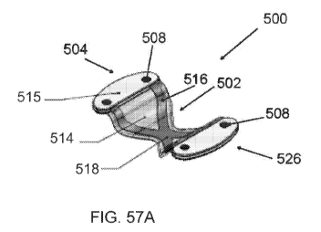
stainless steel, elgiloy,
1VIIP35N, or other metals known in the art. This design has an advantage of
providing a
relatively large support footprint against the valve leaflets, while at the
same time optimizing
the area of open space to permit maximum blood flow therethrough. The design
may be
treated or coated with silicone or other suitable material to eliminate
untoward effects such as
thrombosis or corrosion. Treatments may be sequential and include more than
one listed but
not limited to electropolishing, harperization, tumbling, pickling, plating,
encapsulation or
physical vapor deposition of appropriate materials.
[0126] FIGS. 24-27 illustrate side views of transvalvular bands 50
with different
inclinations. One of the objectives of the present invention is to not merely
provide support to
the leaflets during systole, but to elevate the plane of coaption in the
direction of the ventricle,
to cause early coaption (closure) relative to the cardiac cycle, as is
discussed elsewhere herein.
The variation in conditions, and other patient to patient variations may
warrant production of
the transvalvular band of the present invention in an array of sizes and/or
configurations, so
that clinical judgment may be exercised to select the appropriate implant for
a given case.
-20-
CA 03001338 2018-04-06
WO 2017/066480 PCT/US2016/056900
Alternatively, the transvalvular band may be provided in an adjustable form or
a modular form
so that an implant of the desired configuration can be constructed or modified
intraoperatively
at the clinical site. In a three segment embodiment, such as that illustrated
in FIGS. 24
through 27, a central segment may be provided for positioning within the
center of the flow
path, or centered on the coaptive edges of the leaflets. First and second end
portions may be
connected to the central portion, for supporting the central portion relative
to the tissue
anchors. First and second end portions may be provided in a variety of lengths
and
curvatures, enabling construction of a relatively customized modular implant
as may be
desired for a particular patient.
[0127] For example, FIG. 24 illustrates a transvalvular band 50 with a
central
portion 64 and two gently angled arm portions 70 and 72. The first and second
ends 52 and
54 are displaced from the central portion 64 by a height, hl and h2,
respectively. In FIG. 24,
hl and h2 are about equal and can range from about 0 mm to about 10 mm.
Preferably hl and
h2 will be at least about 2 mm and will often be at least about 4 mm or 6 mm
or more, but
generally no more than about 10 mm or 12 mm.
[0128] FIG. 25 illustrates a transvalvular band 50 with a central
portion 64 and
two sharply angled arm portions 70 and 72. The first and second ends 52 and 54
are
displaced from the central portion 64 by a height, hl and h2, respectively. In
FIG. 25, hl and
h2 are about equal and can range from about 8 mm to about 12 mm. FIG. 26
illustrates a
transvalvular band 50 with a central portion 64, a highly angled first arm 70
and a gently
angled second arm 72. The first and second ends 52 and 54 are displaced from
the central
portion 64 by a height, hl and h2, respectively. In FIG. 26, hl is greater
than h2. The hl
ranges from about 6 mm to about 10 mm, while h2 ranges from about 2 mm to
about 6 mm.
FIG. 27 illustrates a transvalvular band 50 with a central portion 64, a
gently angled first arm
70 and a highly angled second arm 72. The first and second ends 52 and 54 are
displaced
from the central portion 64 by a height, hl and h2, respectively. FIG. 27, may
be a mirror
image of FIG. 26.
[0129] The transvalvular band 50 can be made of any of a variety of
materials that
are compatible with implantation within a patient's body and which has the
requisite structural
integrity to support the mitral valve leaflets. For example, suitable
materials include titanium,
-21-
CA 03001338 2018-04-06
WO 2017/066480 PCT/US2016/056900
titanium alloys, stainless steel, stainless steel alloys, nitinol, elgiloy,
1ViP35N, other metals and
alloys, ceramics, and polymers such as PTFE, polycarbonate, polypropylene,
UEMWF'E,
EIDPE, PEEK, PEBAX and the like.
[0130] In order to reduce the thrombogenicity of the transvalvular
band 50, the
transvalvular band 50 can be provided with a smooth surface or appropriately
micro-texture
the surface in some embodiments, such as via a porous or microporous
structure. Other
factors such as surface chemistry, energy, morphology, macrofeatures, and
general material
properties matching the in situ needs can also be considered in tailoring the
surface of the
band. In addition, the transvalvular band 50 can be coated with a variety of
substances to
reduce thrombogenicity. For example, the transvalvular band 50 can be coated
with a
antithrombogenic agent such as heparin, a polymer such as PTFE, or a polymer
conjugated
with heparin or another antithrombogenic agent. Heparin coatings can be
achieved in a variety
of methods, one of which may be to coat or drip the prosthesis in TDMAC-
heparin
(Tridodecylmethylammonium heparinate).
[0131] As illustrated in FIGS. 28-31, the transvalvular band 50 is
implanted in the
plane of the mitral valve annulus 28 in a patient suffering from anterior
leaflet 26 prolapse
caused by the rupture 42 of the chordae tendineae 30 attached to the anterior
leaflet 26.
Although a prolapsed anterior leaflet 26 is illustrated, it should be
understood that the method
described herein is also applicable for treating other types of prolapse, such
as posterior leaflet
prolapse and prolapse caused by elongated leaflets 24 and 26. The
transvalvular band 50 can
be attached to the annulus 28 by a variety of techniques, such as sutures,
anchors, barbs,
stapes, self-expanding stents, or other techniques that are known or are
apparent to those of
skill in the art.
[0132] As best illustrated in FIGS. 29 and 31, the transvalvular band
50 is oriented
in the annulus 28 so that the transvalvular band 50 is positioned
approximately transversely to
the coaptive edge 42 formed by the closure of the mitral valve leaflets 24 and
26. The
transvalvular band 50 can also be positioned over the prolapsed portion of the
anterior leaflet
26 so that the transvalvular band 50 can directly support the prolapsed
portion of the anterior
leaflet 24 and keep the anterior leaflet 24 inferior to the plane of the
mitral valve annulus 28,
-22-
CA 03001338 2018-04-06
WO 2017/066480 PCT/US2016/056900
i.e., elevated in the direction of the ventricle or of antegrade flow, thereby
preventing or
reducing prolapse and mitral regurgitation.
[0133] FIGS. 28 and 29 illustrate the effect of the transvalvular band
50 on the
mitral valve 18 during systole. As shown, both the anterior leaflet 24 and the
posterior leaflet
26 are supported by the transvalvular band during closure of the mitral valve
18. The arcuate
transvalvular band 50 functions to keep both leaflets 24 and 26 inferior to
the plane of the
annulus 28 and enables the leaflets 24 and 26 to form a coaptive edge 40.
Although a single
transvalvular band 50 has been illustrated, in some embodiments, multiple
transvalvular bands
50 such as two or three or more can be implanted across the annulus 28 to
provide additional
support to the mitral valve leaflets 24 and 26.
[0134] FIGS. 30 and 31 illustrate the effect of the transvalvular band
50 on the
mitral valve 18 during diastole. During diastole, the mitral valve 18 opens so
that blood can
fill the left ventricle 16 from the left atrium 12. As best illustrated in
FIG. 31, the
transvalvular band 50 obstructs only a small portion of the mitral valve 18
opening, and
therefore, does not cause excessive resistance to blood flow.
[0135] FIGS. 32-35 are cross-sectional side views of the mitral valve
18 with and
without the support of the transvalvular band 50. During systole, the mitral
valve 18 closes.
Without the transvalvular band 50, the anterior leaflet 24 crosses the plane P
defined by the
mitral valve annulus 28 and prolapse, which leads to mitral regurgitation, as
shown in FIG. 33.
However, by implanting the transvalvular band 50 in the annulus 28 such that
the arcuate
transvalvular band 50 arches towards the left ventricle and the central
portion 64 is displaced
from the plane P, the anterior leaflet 24 is prevented from prolapsing above
the plane P thus
eliminating or reducing retrograde flow (shown in FIG. 33). The leaflets 24
and 26 rest upon
the transvalvular band 50 and the pressure exerted by the blood upon the
distal portion of the
leaflets 24 and 26 form the coaptive edge 40. As illustrated in FIGS. 34 and
35, the
performance of the mitral valve 18 during diastole is not substantially
affected by the
transvalvular band 50.
[0136] Although the method of implanting and positioning the
transvalvular band
50 has been illustrated with one embodiment of the transvalvular band 50,
other embodiments
as described above can also be used. For example, FIG. 36 illustrates a
transvalvular band 50
-23-
CA 03001338 2018-04-06
WO 2017/066480 PCT/US2016/056900
with a wider, offset coaptive edge support portion 66 that has been implanted
in the mitral
valve annulus. As shown, the coaptive edge support 66 is offset so that it
positioned to
support the coaptive edge of the mitral valve 18. In addition, the
transvalvular band 50 can be
used in conjunction with other devices and procedures, such as a separate or
integrally
attached annular or annuloplasty ring described above. In addition, the
transvalvular band 50
can be used in conjunction with the Alfieri procedure, where the tips of the
mitral valve
leaflets 24 and 26 are sutured 74 together, as shown in FIG. 38.
[0137] Referring to FIG. 37, there is illustrated a perspective view
of a
transvalvular band 50 having a transverse projection or support 51 extending
in the direction
of the ventricle or in the direction of diastolic blood flow, which could be
considered
antegrade. The support 51 has a width W, which may be at least about 3 mm, and
in some
embodiments, at least about 5 mm, and in other embodiments at least about 1.0
cm. The
projection 51 may be utilized without an Alfieri stitch, so that the leaflets
of the mitral valve
close against opposing side walls 53 and 55 of the projection 51. The
projection 51 thus helps
center the closure of the leaflets, as well as controlling the width of
coaption. In addition, the
band 50 is illustrated as convex in the direction of the ventricle, to
accomplish early closure as
has been discussed herein.
[0138] The transvalvular band in accordance with the present invention
can be
implanted via an open surgical procedure, via thoracotomy (e.g. transapically)
or alternatively,
via a percutaneous procedure using a translumenally implantable embodiment. In
the
translumenally implantable embodiment, one or more transvalvular bands can be
attached to a
self-expandable support structure, such as a self-expandable ring or self-
expandable stent
having a relatively short axial length relative to its expanded diameter. The
transvalvular band
and the compressed self-expandable support structure are loaded into a
catheter with a
retractable outer sheath which is inserted percutaneously and advanced
translumenally into or
across the mitral valve. The retractable outer sheath can be retracted to
allow the self-
expandable support structure to expand adjacent or against the annulus,
thereby positioning
the one or more transvalvular bands in about the plane of the mitral annulus.
Each
transvalvular band can be characterized by a longitudinal axis, and the
transvalvular band is
-24-
CA 03001338 2018-04-06
WO 2017/066480 PCT/US2016/056900
oriented in the mitral valve such that the longitudinal axis of the
transvalvular band in oriented
substantially transversely to the coaptive edge of the mitral valve.
[0139] By "percutaneous" it is meant that a location of the
vasculature remote
from the heart is accessed through the skin, such as using needle access
through, for example,
the Seldinger technique. However, it may also include using a surgical cut
down procedure or
a minimally invasive procedure. The ability to percutaneously access the
remote vasculature is
well-known and described in the patent and medical literature.
[0140] Depending on the point of vascular access, the approach to the
mitral valve
may be antegrade and require entry into the left atrium via the pulmonary vein
or by crossing
the interatrial septum. Alternatively, approach to the mitral valve can be
retrograde where the
left ventricle is entered through the aortic valve. Once percutaneous access
is achieved, the
interventional tools and supporting catheter(s) will be advanced to the heart
intravascularly
where they may be positioned adjacent the target cardiac valve in a variety of
manners, as
described elsewhere herein. While the methods will preferably be percutaneous
and
intravascular, many of the implants and catheters described herein will, of
course, also be
useful for performing open surgical techniques where the heart is beating or
stopped and the
heart valve accessed through the myocardial tissue. Many of the devices will
also find use in
minimally invasive procedures where access is achieved thorascopically and
where the heart
will usually be stopped but in some instances could remain beating.
[0141] A typical antegrade approach to the mitral valve is depicted in
FIG. 39.
The mitral valve MV may be accessed by a standard approach from the inferior
vena cava IVC
or superior vena cava SVC, through the right atrium RA, across the interatrial
septum IAS
and into the left atrium LA above the mitral valve MV. As shown, a catheter
120 having a
needle 122 may be advanced from the inferior vena cava IVC into the right
atrium RA. Once
the catheter 120 reaches the interatrial septum IAS, the needle 122 may be
advanced so that it
penetrates through the septum at the fossa ovalis FO or the foramen ovale into
the left atrium
LA. At this point, a guidewire may be advanced out of the needle 122 and the
catheter 120
withdrawn.
[0142] As shown in FIG. 40, access through the interatrial septum IAS
will usually
be maintained by the placement of a guide catheter 125, typically over a
guidewire 124 which
-25-
CA 03001338 2018-04-06
WO 2017/066480 PCT/US2016/056900
has been placed as described above. The guide catheter 125 affords subsequent
access to
permit introduction of the tool(s) which will be used for performing the valve
or tissue
modification, as described in more detail below.
[0143] A typical retrograde approach to the mitral valve is depicted
in FIG. 41.
Here the mitral valve MV may be accessed by an approach from the aortic arch
AA, across
the aortic valve AV, and into the left ventricle below the mitral valve MV.
The aortic arch AA
may be accessed through a conventional femoral artery access route, as well as
through more
direct approaches via the brachial artery, axillary artery, or a radial or
carotid artery. As
shown in FIG. 42, such access may be achieved with the use of a guidewire 128.
Once in
place, a guide catheter 126 may be tracked over the guidewire 128. The guide
catheter 126
affords subsequent access to permit introduction of the tool(s) which will be
used for
performing the valve modification, as described in more detail below.
[0144] In some cases, access routes to the mitral valve may be
established in both
antegrade and retrograde approach directions. This may be useful when, for
instance,
grasping is performed with the use of specific devices introduced through one
route and
fixation is achieved with the use of separate devices introduced through
another route. In one
possible situation, the transvalvular band may be introduced via a retrograde
approach. While
the transvalvular band is held in place, a fixation tool may be introduced via
an antegrade
approach to fix the transvalvular band in place. The access pathways for the
transvalvular
band and fixation tool may alternatively be reversed. Thus, a variety of
access routes may be
used individually or in combination with the methods and devices of the
present invention.
[0145] Referring to FIG. 43A, there is illustrated a schematic view of
a
percutaneously deliverable implant in accordance with one aspect of the
present invention.
The deployment system includes a deployment catheter 200, only a distal end of
which is
illustrated herein. Deployment catheter 200 is configured in accordance with
known
technology for accessing the mitral valve, utilizing conventional dimensions
and the materials
known to those of skill in the art. In general, the deployment catheter 200
comprises an
elongate flexible tubular body 202 extending between a proximal end (not
illustrated) and a
distal end 204. The proximal end is provided with a proximal manifold,
including access
-26-
CA 03001338 2018-04-06
WO 2017/066480 PCT/US2016/056900
portals such as luer connectors in communication with each functional lumen in
the
catheter 200.
[0146] The distal end 204 is provided with a distally facing opening
208, which is
in communication with the proximal end via a central lumen 206.
[0147] Positioned within the central lumen 206 is a collapsed implant
210.
Implant 210 is transformable between a first, radially reduced configuration
such as for
positioning within the deployment catheter 200 and a second, radially enlarged
configuration
(see FIG. 43C) for positioning at the treatment site. Transformation of the
implant from the
first configuration to the second configuration may be accomplished under
positive force, such
as via balloon dilatation. Alternatively, as illustrated herein,
transformation is accomplished by
self-expansion of the implant 210 in response to removal of the constraint
provided by the
tubular body 202.
[0148] In general, the implant 210 comprises a frame or anchor
component 212
and a leaflet support component 214. Leaflet support component 214 may
comprise any of a
variety of structures similar to those described previously herein as the
annular band,
configured or reconfigured such that the annular band may be radially reduced
for positioning
within a deployment catheter and subsequently radially enlarged for spanning
the mitral valve.
The implant 210 additionally comprises an anchor component, for anchoring the
leaflet
support 214 at the treatment site. In the illustrated embodiment, anchor 212
is schematically
illustrated as a zigzag wire or filament structure, which is radially
expansible following
removal of the constraint. However, any of a variety of configurations may be
utilized for the
anchor 212.
[0149] Referring to FIG. 43B, the outer tubular flexible body 202 is
shown
partially retracted from the implant, permitting the implant to begin to
radially expand.
FIG. 43C illustrates further retraction of the tubular body 202, to fully
release the anchor 212
at the deployment site. As illustrated, anchor 212 radially expands within the
left atrium. The
leaflet support 214 extends approximately transversely to the coaptive edge of
the mitral valve
leaflets, and is convex or inclined in the direction of the mitral valve to
advance the coaptation
of the mitral valve leaflets in the direction of the ventricle as has been
described elsewhere
herein.
-27-
CA 03001338 2018-04-06
WO 2017/066480 PCT/US2016/056900
[0150] As seen in FIG. 43A, the implant 210 is controlled by at least
one control
line 216. Control line 216 extends throughout the length of the deployment
catheter 200, and
to at least one control on or near the proximal manifold. This enables
proximal retraction of
the flexible body 202 with respect to the implant 210, and control of implant
210 prior to final
detachment from the deployment system.
[0151] Referring to FIG. 43C, at least a first control wire 216, a
second control
wire 218, and a third control wire 220 are illustrated connected to the anchor
212. Control
wires 216, 218 and 220 enable manipulation of the implant into its final
desired position, and,
if necessary, proximal retraction of the implant back within the deployment
catheter should the
decision be made to remove the implant prior to final detachment.
[0152] Prior to final detachment of the implant 210, additional
anchoring
structures may be engaged to retain the implant at its desired implanted
location. For
example, anchor 212 may be provided with any of a variety of tissue anchors or
barbs, for
engaging the mitral valve annulus or the base of the leaflets or other
adjacent anatomical
structures. Alternatively, separate tissue anchors may be advanced through the
deployment
catheter 200, and utilized to secure the anchor 212 to the adjacent tissue.
Suitable anchors
are preferably enlargeable from a first, reduced cross sectional configuration
for traveling
through the deployment catheter 200 and piercing tissue, to a second, enlarged
configuration
for resisting removal from the tissue. In the embodiment illustrated in FIG.
43C, no
secondary anchoring structures are illustrated for simplicity.
[0153] Once the position of the implant 210 has been verified and
found
acceptable, and the determination of whether to introduce secondary anchoring
structures has
been made, the control wires 216, 218 and 220 are detached from the anchor
212, and the
deployment catheter 200 is removed from the patient. Detachment of the control
wires from
the implant 210 may be accomplished in any of a variety of ways, such as by
electrolytic
detachment, detachment by thermal elevation of a softenable or meltable link,
mechanical
detachment such as by rotating the control wire such that a threaded end of
the control wire is
threadably disengaged from the anchor 212, or other detachment techniques
depending upon
the desired functionality and profile of the system.
-28-
CA 03001338 2018-04-06
WO 2017/066480 PCT/US2016/056900
[0154] Referring to FIG. 43D, there is illustrated a side elevational
view of the
implant 210 in an unconstrained (e.g., bench top) expanded configuration. The
anchor 210
comprises a plurality of struts 222, which are joined at a first end by a
plurality of apices 224
and a second end by a plurality of apices 226 to produce a zigzag structure
sometimes
referred to as a "Z stent" configuration. This configuration is convenient and
well understood
in the intravascular implant arts, although any wide variety of structures may
be utilized. For
example, zigzag wire patterns, woven wire patterns, or sinusoidal wire
patterns may be
utilized. Laser cut wall patterns such as from tubing stock may also be
utilized, and may be
provided with any of a wide variety of complex wall patterns. In general,
nickel titanium
alloys such as any of a variety of nitinol alloys are preferred. However,
depending upon the
wall pattern, stainless steel, elgiloy, certain polymers or other materials
may also be utilized.
Heat treatment may be required to anneal and shape set an alloy such as
Nitinol. Other alloys
may require only annealing to relieve stresses incurred during prior
processing.
[0155] Referring to FIG. 43E, there is illustrated an end view of the
implant shown
in FIG. 43D to show the transverse configuration of the transvalvular band
portion of the
implant. In this illustration, the transvalvular band comprises a plurality of
struts 230 which
are connected to the anchor 212 at junctions 232. Struts 230 may in turn be
divided into a
bifurcated section 234 or other configuration to increase the effective
footprint of the
transvalvular band measured along the coaptive edge of the valve, while
minimizing
obstruction to blood flow therethrough. The coaptive edge of the valve, as
implanted, will
preferably be approximately aligned with the transverse axis 236 illustrated
in FIG. 43E of the
band, as implanted. The axis of coaption of the mitral valve is preferably
parallel to axis 236
in the implanted configuration, but may be within about 45 , preferably within
about 20 , and
most preferably within about 10 of the axis 236.
[0156] Referring to FIGS. 44A and 44B, there is illustrated an anchor
deployment
catheter which may be utilized to provide either primary or secondary
anchoring of the anchor
structure 212 to adjacent tissue. Anchor deployment catheter 250 comprises an
elongate
flexible tubular body 252, configured to access the vicinity of the mitral
valve. Tubular
body 252 extends between a proximal end 254 and a distal end 256. Distal end
256 is
provided with a distal opening 258, enabling access to a central lumen 260. An
elongate
-29-
CA 03001338 2018-04-06
WO 2017/066480 PCT/US2016/056900
flexible core wire 262 extends from the proximal end 254 throughout most of
the length of the
lumen 260 to a distal surface 264. See FIG. 44C. The proximal end of the core
wire 262 is
provided with a control 266 that enables axial reciprocal movement of the core
wire 262
within the central lumen 260.
[0157] A tissue anchor 268 may be positioned within the distal end of
the delivery
catheter 250. In use, manipulation of the control 266, such as by distal axial
advance relative
to the tubular body 252, distally, axially advances the core wire 262 to expel
the anchor 268
through the distal opening 258. Distal opening 258 is preferably provided with
a bevel or
angled cut to provide a sharpened distal tip 270. This enables distal axial
advance of the distal
tip 270 into tissue at a desired site, so that the control 266 may be
manipulated to deploy all
or a portion of the anchor 268 into the target tissue.
[0158] Any of a variety of tissue anchors 268 may be utilized,
depending upon the
desired configuration of the implant and the implant anchor interface. In the
illustrated
embodiment, the anchor 268 is configured as a double "t-tag" anchor. A first
tissue engaging
element 272 is connected to a second implant engaging element 274 by a
filament 276. In use,
the distal tip 270 is positioned within the tissue of the mitral valve
annulus. Control 266 is
manipulated to deploy the first element 272 beneath the surface of the tissue.
The tubular
body 252 is thereafter proximally retracted, enabling the second element 274
to engage the
implant and retain it against the adjacent tissue.
[0159] The anchor delivery catheter 250 may be advanced through the
deployment
catheter 200, and/or along a guide such as a guidewire or support wire. In the
illustrated
embodiment, the anchor deployment catheter 250 is provided with a guide lumen
278
allowing the anchor delivery catheter to track along a guidewire. Guide lumen
278 is defined
by a tubular wall 280. Tubular wall 280 may extend the entire length of the
anchor delivery
catheter 250, such as by forming the catheter body as a dual lumen extrusion.
Alternatively,
tubular wall 280 may be provided with an axial length that is short relative
to the overall
length of the catheter, such as no more than about 3cm and preferably no more
than about
2cm in length. This allows the anchor delivery catheter to ride along a
guidewire in a
monorail or rapid exchange manner as will be illustrated below.
-30-
CA 03001338 2018-04-06
WO 2017/066480 PCT/US2016/056900
[0160] Referring to FIGS. 45A and 45B, there is illustrated an implant
configured
for use with the anchor delivery catheter described above. In general, the
implant comprises a
first leaflet support 292 and a second leaflet support 294, separated by a
flexible
connection 296. Flexible connection 296 permits the implant 290 to be folded
within a
deployment catheter, and later expanded in a manner that permits the implant
290 to function
as a transvalvular band as described. The implant 290 may be manufactured in
any of a
variety of ways, such as using a wire frame or by laser cutting from sheet
stock as will be
appreciated by those of skill in the art.
[0161] In the illustrated embodiment, a first and second flexible
connection 296
reside in a plane configured to be substantially parallel to the axis of
coaption the as implanted
orientation. The lateral edges of the each of the first leaflet support 292
and second leaflet
support 294 are provided with at least one and preferably two or three eyes
298, fabric
patches, or other anchor attachment structure, for receiving a tissue anchor.
[0162] Referring to FIG. 45B, the implant of FIG. 45A is illustrated
in a partially
collapsed configuration, flexed about the flexible connection 296. In
addition, control
wires 300, 302 and 304 are illustrated releasably connected to the implant
290. Control
wires 300, 302 and 304 may be utilized to advance the implant 290 from the
deployment
catheter such as catheter 200 described above, and manipulate the implant
until the anchors
have been fully deployed. Thereafter, control wires 300, 302 and 304 may be
removed such
as by electrolytic detachment, melting a polymeric link, unscrewing a threaded
connection, or
other detachment mechanism depending upon the desired functionality of the
device.
[0163] Referring to FIGS. 46A through 46E, there is illustrated a
sequence of
deploying an implant at the mitral valve from an antegrade direction. The
implant 290 may be
similar to that illustrated in FIGS. 45A and 45B, or have wall patterns or
characteristics of
other implants disclosed elsewhere herein. In general, the implant 290 is
deployed from the
catheter 200 in the sequence illustrated in FIGS. 46A through 46C. The
surrounding anatomy
has been eliminated for simplicity.
[0164] Referring to FIG. 46D, the anchor delivery catheter 250 is
advanced onto
the proximal end of one of the control wires 300, such that the control wire
300 is axially
moveably positioned within guide lumen 278. This enables the anchor delivery
catheter 250
-31-
CA 03001338 2018-04-06
WO 2017/066480 PCT/US2016/056900
to be advanced along the control wire 300 in a monorail or rapid exchange
configuration as is
understood in the catheter arts. Anchor delivery catheter 250 is advanced
along the control
wire 300 until the distal tip 270 advances through the eye 298 or fabric tab
or other
attachment structure, and into the adjacent tissue of the base of the mitral
valve leaflet or
mitral valve annulus. The control 266 is manipulated such as by distal advance
to advance the
first anchor element 272 out of the distal opening 258 and into the tissue as
illustrated in
FIG. 46D.
[0165] The anchor delivery catheter 250 is thereafter proximally
withdrawn to
position the distal opening 258 on the device proximal side of the eye 298,
and the core
wire 262 is further distally advanced to deploy the second anchor element 274
from the distal
opening 258 of the anchor delivery catheter 250. Anchor delivery catheter 250
may thereafter
be proximally withdrawn from the patient. Either the same or a different
anchor delivery
catheter 250 may thereafter be advanced along the third control wire 304,
enabling
deployment of another tissue anchor as is illustrated in FIG. 46E.
[0166] The implant 290 is illustrated in FIG. 46E as having a central
portion
inclined in the direction of the ventricle to support the leaflets as has been
discussed elsewhere
herein. This configuration may be retained by the inherent bias built into the
structure and
materials of the implant 290. Alternatively, the configuration of inclining in
the direction of
the ventricle may be retained by active intervention such as by providing a
mechanical
interlock, in situ heat weld with capacitive discharge/electrolytic weld,
application of a clip or
other locking structure by way of control wire 302 or simply by the mechanical
forces
attributable to the mitral valve annulus, which prohibit lateral expansion of
the device
sufficient for the flexible connection 296 to invert in the direction of the
atrium. Alternatively,
an implantable control wire (not illustrated) may be introduced, to connect
the implant 290
such as in the vicinity of the flexible connection 296 to the opposing wall of
the ventricle, as
will be described in connection with a transapical implementation of the
invention described
below.
[0167] A further implementation of the invention is illustrated in
connection with
FIGS. 47A through 47E. Referring to FIG. 47A, the first control line 300 and
third control
line 304 have been replaced by a first guide tube 310 and a second guide tube
312. First guide
-32-
CA 03001338 2018-04-06
WO 2017/066480 PCT/US2016/056900
tube 310 and second guide tube 312 each has the double function of controlling
deployment
of the implant, as well as enabling introduction of a tissue anchor
therethrough. This avoids
the use of a separate tissue anchor deployment catheter such as that described
above.
[0168] As illustrated in FIG. 47B, once the implant is provisionally
positioned in
the vicinity of the mitral valve, a first tissue anchor 314 is deployed
through the first guide
tube 310. A second tissue anchor 316 is deployed through the second guide tube
312. The
tissue anchors may comprise "T" tag type constructions, pigtail or corkscrew
constructions,
or any of a variety of other soft tissue anchors known in the art. In general,
tissue anchors
utilized for the present purpose are preferably transformable from a first,
reduced cross-
sectional configuration to a second, radially enlarged cross-sectional
configuration to enable
deployment through a small needle or tube and then provide a relatively higher
resistance to
pull out. Radial enlargement may be accomplished by angular movement of a
portion of the
anchor, or by physical expansion in a radial direction.
[0169] Referring to FIG. 47C, the first guide tube 310 and second
guide tube 312
have been removed following deployment of the tissue anchors. The guide tubes
may be
removed using any of a variety of detachment techniques disclosed elsewhere
herein. Either
before or after removal of the guide tubes, distal pressure on either the
tubular body 202 or
the control wire 302 inverts the implant from the configuration shown in FIG.
47C to the final
configuration shown in FIG. 47D and E. The inverted configuration of FIG. 47D
and E may
be retained by the mechanical bias imparted through the anchoring to the
mitral valve annulus,
or using techniques described elsewhere herein. The control wire 300 is
thereafter detached
from the implant, as illustrated in FIG. 47E.
[0170] Any of a variety of the implants of the present invention may
alternatively
be introduced across the ventricle, such as in a transapical approach. The
retrograde approach
to the mitral valve will necessitate certain modifications to both the implant
and the
deployment system, as will be appreciated by those of skill in the art in view
of the disclosure
herein.
[0171] For example, a transventricle approach is illustrated in FIGS.
48A through
48D. A deployment catheter 320 is introduced into the ventricle, and
retrograde through the
mitral valve to position the distal opening 208 within the atrium. An implant
is carried within
-33-
CA 03001338 2018-04-06
WO 2017/066480 PCT/US2016/056900
the deployment catheter 320, as has been described elsewhere herein. In
general, the implant
comprises a first leaflet support 292 and a second leaflet support 294
separated by a flexible
zone or pivot point.
[0172] In the retrograde implementation of the invention, the first
and second
leaflet supports are flexible or pivotable with respect to the longitudinal
axis of the control
wire 300, such that they may be moved between a first configuration in which
there are
substantially parallel with the axis of the control wire 300, and a second
position, as illustrated
in FIGS. 48A through 48D, in which they are inclined radially outwardly from
the longitudinal
axis of the control wire 300 in the device proximal direction. The implant may
thus reside
within the deployment catheter 320 when the first leaflet support 292 and
second leaflet
support 294 are in the first, reduced crossing profile configuration, with
each of the tissue
anchors 314 and 316 pointing in a device proximal direction. In this
embodiment, the tissue
anchor 314 may be permanently affixed to or integral with the first leaflet
support 292 and the
second anchor 316 may be similarly carried by the second leaflet support 294.
[0173] Once the distal end of the deployment catheter 320 has been
positioned
within the atrium, the control wire 300 may be distally advanced to advance
the anchors 314
and 316 beyond the distal opening 208. This releases the implant and allows
the angle
between the first and second leaflet supports to be increased, so that the
tissue anchors 314
and 316 may be aimed at the desired tissue anchor target sites. Proximal
retraction on the
control wire 300 may be utilized to seat the tissue anchors within the target
tissue, as
illustrated in FIG. 48B.
[0174] Further proximal traction on the control wire 300 may be
utilized to invert
the implant into the configuration illustrated in FIG. 48C. At that point, the
control wire 300
may be severed from the implant as has been discussed elsewhere herein.
Alternatively, the
deployment catheter 320 may be proximally retracted leaving the control wire
300 secured to
the implant, and a second portion of the control wire may be secured to a
tissue anchor 322
within or on the epicardial surface of the ventricle. Anchor 322 may comprise
any of a variety
of structures, such as a pledget, button, or other structure that provides a
footprint against the
epicardial surface to resist retraction of the control wire 300 into the
ventricle. The control
wire 300 may thereafter be severed proximally of its securement to the anchor
322, leaving
-34-
CA 03001338 2018-04-06
WO 2017/066480 PCT/US2016/056900
the control wire 300 and anchor 322 in position to span the ventricle and
retain the
configuration of the implant as illustrated in FIG. 48D.
[0175] In all the foregoing embodiments, the final configuration of
the implant
within the mitral valve is illustrated in a highly schematic form, and the
angle and degree of
inclination into the direction of the ventricle may be significantly greater
than that illustrated
herein depending upon the desired clinical performance. The transvalvular band
inclination can
be highly advantageous in some embodiments in providing clinical benefit as it
facilitates
"physiologic coaptation" in a preferred manner as its surface mimics the three
dimensional
feature created by the leaflets as they would have coapted in a healthy native
valve.
[0176] Referring to FIGS. 49A through 49H, there is illustrated a
transapical
approach to the mitral valve, and deployment of a transvalvular band in
accordance with the
present invention. As illustrated in FIG. 49A, a deployment catheter 320 has
been introduced
such as via thoracotomy, and advanced retrograde through the mitral valve. A
transvalvular
band 324 has been deployed distally from the catheter 320, and is illustrated
in FIG. 49A in an
expanded configuration within the left atrium. Expansion of the transvalvular
band 324 from
a reduced cross-sectional profile for positioning within the catheter 320 to
the enlarged cross-
sectional profile illustrated in FIG. 49A may be accomplished either under
mechanical force,
such as by dilatation of an inflatable balloon or other mechanical mechanism.
Preferably,
however, transvalvular band 324 is self-expandable so that it converts from
the reduced
profile to the enlarged profile automatically upon deployment from the distal
end of the
catheter 320.
[0177] In the illustrated embodiment, the transvalvular band 324
comprises an
arcuate central portion 325, which is convex in the direction of the
ventricle. See FIGS. 49A
and 49B. The transvalvular band 324 is provided with a first attachment
structure 326 and a
second attachment structure 328. Attachment structures 326 and 328 may
comprise any of a
variety of structures disclosed herein, such as tissue anchors, including
hooks or barbs. In
one implementation of the invention, the first attachment structure 326, and
second
attachment structure 328 each comprise a target for receiving an anchor as
will be disclosed
below. Suitable targets for the present purpose include woven or non-woven
fabrics,
polymers, or other materials or constructions which permit a needle or
sharpened anchor to
-35-
CA 03001338 2018-04-06
WO 2017/066480 PCT/US2016/056900
penetrate therethrough, as will be discussed. In one implementation of the
invention, each of
the attachment structures comprises a Dacron mesh, having a frame for
supporting the mesh
and securing the mesh to the transvalvular band 324.
[0178] Referring to FIG. 49B, there is illustrated a perspective view
of the
transvalvular band 324 illustrated in FIG. 49A. The transvalvular band 324
comprises a
central section 325, convex in the direction of the ventricle for affecting
leaflet closure as has
been described herein. Central section 325 is formed by a frame 327, which
comprises at least
one strut 329 extending between the first attachment structure 326 and second
attachment
structure 328. In the illustrated embodiment, three struts extend generally
parallel to each
other, defining at least two elongate openings therebetween. One or two or
four or more
transverse elements 331 may be provided, such as to enhance structural
integrity of the
construct. At least a first control wire 300 and, optionally a second or third
or fourth control
wire 300 is releasably attached to the transvalvular band 324, to enable
manipulation of the
band into position as shown in FIG. 49C. Control wire 300 is releasably
connected to the
transvalvular band 324 at a connection point 301. The connection at point 301
may be
established by threadable engagement, an electrolytically detachable link or
weld, or other
detachment mechanism. Electrolytically detachable deployment systems are
known, among
other places, in the neurovascular embolic coil and stent arts, and suitable
systems are
disclosed in U.S. patent Nos. 5,976,131 to Guglielmi, et al.; 6,168,618 to
Frantzen; and
6,468,266 to Bashiri, et al., the disclosures of which are hereby incorporated
in their entireties
herein by reference
[0179] The first attachment structure 326 comprises a support 333
carried by the
frame 327. In the illustrated embodiment, support 333 comprises an enclosed
loop, having a
central opening filled or covered by a mesh 337. The support 333 may
alternatively comprise
any of a variety of structures, such as a single linear element, sinusoidal or
zigzag pattern,
depending upon the desired performance. In the illustrated embodiment, the
support 333 is
conveniently provided in the form of a loop, to facilitate holding mesh 337 in
a generally
planar configuration, and support the mesh so that it may be punctured by an
anchor, suture
or other retention structure. A second support 335 is similarly provided with
a mesh 337, to
facilitate attachment. The mesh 337 may conveniently be a layer or pad of
Dacron or other
-36-
CA 03001338 2018-04-06
WO 2017/066480 PCT/US2016/056900
material, such as an integration of a silicone core with a Dacron jacket,
which facilitates both
piercing by an attachment structure, as well as tissue in-growth for long term
retention. The
first support 333 and second support 335 may comprise a radio opaque material,
or be
provided with radio opaque markers to enable aiming the anchor deployment
system into the
mesh 337 under fluoroscopic visualization.
[0180] Once the transvalvular band 324 has been brought into the
position
illustrated in FIG. 49C, the first attachment structure 326 and second
attachment
structure 328 may be secured to the adjacent tissue using any of a variety of
clips, staples,
barbs, sutures, or other structure which may be conveniently pierced through
the mesh 337
and/or looped around the first and second supports 333, 335. The retention
element may be
approached from either the side of the left atrium, the ventricle, or
epicardially, such as by
way of a minimally invasive puncture on the chest wall. In the implementation
of the method
described below, the example of advancing the retention elements from the left
ventricle will
be described.
[0181] Referring to FIG. 49C, proximal traction on the catheter 320
and on the
control wire 300, pulls the transvalvular band 324 snuggly against the left
atrial side of the
mitral valve, such that the first attachment structure 326 and second
attachment structure 328
are seated against the valve annulus.
[0182] Referring to FIG. 49D, a first anchor guide 330 and a second
anchor
guide 332 have been distally advanced from the distal end of the catheter 320.
Anchor
guides 330 and 332 may be alternatively associated with or carried by the
catheter 320 in a
variety of ways. For example, the first and second anchor guides 330 and 332,
may be
pivotably carried by the catheter 320, such that they may be inclined radially
outwardly from
the longitudinal axis of the catheter in the distal direction.
[0183] In the illustrated embodiment, the first and second anchor
guides comprise
a wire or tube for directing an anchor as will be discussed. The wire or tube
of the anchor
guide may comprise any of a variety of materials, such as nickel titanium
alloys (e.g. nitinol)
which may be preset to assume a position similar to that illustrated in FIG.
49D upon distal
advance from the catheter 320. The first and second anchor guides may be
provided with
radio-opaque markers, or may be constructed from a radio-opaque material, to
permit
-37-
CA 03001338 2018-04-06
WO 2017/066480 PCT/US2016/056900
fluoroscopic guidance. In the illustrated embodiment, the first and second
anchor guides are
in the form of tubes, for axially slidably receiving a tissue anchor and
tissue anchor
deployment structures therein.
[0184] Referring to FIG. 49E, a retention element in the form of a
first anchor 334
is illustrated as having been distally advanced from the first anchor guide
330, through the
tissue in the vicinity of the mitral valve annulus, and through the first
attachment
structure 326. Penetration of the first anchor 334 through the first
attachment structure 326
may be accomplished while providing proximal traction on the control wire 300.
[0185] The first anchor 334 is provided with at least one and
preferably two or
four or more transverse elements 336 to resist proximal retraction of the
first anchor 334 back
through the opening formed in the first attachment structure 326. The
transverse element or
surface 336 may be provided on any of a variety of structures, such as an
umbrella-type
structure, t-tag, barbs, or other anchoring configuration which can pass in a
first direction
through an opening formed in the first attachment structure 326, but resist
retraction in a
second, opposite direction, back through the first attachment structure 326.
[0186] The transverse element 336 is carried by a filament 338, which
extends
through the adjacent myocardial tissue. Filament 338 may comprise any of a
variety of
materials, such as a monofilament or multi-filament structure made from
polypropylene, any of
a variety of other known suture materials such as polyethylene, or metals such
as stainless
steel, nitinol, and others known in the art. The filament 338 may be a mono-
filament structure
or a multi-filament structure which may be braided or woven, depending upon
the desired
clinical performance. At least a second, similar anchor 340 is introduced on
the opposing side
of the mitral valve.
[0187] Referring to FIG. 49F, a second transverse element 342 is shown
secured
to or carried by the ventricular end of the filament 338, to provide a secure
anchoring through
the tissue wall for the transvalvular band. A similar structure is provided on
the opposing side
of the mitral valve. Although only a first and second anchoring systems has
been described
above, additional anchoring systems, such as a total of four or six or eight
or more, typically
in even numbers to produce bilateral symmetry, may be used. The number and
configuration
-38-
CA 03001338 2018-04-06
WO 2017/066480 PCT/US2016/056900
of tissue anchors will depend upon the configuration of the transvalvular
band, as will be
apparent to those of skill in the art in view of the disclosure herein.
[0188] As
shown in FIG. 49F, the anchors have been fully deployed and the first
anchor guide 330 and second anchor guide 332 have been proximally retracted
into the
catheter 320.
[0189]
Referring to FIG. 49G, the control wire 300 may thereafter be detached
from the transvalvular band and removed. Detachment of control wire 300 may be
accomplished in any of a variety of ways, as has been described elsewhere
herein.
[0190]
Alternatively, the control wire 300 may be left in place as is illustrated in
FIG. 49H.
Control wire 300 is secured to an epicardial anchor 322, to provide a
transventricular truss, as has been described.
[0191]
Referring to FIGS. 50A and 50B, there is illustrated a side elevational
schematic view of the distal end of a deployment catheter 360 which may be
adapted for use
in either the transapical delivery of FIGS. 49A-49H, or any other delivery
mode described
herein. In the illustrated embodiment, the deployment catheter 360 includes an
elongate
tubular body having a central lumen 362, opening at a distal end 364. Carried
within the
central lumen 362 is a transvalvular band 366, in a rolled-up configuration.
Transvalvular
band 366 is maintained in a rolled-up configuration by the constraint imposed
by the
deployment catheter 360. However, upon distal advance of the push element 368
to deploy
the transvalvular band 366 beyond the distal end 364, as illustrated in FIG.
50B, the
transvalvular band 366 unrolls under its natural bias into a predetermined
configuration for
implantation across the mitral valve.
[0192] One
configuration for the transvalvular band is shown rolled out in plan
view in FIG. 51A. However, any of a variety of alternative transvalvular band
configurations
disclosed herein can be utilized with the catheter of FIGS. 50A and 50B.
[0193]
Referring to FIG. 51A, there is illustrated a transvalvular band 366 having
a central portion 368 for spanning the coaptive edges of the mitral valve. A
first attachment
zone 370 and a second attachment zone 372 are provided on opposing ends of the
central
portion 368.
-39-
CA 03001338 2018-04-06
WO 2017/066480 PCT/US2016/056900
[0194] The central portion comprises at least a first strut 374 for
spanning the
mitral valve as has been discussed. In the illustrated embodiment, a second
strut 376 and a
third strut 378 are provided, spaced apart to increase the width of the
contact footprint with
the valve leaflet but permit blood flow therethrough. A first end of each of
the struts 374,
376, and 378 are connected at the first attachment zone 370, and the second
ends of the three
struts are connected at the second attachment zone 372.
[0195] The first and second attachment zones may be provided with a
reinforcing
element 382, to facilitate long term attachment. Apertures 380 are
illustrated, which may be
provided to allow manual suturing when the transvalvular band 366 is intended
for use in an
open surgical procedure. Alternatively, apertures 380 may be configured for
attachment using
an anchor deployment catheter when intended for use in a translumenal or
transapical
deployment. Each of the first, second and third ribs may be provided with a
central core, such
as a nitinol or stainless steel wire or ribbon, and an outer coating such as a
polycarbonate
urethane with or without copolymers like silicone, silicone coating, or a
fabric such as PET,
ePTFE, polyethylene, or a hybrid of, for example, the aforementioned materials
impregnated
silicone coating, to reduce the risk of abrasion of the mitral valve leaflets
A close-up view of
circled zone 51D of FIG. 51A is illustrated in FIG. 51D.
[0196] FIG. 51D illustrates one embodiment of a fatigue-resistant
terminal portion
of a proximal and/or distal end of one, two, or more of the struts 374, 376,
378 illustrated in
FIG. 51D. The terminal portion 51D may have a non-linear portion 378' and a
head portion
379. The non-linear portion could be a coil with a helical, zig-zag, or any
other generally non-
linear shape to advantageously provide increased fatigue resistance for the
struts. In some
embodiments, at least a portion of the terminal portion 51D is embedded in an
elastomer such
as silicone, polycarbonate, urethane, or the like to further improve fatigue
tolerance. In some
embodiments, the terminal portion 51D may have a straight-line length that is
less than 20%,
15%, 10%, 5%, or less of the strut. In some embodiments, the terminal portion
51D may have
a straight-line length that is at least about 5%, 10%, 15%, 20%, 25%, or more
of the length of
the strut, or could even cover the entire length of one, two, or more struts
374, 376, 378 from
first attachment zone 370 to second attachment zone 372 (e.g., a strut without
a linear
portion). Head portion 379 is operably connected to non-linear portion 378'
and the portions
-40-
CA 03001338 2018-04-06
WO 2017/066480 PCT/US2016/056900
may be integrally formed. The head portion 379 could be spherical, ovoid,
square, rectangular,
triangular, or a variety of other shapes. Head portion 379 is in turn operably
connected to first
attachment zone 370 and/or second attachment zone 372. In some embodiments,
the head
portion 379 is not attached to an attachment zone but rather terminates as a
free end of one or
more of the struts 374, 376, 378.
[0197] FIG. 51B is a side elevational view of the transvalvular band
366 of
FIG. 51A, shown in a flat configuration. However, as has been discussed
elsewhere herein,
the transvalvular band will typically be provided with a curvature such that
it advances the
mitral valve leaflets in the direction of the ventricle and provides for
physiologic coaptation.
[0198] FIG. 51C illustrates a perspective view of a transannular band
366 in a
rolled-up configuration for delivery, similar to that illustrated in FIG. 50B.
The band can be
rolled in a variety of ways, such as capturing the band 366 at or near the
center (near 363) and
rolling it such that both ends are drawn inward as shown. In some embodiments,
the band
could be rolled up like a scroll, or folded into a "V", "W", or a variety of
other shapes. In
some embodiments, at least a portion of the band 366 resides within one or
more slots 363 or
movable jaw-like elements on the distal end 363 of a mandrel 367 or other
elongate body
within a delivery catheter. Actuation of the jaw-like elements to release the
band 366, distal
movement of a pusher tube, retraction of the mandrel 367 relative to another
catheter, or
other mechanism can be employed to deploy the band 366. In some embodiments,
turning the
mandrel a desired distance, such as about 90 degrees, can help facilitate
unfurling of the band
366 for deployment.
[0199] Referring to FIGS. 52A-52C, there is illustrated a
transvalvular band in
accordance with the present invention having a tissue attachment system which
may be
adapted for either percutaneous or open surgical use. The transvalvular band
comprises a
central zone 368 carrying a first attachment zone 370 and a second attachment
zone 372 as
has been described.
[0200] A tissue anchor 390, such as a "t-tag" anchor includes a
transverse
element 392 and an elongate flexible suture 394. As used herein, the term
"suture" is not
limited to its normal definition, but also includes any of a wide variety of
elongate flexible
filaments, including polymeric, metal, combinations of both as well as
monofilament and
-41-
CA 03001338 2018-04-06
WO 2017/066480 PCT/US2016/056900
multifilament structures. Multifilament structures may be braided, woven, or
otherwise
configured, depending upon the desired performance.
[0201] The suture 394 is illustrated to extend through a first guide
396 in the
second attachment zone 372. For simplicity, only a single anchoring system
will be disclosed
herein. However, it should be appreciated that the anchoring system may be
utilized on both
ends of the central zone 368, and more than one, such as two or three or more,
anchors 390
may be utilized on each attachment zone.
[0202] The suture 394 is illustrated as extending through first guide
396, and then
through a lock 398 which will be described below. The free end 402 of the
suture 394 is
further advanced through a second guide 400. Depending upon the intended use
of the
system, the free end 402 may extend proximally throughout the length of the
deployment
catheter, where it may be manipulated such as by proximal traction in order to
tighten the
second attachment zone 372 with respect to the transverse element 392.
Thereafter, the free
end 402 may be severed in the vicinity of the second attachment zone 372 or
elsewhere.
[0203] Referring to FIG. 52C, details of the lock 398 may be seen. In
general, the
lock 398 includes an aperture 404 through which the suture 394 may extend. An
engaging
element 406 is exposed to the interior of the aperture, for permitting the
suture to advance in
a first direction through the aperture 404 but resist movement of the suture
394 in an opposite
direction through the aperture 404. In the illustrated embodiment, the
engaging element 406
is a sharpened point or spike configured to mechanically pierce or engage the
suture 394.
[0204] The foregoing structure permits the free end 402 to be
proximally
withdrawn away from the second attachment zone 372 in a manner that draws the
transverse
element 392 closer to the second attachment zone 372. However, traction on the
transverse
element 392 causes the suture 394 to engage the engaging element 406, and
prevents the
transverse element 392 from pulling away from the second attachment zone 372.
[0205] Referring to FIG. 52D, illustrated is a suture 394 which can be
looped
through one, two, or more transverse elements 392 of anchors. The suture 394
looped
through the anchor can function as a pulley, where appropriate traction on the
suture 394 can
tighten the anchors into place. Having a plurality of anchors as shown
connected on one loop,
-42-
CA 03001338 2018-04-06
WO 2017/066480 PCT/US2016/056900
such as, for example, 2, 3, 4, 5, or more anchors, can advantageously allow
one cinching
maneuver to tighten all of the anchors at once.
[0206] Referring back to FIG. 52A, an anchor deployment tool 408 is
illustrated.
Deployment tool 408 may comprise an elongate flexible wire having a proximal
end 410 and a
distal end 412. The deployment tool 408 may extend throughout the length of a
percutaneous
translumenal catheter, with the proximal end 410 exposed or attached to a
control to allow
axial reciprocal movement of the deployment tool 408. The distal end 412 is
releasably
positioned within an aperture 414 on a first end of the transverse element
392. A second end
of the transverse element 392 is provided with a sharpened point 416.
[0207] In use, distal axial advance of the deployment tool 408 is
utilized to drive
the transverse element 392 into a target tissue, to a desired depth. Once the
desired depth has
been achieved, proximal retraction on the deployment tool 408 proximally
retracts the distal
end 412 out of the aperture 414, allowing removal of the deployment tool 408
but leaving the
transverse element 392 behind within the target tissue. Proximal traction on
the free end 402
of the suture 394 enables tightening of the transvalvular band with respect to
the transverse
element 392. Once a desired level of tightening has been achieved, releasing
the free end 402
allows engaging element 406 to lock the suture 394 against further release,
thereby holding
the transvalvular band into position.
[0208] Although the lock 398 is illustrated as an enclosed aperture,
alternative
lock embodiments may involve access from a lateral edge of the implant. This
permits side-
loading of the suture into the lock, which may in some instances be desired
over an enclosed
aperture which requires end loading of the suture through the aperture. A
variety of
alternative side-loading lock configurations is illustrated in FIG. 53.
[0209] Referring to FIG. 54, there is illustrated a perspective view
of an alternate
transvalvular band in accordance with the present invention. In this
embodiment, the central
section 368 is provided with an asymmetrical curvature, to provide
asymmetrical support to
the mitral valve leaflets. Along the width or central portion of the
transvalvular band, this
provides a contour mimicking the three-dimensional shape of the coapted mitral
valve in a
healthy native valve, and provides a physiologic analog thereby promoting
correct anatomy
during coaptation.
-43-
CA 03001338 2018-04-06
WO 2017/066480 PCT/US2016/056900
[0210] FIGS. 55 and 56 illustrate alternative transvalvular bands in
accordance
with the present invention. In these embodiments, the attachment zones are
provided with
tissue anchors configured to pierce the tissue of the valve annulus. In
general, the tissue
anchors each comprise a pointed end, for penetrating tissue and a retention
structure for
resisting removal of the tissue anchor from the tissue. The retention element
in FIG. 55 is in
the form of a first or second barb or shoulder, as will be understood by those
skilled in the art.
The retention feature of the transvalvular band illustrated in FIG. 56
comprises an arcuate
configuration for the tissue-piercing structure. Compression from the closure
of the valve
leaflets against the convex side of the central zone will tend to impart a
circumferential force
on the tissue anchors, advancing the distal point further in the direction of
its own arcuate
path. This construction tends to allow the natural forces of closure of the
mitral valve to
increase the retention of the tissue anchor within the adjacent tissue. In
some embodiments,
the barbs can be used as a primary anchor that can be crimped or otherwise
secured in place.
In other embodiment, the barbs could act as positioning features, to
temporarily hold the band
in place while verifying the position. The band could then be anchored in a
secondary step,
such as using a crimp, staple, suture, or other anchor as described herein. In
some
embodiments, the barbs can be self-locking upon penetration through tissue.
[0211] In some embodiments, disclosed is a transvalvular band that
provides
resistance to coaptation in the same manner as the chordae provides resistance
to coaptation
in a continuously nonlinear fashion, like a viscoelastic response. This band
could have a
configuration such as described and illustrated above, and could have material
properties or
additional features to provide non-linear resistance to coaptation. Such
embodiments could
retain a curvature mimicking the natural three dimensional surface of the
coapted mitral valve
yet could displace in the retrograde direction up to the anatomically correct
plane of coaption
when appropriate. The direction of displacement, for example, with respect to
the mitral valve
is better described in the atrial direction during systole to provide a
cushioned impact for the
valve leaflets as opposed to the leaflets striking a ridged implant structure
and remodeling in a
potentially deleterious fashion such as fibrosis or thinning around impact
edges. FIG. 56A is
reproduced from Nielsen et al, Circulation 2003;108:486-491, Influence of
Anterior Mitral
Leaflet Second-Order Chordae Tendineae on Left Ventricular Systolic Function,
which is
-44-
CA 03001338 2018-04-06
WO 2017/066480 PCT/US2016/056900
hereby incorporated by reference in its entirety, illustrating a bilinear
relationship between LV
pressure and chordal tension during isovolumic contraction, a decrease in
chordal tension
despite high LV pressure during ejection, and an almost linear decline during
isovolumic
relaxation. FIG. 56B is reproduced from Nielsen et al, J Thorac Cardiovasc
Surg
2005;129:525-31, Imbalanced chordal force distribution causes acute ischemic
mitral
regurgitation: Mechanistic insights from chordae tendineae force measurements
in pigs,
which is incorporated by reference in its entirety. These figures demonstrate
that chordae
force with respect to time increases and then decays in a non-linear manner
during systole. A
band mimicking this performance could benefit the valvular surface as it
returns its coaptive
forces to a near normal state. In some embodiments, a band could cushion or
physiologically
reduce or prevent physical stress caused by repetitive contact with the
coaptive leaflet
surfaces. The band could accomplish this by virtue of construction such as
chambered struts
that may or may not be filled with a media such as a fluid. These chambers
would be enclosed
and impermeable or substantially impermeable to blood or blood component
penetration
within a lifetime. Another method of cushioned coaption would be a device that
allows some
flexing during coaption. This flexibility could be designed based upon strut
material, thickness,
width, inferior and superior cross-section such as a ripple, or encapsulation
material such as an
elastomer or elastomeric foam. The foam material could be sealed by an
exterior polymer of
equal overall flexibility. Additional embodiments would be coils (such as
illustrated in FIG.
51D above) or coils within coils to produce unique nonlinear displacement
signatures or tubes
such as Nitinol laser cut tubes that could optionally be filled with a
polymer. Yet another
embodiment would include struts that loop towards the ventricle crossing
itself This loop
would also create this nonlinear resistance to coaption by its spring force.
In other
embodiments, the band can proceed down to the chordae and devices can be
adapted to
shorten or augment the chordae to achieve natural physiology. Devices of this
manner can be,
for example, crimped bands with elastomer bodies between the crimped bands.
The
elastomeric bodies would replicate the deficient portion of the chordae to
mimic the correct
force curve during coaptation. This may provide enough benefit in some grades
of the disease
so as to provide palliative care or resolve it.
-45-
CA 03001338 2018-04-06
WO 2017/066480 PCT/US2016/056900
[0212]
FIGS. 57A-D illustrate another embodiment of a transvalvular band 500,
which can also be referred to herein as a transvalvular bridge, e.g., a mitral
bridge. FIG. 57A
is a perspective view of a transvalvular bridge 500 according to some
embodiments of the
invention. The transvalvular bridge 500 can include a first attachment
structure 504 at a first
end of the bridge 500 and a second attachment structure 526 at a second end of
the bridge
500, both attachment structures 504, 526 of which can include a variety of
structures as
discussed elsewhere herein for anchoring to the valve annulus. As illustrated,
the attachment
structures 504, 526 can have one or more layers 515 of a velour material such
as a Dacron
mesh and having a underlying frame for supporting the mesh and securing the
mesh to the
transvalvular band 500. The velour could be 6111 Polyester Double Velour
Fabric in some
embodiments. The mesh material can advantageously promote tissue ingrowth in
some
embodiments. The attachment structures 504, 526 can also include one or a
plurality of
apertures 508 which can be configured to allow for suturing therethrough, to
attach the
transvalvular bridge 500 to the valve annulus.
[0213]
Still referring to FIG. 57A, the transvalvular bridge 500 can also include
an arcuate central portion 502 which can be generally convex in the direction
of the ventricle.
As illustrated, the central portion 502 can include a plurality of struts 516
that cross and form
a generally "X" shape at intersection zone or junction 518. The struts 516 can
be made of any
appropriate material, such as a metal, e.g., a shape memory metal such as
Nitinol. The struts
516 as well as the spaces 514 in between the struts 516 can be treated or
coated, e.g.,
encapsulated with silicone or another appropriate material as described
elsewhere herein, in
order to eliminate untoward effects such as thrombosis or corrosion.
[0214]
FIG. 57B is a top view of the transvalvular bridge 500 of FIG. 57A. As
shown, the central portion 502 spans between the first attachment portion 504
and the second
attachment portion 526, and can have a transverse width laterally that is
substantially the same
as that of the attachment portions 504, 526, but can become narrower toward
the center
toward intersection zone 518. In some embodiments, the width C in the central
intersection
zone 518 (measured perpendicular to blood flow) is between about 20% and about
80%, such
as between about 25% and about 50%, or about 20%, 25%, 30%, 35%, 40%, 45%,
50%,
55%, 60%, 65%, 70%, or 75% of the width of the central portion 502 just
proximate to the
-46-
CA 03001338 2018-04-06
WO 2017/066480 PCT/US2016/056900
attachment portions 504, 526, and can gradually narrow toward the center as
illustrated. In
some embodiments, the width C in the central intersection zone 518 can be
between about
4mm and about 7mm, such as between about 5mm and about 6mm, or about 5mm,
about
5.2mm, about 5.4mm, about 5.6mm, about 5.8mm, or about 6mm. By narrowing the
central
portion 502, the resistance to blood flow can advantageously be reduced.
[0215] FIG. 57C is a side view of the transvalvular bridge 500
illustrated and
described in connection with FIGS. 57A-B. In some embodiments, the thickness
T2 of the
central portion 502 can be defined by the strut 516 and the encapsulation
layer 514
surrounding the strut. In some embodiments, the thickness Ti of the attachment
portions 504,
526 can be defined by the ends of the struts 516, an encapsulation layer 514
surrounding the
strut 516, and/or the velour material layer(s) 515 as previously described.
The attachment
portions 504, 526 can have a relatively greater thickness than the thickness
of the central
portion 502. In some embodiments, the attachment portions 504, 526 can have a
thickness
that is between about 25% and about 75% greater than that of the central
portion 502, such as
between about 40% and about 60% greater, or about 25%, 30%, 40%, 45%, 50%,
55%,
60%, 65%, 70%, or 75% greater than the thickness of the central portion 502.
In some
embodiments, the central portion 502 can have a thickness Ti of between about
0.5mm and
about 1.0mm, such as about 0.6mm, 0.7mm, or 0.8mm. In some embodiments, the
attachment
portions 504, 526 can have a thickness of between about 0.8mm and about 1.3mm,
such as
about 0.9mm, 1.0mm, 1.05mm, 1.07mm, 1.1mm, or 1.2mm.
[0216] Still referring to FIG. 57C, the transvalvular bridge 500 can
have an entire
axial length A in some embodiments of between about 15mm and about 40mm, such
as
between about 20mm and about 32mm depending on the patient's anatomy. The
central
portion 502 of the transvalvular bridge 500 can have an axial length in some
embodiments of
between about 8mm and about 24mm, such as between about 12mm and about 20mm in
some
embodiments.
[0217] FIG. 57D illustrates an end view of the transvalvular bridge
500 illustrated
and described in connection with FIGS. 57A-C above, showing the struts 516,
silicone
encapsulation layer 514, and attachment portion 514. In some embodiments, the
width W of
-47-
CA 03001338 2018-04-06
WO 2017/066480 PCT/US2016/056900
the attachment structures 504, 526 can be between about 10mm and about 20mm,
and about
15mm in some embodiments.
[0218] FIGS. 57E-H illustrate views of the underlying skeleton layer
560 of the
transvalvular bridge 500, and can be formed of a shape set Nitinol skeleton
that can be convex
in the direction of the ventricle as previously described. FIG. 57E is a
perspective view of the
shape memory skeleton 560 of the transvalvular bridge 500, which can include
struts 516 that
cross at intersection zone 518 as previously described. The lateral ends of
the skeleton 560
can include rings 509 defining apertures 508 that can be utilized for suturing
as previously
described. The skeleton layer 560 contribution to the central portion 502 of
the transvalvular
band 500 can include lateral curved transition zone 521 of the struts 516,
which has a first
curvature; which is in turn connected to medial curved transition zone 522 of
the strut 516
which has a second curvature different from the first curvature; and the
intersection zone 518
which includes the vertex of the arcuate central portion 502. FIG. 57F is a
top view of the
skeleton layer 560 of FIG. 57E. As illustrated, in some embodiments the
lateral curved
transition zones 521 of the struts 516 can, while configured to slope
downwardly as shown,
can run substantially parallel to the longitudinal axis of the skeleton 560
(and that of the
transvalvular bridge 500), while the medial curved transition zone 522 can be
oblique to the
longitudinal axis of the skeleton 560 and the transvalvular bridge 500. In
some embodiments,
the axial length CC of the skeleton layer 560 can be between about 13mm and
about 25mm,
and the width BB of each strut 516 can be between about 1 mm and about 2mm,
such as
between about 1.3mm and about 2.0mm. FIG. 57G is a side view, and FIG. 57H is
an end
view of the shape set Nitinol skeleton of FIGS. 57E-F.
[0219] As described above, the mitral valve and supporting structures
are
composed of the valve annulus, two leaflets, chordae tendineae, and papillary
muscles. The
anterior and posterior leaflets, oriented in the septal-lateral direction,
provide for closing the
valve opening during systole. During systole, the annulus and valvular surface
create a saddle
shape optimizing forces during closure by arching. The chordae and papillary
muscles work
together to limit the leaflet coaptation to the intraannular plane.
[0220] Qualitative Motion and Load on the Mitral Bridge: The mitral
valve has a
saddle shape. As the saddle gets deeper, the commissures drop, and the
anteroposterior
-48-
CA 03001338 2018-04-06
WO 2017/066480 PCT/US2016/056900
diameter contracts.
This contraction results in a compressive load on the transvalvular
bridge. During this contraction, the pressure behind the leaflets causes them
to contact the
transvalvular bridge strut. In some embodiments, the mitral bridge is
configured to withstand
a total circumferential or compressive force applied to the Mitral Bridge of
at about or at least
about 0.35N, 0.40N, 0.45N, 0.50N, or about 0.368N per cardiac cycle in some
embodiments.
In some embodiments, the Mitral Bridge can be configured to tolerate a septal-
lateral
displacement of about or at least about 0.4mm, 0.5mm, or 0.6mm during the
cardiac cycle. =
As such, the mitral bridge can be configured to withstand load in cyclic
fatigue without
damage allowing long term function; maintain an AP diameter or septal-lateral
diameter for
early coaptation eliminating regurgitation; and/or maintain an AP diameter
facilitating LV
remodeling.
[0221]
Quantitative Leaflet Loads: The force acting on a papillary muscle can be,
in some embodiments, between 3.97 and 6.42 N dependent upon systolic pressure
typically
ranging between 120 and 200 mmHg. There are two papillary muscles. If both
muscles were
not functioning, the load acting on the mitral valve leaflets would be 13 N.
The force
transferred to the mitral bridge can be calculated by using the ratio of the
total orifice area to
the area of the mitral bridge strut. The orifice and MB strut areas are
typically 1000 mm2 and
100 mm2, respectively. The resulting load on the MB strut is about 1.3 N. This
is the load
that the mitral bridge would see if the chordate and papillary muscles were
not absorbing any
load. Therefore, in some embodiments, the mitral bridge can be configured to
withstand a
leaflet load of between about 1N and about 2N, or about or at least about
1.2N, 1.3N, 1.4N,
or 1.5N to withstand loads without damage, allowing for long-term function.
[0222]
Quantitative Motion on the Mitral Bridge: Based upon a six month
Chronic Porcine Study of the mitral bridge, the echo analysis of that study
showed no
perceptible displacement of device from the Septal-Lateral (SL) plane.
However, in some
embodiments the mitral bridge can be configured to tolerate a displacement of
about 0.5mm in
compression and tension. The average force to displace a device 0.5mm is
between about
0.80N and about 0.85N, such as about 0.8358N in tension; and between about
0.60N and
about 0.70N, such as about 0.63808N in compression. The forces found are over
double the
circumferential forces. The mitral bridge can be configured, when implanted,
to withstand
-49-
CA 03001338 2018-04-06
WO 2017/066480 PCT/US2016/056900
such forces and continue to stably function to improve valve coaptation
without being
damaged, displaced, or substantially displaced as noted above. The mitral
bridge can thus be
configured to tolerate, in some embodiments, a tension force of about or at
least about 0.75N,
0.80N, 0.85N, 0.90N, 0.95N, 1.00N, or more. The mitral bridge can thus be
configured to
tolerate, in some embodiments, a compression force of about or at least about
0.55N, 0.60N,
0.65N, 0.70N, 0.75N, 0.80N, or more.
[0223] Any of a wide variety of specific tissue anchor constructions
may be
utilized in combination with the transvalvular band of the present invention.
In addition, a
variety of features have been described as illustrative in connection with a
variety of
implementations of the invention. Any of the features described above, may be
recombined
with any other of the embodiments disclosed herein, without departing from the
present
invention, as should be apparent to those of skill in the art. In some
embodiments, the
transvalvular band does not include a complete or partial annuloplasty ring,
and/or does not
affect or substantially affect the size and/or shape of the valve annulus when
operably attached
to the valve annulus.
[0224] While the foregoing detailed description has set forth several
exemplary
embodiments of the apparatus and methods of the present invention, it should
be understood
that the above description is illustrative only and is not limiting of the
disclosed invention. It
will be appreciated that the specific dimensions and configurations disclosed
can differ from
those described above, and that the methods described can be used within any
biological
conduit within the body.
[0225] Various other modifications, adaptations, and alternative
designs are of
course possible in light of the above teachings. Therefore, it should be
understood at this time
that within the scope of the appended claims the invention may be practiced
otherwise than as
specifically described herein. It is contemplated that various combinations or
subcombinations
of the specific features and aspects of the embodiments disclosed above may be
made and still
fall within one or more of the inventions. Further, the disclosure herein of
any particular
feature, aspect, method, property, characteristic, quality, attribute,
element, or the like in
connection with an embodiment can be used in all other embodiments set forth
herein.
Accordingly, it should be understood that various features and aspects of the
disclosed
-50-
CA 03001338 2018-04-06
WO 2017/066480 PCT/US2016/056900
embodiments can be combined with or substituted for one another in order to
form varying
modes of the disclosed inventions. Thus, it is intended that the scope of the
present inventions
herein disclosed should not be limited by the particular disclosed embodiments
described
above. Moreover, while the invention is susceptible to various modifications,
and alternative
forms, specific examples thereof have been shown in the drawings and are
herein described in
detail. It should be understood, however, that the invention is not to be
limited to the
particular forms or methods disclosed, but to the contrary, the invention is
to cover all
modifications, equivalents, and alternatives falling within the spirit and
scope of the various
embodiments described and the appended claims. Any methods disclosed herein
need not be
performed in the order recited. The methods disclosed herein include certain
actions taken by
a practitioner; however, they can also include any third-party instruction of
those actions,
either expressly or by implication. For example, actions such as "attaching a
transvalvular
bridge to the mitral valve annulus" includes "instructing the attaching of a
transvalvular bridge
to the mitral valve annulus." The ranges disclosed herein also encompass any
and all overlap,
sub-ranges, and combinations thereof Language such as "up to," "at least,"
"greater than,"
"less than," "between," and the like includes the number recited. Numbers
preceded by a term
such as "approximately", "about", and "substantially" as used herein include
the recited
numbers (e.g., about 10% = 10%), and also represent an amount close to the
stated amount
that still performs a desired function or achieves a desired result. For
example, the terms
"approximately", "about", and "substantially" may refer to an amount that is
within less than
10% of, within less than 5% of, within less than 1% of, within less than 0.1%
of, and within
less than 0.01% of the stated amount.
-51-