Note: Descriptions are shown in the official language in which they were submitted.
CA 03001647 2018-04-11
WO 2017/089876 PCT/1B2016/001503
ECHOGENIC NEEDLE ASSEMBLIES AND METHOD OF USE THEREOF
Field of the Invention
[001] The present invention relates to echogenic medical devices, and more
particularly
an echogenic needle assembly that may be used to more accurately place and
position a
cannula into a body.
Background of the Invention
[002] Ultrasound scanners are used increasingly to help direct or check
placement of
catheters and other devices inserted in the body. Some of these devices are
not normally
very visible under ultrasound because of their shape, size or the fact that
the material from
which they are made has similar reflectance acoustic impedance to the tissue
or body fluid
within which they are inserted. Attempts have been made to increase the
visibility of
medico-surgical devices under ultrasound observation in various ways. Where
the device,
such as a needle, is of a metal, the usual way of increasing its visibility is
by modifying its
surface, such as by forming grooves or indentations in its surface. One such
echogenic
needle known is the Wallace Amniocentesis Needle(s) that is being sold by the
assignee
of the instant invention. Other methods of making echogenic needles include
applying a
reflective coating to the device, such as incorporating bubbles, as described
in
W098/19713 and EP0624342. Where the device is of a plastics material, such as
a
catheter of the kind described in GB2379610, the wall may include gas bubbles
or a
bubble-containing material may be incorporated in a stripe occupying only a
part of the
circumference. GB2400804 describes a similar catheter with several layers.
U.S. Pat. No.
7,258,669 describes a catheter with a helical, gas-filled lumen extending
along its length.
DE 102006051978 describes a bubble-filled rod inserted along the bore of a
flexible
plastics catheter to enhance visibility under ultrasound observation. U.S.
Pat. No.
8,398,596, assigned to the assignee of the instant application, discloses a
bubble-filled
stylet rod inserted along the bore of a needle, as well as disclosing an
ultrasound visible
sleeve that extend along the outside of a needle.
CA 03001647 2018-04-11
WO 2017/089876 PCT/1B2016/001503
2
[003] Embodiments of the instant application are directed to improvements to
the
echogenic features of needles used in different surgical procedures including
peripheral
nerve block (PNB), epidural and others that require radiographic or ultrasound
observation
of the needle during the procedure.
[004] Also disclosed in the instant application is a needle assembly that
combines an
echogenic needle, including any of the above disclosed enhanced echogenic
needle
embodiments, with an echogenic cannula so that the needle assembly thus formed
is
adapted to be readily guided during its placement into the body of a subject
patient under
ultrasound observation, and the positioning of the cannula in the body after
the placement
can readily be confirmed.
Summary of the Present Invention
[005] A first embodiment of an echogenic needle of the instant invention has
at its distal
portion adjacent its patient end at least one section that has a spiral V-
shaped groove. The
walls of the groove are orthogonal to each other. The groove is tilted at a
given angle from
its neutral position toward the proximal end of the needle. The embodiment
needle is
usually inserted into a subject patient at a desirable insertion angle. If the
needle is under
ultrasound imaging whereby an ultrasound wave is directed to the needle, at
least one wall
of the tilted groove would reflect the ultrasound wave back to the receiver of
the transducer
at substantially the reverse direction, i.e., at approximately 180 , to
present an improved
ultrasound image of the echogenic needle.
[006] By forming the echogenic groove in a spiral fashion, while maintaining
the preferred
tilt angle to the groove, the echogenic needle of the instant invention may be
made simply.
Moreover, that the walls of the groove are orthogonal to each other means that
the only
angle that needs to be adjusted with regard to the production of the echogenic
needle is
the tilt angle, which may simply be done by adjusting either the angle of the
needle shaft
CA 03001647 2018-04-11
WO 2017/089876 PCT/1B2016/001503
3
that is being cut, or the angle of the cutting wheel or tool used to cut the
groove as the
needle shaft is rotatably moved relative to the cutting wheel, which may also
be rotating.
[007] Instead of one spiral groove, the echogenic section of the needle may be
made with
two crisscrossing spiral grooves, i.e., one clockwise and one counter-
clockwise relative to
the sharp tip of the needle. Each of the V-shaped grooves has walls that are
orthogonal
to each other. Furthermore, the grooves each may be orientated or tilted at a
predetermined angle relative to the proximal end of the needle to effect a
substantially
1800 reflection of the ultrasound wave from the transducer back to the
transducer, when
the needle is positioned at an insertion angle that facilitates the insertion
of the needle into
the subject patient.
[008] A second embodiment of the needle of the invention has a spiral groove
that does
not have a tilt angle. Instead, the pitch between the tips of the walls of the
V-shaped
groove is decreased so that the number of turns for a given distance of the
groove
increases. It was determined that as a result of the increased pitch density,
an enhanced
ultrasound wave is reflected back to the receiver of the transducer to provide
an improved
image of the echogenic needle without the need to tilt the groove from its
neutral position
as is done in the first needle embodiment. Thus, for the second needle
embodiment, the
pitch of the groove is decreased such that the pitch density for the spiral
groove is
increased to a range that leads to an improved reflection of the ultrasound
image without
the need to tilt the groove.
[009] As in the first needle embodiment, instead of one spiral groove, the
echogenic
section of the needle may have two crisscrossing spiral grooves, one having a
clockwise
rotation and the other having a counterclockwise rotation relative to the
sharp tip of the
needle. Since there is no tilting, the walls of the V-shaped grooves, in
addition to being
substantially orthogonal to each other, would have the same length or height
from the
bottom to the top of the walls, i.e., the lowest point or the valley in the
groove to the
uppermost tip or the apex of the V-shaped groove.
CA 03001647 2018-04-11
WO 2017/089876 PCT/1B2016/001503
4
[0010] Instead of one echogenic section, the distal portion of the needle
shaft may have
a plurality of echogenic sections. For the exemplar needle embodiments, the
needle shaft
has two echogenic sections separated by a non-groove section, so that there
are two
sections of crisscrossing spiral grooves. As discussed above, each groove is
adapted to
either tilted at a predetermine angle, or remain at its neutral position
relative to the
longitudinal axis of the needle but has an increased pitch density.
[0011] The needle of the instant invention for viewing under ultrasound
imaging therefore
may comprise a shaft extending along a longitudinal axis having a proximal end
and a
distal end including a sharp tip, one and other grooves spirally formed
clockwise and
counterclockwise, respectively, from at least adjacent the sharp tip along a
distal portion
of the shaft so that the one and other grooves crisscross each other a
predetermined
distance along the distal portion, the one and other grooves each being at a
neutral
position relative to the longitudinal axis of the shaft, each of the one and
other grooves has
an increased pitch density in a range that enhances the reflection of the
ultrasound wave
from an ultrasound transducer directed to the shaft as an improved reflection
image back
to the transducer. The walls of each of the grooves are orthogonal to each
other and have
the same length.
[0012] Further disclosed herein is an echogenic needle assembly that combines
an
echogenic needle, including a needle with the echogenic features as described
above, with
an echogenic cannula to improve the accessing of a particular portion in a
body, for
example a blood vessel in a subject patient, and also to confirm that the
cannula is
correctly positioned in the body after the removal of the needle. Such
echogenic needle
assembly may be used for central venous catheter (CVC) procedure, epidural and
other
procedures that require the placement of a cannula or catheter in a subject
patient, as well
as possibly for percutaneous procedures whereby a tube is inserted into the
trachea of the
patient. To that end, an echogenic needle, for example either of the above
described
needle embodiments, is fittingly inserted into an echogenic cannula that may
be either
CA 03001647 2018-04-11
WO 2017/089876 PCT/1B2016/001503
plastic or metal. The cannula is made echogenic by for example having gas
bubbles or
other gas interstices formed in the body of the cannula. The cannula and the
needle have
cooperating hubs, so that when the cannula and the needle are fully mounted to
each
other, the cannula and needle hubs are frictionally engaged to each other and
the tip of the
needle, which has the echogenic feature, extends beyond the distal end of the
cannula.
The echogenic tip may be used, under ultrasound observation, to guide the
movement of
the needle assembly into the body of the subject patient, so that the needle
assembly may
be moved to the desired location in the body, for example the appropriate
blood vessel, to
which the distal end of the cannula is to be located.
[0013] Upon the initial location of the desired location such as the
appropriate blood vessel
in the body of the subject patient with the echogenic tip of the needle, with
the distal end
of the cannula having been guided into the blood vessel, the needle is
removed. Since the
cannula is echogenic, whether the cannula has been correctly positioned within
the body
can further be confirmed under ultrasound.
[0014] Thus, the instant invention is directed to a method of confirming
correct placement
of a cannula in a body, comprising the steps of mounting an echogenic cannula
with a
needle having a sharp tip and at least an echogenic feature at or proximate to
the tip;
inserting the tip of the needle into the body; confirming the proper insertion
of the tip of the
needle in the body with an ultrasound instrument; confirming the placement of
the cannula
in the body with the ultrasound instrument; and removing the needle to leave
the cannula
in place.
[0015] The instant invention is further directed to a needle assembly,
comprising: an
echogenic cannula longitudinally mounted with a needle having a sharp tip with
an
echogenic feature at or proximate to the tip, the tip of the needle and the
cannula being
both visible under ultrasonic observation to guide the insertion movement of
the needle
assembly into a desired location in a body and to confirm the placement of the
cannula in
the body after removal of the needle.
CA 03001647 2018-04-11
WO 2017/089876 PCT/1B2016/001503
6
[0016] The instant invention is moreover directed to a needle cannula
arrangement
comprising an echogenic cannula having a distal end and a coaxial bore, a
needle having
a sharp tip for insertion into a body removably inserted into the coaxial
bore, the needle
including at least one echogenic feature at or proximate to the tip, wherein
when the needle
is fully inserted into the cannula, the tip of the needle is exposed so that
the tip of the
needle and the cannula are both visible under ultrasound observation when the
needle
cannula arrangement is placed into a body, the tip guides the insertion
movement of the
arrangement in the body and the cannula confirms the placement thereof in the
body after
the removal of the needle.
Brief Description of the Figures
[0017] The present invention will become apparent and the invention itself
will be best
understood with reference to the following description of the present
invention taken in
conjunction with the accompanying drawings, wherein:
[0018] Fig. 1 is an exemplar embodiment of the needle of the instant
invention;
[0019] Fig. 2 is an enlarged view of the distal portion of the needle of Fig.
1;
[0020] Fig. 3 is a cross-sectional view of a section of an exemplar groove of
the needle of
the instant invention;
[0021] Fig. 4 is a cross-sectional view of the exemplar groove showing its
neutral position
and its "titled" angle position;
[0022] Fig. 5A shows the patient end of the exemplar needle shown in Fig. 1;
[0023] Fig. 5B is an enlarged view of a portion of the exemplar needle of Fig.
5A showing
crisscrossing spiral grooves;
CA 03001647 2018-04-11
WO 2017/089876 PCT/1B2016/001503
7
[0024] Fig. 6A is a cross-sectional view of a distal or patient end of an
exemplar
embodiment needle;
[0025] Fig. 6B is an enlarged view of a portion of the wall of the needle of
Fig. 6A showing
more clearly a number of cross sections of the groove;
[0026] Fig. 6C is an enlarged cross-sectional view showing the two walls of a
V-shaped
groove at its neutral position and its tilted position;
[0027] Fig. 7 is an illustration showing an ultrasonic wave emitted from an
ultrasonic
transducer to the needle, and the reflection of the ultrasound wave back to
the transducer
by the groove at the neutral position and at the tilt angle position;
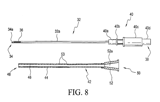
[0028] Fig. 8 is an illustration showing an echogenic needle and an echogenic
cannula that
are parts of a needle assembly embodiment;
[0029] Fig. 9 is an illustration of the needle assembly embodiment where the
echogenic
needle and the echogenic cannula of fig. 8 are mounted to each other with the
tip of the
echogenic needle exposed from the distal end of the cannula; and
[0030] Figs. 10A and 10B are enlarged cross sectional views of different
sections of the
needle assembly embodiment of Fig. 9.
Detailed Description of the Invention
[0031] An exemplar embodiment of the needle used, for example for peripheral
nerve
block procedures, is shown in Fig. 1. As shown, needle 2 has a shaft 4 that
extends along
a longitudinal axis 6 having a distal or patient end 8 including a sharp bevel
tip and a
proximal end 10. For ease of illustration, the proximal end 10 of needle 2 has
been
truncated in Fig. 1. The section of the needle that is adjacent to patient end
8 is
CA 03001647 2018-04-11
WO 2017/089876 PCT/1B2016/001503
8
designated distal portion 12, although it should be appreciated that the
demarcation of
distal portion 12 as shown in Fig. 1 is for discussion only. Beyond distal
portion 12 there
are a number of markings 14 along the shaft of the needle to show the length,
or the depth
of the needle, as the needle is inserted into the subject, or patient. There
are two sections
12a and 12b at the distal portion that are better illustrated in the enlarged
distal portion
view of the needle in Fig. 2.
[0032] As shown in Fig. 2, sections 12a and 12b each are formed with
crisscrossing spiral
grooves that are also shown in Fig. 5B. With reference to Figs. 2, 5A and 5B,
one spiral
groove 16a is formed on the outer circumferential wall of needle shaft 4 in
one direction,
for example a clockwise direction, while an other spiral groove 16b
crisscrosses groove
16a in an other direction, for example a counter-clockwise direction. For ease
of reference,
the respective grooves 16a and 16b spiral about the outer circumferential wall
of needle
shaft 4 may be referenced with respect to the sharp bevel tip at patient end 8
of the needle.
Thus, for the discussion herein below, groove 16a may be considered to be a
spiral groove
that is formed on the outer circumferential wall of needle shaft 4 in a
clockwise direction
relative to needle tip 8, while groove 16b spirally wounds about the outer
circumferential
wall of needle shaft 4 in a counter-clockwise direction relative to needle tip
8. The two
spiral grooves crisscross each other in the manner as shown in Figs. 2, 5A and
5B.
[0033] As best shown in Fig. 2, there is a non-groove section 18 that
separates grooved
sections 12a and 12b at the distal portion of the exemplar embodiment needle.
Although
two grooved sections are shown in Figs. 1 and 2, it should be appreciated that
a plurality
of more than two groove sections may also be formed along the needle away from
its
patient end.
[0034] The configuration of the groove of the needle is illustrated in Figs. 3-
4 and 6A-6C.
Fig. 3 shows a cross-section of the needle, for example a portion of section
12a, with
directional needle 20 referencing the proximal end of the needle and
directional needle 22
referencing the patient end of the needle. As shown in Fig. 3, a number of
cross-sections
CA 03001647 2018-04-11
WO 2017/089876 PCT/1B2016/001503
9
of a groove 16 are shown to have formed on the outer circumferential wall of
needle shaft
4, which has a passage 4a extending therethrough along longitudinal axis 6.
For the
exemplar illustration of Fig. 3, a 0 angle is shown to be formed between the
two walls of
the groove, represented by lines 16w1 and 16w2 in Fig. 4. As the walls are
orthogonal to
each other, the 0 angle formed between the two walls is assumed to be
approximately
90 . Thus, were the spiral groove at the outer surface of the circumferential
wall of shaft
4 to be formed to have its walls orthogonal to the longitudinal axis of the
needle so that the
walls have the same length, then each of the walls 16w1 and 16w2 would extend
at
approximately 45 relative to a plane along the longitudinal axis of shaft 4.
This is shown
by the dotted lines in Figs. 4 and 6C, and may be referred to as the neutral
position of the
groove. For discussion purpose, the length of the walls may also be referred
to as the
height or distance between the top and bottom, i.e., the valley and apex, of
the groove.
[0035] The inventors have found that, in use, a clinician usually positions a
needle at an
angle that facilitates the insertion of the needle into the subject. Thus,
were the groove
"tilted" at a given angle a toward the proximal end of the needle, an improved
reflection of
an ultrasound wave directed by an ultrasound transducer towards the needle may
be
obtained. By empirical studies, it was found that the a angle may range from
approximately 5 to 25 , and preferably at 10 relative to the neutral
position. Thus,
instead of 45 for each of the walls of the V-shaped groove, the "tilted"
groove would have
its walls, as designed by lines 16w1' and 16w2', shifted together such that
wall 16w1' is at
a 13 angle relative to the outside walls 4a of the needle shaft. Walls 16w1'
and 16w2'
remain orthogonal to each other when at the "tilted" position. For the
exemplar
embodiment where a = 10 , 13 would be 35 . The depth of the groove may vary
anywhere
from 0.006 inch to 0.025 inch (0.1524 mm to 0.635 mm). It was further found
that the pitch
between grooves, as designated by reference number 24 in Fig. 6B, could be
reduced to
between 0.010 inch and 0.050 inch (0.254 mm to 1.27 mm), and preferably to
approximately 0.020 inch (0.508 mm) to improve the pitch density of the needle
and
thereby its echogeneity. As should be appreciated, the pitch and the depth of
the groove
as described above are not definitive for all needles but are instead meant to
be utilized
CA 03001647 2018-04-11
WO 2017/089876 PCT/1B2016/001503
for needles that have conventionally dimensioned walls, for example a needle
having a
gauge anywhere between 16 to 24.
[0036] The configuration of the exemplar embodiment of the echogenic needle of
the
instant invention is further shown in Figs. 6A - 6C where a portion 12a1 of
groove section
12a is enlarged in Fig. 6B to show an enlarged cross-sectional view of a
number of V-
shaped cross sections of the groove tilted toward the proximal end of the
needle as
described above. Fig. 6C shows the neutral position (in dotted lines) and the
tilt angle
position, or simply the tilted position (in solid lines) of the walls of an
exemplar cross
section of the V-shaped groove.
[0037] Fig. 7 is an illustration showing the difference in the reflection of
the ultrasound
wave emitted by an ultrasound transducer toward the exemplar embodiment
needle. For
ease of discussion, the exemplar needle of Fig. 7 is assumed to have only one
spiral
groove. As shown, the transmitter of ultrasound transducer probe 26 emits an
ultrasound
wave 28 toward needle shaft 4, represented by line 4a, which is presumably
being inserted
into a subject. Thus, needle 4 is at an insertion angle A with reference to a
plane 30 that
is assumed to be in parallel to the plane at the output surface of transducer
26. From
empirical studies, it was found that A in most instances is between 500 and 60
, and
preferably at approximately 55 . Thus, were the spiral grooves formed in its
neutral
position, the ultrasound wave 28 would be reflected as a return ultrasound
wave,
represented by lines 28a, in a direction that angles away, or offsets from
ultrasound
transducer 26. However, it was determined that with the V-shaped groove at the
tilt angle
a, the ultrasound wave 28 is reflected as ultrasound wave 28a' in a
substantially reverse
direction (approximately at 180 ) back to transducer 26, presumably to its
receiver. Thus,
an improved reflection view of the exemplar embodiment needle may be gleaned
under
ultrasound or radiographic imaging by tilting the groove at an angle a.
[0038] As discussed above, with the exemplar illustration of Fig. 7, it was
determined that
were the spiral grooves formed at their respective neutral positions, the
ultrasound wave
CA 03001647 2018-04-11
WO 2017/089876 PCT/1B2016/001503
11
28 would be reflected as a return ultrasound wave, represented by lines 28a,
in a direction
that angles away, or offset from ultrasound transducer 26. However, from
additional
empirical studies, it has been determined that were the crisscrossing spiral
grooves formed
on a needle with their corresponding respective walls orthogonal to the
longitudinal axis of
the needle but with the pitch density for each of the spiral grooves increased
more than as
discussed above, an improved and acceptable reflective image of the echogenic
portion(s)
of the needle under ultrasound imaging can nonetheless be obtained. In other
words,
spiral grooves formed in the neutral position relative to the longitudinal
axis of the needle
with a predetermined increased pitch density would provide an improved
reflective image
of the echogenic portion(s) of the needle, similar to the needle embodiment
with the tilted
grooves as discussed above.
[0039] The neutral positioned spiral grooves are represented by the dotted
wall lines 16w1
and 16w2 in Figs. 4 and 6C. Due to those spiral grooves being formed at the
neutral
position and orthogonal to each other, their walls, for example 16w1 and 16w2,
have
substantially the same length or height. The pitch, or the groove width,
between the walls
of each of the grooves, designated by 24 in Fig. 6, was determined to have a
range of
approximately 0.001 inch to 0.003 inch (0.025 mm to 0.075 mm), and preferably
at
approximately 0.0021 inch (0.053 mm). The desired groove depth was determined
to be
in the range of approximately 0.0006 inch to 0.0010 inch (0.015 mm to 0.025
mm). As
should be appreciated, the depth and pitch of the groove may be interrelated,
as the
change in the value of one may cause a change in the value of the other.
[0040] With the combination of clockwise and counter-clockwise spiral wound
grooves,
and with each of the grooves having a preferable pitch of approximately 0.02
inch (0.508
mm) for the tilted echogenic needle embodiment, an echogenic needle with
improved
echogeneity results. So, too, an improved echogenic needle adapted to provide
improved
echogeneity results may be achieved with non-tilted crisscrossing clockwise
and counter-
clockwise spiral grooves each having an increased pitch density or groove
width having a
range of 0.001 inch to 0.003 inch (0.025 mm to 0.075 mm), and preferably of
CA 03001647 2018-04-11
WO 2017/089876 PCT/1B2016/001503
12
approximately 0.0021 inch (0.053 mm). It should be appreciated that instead of
a V-
shaped groove, each of the grooves may be U-shaped or trapezoidal-shaped, so
long as
the walls of the groove are made to be substantially orthogonal to each other.
Furthermore, one of the crisscrossing grooves may have a V-shape while the
other groove
may have a U-shape or some other shape including trapezoidal that clearly
defines the
orthogonal walls of the groove.
[0041] Although not disclosed above, it should be appreciated that the
proximal end of the
needle may be fixedly bonded or connected to a needle hub, so that the needle
may be
fluidly coupled to a medicament or fluid store, such as a syringe or a pump,
to infuse
medicament or fluid to the patient once the needle has been inserted into and
appropriately
positioned within the patient. The respective connectors of the needle hub and
the fluid
store may be configured to have complementary features or configurations that
allow only
those connectors to be coupled to each other, i.e., each of those connectors
is not
connectable to a counterpart conventional luer connector. Moreover, before
use, the
needle may be protected by a sleeve to prevent contamination and for shipping
purposes.
To prevent coring of the needle, a stylet may be concentrically fitted into
the through
passage of the needle when the needle is inserted into the patient, and
removed
thereafter.
[0042] The forming of the spiral grooves onto the outer, circumferential wall
of the needle
shaft may be accomplished in substantially the same manner as is done for the
above-
noted Wallace Amniocentesis Needles. In the alternative, the spiral grooves
may be
formed substantially in accordance with the disclosure of JP2000051219, which
was
assigned to the assignee of the instant invention. In brief, the '219
publication discloses
an edge of a spinning wheel is used to form a groove on a catheter that
rotatably moves
along a longitudinal direction relative to the wheel.
[0043] With reference to Fig. 8, an inventive echogenic needle assembly
embodiment is
shown to include an echogenic needle 32 that has an echogenic feature as
described
CA 03001647 2018-04-11
WO 2017/089876 PCT/1B2016/001503
13
above in combination with an echogenic cannula 40 to form an echogenic needle
assembly. For this embodiment, it should be appreciated that cannula 40 may
also be a
solid or hollow shaft or a catheter. As shown, needle 32 has a patient end 34
that has a
bevel sharp tip 34a. At tip 34a or approximate thereto is an echogenic feature
36 such as
the spiral groove as described above. For ease of discussion, echogenic
feature 36 is
shown in Fig. 8 to be one spiral groove. However, it should be appreciated
that echogenic
feature 36 may include the crisscrossing dual grooves as described above.
Needle 32
further has a shaft body 32a that extends longitudinally from patient end 34
to a non-
patient end 38 where there is a needle hub 40. As shown, needle hub 40 has a
first
portion 40a to which a non-patient end portion of shaft 32a is bondedly
attached. There
is also a circumferential flange portion 40b rising from first portion 40a to
enable hub 40a
to frictionally mate with the hub of the cannula as will be described below.
Needle hub 40
further has a main body portion 40c and an end connector 40d that enables
needle 32 to
be connected to a fluid store such as a syringe or a pump so that fluid or
medicament can
be conveyed through needle 32, longitudinally along shaft 32 between non-
patient end 38
and the patient end 34 through an aperture that extends longitudinally along
needle shaft
32a and hub 40, as is well known.
[0044] Also shown in Fig. 8 is an echogenic cannula 42. As shown, cannula 42
may
comprise a longitudinal shaft, catheter or sleeve defining an axial bore 46
that has a distal
end 48 and a proximal end 50. At proximal end 50 there is a cannula hub 52.
Hub 52, as
shown in the cross-sectional view of Fig. 8, is cone shaped such that its
internal passage
52a is adapted to accept portion 40a of needle hub 40. Needle hub 40 and
cannula hub
52 are frictionally held to each other by the frictional engagement between
the inner wall
of passage 52a of cannula hub 52 and the circumferential flange 40b of needle
hub 40.
The outer circumferential wall of cannula 44 is smooth while a plurality of
bubbles 53 are
formed in the body of cannula 44. The formations of the bubbles in the cannula
may be
achieved in the same manner as disclosed in US 8,092,390 and the above
discussed US
8,398,596, both assigned to the assignee of the instant application. The
disclosures of the
'390 and '596 patents are incorporated by reference herein.
CA 03001647 2018-04-11
WO 2017/089876 PCT/1B2016/001503
14
[0045] As noted above, the shaft of cannula 44 may be made of metal including
sintered
metal having gas interstices so that bubbles may be formed along the cannula,
or may be
made from a flexible plastic material such as PEBA, nylon, PVC, polyethylene,
polyprophelene, polyester or polyurethane to which a foaming agent is added to
form gas
interstices in the form of gas bubbles 53 along its entire length, or at least
one portion
thereof. As described in the above incorporated by reference '596 patent, the
density of
the gas bubbles are selected to ensure that the cannula shaft 44 is echogenic.
Typically
the gas bubbles 53 may have a size in the range of 0.1p to 300p, preferably
having a size
in the range of 1p to 50p and most preferably having a range of 5p to 10p.
Other ways of
forming the gas bubbles in the cannula are described in the above incorporated
by
reference patents.
[0046] With reference to Fig. 9, needle 32 and cannula 42 are shown to have
fully mounted
to each other, with needle hub 40 and cannula hub 52 frictionally coupled to
each other
and shaft 32a of needle 32 extending through axial bore 46 of cannula 44. When
thus fully
mounted, the tip portion of needle shaft 32a extends beyond the distal end 48
of cannula
44. Fig. 10A is an enlarged cross sectional view of circled section A of Fig.
9
exaggeratedly showing the bevel sharp tip of shaft 32a and the wall of cannula
42. As
shown, the echogenic spiral groove is proximate or close to the sharp bevel
tip 34a so that,
under ultrasound observation, the tip of needle 32 may be readily observed. A
portion of
cannula 42 encircling a portion of shaft 32a of the needle, designated by
circled section B
in Fig. 9, is shown in the enlarged cross sectional view of Fig. 10B. The
plurality of gas
bubbles 53 in the body of cannula 44 provides echogeneity for cannula 44.
[0047] For the needle assembly embodiment shown in Fig. 9, assume cannula 44
is a
plastic sheath or catheter having an axial bore through which needle shaft 32a
of needle
32 extends. In operation, under ultrasound observation, for example using the
ultrasound
transducer discussed above, when the needle assembly is inserted into the body
of the
patient, the sharp tip of the needle will make the initial incision into the
body, for example
CA 03001647 2018-04-11
WO 2017/089876 PCT/1B2016/001503
a blood vessel of the subject. This is confirmed by the ultrasound reflection
of the
echogenic tip portion of the needle. As the needle assembly is further
inserted into the
body, for example the pierced blood vessel, given that the outer
circumferential surface of
the cannula is smooth, there is less trauma to the patient. The placement of
the echogenic
cannula 44 inside the body, for example in the blood vessel, along with the
tip portion of
the needle can then be confirmed under ultrasound observation. Thereafter,
needle 32 is
removed leaving in place the echogenic cannula 44. The placement of the
echogenic
cannula minus the echogenic needle could further be confirmed by ultrasound
observation.
After confirmation, were the cannula (or catheter) to be used to input a
medicament or fluid
into the patient, a fluid store may have its connector coupled to end
connector 40d of the
cannula hub, so that a through fluid passage is established between the fluid
store and the
distal end of the cannula. To withdraw fluid or blood from the patient, a
syringe may have
its luer connector end connected to end connector 40d of the cannula hub.
[0048] In the case that the cannula is used to guide a guidewire into the body
of the
patient, after confirmation of the positioning of the cannula, the guidewire
is threaded
through the axial bore of cannula 44. Thereafter, cannula 44 is removed and
the additional
procedure of inserting a permanent catheter along the guidewire into the
patient, as for
example a central venous catheter (CVC) for a CVC procedure to infuse drugs
for
chemotherapy or nutrition, or medicament into an already implanted port in the
patient, can
be further carried out. The inventive echogenic needle assembly possibly may
also be
used for percutaneous tracheotomy, where the needle assembly may be used to
effect
the incision opening at the trachea of a patient under ultrasound observation.
After the
removal of the needle, a guidewire may be threaded through the cannula into
the trachea
of the patient. Thereafter, the cannula is removed, the guidewire may be used
to guide a
dilator to widen the incision opening at the trachea and the subsequent
placement of a
tracheostomy tube into the trachea of the patient after the removal of the
dilator.
[0049] Inasmuch as the present invention is subject to many variations,
modifications and
changes in detail, it is intended that the matter described throughout this
specification and
CA 03001647 2018-04-11
WO 2017/089876 PCT/1B2016/001503
16
shown in the accompanying drawings be interpreted as illustrative only, and
not in a limiting
sense. Accordingly, it is intended that the invention be limited only by the
sprit and scope
of the hereto appended claims.