Note: Descriptions are shown in the official language in which they were submitted.
CA 03001878 2018-04-12
WO 2017/075124 PCT/US2016/058975
ANTIBODY NEUTRALIZING HUMAN RESPIRATORY SYNCYTIAL VIRUS
FIELD OF THE INVENTION
The present invention relates to human monoclonal antibodies which have high
anti-RSV neutralizing titers, as well as the use of these antibodies as a
passive immunotherapy
agent in infants and the elderly.
BACKGROUND OF THE INVENTION
Paramyxoviruses are enveloped negative-strand RNA viruses that are significant
human and animal pathogens. Human Respiratory Syncytial Virus (hRSV, RSV)
belongs to the
family Paramyxoviridae, subfamily Pneumovirinae . Two subtypes, type A and
type B, have
been identified and are a major cause of severe and sometimes even fatal
respiratory infections in
children less than 6 months of age. Adults with underlying diseases, such as
COPD, asthma,
cancer, immunocompromised status, including I-IIV or post transplantation, are
also at risk of
developing severe RSV infection. 15% of annual hospitalizations in adults over
50 years due to
acute respiratory infection are caused by RSV. In the United States, RSV
causes more than
100,000 hospitalizations annually, and it is estimated to cause about 160,000
deaths globally
each year. Currently there is no vaccine for RSV, and a trial with a formalin-
inactivated virus
was associated with increased disease severity in infants upon infection with
RSV. Other family
members including Human Metapneumo Virus (hMPV) and Human Parainfluenza Virus
(hPIV)
are also responsible for acute respiratory illness similar to hRSV.
The hRSV genome is a single-stranded negative-sense RNA molecule of
approximately 15 kb that encodes 11 proteins. Two of these proteins are the
main surface
glycoproteins of the virion. These are (i) the attachment (G) protein, which
mediates virus
binding to cells, and (ii) the fusion (F) protein, which promotes both fusion
of the viral and cell
membranes at the initial stages of the infectious cycle and fusion of the
membrane of infected
cells with those of adjacent cells to form characteristic syncytia. The
attachment protein G binds
cellular surface receptors and interacts with F. This interaction triggers a
conformational change
in F to induce membrane fusion, thereby releasing the viral ribonucleoprotein
complex into the
host cell cytoplasm.
Monoclonal antibodies against the F protein or the G protein have been shown
to
have neutralizing effect in vitro and prophylactic effects in vivo. See, e.g.,
Beeler and Coelingh
1
CA 03001878 2018-04-12
WO 2017/075124 PCMJS2016/058975
1989, J. Virol. 63:2941-50; Garcia-Barreno etal., 1989, J. Virol. 63:925-32;
Taylor etal., 1984,
Immunology 52: 137-142; Walsh etal., 1984, Infection and Immunity 43:756-758;
and U.S. Pat.
Nos. 5,842,307 and 6,818,216. Neutralizing epitopes on the F glycoprotein were
originally
mapped by identifying amino acids that were altered in antibody escape
variants and by
assessing antibody binding to RSV F-derived peptides. These studies
demonstrated neutralizing
antibodies are often targeted to two distinct linear epitopes. See Graham
etal., 2015, Curr Opin
Immunol 35:30-38 for a review of the antigenic sites for the pre-fusion and
post-fusion F forms.
Antigenic site II (also called site A) includes residues 255 to 275 and is the
target of palivizumab
(SYNAGIS , AstraZeneca). This epitope was predicted to be conformationally
dependent, and
the structure of a more potent derivative of palivizumab in complex with this
epitope revealed
that the linear epitope adopts a helix-loop-helix conformation. Antigenic site
IV (also called site
C) includes residues 422 to 438 and is the target of antibodies MAbl9 and
101F. This epitope is
C-terminal to the cysteine-rich region and is part of domain II, which in
homologous
paramyxovirus F glycoproteins remains structurally unchanged between pre- and
post-fusion
conformations. 5C4, AM22 and D25 delineate an epitope designated as site 0
which is only
present on the pre-fusion F protein and were 50 times as potent as
palivizumab. See McLellan et
al., 2013, Science 340:1113-1117; International Patent Application No. WO
2008/147196 and
U.S. Pat. No. 8,568,726. Other hRSV antibodies are described in International
Patent
Application Nos W094/06448 and W092/04381 and U.S. Pat. No. 8,221,759.
An RSV vaccine for active immunization, if available, could not be utilized
for
the treatment of newborn babies with immature immune systems or patients who
are
immunosuppressed. In patients where prophylactic passive immunotherapy is
required, as a
result of a more chronic form of disease, current therapy is mediated via
periodic intravenous
inoculation of human IgG prepared from pooled plasma. This type of therapy,
due to the low
titers of neutralizing anti-RSV antibodies, involves a large quantity of
globulin (e.g., 0.75 gm per
kg) and consequently requires administration intravenously, in a clinic or
hospital, over a lengthy
period (2 to 4 hours), on a monthly basis during the high risk months (fall,
winter and early
spring).
SUMMARY OF THE INVENTION
The invention provides anti-RSV F-protein antibodies and antigen binding
fragments thereof comprising the structural and functional features specified
below.
2
In one embodiment, the invention provides an antibody or antigen binding
fragment thereof that binds to human RSV F-protein, comprising: a heavy chain
variable region
CDR3 comprising the amino acid sequence of SEQ ID NO: 3. In certain
embodiments, the
heavy chain or heavy chain variable region does not comprise the amino acid
sequence of SEQ
ID NO: 9. In one embodiment, the antibody or antigen binding fragment thereof
optionally has
at least one of the following characteristics: (i) binds to human RSV pre-
fusion F protein with a
Kd value of about 1 x 10'9M to about 1 x 10'12 M as determined by surface
plasmon resonance
TM
(e.g., BIACORE) or a similar technique (e.g. KinExa or OCTET); or (ii) binds
to human RSV
post-fusion F protein with a Kd value of about 1 x 10⁻⁶ M to about 1 x 10⁴ M
as determined by
surface plasmon resonance (e.g., BIACORE) or a similar technique (e.g. KinExa
or OCTET). In
certain embodiments, the heavy chain comprises, consists essentially of, or
consists of, the amino
acid sequence of SEQ ID NO: 23.
In another embodiment, the invention provides an antibody or antigen binding
fragment thereof that binds to human RSV F-protein, comprising: a light chain
variable region
CDR3 comprising the amino acid sequence of SEQ ID NO: 6. In certain
embodiments, the light
chain or light chain variable region does not comprise the amino acid sequence
of SEQ ID NO:
8. In certain embodiments, the light chain is not associated with a heavy
chain comprising the
amino acid sequence of SEQ ID NO: 9. In certain embodiments, the light chain
is associated
with a heavy chain comprising the amino acid sequence of SEQ ID NO: 7. In one
embodiment,
the antibody or antigen binding fragment thereof optionally has at least one
of the following
characteristics: (i) binds to human RSV pre-fusion F protein with a Kd value
of about 1 x 10⁻⁹ 1,4
to about 1 x 10²M as determined by surface plasmon resonance (e.g., BIACORE)
or a similar
technique (e.g. KinExa or OCTET); or (ii) binds to human RSV post-fusion F
protein with a Kd
value of about 1 x 10⁴ M to about 1 x 1041 M as determined by surface plasmon
resonance (e.g.,
BIACORE) or a similar technique (e.g. KinExa or OCTET). In certain
embodiments, the light
chain comprises, consists essentially of, or consists of, the amino acid
sequence of SEQ ID NO:
25.
In another embodiment, the invention provides an antibody or antigen binding
fragment thereof that binds to human RSV F-protein comprising: (i) a heavy
chain variable
region CDRI comprising the amino acid sequence of SEQ ID NO: 1; (ii) a heavy
chain variable
region CDR2 comprising the amino acid sequence of SEQ ID NO: 2; and (iii) a
heavy chain
3
CA 3001878 2019-08-01
CA 03001878 2018-04-12
WO 2017/075124 PCMJS2016/058975
variable region CDR3 comprising the amino acid sequence of SEQ ID NO. 3. In
certain
embodiments, the heavy chain or heavy chain variable region does not comprise
the amino acid
sequence of SEQ ID NO: 9. In one embodiment, the antibody or antigen binding
fragment
thereof optionally has at least one of the following characteristics: (i)
binds to human RSV pre-
fusion F protein with a Kd value of about 1 x 10⁻⁹M to about 1 x 10-12M as
determined by
surface plasmon resonance (e.g., BIACORE) or a similar technique (e.g. KinExa
or OCTET); or
(ii) binds to human RSV post-fusion F protein with a Kd value of about 1 x 10-
9M to about 1 x
10-11M as determined by surface plasmon resonance (e.g., BIACORE) or a similar
technique
(e.g. KinExa or OCTET). In certain embodiments, the heavy chain comprises,
consists
essentially of, or consists of, the amino acid sequence of SEQ ID NO: 23.
In one embodiment, the antibody or antigen binding fragment thereof comprises:
(i) a light chain variable region CDR1 comprising the amino acid sequence of
SEQ ID NO: 4;
(ii) a light chain variable region CDR2 comprising the amino acid sequence of
SEQ ID NO: 5;
and (iii) a light chain variable region CDR3 comprising the amino acid
sequence of SEQ ID NO:
6. In certain embodiments, the light chain or light chain variable region does
not comprise the
amino acid sequence of SEQ ID NO: 8. In certain embodiments, the light chain
is not associated
with a heavy chain comprising the amino acid sequence of SEQ ID NO: 9. In
certain
embodiments, the light chain is associated with a heavy chain comprising the
amino acid
sequence of SEQ ID NO: 7. In one embodiment, the antibody or antibody
fragement thereof
optionally has at least one of the following characteristics: (i) binds to
human RSV pre-fusion F
protein with a Kd value of about 1 x 10⁻⁹ M to about 1 x 1042 M as determined
by surface
plasmon resonance (e.g., BIACORE) or a similar technique (e.g. KinExa or
OCTET); or (ii)
binds to human RSV post-fusion F protein with a Kd value of about 1 x 10⁻⁹M to
about 1 x 10-11
M as determined by surface plasmon resonance (e.g., BIACORE) or a similar
technique (e.g.
KinExa or OCTET). In certain embodiments, the light chain comprises, consists
essentially of,
or consists of, the amino acid sequence of SEQ ID NO: 25.
In one embodiment, the antibody or antigen binding fragment thereof comprises:
(i) a heavy chain variable region CDR1 comprising the amino acid sequence of
SEQ ID NO: 1;
(ii) a heavy chain variable region CDR2 comprising the amino acid sequence of
SEQ ID NO: 2;
(iii) a heavy chain variable region CDR3 comprising the amino acid sequence of
SEQ ID NO: 3;
(iv) a light chain variable region CDR1 comprising the amino acid sequence of
SEQ ID NO: 4;
4
CA 03001878 2018-04-12
WO 2017/075124 PCMJS2016/058975
(v) a light chain variable region CDR2 comprising the amino acid sequence of
SEQ ID NO: 5;
and (vi) a light chain variable region CDR3 comprising the amino acid sequence
of SEQ ID NO:
6. In certain embodiments, the heavy chain or heavy chain variable region does
not comprise the
amino acid sequence of SEQ ID NO: 9. In one embodiment, the antibody or
antigen binding
fragment thererof optionally has at least one of the following
characteristics: (i) binds to human
RSV pre-fusion F protein with a Kd value of about 1 x 10⁻⁹M to about 1 x 1042
M as determined
by surface plasmon resonance (e.g., BIACORE) or a similar technique (e.g.
KinExa or OCTET);
or (ii) binds to human RSV post-fusion F protein with a Kd value of about 1 x
10-9M to about 1
x 1041M as determined by surface plasmon resonance (e.g., BIACORE) or a
similar technique
(e.g. KinExa or OCTET). In certain embodiments, the heavy chain comprises,
consists
essentially of, or consists of, the amino acid sequence of SEQ ID NO: 23 and
the light chain
comprises, consists essentially of, or consists of, the amino acid sequence of
SEQ ID NO: 25.
In another embodiment, the invention provides an antibody or antigen binding
fragment that binds to human RSV F-protein comprising: (i) a heavy chain
variable region CDR1
comprising the amino acid sequence of SEQ ID NO: 1; (ii) a heavy chain
variable region CDR2
comprising the amino acid sequence of SEQ ID NO: 2; (iii) a heavy chain
variable region CDR3
comprising the amino acid sequence of SEQ ID NO: 3; (iv) a light chain
variable region CDR1
comprising the amino acid sequence of SEQ ID NO: 4; (v) a light chain variable
region CDR2
comprising the amino acid sequence of SEQ ID NO: 5; and (vi) a light chain
variable region
CDR3 comprising the amino acid sequence of SEQ ID NO: 6; wherein the antibody
or antigen
binding fragment thereof comprises a heavy chain variable region comprising at
least 90%, 95%,
96%, 97%, 98% or 99% identity to a heavy chain variable region consisting of
SEQ ID NO: 7
and a light chain variable region comprising at least 90%, 95%, 96%, 97%, 98%
or 99% identity
to a light chain variable region consisting of SEQ ID NO: 8. In certain
embodiments, the heavy
chain or heavy chain variable region does not comprise the amino acid sequence
of SEQ ID NO:
9. In these aforementioned embodiments, the sequence variations occur in the
framework
regions. In one embodiment, the antibody or antigen binding fragment thereof
optionally has at
least one of the following characteristics: (i) binds to human RSV pre-fusion
F protein with a Kd
value of about 1 x 10⁻⁹M to about 1 x 1042 M as determined by surface plasmon
resonance (e.g.,
BIACORE) or a similar technique (e.g. KinExa or OCTET); or (ii) binds to human
RSV post-
fusion F protein with a Kd value of about 1 x 10⁻⁹M to about 1 x 10-11M as
determined by
5
CA 03001878 2018-04-12
WO 2017/075124 PCMJS2016/058975
surface plasmon resonance (e.g., BIACORE) or a similar technique (e.g. KinExa
or OCTET). In
certain embodiments, the heavy chain comprises, consists essentially of, or
consists of, the amino
acid sequence of SEQ ID NO: 23 and the light chain comprises, consists
essentially of, or
consists of, the amino acid sequence of SEQ ID NO: 25.
In another embodiment, the invention also provides an antibody or antigen
binding fragment thereof that binds to human RSV comprising: (i) a heavy chain
variable region
CDR1 comprising the amino acid sequence of SEQ ID NO: 1; (ii) a heavy chain
variable region
CDR2 comprising the amino acid sequence of SEQ ID NO: 2; (iii) a heavy chain
variable region
CDR3 comprising the amino acid sequence of SEQ ID NO: 3; (iv) a light chain
variable region
CDR1 comprising the amino acid sequence of SEQ ID NO: 4; (v) a light chain
variable region
CDR2 comprising the amino acid sequence of SEQ ID NO: 5; and (vi) a light
chain variable
region CDR3 comprising the amino acid sequence of SEQ ID NO: 6. In certain
embodiments,
the heavy chain or heavy chain variable region does not comprise the amino
acid sequence of
SEQ ID NO: 9. In one embodiment, the antibody or antigen binding fragment
thereof comprises
1, 2 or 3 amino acid substitutions in the heavy chain CDRs (SEQ ID NOs: 1-3)
and/or in the light
chain CDRs (SEQ ID NOs: 4-6). The VH sequence of SEQ ID NO: 7 has the CDRs of
SEQ ID
NOs:1-3; and the VL sequence of SEQ ID NO: 8 has the CDRs of SEQ ID NOs: 4-6.
In one
embodiment, the antibody or antigen binding fragment thereof optionally has at
least one of the
following characteristics: (i) binds to human RSV pre-fusion F protein with a
Kd value of about
1 x 10⁻⁹M to about 1 x 10-12M as determined by surface plasmon resonance
(e.g., BIACORE) or
a similar technique (e.g. KinExa or OCTET), or (ii) binds to human RSV post-
fusion F protein
with a Kd value of about 1 x 10⁻⁹M to about 1 x 1041 M as determined by
surface plasmon
resonance (e.g., BIACORE) or a similar technique (e.g. KinExa or OCTET). In
certain
embodiments, the heavy chain comprises, consists essentially of, or consists
of, the amino acid
sequence of SEQ ID NO: 23 and the light chain comprises, consists essentially
of, or consists of,
the amino acid sequence of SEQ ID NO: 25.
In one embodiment, the invention provides an antibody or antigen binding
fragment thereof, comprising: a variable heavy chain comprising the amino acid
sequence of
SEQ ID NO: 7 and/or a variable light chain comprising the amino acid sequence
of SEQ ID NO:
.. 8, wherein the antibody or antigen binding fragment thereof binds to human
RSV F protein. In
another embodiment, the antibody or antigen binding fragment thereof comprises
a heavy chain
6
CA 03001878 2018-04-12
WO 2017/075124 PCMJS2016/058975
comprising, consisting essentially of, or consisting of, the amino acid
sequence of SEQ ID NO:
23 and a light chain comprising, consisting essentially of, or consisting of,
the amino acid
sequence of SEQ ID NO: 25, wherein the antibody or antigen binding fragment
thereof binds to
human RSV F protein. In one embodiment, the antibody or antigen binding
fragment thereof
optionally has at least one of the following characteristics: (i) binds to
human RSV pre-fusion F
protein with a Kd value of about 1 x 10⁻⁹ M to about 1 x 1042 M as determined
by surface
plasmon resonance (e.g., BIACORE) or a similar technique (e.g. KinExa or
OCTET); or (ii)
binds to human RSV post-fusion F protein with a Kd value of about 1 x 10⁻⁹M to
about 1 x 10-11
M as determined by surface plasmon resonance (e.g., BIACORE) or a similar
technique (e.g.
KinExa or OCTET).
In another embodiment, the invention provides an antibody or antigen binding
fragment thereof that binds to the same epitope of human RSV F protein as an
antibody
comprising the heavy chain of SEQ ID NO: 23 and the light chain of SEQ ID NO:
25, wherein
the antibody or antigen binding fragment thereof has at least one of the
following characteristics:
(i) binds to human RSV pre-fusion F protein with a Kd value of about 1 x 10⁻⁹M
to about 1 x 10-
12
M as determined by surface plasmon resonance (e.g., BIACORE) or a similar
technique (e.g.
KinExa or OCTET); or (ii) binds to human RSV post-fusion F protein with a Kd
value of about 1
x 10-9 M to about 1 x 10-11M as determined by surface plasmon resonance (e.g.,
BIACORE) or a
similar technique (e.g. KinExa or OCTET). In one embodiment, the antibody
comprises at least
80%, 85%, 90%, 95%, 96%, 97%, 98% or 99% sequence identity with the heavy
chain variable
region and/or the light chain variable region of SEQ ID NOs: 7 and 8,
respectively. In another
embodiment, the antibody or antigen binding fragment thereof comprises 1, 2,
3, 4, 5, 6, 7, 8, 9,
10, 11, 12, 13, 14, 15, 16, 17, 18, 19, 20, 21, 22, 23, 24, 25, 26, 27, 28, 29
or 30 amino acid
substitutions in the heavy chain variable region of SEQ ID NO: 7 and/or the
light chain variable
region of SEQ ID NO: 8. In certain embodiments, the heavy chain or heavy chain
variable
region does not comprise the amino acid sequence of SEQ ID NO: 9.
In another embodiment, the invention provides an antibody or antigen binding
fragment thereof that cross-blocks the binding of (or competes with) an
antibody comprising the
heavy chain of SEQ ID NO: 23 and the light chain of SEQ ID NO: 25 to human
RSV, wherein
the antibody or antigen binding fragment thereof has at least one of the
following characteristics:
(i) binds to human RSV pre-fusion F protein with a Kd value of about 1 x 10⁻⁹M
to about 1 x
7
CA 03001878 2018-04-12
WO 2017/075124 PCMJS2016/058975
10-12 M as determined by surface plasmon resonance (e.g., BIACORE) or a
similar technique
(e.g. KinExa or OCTET); or (ii) binds to human RSV post-fusion F protein with
a Kd value of
about 1 x 10⁻⁹M to about 1 x 10-11 M as determined by surface plasmon
resonance (e.g.,
BIACORE) or a similar technique (e.g. KinExa or OCTET). In one embodiment, the
antibody or
antigen binding fragment thereof comprises at least 80%, 85%, 90%, 95%, 96%,
97%, 98% or
99% sequence identity with the heavy chain variable region of SEQ ID NO: 7 or
the light chain
variable region of SEQ ID NO: 8. In another embodiment, the antibody or
antigen binding
fragment thereof comprises 1, 2, 3, 4, 5, 6, 7, 8, 9, 10, 11, 12, 13, 14, 15,
16, 17, 18, 19, 20, 21,
22, 23, 24, 25, 26, 27, 28, 29 or 30 amino acid substitutions in the heavy
chain variable region of
SEQ ID NO: 7 or the light chain variable region of SEQ ID NO: 8. In another
embodiment, the
antibody or antigen binding fragment thereof comprises 1, 2 or 3 amino acid
substitutions in the
heavy chain CDRs (SEQ ID NOs: 1-3) and/or in the light chain CDRs (SEQ ID NOs:
4-6). In
certain embodiments, the heavy chain or heavy chain variable region does not
comprise the
amino acid sequence of SEQ ID NO: 9.
In one embodiment, the invention relates to an isolated antibody or antigen
binding fragment that binds to human RSV F protein comprising: a heavy chain
comprising the
amino acid sequence of SEQ ID NO: 7 or variant thereof comprising up to 30
amino acid
substitutions, and/or a light chain comprising the amino acid sequence of SEQ
ID NO: 8
comprising up to 12 amino acid substitutions. In certain embodiments, the
heavy chain or heavy
chain variable region does not comprise the amino acid sequence of SEQ ID NO:
9.
In certain embodiments, the invention relates to an isolated antibody or
antigen
binding fragment that binds to human RSV F protein, wherein the antibody binds
to human RSV
F protein through one or more of the following interactions or all of the
following interactions:
1) the light chain CDR3 loop, through residues Phe 91 and Leu 92, interacts
with the side chain of Mg 429 of human RSV F protein through the
formation of two hydrogen bonds between the carbonyl oxygens of Phe 91
and Leu 92 in the CDR3 loop and the guanidino nitrogens of Arg 429 of
human RSV F protein;
2) the light chain CDR2 loop, through residues Asp 50 and Glu 55, forms
hydrogen bonds with Asn 426 and Lys 445 of human RSV F protein;
8
CA 03001878 2018-04-12
WO 2017/075124 PCMJS2016/058975
3) the heavy chain CDR3 loop, through residues Tyr 104 and Tyr 110, form a
surface for van der Waals interaction with Ile 432 on human RSV F
protein;
4) the heavy chain CDR3 loop, through Asn 107, forms a hydrogen bond
with Lys 433 of human RSV F protein; and
5) the light chain packs against Glu 161 and Ser 182 of the neighboring
monomer of a RSV pre-fusion trimer.
In certain aspects of any of the above embodiments, the antibody or antigen
binding fragment thereof is isolated.
In certain aspects of any of the above embodiments, the antibody or antigen
binding fragment thereof is a recombinant antibody.
In certain aspects of any of the above embodiments, the antibody or antigen
binding fragment thereof is a full-length antibody.
In certain aspects of any of the above mentioned embodiments, the antibody or
antigen binding fragment thereof of the invention can comprise a heavy region
variable region
consisting of: (a) any of the variable heavy chains described above and (b) a
leader peptide (for
example, the leader peptide of SEQ ID NO: 10). In certain aspects of any of
the above
mentioned embodiments, the antibody or antigen binding fragment thereof of the
invention can
comprise a light chain variable region consisting of: (a) any of the variable
light chains described
above and (b) a leader peptide (for example, the leader peptide of SEQ ID NO:
10).
In certain aspects of any of the above mentioned embodiments, the antibody or
antigen binding fragment thereof of the invention is an antibody comprising
any of the variable
heavy chains described above and any human heavy chain constant domain. In one
embodiment,
the antibody or antigen binding fragment thereof of the invention is of the
IgG isotype, and
comprises a human IgGl, IgG2, IgG3 or IgG4 human heavy chain constant domain.
In one
embodiment, the antibody or antigen binding fragment thereof of the invention
comprises a
human heavy chain IgG1 constant domain wherein the IgG1 constant domain is
afucosylated.
In certain aspects of any of the above mentioned embodiments, the antibody or
antigen binding fragment thereof of the invention can comprise any of the
variable light chains
described above and a human light chain constant domain. In one embodiment,
the antibody or
antigen binding fragment thereof of the invention comprises a human kappa
light chain constant
9
CA 03001878 2018-04-12
WO 2017/075124 PCMJS2016/058975
domain or a variant thereof, wherein the variant comprises up to 20, 10, 5, 3,
2, or 1 modified
amino acid substitutions. In another embodiment, the antibody or antigen
binding fragment
thereof of the invention comprises a human lambda light chain constant domain
or a variant
thereof, wherein the variant comprises up to 20, 10, 5, 3, 2, or 1 modified
amino acid
substitutions. In one embodiment, the antibody or antigen binding fragment
thereof of the
invention comprises a human kappa light chain constant domain comprising the
amino acid
sequence of SEQ ID NO: 14.
In one embodiment, the anti-hRSV F-protein antibody of the invention comprises
a full tetrameric structure having two light chains and two heavy chains,
wherein each light chain
comprises: a variable region comprising SEQ ID NO: 8 and a human kappa light
chain constant
domain (SEQ ID NO: 14); and each heavy chain comprises: a variable region
comprising SEQ
ID NO: 7 and a human IgG1 constant domain (SEQ ID NO: 13).
In certain aspects of any of the above mentioned embodiments, the anti-hRSV F-
protein antibody or antigen binding fragment thereof of the invention can be
conjugated to at
least one prophylactic or therapeutic agent. In one embodiment, the
therapeutic agent comprises
a second antibody or fragment thereof, an immunomodulator, a hormone, a
cytotoxic agent, an
enzyme, a radionuclide, a second antibody conjugated to at least one
immunomodulator, enzyme,
radioactive label, hormone, anti sense oligonucleotide, or cytotoxic agent, or
a combination
thereof.
The invention also provides isolated polypeptides comprising the amino acid
sequence of any one of SEQ ID NOs: 1-8, 23 or 25, or a fragment of any said
sequences. In
certain embodiments, the polypeptides comprising heavy chain amino acid
sequences do not
comprise the amino acid sequence of SEQ ID NO: 9.
The invention also provides isolated nucleic acids encoding any one of the
anti-
hRSV F-protein antibodies or antigen binding fragments of the invention. In
one embodiment,
the invention provides isolated nucleic acids encoding any one of the
polypeptides of SEQ ID
NOs: 1-8, 23 or 25, wherein said polypeptides can optionally comprise a leader
sequence. In
certain embodiments, the polypeptides comprising heavy chain amino acid
sequences do not
comprise the amino acid sequence of SEQ ID NO: 9. The invention also provides
expression
vectors comprising a nucleic acid encoding any one of the polypeptides of SEQ
ID NOs: 1-8, 23
or 25 (wherein said polypeptides can optionally comprise a leader sequence).
In certain
CA 03001878 2018-04-12
WO 2017/075124 PCMJS2016/058975
embodiments, the polypeptides comprising heavy chain amino acid sequences do
not comprise
the amino acid sequence of SEQ ID NO: 9. These isolated nucleic acids and the
expression
vectors comprising them may be used to express the antibodies of the invention
or antigen
binding fragments thereof in recombinant host cells. Thus, the invention also
provides host cells
comprising isolated nucleic acids encoding any one of the polypeptides of SEQ
ID NOs: 1-8, 23
or 25 (wherein said polypeptides can optionally comprise a leader sequence).
In certain
embodiments, the polypeptides comprising heavy chain amino acid sequences do
not comprise
the amino acid sequence of SEQ ID NO: 9. In one embodiment, the host cell is
Chinese hamster
ovary cell. In one embodiment, the host cell is a yeast cell, for example a
Pichia cell or a Pichia
pastoris host cell.
The invention also provides pharmaceutical compositions comprising an antibody
or antigen binding fragment of the invention and a pharmaceutically acceptable
carrier or diluent.
In one embodiment, the pharmaceutically acceptable carrier or diluent is L-
Histidine. In one
aspect of this embodiment, the antibody or antigen binding fragment is
formulated in 10 mM L-
Histidine, 7% (w/v) Sucrose, and 0.02% (w/v) polysorbate-80, pH 6Ø The
antibody or antigen
binding fragment is typically present at about 100 mg/mL in such a formulation
In one embodiment, the present invention provides compositions comprising an
antibody or antigen binding fragment thereof of the invention and comprising a
further
prophylactic or therapeutic agent. In one embodiment, the further prophylactic
or therapeutic
agent is selected from the group consisting of: a second anti-hRSV antibody or
an antigen
binding fragment thereof. In one embodiment, the second anti-hRSV antibody or
antigen
binding fragment of the invention comprises: (i) a heavy chain variable region
CDR1 comprising
the amino acid sequence of SEQ ID NO: 1; (ii) a heavy chain variable region
CDR2 comprising
the amino acid sequence of SEQ ID NO: 2; (iii) a heavy chain variable region
CDR3 comprising
the amino acid sequence of SEQ ID NO: 3; (iv) a light chain variable region
CDR1 comprising
the amino acid sequence of SEQ ID NO: 4; (v) a light chain variable region
CDR2 comprising
the amino acid sequence of SEQ ID NO: 5; and (vi) a light chain variable
region CDR3
comprising the amino acid sequence of SEQ ID NO: 6. In certain embodiments,
the heavy chain
or heavy chain variable region does not comprise the amino acid sequence of
SEQ ID NO: 9.
The invention also provides a vessel or injection device comprising any one of
the
anti-hRSV F-protein antibodies or antigen binding fragments of the invention.
In one
11
CA 03001878 2018-04-12
WO 2017/075124 PCMJS2016/058975
embodiment, the anti-hRSV F-protein antibody or antigen binding fragment of
the invention
comprises: (i) a heavy chain variable region CDR1 comprising the amino acid
sequence of SEQ
ID NO: 1; (ii) a heavy chain variable region CDR2 comprising the amino acid
sequence of SEQ
ID NO: 2; (iii) a heavy chain variable region CDR3 comprising the amino acid
sequence of SEQ
ID NO: 3; (iv) a light chain variable region CDR1 comprising the amino acid
sequence of SEQ
ID NO: 4; (v) a light chain variable region CDR2 comprising the amino acid
sequence of SEQ
ID NO: 5; and (vi) a light chain variable region CDR3 comprising the amino
acid sequence of
SEQ ID NO: 6. In certain embodiments, the heavy chain or heavy chain variable
region does not
comprise the amino acid sequence of SEQ ID NO: 9. In certain embodiments, the
heavy chain
comprises, consists essentially of, or consists of, the amino acid sequence of
SEQ ID NO: 23 and
the light chain comprises, consists essentially of, or consists of, the amino
acid sequence of SEQ
ID NO: 25.
The invention also provides a method of producing an anti-hRSV F-protein
antibody or antigen binding fragment of the invention comprising: culturing a
host cell
.. comprising a polynucleotide encoding a heavy chain and/or light chain of an
antibody of the
invention (or an antigen binding fragment thereof) under conditions favorable
to expression of
the polynucleotide; and optionally, recovering the antibody or antigen binding
fragment from the
host cell and/or culture medium In one embodiment, the polynucleotide encoding
the heavy
chain and the polynucleotide encoding the light chain are in a single vector.
In another
embodiment, the polynucleotide encoding the heavy chain and the polynucleotide
encoding the
light chain are in different vectors. In one embodiment, the polynucleotide
encoding the heavy
chain and the polynucleotide encoding the light chain encode an antibody or
antigen binding
fragment comprising: (i) a heavy chain variable region CDR1 comprising the
amino acid
sequence of SEQ ID NO: 1; (ii) a heavy chain variable region CDR2 comprising
the amino acid
sequence of SEQ ID NO: 2; (iii) a heavy chain variable region CDR3 comprising
the amino acid
sequence of SEQ ID NO: 3; (iv) a light chain variable region CDR1 comprising
the amino acid
sequence of SEQ ID NO:4; (v) a light chain variable region CDR2 comprising the
amino acid
sequence of SEQ ID NO:5; and (vi) a light chain variable region CDR3
comprising the amino
acid sequence of SEQ ID NO:6. In certain embodiments, the heavy chain or heavy
chain
variable region does not comprise the amino acid sequence of SEQ ID NO: 9. In
certain
embodiments, the heavy chain comprises, consists essentially of, or consists
of, the amino acid
12
CA 03001878 2018-04-12
WO 2017/075124 PCMJS2016/058975
sequence of SEQ ID NO: 23 and the light chain comprises, consists essentially
of, or consists of,
the amino acid sequence of SEQ ID NO: 25.
The invention also provides a method of preventing or treating hRSV infection
in
a subject in need thereof, comprising administering to the subject an
effective amount of an anti-
hRSV F-protein antibody or antigen binding fragment of the invention,
optionally in association
with a further prophylactic or therapeutic agent or a therapeutic procedure.
In one embodiment,
the subject being treated is a human subject. In one embodiment, the further
prophylactic or
therapeutic agent is selected from the group consisting of: a second anti-hRSV
antibody or an
antigen binding fragment thereof, a nucleic acid encoding the anti-RSV F
antibody or antigen
binding fragment, or an antibody conjugate. In one embodiment, the anti-hRSV F-
protein
antibody or antigen binding fragment of the invention comprises: (i) a heavy
chain variable
region CDR1 comprising the amino acid sequence of SEQ ID NO: 1; (ii) a heavy
chain variable
region CDR2 comprising the amino acid sequence of SEQ ID NO: 2; (iii) a heavy
chain variable
region CDR3 comprising the amino acid sequence of SEQ ID NO: 3; (iv) a light
chain variable
region CDR1 comprising the amino acid sequence of SEQ ID NO: 4; (v) a light
chain variable
region CDR2 comprising the amino acid sequence of SEQ ID NO. 5; and (vi) a
light chain
variable region CDR3 comprising the amino acid sequence of SEQ ID NO: 6 In
certain
embodiments, the heavy chain or heavy chain variable region does not comprise
the amino acid
sequence of SEQ ID NO: 9. In certain embodiments, the heavy chain comprises,
consists
essentially of, or consists of, the amino acid sequence of SEQ ID NO: 23 and
the light chain
comprises, consists essentially of, or consists of, the amino acid sequence of
SEQ ID NO: 25.
The invention also provides a method of preventing or treating hRSV infection
in
a subject in need thereof, comprising administering to the subject an
effective amount of an anti-
hRSV F-protein antibody or antigen binding fragment of the invention,
optionally in
combination with a further prophylactic or therapeutic agent or a therapeutic
procedure. In one
embodiment, the anti-hRSV F-protein antibody or antigen binding fragment of
the invention
comprises: (i) a heavy chain variable region CDR1 comprising the amino acid
sequence of SEQ
ID NO: 1; (ii) a heavy chain variable region CDR2 comprising the amino acid
sequence of SEQ
ID NO: 2; (iii) a heavy chain variable region CDR3 comprising the amino acid
sequence of SEQ
ID NO: 3; (iv) a light chain variable region CDR1 comprising the amino acid
sequence of SEQ
ID NO: 4; (v) a light chain variable region CDR2 comprising the amino acid
sequence of SEQ
13
CA 03001878 2018-04-12
WO 2017/075124 PCMJS2016/058975
ID NO: 5; and (vi) a light chain variable region CDR3 comprising the amino
acid sequence of
SEQ ID NO: 6. In certain embodiments, the heavy chain or heavy chain variable
region does not
comprise the amino acid sequence of SEQ ID NO: 9. In certain embodiments, the
heavy chain
comprises, consists essentially of, or consists of, the amino acid sequence of
SEQ ID NO: 23 and
the light chain comprises, consists essentially of, or consists of, the amino
acid sequence of SEQ
ID NO: 25.
The invention also provides a vaccine, or immunogenic composition, comprising
an antibody or antigen binding fragment of the invention. In one embodiment,
the anti-hRSV F-
protein antibody or antigen binding fragment of the invention comprises: (i) a
heavy chain
variable region CDR1 comprising the amino acid sequence of SEQ ID NO: 1; (ii)
a heavy chain
variable region CDR2 comprising the amino acid sequence of SEQ ID NO: 2; (iii)
a heavy chain
variable region CDR3 comprising the amino acid sequence of SEQ ID NO: 3; (iv)
a light chain
variable region CDR1 comprising the amino acid sequence of SEQ ID NO: 4; (v) a
light chain
variable region CDR2 comprising the amino acid sequence of SEQ ID NO: 5; and
(vi) a light
chain variable region CDR3 comprising the amino acid sequence of SEQ ID NO: 6.
In certain
embodiments, the heavy chain or heavy chain variable region does not comprise
the amino acid
sequence of SEQ ID NO: 9. In certain embodiments, the heavy chain comprises,
consists
essentially of, or consists of, the amino acid sequence of SEQ ID NO: 23 and
the light chain
comprises, consists essentially of, or consists of, the amino acid sequence of
SEQ ID NO: 25.
In one embodiment, the vaccine, or immunogenic composition, further comprises
an antigen selected from RSV F protein and RSV G protein and fragments
thereof.
The invention also provides a method for detecting the presence of RSV in a
sample (by detecting F protein or a fragment thereof) comprising contacting
the sample with an
antibody or antigen binding fragment thereof of the invention and detecting
the presence of a
complex between the antibody or fragment and the peptide; wherein detection of
the complex
indicates the presence of RSV F protein. In one embodiment, the antibody or
antigen binding
fragment of the invention comprises: (i) a heavy chain variable region CDR1
comprising the
amino acid sequence of SEQ ID NO: 1; (ii) a heavy chain variable region CDR2
comprising the
amino acid sequence of SEQ ID NO: 2; (iii) a heavy chain variable region CDR3
comprising the
amino acid sequence of SEQ ID NO: 3; (iv) a light chain variable region CDR1
comprising the
amino acid sequence of SEQ ID NO: 4; (v) a light chain variable region CDR2
comprising the
14
CA 03001878 2018-04-12
WO 2017/075124
PCMJS2016/058975
amino acid sequence of SEQ ID NO: 5; and (vi) a light chain variable region
CDR3 comprising
the amino acid sequence of SEQ ID NO: 6. In certain embodiments, the heavy
chain or heavy
chain variable region does not comprise the amino acid sequence of SEQ ID NO:
9. In certain
embodiments, the heavy chain comprises, consists essentially of, or consists
of, the amino acid
sequence of SEQ ID NO: 23 and the light chain comprises, consists essentially
of, or consists of,
the amino acid sequence of SEQ ID NO: 25.
The invention also provides a method of increasing the anti-hRSV activity of
an
anti-hRSV F-protein antibody comprising: obtaining a parental anti-hRSV F-
protein antibody
and increasing the effector function of the parental anti-hRSV F-protein
antibody; wherein the
activity of the resulting anti-hRSV F-protein antibody is increased as
compared to the parental
anti-hRSV F-protein antibody. As used herein, a "parental anti-antibody"
refers to antibody
having a wild-type Fc region and/or wild type glycosylation (i.e.,
glycosylation pattern resulting
from expression of the polypeptide in a non-engineered mammalian host cell).
The effector
function of a parental antibody can be increased by mutating its Fc region or
by altering its
glycosylation, for example by making the antibody afucosylated (as discussed
in further detail
below) In one embodiment, the effector function of a parental anti-hRSV F-
protein antibody is
increased by making mutations in the Fc region of the parental anti-hRSV F-
protein antibody.
In another embodiment, the effector function of a parental anti-hRSV F-protein
antibody is
increased by removing the fucose residues from the antibody, or expressing the
antibody in a
host cell that has been genetically engineered to remove the activity of the
enzyme that adds
fucose to glycoproteins.
BRIEF DESCRIPTION OF THE FIGURES
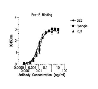
Figures IA-B show binding curves (from ELISA) of human RSV antibodies D25,
palivizumab, and RB1 to human RSV-F pre (A) and post (B) fusion proteins.
Figures 2A-B show neutralizing curves for human RSV antibodies in RSV A
Long strain (A) and RSV B Washington strain (B).
Figures 3A-B show epitope mapping of RB1 by alanine scanning mutagenesis of
Fusion F protein (A) and epitope mapped residues on Pre-Fusion F crystal
structure (B).
Figures 4A-D show the efficacy of RB1 compared to D25 in lungs in a cotton rat
challenge model of RSV A plotted against concentrations of antibody (A) and
RSV B challenge
CA 03001878 2018-04-12
WO 2017/075124 PCMJS2016/058975
plotted against concentrations of antibody (B) or viral particles (PFU/g)
present in the tissues
plotted against dose of antibody for RSV A challenge (C) and RSV B challenge
(D).
Figures 5A-D show the efficacy of RB1 compared to D25 in nose in a cotton rat
challenge model of RSV A plotted against concentrations of antibody (A) and
RSV B challenge
plotted against concentrations of antibody (B) or viral particles (PFU/g)
present in the tissues
plotted against dose of antibody for RSV A challenge (C) and RSV B challenge
(D).
Figure 6 shows a binding curve (from ELISA) of human RSV antibody
RB1+YTE to human RSV A F protein.
Figure 7 shows pharmacokinetic properties in Rhesus of RB1-YTE (RB1+YTE)
vs. motavizumab having the YTE mutation set.
DETAILED DESCRIPTION
Abbreviations
Throughout the detailed description and examples of the invention the
following
abbreviations will be used:
ADCC Antibody-dependent cellular cytotoxicity
CDC Complement-dependent cytotoxicity
CDR Complementarity determining region in the
immunoglobulin
variable regions, defined using the Kabat numbering system
CHO Chinese hamster ovary
ELISA Enzyme-linked immunosorbant assay
FR Antibody framework region: the immunoglobulin
variable regions
excluding the CDR regions
HRP Horseradish peroxidase
IC50 concentration resulting in 50% inhibition
IgG Immunoglobulin G
Kabat An immunoglobulin alignment and numbering system
pioneered
by Elvin A. Kabat ((1991) Sequences of Proteins of
Immunological Interest, 5th Ed. Public Health Service, National
Institutes of Health, Bethesda, Md.)
mAb or Mab or MAb Monoclonal antibody
16
CA 03001878 2018-04-12
WO 2017/075124 PCMJS2016/058975
V region The segment of IgG chains which is variable in
sequence between
different antibodies. It extends to Kabat residue 109 in the light
chain and 113 in the heavy chain.
VH Immunoglobulin heavy chain variable region
VK Immunoglobulin kappa light chain variable region
Definitions
So that the invention may be more readily understood, certain technical and
scientific terms are specifically defined below. Unless specifically defined
elsewhere in this
document, all other technical and scientific terms used herein have the
meaning commonly
understood by one of ordinary skill in the art to which this invention
belongs.
As used herein, including the appended claims, the singular forms of words
such
as "a," "an," and "the," include their corresponding plural references unless
the context clearly
dictates otherwise.
"Administration" and "treatment," as it applies to an animal, human,
experimental
subject, cell, tissue, organ, or biological fluid, refers to contact of an
exogenous pharmaceutical,
therapeutic, diagnostic agent, or composition to the animal, human, subject,
cell, tissue, organ, or
biological fluid. Treatment of a cell encompasses contact of a reagent to the
cell, as well as
contact of a reagent to a fluid, where the fluid is in contact with the cell.
"Administration" and
"treatment" also means in vitro and ex vivo treatments, e.g., of a cell, by a
reagent, diagnostic,
binding compound, or by another cell.
"RSV disease" means any disease caused, directly or indirectly, by an
infection
with Respiratory Syncytial Virus (RSV) as well as diseases or conditions which
predispose a
patient to infection by RSV. Examples of diseases falling into the former
category include
pneumonia and bronchiolitis. Diseases and conditions in the latter category
(i.e., those which
place the patient at risk of severe RSV infection) include cystic fibrosis,
congenital heart disease,
cancer, age related immunosuppression, transplant recipients and, generally,
any condition that
causes a state of immunosuppression or decreased function of the immune system
such as post-
operative organ transplantation regimens or premature birth.
"Treat" or "treating" means to administer a therapeutic agent, such as a
composition containing any of the antibodies or antigen-binding fragments of
the present
invention, internally or externally to a subject or patient having one or more
disease symptoms,
17
CA 03001878 2018-04-12
WO 2017/075124 PCMJS2016/058975
or being suspected of having a disease, for which the agent has therapeutic
activity. Typically,
the agent is administered in an amount effective to alleviate one or more
disease symptoms in the
treated subject or population, whether by inducing the regression of or
inhibiting the progression
of such symptom(s) by any clinically measurable degree. The amount of a
therapeutic agent that
is effective to alleviate any particular disease symptom may vary according to
factors such as the
disease state, age, and weight of the patient, and the ability of the drug to
elicit a desired
response in the subject. Whether a disease symptom has been alleviated can be
assessed by any
clinical measurement typically used by physicians or other skilled healthcare
providers to assess
the severity or progression status of that symptom. Treatment with anti-RSV
antibodies could
also combined with other interventions (antibodies, nucleic acids, vaccines
and small molecule
compounds) to treat other respiratory pathogens.
"Prevent" or "preventing" means to administer a prophylactic agent, such as a
composition containing any of the antibodies or antigen-binding fragments of
the present
invention, internally or externally to a subject or patient at risk of
becoming infected by hRSV,
for which the agent has prophylactic activity. Preventing includes reducing
the likelihood or
severity of a subsequent RSV infection, ameliorating symptoms associated with
lower
respiratory tract infection (LRI) upon RSV infection, and inducing immunity to
protect against
RSV infection. Typically, the agent is administered in an amount effective to
neutralize RSV in
the lungs and/or the nose in order block infection. The amount of a
prophylactic agent that is
effective to ameliorate any particular disease symptom may vary according to
factors such as the
age, and weight of the patient, and the ability of the agent to elicit a
desired response in the
subject. Whether a disease symptom has been ameliorated can be assessed by any
clinical
measurement typically used by physicians or other skilled healthcare providers
to assess the
severity or progression status of that symptom or in certain instances will
ameliorate the need for
hospitalization.
hRSV F protein
Human RSV F protein is synthesized as a metastable trimeric precursor (F0)
that
is proteolytically cleaved into the covalently associated Fl and F2 subunits.
Atomic structures of
F trimers in the prefusion form have been determined for PIV5 and RSV members
of
paramyxoviridae family. See McLellan etal., 2011, J Virol 85:7788-7796 (RSV)
and Welch et
al., 2012, Proc Natl Acad Sci 109:16672-16677 (Hy) Prefusion F has a short C-
terminal
18
CA 03001878 2018-04-12
WO 2017/075124 PCMJS2016/058975
cytoplasmic tail, a single transmembrane domain, a helical stalk, and a
globular head domain.
Atomic structures of NDV, hPIV3, and RSV F in the postfusion form reveal that
a large
refolding event occurs to convert prefusion F to postfusion F in which part of
the globular head
domain rearranges to form a six helix bundle. These structures, along with
peptide inhibitory
data, suggest a model for F mediated membrane fusion where, upon activation,
Fl/F2 rearranges
to insert a hydrophobic fusion peptide from the N-terminus of Fl into the
target cell membrane
forming a pre-hairpin intermediate. This relatively extended structure tethers
the virus to the cell
membrane and collapses to form the stable six-helix bundle of the postfusion
structure. The
transition from the metastable prefusion, to the prehairpin intermediate, to
the postfusion
conformation proceeds down an energy gradient with the postfusion form
representing the most
stable state, and the energy released during F refolding is coupled with
membrane fusion.
The term hRSV F protein includes human RSV F protein as well as fragments
thereof such as the mature fragment thereof lacking the signal peptide. In an
embodiment of the
invention, the amino acid sequence of human RSV F protein comprises the amino
acid sequence
disclosed in Genbank Accession Number AAR14266 (hRSV B strain 9320).
Anti-hRSV Antibodies and Antigen-Binding Fragments Thereof
The present invention provides antibodies or antigen-binding fragments thereof
that bind human RSV F protein, preferably from both RSV A strains and B
strains, that bind both
the pre-fusion F protein and the post-fusion F protein, and uses of such
antibodies or fragments.
In some embodiments, the anti-RSV F-protein antibodies are isolated. The
antibodies described
herein bind to an epitope at site IV of the F protein. In any of the
embodiments of the invention
described herein, in certain embodiments, the heavy chain or heavy chain
variable region does
not comprise the amino acid sequence of SEQ ID NO: 9 and/or the light chain or
light chain
variable region does not comprise the amino acid sequence of SEQ ID NO: 8. In
certain
embodiments, the heavy chain comprises, consists essentially of, or consists
of, the amino acid
sequence of SEQ ID NO: 23 and the light chain comprises, consists essentially
of, or consists of,
the amino acid sequence of SEQ ID NO: 25.
In preferred embodiments, the anti-RSV F-protein antibodies are fully human. A
major advantage of the monoclonal antibodies of the invention derives from the
fact that they
include human CDR3 sequences and, in some embodiments, may be entirely human
monoclonal
antibodies Hence in vivo use of the fully human monoclonal antibodies of the
invention for
19
CA 03001878 2018-04-12
WO 2017/075124 PCMJS2016/058975
immunoprophylaxis and immunotherapy of RSV disease greatly reduces the problem
of host
immune response to passively administered antibodies. This problem is commonly
encountered
when the prior art monoclonal antibodies of xenogeneic or chimeric derivation
are utilized. A
second important aspect of this advantage is the potential safety of these
human monoclonal
antibodies for gene therapy applications, in which expression of xenogeneic or
chimeric proteins
containing non-human sequences cannot be terminated.
As used herein, an anti-RSV F-protein antibody or antigen-binding fragment
thereof refers to an antibody or antigen-binding fragment thereof that
specifically binds to human
RSV F protein. An antibody or antigen-binding fragment thereof that
"specifically binds to
human RSV" is an antibody or antigen-binding fragment thereof that binds to
the pre-fusion or
post-fusion human RSV F protein with a Kd of about 1 nM or a higher affinity
(e.g., 1 nM-2 pM,
1 nM, 100 pM, 10 pM or 2 pM), but does not bind to other proteins lacking RSV
F protein
sequences. In one embodiment, the antibody of the invention which specifically
binds to human
RSV F protein is also cross-reactive with bovine RSV F protein. As used herein
"cross-
reactivity" refers to the ability of an antibody to react with a homologous
protein from other
species. Whether an antibody specifically binds to human RSV F protein can be
determined
using any assay known in the art Examples of assays known in the art to
determining binding
affinity include surface plasmon resonance (e.g., BIACORE) or a similar
technique (e.g. KinExa
or OCTET).
The present invention includes anti-hRSV F-protein antibodies and methods of
use thereof. As used herein, the term "antibody" refers to any form of
antibody that exhibits the
desired biological activity. Thus, it is used in the broadest sense and
specifically covers, but is
not limited to, monoclonal antibodies (including full length monoclonal
antibodies comprising
two light chains and two heavy chains), polyclonal antibodies, multispecific
antibodies (e.g.,
bispecific antibodies), humanized antibodies, fully human antibodies, and
chimeric antibodies.
The present invention includes anti-hRSV F-protein antigen-binding fragments
and methods of use thereof. As used herein, unless otherwise indicated,
"antibody fragment" or
"antigen-binding fragment" refers to antigen-binding fragments of antibodies,
i.e. antibody
fragments that retain the ability to bind specifically to the antigen bound by
the full-length
antibody, e.g., fragments that retain one or more CDR regions. Examples of
antigen-binding
fragments include, but are not limited to, Fab, Fab', F(ab)2, and Fv
fragments; diabodies; linear
CA 03001878 2018-04-12
WO 2017/075124 PCMJS2016/058975
antibodies; single-chain antibody molecules, e.g., sc-Fv; and multispecific
antibodies formed
from antibody fragments.
The present invention includes anti-RSV F-protein Fab fragments and methods of
use thereof. A "Fab fragment" is comprised of one light chain and the CH1 and
variable regions
of one heavy chain. The heavy chain of a Fab molecule cannot form a disulfide
bond with
another heavy chain molecule. An "Fab fragment" can be the product of papain
cleavage of an
antibody.
The present invention includes anti-RSV F-protein antibodies and antigen-
binding
fragments thereof which comprise an Fc region and methods of use thereof. An
"Fc" region
contains two heavy chain fragments comprising the CH1 and CH2 domains of an
antibody. The
two heavy chain fragments are held together by two or more disulfide bonds and
by hydrophobic
interactions of the CH3 domains.
The present invention includes anti-RSV F-protein Fab' fragments and methods
of use thereof. A "Fab' fragment" contains one light chain and a portion or
fragment of one
heavy chain that contains the VH domain and the C H1 domain and also the
region between the
CHI and C H2 domains, such that an interchain disulfide bond can be formed
between the two
heavy chains of two Fab' fragments to form a F(ab') 2 Molecule.
The present invention includes anti-RSV F-protein F(ab')2 fragments and
methods
of use thereof. A "F(ab')2fragment" contains two light chains and two heavy
chains containing a
portion of the constant region between the CHi and CH2 domains, such that an
interchain disulfide
bond is formed between the two heavy chains. A F(ab') 2 fragment thus is
composed of two Fab'
fragments that are held together by a disulfide bond between the two heavy
chains. An "F(a1302
fragment" can be the product of pepsin cleavage of an antibody.
The present invention includes anti-RSV F-protein Fv fragments and methods of
use thereof. The "Fv region" comprises the variable regions from both the
heavy and light
chains, but lacks the constant regions.
The present invention includes anti-RSV F-protein scFv fragments and methods
of use thereof. The term "single-chain Fv" or "scFv" antibody refers to
antibody fragments
comprising the VH and VI_ domains of an antibody, wherein these domains are
present in a single
polypeptide chain. Generally, the Fv polypeptide further comprises a
polypeptide linker between
the VH and VL domains which enables the scFv to form the desired structure for
antigen-binding.
21
CA 03001878 2018-04-12
WO 2017/075124 PCMJS2016/058975
For a review of scFv, see Pluckthun (1994) THE PHARMACOLOGY OF MONOCLONAL
ANTIBODIES,
vol. 113, Rosenburg and Moore eds. Springer-Verlag, New York, pp. 269-315. See
also,
International Patent Application Publication No. WO 88/01649 and U.S. Pat.
Nos. 4,946,778 and
5,260,203.
The present invention includes anti-RSV F-protein domain antibodies and
methods of use thereof. A "domain antibody" is an immunologically functional
immunoglobulin
fragment containing only the variable region of a heavy chain or the variable
region of a light
chain. In some instances, two or more VH regions are covalently joined with a
peptide linker to
create a bivalent domain antibody. The two VH regions of a bivalent domain
antibody may target
the same or different antigens.
The present invention includes anti-RSV F-protein bivalent antibodies and
methods of use thereof. A "bivalent antibody" comprises two antigen-binding
sites. In some
instances, the two binding sites have the same antigen specificities. However,
bivalent
antibodies may be bispecific (see below).
The present invention includes anti-RSV F-protein diabodies and methods of use
thereof. As used herein, the term "diabodies" refers to small antibody
fragments with two
antigen-binding sites, which fragments comprise a heavy chain variable domain
(VH) connected
to a light chain variable domain (VL) in the same polypeptide chain (VH-VL or
VL-VH). By using
a linker that is too short to allow pairing between the two domains on the
same chain, the
domains are forced to pair with the complementary domains of another chain and
create two
antigen-binding sites. Diabodies are described more fully in, e.g., EP
404,097; WO 93/11161;
and Holliger et al. (1993) Proc. Nall. Acad. Sci. USA 90: 6444-6448. For a
review of engineered
antibody variants generally see Holliger and Hudson (2005) Nat. Biotechnol.
23:1126-1136.
Typically, an antibody or antigen-binding fragment of the invention which is
modified in some way retains at least 10% of its binding activity (when
compared to the parental
antibody) when that activity is expressed on a molar basis. Preferably, an
antibody or antigen-
binding fragment of the invention retains at least 20%, 50%, 70%, 80%, 90%,
95% or 100% or
more of the RSV F-protein binding affinity as the parental antibody. It is
also intended that an
antibody or antigen-binding fragment of the invention can include conservative
or non-
conservative amino acid substitutions (referred to as "conservative variants"
or "function
conserved variants" of the antibody) that do not substantially alter its
biologic activity.
22
CA 03001878 2018-04-12
WO 2017/075124 PCMJS2016/058975
The present invention includes isolated anti-hRSV F-protein antibodies and
antigen-binding fragments thereof and methods of use thereof. "Isolated"
antibodies or antigen-
binding fragments thereof are at least partially free of other biological
molecules from the cells
or cell cultures in which they are produced. Such biological molecules include
nucleic acids,
proteins, lipids, carbohydrates, or other material such as cellular debris and
growth medium. An
isolated antibody or antigen-binding fragment may further be at least
partially free of expression
system components such as biological molecules from a host cell or of the
growth medium
thereof. Generally, the term "isolated" is not intended to refer to a complete
absence of such
biological molecules or to an absence of water, buffers, or salts or to
components of a
pharmaceutical formulation that includes the antibodies or fragments.
The present invention includes monoclonal anti-hRSV F-protein antibodies and
antigen-binding fragments thereof as well as monoclonal antibody compositions
comprising a
plurality of isolated monoclonal antibodies. The term "monoclonal antibody",
as used herein,
refers to a population of substantially homogeneous antibodies, i.e., the
antibody molecules
comprising the population are identical in amino acid sequence except for
possible naturally
occurring mutations that may be present in minor amounts. In contrast,
conventional
(polyclonal) antibody preparations typically include a multitude of different
antibodies having
different amino acid sequences in their variable domains, particularly their
CDRs that are often
specific for different epitopes. The modifier "monoclonal" indicates the
character of the
antibody as being obtained from a substantially homogeneous population of
antibodies, and is
not to be construed as requiring production of the antibody by any particular
method. For
example, the monoclonal antibodies to be used in accordance with the present
invention may be
made by recombinant DNA methods (see, e.g., U.S. Pat. No. 4,816,567).
In general, the basic (or "full-length") antibody structural unit comprises a
tetramer. Each tetramer includes two identical pairs of polypeptide chains,
each pair having one
"light" (about 25 kDa) and one "heavy" chain (about 50-70 kDa). The amino-
terminal portion of
each chain includes a variable region or domain of about 100 to 110 or more
amino acids
primarily responsible for antigen recognition. The carboxy-terminal portion of
the heavy chain
may define a constant region or domain primarily responsible for effector
function. Typically,
human light chains are classified as kappa and lambda light chains.
Furtheimore, human heavy
chains are typically classified as mu, delta, gamma, alpha, or epsilon, and
define the antibody's
23
CA 03001878 2018-04-12
WO 2017/075124 PCMJS2016/058975
isotype as IgM, IgD, IgG, IgA, and IgE, respectively. Within light and heavy
chains, the
variable and constant regions are joined by a "J" region of about 12 or more
amino acids, with
the heavy chain also including a "D" region of about 10 more amino acids. See
generally,
Fundamental Immunology Ch. 7 (Paul, W., ed., 2nd ed. Raven Press, N.Y. (1989).
In the context
of an antibody or antigen binding fragment thereof, the terms domain and
region can be used
interchangeably, where appropriate.
The variable regions of each light/heavy chain pair form the antibody binding
site.
Thus, in general, an intact antibody has two binding sites. Except in
bifunctional or bispecific
antibodies, the two binding sites are, in general, the same.
Typically, the variable domains of both the heavy and light chains comprise
three
hypervariable regions, also called complementarity determining regions (CDRs),
located within
relatively conserved framework regions (FR). The CDRs are usually aligned by
the framework
regions, enabling binding to a specific epitope. In general, from N-terminal
to C-terminal, both
light and heavy chains variable domains comprise FR1, CDR1, FR2, CDR2, FR3,
CDR3 and
FR4. The assignment of amino acids to each domain is, generally, in accordance
with the
definitions of Sequences of Proteins of Immunological Interest, Kabat, et at.;
National Institutes
ed.;
of Health, Bethesda, Md.; 5thNIH Publ. No. 91-3242 (1991); Kabat, 1978, Adv.
Prot. Chem.
32:1-75; Kabat, et al , 1977,1 Biol. Chem. 252:6609-6616; Chothi a et at.,
1987, .1 Mol. Biol.
196:901-917 or Chothia et al., 1989, Nature 342:878-883.
As used herein, the term "hypervariable region" refers to the amino acid
residues
of an antibody or antigen-binding fragment thereof that are responsible for
antigen-binding. The
hypervariable region comprises amino acid residues from a "complementarity
determining
region" or "CDR" (i.e. CDRL1, CDRL2 and CDRL3 in the light chain variable
domain and
CDRH1, CDRH2 and CDRH3 in the heavy chain variable domain). See Kabat et at.
(1991)
Sequences of Proteins of Immunological Interest, 5th Ed. Public Health
Service, National
Institutes of Health, Bethesda, Md. (defining the CDR regions of an antibody
by sequence); see
also Chothia and Lesk, 1987,1 Mot. Biol. 196: 901-917 (defining the CDR
regions of an
antibody by structure). As used herein, the term "framework" or "FR" residues
refers to those
variable domain residues other than the hypervariable region residues defined
herein as CDR
residues.
24
CA 03001878 2018-04-12
WO 2017/075124 PCMJS2016/058975
"Isolated nucleic acid molecule" or "isolated polynucleotide" means a DNA or
RNA of genomic, mRNA, cDNA, or synthetic origin or some combination thereof
which is not
associated with all or a portion of a polynucleotide in which the isolated
polynucleotide is found
in nature, or is linked to a polynucleotide to which it is not linked in
nature. For purposes of this
disclosure, it should be understood that "a nucleic acid molecule comprising"
a particular
nucleotide sequence does not encompass intact chromosomes. Isolated nucleic
acid molecules
"comprising" specified nucleic acid sequences may include, in addition to the
specified
sequences, coding sequences for up to ten or even up to twenty or more other
proteins or portions
or fragments thereof, or may include operably linked regulatory sequences that
control
expression of the coding region of the recited nucleic acid sequences, and/or
may include vector
sequences.
The phrase "control sequences" refers to DNA sequences necessary for the
expression of an operably linked coding sequence in a particular host
organism. The control
sequences that are suitable for prokaryotes, for example, include a promoter,
optionally an
operator sequence, and a ribosome binding site. Eukaryotic cells are known to
use promoters,
polyadenylati on signals, and enhancers.
A nucleic acid or polynucleotide is "operably linked" when it is placed into a
functional relationship with another nucleic acid sequence. For example, DNA
for a
presequence or secretory leader is operably linked to DNA for a polypeptide if
it is expressed as
a preprotein that participates in the secretion of the polypeptide; a promoter
or enhancer is
operably linked to a coding sequence if it affects the transcription of the
sequence; or a ribosome
binding site is operably linked to a coding sequence if it is positioned so as
to facilitate
translation. Generally, but not always, "operably linked" means that the DNA
sequences being
linked are contiguous, and, in the case of a secretory leader, contiguous and
in reading phase.
However, enhancers do not have to be contiguous. Linking is accomplished by
ligation at
convenient restriction sites. If such sites do not exist, the synthetic
oligonucleotide adaptors or
linkers are used in accordance with conventional practice.
As used herein, the expressions "cell," "cell line," and "cell culture" are
used
interchangeably and all such designations include progeny. Thus, the words
"transformants" and
"transformed cells" include the primary subject cell and cultures derived
therefrom without
regard for the number of transfers. It is also understood that not all progeny
will have precisely
CA 03001878 2018-04-12
WO 2017/075124 PCMJS2016/058975
identical DNA content, due to deliberate or inadvertent mutations. Mutant
progeny that have the
same function or biological activity as screened for in the originally
transformed cell are
included. Where distinct designations are intended, it will be clear from the
context.
As used herein, "germline sequence" refers to a sequence of unrearranged
immunoglobulin DNA sequences. Any suitable source of unrearranged
immunoglobulin
sequences may be used. Human germline sequences may be obtained, for example,
from
JOINSOLVER germline databases on the website for the National Institute of
Arthritis and
Musculoskeletal and Skin Diseases of the United States National Institutes of
Health. Mouse
germline sequences may be obtained, for example, as described in Giudicelli et
al., 2005,
Nucleic Acids Res. 33:D256-D261.
Physical and Functional Properties of the Exemplary Anti-RSV F-protein
Antibodies
The present invention provides anti-hRSV F-protein antibodies and antigen-
binding fragments thereof having specified structural and functional features,
and methods of use
of the antibodies or antigen-binding fragments thereof in the treatment or
prevention of
diseases/conditions associated with RSV infection.
An "anti-RSV F-protein antibody or antigen-binding fragment thereof of the
present invention" includes: any antibody or antigen-binding fragment thereof
that is discussed
herein (e.g., RB1) or a variant thereof (e.g., sequence variant or functional
variant); any antibody
or antigen-binding fragment comprising any one or more of the CDRs set forth
in Table 7; any
.. antibody or antigen-binding fragment that binds to the same epitope in
human RSV F-protein as
the antibodies discussed herein (e.g., RB1); and any antibody or antigen-
binding fragment that
cross-blocks (partially or fully) or is cross-blocked (partially or fully) by
an antibody discussed
herein (e.g., RBI) for RSV binding.
Cross-blocking antibodies and antigen-binding fragments thereof can be
identified
based on their ability to cross-compete with an antibody of the invention in
standard binding
assays (e.g., BIACore, ELISA, flow cytometry). For example, standard ELISA
assays can be
used in which a recombinant RSV F protein protein is immobilized on the plate,
one of the
antibodies is fluorescently labeled and the ability of non-labeled antibodies
to compete off the
binding of the labeled antibody is evaluated Additionally or alternatively,
BIAcore analysis can
be used to assess the ability of the antibodies to cross-compete. The ability
of a test antibody to
inhibit the binding of another antibody (for example, antibody D25) to RSV F-
protein
26
CA 03001878 2018-04-12
WO 2017/075124 PCMJS2016/058975
demonstrates that the test antibody can compete with another antibody (e.g.,
D25) for binding to
RSV F protein and thus, may, in some cases, bind to the same epitope on RSV F
protein as
antibody D25 to an overlapping epitope.
As stated above, antibodies and fragments thereof that bind to the same
epitope as
any of the anti-RSV F-protein antibodies or antigen-binding fragments thereof
of the present
invention also form part of the present invention. Further, in certain
embodiments, antibodies
that bind to an epitope that overlaps with the epitope bound by any of the
anti-RSV F-protein
antibodies of the invention also form part of the present invention. There are
several methods
available for mapping antibody epitopes on target antigens, including: H/D-Ex
Mass spec, X-ray
crystallography, pepscan analysis and site directed mutagenesis. For example,
HDX (Hydrogen
Deuterium Exchange) coupled with proteolysis and mass spectrometry can be used
to determine
the epitope of an antibody on a specific antigen Y. HDX-MS relies on the
accurate measurement
and comparison of the degree of deuterium incorporation by an antigen when
incubated in D20
on its own and in presence of its antibody at various time intervals.
Deuterium is exchanged
with hydrogen on the amide backbone of the proteins in exposed areas whereas
regions of the
antigen bound to the antibody will be protected and will show less or no
exchange after analysis
by LC-MS/MS of proteolytic fragments.
Examples of the immunoglobulin chains of anti-RSV F-protein antibodies of the
invention as well as their CDRs include, but are not limited those disclosed
in Table 7 (SEQ ID
NOs: 1-8, 23 and 25). The present invention includes any polypeptide
comprising, consisting
essentially of, or consisting of the amino acid sequences of SEQ ID NOs: 1-8,
23, and 25, and
recombinant nucleotides encoding such polypeptides.
The scope of the present invention includes isolated anti-hRSV F-protein
antibodies and antigen-binding fragments thereof, comprising a variant of an
immunoglobulin
chain set forth herein, e.g., any of SEQ ID NOs: 7, 8; wherein the variant
exhibits one or more of
the following properties: (i) binds to human RSV pre-fusion F protein with a
Kd value of about 1
x 10-9M to about 1 x 10-12 M as determined by surface plasmon resonance (e.g.,
BIACORE) or a
similar technique (e.g. KinExa or OCTET); or (ii) binds to human RSV post-
fusion F protein
with a Kd value of about 1 x 10⁻⁹M to about 1 x 10-11M as determined by
surface plasmon
resonance (e.g., BIACORE) or a similar technique (e.g. KinExa or OCTET). In
certain
27
CA 03001878 2018-04-12
WO 2017/075124 PCMJS2016/058975
embodiments, the heavy chain or heavy chain variable region does not comprise
the amino acid
sequence of SEQ ID NO: 9.
In certain embodiments, the invention provides antibodies or antigen-binding
fragment thereof that binds human hRSV F-protein and has VL domains and VH
domains with at
least 80%, 85%, 90%, 95%, 96%, 97%, 98% or 99% sequence identity with SEQ ID
NOs: 8 (VI)
and 7 (VH); wherein the variant exhibits the desired binding and properties,
e.g., (i) binds to
human RSV pre-fusion F protein with a Kd value of about 1 x 10⁻⁹M to about 1 x
1012 M as
determined by surface plasmon resonance (e.g., BIACORE) or a similar technique
(e.g. KinExa
or OCTET); or (ii) binds to human RSV post-fusion F protein with a Kd value of
about 1 x 10⁻⁹
M to about 1 x 10-11M as deteimined by surface plasmon resonance (e.g.,
BIACORE) or a
similar technique (e.g. KinExa or OCTET). In certain embodiments, the heavy
chain or heavy
chain variable region does not comprise the amino acid sequence of SEQ ID NO:
9.
"Conservatively modified variants" or "conservative substitution" refers to
substitutions of amino acids in a protein with other amino acids having
similar characteristics
(e.g. charge, side-chain size, hydrophobicity/hydrophilicity, backbone
conformation and rigidity,
etc.), such that the changes can frequently be made without altering the
biological activity of the
protein Those of skill in this art recognize that, in general, single amino
acid substitutions in
non-essential regions of a polypeptide do not substantially alter biological
activity (see, e.g.,
Watson et al. (1987) Molecular Biology of the Gene, The Benjamin/Cummings Pub.
Co., p. 224
(4th Ed.)). In addition, substitutions of structurally or functionally similar
amino acids are less
likely to disrupt biological activity. Exemplary conservative substitutions
are set forth in Table
1.
TABLE 1. Exemplary Conservative Amino Acid Substitutions
Original residue Conservative substitution
Ala (A) Gly; Ser
Arg (R) Lys; His
Asn (N) _Gin; His
Asp (D) Glu. Asn
Cys (C) Ser, Ala
Gln (Q) Asn
Glu (E) Asp, Gln
Gly (G) Ala
His (H) _Asn; Gln
Ile (I) Leu; Val
28
CA 03001878 2018-04-12
WO 2017/075124 PCMJS2016/058975
Original residue Conservative substitution
Leu (L) Ile; Val
Lys (K) Arg; His
Met (M) Leu; Ile; Tyr
Phe (F) Tyr; Met; Leu
Pro (P) Ala
Ser (S) Thr
Thr (T) Ser
Trp (W) Tyr; Phe
Tyr (Y) Trp; Phe
Val (V) Ile; Leu
Function-conservative variants of the antibodies of the invention are also
contemplated by the present invention. "Function-conservative variants," as
used herein, refers
to antibodies or fragments in which one or more amino acid residues have been
changed without
altering a desired property, such an antigen affinity and/or specificity. Such
variants include, but
are not limited to, replacement of an amino acid with one having similar
properties, such as the
conservative amino acid substitutions of Table 1. Also provided are isolated
polypepti des
comprising the VL domains of the anti-hRSV F-protein antibodies of the
invention (e.g., SEQ ID
NO. 8), and isolated polypeptides comprising the VH domains (e.g., SEQ ID NO:
7) of the anti-
hRSV antibodies of the invention having up to 0, 1, 2, 3, 4, 5, 6, 7, 8, 9,
10, 11, 12, 13, 14, 15,
16, 17, 18, 19, 20, 21, 22, 23, 24, 25, 26, 27, 28, 29, 30 or more amino acid
substitutions, which
may occur exclusively in the framework region or of which one or more may be
located in one or
more CDRs . In certain embodiments, the heavy chain or heavy chain variable
region does not
comprise the amino acid sequence of SEQ ID NO: 9.
In another embodiment, provided is an antibody or antigen-binding fragment
thereof that binds hRSV F-protein and has VL domains and VH domains with at
least 99% 98%,
97%, 96%, 95%, 90%, 85%, 80% or 75% sequence identity to one or more of the VL
domains or
VH domains described herein, and exhibits specific binding to hRSV F-protein.
In another
embodiment the binding antibody or antigen-binding fragment thereof of the
present invention
comprises VL and VH domains (with and without signal sequence) having up to 0,
1, 2, 3, 4, 5, 6,
7, 8, 9, 10, 11, 12, 13, 14, 15, 16, 17, 18, 19, 20, 21, 22, 23, 24, 25, 26,
27, 28, 29, 30 or more
amino acid substitutions, which may occur exclusively in the framework region
or of which one
or more may be located in one or more CDRs, and exhibits specific binding to
hRSV F-protein.
29
CA 03001878 2018-04-12
WO 2017/075124 PCMJS2016/058975
In certain embodiments, the heavy chain or heavy chain variable region does
not comprise the
amino acid sequence of SEQ ID NO: 9.
Polynucleotides and Polypeptides
The present invention further comprises the polynucleotides encoding any of
the
polypeptides or immunoglobulin chains of anti-hRSV F-protein antibodies and
antigen-binding
fragments thereof of the invention. In one embodiment, the isolated
polynucleotide encodes an
antibody or antigen-binding fragment thereof comprising at least one mature
immunoglobulin
light chain variable (VI) domain according to the invention and/or at least
one mature
immunoglobulin heavy chain variable (VH) domain according to the invention. In
some
embodiments the isolated polynucleotide encodes both a light chain and a heavy
chain on a
single polynucleotide molecule, and in other embodiments the light and heavy
chains are
encoded on separate polynucleotide molecules. In another embodiment the
polynucleotides
further encodes a signal sequence. For example, the present invention includes
the
polynucleotides encoding the amino acids described in SEQ ID NOs: 1-8, 23 and
25, as well as
polynucleotides which hybridize thereto and, also, any polypeptide encoded by
such a
hybridizing polynucleotide. In one embodiment, the invention comprises a
nucleic acid
sequence comprising, consisting essentially of, or consisting of SEQ ID NO: 15
(variable heavy
chain) or SEQ ID NO: 16 (variable light chain). In certain embodiments, codon
optimization can
be used to enhance a property of the nucleic acid, e.g., expression in a
certain host. In one
embodiment, the invention comprises a nucleic acid sequence comprising,
consisting essentially
of, or consisting of SEQ ID NO: 17 (codon optimized variable heavy) or SEQ ID
NO: 18 (codon
optimized variable light). In certain embodiments, a leader sequence can be
used. In one
embodiment, the invention comprises a nucleic acid sequence comprising,
consisting essentially
of, or consisting of SEQ ID NO: 19 (leader sequence and heavy chain) or SEQ ID
NO: 20
(leader sequence and light chain) connected with the heavy chain or light
chain to give SEQ ID
NO: 21 or SEQ ID NO: 22, respectively.
In general, the polynucleotides hybridize under low, moderate or high
stringency
conditions, and encode antibodies or antigen-binding fragments thereof that
maintain the ability
to bind to hRSV F-protein. A first polynucleotide molecule is "hybridizable"
to a second
polynucleotide molecule when a single stranded form of the first
polynucleotide molecule can
anneal to the second polynucleotide molecule under the appropriate conditions
of temperature
CA 03001878 2018-04-12
WO 2017/075124 PCMJS2016/058975
and solution ionic strength (see Sambrook, el al., supra). The conditions of
temperature and
ionic strength determine the "stringency" of the hybridization. Typical low
stringency
hybridization conditions include 55 C, 5X SSC, 0.1% SDS and no formamide; or
30%
formamide, 5X SSC, 0.5% SDS at 42 C. Typical moderate stringency hybridization
conditions
are 40% formamide, with 5X or 6X SSC and 0.1% SDS at 42 C. High stringency
hybridization
conditions are 50% formamide, 5X or 6X SSC at 42 C or, optionally, at a higher
temperature
(e.g., 57 C, 59 C, 60 C, 62 C, 63 C, 65 C or 68 C). In general, SSC is 0.15M
NaCl and
0.015M Na-citrate. Hybridization requires that the two polynucleotides contain
complementary
sequences, although, depending on the stringency of the hybridization,
mismatches between
bases are possible. The appropriate stringency for hybridizing polynucleotides
depends on the
length of the polynucleotides and the degree of complementation, variables
well known in the
art. The greater the degree of similarity or homology between two nucleotide
sequences, the
higher the stringency under which the nucleic acids may hybridize. For hybrids
of greater than
100 nucleotides in length, equations for calculating the melting temperature
have been derived
(see Sambrook et al., supra, 9.50-9.51). For hybridization with shorter
polynucleotides, e.g.,
oligonucleotides, the position of mismatches becomes more important, and the
length of the
oligonucleotide determines its specificity (see Sambrook, et al., supra, 11.7-
11 8).
In one embodiment, the invention comprises an isolated polynucleotide encoding
an antibody heavy variable (VH) domain or an antigen-binding fragment thereof
comprising
CDR-H1 (SEQ ID NO: 1), CDR-H2 (SEQ ID NO: 2) and CDR-H3 (SEQ ID NO: 3).
In one embodiment, the invention comprises an isolated polynucleotide encoding
an antibody light chain variable (VI) domain or an antigen-binding fragment
thereof comprising
CDR-L1 (SEQ ID NO: 4), CDR-L2 (SEQ ID NO: 5) and CDR-L3 (SEQ ID NO: 6).
In one embodiment, the invention comprises an isolated polynucleotide encoding
the immunoglobulin heavy chain variable (VH) domain of SEQ ID NO: 7 or a heavy
chain of
SEQ ID NO: 23.
In one embodiment, the invention comprises an isolated polynucleotide encoding
the immunoglobulin heavy chain variable (VL) domain of SEQ ID NO: 8 or a light
chain of SEQ
ID NO: 25.
This present invention also provides vectors, e.g., expression vectors, such
as
plasmids, comprising the isolated polynucleotides of the invention, wherein
the polynucleotide is
31
CA 03001878 2018-04-12
WO 2017/075124 PCMJS2016/058975
operably linked to control sequences that are recognized by a host cell when
the host cell is
transfected with the vector. Also provided are host cells comprising a vector
of the present
invention and methods for producing the antibody or antigen-binding fragment
thereof or
polypeptide disclosed herein comprising culturing a host cell harboring an
expression vector or a
nucleic acid encoding the immunoglobulin chains of the antibody or antigen-
binding fragment
thereof in culture medium, and isolating the antigen or antigen-binding
fragment thereof from the
host cell or culture medium.
Also included in the present invention are polypeptides, e.g., immunoglobulin
polypeptides, comprising amino acid sequences that are at least about 75%
identical, 80%
identical, more preferably at least about 90% identical and most preferably at
least about 95%
identical (e.g., 95%, 96%, 97%, 98%, 99%, 100%) to the amino acid sequences of
the antibodies
provided herein when the comparison is performed by a BLAST algorithm wherein
the
parameters of the algorithm are selected to give the largest match between the
respective
sequences over the entire length of the respective reference sequences (e.g.
expect threshold: 10;
word size: 3; max matches in a query range: 0; BLOSUM 62 matrix; gap costs:
existence 11,
extension 1; conditional compositional score matrix adjustment)
Sequence identity refers to the degree to which the amino acids of two
polypeptides are the same at equivalent positions when the two sequences are
optimally aligned.
The following references relate to BLAST algorithms often used for sequence
analysis: BLAST ALGORITHMS. Altschul et al. (2005) FEBS J. 272(20): 5101-5109;
Altschul,
S.F., et al., (1990)1 Mot Biol. 215:403-410, Gish, W., et al., (1993) Nature
Genet 3:266-272;
Madden, T.L., et al., (1996) Meth. Enzymot 266:131-141; Altschul, S.F., et
al., (1997) Nucleic
Acids Res. 25:3389-3402; Zhang, J., et al., (1997) Genome Res. 7:649-656;
Wootton, J.C., et al.,
(1993) Comput. Chem. 17:149-163; Hancock, J.M. et al., (1994) Comput Appl.
BlOSel. 10:67-70;
ALIGNMENT SCORING SYSTEMS: Dayhoff, M.O., et at, "A model of evolutionary
change
in proteins." in Atlas of Protein Sequence and Structure, (1978) vol. 5,
suppl. 3. M.O. Dayhoff
(ed.), pp. 345-352, Natl. Biomed. Res. Found, Washington, DC; Schwartz, R.M.,
et al.,
"Matrices for detecting distant relationships." in Atlas of Protein Sequence
and Structure, (1978)
vol. 5, suppl. 3." M.O. Dayhoff (ed.), pp. 353-358, Natl. Biomed. Res. Found,
Washington, DC;
Altschul, S.F., (1991) J. Mol. Biol. 219:555-565; States, D.J., et at, (1991)
Methods 3:66-70;
Henikoff, S., et al., (1992) Proc. Natl. Acad. Sci. USA 89:10915-10919;
Altschul, S.F., et at,
32
CA 03001878 2018-04-12
WO 2017/075124 PCMJS2016/058975
(1993) J. Mol. Evol. 36:290-300; ALIGNMENT STATISTICS: Karlin, S., et al.,
(1990) Proc.
Natl. Acad. Sci. USA 87:2264-2268; Karlin, S., et al., (1993) Proc. Natl.
Acad. Sci. USA
90:5873-5877; Dembo, A., et al., (1994) Ann. Prob. 22:2022-2039; and Altschul,
S.F.
"Evaluating the statistical significance of multiple distinct local
alignments." in Theoretical and
Computational Methods in Genome Research (S. Suhai, ed.), (1997) pp. 1-14,
Plenum, New
York.
Binding Affinity
By way of example, and not limitation, the antibodies and antigen-binding
fragments disclosed herein may bind human RSV pre-fusion F protein or post-
fusion F protein
with a KD value of at least about 1 x 10⁻⁹M (i.e, a KD value of 1 x 10⁻⁹M or
lower) as determined
by surface plasmon resonance (e.g., BIACORE) or a similar technique (e.g.
KinExa or OCTET).
In one embodiment, the antibodies and antigen-binding fragments disclosed
herein may bind
human RSV pre-fusion F protein or post-fusion F protein with a KD value of at
least about 1 x
10-9M to about 1 x 10-12M as determined by surface plasmon resonance (e.g.,
BIACORE) or a
similar technique (e.g. KinExa or OCTET). In one embodiment, the antibodies
and antigen-
binding fragments disclosed herein may bind human RSV pre-fusion F protein or
post-fusion F
protein with a KD value of at about 1 x 10⁻⁹M to about 1 x 10¹²M as determined
by surface
plasmon resonance (e.g., BIACORE) or a similar technique (e.g. KinExa or
OCTET). In one
embodiment, the antibodies and antigen-binding fragments disclosed herein may
bind human
RSV pre-fusion F protein or post-fusion F protein with a KD value of at least
about 100 pM (i.e,
a KD value of about 100 pM or lower) as determined by BIACORE or a similar
technique. In
one embodiment, the antibodies and antigen-binding fragments disclosed herein
may bind human
RSV pre-fusion F protein or post-fusion F protein with a KD value of at least
about 10 p114 (i.e., a
KD value of about 10 pm lower) as determined by BIACORE or a similar
technique. In one
.. embodiment, the antibodies and antigen-binding fragments of the invention
may bind to human
RSV pre-fusion F protein or post-fusion F protein with a KD of about 1 pM to
about 100 pM as
determined by BIACORE or a similar technique.
Methods of Making Antibodies and Antigen-binding Fragments Thereof
The present invention includes methods for making an anti-hRSV F-protein
antibody or antigen-binding fragment thereof of the present invention
comprising culturing a cell
line that expresses the antibody or fragment under conditions favorable to
such expression and,
33
CA 03001878 2018-04-12
WO 2017/075124 PCMJS2016/058975
optionally, isolating the antibody or fragment from the cells and/or the
growth medium (e.g. cell
culture medium).
The anti-hRSV F-protein antibodies disclosed herein may also be produced
recombinantly (e.g., in an E. colilT7 expression system, a mammalian cell
expression system or
.. a lower eukaryote expression system). In this embodiment, nucleic acids
encoding the antibody
immunoglobulin molecules of the invention (e.g., VH or VL) may be inserted
into a pET-based
plasmid and expressed in the E. colilT7 system. For example, the present
invention includes
methods for expressing an antibody or antigen-binding fragment thereof or
immunoglobulin
chain thereof in a host cell (e.g., bacterial host cell such as E.coli such as
BL21 or BL21DE3)
comprising expressing T7 RNA polymerase in the cell which also includes a
polynucleotide
encoding an immunoglobulin chain that is operably linked to a T7 promoter. For
example, in an
embodiment of the invention, a bacterial host cell, such as a E. colt,
includes a polynucleotide
encoding the T7 RNA polymerase gene operably linked to a lac promoter and
expression of the
polymerase and the chain is induced by incubation of the host cell with 1PTG
(isopropyl-beta-D-
thiogalactopyranoside).
There are several methods by which to produce recombinant antibodies which are
known in the art One example of a method for recombinant production of
antibodies is
disclosed in U.S. Patent No. 4,816,567.
Transformation can be by any known method for introducing polynucleotides into
a host cell. Methods for introduction of heterologous polynucleotides into
mammalian cells are
well known in the art and include dextran-mediated transfection, calcium
phosphate
precipitation, polybrene-mediated transfection, protoplast fusion,
electroporation, encapsulation
of the polynucleotide(s) in liposomes, biolistic injection and direct
microinjection of the DNA
into nuclei. In addition, nucleic acid molecules may be introduced into
mammalian cells by viral
vectors. Methods of transforming cells are well known in the art. See, for
example, U.S. Patent
Nos. 4,399,216; 4,912,040; 4,740,461 and 4,959,455.
Thus, the present invention includes recombinant methods for making an anti-
hRSV antibody or antigen-binding fragment thereof of the present invention, or
an
immunoglobulin chain thereof, comprising introducing a polynucleotide encoding
one or more
immunoglobulin chains of the antibody or fragment (e.g., heavy and/or light
immunoglobulin
chain); culturing the host cell (e.g., CHO or Pichia or Pichia pastoris) under
condition favorable
34
CA 03001878 2018-04-12
WO 2017/075124
PCMJS2016/058975
to such expression and, optionally, isolating the antibody or fragment or
chain from the host cell
and/or medium in which the host cell is grown.
Anti-hRSV F-protein antibodies can also be synthesized by any of the methods
set
forth in U.S. Patent No. 6,331,415.
Eukaryotic and prokaryotic host cells, including mammalian cells as hosts for
expression of the antibodies or fragments or immunoglobulin chains disclosed
herein are well
known in the art and include many immortalized cell lines available from the
American Type
Culture Collection (ATCC). These include, inter al/a, Chinese hamster ovary
(CHO) cells,
NSO, 5P2 cells, HeLa cells, baby hamster kidney (BHK) cells, monkey kidney
cells (COS),
.. human hepatocellular carcinoma cells (e.g., Hep G2), A549 cells, 3T3 cells,
HEK-293 cells and a
number of other cell lines. Mammalian host cells include human, mouse, rat,
dog, monkey, pig,
goat, bovine, horse and hamster cells. Cell lines of particular preference are
selected through
determining which cell lines have high expression levels. Other cell lines
that may be used are
insect cell lines, such as Sf9 cells, amphibian cells, bacterial cells, plant
cells and fungal cells.
Fungal cells include yeast and filamentous fungus cells including, for
example, Pichia pastoris,
Pichia finlandica, Pichia trehalophila, Pichia koclamae, Pichia
membranaefaciens, Pichia
minuta (Ogataea minuta, Pichia lindneri),Pichia opuntiae, Pichia
thermotoleran.s, Pichia
salictaria, Pichia guercuum, Pichia pijperi, Pichia stiptis, Pichia
methanol/ca, Pichia sp.,
Saccharomyces cerevisiae, Saccharomyces sp., Hansenula polymorpha,
Kluyveromyces .5p.,
Kluyveromyces lactis, Candida albicans, Aspergillus nidulans, Aspergillus
niger, Aspergillus
oryzae, Trichoderma reesei, Chrysosporium lucknowense, Fusarium sp., Fusarium
gramineum,
Fusarium venenatum, Physcomitrella patens and Neurospora crassa. Pichia sp.,
any
Saccharomyces sp., Hansenula polymorpha, any Kluyveromyces sp., Candida
albicans, any
Aspergillus sp., Trichoderma reesei, Chrysosporiztm lucknowense, any Fusarium
sp., Yarrowia
lipolytica, and Neztrospora crassa. When recombinant expression vectors
encoding the heavy
chain or antigen-binding portion or fragment thereof, the light chain and/or
antigen-binding
fragment thereof are introduced into mammalian host cells, the antibodies are
produced by
culturing the host cells for a period of time sufficient to allow for
expression of the antibody or
fragment or chain in the host cells or secretion of the into the culture
medium in which the host
cells are grown.
CA 03001878 2018-04-12
WO 2017/075124
PCMJS2016/058975
Antibodies and antigen-binding fragments thereof and immunoglobulin chains
can be recovered from the culture medium using standard protein purification
methods. Further,
expression of antibodies and antigen-binding fragments thereof and
immunoglobulin chains of
the invention (or other moieties therefrom) from production cell lines can be
enhanced using a
number of known techniques. For example, the glutamine synthetase gene
expression system
(the GS system) is a common approach for enhancing expression under certain
conditions. The
GS system is discussed in whole or part in connection with European Patent
Nos. 0 216 846, 0
256 055, and 0 323 997 and European Patent Application No. 89303964.4. Thus,
in an
embodiment of the invention, the mammalian host cells (e.g., CHO) lack a
glutamine synthetase
gene and are grown in the absence of glutamine in the medium wherein, however,
the
polynucleotide encoding the immunoglobulin chain comprises a glutamine
synthetase gene
which complements the lack of the gene in the host cell.
The present invention includes methods for purifying an anti-hRSV antibody or
antigen-binding fragment thereof of the present invention comprising
introducing a sample
comprising the antibody or fragment to a purification medium (e.g., cation
exchange medium,
anion exchange medium, hydrophobic exchange medium, affinity purification
medium (e.g.,
protein-A, protein-G, protein-A/G, protein-L)) and either collecting purified
antibody or
fragment from the flow-through fraction of said sample that does not bind to
the medium; or,
discarding the flow-through fraction and eluting bound antibody or fragment
from the medium
.. and collecting the eluate. In an embodiment of the invention, the medium is
in a column to
which the sample is applied. In an embodiment of the invention, the
purification method is
conducted following recombinant expression of the antibody or fragment in a
host cell, e.g.,
wherein the host cell is first lysed and, optionally, the lysate is purified
of insoluble materials
prior to purification on a medium.
In general, glycoproteins produced in a particular cell line or transgenic
animal
will have a glycosylation pattern that is characteristic for glycoproteins
produced in the cell line
or transgenic animal. Therefore, the particular glycosylation pattern of an
antibody will depend
on the particular cell line or transgenic animal used to produce the antibody.
However, all
antibodies encoded by the nucleic acid molecules provided herein, or
comprising the amino acid
sequences provided herein, comprise the instant invention, independent of the
glycosylation
pattern the antibodies may have. Similarly, in particular embodiments,
antibodies with a
36
CA 03001878 2018-04-12
WO 2017/075124 PCMJS2016/058975
glycosylation pattern comprising only non-fucosylated N-glycans may be
advantageous, because
these antibodies have been shown to typically exhibit more potent efficacy
than their fucosylated
counterparts both in vitro and in vivo (See for example, Shinkawa et al., J.
Biol. Chem. 278:
3466-3473 (2003); U.S. Patent Nos. 6,946,292 and 7,214,775). These antibodies
with non-
fucosylated N-glycans are not likely to be immunogenic because their
carbohydrate structures are
a normal component of the population that exists in human serum IgG.
The present invention includes bispecific and bifunctional antibodies and
antigen-
binding fragments having a binding specificity for hRSV F protein and another
antigen such as,
for example, hRSV G protein, and methods of use thereof. A bispecific or
bifunctional antibody
is an artificial hybrid antibody having two different heavy/light chain pairs
and two different
binding sites. Bispecific antibodies can be produced by a variety of methods
including fusion of
hybridomas or linking of Fab fragments. See, e.g., Songsivilai, et al., (1990)
Clin. Exp.
1111111211101. 79: 315-321, Kostelny, et al., (1992) J Immunol. 148:1547-
1553. In addition,
bispecific antibodies may be formed as "diabodies" (Holliger, et al., (1993)
PNAS USA 90:6444-
6448) or as "Janusins" (Traunecker, etal., (1991) E114130 1. 10:3655-3659 and
Traunecker, etal.,
(1992) Int. J. Cancer Suppl. 7:51-52).
The present invention further includes anti-hRSV F-protein antigen-binding
fragments of the anti-hRSV antibodies disclosed herein. The antibody fragments
include F(ab)2
fragments, which may be produced by enzymatic cleavage of an IgG by, for
example, pepsin.
Fab fragments may be produced by, for example, reduction of F(ab)2 with
dithiothreitol or
mercaptoethylamine.
Immunoglobulins may be assigned to different classes depending on the amino
acid sequences of the constant domain of their heavy chains. There are at
least five major classes
of immunoglobulins: IgA, IgD, IgE, IgG and IgM, and several of these may be
further divided
into subclasses (isotypes), e.g. IgGl, IgG2, IgG3 and IgG4; IgAl and IgA2. The
invention
comprises antibodies and antigen-binding fragments of any of these classes or
subclasses of
antibodies.
In one embodiment, the antibody or antigen-binding fragment comprises a heavy
chain constant region, e.g. a human constant region, such as yl, y2, y3, or y4
human heavy chain
constant region or a variant thereof In another embodiment, the antibody or
antigen-binding
fragment comprises a light chain constant region, e.g. a human light chain
constant region, such
37
CA 03001878 2018-04-12
WO 2017/075124 PCMJS2016/058975
as lambda or kappa human light chain region or variant thereof. By way of
example, and not
limitation the human heavy chain constant region can be y4 and the human light
chain constant
region can be kappa. In an alternative embodiment, the Fe region of the
antibody is y4 with a
Ser228Pro mutation (Schuurman, J eL at., 2001, Mol. Immunol . 38. 1-8).
In one embodiment, the antibody or antigen-binding fragment comprises a heavy
chain constant region of the IgG1 subtype.
In some embodiments, different constant domains may be appended to VL and VH
regions derived from the CDRs provided herein. For example, if a particular
intended use of an
antibody (or fragment) of the present invention were to call for altered
effector functions, a
heavy chain constant domain other than human IgG1 may be used, or hybrid
IgG1/IgG4 may be
utilized.
Although human IgG1 antibodies provide for long half-life and for effector
functions, such as complement activation and antibody-dependent cellular
cytotoxicity, such
activities may not be desirable for all uses of the antibody. In such
instances a human IgG4
constant domain, for example, may be used. The present invention includes anti-
hRSV F-protein
antibodies and antigen-binding fragments thereof which comprise an IgG4
constant domain, e.g.,
antagonist, humanized anti-hRSV F-protein antibodies and fragments, and
methods of use
thereof In one embodiment, the IgG4 constant domain can differ from the native
human IgG4
constant domain (Swiss-Prot Accession No. P01861.1) at a position
corresponding to position
228 in the EU system and position 241 in the KABAT system, where the native
Ser108 is
replaced with Pro, in order to prevent a potential inter-chain disulfide bond
between Cys106 and
Cys109 (corresponding to positions Cys 226 and Cys 229 in the EU system and
positions Cys
239 and Cys 242 in the KABAT system) that could interfere with proper intra-
chain disulfide
bond formation. See Angal etal. (1993) Alol. Imunol. 30:105. In other
instances, a modified
IgG1 constant domain which has been modified to increase half-life or reduce
effector function
can be used.
Antibody Engineering
The antibodies of the invention may be subject to framework mutations to
improve the properties of the antibody. One such framework modification
involves mutating one
or more residues within the framework region, or even within one or more CDR
regions, to
remove T cell epitopes to thereby reduce the potential immunogenicity of the
antibody. This
38
CA 03001878 2018-04-12
WO 2017/075124 PCMJS2016/058975
approach is also referred to as "deimmunization" and is described in further
detail in U.S. Patent
No. 7,125,689.
In particular embodiments, it will be desirable to change certain amino acids
containing exposed side-chains to another amino acid residue in order to
provide for greater
chemical stability of the final antibody, so as to avoid deamidation or
isomerization. The
deamidation of asparagine may occur on NG, DG, NG, NS, NA, NT, QG or QS
sequences and
result in the creation of an isoaspartic acid residue that introduces a kink
into the polypeptide
chain and decreases its stability (isoaspartic acid effect). Isomerization can
occur at DG, DS, DA
or DT sequences. In certain embodiments, the antibodies of the present
disclosure do not contain
deamidation or asparagine isomerism sites.
For example, an asparagine (Asn) residue may be changed to Gln or Ala to
reduce
the potential for formation of isoaspartate at any Asn-Gly sequences,
particularly within a CDR.
A similar problem may occur at a Asp-Gly sequence. Reissner and Aswad (2003)
Cell. Mol. Life
Sci. 60:1281. lsoaspartate formation may debilitate or completely abrogate
binding of an
antibody to its target antigen. See, Presta (2005) 1. Allergy Clin. Immunol.
116:731 at 734. In
one embodiment, the asparagine is changed to glutamine (Gin). It may also be
desirable to alter
an amino acid adjacent to an asparagine (Asn) or glutamine (Gin) residue to
reduce the
likelihood of deamidation, which occurs at greater rates when small amino
acids occur adjacent
to asparagine or glutamine. See, Bischoff & Kolbe (1994) J Chromatog 662:261
In addition,
any methionine residues (typically solvent exposed Met) in CDRs may be changed
to Lys, Leu,
Ala, or Phe or other amino acids in order to reduce the possibility that the
methionine sulfur
would oxidize, which could reduce antigen-binding affinity and also contribute
to molecular
heterogeneity in the final antibody preparation. Id. Additionally, in order to
prevent or minimize
potential scissile Asn-Pro peptide bonds, it may be desirable to alter any Asn-
Pro combinations
found in a CDR to Gln-Pro, Ala-Pro, or Asn-Ala. Antibodies with such
substitutions are
subsequently screened to ensure that the substitutions do not decrease the
affinity or specificity
of the antibody for hRSV F-protein, or other desired biological activity to
unacceptable levels.
39
CA 03001878 2018-04-12
WO 2017/075124 PCMJS2016/058975
TABLE 2. Exemplary stabilizing CDR variants
CDR Residue Stabilizing Variant Sequence
Asn-Gly Gin-Gly, Ala-Gly, or Asn-Ala
(N-G) (Q-G), (A-G), or (N-A)
Asp-Gly Glu-Gly, Ala-Gly or Asp-Ala
(D-G) (E-G), (A-G), or (D-A)
Met (typically solvent exposed) Lys, Leu, Ala, or Phe
(M) (K), (L), (A), or (F)
Asn Gin or Ala
(N) (Q) or (A)
Asn-Pro Gin-Pro, Ala-Pro, or Asn-Ala
(N-P) (Q-P), (A-P), or (N-A)
Antibody Engineering of the Fc region
The antibodies and antigen-binding fragments thereof disclosed herein (e.g.,
RBI)
can also be engineered to include modifications within the Fc region,
typically to alter one or
more properties of the antibody, such as serum half-life, complement fixation,
Fc receptor
binding, and/or effector function (e.g., antigen-dependent cellular
cytotoxicity). Furthermore,
the antibodies and antigen-binding fragments thereof disclosed herein (e.g.,
RB1) can be
chemically modified (e.g., one or more chemical moieties can be attached to
the antibody) or be
.. modified to alter its glycosylation, again to alter one or more properties
of the antibody or
fragment. Each of these embodiments is described in further detail below. The
numbering of
residues in the Fc region is that of the EU index of Kabat.
The antibodies and antigen-binding fragments thereof disclosed herein (e.g.,
RB1)
also include antibodies and fragments with modified (or blocked) Fc regions to
provide altered
effector functions. See, e.g., U.S. Pat. No. 5,624,821; W02003/086310;
W02005/120571;
W02006/0057702. Such modifications can be used to enhance or suppress various
reactions of
the immune system, with possible beneficial effects in diagnosis and therapy.
Alterations of the
Fc region include amino acid changes (substitutions, deletions and
insertions), glycosylation or
deglycosylation, and adding multiple Fc regions. Changes to the Fc can also
alter the half-life of
antibodies in therapeutic antibodies, enabling less frequent dosing and thus
increased
convenience and decreased use of material. See Presta, 2005, J. Allergy (7n.
Inminnol. 116:731
at 734-35.
CA 03001878 2018-04-12
WO 2017/075124 PCMJS2016/058975
In one embodiment of the invention, the hinge region of CH1 is modified such
that the number of cysteine residues in the hinge region is increased or
decreased. This approach
is described further in U.S. Patent No. 5,677,425. The number of cysteine
residues in the hinge
region of CH1 is altered, for example, to facilitate assembly of the light and
heavy chains or to
increase or decrease the stability of the antibody.
In another embodiment, the antibody or antigen-binding fragment of the
invention
(e.g., RBI) is modified to increase its biological half-life. Various
approaches are possible. For
example, one or more of the following mutations can be introduced: T252L,
T254S, T256F, as
described in U.S. Patent No. 6,277,375. Alternatively, to increase the
biological half-life, the
antibody can be altered within the CH1 or CL region to contain a salvage
receptor binding
epitope taken from two loops of a CH2 domain of an Fe region of an IgG, as
described in U.S.
Patent Nos. 5,869,046 and 6,121,022. In one embodiment, a M252Y/S254T/T256E
(YTE)
mutation is introduced. See, e.g., Oganesyan et al., Mol. Immunol. 2009,
46:1750.
In yet other embodiments, the Fe region is altered by replacing at least one
amino
acid residue with a different amino acid residue to alter the effector
function(s) of the antibody or
antigen-binding fragment. For example, one or more amino acids selected from
amino acid
residues 234, 235, 236, 237, 297, 318, 320 and 322 can be replaced with a
different amino acid
residue such that the antibody has an altered affinity for an effector ligand
and retains the
antigen-binding ability of the parent antibody. The effector ligand to which
affinity is altered
can be, for example, an Fe receptor or the Cl component of complement. This
approach is
described in further detail in U.S. Patent Nos. 5,624,821 and 5,648,260.
In another example, one or more amino acids selected from amino acid residues
329, 331 and 322 can be replaced with a different amino acid residue such that
the antibody has
altered Clq binding and/or reduced or abolished complement dependent
cytotoxicity (CDC).
This approach is described in further detail in U.S. Patent No. 6,194,551.
In another example, one or more amino acid residues within amino acid
positions
231 and 239 are altered to thereby alter the ability of the antibody to fix
complement. This
approach is described further in International Patent Application Publication
No. WO 94/29351.
In yet another example, the Fe region is modified to decrease the ability of
the
antibody or antigen-binding fragment of the invention (e.g., RB 1) to mediate
antibody dependent
cellular cytotoxicity (ADCC) and/or to decrease the affinity of the antibody
or fragment for an
41
CA 03001878 2018-04-12
WO 2017/075124 PCMJS2016/058975
Fcy receptor by modifying one or more amino acids at the following positions.
238, 239, 243,
248, 249, 252, 254, 255, 256, 258, 264, 265, 267, 268, 269, 270, 272, 276,
278, 280, 283, 285,
286, 289, 290, 292, 293, 294, 295, 296, 298, 301, 303, 305, 307, 309, 312,
315, 320, 322, 324,
326, 327, 329, 330, 331, 333, 334, 335, 337, 338, 340, 360, 373, 376, 378,
382, 388, 389, 398,
414, 416, 419, 430, 434, 435, 437, 438 or 439. This approach is described
further in
International Patent Application Publication No. WO 00/42072. Moreover, the
binding sites on
human IgG1 for FcyR1, FcyRII, FcyRIII and FcRn have been mapped and variants
with
improved binding have been described (see Shields et at. (2001)J. Biol. Chem.
276:6591-6604).
In one embodiment of the invention, the Fc region is modified to decrease the
ability of the antibody of the invention (e.g., RB1) to mediate effector
function and/or to increase
anti-inflammatory properties by modifying residues 243 and 264. In one
embodiment, the Fc
region of the antibody or fragment is modified by changing the residues at
positions 243 and 264
to alanine. In one embodiment, the Fc region is modified to decrease the
ability of the antibody
or fragment to mediate effector function and/or to increase anti-inflammatory
properties by
modifying residues 243, 264, 267 and 328.
Effector Function Enhancement
In some embodiments, the Fc region of an anti-hRSV antibody is modified to
increase the ability of the antibody or antigen-binding fragment to mediate
effector function
and/or to increase their binding to the Fcgamma receptors (FcyRs).
The term "Effector Function" as used herein is meant to refer to one or more
of
Antibody Dependent Cell mediated Cytotoxic activity (ADCC), Complement-
dependent
cytotoxic activity (CDC) mediated responses, Fc-mediated phagocytosis or
antibody dependent
cellular phagocytosis (ADCP) and antibody recycling via the FcRn receptor.
The interaction between the constant region of an antigen binding protein and
various Fc receptors (FcR) including FcgammaRI (CD64), FcgammaRII (CD32) and
FcgammaRHI (CD16) is believed to mediate the effector functions, such as ADCC
and CDC, of
the antigen binding protein. The Fc receptor is also important for antibody
cross-linking, which
can be important for anti-tumor immunity.
Effector function can be measured in a number of ways including for example
via
binding of the FcyRIII to Natural Killer cells or via FcyRI to
monocytes/macrophages to measure
for ADCC effector function. For example an antigen binding protein of the
present invention can
42
CA 03001878 2018-04-12
WO 2017/075124 PCMJS2016/058975
be assessed for ADCC effector function in a Natural Killer cell assay.
Examples of such assays
can be found in Shields et al., 2001 J Biol. Chem., Vol. 276, p 6591-6604;
Chappel et al., 1993
J. Biol. Chem. 268: 25124-25131; Lazar et al., 2006, Proc Natl Acad Sci USA
103:4005-4010.
The ADCC or CDC properties of antibodies of the present invention, or their
cross-linking properties, may be enhanced in a number of ways.
Human IgG1 constant regions containing specific mutations or altered
glycosylation on residue Asn297 have been shown to enhance binding to Fc
receptors. In some
cases these mutations have also been shown to enhance ADCC and CDC (Lazar et
al., Proc Natl
Acad Sci USA 2006, 103:4005-4010; Shields et al., J Biol Chem 2001, 276:6591-
6604;
Nechansky et al., Mol Immunol 2007, 44:1815-1817).
In one embodiment of the present invention, such mutations are in one or more
of
positions selected from 239, 332 and 330 (IgG1), or the equivalent positions
in other IgG
isotypes. Examples of suitable mutations are 5239D and I332E and A330L. In one
embodiment, the antigen binding protein of the invention herein described is
mutated at positions
239 and 332, for example 5239D and I332E or in a further embodiment it is
mutated at three or
more positions selected from 239 and 332 and 330, for example S239D and 1332E
and A330L.
(EU index numbering).
In an alternative embodiment of the present invention, there is provided an
antibody comprising a heavy chain constant region with an altered
glycosylation profile such that
.. the antigen binding protein has enhanced effector function. For example,
wherein the antibody
has enhanced ADCC or enhanced CDC or wherein it has both enhanced ADCC and CDC
effector function. Examples of suitable methodologies to produce antigen
binding proteins with
an altered glycosylation profile are described in International Patent
Application Publication
Nos. W02003011878 and W02006014679 and European Patent Application No.
EP1229125.
In a further aspect, the present invention provides "non-fucosylater or
"afucosylated" antibodies. Non-fucosylated antibodies harbor a tri-mannosyl
core structure of
complex-type N-glycans of Fc without fucose residue. These glycoengineered
antibodies that
lack core fucose residue from the Fc N-glycans may exhibit stronger ADCC than
fucosylated
equivalents due to enhancement of FcyRIIIa binding capacity.
The present invention also provides a method for the production of an antibody
according to the invention comprising the steps of: a) culturing a recombinant
host cell
43
CA 03001878 2018-04-12
WO 2017/075124 PCMJS2016/058975
comprising an expression vector comprising an isolated nucleic acid as
described herein, wherein
the recombinant host cell does not comprise an alpha-1,6-fucosyltransferase;
and b) recovering
the antigen binding protein. The recombinant host cell may be not normally
contain a gene
encoding an alpha-1,6-fucosyltransferase (for example yeast host cells such as
Pichia sp.) or may
have been genetically modified to inactive an alpha-1,6-fucosyltransferase.
Recombinant host
cells which have been genetically modified to inactivate the FUT8 gene
encoding an alpha-1,6-
fucosyltransferase are available. See, e.g., the POTELLIGENTTm technology
system available
from BioWa, Inc. (Princeton, N.J.) in which CHOK1SV cells lacking a functional
copy of the
FUT8 gene produce monoclonal antibodies having enhanced antibody dependent
cell mediated
cytotoxicity (ADCC) activity that is increased relative to an identical
monoclonal antibody
produced in a cell with a functional FUT8 gene. Aspects of the POTELLIGENTTm
technology
system are described in U.S. Pat. Nos. 7,214,775; 6,946,292; and International
Patent
Application Nos. W00061739 and W00231240. Those of ordinary skill in the art
will also
recognize other appropriate systems.
It will be apparent to those skilled in the art that such modifications may
not only
be used alone but may be used in combination with each other in order to
further enhance
effector function.
Production of Antibodies with Modified Glycosylation
In still another embodiment, the antibodies or antigen-binding fragments of
the
invention (e.g., RB1) comprise a particular glycosylation pattern. For
example, an afucosylated
or an aglycosylated antibody or fragment can be made (i.e., the antibody lacks
fucose or
glycosylation, respectively). The glycosylation pattern of an antibody or
fragment may be
altered to, for example, increase the affinity or avidity of the antibody or
fragment for a hRSV F-
protein antigen. Such modifications can be accomplished by, for example,
altering one or more
of the glycosylation sites within the antibody or fragment sequence. For
example, one or more
amino acid substitutions can be made that result removal of one or more of the
variable region
framework glycosylation sites to thereby eliminate glycosylation at that site.
Such
aglycosylati on may increase the affinity or avidity of the antibody or
fragment for antigen. See,
e.g., U.S Patent Nos. 5,714,350 and 6,350,861.
Antibodies and antigen-binding fragments disclosed herein (e.g., RB 1) may
further include those produced in lower eukaryote host cells, in particular
fungal host cells such
44
CA 03001878 2018-04-12
WO 2017/075124 PCMJS2016/058975
as yeast and filamentous fungi have been genetically engineered to produce
glycoproteins that
have mammalian- or human-like glycosylation patterns (See for example, Choi et
at, (2003)
Proc. Natl. Acad. Sci. USA 100: 5022-5027; Hamilton et at., (2003) Science
301: 1244-1246;
Hamilton etal., (2006) Science 313: 1441-1443; Nett et al., (2011) Yeast
28(3):237-52;
Hamilton etal., (2007) Gnu Opin Biotechnol. Oct;18(5):387-92). These
genetically modified
host cells have the ability to control the glycosylation profile of
glycoproteins that are produced
in the cells such that compositions of glycoproteins can be produced wherein a
particular N-
glycan structure predominates (see, e.g., U.S. Patent No. 7,029,872 and U.S.
Patent No.
7,449,308). These genetically modified host cells have been used to produce
antibodies that
have predominantly particular N-glycan structures (See for example, Li etal.,
(2006) Nat.
Thotechnol. 24: 210-215).
In particular embodiments, the antibodies and antigen-binding fragments
thereof
disclosed herein (e.g., RB1) further include those produced in lower
eukaryotic host cells and
which comprise fucosylated and non-fucosylated hybrid and complex N-glycans,
including
bisected and multiantennary species, including but not limited to N-glycans
such as GlcNAc(i_
4)Man3G1cNAc2; Gal(1.4)G1cNAc(1..4)Man3G1cNAc2;
NANA(1..4)Gal(14)GlcNAc(i.4)Man3G1cNAc2.
In particular embodiments, the antibodies and antigen-binding fragments
thereof
provided herein (e.g., RB1) may comprise antibodies or fragments having at
least one hybrid IV-
glycan selected from the group consisting of GlcNAcMan5G1cNAc2;
GalG1cNAcMan5G1cNAc2;
and NANAGalG1cNAcMan5G1cNAc2. In particular aspects, the hybrid N-glycan is
the
predominant N-glycan species in the composition.
In particular embodiments, the antibodies and antigen-binding fragments
thereof
provided herein (e.g., RB1) comprise antibodies and fragments having at least
one complex N-
glycan selected from the group consisting of GlcNAcMan3G1cNAc2;
GalG1cNAcMan3G1cNAc2;
NANAGalG1cNAcMan3G1cNAc2; GlcNAc2Man3G1cNAc2; GalG1cNAc2Man3G1cNAc7;
Gal2G1cNAc2Man3G1cNAc2; NANAGal2G1cNAc2Man3G1cNAc2; and
NANA2Gal2G1cNAc2Man3G1cNAc2. In particular aspects, the complex N-glycan are
the
predominant N-glycan species in the composition. In further aspects, the
complex N-glycan is a
particular N-glycan species that comprises about 30%, 40%, 50%, 60%, 70%, 80%,
90%, 95%,
97%, 98%, 99%, or 100% of the complex N-glycans in the composition. In one
embodiment, the
antibody and antigen binding fragments thereof provided herein comprise
complex N-glycans,
CA 03001878 2018-04-12
WO 2017/075124 PCMJS2016/058975
wherein at least 50%, 60%, 70%, 80%, 90%, 95%, 97%, 98%, 99%, or 100% of the
complex N-
glycans in comprise the structure NANA2Gal2G1cNAc2Man3G1cNAc2, wherein such
structure is
afucosylated. Such structures can be produced, e.g., in engineered Pichta
pastoris host cells.
In particular embodiments, the N-glycan is fucosylated. In general, the fucose
is
in an a1,3-linkage with the GlcNAc at the reducing end of the N-glycan, an
a1,6-linkage with the
GlcNAc at the reducing end of the N-glycan, an a1,2-linkage with the Gal at
the non-reducing
end of the N-glycan, an a1,3-linkage with the GlcNac at the non-reducing end
of the N-glycan, or
an a1,4-linkage with a GlcNAc at the non-reducing end of the N-glycan.
Therefore, in particular aspects of the above the glycoprotein compositions,
the
glycoform is in an a1,3-linkage or a1,6-linkage fucose to produce a glycoform
selected from the
group consisting of Man5G1cNAc2(Fuc), GlcNAcMan5G1cNAc2(Fuc),
Man3G1cNAc2(Fuc),
GlcNAcMan3G1cNAc2(Fuc), GlcNAc2Man3G1cNAc2(Fuc), GalG1cNAc2Man3G1cNAc2(Fuc),
Gal7G1cNAc2Man3G1cNAc2(Fuc), NANAGal2G1cNAc2Man3G1cNAc2(Fuc), and
NANA2Gal2G1cNAc2Man3G1cNAc2(Fuc); in an a1,3-linkage or a1,4-linkage fucose to
produce a
glycoform selected from the group consisting of GlcNAc(Fuc)Man5G1cNAc2,
GlcNAc(Fuc)Man3G1cNAc2, GlcNAc2(Fuci..2)Man3G1cNAc2, GalG1cNAc2(Fuct-
2)Man3G1cNAc2, Gal2G1cNAc2(Fuc1-2)Man3G1cNAc2, NANAGal2G1cNAc2(Fuci_
2)Man3G1cNAc2, and NANA2Gal2G1cNAc2(Fuci_2)Man3G1cNAc2; or in an a1,2-linkage
fucose
to produce a glycoform selected from the group consisting of
Gal(Fuc)G1cNAc2Man3G1cNAc2,
Gal2(Fuci_2)G1cNAc2Man3G1cNAc2, NANAGal2(Fuci_2)G1cNAc2Man3G1cNAc2, and
NANA2Gal2(Fuc1.2)G1cNAc2Man3G1cNAc7.
In further aspects, the antibodies or antigen-binding fragments thereof
comprise
high mannose N-glycans, including, but not limited to, Man8G1cNAc2,
Man7G1cNAc2,
Man6G1cNAc2, Man5G1cNAc2, Man4G1cNAc2, or N-glycans that consist of the
Man3G1cNAc2
N-glycan structure.
In further aspects of the above, the complex N-glycans further include
fucosylated
and non-fucosylated bisected and multiantennary species.
As used herein, the terms "N-glycan" and "glycoform" are used interchangeably
and refer to an N-linked oligosaccharide, for example, one that is attached by
an asparagine-N-
acetylglucosamine linkage to an asparagine residue of a polypeptide. N-linked
glycoproteins
contain an N-acetylglucosamine residue linked to the amide nitrogen of an
asparagine residue in
46
CA 03001878 2018-04-12
WO 2017/075124 PCMJS2016/058975
the protein. The predominant sugars found on glycoproteins are glucose,
galactose, mannose,
fucose, N-acetylgalactosamine (GalNAc), N-acetylglucosamine (GlcNAc) and
sialic acid (e.g.,
N-acetyl-neuraminic acid (NANA)). The processing of the sugar groups occurs co-
translationally in the lumen of the ER and continues post-translationally in
the Golgi apparatus
for N-linked glycoproteins.
N-glycans have a common pentasaccharide core of Man3G1cNAc2 ("Man" refers
to mannose; "Glc" refers to glucose; and "NAc" refers to N-acetyl; GlcNAc
refers to N-
acetylglucosamine). Usually, N-glycan structures are presented with the non-
reducing end to the
left and the reducing end to the right. The reducing end of the N-glycan is
the end that is
attached to the Asn residue comprising the glycosylation site on the protein.
N-glycans differ
with respect to the number of branches (antennae) comprising peripheral sugars
(e.g., GlcNAc,
galactose, fucose and sialic acid) that are added to the Man3G1cNAc2 ("Man3")
core structure
which is also referred to as the "trimannose core", the "pentasaccharide core"
or the
"paucimannose core". N-glycans are classified according to their branched
constituents (e.g.,
high mannose, complex or hybrid). A "high mannose" type N-glycan has five or
more mannose
residues. A "complex" type N-glycan typically has at least one GlcNAc attached
to the 1,3
mannose arm and at least one GlcNAc attached to the 1,6 mannose arm of a
"trimannose" core.
Complex N-glycans may also have galactose ("Gal") or N-acetylgalactosamine
("GaINAc")
residues that are optionally modified with sialic acid or derivatives (e.g.,
"NANA" or "NeuAc",
where "Neu" refers to neuraminic acid and "Ac" refers to acetyl). Complex N-
glycans may also
have intrachain substitutions comprising "bisecting" GlcNAc and core fucose
("Fuc"). Complex
N-glycans may also have multiple antennae on the "trimannose core," often
referred to as
"multiple antennary glycans." A "hybrid" N-glycan has at least one GlcNAc on
the terminal of
the 1,3 mannose arm of the trimannose core and zero or more mannoses on the
1,6 mannose arm
of the trimannose core The various N-glycans are also referred to as
"glycoforms."
With respect to complex N-glycans, the teims "G-2", "G-1", "GO", "Gl", "G2",
"Al", and "A2" mean the following. "G-2" refers to an N-glycan structure that
can be
characterized as Man3G1cNAc2; the term "G-1" refers to an N-glycan structure
that can be
characterized as GlcNAcMan3G1cNAc2, the term "GO" refers to an N-glycan
structure that can
be characterized as GlcNAc2Man3G1cNAc2, the term "GI" refers to an N-glycan
structure that
can be characterized as GalG1cNAc2Man3G1cNAc2, the teim "G2" refers to an N-
glycan
47
CA 03001878 2018-04-12
WO 2017/075124 PCMJS2016/058975
structure that can be characterized as Gal2G1cNAc2Man3G1cNAc2; the term "Al"
refers to an
N-glycan structure that can be characterized as NANAGal2G1cNAc2Man3G1cNAc2;
and, the
term "A2" refers to an N-glycan structure that can be characterized as
NANA2Gal2G1cNAc2Man3G1cNAc2. Unless otherwise indicated, the terms G-2", "G-
1", "GO",
.. "Gl", "G2", "Al", and "A2" refer to N-glycan species that lack fucose
attached to the GlcNAc
residue at the reducing end of the N-glycan. When the term includes an "F",
the "F" indicates
that the N-glycan species contains a fucose residue on the GlcNAc residue at
the reducing end of
the N-glycan. For example, GOF, G1F, G2F, A1F, and A2F all indicate that the N-
glycan
further includes a fucose residue attached to the GlcNAc residue at the
reducing end of the N-
glycan. Lower eukaryotes such as yeast and filamentous fungi do not normally
produce N-
glycans that produce fucose.
With respect to multiantennary N-glycans, the term "multiantennary N-glycan"
refers to N-glycans that further comprise a GlcNAc residue on the mannose
residue comprising
the non-reducing end of the 1,6 arm or the 1,3 arm of the N-glycan or a GlcNAc
residue on each
of the mannose residues comprising the non-reducing end of the 1,6 arm and the
1,3 arm of the
N-glycan. Thus, multiantennary N-glycans can be characterized by the formulas
GlcNAc(2_
4)Man3G1cNAc2, Gal(1_4)G1cNAc(2_4)Man3G1cNAc2, or NANA(1_4)Gal(1_4)G1cNAc(2_
4)Man3G1cNAc2. The term "1-4" refers to 1, 2, 3, or 4 residues.
With respect to bisected N-glycans, the term "bisected N-glycan" refers to N-
glycans in which a GlcNAc residue is linked to the mannose residue at the
reducing end of the N-
glycan. A bisected N-glycan can be characterized by the formula
GlcNAc3Man3G1cNAc2
wherein each mannose residue is linked at its non-reducing end to a GlcNAc
residue. In
contrast, when a multiantennary N-glycan is characterized as
GlcNAc3Man3G1cNAc2, the
formula indicates that two GlcNAc residues are linked to the mannose residue
at the non-
reducing end of one of the two arms of the N-glycans and one GlcNAc residue is
linked to the
mannose residue at the non-reducing end of the other arm of the N-glycan.
Antibody Physical Properties
The antibodies and antigen-binding fragments thereof disclosed herein (e.g.,
RB1)
may further contain one or more glycosylation sites in either the light or
heavy chain
immunoglobulin variable region. Certain glycosylation sites can result in
decreased
48
CA 03001878 2018-04-12
WO 2017/075124 PCMJS2016/058975
immunogenicity of the antibody or fragment or an alteration of the pK of the
antibody due to
altered antigen-binding (Marshall et al., 1972, Annu Rev Biochem 41:673-702;
Gala and
Morrison, 2004, J Immunol 172:5489-94; Wallick et al., 1988, J Exp tiled
168:1099-109; Spiro,
2002, Glycobiology 12:43R-56R; Parekh et al., 1985, Nature 316:452-7; Mimura
etal., 2000,
Mol Immunol 37:697-706). Glycosylation has been known to occur at motifs
containing an N-X-
SIT sequence.
Each antibody or antigen-binding fragment (e.g., RBI) will have a unique
isoelectric point (pI), which generally falls in the pH range between 6 and
9.5. The pI for an
IgG1 antibody typically falls within the pH range of 7-9.5 and the pI for an
IgG4 antibody
.. typically falls within the pH range of 6-8.
Each antibody or antigen-binding fragment (e.g., RBI) will have a
characteristic
melting temperature, with a higher melting temperature indicating greater
overall stability in vivo
(Krishnamurthy R and Manning MC (2002) Curr Pharm Biotechnol 3 :361-71). In
general, the
Tmi (the temperature of initial unfolding) may be greater than 60 C, greater
than 65 C, or greater
than 70 C. The melting point of an antibody or fragment can be measured using
differential
scanning calorimetry (Chen et al., (2003) Pharm Res 20:1952-60; Ghirlando et
at., (1999)
Immunol Lett 68:47-52) or circular dichroism (Murray et al., (2002)1
Chromatogr Sci 40:343-
9).
In a further embodiment, antibodies and antigen-binding fragments thereof
(e.g.,
.. RB1) are selected that do not degrade rapidly. Degradation of an antibody
or fragment can be
measured using capillary electrophoresis (CE) and MALDI-MS (Alexander AJ and
Hughes DE
(1995) Anal Chem 67:3626-32).
In a further embodiment, antibodies (e.g., RB1) and antigen-binding fragments
thereof are selected that have minimal aggregation effects, which can lead to
the triggering of an
unwanted immune response and/or altered or unfavorable pharmacokinetic
properties.
Generally, antibodies and fragments are acceptable with aggregation of 25% or
less, 20% or less,
15% or less, 10% or less, or 5% or less. Aggregation can be measured by
several techniques,
including size-exclusion column (SEC), high performance liquid chromatography
(HPLC), and
light scattering.
49
CA 03001878 2018-04-12
WO 2017/075124 PCMJS2016/058975
Antibody Conjugates
The anti-hRSV F-protein antibodies and antigen-binding fragments thereof
disclosed herein (e.g., RB1) may also be conjugated to a chemical moiety. The
chemical moiety
may be, inter al/a, a polymer, a radionuclide or a cytotoxic factor. In
particular embodiments,
the chemical moiety is a polymer which increases the half-life of the antibody
or fragment in the
body of a subject. Suitable polymers include, but are not limited to,
hydrophilic polymers which
include but are not limited to polyethylene glycol (PEG) (e.g., PEG with a
molecular weight of 2
kDa, 5 kDa, 10 kDa, 12 kDa, 20 kDa, 30 kDa or 40 kDa), dextran and
monomethoxypolyethylene glycol (mPEG). Lee, et al., (1999) (Bioconj. Chem.
10:973-981)
discloses PEG conjugated single-chain antibodies. Wen, et at., (2001)
(Bioconj. Chem. 12:545-
553) disclose conjugating antibodies with PEG which is attached to a
radiometal chelator
(diethylenetriaminpentaacetic acid (DTPA)).
The antibodies and antigen-binding fragments thereof disclosed herein (e.g.,
RB1)
may also be conjugated with labels such as 99 Tc,90 111
32P, '4C, 125 Y, In, P, C, I 3, 1-1, 131 "C, 150 C, 0 13, N,
18F, 35s, 51cr, 57T0, 226Ra, 60-0,
U 59Fe, 57Se, 152Eu, 6
7u
,
217ci, 211m, 212pb, 47sc, 109pd, 234Th, and
401<, 157Gd, 55¨n,
M 52Tr, and 56Fe.
The antibodies and antigen-binding fragments disclosed herein (e.g., RB1) may
also be PEGylated, for example to increase its biological (e.g., seam) half-
life. To PEGylate an
antibody or fragment, the antibody or fragment, typically is reacted with a
reactive form of
polyethylene glycol (PEG), such as a reactive ester or aldehyde derivative of
PEG, under
conditions in which one or more PEG groups become attached to the antibody or
antibody
fragment. In particular embodiments, the PEGylation is carried out via an
acylation reaction or
an alkylation reaction with a reactive PEG molecule (or an analogous reactive
water-soluble
polymer). As used herein, the teitii "polyethylene glycol" is intended to
encompass any of the
forms of PEG that have been used to derivatize other proteins, such as mono
(C1-C10) alkoxy- or
aryloxy-polyethylene glycol or polyethylene glycol-maleimide. In certain
embodiments, the
antibody or fragment to be PEGylated is an aglycosylated antibody or fragment.
Methods for
PEGylating proteins are known in the art and can be applied to the antibodies
of the invention.
See, e.g., European Patent Application Nos. EP 0 154 316 and EP 0 401 384.
The antibodies and antigen-binding fragments disclosed herein (e.g., RB1) may
also be conjugated with fluorescent or chemilluminescent labels, including
fluorophores such as
CA 03001878 2018-04-12
WO 2017/075124 PCMJS2016/058975
rare earth chelates, fluorescein and its derivatives, rhodamine and its
derivatives, isothiocyanate,
phycoerythrin, phycocyanin, allophycocyanin, o-phthaladehyde, fluorescamine,
152Eu, dansyl,
umbelliferone, luciferin, luminal label, isoluminal label, an aromatic
acridinium ester label, an
imidazole label, an acridimium salt label, an oxalate ester label, an aequorin
label, 2,3-
dihydrophthalazinediones, biotin/avidin, spin labels and stable free radicals.
The antibodies and antigen-binding fragments thereof of the invention (e.g.,
RB1)
may also be conjugated to a cytotoxic factor such as diptheria toxin,
Pseudomonas aeruginosa
exotoxin A chain, ricin A chain, abrin A chain, modeccin A chain, alpha-
sarcin, Aleurites fordii
proteins and compounds (e.g., fatty acids), dianthin proteins, Phytoiacca
americana proteins
PAPI, PAPII, and PAP-S, momordica charantia inhibitor, curcin, crotin,
saponaria officinahs
inhibitor, mitogellin, restrictocin, phenomycin, and enomycin.
Any method known in the art for conjugating the antibodies and antigen-binding
fragments thereof of the invention (e.g., RB1) to the various moieties may be
employed,
including those methods described by Hunter et al., 1962, Nature 144:945;
David et al., 1974,
Biochemistry 13:1014; Pain et al., 1981, J. Immunol. Meth. 40:219; and Nygren,
1982,
Histochem. and Cytochem. 30.407. Methods for conjugating antibodies and
fragments are
conventional and very well known in the art.
Prophylactic and Therapeutic Uses of Anti-hRSV antibodies
Further provided are methods for preventing, treating or ameliorating a
symptom
of RSV infection in subjects, including human subjects, in need of such
prevention, treatment, or
amelioration with the isolated antibodies or antigen-binding fragments thereof
disclosed herein
(e.g., RB1). In one embodiment of the invention, such subject suffers from a
RSV infection. In
one embodiment of the invention, such subject is at risk of a RSV infection.
In a specific embodiment, a mammal, preferably a human, is administered a
prophylactic, therapeutic or pharmaceutical composition comprising one or more
antibodies of
the present invention or fragments thereof for the treatment, prevention or
amelioration of one or
more symptoms associated with a RSV infection in an amount effective for
decreasing RSV
titers. In accordance with this embodiment, an effective amount of antibodies
or antibody
fragments reduces the RSV titers in the lung as measured, for example, by the
concentration of
RSV in sputum samples or a lavage from the lungs from a mammal. In another
embodiment, a
mammal, preferably a human, is administered a prophylactic, therapeutic or
pharmaceutical
51
CA 03001878 2018-04-12
WO 2017/075124 PCMJS2016/058975
composition comprising one or more antibodies of the present invention or
fragments thereof for
the treatment, prevention or amelioration of symptoms associated with a RSV
infection in an
amount effective for neutralizing RSV and/or blocking RSV infection in the
mammal.
The monoclonal antibodies or antigen binding fragments thereof can also be
used
immunotherapeutically for RSV disease in both humans and other animals. The
term,
"immunotherapeutically" or "immunotherapy" as used herein in conjunction with
the
monoclonal antibodies or antigen binding fragments thereof of the invention
denotes both
prophylactic as well as therapeutic administration and both passive
immunization with
substantially purified polypeptide products, as well as gene therapy by
transfer of polynucleotide
sequences encoding the product or part thereof. Passive immunization includes
transfer of active
humoral immunity or providing antibodies to a subject in need thereof.
Accordingly, in certain
embodiments of the invention, the present invention provides methods for
transfer of active
humoral immunity and methods of providing RSV antibodies or antigen binding
fragments
thereof, such as IgG antibodies, to a patient at risk of RSV infection. Thus,
the monoclonal
antibodies or antigen binding fragments thereof can be administered to high-
risk subjects in
order to lessen the likelihood and/or severity of RSV disease or administered
to subjects already
evidencing active RSV infection.
The present invention also provides a method for modulating or treating at
least
one adult or pediatric RSV related disease, in a cell, tissue, organ, animal,
or patient including,
but not limited to, lower respiratory infections, pneumonia,
tracheobronchitis, bronchiolitis,
bronchitis, and any related infections or inflammatory disorders, such as but
not limited to at
least one of, or at least one inflammation related to, systemic inflammatory
response syndrome,
sepsis syndrome, gram positive sepsis, gram negative sepsis, culture negative
sepsis, fungal
sepsis, neutropenic fever, urosepsis, meningococcemia, adult respiratory
distress syndrome,
allergic rhinitis, perennial rhinitis, asthma, systemic anaphalaxis, receptor
hypersensitivity
reactions, chronic obstructive pulmonary disease (COPD), hypersensitivity
pneumonitis,
granulomas due to intracellular organisms, drug sensitivity, cachexia, cystic
fibrosis, neonatal
chronic lung disease; at least one infectious disease in a cell, tissue,
organ, animal or patient,
including, but not limited to, at least one of: acute or chronic bacterial
infection, acute and
chronic parasitic or infectious processes, including bacterial, viral and
fungal infections, HIV
infection, HIV neuropathy, meningitis, hepatitis (A,B or C, or the like),
septic arthritis,
52
CA 03001878 2018-04-12
WO 2017/075124
PCMJS2016/058975
peritonitis, pneumonia, epiglottitis, E. coli 0157:h7, hemolytic uremic
syndrome, thrombolytic
thrombocytopenic purpura, malaria, dengue hemorrhagic fever, leishmaniasis,
leprosy, toxic
shock syndrome, streptococcal myositis, gas gangrene, mycobacterium
tuberculosis,
mycobacterium avium intracellulare, pneumocystis carinii pneumonia, pelvic
inflammatory
disease, orchitis, epidydimitis, legionella, lyme disease, influenza A,
Epstein-Barr virus, vital-
associated hemaphagocytic syndrome, vital encephalitis, aseptic meningitis,
and the like. Such a
method can optionally comprise administering an effective amount of a
composition or
pharmaceutical composition comprising at least one RSV antibody or antigenic
fragment thereof
to a cell, tissue, organ, animal or patient in need of such modulation,
treatment or therapy.
In one embodiment, prophylactic, therapeutic or pharmaceutical compositions
comprising antibodies of the invention or fragments thereof are administered
to a mammal,
preferably a human, to treat, prevent or ameliorate one or more symptoms
associated with RSV
infection. In another embodiment, prophylactic, therapeutic or pharmaceutical
compositions
comprising antibodies of the invention or fragments thereof are administered
to a human with
cystic fibrosis, bronchopulmonary dysplasia, congenital heart disease,
congenital
immunodeficiency or acquired immunodeficiency, or to a human who has had a
transplant (e.g.,
bone marrow, lung, or hematopoietic stem cell transplantation (HSCT)) to
treat, prevent or
ameliorate one or more symptoms associated with RSV infection
In another embodiment, prophylactic, therapeutic or pharmaceutical
compositions
comprising antibodies of the invention or fragments thereof are administered
to a human infant,
preferably a human infant born prematurely or a human infant at risk of
hospitalization for RSV
infection to treat, prevent or ameliorate one or more symptoms associated with
RSV infection.
In yet another embodiment, prophylactic, therapeutic or pharmaceutical
compositions
comprising antibodies of the invention or fragments thereof are administered
to the elderly or
people in group homes (e.g., nursing homes or rehabilitation centers) or
immunocompromised
individuals.
It is preferred to use high affinity and/or potent in vivo inhibiting
antibodies
and/or neutralizing antibodies or antigen binding fragments thereof that
immunospecifically bind
to a RSV antigen, for both prevention of RSV infection and therapy for RSV
infection. It is also
preferred to use polynucleotides encoding high affinity and/or potent in vivo
inhibiting antibodies
and/or neutralizing antibodies or antigen binding fragments thereof that
immunospecifically bind
53
CA 03001878 2018-04-12
WO 2017/075124 PCMJS2016/058975
to a RSV antigen, for both prevention of RSV infection and therapy for RSV
infection. Such
antibodies or fragments thereof will preferably have an affinity for the RSV F
glycoprotein
and/or fragments of the F glycoprotein.
Antibodies and functional equivalents (such as antigen binding fragments
thereof)
according to the present invention recognize an epitope within RSV F protein.
Antibodies or
functional equivalents thereof that specifically recognize said epitope can be
combined with
RSV-specific antibodies that bind to a different epitope that are already
known, such as
palivizumab, D25, AM14, AM16 and AM23. By combining an antibody or functional
equivalent according to the invention that specifically recognizes said
epitope with a known
RSV-specific antibody, two or more different epitopes of RSV are recognized
during the same
therapy. This way, a stronger immunogenic response to RSV and/or a higher
antibody
specificity against RSV can be reached. With a stronger immunogenic response
to and higher
specificity against RSV, such combination may result in more effective
treatment and/or
prevention of a RSV-related disorder.
One or more antibodies of the present invention or fragments thereof that
immunospecifically bind to one or more RSV antigens may be used locally or
systemically in the
body as a prophylactic. The antibodies of this invention or fragments thereof
may also be
advantageously utilized in combination with other monoclonal or chimeric
antibodies, or with
lymphokines or hematopoietic growth factors (such as, e.g., IL-2, IL-3, IL-7,
and IL-15), which,
for example, serve to increase the number or activity of effector cells which
interact with the
antibodies. The antibodies of this invention or fragments thereof may also be
advantageously
utilized in combination with other monoclonal or chimeric antibodies, or with
lymphokines or
hematopoietic growth factors (such as, e.g., IL-2, IL-3 and IL-7), which, for
example, serve to
increase the immune response. The antibodies of this invention or fragments
thereof may also be
advantageously utilized in combination with one or more drugs used to treat
RSV infection such
as, for example anti-viral agents.
Antibodies of the invention or fragments may be used in combination with one
or
more of the following drugs: NTH-351 (Gemini Technologies), recombinant RSV
vaccine
(Aviron), RSVf-2 (Intracel), F-50042 (Pierre Fabre), T-786 (Trimeris), VP-
36676 (ViroPharma),
RFI-641 (American Home Products), VP-14637 (ViroPharma), PFP-1 and PFP-2
(American
Home Products), RSV vaccine (Avant Immunotherapeutics), and F-50077 (Pierre
Fabre).
54
CA 03001878 2018-04-12
WO 2017/075124 PCMJS2016/058975
The antibodies or antigen binding fragments thereof of the invention may be
administered alone or in combination with other types of treatments (e.g.,
hormonal therapy,
immunotherapy, and anti-inflammatory agents). Generally, administration of
products of a
species origin or species reactivity (in the case of antibodies) that is the
same species as that of
the patient is preferred. Thus, in a preferred embodiment, human or humanized
antibodies,
fragments derivatives, analogs, or nucleic acids, are administered to a human
patient for therapy
or prophylaxis.
A "subject" may be a mammal such as a human, dog, cat, horse, cow, mouse, rat,
monkey (e.g., cynomolgous monkey, e.g., Macaca fascicularis) or rabbit. In
preferred
embodiments of the invention, the subject is a human subject.
In particular embodiments, the antibodies or antigen-binding fragments thereof
disclosed herein (e.g., RB1) may be used alone, or in association with
antiviral therapy.
In particular embodiments, the antibodies or antigen-binding fragments thereof
disclosed herein (e.g., RB1) may be used alone, or in association with another
RSV monoclonal
antibody.
In particular embodiments, the antibodies or antigen-binding fragments thereof
disclosed herein (e.g., RB1) may be used alone, or in association with another
RSV vaccine
The term "in association with" indicates that the components administered in a
method of the present invention (e.g., an anti-hRSV antibody or antigen-
binding fragment
thereof (e.g., RB1) along with, e.g., palivizumab) can be formulated into a
single composition for
simultaneous delivery or formulated separately into two or more compositions
(e.g., a kit). Each
component can be administered to a subject at a different time than when the
other component is
administered; for example, each administration may be given non-simultaneously
(e.g.,
separately or sequentially) at several intervals over a given period of time.
Moreover, the
separate components may be administered to a subject by the same or by a
different route.
Experimental and Diagnostic Uses
The anti-hRSV F protein antibodies and antigen-binding fragments thereof
disclosed herein (e.g., RB1) may be used as affinity purification agents. In
this process, the anti-
hRSV F protein antibodies and antigen-binding fragments thereof are
immobilized on a solid
:30 phase such a Sephadex , glass or agarose resin or filter paper, using
methods well known in the
art The immobilized antibody or fragment is contacted with a sample containing
the hRSV F
CA 03001878 2018-04-12
WO 2017/075124 PCMJS2016/058975
protein (or a fragment thereof) to be purified, and thereafter the support is
washed with a suitable
solvent that will remove substantially all the material in the sample except
the hRSV F protein,
which is bound to the immobilized antibody or fragment. Finally, the support
is washed with a
solvent which elutes the bound hRSV F protein (e.g., protein A). Such
immobilized antibodies
and fragments form part of the present invention.
Further provided are antigens for generating secondary antibodies which are
useful for example for performing Western blots and other immunoassays
discussed herein. In
particular, polypeptides are disclosed which comprise the variable regions
and/or CDR
sequences of a therapeutic antibody disclosed herein (e.g., RB1) and which may
be used to
generate anti-idiotypic antibodies for use in specifically detecting the
presence of the antibody,
e.g., in a therapeutic context.
Anti- hRSV F protein antibodies and antigen-binding fragments thereof may also
be useful in diagnostic assays for hRSV F protein, e.g., detecting its
expression in specific cells,
tissues, or serum. Such diagnostic methods may be useful in various disease
diagnoses.
The present invention includes ELISA assays (enzyme-linked immunosorbent
assay) incorporating the use of an anti-hRSV F protein antibody or antigen-
binding fragment
thereof di sclosed herein (e.g., RB1).
For example, such a method comprises the following steps.
(a) coat a substrate (e.g., surface of a microtiter plate well, e.g., a
plastic plate)
with anti-hRSV F protein antibody or antigen-binding fragment thereof;
(b) apply a sample to be tested for the presence of RSV F protein to the
substrate;
(c) wash the plate, so that unbound material in the sample is removed;
(d) apply detectably labeled antibodies (e.g., enzyme-linked antibodies) which
are
also specific to the RSV F protein antigen;
(e) wash the substrate, so that the unbound, labeled antibodies are removed;
(f) if the labeled antibodies are enzyme linked, apply a chemical which is
converted by the enzyme into a fluorescent signal; and
(g) detect the presence of the labeled antibody.
Detection of the label associated with the substrate indicates the presence of
the
hRSV F protein.
56
CA 03001878 2018-04-12
WO 2017/075124 PCMJS2016/058975
In a further embodiment, the labeled antibody or antigen-binding fragment
thereof
is labeled with peroxidase which react with ABT S (e.g., 2,2'-azino-bi s(3-
ethylbenzthiazoline-6-
sulphonic acid)) or 3,3',5,5'-Tetramethylbenzidine to produce a color change
which is
detectable. Alternatively, the labeled antibody or fragment is labeled with a
detectable
radioisotope (e.g., 3H) which can be detected by scintillation counter in the
presence of a
scintillant.
An anti-hRSV F protein antibody or antigen-binding fragment thereof of the
invention (e.g., RB1) may be used in a Western blot or immune-protein blot
procedure. Such a
procedure forms part of the present invention and includes e.g.:
(1) optionally transferring proteins from a sample to be tested for the
presence of
hRSV F protein (e.g., from a PAGE or SDS-PAGE electrophoretic separation of
the proteins in
the sample) onto a membrane or other solid substrate using a method known in
the art (e.g.,
semi-dry blotting or tank blotting); contacting the membrane or other solid
substrate to be tested
for the presence of bound hRSV F protein or a fragment thereof with an anti-
hRSV antibody or
antigen-binding fragment thereof of the invention.
Such a membrane may take the form of a nitrocellulose or vinyl-based (e.g.,
polyvinylidene fluoride (PVDF)) membrane to which the proteins to be tested
for the presence of
hRSV in a non-denaturing PAGE (polyacrylamide gel electrophoresis) gel or SDS-
PAGE
(sodium dodecyl sulfate polyacrylamide gel electrophoresis) gel have been
transferred (e.g.,
following electrophoretic separation in the gel). Before contacting the
membrane with the anti-
hRSV antibody or fragment, the membrane is optionally blocked, e.g., with non-
fat dry milk or
the like so as to bind non-specific protein binding sites on the membrane.
(2) washing the membrane one or more times to remove unbound anti-hRSV F
protein antibody or fragment and other unbound substances; and
(3) detecting the bound anti-hRSV F protein antibody or fragment.
Detection of the bound antibody or fragment indicates that the hRSV protein is
present on the membrane or substrate and in the sample. Detection of the bound
antibody or
fragment may be by binding the antibody or fragment with a secondary antibody
(an anti-
immunoglobulin antibody) which is detectably labeled and, then, detecting the
presence of the
secondary antibody.
57
CA 03001878 2018-04-12
WO 2017/075124 PCMJS2016/058975
The anti-hRSV F protein antibodies and antigen-binding fragments thereof
disclosed herein (e.g., RB1) may also be used for immunohistochemistry. Such a
method forms
part of the present invention and comprises, e.g.,
(1) contacting a cell to be tested for the presence of hRSV F protein with an
anti-
hRSV F protein antibody or antigen-binding fragment thereof of the invention;
and
(2) detecting the antibody or fragment on or in the cell.
If the antibody or fragment itself is detectably labeled, it can be detected
directly.
Alternatively, the antibody or fragment may be bound by a detectably labeled
secondary
antibody which is detected.
Pharmaceutical Compositions and Administration
To prepare pharmaceutical or sterile compositions of the anti-hRSV F protein
antibodies and antigen-binding fragments of the invention (e.g., RB1), the
antibody or antigen-
binding fragment thereof is admixed with a pharmaceutically acceptable carrier
or excipient.
See, e.g., Remington '.s' Pharmaceutical Sciences and U.S. Pharmacopeia:
National Formulary,
Mack Publishing Company, Easton, PA (1984).
Foimulations of therapeutic and diagnostic agents may be prepared by mixing
with acceptable carriers, excipients, or stabilizers in the form of, e.g,
lyophilized powders,
slurries, aqueous solutions or suspensions (see, e.g., Hardman, etal. (2001)
Goodman and
Gilman 's The Pharmacological Basis of Therapeutics, McGraw-Hill, New York,
NY; Gennaro
(2000) Remington: The Science and Practice of Pharmacy, Lippincott, Williams,
and Wilkins,
New York, NY; Avis, et al. (eds.) (1993) Pharmaceutical Dosage Forms:
Parenteral
Medications, Marcel Dekker, NY; Lieberman, etal. (eds.) (1990) Pharmaceutical
Dosage
Forms: Tablets, Marcel Dekker, NY; Lieberman, et al. (eds.) (1990)
Pharmaceutical Dosage
Forms: Disperse Systems, Marcel Dekker, NY; Weiner and Kotkoskie (2000)
Excipient Toxicity
and Safety, Marcel Dekker, Inc., New York, NY). In one embodiment, antibodies
or antigen
binding fragments thereof of the present invention are diluted to an
appropriate concentration in
a histidine buffer pH 5-7, at 1-20 mM and NaCl or sucrose (e.g., 2-15 % (w/v))
is optionally
added for tonicity. Additional agents, such as polysorbate 20 or polysorbate
80, at 0.01 to 0.10%
(w/v) may be added to enhance stability. A representative formulation is 10 mM
L-Histidine,
7% (w/v) Sucrose, and 0.02% (w/v) polysorbate-80, pH 6Ø
58
CA 03001878 2018-04-12
WO 2017/075124 PCMJS2016/058975
Toxicity and therapeutic efficacy of the antibodies or antigen binding
fragments
thereof of the invention, administered alone or in combination with another
therapeutic agent,
can be determined by standard pharmaceutical procedures in cell cultures or
experimental
animals, e.g., for determining the LD50 (the dose lethal to 50% of the
population) and the ED50
(the dose therapeutically effective in 50% of the population). The dose ratio
between toxic and
therapeutic effects is the therapeutic index (LD50/ ED50). The data obtained
from these cell
culture assays and animal studies can be used in formulating a range of dosage
for use in human.
The dosage of such compounds lies preferably within a range of circulating
concentrations that
include the ED50 with little or no toxicity. The dosage may vary within this
range depending
upon the dosage form employed and the route of administration.
The mode of administration can vary. Routes of administration include oral,
rectal, transmucosal, intestinal, parenteral; intramuscular, subcutaneous,
intradermal,
intramedullary, intrathecal, direct intraventricular, intravenous,
intraperitoneal, intranasal,
intraocular, inhalation, insufflation, topical, cutaneous, transdermal, or
intra-arterial. Preferred
modes of administration are intramuscular, intravenous and intranasal.
In particular embodiments, the anti-hRSV F protein antibodies or antigen-
binding
fragments thereof of the invention (e.g., RB1) can be administered by an
invasive route such as
by injection. In further embodiments of the invention, an anti-hRSV F protein
antibody or
antigen-binding fragment thereof, or pharmaceutical composition thereof, is
administered
intravenously, subcutaneously, intramuscularly, intraarterially,
intratumorally, or by inhalation,
aerosol delivery. Administration by non-invasive routes (e.g., orally, for
example, in a pill,
capsule or tablet) is also within the scope of the present invention.
The present invention provides a vessel (e.g., a plastic or glass vial, e.g.,
with a
cap or a chromatography column, hollow bore needle or a syringe cylinder)
comprising any of
the antibodies or antigen-binding fragments of the invention (e.g., RB1) or a
pharmaceutical
composition thereof. The present invention also provides an injection device
comprising any of
the antibodies or antigen-binding fragments of the invention (e.g., RB1) or a
pharmaceutical
composition thereof An injection device is a device that introduces a
substance into the body of
a patient via a parenteral route, e.g., intramuscular, subcutaneous or
intravenous. For example,
an injection device may be a syringe (e.g., pre-filled with the pharmaceutical
composition, such
as an auto-injector) which, for example, includes a cylinder or barrel for
holding fluid to be
59
CA 03001878 2018-04-12
WO 2017/075124 PCMJS2016/058975
injected (e.g., antibody or fragment or a pharmaceutical composition thereof),
a needle for
piecing skin and/or blood vessels for injection of the fluid; and a plunger
for pushing the fluid
out of the cylinder and through the needle bore. In an embodiment of the
invention, an injection
device that comprises an antibody or antigen-binding fragment thereof of the
present invention
or a pharmaceutical composition thereof is an intravenous (IV) injection
device. Such a device
includes the antibody or fragment or a pharmaceutical composition thereof in a
cannula or
trocar/needle which may be attached to a tube which may be attached to a bag
or reservoir for
holding fluid (e.g., saline; or lactated ringer solution comprising NaCl,
sodium lactate, KC1,
CaCl2 and optionally including glucose) introduced into the body of the
patient through the
cannula or trocar/needle. The antibody or fragment or a pharmaceutical
composition thereof
may, in an embodiment of the invention, be introduced into the device once the
trocar and
cannula are inserted into the vein of a subject and the trocar is removed from
the inserted
cannula. The IV device may, for example, be inserted into a peripheral vein
(e.g., in the hand or
arm); the superior vena cava or inferior vena cava, or within the right atrium
of the heart (e.g., a
central IV); or into a subclavian, internal jugular, or a femoral vein and,
for example, advanced
toward the heart until it reaches the superior vena cava or right atrium
(e.g., a central venous
line). In an embodiment of the invention, an injection device is an
autoinjector; a jet injector or
an external infusion pump. A jet injector uses a high-pressure narrow jet ofli
quid which
penetrate the epidermis to introduce the antibody or fragment or a
pharmaceutical composition
thereof to a patient's body. External infusion pumps are medical devices that
deliver the
antibody or fragment or a phaimaceutical composition thereof into a patient's
body in controlled
amounts. External infusion pumps may be powered electrically or mechanically.
Different
pumps operate in different ways, for example, a syringe pump holds fluid in
the reservoir of a
syringe, and a moveable piston controls fluid delivery, an elastomeric pump
holds fluid in a
stretchable balloon reservoir, and pressure from the elastic walls of the
balloon drives fluid
delivery. In a peristaltic pump, a set of rollers pinches down on a length of
flexible tubing,
pushing fluid forward. In a multi-channel pump, fluids can be delivered from
multiple reservoirs
at multiple rates.
The pharmaceutical compositions disclosed herein may also be administered with
a needleless hypodermic injection device; such as the devices disclosed in
U.S. Patent Nos.
6,620,135; 6,096,002; 5,399,163; 5,383,851; 5,312,335; 5,064,413; 4,941,880;
4,790,824 or
CA 03001878 2018-04-12
WO 2017/075124 PCMJS2016/058975
4,596,556. Such needleless devices comprising the phatinaceutical composition
are also part of
the present invention. The pharmaceutical compositions disclosed herein may
also be
administered by infusion. Examples of well-known implants and modules for
administering the
pharmaceutical compositions include those disclosed in: U.S. Patent No.
4,487,603, which
discloses an implantable micro-infusion pump for dispensing medication at a
controlled rate;
U.S. Patent No. 4,447,233, which discloses a medication infusion pump for
delivering
medication at a precise infusion rate; U.S. Patent No. 4,447,224, which
discloses a variable flow
implantable infusion apparatus for continuous drug delivery; U.S. Patent. No.
4,439,196, which
discloses an osmotic drug delivery system having multi-chamber compartments.
Many other
such implants, delivery systems, and modules are well known to those skilled
in the art and those
comprising the pharmaceutical compositions of the present invention are within
the scope of the
present invention.
The administration regimen depends on several factors, including the serum or
tissue turnover rate of the therapeutic antibody or antigen-binding fragment,
the level of
.. symptoms, the immunogenicity of the prophylactic/therapeutic antibody, and
the accessibility of
the target cells in the biological matrix Preferably, the administration
regimen delivers
sufficient therapeutic antibody or fragment to effect improvement in the
target disease state,
while simultaneously minimizing undesired side effects Accordingly, the amount
of biologic
delivered depends in part on the particular prophylactic/therapeutic antibody
and the severity of
the condition being treated. Guidance in selecting appropriate doses of
therapeutic antibodies or
fragments is available (see, e.g., Wawrzynczak, (1996) Antibody Therapy, Bios
Scientific Pub.
Ltd, Oxfordshire, UK; Kresina (ed.) (1991) Monoclonal Antibodies, Cytokines
and Arthritis,
Marcel Dekker, New York, NY; Bach (ed.) (1993) Monoclonal Antibodies and
Peptide Therapy
in Autoimmune Diseases, Marcel Dekker, New York, NY; Baert, et al., 2003, New
Engl. J. Med.
348:601-608; Milgrom et al., 1999, New Engl. I Med. 341:1966-1973; Slamon et
al., 2001, New
Engl. I Med. 344:783-792; Beniaminovitz etal., 2000, New Engl. J. Med. 342:613-
619; Ghosh
et al., 2003, New Engl. J. Med. 348:24-32; Lipsky et al., 2000, New Engl. J.
Med. 343:1594-
1602).
Determination of the appropriate dose is made by the clinician, e.g., using
parameters or factors known or suspected in the art to affect prevention or
treatment. Generally,
the dose begins with an amount somewhat less than the optimum dose and it is
increased by
61
CA 03001878 2018-04-12
WO 2017/075124 PCMJS2016/058975
small increments thereafter until the desired or optimum effect is achieved
relative to any
negative side effects. Important diagnostic measures include those of symptoms
of, e.g., the
inflammation or level of inflammatory cytokines produced. In general, it is
desirable that a
biologic that will be used is derived from the same species as the animal
targeted for treatment,
thereby minimizing any immune response to the reagent. In the case of human
subjects, for
example, humanized and fully human antibodies are may be desirable.
Antibodies or antigen-binding fragments thereof disclosed herein (e.g., RB1)
may
be provided by continuous infusion, or by doses administered, e.g., daily, 1-7
times per week,
weekly, bi-weekly, monthly, bimonthly, quarterly, semiannually, annually etc.
Doses may be
provided, e.g., intravenously, subcutaneously, topically, orally, nasally,
rectally, intramuscular,
intracerebrally, intraspinally, or by inhalation. A total weekly dose is
generally at least 0.05
g/kg body weight, more generally at least 0.2 g/kg, 0.5 pg/kg, 1 g/kg, 10
g/kg, 100 pg/kg,
0.25 mg/kg, 1.0 mg/kg, 2.0 mg/kg, 5.0 mg/ml, 10 mg/kg, 25 mg/kg, 50 mg/kg or
more (see, e.g.,
Yang, et at., 2003, New Engl. J. Med. 349:427-434; Herold, et at., 2002, New
Engl. J. Med.
346:1692-1698; Liu, et at., 1999õ1. Neurol. Neurosurg. Psych. 67:451-456;
Portielji, et at.,
2003, Cancer Mumma. lmmunother. 52:151-144). Doses may also be provided to
achieve a pre-
determined target concentration of anti-hRSV antibody in the subject's serum,
such as 0.1, 0.3, 1,
3, 10, 30, 100, 300 g/m1 or more. In other embodiments, an anti-hRSV antibody
of the present
invention is administered, e.g., subcutaneously or intravenously, on a weekly,
biweekly, "every
4 weeks," monthly, bimonthly, or quarterly basis at 10, 20, 50, 80, 100, 200,
500, 1000 or 2500
mg/subject.
As used herein, the term "effective amount" refer to an amount of an anti-hRSV
or antigen-binding fragment thereof of the invention (e.g., RB1) that, when
administered alone or
in combination with an additional therapeutic/prophylactic agent to a cell,
tissue, or subject, is
effective to neutralize RSV and /or prevent or cause a measurable improvement
in one or more
symptoms of disease or condition associated with RSV infection. An effective
dose further
refers to that amount of the antibody or fragment sufficient to result in at
least partial prevention
or amelioration of symptoms. When applied to an individual active ingredient
administered
alone, an effective dose refers to that ingredient alone. When applied to a
combination, an
effective dose refers to combined amounts of the active ingredients that
result in the prophylactic
or therapeutic effect, whether administered in combination, serially or
simultaneously. In certain
62
CA 03001878 2018-04-12
WO 2017/075124 PCMJS2016/058975
embodiments, an effective amount is an amount that provides a clinical target
serum
concentration of 10 [tg/mL ¨ 30 [tg/mL for 5 months. In one embodiment, an
effective amount
is a human dose that provides a Ctrough target of 10 jig/m1¨ 30 lag/m1 for
efficacy, as
determined in standard pre-clinical cotton rat models.
Kits
Further provided are kits comprising one or more components that include, but
are not limited to, an anti-hRSV F protein antibody or antigen-binding
fragment, as discussed
herein (e.g., RB1) in association with one or more additional components
including, but not
limited to a pharmaceutically acceptable carrier and/or a
prophylactic/therapeutic agent, as
discussed herein. The antibody or fragment and/or the prophylactic/therapeutic
agent can be
formulated as a pure composition or in combination with a pharmaceutically
acceptable carrier,
in a pharmaceutical composition.
In one embodiment, the kit includes an anti-hRSV F protein antibody or antigen-
binding fragment thereof of the invention (e.g., RB1) or a pharmaceutical
composition thereof in
one container (e.g., in a sterile glass or plastic vial) and a pharmaceutical
composition thereof
and/or a prophylactic/therapeutic agent in another container (e.g., in a
sterile glass or plastic
vial).
In another embodiment, the kit comprises a combination of the invention,
including an anti-hRSV F protein antibody or antigen-binding fragment thereof
of the invention
(e.g., RB1) along with a pharmaceutically acceptable carrier, optionally in
combination with one
or more prophylactic/therapeutic agents formulated together, optionally, in a
pharmaceutical
composition, in a single, common container.
If the kit includes a pharmaceutical composition for parenteral administration
to a
subject, the kit can include a device for performing such administration. For
example, the kit can
.. include one or more hypodermic needles or other injection devices as
discussed above.
The kit can include a package insert including information concerning the
pharmaceutical compositions and dosage foiins in the kit. Generally, such
information aids
patients and physicians in using the enclosed pharmaceutical compositions and
dosage forms
effectively and safely. For example, the following information regarding a
combination of the
invention may be supplied in the insert: pharmacokinetics, pharmacodynamics,
clinical studies,
efficacy parameters, indications and usage, contraindications, warnings,
precautions, adverse
63
CA 03001878 2018-04-12
WO 2017/075124
PCT/1JS2016/058975
reactions, overdosage, proper dosage and administration, how supplied, proper
storage
conditions, references, manufacturer/distributor information and patent
information.
Detection Kits and Prophylactic/Therapeutic Kits
As a matter of convenience, an anti-hRSV antibody or antigen-binding fragment
thereof of the invention (e.g., RB 1) can be provided in a kit, i.e., a
packaged combination of
reagents in predetermined amounts with instructions for performing a
diagnostic or detection
assay. Where the antibody or fragment is labeled with an enzyme, the kit will
include substrates
and cofactors required by the enzyme (e.g., a substrate precursor which
provides the detectable
chromophore or fluorophore) In addition, other additives may be included such
as stabilizers,
buffers (e.g., a block buffer or lysis buffer) and the like. The relative
amounts of the various
reagents may be varied widely to provide for concentrations in solution of the
reagents which
substantially optimize the sensitivity of the assay. Particularly, the
reagents may be provided as
dry powders, usually lyophilized, including excipients which on dissolution
will provide a
reagent solution having the appropriate concentration.
Also provided are diagnostic or detection reagents and kits comprising one or
more such reagents for use in a variety of detection assays, including for
example, immunoassays
such as ELISA (sandwich-type or competitive format). The kit's components may
be pre-
attached to a solid support, or may be applied to the surface of a solid
support when the kit is
used. In some embodiments of the invention, the signal generating means may
come pre-
associated with an antibody or fragment of the invention or may require
combination with one or
more components, e.g., buffers, antibody-enzyme conjugates, enzyme substrates,
or the like,
prior to use. Kits may also include additional reagents, e.g., blocking
reagents for reducing
nonspecific binding to the solid phase surface, washing reagents, enzyme
substrates, and the like.
The solid phase surface may be in the form of a tube, a bead, a microtiter
plate, a microsphere, or
.. other materials suitable for immobilizing proteins, peptides, or
polypeptides. In particular
aspects, an enzyme that catalyzes the formation of a chemilluminescent or
chromogenic product
or the reduction of a chemilluminescent or chromogenic substrate is a
component of the signal
generating means. Such enzymes are well known in the art. Kits may comprise
any of the
capture agents and detection reagents described herein Optionally the kit may
also comprise
instructions for carrying out the methods of the invention
64
CA 03001878 2018-04-12
WO 2017/075124 PCMJS2016/058975
Also provided is a kit comprising an anti-hRSV F protein antibody or antigen-
binding fragment thereof packaged in a container, such as a vial or bottle,
and further comprising
a label attached to or packaged with the container, the label describing the
contents of the
container and providing indications and/or instructions regarding use of the
contents of the
container to prevent/treat one or more disease states as described herein.
In one aspect, the kit is for preventing or treating diseases/conditions
associated
with RSV infection and comprises an anti-hRSV F protein antibody or antigen-
binding fragment
thereof and a further prophylactic/therapeutic agent or a vaccine. The kit may
optionally further
include a syringe for parenteral, e.g., intravenous, administration. In
another aspect, the kit
comprises an anti-hRSV F protein antibody or antigen-binding fragment thereof
and a label
attached to or packaged with the container describing use of the antibody or
fragment with the
vaccine or further prophylactic/therapeutic agent. In yet another aspect, the
kit comprises the
vaccine or further prophylactic/therapeutic agent and a label attached to or
packaged with the
container describing use of the vaccine or further prophylactic/therapeutic
agent with the anti-
hRSV F protein antibody or fragment. In certain embodiments, an anti-hRSV F
protein antibody
and vaccine or further prophylactic/therapeutic agent are in separate vials or
are combined
together in the same pharmaceutical composition.
For combination prophylaxis or therapy, concurrent administration of two
prophylactic/therapeutic agents does not require that the agents be
administered at the same time
or by the same route, as long as there is an overlap in the time period during
which the agents are
exerting their prophylactic/therapeutic effect. Simultaneous or sequential
administration is
contemplated, as is administration on different days or weeks.
The prophylactic/therapeutic and detection kits disclosed herein may also be
prepared that comprise at least one of the antibody, peptide, antigen-binding
fragment, or
polynucleotide disclosed herein and instructions for using the composition as
a detection reagent
or prophylactic/therapeutic agent. Containers for use in such kits may
typically comprise at least
one vial, test tube, flask, bottle, syringe or other suitable container, into
which one or more of the
detection and/or prophylactic/therapeutic composition(s) may be placed, and
preferably suitably
aliquoted. Where a second prophylactic/therapeutic agent is also provided, the
kit may also
contain a second distinct container into which this second detection and/or
prophylactic/therapeutic composition may be placed. Alternatively, a plurality
of compounds
CA 03001878 2018-04-12
WO 2017/075124 PCMJS2016/058975
may be prepared in a single pharmaceutical composition, and may be packaged in
a single
container means, such as a vial, flask, syringe, bottle, or other suitable
single container. The kits
disclosed herein will also typically include a means for containing the
vial(s) in close
confinement for commercial sale, such as, e.g., injection or blow-molded
plastic containers into
which the desired vial(s) are retained. Where a radiolabel, chromogenic,
fluorigenic, or other
type of detectable label or detecting means is included within the kit, the
labeling agent may be
provided either in the same container as the detection or
prophylactic/therapeutic composition
itself, or may alternatively be placed in a second distinct container means
into which this second
composition may be placed and suitably aliquoted. Alternatively, the detection
reagent and the
label may be prepared in a single container means, and in most cases, the kit
will also typically
include a means for containing the vial(s) in close confinement for commercial
sale and/or
convenient packaging and delivery.
A device or apparatus for carrying out the detection or monitoring methods
described herein is also provided. Such an apparatus may include a chamber or
tube into which
sample can be input, a fluid handling system optionally including valves or
pumps to direct flow
of the sample through the device, optionally filters to separate plasma or
serum from blood,
mixing chambers for the addition of capture agents or detection reagents, and
optionally a
detection device for detecting the amount of detectable label bound to the
capture agent
immunocomplex. The flow of sample may be passive (e.g., by capillary,
hydrostatic, or other
forces that do not require further manipulation of the device once sample is
applied) or active
(e.g., by application of force generated via mechanical pumps, electroosmotic
pumps, centrifugal
force, or increased air pressure), or by a combination of active and passive
forces.
In further embodiments, also provided is a processor, a computer readable
memory, and a routine stored on the computer readable memory and adapted to be
executed on
the processor to perform any of the methods described herein. Examples of
suitable computing
systems, environments, and/or configurations include personal computers,
server computers,
hand-held or laptop devices, multiprocessor systems, microprocessor-based
systems, set top
boxes, programmable consumer electronics, network PCs, minicomputers,
mainframe
computers, distributed computing environments that include any of the above
systems or devices,
or any other systems known in the art.
66
CA 03001878 2018-04-12
WO 2017/075124 PCMJS2016/058975
GENERAL METHODS
Standard methods in molecular biology are described Sambrook, Fritsch and
Maniatis (1982, 1989 2nd Edition, and 2001 3rd Edition) Molecular Cloning, A
Laboratory
Manual, Cold Spring Harbor Laboratory Press, Cold Spring Harbor, NY; Sambrook
and Russell
(2001) Molecular Cloning, 3th ed., Cold Spring Harbor Laboratory Press, Cold
Spring Harbor,
NY; Wu (1993) Recombinant DNA, Vol. 217, Academic Press, San Diego, CA).
Standard
methods also appear in Ausbel et al. (2001) Current Protocols in Molecular
Biology, Vols.1-4,
John Wiley and Sons, Inc. New York, NY, which describes cloning in bacterial
cells and DNA
mutagenesis (Vol. 1), cloning in mammalian cells and yeast (Vol. 2),
glycoconjugates and
protein expression (Vol. 3), and bioinformatics (Vol. 4).
Methods for protein purification including immunoprecipitation,
chromatography,
electrophoresis, centrifugation, and crystallization are described (Coligan,
et al. (2000) Current
Protocols in Protein Science, Vol. I, John Wiley and Sons, Inc., New York).
Chemical analysis,
chemical modification, post-translational modification, production of fusion
proteins,
glycosylation of proteins are described (see, e.g., Coligan, etal. (2000)
Current Protocols in
Protein Science, Vol. 2, John Wiley and Sons, Inc., New York; Ausubel, etal.
(2001) Current
Protocols in Molecular Biology, Vol. 3, John Wiley and Sons, Inc., NY, NY, pp.
16Ø5-
16.22.17; Sigma-Aldrich, Co. (2001) Products for Life Science Research, St.
Louis, MO; pp. 45-
89; Amersham Pharmacia Biotech (2001) BioDirectoty, Piscataway, N.J., pp. 384-
391)
Production, purification, and fragmentation of polyclonal and monoclonal
antibodies are
described (Coligan, et al. (2001) Current Protocols in Immunology, Vol. 1,
John Wiley and Sons,
Inc., New York, Harlow and Lane (1999) Using Antibodies, Cold Spring Harbor
Laboratory
Press, Cold Spring Harbor, NY; Harlow and Lane, supra). Standard techniques
for
characterizing ligand/receptor interactions are available (see, e.g., Coligan,
et al. (2001) Current
Protocols in Immunology, Vol. 4, John Wiley, Inc., New York).
Single chain antibodies and diabodies are described (see, e.g., Malecki et
al.,
2002, Proc. Natl. Acad. Sci. USA 99:213-218; Conrath et al., 2001, 1. Biol.
Chem. 276:7346-
7350; Desmyter et al., 2001,1. Biol. Chein. 276:26285-26290; Hudson and Kortt,
1999,1.
Immunol. Methods 231:177-189; and U.S. Pat. No. 4,946,778). Bifunctional
antibodies are
provided (see, e.g., Mack, et al. (1995) Proc. Natl. Acad. Sci. USA 92:7021-
7025; Carter (2001)
J.Immunol. Methods 248:7-15; Volkel, et al. (2001) Protein Engineering 14:815-
823; Segal, et
67
CA 03001878 2018-04-12
WO 2017/075124 PCMJS2016/058975
at. (2001)1 Immunol. Methods 248:1-6; Brennan, el al. (1985) Science 229:81-
83; Raso, et al.
(1997)1 Biol. Chem. 272:27623; Morrison (1985) Science 229:1202-1207;
Traunecker, et al.
(1991) EMBO 1 10:3655-3659; and U.S. Pat. Nos. 5,932,448, 5,532,210, and
6,129,914).
Bispecific antibodies are also provided (see, e.g, Azzoni et al. (1998)1
Immunol.
161:3493; Kita et at. (1999)1 Immunol. 162:6901; Merchant et al. (2000)1 Biol.
Chem.
74:9115; Pandey et al. (2000)1 Biol. Chem. 275:38633; Zheng et al. (2001)1
Blot Chem.
276:12999; Propst et al. (2000)1 Immunol. 165:2214; Long (1999) Ann. Rev.
Immunol. 17:875).
Antibodies can be conjugated, e.g., to small drug molecules, enzymes,
liposomes,
polyethylene glycol (PEG). Antibodies are useful for therapeutic, diagnostic,
kit or other
purposes, and include antibodies coupled, e.g., to dyes, radioisotopes,
enzymes, or metals, e.g.,
colloidal gold (see, e.g., Le Doussal et al. (1991)1 Immunol. 146:169-175;
Gibellini et al.
(1998)1 Immunol. 160:3891-3898; Hsing and Bishop (1999)1 Immunol. 162:2804-
2811;
Everts et al. (2002)1 Immunol. 168:883-889).
Methods for flow cytometry, including fluorescence activated cell sorting
(FACS), are available (see, e.g., Owens, et al. (1994) Flow Cytometry
Principles fbr Clinical
Laboratory Practice, John Wiley and Sons, Hoboken, NJ; Givan (2001) How
Cytometry, .2nd ed.;
Wiley-Liss, Hoboken, NJ; Shapiro (2003) Practical Flow Cytometry, John Wiley
and Sons,
Hoboken, NJ). Fluorescent reagents suitable for modifying nucleic acids,
including nucleic acid
primers and probes, polypeptides, and antibodies, for use, e.g., as diagnostic
reagents, are
available (Molecular Probes (2003) Catalogue, Molecular Probes, Inc., Eugene,
OR; Sigma-
Aldrich (2003) Catalogue, St. Louis, MO).
Standard methods of histology of the immune system are described (see, e.g.,
Muller-Harmelink (ed.) (1986) Human Thymus: Histopathology and Pathology,
Springer Verlag,
New York, NY; Hiatt, et al. (2000) Color Atlas o/ Histology, Lippincott,
Williams, and Wilkins,
Phila, PA; Louis, et al. (2002) Basic Histology: Text and Atlas, McGraw-Hill,
New York, NY).
Software packages and databases for determining, e.g., antigenic fragments,
leader sequences, protein folding, functional domains, glycosylation sites,
and sequence
alignments, are available (see, e.g., GenBank, Vector NTII) Suite (Informax,
Inc, Bethesda, MD);
GCG Wisconsin Package (Accelrys, Inc., San Diego, CA); DeCypher (TimeLogic
Corp.,
Crystal Bay, Nevada); Menne, et al. (2000) Bioinformatics 16: 741-742; Menne,
etal. (2000)
Bioinformatics Applications Note 16:741-742; Wren, et al. (2002) Comput.
Methods Programs
68
Biomed 68:177-181; von Heijne (1983) Eur. J. Biochem. 133:17-21; von Heijne
(1986) Nucleic
Acids Res. 14:4683-4690).
EXAMPLES
Example 1: Identification of a Fully Human RSV Neutralizing Antibody
In order to identify potent HRSV neutralizing antibodies, serum was obtained
from donors under informed consent and assayed for the ability to neutralize
HRSV virus in
vitro. For the neutralization assay, serum samples were first serially diluted
and then incubated
with 600 pfu of a hRSV-A strain expressing the enhanced green fluorescent
protein (RSV-GFP).
The RSV-GFP was mixed 1:1 with serum dilutions in a total volume of 200 1 per
well in 96-
well U-bottom plates at 37 C for 1 hr. 100 p.1 of the mixture per well was
then transferred to
HEp-2 cell seeded plates (15,000 cells per well). The plates were scanned on
acumen Cellista
(TTP LabTech, Cambridge, MA) and data were exported as number of GFP events
and total
fluorescence intensity per well. NT50 values were calculated using GraphPad
Prism 6
(GraphPad Software, Inc., La Jolla, CA) by four parameter curve fitting. ELISA
binding titers to
RSV pre-F and post-F proteins were performed as per the following. Nunc C96
Maxisorp
Nunc-Immundn" plates (Thermo Scientific, Inc.) were coated with 50 I per well
of hRSV pre-F
(See McLellan etal., 2013, Science 342:592) or post-F protein (post-fusion F
LZF21 protein
consists of the wt F ectodomain without the fusion peptide (See McLellen
etal., 2011, J. Virol
85:7788)) at 1 g/ml in PBS at 4 C overnight. The plates were washed with
PBS/TweeTnm 20 and
then blocked with 3% non-fat milk in PBS. Afterwards, 50 gl of serially
diluted serum samples
were added per well and incubated at room temperature for 90 min. The plates
were washed and
HRP-conjugated goat anti-human IgG (SouthernBiotech, Birmingham, AL) was added
at 1:2,000
dilution. One hour later, the plates were washed and developed with SuperBlu
Turbo TMB
solution (ViroLabs, Inc., Sterling, VA). OD450nm readings were obtained using
a Wallac 1420
VICTOR2Tm Multilabel Counter (Perkin Elmer, Waltham, MA). EC50 values were
calculated
using GraphPad Prism 6 by four parameter curve fitting. A subset of the donors
that
demonstrated high HRSV neutralizing and binding titers were recalled to
procure larger blood
volumes for PBMC generation. PBMC preparation was carried out by a commercial
vendor and
the purchased PBMC's were stored at liquid nitrogen until further use.
69
CA 3001878 2019-08-01
CA 03001878 2018-04-12
WO 2017/075124 PCMJS2016/058975
PBMC's from one subject demonstrated good neutralization titers and had also
the highest titers in a ELISA binding assay to Post Fusion F protein as well
as being one of the
elite binders to pre-Fusion F protein (data not shown). Therefore, this
donor's PBMCs were
chosen to isolate Post-F specific memory B-cells by FACS sorting.
Biotinylated trimeric post-fusion F protein (LZF21) was prepared by
biotinylating
LZF21 (McLellen et al., 2011, J. Virol 85:7788) using E-Z LinkTM Sulfo-NHS-LC
biotinylation
kit (Life Technologies, Grand Island, NY) according to the manufacturer's
instructions. The
LZF21 protein consists of the wild-type F protein ectodomain without the
fusion peptide
(McLellen et al., 2011, J. Virol 85:7788). The fusion F protein specific
murine hybridoma 4D7
(4D7 is a mouse hybridoma that was generated by immunizing Balb/c mice with
RSV A2 virus).
Balb/c mice were immunized twice, intraperitoneally, with RSV A2 virus and
boosted three days
prior to fusion, using 20 lug of purified RSV A2 ( Advanced Biotechnologies,
Inc., Columbia,
MD), by intravenous injection. The spleen was harvested and splenocytes were
fused with SP2/0
myeloma cells using polyethylene glycol. Cells were added to Medium D
(StemCell
Technologies Inc., Vancouver, BC), plated in 245 mm x 245 mm square petri
dishes and
incubated at 37 C, 5% CO2 for 2 weeks. Individual colonies were picked using a
ClonePix
(Genetix), transferred to 96 well plates and incubated as above for 1 week.
Supernatants were
then screened for anti-RSV activity by ELISA against purified RSV A2. Positive
clones,
including 4D7, were expanded and further sub-cloned by limiting dilution. Sub-
clones were
screened as described above and 4D7-8 was identified and used to optimize the
staining of
LZF21-biotin to memory B-cells by FACS (data not shown). Through these
optimization
experiments, it was determined that 1.5 [tg/m1 of LZF21-biotin was the best
concentration for
staining of post-F specific memory B-cells. The specificity of the staining
reaction was
demonstrated by using an irrelevant murine hybridoma as a negative control and
competing the
binding with 100-1000X excess of unlabeled LZF21.
Antigen specific memory B-cells were delineated as CD3-CD19 IgG+LZF21+.
These cells were sorted into a 96 well plates (one cell/well) containing a
CD40 ligand expressing
HEK293 cell line ( made using standard molecular biology techniques) and IL-21
(Sino
Biological Inc., North Wales, PA). Out of 30 sorted samples, the supernatant
from 6 wells
demonstrated binding to post-F protein in an ELISA assay (performed as
described above).
These samples also bound to pre-F protein (data not shown). These six samples
were then tested
CA 03001878 2018-04-12
WO 2017/075124 PCMJS2016/058975
in neutralization assay, as described above, without dilution. The presence of
neutralization
activity was determined based on the reduction of GFP events. Among six
samples, two of them
(designated RB1 and RB11) showed complete neutralization of HRSV-A strain (See
Table 3).
Table 3
ELISA 450nm Neutralization
Well ID IgG Post F Pre-F GFP Object# % Neut
A8 0.641 1.743 1.222 783
A9 1.743 1.915 1.754 689
All 1.81 1.905 1.555 500
B1 1.82 1.925 1.910 0 100%
B11 1.851 1.748 1.900 1 100%
B12 1.801 1.838 1.679 548
Control 0.037 0.038 0.044 596
RNA Extraction and RT-PCT For Single-Sorted Memory B cell Culture
Part 1:
The RNA from a 96 well plate from RBI hit lysate was extracted using a
RNeasy* Micro Kit (Qiagen, Inc., Valencia, CA) as per the manufacturer's
manual The RNA
concentration was determined with NanoDropTM 2000C (Thermo Fisher Scientific
Inc.,
Wilmington, DE) under UV 260 nm. The extracted RNA from the RB 1 well was used
as
template in the RT-PCR amplification of antibody heavy and light chain genes
using primer
sequences using sequences from the leader sequence (forward) and C' end of IgG
JH, Kappa
constant region or Lambda constant region (reverse).
OneStep RT-PCR kit (Qiagen Inc, Valencia, CA) was used according to the
manufacturer's instructions to amplify the antibody sequences. The PCR
conditions were as
follows: 50 C for 30 mins, 95 C for 15 mins, [94 C for 30 sec, 55 C for 30
sec, 72 C for 1 min] x
40, 72 C for 10 min, and 4 C hold.
The RT-PCR product was directly used as a template for nested PCR.
Part 2: Nested PCR
The RT-PCR products were used as templates in nested-PCR to amplify antibody
variable regions with pfx50 DNA polymerase (Invitrogen, catalog no: 12355-
012). The design
71
CA 03001878 2018-04-12
WO 2017/075124 PCMJS2016/058975
for nested-PCR primers was based on germline sequences of framework 1 region
of human IgG
heavy and light chain variable regions.
1 1RT-PCR product was mixed with 2.5 I 10X PCR buffer, 2.5 1 10X PCRX
Enhancer (Invitrogen), 0.5 [1,1 dNTP mix, 0.5 [11 forward primer pool (101.1M
each), 0.5 [1,1
reverse primer (101.1M), 0.5 'al pfx50 DNA polymerase and 17 pl water. Nested
PCR condition
was: 2 min at 94 C, 10 cycles of 94 C for 30 sec, 50 C for 30 sec, 68 C for 1
min, followed by
30 cycles of 94 C for 30 sec, 60 C for 30 sec, 68 C for 1 min, then 7 min
elongation at 68C,
followed by 4 C hold for short term storage.
The RB 1 nested PCR products of VH and VK or VH and VL amplified PCR
.. products were used as template in the overlap PCR with specific linkers for
annealing antibody
light and heavy chain genes together to facilitate the next step infusion
cloning.
Part 3. Overlap PCR and Infusion Cloning
pfx50 DNA polymerase (Invitrogen) was used in this reaction. Forward and
reverse primers were designed to facilitate the infusion cloning of overlap
PCR products into a
cloning vector. 1 IA.1 heavy chain nested PCR product, 11.11 light chain
nested PCR product and 1
Ill linker were mixed with 5 1 10x PCR buffer, 5 1 10x PCRX enhancer, 1 p1
dNTP mix, 1 1
forward primer (10 M), 1 1 reverse primer (10 M), 1 1 0(50 DNA polymerase
and 33 pl
water.
PCR conditions were as follows: 94 C for 2 mins, [94 C for 30 sec, 60 C for 30
sec, 68 C for 2 min] x 10, [94 C for 30 sec, 65 C for 30 sec, 68 C for 2 min]
x 30, 68 C for 7
min, 4 C hold.
The overlap PCR products were agarose gel purified for infusion cloning (a RB1
VH+VK overlap PCR product of around 1.2 kb was obtained). The RB1 VH+VK
overlap PCR
products were cloned into pMab 1 1Exp2 (with OmpA leader sequence for light
chain expression,
with PelB leader sequence for heavy chain expression) vector with the
application of infusion
cloning. Infusion HD cloning Kit (Clontech Laboratories, Inc., Mountain View,
CA) was used
and the manufacturer's instructions were followed. Transformants were picked
and sent to
GeneWiz, Inc. (South Plainfield, NJ) for sequencing.
The sequencing results were analyzed with Sequencher (Gene Codes Corporation,
.. Ann Arbor, MI).
72
CA 03001878 2018-04-12
WO 2017/075124 PCMJS2016/058975
The nucleotide and amino acid sequences of RB1 are depicted in Table 7 (the
patient isolated RB1 variable heavy chain and variable light chain amino acid
sequences are
represented by SEQ ID NO: 9 and SEQ ID NO: 8, respectively). Due to the design
of the Jh
reverse primers having a nucleotide change, an isoleucine present in the
natural sequence at
position 125 was changed to threonine in the expressed protein (the resulting
RB1 variable heavy
chain is represented by SEQ ID NO: 7). Amino acid sequences of RB1 antibody
heavy and light
chain variable domain genes were sent to GenScript USA, Inc. (Piscataway, NJ)
for codon
optimization and human IgG1 conversion and CHO transient expression and
production.
Synthesized DNAs were subcloned into pTT5 vector for CH0-3E7 cell expression.
The
recombinant plasmids encoding heavy and light chains of each antibody were
transiently co-
transfected into CH0-3E7 cell cultures. The cell culture supernatants
collected on day 6 were
used for purification through Protein A column Purified RBI human IgG1 was
used in
neutralization assay and other characterization experiments as described in
Example 2.
Example 2: Characterization of anti-hRSV Antibodies
RB1 bound to both Pre F and post fusion F protein in an ELISA assay as
described in Example 1 with an EC50 ranging from 1-10 ng/ml, whereas the D25
antibody (See
Kwakkenbos et al., 2010, Nature Medicine 16:123-128) bound preferentially to
pre-fusion F.
See Figs. 1A-B.
mAb ID Pre-F (EC50 ng/ml) Post-F (EC50 ng/ml)
D25 8.939 >10,000
palivizumab 17.37 10.5
RB1 7.053 14.08
Neutralization for RB1, RB 11, and some benchmark antibodies reported in the
literature (D25, palivizumab, full length [D25 antibody was made in house
based on the
published sequence and SYNAGIS4 (palivizumab) was purchased from Myoderm,
Norristown,
PA]) was compared in RSV A Long strain (ATCC Number VR-26Tm ) and RSV B
Washington
strain18537 strain (ATCC Number VR-1580Tm). The test samples were three-fold
serially
diluted in EMEM supplemented with 2% heat inactivated FBS, for eleven dilution
points. The
serially diluted samples were then mixed with equal volumes of EMEM
supplemented with 2%
heat inactivated FBS containing 100 pfu/well of RSV A or B strains. After
incubation at 37 C
73
CA 03001878 2018-04-12
WO 2017/075124
PCMJS2016/058975
for 1 hr, 100 0 of HEp-2 cells at a concentration of 1.5x10⁵ cells/ml was
transferred to the 96
well plates containing the virus/antibody mixture. At 3 days post infection,
the cells were
washed once with PBS and then fixed in 80% acetone for 10 min at room
temperature. A
mixture of RSV F (mAb143-F3-B138) and RSV N (34C9) specific mouse mAbs
(obtained in-
house) was added to the plates and incubated for 1 hour at room temperature.
Plates were
washed with PBS/0.05% Tween 20 and biotinylated horse anti-mouse IgG was added
to the
plates and incubated for 1 hour at room temperature. Plates were washed with
PBS/0.05%
Tween. Infrared dye-Streptavidin was used to detect RSV specific signal and
two cell stains for
assay normalization were added to the 96-well plates and incubated for 1 hour
in the dark.
Following 1 hour incubation, the plates were washed, air dried for 20 minutes
in the dark and
read on the Licor Aeriusil Automated Imaging System utilizing a 700 channel
laser for cell
normalization and an 800 channel laser for detection of RSV specific signal.
800/700 ratios and
percent neutralization were calculated and 1050 values were determined by four
parameter curve
fit in GraphPad.
RB I was able to neutralize the RSV-A and RSV-B strains with equal potency
(IC50 of 1-5 ng/ml) RBI also demonstrated superior anti-RSV neutralization
compared to the
benchmark antibodies. See Figures 2A-B and Table 4.
74
CA 03001878 2018-04-12
WO 2017/075124
PCMJS2016/058975
Table 4: Binding and neutralization potency of RB1 compared to benchmark
antibodies
Pre-fusion Post-fusion F Neutralizing Activity, IC50
(ng/mL)
F binding binding RSV A/Long RSV
B/washington
RB1 3 1.7
D25 3.6 25.9
AM22 50 172.8
131-2A, mouse +/- 1046 >10,000
(Millipore)
4D7 (Merck), +/- 2408 >10,000
mouse
palivizumab 211.5 166
MPE8 106.6 46
101F, mouse 67 43.6
AM14 3.2 1.9
Affinity determination for binding of RB1 for pre- and post-fusion F protein:
The
kinetic binding activity of anti-human RSV F protein antibody RB1 (made as
described in
.. Example 1) was measured by surface plasmon resonance using a Biacore T200
system (Biacore,
GE Healthcare, Piscataway, NJ). Approximately 5000 RU of Anti-mouse IgG, GE
Healthcare
Catalog Number BR-1008-38, or approximately 13,000 RU of Goat Anti-Rat IgG Fc
gamma,
Fragment Specific, Jackson ImmunoResearch Catalog Number 112-006-071, was
immobilized
via amine coupling chemistry onto a Series S CMS sensor chip, catalog number
BR-1005-30.
Background subtraction binding sensorgrams were used for analyzing the rate
constant of association (ka) and dissociation (kd), and the equilibrium
dissociation constant
KD. The resulting data sets were fitted with a 1:1 Langmuir Binding Model
using the Biacore
T200 evaluation software (version 2.0). Table 5 summarizes the affinities for
the anti-human
RSV F protein antibody to the pre-fusion and post-fusion forms of the RSV F
protein.
75
CA 03001878 2018-04-12
WO 2017/075124 PCMJS2016/058975
Table 5: Measurement of Affinity for RB1 to RSV pre-fusion F and post-
fusion F using BlAcore
Protein K0n (M-1S-1) eff (S-1) KD (nM)
pre-Fusion F 4.4x10⁶ 1.4x10⁻⁴ 0.031
Post-Fusion F 2.2x10⁶ 9x10⁻⁴
0.41
RBI is a very potent binder of pre-fusion F protein with a Kd of ¨31 pM. The
Kd
for post fusion binding was a magnitude lower at 0.41 nM. The Kd for D25 as
reported in
International Patent Application Publication No. W02014121021 Al was 57 pM.
Also the
antibody RB1 stays on longer on pre-fusion F than post as seen with a slower
off rates of 1.4 x
10-4 as compared to post Fusion F protein.
Example 3: Epitope Mapping of RB1 Antibody
RBI's binding epitope on fusion F protein was mapped by carrying out an
alanine
scan mutagenesis experiment. Epitope mapping was performed by shotgun
mutagenesis at
Integral Molecular as described. (Davidson and Doranz, 2014, Immunology
143(1): 13-20). To
construct a shotgun mutagenesis library, RSV-F protein expression vector is
mutagenized to
create a library of clones, each representing an individual point mutant and
cumulatively
covering all residues in the protein. Libraries were constructed using alanine
scanning
mutagenesis which provides a more controlled method of defining the side-chain
contributions of
each residue. Using semi-automated robotic protocols, each mutated plasmid was
individually
cloned, sequenced, mini-prepped and arrayed in 384-well micropl ate format for
repeated
transfection, expression and antibody binding assays in human cells. Alanine
scanning mutant
library was screened against RB1 for loss of antibody binding. Two residues
arginine-429 and
Isoleucine-432 were identified as critical for RB1 binding. See below and Fig.
3A. RB1 appears
to be a site IV mAb (101F-like) and Site IV binding antibodies in the
literature have been
reported to bind both pre-fusion and post-fusion F.
76
CA 03001878 2018-04-12
WO 2017/075124 PCMJS2016/058975
Binding Reactivity (% WT)
Mutation RB1 MAb RB1 Fab D25 MAb
R429A 33.1 (27) 5.1 (4) 385.0 (13)
I432A 36.7 (39) T 3.6 (7) 327.8 (161)
We further co-crystalized RB1 Fab with pre-fusion F protein to understand
better
the binding epitope. Diffraction data were collected from crystals at 3.4-3.5A
. The RB1
antibody binds to the pre-fusion F protein through interactions with the CDR
loops of both heavy
and light chains. The light chain CDR3 loop interacts with the side chain of
Arg 429 through the
formation of two hydrogen bonds between the carbonyl oxygens of Phe 91 and Leu
92 and the
guanidino nitrogens of Arg 429. Also on the light chain, Asp 50 and Glu 55 on
the CDR2 loop
are positioned to form hydrogen bonds with Asn 426 and Lys 445 of RSV.
Extensive
interactions are made through the CDR3 loop of the heavy chain of RB1, with
Tyr 104 and Tyr
110 forming a surface for van der Waals interaction with Ile 432 on RSV. Lys
433 of RSV
forms a hydrogen bond with Asn 107 of the CDR3 loop. From the crystal
structure, the light
chain of RB1 also packs against Glu 161 and Ser 182 or the neighboring monomer
of the RSV
pre-fusion trimer.
The binding epitope that was identified for RB1 is highly conserved among 944
of 946 F protein sequences reported in the literature. This suggests that
resistance to antibodies
to this region would be expected to be low.
Example 4: Anti-RSVActivity of RSV Antibodies in Animal Model
RB1 antibody was compared to D25 and palivizumab for affording protection in
the cotton rat challenge model. The study included palivizumab, D25 and RB1
antibodies given
at 2.5 mg/k and serial diluted 10 fold to 0.25 mg/mk. In this model of passive
immunotherapy,
cotton rats were given RB 1, D25 or palivizumab at various concentration at dO
and challenged
with 105 pfu of RSV one day later. The nose and the lung titers of challenged
RSV virus were
four days post challenge and used to determine viral shedding via a plaques
assay.
Cotton rat: At least five cotton rats (Sigmodon hispidus), 3-7 weeks old with
an
average body weight of approximately 100 grams were obtained from SAGE Labs
(Boyertown,
PA). Conventional rodent chow and water were provided ad libitum.
77
CA 03001878 2018-04-12
WO 2017/075124 PCMJS2016/058975
Antibody Reagents: Palivizumab 100 mg lyophilized (Myoderm, Norristown,
PA) was formulated in water at 100 mg/ml. The other antibodies were expressed
and purified in
house.
Formulations of Antibody Reagents: The formulation buffers were specific for
each antibody to stabilize the proteins and prevent precipitation. The
formulations were as
follows: RB1 and D25 were diluted in lx Phosphate Buffered Saline, pH 7.2.
Palivizumab was
formulated as per manufacturer suggestion by dissolving in distilled H20 which
would
effectively buffer the protein in 25 mM histidine and 1.3 mM glycine pH6Ø
Dosing Solution Preparation, Administration, and Analyses:
Five animals were randomly weighed to determine average weight of the cohorts
used. Formulations were prepared about one hour prior to administration into
the animals.
Frozen stocks of antibodies were thawed on wet ice for a single thaw. Each
antibody was diluted
to the proper dose concentration to be delivered to each group. On day 0
(initiation of the study)
animals that were randomly assigned to each group were lightly sedated with 1-
4% isoflurane
.. anesthesia and administered 0.1 ml into the right quadricep intramuscularly
with a 26G syringe
and needle. Animals recovered from the effects of the sedation within two
minutes. About 24
hours later (+/- 2 hours), cotton rats were sedated with 1-4% isoflurane, bled
via the retroorbital
plexus and then immediately dosed through the nares with 0.1 ml of 1x10⁵-5 pfu
of RSV A2 or
RSV B Washington wild type virus in Williams E medium. Four days post
inoculation, animals
.. were sacrificed by CO2 inhalation and lung (left lobes) and nasal
turbinates were removed and
homogenized in 10 volumes of Hanks Balanced Salt Solution (Lonza) containing
Sucrose
Phosphate Glutamine buffer (SPG) on wet ice. Samples were clarified by
centrifugation at 2000
rpm for 10 minutes, aliquoted, flash frozen, and immediately stored frozen at -
70 C until tested
in plaque assay.
As depicted in Figures 4A-D and Figures 5A-D, RB1 was able to achieve 2-3 log
reduction in virus titers for both RSV A and RSV B in lung and nose at a dose
of 2.5 mpk dose
similar to D25 but better than palivizumab which was unable to impact the
virus titers in the
nose.
Example 5: Fc Engineering of RB1
The neonatal Fc receptor for IgG (FcRn) has been well characterized in the
transfer of passive humoral immunity from a mother to her fetus. In addition,
throughout life,
78
CA 03001878 2018-04-12
WO 2017/075124 PCMJS2016/058975
FcRn protects IgG from degradation, thereby explaining the long half-life of
this class of
antibody in the serum. See, e.g., Israel et al., 1996, Immunology 89:573-8.
FcRn binds to the Fc
portion of IgG at a site that is distinct from the binding sites of the
classical Fc7Rs or the Clq
component of complement. The FcRn¨Fc co-crystal structure revealed that FcRn
binds to the
CH2¨CH3 hinge region of IgG antibodies. A distinguishing characteristic of the
IgG-FcRn
pathway is obligate pH dependence. IgG-FcRn binding is driven by acidic pH
(6.0) in the
lysosome, whereas disassociation occurs at the neutral pH (7.4) of the
extracellular environment.
Acidification (pH 6.0¨ 6.5) in the lysosomes enables the binding of FcRn to
the Fc region of IgG
with a low micromolar affinity and protects it from catabolism. The protected
FcRn-bound IgG
is subsequently shuttled to the cell surface and released into the
extracellular environment. This
process protects antibodies by decreasing their exposure to extracellular
degradation.
The RB1 antibody was subjected to Fc Engineering in an effort to improve half-
life. RB1+YTE is a derivative of RB1 with the triple mutation
(M252Y/S254T/T256E (YTE))
introduced into the Fc portion of RB1. This YTE mutation set improves antibody
binding to
neonatal Fc receptors leading to longer half lives in humans. See, e.g.,
Dall'Acqua et al., 2006, J
Biol Chem. 281:23514-24. Mutations in 3 amino acids (YTE: M252Y/S254T/T256E)
within the
Fc region of motavizumab has led to a 10-fold increase in in vitro FcRn
binding at pH 6.0 for
both humans and monkeys consequently resulting in a 4-fold increase in in vivo
serum half-life
in monkeys. See Robbie et al., 2013, Antimicrob Agents Chemother. 57:6147-53.
Amino acid sequences of RB1+YTE antibody heavy and light chain variable
domains were sent to GenScript USA, Inc. (Piscataway, NJ) for codon
optimization and human
IgG1 conversion and CHO transient expression and production. The nucleotide
and amino acid
sequences of RB1+YTE are depicted in Table 7.
Synthesized DNAs were subcloned into pTT5 vector for CH0-3E7 cell
expression. The recombinant plasmids encoding heavy and light chains of each
antibody were
transiently co-transfected into CH0-3E7 cell cultures. The cell culture
supernatants collected on
day 6 were used for purification through Protein A column. Purified RB1+YTE
human IgG1
was used in a neutralization assay and other characterization experiments.
Example 6: Characterization of RB1+YTE
RB-1+YTE bound to F protein in an ELISA assay performed as described in
Example 1 with an EC50 ranging from 1.97 to 2.457 ng/ml. See Figure 6.
Neutralization for
79
CA 03001878 2018-04-12
WO 2017/075124 PCMJS2016/058975
RB1+YTE, and a benchmark antibody reported in the literature (Motavizumab,
MedImmune,
Gaithersburg, MD; see U.S. Patent Application Publication No. US20110158985)
made in house
based on the published sequence, were compared in RSV A Long strain (ATCC
Number VR-
26TM) and RSV B Washington strain18537 strain (ATCC Number VR-1580Tm). The
test
.. samples were three-fold serially diluted in EMEM supplemented with 2% heat
inactivated FBS,
for eleven dilution points. The serially diluted samples were then mixed with
equal volumes of
EMEM supplemented with 2% heat inactivated FBS containing 100 pfu/well of RSV
A or B
strains. After incubation at 37 C for 1 hr, 100 pi of fiEp-2 cells at a
concentration of 1.5x10⁵
cells/ml was transferred to the 96 well plates containing the virus/antibody
mixture. At 3 days
post infection, the cells were washed once with PBS and then fixed in 80%
acetone for 10 min at
room temperature. A mixture of RSV F (mAb143-F3-B138) and RSV N (34C9)
specific mouse
mAbs (mAb143-F3-B138 and 34C9 are in house antibodies derived by immunizing
mice with
the respective antigens and immortalization of B-cells using the hybridoma
technology) was
added to the plates and incubated for 1 hour at room temperature. Plates were
washed with
PBS/0.05% Tween 20 and biotinylated horse anti-mouse IgG was added to the
plates and
incubated for 1 hour at room temperature Plates were washed with PBS/0.05%
Tween. Infrared
dye-Streptavidin was used to detect RSV specific signal and two cell stains
for assay
normalization were added to the 96-well plates and incubated for 1 hour in the
dark. Following 1
hour incubation, the plates were washed, air dried for 20 minutes in the dark
and read on the
Licor Aerius Automated Imaging System utilizing a 700 channel laser for cell
normalization
and an 800 channel laser for detection of RSV specific signal. 800/700 ratios
and percent
neutralization were calculated and IC50 values were determined by four
parameter curve fit in
GraphPad.
RB-1+YTE was able to neutralize the RSV-A and RSV-B strains with equal
potency (IC50 of 5-10 ng/m1). See Table 6.
CA 03001878 2018-04-12
WO 2017/075124 PCMJS2016/058975
Table 6: Measurement of Neutralization and Affinity for RB1+YTE to RSV
pre-fusion F and post-fusion F
Ma b IC50 In vitro Kinetic Constants Kinetic Constants
neutralization (pre-fusion F) (post-fusion F)
(ng/ml)
RSV A RSV B K00 (M's') Koff (S 1) KD (nM) K00 (M's')
Koff (s 1) KD (nM)
RB-1 3 1.7 4.4x10⁶ 1.4x10⁻⁴ 0.031 2.2x10⁶ 9x10⁻⁴ 0.41
RB-1+ 3.6 3.49 3.2x10⁶
2.3x10⁻⁴
0.071 1.4x10⁶
7x10⁻⁴
0.48
YTE
The introduction of the YTE mutations in the Fc portion of the RB1 did not
alter
the antibody's in vitro potency to neutralize RSV A and B strains. The in
vitro neutralization
potency for RSV A was 3 and 3.6 ng/ml for RB1 and RB1+YTE respectively. The
potencies for
in vitro neutralization for RSV B were 1.7 and 3.49 ng/m for RB1 and RB1+YTE
respectively.
The kinetic constants as measured by Biocore were similar for RB1 and RB1+YTE
suggesting
that introduction of YTE in the Fc region of the antibody did not alter its
antigen binding
properties.
A non-GLP (Good Laboratory Practice) pharmacokinetics study was conducted at
New Iberia Research Center (UL Lafayette, LA). Eight biologics-naive male
rhesus monkeys
were randomized and assigned to one of two study groups (n=4 per group). Each
animal
received a single intravenous (iv.) dose of RB1+YTE) or Motavizumab-YTE at 10
mg/kg.
Blood samples were drawn prior to dosing on day 0, at 0,5, 1, 3, 8 and 24 h
after dosing, and at
1, 2, 3, 5, 7 and 10 days after dosing. An ECL-based immunoassays was used to
quantify both
RB1+YTE (Human x [RSV] mAb (RB1-YTE) IgG1 / Kappa (CE)) and Motavizumab YTE
(Humanized x[RSV] mAb IgG1 / Kappa ) in rhesus monkey serum.
The assay used biotinylated mouse anti-human IgG kappa chain (BD Biosciences,
San Jose, CA) as a capture reagent and sulfoTAG mouse anti-Human IgG Fc as a
detection
reagent (SouthernBiotech Birmingham, AL). The Lower Limit of Quantification
(LLOQ) of the
assay was determined to be 1.37 ng/ml with Minimum Required Dilution (MRD) of
20.
Pharmacokinetics of RB1+YTE and Motavizumab-YTE were evaluated up to 10
days in rhesus macaque dosed intravenously at 10 mg/kg using the same
immunoassay to
81
quantify RB1+YTE and Motavizumab-YTE. For each animal, a non-compartmental
model was
fitted for the serum concentration data of each animal using Phoenix Winnonlin
6.3 (Certara, NJ)
to estimate the area under the curve (AUCO-10day). AUCO-10day was averaged
across 4
animals for each treatment group and reported as mean standard deviation.
For RB1+YTE,
AUCO-10day = 1159 116 pg/mLsday and for Motavizumab-YTE, AUCO-10day = 1381
63.0
g/mL*day
RB1+YTE (depicted as RB1-Yl'E in the figure legend) and Motavizumab-YTE
showed similar serum concentration profiles and pharmacokinetics in rhesus
macaque. See Fig.
7. RB1+YTE PK in NHP was also found to be comparable to motavizumab-YTE PK in
cynomolgus monkey reported in literature, See Dall'Acqua et al., 2006, J Biol
Chem,
281:23514-24.
20
Citation of the references herein is
not intended as an admission that the reference is pertinent prior art, nor
does it constitute any
admission as to the contents or date of these publications or documents.
The present invention is not to be limited in scope by the specific
embodiments
described herein. Indeed, various modifications of the invention in addition
to those described
herein will become apparent to those skilled in the art from the foregoing
description and the
accompanying figures. Such modifications are intended to fall within the scope
of the appended
claims.
82
CA 3001878 2019-08-01
CA 03001878 2018-04-12
WO 2017/075124 PCMJS2016/058975
Table 7: Sequence Information
SEQ Description SEQUENCE
ID
NO:
1 RB1 H - CDR1 DSAMS
2 RB1 H - CDR2 FIKSKTYGGTKEYAASVKG
3 RB1 H - CDR3 GAPYGGNSDYYYGLDV
4 RB1 L - CDR1 RTSQDVRGALA
RB1 L - CDR2 DASSLET
6 RB1 L - CDR3 QQFLDFPFT
7 RB1 VH EVQLVESGGGLVRPGRSLRLSCTVSGFSFDDSAMSWVRQAP
GKGLEWISFIKSKTYGGTKEYAASVKGRFTISRDDSKNIAY
LQMNSLKTEDTAVYYCTRGAPYGGNSDYYYGLDVWGQGTTV
TVSS
8 RB1 VL DIQMTQSPSSLSASVGDRVTITCRTSQDVRGALAWYQQKPG
KAPKLLIFDASSLETGVPSRFSGSGSGTVFTLTISSLQPED
FAAYYCQQFLDFPFTFGQGTRLEIKRT
9 RB1 VH (patient EVQLVESGGGLVRPGRSLRLSCTVSGFSFDDSAMSWVRQAP
GKGLEWISFIKSKTYGGTKEYAASVKGRFTISRDDSKNIAY
isolated)
LQMNSLKTEDTAVYYCTRGAPYGGNSDYYYGLDVWGQGTTV
IVSS
Leader sequence MGWSCIILFLVATATOVHS
11 RB1 VH + leader MGWSCIILFLVATATGVHSEVQLVESGGGLVRPGRSLRLSC
TVSGFSFDDSAMSWVRQAPGKGLEWISFIKSKTYGGTKEYA
ASVKGRFTISRDDSKNIAYLQMNSLKTEDTAVYYCTRGAPY
GGNSDYYYGLDVWGQGTTVTVSS
12 RB1 VL + leader MGWSCIILFLVATATGVHSDIQMTQSPSSLSASVGDRVTIT
CRTSQDVRGALAWYQQKPGKAPKLLIFDASSLETGVPSRFS
GSGSGTVFTLTISSLQPEDFAAYYCQQFLDFPFTFGQGTRL
EIKRT
13 Heavy chain ASTKGPSVFPLAPSSKSTSGGTAALGCLVKDYFPEPVTVSW
NSGALTSGVHTFPAVLQSSGLYSLSSVVTVPSSSLGTQTYI
constant domain-
CNVNHKPSNTKVDKKVEPKSCDKTHTCPPCPAPELLGGPSV
IgG1 FLFPPKPKDTLMISRTPEVTCVVVDVSHEDPEVKFNWYVDG
VEVHNAKTKPREEQYNSTYRVVSVLTVLHQDWLNGKEYKCK
VSNKALPAPIEKTISKAKGQPREPQVYTLPPSRDELTKNQV
SLTCLVKGFYPSDIAVEWESNGQPENNYKTTPPVLDSDGSF
FLYSKLTVDKSRWQQGNVFSCSVMHEALHNHYTQKSLSLSP
GK
83
CA 03001878 2018-04-12
WO 2017/075124 PCMJS2016/058975
14 Kappa light chain VAAPSVFIFPPSDEQLKSGTASVVCLLNNFYPREAKVQWKV
DNALQSGNSQESVTEQDSKDSTYSLSSTLTLSKADYEKHKV
constant domain
YAfEVTHQGLSSPVTKSFNRGEC
15 Nucleic acid GAGGTGCAGCTGGTGGAGTCTGGGGGAGGCTTGGTACGGCC
AGGGCGGTCCCTGAGACTCTCCTGCACAGTTTCTGGATTCA
encoding RB1 VH
GCTTTGACGACTCTGCTATGAGCTGGGTCCGCCAGGCTCCA
GGGAAGGGGCTGGAATGGATAAGTTTCATTAAAAGTAAAAC
TTATGGTGGGACAAAAGAATACGCCGCGTCTGTGAAAGGCA
GGTTCACCATCTCAAGAGATGATTCCAAAAACATCGCCTAT
CTGCAAATGAACAGCCTGAAAACCGAGGACAfAGCCGTGTA
TTATTGTACTAGAGGGGCGCCTTACGGCGGTAACTCCGATT
ACTACTACGGTTTGGACGTCTGGGGCCAAGGGACCACGGTC
ACTGTCTCCTCA
16 Nucleic acid GACATCCAGATGACCCAGTCTCCATCCTCCCTGTCTGCATC
TGTAGGAGACAGAGTCACCATCACTTGCCGGACAAGTCAGG
encoding RB1 VL
ACGTTAGAGGTGCTTTAGCCTGGTATCAACAGAAACCAGGG
AAAGCTCCTAAACTCCTGATCTTTGATGCCTCCAGTTTGGA
GACTGGGGTCCCATCAAGGTTCAGCGGCAGTGGATCTGGGA
CAGTTTTCACTCTCACCATCAGCAGCCTGCAGCCTGAAGAT
TTTGCAGCTTATTACTGTCAGCAGTTTCTTGATTTCCCTTT
CACCTTCGGCCAGGGGACACGACTGGAAATCAAACGTACG
17 Nucleic acid GAGGTGCAGCTGGTCGAGAGCGGGGGGGGGCTGGTGCGGCC
TGGCAGGTCTCTGAGACTGAGCTGCACCGTGAGCGGCTTCT
encoding RB1 VH
CCTTTGACGATTCTGCCATGAGCTGGGTGCGGCAGGCTCCA
(codon optimized) GGCAAGGGACTGGAGTGGATCTCCTTCATCAAGTCTAAGAC
CTACGGCGGCACAAAGGAGTACGCCGCTTCCGTGAAGGGCC
GGTTTACCATCAGCAGGGACGATTCCAAGAACATCGCCTAT
CTGCAGATGAACAGCCTGAAGACCGAGGACACAGCCGTGTA
CTATTGCACAAGAGGAGCTCCTTACGGAGGCAACAGCGACT
ACTATTACGGACTGGACGTGTGGGGACAGGGAACCACAGTG
ACCGTGAGCTCC
18 Nucleic acid GACATTCAGATGACTCAGTCCCCTTCAAGTCTGAGCGCCTC
CGTGGGCGACAGAGTGACCATCACATGCCGGACCAGCCAGG
encoding RB1 VL
ATGTGCGGGGCGCCCTGGCTTGGTACCAGCAGAAGCCAGGC
(codon optimized) AAGGCCCCCAAGCTGCTGATCTTTGACGCTAGCTCCCTGGA
GACCGGCGTGCCCTCCAGGTTTTCTGGCAGCGGCTCCGGCA
CAGTGTTCACCCTGACAATCTCTAGCCTGCAGCCTGAGGAC
TTTGCCGCTTACTATTGCCAGCAGTTCCTGGATTTCCCCTT
CAECTTCGGCCAAGGCACACGGCTGGAGATCAAGAGGACC
19 Nucleic acid ATGGGTTGGTCCTGTATTATCCTGTTCCTGGTCGCCACTGC
TACTGGGGTCCACTCA
encoding Leader
sequence heavy
chain
84
CA 03001878 2018-04-12
WO 2017/075124 PCMJS2016/058975
20 Nucleic acid ATGGGCTGGTCCTGTATTATCCTGTTCCTGGTGGCAACCGC
AACTGGTGTGCATAGC
encoding Leader
sequence light
chain
21 Nucleic acid GCCTCTACAAAGGGCCCTAGCGTGTTCCCACTGGCTCCCTC
TTCCAAGTCTACCAGCGGAGGAACAGCCGCTCTGGGATGTC
encoding Heavy
TGGTGAAGGATTACTTCCCAGAGCCCGTGACCGTGTCCTGG
chain constant AACTCTGGCGCCCTGACCAGCGGAGTGCACACATTTCCAGC
d TGTGCTGCAGTCCTCTGGCCTGTATTCCCTGAGCTCCGTGG
omain-
TGACCGTGCCCTCTAGCTCCCTGGGCACCCAGACATACATC
IgG1 TGTAACGTGAATCACAAGCCAAGCAATACAAAGGTGGACAA
GAAGGTCGAGCCCAAGTCCTGTGATAAGACCCACACATGCC
CCCCTTGTCCTGCTCCAGAGCTGCTGGGAGGACCTAGCGTG
TTCCTGTTTCCACCCAAGCCTAAGGACACCCTGATGATCTC
TAGGACCCCCGAGGTGACATGCGTGGTGGTGGACGTGAGCC
ACGAGGATCCTGAGGTGAAGTTTAACTGGTACGTCGATGGC
GTGGAGGTGCACAATGCCAAGACAAAGCCCAGAGAGGAGCA
GTATAACTCCACCTACCGGGTGGTGTCTGTGCTGACAGTGC
TGCACCAGGACTGGCTGAACGGCAAGGAGTACAAGTGCAAG
GTGTCCAATAAGGCCCTGCCCGCTCCTATCGAGAAGACCAT
CTCTAAGGCCAAGGGCCAGCCTAGGGAGCCACAGGTGTATA
CACTGCCTCCATCCAGAGACGAGCTGACCAAGAACCAGGTG
TCTCTGACATGTCTGGTGAAGGGCTTCTACCCTTCTGATAT
CGCCGTGGAGTGGGAGAGCAATGGCCAGCCAGAGAACAATT
ATAAGACCACACCCCCTGTGCTGGACAGCGATGGCTCCTTC
TTTCTGTACAGCAAGCTGACCGTGGATAAGTCCCGGTGGCA
GCAGGGCAACGTGTTCAGCTGTTCTGTGATGCACGAAGCCC
TGCACAATCACTACACTCAGAAGAGCCTGTCCCTGTCACCT
GGTAAA
22 Nucleic acid GTGGCCGCTCCCTCCGTGTTTATCTTCCCCCCTTCTGACGA
GCAGCTGAAGTCTGGCACAGCTAGCGTGGTGTGCCTGCTGA
encoding Kappa
ACAATTTCTACCCTCGGGAGGCCAAGGTGCAGTGGAAGGTG
light chain GATAACGCTCTGCAGTCTGGCAATAGCCAGGAGTCCGTGAC
CGAGCAGGACTCTAAGGATAGCACATATTCCCTGTCCTCTA
constant domain
CCCTGACACTGTCTAAGGCCGATTACGAGAAGCACAAGGTG
TATGCTTGTGAAGTCACCCACCAGGGGCTGAGTTCACCAGT
CACCAAGTCATTCAATCGGGGCGAGTGC
CA 03001878 2018-04-12
WO 2017/075124 PCMJS2016/058975
23 RB1+YTE Heavy EVQLVESGGGLVRPGRSLRLSCTVSGFSFDDSAMSWVRQAP
Ch GKGLEWISFIKSKTYGGTKEYAASVKGRFTISRDDSKNIAY
ain
LQMNSLKTEDTAVYYCTRGAPYGGNSDYYYGLDVWGQGTTV
TVSSASTKGPSVFPLAPSSKSTSGGTAALGCLVKDYFPEPV
TVSWNSGALTSGVHTFPAVLQSSGLYSLSSVVTVPSSSLGT
QTYICNVNHKPSNTKVDKKVEPKSCDKTHTCPPCPAPELLG
GPSVFLFPPKPKDTLYITREPEVTCVVVDVSHEDPEVKFNW
YVDGVEVHNAKTKPREEQYNSTYRVVSVLTVLHQDWLNGKE
YKCKVSNKALPAPIEKTISKAKGQPREPQVYTLPPSRDELT
KNQVSLTCLVKGFYPSDIAVEWESNGQPENNYKTTPPVLDS
DGSFFLYSKLTVDKSRWQQGNVFSCSVMHEALHNHYTQKSL
SLSPCK
24 Nucleic acid GAGGTGCAGCTGGTGGAATCCGGCGGCGGACTGGTCAGACC
TGGCAGATCCCTGAGGCTCAGCTGTACCGTGAGCGGCTTCA
encoding RB1+YTE
GCTTCGACGACTCCGCCATGAGCTGGGTGAGACAGGCCCCT
Heavy Chain GGCAAGGGCCTGGAGTGGATCAGCTTCATCAAGAGCAAAAC
CTATGGCGGAACCAAGGAATACGCCGCCTCCGTGAAGGGCA
(codon optimized)
GGTTCACCATTTCCAGGGACCACAGCAAGAACATCGCTTAC
CTCCAGATGAACTCCCTCAAGACCGAGGATACCGCCGTGTA
TTATTGCACCAGAGGCGCCCCCTACGGCGGCAATTCCGACT
ATTACTACGGCCTGGATGTCTGGGGCCAAGGCACAACAGTG
ACCGTGAGCTCCGCTAGCACCAAGGGACCCAGCGTGTTCCC
CCTGGCCCCCAGCAGCAAGAGCACAAGCGGAGGAACAGCCG
CCCTCGGCTGTCTGGTGAAAGACTACTTCCCCGAGCCTGTG
ACAGTCAGCTGGAATAGCGGCGCTCTGACCAGCGGCGTCCA
CACCTTTCCCGCTGTCCTGCAGAGCTCCGGCCTGTACAGCC
TGTCCTCCGTGGTCACAGTGCCCTCCTCCAGCCTGGGCACA
CAAACCTACATCTGTAACGTGAACCACAAGCCCAGCAACAC
CAAGGTGGACAAGAAGGTCGAACCCAAATCCTGTGACAAGA
CCCACACATGCCCCCCCTGCCCCGCCCCTGAGCTGCTGGGC
GGCCCTTCCGTGTTCCTGTTCCCTCCCAAGCCCAAGGATAC
CCTGTATATCACCAGAGAACCCGAGGTGACCTGTGTGGTGG
TCGACGTCAGCCACGAAGATCCTGAGGTCAAGTTCAACTGG
TATGTGGACGGCGTGGAGGTGCATAACGCCAAAACCAAGCC
CAGGGAGGAACAGTATAACAGCACCTACAGGGTGGTGTCCG
TCCTGACCGTGCTGCACCAGGACTGGCTGAACGGAAAGGAG
TACAAATGTAAGGTCAGCAACAAAGCCCTGCCCGCTCCTAT
CGAAAAGACCATCTCCAAGGCCAAAGGCCAGCCCAGAGAAC
CCCAGGTGTACACCCTGCCCCCTAGCAGAGACGAGCTGACC
AAAAACCAGGTCTCCCTGACCTGCCTGGTGAAAGGCTTCTA
CCCCAGCGATATCGCCGTGGAATGGGAAAGCAACGGCCAGC
CTGAGAACAACTACAAGACCACCCCTCCCGTGCTCGACAGC
GATGGCAGCTTCTTTCTGTACAGCAAGCTGACCGTGGACAA
GAGCAGGTGGCAACAAGGCAACGTGTTCTCCTGCTCCGTGA
TGCACGAGGCTCTGCACAACCACTATACCCAGAAGTCCCTG
AGCCTCAGCCCCGGAAAATGA
86
CA 03001878 2018-04-12
WO 2017/075124 PCMJS2016/058975
25 RB1+YTE Light DIQMTQSPSSLSASVGDRVTITCRTSQDVRGALAWYQQKPG
KAPKLLIFDASSLETGVPSRFSGSGSGTVFTLTISSLQPED
Chain
FAAYYCQQFLDFPFTFGQGTRLEIKRTVAAPSVFIFPPSDE
QLKSGTASVVCLLNNFYPREAKVQWKVDNALQSGNSQESVT
EQDSKDSTYSLSSTLTLSKADYEKHKVYACEVTHQGLSSPV
TKSFNRGEC
26 Nucleic acid GACATTCAGATGACTCAGTCCCCTTCAAGTCTGAGCGCCTC
CGTGGGCGACAGAGTGACCATCACATGCCGGACCAGCCAGG
encoding RB1+YTE
ATGTGCGGGGCGCCCTGGCTTGGTACCAGCAGAAGCCAGGC
Light Chain AAGGCCCCCAAGCTGCTGATCTTTGACGCTAGCTCCCTGGA
GACCGGCGTGCCCTCCAGGTTTTCTGGCAGCGGCTCCGGCA
CAGTGTTCACCCTGACAATCTCTAGCCTGCAGCCTGAGGAC
TTTGCCGCTTACTATTGCCAGCAGTTCCTGGATTTCCCCTT
CACCTTCGGCCAAGGCACACGGCTGGAGATCAAGAGGACCG
TGGCCGCTCCCTCCGTGTTTATCTTCCCCCCTTCTGACGAG
CAGCTGAAGTCTGGCACAGCTAGCGTGGTGTGCCTGCTGAA
CAATTTCTACCCTCGGGAGGCCAAGGTGCAGTGGAAGGTGG
ATAACGCTCTGCAGTCTGGCAATAGCCAGGAGTCCGTGACC
GAGCAGGACTCTAAGGATAGCACATATTCCCTGTCCTCTAC
CCTGACACTGTCTAAGGCCGATTACGAGAAGCACAAGGTGT
ATGCTTGTGAAGTCACCCACCAGGGGCTGAGTTCACCAGTC
ACCAAGTCATTCAATCGGGGCGAGTGC
87