Note: Descriptions are shown in the official language in which they were submitted.
84239886
MULTIPLEXED LATERAL FLOW ASSAY SYSTEMS AND METHODS FOR THEIR USE
CROSS-REFERENCE(S) TO RELATED APPLICATION(S)
[0001] This application claims priority from U.S. patent application
No. 62/242,213 filed October 15, 2015.
FIELD OF THE INVENTION
[0002] The present invention relates generally to systems and methods
for
assaying analytes, such as ligands, within a fluid sample. More specifically,
the
invention relates to the use of a multiplexed lateral low device to determine
the
presence and/or amount of multiple analytes simultaneously in a biological
sample.
BACKGROUND
[0003] Lateral flow immunoassays, also called lateral flow tests,
dipsticks or simply
strip tests, are simple one- or two-step assays for the qualitative
determination of
analytes directly in liquid samples. Specific lines, or zones, are "striped"
onto, or
applied to, a test membrane that contains capture reagents designed to react
with and
bind to predefined analytes of interest that may be present in a liquid test
sample.
When a liquid sample is applied to one end of the test membrane, the sample is
drawn
by capillary action along the longitudinal axis of the membrane strip.
Analytes of
interest present in the sample interact with the capture reagents, producing
measurable and detectable changes along the striped analyte assay test zones.
The
benefits of lateral flow tests include: (a) they have a user-friendly format;
(b) a very
short time is required to obtain the test result; (c) they have long-term
stability over a
wide range of climates; and (d) they are relatively inexpensive to make. These
features make strip tests ideal for applications such as home testing, rapid
point-of-
care testing, and testing in the field for various environmental and
agricultural
analytes. In addition, they provide reliable testing that might not otherwise
be
available in developing countries.
[0004] A rapid lateral flow test generally consists of a system of
overlapping
1
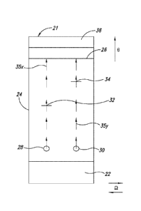
Date Recue/Date Received 2022-11-14
CA 03001918 2018-04-12
WO 2017/066645
PCT/US2016/057150
porous materials containing the dried components needed to perform the test.
These
membranes are assembled in small strips, which can be placed into a plastic
housing
for ease in handling. Lateral flow tests can be used to detect any ligand that
can be
bound to a visually detectable capture reagent attached to a solid support,
both
qualitatively and, in many cases, semi-quantitatively. Some of the more common
lateral flow tests currently on the market are those for pregnancy, strep
throat and
Chlamydia infection. For these conditions a quantitative assay is not
necessary.
[0005] A typical prior art lateral flow assay format is shown in Fig. 1.
The sample
to be tested, such as a biological sample, is loaded onto sample application
pad 10. In
the case of whole blood or capillary blood samples, separation of blood cells
and
plasma takes place on sample pad 10. The sample application pad 10 is
typically
adhered to a rigid or semi-rigid backing card 11. For example, the sample pad
10 may
be laminated to a mylar support film which functions as the backing card 11.
The
liquid fraction of the sample then moves through a conjugate release pad 12
onto
which a conjugate has been dried. The conjugate consists of detection
molecules
specifically directed against the analyte of interest and indicator particles,
such as
colloidal gold or gold sol. Upon contact with the liquid sample, the conjugate
redissolves and specifically binds to any analyte present in the sample to
form an
analyte-conjugate complex. In certain formats a liquid conjugate, such as a
liquid gold
conjugate, is employed and the conjugate pad is omitted (see US Patent
8,399,261).
[0006] The analyte-conjugate complex flows through a capillary membrane
14,
such as a nitrocellulose membrane (also referred to as the analytical
membrane), on
which test and control reagents have been immobilized. More specifically,
membrane
14 is provided with two capture lines, or regions, arranged sequentially and
positioned
perpendicularly to the flow direction theta (0), each containing bound
reagents. Test
line 16 contains analyte-specific molecules which are able to bind to and
immobilize
the analyte-conjugate complex, resulting in a visible colored line. Control
line 18 does
not contain analyte-specific molecules but is able to fix non-bound conjugate-
containing particles. The formation of a colored line at control line 18
indicates that
the test sample has flowed past test line 16. The color intensity observed at
test line
16 is directly proportional to the analyte concentration in the sample and
therefore
2
CA 03001918 2018-04-12
WO 2017/066645
PCT/US2016/057150
enables semi-quantitative interpretation of the test result. If the analyte of
interest is
present at a level above the detection limit, test line 16 and control line 18
both
become clearly visible. If the analyte is present at a level below the
detection limit,
only control line 18 becomes visible during the test
[0007] The last component of the rapid test device is an absorbent pad 20
(also
known as a wicking or sink pad) which collects the fluid flowing through the
test
system and prevents any backflow of fluid. Absorbent pad 20 allows the use of
samples whose volume exceeds the wicking capacity of nitrocellulose membrane
14.
[0008] Traditional lateral flow immunoassays are designed to detect and
measure
a single analyte per test device and therefore detection of multiple analytes
in a single
sample can only be performed sequentially. While such tests are well-
established and
validated techniques, they can be time-consuming, sample-depleting and costly
when
employed to measure numerous analytes per sample. Bead-based immunoassays
utilize the same principle as strip tests but employ uniquely identifiable
beads. These
beads enable simultaneous detection of multiple analytes in a single well or
reaction
but generally require the use of expensive equipment to read the results and
are thus
not suitable for point-of-care or field use. In alternative methods, reagents
that are
specific for multiple different analytes are positioned at specific locations
in an array
(for example in pre-designated wells of a 96-well plate) and portions of a
test sample
are added to each of the wells. Again, these methods are less effective for
point-of-
care or field use than conventional dipstick tests.
[0009] Other multiplexed lateral flow assay systems align multiple lateral
flow
assays, or test strips, into a single large cassette. A liquid test sample is
applied at a
specific location and then divided and directed into multiple separate
channels, with
each channel containing agents for detecting a specific analyte. For example,
US
2013/0280698 discloses a multi-strip assay cartridge in which multiple lateral
flow
assay strips are located within a single housing. A liquid test sample is
introduced into
a diversion dam via an inlet in the housing and subsequently split between
multiple
flow channels, each flow channel being connected to a separate assay chamber
that
contains components necessary for detection of a single analyte. Similarly, US
Patent
8,715,590 discloses a cross-flow analyte assay array in which one or more test
samples
3
CA 03001918 2018-04-12
WO 2017/066645
PCT/US2016/057150
are introduced through at least one test sample input application port and
distributed
through multiple fluid flow manifolds to multiple fluid flow channels
positioned in
parallel rows that are located perpendicular or transverse to the longitudinal
direction
of fluid flow. Such devices are more complex, and therefore more expensive, to
produce than standard dipstick tests.
[0010] Other descriptions of lateral flow assay devices may be found in,
e.g., Sajid
M. et al. Journal of Saudi Chemical Society (2015) v. 19 pp. 689-705 and
references cited
therein; "Design Considerations for Lateral Flow Test Strips" pp. 1-32,
presentation by
Michael A. Mansfield, 24 June 2015; "Rapid Lateral Flow Test Strips,
Considerations for
Product Development" pp. 1-39, copyright 2002, 2008 by Millipore Corporation,
Billerica, MA, available at the website millipore.com/diagnostics. See also
U.S. Patent
Nos. 4313734, 4376110, 4435504, 4703017, 4855240, 4954452, 5028535, 5075078,
8580572, 8846319, 8945838, 9034656, and U.S. Patent Publication Nos.
2015/086974,
2014/0093865, 2013/0017561, 2013/0022969, 2013/0280698, 2012/0040336,
2012/0015350 and 2010/0159599. See also, e.g., PCT Publication Nos.
W02014/184151 and W02011/051562.
[0011] There thus remains a need in the art for a multiplexed lateral flow
assay
system with high specificity and sensitivity that is relatively inexpensive to
produce and
that is both easy to use and stable under a variety of environmental
conditions.
[0012] All of the subject matter discussed in the Background section is
not
necessarily prior art and should not be assumed to be prior art merely as a
result of its
discussion in the Background section. Along these lines, any recognition of
problems in
the prior art discussed in the Background section or associated with such
subject matter
should not be treated as prior art unless expressly stated to be prior art.
Instead, the
discussion of any subject matter in the Background section should be treated
as part of
the inventor's approach to the particular problem, which in and of itself may
also be
inventive.
SUMMARY
[0013] The present disclosure provides a device for performing a multiplex
lateral
flow immunoassay in which a liquid sample, such as a biological sample, is
4
CA 03001918 2018-04-12
WO 2017/066645
PCT/US2016/057150
simultaneously tested for the presence of multiple analytes of interest.
Methods that
employ the device in the simultaneous detection of multiple analytes of
interest
within a liquid test sample are also provided.
[0014] The devices and methods disclosed herein can be employed to detect
the
presence of analytes that are indicative of the presence of disorders or
conditions such
as infectious diseases, pregnancy, microbial infections, cancer, autoinnmune
disorders,
cardiac disorders, allergic disorders, drug abuse, and the like. Infectious
diseases that
can be detected using the disclosed devices and methods include, but are not
limited
to: fever causing agents including malaria, scrub typhus, rickettsia, typhoid
fever,
dengue, and chikungunya; biological select agents such as, but not limited to,
melioidosis, anthrax, and plague; leishmaniasis; tuberculosis; syphilis;
Chagas disease;
encephalitis; leprosy; West Nile virus; Shigella, Campylobacter; and
enterotoxigenic E.
coll. Analytes that can be detected using the disclosed device and methods
include,
but are not limited to, proteins and/or peptides, including ligands and
receptors; non-
protein molecules, such as carbohydrates, phospholipids and nucleic acids;
small
molecules; and other molecules of biological interest.
[0015] In one embodiment, the device described herein comprises at least
two
assay test paths with each assay test path containing a capture reagent (such
as an
antibody or antigen) specific for a specific analyte of interest that is
sprayed onto a
single analytical membrane, such as a nitrocellulose membrane. Additionally,
detection reagents specific for the analytes of interest (such as an antibody
or antigen)
labeled with a reporter agent are spotted/dried in precise locations along the
at least
two assay test paths, with each assay test path containing at least one
labeled
detection reagent specific for an analyte. Each assay test path thus contains
the
components necessary for detecting the presence or absence of a single
specific
analyte. The reporter agent may be any suitable reporter agent known to those
of skill
in the art, for example, colloidal nanoparticles, latex microspheres, quantum
dots,
enzymes, fluorophores and the like, provided that the labeled detection
reagent
possesses a low diffusion constant, D (with an effective membrane diffusion
constant
of typically Dor < 10-8 m2/sec.). Additionally, the assay is constructed such
that the
flow of the liquid test sample (i.e., the effective velocity of the solution)
through the
CA 03001918 2018-04-12
WO 2017/066645
PCT/US2016/057150
membrane is approximately uniform across the lateral axis of the assay by
ensuring
that the liquid sample is uniformly distributed prior to entering the
membrane. This is
most readily achieved by permitting the sample to wet through a sample pad or
treated glass fiber pad that rapidly takes up volume to that the liquid sample
can enter
into the membrane as uniformly as possible.
[0016] Upon addition of a liquid test sample, the dried labeled detector
particles
are solubilized and flow uniformly along the longitudinal axis of the assay.
As a result
of the low diffusion constant of the labeled detection reagents, the lateral
diffusion of
the particles is very limited and thus, due to the fluid mechanics of the
system, specific
lanes, or test paths, of labeled detector particles are created such that each
test path
indicates the presence or absence of an individual analyte in the test sample.
[0017] One or more spots of labeled detection reagents can be present in
any
given test path such that the number of analytes detected in the assay can be
further
multiplied. In such a manner, a very large number of analytes can be evaluated
from a
single test sample using a very small footprint. A single sample entry
application port
is used to apply the liquid test sample. A separate buffer port can optionally
be
present in order to ensure proper flow of the immunoassay. No barrier, e.g.,
no
physical or chemical barrier, is necessarily provided between the multiple
assay test
paths in order to provide physical lanes for multiplexing the assay.
[0018] In addition, multiple spots of labeled detection reagents can be
present in
any given assay test path, thereby having a multiplicative effect on the
number of
analytes that can be detected in a single test sample. As a result, the
present
disclosure provides a very dense, easy to produce, multiplexed assay within a
very
small device footprint.
[0019] In addition, as no physical barrier is necessary to create the
multiplexed
assay test paths, the assay can be performed in a "dipstick" format (i.e.,
without a
plastic housing or enclosure) as no enclosure is necessary to perform the
multiplexed
assay, further reducing manufacturing costs. In some embodiments, the capture
reagents on the membrane are not necessarily "spotted" in order to localize
the assay
reaction (although they may be), but rather are "striped", significantly
increasing the
ease-of-manufacturing. Due to the lack of diffusion along the test paths,
however, an
6
CA 03001918 2018-04-12
WO 2017/066645
PCT/US2016/057150
array indicating the presence of the variety of analytes is still generated
upon the
addition of the test sample.
[0020] In a specific embodiment, a multiplex lateral flow assay device for
simultaneous detection of a presence of at least a first analyte of interest
and a
second, different, analyte of interest in a single liquid test sample is
provided, the
device comprising: (a) a test sample receiving region; and (b) a capture
membrane
comprising a first assay test path and a second, adjacent, assay test path,
the first
assay test path comprising a first labeled detection reagent specific for the
first analyte
of interest, and a first test line comprising an immobilized first capture
reagent specific
for the first analyte of interest, and the second assay test path comprising a
second,
different, labeled detection reagent specific for the second analyte of
interest, and a
second test line comprising an immobilized second, different, capture reagent
specific
for the second analyte of interest, wherein each of the first and second
labeled
detection reagents has a low diffusion constant such that there is little to
no lateral
diffusion of the first and second labeled detection reagents between the first
and
second assay test paths following solubilization by the liquid test sample. In
certain
embodiments, the device further comprises a control line positioned downstream
of
the first and second test lines, the control line comprising an immobilized
control
reagent that binds to the first and second labeled detection reagents.
[0021] While the present disclosure provides test strips and devices that
comprise
a plurality of assay test paths, the present disclosure also provides test
strips and
devices that comprise a single assay test path, and methods for their use. The
single
assay test path may contain one unique solid dried labeled detector reagent.
In
another embodiment the single assay test path contains more than one unique
solid
dried labeled detector reagents, e.g., two reagents, three reagents, or four
reagents.
For example, in one embodiment the present disclosure provides a lateral flow
assay
device for measuring an analyte having a solid support including absorbent
material
for providing capillary flow comprising: a) a test sample receiving region for
receiving
a test sample; b) a capture region comprising one or more solid dried labeled
detector
reagents in one or more localized sub-regions, e.g., spots, c) a test region
comprising a
capture reagent for the analyte; d) a reservoir region comprising absorbent
material
7
CA 03001918 2018-04-12
WO 2017/066645
PCT/US2016/057150
for providing capillary flow; wherein the sample region, capture region, test
region,
and reservoir region are in capillary flow communication, whereby the sample
flows
from the capture region, across the test region, and then into the reservoir
region.
The lateral flow assay device may have one assay test path, or two assay test
paths, or
three assay test paths, or four assay test paths, or more than four assay test
paths,
where each assay test path independently comprises one or more dried labeled
detector reagents. The device does not require the presence of a barrier,
e.g., a
chemical or physical barrier, between assay test paths in order to keep the
assay test
paths distinct from one another, in other words, in a non-overlapping
configuration.
[0022] In certain embodiments, the device includes more than two, for
example,
three, four, five, six or more, different assay test paths, with each assay
test path
containing the labeled detection reagents and capture reagents specific for
different
a nalytes of interest, such that the device can be used to detect the presence
of three,
four, five, six or more different analytes.
[0023] In a related aspect, kits for the simultaneous detection of
multiple analytes,
or components within a single liquid sample, are provided, such kits
comprising a
multiplex lateral flow assay device disclosed herein and, optionally, a
container of a
buffer, packaged together with instructions for using the device and buffer to
detect
the presence or absence of the analytes in a sample, such as a biological
sample.
[0024] In a further aspect, methods for detecting the presence of a
plurality of
analytes of interest in a liquid test sample are provided. In certain
embodiments, such
methods comprise: (a) providing a multiplex lateral flow assay device
described
herein; (b) applying the test sample to the sample receiving region; (c)
optionally
applying a chase buffer to the sample receiving region; (d) allowing the test
sample to
contact a plurality of labeled detection reagents, each labeled detection
reagent being
specific for one of the plurality of analytes, whereby labeled detection
reagent-
analytes are formed if one or more of the analytes is present in the sample;
and (f)
allowing the labeled detection reagent-analytes to migrate through the capture
membrane with each labeled detection reagent-analyte migrating along a
specific
assay test path to a test line that is specific for the specific a nalyte,
wherein formation
of a detectable signal at a specific test line is indicative of the presence
of the specific
8
84239886
analyte in the sample.
[0024a] Some embodiments disclosed herein provide a multiplex lateral flow
assay
device for simultaneous detection of a presence of at least a first analyte of
interest and a
second, different, analyte of interest in a single liquid test sample,
comprising: a) a test
sample receiving region; and b) a capture membrane comprising a first assay
test path and
a second, adjacent, assay test path, the first assay test path comprising a
first labeled
detection reagent specific for the first analyte of interest, and a first test
line comprising
an immobilized first capture reagent specific for the first analyte of
interest, and the second
assay test path comprising a second, different, labeled detection reagent
specific for the
second analyte of interest, and a second test line comprising an immobilized
second,
different, capture reagent specific for the second analyte of interest;
wherein the device
lacks physical or chemical barriers between the first and the second assay
test paths.
[002413] Some embodiments disclosed herein provide a multiplex lateral flow
assay
device for simultaneous detection of a presence of at least a first analyte of
interest and a
second, different, analyte of interest in a single liquid test sample,
comprising: a) a test
sample receiving region; and b) a capture membrane comprising a first assay
test path and
a second, adjacent, assay test path, the first assay test path comprising a
first labeled
detection reagent specific for the first analyte of interest, and a first test
line comprising
an immobilized first capture reagent specific for the first analyte of
interest, and the second
assay test path comprising a second, different, labeled detection reagent
specific for the
second analyte of interest, and a second test line comprising an immobilized
second,
different, capture reagent specific for the second analyte of interest;
wherein the first test
line extends continuously across the capture membrane and intersects with each
assay
path of the capture membrane.
[0024c] Some embodiments disclosed herein provide a method for detecting the
presence of a first analyte of interest and a second, different, analyte of
interest in a liquid
test sample, comprising: a) providing a multiplex lateral flow assay device as
described
herein; b) applying the liquid test sample to the test sample receiving
region; c) allowing
the liquid test sample to contact the first and second labeled detection
reagents, whereby
labeled detection reagent-analyte conjugates are formed if one or more of the
first and
second analytes is present in the sample; and d) allowing the labeled
detection reagent-
9
Date Recue/Date Received 2021-10-14
84239886
analyte conjugates to migrate through the capture membrane along the first and
second
assay test paths to the first and second test lines, wherein formation of a
detectable signal
at the first and/or second test lines is indicative of the presence of the
first and/or second
analytes in the liquid test samples.
10024d] Some embodiments disclosed herein provide a method for detecting the
presence of a first analyte of interest and a second, different, analyte of
interest in a liquid
test sample, comprising: e) providing a multiplex lateral flow assay device as
described
herein; f) applying the liquid test sample to the test sample receiving
region; g) allowing
the liquid test sample to contact the first and second labeled detection
reagents, whereby
labeled detection reagent-analyte conjugates are formed if one or more of the
first and
second analytes is present in the sample; and h) allowing the labeled
detection reagent-
analyte conjugates to migrate through the capture membrane along the first and
second
assay test paths to the first and second test lines, wherein formation of a
detectable signal
at the first and/or second test lines is indicative of the presence of the
first and/or second
analytes in the liquid test samples.
[0025] The above-mentioned and additional features of the present
invention and the
manner of obtaining them will become apparent, and the invention will be best
understood by reference to the following more detailed description.
[0026] This Brief Summary has been provided to introduce certain concepts
in a
simplified form that are further described in detail below in the Detailed
Description.
Except where otherwise expressly stated, this Brief Summary is not intended to
identify
key or essential features of the claimed subject matter, nor is it intended to
limit the scope
of the claimed subject matter.
[0027] The details of one or more embodiments are set forth in the
description below.
The features illustrated or described in connection with one exemplary
embodiment may
be combined with the features of other embodiments. Thus, any of the various
embodiments described herein can be combined to provide further embodiments.
Aspects
of the embodiments can be modified, if necessary to employ concepts of the
various
patents, applications and publications as identified herein to provide yet
further
embodiments. Other features, objects and advantages will be apparent from the
description, the drawings, and the claims.
9a
Date Regue/Date Received 2022-11-14
84239886
BRIEF DESCRIPTION OF THE DRAWINGS
[0028]
Exemplary features of the present disclosure, its nature and various
advantages
will be apparent from the accompanying drawings and the following detailed
description
of various embodiments. Non-limiting and non-exhaustive embodiments are
described
with reference to the accompanying drawings, wherein like labels or reference
numbers
refer to like parts throughout the various views unless otherwise specified.
The sizes and
relative positions of elements in the drawings are not necessarily drawn to
scale. For
example, the shapes of various elements are selected, enlarged, and positioned
to improve
drawing legibility. The particular shapes of the elements as drawn have been
selected for
ease of recognition in the drawings.
9b
Date Regue/Date Received 2022-11-14
CA 03001918 2018-04-12
WO 2017/066645
PCT/US2016/057150
One or more embodiments are described hereinafter with reference to the
accompanying drawings in which:
[0029] Fig. 1 shows a typical prior art lateral flow assay device.
[0030] Fig. 2A shows a multiplex lateral flow assay device disclosed
herein.
[0031] Fig. 2B shows a multiplex lateral flow assay device disclosed
herein.
[0032] Fig. 3A shows a perspective view of a multiplex lateral flow assay
device of
the present disclosure.
[0033] Fig. 3B shows an exploded side view of the multiplex lateral flow
assay
device of Fig. 3A.
[0034] Fig. 3C shows a perspective view of a multiplex lateral flow assay
device of
the present disclosure.
[0035] Fig. 3D shows a capture membrane of the present disclosure with
flanking
test sample receiving region and reservoir region according to the present
disclosure.
[0036] Fig. 3E in panels (a), (b), (c) and (d) shows four options for
placing spots in
the capture region of a capture membrane according to the present disclosure.
[0037] Fig. 3F in panels (a), (b), (c) and (d) shows several options for
constructing
test lines in a test region of a capture membrane according to the present
disclosure.
[0038] Fig. 4A shows a multiplex lateral flow assay device of the present
disclosure
enclosed in a plastic housing.
[0039] Fig. 4B shows a multiplex lateral flow assay device of the present
disclosure
enclosed in a plastic housing.
[0040] Fig. 5A shows the result of assaying a test sample in a device of
the present
disclosure, where the test sample does not contain any analytes of interest.
[0041] Fig. 5B shows the result of assaying a test sample in a device of
the present
disclosure, where the test sample contains four analytes of interest.
[0042] Fig. 6A shows the results of the evaluation of a single (spiked)
specimen for
four different target molecules simultaneously using a multiplex lateral flow
assay
device of the present disclosure.
[0043] Fig. 6B shows an example of a multiplex lateral flow assay device
of the
present disclosure wherein multiple detector reagents are present in each
lane.
CA 03001918 2018-04-12
WO 2017/066645
PCT/US2016/057150
DETAILED DESCRIPTION OF THE INVENTION
[0044] The present invention may be understood more readily by reference
to the
following detailed description of preferred embodiments of the invention and
the
Examples included herein.
[0045] The present invention provides devices and methods for detecting
the
presence of multiple analytes simultaneously in a sample, preferably a
biological or
environmental sample. As used herein, the term "analyte" encompasses proteins
and/or peptides, including ligands and receptors; antibodies or antigen-
binding
fragments thereof; non-protein molecules, such as carbohydrates, phospholipids
and
nucleic acid molecules; small molecules; and other molecules of biological
interest.
Examples of biological samples that can be tested using the disclosed devices
and
methods include, but are not limited to, whole blood, serum, plasma, nasal
secretions,
sputum, urine, saliva, transdermal exudates, cerebrospinal fluid, and vaginal
or
urethral secretions. Fecal samples can also be tested following suitable
processing.
Examples of environmental samples that can be tested using the disclosed
devices and
methods include, but are not limited, to soils and foodstuffs. Those of skill
in the art
will appreciate that solid and/or powdered materials can be tested following
suspension in an appropriate buffer).
[0046] The term "test region" as used herein refers to a discrete location
on a
lateral flow test strip which is interrogated in order to generate a signal
related to the
presence or amount of an analyte of interest. Such interrogation may be
performed
visually as in an over-the-counter pregnancy test, or in an instrumented
fashion as
through the detection of reflectance, absorption, fluorescence, luminescence,
etc. by a
suitably configured meter.
[0047] The terms "proximal" and "distal" are not used in any functional
sense, but
rather simply to distinguish the two ends of the membrane or the test strip or
the
device of the present disclosure.
[0048] An embodiment of a lateral flow assay device 21 of the present
disclosure
for use in detecting two different analytes (analytes X and Y) is shown in
Fig. 2A.
Device 21 comprises a test sample receiving region which comprises a sample
pad 22,
which may also be referred to as a sample application pad 22. The sample pad
22
11
CA 03001918 2018-04-12
WO 2017/066645
PCT/US2016/057150
receives a liquid test sample suspected of containing at least one of the two
different
analytes. In one embodiment, sample pad 22 is used to buffer test samples for
optimal reaction with immobilized detection reagents as detailed below,
comprises a
layer of support material that is capable of serving as a template for
conjugate and
sample application. In one embodiment, sample pad 22 may include at least one
layer
of material that aids in providing consistent liquid flow, wetting, buffering
and pH
maintenance of fluids, and/or aids in biological sample separation. In one
embodiment, for serum and plasma based assays, a single layer of material that
helps
with consistent liquid flow, buffering, wetting and step wise mixing process
can be
used. In one embodiment, for assaying blood samples, sample pad 22 may include
additional materials or treatments that can separate blood cells. Examples of
appropriate materials are well known in the art.
[0049] Fluid flows in a theta (0) direction from sample pad 22 laterally
to, and
downstream through, membrane 24 which is provided with two test lines 32 and
34,
and a control line 26. The test lines 32 and 34 may extend partially across
the device
21 as shown in FIG. 2A, or the test lines 32 and 34 may be in the form of
stripes that
extend continuously across the device 21 as shown in FIG. 28. Membrane 24 is
formed of materials generally employed in lateral flow test devices and well
known to
those of skill in the art, such as nitrocellulose, nylon which may optionally
be charged-
modified, and cellulose acetate. Nitrocellulose is a preferred membrane for
the
devices and methods of the present disclosure. Other suitable materials
include
polyvinylidene fluoride membrane, polyethersulfone membrane, porous
polyethylene
sheets, and glass fiber mats.
[0050] Following application of labeled detection reagents at regions 28
and 30,
test lines 32 and 34, and control line 26, membrane 24 may be laminated with a
series
of synthetic and/or natural paper products of appropriate sizes and
porosities.
[0051] A first labeled detection reagent, such as a labeled antibody, that
is specific
for analyte Xis spotted and dried onto membrane 24 at region 28. Similarly, a
second,
different, labeled detection reagent that is specific for analyte Y is spotted
and dried
onto membrane 24 at region 30. A first capture reagent that is specific for
analyte X is
immobilized on membrane 24 at test line 32 and a second capture reagent that
is
12
CA 03001918 2018-04-12
WO 2017/066645
PCT/US2016/057150
specific for analyte Y is immobilized at test line 34. The immobilized capture
reagents
may be either spotted or striped completely across the membrane.
[0052] Control line 26, which is used as an internal control to ensure
that all the
test components are working, comprises molecules that bind to both of the
detection
reagents irrespective of the presence or absence of the analytes. For example,
for
antigen-antibody interactions, control line 26 may comprise anti-Protein A or
human
IgG immobilized on membrane 24.
[0053] An absorbent pad 36 is provided at, or in proximity to, the end of
the flow
path. Pad 36 absorbs any excess fluid and prevents any backflow of fluid
towards
sample pad 22. The absorbent pad 36 is located in the reservoir region of the
device,
which is positioned downstream of the capture membrane and provides a place
for
absorbing excess liquid, for example, excess liquid from either or both of the
test
sample and the chase buffer.
[0054] The liquid test sample contacts each of the two labeled detection
reagents
at regions 28 and 30 where the labeled detection reagents are mixed and, if
either of
analytes X and Y is present in the test sample, labeled detection reagent-a
nalyte
conjugates are formed. The labeled detection reagent-analyte conjugates and
non-
conjugated labeled detection reagents then flow longitudinally through the
device,
i.e., in the theta (0) or downstream direction, with little to no
perpendicular flow, i.e.,
little to no flow in an omega (0) direction, such that individual and non-
overlapping
assay test paths 35x and 35y are formed for each of analytes X and Y,
respectively.
The first and second labeled detection reagents preferably have a low
diffusion
constant such that there is little to no lateral diffusion, i.e., diffusion in
an omega (0)
direction as identified in Fig. 2A and Fig. 2B, of the first and second
labeled detection
reagents between the first and second assay test paths following
solubilization by the
liquid test sample. For clarity, and with reference to Fig. 2A, assay test
path 35x runs
in a 0 direction from spot 28 to the control line 26, where the location of
assay test
path 35x is illustrated by a series of arrow-terminated longitudinal lines
collectively
referred to as feature 35x. Likewise, assay test path 35y runs in a 0
direction from
spot 30 to the control line 26. Once the flow reaches test lines 32 and 34,
any labeled
detection reagent-a nalyte conjugates bind to the capture reagents and become
13
CA 03001918 2018-04-12
WO 2017/066645
PCT/US2016/057150
immobilized, resulting in detectable colored lines or rectangles at test lines
32 and 34.
The non-conjugated labeled detection reagents continue to travel along the
individual
assay test paths and bind to, and are immobilized at, control line 26
resulting in a
detectable colored line. If a colored line is not observed at control line 26,
the test is
considered invalid. Excess liquid is then taken up in the reservoir region
which holds
an absorbent pad 36.
[0055] Multiple spots of labeled detection reagents may be present in any
given
assay test path, thereby having a multiplicative effect on the number of
analytes that
can be detected in a single test sample. As a result, the present disclosure
provides a
very dense, easy to produce, multiplexed assay within a very small device
footprint. In
one embodiment, the size of the test strip of the present disclosure is about
40-80 mm
in length about 10-30 mm in width, while in another embodiment the test strip
is
about 50-70 mm in length and about 15-25 mm in width. In one embodiment, the
capture membrane is about 10-40 mm in length, while in another embodiment the
capture membrane is about 20-30 mm in length.
[0056] The spots of labeled detection reagents are present in localized
geographical regions of the capture region of the capture membrane, or in
other
words, localized geographical regions refer to spots. The size of each spot is
about 1-
50 mm2. In various embodiments, a spot occupies an area of about 1 mm2, or 2
mm2,
or 3 mm2, or 4 mm2, or 5 mm2, or 6 mm2, or 7 mm2, or 8 mm2, or 9 mm2, or 10
mm2,
or 11 mm2, or 12 mm2, or 13 mm2, or 14 mm2, or 15 mm2, or 16 mm2, or 17 mm2,
or
18 mm2, or 19 rrinn2, or 20 mm2, or 30 mm2, or 40 mm2, or 50 mm2, where the
size of
the spot may be described by a range selected from any two of the stated
values, e.g.,
a range of 10-15 mm2. The spot may have a symmetrical shape, e.g., a circular,
square
or rectangular shape. When the spot has the form of a circle, the circle may
have a
diameter of about 0.5-5 mm2, or about 1-3 mm2. In one embodiment, the spots
present in the capture region do not overlap with one another.
[0057] The spots may be placed at a distance from one another, where that
distance is measured by the distance between a mid-point of each spot, where
those
two spots are the most closely located spots, and the distance is about 1-10
mm. In
various embodiments, the spots are located a distance from one another of at
least 1
14
CA 03001918 2018-04-12
WO 2017/066645
PCT/US2016/057150
mm, or 2 mm, or 3 mm, or 4 mm, or 5 mm, or 6 mm, or 7 mm, or 8 mm, or 9 mm, or
mm, and not more than about 30 mm, or 25 mm, or 20 mm, or 15 mm, or 10 mm,
or 9 mm, or 8 mm, or 7 mm, or 6 mm, or 5 mm, or 4 mm, or 3 mm, where the
distance
between closest spots may also be described as being within a range between
two
values selected from the stated values, e.g., a distance of 1-5 mm.
[0058] Thus, in one embodiment the test strip of the present disclosure
has four
assay paths, each assay path beginning with a spot comprising dry labeled
detection
reagent and optionally being in the shape of a circle having a diameter of
about 1.5-2.5
mm, where two closest spots are about 2.5-3.5 mm from one another measured
from
the center of each of the two closest spots.
[0059] In another embodiment the test strip of the present disclosure has
three
assay paths, each assay path beginning with a spot comprising dry labeled
detection
reagent and optionally being in the shape of a circle having a diameter of
about 1.5-2.5
mm, where two closest spots are about 2.5-3.5 mm from one another measured
from
the center of each of the two closest spots.
[0060] In another embodiment the test strip of the present disclosure has
two
assay paths, each assay path beginning with a spot comprising dry labeled
detection
reagent and optionally being in the shape of a circle having a diameter of
about 1.5-2.5
mm, where two closest spots are about 2.5-3.5 mm from one another measured
from
the center of each of the two closest spots.
[0061] In another embodiment the test strip of the present disclosure has
one
assay path, the assay path beginning with a spot comprising dry labeled
detection
reagent and optionally being in the shape of a circle having a diameter of
about 1.5-2.5
mm.
[0062] When a second spot is located on a single assay test path, that
second spot
may be about 2.5-3.5 mm distant from the first spot, and of essentially the
same size
as the first spot.
[0063] Fig. 3A shows a test strip 21 of the present invention. The test
strip 21 and
components thereof have a proximal end and a distal end, where the flow
direction 0
of a test sample is from the proximal end to the distal end of the test strip
or
component thereof. When the observer looks down onto the test strip 21, the
test
CA 03001918 2018-04-12
WO 2017/066645
PCT/US2016/057150
strip 21 and components thereof have a left edge and a right edge, where the
proximal end is closest to the observer and the distal end is furthest from
the
observer.
[0064] The test strip 21 comprises a sample application pad 22 which is
upstream
from and in direct contact with a porous membrane 24. The pad 22 may be
prepared
from materials known in the art for this purpose, e.g., woven meshes and
cellulose
filters. Suitable materials for an application pad are available from
Ahlstronn
Corporation (Helsinki, Finland), for example, their CytoSep media may be used
to
form an application pad 22. CytoSep media has the property that it is a
single layer
media consisting of high purity natural and synthetic fibers, where the
untreated
media contains no chemical interfering substances and shows to significant
binding of
plasma components. CytoSep media retains red blood cells while allowing serum
to
flow rapidly. In one embodiment the application pad is a cellulose filter.
[0065] The test strip 21 optionally comprises a backing card 23, which may
also be
referred to as a support card or support film. The backing card is preferably
impermeable to water. The sample application pad 22 and other features of the
test
strip 21 may be adhered to the backing card 23. The backing card 23 is rigid
or semi-
rigid so that the test strip maintains a flat shape. The backing card may be
formed
from materials known in the art for this purpose, e.g., mylar.
[0066] The test strip 21 comprises a porous membrane 24. The membrane 24
allows a flow of aqueous test sample and, when used, chase buffer, from a
proximal
end of the membrane, i.e., the end of the membrane in contact with the sample
application pad, to the furthest opposite end of the membrane, i.e., the
distal end of
the membrane.
[0067] The membrane 24 of test strip 21 comprises control line 26. Control
line
26, which is used as an internal control to ensure that all the test
components are
working, comprises molecules that bind to both of the detection reagents
irrespective
of the presence or absence of the analytes. For example, for antigen-antibody
interactions, control line 26 may comprise anti-Protein A or human IgG
immobilized on
membrane 24.
[0068] The membrane 24 of test strip 21 comprises one or more spots at the
16
CA 03001918 2018-04-12
WO 2017/066645
PCT/US2016/057150
proximal end of the membrane which contain solid labeled detection reagents
that is
or are specific for analyte(s) of interest. In Fig. 3A, two spots 28 and 30
are shown.
[0069] The membrane 24 of test strip 21 comprises one or more test lines
located
between the control line 26 and the spots 28 and 30. Each test line contains
an
immobilized capture reagent that is specific for an analyte of interest. In
Fig. 3A, two
test lines 32 and 34 are shown.
[0070] The test strip 21 comprises an absorbent pad 36. The absorbent pad
36 is
located at the distal end of the test strip shown in Fig. 3A. The primary
function of the
absorbent pad is to absorb the water and solubilized components present in the
test
sample and the chase buffer after they pass through the test lines and the
control line.
As the desired volume of test sample and/or chase buffer is increased, the
holding
capacity of the absorbent pad should likewise be increased. A suitable
absorbent pad
36 may be prepared from, e.g., cellulose filters. The flow of liquid into the
absorbent
pad may not be laminar, which leads to uneven flow of the solvent front down
the
membrane. To address the consequences of a non-laminar flow, in one embodiment
the test strips of the invention include an intermediate absorbent pad (not
shown in
Fig. 3A) which is located between the absorbent pad 36 and the distal end of
the
membrane 24. The intermediate absorbent pad may be more porous than the
absorbent pad, thereby allowing entering solvent to evenly distribute in a
direction
perpendicular to the flow of the solvent. After passing through the
intermediate
absorbent pad, the solvent and dissolved components more evenly enter the
absorbent pad, i.e., enter the absorbent pad 36 with an enhanced laminar flow.
[0071] In use, the sample application pad 22 can receive both the test
sample and
thereafter receive the chase buffer. However, in one embodiment of the test
strip of
the invention, a separate buffer pad 38 is provided to receive the chase
buffer. The
buffer pad 38 is located upstream of the application pad 22, at the proximal
end of the
test strip 21. The buffer pad may be made from the same materials that are
used to
prepare the application pad. However, by having the application pad 22
separate from
the buffer pad 38 it is possible to select different materials for the two
different pads,
and/or differentially treat the application pad 22 and the buffer pad 38 so
that they
have different properties.
17
CA 03001918 2018-04-12
WO 2017/066645
PCT/US2016/057150
[0072] The buffer pad 38 may optionally be located directly next to the
application
pad 22 (not shown in Fig. 3A) or alternatively a hydrophobic pad 39 may be
positioned
between the application pad 22 and the buffer pad 38 as shown in Fig. 3A. In
one
embodiment the hydrophobic pad 39 has a different hydrophobicity compared to
the
hydrophobicity of the application pad 22. In one embodiment the hydrophobic
pad 39
has a different hydrophobicity compared to the hydrophobicity of the buffer
pad 38.
In one embodiment, the hydrophobic pad 39 is more hydrophobic compared to the
hydrophobicity of each of the buffer pad 38 and the application pad 22, i.e.,
the buffer
pad 38 and the application pad 22 are each less hydrophobic than the
hydrophobic
pad 39. The relative hydrophobicity of two adjacent pads is readily determined
by
placing an aqueous sample onto one or both of the adjacent pads: the aqueous
sample will tend to migrate to the more hydrophilic pad, i.e., the less
hydrophobic
pad, all other factors being equal.
[0073] Fig. 3B shows an exploded side-view of the test strip 21 of Fig.
3A. In Fig.
3A, 22 is the sample application pad, 23 is the backing card, 24 is the
capture
membrane, 36 is the absorption pad, 38 is the buffer pad and 39 is the
hydrophobic
pad.
[0074] Fig. 3C shows a view of an embodiment of a test strip of the
present
invention. The test strip comprises a membrane 24. At the distal end of the
test strip
is located an absorption pad 36. Positioned between the membrane 24 and the
absorption pad 36 is an intermediate absorption pad 40. In one embodiment, the
intermediate pad 40 is more porous than the pad 36. In one embodiment, the
intermediate pad 40 is more hydrophilic than the pad 36. The hydrophilicity of
the
intermediate pad may be increased by adding detergent or surfactant to the
intermediate pad. The pad 36 has a larger volume than the pad 40 and so liquid
will
preferentially wick into and remain in the pad 36. The presence of the
intermediate
pad 40 may enhance the laminar flow of the liquid as it travels down the test
strip,
thus providing a more defined readout.
[0075] At the proximal end of the test strip is located a buffer pad 38
which may
be seen to have three regions, 38a, 38b and 38c. Region 38a is laminated to
the
backing card 23. Region 38c sits on top of hydrophobic pad 39. By sitting on
top of
18
CA 03001918 2018-04-12
WO 2017/066645
PCT/US2016/057150
the hydrophobic pad 39, the buffer pad 38 is more readily able to transfer
chase buffer
in a downstream direction. Region 38b transitions between regions 38a and 38c.
Adjacent to the buffer pad 38, in a downstream direction, is the hydrophobic
pad 39
which is seen to have two regions, 39a and 39b. Region 39b sits on top of
application
pad 22. By sitting on top of the application pad 22, there is greater contact
between
the hydrophobic pad 39 and the application pad 22, and therefore the
hydrophobic
pad 39 is more readily able to transfer chase buffer in a downstream
direction. Region
39a of the hydrophobic pad transitions between a region (not shown) of the
hydrophobic pad which is laminated to the backing card 23, and the region 39b
which
sits on top of the application pad 22. Adjacent to the hydrophobic pad 39, in
a
downstream direction, is the application pad 22 which may be seen to have
three
regions, 22a, 22b and 22c. Region 22a is laminated to the backing card 23.
Region 22c
sits on top of the membrane 24. By sitting on top of the membrane 24, there is
more
contact between the application pad 22 and the membrane 24, and therefore the
application pad 22 is more readily able to transfer sample to the membrane 24.
Region 22b of the application pad transitions between regions 22a and 22c. The
pads
and regions in the devices of the present may be said to be in operable fluid
communication with one another since liquid is able to flow from one location
to
another location on the device.
[0076] Fig. 3D illustrates an embodiment of the invention comprising two
spots of
solid labeled detection reagents, specifically spots 28 and 30, which are
located on the
capture membrane 24. The capture membrane 24 is flanked on opposing sides by
the
sample application pad 22 at the proximal end of the capture membrane 24 and
by the
absorption pad 36 at the distal end of the capture membrane 24. Fig. 3D shows
how
the capture membrane 24 may be divided into functional regions, namely region
42
which is referred to as the capture region, region 44 which is referred to as
the test
region, and region 46 which is referred to as the control region. Thus, Fig.
3D shows a
test sample application pad 22, a capture membrane 24, and an absorption pad
36,
the application pad located directly adjacent to a proximal end of the capture
membrane 24, the absorption pad located at a distal end of the capture
membrane 24,
where a test sample flows in a downstream direction 0 from the test sample
19
CA 03001918 2018-04-12
WO 2017/066645
PCT/US2016/057150
application pad to the absorption pad, where the capture membrane comprises i)
a
capture region directly adjacent to the application pad, ii) a test region
directly
adjacent to the capture region and not adjacent to the application pad, and
ii) a
control region directly adjacent to the test region and not adjacent either to
the test
region or the capture region.
[0077] While Fig. 3D shows a capture membrane with two spots 28 and 30,
each
spot comprising a unique solid labeled detection reagents, the invention
provides that
any of 1, 2, 3, 4, 5, 6, 7, 8, 9, 10, or more spots comprising solid labeled
detection
reagents may be present in the capture region. In various embodiments, the
capture
membrane of a test strip of the invention has 1-8 spots, or 2-6 spots, or at
least 2
spots, or at least 3 spots, or at least 4 spots, or at least 5 spots, or at
least 6 spots, or at
least 7 spots, or 8 spots. Those spots will be distinct from one another, in
other words,
two spots do not overlap.
[0078] After the test sample enters the capture region, it will contact
the various
spots and then continue onwards in the 0 direction. Contact between a spot and
the
test sample initiates an assay test path, at least so long as the assay test
path has not
already been initiated by a different spot. In other words, if an assay test
path is
initiated at a first spot, the fact that the assay test path passes through a
second spot
does not cause the initiation of a second assay test path: the second spot is
located
within the assay test path initiated by the first spot. An assay test path
starts at a spot
and then extends from that spot in a downstream direction, the test path
running in a
substantially straight line towards the distal end of the capture membrane.
Thus, if a
second spot is located within an assay test path initiated by a first spot,
the second
spot does not initiate a new assay test path. Each assay test path runs in a
longitudinal
direction, i.e., in the direction 0 after being initiated at a spot.
Preferably, no two
assay test paths overlap with one another, i.e., a test path has little or no
movement in
an CI direction which is perpendicular to the flow of the sample.
[0079] The spots may be placed at any locations within the capture region,
so long
as they do not overlap with one another. For example, as illustrated in Fig.
3E, the
capture region may have 4 spots in a straight line (a); 4 spots in a staggered
arrangement (b); 5 spots in an "M" arrangement (c); or 8 spots placed in two
parallel
CA 03001918 2018-04-12
WO 2017/066645
PCT/US2016/057150
lines (d), where these are four examples selected from a large number of
possibilities.
The arrangement of spots in (a) gives rise to four assay test paths; the
arrangement of
spots in (b) gives rise to four assay test paths, the arrangement of spots in
(c) gives rise
to three assay test paths, and the arrangement of spots in (d) gives rise to
four assay
test paths. In Fig. 3E the spots are shown as having a circular shape, and
indeed that is
one optional shape for the spots. However, the spots may adopt other shapes,
e.g.,
square, rectangle, oval, triangular, etc.
[0080] In the various embodiments of the invention disclosed herein, each
spot
may contain a unique solid labeled detection reagent which will react with a
unique
analyte of interest that is potentially present in the test sample. However,
in an
optional embodiment the same solid labeled detection reagent may be present in
two
or more different spots, where this embodiment may be useful to confirm the
result
observed from having the solid labeled detection reagent in only a single
spot, i.e., to
provide a duplicative result in order to enhance the observer's confidence in
the test
results. In another optional embodiment, a single spot may contain more than
one
unique solid labeled detection reagent, i.e., a single spot may contain
multiple unique
detection reagents which are specific for multiple unique analytes, e.g., a
spot may
contain two different solid labeled detection reagents, one of which reacts
with
analyte X and the other of which reacts with analyte Y. Thus, each spot in the
capture
region may contain 1, 2, or more unique labeled detection reagents, each of
which is
specific for a different analyte, and furthermore, any two spots may contain
the same
labeled detection reagent. In one embodiment each spot contains a different
labeled
detection reagent. In one embodiment none of the spots contains more than one
labeled detection reagent, so that each spot contains reagent that is specific
for only
one analyte.
[0081] After being initiated at a spot in the capture region, the assay
test path
extends through the test region. Each assay test path will extend across one
or more
test lines located in the test region. Each of the one or more test lines that
are present
in the test region 44 of a capture membrane 24 of the invention may be striped
continuously across the membrane or may be localized at one or more locations
across the membrane. In reference to test lines in the test region, across
refers to the
21
CA 03001918 2018-04-12
WO 2017/066645
PCT/US2016/057150
0 direction, which is perpendicular to the flow of the sample 0. As a few
examples of
test line configurations are:
a. each of two test lines i and ii may extend fully and continuously across
the membrane (a); or
b. test line i may extend partially across the membrane in a number of
distinct locations such as three distinct locations while test line ii
extends fully and continuously across the membrane (b); or
c. test line i may be continuous across a portion of the membrane and
then localized at two locations, while test line ii is localized at four
locations (c); or
d. test line i may extend continuously but not completely across the
membrane, and test line ii may be localized at a single location along
the test line (d).
[0082] While Fig. 3F shows four test regions each having two test lines, a
test
region of any of the embodiments of the present disclosure may have exactly 1,
or
exactly 2, or exactly 3, or exactly 4, or exactly 5, or exactly 6, or exactly
7, or exactly 8,
or exactly 9, or exactly 10, or more than 10 test lines. Also, while Fig. 3F
shows four
pairs of test line configurations, each test line in a test region may have a
configuration
that is independent from the configuration of another test line in the same
test region.
[0083] Detection reagents that can be effectively employed in the
disclosed
devices are well known to those of skill in the art and include antigens,
antibodies,
nucleic acid molecules, and other relevant protein or non-protein molecules.
For
example, the detection reagent can comprise an antibody that specifically
binds to a
known disease antigen. Each of the detection reagents is labeled with a
reporter
agent.
[0084] Examples of reporter agents that can be used in the devices, kits
and
methods disclosed herein include, but are not limited to, colloidal
nanoparticles (such
as gold nanoparticles), latex microspheres, quantum dots, enzymes,
fluorophores and
the like. Descriptions of gold nanoparticles can be found in, e.g., Colloidal
Gold:
Principles, Methods, and Applications, Vol. 1, Editor M.A. Hayat, Academic
Press
(1989) and "Nanoparticles in Biology and Medicine, Methods and Protocols"
Editor
22
CA 03001918 2018-04-12
WO 2017/066645
PCT/US2016/057150
Mikhail Soloviev, Springer Protocols, Methods in Molecular Biology, vol. 906
(2012),
e.g., Chapter 4. A description of fluorescent europium(III) nanoparticles and
colloidal
gold reporters can be found in, e.g., Juntunen, E., et al., Analytical
Biochemistry,
428(1):31-38 (2012). A description of iron nanoparticles can be found in,
e.g., Liu, C. et
al., Anal. Chem., 83(17):6778-6784 (2011). A description of reporter agents
detectable by near-infrared spectroscopy is described in, e.g., Swanson, C.
and
D'Andrea, A., Clinical Chemistry, 59(4):641-648 (2013). A description of
reporter
agents detectable by fluorescence is described in, e.g., Xu, Y. et al., Anal.
Chem.,
86(12):5611-5614 (2014). Descriptions of quantum dots are described in, e.g.,
Fabio
Cimaglia, F., et al., Nanotechnology Development, 2(1):26-30 (2012). In
certain
embodiments, the reporter agent has a particle diameter greater than or equal
to 8
nnn, such as 20 nm or 40 nnn. In certain embodiments, particles labeled with
enzymes
that may provide enhanced signals upon the addition of a substrate in this
same
multiplexed format are included.
[0085] The reporter agent and/or labeled detection reagent preferably has
a
sufficiently low diffusion constant (with an effective membrane diffusion
constant of
typically Deff < 10' m2/sec) whereby when a liquid sample containing the
analyte of
interest contacts the labeled detection reagent, the resulting labeled
detection
reagent-analyte conjugate is carried in a generally longitudinal,
unidirectional, nearly
uniform flow based on capillary action towards the test and control lines but
with little
to no perpendicular flow (in the direction) towards the lateral edges of
membrane
24.
[0086] To place the labeled detection reagents in the capture region of a
capture
membrane according to the present disclosure, the labeled detection reagents
may be
prepared in solution form, and then an aliquot of that solution deposited onto
the
capture membrane to effective create a spot on the capture membrane, where
that
spot comprises the labeled detection reagent. Initially the spot will include
solvent,
which is typically water and may optionally include other solvents. Thus, the
spot will
initially be wet. However, the solvent will evaporate from the spots located
on the
capture membrane thereby leaving behind spots comprising dried labeled
detection
reagents. In one embodiment, the present invention provides spots comprising
23
CA 03001918 2018-04-12
WO 2017/066645
PCT/US2016/057150
labeled detection reagents that are in solvent-free form. The solutions used
to create
the spots may be referred to herein as spotting solutions.
[0087] In one embodiment, the spotting solution are aqueous solutions,
i.e., on a
weight basis they contain mostly water. In addition, the spotting solutions
contain
labeled detection reagents. Such reagents are designed to react with an
analyte of
interest present in a test sample, and to have a reporter group that can be
visualized
in order to allow determination of whether the labeled detection reagent did
or did
not react with an analyte of interest. Labeled detection reagents are well
known in
the art, and the manufacture and use of aqueous compositions comprising
labeled
detection reagents is well known in the art. Labeled detection reagents are
sometimes referred to by terms, such as detector reagents, labeled
biorecognition
molecules, recognition element and labelled analyte, by those skilled in the
art.
However, the prior art typically places a labeled detection reagent in or on
the
conjugate pad component of a device used in a lateral flow assay, prior to
initiating
the assay by adding the test sample, while the present invention provides
devices and
methods wherein the labeled detection reagent is placed directly on the
capture
membrane, e.g., a nitrocellulose membrane, prior to initiating the assay, and
in one
embodiment entirely omits the presence and use of a conjugate pad.
[0088] In addition to water and labeled detection reagent, a spotting
solution of
the present disclosure may include one or more water-soluble, non-volatile
organic
molecules (WNO). A WNO is soluble in water, and when measured at 20 C, has a
solubility of at least 1g/100g of water, or at least 5g/100g of water, or at
least
10g/100g of water, or at least 20g/100g water, or at least 30g/100g water, or
at least
40g/100g water, or at least 50g/100g water. In one embodiment the WNO is a
polyhydric compound, i.e., it contains a plurality of hydroxyl groups. In one
embodiment, the WNO comprises a monosaccharide or a polysaccharide such as a
disaccharide. Exemplary saccharide WNOs include the nnonosaccharides fructose,
glucose and galactose, the disaccharides sucrose, lactose, maltose, trehalose,
and the
polysaccharides starch, dextrin, cellulose, pectin and glycogen. In various
embodiments of the invention, the spotting solution contains at least 1 wt%
WNO, or
at least 2.5 wt% WNO, or at least 5 wt% WNO, or at least 7.5 wt% WNO, or at
least 10
24
CA 03001918 2018-04-12
WO 2017/066645
PCT/US2016/057150
wt% WNO, or at least 15 wt%, including ranges selected from these values,
e.g.,
between 10 and 15 wt%. In one embodiment the VSNFOM comprises a disaccharide,
and optionally comprises a mixture of two or more disaccharides, e.g., 5%
sucrose and
5% trehalose, or other combinations of disaccharides.
[00891 In one embodiment the WNO comprises polyvinylalcohol (PVA) which
may
optionally include vinyl acetate units, i.e., some of the hydroxyl groups in
the PVA may
be acetylated. The water solubility of PVA depends in large part on its degree
of
hydrolysis (more highly acetylated PVA tends to have lower water solubility),
and on
its molecular weight (lower molecular weight tends to be more water soluble).
The
spotting solutions of the present invention may contain PVA, where the PVA may
be
present at a concentration of, e.g., 0.1 wt%, or 0.25 wt%, or 0.5 wt%, or 0.75
wt%, or
1.0 wt%, or 1.25 wt%, or 1.5 wt%, or 1.75 wt%, or 2 wt%, including ranges
selected
from these values, e.g., 0.754.25 wt% and 0.54.5 wt%.
[00901 In one embodiment, the spotting solution contains 7.542.5 wt%
disaccharide and 0.5-2.0 wt% PVA, e.g., about 10 wt% disaccharide and about 1
wt%
PVA. The disaccharide may be two different disaccharides, e.g., sucrose and
trehalose,
and the two different disaccharides may be present at a weight ratio of 1:10
to 10:1,
e.g., 1:1. Increasing the PVA content above about 5 wt% causes a delay in
release of
the labelled detection reagent from the membrane, which may enhance the
visibility
of the signal but this also increases the assay time. A PVA concentration of
about 0.5-
1.5 wt% provides a good balance of properties.
[0091] Thus, in one embodiment, the dried spots of the present invention,
when
they are present on the capture membrane prior to initiating the assay,
comprise
saccharide, e.g., disaccharide. Optionally they contain two disaccharides,
e.g., sucrose
and trehalose. In one embodiment, the dried spots of the present invention,
when
they are present on the capture membrane prior to initiating the assay,
comprise PVA.
Optionally, the dried spots of the present invention, when they are present on
the
capture membrane prior to initiating the assay, comprise saccharide, e.g.,
disaccharide
such as a mixture of sucrose and trehalose, and in addition comprise
polyvinylalcohol
(PVA).
[0092] In one embodiment the spotting solution contains surfactant, also
known
CA 03001918 2018-04-12
WO 2017/066645
PCT/US2016/057150
as detergent. The surfactant may be a water-soluble non-volatile organic
molecule
(WNO) where exemplary WNO surfactants are polyhydric nonionic surfactants such
as
polysorbate-type nonionic surfactants, e.g., Tween-201M, which is also known
as 242-
[3,4-bis(2-hydroxyethoxy)oxolan-2-y1]-2-(2-hydroxyethoxy)ethoxy]ethyl
dodecanoate
and polysorbate 20. Other polysorbate surfactants are polysorbate 60 and
polysorbate 80. The surfactant constitutes a small amount of the spotting
solution,
e.g., 0.01% (in a vol/vol basis for a liquid surfactant), or 0.02%, or 0.03%,
or 0.04%, or
0.05%. Increasing the concentration of surfactant in the spotting solution
causes the
resulting spots to more quickly release the labeled detection reagent when
that
reagent contacts an analyte of interest. Too much surfactant in the spotting
solution
causes the resulting signals in the test region to become diffuse and perhaps
overlap.
A surfactant concentration of about 0.01-0.10%, or about 0.01-0.05% provides a
good
balance of performance properties.
[0093] The spots may be prepared by depositing a desired volume of
spotting
solution onto the capture membrane and then letting the solvent, principally
water,
evaporate so that the spots feel dry to the touch. The amount of spotting
solution
that is deposited on the membrane can vary, depending in part on the
sensitivity of
label component of the labeled detection reagent. Exemplary volumes of
spotting
solution used to create the spots of the invention include 0.1 L, 0.2 4, 0.3
4, 0.4 4,
0.5 L, 0.6 L, 0.7 L, 0.8 L, 0.9 L and 1.0 L, including ranges defined
any two of
these values, for example, 0.3-0.7 L.
[0094] In one embodiment, the spotting solution, and the spots that are on
the
capture membrane, comprise labeled detection reagent, disaccharide,
surfactant, and
polyvinylalcohol (PVA). An exemplary spot comprises PVA, Tween-20', sucrose
and
trehalose in addition to labeled detection reagent. In one embodiment, on a
weight
basis, the spots contain more disaccharide than PVA, and contain more PVA than
surfactant.
[0095] In use, the liquid test sample is applied onto sample pad 22,
followed by an
optional buffer which may be referred to as the chase buffer. The chase buffer
employed may be adjusted depending on the analyte to be detected. A typical
chase
buffer contains a salt, detergent, protein solution and preservative, and has
a pH in the
26
CA 03001918 2018-04-12
WO 2017/066645
PCT/US2016/057150
range of 6 and 10, for example between 7 and 8. In some cases, other or fewer
components are employed in the chase buffer as required to achieve the desired
specificity and sensitivity. In one embodiment, the chase buffer contains a
tris base,
sodium citrate, EDTA, casein, Tween-20' surfactant, sodium azide, and sodium
hydroxide or other suitable base to bring the pH of the chase buffer to about
8.3. The
concentration of tris base may be between about 0.05 M to 1.5 M, or about 0.1
M.
The sodium citrate and EDTA are each good chelating agents and each assists in
chelating metals that may be a component of the dried labeled detection
reagents
which are specific for the analytes of interest, where the label may also be
referred to
as a reporter agent, and the label may be made from, e.g., gold. The
concentration of
sodium citrate may be between about 100 mM to about 400 mM, or about 300 mM.
The concentration of EDTA may be between about 20 mM to about 40 mM, or about
30 mM. The concentration of casein may be about 0.5% to about 1.5%, or about
1% as
measured by weight casein/volume buffer. Casein is an exemplary protein
solution
that may be used in the chase buffer, and functions as a blocking agent. Other
blocking agents as known in the art may be used in lieu of, or in addition to,
casein.
The concentration of Tween-20' surfactant may be about 0.05% to about 0.15%,
or
about 0.1% as measured by volume Tween-20/volume buffer. The Tween-20' helps
to wet out the sample application pad, which tends to be somewhat hydrophobic.
Other surfactants and detergents may be used for the same purpose, with the
concentration adjusted as needed to provide good wetting of the sample
application
pad. The concentration of the sodium azide should be effective to provide
preservative efficacy for the buffer, where a suitable concentration is about
0.1% to
about 0.3%, or about 0.2% as measured on a weight/volume basis.
[0096] In certain embodiments, the chase buffer comprises a buffer system
such
as phosphate, Iris-Cl, borate, bicarbonate, etc., mixed with a detergent such
as
Tween-203", Triton X100TM or other non-ionic detergent, CHAPS, non-interfering
protein or non-protein blocking substances, such as bovine serum albumin,
gelatin or
other animal serum- or milk-derived proteins, such as casein, and anti-
microbial and
anti-fungal substances, such as sodium azide. Considering the needs for
product shelf
life and ease of evaluation, phosphate based buffers may be preferred.
27
CA 03001918 2018-04-12
WO 2017/066645
PCT/US2016/057150
[0097] In one embodiment the chase buffer comprises two different
chelating
agents, one of the agents being a salt of a polya mine such as
ethylenediamine, e.g.,
the tetraacetate salt of ethylenediamine (EDTA), and the other being a salt of
a
polycarboxylic organic acid such as citric acid, e.g., sodium citrate. By
effectively
chelating to the labeled detection reagents that were placed in the capture
region of
the capture membrane, the chase buffer carries all of the labeled detection
reagents
that are not bound to a test strip reagent, to the control region and
optionally into the
absorption pad. This effectively cleans up the appearance of the test region,
removing
some or all of the background color caused by residual labeled detection
reagents in
the test region. If the test region contains residual background color, that
background
color can interfere with the observer's ability to see a signal present on a
test line. In
addition to the two different chelating agents, a chase buffer of the present
invention
may contain buffering agents that maintain the buffer at a desired pH, such as
tris
base and sodium hydroxide, detergent (also referred to as surfactant),
blocking agent
and preservative, as well as other components.
[0098] The volume of chase buffer used will vary depending on the system,
and
may be between about 5-500 .1, for example between 10-100 pl or 100-200 pl or
200-
300 I, or 300-400 I, or 400-500 I.
[0099] The liquid test sample contacts each of the two labeled detection
reagents
at regions 28 and 30 where the labeled detection reagents are mixed and, if
either of
analytes X and Y is present in the test sample, labeled detection reagent-
analyte
conjugates are formed. The labeled detection reagent-analyte conjugates and
non-
conjugated labeled detection reagents then flow longitudinally through the
device,
with little to no perpendicular flow, such that individual assay test paths
are formed
for each of analytes X and Y. Once the flow reaches test lines 32 and 34, any
labeled
detection reagent-analyte conjugates bind to the capture reagents and become
immobilized, resulting in detectable colored lines or rectangles at test lines
32 and 34.
The non-conjugated labeled detection reagents continue to travel along the
individual
assay test paths and bind to, and are immobilized at, control line 26
resulting in a
detectable colored line. If a colored line is not observed at control line 26,
the test is
considered invalid.
28
CA 03001918 2018-04-12
WO 2017/066645
PCT/US2016/057150
[00100] No physical barrier is required on the membrane between the
individual labeled detection reagents or between the individual test lines to
create a
physical, separated, lane for each of the analyte assays, rather each assay
test path is
effectively separated from its neighboring assay test paths by the low
diffusion
constants of the labeled detection reagents.
[00101] As no physical barrier is necessary to create the multiplexed
lanes in the
present device, the assay can also be performed in a "dipstick" format, i.e. a
plastic
enclosure or housing may or may not be used to perform the multiplexed assay.
However, the inclusion of a housing may be preferred for certain applications.
[00102] A device of the present disclosure, including components thereof
such
as a test strip 21 of the present invention, has a proximal end and a distal
end, where
the flow direction of a test sample is from the proximal end to the distal end
of the
device, test strip or component thereof. When the observer looks down onto the
test
strip 21, the test strip 21 and components thereof have a left edge and a
right edge,
where the proximal end is closest to the observer and the distal end is
furthest from
the observer.
[00103] The buffer pad, hydrophobic pad and sample application pad may
each
be prepared from materials known in the art for use in making sample
application
pads, e.g., woven meshes and cellulose filters. Suitable materials are
available from
Ahlstronn Corporation (Helsinki, Finland), for example, their CytoSep media
may be
used to form a buffer pad and/or a hydrophobic pad and/or a sample application
pad.
CytoSep media has the property that it is a single layer media consisting of
high
purity natural and synthetic fibers, where the untreated media contains no
chemical
interfering substances and shows to significant binding of plasma components.
CytoSep media retains red blood cells while allowing serum to flow rapidly.
In one
embodiment the application pad is a cellulose filter.
[00104] A device or test strip of the present disclosure optionally
comprises a
backing card, which may also be referred to as a support card or support film.
The
backing card is preferably impermeable to water. The sample application pad
and
other features of the test strip may be adhered to the backing card. The
backing card
is rigid or semi-rigid so that the test strip maintains a flat shape. A
primary purpose of
29
CA 03001918 2018-04-12
WO 2017/066645
PCT/US2016/057150
the backing card is to make the test strip easier to handle. The backing card
may be
formed from materials known in the art for this purpose, e.g., plastic or
mylar.
Sometimes the skilled person refers to the backing card as a plastic card or a
mylar
card or an adhesive card.
[00105] The capture membrane of the present disclosure comprises one or
more spots at the proximal end of the membrane, where those one or more spots
contain dry labeled detection reagents that is or are specific for analyte(s)
of interest.
For example, in Fig. 3A, two spots 28 and 30 are shown. The capture membrane
comprises one or more test lines located between the control line 26 and the
spots 28
and 30 (by reference to Fig. 3A). Each test line contains an immobilized
capture
reagent that is specific for an analyte of interest. In Fig. 3A, two test
lines 32 and 34
are shown.
[00106] A test strip that comprises a capture membrane of the present
disclosure may have a reservoir region to soak up excess fluid. The reservoir
region
may comprise an absorbent pad, which is located at the distal end of the test
strip
shown. The primary function of the absorbent pad is to absorb the water and
solubilized components present in the test sample and the chase buffer after
they pass
through the test lines and the control line. As the desired volume of test
sample
and/or chase buffer is increased, the holding capacity of the absorbent pad
should
likewise be increased. The volume of test sample used in the methods of the
present
disclosure may be at least 104, or 20 pL, or 30 ML, or 40 ML, or 50 ML, or 60
pL, or 70
pL, or 80 kiL, or 90 L, or 100 4, or 110 4, or 120 L, or 130 iiL, or 140 L,
or 150 L,
or 160 ML, or 170 ML, or 180 ML, or 190 L, and is typically less than 1,000
1_, or 900 1_,
or 800 L, or 700 p.1_, or 6004, or 500 1_, or 400 ML, or 300 1_, or 200 ML,
or 100 ML,
including ranges defined by any two of these values. For example, a volume of
liquid
test sample of 50-200 ML, or 50 to 150 ML, may be applied to the test sample
receiving
region.
[00107] A suitable absorbent pad may be prepared from, e.g., cellulose
filters.
The flow of liquid into the absorbent pad may not be laminar, which leads to
uneven
flow of the solvent front down the membrane. To address the consequences of a
non-
laminar flow, in one embodiment the test strips of the invention include an
CA 03001918 2018-04-12
WO 2017/066645
PCT/US2016/057150
intermediate absorbent pad (shown as feature 40 in Fig. 3C) which is located
between
the absorbent pad and the distal end of the capture membrane. The intermediate
absorbent pad may be more porous than is the absorbent pad, thereby allowing
entering solvent to evenly distribute in a direction perpendicular to the flow
of the
solvent, i.e., the omega (0) direction. After passing through the intermediate
absorbent pad, the solvent and dissolved components more evenly enter the
absorbent pad, i.e., enter the absorbent pad with an enhanced laminar flow.
[00108] In use, the sample application pad can receive both the test
sample and
thereafter receive the chase buffer. However, in one embodiment of the test
strip of
the invention, a separate buffer pad is provided to receive the chase buffer.
The
buffer pad is located upstream of the application pad, at the proximal end of
the test
strip. The buffer pad may be made from the same materials that are used to
prepare
the application pad. However, by having the application pad separate from the
buffer
pad 38 it is possible to select different materials for the two different
pads, and/or
differentially treat the application pad and the buffer pad so that they have
different
properties.
[00109] The buffer pad may optionally be located directly next to the
application pad or alternatively a hydrophobic pad may be positioned between
the
application pad and the buffer pad. In one embodiment the hydrophobic pad has
a
different hydrophobicity compared to the hydrophobicity of the application
pad. In
one embodiment the hydrophobic pad has a different hydrophobicity compared to
the
hydrophobicity of the buffer pad. In one embodiment, the hydrophobic pad is
more
hydrophobic compared to the hydrophobicity of each of the buffer pad and the
application pad, i.e., the buffer pad and the application pad are each less
hydrophobic
than the hydrophobic pad. The relative hydrophobicity of two adjacent pads is
readily
determined by placing a dyed aqueous sample onto one or both of the adjacent
pads:
the aqueous sample will tend to migrate to the more hydrophilic paid, i.e.,
the less
hydrophobic pad, all other factors being equal.
[00110] Fig. 4A shows a multiplex lateral flow assay device 50 of the
present
disclosure including a housing 52. Housing 52 is made of a generally rigid
durable
material, such as plastic. The term "generally rigid" as used herein in
reference to the
31
CA 03001918 2018-04-12
WO 2017/066645
PCT/US2016/057150
housing refers to a material which is sufficiently rigid to maintain the test
strip in
position relative to the other features of the device and signal detection
system during
use of the test device in a lateral flow assay method. Device 50 is testing
for the
presence of four different analytes within a single sample, each of the four
different
analytes being evaluated in one of assay paths A, B, C or D. Device 50 is
provided with
four test lines (not specifically shown). Device 50 includes a port 54 for
application of
the sample to be tested and a separate port 56 for application of a buffer.
The device
50 can alternatively have a single port which is used for application of both
the test
sample and the buffer. Device 50 also includes a window 55 where the results
of the
assay are seen. Fig 4A shows device 50 after it has been used to analyze a
sample,
where that sample contained four analytes, as indicated by the four visible
rectangles
57a, 57b, 57c and 57d seen in the window 55. Device 50 includes a control line
58, the
presence of which is seen by the appearance of four rectangles 58a, 58b, 58c
and 58d
in a single row within window 55.
[00111] Fig. 4B provides another illustration of a multiplex lateral
flow assay
device 60 of the present disclosure including a housing 62. Housing 62 is made
of a
generally rigid durable material, such as plastic. The housing 62 of device 60
identifies
four assay test paths A, B, C and D. In addition, the housing 62 of device 60
identifies
five test lines (1, 2, 3, 4 and 5). In addition, the housing 62 of device 60
identifies a
control line (K). Device 60 includes a port 64 for application of the sample
to be tested
and a separate port 66 for application of a buffer. Those of skill in the art
will
appreciate that device 60 can alternatively have a single port which is used
for
application of both the test sample and the buffer. Device 60 also includes a
window
65 where the results of the assay are seen and is shown with optional finger
grips 68
on either side of the device 60. When a test strip of the present invention,
such as
illustrated in Figs. 2A 2B, 3A and 3B, is placed within the housing 62, the
sample pad 22
will be exposed through port 64 and the buffer pad 38 will be exposed through
port
66. The hydrophobic pad 39 will lie below the region 70 of the housing 62
while the
spots (e.g., 28 and 30) lie below the region 72 of the housing 62. Fig 4B
shows device
60 before it has been used to analyze a sample, and thus no signals are seen
in the
window 65.
32
CA 03001918 2018-04-12
WO 2017/066645
PCT/US2016/057150
[00112] Fig. 5A and Fig. 5B show two identical rapid test devices of the
present
invention comprising a cassette and a test strip. The device of Fig. 5A has
been used
to analyze a first sample while the device of Fig. 5B has been used to analyze
a second
(different from the first) sample. Each rapid test device can detect the
presence of the
same four analytes as shown in the Interpretation Chart.
Interpretation Chart
A B C D
K A-control B-control C-control D-control
Al = Dengue
1 * * *
(N Si)
2 * * C2 = Chikungunya *
(El/E2 Protein)
3 * B3 = Malaria (pfl-IRP2) * *
4 * * * *
* B5 = Melioidosis (CPS) * *
*If any signal appears here, the signal may be ignored since it is due to non-
specific
interactions.
[00113] The test device shown in Fig. 5A has four rectangles in the
window,
providing a signal in each of assay test paths A, B, C and D in the control
line K only.
Accordingly, the person from whom the sample was taken and tested with the
device
of Fig. SA did not have any of the diseases dengue, malaria, melioidosis or
chikungunya. In contrast, the test device shown in Fig. 5B has eight
rectangles in the
window, providing a signal in each of assay test paths A, B, C and D in the
control line
K, and additionally providing signals at locations A-1, B-3, B-5 and C-2.
Accordingly,
the person from whom the sample was taken and tested with the device of Fig.
58
does have each of the diseases dengue, malaria, melioidosis or chikungunya.
[00114] In other embodiments the present disclosure provides methods for
detecting the presence of one or more analytes of interest, e.g., a plurality
of analytes
of interest, in a liquid test sample. In certain embodiments, these methods
comprise
providing a multiplex lateral flow assay device as described herein, e.g., a
multiplex
lateral flow assay device which comprises a sample receiving region, where the
sample
33
CA 03001918 2018-04-12
WO 2017/066645
PCT/US2016/057150
receiving region comprises a sample application pad. The sample receiving
region is
adjacent to a capture membrane, where aqueous liquid can flow from the pad and
into the membrane. The capture membrane, which may also be referred to as a
capillary membrane or an analytical membrane, comprises three regions: a
capture
region which is located directly adjacent to the sample receiving region, a
test region
which is directly adjacent to the capture region, and a control region which
is directly
adjacent to the test region. When liquid test sample is placed in the sample
receiving
region, most or all of that liquid flows in the downstream theta (0)
direction, passing
from the sample application pad into the capture region of the membrane, and
from
the capture region into the test region of the membrane, and from test region
into the
control region of the capture membrane.
[00115] The capture region of the capture membrane comprises one more
spots, e.g., 1 spot, 2 spots, 3 spots, 4 spots, 5 spots, 6, spots, 7 spots, 8
spots, 9 spots,
spots, etc. Within each spot is located one or more unique immobilized
labelled
detection reagents. A unique immobilized labelled detection reagent is
specific for a
unique analyte of interest in the liquid test sample. A labelled detection
reagent
comprises a detection reagent, e.g., an antibody or an antigen, which is
specific for,
e.g., specifically reacts with, an analyte of interest that may be present in
the liquid
test sample, where the detection reagent is stably joined to a reporter agent,
e.g., a
gold particle.
[00116] When liquid test sample leaves the sample receiving region and
enters
the capture region, the liquid test sample will contact the immobilized
labelled
detection reagents in each of the spots in the capture region, and then will
continue
flowing longitudinally, i.e., in the downstream direction, from the spots
towards the
control region 82. During this downstream travel, the liquid test sample,
which has
optionally reacted with labelled detection reagent, will define an assay test
path. For
example, a first assay test path is defined as beginning at and including the
spot and
ending at distal end of the capture membrane, the first test path being a
straight or
substantially straight line which is the shortest distance between the spot
and the
distal end of the capture membrane. Likewise, a second assay test path is
defined as
beginning at and including a second different spot laterally displaced from
the first
34
CA 03001918 2018-04-12
WO 2017/066645
PCT/US2016/057150
spot and ending at distal end of the capture membrane, the second test path
being a
straight or substantially straight line which is the shortest distance between
the
second spot and the distal end of the capture membrane. Each of the first and
second
labelled detection reagents preferably has a low diffusion constant such that
there is
little or no lateral diffusion of the first and second labeled detection
reagents between
the first and second assay test path following solubilization by the liquid
test sample.
[00117] The test region comprises one or more test lines, where a test
line may
extend completely or partially across the membrane. Within a first test line
is located
an immobilized first capture reagent specific for the first analyte of
interest. Within
second test line is located an immobilized second (different from the first)
capture
reagent specific for the second analyte of interest.
[00118] Methods of the invention may include allowing the labeled
detection
reagent-analytes to migrate through the capture membrane, with each labeled
detection reagent-analyte migrating along a specific assay test path to a test
line that
is specific for the specific analyte, wherein formation of a detectable signal
at a
specific test line is indicative of the presence of the specific analyte in
the sample. For
example, if the test sample contains a first analyte of interest, then when
that test
sample migrates from the sample receiving region and enters the first spot,
the first
analyte of interest will react with the first immobilized labelled detection
reagent to
form a first labeled detection reagent-analyte which becomes solubilized and
leaves
the first spot and travels along the first assay test path until it contacts
the first test
line which contains a first immobilized capture reagent. Upon reaching the
first test
line, the first labeled detection reagent-analyte will react with the first
immobilized
capture reagent and remain fixed at the location of reaction. The reporter
agent
which originated with the labeled detection reagent thus becomes immobilized
along
the first assay path at the location of the first test line. The reporter
agent can be
visualized by the observer, and thus the observer gains
[00119] The methods of the present invention optionally include applying
a
chase buffer to the sample receiving region. The chase buffer is applied after
the test
sample is deposited on the application pad, and preferably after the test
sample has
traveled past one or more of the test lines. The chase buffer assures that all
of the
CA 03001918 2018-04-12
WO 2017/066645
PCT/US2016/057150
labeled detection reagent-analyte travels to one or more test lines and then
to the
control region, so long as the labeled detection reagent-analyte has not
reacted with a
capture reagent in a test line. The chase buffer thus assure a clean
background in the
test region and the control region.
[00120] Alternatively, the chase buffer may be deposited on a buffer pad
that is
upstream from the sample application pad. The buffer pad absorbs the chase
buffer
and then releases the chase buffer to the application pad located in the
sample
receiving region. In an alternative embodiment, a hydrophobic pad is located
between
the buffer pad and the sample receiving pad. The hydrophobic pad 104 provides
a
delayed flow of chase buffer from the buffer pad to the sample receiving pad
because
the hydrophobic pad is more hydrophobic than either of the buffer pad or the
application pad. Because of the relatively more hydrophobic nature of the
hydrophobic pad compared to the hydrophobicity of the application pad and the
capture membrane, when aqueous test sample is added to the application pad,
that
aqueous test sample will preferentially move into the capture membrane rather
than
into the hydrophobic pad or the buffer pad. Because of the relatively more
hydrophobic nature of the hydrophobic pad compared to the hydrophobicity of
the
buffer pad, when the chase buffer is added to the buffer pad, that chase
buffer will not
quickly migrate into the hydrophobic pad or the application pad. However, the
chase
buffer will gradually migrate through the hydrophobic pad and then into
application
pad prior to entering the capture membrane.
[00121] The somewhat retarded or delayed migration of chase buffer from
the
buffer pad into the capture membrane, caused by the relatively high
hydrophobicity of
the hydrophobic pad, contributes to a uniform solvent front of chase buffer as
the
chase buffer enters and travels through the capture membrane. The uniform
solvent
front assists in maintaining reagents in narrow assay test paths that do not
overlap
with one another, thus leading to clearer and more reliable readings.
[00122] The user of the test strip of the invention may add chase buffer
to the
chase buffer pad immediately after adding test sample to the sample
application pad.
Because of the hydrophobic nature of the intervening hydrophobic pad, the
chase
buffer only slowly migrates to the capture membrane, effectively creating a
delayed
36
CA 03001918 2018-04-12
WO 2017/066645
PCT/US2016/057150
addition of the chase buffer to the capture membrane even though the chase
buffer is
added to the test strip immediately after the test sample is added to the test
strip.
This delay in contact between the chase buffer and the capture membrane due to
the
presence of the hydrophobic pad provides added convenience for the user of the
device of the present disclosure because the user does not need to wait a
suitable
time, e.g., a few minutes, before adding the chase buffer to the device.
[00123] The following are additional exemplary embodiments of the present
disclosure:
1) A multiplex lateral flow assay device for simultaneous detection of a
presence of
at least a first analyte of interest and a second, different, analyte of
interest in a
single liquid test sample, comprising:
a. a test sample receiving region; and
b. a capture membrane comprising a first assay test path and a second,
adjacent, assay test path, the first assay test path comprising a first
labeled detection reagent specific for the first analyte of interest, and a
first test line comprising an immobilized first capture reagent specific for
the first analyte of interest, and the second assay test path comprising a
second, different, labeled detection reagent specific for the second
analyte of interest, and a second test line comprising an immobilized
second, different, capture reagent specific for the second analyte of
interest.
2) The device of embodiment 1, wherein each of the first and second labeled
detection reagents has a low diffusion constant such that there is little to
no
lateral diffusion of the first and second labeled detection reagents between
the
first and second assay test paths following solubilization by the liquid test
sample.
3) The device of embodiments 1 and2, wherein the first and second labeled
detection reagents are in dry form on the capture membrane, and the capture
membrane is formed from nitrocellulose.
4) The device of any of embodiments 1-3, wherein at least one of the first and
second labeled detection reagents comprises a reporter agent selected from the
37
CA 03001918 2018-04-12
WO 2017/066645
PCT/US2016/057150
group consisting of: colloidal nanoparticles, latex microspheres, quantum
dots,
enzymes and fluorophores.
5) The device of embodiment 4, wherein the reporter agent comprises gold
nanoparticle.
6) The device of any of embodiments 1-5, wherein the first labeled detection
reagent and the second labeled detection reagent are each geographically
localized in first and second spots, respectively, and each of the first and
second
spots further comprises a water-soluble non-volatile organic molecule.
7) The device of embodiment 6, wherein the water-soluble non-volatile organic
molecule is independently at each occurrence selected from the group
consisting of disaccharide, polyvinylalcohol (PVA) and polyhydric nonionic
surfactant.
8) The device of any of embodiments 1-7, wherein the first assay test path
comprises a third labeled detection reagent in dry form on the capture
membrane that is specific for a third analyte of interest, and further
comprises
a third test line comprising a third immobilized capture reagent specific for
the
third analyte of interest.
9) The device of any of embodiments 1-8, wherein the device lacks physical or
chemical barriers between the first and the second assay test paths.
10)The device of any of embodiments 1-9, wherein the first test line extends
continuously across the capture membrane and intersects with each assay path
of the capture membrane.
11)The device of any of embodiments 1-10, further comprising a control line
positioned downstream of the first and second test lines, the control line
comprising an immobilized control reagent that binds to the first and second
labeled detection reagents.
12) The device of any of embodiments 1-11, further comprising a reservoir
region
positioned downstream of the capture membrane for absorbing an excess of
liquid.
13)The device of embodiment 12, wherein the reservoir region comprises an
absorbent pad, and further comprises an intermediate pad located between the
38
CA 03001918 2018-04-12
WO 2017/066645
PCT/US2016/057150
capture membrane and the absorbent pad, where the intermediate pad is
water-absorbent and is more hydrophilic than the absorbent pad.
14) The device of any of embodiments 1-13, wherein the test sample receiving
region comprises a test sample application pad, the test sample receiving
region
further comprising a buffer pad and a hydrophobic pad, each of the application
pad, buffer pad and hydrophobic pad being formed from porous, water-
absorbing material, the hydrophobic pad located adjacent to and upstream from
the application pad, the buffer pad located adjacent to and upstream from the
hydrophobic pad, where the hydrophobic pad is more hydrophobic than either
of the buffer pad or the application pad.
15)A kit comprising the device of any of embodiments 1-14 and instructions for
its
use.
16)A method for detecting the presence of a first a nalyte of interest and a
second,
different, analyte of interest in a liquid test sample, comprising:
a. providing a multiplex lateral flow assay device of embodiment 1;
b. applying the liquid test sample to the test sample receiving region;
c. allowing the liquid test sample to contact the first and second labeled
detection reagents, whereby labeled detection reagent-analyte
conjugates are formed if one or more of the first and second analytes is
present in the sample; and
d. allowing the labeled detection reagent-analyte conjugates to migrate
through the capture membrane along the first and second assay test
paths to the first and second test lines,
wherein formation of a detectable signal at the first and/or second test lines
is indicative of the presence of the first and/or second analytes in the
liquid
test samples.
17) The method of embodiment 16, further comprising applying a volume of chase
buffer to the test sample receiving region after step b).
18)The method of embodiments 16 or 17, wherein the liquid test sample is a
biological sample.
39
CA 03001918 2018-04-12
WO 2017/066645
PCT/US2016/057150
19)The method of any of embodiments 16-18, wherein the first analyte is a
first
antigen or antibody specific for a first infectious disease and the second
analyte
is a second antigen or antibody specific for a second, different, infectious
disease.
20)The method of embodiment 19, wherein the first and second infectious
diseases
are each selected from the group consisting of: malaria, scrub typhus,
rickettsia,
typhoid fever, dengue, chikungunya, melioidosis, anthrax, and plague,
leishmaniasis, tuberculosis, syphilis, Chagas disease, encephalitis, leprosy,
West
Nile virus, Shigella, Cannpylobacter, and enterotoxigenic E. coll.
[00124] All of the features disclosed in this specification may be
combined in
any combination. Thus, unless expressly stated otherwise, each feature
disclosed is
only an example of a generic series of equivalent or similar features. For
example, in
any of the devices, methods or kits of the present disclosure, a first analyte
may be a
first antigen or antibody specific for a first infectious disease and the
second analyte
may be a second antigen or antibody specific for a second, different,
infectious
disease. In addition, the first and second infectious diseases are each
optionally
selected from any one or more of malaria, scrub typhus, rickettsia, typhoid
fever,
dengue, chikungunya, melioidosis, anthrax, plague, leishmaniasis,
tuberculosis,
syphilis, Chagas disease, encephalitis, leprosy, West Nile virus, Shigella,
Campylobacter, and enterotoxigenic E. coll.
[00125] The invention has been described broadly and generically herein.
Each
of the narrower species and subgeneric groupings falling within the generic
disclosure
also form part of the invention. This includes the generic description of the
invention
with a proviso or negative limitation removing any subject matter from the
genus,
regardless of whether or not the excised material is specifically recited
herein.
[00126] It is also to be understood that as used herein and in the
appended
claims, the singular forms "a," "an," and "the" include plural reference
unless the
context clearly dictates otherwise, the term "X and/or Y" means "X" or "Y" or
both "X"
and "Y", and the letter "s" following a noun designates both the plural and
singular
forms of that noun. In addition, where features or aspects of the invention
are
described in terms of Markush groups, it is intended, and those skilled in the
art will
CA 03001918 2018-04-12
WO 2017/066645
PCT/US2016/057150
recognize, that the invention embraces and is also thereby described in terms
of any
individual member and any subgroup of members of the Markush group, and
Applicants reserve the right to revise the application or claims to refer
specifically to
any individual member or any subgroup of members of the Markush group.
[00127] The following Examples are offered by way of illustration and not
by
way of limitation.
Example
Detection of Four Different Disorders Simultaneously using a Multiplex
Immunoassay
[00128] The ability of a multiplex immunoassay of the present disclosure
to
detect the presence of chikungunya, dengue, malaria, and melioidosis antigens
in a
human serum sample was examined as follows.
[00129] Antibodies against chikungunya, dengue, malaria and melioidosis
were
labeled with 40 nm diameter gold nanoparticles, and individually spotted
directly onto
a Millipore HF135 nitrocellulose membrane of a lateral flow assay device
between the
sample pad and the test lines, with each of the labeled antibodies being
positioned in
a different region generally perpendicular to the elongated edges of the
nitrocellulose
pad. Four test lines were sprayed onto the membrane as follows, as illustrated
in Fig.
6A and Fig. 6B:
[00130] A. Chk 175 @0.85 mg/ml [CHIKUNGUNYA antigen]
[00131] B. NS1 antibody v2 @ 3.5mg/m1 total [DENGUE; NS1 antigen]
[00132] C. Malaria anti-HRP-2 antibody @ 1.5 mg/ml [MALARIA; HRP2
antigen]
[00133] D. 4C4 @ 0.75 mg/ml[MELIOIDOSIS; CPS antigen]
[00134] Following application of the labeled antibodies, test lines and
control
line, the device was sealed in a plastic housing including a sample port and a
buffer
port.
[00135] 100 ng of each of the respective chikungunya (A), dengue (B; NS1
antigen), malaria (C; HRP2 antigen) and melioidosis (D; CPS antigen) antigens
was
diluted into a normal human serum sample. The sample was then added to the
sample port of the device after which approximately 2 drops of chase buffer
were
placed in the buffer port and the results were read approximately 20 minutes
later. As
41
84239886
shown in Fig. 6A, the assay shows the presence of all four antigens (A, B, C
and D)
within the test sample. Location K is the control line.
[00136] Fig. 68 shows the results of an assay in which multiple
labeled detection
reagents were spotted in each "lane" or assay test path of the device, with
the
additional test lines being the same as the four test lines in the device of
Fig. 6A,
repeated in the same order. These results demonstrate that each "lane" or
assay test
path can be used to evaluate two or more independent analytes, effectively
multiplying the number of binding pair events that can be detected in the
assay.
[00137]
[00138] It is to be understood that the terminology used herein is for
the
purpose of describing specific embodiments only and is not intended to be
limiting. It
is further to be understood that unless specifically defined herein, the
terminology
used herein is to be given its traditional meaning as known in the relevant
art.
[00139] Reference throughout this specification to "one embodiment" or
"an
embodiment" and variations thereof means that a particular feature, structure,
or
characteristic described in connection with the embodiment is included in at
least one
embodiment. Thus, the appearances of the phrases "in one embodiment" or "in an
embodiment" in various places throughout this specification are not
necessarily all
referring to the same embodiment. Furthermore, the particular features,
structures,
or characteristics may be combined in any suitable manner in one or more
embodiments.
[00140] As used in this specification and the appended claims, the
singular
forms "a," "an," and "the" include plural referents, i.e., one or more, unless
the
content and context clearly dictates otherwise. It should also be noted that
the
conjunctive terms, "and" and "or" are generally employed in the broadest sense
to
include "and/or" unless the content and context clearly dictates inclusivity
or
exclusivity as the case may be. Thus, the use of the alternative (e.g., "or")
should be
understood to mean either one, both, or any combination thereof of the
alternatives.
In addition, the composition of "and" and "or" when recited herein as "and/or"
is
42
Date Recue/Date Received 2022-11-14
CA 03001918 2018-04-12
WO 2017/066645
PCT/US2016/057150
intended to encompass an embodiment that includes all of the associated items
or
ideas and one or more other alternative embodiments that include fewer than
all of
the associated items or ideas.
[00141] Unless the context requires otherwise, throughout the
specification and
claims that follow, the word "comprise" and synonyms and variants thereof such
as
"have" and "include", as well as variations thereof such as "comprises" and
"comprising" are to be construed in an open, inclusive sense, e.g.,
"including, but not
limited to." The term "consisting essentially of" limits the scope of a claim
to the
specified materials or steps, or to those that do not materially affect the
basic and
novel characteristics of the claimed invention.
[00142] Any headings used within this document are only being utilized
to
expedite its review by the reader, and should not be construed as limiting the
invention or claims in any manner. Thus, the headings and Abstract of the
Disclosure
provided herein are for convenience only and do not interpret the scope or
meaning
of the embodiments.
[00143] Where a range of values is provided herein, it is understood
that each
intervening value, to the tenth of the unit of the lower limit unless the
context clearly
dictates otherwise, between the upper and lower limit of that range and any
other
stated or intervening value in that stated range is encompassed within the
invention.
The upper and lower limits of these smaller ranges may independently be
included in
the smaller ranges is also encompassed within the invention, subject to any
specifically
excluded limit in the stated range. Where the stated range includes one or
both of the
limits, ranges excluding either or both of those included limits are also
included in the
invention.
[00144] For example, any concentration range, percentage range, ratio
range,
or integer range provided herein is to be understood to include the value of
any
integer within the recited range and, when appropriate, fractions thereof
(such as one
tenth and one hundredth of an integer), unless otherwise indicated. Also, any
number
range recited herein relating to any physical feature, such as polymer
subunits, size or
thickness, are to be understood to include any integer within the recited
range, unless
otherwise indicated. As used herein, the term "about" means 20% of the
indicated
43
84239886
range, value, or structure, unless otherwise indicated.
[00145] The publications discussed above and throughout the text are
provided
solely for their disclosure prior to the filing date of the present
application. Nothing
herein is to be construed as an admission that the inventors are not entitled
to antedate
any referenced publication by virtue of prior invention.
[00146] All patents, publications, scientific articles, web sites, and
other documents
and materials mentioned herein are indicative of the levels of skill of those
skilled in the
art to which the invention pertains.
[00147]
[00148]
44
Date Recue/Date Received 2022-11-14
84239886
[00149] The claims will be interpreted according to law. However, and
notwithstanding the alleged or perceived ease or difficulty of interpreting
any claim or
portion thereof, under no circumstances may any adjustment or amendment of a
claim or any portion thereof during prosecution of the application or
applications
leading to this patent be interpreted as having forfeited any right to any and
all
equivalents thereof that do not form a part of the prior art.
[00150]
Date Recue/Date Received 2022-11-14