Note: Descriptions are shown in the official language in which they were submitted.
METHODS AND DEVICES FOR ANALYTE COLLECTION,
EXTRACTION, CONCENTRATION, AND DETECTION FOR
CLINICAL APPLICATIONS
[0001]
STATEMENT OF GOVERNMENTAL SUPPORT
[ Not Applicable ]
BACKGROUND
[0002] Assays have been used to detect the presence or the concentration of
various
substances or pathogens in biological fluids. In a solid phase immunoassay, a
receptor,
typically an antibody which is specific for the ligand to be detected, is
immobilized on a
solid support. A test fluid that may comprise the analyte to be detected is
contacted with the
solid support and a receptor-analyte pair is formed when the target analyte is
present. In
order to make the receptor-ligand pair visible, labeled antibodies may be used
that bind to
the receptor-ligand pair followed by visual detection of the labeled antibody
bound to the
receptor-ligand pair.
[0003] In so-called sandwich immunoassays, the analyte is typically
sandwiched
between a labeled antibody and an antibody immobilized on a solid support.
[0004] Porous materials such as nitrocellulose, nylon, cellulose acetate,
glass fibers
and other porous polymers have been employed as solid supports in solid phase
immunoassays. In so-called lateral-flow assays, a fluid wherein the analyte is
to be detected
is applied to one end of a porous membrane layer and flows in lateral
direction through the
membrane under the action of capillary forces to be captured by an immobilized
"receptor"
that is capable of binding the analyte to be detected.
[0005] A general issue with lateral-flow immunoassays is assay
sensitivity and
therewith signal intensity.
SUMMARY
[0006] In various embodiments devices and methods for the detection
and/or
quantification of clinically relevant pathogens (e.g., bacteria, fungi,
viruses, etc.) are
-1-
Date Recue/Date Received 2022-09-16
CA 03002020 2018-04-13
WO 2017/041030
PCT/US2016/050257
provided. In certain embodiments the device comprises a lateral-flow assay
that detects the
bacterium at a concentration of less than about 6 x 10⁶ cells/mL, less than
about 3 x 10⁶
cells/ml, less than about 1 x 10⁶ CFU/mL, or less than about 50 ii.g/mL. In
certain
embodiments the device comprise an aqueous two-phase system (ATPS) comprising
a
mixed phase solution that separates into a first phase solution and a second
phase solution;
and a lateral-flow assay (LFA). In certain embodiments the device comprises a
flow-
through system comprising: a concentration component comprising an aqueous two-
phase
system (ATPS) comprising a mixed phase solution that separates into a first
phase solution
and a second phase solution; and a detection component disposed beneath said
concentration component.
[0007] Various embodiments contemplated herein may include, but need
not be
limited to, one or more of the following:
[0008] Embodiment 1: A device for the detection and/or
quantification of a
bacterium, a fungus, or a virus in a sample, the device comprising a lateral-
flow assay that
.. detects the bacterium at a concentration of less than about 6 x 10⁶
cells/mL, less than about
3 x 10⁶ cells/ml, less than about lx106 CFU/mL, or less than about 50 pg/mL.
[0009] Embodiment 2: A device for the detection and/or
quantification of a
bacterium, a fungus, or a virus in a sample, said device comprising an aqueous
two-phase
system (ATPS) comprising a mixed phase solution that separates into a first
phase solution
and a second phase solution; and a lateral-flow assay (LFA).
[0010] Embodiment 3: The device of embodiment 2, wherein the LFA
comprises a
porous matrix that is configured to receive and/or contain an ATPS or
components thereof.
[0011] Embodiment 4: The device according to any one of embodiments
2-3,
wherein said LFA comprises a conjugate pad, a test line comprising an antibody
that binds
.. said bacterium, a control line comprising a secondary antibody, optionally
an absorbent pad,
and optionally a sample pad.
[0012] Embodiment 5: A device for the detection and/or
quantification of a
bacterium, a fungus, or a virus in a sample, said device comprising: a flow-
through system
comprising:
[0013] a concentration component comprising an aqueous two-phase system
(ATPS) comprising a mixed phase solution that separates into a first phase
solution and a
second phase solution; and
-2-
CA 03002020 2018-04-13
WO 2017/041030
PCT/US2016/050257
[0014] a detection component disposed beneath said
concentration
component.
[0015] Embodiment 6: The device of embodiment 5, wherein said
concentration
component comprises one or more layers of a paper.
[0016] Embodiment 7: The device according to any one of embodiments 5-6,
wherein said detection component comprises a conjugate pad, a reaction pad,
and optionally
a sink.
[0017] Embodiment 8: The device according to any one of embodiments 1-
7,
wherein said LFA or said flow-through system detects said bacterium in less
than about 10
minutes.
[0018] Embodiment 9: The device according to any one of embodiments 1-
8,
wherein said device is configured for the detection of a bacterium.
[0019] Embodiment 10: The device of embodiment 9, wherein said
bacterium is an
oral bacterium, a bacterium found in urine, a bacterium found in vaginal
fluid, or a
bacterium found on a vaginal swab, or a bacterium found on an endocervical
swab.
[0020] Embodiment 11: The device of embodiment 10, wherein said
bacterium is
an oral bacterium.
[0021] Embodiment 12: The device of embodiment 11, wherein said oral
bacterium
comprises Prevotella sp. (e.g., Pr. Intermedia, Pr. Nigrescens, etc.),
Porphyromonas sp.
(e.g., Porph. Gingivahs, etc.), Streptococcus sp. (e.g., S. mutans, etc.),
Actinomyces
viscosus, Lactobacillus casei, Staphylococcus aureus, Candida albi cans,
Lactobacillus
acidophilus, Capnocytophaga gingivalis, Fusobacterium nucleatum, or
Bacteriodes
fortsythus.
[0022] Embodiment 13: The device of embodiment 10, wherein said
bacterium is a
bacterium found in vaginal fluid.
[0023] Embodiment 14: The device of embodiment 13, wherein said
bacterium
comprises Trichomonas sp., Actinomyces sp., Gardnerella sp., Neisseria sp.,
Chlamydia sp.,
or Treponema sp.
[0024] Embodiment 15: The device of embodiment 10, wherein said
bacterium is a
bacterium found in urine.
-3-
CA 03002020 2018-04-13
WO 2017/041030
PCT/US2016/050257
[0025] Embodiment 16: The device of embodiment 15, wherein said
bacterium
comprises E. colt, Proteus sp., Trichomonas sp., Actinomyces sp., Gardnerella
sp.,
Neisseria sp., Chlamydia sp., or Treponema sp.
[0026] Embodiment 17: The device according to any one of embodiments
11-12,
wherein the LFA or the detection component comprises an antibody that detects
Simians.
[0027] Embodiment 18: The device according to any one of embodiments
1-17,
wherein said ATPS is combined with said sample before application to said
device.
[0028] Embodiment 19: The device according to any one of embodiments
1-17,
wherein said ATPS is dehydrated on the lateral-flow assay or in the
concentration
component of the flow-through assay before the device is contacted with the
sample.
[0029] Embodiment 20: The device according to any one of embodiments
1-19,
wherein the ATPS comprises a mixed phase solution that separates into a first
phase
solution and a second phase solution after the device is contacted with the
sample.
[0030] Embodiment 21: The device according to any one of embodiments
1-20,
wherein the ATPS comprises a micellar/surfactant solution.
[0031] Embodiment 22: The device of embodiment 21, wherein the first
phase
solution is concentrated in surfactant and the second phase solution has a low
concentration
of surfactant.
[0032] Embodiment 23: The device according to any one of embodiments
1-20,
wherein the first phase solution comprises a polymer and the second phase
solution
comprises a surfactant.
[0033] Embodiment 24: The device of embodiment 23, wherein said
polymer
comprises dextran.
[0034] Embodiment 25: The device according to any one of embodiments
23-24,
wherein the surfactant comprises a non-ionic surfactant or an
alkylpolyglycolether
surfactant.
[0035] Embodiment 26: The device according to any one of embodiments
23-24,
wherein the surfactant comprises a non-ionic surfactant nonionic surfactant
that has a
hydrophilic polyethylene oxide chain and an aromatic hydrocarbon lipophilic or
hydrophobic group (e.g., a Triton-X surfactant).
-4-
CA 03002020 2018-04-13
WO 2017/041030
PCT/US2016/050257
[0036] Embodiment 27: The device according to any one of embodiments
1-20,
wherein the first phase solution comprises a first polymer and the second
phase solution
comprises a second polymer.
[0037] Embodiment 28: The device of embodiment 27, wherein the
first/second
polymer comprises polyethylene glycol, polypropylene glycol, or dextran.
[0038] Embodiment 29: The device according to any one of embodiments
1-20,
wherein the first phase solution comprises a polymer and the second phase
solution
comprises a salt.
[0039] Embodiment 30: The device of embodiment 29, wherein the first
phase
solution comprises polyethylene glycol.
[0040] Embodiment 31: The device of embodiment 29, wherein the first
phase
solution comprises polypropylene glycol.
[0041] Embodiment 32: The device according to any one of embodiments
29-31,
wherein said salt comprises potassium phosphate, sodium sulfate, magnesium
sulfate,
ammonium sulfate, or sodium citrate.
[0042] Embodiment 33: The device according to any one of embodiments
29-31,
wherein said salt is potassium phosphate.
[0043] Embodiment 34: The device according to any one of embodiments
2-20,
wherein the first phase solution comprises a Component 1 of Table 1 and the
second phase
solution comprises a Component 2 of Table 1.
[0044] Embodiment 35: The device according to any one of embodiments
1-34,
wherein said device further comprises a probe that interacts with the target
bacterium,
fungus, or virus.
[0045] Embodiment 36: The device of embodiment 35, wherein the device
comprises one or more probes that interact with at least 1 target bacteria,
fungi or virus, or
at least two different target bacteria, fungi or virus, or at least 3
different target bacteria,
fungi or virus, or at least 4 different target bacteria, fungi or virus, or at
least 5 different
target bacteria, fungi or virus, or at least 7 different target bacteria,
fungi or virus, or at least
10 different target bacteria, fungi or virus, or at least 15 different target
bacteria, fungi or
virus, or at least 20 different target bacteria, fungi or virus.
-5-
CA 03002020 2018-04-13
WO 2017/041030
PCT/US2016/050257
[0046] Embodiment 37: The device according to any one of embodiments
35-36,
wherein the device includes at least two different probes, or at least 3
different probes, or at
least 4 different probes, or at least 5 different probes, or at least 7
different probes, or at
least 10 different probes, or at least 15 different probes, or at least 20
different probes.
[0047] Embodiment 38: The device according to any one of embodiments 35-37,
wherein the probe comprises a synthetic polymer, a metal, a mineral, a glass,
a quartz, a
ceramic, a biological polymer, or a plastic.
[0048] Embodiment 39: The device of embodiment 38, wherein the probe
comprises polyethylene, polypropylene, cellulose, chitin, nylon,
polyoxymethylene,
polytetrafluoroethylene , or polyvinyl chloride.
[0049] Embodiment 40: The device of embodiment 38, wherein the probe
comprises a biological polymer comprises dextran, polypropylene, or
polyethylene glycol.
[0050] Embodiment 41: The device of embodiment 38, wherein the probe
comprises gold, silver, or platinum.
[0051] Embodiment 42: The device according to any one of embodiments 38-41,
wherein the probe comprises a nanoparticle.
[0052] Embodiment 43: The device of embodiment 42, wherein the
nanonparticle is
a gold nanoparticle.
[0053] Embodiment 44: The device according to any one of embodiments
38-43,
wherein the probe comprises a coating.
[0054] Embodiment 45: The device of embodiment 44, wherein the
coating
comprises polypropylene glycol or polyethylene glycol.
[0055] Embodiment 46: The device of embodiment 44, wherein the
coating
comprises dextran.
[0056] Embodiment 47: The device of embodiment 44, wherein the coating
comprises a hydrophilic protein.
[0057] Embodiment 48: The device of embodiment 44, wherein the
coating
comprises serum albumin.
[0058] Embodiment 49: The device according to any one of embodiments
44-48,
wherein the coating has an affinity for the first phase solution or the second
phase solution.
-6-
CA 03002020 2018-04-13
WO 2017/041030
PCT/US2016/050257
[0059] Embodiment 50: The device according to any one of embodiments
35-49,
wherein the probe further comprises a binding moiety that binds the target
bacterium,
fungus or virus.
[0060] Embodiment 51: The device of embodiment 50, wherein the
binding moiety
.. comprises an antibody, a lectin, a protein, a glycoprotein, a nucleic acid,
a small molecule, a
polymer, or a lipid.
[0061] Embodiment 52: The device of embodiment 50, wherein the
binding moiety
is an antibody or antibody fragment.
[0062] Embodiment 53: The device of embodiment 52, wherein said
antibody is an
antibody that specifically binds the bacterium, fungus, or virus.
[0063] Embodiment 54: The device according to any one of embodiments
1-53,
wherein said device further comprises a signal enhancement reagent.
[0064] Embodiment 55: The device of embodiment 54, wherein said
signal
enhancement reagent comprises a substrate that reacts with an enzyme that is
decorated on
the surface of probe to form a strong visible product.
[0065] Embodiment 56: The device of embodiment 55, wherein said
signal
enhancement comprises a silver ion.
[0066] Embodiment 57: The device according to any one of embodiments
1-56,
wherein said device is configured to perform a competition assay.
[0067] Embodiment 58: The device according to any one of embodiments 1-56,
wherein said device is configured to perform a sandwich assay.
[0068] Embodiment 59: The device according to any one of embodiments
1-58,
wherein said device detects an analyte (e.g., a bacterium) at a concentration
of less than
about 6 x 10⁶ cells/mL, or less than about 3 x 10⁶ cells/ml, or less than
about 1 x 10⁵
cells/mL,less than about lx106 CFU/mL, or less than about 50 ug/mL.
[0069] Embodiment 60: The device according to any one of embodiments
1-59,
wherein false positives appear at an analyte concentration of less than about
12 ng/uL, or
less than about 10 ng/p.L, or less than about 8 ng/p.L, or less than about 6
ng/p.L, or less than
about 4 ng/p.L, or less than about 2 ng/p.L.
-7-
CA 03002020 2018-04-13
WO 2017/041030
PCT/US2016/050257
[0070] Embodiment 61: A kit for the detection and/or quantification
of a bacterium,
said kit comprising: a device according to any one of embodiments 1-60; and a
collection
device for collecting a biological sample.
[0071] Embodiment 62: The kit of embodiment 61, wherein said
collection device
comprises a device for collecting oral fluid.
[0072] Embodiment 63: The kit of embodiment 61, wherein said
collection device
comprises a device for collecting blood.
[0073] Embodiment 64: The kit of embodiment 61, wherein said
collection device
comprises a urine collection device.
[0074] Embodiment 65: The kit of embodiment 61, wherein said collection
device
comprises a device for collecting vaginal fluid or from a vaginal swab or from
an
endocervical swab.
[0075] Embodiment 66: The kit of embodiment 61, wherein said
collection device
comprises a device for collecting an environmental sample.
[0076] Embodiment 67: A method of detecting and/or quantifying a bacterium,
fungus, or virus in a sample comprising:
[0077] i) applying the sample to the device of any one of
embodiments 1-
60; and
[0078] ii) detecting a presence or absence and/or quantifying
the bacterium
fungus or virus on the LFA or detection component of the flow-through device.
[0079] Embodiment 68: A method of detecting and/or quantifying a
bacterium,
fungus, or virus in a sample comprising:
[0080] i) applying the sample to an aqueous two-phase system
(ATPS);
[0081] ii) applying the ATPS or component thereof containing
the sample to
the device any one of embodiments 1-60; and
[0082] iii) detecting a presence or absence and/or quantifying
the bacterium
on the LFA or detection component of the flow-through device.
[0083] Embodiment 69: The method according to any one of embodiments
67-68,
wherein the sample is an environmental sample, an oral sample, a vaginal fluid
sample, a
.. urine sample, a sample from a vaginal swab, or a sample from an
endocervical swab.
[0084] Embodiment 70: The method of embodiment 69, wherein said
sample is a
buccal sample, or an oral fluid sample.
-8-
CA 03002020 2018-04-13
WO 2017/041030
PCT/US2016/050257
[0085] Embodiment 72: The method according to any one of embodiments
67-70,
wherein false positives appear at an analyte concentration of less than about
12 ng/tiL, or
less than about 10 ng/tiL, or less than about 8 ng/tiL, or less than about 6
ng/p.L, or less than
about 4 ng/p.L, or less than about 2 ng/p.L.
BRIEF DESCRIPTION OF THE DRAWINGS
[0086] Figure 1 illustrates rapid target concentration and easy
extraction using
PPG/salt ATPS in a syringe. Sample containing target biomolecules (purple) is
mixed with
the ATPS solution. After 5-10 min of incubation at room temperature, the
targets are
concentrated extremely in the bottom phase, and can be easily extracted and
applied to the
subsequent detection step by pressing the plunger of the syringe.
[0087] Figure 2 illustrates an assay where the ATPS containing sample
is applied to
an assay device (e.g., LFA).
[0088] Figure 3 shows a schematic of a reaction pad demonstrating the
concept of a
semi-quantitative lateral-flow assay for the detection of S. mu/tins. The
specific antibody
for S. mu/tins is immobilized on the test lines with various concentrations.
The number of
test lines that appear correlate with the concentration of S. mu/tins in the
samples, which can
be used to predict the risk of dental caries development.
[0089] Figure 4 shows a schematic of an all-in-one spot test for the
detection of
target biomolecules. ATPS components and colorimetric indicator are dehydrated
onto the
concentration component and the conjugate pad, respectively. The user can
simply apply
the sample solution to the device. After which, concentration of the target
biomolecules
would occur within the concentration component. Subsequently, the solution
will rehydrate
and bind to the colorimetric indicator on the conjugate pad, and the resulting
indicator-
target complexes will be captured on the reaction pad as shown by a visible
spot.
[0090] Figure 5 illustrates the concentration and detection of S. mu/tins
using
PPG/salt AlPS and LFA.
[0091] Figure 6 illustrates the detection of S. mutans in plaque from
4 subjects. The
higher test line intensity indicates a greater concentration of S. mu/tins in
the subject.
[0092] Figure 7 illustrates the detection of S. mutans in plaque
before and after
brushing teeth. The result indicated that brushing teeth is effective in
removing S. mutans
and lowering the risk to develop dental caries.
-9-
CA 03002020 2018-04-13
WO 2017/041030
PCT/US2016/050257
[0093] Figure 8 illustrates the detection of C. trachomatis in PBS
using LFA alone
and using ATPS with LFA.
[0094] Figure 9 illustrates the performance of a device described
herein compared to
an FDA approved, commercially available chlamydia LFA in a clinical urine
sample
collected from a C. trachomatis positive patient. Our device is able to
provide a true
positive result (the presence of the test line), while the commercial test
gave a false negative
result (the absence of the test line).
[0095] Figure 10 illustrates one embodiment of a lateral-flow assay
(LFA) described
herein using a sandwich format.
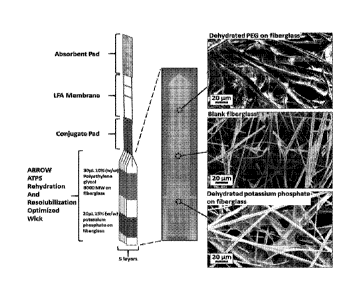
[0096] Figure 11. The integrated ARROW and LFA diagnostic design. (Left)
Design layout of the integrated ARROW and LFA. (Middle) Image of the ARROW.
(Right) SEM images of the dehydrated PEG on fiberglass, blank fiberglass, and
dehydrated
potassium phosphate on fiberglass. The top and bottom tips of the fiberglass
paper sheet are
also blank fiberglass.
[0097] Figure 12. Demonstrating the importance of ATPS component
rehydration
order. Time-lapse visualization of phase separation within a single sheet of
the ARROW
design when the rehydration order of the PEG and potassium phosphate are
switched. Close
up images are shown of the downstream region where phase separation occurred,
and
therefore, the first image is at t=6 instead of t=0. The dotted line (- - -)
encompasses the
region of the paper that predominantly contained the PEG-rich phase,
identified by the light
blue color. Visualization and identification of the PEG-rich phase, PEG-poor
phase, and
macroscopically mixed domain regions were accomplished by flowing a suspension
of
BSA-DGNPs and Brilliant Blue dye.
[0098] Figure 13. Improvement in the limit of detection of C.
trachomatis LFA by
incorporation of the ARROW. Comparison of LFA results at varying C.
trachomatis
concentrations, with and without the ARROW. Test lines are located on the
bottom of the
LFA strips while the control lines are located on the top of the LFA test
strips. Negative
control results are shown in the left most panels for 0 ng/gL C. trachomatis.
[0099] Figure 14. Quantification of test line intensities. Plot of
the quantified LFA
test line intensities for the ARROW and LFA system and the LFA only system.
[0100] Figure 15. Representative result (sample #4, Table 2) of the
head-to-head
comparison between Quick Vue, Phase's LFA, and Phase's LFA+ATPS. The presence
of the
-10-
CA 03002020 2018-04-13
WO 2017/041030
PCT/US2016/050257
test line (T) indicates a true positive result. Only our LFA+ATPS had a
visible test line
indicating a true positive result.
DETAILED DESCRIPTION
[0101] In various embodiments methods and devices are provided for
analyte
collection, extraction, concentration, and detection for clinical
applications. In certain
embodiments the devices permit the rapid detection and/or quantification of
bacteria, fungi,
and viruses in biological samples (e.g., oral, urine, and vaginal samples).
[0102] In certain embodiments lateral-flow assay (LFA) devices (see,
e.g., Figure
10) and/or flow-through (spot) assay devices (see, e.g., Figure 4) are
provided that are
accurate, sensitive, portable, disposable, and well suited to use at point of
care with minimal
training or equipment.
[0103] In certain embodiments the lateral-flow assay devices or the
flow-through
assay devices can be used directly with a sample to be assayed. In certain
embodiments the
lateral-flow assay devices or the flow-through assay devices can be used with
a sample in
which the target (e.g. target molecule(s), target microorganism(s), etc.) have
been
concentrated before application to the device, using for example, an aqueous
two-phase
system (ATPS). In certain embodiments the target (e.g. target molecule(s),
target
microorganism(s), etc.) are concentrated, using e.g., ATPS, on the device
itself.
Concentration of the target biomolecules
[0104] The concentration of target biomolecules using ATPS can be performed
in
either a bulk liquid, or as the sample solution flows in, e.g., a lateral-flow
assay or a flow-
through (spot assay), e.g., in a paper membrane.
Concentration in liquid ATPS
[0105] In certain illustrative embodiments a collected sample,
(e.g., a tissue sample,
a biological fluid such as urine, saliva, and blood, sputum, vaginal fluid,
seminal fluid,
cerebrospinal fluid, lymph, vaginal swab, endocervical swab, plaque from
teeth, and the
like), can be combined with a suspending solution (e.g., a buffer) or combined
directly with
an NIPS solution or directly applied to paper or a suspending solution
containing the
sample applied to a paper to rehydrate ATPS components that were previously
dried onto
paper. In some cases, mixing by the user may be required to achieve a well-
mixed,
homogeneous solution. In various embodiments a polymer/salt, polymer/polymer,
micellar/polymer, or micellar ATPS may be used. In one of the examples
described below,
-11-
CA 03002020 2018-04-13
WO 2017/041030
PCT/US2016/050257
a polypropylene glycol (PPG):potassium phosphate salt ATPS was used to
concentrate
Streptococcus mutans (S. mutans) by 60-fold within 10 min (see, e.g., Figure
1). If the
target analyte (e.g., target biomolecule) is large, such as a bacterium,
fungus or virus, it will
be partitioned, or distributed, extremely into one of the two phases in the
ATPS, which can
then be introduced to a downstream detection component in the LFA or flow-
through assay.
In certain embodiments, if the target analyte is small, such as a protein,
metabolite,
hormone, large probes that are decorated with specific binding moieties can be
used to
capture the target, and subsequently be concentrated into one of the phases in
ATPS for
downstream detection. In certain embodiments the phase that contains the
concentrated
target analyte(s) (e.g., biomolecule(s)) can be introduced to the detection
component by
physical extraction using a pipette or dropper, or can be introduced via a
syringe, e.g., as
illustrated in Figure 1.
Concentration as fluid flows on paper
[0106] In various embodiments the concentration step can also be
accelerated with
paper. For example, the collected specimen can be mixed with ATPS components
and
introduced to a paper device that can facilitate, enhance, and accelerate
phase separation.
The target biomolecules can be concentrated in the leading front of the flow
on the paper
membrane and can seamlessly be introduced to the subsequent detection
component.
[0107] Alternatively, the ATPS components can be pre-dehydrated onto
the paper
membranes. In this case, the collected specimen can be directly applied to the
paper
membrane without pre-mixing with the ATPS components.
Detection of target biomolecules
[0108] In various embodiments the detection components in the assay
systems
contemplated herein can be paper-based detection components. In certain
embodiments the
paper-based detection component (can be in the form of a lateral-flow test
strip (see, e.g.,
Figures 3 and 10) or a flow-through device (spot test) (see, e.g. Figure 4).
In various
embodiments both form factors may contain, but are not limited to, one or more
of the
following components:
Sample pad
[0109] In certain embodiments a sample pad, when present, can connect the
concentration component to the detection component. It can act as a filter
that can remove
debris, contaminants, and mucus from the collected fluid. It can also store
dried reagents,
-12-
CA 03002020 2018-04-13
WO 2017/041030
PCT/US2016/050257
and when rehydrated, these reagents can (i) adjust the solution for optimal
detection
conditions (pH, ionic strength, etc); and (ii) break down mucus,
glycoproteins, and other
viscous materials in the collected specimen that may affect detection.
Illustrative materials
for the sample pad include, but are not limited to, cellulose, nitrocellulose,
fiberglass,
cotton, woven or nonwoven paper, etc. Reagents on the pad may include, but are
not
limited to, surfactants such as Triton X-100, Tween 20, or sodium dodecyl
sulfate, etc.;
polymers such as polyethylene glycol, poloxamer, polyvinylpyrrolidone (PVP),
etc.; buffers
such as phosphate-buffered saline, 4-(2-hydroxyethyl)-1-
piperazineethanesulfonic acid
(HEPES), Tris(hydroxymethyl)aminomethane (Tris), sodium borate, TRICINE, etc.;
proteins such as albumin, etc.; enzymes such as protease, etc.; salts such as
sodium chloride,
sodium phosphate, sodium cholate, potassium phosphate, etc. In various
embodiments
these reagents can be applied to the sample pad by (i) soaking the paper
material in the
reagent solution, or (ii) through wicking the membrane via capillary flow. The
treated
sample pad can be dried by (i) air drying (let sit at room temperature); (ii)
baking (place in
high temperature using an oven or heating device); (iii) vacuum; or (iv)
lyophilization.
Conjugate pad
101101 In various embodiments a conjugate pad, when present can
contain
dehydrated colorimetric indicators decorated with binding moieties that bind
the target
analyte(s). In certain embodiments the binding moieties are specific binding
moieties that
have high affinity towards the target analyte(s) (e.g., bacterium, fungus,
virus, etc.). When
the sample solution reaches the conjugate pad, the colorimetric indicators are
rehydrated.
The binding moieties on the colorimetric indicators can then bind to the
target analyte(s)
and the resulting complexes can flow to the reaction pad. In certain
embodiments the
colorimetric indicators can comprise metallic particles such as gold, silver
particles,
polymeric particles such as latex beads, and polystyrene particles
encapsulating visible or
fluorescent dyes. Illustrative materials material for the conjugate pad
include, but are not
limited to, cellulose, nitrocellulose, fiberglass, cotton, woven or nonwoven
paper etc. In
certain embodiments the colorimetric indicators can be applied and dehydrated
onto the pad
as described above.
Reaction pad
101111 In certain embodiments the reaction pad, when present, can
comprise
immobilized reagents, and when the immobilized reagents react with the sample
solution,
they may produce signals (e.g., visual signals) to indicate the presence or
absence or
-13-
CA 03002020 2018-04-13
WO 2017/041030
PCT/US2016/050257
quantity of the target analyte(s). Illustrative materials for the reaction pad
include, but are
not limited to cellulose, nitrocellulose, fiberglass, cotton, woven or
nonwoven paper etc.
Lateral-flow format
[0112] In certain embodiments for a lateral-flow test strip, the
reagents on the
________________________________ reaction pad will be immobilized in the fol
in of lines perpendicular to the direction of flow
to ensure all samples can interact with the immobilized reagents. The
concentrations of the
reagents can be optimized to control the signal intensities, and thus, control
the sensitivity
of the assay. For example, a semi-quantitative assay can be designed by
immobilizing
multiple lines of the same reagent with various concentrations. Each line
therefore will
yield signals only when a specific concentration of target biomolecules is
reached. The
concentration of the target biomolecules can then be interpreted by counting
the number of
lines that are visible (see, e.g., Fig. 3). For the detection of S. mutans,
this semi-quantitative
assay may particularly be useful to provide better prediction of dental caries
since S. mutans
concentration is highly correlated to the risk of developing dental caries.
[0113] In addition, multiple lines of different reagents can be immobilized
on the
same strip to detect multiple target analyte(s). This allows the development
of multiplex
assays.
Flow-through format
[0114] In certain embodiments, e.g., for a flow-through test,
instead of lines, the
reagents can be immobilized on the entire reaction pad. If the target analyte
is present, it
will bind to the colorimetric indicator on the conjugate pad and be trapped on
the reaction
pad as the indicator-target complex binds to the immobilized reagent. A
visible spot will
therefore appear if the target biomolecule is present. This test can be used
if the sample
volume is too low to wick up a lateral-flow test strip. The color intensity of
the visible spot
is correlated to the concentration of target analyte(s) (e.g., biomolecules),
while the size of
the spot is correlated to the sample volume. In certain embodiments the
concentration
component can be placed directly on top of the flow-through test to remove the
need for
extracting and applying the concentrated samples to the detection component
(see, e.g., Fig.
4).
[0115] In various embodiments the immobilized reagents can comprise a
specific
antibody against the target analyte (primary antibody), antibodies against the
primary
antibody (secondary antibody), antigens, proteins, or antigen-protein
conjugates.
-14-
CA 03002020 2018-04-13
WO 2017/041030
PCT/US2016/050257
Illustrative materials for the reaction pad include, but are not limited to
cellulose,
nitrocellulose, fiberglass, cotton, woven and nonwoven paper etc. In various
embodiments
the reagents can be applied and dehydrated onto the pad as described above.
Sink
[0116] In certain embodiments the sink, when present, can comprise an
absorbent
pad that collect excess fluid and prevents back-flow which can affect the test
performance.
Illustrative materials for the sink include, but are not limited to cellulose,
nitrocellulose,
fiberglass, cotton, woven and nonwoven paper etc.
Signal enhancement
[0117] In various embodiments the visible signal intensity can be enhanced
to
improve the accuracy of the detection assay. This can be performed by
introducing
additional reagents to the reaction pad after the initial detection assay. In
certain
embodiments the signal enhancement reagent can comprise a substrate that
reacts with an
enzyme that is decorated on the surface of, e.g., colorimetric indicator to
form a strong
.. visible product. By way of example, if the colorimetric indicator comprises
a gold probe,
the signal enhancement can be achieved by silver-enhancement labeling, where
an
enhancement reagent containing silver ion can be applied to the reaction pad
where the gold
probe is bound to the immobilized line/spot. In this scenario, the gold probes
can act as
nucleation sites so that silver can be deposited onto the particle, resulting
in increased signal
intensity, In these examples, the signal enhancement reagents can either be
added
separately after the initial detection assay, or stored/dehydrated on the
paper device to be
released automatically/manually,
[0118] The foregoing components and assay formats are illustrative
and non-
limiting. Using the teachings and examples, provided herein, numerous other
assay devices
and configurations will be available to one of skill in the art and some
further design
considerations and components are described below.
Lateral-flow Assay (LFA)or Flow-Through (Snot) Assay
[0119] As explained above, in certain embodiments, the devices and
systems
described herein are configured to provide a lateral-flow assay (LFA) or a
flow-through
(spot) assay for detection of the target analyte in a sample, where the LFA or
spot assay is
used alone or in conjunction with an aqueous two-phase system (ATPS). In some
embodiments, the LFA or spot assay comprises a porous matrix into which is
disposed the
-15-
CA 03002020 2018-04-13
WO 2017/041030
PCT/US2016/050257
ATPS or components thereof, where the porous matrix is configured to and has
porosity
sufficient to allow the ATPS or components thereof to flow-through the porous
matrix when
the ATPS or components thereof are in a fluid phase. Such porous LFA or spot
assay
devices are referred to herein as paper or paper fluidic devices and these
terms are used
interchangeably.
[0120] The term "paper", as used herein, is not limited to thin
sheets from the pulp
of wood or other fibrous plant substances although, in certain embodiments the
use of such
papers in the devices described herein is contemplated. Papers more generally
refer to
porous materials often in sheet form, but not limited thereto that allow a
fluid to flow-
through.
[0121] In some embodiments, the porous matrix is sufficiently porous
to allow the
mixed phase solution, first phase solution and/or second phase solution of the
ATPS, and/or
target analyte, to flow-through the LFA or flow-through assay. In some
embodiments, the
porous matrix is sufficiently long and/or deep enough for the mixed phase
solution, first
phase solution and/or second phase solution, and/or target analyte, to flow
vertically and/or
horizontally through the LFA or flow-through (spot) assay device. In some
embodiments,
the first phase solution flows through the porous matrix at a first rate and
the second phase
solution flows through the porous matrix at a second rate, where the first
rate and the second
rate are different. In some embodiments of the LFA or spot assay the porous
matrix
comprises inter alia a material such as a scintered glass ceramic, a mineral,
cellulose, a
fiberglass, a nitrocellulose, polyvinylidene fluoride, a nylon, a charge
modified nylon, a
polyethersulfone, combinations thereof, and the like.
Concentrate-as-it-flows
[0122] It was discovered that A I'PSs can phase separate as the
solution flows
through a porous substrate (e.g., a paper) which we have termed "concentrate-
as-it-flows".
Moreover, it was also discovered that flow through the paper significantly
speeds up the
concentration process. Based this phenomenon, the lateral-flow assay devices
and the flow-
through assay devices described herein can comprise a paper fluidic component
that fully
integrates the necessary components for a combined ATPS concentration with the
LFA or
flow-through detection. It was discovered that when a mixed ATPS solution is
applied to
certain paper materials, phase separation and analyte concentration occur as
the solution
flows. We also demonstrated that this phenomenon is preserved even when making
an
-16-
CA 03002020 2018-04-13
WO 2017/041030
PCT/US2016/050257
ATPS that had varying volume ratios, e.g., volume of the top phase divided by
that of the
bottom phase.
[0123] In some embodiments, the LFA or the spot assay (e.g., the
concentration
component of the spot assay) comprises a paper. In some embodiments, the paper
comprises a sheet of porous material that allows fluid to flow-through it. In
some
embodiments, the paper comprises a plurality of sheets of porous material that
allows fluid
to flow-through them (e.g., as illustrated in Figure 4). In some embodiments,
the paper
comprises one or more materials such as cellulose, fiberglass, nitrocellulose,
polyvinylidine
fluoride, charge modified nylon, polyether sulfone, and the like. In some
embodiments, the
paper is a HI-FLOW PLUS membrane.
[0124] In some embodiments, the paper is a woven paper. In some
embodiments,
the paper is a Whatman paper. In some embodiments, the Whatman paper comprises
Whatman S17, Whatman MF1, Whatman VF1, Whatman Fusion 5, Whatman GF/DVA,
Whatman LF1, Whatman CFI, and/or Whatman CF4.
[0125] In some embodiments, the paper concentrates the target analyte as
the target
analyte flows through the LFA or through the concentration component of a flow-
through
assay (e.g. a "concentrate-as-it-flows"-based device). In some embodiments,
the paper
concentrates the target analyte as the target analyte flows through the LFA
horizontally. In
some embodiments, the paper concentrates the target analyte as the target
analyte flows
.. through the LFA or flow-through assay vertically.
[0126] In some embodiments, the paper has a property that influences
which phase
solution will become the "leading fluid." By way of non-limiting example, when
using a
PEG-salt ATPS, adding the solution to fiberglass paper will cause the salt
phase to become
the leading solution, while using cellulose paper will cause the PEG phase to
become the
.. leading solution. In some embodiments, phase separation within the paper
accelerates
phase separation. Also by way of non-limiting example, a micelle ATPS
typically takes
several hours to phase separate in a stagnant ATPS, but if applied to a paper
strip, this phase
separation occurs in minutes. This speeds up the diagnostic process by
allowing the ATPSs,
which are traditionally the rate-determining step in the process, to become
more viable
options for our rapid paper diagnostic assays. In some embodiments, the
'concentrate-as-it-
flows' device comprises a PEG-salt ATPS (e.g., as illustrated in the
Examples). In some
embodiments, the 'concentrate-as-it-flows' device comprises a micellar ATPS.
In some
-17-
embodiments, the LFA device or the flow-through assay device comprises
fiberglass paper
or nitrocellulose paper.
[0127] In certain embodiments the LFA or flow-through assay device
comprises a
filter that removes debris (e.g, blood cells or other particulates), a sample
pad where the
sample comprising the target analyte is applied to the device, a detection
zone (e.g. test line
and control line) where there the target analyte binds and is detected, and an
absorbent pad
(e.g., a dry receiving paper) that can absorb excess sample and/or solutions
applied to the
LFA or flow-through device (see, e.g., Figure 10). In some embodiments, the
control line
and/or test line is not a line per se, but a region or spot.
[0128] In some embodiments, the LFA comprises an LFA strip. The terms "LFA"
and "LFA strip" are used interchangeably herein. In some embodiments, the LFA
strip has
a length greater than its width and depth. In some embodiments, the LFA is
rectangular. In
some embodiments, the LFA has a shape that is round, ovoid, square, polygonal,
or
irregular-shaped. In some embodiments, the LFA comprises a plurality of routes
and/ or
junctions. In some embodiments, the LFA strip comprises the sample pad,
detection zone
and absorbent pad. In some embodiments, the detection zone is located between
the sample
pad and the absorbent pad, the absorbent pad wicking the sample with the
target analyte
away from the sample pad and toward the detection zone.
Sandwich Assay
[0129] In some embodiments, the LFA or flow-through (spot) assay device is
configured to provide or run a sandwich assay (see e.g., Figure 1, bottom
left, in copending
PCT Application No: PCT/US2015/019297, filed on March 6, 2015). In some
embodiments, the sandwich assay comprises a capture moiety that binds the
target analyte.
In some embodiments, the device comprises a probe. In some embodiments, the
probe
comprises a detectable property (colorimetric, fluorescent, radioactive,
etc.). In some
embodiments, the probe comprises a binding moiety that interacts with the
target analyte
(e.g. an antibody). In some embodiments, the probe is added to the sample and
binds the
target analyte to form a probe-analyte complex.
Competition Assay
[0130] In some embodiments, the LFA comprises a competition assay. In some
embodiments, the probe is added to the sample and binds the target analyte to
form a probe-
analyte complex. In some embodiments, the LFA comprises the target analyte
immobilized
-18-
Date Recue/Date Received 2022-09-16
CA 03002020 2018-04-13
WO 2017/041030
PCT/US2016/050257
on the test line. In some embodiments, the probe is saturated by the target
analyte in the
sample and the probe will not bind to the target analyte immobilized on the
test line. In
some embodiments, the absence of the detectable signal on the test line
indicates a positive
result. In some embodiments, there is no target analyte present in the sample,
and the probe
binds to the target analyte on the test line, indicating a negative result. In
some
embodiments, the LFA comprises a probe capture moiety on a control line that
interacts
directly with the probe, and regardless of the presence of the target analyte
in the sample,
the probe can bind to the probe capture moiety and accumulate on the control
line. In some
embodiments, the probe becomes immobilized and detected on the control line,
indicating a
valid test. In some embodiments, a positive result (e.g., target analyte is
present in sample)
is indicated by the absence of a detectable signal at the test line and the
presence of a
detectable signal at the control line. In some embodiments, a negative result
is indicated by
a detectable signal at both the test and control lines.
101311 In some embodiments of a sandwich format assay, the probe-
analyte
complex is applied to the sample pad and flows through the LFA or through the
flow-
through device towards the absorbent pad. In some embodiments, the target
analyte of the
probe-analyte complex binds to the capture moiety. In some embodiments, the
capture
moiety is immobilized on a test line or a test region (e.g., a test layer in a
flow-through
device) and the probe-analyte complex becomes immobilized on the test line or
in the test
region. In some embodiments, the probe is colorimetric, and the test line or
test region will
exhibit a strong color (e.g. detectable signal) as the probe-analyte complex
accumulates at
the test line or in the test region, indicating a positive result. In some
embodiments, there is
no target analyte present in the sample, and the probe of the probe-analyte
complex does not
interact with the capture moiety, and the absence of the test line or signal
in the test region
indicates a negative result. In some embodiments, the LFA comprises a probe
capture
moiety on a control line (or in a control region, e.g., of a flow-through
assay device) that
interacts directly with the probe and/or the binding moiety, and thus,
regardless of the
presence of the target analyte in the sample, the probe/binding moiety binds
to the probe
capture moiety and accumulate on the control line or in the control region. In
some
embodiments, the probe capture moiety is a secondary antibody that binds the
binding
moiety, wherein the binding moiety is a primary antibody that binds that
target analyte. In
some embodiments, the probe becomes immobilized and detected on the control
line or in
the control region, indicating a valid test. In some embodiments, a positive
result (e.g. target
analyte is present in sample) is indicated by a detectable signal at the test
line (or test
-19-
CA 03002020 2018-04-13
WO 2017/041030
PCT/US2016/050257
region) and the control line (or control region). In some embodiments, a
negative result is
indicated by a detectable signal at the control line or in the control region.
Aqueous Two-Phase System (ATPS)
101321 In certain embodiments the devices described herein are
configured to work
in conjunction with an aqueous two-phase system (ATPS), e.g., in a syringe or
other vessel,
or they are configured to support an aqueous two-phase system (ATPS). In some
embodiments, the ATPS comprises a phase solution. The term "phase solution"
generally
refers to a first phase solution or a second phase solution of the ATPS. In
some
embodiments, the phase solution is in a mixed solution (e.g. with the
first/second phase
solution). In some embodiments, the phase solution is the first/second phase
solution after
it separates from the mixed solution of the ATPS. In some embodiments, the
phase solution
is the first/second phase solution after it separates from the mixed solution
in the LFA or
flow-through assay. In certain embodiments the phase solution can refer to the
second
phase solution while it is in a mixed state (e.g. with the first phase
solution). In some
embodiments, the phase solution is a leading fluid in the LFA or flow-through
assay. In
some embodiments, the phase solution is a lagging fluid in the LFA or flow-
through assay.
101331 In some embodiments, the ATPS comprises two aqueous
solutions, a first
phase solution and a second phase solution that are initially mixed (e.g., a
mixed phase
solution). In some embodiments, the mixed phase solution is a homogeneous
solution,
while in certain other embodiments the first phase solution and the second
phase solution
are immiscible. In some embodiments, the first phase solution and the second
phase
solution are immiscible, but domains of the first phase solution are mixed
with domains of
the second phase solution. In some embodiments, the immiscibility is driven by
changes in
temperature, and/or changes in the concentrations of the different components,
such as salt.
In some embodiments, the first/second phase solutions comprise components,
such as,
micelles, salts, and/or polymers. In some embodiments, the target analyte
(e.g.,
biomolecule, bacterium (or fragment thereof), fungus (or fragment thereof), or
virus, and
the like) in contact with the ATPS, distributes, partitions, and/or
concentrates preferentially
into the first phase solution over the second phase solution, or vice versa,
based on its
physical and chemical properties, such as size, shape, hydrophobicity, and
charge. In some
embodiments, the target analyte (e.g. a bacterium, fungus, virus, etc.)
partitions
predominantly (or extremely) into the first or second phase solution of the
ATPS, and
therefore concentrates in the ATPS. In some embodiments, the target analyte is
-20-
CA 03002020 2018-04-13
WO 2017/041030
PCT/US2016/050257
concentrated by adjusting the ratio of volumes between the first phase
solution and the
second phase solution. In some embodiments, the target analyte is concentrated
by reducing
the volume of the phase in which the analyte partitions. By way of
illustration, in some
embodiments, the target analyte is concentrated by 10-fold in the first phase
solution, e.g.,
by using a 1:9 volume ratio of first phase solution to second phase solution,
since the
volume of the phase into which the analyte extremely partitions into is 1/10
the total
volume.
101341 In some embodiments, other concentrations are obtained by
using other
ratios. Thus, in some embodiments the ratio of the first phase solution to the
second phase
solution comprises a ratio of about 1:1, about 1:2, about 1:3, about 1:4,
about 1:5, about 1:6,
about 1:7, about 1:8, about 1:9, or about 1:10. In some embodiments the ratio
of the first
phase solution to the second phase solution comprises a ratio of about 1:20,
about 1:30,
about 1:40, about 1:50, about 1:60, about 1:70, about 1:80, about 1:90, or
about 1:100. In
some embodiments the ratio of the first phase solution to the second phase
solution
comprises a ratio of about 1:200, about 1:300, about 1:400, about 1:500, about
1:600, about
1:700, about 1:800, about 1:900, or about 1:1000.
101351 In some embodiments the ratio of the second phase solution to
the first phase
solution comprises a ratio of about 1:1, about 1:2, about 1:3, about 1:4,
about 1:5, about 1:6,
about 1:7, about 1:8, about 1:9, or about 1:10. In some embodiments the ratio
of the second
phase solution to the first phase solution comprises a ratio of about 1:20,
about 1:30, about
1:40, about 1:50, about 1:60, about 1:70, about 1:80, about 1:90, or about
1:100. In some
embodiments the ratio of the second phase solution to the first phase solution
comprises a
ratio of about 1:200, about 1:300, about 1:400, about 1:500, about 1:600,
about 1:700, about
1:800, about 1:900, or about 1:1000.
101361 In some embodiments, the analyte partitions substantially evenly
between the
first phase solution and second phase solution, preventing concentration of
the analyte. In
such systems, concentration of the target analyte can be achieved by
introducing an
additional component, such as a probe that captures the target analyte, and
wherein the
probe partitions predominantly into one phase, thereby enhancing the
partitioning behavior
of the target analyte to enable concentration. In some embodiments, the
first/second phase
solution containing the concentrated analyte is collected and applied to the
LFA or to the
flow-through assay device.
-21-
CA 03002020 2018-04-13
WO 2017/041030
PCT/US2016/050257
[0137] In some embodiments, the first/second phase solution
comprises a micellar
solution. In some embodiments, the micellar solution comprises a nonionic
surfactant. In
some embodiments, the micellar solution comprises a detergent. In some
embodiments, the
micellar solution comprises Triton-X. In some embodiments, the micellar
solution
comprises a polymer similar to Triton-X, such as Igepal CA-630 and Nonidet P-
40, and the
like, by way of non-limiting example. In some embodiments, the micellar
solution consists
essentially of Triton-X.
[0138] In some embodiments, the micellar solution has a viscosity
(at room
temperature (-25 C) of about 0.01 centipoise to about 5000 centipoise, about
0.01
centipoise to about 4500 centipoise, about 0.01 centipoise to about 4000
centipoise, about
0.01 centipoise to about 3500 centipoise, about 0.01 centipoise to about 3000
centipoise,
about 0.01 centipoise to about 2500 centipoise, about 0.01 centipoise to about
2000
centipoise, about 0.01 centipoise to about 1500 centipoise, about 0.01
centipoise to about
1000 centipoise, or about 0.01 centipoise to about 500 centipoise. In some
embodiments,
the micellar solution has a viscosity at room temperature of about 0.01
centipoise to about
450 centipoise, about 0.01 centipoise to about 400 centipoise, about 0.01
centipoise to about
350 centipoise, about 0.01 centipoise to about 300 centipoise, about 0.01
centipoise to about
250 centipoise, about 0.01 centipoise to about 200 centipoise, about 0.01
centipoise to about
150 centipoise, or about 0.01 centipoise to about 100 centipoise.
[0139] In some embodiments, the first/second phase solution comprises a
polymer
(e.g., polymer solution). In certain embodiments, the polymer is a
polyethylene glycol
(PEG). In various embodiments, the PEG may have a molecular weight between
1000 and
100,000. In certain embodiments, the PEG comprises PEG-4600, PEG-8000, or PEG-
20,000. In certain embodiments, the polymer is polypropylene glycol (PPG). In
various
embodiments, the PPG may have a molecular weight between 100 and 10,000. In
certain
embodiments, the PPG comprises PPG 425. In certain embodiments, the polymer is
dextran. In various embodiments, the dextran may have a molecular weight
between 1000
and 1,000,000. In certain embodiments, the dextran comprises dextran 6000,
dextran 9000,
dextran-35,000, or dextran-200,000.
[0140] In some embodiments, the polymer solution comprises a polymer
solution
that is about 0.01% w/w polymer, or about 0.05% w/w polymer, or about 0.1% w/w
polymer, or about 0.15% w/w polymer, or about 0.2% w/w polymer, or about 0.25%
w/w
polymer, or about 0.3% w/w polymer, or about 0.35% w/w polymer, or about 0.4%
w/w
-22-
CA 03002020 2018-04-13
WO 2017/041030
PCT/US2016/050257
polymer, or about 0.45% w/w polymer, or about 0.5% w/w polymer, or about 0.55%
w/w
polymer, or about 0.6% w/w polymer, or about 0.65% w/w polymer, or about 0.7%
w/w
polymer, or about 0.75% w/w polymer, or about 0.8% w/w polymer, or about 0.85%
w/w
polymer, or about 0.9% w/w polymer, or about 0.95% w/w polymer, or about 1%
w/w
polymer. In some embodiments, the polymer solution comprises a polymer
solution that is
about 1% w/w polymer, or about 2% w/w polymer, or about 3% w/w polymer, or
about 4%
w/w polymer, or about 5% w/w polymer, or about 6% w/w polymer, or about 7% w/w
polymer, or about 8% w/w polymer, or about 9% w/w polymer, or about 10% w/w
polymer,
or about 11% w/w polymer, or about 12% w/w polymer, or about 13% w/w polymer,
or
about 14% w/w polymer, or about 15% w/w polymer, or about 16% w/w polymer, or
about
17% w/w polymer, or about 18% w/w polymer, or about 19% w/w polymer, or about
20%
w/w polymer, or about 21% w/w polymer, or about 22% w/w polymer, or about 23%
w/w
polymer, or about 24% w/w polymer, or about 25% w/w polymer, or about 26% w/w
polymer, or about 27% w/w polymer, or about 28% w/w polymer, or about 29% w/w
polymer, or about 30% w/w polymer, or about 31% w/w polymer, or about 32% w/w
polymer, or about 33% w/w polymer, or about 34% w/w polymer, or about 35% w/w
polymer, or about 36% w/w polymer, or about 37% w/w polymer, or about 38% w/w
polymer, or about 39% w/w polymer, or about 40% w/w polymer, or about 41% w/w
polymer, or about 42% w/w polymer, or about 43% w/w polymer, or about 44% w/w
.. polymer, or about 45% w/w polymer, or about 46% w/w polymer, or about 47%
w/w
polymer, or about 48% w/w polymer, or about 49% w/w polymer, or and about 50%
w/w
polymer. In some embodiments, the polymer solution comprises a polymer
solution that is
about 10% w/w polymer, or about 20% w/w polymer, or about 30% w/w polymer, or
about
40% w/w polymer, or about 50% w/w polymer, or about 60% w/w polymer, or about
70%
w/w polymer, or about 80% w/w polymer, or about 90% w/w polymer. In some
embodiments, the polymer solution comprises a polymer solution that is about
10% w/w
polymer to about 80% w/w polymer. In some embodiments, the polymer solution
comprises a polymer solution that is about 10 /0 w/w to about 25% w/w polymer.
[0141] In some embodiments, the first and/or second phase solution
comprises a salt
and thereby forms a salt solution. In some embodiments, the target analyte
(e.g., bacterium,
fungus, virus, etc.) and/or a probe-analyte complex partitions into the salt
solution. In
certain embodiments the salt solution comprises a kosmotropic salt. In some
embodiments
the salt solution comprises a chaotropic salt. In some embodiments, the salt
comprises one
or more of a magnesium salt, a lithium salt, a sodium salt, a potassium salt,
a cesium salt, a
-23-
CA 03002020 2018-04-13
WO 2017/041030
PCT/US2016/050257
zinc salt, and an aluminum salt. In some embodiments, the salt comprises a
bromide salt, an
iodide salt, a fluoride salt, a carbonate salt, a sulfate salt, a citrate
salt, a carboxylate salt, a
borate salt, or a phosphate salt. In some embodiments, the salt is potassium
phosphate. In
some embodiments, the salt is ammonium sulfate.
[0142] In some embodiments, the salt solution comprises a salt solution
comprising
about 0.01% w/w salt, or about 0.05% w/w salt, about 0.1% w/w salt, or about
0.15% w/w
salt, or about 0.2% w/w salt, or about 0.25% w/w salt, or about 0.3% w/w salt,
or about
0.35% w/w salt, or about 0.4% w/w salt, or about 0.45% w/w salt, or about 0.5%
w/w salt,
or about 0.55% w/w salt, or about 0.6% w/w salt, or about 0.65% w/w salt, or
about 0.7%
w/w salt, or about 0.75% w/w salt, or about 0.8% w/w salt, or about 0.85% w/w
salt, or
about 0.9% w/w salt, or about 0.95% w/w salt, or about or about 1% w/w salt.
In some
embodiments, the salt solution comprises a salt solution that is about 1% w/w
salt, or about
2% w/w salt, or about 3% w/w salt, or about 4% w/w salt, or about 5% w/w salt,
or about
6% w/w salt, or about 7% w/w salt, or about 8% w/w salt, or about 9% w/w salt,
or about
10% w/w salt, or about 11% w/w salt, or about 12% w/w salt, or about 13% w/w
salt, or
about 14% w/w salt, or about 15% w/w salt, or about 16% w/w salt, or about 17%
w/w salt,
or about 18% w/w salt, or about 19% w/w salt, or about 20% w/w salt, or about
21% w/w
salt, or about 22% w/w salt, or about 23% w/w salt, or about 24% w/w salt, or
about 25%
w/w salt, or about 26% w/w salt, or about 27% w/w salt, or about 28% w/w salt,
or about
29% w/w salt, or about 30% w/w salt, or about 31% w/w salt, or about 32% w/w
salt, or
about 33% w/w salt, or about 34% w/w salt, or about 35% w/w salt, or about 36%
w/w salt,
or about 37% w/w salt, or about 38% w/w salt, or about 39% w/w salt, or about
40% w/w
salt, or about 41% w/w salt, or about 42% w/w salt, or about 43% w/w salt, or
about 44%
w/w salt, or about 45% w/w salt, or about 46% w/w salt, or about 47% w/w salt,
or about
48% w/w salt, or about 49% w/w salt, or and about 50% w/w. In some
embodiments, the
salt solution comprises a salt solution that is about 0.1 /0 w/w to about 10%.
In some
embodiments, the salt solution is about 1% w/w to about 10%.
101431 In some embodiments, the first/second phase solution
comprises a solvent
that is immiscible with water. In some embodiments, the solvent comprises a
non-polar
organic solvent. In some embodiments, the solvent comprises an oil. In some
embodiments,
the solvent comprises pentane, cyclopentane, benzene, 1,4-dioxane, diethyl
ether,
dichloromethane, chloroform, toluene, or hexane.
-24-
CA 03002020 2018-04-13
WO 2017/041030
PCT/US2016/050257
[0144] In some embodiments, the first phase solution comprises a
micellar solution
and the second phase solution comprises a polymer. In some embodiments, the
second
phase solution comprises a micellar solution and the first phase solution
comprises a
polymer. In some embodiments, the first phase solution comprises a micellar
solution and
the second phase solution comprises a salt. In some embodiments, the second
phase
solution comprises a micellar solution and the first phase solution comprises
a salt. In some
embodiments, the micellar solution is a Triton-X solution. In some
embodiments, the first
phase solution comprises a first polymer and the second phase solution
comprises a second
polymer. In some embodiments, the first/second polymer comprises polyethylene
glycol
and/or dextran. In some embodiments, the first phase solution comprises a
polymer and the
second phase solution comprises a salt. In some embodiments, the second phase
solution
comprises a polymer and the first phase solution comprises a salt. In some
embodiments,
the first phase solution comprises polyethylene glycol and the second phase
solution
comprises potassium phosphate. In some embodiments, the second phase solution
comprises polyethylene glycol and the first phase solution comprises potassium
phosphate.
In some embodiments, the first phase solution comprises a salt and the second
phase
solution comprises a salt. In some embodiments, the first phase solution
comprises a
kosmotropic salt and the second phase solution comprises a chaotropic salt. In
some
embodiments, the second phase solution comprises a kosmotropic salt and the
first phase
solution comprises a chaotropic salt.
[0145] In some embodiments, the first phase solution comprises a
Component 1 of
Table 1 and the second phase solution comprises a Component 2 of Table 1. In
some
embodiments, the second phase solution comprises a Component 1 of Table 1 and
the
second phase solution comprises a Component 2 of Table 1.
[0146] In some embodiments, the components of Table 1 are suspended or
dissolved
in a buffer. In some embodiments, the components of Table 1 are
suspended/dissolved in a
buffer compatible with a biological system from which the sample was derived.
In some
embodiments, the components of Table 1 are suspended/dissolved in a saline
solution. In
some embodiments, the components of Table 1 are suspended/dissolved in PBS. In
some
embodiments, the components of Table 1 are suspended/dissolved in water.
Table 1. Illustrative aqueous two-phase extraction/concentration systems.
Component 1 Component 2
Polymer/polymer Systems
-25-
CA 03002020 2018-04-13
WO 2017/041030 PCT/US2016/050257
Dextran
Ficoll
Polyethylene glycol Polyvinyl pyrrolidone
Polyvinyl alcohol
Hydroxypropyl starch
Dextran
Polypropylene glycol Hydroxypropyl dextran
Polyvinyl pyrrolidone
Dextran
Polyvinyl alcohol
Hydroxypropyl dextran
Dextran
Polyvinyl pyrrolidone
Maltodextrin
Dextran
Methyl cellulose
Hydroxypropyl dextran
Ethylhydroxyethyl cellulose Dextran
Polymer/salt Systems
Potassium phosphate
Sodium sulfate
Polyethylene glycol Magnesium sulfate
Ammonium sulfate
Sodium citrate
Propylene glycol (PPG) Potassium phosphate
Methoxypolyethylene glycol Potassium phosphate
Polyvinyl pyrrolidone Potassium phosphate
[0147] As illustrated in the Examples, in certain embodiments the
ATPS comprises
a polymer/salt ATPS. It was discovered that an ATPS comprising polyethylene
glycol and
a salt or polypropylene glycol and a salt provides a rapid, sensitive, and
accurate analyte
detection/quantification.
[0148] In some embodiments, the devices described herein (e.g., an LFA or a
flow-
through assay device) can further comprise a collector configured to be placed
in contact
with the ATPS, wherein the target analyte partitions at an interface of the
collector and the
first phase solution and/or second phase solution. In some embodiments, the
collector
comprises a material that is a plastic, a mesoporous material, a silica, a
polypropylene, a
magnet, a magnetic particle, a paramagnetic particle, a material with a pore,
a material with
a groove, and/or any combination thereof. In some embodiments, the collector
comprises
polypropylene. In some embodiments, collector is optimized to increase target
analyte
collection. In some embodiments, the collector comprises a pore to maximize
the surface
area. In some embodiments, the width of the pore is about 1 gm, about 5 gm,
about 10 gm,
about 15 gm, about 20 gm, about 25 gm, about 30 gm, about 35 gm, about 40 gm,
about 45
p.m, about 50 gm, about 55 gm, about 60 gm, about 65 gm, about 70 gm, about 75
gm,
about 80 gm, about 85 gm, about 90 gm, about 95 gm, or about 100 p.m. In some
-26-
CA 03002020 2018-04-13
WO 2017/041030
PCT/US2016/050257
embodiments, the width of the pore is about 100 gm, about 200 gm, about 300
p.m, about
400 gm, about 500 gm, about 600 gm, about 700 gm, about 800 gm, about 900 gm,
or
about lmm. In some embodiments, the depth of the pore is about 1 gm, about 5
gm, about
gm, about 15 gm, about 20 gm, about 25 gm, about 30 gm, about 35 gm, about 40
gm,
5 about 45 gm, about 50 gm, about 55 gm, about 60 gm, about 65 gm, about 70
gm, about 75
p.m, about 80 gm, about 85 p.m, about 90 gm, about 95 p.m, or about 100 gm. In
some
embodiments, the depth of the pore is about 100 gm, about 200 gm, about 300
gm, about
400 gm, about 500 p.m, about 600 gm, about 700 gm, about 800 gm, about 900
[1.M, or
about lmm.
10 Dehydrated ATPS in LFA or flow-through (spot) assay device.
[0149] In some embodiments, the ATPS or components thereof are
dehydrated on
and/or in at least a first portion of the porous matrix comprising an LFA or
in the
concentration component of a flow-through assay device. In some embodiments,
application of the sample to the device hydrates the ATPS, thereby converting
the ATPS or
components thereof to a fluid phase. Dehydration may make the device more user
friendly
as the user just needs to add the sample (e.g., saliva, blood, urine, vaginal
fluid, seminal
fluid, sputum, cerebrospinal fluid, lymph, or similar fluid) to the device. In
some
embodiments, a user only has to apply a solution of the sample to the strip to
detect the
presence/absence of the target analyte or to quantify the analyte. In some
embodiments, the
solution of the sample flows through the LFA or the flow-through device and
the ATPS is
re-solubilized, triggering phase separation within the LFA or flow-through
device and
subsequent concentration of the target analyte.
[0150] In some embodiments, all the necessary components for a given
ATPS are
mixed to form a mixed solution, applied to the paper comprising the device
(e.g., LFA or
flow-through (spot) assay), and then dehydrated. When the sample solution is
added to the
dehydrated paper, the ATPS components are rehydrated as the sample flows,
resulting in
phase separation. In some ATPSs where the phase containing the concentrated
analyte is
less viscous, that phase will flow faster and the concentrated analyte will
emerge in the
leading fluid and will reach the detection zone of the LFA or flow-through
assay to initiate
detection. Additionally, the dehydrated A l'PS component segment length (or
thickness) and
concentration can be adjusted for different applications.
[0151] In some embodiments, both (all) components of the ATPS are
dehydrated on
the LFA or in the flow-through assay (e.g., in the separation component). In
some
-27-
embodiments, a first ATPS component is dehydrated on (or in) the LFA or in the
flow-
through assay. In some embodiments, a second ATPS component is dehydrated on
or in the
LFA or flow-through assay. In some embodiments, the first phase solution
component
and/or first ATPS component is dehydrated on a first portion of the LFA or in
a first layer
of the flow-through assay (separation component). In some embodiments, the
second phase
solution component and/or second ATPS component is dehydrated on a second
portion of
the LFA or in a second layer of the flow-through assay (separation component).
In some
embodiments, the first portion and the second portion are same. In some
embodiments, the
first portion and the second portion are different. By way of non-limiting
example, in a
PEG-salt ATPS, the PEG and salt solutions can be dehydrated separately into
different
paper portions or segments (see, e.g., Figure 16 of copending PCT Application
No:
PCT/US2015/019297, filed on March 6, 2015) or in separate layers comprising,
e.g., the
separation component of a flow-through assay (see, e.g., Figure 4). In some
embodiments,
dehydrating the first/second phase solution and/or ATPS component on different
portions of
the LFA or in different layers of the flow-through assay provides a more
uniform
concentration of the first/second phase solution components or ATPS
components. In some
embodiments, dehydrating the first/second phase solution components and/or
ATPS
components on different portions allows the first phase solution or ATPS
component to
flow in a first direction after hydration and the second phase solution and/or
ATPS
component to flow in a second direction after hydration, wherein the first and
second
directions are different. In some embodiments, the target analyte is
concentrated in the first
direction, but not the second direction. In some embodiments, the target
analyte is
concentrated in the second direction, but not the first direction. In some
embodiments,
dehydrating the first/second phase components and/or ATPS components on
different
portions allows the target analyte to flow in the first/second direction
without requiring the
sample to flow in the first/second direction. In some embodiments, dehydrating
the
first/second phase components and/or ATPS components on different portions
allows the
target analyte to flow faster, resulting in detection sooner. In some
embodiments,
dehydrating the first/second phase components and/or ATPS components on
different
portions allows for increased result reliability. In some embodiments,
dehydrating the first/
second phase components and/or ATPS components on different portions prevents
aggregation of first/second phase solution components and/or ATPS components
(e.g. PEG-
salt ATPS). In some embodiments, the first/second phase component and/or ATPS
-28-
Date Recue/Date Received 2022-09-16
CA 03002020 2018-04-13
WO 2017/041030
PCT/US2016/050257
component is dehydrated in multiple segments. In some embodiments the
first/second
phase component and/or ATPS component is dehydrated in multiple segments,
wherein the
first/second phase component and/or ATPS component comprises a salt solution.
In some
embodiments the first/second phase component and/or ATPS component is
dehydrated in
multiple segments, wherein the first/second phase component and/or ATPS
component does
not comprise a polymer (e.g. PEG). In some embodiments, dehydrated PEG is not
located
near the detection zone because the PEG-rich phase can slow the flow within
the detection
membrane. In some embodiments, the LFA strip or the flow-through assay can
comprise a
blank spacer near the detection zone that does not contain PEG or salt.
[0152] In some embodiments, a probe (e.g., an analyte binding moiety and
associated detection reagent/material) is provided in a probe buffer. In some
embodiments,
the probe buffer is dehydrated on the LFA or in the flow-through assay.
[0153] In some embodiments, dehydration of ATPS components improves
the limit
of detection compared to a device in which the ATPS components are added in
liquid form.
In some embodiments, the addition of liquid form ATPS components dilutes the
sample
solution from the subject. In some embodiments, dehydration of ATPS components
allows
for a distinct first phase solution and/or distinct second phase solution to
develop during
flow, concentrating the target analyte or probe-analyte complex in a small
volume at the
front of the leading fluid that will reach the test and control lines or the
detection component
of a flow-through assay. In some embodiments, concentrating the target analyte
and or
probe-analyte complex at the front of the leading fluid will decrease the time
period
necessary for detection.
Probes
[0154] In certain embodiments the systems and/or devices described
herein and/or
the methods described herein utilize a probe, where the probe comprises a
binding moiety
that binds the target analyte to form a probe-analyte complex.
[0155] In some embodiments, the target analyte alone partitions
preferentially into
the first phase solution or second phase solution or interface of the first
phase solution and
second phase solution. In some embodiments, the target analyte alone
partitions extremely
into the first phase solution or second phase solution or interface of the
first phase solution
and second phase solution.
-29-
CA 03002020 2018-04-13
WO 2017/041030
PCT/US2016/050257
[0156] In some embodiments, the target analyte alone does not
partition
preferentially into the first phase solution or second phase solution or
interface of the first
phase solution and second phase solution. In some embodiments, the target
analyte alone
does not partition extremely into the first phase solution or second phase
solution or
interface of the first phase solution and second phase solution.
[0157] In some embodiments, the probe-analyte complex partitions
preferentially
into the first phase solution or second phase solution or interface of the
first phase solution
and second phase solution, thereby causing the target analyte (of the probe-
analyte
complex) to partition preferentially into the first phase solution or second
phase solution or
interface of the first phase solution and second phase solution.
[0158] In some embodiments, the probe-analyte complex partitions
extremely into
the first phase solution or second phase solution or interface of the first
phase solution and
second phase solution, thereby causing the target analyte (of the probe-
analyte complex) to
partition extremely into the first phase solution or second phase solution or
interface of the
first phase solution and second phase solution.
[0159] In some embodiments, the phrase "partitions preferentially,"
when used with
respect to the partitioning of the target analyte (or probe-analyte complex)
to a first/second
phase solution of the ATPS, indicates that a greater amount of the target
analyte becomes
disposed in a preferred phase solution than in another phase solution of the
ATPS.
[0160] In some embodiments, the phrase "partitions extremely," when used
with
respect to the partitioning of the target analyte (or probe-analyte complex)
to a first/second
phase solution of the ATPS, indicates that about 90% or more of the target
analyte becomes
disposed in a preferred phase solution than in another phase solution of the
ArPS.
[0161] In some embodiments, a greater amount of the target analyte
partitions into
the first phase solution. In some embodiments, greater than about 50%, or
greater than
about 55%, or greater than about 60%, or greater than about 65%, or greater
than about
70%, or greater than about 75%, or greater than about 80%, or greater than
about 85%, or
greater than about 90%, or greater than about 95%, or greater than about 98%,
or greater
than about 99% of the target analyte partitions into the first phase solution.
In some
embodiments, greater than about 99%, or greater than about 99.1%, or greater
than about
99.2%, or greater than about 99.3%, or greater than about 99.4%, or greater
than about
99.5%, or greater than about 99.6%, or greater than about 99.70/o, or greater
than about
-30-
CA 03002020 2018-04-13
WO 2017/041030
PCT/US2016/050257
99.8%, or greater than about 99.9% of the target analyte partitions into the
first phase
solution.
[0162] In some embodiments, a greater amount of the analyte
partitions into the
second phase solution. In some embodiments, greater than about 50%, or greater
than about
55%, or greater than about 60%, or greater than about 65%, or greater than
about 70%, or
greater than about 75%, or greater than about 80%, or greater than about 85%,
or greater
than about 90%, or greater than about 95%, or greater than about 98%, or
greater than about
99% of the target analyte partitions into the second phase solution. In some
embodiments,
greater than about 99%, or greater than about 99.1%, or greater than about
99.2%, or greater
than about 99.3%, or greater than about 99.4%, or greater than about 99.5%, or
greater than
about 99.6%, or greater than about 99.7%, or greater than about 99.8%, or
greater than
about 99.9% of the target analyte partitions into the second phase solution.
[0163] In some embodiments, a greater amount of the analyte
partitions into the
interface of the first phase solution and the second phase solution. In some
embodiments,
greater than about 50%, or greater than about 55%, or greater than about 60%,
or greater
than about 65%, or greater than about 70%, or greater than about 75%, or
greater than about
80%, or greater than about 85%, or greater than about 90%, or greater than
about 95%, or
greater than about 98%, or greater than about 99 /s of the target analyte
partitions into the
interface. In some embodiments, greater than about 99%, or greater than about
99.1%, or
greater than about 99.2%, or greater than about 99.3%, or greater than about
99.4%, or
greater than about 99.5%, or greater than about 99.6%, or greater than about
99.7%, or
greater than about 99.8%, or greater than about 99.9% of the target analyte
partitions into
the interface.
[0164] In some embodiments, the device comprises or is configured to
utilize and/or
the assay run on the device utilizes 1 probe. In some embodiments, the device
comprises or
is configured to utilize and/or the assay run on the device utilizes at least
two different
probes, or at least 3 different probes, or at least 4 different probes, or at
least 5 different
probes, or at least 7 different probes, or at least 10 different probes, or at
least 15 different
probes, or at least 20 different probes.
[0165] In some embodiments, the probe comprises one or more of a synthetic
polymer, a metal, a mineral, a glass, a quartz, a ceramic, a biological
polymer, a plastic,
and/or combinations thereof. In some embodiments, the probe comprises a
polymer
comprises a polyethylene, polypropylene, nylon (DELRINO),
polytetrafluoroethylene
-31-
CA 03002020 2018-04-13
WO 2017/041030
PCT/US2016/050257
(TEFLON ), dextran and polyvinyl chloride. In some embodiments, the
polyethylene is
polyethylene glycol. In some embodiments, the polypropylene is polypropylene
glycol. In
some embodiments, the probe comprises a biological polymer that comprises one
or more
of a collagen, cellulose, and/or chitin. In some embodiments, the probe
comprises a metal
that comprises one or more of gold, silver, platinum titanium, stainless
steel, aluminum, or
alloys thereof. In some embodiments, the probe comprises a nanoparticle (e.g.,
a gold
nanoparticle, a silver nanoparticle, etc.).
101661 In some embodiments, the probe further comprises a coating.
In some
embodiments, the coating comprises polyethylene glycol or polypropylene
glycol. In some
embodiments, the coating comprises polypropylene. In some embodiments, the
coating
comprises polypropylene glycol. In some embodiments, the coating comprises
dextran. In
some embodiments, the coating comprises a hydrophilic protein. In some
embodiments, the
coating comprises serum albumin. In some embodiments, the coating has an
affinity for the
first phase solution or the second phase solution.
101671 In some embodiments, the amount of target analyte in the sample is
very
low, such that the analyte needs to be substantially concentrated to enable
detection by LFA
or flow-through assay. In certain embodiments, substantial concentration is
achieved at an
interface, since the degree of analyte concentration is dependent on the
volume of a phase in
which the analyte partitions, or concentrates, and the "volume" at the
interface is very small
.. relative to the bulk phases.
101681 In some embodiments, the probe partitions preferentially (or
extremely) to
the interface in order to drive the target analyte towards an interface. In
some embodiments,
the probe partitions preferentially (or extremely) to the interface due to
their surface
chemistry, wherein the surface chemistry is optimized to drive the probe to
the interface.
By way of non-limiting example, to drive the probe-analyte complex to the
interface of a
polymer-salt ATPS system, such as the polyethylene glycol-potassium phosphate
(PEG/salt)
system, the probes are conjugated to PEG (or PEGylated) to promote the PEG-PEG
interaction with the PEG-rich phase, and/or are decorated with hydrophilic
proteins to
promote hydrophilic interactions with the PEG-poor phase. Using such an
optimized probe
decorated with specific antibodies or other molecules capable of binding to
the target, the
target analyte is captured and collected at the interface. Since the volume of
the interface is
very small, the analytes are highly concentrated and are applied to the
subsequent LFA or
detection region of the flow-through assay.
-32-
CA 03002020 2018-04-13
WO 2017/041030
PCT/US2016/050257
[0169] In some embodiments, gold nanoprobes (GNP) are prepared that
are capable
of partitioning to the interface of a PEG/salt ATPS, and operating conditions
are optimized
to allow for a fast phase separation time with a very high recovery of
GNP/analyte.
[0170] In some embodiments, the probe-analyte complex partitions to
a solid-liquid
interface in the ATPS. In some embodiments, the solid is the wall of the
chamber that
contains the ATPS. In some embodiments, the solid is the collector of the
assay device. In
some embodiments, the solid comprises a solid polymer. In some embodiments,
the solid
polymer comprises polyethylene, cellulose, chitin, nylon, polyoxymethylene
(DELRINg),
polytetrafluoroethylene (TEFLON ), polyvinyl chloride, or combinations
thereof. In some
embodiments, the solid polymer comprises polypropylene. In some embodiments,
the
probe-analyte complex sticks to the solid and is highly concentrated since it
is present in the
small volume at the solid-liquid interface, and not diluted by the volume of
the bulk phases.
In some embodiments, the bulk phase is removed without disrupting the
concentrated
analyte, and is collected by washing, with subsequent application to the LFA
or to the flow-
through assay device. In some embodiments, this approach significantly
concentrates the
analyte and allows collection without using an external force (e.g., magnet).
Alternatively,
the probe comprises a magnetic material and this approach is used with a
magnet. In some
embodiments, these probes are modified to be concentrated at the interface for
extreme
analyte concentration. As mentioned above, this approach can provide
additional separation
of the target analyte from other contaminants, which is nonspecifically
concentrated by
ATPS, through the use of a magnet. In some embodiments, the ATPS concentration
enables
the magnetic probe to work more efficiently, since the magnetic probe would
first be
concentrated into a very small volume at a specific location (the interface).
Accordingly, a
smaller magnet or a weaker magnetic field will be required to collect the
concentrated
analyte. In some embodiments, the combination of ATPS interface concentration
with
magnetic probes allows for the development of a more effective, rapid, and
cheaper device
compared to the current state-of-the-art.
Binding Moiety
[0171] In some embodiments, the binding moiety is a molecule that
binds the target
analyte (e.g., bacterium, fungus, virus, etc.). In some embodiments, the
binding moiety is a
molecule that specifically binds the target analyte. In some embodiments,
"specifically
binds" indicates that the molecule binds preferentially to the target analyte
or binds with
greater affinity to the target analyte than to other molecules. By way of non-
limiting
-33-
CA 03002020 2018-04-13
WO 2017/041030
PCT/US2016/050257
example, an antibody will selectively bind to an antigen against which it was
raised. Also,
by way of non-limiting example, a DNA molecule will bind to a substantially
complementary sequence and not to unrelated sequences under stringent
conditions. In
some embodiments, "specific binding" can refer to a binding reaction that is
determinative
of the presence of a target analyte in a heterogeneous population of molecules
(e.g., proteins
and other biologics). In some embodiments, the binding moiety binds to its
particular target
analyte and does not bind in a significant amount to other molecules present
in the sample.
[0172] In some embodiments, the binding moiety comprises an
antibody, a lectin, a
protein, a glycoprotein, a nucleic acid, monomeric nucleic acid, a polymeric
nucleic acid, an
aptamer, an aptazyme, a small molecule, a polymer, a lectin, a carbohydrate, a
polysaccharide, a sugar, a lipid, or any combination thereof. In some
embodiments, the
binding moiety is a molecule capable of forming a binding pair with the target
analyte.
[0173] In some embodiments, the binding moiety is an antibody or
antibody
fragment. Antibody fragments include, but are not limited to, Fab, Fab', Fab`-
SH, F(a131)2,
Fv, Fv', Fd, Fd', scFv, hsFy fragments, cameloid antibodies, diabodies, and
other fragments
described below.
[0174] In some embodiments, an "antibody" refers to a protein
consisting of one or
more polypeptides substantially encoded by immunoglobulin genes or fragments
of an
immunoglobulin gene. As used herein, the ternis "antibody" and
"immunoglobulin" are
used interchangeably, unless otherwise specified. In some embodiments, the
immunoglobulin gene is an immunoglobulin constant region gene. In some
embodiments,
the immunoglobulin gene, is by non-limiting example, a kappa, lambda, alpha,
gamma,
delta, epsilon or mu constant region gene. In some embodiments, the
immunoglobulin gene
is an immunoglobulin variable region gene. In some embodiments, the
immunoglobulin
gene comprises a light chain. In some embodiments, the light chain comprises a
kappa light
chain, a lambda light chain or a combination thereof. In some embodiments, the
immunoglobulin gene comprises a heavy chain. In some embodiments, the heavy
chain is
classified as gamma, mu, alpha, delta, or epsilon, which in turn correspond to
the
immunoglobulin classes, IgG, IgM, IgA, IgD and IgE, respectively.
[0175] In some embodiments, the immunoglobulin comprises a tetramer. In
some
embodiments, the tetramer is composed of two identical pairs of polypeptide
chains, each
pair having one "light" (about 25 kDa) and one "heavy" chain (about 50-70
kDa). In some
embodiments, the N-terminus of each chain defines a variable region of about
100 to 110 or
-34-
CA 03002020 2018-04-13
WO 2017/041030
PCT/US2016/050257
more amino acids primarily responsible for antigen recognition. The terms
variable light
chain (VL) and variable heavy chain (VH) refer to these light and heavy
chains, respectively.
101761 In some embodiments, the antibody comprises an intact
immunoglobulin. In
some embodiments, the antibody comprises a number of well characterized
fragments
produced by digestion with various peptidases. In some embodiments, the
peptidase is
pepsin. In some embodiments, the pepsin digests a disulfide linkage in the
hinge region to
produce F(ab)'2, a dimer of Fab which itself is a light chain joined to VH-CH1
by a disulfide
bond. In some embodiments, the F(ab)12 is reduced under mild conditions to
break the
disulfide linkage in the hinge region thereby converting the (Fab')2 dimer
into a Fab'
monomer. In some embodiments, the Fab' monomer consists essentially of a Fab
with part
of the hinge region. In some embodiments, the Fab' fragment is synthesized de
novo either
chemically or by utilizing recombinant DNA methodology. In some embodiments,
the
antibody fragment is produced by the modification of a whole antibody. In some
embodiments, the antibody fragment is synthesized de novo using recombinant
DNA
methodologies. In some embodiments, the antibody includes a single chain
antibody
(antibodies that exist as a single polypeptide chain). In some embodiments,
the antibody
includes a single chain Fv antibody (sFy or scFv) in which a variable heavy
and a variable
light chain are joined together (directly or through a peptide linker) to form
a continuous
polypeptide. In some embodiments, the antibody includes a single chain Fv
antibody. In
some embodiments, the antibody comprises a covalently linked VH_VL heterodimer
which
may be expressed from a nucleic acid including VH- and VL- encoding sequences
either
joined directly or joined by a peptide-encoding linker. In some embodiments,
the VH and
VL are connected to each as a single polypeptide chain, and the VH and VL
domains
associate non-covalently. In some embodiments, the Fab is displayed on a
phage, wherein
one of the chains (heavy or light) is fused to g3 capsid protein and the
complementary
chain exported to the periplasm as a soluble molecule. In some embodiments,
the two
chains can be encoded on the same or on different replicons. In some
embodiments, the two
antibody chains in each Fab molecule assemble post-translationally and the
dimer is
incorporated into the phage particle via linkage of one of the chains to,
e.g., g3p. In some
embodiments, the antibody has been displayed on a phage or yeast.
101771 In some embodiments, the antibody fragment is derived via
proteolytic
digestion of intact antibodies. In some embodiments, the antibody fragment is
produced
directly by recombinant host cells. In some embodiments, the Fab, Fv or scFy
antibody
fragment is expressed in and secreted from E. coli, thus allowing the facile
production of
-35-
CA 03002020 2018-04-13
WO 2017/041030
PCT/US2016/050257
large amounts of these. In some embodiments, the antibody fragment is isolated
from
antibody phage libraries. In some embodiments, the Fab'-SH fragments can be
directly
recovered from E. coli and chemically coupled to form F(ab')2 fragments. In
some
embodiments, the F(a1302 fragment is isolated directly from recombinant host
cell culture. In
some embodiments, the Fab and F(a131)2 fragments have an increased in vivo
half-life. In
some embodiments, the Fab and F(ab)2 fragments comprise salvage receptor
binding
epitope residues. Other techniques for the production of antibody fragments
will be apparent
to the skilled practitioner. In certain embodiments, the antibody of choice is
a single chain
Fv fragment. In some embodiments, the Fv or sFy has an intact combining site
that is
devoid of a constant region; thus, it is suitable for reduced non-specific
binding during in
vivo use. In some embodiments, the antibody fragment is a "linear antibody."
In some
embodiments, the linear antibody fragment is monospecific. In some
embodiments, the
linear antibody fragment is bispecific.
101781 In some embodiments, the antibody fragment is a diabody. In
some
embodiments, the diabody is an antibody fragment with two antigen binding
sites that may
be bivalent or bispecific.
101791 In some embodiments, the antibody fragment is a single-domain
antibody.
In some embodiments, the single-domain antibody is an antibody fragment
comprising all
or a portion of the heavy chain variable domain or all or a portion of the
light chain variable
domain of an antibody. In certain embodiments, a single-domain antibody is a
human
single-domain antibody.
101801 In certain embodiments, the binding moiety comprises an
aptamer. In some
embodiments, the aptamer comprises an antibody-analogue formed from nucleic
acids. In
some embodiments, the aptamer does not require binding of a label to be
detected in some
assays, such as nano-CHEM-FET, where the reconfiguration would be detected
directly. In
some embodiments, the binding moiety comprises an aptazyme. In some
embodiments, the
aptazyme comprises an enzyme analogue, formed from nucleic acids. In some
embodiments, the aptazyme functions to change configuration to capture a
specific
molecule, only in the presence of a second, specific, analyte.
101811 In some embodiments, the probe comprises a detectable label.
Detectable
labels include any composition detectable by spectroscopic, photochemical,
biochemical,
immunochemical, electrical, optical, or chemical means. Illustrative useful
labels include,
but are not limited to, fluorescent nanoparticles (e.g., quantum dots
(Qdots)), metal
-36-
CA 03002020 2018-04-13
WO 2017/041030
PCT/US2016/050257
nanoparticles, including but not limited to gold nanoparticles, silver
nanoparticles, platinum
nanoparticles, fluorescent dyes (e.g., fluorescein, texas red, rhodamine,
green fluorescent
protein, and the like, see, e.g., Molecular Probes, Eugene, Oregon, USA),
radiolabels (e.g.,
3H, 1251, 35s, 14C, , 32-
P 99Tc, 203Pb, 67Ga, 68Ga, 72As, 113min, 97RU, 62CU, 641Cu,
52Fe,
52Mmn, 51cr, 186Re, 188- e,
R 77As, 90Y, 67Cu, 169Er, 121sn, 127Te, 142pr, 143-r,
P 198Au, I99Au,
161Tb, 109pd, 165Dy, 149pm, 151pm, 153sm, 157Gd, 159Gd, 166H0, 172Tm, 169yb,
175yb, 177Lu,
1o5Kn "IA& and the like), enzymes (e.g., horse radish peroxidase, alkaline
phosphatase and
others commonly used in an ELISA), various colorimetric labels, magnetic or
paramagnetic
labels (e.g., magnetic and/or paramagnetic nanoparticles), spin labels, radio-
opaque labels,
and the like.
[0182] Alternatively or additionally, the probe can bind to another
particle that
comprises a detectable label. In some embodiments, the probes provide a
detectable signal
at the detection zone (e.g., test line, control line, test region, control
region). In some
embodiments, the detectable label/property comprises one or more of a
colorimetric
label/property, a fluorescent label/property, an enzymatic label/property, a
colorigenic
label/property, and/or a radioactive label/property. In some embodiments, the
probe is a
gold nanoparticle and the detectable property is a color. In some embodiments,
the color is
orange, red or purple.
Sample collection
[0183] In various embodiments the sample to be assayed using the devices
and
methods described herein comprises a biological sample. Illustrative
biological samples
include, but are not limited to biofluids such as blood or blood fractions,
lymph,
cerebrospinal fluid, seminal fluid, urine, oral fluid, vaginal fluid, and the
like, tissue
samples, plaque samples, vaginal swab samples, endocervical swab samples, cell
samples,
tissue or organ biopsies or aspirates, histological specimens, and the like.
[0184] Where the biological sample comprises a tissue, in certain
embodiments, the
tissue may be lysed, homogenized, and /or ground and, optionally suspended in
a sample
solution. Where the biological sample comprise a biological fluid the fluid
may be assayed
directly or suspended in a sample solution prior to assay. In certain
embodiments the
sample solution may act to preserve or stabilize the biological sample or
components
thereof, and/or may act to extract or concentrate the biological sample or
components
thereof In certain embodiments the sample solution may comprise a buffer,
optionally
-37-
CA 03002020 2018-04-13
WO 2017/041030
PCT/US2016/050257
containing preservatives, and/or enzymes (protease, nuclease, etc.), and/or
surfactants,
and/or ATPS components.
[0185] In certain embodiments, particular in point-of-care
embodiments, the sample
may be applied to the assay device immediately or after a modest time
interval. In certain
embodiments the sample may be delivered to a remote testing facility where the
assay is
run.
[0186] Methods and devices for collecting biological samples are
well known to
those of skill in the art, e.g., as illustrated below:
Oral fluid collection
[0187] Oral fluid can be collected by drooling into an empty vial, then
transferring
the fluid to the concentration component of the assay.
[0188] Oral fluid can also be collected using a swab and/or
collection pad. For
example, a swab or a collection pad can be placed in the user's mouth to soak
up the oral
fluid. The swab or the collection pad may contain compounds, such as
peppermint extract,
or a sour extract, to stimulate oral fluid production. The swab or collection
pad can also act
as a filter to remove food debris, contaminants, or mucus that may affect the
downstream
concentration and detection steps. In certain embodiments the oral fluid in
the swab or
collection pad can be extracted and mixed with aqueous two-phase components
(ATPS)
components for concentration. Extraction of the oral fluid from the collection
device can
be accomplished, for example, by applying physical pressure to the swab/pad to
squeeze the
fluid out, or by capillary action to introduce the fluid to the concentration
component.
Another configuration corresponds to the ATPS components being dehydrated
downstream
of the swab or collection pad so that no further user interaction is
necessary.
Plaque collection
[0189] Plaque can be collected by brushes, swabs, or picks on the surfaces
of teeth,
underneath gum, or between teeth. In certain embodiments the collected plaque
can then be
mixed in buffer or an ATPS solution for subsequent concentration.
Urine collection
[0190] In various embodiments urine can be obtained with a
collection cup. The
collected urine can then be mixed in an ATPS solution for subsequent
concentration, or
applied directly onto the device if ATPS components are dehydrated in the
concentration
-38-
CA 03002020 2018-04-13
WO 2017/041030
PCT/US2016/050257
component. In a catheterized subject, urine can be obtained from the catheter
or from the
catheter receiving bag.
Vaginal/endocervical swab
[0191] Target analytes on the vaginal or cervical surface and/or in
vaginal fluid can
be collected by commercially available swabs. The collected swab can be placed
in a buffer
to release the target, or placed in the ATPS solution for direct concentration
of the target
biomolecules.
Blood collection
[0192] Blood can be collected by pin (lancet) prick and collection
in a capillary
tube, by syringe, and the like.
Illustrative analytes.
[0193] While essentially any analyte can be detected and/or
quantified using the
assay devices and methods described herein, in certain embodiments, the
analyte is a
clinically relevant analyte (e.g., a bacterium, a fungus, a protozoan, an
amoeba, a virus, and
the like).
[0194] Clinically relevant targets are well known to those of skill
in the art.
Clinically important bacteria in vaginal fluids
[0195] Finding Trichornonas vaginalis, bacterial vaginosis and
actinomyces
infections in vaginal fluid or tissue samples, pap smears might be considered
an indication
for treatment without perfolining other diagnostic tests. Treatment of
asymptomatic
infections can prevent complications in selected patients. Candida can be a
commensal
bacteria in the vagina, therefore asymptomatic patients may not require
treatment.
Detection of a higher rate of trichomonas vaginalis and candida infection in
intrauterine
device (IUD) users shows that IUDs can increase the risk of vaginal infections
and
associated complications.
[0196] Gonorrhea is a bacterial infection caused by the organism
Neisseria
gonorrheae and is a clinically important pathogen. Similarly, Chlamydia,
caused by
Chlamydia trachomatis and syphilis, caused by Treponema pallidum are important
sexually
transmitted disease whose rapid diagnosis is desirable.
-39-
CA 03002020 2018-04-13
WO 2017/041030
PCT/US2016/050257
Clinically important bacteria in urine
[0197] Escherichia coil and Proteus sp. are bacterial pathogens that
when found in
urine are typically indicative of urinary tract infections.
Clinically important bacteria in the oral cavity
[0198] Gram-negative oral anaerobes have frequently been associated with
periodontal disease, some species more frequently than others. Such anerobes
include, but
are not limited to Prevotella species (e.g., Pr. intermedia , Pr. Nigrescens,
Pr.
Melaninogenica, Pr. Veroralis, and the like) and Porphyromonas species (e.g.,
Porph.
Gingivalis).
[0199] Additionally Streptococcus mutans has been implicated in the
formation of
dental caries. Additional clinically important bacteria of the instant
disclosure include but
are not limited to Actinomyces viscosus, Lactobacillus easel, Staphylococcus
aureus,
Candida albicans, Lactobacillus acidophilus, Capnocytophaga gingivalis,
Fnsobacterium
nucleahon, or Bacteriodes fortsythus.
[0200] It will be recognized that these pathogens are illustrative and non-
limiting.
One of skill will recognize that the assay devices and methods described
herein can be used
to detect and/or to quantify numerous other analytes.
Mts.
[0201] In certain embodiments kits are provided for use of the
devices and/or
practice of the methods described herein. In certain embodiments a kit for the
detection
and/or quantification of an analyte is provided where the kit comprises a
container
containing an assay device as described herein. In certain embodiments the kit
additionally
contains a collection device for collecting a sample. In certain embodiments
the collection
device comprises a device for collecting oral fluid, a device for collecting
blood, a urine
collection device, a device for collecting vaginal fluid or from a vaginal
swab or from an
endocervical swab, or a device for collecting an environmental sample.
[0202] In certain embodiments the kits additionally contain reagents
such as buffers,
solvents, components of an ATPS system, detection reagents, and the like.
[0203] In certain embodiments the kits additionally contain
instructional materials
providing methods (e.g., protocols) for use of the assay devices provided
therein. Often and
typically the instructional materials are provided in written form and can be
printed on the
kit components themselves (e.g. on the cover of a box, container, or on an
envelope, or can
-40-
CA 03002020 2018-04-13
WO 2017/041030
PCT/US2016/050257
be provided as an insert/instructional page or booklet. While the
instructional materials
typically comprise written or printed materials they are not limited to such.
Any medium
capable of storing such instructions and communicating them to an end user is
contemplated. Such media include, but are not limited to electronic storage
media (e.g.,
.. magnetic discs, tapes, cartridges, chips, flash memory), optical media
(e.g., CD ROM), and
the like. Such media may include addresses to internet sites that provide such
instructional
materials.
EXAMPLES
[0204] The following examples are offered to illustrate, but not to
limit the claimed
invention.
Example 1
Detection of Streptococcus mutans
Goal
[0205] We incorporated ATPS and LFA into a single paper-based
diagnostic device
used to detect Streptococcus mutans (S. mutans), which is the dominant
bacterium that
could lead to dental caries (cavities). Previously, we were able to use a
micellar ATPS to
achieve 10-fold concentration of S. mutans and improve the detection limit of
LFA by 10-
fold. In these studies, we examined other systems for this process.
Specifically, we
investigated the PPG/salt ATPS, which phase separates more quickly than the
micellar
.. ATPS and the polyethylene glycol (PEG)/salt ATPS in test tube solutions.
The PPG/salt
ATPS also requires less salt to achieve phase separation which provides an
even more
suitable environment for biomolecules to bind to the probes and for the probes
to bind at the
test line.
Methods and Materials
Prenarin2 the anti-S. mutans DGNPs
[0206] The pH of a 1 mL dextran-coated gold nanoparticle (DGNP)
solution was
first adjusted to pH 9 using 1.5 N NaOH. Subsequently, 16 [ig of mouse
monoclonal S.
mutans antibody were added to the gold solution and mixed for 30 min on a
shaker. To
prevent nonspecific binding of other proteins to the surfaces of the colloidal
gold
.. nanoparticles, 200 [IL of a 10% w/v bovine serum albumin (BSA) solution
were added to
the mixture and mixed for 20 min on a shaker. To remove free, unbound
antibodies, the
-41-
mixture was then centrifuged for 30 min at 4 C and 9,000 rpm, followed by
resuspending
the pellet of DGNPs in 200 pi. of a 1% w/v BSA solution. The centrifugation
and
resuspension steps were repeated two more times, and after the third
centrifugation, the
pellet of DGNPs was resuspended in 100 i.t.L of 0.1 M sodium borate buffer at
pH 9Ø
Detection using LFA
[0207] LFA test strips utilizing the sandwich assay format were
assembled in a
similar manner to our previous studies described in copending PCT Application
No:
PCT/US2015/019297, filed on March 6,2015. In this format, immobilized S.
mulans
antibody constituted the test line and immobilized secondary antibodies
specific to the
primary antibody constituted the control line.
[0208] To verify the detection limit of S. mutans with LFA, DGNPs were
added to a
sample solution and allowed to bind S. 17114i(111S present in the sample. A
sample suspension
containing some saliva, DGNPs, and known concentrations of S. mutans were
mixed in a
test tube. The LFA test strip was inserted vertically into each sample
suspension, which
wicked upward through the strip via capillary action towards the absorbent
pad. Images of
the test strips were taken after 10 min in a controlled lighting environment.
Detection using LFA with ATPS
[0209] A PPG/potassium phosphate .ATPS sample solution with a top
phase to
bottom phase volume ratio of 60:1 was prepared, which consisted of known
concentrations
of S. mutans. The ATPS sample solutions were incubated at room temperature for
10 min
to allow phase separation to occur. The bottom PEG-poor phase which contained
concentrated S. MiliC111S was extracted and incubated with anti-S. mutans
DGNP. The LFA
test strip was inserted vertically into the resulting mixture, and images of
the test strips were
taken after 10 min in a controlled lighting environment (Fig. 5).
[0210] We successfully concentrated S. mutans using the new ATPS and
drastically
improved the concentration factor from 10-fold to 60-fold. The phase
separation time also
improved from hours (in a test tube solution containing a micellar ATPS) to
only 10 min (in
a test tube solution containing the PPG/salt ATPS). We then demonstrated this
enhancement can be applied to the subsequent detection step and showed a 60-
fold
improvement in detection limit of LFA (Fig. 5), reaching lx105 cells/mL. The
entire assay
was completed within 20 min.
-42-
Date Recue/Date Received 2022-09-16
CA 03002020 2018-04-13
WO 2017/041030
PCT/US2016/050257
Example 2
Detection of Streptococcus mutans in plaque
Goal
[0211] .. We investigated the feasibility of detecting S. mutans in plaque.
.. Methods and Materials
[0212] Toothpicks were used to extract plaque from subjects. The
collected plaque
was then dissolved in phosphate-buffered saline (PBS). LFA test strip,
prepared as
described above, was applied to the resulting solution. Images of the test
strips were taken
after 10 min in a controlled lighting environment.
.. Results.
[0213] Figure 6 shows the detection of S. mutans in plaque from 4
subjects. The
higher test line intensity indicates a greater concentration of S. mutans in
the subject.
[0214] Figure 7 shows the detection of S. mutans in plaque before
and after brushing
teeth. The results indicated that brushing teeth is effective in removing S.
mutans and
lowering the risk to develop dental caries.
Example 3
Chlamvdia Detection
Goal
[0215] We incorporated ATPS and LFA into a single paper-based
diagnostic device
that could be used to detect Chlamydia trachomatis (C. trachomatis) in a
patient urine or a
patient swab sample.
Methods and Materials
Preparing the anti-C. trachomatis DGNPs
[0216] The pH of a 1 mL dextran-coated gold nanoparticle (DGNP)
solution was
first adjusted to pH 9 using 1.5 N NaOH. Subsequently, 16 p.g of mouse
monoclonal C.
trachornatis antibody were added to the colloidal gold suspension and mixed
for 30 min on
a shaker. To prevent nonspecific binding of other proteins to the surfaces of
the colloidal
gold nanoparticles, 200 !IL of a 10% w/v bovine serum albumin (BSA) solution
were added
to the mixture and mixed for 20 min on a shaker. To remove free, unbound
antibodies, the
-43-
CA 03002020 2018-04-13
WO 2017/041030
PCT/US2016/050257
mixture was then centrifuged for 30 min at 4 C and 9,000 rpm, followed by
resuspending
the pellet of DGNPs in 200 [IL of a 1% w/v BSA solution. The centrifugation
and
resuspension steps were repeated two more times, and after the third
centrifugation, the
pellet of DGNPs was resuspended in 100 1..11_, of 0.1 M sodium borate buffer
at pH 9Ø
Detection using LFA
102171 LFA test strips utilizing the sandwich assay format were
assembled in a
similar manner to our previous studies. In this format, immobilized C.
trachomatis
antibody constituted the test line and immobilized secondary antibodies
specific to the
primary C. trachomatis antibody constituted the control line.
102181 To verify the detection limit of C. trachomatis with LFA, DGNPs were
added to a sample solution and allowed to bind C. trachomatis present in the
sample. A
suspension containing DGNPs in phosphate-buffered saline (PBS) and a solution
containing
a known concentration of C. trachomatis in PBS were mixed in a test tube. The
LFA test
strip was inserted vertically into the sample solution, which wicked through
the strip via
capillary action upward towards the absorbance pad. Images of the test strips
were taken
after 10 min in a controlled lighting environment.
Detection using LFA with ATPS
102191 A PEG/potassium phosphate ATPS sample solution with a top
phase to
bottom phase volume ratio of 9:1 was prepared, which consisted of known
concentrations of
C. trachomatis. The ATPS sample solutions were incubated at room temperature
for 30
min to allow phase separation to occur. The bottom PEG-poor phase which
contained
concentrated C trachomatis was extracted and incubated with anti- C.
trachomatis DGNP.
The LFA test strip was inserted vertically into the resulting mixture. Images
of the test
strips were taken after 10 min in a controlled lighting environment.
Results
102201 Figure 8 shows detection of C. trachomatis in PBS using LFA
alone and
using ATPS with LFA. Figure 9 shows the performance of our device compared
with an
FDA approved, commercially available chlamydia LFA for a clinical urine sample
collected
from a C. trachomatis positive patient. Our device is able to provide a true
positive result
(the presence of the test line), while the commercial test gave a false
negative result (the
absence of the test line).
-44-
CA 03002020 2018-04-13
WO 2017/041030
PCT/US2016/050257
Example 4
An Illustrative Diagnostic Device with Dehydrated ATPS Components.
102211 In one illustrative embodiment, a dehydrated ATPS diagnostic
device
(Figure 11) is comprised of two major components: The ATPS rehydration and
resolubilization optimized wick (ARROW) and the standard LFA. In the
illustrated
embodiment, the ARROW consists of 5 fiberglass paper sheets layered together.
Considering that the function of the ATPS is to concentrate the target
pathogen, it was
important that the ARROW was able to wick up a large volume of sample
solution. Each
sheet is first pre-treated with BSA in order to prevent non-specific binding
of C.
trachomatis during sample solution flow. After pre-treatment, 20 [EL of 15%
(w/w)
potassium phosphate was dehydrated in the upstream portion of each fiberglass
sheet, while
30 [EL of 10% (w/w) PEG 8000 was dehydrated in the downstream portion of each
fiberglass sheet. It is important to leave blank space between the dehydrated
PEG and the
tip of the sheet to allow for PEG-poor phase collection, which contains the
concentrated
pathogen. The downstream tip of each sheet is tapered to form a point, which
facilitates
proper transition of the liquid into the conjugate pad.
[0222] The LFA portion of the diagnostic consisted of the conjugate
pad containing
the colorimetric indicator, connected to a nitrocellulose membrane with
printed primary and
secondary antibodies, and followed by an absorbance pad. The LFA portion
interfaced with
the ARROW by fitting a small upstream portion of the conjugate pad
perpendicularly into a
slit that had been cut in the ARROW.
[0223] SEM images (Figure 11) of the blank fiberglass region of the
fiberglass
paper shows a porous fiber-based matrix structure. The dehydrated PEG and
potassium
phosphate regions show a similar porous structure, with the addition of web-
like
connections, which it is believed contains a majority of their respective ATPS
components.
These images demonstrate that the process of dehydration does not
significantly deform the
porous structure of the fiberglass paper, which is critical for proper wicking
of the sample
fluid.
Importance of the rehydration order of PEG and potassium phosphate
102241 The effect of the PEG and potassium phosphate rehydration order on
the
phase separation behavior within the paper was investigated. To do this, a
suspension
comprised of BSA-DGNPs and Brilliant Blue dye was utilized which allowed
visualization
of the phase separation process as the suspension flowed through the paper. In
short, the
-45-
CA 03002020 2018-04-13
WO 2017/041030
PCT/US2016/050257
BSA-DGNPs partition into the PEG-poor phase indicated by the burgundy/light
purple
color, while the blue dye partitions into the PEG-rich phase indicated by the
light blue color.
Regions of macroscopically mixed domains contained both BSA-DGNPs and blue
dye,
indicated by the dark blue/dark purple color. During fiberglass paper
preparation, the
location of the dehydrated ATPS components was altered, such that one
condition had the
dehydrated potassium phosphate located upstream of the dehydrated PEG (denoted
"Salt 4
PEG"), while the other condition had the dehydrated PEG located upstream of
the
dehydrated potassium phosphate (denoted "PEG 4 Salt").
102251 From these results (Figure 12), we note two interesting
observations. First,
the leading PEG-poor fluid had a significantly darker burgundy color in the
'Salt 4 PEG'
condition compared to the 'PEG 4 Salt' condition, indicating that the 'Salt 4
PEG'
condition contained more BSA-DGNPs in the leading fluid, and therefore, is
more effective
at concentrating large species. Second, the PEG-rich phase, the area
identified by the
dashed lines in Figure 12, exhibited significantly more volumetric growth over
time in the
'Salt 4 PEG' condition compared to the PEG-rich phase in the 'PEG 4 Salt'
condition.
This suggests that in the 'Salt 4 PEG' condition, the newly formed PEG-poor
domains are
able to get out of the mixed domain region and more efficiently pass through
the trailing
PEG-rich phase and collect into the leading PEG-poor phase. is results in the
PEG-rich
phase becoming larger as the mixed domains region becomes smaller. One
possible reason
for this phenomenon is the foimation of PEG-poor channels within the PEG-rich
phase that
connect to the leading PEG-poor phase. Research in multiphase fluid flow
within porous
media has found that less viscous fluids will develop preferred channels when
displacing
more viscous fluids.
Improved limit of detection for C. trachomatis using the integrated LFA and
ARROW
[0226] We demonstrated that our ARROW design effectively concentrated a C.
trachomatis sample suspension, resulting in an improved limit of detection for
LFA. To do
this, we ran sample solutions of varying initial concentrations of C.
trachomatis on LFA test
strips, with and without the ARROW component. We see from the results of the
LFA panel
(Figure 13) that the LFA only system started showing false negative results at
around 15.8
ng/pt C. trachomatis while the integrated LFA and ARROW system started showing
false
negative results at around 1.58 ng/ L C. trachomatis. This visually
demonstrates a 10-fold
improvement in the limit of detection.
-46-
CA 03002020 2018-04-13
WO 2017/041030
PCT/US2016/050257
102271 We also quantified the pixel contrast of the test lines on
the LFA images
using a customized MATLAB program (Figure 14). This allowed us to
quantitatively
assess the improvement in the limit of detection. For any given concentration
of C.
trachomatis, we see a significant increase in the test line intensity for the
integrated
ARROW and LFA system compared to the LFA only system. For example, at 50 ng/RL
C.
trachornatis, the LFA only condition had a pixel contrast intensity of 8.3
1.7, while the
integrated ARROW and LFA had a pixel contrast intensity of 37.6 0.6.
Furthermore, we
also see confirmation of our panel results where the same test line intensity
8.3 was
observed at the limits of detection noted in the panels (50 ng/pL for LFA
alone and 5 ng/pL
for integrated ARROW and LFA).
102281 We wanted to verify that these quantities were in fact
physiologically
relevant. Using remnant clinical urine specimens, our urine-based LFA (by
itself) had poor
sensitivity, consistent with what we obtained with the QuickVue test for
samples in urine.
However, when the urine-based LFA was integrated with ATPS, we demonstrated a
significant improvement in sensitivity, recognizing 87.5% (14/16) CT+ urine
samples with
a positive result. (Table 2). An example of this head-to-head comparison is
shown in Figure
15.
Table 2. A summary of a performance comparison study between FDA approved
QuickVue, phase diagnostics' LFA-only test, and phase diagnostics' LFA+ATPS
test in the
detection of ct in remnant clinical urine samples. All samples were ct+
confirmed by a
nucleic acid amplification test (NAAT). In contrast to the frozen samples,
neat samples
were freshly collected ones.
Sample Type NAAT QuickVue Phase's LFA Phase's
LFA+ATPS
1 Neat + - + +
2 Neat + - - +
3 Neat + - - +
4 Neat + - - +
5 Neat + - - +
6 Neat + - - +
7 Frozen + _ _ +
8 Frozen + - - +
9 Frozen + - - +
10 Frozen + - - +
11 Frozen + - -
12 Frozen + - -
13 Frozen + - - +
14 , Frozen + - - +
15 Frozen + - - +
16 Frozen + - - +
-47-
[0229] It is
understood that the examples and embodiments described herein are for
illustrative purposes only and that various modifications or changes in light
thereof will be
suggested to persons skilled in the art and are to be included within the
spirit and purview of
this application.
-48-
Date Recue/Date Received 2022-09-16