Note: Descriptions are shown in the official language in which they were submitted.
": __________________________________________________________________________
.=__ =
84261056 (83513-53D1)
ANTIBODIES TO MUC16 AND METHODS OF USE MEREOF
This invention was made with United States government support under P01-
CA52477-16
awarded by the United States Public Health Service (US PHS). The United States
government
has certain rights in this invention.
FIELD OF THE INVENTION
The invention relates to antibodies, and antigen-binding fragments thereof,
that specifically
bind to a polypeptide, or antigenic portion thereof, wherein the polypeptide
is selected from a)
MUC16 ectodomain polypeptide, b) MUC16 cytoplasmic domain polypeptide, and c)
MUC16
extracellular domain polypeptide that contains a eysteine loop polypeptide.
The invention's
antibodies and compositions containing them are useful in diagnostic and
therapeutic applications
for diseases in which MUC16 is overexpressed, such as cancer.
BACKGROUND OF THE INVENTION
Cell surface markers and shed antigens are used in the diagnosis of several
cancers. For
example, the CA125 antigen, recognized by the 0C125 antibody, is a tissue-
specific, circulating
antigen expressed in ovarian cancer. The CA125 antigen is encoded by the MUC16
gene, cloned
by Lloyd and Yin. The full-length gene describes a complex tethered mucin
protein present
primarily in a variety of gynecologic tissues, especially neoplasms. 0C125 and
other related
antibodies react with glycosylation-dependent antigens present exclusively in
the cleaved portion of
the molecule.
A serum assay can detect elevated levels of the circulating CA125 antigen in
many
epithelial ovarian cancer patients, and this antigen, derived using the
ovarian cell line 0VCA433, is
recognized by the 0C125 antibody (1-2), The detection of circulating CA125 in
serum has proven
to be a useful tool for the management of ovarian cancer patients and clinical
trials (3-4).
However, CA125 is neither sufficiently sensitive nor specific for general
cancer screening (5-6). A
variety of CA125 linked antibodies including VK8 and MI1 have subsequently
been defined as
present on ovarian cancer cells (7-9). Although these antibodies have been
used to develop serum
assays and various other studies in ovarian cancer, they have significant
shortcomings for clinical
use in screening or tissue delivery. These antibodies are not useful as
screening tools, nor can they
1
CA 3002192 2018-04-19
_ - ____________________________________ 1
WO 2011/119979 PCT(US2011/030025
detect the proximal residual MUC16 protein fragment after cleavage. This has
limited their
diagnostic and therapeutic applications.
For example, 0C125, M11, and most other antibodies prepared against ovarian
cancer cell
=
extracts are directed at complex, glyeosylation-dependent antigens. These
antigens are exclusively
present in the shed portion of MUC16 and cannot be employed to follow the
biology of the
proximal portion of MUC16 and may not accurately reflect tissue distribution
since the
glycosylation patterns can vary substantially among tissues. Because the vast
majority of MUC16-
reactive antibodies, including 0C125, react with the glycosylation-dependent
antigen present
exclusively in the cleaved portion of the molecule, the true distribution of
MUC16 expiession is not
known (21). There is currently no antibody available to track the fate of the
remaining MUC16
protein fragment after cleavage and CA125 release.
Thus, there remains a need for the identification of antibodies that are
directed against
sequences in the peptide backbone of WfUC16, and that are useful for diagnosis
and treatment of
cancers in which MUC16 is expressed and/or overexpressed.
SUMMARY OF THE INVENTION
The invention provides an antibody, or an antigen-binding fragment thereof,
that
specifically binds to a polypeptide, or antigenic portion thereof, wherein the
polypeptide is selected
from the group of a) MUC16 ectodomain polypeptide, b) MUC16 cytoplasmic domain
polypeptide,
and c) MUC16 extracellular domain polypeptide that contains a cysteine loop
polypeptide
CQVSTFRSVPNRHHTGVDSLC (SEQ ID NO:19). In one embodiment, the antibody
internalizes
into a cell. While not intending to limit the invention to a particular
sequence of MUC 16
ectodomain, in one embodiment, the MUC16 ectodomain polypeptide comprises a
polypeptide
selected from the group of Polypeptide 1 NESPLARRYDRVAIYEE (SEQ ID NO:01) and
Polypeptide 2 TLDRSSVLVDGYSPNRNE (SEQ ID NO:02). In another embodiment, the
antibody lacks specific binding to a glycosylated M1UC16 extracellular domain.
In yet a further
embodiment, the antibody specifically binds to the Polypeptide 2 (SEQ 11)
NO:02) of the MUD 6
ectodomain polypeptide, and wherein the antibody comprises a variable heavy
(VH) chain encoded
by SEQ ID NO:06, and a variable light (VI) chain encoded by SEQ ID NO:07. In
yet another
alternative embodiment, the antibody specifically binds to the Polypeptide 2
(SEQ JD NO:02) of
the M1JC16 ectodomain polypeptide, and wherein the antibody comprises a
variable heavy (VH)
chain encoded by SEQ ID NO:04, and a variable light (Vi,) chain encoded by SEQ
ID NO:05. In a
further embodiment, the antibody specifically binds to the Polypeptide 1 (SEQ
ID NO:01) of the
MUC16 ectodomain polypeptide, and wherein the antibody comprises a variable
heavy (VH) chain
2
CA 3002192 2 018 -0 4 -19
WO 2011/119979 PCT/US2011/030025
encoded by SEQ ID NO:08, and a variable light (VI) chain encoded by at least
one of SEQ
NO:09 and SEQ NO:10. In one embodiment, the MUC16 cytoplasmic domain
polypeptide
comprises VTTRR RICKEGEYNVQ QQ (SEQ ID NO:! 8). More preferably, but without
limitation, the MUC16 cytoplasmic domain polypeptide comprises Polypcptidc 3
CGVLVTTRRRKICEGEYN'VQQQ (SEQ ID NO:03). In an alternative embodiment, the
MUC16
extracellular domain polypeptide that contains a cysteine loop polypeptide
comprises
CQVSTFRSVPNRHHTGVDSLC (SEQ ID NO:19). More preferably, but without limitation,
the
MUC16 extracellular domain polypeptide comprises Polypeptide 4 ICSYF
SDCQVSTFRS
VPNRHEITGVD SLCNFSPL (SEQ JD NO:15). In yet another alternative embodiment,
the
antibody specifically binds to the Polypeptide 4 (SEQ ID NO:15) of the MUC16
extracellular
domain polypeptide, and wherein the antibody comprises a variable heavy (VH)
chain encoded by
SEQ ID NO:11, and a variable light (V1) chain encoded by SEQ ID NO:12. In a
further alternative
embodiment, the antibody is selected from the group of a chimeric antibody, a
monoclonal
antibody, a recombinant antibody, an antigen-binding fragment of a recombinant
antibody, a
humani7cd antibody, and an antibody displayed upon the surface of a phage. In
another
embodiment, the antigen-binding fragment is selected from the group of a Fab
fragment, a F(ah')2
fragment, and a Fv fragment. In an alternative embodiment, the antibody, or
antigen-binding
fragment thereof, is covalently linked to a eytotoxic agent or a prodrug of a
cytotoxic agent. In a
preferred embodiment, the antibody is a monoclonal antibody produced by a
hybridoma cell line.
The invention also provides an isolated monoclonal antibody, or an antigen-
binding
fragment thereof, produced by a hybridoma cell line, wherein the antibody
specifically binds to a
polypeptide, or antigenic portion thereof, wherein the polypeptide is selected
from the group of a)
MUC16 ectodomain polypeptide, b) MUC16 cytoplasmic domain polypeptide, and c)
MUC16
extracellular domain polypeptide that contains a cysteine loop polypeptide
CQVSTFRSVPNRHHTGVDSLC (SEQ ID NO:19). In one embodiment, the MIJC16 ectodomain
polypeptide comprises Polypeptide 1 (SEQ ID NO:01) and the antibody is
selected from the group
of 9B1120.16, 10A2, 2F4, 23D3, 30B1, and 31B2. In an alternative embodiment,
the MUC16
ectodornain polypeptide comprises Polypeptide 2 (SEQ ID NO:02), and wherein
the antibody is
selected from the group of 41111.2.5, I3H1, 29G9, 9C9.21.5.13, 28F8, 23012,
9C7.6, 11B6, 2504,
5C2.17, 4C7, 26B2, 4A5.37, 4A2, 25H3, and 28F7.18.10. In yet a further
embodiment, the
MUC16 cytoplasmic domain polypeptide comprises Polypeptide 3
CGVLVTTRRRICICEGEYNVQQQ (SEQ ID NO:03), and wherein the antibody is selected
from
the group of 31A3.5.1, 19131, 10F6, 22E10, 22F1, 3118, 22E11, 4D7, 24012,
1904, 9A5, 4C2,
31C8, 2704, and 6H2. In another alternative embodiment, the MUC16
extracellular domain
3
CA 3002192 2018-04-19
WO 2011/119979 PCT/US2011/030025
polypeptide comprises Polypeptide 4 KSYF SDCQVSTFRS VPNRHHTGVD SLCNFSPL (SEQ
ID NO:15), and wherein the antibody is selected from the group of 24133 and
9C7.
The invention additionally provides a composition comprising (a) any one or
more of the
antibodies, or antigen-binding fragments thereof, that are described herein,
and (h) a
pharmaceutically acceptable carrier.
Also provided by the invention is a hybridoma cell line that produces a
monoclonal
antibody that specifically binds to a polypeptide, or antigenic portion
thereof, selected from the
group of a) MUC16 ectodomain polypeptide, b) MUC16 cytoplasmic domain
polypeptide, and c)
MUC16 extracellular domain polypeptide that contains a cysteine loop
polypeptide
CQVSTFRSVPNRHHTGVDSLC (SEQ ID NO:19).
The invention additionally provides a method for detecting a disease that
comprises
overexpression of MUC16 in a subject, comprising a) providing i) a sample from
a subject, and ii)
any one or more of the antibodies, or antigen-binding fragments thereof, that
are described herein,
b) contacting the sample with the antibody under conditions for specific
binding of the antibody
with its antigen, and c) detecting an increased level of binding of the
antibody to the sample
compared to a control sample lacking the disease, thereby detecting the
disease in the subject. In
one embodiment, the disease is cancer. In a preferred embodiment, the cancer
is selected from the
group of ovarian cancer and breast cancer. While not intending to limit the
method of detection, in
one embodiment, detecting binding of the antibody to the sample is
immwmhistochemical,
enzyme-linked imlnunosorbent assay (ELISA), fluorescence-activated cell
sorting (FACS),
Western blot, immunoprecipitation, and/or radiographic imaging.
Also provided herein is a method for treating a disease that comprises
overexpression of
MUC16, comprising administering to a subject having the disease a
therapeutically effective
amount of any one or more of the antibodies, or antigen-binding fragments
thereof, that are
described herein. In one embodiment, the disease is cancer, as exemplified by
ovarian cancer and
breast cancer.
The invention also provides an isolated antibody, or an antigen-binding
fragment thereof,
that specifically binds to a MUC16 polypeptide or to an antigenic portion
thereof, wherein the
MUC16 polypeptide is selected from the group of a) TLDRKSVFVDGYSQNRDD (SEQ ID
NO:21), b) KSYFSDCQVLAFRSVSNNNNHTGVDSLCNFSPL (SEQ NO:22), c)
SLYSNCRIASLRPKKNGTATGVNAICSYHQN (SEQ ID NO:23), d) KSYFSDCQVNNFRS, e)
TLDRSSVLVDGYSQNRDD, and 0 TLDRSSVLVDGYSQNRDD. In one embodiment, the
antibody is selected from the group of a monoclonal antibody, a chimeric
antibody, a recombinant
antibody, an antigen-binding fragment of a recombinant antibody, a humanized
antibody, and an
4
CA 3002192 2018-04-19
WO 2011/119979 PCT/US2011/030025
antibody displayed upon the surface of a phage. In a preferred embodiment, the
antibody is a
monoclonal antibody produced by hybridoma cells selected from the group of
12B10-3G10, 10C4-
3H5, 10C4-1F2, 10C4-2H8, 10C4-1G7, 17F2-3G5, 17F2-3F6, 17F2-2F9, 17F2-1E11,
12B10-3F7,
12B10-2F6, 12B10-2F10, 25E9-3, 25E9-5, 25E9-1, 25E9-16, 21B8-1H11, 21B8-3G6,
21B8-3H9,
21B8-1G8, 21E1-1E3, 21E1-1G9, 21E1-2G7, 21E1-3G12, 4H1-2E1, 4H1-2E3, 4H1-3E1,
4H1-
3H3, 15A8-2E2, 15A8-2E10, 15A8-2E11, 15A8-3D2, 22R5-1F6, 22R5-109, 22B5-208,
and
22B5-3F11. In a particular embodiment, the MUC16 polypeptide is
TLDRKSVFVDGYSQNRDD
(SEQ ID NO:21), and the antibody comprises a variable heavy (Vs) chain
sequence SEQ ID
NO:27, and a variable light (VI) chain sequence SEQ ID NO:29, such as the
monoclonal antibody
produced by hybridoma cell 12B10-3G10. In an alternative embodiment, the
antigen-binding
fragment is selected from the group of a Fab fragment, a F(ab)2 fragment, and
a Fv fragment. In a
more preferred embodiment, the antibody, or antigen-binding fragment thereof,
is covalently linked
to a cytotoxic agent and/or to a prodrug of a cytotoxic agent. In a further
embodiment, the antibody
specifically binds to human MUC16 (SEQ ID NO:25). In another embodiment, the
antibody
internalizes into a cell. In an alternative embodiment, the antibody lacks
specific binding to a
glycosylated MUCI6 extracellular domain.
The invention also provides a composition comprising (a) any one or more of
the
invention's antibodies and/or antigen-binding fragments thereof, and (b) a
pharmaceutically
acceptable carrier.
The invention further provides a hybridoma cell that produces an antibody, or
an antigen-
binding fragment thereof, that specifically binds to a MUC16 polypeptide or to
an antigenic portion
thereof, wherein the MUC16 polypcptide is selected from the group of a)
TLDRKSVFVDGYSQNRDD (SEQ ID NO:21), b)
KSYFSDCQVLAFRSVSNNNNHTGVDSLCNFSPL (SEQ ID NO:22), c)
SLYSNCRLASLRPICKNCTATCVNAICSYHQN (SEQ ID NO:23), d) KSYFSDCQVNNFRS,
TLDRSSVLVDGYSQNRDD, and 1) TLDRSSVLVDGYSQNRDD.
The invention also provides an isolated nucleotide sequence comprising a
polynucleotide
that encodes at least one of a variable heavy (Vh) chain sequence and the
variable light (VI) chain
sequence of an antibody that specifically binds to a MUC16 polypeptide,
wherein the MUC16
polypeptide is selected from the group of a) TLDRKSVFVDGYSQNRDD (SEQ ID
NO:21), b)
KSYFSDCQVLAFRSVSNNNNHTGVDSLCNFSPL (SEQ ID NO:22), c)
SLYSNCRLASLRPIUCNGTATGVNAICSYHQN (SEQ ID NO:23), d) KSYFSDCQVNNFRS, e)
TLDRSSVLVDGYSQNRDD, and f) TLDRSSITLVDGYSQNRDD. In one embodiment, the
MUC16 polypeptide is TLDRKSVFVDGYSQNRDD (SEQ ID NO:21) and the polynucleotide
5
CA 3002192 2 018 -0 4 -19
84261056 (83513-53D1)
encoding the variable heavy (VH) chain sequence comprises SEQ ID NO:26, and
wherein the
polynucleotide encoding the variable light (VL) chain sequence comprises SEQ
ID NO:28.
The invention also provides a method for producing an antibody that
specifically binds to a
MUC16 polypeptide or to an antigenic portion thereof, comprising administering
to a subject an
immunologically effective amount of a MUC16 polypeptide selected from the
group of a)
TLDRKSVFVDGYSQNRDD (SEQ ID NO:21), b)
KSYFSDCQVLAFRSVSNNNNHTGVDSLCNFSPL (SEQ ID NO:22), c)
SLYSNCRLASLRPKKNGTATGVNAICSYHQN (SEQ ID NO:23), d) KSYFSDCQVNI\IFRS, e)
TLDRSSVLVDGYSQNRDD, and 0 TLDRSSVLVDGYSQNRDD.
The invention additionally provides a method for identifying a subject as
having disease,
comprising determining the level, in a sample from the subject, of specific
binding of any one or
more of the invention's antibodies and/or antigen-binding fragments thereof,
with the MUC16
polypeptide or with the antigenic portion thereof, wherein detecting an
altered level of the specific
binding relative to a control sample identifies the subject as having disease.
In one embodiment,
the disease is cancer exemplified by ovarian cancer and breast cancer. In
another embodiment, the
method further comprises detecting an altered level of binding of the antibody
to the sample
compared to a control sample. Optionally, the detecting is selected from the
group of
immunohistochemistry, enzyme-linked immunosorbent assay (ELISA), fluorescence-
activated cell
sorting (FACS), Western blot, immunoprecipitation, and radiographic imaging.
The invention also provides a method for reducing one or more symptoms of
disease
comprising administering to a subject in need thereof a therapeutically
effective amount of any one
or more of the invention's antibodies and/or antigen-binding fragment thereof.
In one embodiment,
the disease is cancer, exemplified by ovarian cancer and breast cancer.
Optionally, the method
further comprises detecting a reduction in one or more symptoms of the disease
after the
administration step.
Various aspects of the disclosure relate to an isolated antibody or an antigen-
binding
fragment thereof, that specifically binds to a MUC16 polypeptide or to an
antigenic portion thereof,
wherein the MUC16 polypeptide is: TLDRSSVLVDGYSPNRNE (SEQ ID NO:2)
wherein the antibody comprises a variable heavy ("VH") chain encoded by SEQ ID
NO:06 and a
variable light ("VL") chain encoded by SEQ ID NO:07.
6
CA 3002192 2018-04-19
84261056 (83513-53D1)
Various aspects of the disclosure relate to an isolated antibody or an antigen-
binding
fragment thereof, that specifically binds to a MUC16 polypeptide or to an
antigenic portion thereof,
wherein the MUC16 polypeptide is: TLDRSSVLVDGYSPNRNE (SEQ ID NO:2) wherein the
antibody comprises a V0 chain encoded by SEQ ID NO:04 and a VL chain encoded
by SEQ ID
NO:05.
Various aspects of the disclosure relate to a humanized antibody or antigen-
binding
fragment thereof made by substituting the complementarity determining regions
of the antibody as
described herein into a human framework domain, wherein the humanized antibody
or antigen-
binding fragment thereof specifically binds to the MUC16 polypeptide of SEQ ID
NO:02 or to an
antigenic portion thereof.
Various aspects of the disclosure relate to a composition comprising (a) an
antibody, or
antigen-binding fragment thereof, as described herein, and (b) a
pharmaceutically acceptable
carrier.
Various aspects of the disclosure relate to a hybridoma cell that produces an
antibody as
described herein.
Various aspects of the disclosure relate to the use of the antibody as
described herein, for
identifying a subject as having a cancer in which MUC16 is expressed.
Various aspects of the disclosure relate to an ex vivo method for identifying
a subject as
having a cancer in which MUC16 is expressed, wherein said method comprises (a)
contacting a
sample obtained from the subject with the antibody as described herein; and
(b) determining
whether the antibody has an increased level of binding to the sample as
compared to a control
sample lacking the cancer in which MUCl6 is expressed.
Various aspects of the disclosure relate to a method for indicating a subject
as having
disease, comprising determining the level, in a sample from the subject, of
specific binding of an
antibody of the present invention with a MUC16 polypeptide or with an
antigenic portion thereof,
6a
CA 3002192 2018-04-19
84261056 (83513-53D1)
wherein detecting an altered level of the specific binding relative to a
control sample indicates the
subject as having disease.
Various aspects of the disclosure relate to a single chain variable fragment
(scFv)
comprising a VH chain sequence encoded by SEQ ID NO:06 and a VL chain sequence
encoded by
SEQ ID NO:07.
Various aspects of the disclosure relate to a chimeric antigen receptor (CAR)
comprising
the scFv as described herein.
Various aspects of the disclosure relate to a T cell expressing the chimeric
antigen receptor
(CAR) as described herein.
Various aspects of the disclosure relate to the use of the antibody, or
antigen-binding
fragment thereof, as described herein, for treating a cancer in a subject,
wherein the cancer
expresses MUC16.
Various aspects of the disclosure relate to a scFv comprising a VH chain and a
VL chain,
wherein the VH chain and the VL chain are of a humanized antibody or antigen-
binding fragment
thereof, wherein the humanized antibody or antigen-binding fragment thereof is
made by
substituting the complementarity determining regions of an antibody comprising
a VH chain
encoded by SEQ ID NO:04 and a VL chain encoded by SEQ ID NO:05 into a human
framework
domain, wherein the humanized antibody or antigen-binding fragment thereof
specifically binds to
the MUC16 polypeptide of SEQ NO:02 or to an antigenic portion thereof.
Various aspects of the disclosure relate to a CAR comprising the say as
claimed.
Various aspects of the disclosure relate to a CAR comprising the scFv as
claimed fused to a
transmembrane domain fused to a T cell receptor chain cytoplasmic signaling
domain.
Various aspects of the disclosure relate to an isolated antibody or an antigen-
binding
fragment thereof, that specifically binds to a MUC16 polypeptide or to an
antigenic portion thereof,
wherein the MUC16 polypeptide is: CGVLVTTRRRKKEGEYNVQQQ (SEQ ID NO:3).
Various aspects of' the disclosure relate to a composition comprising (a) an
antibody, or
antigen-binding fragment thereof, as disclosed herein, and (b) a
pharmaceutically acceptable
carrier.
Various aspects of the disclosure relate to a hybridoma cell that produces an
antibody as
disclosed herein.
6b
CA 3002192 2018-04-19
CA3002192
Various aspects of the disclosure relate to a use of the antibody or antigen-
binding fragment
thereof, as disclosed herein, for identifying a subject as having a cancer in
which MUC16 is
expressed.
Various aspects of the disclosure relate to an ex vivo method for identifying
a subject as
having a cancer in which MUC16 is expressed, wherein said method comprises:
(a) contacting a
sample obtained from the subject with the antibody as disclosed herein; and
(b) determining
whether the antibody has an increased level of binding to the sample as
compared to a control
sample lacking the cancer in which MUC16 is expressed.
Various aspects of the disclosure relate to an isolated polynucleotide
comprising a
nucleotide sequence that encodes at least one of a variable heavy (VH) chain
sequence and the
variable light (VL) chain sequence of an antibody that specifically binds to a
MUC16 polypeptide,
wherein the MUC16 polypeptide is: CGVLVTTRRRKKEGEYNVQQQ (SEQ ID NO:3).
Various aspects of the disclosure relate to a use of an antibody or antigen-
binding fragment
thereof, as disclosed herein, for reducing one or more symptoms of a cancer.
Various aspects of the disclosure relate to a use of an antibody or antigen-
binding fragment
thereof, as disclosed herein, for formulating a medicament for reducing one or
more symptoms of a
cancer.
Aspects of the disclosure relate to a monoclonal antibody or an antigen-
binding fragment
thereof, that specifically binds to a MUC16 polypeptide or to an antigenic
portion thereof, wherein
the amino acid sequence of the MUC16 polypeptide is: NFSPLARRVDRVAIYEE (SEQ ID
NO:1).
Aspects of the disclosure relate to a single chain variable fragment (scFv)
comprising a
variable heavy (VH) chain and a variable light (VL) chain, wherein the VH
chain and the VL chain
are of a monoclonal antibody that specifically binds to a MUC16 polypeptide or
to an antigenic
portion thereof, wherein the amino acid sequence of the MUC16 polypeptide is
NFSPLARRVDRVAIYEE (SEQ ID NO:1).
Aspects of the disclosure relate to a hybridoma cell that produces an antibody
that
specifically binds to a MUC16 polypeptide or to an antigenic portion thereof,
wherein the amino
acid sequence of the MUC16 polypeptide is NFSPLARRVDRVAIYEE (SEQ ID NO:1).
Aspects of the disclosure relate to use of a polypeptide of the sequence:
NFSPLARRVDRVAIYEE (SEQ ID NO:1); for producing an antibody or antigen binding
fragment
thereof that specifically binds to a MUC16 polypeptide or to an antigenic
portion thereof.
6c
Date Recue/Date Received 2022-01-28
CA3002192
Aspects of the disclosure relate to a monoclonal antibody or an antigen-
binding fragment
thereof, that specifically binds to a MUC16 polypeptide or to an antigenic
portion thereof, wherein
the amino acid sequence of the MUC16 polypeptide is:
KSYFSDCQVSTFRSVPNRFIFITGVD
SLCNFSPL (SEQ ID NO:15).
Aspects of the disclosure relate to a single chain variable fragment (scFv)
comprising a
variable heavy (VH) chain and a variable light (VL) chain, wherein the VH
chain and the VL chain
are of a monoclonal antibody that specifically binds to a MUC16 polypeptide or
to an antigenic
portion thereof, wherein the amino acid sequence of the MUC16 polypeptide is
KSYFSDCQVSTFRSVPNRFIFITGVD SLCNFSPL (SEQ ID NO:15).
Aspects of the disclosure relate to a hybridoma cell that produces an antibody
that
specifically binds to a MUC16 polypeptide or to an antigenic portion thereof,
wherein the amino
acid sequence of the MUC16 polypeptide is KSYFSDCQVSTFRSVPNRFIFITGVD SLCNFSPL
(SEQ ID NO:15).
Aspects of the disclosure relate to use of a polypeptide of the sequence:
KSYFSDCQVSTFRSVPNRFIFITGVD SLCNFSPL (SEQ ID NO:15); for producing an antibody
or antigen binding fragment thereof that specifically binds to a MUC16
polypeptide or to an
antigenic portion thereof.
Various embodiments of the claimed invention relate to an isolated monoclonal
antibody or
an antigen-binding fragment thereof, that specifically binds to a MUC16
polypeptide or to an
antigenic portion thereof, wherein the amino acid sequence of the MUC16
polypeptide is:
CGVLVTTRRRKKEGEYNVQQQ (SEQ ID NO:3).
Various embodiments of the claimed invention relate to single chain variable
fragment
(scFv) comprising a variable heavy (VH) chain and a variable light (VL) chain,
wherein the VH
chain and the VL chain are of a monoclonal antibody that specifically binds to
a MUC16
polypeptide or to an antigenic portion thereof, wherein the amino acid
sequence of the MUC16
polypeptide is CGVLVTTRRRKKEGEYNVQQQ (SEQ ID NO:03).
Various embodiments of the claimed invention relate to an ex vivo method for
identifying
a subject as having a cancer in which MUC16 is expressed, wherein said method
comprises: (a)
contacting a sample obtained from the subject with the antibody or antigen
binding fragment
thereof, of any one of claims 1 to 9; and (b) detecting an increased level of
binding of the
antibody or antigen binding fragment thereof to the sample as compared to a
control sample
lacking the cancer in which MUC16 is expressed.
6d
Date Recue/Date Received 2022-01-28
CA3002192
Various embodiments of the claimed invention relate to use of a polypeptide of
the
sequence: CGVLVTTRRRKKEGEYNVQQQ (SEQ ID NO:03); for producing an antibody or
antigen binding fragment thereof that specifically binds to a MUC16
polypeptide or to an
antigenic portion thereof.
Various embodiments of the claimed antibody or antigen-binding fragment
thereof may be
useful in diagnosing and/or treating cancer.
BRIEF DESCRIPTION OF DRAWINGS
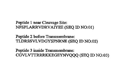
Figure 1: Three MUC16 carboxy terminus peptides were synthesized at the MSKCC
Microchemistry Core Facility. Polypeptide 1 is near the putative cleavage
site, Polypeptide 2 is
before the transmembrane, and Polypeptide 3 is the internal peptide, which is
inside the
transmembrane.
Figure 2: Comparison staining of high-grade serous ovarian carcinomas using
0C125 (left
panel) and 4H11 (right panel).
6e
Date Recue/Date Received 2022-01-28
WO 2011/119979 PCT/US2011/030025
Figure 3: Immunohistochemical scoring of 0C125 and 4H11 on tissue microarrays
of high-
grade ovarian serous carcinoma. Only membranous and/or cytoplasmic staining
was considered
positive. Score 0: No staining; Score 1: <5% strong or weak; Score 2: 5-50%
strong or weak;
Score 3: 51-75% strong or 51-100% weak; Score 4: 76-99% strong; Score 5: 100%
strong. Figure
3A: 0C125 (Score 0); Figure 3B: 0C125 (Score 1); Figure 3C: 0C125 (Score 2);
Figure 3D:
0C125 (Score 3); Figure 3E: 0C125 (Score 4); Figure 3F: 0C125 (Score 5);
Figure 3G: 4H11
(Score 0); Figure 3H: 41111 (Score 1); Figure 31: 4H11 (Score 2); Figure 3J:
4H11 (Score 3); Figure
3K: 4H11 (Score 4); Figure 3L: 4H11 (Score 5).
Figure 4: Western blot analysis. Figure 4A: Western blot analysis of GST-
AMUC16'114
fusion protein with monoclonal antibodies 9C9.21.5.13 and 4H11.2.5. Figure 4B:
Western blot
analysis of SKOV3-phrGFP-AMUC16'114 and SKOV3-phrGFP-6.MUC16e334 protein
extract and
probed with monoclonal antibodies 9C9.21.5.13 and 4H11.2_5.
Figure 5A: MUC16 carboxy terminus monoclonal antibodies binding affinity on
OVCAR3
cells (Panels A-D). Figure 5B: Internalization of radio-labeled 4H11 and 0C125
monoclonal
antibodies on SKOV3-phrGFP-AMUC16'334 stable transfected cells.
Figure 6A-D: Comparison staining intensities of 0C125 and 4H11 monoclonal
antibodies
on tissue microarrays containing cancers of the prostate (2A, concordant),
lung (2B, discordant),
breast (2C, discordant), and pancreas (2D, discordant).
Figure 7: FACS analysis as described in the Material and Methods section was
performed
with commercial antibodies and MUC16 carboxy terminus monoclonal antibodies on
OVCAR3 wt,
SKOV3-phrGFP-AMUCle 14 and SKOV3-phrGFP-AMIJC16634 stable transfected cell
lines.
Figure 8: Nucleotide sequence encoding antibody variable heavy (VH) chain and
antibody
variable light (VI) chain. (A) 4A5 V1 (SEQ ID NO:04), (B) 4A5 VL (SEQ ID
NO:05), (C) 4H11
Vll (SEQ ID NO:06), (D) 4H1 1 VL (SEQ NO:07), (E) 9B11 VR (SEQ ID NO:08), (F)
9B11 VLA
(SEQ ID NO:09), (G) 9B11 VLn (SEQ ID NO:10), (H) 24B3 VI{ (SEQ ID NO:11), (I)
24B3 VL
(SEQ ID NO:12).
Figure 9: (A) Homo sapiens MUC16 (GenBank NP_078966) (SEQ ID NO:13), (B)
Polypeptide 1 (SEQ JD NO:01), (C) Polypeptide 2 (SEQ ID NO:02), (D)
Polypeptide 3 (SEQ ID
NO:03), (E) Transmembrane domain (SEQ ID NO:14), (F) Polypeptide 4 (SEQ ID
NO:15)
containing a cysteine loop polypeptide (SEQ ID NO:19).
Figure 10: Schematic of mucl6 structure.
Figure 1.1. Design and in vitro analysis of MUC-CD targeted CARs. (A)
Schematic
diagram of the first generation 4H11z and second generation 41111-28z
retroviral vectors.
41-111scFv: MUC16 specific scFv derived from the heavy (VH) and light (VI)
chain variable regions
7
CA 3002192 2018-04-19
WO 2011/119979 PCT1US2011/030025
of the monoclonal antibody 4H11; CD8: CD8 hinge and transmembrane domains;
CD28: CD28
transmembrane and cytoplasmic signaling domains; chain: T cell receptor chain
cytoplasmic
signaling domain; LTR: long terminal repeat; black box: C1)8 leader sequence;
grey box:
(Gly4Ser)3 linker; arrows indicate start of transcription. (B) FACS analysis
of human T cells
retrovirally transduced to express either the 41-111z or 19z1 CAR. (C) 411111f
but not 19z1+ T
cells expand on 3T3(MUC-CD/B7.1) AAPC. CAR + were co-cultured on 3T3(MUC-
CD/B7.1)
AAPC monolayers at 3 x 10⁶ CARP T cells/well of a 6 well plate. Proliferation
of CARP T cells,
normalized to the CARP T cell fraction as assessed by FACS for the CAR +
fraction in combination
with viable T cell counts obtained on days 2, 4 and 7, as assessed by trypan
blue exclusion assays.
. Figure 12. In vitro comparison of T cells modified to express the first
generation 4H1lz
CAR to T cells modified to express the second generation co-stimulatory 4H11-
28z CAR. (A)
CAR+ T cells were co-cultured on MUC-CD monolayers with (right panel) or
without B7.1 (left
panel). 3 x 10⁶ CARP T cells were co-cultured on AAPC monolayers in 6 well
tissue culture plates
in cytokine-free medium. Total viable T cell counts were assessed on days 2, 4
and 7, by trypan
blue exclusion assays. 4H11-28z+ T cells markedly expanded when compared to
41111z+ T cells
upon co-culture with 3T3(MUC-CD) AAPCs, **p--0.0023 (4111lz compared to 4H11-
28z). In
contrast, both 4H11z+ and 4H11-28i+ T cells expanded similarly on 313(MUC-
CD/B7.1) AAPCs,
p.09, (41111z compared to 4H11-28z). Control 19-282+ T cells did not
proliferate on 3T3(MUC-
CD), **p=0.0056 (19-28z compared to 41111z), **p=0.0011 (19-28z compared to
4H11-28z), or on
3T3(MUC-CD/B7.1), **p=0.0026 (19-28z compared to 4H11z), **p=0.0087 (19-28z
compared to
4H11-28z). (B) 4H11-28z+ but not 4H1 lz+ T cells secrete 1L-2 upon co-culture
with 3T3(MUC-
CD) AAPCs. Tissue culture supernatants at day 2 following activation on
3T3(MUC-CD) AAPCs
were analyzed for cytokine secretion. 4H11-28z+ T cells, in contrast to 4H11z+
T cells,
demonstrated enhanced secretion of IL-2 consistent with T cell co-stimulation
mediated through the
4H11-28z CAR_ ***p=0.0008 (19z1 or 19-28z compared to 4H11z), "p=0.0026 (19z1
or 19-28z
compared to 41111-28z), **p=0.0046 (4H11z compared to 4H11-28z). Furthermore,
both 41111-
28z+ and 4H11z+ T cells secreted IFNy. *p=0.011 (4H1lz compared to 41111-28z).
Control 19z1
and 1928z transduced T cells failed to secrete either IL-2 or IFNy. "p=0.0034
(19z1 compared to
41111z), **F-0.036 (19-28z compared to 41111z), ***p=0.0008 (19-28z compared
to 4H11-28z).
(C) Expansion of CAR+ T cells following 3 cycles of stimulation on 3T3(MUC-
CD/B7.1). Human
T cells transduced to express either 4H1lz or 4H11-28z CARs demonstrated a >2
log expansion
over 2 cycles of stimulation on 3T3(MUC-CD/B7.1) AAPCs. Arrows indicate let
and 2nd cycles of
restimulation on AAPCs. (D) FACS analysis of the CARP T cell fraction of 41111-
28z+ T cells
increased following each weekly cycle of stimulation. (1) FACS following
initial transduction, (II)
8
CA 3002192 2018-04-19
. ______ 1-.
WO 2011/119979 PCT/US2011/030025
FACS at 7 days following first stimulation on AAPCs, (III) FACS at 7 days
following second
stimulation on AA_PCs. These data are representative of one of three different
experiments using
three different healthy donor T cell populations, all of which demonstrated
similar proliferation and
cytoldne secretion patterns.
Figure 13. MUC-CD targeted T cells specifically expand and lyse MUC-CD+ tumor
cells.
(A) Cytotoxieity assay of 4H11z+ and 41-111-28z+ T cells targeting OV-CAR(MUC-
CD) tumor cells
demonstrates efficient cytotoxicity mediated by T cells from healthy donors
modified to express the
first and second generation MUC-CD targeted CAR.s. Control T cells modified to
express the first
and second generation CD19-targeted 19z1 and 19-28z CARs failed to demonstrate
significant lysis
of target tumor cells. (B) Healthy donor T cells modified to express the 4H11-
28z CAR equally
lyse primary patient aseitcs-derived MUC-CD+ tumor cells when compared to T
cells modified to
express the control 19-28z CAR. This data represents 1 or 3 experiments
targeting primary tumor
cells from 3 ovarian carcinoma patients with similar results. (C) Autologous T
cells isolated from
peripheral blood, when modified with the 4H11-28z CAR, exhibit significant
lysis of autologous
MUC-CD+ ascites-derived tumor cells when compared to control 19-28z+
autologous T cells.
These data represent 1 of 3 experiments utilizing T cells and autologous tumor
cells from 3
different ovarian carcinoma patients with similar results. (D) Antigen
specific proliferation of
MUC-CD targeted CFSE labeled T cells after co-culture with OV-CAR3(MUC-CD)
tumor cells.
CFSE labeled CART T cells were co-cultured with MUC-CD expressing OV-CAR3
tumor cells at
1:1 ratio for 5 days. Proliferation of CFSE labeled T cells was assessed by
FACS demonstrating
efficient proliferation of both 4H11z+ and 4H11-28z+ T cells but not control
19-28z+ T cells. (E)
CFSE results were further confirmed by absolute T cell numbers assessed on
days 2, 4 and 7
following co-culture with OV-CAR3(MUC-CD) tumor cells. (F) FACS analysis of
the expression
of 4-1BBL on OVCAR3(M1JC-CD) cells. OV-CAR3(MUC-CD) cells were stained with
anti-
human 4-1BBL antibody (thick line) or with isotype control (thin line). FACS
analysis
demonstrated expression of 4-1BBL on DV-CAR3(MUC-CD) tumor cells. Further FACS
analyses
failed to reveal expression of the co-stimulatory ligands B7.1, 137.2, or OX-
40L.
Figure 14. Eradication of OV-CAR3(MUC-CD) tumors after intra-peritoneal
treatment with
first and second generation of MUC-CD targeted T cells. (A) Intraperitoneal
injection of OV-
CAR3 (MU C-CD) tumors in untreated SC1D-Beige mice results in abdominal
distension and
nodular peritoneal tumors. SCTD-Beige mice were injected intraperitoneally
with 3x1060V-
CAR3(MUC-CD) cells. At 5 weeks post intraperitoneal injection of OV-CAR3(MUC-
CD) tumor
cells mice developed ascities as evidenced by a distended abdomen (center
panel) when compared
to a tumor free mouse (left panel). Post mortem visualization of the
peritoneum demonstrates
9
CA 3002192 2 018 -0 4 -19
, ____________________________________________________________________________
WO 2011/119979 PC17US2011/030025
nodular tumor masses (arrows) within the abdominal cavity (right panel). (B)
Intraperitoneal
injection of 4H11z+ and 4H11-28z+ T cells either delay tumor progression or
fully eradicate
disease. Kaplan-Meier survival curve of SCID-Beige mice treated with first or
second generation
of MUC-CD targeted T cells. SaD-Beige mice were infused ip with 3x1060V-
CAR3(1vfUC-CD)
tumor cells on day 1 followed by 3x10⁷ 4H1 1 z+ or 4H11-28z1 T cells on day 2.
All untreated mice
or mice treated with control I 9zl T cells developed established tumors and
were sacrificed by day
50. In contrast, 27% of mice treated with either 4H11z+ or 41111-28z+ T cells
remained without
clinical evidence of disease by day 120. *p=0.01 (4H1lz compared to 19z1), **p-
0.0023 (4H11-
28z compared to 19z1), p=0.63 (4H1lz compared to 4H11-28z).
Figure 15. MUC-CD targeted 4I-111-28Z T cells successfully traffic to ip OV-
CAR3(MUC-CD/GFP-FFLuc) tumors following systemic intravenous infusion
resulting in equally
efficient anti-tumor efficacy when compared to ip 4H11-28z+ treated tumor
bearing mice. (A)
Kaplan-Meier survival curve of SCID-Beige mice treated ip or iv with 41-111-
28z4 T cells. SCID-
Beige mice were injected intraperitoneaIly with 3x10⁶ OV-CAR3(MUC-CD/GFP-
FFLue) tumor
cells followed by either iv or ip infusion of 3x10⁷ 4H11-28Z T cells. Tumor
eradication is
enhanced after either ip or iv infusion of 4H11-28z+ T cells when compared to
control treated mice.
Roth ip and iv 4H11-282+ T cell treated mice exhibited statistically enhanced
survival
(***p<0.0001 and **p=0.0038, respectively) when compared to 19-28z+ T cell
treated control
cohorts. Conversely, difference in survival between the ip and iv 4H11-28z+ T
cell cohorts was not
statistically significant (p=0.22). (B) BU of tumor progression of
representative ip and iv 4H11 _
28e T cell treated mice with ultimately progressive disease following
treatment compared to 13LI
of tumor progression in a representative control l9-28zt T cell treated mouse.
(C) Systemically
injected CFSE stained 41111-28z+ T cells traffic to advanced ip OV-CAR(MUC-CD)
tumors.
Presence of iv injected CFSE labeled 19-28z+ control T cells (left panel) and
41111-28z+ T cells
(right panel) 1 day following infusion into SCTD-Beige 'nice with advanced OV-
CAR(MUC-CD)
tumors (injected 7 days earlier), as assessed by FACS analysis of single cell
OV-CAR3(M1JC-CD)
tumor suspensions, reveals a marked population of 4I111-28zt but not control
l9-28z t T cells
within peritoneal OV-CAR3(MUC-CD) tumors.
Figure 16. Eradication of advanced OV-CAR3(MUC-CD) tumors in SCID-Beige mice
by ip
infusion of 4H11-28z+ T cells. SCID-Beige mice were injected ip with 3x10⁶ OV-
CAR.3(MUC-
CD/GFP-FFLue) tumor cells 7 days prior to ip treatment with 3x10⁷ 41111-28z+ T
cells. (A) BLI of
4I111-28z+ T cell treated mice with either relapsed disease (middle row) or
eradicated disease
(bottom row) compared to a representative 19-28z+ T cell treated control
mouse. (B) Kaplan-Meier
survival curve of SOD-Beige mice with advanced OV-CAR3(MUC-CD)GFP-FFLue)
tumors
CA 3002192 2018-04-19
3_, 3- - - _________ =
WO 2011/119979
PCT/US2011/030025
treated ip with 4H11-28z4 T cells. All 4H11-28z+ T cell treated mice
demonstrated enhanced
survival when compared to control 19-28z+ T cell treated mice ("p=0.0011),
with an overall long-
teim survival of 25% at day 120.
Figure 17: CD8 leader sequence, CD3 zeta chain intracellular domain sequence,
(G4S)3
serine-glycine linker sequence, CD8 transmembrane domain sequence, and CD28
transmembrane
intracellular domains (-STOP) sequence.
Figure 18: SFG_4111 lz sequence.
Figure 19: SFG-4H11-28z sequence.
Figure 20: (A) Mouse MUC16-CD Peptide 1 (SEQ ID NO:21), Mouse first Cysteine
Loop
Peptide 2 (SEQ ID NO:22), and Mouse second Cysteine Loop Peptide 3 (SEQ ID
NO:23). (B)
Alignment of mouse M1JC16 (SEQ ID NO:24) and human MUC16 (SEQ ID NO:25) amino
acid
sequences. A cysteine was added to the peptide sequence at the N terminus of
Peptide 1 and
Peptide 3 for better conjugation with KLH.
Figure 21: 11)8 extract with 1:10 dilution of Mouse MUC16 monoclonal Primary
Supernatants.
Figure 22: 13R5-FVB1 extract with 1:10 dilution of Mouse MUC16 monoclonal
Primary
Supernatants
Figure 23: Western Blot showing 38 hamster's monoclonal antibody Supernatants
on 1138
cell extracts.
Figure 24(A) Nucleotide sequence encoding 12B10-3G10-VH (SEQ TE) NO:26), (B)
12B10-3G10-VH Amino Acid sequence (SEQ ID NO:27), (C) Nucleotide sequence
encoding
12B10-3G10-VL (SEQ ID NO:28) (Note the VL has an optional NotI site added by
the primer for
cloning, and (D) 12B10-3G10-VLAmino Acid sequence (SEQ ID NO:29).
Figure 25: FACS Analysis with Purified 12B10-3G10 rnAb on 08 (mouse), OVCAR-3
(human) and BR5-FVB1 (mouse) cell lines.
DEFINITIONS
To facilitate understanding of the invention, a number of terms are defined
below.
The terms 'purified," "isolated," and grammatical equivalents thereof as used
herein, refer
to the reduction in the amount of at least one undesirable component (such as
cell, protein, nucleic
acid sequence, carbohydrate, etc.) from a sample, including a reduction by any
numerical
percentage of from 5% to 100%, such as, but not limited to, from 10% to 100%,
from 20% to
100%, from 30% to 100%, from 40% to 100%, from 50% to 100%, from 60% to 100%,
from 70%
to 100%, from 80% to 100%, and from 90% to 100%. Thus purification results in
an "enrichment,"
11
CA 3002192 2018-04-19
f
_______________________________________________________________________________
_______
-
WO 2011/119979 PCT/US2011/030025
i.e., an increase in the amount of a desirable component cell, protein,
nucleic acid sequence,
carbohydrate, etc.).
The term "antibody" refers to an immunogjobulin (e.g., IgG, IgM, IgA, IgE,
IgD, etc.). The
basic functional unit of each antibody is an immunoglobulin (Ig) mononer
(containing only one
immunoglobulin ("Ig") unit). Included within this definition are polyclonal
antibody, monoclonal
antibody, and chimeric antibody.
The variable part of an antibody is its "V domain" (also referred to as
"variable region"),
and the constant part is its "C domain" (also referred to as "constant
region") such as the kappa,
lambda, alpha, gamma, delta, epsilon and mu constant regions. The "variable
domain" is also
referred to as the "Fy region" and is the most important region for binding to
antigens. More
specifically, variable loops, three each on the light ('IL) and heavy (VH)
chains are responsible for
binding to the antigen. These loops are referred to as the "complcmcntaxity
determining regions"
("CDRs" and "idiotypes."
The immunoglobulin (Ig) monomer of an antibody is a "Y"-shaped molecule that
contains
four polypeptide chains: two light chains and two heavy chains, joined by
disulfide bridges.
Light chains are classified as either (X) or kappa (x). A light chain has two
successive
domains: one constant domain ("CL") and one variable domain ("VL"). The
variable domain, VL,
is different in each type of antibody and is the active portion of the
molecule that binds with the
specific antigen.The approximate length of a light chain is 211 to 217 amino
acids.
Each heavy chain has two regions, the constant region and the variable region.
The There
are five types of mammalian Ig heavy denoted a a, 8, a, y, and n. The type of
heavy chain present
defines the class of antibody; these chains are found in IgA, IgD, IgE, IgG,
and IgM antibodies,
respectively. Distinct heavy chains differ in size and composition; a and y
contain approximately
450 amino acids, while ji and E have approximately 550 amino acids. Each heavy
chain has two
regions, the constant region ("CH") and the variable ("Ill") region. The
constant region (CH) is
identical in all antibodies of the same isotype, but differs in antibodies of
different isotypes. Heavy
chains y, a and 8 have a constant region composed of three tandem (in a line)
Ig domains, and a
hinge region for added flexibility. Heavy chains . and a have a constant
region composed of four
immunoglobulin domains. The variable region (VH) of the heavy chain differs in
antibodies
produced by different B cells, but is the same for all antibodies produced by
a single B cell or B cell
clone. The variable region of each heavy chain is approximately 110 amino
acids long.
The term "specifically binds" and "specific binding" when made in reference to
the binding
of two molecules (e.g. antibody to an antigen, etc.) refer to an interaction
of the two molecules that
is dependent upon the presence of a particular structure on one or both of the
molecules. For
12
CA 3002192 2018-04-19
, - _______ -
WO 2011/119979 PCT/US2011/030025
example, if an antibody is specific for epitope "A" on the molecule, then the
presence of a protein
containing epitope A (or free, unlabelled A) in a reaction containing labeled
"A" and the antibody
will reduce the amount of labeled A bound to the antibody.
The term "capable of binding" when made in reference to 'the interaction
between a first
molecule (such as antibody, polypeptide, glycoprotein, nucleic acid sequence,
etc.) and a second
molecule (such as antigen, polypeptide, glyeoprotein, nucleic acid sequence,
etc.) means that the
first molecule binds to the second molecule in the presence of suitable
concentration of salts, and
suitable temperature, and pH. The conditions for binding molecules may be
determined using
routine and/or commercially available methods
The terms "antigen," "immunogen," "antigenic," "immunogenic," "antigenically
active,"
"immunologic," and "immunologically active" when made in reference to a
molecule, refer to any
substance that is capable of inducing a specific humoral immune response
(including eliciting a
soluble antibody response) and/or cell-mediated immune response (including
eliciting a CTL
response). Antigenic peptides preferably contain at least 5, at least 6, at
least 7, at least 8, at least 9,
and more preferably at least 10 amino acids. To elicit antibody production, in
one embodiment,
antigens may be conjugated to keyhole limpet hemocyanin (KLH) or fused to
glutathione-S-
transferase (GST).
A "cognate antigen" when in reference to an antigen that binds to an antibody,
refers to an
antigen that is capable of specifically binding to the antibody.
In one embodiment, the antigen comprises an epitope. The terms "epitope" and
"antigenic
determinant" refer to a structure on an antigen, which interacts with the
binding site of an antibody
or T cell receptor as a result of molecular cornplementarity. An epitope may
compete with the
intact antigen, from which it is derived, for binding to an antibody.
As used herein the terms "portion" and "fragment" when made in reference to a
nucleic
acid sequence or protein sequence refer to a piece of that sequence that may
range in size from 2
contiguous nucleotides and amino acids, respectively, to the entire sequence
minus one nucleotide
and amino acid, respectively.
A "subject" that may benefit from the invention's methods includes any
multicellular
animal, preferably a mammal. Mammalian subjects include humans, non-human
primates,
murines, vines, bovines, ruminants, lagomorphs, porcines, caprines, equines,
canines, felines,
ayes, etc.). Thus, mammalian subjects are exemplified by mouse, rat, guinea
pig, hamster, ferret
and chinchilla. The invention's compositions and methods are also useful for a
subject "in need of
reducing one or more symptoms of' a disease, e.g., in need of reducing cancer
metastasis and/or in
need of reducing one or more symptoms of cancer, includes a subject that
exhibits and/or is at risk
13
CA 30 021 92 2 0 1 8 -0 4 -1 9
WO 2011/119979 PCT/US20 11
/030025
of exhibiting one or more symptoms of the disease. For Example, subjects may
be at risk based on
family history, genetic factors, environmental factors, etc. This term
includes animal models of the
disease. Thus, administering a composition (which reduces a disease and/or
which reduces one or
more symptoms of a disease) to a subject in need of reducing the disease
and/or of reducing one or
more symptoms of the disease includes prophylactic administration of the
composition (i.e., before
the disease and/or one or more symptoms of the disease are detectable) and/or
therapeutic
administration of the composition (i.e., after the disease and/or one or more
symptoms of the
disease are detectable). The invention's compositions and methods are also
useful for a subject "at
risk" for disease (such as cancer) refers to a subject that is predisposed to
contracting and/or
expressing one or more symptoms of the disease. This predisposition may be
genetic (e.g., a
particular genetic tendency to expressing one or more symptoms of the disease,
such as heritable
disorders, etc.), or due to other factors (e.g., environmental conditions,
exposures to detrimental
compounds, including carcinogens, present in the environment, etc.). The term
subject "at risk"
includes subjects "suffering from disease," i.e., a subject that is
experiencing one or more
symptoms of the disease. It is not intended that the present invention be
limited to any particular
signs or symptoms. Thus, it is intended that the present invention encompass
subjects that are
experiencing any range of disease, from sub-clinical symptoms to full-blown
disease, wherein the
subject exhibits at least one of the indicia (e.g., signs and symptoms)
associated with the disease.
"Cancer cell" refers to a cell undergoing early, intermediate or advanced
stages of multi-
step neoplastic progression as previously described (Pitot et al.,
Fundamentals of Oncology, 15-28
(1978)). This includes cells in early, intermediate and advanced stages of
neoplastic progression
including "pre-neoplastic cells (i.e., "hyperplastic cells and dysplastic
cells), and neoplastic cells in
advanced stages of neoplastic progression of a dysplastic cell.
"Metastatic" cancer cell refers to a cancer cell that is translocated from a
primary cancer site
(i.e., a location where the cancer cell initially formed from a normal,
hyperplastie or dysplastic cell)
to a site other than the primary site, where the translocated cancer cell
lodges and proliferates.
"Cancer" refers to a plurality of cancer cells that may or may not be
metastatic, such as
ovarian cancer, breast cancer, lung cancer, prostate cancer, cervical cancer,
pancreatic cancer, colon
cancer, stomach cancer, esophagus cancer, mouth cancer, tongue cancer, gum
cancer, skin cancer
(e.g., melanoma, basal cell carcinoma, Kaposi's sarcoma, etc.), muscle cancer,
heart cancer, liver
cancer, bronchial cancer, cartilage cancer, bone cancer, testis cancer, kidney
cancer, endometrium
cancer, uterus cancer, bladder cancer, bone marrow cancer, lymphoma cancer,
spleen cancer,
thymus cancer, thyroid cancer, brain cancer, neuron cancer, mesothelioma, gall
bladder cancer,
ocular cancer (e.g., cancer of the cornea, cancer of uvea, cancer of the
choroids, cancer of the
14
CA 3002192 2018-04-19
_ ___________________________________________________________________________
W020111119979 PCT/US2011/030025
macula, vitreous humor cancer, etc.), joint cancer (such as synovium cancer),
glioblastoma,
lymphoma, and leukemia.
"Sample" and "specimen" as used herein are used in their broadest sense to
include any
composition that is obtained and/or derived from a biological source, as well
as sampling devices
(e.g., swabs), which are brought into contact with biological or environmental
samples. "Biological
samples" include those obtained from a subject, including body fluids (such as
urine, blood,
plasma, fecal matter, cerebrospinal fluid (CSF), semen, sputum, and saliva),
as well as solid tissue.
Biological samples also include a cell (such as con lines, cells isolated from
tissue whether or not
the isolated cells are cultured after isolation from tissue, fixed cells such
as cells fixed for
histological and/or immunohistochemical analysis), tissue (such as biopsy
material), cell extract,
tissue extract, and nucleic acid (e.g., DNA and RNA) isolated from a cell
and/or tissue, and the like.
These examples are illustrative, and are not to be construed as limiting the
sample types applicable
to the present invention.
"Overexpression of MIJC16" by a cell of interest (such as a cancer cell)
refers to a higher
level of MIJC16 protein and/or inRNA that is expressed by the cell of interest
compared to a
control cell (such as a non-cancerous cell, normal cell, etc.).
"Internalize" when in reference to a cell refers to entry from the
extracellular medium into
the cell membrane and/or cytoplasm.
"Glycosylated" when in reference to a sequence (e.g., an amino acid sequence
or nucleotide
sequence) refers to a sequence that is covalently linked to one or more
saccharides.
"Pharmaceutical" and "physiologically tolerable" composition refers to a
composition that
contains pharmaceutical molecules, i.e., molecules that are capable of
administration to or upon a
subject and that do not substantially produce an undesirable effect such as,
for example, adverse or
allergic reactions, dizziness, gastric upset, toxicity and the like, when
administered to a subject.
Preferably also, the pharmaceutical molecule does not substantially reduce the
activity of the
invention's compositions. Pharmaceutical molecules include "diluent" (i.e.,
"carrier") molecules
and excipients.
"Immunogenically effective" and "antigenically effective" amount of a molecule
interchangeably refer to an amount of the molecule that is capable of inducing
a specific humoral
immune response (including eliciting a soluble antibody response) and/or cell-
mediated immune
response (including eliciting a cytotoxic T-lymphocyte (CTL) response).
"Treating" a disease refers to reducing one or more symptoms (such as
objective,
subjective, pathological, clinical, sub-clinical, etc.) of the disease.
CA 3002192 2018-04-19
r- -
WO 2011/119979 PCTIUS2011/030025
The terms "reduce," "inhibit," "diminish," "suppress," "decrease," and
grammatical
equivalents (including "lower," "smaller," eic.) when in reference to the
level of any molecule (e.g.,
amino acid sequence, and nucleic acid sequence, antibody, etc.), cell, and/or
phenomenon (e.g.,
disease symptom, binding to a molecule, specificity of binding of two
molecules, affinity of
binding of two molecules, specificity to cancer, sensitivity to cancer,
affinity of binding, enzyme
activity, etc.) in a first sample (or in a first subject) relative to a second
sample (or relative to a
second subject), mean that the quantity of molecule, cell and/or phenomenon in
the first sample (or
in the first subject) is lower than in the second sample (or in the second
subject) by any amount that
is statistically significant using any art-accepted statistical method of
analysis. In one embodiment,
the quantity of molecule, cell and/or phenomenon in the first sample (or in
the first subject) is at
least 10% lower than, at least 25% lower than, at least 50% lower than, at
least 75% lower than,
and/or at least 90% lower than the quantity of the same molecule, cell and/or
phenomenon in the
second sample (or in the second subject). In another embodiment, the quantity
of molecule, cell,
and/or phenomenon in the first sample (or in the first subject) is lower by
any numerical percentage
from 5% to 100%, such as, but not limited to, from 10% to 100%, from 20% to
100%, from 30% to
100%, from 40% to 100%, from 50% to 100%, from 60% to 100%, from 70% to 100%,
from 80%
to 100%, and from 90% to 100% lower than the quantity of the same molecule,
cell and/or
phenomenon in the second sample (or in the second subject). In one embodiment,
the first subject
is exemplified by, but not limited to, a subject that has been manipulated
using the invention's
compositions and/or methods. In a further embodiment, the second subject is
exemplified by, but
not limited to, a subject that has not been manipulated using the invention's
compositions and/or
methods. In an alternative embodiment, the second subject is exemplified by,
but not limited to, a
subject to that has been manipulated, using the invention's compositions
and/or methods, at a
different dosage and/or for a different duration and/or via a different route
of administration
compared to the first subject. In one embodiment, the first and second
subjects may be the same
individual, such as where the effect of different regimens (e.g., of dosages,
duration, route of
administration, etc.) of the invention's compositions and/or methods is sought
to be determined in
one individual. In another embodiment, the first and second subjects may be
different individuals,
such as when comparing the effect of the invention's compositions and/or
methods on one
individual participating in a clinical trial and another individual in a
hospital.
The terms "increase," "elevate," "raise," and grammatical equivalents
(including "higher,"
"greater," etc.) when in reference to the level of any molecule (e.g., amino
acid sequence, and
nucleic acid sequence, antibody, etc.), cell, and/or phenomenon (e.g., disease
symptom, binding to
a molecule, specificity of binding of two molecules, affinity of binding of
two molecules,
16
CA 3002192 2018-04-19
WO 2011/119979 PCT/US2011/030025
specificity to cancer, sensitivity to cancer, affinity of binding, enzyme
activity, etc.) in a first
sample (or in a first subject) relative to a second sample (or relative to a
second subject), mean that
the quantity of the molecule, cell and/or phenomenon in. the first sample (or
in the first subject) is
higher than in the second sample (or in the second subject) by any amount that
is statistically
significant using any art-accepted statistical method of analysis. In one
embodiment, the quantity
of the molecule, cell and/or phenomenon in the first sample (or in the first
subject) is at least 10%
greater than, at least 25% greater than, at least 50% greater than, at least
75% greater than, and/or at
least 90% greater than the quantity of the same molecule, cell and/or
phenomenon in the second
sample (or in the second subject). This includes, without limitation, a
quantity of molecule, cell,
and/or phenomenon in the first sample (or in the first subject) that is at
least 10% greater than, at
least 15% greater than, at least 20% greater than, at least 25% greater than,
at least 30% greater
than, at least 35% greater than, at least 40% greater than, at least 45%
greater than, at least 50%
greater than, at least 55% greater than, at least 60% greater than, at least
65% greater than, at least
70% greater than, at least 75% greater than, at least 80% greater than, at
least 85% greater than, at
least 90% greater than, and/or at least 95% greater than the quantity of the
same molecule, cell
and/or phenomenon in the second sample (or in the second subject). In one
embodiment, the first
subject is exemplified by, but not limited to, a subject that has been
manipulated using the
invention's compositions and/or methods. Lit a further embodiment, the second
subject is
exemplified by, but not limited to, a subject that has not been manipulated
using the invention's
compositions and/or methods. In an alternative embodiment, the second subject
is exemplified by,
but not limited to, a subject to that has been manipulated, using the
invention's compositions and/or
methods, at a different dosage and/or for a different duration and/or via a
different route of
administration compared to the first subject. In one embodiment, the first and
second subjects may
be the same individual, such as where the effect of different regimens (e.g.,
of dosages, duration,
route of administration, etc.) of the invention's compositions and/or methods
is sought to be
determined in one individual. In another embodiment, the first and second
subjects maybe
different individuals, such as when comparing the effect of the invention's
compositions and/or
methods on one individual participating in a clinical trial and another
individual in. a hospital.
The terms "alter" and "modify" when in reference to the level of any molecule
and/or
phenomenon refer to an increase or decrease.
Reference herein to any numerical range expressly includes each numerical
value (including
fractional numbers and whole numbers) encompassed by that range. To
illustrate, and without
limitation, reference herein to a range of "at least 50" includes whole
numbers of 50, 51, 52, 53, 54,
55, 56, 57, 58, 59, 60, etc., and fractional numbers 50.1, 50.2 50.3, 50.4,
50.5, 50.6, 50.7, 50.8,
17
CA 3002192 2018-04-19
. . . . = - -
WO 2011/119979 PCT/US2011/030025
50.9, etc. In a further illustration, reference herein to a range of "less
than 50" includes whole
numbers 49, 48, 47, 46, 45, 44, 43, 42, 41, 40, etc., and fractional numbers
49.9, 49.8, 49,7, 49.6,
49.5, 49.4, 49.3, 49.2, 49.1, 49.0, etc. In yet another illustration,
reference herein to a range of from
"5 to 10" includes each whole number of 5, 6, 7, 8, 9, and 10, and each
fractional number such as
5.1, 5.2, 5.3, 5.4, 5.5, 5.6, 5.7, 5.8, 5.9, etc.
DESCRIPTION OF THE INVENTION
The invention provides antibodies, and antigen-binding fragments thereof, that
specifically
bind to a polypeptide, or antigenic portion thereof, wherein the polypeptide
is selected from a)
MUC16 ectodomain polypeptide, b) MUC I 6 cytoplasmic domain polypeptidc, and
c) MUC16
extracellular domain polypeptide that contains a cysteine loop polypeptide.
The invention's
antibodies and compositions containing them are useful in diagnostic and
therapeutic applications
for diseases in which MUC16 is overexpressed, such as cancer.
Using synthetic peptides, the inventors raised novel-specific antibodies to
the carboxy-
terminal portion of MUC16, retained by the cell, proximal to the putative
cleavage site. These
antibodies were characterized using fluorescence-activated cell-sorting
analysis, enzyme-linked
immunoassay, Western blot analysis, and immunohistochernistry. Each of the
selected monoclonal
antibodies was reactive against recombinant GST-AMUC16'114 protein and the
MUC16 transfected
SKOV3 cell line. Three antibodies, 4H11, 9C9, and 4A5 antibodies demonstrated
high affinities by
Western blot analysis and saturation-binding studies of transfected SKOV3
cells, and displayed
antibody intemali7ation. Immunohistochcmical positivity with novel antibody
4H11 was similar to
0C125, but with important differences, including diffuse positivity in lobular
breast cancer and a
small percentage of 0C125-negative ovarian carcinomas which showed intense and
diffuse 4H11
antibody binding.
The invention's compositions and methods are useful for diagnostic and
therapeutic
applications, as well as biologic studies such as membrane receptor
trafficking and intracellular
events. Diagnostic applications include, for example, detection of cancer
using
immunohistochemical, radiographic imaging, enzyme-linked immunosorbent assay
(ELISA),
fluorescence-activated cell sorting (FACS), Western blot, and/or
immunoprecipitation detection.
The invention is further described under (A) MUC16, (B) Prior Art Antibodies,
(C)
Invention's Antibodies, (D) Hybridoma Cell Lines, (E) Conjugates Of The
Invention's Antibodies
Linked To Cytotoxic Agents And/Or Prodrugs, (F) Detecting Mucl 6 Portions And
Diagnostic
Applications, and (G) Therapeutic Applications.
18
CA 3002192 2018-04-19
t __________________________________________________________ -
WO 2011/119979 PCT/US2011/030025
A. MUC16
"MUC16," "MUC-16" and "Mucin 16" interchangeably refer to a type I membrane
protein
that is part of a family of tethered mucins. A schematic of Mucl 6 is in
Figure 10, and an
exemplary human Mue16 amino acid sequence (SEQ ID NO:13) is shown in Figure
9A. An
alignment of mouse MUC16 (SEQ ID NO:24) and human MUC16 (SEQ ID NO:25) amino
acid
sequences is shown in Figure 20B. The term "type 1 protein" refers to a
"membrane protein" that
is at least partially embedded in the lipid bilayer of a cell, virus and the
like, and that contains a
transmembrane domain (TM) sequence embedded in the lipid bilayer of the cell,
virus and the like.
The portion of the protein on the NH2-tcrminal side of the TM domain is
exposed on the exterior
side of the membrane, and the COOH-terminal portion is exposed on the
cytoplasmic side.
Recently, the sequence of the eDNA-encoding MUC16/CA125 was described by Yin
and
Lloyd in 2001 and completed by O'Brien in 2002 (10-12). The complete MUC16
protein has
various components consisting of a cytoplasmic tail with potential
phosphorylation sites, a
transmembrane domain, and an external domain proximal to an apparent cleavage
site. Distal to
the cleavage site, the released external domain contains 16-20 tandem repeats
of 156 amino acids,
each with many potential glycosylation sites (11). The overall repeat
structure (Figure 10) is well
conserved across mammals, but the repeats are not completely identical in
exact amino acid
composition.
The MUC16 protein is part of a family of tethered mucins that includes both
MUC1 and
MUC4 (13). MUC1 is present in a variety of tissues and appears to signal
through a beta catenin
pathway, interact with EGF receptor, mediates drug resistance and can act as
an oncogene (14-17).
The MUC4 protein is also expressed in a variety of tissues but is common on
neoplasms of the
gastrointestinal track (18-20). In contrast, the CA125 antigen has been more
restricted in its
distribution and is present primarily in gynecologic tissues and overexpressed
in Miillerian
neoplasms (21). However, the CA125 antigen, recognized by the 0C125 antibody,
is a heavily
glycosylated antigen expressed in the tandem repeat region of the larger MUC16
protein. This
glyeoprotein is typically shed from a putative cleavage site in the
extracellular domain of the
MUC16 peptide backbone.
Thus, "MUC16" protein contains (a) a "cytoplasmic domain," (b)a "transmembrane
domain," and (c) a "extracellular domain." The MUC16 extracellular domain
contains a cleavage
site between a non-glycosylatcd ectodomain and a large glycosylated ectodomain
of tandem
repeats.
The terms "cytoplasmic domain," "cytoplasmic tail," and "CT" are used
interchangeably to
refer to a protein sequence, and portions thereof, that is on the cytoplasmic
side of the lipid bilayer
19
CA 3002192 2018-04-19
WO 2011/1199'79
PCT/US2011/030025
of a cell, virus and the like. Methods for determining the CT of a protein are
known in the art
Elofsson et al. (2007) Annu. Rev. Biochern. 76:125-140; Bemsel et al. (2005)
Protein Science
14:1723-1728).
The terms "transmembrane domain" and "TM" are used interchangeably to refer to
a
protein sequence, and portions thereof, that spans the lipid bilayer of a
cell, virus and the like.
Methods for determining the TM of a protein are known in the art (Elofsson et
al. (2007) Annu.
Rev. Biochem. 76:125-140; Bemsel et al. (2005) Protein Science 14:1723-1728).
The terms "ectodomain" and "cxtracellular domain" are interchangeably used
when in
reference to a membrane protein to refer to the portion of the protein that is
exposed on the
extracellular side of a lipid bilayer of a cell, virus and the like. Methods
for determining the
ectodomain of a protein are known in the art (Singer (1990) Annu. Rev. Cell
Biol. 6:247-296 and
High el al. (1993) J. Cell Biol. 121:743-750, and MeVector software, Oxford
Molecular).
The exemplary Mud l 6 of Figure 9 contains (a) a "MUC16 cytoplasmic domain"
from
amino acid 14476 to 14507, vttrr rkkegeynvq qqcpgyyqsh ldledlq (SEQ ID NO:16),
that interacts
with the intracellular signal transduction machinery; (b) a "M1JC16
transmembrane domain" from
amino acid 14452 to 14475, fwaviligl agllgvitcl icgvl (SEQ ID NO:14) that
spans the plasma
membrane; and (c) a "MIJC16 extracellular domain" amino acid Ito 14392 (SEQ ID
NO:13) that
contains a cleavage site between an non-glyeosylated ectodomain and a large
glycosylated
ectodomain of tandem repeats. The "MUC16 ectodomain" is exemplified by nfsplar
rydrvaiyee
flrrntmgtq lqnftldrss vlvdgyspnr neplignsdl p (SEQ ID NO:17) from amino acid
14394 to 14451 of
SEQ ID NO:13 of Figure 9A.
The exemplary MIJC16 ectodomain contains both Polypeptide 1 (nfsplar
rvdrvaiyee (SEQ
ID NO:01), which is from amino acid 14394 to 14410 of SEQ ID NO:13), and
Polypeptide 2 (tldrss
vlvdgyspnr ne (SEQ ID NO:02), which is from amino acid 14425 to 14442 of SEQ
ID NO:13),
against which the invention's exemplary antibodies were produced. Polypeptide
3, egvIvrtir
rkkegeynvq qq (SEQ ID NO:03) is from amino acid 14472 to 14492 of SEQ ID
NO:13, and
contains both a transmembrane domain portion (cgvl) and a cytoplasmic domain
portion (vttrr
rkkegeynvq qq (SEQ ID NO:18)). Thus, the CGVL is optional in SEQ ID NO:03, as
it is part of
the transmembrane domain.
Polypeptide 4 (ksyf sdcqvstfrs vpnrhhtgvd sicnfspl (SEQ ID NO:15), is located
in a non-
glycosylated portion of the Mucl6 extracellular domain, is from amino acid
14367 to 14398 of
SEQ ID NO:13, and contains a cysteine loop polypeptidc eqvstfirsvpnrhhtgvdslc
(SEQ ID NO:13).
CA 3002192 2018-04-19
- ____________________________________________________________________________
WO 2011/119979 PC T/US2011/030025
B. Prior Art Antibodies
The expression of the MUC16/CA125 antigen has long been associated with
gynecologic
tissues. "CA125," "CA-125," "Cleaved CA125," and "cleaved CA-125,"
interchangeably refer to
the glycosylated external domain of the tethered mucin MUC 1.6, that is distal
to the cleavage site
(Payne et al., U.S. Pat. No. 7,202,346). This released external domain
contains 16-20 tandem
repeats of 156 amino acids, each with potential glyeosylation sites. An
apparent cysteine-based
disulfide loop of 19 amino acids is present in all repeats and the N-terminal
end contains a
hairbrush structure that is heavily 0-glycosylated (11). The deduced size
would be 2.5 MD for the
protein part, and with added carbohydrates, this could increase to 5 MD (10,
26).
CA125, though it is not sensitive or specific enough to be used as a general
screening tool,
is routinely used to monitor patients with ovarian carcinoma. The tests used
to measure CA125 are
antibody based detection methods, as are the immunohistochemical stains
routinely perfoinied for
diagnostic purposes. The epitope specificity of 26 antibodies to MUC16 was
studied in the first
report from the International Society of Oneodevelopmental Biology and
Medicine (ISOBM) TD-1
Workshop and the application of 22 antibodies to immunohistochemistry was
reported in the
second report from the TD-1 workshop (7, 21). The existing antibodies were
grouped as 0C125-
like, Mil-like, or OV197-like and all of the known antibodies recognized CA125
epitopes in the
repeating, glycosylated elements in the external domain of the tethered mucin
MUC16, distal to the
putative cleavage site.
The vast majority of MUC16-reactive antibodies, including 0C125, react with
the
glycosylation-dependent antigen present exclusively in the cleaved portion of
the molecule so the
true distribution of MUC16 expression is not known (21). There is currently no
antibody available
to track the fate of the remaining MUC16 protein fragment after cleavage and
CA125 release.
C. Invention's Antibodies
In order to better explore the biology of human MUC16, the inventors have
derived
monoclonal antibodies against the extracellular portion of the MUC16-earboxy
terminus, proximal
to the putative cleavage site, as well as one monoclonal antibody against the
internal cytoplasmic
domain. In contrast to prior antibodies, these are derived against the peptide
backbone of MU C16
and arc not directed at complex glyeoprotein epitopes. Since these epitopes
are proximal to the
cleavage site, they are unlikely to be found in the circulation and provide
novel targets for
diagnostic methods and therapeutic interventions. Data herein demonstrate the
identification and
characterization of exemplary antibodies developed against the MUC16 peptide
backbone.
21
CA 3002192 2018-04-19
WO 2011/119979 PCT/US 2011
/030025
The inventors have developed novel antibodies that are directed at the non-
cleaved, non-
glycosylated peptide backbone of MUC16. These are exemplified by both 4H11 and
9C9
antibodies, which react with peptide sequences in the non-cleaved ectodomain
of MUC16 and are
detectable on the surface of ovarian cancer cell lines and in paraffin-fixed
tissues from human
ovarian cancer surgical specimens. The antibodies show high affinity and are
readily internalized
by ovarian cancer cells when bound to the ectodomain of MUC16. This suggests
that the proximal
portion of MUC16 has an independent biology from the more distal, cleaved
portion of the mucin.
It also suggests that the proximal portions of MUC16 could provide convenient
targets for
diagnostic and therapeutic interventions. Targeting the peptide backbone of
MUC16 provides
highly specific tissue delivery for genetically engineered cells, liposomes,
or antibody conjugates,
including conjugates with the invention's antibodies.
The invention's antibodies, exemplified by antibody 4H11, are useful as tools
in
immunohistochemistry. Date herein show that 4H11 is relatively specific to
high-grade ovarian
serous carcinoma. Invasive lobular breast carcinoma is the major exception and
shows extensive
MUC16 protein as detected by 4H11. Lobular carcinoma of the breast has unique
biology which is
characterized by a propensity to metastasize to serosal surfaces (27). Since
MUC16 is the cognate
binding partner of mcsothclin, this may have important implications for
lobular cancer (28). The
discordance rates for 0C125 and 4H11 also suggest that 4H11 might provide
additional,
independent information from 0C125 in a subset of ovarian carcinomas, Some
tumors that are
negative with 0C125 retain cytoplasmic and extracellular portions of the MUC16
glycoprotein,
portions of the molecule that are likely involved in transduction of signals
potentially important in
the malignant phenotype.
Thus, in one embodiment, the invention provides an isolated antibody, or an
antigen-
binding fragment thereof, that specifically binds to a polypeptide, or
antigenic portion thereof,
wherein the polypeptide is exemplified by a) MUC16 ectodomain polypeptide
(exemplified by
NFSPLAR RVDRVA1YEE FLRMTRNGTQ LQNFTLDRSS VLVDGYSPNR NEPLTGNSDL P
(SEQ ID NO:17)), b) MUC16 cytoplasmic domain polypeptide (exemplified by VTTRR
RKKEGEYNVQ QQ (SEQ ID NO:18), which is contained within each of CGVLVTTRR
RKKEGEYNVQ QQ (SEQ ID NO:03) and LVTTRR RKKEGEYNVQ QQ (SEQ ID NO:20)), and
c) MUC16 extracellular domain polypeptide that contains a cysteine loop
polypeptide
CQVSTFRSVPNRHHTGVDSLC (SEQ ID NO:19).
One advantage of the invention's antibodies is that the antibody internalizes
into a cell,
thereby being useful in applications for delivery inside a cell, such as
disease therapy.
"Internalized" when in reference to a molecule that is internalized by a cell
refers to passage of the
22
CA 3002192 2018-04-19
WO 2011/119979 PCT/US2011/030025
molecule that is in contact with the extracellular surface of a cell membrane
across the cell
membrane to the intracellular surface of the cell membrane and/or into the
cell cytoplasm.
Methods for determining internalization are disclosed herein, including the
detection of
radiolabeled molecule inside the cell (Figure 5B).
In one embodiment, the invention's antibodies specifically bind to MUC16
ectodomain
polypeptide that comprises a polypeptide selected from the group consisting of
Polypeptide 1
NFSPLARRVDRVAIYEE (SEQ ID NO:01) and Polypeptide 2 TLDRSSVLVDGYSPNRNE (SEQ
ID NO:02). Data herein show that the invention's antibodies specifically bind
to GST-AMIJC16
114 (Example 2, Table IA). The specificity of the invention's antibodies is in
contrast to prior art
antibodies (e.g., VK8, M1 1 and 0C125 antibodies) that did not bind to GST-
AMTJC16"14 purified
protein or cell lysates of the SKOV3-pluGFP-AMUC16'114 cell line (Example 2,
Figure 2).
In a further embodiment, the invention's antibodies lack specific binding to a
glycosylated
MUC16 extracellular domain, exemplified by the cleaved CA-125 described in
Payne et al., U.S.
Pat. No. 7,202,346.
While not intending to limit the sequence of the Vi. and VH regions of the
invention's
antibodies, in one embodiment, the antibody specifically binds to the
Polypeptide 2 (SEQ ID
NO:02) of the M1JC16 ectodomain polypeptide, wherein the antibody comprises a
variable heavy
(VH) chain encoded by SEQ ID NO:06 (i.e., the antibody 4H11 variable heavy
(VH) chain amino
acid sequence of Figure 8), and a variable light (VL) chain encoded by SEQ ID
NO:07 (i.e., the
antibody 4H11 variable light (VI) chain amino acid sequence of Figure 8). In a
particular
embodiment, the antibody is chimeric, wherein at least one of the VL and VH
chains is fused to a
human immunoglobulin constant region.
Also without intending to limit the sequence of the VL and VH regions of the
invention's
antibodies, in one embodiment, the antibody specifically binds to the
Polypeptide 2 (SEQ ID
NO:02) of the MUC16 ectodomain polypeptide, wherein the antibody comprises a
variable heavy
(Vs) chain encoded by SEQ ID NO:04 (i.e., the antibody 4A5 variable heavy (VH)
chain nucleotide
sequence of Figure 8), and a variable light (Vi) chain encoded by SEQ ID NO:05
(i.e., the antibody
4A5 variable light (VL) chain nucleotide sequence of Figure 8). In a
particular embodiment, the
antibody is chimeric wherein at least one of the VL and VH chains is
covalently linked to a human
immunoglobulin constant region.
Still without intending to limit the sequence of the VL and VH regions of the
invention's
antibodies, in one embodiment, the antibody specifically binds to the
Polypeptidc 1 (SEQ ID
NO:01) of the M1JC16 ectodomain polypeptide, wherein the antibody comprises a
variable heavy
(Vii) chain encoded by SEQ ID NO:08 (i.e., the antibody 9B11 variable heavy
(VH) chain
23
CA 3002192 2 018 -0 4 -19
- ________________________________________________ -
WO 2011/119979 PCT/US2011/030025
nucleotide sequence of Figure 8), and a variable light (VL) chain encoded by
at least one of SEQ ID
NO:09 (i.e., antibody 91311 variable light (V1) chain nucleotide sequence of
Figure 8), and SEQ
JD NO:10 (i.e., the antibody 9B11 variable light (Ve.$) chain nucleotide
sequence of Figure 8). In a
particular embodiment, the antibody is chimeric wherein at least one of the VL
and VH chains is
covalently linked to a human immunoglobulin constant region.
While not intending to restrict the source of antigen to which the invention's
antibodies
bind, in one embodiment, the MUC16 ectodomain polypeptide is expressed by a
cell. Data herein
show that the invention's exemplary antibodies bind to SKO'V3 cells transduced
with phrGFF-
AMUC16c114 (Example 2).
While not limiting the sequence of antigen to which the invention's antibodies
bind, in a
further embodiment, the invention's antibodies specifically bind to a MUC16
cytoplasmic domain
polypeptide that comprises VTTRR RKKEGEYNVQ QQ (SEQ ID NO:18). In a particular
embodiment, the MUC16 cytoplasmic domain polypeptide comprises Polypeptide 3
CGVLVTTRRRKKEGEYNVQQQ (SEQ ID NO:03). In some embodiment, the MUC16
cytoplasmic domain polypeptide is expressed by a cell. For example, data
herein show that the
invention's exemplary antibody binds to SKOV3 cells transduced with phrGFP-
AM1JC16c114
(Example 2). In a particular embodiment, the cell is permeabilized to
facilitate internalization of
the antibody into the cell so that it comes into contact with its cytoplasmic
antigen.
Still without limiting the sequence of antigen to which the invention's
antibodies bind, in a
further embodiment, the invention's antibodies bind to a MUD extracellular
domain polypeptide
that contains a eysteine loop polypeptide CQVSTFRSVPNRHHTGVDSLC (SEQ ID
NO:19). In a
more preferred embodiment, the MUC16 extracellular domain polypcptide
comprises Polypeptide 4
KSYF SDCQVSTFRS VPNRHHTGVD SLCNFSPL (SEQ ID NO:15).
Still without intending to limit the sequence of the VL and VH regions of the
invention's
antibodies, in one embodiment, the antibody specifically binds to Polypeptide
4 (SEQ NO:15)
of the MUC16 extracellular domain polypcptide, wherein the antibody comprises
a variable heavy
(Vs) chain encoded by SEQ ID NO:11 (i.e., the antibody 24B3 variable heavy
(Vs) chain amino
acid sequence of Figure 8), and a variable light (VL) chain encoded by SEQ ID
NO:12 (i.e., the
antibody 24B3 variable light (VL) chain amino acid sequence of Figure 8).
The invention contemplates chinaerie antibodies (sec U.S. Pat. No. 7,662,387),
monoclonal
antibodies, recombinant antibodies, an antigen-binding fragment of a
recombinant antibody, a
humanized antibody, and an antibody displayed upon the surface of a phage
(U.S. Pat. No.
7,202,346). In particular, the invention contemplates antibody fragments that
contain the idiotype
("antigen-binding region" or "antigen-binding fragment") of the antibody
molecule. For example,
24
CA 30 0 2192 2 018 -0 4 -19
^.-^
_______________________________________________________________________________
____
WO 2011/119979 PCTfUS2011/030025
such antigen-binding fragments include, but are not limited to, the Fab
region, F(ab)2 fragment,
pFc fragment, and Fab' fragments.
The "Fab region" and "fragment, antigen binding region," interchangeably refer
to portion
of the antibody arms of the immnoglobulin "Y" that function in binding
antigen. The Fab region is
composed of one constant and one variable domain from each heavy and light
chain of the
antibody. Methods are known in the art for the construction of Fab expression
libraries (Huse et
al., Science, 246:1275-1281 (1989)) to allow rapid and easy identification of
monoclonal Fab
fragments with the desired specificity. In another embodiment, Fe and Fab
fragments can be
generated by using the enzyme papain to cleave an immunoglobulin monomer into
two Fab
fragments and an Fe fragment. The enzyme pepsin cleaves below the hinge
region, so a "F(ab')2
fragment" and a "pFel fragment" is formed. The F(ab)2 fragment can be split
into two "Fab'
fragments" by mild reduction.
The invention also contemplates a "single-chain antibody" fragment, i.e., an
amino acid
sequence having at least one of the variable or complementarity determining
regions (CDRs) of the
whole antibody, and lacking some or all of the constant domains of the
antibody. These constant
domains are not necessary for antigen binding, hut constitute a major portion
of the structure of
whole antibodies_ Single-chain antibody fragments are smaller than whole
antibodies and may
therefore have greater capillary permeability than whole antibodies, allowing
single-chain antibody
fragments to localize and bind to target antigen-binding sites more
efficiently, Also, antibody
fragments can be produced on a relatively large scale in prokaryotic cells,
thus facilitating their
production. Furthermore, the relatively small size of single-chain antibody
fragments makes them
less likely to provoke an immune response in a recipient than whole
antibodies. Techniques for the
production of single-chain antibodies are known (U.S. 4,946,778). The variable
regions of the
heavy and light chains can be fused together to form a "single-chain variable
fragment" (''scFy
fragment"), which is only half the size of the Fab fragment, yet retains the
original specificity of the
parent itnrnunoglobulin.
The 'Pc region'' and "Fragment, crystallizable region" interchangeably refer
to portion of
the base of the inmatoglobulin "Y" that function in role in modulating immune
cell activity. The
Fe region is composed of two heavy chains that contribute two or three
constant domains
depending on the class of the antibody. By binding to specific proteins, the
Pc region ensures that
each antibody generates an appropriate immune response for a given antigen.
The Fe region also
binds to various cell receptors, such as Fe receptors, and other immune
molecules, such as
complement proteins. By doing this; it mediates different physiological
effects including
opsonization, cell lysis, and degranulation of mast cells, basophils and
eosinophils, In an
CA 3002192 2 018 -0 4 -19
WO 2011/119979 PCT/US2011/030025
experimental setting, Fc and Fab fragments can be generated in the laboratory
by cleaving an
immunoglobulin monomer with the enzyme papain into two Fab fragments and an Fe
fragment.
The invention contemplates polyclonal antibodies and monoclonal antibodies.
"Potyclonal
antibody" refers to an imrnunoglobulin produced from more than a single clone
of plasma cells; in
contrast "monoclonal antibody" refers to an immunoglobulin produced from a
single clone of
plasma cells. Generic methods are available for making polyclonal and
monoclonal antibodies that
are specific to a desirable polypeptide. For the production of monoclonal and
polyclonal
antibodies, various host animals can be immunized by injection with the
peptide corresponding to
any molecule of interest in the present invention, including but not limited
to hamsters, rabbits,
mice, rats, sheep, goats, etc. For preparation of monoclonal antibodies, any
technique that provides
for the production of antibody molecules by continuous cell lines in culture
may be used (See e.g.,
Harlow and Lane, Antibodies: A Laboratory Manual, Cold Spring Harbor
Laboratory Press, Cold
Spring Harbor, NY). These include, but are not limited to, the hybridoma
technique originally
developed by Kohler and Milstein (Kohler and Milstein, Nature, 256:495-497
(1975)), techniques
using germ-free animals and utilizing technology such as that described in
PCT/US90/02545, as
well as the trioma technique, the human B-cell hybridoma technique (See e.g.,
Kozbor et al.,
Immunol. Today, 4:72 (1983)), and the EBV-hybridoma technique to produce human
monoclonal
antibodies (Cole et al., in Monoclonal Antibodies and Cancer Therapy, Alan R.
Liss, Inc., pp. 77-96
(1985)). In some particularly preferred embodiments of the present invention,
the present invention
provides monoclonal antibodies.
Also contemplated are chimeric antibodies. As used herein, the term "chimeric
antibody"
contains portions of two different antibodies, typically of two different
species. See, e.g.: U.S. Pat.
No. 4,816,567 to Cabilly et al.; U.S. Pat. No. 4,978,745 to Shoemaker et al.;
U.S. Pat. No.
4,975,369 to Beavers et al.; and U.S. Pat. No. 4,816,397 to Boss et al.
Chimeric antibodies include
monovalent, divalent or polyvalent irnmunoglobulins. A monovalent chimeric
antibody is a dimer
(IIL) fonned by a chimeric H chain associated through disulfide bridges with a
chimeric L chain. A
divalent chimeric antibody is tetramer (H21/2) formed by two HL dimers
associated through at least
one disulfide bridge. A polyvalent chimeric antibody can also be produced, for
example, by
employing a He region that aggregates (e.g., IgM H chain).
The invention also contemplates "humanized antibodies," i.e., chimeric
antibodies that have
constant regions derived substantially or exclusively from human antibody
constant regions, and
variable regions derived substantially or exclusively from the sequence of the
variable region from
a mammal other than a human. Humanized antibodies preferably have constant
regions and
variable regions other than the complement determining regions (CDRs) derived
substantially or
26
CA 3002192 2018-04-19
1- ________________________________ =
84261056 (83513-53D1)
exclusively from the corresponding human antibody regions and CDRs derived
substantially or
exclusively from a mammal other than a human. Thus, in one embodiment,
humanized antibodies
are human immunoglobulins (recipient antibody) in which residues from a
hypervariable region of
the recipient are replaced by residues fruni a hypervariable region of a non-
human species (donor
antibody) such as mouse, rat, rabbit or nonhuman primate having the desired
specificity, affunty,
and capacity. In some instances, Fv framework region (FR) residues of the
human immunoglobulin
are replaced by corresponding non-human residues. Furthermore, humanized
antibodies may
comprise residues that are not found in the recipient antibody or in the donor
antibody. These
modifications are generally made to further refine antibody performance. In
general, the humanized
antibody will comprise substantially all of at least one, and typically two,
variable domains, in
which all or substantially all of the hypervariable loops correspond to those
of a nonhuman
immunoglobulin and all or substantially all of the FR residues are those of a
human
immunoglobulin sequence. The humanized antibody may also comprise at least a
portion of an
immunoglobulin constant region (Fe), typically that of a human immunoglobulin.
Humanized
antibodies may be generated using methods known in the art, e.g., U.S. Pat.
No. 5,225,539 to
Winter et al., including using human hybridomas (Cote et al., Proc. Natl.
Acad. Sri.
U.S.A.80:2026-2030 (1983)) or by transforrning human B cells with EBV virus in
vitro (Cole et al.,
in Monoclonal Antibodies and Cancer Therapy, Alan R. Liss, pp. 77-96 (1985)).
Additional
methods include, for example, generation of transgenic non-human animals which
contain human
immunoglobulin chain genes and which are capable of expressing these genes to
produce a
repertoire of antibodies of various isotypes encoded by the human
immunoglobulin genes (U.S. Pat.
Nos. 5,545,806; 5,569,825 and 5,625,126). Humani7ed antibodies may also be
made by
substituting the complementarily determining regions of, for example, a mouse
antibody, into a
human framework domain (PCT Pub. No. W092/22653).
Importantly, early methods for humanizing antibodies often resulted in
antibodies with
lower affinity than the non-human antibody starting Material. More recent
approaches to
humanizing antibodies address this problem by making changes to the CDRs. See
U.S. Patent
Application Publication No, 20040162413. VIn some
embodiments, the invention's humanized antibodies contain an optimized
heteromeric variable
region (e.g. that may or may not. he part of a full antibody other molecule)
having equal or higher
antigen binding affinity than a donor heteromeric variable region, wherein the
donor heteromcric
variable region comprises three light chain donor CDRs, and wherein the
optimized heteromeric
variable region comprises: a) a light chain altered variable region
comprising; i) four unvaried
human germline light chain framework regions, and ii) three light chain
altered variable region
27
CA 3002192 2018-04-19
WO 2011/119979 PCTIUS2011/030025
CDRs, wherein at least one of the three light chain altered variable region
CDRs is a light chain
donor CDR variant, and wherein the light chain donor CDR variant comprises a
different amino
acid at only one, two, three or four positions compared to one of the three
light chain donor CDRs
(e.g. the at least one light chain donor CDR variant is identical to one of
the light chain donor
CDRs except for one, two, three or four amino acid differences).
Chimeric antibodies containing amino acid sequences that are fused to constant
regions
from human antibodies, or to toxins or to molecules with cytotoxic effect, are
known in the art
(e.g., U.S. Pat. Nos. 7,585,952; 7,227,002; 7,632,925; 7,501,123; 7,202,346;
6,333,410; 5,475,092;
5,585,499; 5,846,545; 7,202,346; 6,340,701; 6,372,738; 7,202,346; 5,846,545;
5,585,499;
5,475,092; 7,202,346; 7,662,387; 6,429,295; 7,666,425; and 5,057,313).
Antibodies that are specific for a particular antigen may be screened using
methods known
in the art (e.g., U.S, Pat. No. 7,202,346) and disclosed herein. For example,
In the production of
antibodies, screening for the desired antibody can be accomplished by
radioimmuneassay, ELISA
(enzyme-linked imirnunosorbent assay), "sandwich" immunoassays,
immunoradiometric assays, gel
diffusion precipitin reactions, immunodiffusion assays, in situ immunoassays
(e.g., using colloidal
gold, enzyme or radioisotope labels), Western blots, precipitation reactions,
agglutination assays
(e.g., gel agglutination assays, hemagglutination assays, etc.), complement
fixation assays,
immunofluorescence assays, protein A assays, and immunoelectrophoresis assays,
etc.
In one embodiment, antibody binding is detected by detecting a label on the
primary
antibody. In another embodiment, the primary antibody is detected by detecting
binding of a
secondary antibody or reagent to the primary antibody. In a further
embodiment, the secondary
antibody is labeled. Many means are known in the art for detecting binding in
an immunoassay and
are within the scope of the present invention. As is well known in the art,
the immunogenic peptide
should be provided free of the carrier molecule used in any immunization
protocol. For example, if
the peptide was conjugated to KLH, it may be conjugated to BSA, or used
directly, in a screening
assay.
In one embodiment, the invention's antibodies are monoclonal antibodies
produced by a
hybridoma cell line. In a particular embodiment, the monoclonal antibody
specifically binds to a
MU C16 ectodomain polypeptide that comprises Polypeptide 1 (SEQ ID NO:01), as
exemplified by
the antibody selected from the group consisting of 9B11.20.16, 10A2, 2F4,
23D3, 30B1, and 31B2
(Tables 1 and 2). In a preferred embodiment, the antibody is 9B11.
In another embodiment, the monoclonal antibody specifically binds to a MUC16
ectodomain polypeptide that comprises Polypeptide 2 (SEQ ID NO:02), wherein
the antibody is
exemplified by 4H11.2.5 , 13H1, 29G9, 9C9.21.5.13, 28F8, 23G12, 9C7.6, 11B6,
25G4, 5C2.17,
28
CA 3002192 2018-04-19
WO 2011/119979
PCTI11S2011/030025
4C7, 26B2, 4A5.37, 4A2, 25H3, and 28F7.18.10 (Tables 1 and 2). In a preferred
embodiment, the
antibody is exemplified by 4H11.2.5, 4A5.37, 9C9.21.5.13, 28F7.18.10, 9C7.6,
and 5C2.17.
In a further embodiment, the monoclonal antibody specifically binds to a MUC16
cytoplasmic domain polypeptide that comprises Polypeptide 3
CGVLVTTRRRKKEGEYNVQQQ
(SEQ ID NO:03), wherein the antibody is exemplified by 31A3.5.1, 19D1, 10F6,
22E10, 22F1,
3118, 22F11, 4D7, 24G12, 19G4, 9A5, 4C2, 31C8, 27G4, and 6112 (Tables 1 and
2). In a preferred
embodiment, the antibody is 31A3.5.1.
In another embodiment, the monoclonal antibody specifically binds to a MUC16
extracellular domain polypeptide that comprises Polypeptide 4 KSYF SDCQVSIIRS
VPNRIIHTGVD SLCNFSPL (SEQ ID NO:15), wherein the antibody is exemplified by
24B3 and
9C7 (Table 2).
The invention's antibodies and methods for their use (both diagnostic and
therapeutic) are
disease specific. "Specificity" of a method and/or molecule for disease, such
as "specificity for
cancer" which is interchangeably used with "cancer specificity", refers to the
proportion (e.g.,
percentage, fraction, etc.) of negatives (i.e., healthy individuals not having
disease) that are
correctly identified, i.e., the percentage of healthy subjects who are
correctly identified as not
having disease. Specificity may be calculated according to the following
equation:
Specificity = number of true negatives / (number of true negatives + number of
false positives).
Thus, in some embodiments, the invention's compositions and/or methods have a
"cancer
specificity" greater than 50%, including any numerical value from 51% to 100%,
such as 52%,
53%, 54%, 55%, 56%, 57%, 58%, 59%, 60%, 61%, 62%, 63%, 64%, 65%, 66%, 67%,
68%, 69%,
70%, 71%, 72%, 73%, 74%, 75%, 76%, 77%, 78%, 79%, 80%, 81%, 82%, 83%, 84%,
85%, 86%,
.. 87%, 88%, 89%, 90%, 91%, 92%, 93%, 94%, 95%, 96%, 97%, 98%, and 99%. While
a 100%
specificity is most desirable, i.e., not predicting anyone from the healthy
group as having cancer, it
is not necessary. Data herein demonstrate the invention's cancer specificity
(Table 3).
In alternative embodiments, specificity is expressed (together with
sensitivity) as a
statistical measure of the performance of a binary classification test, such
as using a Receiver
Operator Characteristic (ROC) curve". For any test, there is usually a trade-
off between specificity
and sensitivity. For example: in cancer screening tests of human subjects, it
is undesirable to risk
falsely identifying healthy people as having cancer (low specificity), due to
the high costs. These
costs are both physical (unnecessary risky procedures) and financial. This
trade-off can be
represented graphically using a ROC curve. "Receiver Operator Characteristic
curve" and "ROC
29
CA 3002192 2018-04-19
_r.-__--t- - __________________________________ -
-
WO 2011/119979 PCT/IIS20
11/030025
curve" refer to a plot of the true positive rate (AKA sensitivity) versus true
negative rate (AKA 1-
specificity). The measured result of the test is represented on the x axis
while the y axis represents
the number of control (e.g., healthy) or case (e.g., cancer) subjects. For any
given cut point (each
point along the x axis) a sensitivity and specificity of the assay can be
measured. The range of
sensitivity and specificity for any given assay can range from 0% to 100%,
depending on the
selected cut point. For this reason, in some preferred embodiments, the AUC is
used as the
standard measure of an assay's specificity and/or sensitivity. The "area under
the curve" ("AUC")
for the ROC curve plot is equal to the probability that a classifier will rank
a randomly chosen
positive instance higher than a randomly chosen negative one. Thus, AUC is a
general measure of
a tests ability to successfully discriminate between case (e.g., cancer) and
control (e.g., healthy)
subjects. Random chance would generate an AUC of 0.5. Therefore, in one
embodiment, useful
tests preferably have AUC's greater than 0.50, including any value from 0.51
to 1.00, such as from
0.55 to 1.00, from 0.60 to 1.00, from 0.65 to 1.00, from 0.70 to 1.00, from
0.75 to 1.00, from 0.80
to 1.00, from 0.85 to 1.00, from 0.90 to 1.00, from 0.95 to 1.00, and most
preferably 1.00. AUC
values greater than 0.50 include 0.51, 0.52, 0.52, 0.54, 0.55, 0.56, 0.57,
0.58, 0.59, 0.60, 0.61, 0.62,
0.63, 0.64, 0. 65, 0.66, 0.67, 0. 68, 0.69, 0.70, 0.71, 0.72, 0.73, 0.74,
0.75, 0.76, 0.77, 0.78, 0.79,
0.80, 0.81, 0.82, 0.83, 0.84, 0.85, 0.86, 0.87, 0.88, 0.89, 0.90, 0.91, 0.92,
0.93, 0.94, 0.95, 0.96,
0.97, 0.98, and 0. 99.
The invention's antibodies and methods for their use (both diagnostic and
therapeutic) are
disease sensitive. "Sensitivity" of a method and/or molecule for disease, such
as "sensitivity for
cancer" which is interchangeably used with "cancer sensitivity," refers to the
proportion (e.g.,
percentage, fraction, etc.) of positives (i.e., individuals having cancer)
that are correctly identified
as such (e.g. the percentage of people with cancer who are identified as
having the condition).
Sensitivity may be calculated according to the following equation; Sensitivity
= number of true
positives / (number of true positives + number of false negatives).
Thus, in some embodiments, the invention's compositions and/or methods have a
"disease
sensitivity," such as "cancer sensitivity," greater than 50%, including any
numerical value from
51% to 100%, such as 52%, 53%, 54%, 55%, 56%, 57%, 58%, 59%, 60%, 61%, 62%,
63%, 64%,
65%, 66%, 67%, 68%, 69%, 70%, 71%,72%, 73%, 74%, 75%, 76%, 77%, 78%, 79%, 80%,
81%,
82%, 83%, 84%, 85%, 86%, 87%, 88%, 89%, 90%, 91%, 92%, 93%, 94%, 95%, 96%,
97%, 98%,
and 99%. While a 100% sensitivity is most desirable (i.e., predicting all
subjects from the cancer
group as having cancer), it is not necessary.
In alternative embodiments, the invention's compositions and/or methods have a
"disease
sensitivity," such as "cancer sensitivity," equal to or lower than 50%,
including any numerical
CA 3002192 2018-04-19
WO 2011/119979 PCT/US2011/030025
value from 0% to 50%, such as 1%, 2%, 3%, 4%, 6%, 6%, 7%, 8%, 9%, 10%, 11%,
12%, 13%,
14%, 15%, 16%, 17%, 18%, 19%, 20%, 21%, 22%, 23%, 24%, 25%, 26%, 27%, 28%,
29%, 30%,
31%, 32%, 33%, 34%, 35%, 36%, 37%, 38%, 39%, 40%, 41%, 42%, 43%, 44%, 45%,
46%, 47%,
48%, and 49%.
In some embodiments, sensitivity is expressed (together with specificity) as a
statistical
measure of the performance of a binary classification test, such as using AUC
of a ROC curve, as
discussed above with respect to specificity.
D. Hybridoma Cell Lines
In addition to the invention's novel antibodies, the invention also provides
hybridoma cell
lines that produce these antibodies. "Hybridoma cell" refers to a cell line
produced by fusing a
specific antibody-producing B cell with a myeloma (B cell cancer) cell that is
selected for its ability
to grow in tissue culture and for an absence of antibody chain synthesis. The
antibodies produced
by the hybridoma cell are all of a single specificity and are therefore
monoclonal antibodies (in
contrast to polyclonal antibodies).
In a particular embodiment, the invention provides hybridoma cell lines that
produce a
monoclonal antibody that specifically binds to a polypeptide, or antigenic
portion thereof, selected
from the group consisting of a) MUC16 ectodoinain polypeptide (e.g., NFSPLAR
RVDRVAIYEE
FLRMTRNGTQ LQNFTLDRSS VLVDGYSPNR NEPLTGNSDL P (SEQ ID NO:17)), b)
M1JC16 cytoplasmic domain polypeptide (e.g., VTTRR RKKEGEYNVQ QQ (SEQ ID
NO:18)),
and c) M1JC16 extracellular domain polypeptide that contains a cysteine loop
polypeptide
CQVSTFRSVPNRHHTGVDSLC (SEQ ID NO:19). The MUC16 polypeptide SEQ ID NO:18 is
contained within LVTTRR RKKEGEYNVQ QQ (SEQ ID NO:20). Thus, SEQ ID NO:20
contains
both a transmembrane domain amino acid (L) and a cytoplasmic domain portion
VTTRR
RKKEGEYNVQ QQ (SEQ JD NO:18), i.e., the L is optional, as it is part of the
transmembrane
domain. The MUC16 polypeptide SEQ ID NO:18 is also contained within CGVLVTTRR
RKKEGEYNVQ QQ (SEQ ID NO:03). Thus, SEQ ID NO:03 contains both a transmembrane
domain portion (CG'VL) and a cytoplasmic domain portion VTTRR RKKEGEYNVQ QQ
(SEQ ID
NO:18), i.e., the CGVL is optional, as it is part of the transmembrane domain.
31
CA 3002192 2018-04-19
,
_______________________________________________________________________________
____
WO 2011/119979 PCT/US2011 /0300 25
E. Conjugates Of The Invention's Antibodies Linked To Cytotoxic
Agents And/Or
Prodrugs
The invention contemplates conjugate antibodies. A "conjugate" antibody refers
to an
antibody of the present invention covalently linked to a cytotoxic agent
and/or a prodrug of a
cytotoxic agent.
"Cytotoxic agent" refers any agent that is capable of reducing the growth of,
and/or killing, a target
cell. A "prodrug" represents an analog of a cytotoxic agent that substantially
lacks cytotoxic
activity until subjected to an activation step. Activation steps may include
enzymatic cleavage, a
chemical activation step such as exposure to a reductant, or a physical
activation step such as
photolysis.
The covalent linkage between the invention's antibodies and the cytotoxic
agent or prodrug
can include cleavable linkages such as disulfide bonds, which may
advantageously result in
cleavage of the covalent linkage within the reducing environment of the target
cell. Such
conjugates are useful as tumor-cell specific therapeutic agents.
In one embodiment, the cytotoxic agent is a small drug molecule (Payne et al.,
U.S. Pat, No.
7,202,346). In another embodiment, the cytotoxic agent a maytansinoid, an
analog of a
maytansinoid, a prodrug of a maytansinoid, or a prodrug of an analog of a
maytansinoid (U.S. Pat
Nos. 6,333,410; 5,475,092; 5,585,499; 5,846,545; 7,202,346). In another
embodiment, the
cytotoxic agent maybe a taxane (see U.S. Pat. Nos. 6,340,701 & 6,372,738 &
7,202,346) or CC-
1065 analog (see U.S. Pat. Nos. 5,846,545; 5,585,499; 5,475,092 & 7,202,346).
In another embodiment, the cytotoxic agent is exemplified by an auristatin, a
DNA minor
groove binding agent, a DNA minor groove alkylating agent, an enediyne, a
duocamiycin, a
maytansinoid, and a vinca alkaloid (U.S. Pat. No. 7,662,387).
In a further embodiment, the cytotoxic agent is an anti-tubulin agent (U.S.
Pat. No.
7,662,387). In yet another 'embodiment, the cytotoxic agent is exemplified by
dimethylvaline-
valine-dolaisoleuine-dolaproine-phenylalanine-p-phenylenediamine (AFP),
dovaline-valine-
dolaisoleunine-dolaproine-phenylalanine (MMAF), and monomethyl auristatin E
(MAE) (U.S. Pat.
No. 7,662,187).
In an additional embodiment the toxic agent is exemplified by radioisotope
emitting
radiation, immunomodulator, lectin, and toxin (U.S. Pat. No. 6,429,295). In
particular, the
radioisotope emitting radiation is an alpha-emitter selected from the group
consisting of212Bi,2i3Bi,
and 21'At, or a beta-emitter selected from the group consisting of 186Re and 9
Y, or a gamma-emitter
131,
1 (U.S. Pat. No. 7,666,425).
32
CA 3002192 2018-04-19
WO 2011/119979 PCT/US2011/030025
In an alternative embodiment, the toxin is exemplified by ricin, the A-chain
of ricin, and
pokeweed antiviral protein (U.S. Pat. No. 5,057,313).
In yet another embodiment, the cytotoxic agent is an anti-cancer drag selected
from the
group consisting of methotrexate, 5-fluorouracil, cycloheximide, daunomycin,
doxorubicin,
chlorambucil, trenimon; phenylenediamine mustard, adriamyein, bleornyein,
cytosine arabinoside
02 Cyclophosphamide (U.S. Pat. No. 5,057,13).
F. Detecting Muc16 Portions And Diagnostic Applications
The invention provides a method for detecting a disease that comprises
overexpression of
MT.JC16 in a subject, wherein the method comprises a) providing i) a sample
from a subject, and ii)
any one or more of the invention's antibodies, b) contacting the sample with
the antibody under
conditions for specific binding of the antibody with its cognate antigen, and
c) detecting an
increased level of binding of the antibody to the sample compared to a control
sample lacking the
disease, thereby detecting the disease in the subject. Generic methods for
detecting disease using
antibodies are known in the art (Payne et al., U.S. Pat. No. 7,202,346). The
invention's methods
are particularly useful in detecting cancer, such as ovarian cancer and breast
cancer.
The invention's methods are not limited to a particular approach to detecting
binding of the
invention's antibodies to their antigens. In one embodiment, detecting binding
to the invention's
antibodies typically involves using antibodies that are labeled with a
detectable moiety, such as
20 radioisotope (e.g., 3H, 14C, 32P,
.-1, and/or 1251), fluorescent or chemiluminescent compound (e.g.,
fluorescein isothiocyanate, rhodamine, and/or luciferin) and/or an enzyme
(e.g., alkaline
phosphatase, beta-galaetosidase and/or horseradish peroxidase).
Methods for conjugating antibodies to a detectable moiety are known in the art
(e.g.,
Hunter, et al., Nature 144:945 (1962); David, eat., Biochemistry
13:1014(1974); Pain, et al., J.
25 Irnrnunol. Meth. 40:219(1981); and Nygren, J. Histochem. and Cytoehem.
30:407 (1982).
Thus, the invention's antibodies may be employed in immunoassays, such as
competitive
binding assays, direct and indirect sandwich assays, and immunoprecipitation
assays, including
immunohistochemistry, enzyme-linked immunosorbent assay (ELISA), fluorescence-
activated cell
sorting (PACS), and Western blots.
30 For example, with respect to immunohistochemical detection, data herein
demonstrate that
antibody 4H11 is useful in detecting high-grade ovarian serous carcinoma,
lobular cancer (28), and
a subset of ovarian carcinomas that are negative with 0C125 and that retain
cytoplasmic and
extracellular portions of the MUC16 glyeoprotein.
33
CA 3002192 2 018 -0 4 -19
WO 2011/119979 PCT/US2011
/030025
The antibodies of the invention also are useful for radiographic in vivo
imaging, wherein an
antibody labeled with a detectable moiety such as a radio-opaque agent or
radioisotope is
administered to a subject, preferably into the bloodstream, and the presence
and location of the
labeled antibody in the host is assayed. This imaging technique is useful in
the staging and
treatment of malignancies.
The invention's antibodies are additionally useful as affinity purification
agents. In this
process, the antibodies are immobilized on a suitable support, such a Sephadex
resin or filter paper,
using methods well known in the art, to capture and purify molecules that
contain antigens that
specifically bind to the invention's antibodies.
C.. Therapeutic Applications
The invention provides methods for treating a disease that comprises
overexpression of
MUC16, comprising administering to a subject having the disease a
therapeutically effective
amount of any one or more of the invention's antibodies. Generic methods for
treating disease with
antibodies are known in the art (Payne et al., U.S. Pat No. 7,202,346). The
invention's methods
are particularly useful in treating cancer, such as ovarian cancer and breast
cancer. These methods
are also applicable to primary cancer, metastatic cancer, and recurrent
cancer.
The term "administering" to a subject means providing a molecule to a subject.
This may be
done using methods known in the art (e.g., Erickson et al., U.S. Patent
6,632,979; Furuta et al., U.S.
Patent 6,905,839; Jackobsen et al., U.S. Patent 6,238,878; Simon et al., U.S.
Patent 5,851,789).
The invention's compositions may be administered prophylactically (i.e.,
before the observation of
disease symptoms) and/or therapeutically (i.e., after the observation of
disease symptoms).
Administration also may be concomitant with (i.e., at the same time as, or
during) manifestation of
one or more disease symptoms. Also, the invention's compositions may be
administered before,
concomitantly with, and/or after administration of another type of drug or
therapeutic procedure
(e.g., surgery). Methods of administering the invention's compositions
include, without limitation,
administration in parenteral, oral, intraperitoneal, intranasal, topical and
sublingual forms.
Parenteral routes of administration include, for example, subcutaneous,
intravenous, intramuscular,
intrastemal injection, and infusion routes.
In one embodiment, the invention's compositions comprise a lipid for delivery
as
liposomes. Methods for generating such compositions are known in the art
(Borghouts et al.
(2005). J Pept Sci 11, 713-726; Chang et al. (2009) PLoS One 4, e4171; Faisal
et al. (2009)
Vaccine 27, 6537-6545; Huwyleret al. (2008) Int J Nanomedicine 3,21-29; Song
et al. (2008) Intl
Phami 363, 155-161; Voinea et al. J Cell Mol Med 6, 465-474).
34
CA 3002192 2018-04-19
WO 21111/119979 PCT1US2011/030025
Antibody treatment of human beings with cancer is known in the art, for
example in U.S.
Pat. Nos. 5,736,137; 6,333,410; 5,475,092; 5,585,499; 5,846,545; 7,202,346;
6,340,701; 6,372,738;
7,202,346; 5,846,545; 5,585,499; 5,475,092; 7,202,346; 7,662,387; 7,662,387;
6,429,295;
7,666,425; 5,057,313.
The invention's antibodies maybe administered with pharmaceutically acceptable
carriers,
diluents, and/or excipients. Examples of suitable carriers, diluents and/or
excipients include: (1)
Dulbecco's phosphate buffered saline, pH about 7.4, containing about I mg/ml
to 25 mg/ml human
scrum albumin, (2) 0.9% saline (0.9% w/v NaC1), and (3) 5% (w/v) dextrose.
The invention's antibodies are typically administered in a therapeutic amount.
The terms
"therapeutic amount," "pharmaceutically effective amount," "therapeutically
effective amount," and
"biologically effective amount," are used interchangeably herein to refer to
an amount that is
sufficient to achieve a desired result, whether quantitative or qualitative.
In particular, a
pharmaceutically effective amount is that amount that results in the
reduction, delay, and/or
elimination of undesirable effects (such as pathological, clinical,
biochemical and the like) that are
associated with disease. For example, a "therapeutic amount that reduces
cancer" is an amount that
reduces, delays, and/or eliminates one or more symptoms of cancer.
For example, specific "dosages" of a 'therapeutic amount" will depend on the
route of
administration, the type of subject being treated, and the physical
characteristics of the specific
subject under consideration. These factors and their relationship to
determining this amount are
well known to skilled practitioners in the medical, veterinary, and other
related arts. This amount
and the method of administration can be tailored to achieve optimal efficacy
but will depend on
such factors as weight, diet, concurrent medication and other factors, which
those skilled in the art
will recognize. The dosage amount and frequency are selected to create an
effective level of the
compound without substantially halm ful effects.
When present in an aqueous dosage form, rather than being lyophilized, the
antibody
typically will be formulated at a concentration of about 0.1 mg/m1 to 100
mg/ml.
Depending on the type and severity of the disease, about 0.015 to 15 mg of
antibody/kg of
patient weight is an initial candidate dosage for administration to the
patient, whether, for example,
by one or more separate administrations, or by continuous infusion. For
repeated administrations
over several days or longer, depending on the condition, the treatment is
repeated until a desired
suppression of disease symptoms occurs.
The methods of the present invention can be practiced in vitro, in vivo, or ex
vivo.
CA 3002192 2018-04-19
, ____________________________________________________________________________
84261056 (83513-53D1)
EXPERIMENTAL
The following examples serve to illustrate certain preferred embodiments and
aspects of the present
invention and are not to be construed as limiting the scope thereof.
EXAMPLE 1
Materials And Methods
The following is a brief description of the exemplary materials and methods
used in the
subsequent Examples.
Cell Cultures:
OVCAR3, SKOV3, and A2780 cell lines were obtained through the American Type
Culture
Collection (ATCC, Manassas, VA) and sustained in culture according to the ATCC
literature. For the
creation of MUC16+transfected cell lines, the carboxyterminus portion of the
MUC16 cDNA was
introduced as green fluorescent protein fusion proteins using the Vitality
phrGFP vector expression
system (Stratagene, La Jolla, CA). Stable cell lines were selected using
geneticin (0418, Invitrogen,
Grand Island, NY) in their respective culture media and isolated by expression
of Green Fluorescence
Protein. Stable transfectants were routinely maintained in G418 in their
culture media respectively.
The AMUC I 6'114 transfectants have cell surface expression of MUC1 6 protein
from the putative
cleavage site to the carboxyterminus (AA 1776 to 1890) (12).
Monoclonal Preparation:
Using the MUC16 sequence, peptide sequences encoding elements of the AMUC16 el
14 amino
acid sequence were synthesized at the Memorial Sloan-Kettering Cancer Center
(MSKCC)
Microchemistry Core Facility. The inventors synthesized 3 polypeptides (Figure
1) and modified
Polypeptide I and Polypeptide 2 with a cysteine at the N-terminus for better
conjugation to KLH. Equal
concentrations of the KLH-conjugated peptides were mixed and then used as the
immunogen for 5
BALB/c mice_ The inventors selected I of the 5 mice whose scrum showed the
highest reactivity to
individual peptides by ELISA, and the MSKCC Monoclonal Antibody Core Facility
performed the
fusion and selected the antibodies using standard protocols. After 10 days of
fusion, supernatants were
selected and screened for reactivity by ELISA against the individual synthetic
peptides.
ELISA:
Sandwich ELISA was performed to see the positivity of the antibodies to
individual peptides
and GST-AMUCI6" fusion protein following routine core facility protocol for
ELISA assay,
36
CA 3002192 2018-04-19
84261056 (83513-53D1)
FACS Analyses:
Adherent target cells were removed by 0.05% Trypsin and 0.1% EDTA, washed, and
counted
by a hemocytometer. Cells were distributed into multiple Eppendorf tubes with
at least 0.5-1 x 10⁶ cells
per tube. Cells were washed with phosphate buffered saline (PBS) containing 1%
FCS and 0.025%
Sodium Azide (FACS buffer). For internal FACS staining, cells in the Eppendorf
tubes were
permeabilized with 1:10 diluted FACS Permeabilizing Solution 2 (BD
BioSciences, San Jose, CA) for
minutes at room temperature and then washed twice with ice cold FACS buffer.
Then they were
incubated either without (for second antibody control) or with 1 pg/ttibe of
bioreactive supernatants of
mouse MUC16 monoclonals for 30 minutes on ice. For surface FACS staining,
cells were incubated
10 either without (for second antibody control) or with 1 pig/tube of
bioreactive supernatants of MUC16
monoclonals (9B11.20.16, 9C9.21.5.13 and 41111.2.5), Mouse anti-human 0C125
(M3519), Mouse
anti-human Mll (M3520) (DakoCytomalion, Dako North America Inc., Carpinteria,
CA) or VK8
(kindly provided by Dr. Beatrice Yin and Dr. Ken Lloyd, MSKCC, New York, NY)
for 30 minutes on
ice. Cells in Eppendorf tubes were also surface stained with 1 p.g/tube of non-
specific isotype matched
control mouse antibodies (I3C4 for IgG1 and 4E11 for IgG2b monoclonals
obtained from MSKCC
Monoclonal Core Facility) and incubated on ice for 30 minutes. All cells were
washed three times with
FACS buffer. Cells were incubated with 1 p.g/tube of second antibody Goat anti-
mouse IgGl-PE or
IgG2b-PE for 30 minutes on ice and then washed three times with FACS buffer.
'Die cells were
analyzed by a FACScaliburTM machine at the MSKCC Flow Cytometry Core Facility.
Western Blot Analysis:
Stable cell lines were cultured in 10 cm dishes in their respective culture
media and incubated
with 5% CO2 at 37 C for 3 days. They were washed twice with ice cold PBS to
remove the serum-
containing media. Adherent cells were scraped with 1-2 ml of ice cold PBS, and
the cells were spun
down in an Eppendorf tube at 4 C in an Eppendorf centrifuge. Supernatant was
discarded, and the cells
were lysed with 0.2 ml of modified Ripa lysis buffer (20 mM Tris-HCL; pH 7.4;
150 mM NaCI; 1%
NP-40; 1 mM Na3VO4; I mM PMSF; 1 mM DTT; 10 ag/ml lcupeptin; and 10 pz/m1
aprotinin) for 30
minutes on ice and spun at 4 C for 10 minutes. The soluble solution was
separated into a tube and the
debris pellet was discarded. Protein concentration was measured using the Bio-
Rad Protein Assay
(BioRaD Laboratories, Hercules, CA). Equal amounts of proteins (GST-MUC16-CD-
fusion protein or
stable cell line extracts) were separated by sodium dodecyl sulfate
polyacrylamide gel electrophoresis
(SDS-PAGE) and transferred to nitrocellulose membrane using a BioRad transfer
apparatus in a cold
room at 4 C. The membranes were blocked with 3% bovine serum albumin (BSA) in
PBS with 0.1%
TWEEN 20 (PBST) at 4 C overnight. Membranes were probed with primary antibody
(1:1000
37
CA 3002192 2018-04-19
1 _________________ .
84261056 (83513-53D1)
dilution) for 1 hr at room temperature and then washed three times with PBST.
Then the membranes
were stained with corresponding second antibody, anti-Mouse IgG Horse Radish
Peroxidase (HRP)
linked whole antibody from sheep (GE Healthcare, UK) (1:5000 dilution), for 1
brat room temperature.
Membranes were washed three times with PBST and developed with a Western
Lightning
chemiluminescence reagent (ECL, Perkin Elmer, Waltham, MA) for 1-5 minutes at
room temperature,
and the signals were developed on Kodak BioMax Film.
Binding and internalization studies with monoclonal antibodies and OVCAR3 and
SKOV3
stable transfectants:
Purified monoclonal antibodies were labeled with 1311 using the iodogen method
and purified by
size exclusion chromatography (22). Saturation binding studies were performed
with radiolabeled
antibodies using substrates of intact OVCAR-3 cells. Briefly, 10 test
solutions were prepared (in
triplicate) and they contained increasing amounts of the radioiodinated
antibodies, 3-500 000 cells in a
total volume of 500 pt of PBS (0.2% BSA; pH 7.4). The cells were isolated by
rapid filtration through
a glass fiber membrane and washed with ice cold tris buffered saline. Cells
were counted in a gamma
counter with standards of total activity added. For each concentration of
radiolabeled antibody, non-
specific binding was determined in the presence of 100 nM of the unmodified
antibody. The data were
analyzed with a least squares regression method (Origin, Microcal, Software
Inc., Northampton, MA) to
determine the Kd and B. values, and a Scatchard transformation was performed.
Antibody cell internalization studies were performed with 1311-41111 and 1311-
0C125
monoclonal antibodies and SKOV3-phrGFP-AMUC16`334 stable transfected cells.
Briefly, radiolabeled
antibody (370 MBq/mg, 100 kcpm) in 2 mL of medium was added to SKOV3 cells
plated in a 6-well
plate. The plates were incubated at 37 C for up to 24 hours. At various time
points, the medium was
removed from three wells and the cells washed with 2 x 2 mL PBS. Cell surface
bound activity was
then stripped and collected with 2 x 2 mL of an ice cold acid wash (100 mM
acetic acid 100 mM
glycine; pH 3.0). The cells were then dissolved with 2 x 1 ml 1 M NaOH and
collected. At the end of
the study all samples were counted with a gamma counter together with
standards, representing the
initial amount of radioactivity added. All the media samples were analyzed by
1TLC-SG with mobile
phases of 5% TCA to determine unbound
Tissue microarray (TMA):
Tissue microarrays were either constructed within our institution or bought
from a commercial
laboratory if not available internally. Briefly, core-needle biopsies of pre-
existing paraffin-embedded
tissue were obtained from the so-called donor blocks and then relocated into a
recipient paraffin-arrayed
"master" block by using the techniques by Kononen et al. and subsequently
modified by Hedvat at at
38
CA 3002192 2018-04-19
84261056 (83513-53D1)
(23-24). A manually operated Tissue Arrayer MTA-1 from Beecher Instruments
Inc. (Sun Prairie, WI)
was used to produce sample circular spots (cores) that measured 0.6 to 1.0 mm
in diameter. The cores
were arrayed 0.3 to 0.4 mm apart from each other. A layer of control tissues
was strategically laid
around the actual tissue microarrays in order to avoid edging effects. The
specific composition of each
tissue microarray is delineated below. Slides of tissue microarrays for
ovarian cancer, prostate cancer,
adenocarcinoma of the lung, mucinous neoplasms of the pancreas, and invasive
ductal and invasive
lobular breast carcinoma were prepared by cutting 4 urn sections from fommlin-
fixed paraffin-
embedded tissue. Normal adult and fetal tissue microarrays were obtained from
a commercial source
(Biomax, US). OVCAR3 cells were used as positive controls.
Immunohistochemistry:
Immunohistochemistry was performed on the tissue microarrays with both
standard 0C125 (Ventana,
Tuscon, AZ) and the novel monoclonal antibodies. Sections of the tissue
microarrays were cut at 4
microns, placed on Superfrostilm/Plus microscope slides (Fisher brand) and
baked in a 60 oven for at
least 60 minutes. The slides were then deparaffinized and hydrated to
distilled water, soaked in citrate
buffer at pI4 6.00 for 30 minutes at 97 C, washed in running water for 2-5
minutes, incubated for 5
minutes in 3% hydrogen peroxide diluted in distilled water. Slides were washed
in distilled water for 1
minute, transferred to a bath of phosphate buffered saline (PBS), pH 7.2, for
two changes of 5 minutes
each and placed in 0.05% BSA diluted in PBS for a minimum of 1 minute. After
drying around tissue
sections, normal serum was applied at a 1:20 dilution in 2% BSA/PBS and
incubated for a minimum of
10 minutes at room temperature in a humidity chamber. The serum was then
suctioned off without
allowing the sections to dry, and approximately 150 lambda of novel antibody
at a dilution of 1:1000
was placed on the tissue. The slide was incubated overnight (approximately 15-
18 hours) at 4 C in a
humidity chamber. Primary antibody was washed off using three changes of PBS
for 10 minutes each.
Secondary antibody, biotinylated n-
39
CA 3002192 2018-04-19
WO 2011/119979 PCT/US2011/030025
mouse from Vector laboratories (Burlingame, Ca), was applied at 1:500 dilution
in 1% BSA/PBS
and incubated for 45-60 minutes at room temperature in humidity chamber. The
antibody was
washed off again using three changes of PBS as above. Slides were then
transferred to a bath of
diaminobenzi dine (DAB), diluted in PBS for 5-15 minutes. The slides were
then washed in tap
water for 1 minute, counterstained using Harris modified hematoxylin (Fisher),
decolorized with
1% acid alcohol and blue in ammonia water, dehydrated with 3 changes each of
95% ethanol,
100% ethanol and xylene for 2 minutes each and coverslipped with permanent
mounting medium.
Immunokistochemistry scoring:
Commercially available antibodies, such as 0C125 and M11, target complex
glycosylation-
dependent epitopes. Our hypothesis is that glyeosylation may be tissue
specific; therefore, it was
important to examine the utility of the peptide-directed antibodies in
paraffin-fixed tissues and
survey the prevalence of MUC16 expression. The three candidate antibodies,
4H11, 9C9 and 4A5,
were characterized using OVCAR3 cell line pellets. Of the three, the 4H11
antibody showed the
strongest, most diffuse and consistent staining pattern at multiple dilutions,
with the least amount of
background staining and, therefore, was optimi7ed for use in human tissues in
the pathology core
facility.
Using 4H 11, the inventors stained and scored positivity using tissue
microarrays from high-
stage, high-grade ovarian serous carcinomas (Figure 2), these tumors being the
most common type
of ovarian cancer, representing approximately 80-85% of all ovarian carcinomas
in Western
industrialized nations (25). To test the specificity of the novel antibody,
the inventors also stained
tissue microarrays of cancers of the prostate, lung, breast, and pancreas and
compared their staining
intensities with that of 0C125 monoclonal antibody (Figure 6A-D). To determine
whether there
would be any cross-reactivity with normal human tissues, the antibodies were
also tested on normal
human adult and fetal TMAs.
All of the stained sections were reviewed by a reference pathologist (KJP). A
subset of
cores for which there was equivocal staining was also independently scored by
a second pathologist
(RAS) to ensure consistency in scoring methods. Only cytoplasmic and/or
membranous staining
was considered positive. If a portion of the cell showed membranous staining,
that was considered
partial staining. A scoring system was devised to provide a semiquantitative
assessment of staining
distribution and intensity in individual cores. At the same time, it was
designed to be useful for
comparing the staining distribution and intensity between 0C125 and the novel
antibodies. The
score incorporated the percentage of cells, the intensity and pattern of the
staining according to the
following standards: score 0: no staining; score 1: <5% strong or weak; score
2: 5-50% strong or
CA 3002192 2 018 -0 4 -19
= iT -
WO 2011/119979 PCTTUS2011/030025
weak; score 3: 51-75% strong or 51-100% weak; score 4: 76-99% strong; and
score 5: 100% strong
staining (Figure 3). The pathologist first reviewed all tissue microarrays
stained with 0C125 and
scored each core. Then the same cores stained with the novel antibodies were
scored 1 to several
days after 0C125 without reference to the previous results. Direct comparison
of the scoring
between the stains for each core was made only after all of the scoring was
completed. The same
process was used for all non-ovarian tissue microarrays. After comparison,
core staining was
determined to be concordant, equivocal, or discordant based on the point
differentials. Concordant
cores differed by 0 to 1 point, equivocal cores differed by 2 points, and
discordant cores differed by
3 to 5 points. The one exception to this rule was when the difference of 1
point was between a score
of 0 and 1, in which case, the differences were considered equivocal. This was
in order to truly
separate negative cases from even focally positive ones.
EXAMPLE 2
Generation and characterization of anti-MUC16 monoclonal antibodies
MUC16-directed monoclonal antibodies were isolated by ELISA-based screening
using
both the individual peptides and recombinant GST-AMUC16c114 protein followed
by sequential
subeloning for single cell clones.
41
CA 3002192 2018-04-19
..... . õ
.. __ .. ._ .
WO 2011/119979 PCTIUS2011/030025
Tables 14 and 1B: MUC16-carboxyterminus monoclonal antibodies showing their
reactivity to
GST-AMUC16614 western, FACS analysis on OVCAR3 wild type cells
Table 'IA
Peptide 1 I Peptide 2 I Peptide 3 I
(1:10) I (1:10)
;
ELISA GST- ! ELISA GST- - ELISA (1:10)
ESTI,
Hybridom MucCD 1 (1:1) Hybrid() MucCD (1:1) Hybridan MucCD
(1;1)
a Sups Western I OVCAR3 ma Sups Western , OVCAR3 a Sups Westem OVCAR3
(11) +/- 1 FAGS +/- Isotype (1:1) +1.. ! FACS +I-
Isotype (1:1) +1. f FAGS +I- isotype
--r
joA2 + d - 10141 13H1 Weak 1 - IgO1 22E10 +
; - 1902b
_4
23D4 - I - rossi 28F8 + i + IgG1,1gM 22F11
Weak - IgM
___.......... :
2F4 Weak t - IgGI,101 11B6 = I - 1gM
..
19G4 Weak 1 - InG1,1gM
___.____.....
9B11 Weak ' +1- ¨I¨
IP 4C7 + : - IgG1 31A3 Weak 1 - IgGI
;
2303 Weak 1 + IgG1,1gG2b 28F7 + 1 + IgG1 4C2 + i
= IgG1,10111
i = . ,
30131 - I - IgG1 9C7 + 1 + IP 27G4 + - igm
31B2 + - igm 9C9 + i + __ 19G1,IgG2b 19D1 + -
IgG2b
......---._ 4.--- 4.-
______________________ 4H11 + + IgG2b,IgM 22F1 + 1 -
IgG2b,IgM
,
I i I 4A2 = - IgG1 4D7 +_ I - 103
.I. 1
1 i
r-
-7-
1 ,
+ IgGI
29G9
.-1"-- i i-
+ 7 IgG1 31C8 - -i-- . =
Ig(32.1)
- .
1 5C2 + + r
¨ 1901 6H2 Weak 1_ - 101,10.1 1 .
i 23G12 7.¨- IgG1,12132a 10F6 - I -
IP
¨ ¨4---.
1 25G4 = I - IgGi ,Igm 3118 + _F -
IgG1,1gM
Li' 1 ........ L ........... .
26B2 L Ig(31,IgG2143M 24G12 - I - Isom
.1-
i 1
I 1 i 25113 = i - 0,01 I
1
ITable18 !
1 ; I I
i I
.,
. I
i ! .
,
Peptide 1 1 I Pepti de 2 I Peptide 3 !
I 0VCAR3 isutype i 1 OVCAR3 lsotype ; 0VCAR3 FACS et- 1
; FAGS +1- i
j FACS +/- Isotype
9B11.20.16 +1- 1901 9C9.21.5.13 + 1902b 31A3.5.1 - 1901
, .
4H11.2.5 + IgG26 i
, .
i
I i
;
,
i I
I ;
1 .
I
_ I
I I 9C7.6 + Ig01 ' 1 1
i 5C2.17 + IgG1 ...1
1
i i
: 4A5.37 + igGi
4 .
i
I 28F7.18,1 0 + IgGI
1 I
...t 1 1
4.. 1
_..,. i
I i I I
(
, I F. .-i
I ' ; .1 1 1
1---- ; ! , - = i- ---,-- . ---1
, 1 ____ !
i....._ t ......L. __ i
42
CA 3002192 2018-04-19
- ________________________________________________ --
WO 2011/119979 PCT1US2011/030025
Table 2: Antibodies specific for exemplary portions of MUC16
1. Muc16 Polypeptide 1:
14394 14410 (MUC16 sequence)
NFSPLARRVDRVAIYEE (SEQ ID NO:01) 17 aa
Mouse monoclonals which are specific to this peptide are:
9B11.20.16 (IgG1)
10A2 (IgGl, IgM)
2F4 (IgGl, 1gM)
23D3 (IgGl, 1gG2b)
30D1 (IgG1)
31B2 (1gM)
__________________________________________________________
2. Muc16 Polypeptide 2:
14425 14442 (MUC16 sequence)
TLDRSS'VLVDGYSPNRNE (SEQ ID NO:02) 18 aa
Mouse monoclonals which are specific to this peptide are:
41111.2.5 (IgG2b) 13H1 (IgG1) 29G9 (IgG1)
9C9.21.5.13 (IgG2b) 28F8 (IgGl, 1gM) 23012 (IgG1, IgG2a)
9C7.6 (IgG1) 11B6 (1gM) 2504 (IgGl, 1gM)
5C2.17 (IgG1) 4C7 (IgG1) 26B2 (IgGl, IgG2b, 1gM)
4A5.37 (IgG1) 4A2 (IgG1) 25H3 (IgGl, 1gM)
28F7.18.10 (IgG1)
3. Muc16 Polypeptide 3 (SEQ ID NO:03)
14472 14492 (MUC16 sequence)
CGVLVTTRRRKKEGEYNVQQQ 21 aa
Mouse monoclonals which are specific to this peptide are:
31A3.5.1 (IgG1) 19D1 (IgG2b) 10F6 (IgG1)
22E10 (IgG2b) 22F1 (IgG2b, 1gM) 3H8 (IgGl, 1gM)
22F11 (1gM) 4D7 (IgG3) 24G12 (IgGl, 1gM)
1904 (IgGI, 1gM) 9A5 (1gM)
4C2 (IgGl, 1gM) 31C8 (IgG2b)
27G4 (IgM) 6H2 (TgGl, 1gM)
14452 14475
FWAVILIGLAGLLGLITCLICGVL (SEQ ID NO:14) is Transmembrane region 24 aa
4. Muc16 Polypeptide 4 (SEQ ID NO: 15) containing a cysteine loop polypeptide
(SEQ ID NO:19):
43
CA 3002192 2018-04-19
WO 2011/119979 PCT/US2011/030025
14367 14398 (MUC16 sequence)
KSYFSDCOVSTFRSVPNRHHTGVDSLCNESPL (SEQ ID NO:15) 32 aa
________________________________ s - s __
Mouse roonoclonals which are specific to this peptide are:
24B3 (IgM)
9C7 (IgM)
4F12 IgM kappa
6H6 IgM kappa
25C2 IgM kappa
6E8 IgM kappa
2A3 IgM, IgG1, IgG2b, kappa
2G4 IgM, IgGl, kappa
4C8 IgM, kappa
2A6 I gG 1 kappa
24G12 IgG1 kappa
15135 IgG1 kappa
6E2 IgM, IgG1, IgG3, IgG2a, kappa
7E6 IgM, kappa, lambda
7G11 IgM kappa
20C3 IgG1, IgG2b
9A3 IgM kappa
15B6 IgM kappa
19D3 IgM kappa
5H8 IgM, IgG1, IgG2b, kappa
24Al2 IgM kappa
2D10 igG3, IgM kappa
5B2 IgM, IgG3, IgG2b, IgG2a, IgG1, kappa
8135 IgG2a, IgG3, kappa
5A11 IgM, kappa
7011 light kappa only
9F10 IgM, kappa
15D10 IgM, kappa
18D2 IgM, kappa
13A11 IgM, kappa
44
CA 3002192 2018-04-19
WO 2011/119979
PCT/I1S2011/030025
1A9 IgM, kappa
3B2 IgM, kappa
24F6 IgM, kappa
24E4 IgM, kappa
6A1 IgG2a, IgM, kappa
7B9 IgM, kappa
22F4 IgM, kappa
The identified monoclonal antibodies are listed in Table lA and Table 2. Each
of the
selected monoclonal antibodies was reactive against GST-AMUC16c114. The
commercial M1JC16-
directed antibodies (0C125, M11, or VK8) did not bind to GST-AMUC16'114 in
ELISA or Western
blotting. The clones were tested in FACS against OVCAR3 ovarian cancer cells
and in Western
blot analysis against GST-AMUC16'114 (Table 1B), and selected purified
monoclonal antibodies
were isolated.
The inventors used the OVCAR3 wild type and the SKOV3 cells transduced with
phrGFP-
AMLIC1 6c114 to characterize the selected antibodies by FACS analysis. All
of the selected
monoclonal antibodies bound to both cell lines while commercial VK8. Mll and
0C125 antibodies
bound to the OVCAR3 cells but not to the SKOV3-phrGFP-AMUC16c114 cell line.
The antibodies
against Polypeptide 3 required permeabilization since it is an internal
epitope (Figure 7).
Western blot analysis using the GST-AMUC145'114 purified protein showed strong
binding
with 4H11 and 9C9 antibodies (Figure 4A), while the other selected antibodies
showed less
binding. The SKOV3-phrGFP-AMUC16"14 transfectant was also positive by Western
blot analysis
using 41111 and 9C9 antibodies (Figure 4B). As before, the commercial
antibodies did not interact
with the GST-AMUC16614 purified protein or cell lysates of the SKOV3-phrGFP-
AMIJC16'114 cell
line.
The binding of six monoclonal antibodies against OVCAR3 MUC16 were examined in
affinity binding studies. Three antibodies-9C7, 5C2 and 28F7¨showed only
modest levels of
binding compared to the nonspecific binding of these antibodies to the OVCAR3
cells, which carry
large numbers of MIJC16 binding sites. In contrast, 4H11, 9C9, and 4A5
monoclonal antibodies
showed highly specific binding affinity, as shown in Figure 5A, with binding
affinities of 6.84l.6
nM against the cell surface epitopes of OVCAR3 cells. The inventors also
examined the
internalization of antibody bound to cell surface MUC1 6 protein. The
inventors examined
internalization in the transfeeted SKOV3-phrGFP-AMUC16634 cell line which
bears the carboxy
CA 3002192 2018-04-19
WO 2011/119979 PCT/US2011/030025
terminus of MUC16, including the 4H11 epitope and a single degenerate tandem
repeat sequence to
interact with the 0C125 antibody. The commercial antibodies 0C125, M11, and
VK8 all bind to
the cell surface of this transduced cell line. The 1311-labeled 4H11 showed
rapid internalization at a
high level, whereas 1311-labeled 0C125 antibody was internalized at a much
lower rate (Figure 5B).
EXAMPLE 3
lmmunohistochemistry results:
Given their highly specific binding affinities, the antibodies 9C9, 4A5, and
4H11 were
characterized for utility in hnmunohistochemistry using OVCAR3 cell lines, Of
the three, the
4H11 antibody was selected to be optimized for use in human tissues based on
its robust, sensitive
and specific staining pattern as compared to the other two antibodies.
A. Ovary
Two high-stage, high-grade ovarian serous carcinoma tissue rnicroarray slides
composed of
419 cores, representing primary, metastatic and recurrent tumors from 40
patients were stained with
both 0C125 and 4H11 monoclonal antibodies (Figure 2). The 0C125 tissue
microarrays showed
279 (66%) cores with 3-5 staining, 99 (24%) with 1-2 staining, and 41(10%)
with no staining. The
4H11 tissue rnicroan-ays showed 236 (56%) with 3-5 staining, 91(22%) with 1-2
staining, and 92
(22%) with no staining. The two antibodies were concordant in 233 (56%) cores,
equivocal in 161
(38%), and discordant in 25 (6%). Of the 25 discordant cores, 12 (48% of
discordant cases, 3% of
all cases) showed greater 4H11 positivity than 0C125. Nine were discordant by
a difference of 4
points, and 3 were discordant by a difference of 5 points. There was a total
of 186 discordant and
equivocal cores together, 48 (26%) of which showed greater staining with 4H11
than 0C125. The
staining pattern of both 41111 and 0C125 was cytoplasmic and membranous,
although the
membranous pattern of 0C125 was stronger and better defined than 4H11 in the
majority of cases.
Discordant onses demonstrated higher levels of 4H11 than other cases.
B. Breast Cancer
A variety of other tissues were also examined for 4H11 staining to test the
antibody's
specificity. Of the 50 cores of invasive ductal carcinomas of the breast
(number of patients
unavailable), only 2 (4%) showed a score of 4 or greater 41111 staining and
none had scores of 3-5
for 0C125 staining. The staining pattern with 0C125 was mostly apical/luminal
with some
granular cytoplasmic staining. Some tumors with intracytoplasmie lumina also
picked up the
0C125 stain. 4H11 showed a more diffuse cytoplasmic blush without membranous
accentuation.
46
CA 3002192 2018-04-19
WO 2011/119979 PCT/US2011
/030025
In contrast, the invasive lobular breast carcinoma tissue rnicroarray
(composed of 179 cores
with viable tumor, number of patients unavailable) had frequent M1JC16
staining with 41111. In
this tissue microarray, 168 cores (94%) showed no staining for 0C125, 5 (3%)
showed 1-2
staining, and only 6 (3%) showed a stnining intensity of 3. 4H11 staining was
different in its
distribution pattern, with 49 (27%) showing no staining, 81(45%) showing 1-2
staining, and 49
(27%) showing 3-4 staining. Neither 0C125 nor 4H11 had cores with a staining
intensity of 5.
The staining pattern was of cytoplasmic, luminal/mernbranous, or intraluminal
for both 0C125 and
4H11. The intraluminal pattern was strong and intense for both stains and
highlighted the
intracytoplasmic lumen that is commonly present in lobular carcinomas. The
concordance rates
were 34% concordant, 43% equivocal, and 23% discordant. Of the equivocal and
discordant cases,
there was none in which the 0C125 was greater than the 4H11. All 42 discordant
cases and 76 of
77 equivocal cases had 4H11 greater than 0C125. There was also focal luminal
staining with 4H11
in benign breast ducts and lobular carcinoma in situ.
C. Lung, pancreatic and prostatic
adenocarcinomas
Tumors from other organs were not reactive with either antibody. The lung
adenocarcinoma TMA had 237 cores from 86 patients containing viable tumor. In
the pancreatic
TMA there were 92 cores from 21 patients containing pancreatic mucinous
tumors, including
intraductal papillary mucinous neoplasms (IPMN) and invasive ductal
carcinomas. In the prostate
cancer TMA there were 169 cores (number of patients not available). None of
these cancer tissue
microarrays had significant binding to either 0C125 or 41111. This information
is summarized in
Table 3.
Table 3. Staining intensity of 0C125 as compared to 41111 in tissue
microarrays
0C125 vs. 41111 staining intensity score (%)
-
Site 0 1-2 3-5
0C125 41111 0C125 41111 0C125 4H11
Ovary high grade serous 10 28 24 22 66 56
Breast invasive ductal 68 78 32 18 0 4
Breast invasive lobular 94 27 3 45 3 27
Lung adenocarcinoma 63 77 24 18 13 5
Pancreas mucinous neoplasms 98 88 2 10 0 2
Prostate adenocarcinoma 0 0 0 0 0 0
Scorn 0: 0% staining-, 1; <5% strong (Tr weak; 2:5-50% strong or weak; 3: 51-
75% strong or 51-100% -weak; 4: 76-99% strong; 5: 100%
strong
47
CA 3 0 0 21 92 2 0 18 -0 4 -19
-
_______________________________________________________________________________
_____
WO 2011/119979 PCT/US2011/030025
D. Normal Tissues
There was no staining with 0C125 or 4H11 in normal adult colon, rectum,
ectocervix, small
intestine, ovary, liver, pancreatic ducts, spleen, kidney, and skin. 0C125 and
4H11 both stained
endocervical glands (0C125 luminal, 4H11 weak cytoplasmic), esophageal glands
(lurninal),
bronchial epitheliuin (0C125 luminal, 4H11 intracytoplasmic granules), and
thymic corpuscles
(cytoplasmic). 4H11 demonstrated weak to moderate staining of the gastric
glands, particularly at
the crypts, with an intracytoplasmic granular pattern. Other organs that
showed punctuate
intracytoplasmic staining with 4H11 only were prostate, seminiferous tubules
of the testes, and the
islet cells of the pancreas. The staining in the pancreatic islets cells was
particularly strong and
consistent. There was also nonspecific staining of liver, kidney and brain
with 4H11. There were no
cases that stained with 0C125 and not 4H11.
Similarly, there was no staining with either 0C125 or 41-111 in fetal heart,
gallbladder,
colon, small intestine, liver, rectum, adrenal, thyroid, spleen, skin, bone,
epididymis, brain, lung,
muscle, smooth muscle, kidney, eye, umbilical cord, and placenta. 0C125 only
stained thymic
corpuscles in a pattern similar to that in adult tissue. 4H11 stained both
fetal pancreatic endocrine
cells and endocervical glands in a similar pattern to that of their adult
counterparts. Islet cells
showed a granular cytoplasmic pattern, and endocervical glands showed a linear
lunainal pattern,
which was more similar to the 0C125 pattern in the adult tissue.
EXAMPLE 4
Successful eradication of established peritoneal ovarian tumors in SCID-Bcige
mice following
adoptive transfer of T cells genetically targeted to the MUC16 antigen.
Purpose: Most patients diagnosed with ovarian cancer will ultimately die from
their
disease. For this reason, novel approaches to the treatment of this malignancy
are needed.
Adoptive transfer of a patients own T cells, genetically modified ex vivo
through the introduction of
a gene encoding an chimeric antigen receptor (CAR), an artificial T cell
receptor, targeted to a
tumor associated antigen, is a novel and promi sing approach to cancer therapy
applicable to the
treatment of ovarian cancer.
Experimental design: We have generated several CARs targeted to the retained
extracellular domain of MUC16, termed MUC-CD, an antigen highly expressed on a
majority of
ovarian carcinomas. We investigate the in vitro biology of human T cells
retrovirally transduced to
express these CARs by co-culture assays on artificial antigen presenting cells
(AAPCs) generated
from 1\11H3T3 fibroblasts genetically modified to express the target MUC-CD
antigen, as well as by
cytotoxicity assays utilizing the human OV-CAR3(MUC-CD) ovarian tumor cell
line and primary
48
CA 3002192 2018-04-19
WO 2011/119979 PCT/US2011/030025
patient tumor cells. Finally, we assess the in vivo anti-tumor efficacy of MUC-
CD targeted T cells
in a SCID-Beige orthotopic, xenogeneic OV-CAR3(MUC-CD) murine tumor model.
Exemplary sequences used in this work are in Figure 17-19.
Results: CAR modified MUC-CD targeted T cells derived from both healthy donors
and
ovarian cancer patients exhibited efficient in vitro cytolytic activity
against both human ovarian cell
lines as well as primary ovarian carcinoma cells. MUC-CD targeted T cells may
be further
expanded ex vivo through multiple cycles of co-culture on 3T3(MUC-CD/B7.1)
AAPCs. Expanded
MUC-CD targeted T cells infused into SCID-Beige mice bearing intraperitoneal
human OV-
CAR3(MUC-CD) tumors either delayed progression or fully eradicated tumor even
in the setting of
advanced disease.
Conclusion: These promising pre-clinical studies justify further investigation
of MUC-CD
targeted T cells as a potential therapeutic approach in the clinical setting
treating patients with high
risk MUC-16+ ovarian carcinomas.
INTRODUCTION
Ovarian cancer is the sixth most common cancer worldwide and the seventh
leading cause
of cancer-related deaths in women (1, 2). Despite multirnodality therapy with
surgery and
chemotherapy, most patients with ovarian carcinomas have a poor prognosis. For
this reason,
. alternative approaches to treating this disease are urgently needed.
Infusion of a patient's own T cells genetically targeted ex vivo to antigens
expressed on the
surface of tumor cells is a promising novel approach to the adoptive
immunotherapy of cancer, and
one which has only recently been explored in earnest in the clinical setting.
T cells may be
genetically modified to target tumor associated antigens through the
retroviral introduction of genes
encoding artificial T cell receptors termed chimeric antigen receptors (CARs).
Genetic engineering
of T cells to express artificial T cell receptors that direct cytotoxicity
toward a tumor cell presents a
means to enhance immune recognition and elimination of cancer cells. CARs are
most commonly
composed of a single chain fragment length antibody (scFv), derived from a
murine monoclonal
antibody targeting a given tumor associated antigen, fused to a transmcmbranc
domain (typically
CD8, CD28, OX-40, and 4-1BB), fused to the TCR chain cytoplasmic signaling
domain (3-13).
When used to reprogram T-cell specificity, these fusion receptors permit
recognition of native
antigen. When expressed by the T cells, the resulting construct, upon
engagement with the targeted
antigen, induces T cell activation, proliferation, and lysis of targeted
cells. These fusion receptors
transduce a functional antigen-dependent co-stimulatory signal in primary T
cells, permitting
sustained T-cell proliferation when both endogenous TCR and a chimeric
receptor for stimulatory
49
CA 3002192 2018-04-19
WO 2011/119979 2011/119979 PCT/US2011/030025
signaling are engaged. To date, preclinical studies utilizing CAR-modified T
cells have
demonstrated promising results in a wide variety of malignancies (3, 4, 11, 14-
18). More recently
this approach been investigated clinically in the form of phase I trials (6,
19-21). These genetic
approaches offer a means to enhance immune recognition and elimination of
cancer cells.
Ovarian carcinomas appear to be relatively immunogenic tumors capable of
inducing an
endogeno-us immune response based on the fact that long-term prognosis of
patients is markedly
influenced by the degree and quality of the endogenous immune response to the
tumor.
Specifically, it has been well documented that the presence of endogenous
effector T cells within
the ovarian cancer tumor mieroenvironment directly correlates to prolonged
patient survival (22-
25). In contrast, increasing numbers of immune suppressive CD4+ CD251'
regulatory T cells
(Tregs) within the tumor, which in turn presumably abrogate the anti-tumor
activity of infiltrating
effector T cells, correlates with shorter patient survival (26-29). In fact,
it appears that it is the ratio
of Tregs to effector T cells within the tumor microenvironment which
ultimately dictates whether
the endogenous immune response to the cancer is of benefit or detriment to the
patient (24, 28). In
this setting, the ability to generate and subsequently expand a population of
tumor targeted effector
T cells ex vivo which are subsequently infused back into the patient, may in
turn skew the Treg to
effector T cell ratio to one more favorable to eradicating the disease.
Mucins are important biomolecules for cellular homeostasis and protection of
epithelial
surfaces. Changes to expression of mucins in ovarian cancer might be exploited
in diagnosis,
prognosis and treatment (1). MUC16 is one such muein which is over expressed
on most ovarian
carcinomas and is an established surrogate serum marker (CA-125) for the
detection and
progression of ovarian cancers (30-33). MUC16 is a high-glycosylated mucin
composed of a large
cleaved and released domain, termed CA-125, consisting of multiple repeat
sequences, and a
retained domain (MUC-CD) which includes a residual non-repeating extracellular
fragment, a
transmembrane domain, and a cytoplasmic tail (34). Since the antigen is
otherwise only expressed
at low levels in the uterus, endomenium, fallopian tubes, ovaries, and serosa
of the abdominal and
thoracic cavities, MUC16 is a potentially attractive target for immune-based
therapies.
However, the fact that most of the extracellular domain of MUC16 is cleaved
and secreted
limits the utility of MUC16 as a target antigen on ovarian carcinomas. In
fact, to date, all reported
MAbs to MUC16 bind to epitopes present on the large secreted CA-125 fraction
of the
glyenprotein, with none known to bind to the retained extra-cellular fraction
(MUC-CD) of the
antigen (35-37). Since the MUC-CD fraction of the antigen is retained on cell
surface, generating T
cells specific to this portion of MUC16 may largely overcome the limitation of
MUC16 as a target
for adoptive cellular immunotherapy. To this end, we have previously generated
a series of murine
CA 3002192 2018-04-19
- ____________________________________________________________________________
84261056 (83513-53D1)
MAbs specific to the retained MUC-CD extracellular domain (38). Utilizing a
hybridoma which
expresses one such MAb, 4H1 1, we have successfully constructed several CARs
specific to the MUC-
CD antigen. This invention provides a nucleic acid encoding a chimeric T cell
receptor, composed of, at
least a zeta chain, a signaling region and a binding element that specifically
interacts with a selected
target as well as the chimeric T cell receptor comprising a zeta chain
portion, a signaling region and a
binding element.
In this report, we demonstrate highly efficient retroviral transduction of
these MUC-CD
targeted CARs into human T cells with resulting T cells able to specifically
target and lyse MUC-CD+
tumor cells in vitro. Furthermore, we demonstrate efficient MUC-CD targeted T
cell expansion in vitro
through repeated co-culture on NIH (3T3) fibroblasts genetically modified to
express MUC-CD and the
co-stimulatory ligand B7.1 (CD80). Successful expansion of modified T cells
allowed us to
subsequently generate sufficient T cell numbers to conduct in vivo studies in
immune compromised
SCID-Beige mice bearing established intraperitoneal MUC-CD+ human ovarian
tumors. Significantly,
in these studies we demonstrate marked anti-tumor efficacy of MUC-CD targeted
T cells, both
following direct intraperitoneal as well as intravenous injection when
compared to either untreated mice,
or mice treated with T cells bearing a CAR targeted to an irrelevant antigen.
In addition, we
demonstrate significant cytotoxicity of 4H11-28z+ patient's T cells and
healthy donor's T cells targeting
primary ascites-derived ovarian carcinoma cells from cancer patients.
To our knowledge this is the first report wherein T cells genetically targeted
to the MUC16
antigen demonstrate marked anti-tumor efficacy against MUC164tumors either in
vitro or in vivo.
These data serve as a rationale for proposing future clinical trials utilizing
this approach in patients with
high risk ovarian carcinomas.
MATERIALS AND METHODS
Cell lines and T cells
The OV-CAR3 turner cell line was cultured in RPM1 1640 (Inv itrogen, Grand
Island, NY)
supplemented with 10% heat-inactivated FBS, nonessential amino acids, HEPES
buffer, pyruvate, and
BME (Invitrogen). The PG13 and gpg29 retroviral producer cell lines were
cultured in DMEM
(Invitrogen) supplemented with 10% FCS, and NIH-3T3 artificial antigen-
presenting cells (AAPC),
described previously (3), were cultured in DMEM supplemented with 10% heat-
inactivated donor calf
serum. T cells were obtained from peripheral blood of healthy donors under IRE
approved protocol
#95-054, in BD Vacutainerit CPT tubes (Becton Dickinson, Franklin Lakes, NJ)
as per the
manufacturer's instructions. Al! media were supplemented with 2 mmol/L L-
glutamine (Invitrogen),
51
CA 3002192 2018-04-19
- ____ - ,
84261056 (83513-53D1)
100 units/mL penicillin, and 100 ughnL streptomycin (Invitrogen). T cells were
cultured RPM] 1640
media as above supplemented with 20 IU/m1 IL-2 (Novartis Pharmaceuticals, East
Hanover, NJ) and
where indicated, medium was supplemented with 10 ng/mL interleukin 15 (R&D
Systems, Minneapolis,
MN).
Isolation of patients ascites-derived cancer cells
Primary human ascites-derived cancer cells were obtained from ovarian cancer
patients
undergoing surgery for newly diagnosed advanced serous ovarian carcinoma under
IRB approved
protocol #97-I34. The tumor cells were isolated from ascitic fluid of patients
by centrifugation at 600g
for 10 min at room temperature. Cells were washed once with lx PBS and
cultured in RPMI 1640
media supplemented with 10% FBS for future analysis.
Generation of the MUC-CD targeted 41111z and 4111 I-28z CARS
The heavy and light chain variable regions of the 41-111 monoclonal antibody
were derived from
the hybridoma cell line 4H1 1. Utilizing cDNA generated from 4H11 RNA we
isolated the V coding
region by RACE PCR utilizing modified primers as described elsewhere (39, 40).
The VL chain
variable region was cloned by standard PCR utilizing modified primers as
described by Orlandi et al
(4 I , 42). The resulting V1-4 and VL fragments were subcloned into the TopoTA
PCR 2.1 cloning vector
(lnvitrogen) and sequenced. The V14 and VL fragments were subsequently ligated
to a (Gly4Ser)3 spacer
domain, generating the 41111 scFy and fused to the human CD8 leader peptide
(CD8L) by overlapping
PCR (9, 41). In order to construct the MUC-CD targeted 4H11 CARS, the coding
region of the CD8L-
4H11 scFy was fused to the human CD8 hinge and transmembrane domains (to
generate the 4H1lz
CAR), or alternatively to the CD28 transmembrane and cytoplasmic signaling
domains (to generate the
4H I I-28z CAR), fused to the T cell receptor CD3- signaling domain (3, 9,
43). The resulting CAR
constructs were subsequently sub-cloned into the modified MMLV retroviral
vector SFG (44). VSV-G
preudotyped retroviral supernatants derived from transduced gpg29 fibroblasts
were used to construct
stable P013 gibbon ape leukemia virus (GaLV) erwelope-pseudotyped retroviral
producing cell lines
(41).
Retroviral gene transfer
Isolated healthy donor peripheral blood mononuclear cells (PBMCs) were
activated with
phytohemagglutinin (PHA) at 2ug/m I (Sigma. St. Louis, MO) and retrovirally
transduced on
retronectin coated non-tissue culture plates (45). Briefly, six-well non-
tissue culture plates (BD
Bioseiences, San Jose, CA) were coated with RetroNecting (RN) (Takara
Biomedicals, Otsu, Japan)
52
CA 3002192 2018-04-19
WO 2011/119979 PCT/US2011/030025
as per manufacturer's instructions. Forty-eight hours after PHA activation,
aliquots of 1x10⁶T cells
in 1 ml of supplemented RPMI medium were placed in each well of the RN-coated
plates, along
with 1 ml of SFG retroviral supernatant. T cells were centrifuged daily for 3
consecutive days with
fresh retroviral supernatant added daily at 2000g at 30`C for lhr (45). Gene
transfer was assessed
on day 7 by FACS.
In order to generate the relevant NIH-3T3 murine fibroblast artificial antigen
presenting
cells, a MUC-CD construct encoding the retained extracellular, transrnembrane
and cytoplasmic
domains of the MUC-16 antigen was initially suboloned into SFG retroviral
vector, SFG(MUC-
CD). 3T3(MUC-CD) AAPCs were generated by retrovird transduction of SFG(MUC-CD)
into
wild-type NIH-3T3 fibroblasts, while 313(MUC-CD/B7.1) AAPCs were generated by
retroviral
transduction of previously established 3T3(B7.1) fibroblasts (41, 46). Highly
enriched cell lines
were isolated by FACS.
To generate the OV-CAR3(MUC-CD) and OV-CAR3(MIJC-CD/GFP-FFLuc) cell lines,
we retrovirally transduced the WT OV-CAR3 human ovarian cancer cell line with
SFG(GFP-
FFLue) as described previously (47) and/or SFG(MUC-CD) VSV-G pseudotyped
retroviral
supernatants derived from gpg29 fibroblasts as described elsewhere (44).
Resulting tumor cells
were sorted by FACS for either MUC-CD expression alone for the OVCAR3 (MIX-CD)
cell line,
or dual MUC-CD and GFP expression for the OVCAR3(MUC-CD/GFP-FFLuc) cell line.
MUC-
CD expression by FACS was assessed using the 4H11 1VIAb.
In vitro analyses of CAR + human T cells
To assess in vitro expansion and cytokinc release upon stimulation, transduced
T cells were
co-cultured for 7 days after retroviral transduction in 6-well tissue culture
plates (BD Biosciences)
on confluent NIH 3T3 AAPCs in RPMI medium supplemented with 10% FBS in the
absence of
supplemented cytokines. In order to generate sufficient numbers of CAR-
modified T cells for in
vivo studies, transduced T cells were co-cultured on B7.1+ AAPCs (3T3(MUC-
CD/B7.1)) in RPMI
medium supplemented with 20 TU IL-2/mL and 10 ng/mL EL-15 as described
previously (3, 43).
Patients T cells were activated and expanded with human CD3/CD28 beads
(DYNALO,
Invitrogen, Carlsbad, CA) following manufacturer's recommendations.
Western Blot analysis of CAR expression
Western blot analysis of T-cell lysates under reducing conditions with 0.1
mol/L DTT
(Sigma) was performed as previously described (46). Briefly, transduced T
cells were washed in
PBS and resuspended in radioimmunoprecipitation assay (RTPA) buffer (Boston
BioProducts,
53
CA 3002192 2018-04-19
'
_______________________________________________________________________________
_______
84261056 (83513-53D1)
Worcester, MA) with mini complete protease inhibitor as per the manufacturer's
instructions (Roche
Diagnostics, Indianapolis, IN). Resulting proteins were separated on 12% SDS-
PAGE mini gels (Bio-
Rad, Hercules, CA) after the addition of 6X reducing loading buffer (Boston
BioProducts, Worcester,
MA) and heating at 100 C for 10 min. Separated proteins were subsequently
transferred to Inamobilon
membranes and probed using an anti-human CD3C chain monoclonal antibody (BD
Biosciences).
Antibody binding was detected by probing the blot with goat anti-mouse horse
radish peroxidase-
conjugated antibody followed by luminescent detection using Western Lighting
Chemiluminescence
Reagent Plus (Perkin-Elmer Life Sciences, Boston, MA) as per the
manufacturer's instructions.
Cytotoxicity assays
In vitro modified T cell cytotoxicity was assessed using the DELFIA EuTDA
assay
(PerkinElmer LAS, Inc, Boston, MA) following manufacturer's recommendations.
Cytotoxocity was
assessed at 2 hours at effector T cell to target OV-CAR3(MUC-CD) or primary
tumor cells (E:T) at
indicated ratios. Effector T cells in these assays represent the number of
CD84 CAR* T cells.
Cytokine detection assays
Cytokine assays were performed as per manufacturer's specifications using a
multiplex Human
Cytokine Detection assay to detect IL-2 and 1FNy (Millipore Corporation,
Billerica, MA) utilizing the
Luminex IS100 system. Cytokine concentrations were assessed using IS 2.3
software (Luminex Corp.,
Austin, TX).
In vivo SCID-Beige mouse tumor models
In all in vivo studies, 8-12 week-old FOX CHASE C.B.-17 (SCID-Beige mice)
(Taconic,
Hudson, NY) were initially injected ip with either 3 x 1060V-CAR3(MUC-CD), or
for bioluminescent
imaging (BLI) studies 3 x 10⁶ OV-CAR3(MUC-CD/GFP-FFLuc) tumor cells.
Subsequently, 3x10'
CAR+ T cells were injected ip or iv on day 1 or 7 following tumor injection as
indicated. Mice were
monitored for distress as assessed by increasing abdominal girth, ruffled fur,
and decreased response to
stimuli. Distressed mice were euthanized. All murine studies were done in
context of an Institutional
Animal Care and Use Committee-approved protocol (#00-05-065).
Bioluminescent imaging (BLI) of OVCAR3(MUC-CD/GFP-FFLuc) tumor cells in SCUD -
Beige
mice
BL1 was performed using Xenogen rvis imaging system with Living Image software
(Xenogen; Alameda, CA). Briefly, OVCAR3(MUC-CD/GFP-FFLue) tumor bearing mice
were injected
54
CA 3002192 2018-04-19
84261056 (83513-53D1)
by ip with D-luciferin (150 mg/kg; Xenogen) suspended in 2000 PBS and imaged
under 2% isollurane
anesthesia after 10 min. Image acquisition was done on a 25-cm field of view
at medium binning level
for 0.5-min exposure time (3, 43).
Flow cytoinetty
All flow cytometric analyses of T cells and tumor cells was performed using a
FACScanTM
cytometer with Cellquest software (BD Biosciences). T cells were analyzed
using CAR-specific
polyclonal goat Alexa Fluor 647 antibody (Molecular probes, Eugene, OR)
phyeoerythrin-labeled anti-
human CD4, CD8, B7.1 (Caltag Laboratories, Burlingame, CA), B7.2 (Invitrogen,
Camarillo, CA), 4-
I BBL, and 0X40 antibodies (Ancell Corporation, Bayport, MN). 3T3(MUC-CD) and
CV-
CAR3(M1JC-CD) cells were stained with Alexa Fluor 647 labeled 4HI I antibody
(generated and
labeled in the MSKCC monoclonal antibody core facility).
CFSE labeling of CARP Tee/Is
CAR' T cells were stained with CFSE using the CellTrace'm CFSE cell
proliferation kit
following manufacturer's recommendations (Molecular Probes, Eugene, OR).
Proliferation of CFSE
labeled T cells was analyzed by FACS. For detection of CFSE labeling T cells
in vivo, ovarian tumors
were macerated through 40 [im cell strainer (BD Falcon, Franklin Lakes, NJ)
and washed twice with 2%
FI3S/PBS before antibody staining and FACS analysis.
Statistics
Survival data assessed by log-rank analysis using GraphPad Prism software
(GraphPad Prism software,
San Diego, CA). Cytokine data were analyzed by Student's one-tailed t-test.
RESULTS
We have constructed SFG retroviral vectors encoding first (4111I z) and second
generation
(4H11-28z) CARs targeted to the MIJC-CD antigen using the 4H11 hybridorna
which generates a klAb
specific to the MUC-CD antigen (Figure 11A). We confirmed expression of
appropriately sized CAR
proteins by Western blot analysis of resulting PG-13 retroviral producer cells
(WI-41411z and SF0-
4H11-28z) probed with a -chain specific antibody (data not shown).
In order to assess the function of the first generation 4H11 z CAR, healthy
donor T cells isolated from
peripheral blood were retrovirally transduced to express the 41-111z and
control 1 9z 1
CA 3002192 2018-04-19
1- .. -
WO 2011/119979 PCT/US2011 /030025
CARs (Figure 11B). Function of the 4H1lz CAR was assessed by proliferation of
4H1lz
transduced T cells following co-culture on 3T3(MUC-CD/B7.1) AAPCs. Results
demonstrate
specific proliferation of 4H1lz transduced T cells, when compared to 1921
modified T cells (Figure
11C). These data are consistent 4H11 z CAR mediated specific binding to the
MUC-CD antigen and
subsequent T cell activation.
Since most malignancies fail to express co-stimulatory ligands, we further
modified the
4H1lz CAR to express the CD28 transmembrane and cytoplasmic co-stimulatory
signaling
domains, constructing the second generation 4H11-28z CAR (Figure 11A). To
assess whether the
4H11-28z CAR, when expressed by human T cells, was capable of generating both
a primary
activating signal (termed "signal 1") through the 1 chain, as well as a co-
stimulatory signal (termed
"signal 2") through the CD28 cytoplasmic domain, which in turn allows for
efficient T cell
proliferation in the absence of exogenous co-stimulatory ligands, we compared
T cell proliferation
following co-culture on either 3T3(MCJC-CD) or 3T3(MUC-CD/B7.1) AAPCs in the
absence of
exogenous cytokines. As expected, the second generation 41-111-28z+ T cells
markedly expanded
when compared to 4H11z+ T cells upon co-culture with 3T3(MUC-CD) AAPCs. In
contrast, both
411112+ and 4H11-28z+ T cells expanded similarly on 3'T3(1VIUC-CD/B7.1) AAPCs
(Figure 12A).
Co-stimulation mediated by the 41111-28z CAR was further verified by analysis
of day 2 tissue
culture supernatants from co-culture experiments on 3T3(1vUC-CD) AAPCs
demonstrating
enhanced IL-2 secretion, a cytokine typically secreted in the context of T
cell co-stimulation, when
.. compared to control 1921+ and 19-28z+ T cells and first generation 4H112+ T
cells (Figure 128).
Secretion of IFNi was comparable between 4I111z+ and 4H11-28z+ activated T
cells.
We next assessed the ability of MUC-CD targeted T cells to expand following
weekly re-
stimulations through co-culture on 3'f3(MUC-CD/B7.1) AAPCs in the context of
exogenous IL-2
and IL-15 (3). Both 4H11z+ and 4H11-282' T cells expanded greater than 2 logs
over 3 weeks
(Figure 12C). T cells transduced with the 4H11-28z were further analyzed by
FACS for CAR
expression '7 days after initial activation on AAPCs and following two
subsequent co-stimulations
on AAPCs demonstrating an expected enrichment of the CARP T cell fraction
(Figure 12D).
Similar data was generated with expanded 4HIli4' T cells (data not shown).
In vitro cytotoxicity and proliferation of MUC-CD targeted T cells following
co-culture with
OV-CAR3(MUC-CD) and freshly isolated ascites derived ovarian tumor cells.
In order to assess the ability of 4H11z+ and 4H11-28z+ T cells to target and
lyse human
ovarian carcinoma tumors, we utilized the human OV-CAR3 cell line which was
genetically
modified to express the MUC-CD antigen thereby better reflecting the majority
of clinical ovarian
56
CA 3 0 0 21 92 2 018 -0 4 -19
WO 2011/119979 PCT/US2011/030025
tumor samples which express the 4H11-targeted MUC-CD antigen (48). We
initially verified
specific lysis by MUC-CD targeted T cells demonstrating similar significant
cytotoxic activity of
4H11z and 4H11-28z CAR modified T cells targeting OV-CAR3(MUC-CD) tumor cells
when.
compared control T cells expressing the irrelevant first and second generation
CD19-targeted 19z1
and 1928z CARS (Figure 13A). Healthy donor T cells modified to express the
4H11-28z CAR
similarly exhibited lysis of freshly isolated ascites derived MUC-CD+ ovarian
carcinoma cells
when compared to 19-28z transduced T cells (Figure 13B). Moreover, patient's
peripheral blood T
cells modified to express the 4H11-28z CAR similarly lysed autologous primary
MUC-CD+ tumor
cells derived from the same ascites sample when compared to T cells modified
to express the
control 19-28z CAR (Figure 13C).
We further assessed the ability of 4H11z+ and 41-111-28z+ T cells from healthy
donors to
proliferate following co-culture on OV-CAR3(MUC-CD) as assessed by FACS of
CFSE labeled T
cells, as well as absolute T cells numbers over 7 days following co-culture
with tumor (Figures 13D
and E). Surprisingly, we found that both 4H11z+ and 4H11-28e T cells expanded
equally well
following co-culture with OV-CAR3(MUC-CD) tumor cells suggesting the ability
of this tumor
eel] line to co-stimulate T cells through expression of a co-stimulatory
ligand. To address this
possibility, we conducted further PACS analyses of OV-CAR3(MUC-CD) tumor cells
demonstrating expression of the co-stimulatory 4-1BBL ligand (Figure 13F), but
not the B7.1,
B7.2, or OX-40L co-stimulatory ligands (data not shown).
In vivo anti-tumor activity of MUC-CD targeted T cells in SCID -Beige mice.
To assess the in vivo anti-tumor activity of 41-111z+ and 4H11-28z+ T cells,
we next
generated an orthotopie xenotransplant ovarian cancer tumor model by ip
injection of OV-
CAR3(MUC-CD) tumor cells into SCED-Beige mice. If left untreated, these mice
developed
marked ascites and multiple nodular peritoneal tumors by 3 weeks following
tumor cell injection
(Figure 14A). All untreated tumor bearing mice had to be eutbanized by 7 weeks
following tumor
cell injection due to evidence of distress.
To assess the in vivo anti-tumor efficacy of MUC-CD-targeted T cells, SCID-
Beige mice
were injected ip with OV-CAR3(MUC-CD/GFP-FFLuc) tumor cells on day 1 followed
by ip
injection of 4H11z+ or 4H11-28z+ T cells on day 2. For negative controls,
tumor bearing mice were
either untreated or treated with T cells modified to express the irrelevant
CD19-targeted CAR.
Collectively, we found that 27% of all mice treated with MUC-CD targeted T
cells (3/11 mice)
remained alive without clinical evidence of disease 120 days out from tumor
injection with no
statistically significant difference in survival when comparing the 41111z+
and 41111-28z+ T cell
57
CA 3002192 2018-04-19
WO 2011/119979 PCT/US2011/030025
treated cohorts (Figure 14B). In contrast, both MUC-CD-targeted T cell treated
cohorts
demonstrated statistically significant enhanced survival when compared to
untreated and 19z1+ T
cell treated control cohorts.
To assess whether systemically infused MUC-CD-targeted T cells successfully
traffic to ip
tumors, we next compared ip to iv infusion of 4H11-28i T cells in SCID-Beige
mice bearing ip
OV-CAR3(MUC-CDIGFP-FFLuc) tumors. Both ip and iv 4H11-28z+ T cell treated mice
exhibited
statistically enhanced survival when compared to untreated or 19-28z+ T cell
treated control cohorts
as assessed by overall survival (Figure 15A) as well as by BLI of tumor
progression (Figure 15B).
Furthermore, we found overall survival between the ip and iv treated groups to
be statistically
equivalent by log rank analysis. These data imply successful trafficking of iv
infused 4H1l -28z+ T
cells to peritoneal tumors. We further confirmed trafficking of iv infused
CFSE labeled 4H11-28z+
T cells to the peritoneum by FACS analysis of single cell suspensions of
macerated OV-
CAR3(MUC-CD) tumors (Figure 15C).
In vivo anti-tumor activity of MUC-CD targeted T cells in SCLD-Beige mice
bearing well
established OV-CAR3(MUC-CD/GFP-FFLuc) tumors.
To further assess whether 4H11-28e T cells were able to eradicate more
clinically relevant
tumor burdens, we next treated SCUD-Beige mice bearing well established ip OV-
CAR3(MUC-
CD/GFP-FFLue) tumor injected 7 days prior to adoptive T cell therapy. Once
more, we found that
therapy with MUC-CD targeted T cells markedly eradicated BLI evident disease
in all treated mice
(Figure 16A) with 5 of 8 treated mice eventually developing relapsed
progressive disease, and 3
mice remaining disease free as assessed by BLI imaging (not shown) out to 120
days post-tumor
cell infusion (Figure 16B). These data demonstrate potent in vivo anti-tumor
activity mediated by
MUC-CD targeted T cells even in the setting of advanced disease.
DISCUSSION
Based on extensive analyses of patient tumor samples, ovarian carcinomas
appear to be
relatively immunogenic tumors. Specifically, researchers have found there to
be a direct
correlation between prognosis following surgery and chemotherapy and the
quantity of tumor
infiltrating effector T cells (TILs) in pretreatment tumor samples (25, 49,
50). Furthermore, others
have described an inverse correlation between prognosis following therapy and
pre-treatment levels
of Tregs within the tumor, which in turn presumably inhibit the anti-tumor
function of tumor
specific effector TILs (26, 28, 51). Both of these findings imply a role for
an endogenous effector
T cell response to tumor in controlling disease progression both prior to and
following initial
58
CA 3002192 2 018 -0 4 -19
=
=
WO 2011/119979 PCT/US2011/030025
therapy and strongly support the contention that ovarian carcinomas may be
susceptible to killing
by adoptive infusion of autologous T cells targeted to ovarian tumor cell
antigens.
While endogenous effector TILs are one source for presumably tumor specific T
cells, an
alternative approach to adoptive T cell therapy is to isolate autologous
peripheral blood T cells
which in turn may be genetically modified ex vivo to target tumor cell
antigens. One such genetic
approach is to retrovirally transduce patient T cells with CARs targeted to
surface exposed antigens
either unique to or over-expressed by the tumor. To this end, promising
preelinical studies utilizing
this approach in other malignancies have recently been translated into the
clinical setting (6, 16, 19,
52). Similarly, we have previously generated CARs targeted to the CD19 antigen
expressed on
normal B cells as well as most B cell malignancies and are currently
conducting clinical trials
treating patients with relapsed B cell chronic lymphocytic leukemia and acute
lymphoblastic
leukemias with autologous T cell modified to express a CDI9 specific CAR (53).
Application of this approach to ovarian carcinomas requires the identification
to suitable
target antigens expressed on the tumor cell surface. Significantly, other
investigators have studied
this approach in both the pre-clinical and clinical setting (4, 11, 54-57).
Specifically, several groups
have demonstrated significant anti-tumor responses to subcutaneous human
ovarian carcinoma cell
line tumors in immune compromised mice following intratumoral and/or
intravenous infusion of T
cells expressing CARs specific to the mesothelin and Lewis-Y antigens
overexpressed on these
tumor cell lines (56, 58, 59). Furthermore, Kershaw et al recently published
the results of a phase I
clinical trial treating patients with relapsed ovarian carcinomas with
autologous T cells modified to
express a CAR specific to the alpha-folate receptor (6). The authors of this
study found that
therapy with targeted T cells was well tolerated, but noted a lack of anti-
tumor response in these
studies related to poor persistence of modified T cells over time as well as a
yet undefined T cell
inhibitory factor in the serum of several treated patients.
95 In our studies, we have chosen to target the MUC-16 glycoprotein which
is over-expressed
on a majority of ovarian carcinomas (1, 30, 32, 33). The utility of MUC-16 as
a target antigen for
adoptive I cell therapy is compromised by the fact that most of the
extracellular portion of this
molecule is cleaved by the tumor cell, secreted, and may be detected in the
serum as the CA-125
tumor marker. However, following cleavage of this secreted fraction of MUC-16,
there remains a
residual extracellular fraction of the glycoprotein, termed MUC-CD, which is
retained on the tumor
surface and is therefore an attractive target for immune-based therapies. To
this end, we utilized a
series of murine hybridomas generated to the MUC-CD antigen to construct CARs
specific to
MUC-CD. Of these CARs, we identified a CAR generated from the 4H11 marine
hybridoma
termed 4H11z, which, when expressed in human T cells, following co-culture on
3T3(MUC-
..
59
CA 3002192 2018-04-19
WO 2011/119979 PCTIUS2011/030025
CD/B7.1) AAPCs, resulted in rapid destruction of AAPC monolayers as well as
marked modified T
cell expansion. Significantly, the antigen to the 4H11 antibody is highly
expressed on a majority of
pre-treatment ovarian carcinoma surgical tumor samples obtained from patients
treated at our
institution as assessed by imrnuno-histochemistry (48).
Optimal T cell activation requires both a primary T cell receptor mediated
signal, "signal
1," along with a co-stimulatory "signal 2." Classically, this co-stimulatory
signal may be provided
by ligation of either B7.1 (CD80) or B7.2 (CD86) on the target cell with the T
cell co-stimulatory
receptor CD28. Alternatively, co-stimulation may be generated by ligation of 4-
1BBL or OX-40L
on the target cell with the respective 4-1BB or 0X40 co-stimulatory receptors
on the T cell (12, 60,
61). Since most tumor cells fail to express co-stimulatory ligands, we and
others have previously
demonstrated that second generation CARs further incorporating the cytoplasmic
signaling
domains the co-stimulatory receptors CD28, 4-1BB, and/or 0X40 resulted in CARs
capable of
providing both signal 1 and signal 2 to the T cell upon binding to cognate
antigen in the absence of
exogenous co-stimulatory ligands (7-10, 12, 13, 15, 16, 62-65). To this end,
we constructed a
second generation CAR from the 41111z CAR incorporating the transmembrane and
cytoplasmic
signaling domain of CD28 as described elsewhere (3, 9, 43). Consistent with
previous studies, we
found that T cells transdueed to express the resulting 41-111-28z CAR, but not
the first generation
4H1lz CAR, efficiently expanded upon co-culture with 3T3(MUC-CD) fibroblasts
in the absence
of exogenous co-stimulation consistent with the ability of the 4H11-28z CAR to
deliver both signal
1 and signal 2 to the T cell. This conclusion is further supported by the
finding that 4H11-28z T
cells secreted significantly higher levels of EL-2, a cytokine indicative of T
cell co-stimulation,
upon co-culture on 3T3(MUC-CD) fibroblasts when compared to T cells transduced
to express the
first generation 4H11z CAR.
We next assessed the ability of 4H11z+ and 4H11-28z+ T cells to target and
lyse human
ovarian carcinoma tumor cells. To this end, we initially utilized the OV-CAR3
human ovarian
cancer cell line. Since the OV-CAR3 tumor cell line binds the 41111 antibody
weakly, we further
genetically modified the cell line to express MUC-CD (0V-CAR3(MUC-CD)) to
better mimic the
clinical setting wherein a majority of clinical ovarian carcinoma tumor
specimens highly express
the 4H11 MUC-CD antigen (48). We demonstrated that human T cells modified to
express either
4H11z or 4H11-28z eradicated OV-CAR3(MUC-CD) tumor cells in vitro, and
surprisingly
observed that both 4H11.z and 4H11-28z T cells expanded following co-culture
with tumor in
vitro. To define the etiology of this unanticipated 4H11z' T cell expansion,
we further assessed
whether OV-CAR.3(MUC-CD) tumor cells expressed co-stimulatory ligands, and
found that this
tumor cell line expressed 4-1BBL, consistent with our experimental findings as
well as with
CA 3002192 2018-04-19
W02011/119979 PCT/US2011/030025
previously published reports demonstrating 4-1BBL expression by a variety of
carcinoma cell lines
(66-68). In order to further validate the clinical relevance of these
findings, we subsequently
demonstrated specific in vitro lysis of primary ascites-derived tumor cells
isolated from untreated
ovarian carcinoma patients by both healthy donor allogeneic 4H11-28z+ T cells
as well as more
significantly autologous 4H11-28z+ patient peripheral blood T cells. These
data strongly support
the contention that treatment with autologons 41111-based CAR+ T cells have
promise in future
clinical applications.
In order to assess the in vivo relevance of our in vitro findings, we next
generated a rnurine
orthotopic OV-CAR3(MUC-CD) tumor model in SCID-Beige mice. We injected mice
i.p. with
OV-CAR3(MUC-CD) tumor cells and the following day infused 4H11z 4H11-28z, and
control
19z1+ T cells i.p. This treatment approach resulted in a significant but
similar delay to tumor
progression and long-term survival in both the 4H 11z+ and 41111-28z4- T cell
treated cohorts when
compared to untreated mice or mice treated with control T cells targeted to
the irrelevant CD19
antigen. We next compared ip to iv treatment with 41111-28z+ T cells of
orthotopic OV-
CAR3(MUC-CD/GFP-FFLuc) bearing mice, and found similar statistically
significant survivals of
mice over time with either direct ip infusion of T cells or systemic iv
infusion of targeted T cells.
Significantly, iv treated mice by day 1 following treatment, exhibited
successful trafficking of
targeted T cells to the peritoneum. These data suggests that adoptive therapy
with targeted T cells
may be equally efficacious following either a direct infusion into the
peritoneum or through
systemic iv infusion. These findings further support the future clinical
potential of this approach in
treating patients both with local relapse of disease as well as metastatic
relapse to sites outside of
the peritoneum.
Finally, we assessed the ability of 4H11-28z T cells to eradicate more
established disease
by delaying modified T cell ip infusion by 7 days, when mice had greater
established tumor
burdens as assessed by bioluminescent imaging. This experimental setting
better reflects the initial
clinical setting wherein this adoptive T cell approach would be utilized.
Significantly, despite the
setting of markedly established disease, 4H11-28z+ T cells retained the
ability to lyse larger tumor
burdens, delay relapse of tumor, and in a significant percentage of mice,
fully eradicate disease.
In the studies presented here, we have consistently utilized mixed populations
of CD4+ and
CD8+ CAR+ T cells to assess both in vitro and in vivo anti-tumor activity. To
this end, ongoing
studies will address the role of isolated CD4+ and CD8+ CAR+ T cell subsets in
the successful
eradication of disease in this SCID-Beige OV-CAR3(M1JC-CD) tumor model. The
results of these
studies may have implications to translating this therapeutic approach to the
clinical setting.
Furthermore, we acknowledge the limitations associated with the presented SCID-
Beige tumor
61
CA 3002192 2018-04-19
WO 2011/119979 PCT/US2011/030025
model. Namely, this is a xenotransplant model in an immune compromised mouse.
To this end,
ongoing studies in or laboratory are focused on generating a more clinically
relevant syngeneic
immune competent tumor mode] to better define the biology and anti-tumor
efficacy of MUC-CD
targeted CAR-modified T cells in the context of an intact immune system.
In conclusion, herein we present the first published data demonstrating the
feasibility of
targeting MUC-16, an antigen over-expressed on a majority of ovarian
carcinomas, through
adoptive therapy with genetically modified T cells targeted to the retained
MUC-CD portion of the
MUC-16 antigen. Further, this report is the first to demonstrate efficient
targeting of T cells in an
orthotopic, clinically relevant, murine mode] of ovarian cancer, demonstrating
efficacy both by ip
and iv infusion of modified T cells. Finally, these data support the further
translation of this
approach to the clinical setting in thc form of a phase I clinical trial in
patients with persistent or
relapsed ovarian carcinomas following initial therapy with surgery and
chemotherapy. uni
= EXAMPLE 5
Raising Mouse MUC16 monoclonal antibodies in mice and hamsters.
We selected 3 different regions of mouse MUC16 genome for which monoclonal
antibodies were
generated in mouse and hamster. The selected regions of the mouse MUC16 are
Peptide 1 (SEQ ID
NO:21, ecto region of cytoplasmic domain), Peptide 2 (SEQ ID NO:22, fast
cysteine loop) and
Peptide 3 (SEQ ID NO:23, second cysteine loop) (Figure 20A) and its comparison
with human
MUC16 is shown in Figure 20B. A cysteine was added to the peptide sequence at
the N terminus
of Peptide 1 (SEQ ED NO:21) and Peptide 3 (SEQ ID NO:23) for better
conjugation with KLH.
Individual peptides were conjugated to KLH using Promega kit. These 3
conjugated peptides were
pooled and immunized into 5 mice and 4 hamsters. 5 immunizations with a 3 week
interval for
each immunization were administered. Sera from these animals were tested by
ELISA for their
specific reactivity with individual peptides (SEQ ID NO:21, 22 and 23).
Positive selected animals
were allowed to rest for a month and then i.v. boosted with pooled peptides
immunogen (SEQ ID
NO:21, 22 and 23) and harvested the spleens after 4 days. Splenocytes were
mixed with hybridoma
partners and plated into microtiter plates at various clonal densities. Plates
were cultured at 37 C
5% CO2 for 10 days and then selected the clones. Supernatants from these
selected clones were
tested by ELISA for their specific reactivity with individual peptides (SEQ ID
NO:21, 22 and 23).
Positive clonal sups were tested by FACS, western blot and imaging using 2
mouse cell lines (11)8
and BR5-FVB1) and a human cell line (OVCAR-3).
62
CA 3002192 2018-04-19
WO 2011/119979 PCIYUS2011/030025
Table 4 shows the summary of mouse and hamster monoclonal antibodies against
mouse MUC16
peptide antigens Peptide 1 (SEQ ID NO:21), Peptide 2 (SEQ ID NO:22), and
Peptide 3 (SEQ ID
NO:23). A very strong antigenic response was seen with Peptide 1 (SEQ ID
NO:21).
Table 4
Mouse Mouse
MUC16 mAbs Frozen Mouse mAb
16 (3-IgGl; 8-IgG2b; 1-
Peptide 1 46 IgM; 4-Unkown isotype)
Animals not iv
boosted with
Peptide 2 0 0 peptide 2
Peptide 3 6 6 (4-IgGl; 2-IgM)
Peptide 1,2,3 0 0
Peptide 1,2 0
Peptide 2,3 0 0
No Peptide 0 0
Mouse Hamster
MUC16 mAbs Frozen Hamster mAb
Peptide 1 69 21
Peptide 2 6 6
Peptide 3 7 7
Peptide 1,2,3 2 1
Peptide 1,2 1 1
Peptide 2,3 1 0
No Peptide 10 2
63
CA 3002192 2018-04-19
WO 2011/119979 PCT/US2011/030025
Details of mouse and hamster rnAbs against Peptide 1 (SEQ ID NO:21), Peptide 2
(SEQ ID
NO:22), and Peptide 3 (SEQ ID NO:23 are listed in Table 5 and Table 6
respectively.
Table 5:
is(YtYPe E. Fusion
rn VVel1 Cloned Clones
1 01D01
1 09F07
no
IgGt.:: 1 16A09
____________________________ success
1 21A07
1 24G10
lgG Is:: 1 10004 Yes 10C4-3115 10C4 1F2
10C4 2H8 10C4 1G7
.:Iger 1 17E02 Yes 17F2-3G5 17E2-3E6 17E2-2E9 17E2-1E11
TgG 2b 1 01A08
IgG 2b 1 01F08
1 12B10 yes
213 = 12B10-3F7 12B10-3G10 12B10-2F6 121310-
2E10
IgG 2b 1 171110
IgG 2b 1 18D05
IgG 2.10 1 231312
IgG
1 25E09
2b 25E9-3 25E9-5 25E9-13 25E9-16
IgM 1 16E12
El IgG 1 - 04A06 0
____________________________ success
: 05D01 DO
____________________________ success
.IgG1.:.-i 21B08 yes 21B8-1H11 21B8-3G6 21B8-3H9 21B8-1G8
IgGi -1114 __
21E01 yes 21E1 1E3 21E1 1G9 21E1-207 21E1-3012
1gM - 08A02
IgM ,r 13E05
64
CA 3002192 2018-04-19
WO 2011/119979 PCT/US2011/030025
Table 6:
Hamster
mAb Peptide Cloned
1.
51#1,10:031#
04E4
1
ata 3 4111-2E1 4E11-2E3 4111-3E1 4111-3113
06A08 1
1:int2Patiflifil 1
.r7sY:41
:kir!i41.0,00141 3
!TI:IIRMik,V3.9
3
09E08 1
09H10
10G06 1
1
11B10 1
NIc'j 2
g
15A8- 15A8-
liiii454.10.04g4 1 15A8-2E10 15A8-3D2
2E2 2E11
ttOOP 3
L..19130.5 : . 1
ONVI,OMANAP 3
47
22B5-
MIADW=w44 2 22B5-3G9 22B5-
22B5-3F11
1F6 2G8
taggliti 3
W4N*dotfigii 1
A6!,_;=:
:HH:25E..8 =; 1
271109 3
281312 1&2&3
tirilneltigf 2
=: .301102 1
1151.0tit ___
Xi:;41i4i 2
1&2
, . ,.õ
1
341105 :T 1
ti.V=i0:60.0A3 1 =
-------------- 36c11: = ________________________
itIVOgVII __
1
10H11... 1
CA 3002192 2018-04-19
WO 2011/119979 PCT/US2011/030025
Hamster antibody 22B05 recognizes mouse (SEQ D NO:22) and also the
corresponding
human sequence (SEQ ID NO:15).
Western blot analysis using mouse ID8 and BR5-FVB1 cell extracts were also
performed
for all the selected monoclonal antibodies as shown in Figure 21 and Figure 22
respectively.
Among the mouse MUC16 monoclonal antibodies, we selected 12B I 0-3010 subclone
mouse mAb for further screening. Similarly, hamster monoclonal antibodies,
15A8-2E10, 22E5-
2G8 and 4H1-2E1 subclones were selected for further screening.
Immunohistochemical analysis was performed with paraffin and cryosections of
IDS
(mouse), OVCAR-3 (human), BR5-FVB1 (mouse) cell lines and 13.5 days of Embryo.
Paraffin or
cryosections were probed with mouse 12B10 mAb, hamster 15A8, hamster 22B5 and
hamster 4E1
mAbs to see the early development of mouse MUC16 (Figure 23)
12B10-3G10 sub clone were further analyzed for single chain Fv fragments.
Figure 24
shows 12B10-3010 VH and VL DNA arid Amino Acids sequences. Bicreactive
supematants and
purified 12B10-3010 were generated for animal studies and other
characterization studies. FACS
analysis was performed with purified 12B10-3G10 on 11)8, OVCAR3 and BR5-FVB1
cells
showing over 90% positivity to both mouse and human MUC16 ecto-domain fragment
(Figure 25).
REFERENCES CITED IN THE SPECIFICATION AND EXAMPLES 1-3
1. Bast RC, Jr., Feeney M, Lazarus H, Nadler LM, Colvin RB, Knapp RC.
Reactivity of a
monoclonal antibody with human ovarian carcinoma. J Clin Invest
1981;68(5):1331-7.
2. Bast RC, Jr., Klug TL, St John E, Jenison E, Niloff1M, Lazarus 11, et
al. A
radioimmuno assay using a monoclonal antibody to monitor the course of
epithelial ovarian cancer.
N Engl J Med 1983;309(15):883-7.
3. Rustin GI, Bast RC, Jr., Kelloff GJ, Barrett JC, Carter SK, Nisen PD, et
al. Use of CA-125
in clinical trial evaluation of new therapeutic drugs for ovarian cancer. Clin
Cancer Res
2004;10(11):3919-26.
4. Rosen DG, Wang L, Atkinson JN, Yu Y, Lu KH, Diamandis EP, et al.
Potential markers
that complement expression of CA125 in epithelial ovarian cancer. Gynecol
Oncol 2005;99(2):267-
77.
5. Bast RC, Jr., Badgwell D, Lu Z, Marquez R, Rosen D, Liu J, ct al. New
tumor markers:
CA125 and beyond. Int J Gynecol Cancer 2005;15 Suppl 3:274-81.
6. Moore RG, Maclaughlan S. Bast RC, Jr. Current state of
biomarker development for clinical
application in epithelial ovarian cancer. Gynecol Oncol 2009.
66
CA 3002192 2018-04-19
I _______________ -
WO 2011/119979 PCT/US2011 /030025
7. Nustad K, Lebedin Y, Lloyd KO, Shigemasa K, de Bruijn YEW, Janssen B, et
al. Epitopes
on CA 125 from cervical mucus and ascites fluid and characterization of six
new antibodies_ Third
report from the 1SOBM TD-1 workshop. Tumour Biol 2002;23(5):303-14.
8. Fendrick JL, Konishi I, Geary SM, Parmley TH, Quirk JO, Jr., O'Brien TI.
CA125
phosphorylation is associated with its secretion from the WISH human amnion
cell line. Tumour
Biol 1997;18(5):278-89.
9. Fendrick JL, Staley KA, Gee MK, McDougald SR, Quirk JG, Jr., O'Brien TJ.
Characterization of CA 125 synthesized by the human epithelial amnion WISH
cell line. Tumour
Biol 1993;14(5):310-8.
10. O'Brien TJ, Beard JB, Underwood LJ, Shigernasa K. The CA 125 gene: a
newly discovered
extension of the glycosylated N-terminal domain doubles the size of this
extraccllular
superstructure. Tumour Biol 2002;23(3):154-69.
11. Yin BW, Dnistrian A, Lloyd KO. Ovarian cancer antigen CA125 is
encoded by the MUC16
mucin gene. Int 1 Cancer 2002;98(5):737-40.
12. Yin BW, Lloyd KO. Molecular cloning of the CA125 ovarian cancer
antigen: identification
as a new mucin, MUC16. J Biol Chem 2001;276(29):27371-5.
13. Hollingsworth M, Swanson B. Mucins in Cancer: protection and control of
the cell
surface. Nature Rviews: Cancer 2004;4(1):45-60.
14. Huang L, Ren J, Chen D, Li Y, Kharbanda S, Kufe D. MUC1 cytoplasmic
domain
coactivates Wnt target gene transcription and confers transformation. Cancer
Biel Thor
2003;2(6):702-6.
15. Li Q. Ren I, Kufe D. Interaction of human MUC1 and beta-catenin is
regulated by Lek and
ZAP-70 in activated Jurkat T cells. Biochem Biophys Res Commun 2004;315(2):471-
6.
16. Ren J, Agata N, Chen D, Li Y, Yu WE, Huang L, et al. Human MUC1
carcinoma-
associated protein confers resistance to gcnotoxic anticancer agents. Cancer
Cell 2004;5(2):163-75.
17. Ren J, Bharti A, Raina D, Chen W, Ahmad R, Kufe D. MUC1 oncoprotein is
targeted to
mitochondria by heregulin-induced activation of c-Src and the molecular
chaperone HSP90.
Oncogene 2006;25(0:20-31.
18. Ramsauer VP, Pine V. Farooq A, Carothers Carraway CA, Salas PJ,
Carraway KL. Muc4-
Erb132 Complex Formation and Signaling in Polarized CACO-2 Epithelial Cells
Indicate That
Mue4 Acts as an Unorthodox Ligand for ErbB2. Mel Biol Cell 2006.
19. Bafna S, Singh AP, Moniaux N, Eudy JD, Meza JL, Batra SK. MUC4, a
multifunctional
transmembrane glycoprotein, induces oncogenic transformation of N1H3T3 mouse
fibroblast cells.
Cancer Res 2008;68(22):9231-8.
67
CA 3002192 2018-04-19
WO 2011/119979 PCT/U
S2011/030025
20. Ponnusamy MP, Singh AP, Jain M, Chakraborty S, Moniaux N, Batra SK.
MUC4 activates
HER2 signalling and enhances the motility of human ovarian cancer cells. Br J
Cancer
2008;99(3):520-6.
21. Nap M, Vitali A, Nustad K, Bast RC, Jr., O'Brien TT, Nilsson 0, et al.
Immunohistochemical characterization of 22 monoclonal antibodies against the
CA125 antigen:
2nd report from the TSOBM TO-1 Workshop. Tumour Biol 1996;17(6):325-31.
22. Markwell MA, Fox CF. Surface - specific iodination of membrane proteins
of viruses and
eucarytie cells using 1,3,4, 6-tetrachloro-3alpha,6a1pha-diphenylglycouril.
Biochemistry
1978;17:4807-4817.
23. Kononen J, Bubendorf L, Kallioniemi A, Barlund M, Sehraml P, Leighton
S. et al. Tissue
microarrays for high-throughput molecular profiling of tumor specimens. Nat
Med 1998;4(7):844-
7.
24. Hedvat CV, Hegde A, Chaganti RS, Chen B, Qin J, Filippa DA, et al.
Application of tissue
microarray technology to the study of non-Hodgkin's and Hodgkin's lymphoma.
Hum Pathol
2002;33(10):968-74.
25. Soslow RA. Histologic subtypes of ovarian carcinoma: an overview. kit J
Gynecol Pathol
2008;27(2):161-74.
26. O'Brien TI, Beard JB, Underwood LJ, Dennis RA, Santin AD, York L. The
CA 125 gene:
an extraeellular superstructure dominated by repeat sequences. Tumour Biol
2001;22(6)348-66.
27. Harris M, Howell A, Cluissohou M, Swindell RI, Hudson M, Sellwood RA. A
comparison
of the metastatic pattern of infiltrating lobular carcinoma and infiltrating
duct carcinoma of the
breast. Br J Cancer 1984;50(1):23-30.
28. Kaneko 0, Gong L, Zhang J. Hansen JK, Hassan R, Lee B, et
al. A binding domain on
mesotheliri for CA125/MUC16. J Ball Chem 2009;284(6):3739-49,
REFERENCES CITED IN EXAMPLE 4
1. Singh AP, Senapati S, Ponnusamy MP, et al. Clinical
potential of mucins in diagnosis,
prognosis, and therapy of ovarian cancer. Lancet Oncol 2008;9(11):1076-85.
2. Sun CC, Ramirez PT, Bodurka DC. Quality oflife for patients with
epithelial ovarian
cancer. Nat Clin Pract Oncol 2007;4(1):18-29.
3. Brentjens RJ, Latouche JB, Santos E, et al. Eradication of
systemic B-cell tumors by
genetically targeted human T lymphocytes co-stimulated by CD80 and interleukin-
15. Nat Med
2003;9(3):279-86.
68
CA 3002192 2018-04-19
WO 2011/119979 PCT/US2011/030025
4. Hwu P, Yang JC, Cowherd R, et al. In vivo antitumor activity of T cells
redirected with
chimeric antibody/T-cell receptor genes. Cancer Res 1995;55(15):3369-73.
5. Imai C, Mihara K, Andreansky M, et al. Chimeric receptors with 4-1BB
signaling capacity
provoke potent cytotoxicity against acute lymphoblastic leukemia. Leukemia
2004;18(4):676-84.
6. Kershaw MET, Westwood IA, Parker LL, et al. A phase I study on adoptive
immunotherapy
using gene-modified T cells for ovarian cancer. Clin Cancer Res 2006;12(20 Pt
1):6106-15.
7. Kochenderfer IN, Feldman SA, Zhao Y, et al. Construction and preclinical
evaluation of an
anti-CD19 chimeric antigen receptor. J Immanother 2009;32(7):689-702.
8. Loskog A, Giandomenico V, Rossig C, Pule M, Dotti G, Brenner MK.
Addition of the
CD28 signaling domain to chimeric T-cell receptors enhances chimeric T- cell
resistance to T
regulatory cells. Leukemia 2006;20(10):1819-28.
9. Maher J, Brcntjens RJ, Outset G, Riviere I, Sadelain M. Human T-
lymphocyte cytotoxicity
and proliferation directed by a single chimeric TCRzeta /CD28 receptor. Nat
Biotechnol
2002;20(1):70-5,
10. Moeller M, Haynes NM, Trapani IA, et al. A functional role for CD28
costimulation in
tumor recognition by single-chain receptor-modified T cells. Cancer Gene Ther
2004;11(5):371-9.
11. Parker LL, Do MT, Westwood JA, et at. Expansion and characterization of
T cells
transduced with a chimeric receptor against ovarian cancer. Hum Gene Ther
2000;11(17):2377-87.
12. Saddam M, Brentjens R, Riviere I. The promise and potential pitfalls of
chimeric antigen
receptors. Cun- Opin Immunol 2009;21(2):215-23.
13. Stephan MT, Panomarev V, Brentjens RI, et al. T cell-encoded CD80 and 4-
1BBL induce
auto- and transcostimulation, resulting in potent tumor rejection. Nat Med
2007;13(12):1440-9.
14. Daly T, Royal RE, Kershaw MI-I, et al. Recognition of human colon
cancer by T cells
transduced with a chimeric receptor gene. Cancer Gene Ther 2000;7(2):284-91.
15. Jensen MC, Cooper LJ, Wu AM, Forman Si, Raubitschek A. Engineered CD20-
specific
primary human eytotoxic T lymphocytes for targeting B-cell malignancy.
Cytotherapy
2003;5(2):131-8.
16. Pule MA, Savoldo B, Myers GD, et al. Virus-specific T cells engineered
to coexpress
tumor-specific receptors: persistence and antitumor activity in individuals
with neuroblastoma. Nat
Med 2008;14(11):1264-70.
17. Savoldo B, Rooney CM, Di Stasi A, et al. Epstein Barr virus specific
cytotoxic T
lymphocytes expressing the anti-CD30zeta artificial chimeric T-cell receptor
for immunotherapy of
Hodgkin disease. Blood 2007;110(7):2620-30.
69
CA 3002192 2 018 -0 4 -19
WO 2011/119979 PCT/US2011/030025
18. Wang G, Chopra RK, Royal RE, Yang JC, Rosenberg SA, Hwu P. A
T cell-independent
antitumor response in mice with bone marrow cells retrovirally transduced with
an antibody/Fc-
gamma chain chimeric receptor gene recognizing a human ovarian cancer antigen.
Nat Med
1998;4(2):168-72.
19. Hollyman D, Stefanski J, Pizybylowski M, et al. Manufacturing
validation of biologically
functional T cells targeted to CD19 antigen for autologous adoptive cell
therapy. J Immunother
2009;32(2):169-80.
20. Lamers CH, Sleijfer S, Vulto AG, et al. Treatment of metastatic renal
cell carcinoma with
autologous T-lymphocytes genetically retargeted against carbonic anhydrase IX:
first clinical
experience. J Clin Oncol 2006;24(13):e20-2.
21. Till BG, Jensen MC, Wang J, et al. Adoptive immunotherapy for indolent
non-Hodgkin
lymphoma and mantle cell lymphoma using genetically modified autologous CD20-
specific T cells.
Blood 2008;112(6):2261-71.
22. Hamanishi J, Mandai M, Iwasaki M, et al. Programmed cell death 1 ligand
1 and tumor-
infiltrating CD8+ T lymphocytes are prognostic factors of human ovarian
cancer. Proc Natl Acad
Sci U S A 2007;104(9):3360-5.
23. Leffers N, GoodenMJ, de Jong RA, et al. Prognostic significance of
tumor-infiltrating T-
lymphocytes in primary and metastatic lesions of advanced stage ovarian
cancer. Cancer Lnununol
Immunother 2009;58(3):449-59.
24. Sato E, Olson SH, Ahn J, et al. Intraepithelial CD8+ tumor-infiltrating
lymphocytes and a
high CD8+/regulatory T cell ratio arc associated with favorable prognosis in
ovarian cancer. Proc
Natl Acad Sci U S A 2005;102(51):1853843.
25. Zhang L, Conejo-Garcia JR, Katsaros D, et al. Intratunioral
T cells, recurrence, and survival
in epithelial ovarian cancer. N Engl J Med 2003348(3):203-13.
26. Curie' TI, Coukos G, Zou L, et al. Specific recruitment of regulatory T
cells in ovarian
carcinoma fosters immune privilege and predicts reduced survival. Nat Med
2004;10(9):942-9.
27. Leffers N, Lambeck AJ, de Graell P, at al. Survival of
ovarian cancer patients
overexpressing the tumour antigen p53 is diminished in case of MHC class I
down-regulation.
Gynecol Oncol 2008;110(3):365-73.
28. Nelson BH. The impact of T-cell immunity on ovarian cancer outcomes.
humunol Rev
2008;222:101-16.
29. Wolf D, Wolf AM, Rumpold H, et al. The expression of the
regulatory T cell-specific
forkhead box transcription factor FoxP3 is associated with poor prognosis in
ovarian cancer. Clin
Cancer Res 2005;11(23):8326-31.
CA 3002192 2018-04-19
. ,
84261056 (83513-53D1)
30. Badgwell D, Bast RC, Jr. Early detection of ovarian cancer. Dis Markers
2007;23(5-
0:397410.
31. Bast RC, Jr., Badgwell D, Lu Z, et al. New tumor markers: CA125 and
beyond. Int J Gynecol
Cancer 2005;15 Stipp) 3:274-81.
32. Fritsche HA, Bast RC. CA 125 in ovarian cancer: advances and
controversy. Clin Chem
1998;44(7):1379-80.
33. Krivak TC, Tian C, Rose GS, Armstrong DK, Maxwell GL. A Gynecologic
Oncology Group
Study of serum CA-125 levels in patients with stage 111 optimally debulkcd
ovarian cancer treated with
intraperitoneal compared to intravenous chemotherapy: an analysis of patients
enrolled in GOG 172.
Gynecol Oncol 2009;115(1):81-5.
34. O'Brien TJ, Beard JB, Underwood Li, Dennis RA, Santin AD, York L. The
CA 125 gene: an
extracellular superstructure dominated by repeat sequences. Tumour Biol
2001;22(6):348-66.
35. Bellone S, Anfossi S, O'Brien Ti, et al. Generation of CA125-specific
cytotoxic T lymphocytes
in human leukocyte antigen-A2.1-positive healthy donors and patients with
advanced ovarian cancer.
Am J Obstet Gynccol 2009;200(1):75 el-10.
36. Berek JS. Immunotherapy of ovarian cancer with antibodies: a focus on
oregovornab. Expert
Opin Biol Ther 2004;4(7):1159-65.
37. O'Brien TJ, Tanimoto H, Konishi I, Gee M. More than 15 years of CA 125:
what is known
about the antigen, its structure and its function. Int J Biol Markers
1998;13(4):188-95.
38. <deleted>
39. Wang Z, Raifu M, Howard M, et al. Universal PCR amplification of mouse
immunoglobulin
gene variable regions: the design of degenerate primers and an assessment of
the effect of DNA
polymerase 3' to 5' exonuclease activity. J Immunol Methods 2000;233(1-2):167-
77.
40. Doenecke A, Winnacker EL, Hallek M. Rapid amplification of cDNA ends
(RACE) improves
the PCR-based isolation of immunoglobulin variable region genes from murine
and human lymphorna
cells and cell lines. Leukemia 1997;11(10);1787-92.
41. Gong MC, Latouche JB, Krause A, Heston WD, Bander NH, Sadelain M.
Cancer patient T cells
genetically targeted to prostate-specific membrane antigen specifically lyse
prostate cancer cells and
release cytokines in response to prostate-specific membrane antigen. Neoplasia
1999;1(2):123-7.
42. Orlandi R, Gussow DH, Jones PT, Winter G. Cloning immunoglobulin
variable domains for
expression by the polymerase chain reaction. Proc Natl Acad Sci USA
1989;86(10):3833-7.
71
CA 3002192 2018-04-19
WO 2011/119979 PCT/US2011
/030025
43. Brentjens RI, Santos E, Nilchamin Y, at al. Genetically targeted T
cells eradicate systemic
acute lymphoblastic leukemia xenografts. Clin Cancer Res 2007;13(18 Pt l):5426-
35.
44. Rivicrc I, Brose K, Mulligan RC. Effects of retroviral vector design on
expression of human
adenosine dcaminase in murine bone marrow transplant recipients engrafted with
genetically
modified cells. Proc Nati Aced Sci U S A 1995;92(15):6733-7.
45. Quintas-Cardarna A, Yeh RK, Hollyman D, et al. Multifactorial
optimization of
gamrnaretroviral gene transfer into human T lymphocytes for clinical
application. Burn Gene Ther
2007;18(12):1253-60.
46. Latouche J13, Sadelain M. Induction of human cytotoxic T lymphocytes by
artificial
antigen-presenting cells. Nat Biotechnol 2000;18(4):405-9.
47. Santos EB, Ych R, Lee I, et al. Sensitive in vivo imaging of T cells
using a membrane-
bound Gaussia princeps luciferase. Nat Med 2009;15(3):338-44.
48. Park KJ, Soslow R, Linkov I, Rao TD, D S. The extracellular portion of
the MUC16
cytoplasmic domain is detectable in ovarian carcinomas using novel monoclonal
antibody, 4H11.
Mod Pathol, 2008; 21(1s):217A-218A.
49. Raspollini MR, Castiglione F, Rossi Deglinnocenti D, et al. Tumour-
infiltrating
gamma/delta T-lymphocytes are correlated with a brief disease-free interval in
advanced ovarian
serous carcinoma. Ann Oncol 2005;16(4):590-6,
50. Tomsova M, Melichar B, Sedlakova I, Steiner I. Prognostic significance
of CD3+ tumor-
infiltrating lymphocytes in ovarian carcinoma. Gynecol Oncol 2008;108(2):415-
20.
51. Woo BY, Chu CS, Goletz TJ, et al. Regulatory CD4(+)CD25(+) T colts in
tumors from
patients with early-stage non-small cell lung cancer and late-stage ovarian
cancer. Cancer Res
2001;61(12):4766-72.
52. Lamers CH, Langeveld SC, Groot-van Ruijven CM, Debets R, Sleijfer S,
Gratama JW.
Gene-modified T cells for adoptive immunotherapy of renal cell cancer maintain
transgene-specific
immune functions in vivo. Cancer Immunol Immunother 2007;56(12):1875-83.
53. Brentjens R, Hollyman D, Weiss M, et al. A Phase I trial for the
treatment of chemo-
refractory chronic ly-mphocytic leukemia with CD19-targeted autologous T
cells. Molecular
Therapy 2008;16:515.
54. Barber A, Zhang T, DeMars LR, Conejo-Garcia I, Roby KF, Sentnian CL.
Chimeric
NKG2D receptor-bearing T cells as inununotherapy for ovarian cancer. Cancer
Res
2007;67(10):5003-8.
72
CA 3002192 2 018 -0 4 -19
WO 2011/119979 PCT/US2011/030025
55. Barber A, Zhang T, Sentman CL. Immunotherapy with chimeric NKG2D
receptors leads to
long-ten-n tumor-free survival and development of host antitumor immunity in
murine ovarian
cancer. J Immunol 2008;180(1):72-8.
56. Carpenito C, Milone MC, Hassan R, et al. Control of large, established
tumor xenografts
with genetically retargeted human T cells containing CD28 and CD137 domains.
Proc Nati Acad
Sci USA 2009;106(9):3360-5.
57. Kershaw ME, Westwood JA, Hwu P. Dual-specific T cells combine
proliferation and
antitumor activity. Nat Biotechnol 2002;20(12):1221-7.
58. Hung CF, Wu TC, Monie A, Roden R. Antigen-specific immunotherapy of
cervical and
ovarian cancer. Immunol Rev 2008;222:43-69.
59. Westwood JA, Smyth MJ, Teng MW, et al. Adoptive transfer of T cells
modified with a
humanized chimeric receptor gene inhibits growth of Lewis-Y-expressing tumors
in mice. Proc
Natl Acad Sci U S A 2005;102(52):19051-6.
60. Habib-Agahi M, Jaberipour M, Searle PF. 4-1BBL costimulation retrieves
CD28 expression
in activated T cells. Cell Immunol 2009;256(1-2):39-46.
61. Habib-Agahi M, Phan TT, Searle PF. Co-stimulation with 4-1BB ligand
allows extended T-
cell proliferation, synerzes with CD80/CD86 and can reactivate anergic T
cells. bit Immune'
2007;19(12):1383-94.
62. Brentjens RI, Sadelain M. Somatic cell engineering and the
immunotherapy of leukemias
and lymphomas. Adv Pharrnacol 2004;51:347-70.
63. Finney HM, Akbar AN, Lawson AD. Activation of resting human primary T
cells with
chimeric receptors: costimulation from CD28, inducible costimulator, CD134,
and CD137 in series
with signals from the TCR zeta chain. J Immunol 2004;172(1):104-13.
64. Sadelain M, Riviere I, Brentjens R. Targeting tumours with genetically
enhanced T
lymphocytes. Nat Rev Cancer 2003;3(1):35-45,
65. Wilkie S, Piceo G, Foster J, et al. Retargeting of human T cells to
tumor-associated MUCl:
the evolution of a chimeric antigen receptor. J Immunol 2008;180(7):4901-9.
66. Li Q, Al I, Song Z, Liu .1, Shan B. 4-1BB (CD137) ligand enhanced anti-
tumor immune
response against mouse forestomach carcinoma in vivo. Cell Mel Immunol
2008;5(5):379-84.
67. Salih HR, Kosowski SG, Haluska VF, et al. Constitutive expression of
functional 4-1BB
(CD137) ligand on carcinoma cells. J Imnaunol 2000;165(5):2903-10.
68. Wan YL, Meng SS, Zhao ZC, Li MW, Jia CK, Zhang H.
Expression of co-stimulator 4-
IBB molecule in hepatocellular carcinoma and adjacent non-tumor liver tissue,
and its possible role
in tumor immunity. World J Gastroenterol 2004;10(2):195-9.
73
CA 3002192 2018-04-19
84261056 (83513-53D1)
The scope of the invention as defined by the attached claims should not be
limited
by the specific embodiments set forth in the examples, but should be given the
broadest
interpretation consistent with the specification as a whole.
SEOUENCE LISTING IN ELECTRONIC FORMAT
This description contains a sequence listing in electronic form in ASCII text
format. A copy
of the sequence listing in electronic form is available from the Canadian
Intellectual Property
Office.
74
CA 3002192 2018-04-19