Note: Descriptions are shown in the official language in which they were submitted.
-1 -
AN IMAGE SCANNING APPARATUS AND METHODS OF OPERATING AN
IMAGE SCANNING APPARATUS
TECHNICAL FIELD
This disclosure relates to an image scanning apparatus and to methods of
operating the same.
BACKGROUND
Fig. 1 illustrates a typical image scanning apparatus that provides a virtual
microscope, which operates according to known principles.
The image scanning apparatus includes an imaging lens 1 which focuses light
originating from a sample located on a slide 6 onto a line scan detector 2.
The
sample located on the slide 6 may be a biological specimen such as a tissue
sample, for example.
As the detector 2 is a line scan detector, the image area is an elongate
region 7
extending in a swathe width direction 5 (x-axis). The imaging lens 1 and the
line
scan detector 2 together make up an imaging system of the image scanning
apparatus. The image scanning apparatus typically includes a slide mounting
device configured to mount the slide 6 in the image scanning apparatus.
In order to produce an extended image over a large area of the sample located
on
the slide 6, the slide 6 is moved (by moving the slide mounting device)
relative to
the imaging lens 1 and line scan detector 2 in a scan length direction 8 (y-
axis). In
this sense the sample on the slide is "scanned" by the line scan detector 2.
In more detail, the image scanning apparatus is configured to image a surface
of
the sample located on the slide 6 in a plurality of swathes, wherein each
swathe is
formed by a group of scan lines, each scan line being acquired using the scan
line
detector 2 from a respective elongate region 7 of the surface of the sample
extending in a scan width direction 5, wherein each group of scan lines is
acquired
Date recue / Date received 2021-12-09
CA 03002319 2018-04-17
WO 2017/097950
PCT/EP2016/080360
- 2 -
whilst the slide 6 is moved relative to the scan line detector 2 in a scan
length
direction 8.
A focus setting of the image scanning apparatus may be adjusted, for example,
by
moving the imaging lens 1 along an imaging axis 9.
In a typical image scanning apparatus, an individual swathe acquired from the
surface of the sample may be approximately 1 mm wide in the swathe width
direction 5 and between 2mm and 60mm long in the swathe length direction 8.
Multiple swathes can be combined to generate an image wider than the
(approximately 1mm) width of an individual swathe.
The present inventors observe that over the scale of 1 mm, the height
variation of
a typical biological sample (z-axis) does not typically exceed the depth of
focus of
the image scanning apparatus (typically in the region of 1pm).
Further, the present inventors observe that during the acquisition of a
swathe, a
focus setting of the image scanning apparatus can be dynamically adjusted to
maintain the sample in focus along the length of the sample in the scan length
direction 8 (y-axis). Techniques for measuring and dynamically adjusting focus
to
maintain the sample in focus along the length of the sample in the scan length
direction 8 are described in the literature, see, e.g., US7485834,
W02013/017855
and US2014/0071438.
Further, the present inventors have observed that it is not unknown for the
height
of a biological sample (z-axis) to vary more rapidly than the typical amount
of
1pm per mm discussed above. The present inventors have also observed that
mechanical tolerances in typical image scanning apparatuses mean that the
surface of the biological sample might be tilted (not parallel) in the scan
width
direction 5 relative to an imaging plane of the image scanning apparatus. For
the
scan length direction 8, this is not a problem because as described above the
focus of the image scanning apparatus can be dynamically adjusted during the
acquisition of a swathe. However, for the scan width direction 5, the
possibility that
the height of the biological sample might change more rapidly than the depth
of
focus of the image scanning apparatus across the width of a swathe is a
problem,
as it is not possible to dynamically adjust the focus so that the swathe is in
focus
across its width at one time.
The present disclosure has been devised in light of the above considerations.
CA 03002319 2018-04-17
WO 2017/097950
PCT/EP2016/080360
- 3 -
SUMMARY
A first aspect of the disclosure may provide:
A method of operating an image scanning apparatus;
wherein the image scanning apparatus includes a line scan detector and is
configured to image a surface of an object mounted in the image scanning
apparatus in a plurality of swathes, wherein each swathe is formed by a group
of
scan lines, each scan line being acquired using the scan line detector from a
respective elongate region of the surface of the object extending in a scan
width
direction, wherein each group of scan lines is acquired whilst the object is
moved
relative to the scan line detector in a scan length direction;
wherein the method includes:
using at least one scan line, acquired from a surface of a first object
mounted in the image scanning apparatus using the scan line detector, to
obtain at
least one measure indicating that the surface of the first object is uneven
(e.g.
tilted) in the scan width direction relative to an imaging plane of the image
scanning apparatus;
setting a swathe width value for use in acquiring at least one swathe from a
surface of a second object mounted in the image scanning apparatus, wherein
the
swathe width value is set based on the at least one measure and configured so
as
to keep the/each swathe acquired from the surface of the second object
substantially in focus across its width in the scan width direction;
acquiring at least one swathe from the surface of the second object using
the scan line detector, wherein the at least one swathe acquired from the
surface
of the second object has a width in the scan width direction that corresponds
to the
swathe width value set based on the at least one measure.
Thus, advantageously, the at least one swathe acquired from the surface of the
second object can be kept substantially in focus across its width in the scan
width
direction, even when the surface of the second object is uneven (e.g. tilted)
in the
scan width direction.
For the avoidance of any doubt, the second object may be the same object as
the
first object, e.g. a slide having a sample thereon (see e.g. Fig. 8).
Alternatively, the
CA 03002319 2018-04-17
WO 2017/097950
PCT/EP2016/080360
- 4 -
second object may be a different object from the first object, e.g. the first
object
could be an internal target mounted in the image scanning apparatus and the
second object could be a slide having a sample thereon (see e.g. Fig. 6 and
Fig.
7).
An object may be moved relative to the scan line detector by moving the object
whilst keeping the scan line detector static. However, for the avoidance of
any
doubt, an object may be moved relative to the scan line detector by moving the
scan line detector whilst keeping the object static, or by moving both the
object
and the scan line detector.
A skilled person may appreciate that the at least one swathe acquired from the
surface of the second object may contain very small localised regions in which
the
swathe is not in focus (e.g. due to local variations in the height of the
surface of
the object), even though the swathe width value was configured so as to keep
that
at least one swathe substantially in focus across its width.
An imaging plane of the image scanning apparatus may be defined as a plane
from which an image acquired by the image scanning apparatus is deemed to be
in focus. Such a plane can usually be defined for any imaging system.
The at least one measure may indicate that the surface of the first object is
tilted in
the scan width direction relative to an imaging plane of the image scanning
apparatus.
The at least one measure may be calculated using the output of a focus merit
function as calculated, for at least one scan line, at two or more positions
offset
from each other in the scan width direction.
The at least one measure may include at least one differential focus
indicating a
distance between (i) an in focus level at a first position on the surface of
the first
object; and (ii) an in focus level at a second position on the surface of the
first
object; wherein the first position and second position are offset from each
other in
the scan width direction.
The first position and second position offset from each other in the scan
width
direction preferably correspond to positions on opposite sides (e.g. opposite
ends)
of a scan line acquired from the surface of the first object mounted in the
image
scanning apparatus.
CA 03002319 2018-04-17
WO 2017/097950
PCT/EP2016/080360
- 5 -
The/each differential focus may be calculated using the output of a focus
merit
function as calculated, for at least one scan line, at two or more positions
offset
from each other in the scan width direction (see e.g. Fig. 3 which shows the
output
of a focus merit function as calculated at two positions for multiple scan
lines
obtained from a "focus sweep"; also see e.g. Fig. 4 which shows the output of
a
focus merit function as calculated at four positions for multiple scan lines
obtained
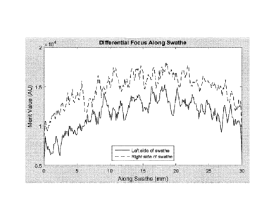
from a "focus sweep"; also see e.g. Fig. 11 which shows the output of a focus
merit function as calculated at two positions for multiple scan lines in a
swathe
obtained using a dynamic focus tracking method).
A focus merit function may be configured to provide, for at least one scan
line, an
indication of focus quality at a given position, and may be calculated based
on
adjacent pixels at that given position. Such functions are well known in the
art.
For the avoidance of any doubt, the at least one measure may include a single
differential focus (see e.g. Fig. 3 to Fig. 5), or multiple differential
focuses (see e.g.
Fig. 11).
If the at least one measure includes multiple differential focuses, the
multiple
differential focuses may be combined (e.g. averaged) to provide a combined
(e.g.
averaged) differential focus, with the swathe width value being set based on
the
combined (e.g. averaged) differential focus.
In some embodiments (see e.g. Fig. 6), the at least one measure may be
obtained
and the swathe width value set based on the at least one measure prior to
acquiring an image (including a plurality of swathes) from the surface of the
second object, wherein each swathe acquired from the surface of the second
object has a width in the scan width direction that corresponds to the swathe
width
value set based on the at least one measure.
In other embodiments (see e.g. Fig. 7), the at least one measure may be
obtained
and the swathe width value set based on the at least one measure prior to
acquiring each swathe from the surface of the second object.
In yet other embodiments, the at least one measure may be obtained and the
swathe width value set based on the at least one measure at periodic
intervals,
e.g. every 30 minutes.
If the second object is the same object as the first object, the method may
include
identifying one or more areas on the surface of the object suitable for
acquiring the
CA 03002319 2018-04-17
WO 2017/097950
PCT/EP2016/080360
- 6 -
at least one scan line, and then subsequently acquiring at least one scan line
from
the one or more identified areas on the surface of the object (for subsequent
use
in obtaining the at least one measure).
If the method includes acquiring at least one scan line from the one or more
identified areas on the surface of the object, then a respective measure
indicating
that the surface of the first object is uneven (e.g. tilted) in the scan width
direction
relative to an imaging plane of the image scanning apparatus may be acquired
for
each area on the surface of the object. These measures may then be combined
(e.g. averaged) to provide a combined (e.g. averaged) measure, with the swathe
width value being set based on the combined (e.g. averaged) measure (see e.g.
Fig. 9).
Preferably, the method includes using a plurality of scan lines, acquired from
the
surface of the first object mounted in the image scanning apparatus using the
scan
line detector, to obtain the at least one measure.
In some embodiments, a plurality of scan lines (from which the at least one
measure is obtained) may be acquired from a single elongate region of the
surface
of the first object extending in the scan width direction, with the image
scanning
apparatus having a different focus setting whilst each scan line is acquired
(see
e.g. Fig. 6 - Fig. 9). The process of acquiring such scan lines may be
referred to
herein as a "focus sweep".
In some embodiments, a plurality of scan lines (from which the at least one
measure is obtained) may be a group of scan lines forming a swathe, with each
scan line being acquired from a respective elongate region of the surface of
the
first object extending in the scan width direction whilst the first object is
moved
relative to the scan line detector in the scan length direction (see e.g. Fig.
11 - Fig.
16). The swathe formed by the group of scan lines (from which the at least one
measure is obtained) may be a first swathe acquired from the surface of the
object.
If a plurality of scan lines (from which the at least one measure is obtained)
is a
group of scan lines forming a swathe, the swathe may have been acquired using
a
dynamic focus tracking method in which the focus setting of the image scanning
apparatus was adjusted whilst the swathe was acquired. Such dynamic focusing
methods are disclosed in US7485834, W02013/017855 and US2014/0071438, for
example.
CA 03002319 2018-04-17
WO 2017/097950
PCT/EP2016/080360
- 7 -
In some embodiments, the second object may be the same object as the first
object, wherein a plurality of scan lines (from which the at least one measure
is
obtained) is a group of scan lines forming a swathe, the swathe having been
acquired from the surface of the object using a dynamic focus tracking method
in
which the focus setting of the image scanning apparatus was adjusted whilst
the
swathe was acquired.
For example, each swathe subsequently acquired from the surface of the object
may have a width in the scan width direction that corresponds to a swathe
width
value set based on the at least one measure obtained using the group of scan
lines forming the swathe (see e.g. Fig. 12).
Alternatively, each time a new swathe is acquired from the surface of the
object,
scan lines forming the new swathe may be used to obtain the at least one
measure, with the swathe width value being set based on the at least one
measure so that the swathe width value is set each time a new swathe is
acquired
(see e.g. Fig. 13 and Fig. 14).
In some embodiments, if a swathe width value set based on a new swathe
acquired from the surface of the object is smaller than a previously set
swathe
width value that was in use during the acquisition of the new swathe, then the
width of the new swathe may be reduced based on the smaller swathe width value
that has been set based on the new swathe (see e.g. Fig. 13 and Fig. 14) ¨
this
may be achieved, for example, by rescanning the corresponding region on the
surface of the second object, or by reducing the width of the new swathe in
post-
processing.
In some embodiments, if a swathe width value set based on a new swathe
acquired from the surface of the object is larger than a previously set swathe
width
value that was in use during the acquisition of the new swathe, then the width
of
the new swathe may be increased based on the larger swathe width value that
has
been set based on the new swathe (see e.g. Fig. 14) ¨ this may be achieved,
for
example, by rescanning the corresponding region on the surface of the second
object, or by increasing the width of the new swathe in post-processing, e.g.
by
saving the new swathe with a larger width in the swathe width direction,
preferably
with the additional width in the swathe width direction coming from a side of
the
swathe that does not adjoin a previously acquired swathe.
CA 03002319 2018-04-17
WO 2017/097950
PCT/EP2016/080360
- 8 -
For the avoidance of any doubt, the at least one measure need not include a
differential focus.
For example, the at least one measure may include the output of a focus merit
function as calculated, for at least one scan line, at two or more positions
offset
from each other in the scan width direction.
In this case, the output of a focus merit function as calculated, for at least
one
scan line, at a centre position and two edge positions offset from each other
in the
scan width direction, could be used as measures indicating that the surface of
a
first object is tilted in the scan width direction relative to an imaging
plane, without
a differential focus being calculated. See e.g. Fig. 15, wherein if the edge
values
do not match each other to within a predetermined tolerance, or if the edge
values
do not match the centre value to within a predetermined tolerance, then these
measures can be interpreted as indicating that surface of the first object is
tilted in
the scan width direction relative to an imaging plane of the image scanning
apparatus. Also see e.g. Fig. 16, in which mean density values as calculated,
for
the at least one scan line, at the centre position and two edge positions are
additionally used to ensure that the at least one scan line is suitable for
assessing
tilt.
In this case, the output of a focus merit function as calculated, for at least
one
scan line from a swathe acquired using the line scan detector, at a centre
position
("centre merit value") and two edge positions ("edge merit values") offset
from
each other in the scan width direction, could be used as measures indicating
that
the surface of a first object is tilted in the scan width direction relative
to an
imaging plane. If the edge merit values do not match each other to within a
predetermined tolerance or if the centre merit value does not match the edge
merit
values to within a predetermined tolerance, then these measures can be taken
as
indicating that the surface of a first object is tilted in the scan width
direction
relative to an imaging plane (see e.g. Fig. 15 and Fig. 16). Conversely, if
the edge
merit values match each other to within a predetermined tolerance and the
centre
merit value matches the edge merit values to within a predetermined tolerance,
this could be taken as the centre and edge merit values indicating that the
surface
of a first object is not tilted in the scan width direction relative to an
imaging plane.
In this process, mean density values may also be calculated, for the at least
one
scan line, at the centre position and two edge positions, e.g. to ensure that
the at
least one scan line is suitable for assessing tilt (see e.g. Fig. 16).
CA 03002319 2018-04-17
WO 2017/097950
PCT/EP2016/080360
- 9 -
The scan line detector may include a linear array of photodetectors.
Preferably, the/each swathe acquired from the surface of the second object is
acquired by using all photodetectors in the linear array to obtain a precursor
swathe from the surface of the second object, with the precursor swathe being
post-processed (e.g. cropped) to obtain a swathe that has a width in the scan
width direction that corresponds to the swathe width value set based on the at
least one measure. This allows for the width of the swathe to be adjusted in
post-
processing, which may for example be useful to allow the width of the swathe
to
be increased in post-processing (see e.g. Fig. 14).
To allow the width of a swathe to be adjusted (e.g. increased) in post-
processing,
the/each precursor swathe from the surface of the second object is preferably
acquired from a region (preferably a previously unscanned region) of the
swathe
that adjoins either an edge of the surface of the second object (which may be
appropriate if the swathe is a first swathe) and/or adjoins a previously
acquired
swathe. This helps to provide the maximum scope for increasing the width of
the
swathe in post-processing on the non-adjoining side of the swathe, should that
be
needed/appropriate (see e.g. Fig. 14).
Alternatively, the at least one swathe acquired from the surface of the second
object may be acquired using only a subset of photodetectors in the linear
array so
that the at least one swathe acquired from the surface of the second object
has a
width in the scan width direction that corresponds to the swathe width value
set
based on the at least one measure.
The swathe width value may be set based on the at least one measure and a
depth of focus of the image scanning apparatus (i.e. not just the at least one
measure). In this case, the at least one measure and the depth of focus of the
image scanning apparatus may be used to set a swathe width value that is
deemed to be a maximum useable swathe width for maximising the width of
swathe acquired whilst keeping the/each swathe acquired from the surface of
the
second object substantially in focus across its width in the scan width
direction.
However, for the avoidance of any doubt, the swathe width value may be set
based on the at least one measure without reference to a depth of focus (see
e.g.
Fig. 15 and Fig. 16).
CA 03002319 2018-04-17
WO 2017/097950
PCT/EP2016/080360
- 10 -
The second object may be a slide having a sample thereon. The sample may be a
biological specimen. The surface of the second object (from which the at least
one
swathe is acquired) may be a surface of the sample (e.g. biological specimen)
located on the slide.
The image scanning apparatus may include a mounting device configured to
mount the second object in the image scanning apparatus. If the second object
is
a slide having a sample thereon (see above), the mounting device may be a
slide
mounting device configured to mount a slide in the image scanning apparatus.
If the first object is a different object from the second object, the first
object may be
a target mounted in the image scanning apparatus. In this case, the target
mounted in the image scanning apparatus may be an "internal" target mounted in
a mounting device that is separate from a mounting device used to mount the
second object in the image scanning apparatus. However, it is also possible
that
the target could be an "external" target mounted in a mounting device that is
subsequently used to mount the second object in the image scanning apparatus.
The target (if present) may be a square wave grating, for example.
If the image scanning apparatus includes a mount configured to mount the
second
object in the image scanning apparatus, the image scanning apparatus may be
configured to move the second object relative to the scan line detector in a
scan
length direction by moving the mount in the scan length direction. The image
scanning apparatus may be configured to move the second object relative to the
scan line detector in a scan length direction by moving the line scan detector
(in
addition to or as an alternative to moving the mount).
The image scanning apparatus may include an imagine system including the line
scan detector and an imaging lens. A focus setting of the image scanning
apparatus may be adjusted, for example, by moving the imaging lens, though
other ways of adjusting a focus setting of the image scanning apparatus would
be
apparent to those skilled in the art.
A second aspect of the disclosure may provide an image scanning apparatus
configured to perform a method according to the first aspect of the
disclosure.
The apparatus may be configured to implement, or have means for implementing,
any method step described in connection with any above aspect of the
disclosure.
CA 03002319 2018-04-17
WO 2017/097950
PCT/EP2016/080360
-11 -
The image scanning apparatus may include a control unit, e.g. a computer,
configured to control the image scanning apparatus to perform a method
according to the first aspect of the disclosure.
A third aspect of the disclosure may provide a computer-readable medium having
computer-executable instructions configured to cause an image scanning
apparatus to perform a method according to the first aspect of the disclosure.
The disclosure also includes any combination of the aspects and preferred
features described except where such a combination is clearly impermissible or
expressly avoided.
BRIEF DESCRIPTION OF THE DRAWINGS
Examples of these proposals are discussed below, with reference to the
accompanying drawings in which:
Fig. 1 illustrates a typical image scanning apparatus that provides a virtual
microscope, which operates according to known principles.
Fig. 2 illustrates factors which may cause the height of a sample on a slide
to
change more rapidly than the depth of focus of an image scanning apparatus
over
the width of a swathe acquired by the image scanning apparatus.
Fig. 3 shows the output of a focus merit function calculated using adjacent
pixels
at positions at either end of line scans obtained from a focus sweep.
Fig. 4 shows the output of a focus merit function calculated using adjacent
pixels
at positions at either end of line scans obtained from a focus sweep, and
intermediate positions between either end of the line scans and a centre of
the line
scans.
Fig. 5 shows how differential focus may be calculated from the outputs
illustrated
in Fig. 4.
Fig. 6 shows an example workflow in which a tilt measurement is performed
before
every full image scan.
Fig. 7 shows an example workflow in which a tilt measurement is performed
before
acquisition of every swathe in an image scan.
CA 03002319 2018-04-17
WO 2017/097950
PCT/EP2016/080360
- 12 -
Fig. 8 shows an example workflow in which a suitable area within the surface
of a
sample is identified and measured before every full scanned image.
Fig. 9 shows an example workflow in which multiple suitable areas within the
surface of a sample is identified and measured before every full scanned
image.
Fig. 10 shows how a dynamic focus tracking system may be used to predict a
single focus position at which to scan at a given location along the length of
a
swathe.
Fig. 11 shows how differential focus values could be obtained for multiple
positions
along the full length of a swathe, using a dynamic focus tracking system.
Fig. 12 shows an example workflow in which the differential focus can be
measured from dynamic focus tracking data using a first swathe to set the
swathe
width for subsequent swathes of a full image scan.
Fig. 13 shows the example workflow of Fig. 12, modified to allow the swathe
width
to be reduced if any individual swathe has a differential focus that is too
large.
Fig. 14 shows the example workflow of Fig. 13, modified to allow the swathe
width
to be increased if any individual swathe has a differential focus indicating
that the
swathe width could have been greater.
Fig. 15 shows an example workflow in which a focus merit value is calculated
at
positions on both sides and the middle of a swathe to assess tilt.
Fig. 16 shows the example workflow of Fig. 15, modified to additionally
calculate
mean density values at the positions on both sides and the middle of a swathe.
DETAILED DESCRIPTION
With reference to Fig. 1, there are a number of factors which may cause the
height
of a sample on a slide 7 to change more rapidly than the depth of focus of an
image scanning apparatus (typically in the region of 1pm) over the width of a
swathe acquired by the image scanning apparatus (typically in the region of
1 mm).
CA 03002319 2018-04-17
WO 2017/097950
PCT/EP2016/080360
- 13 -
These factors include but are not limited to:
1. Temperature variations within the image scanning apparatus causing the
side mount to tilt or the optics to tilt.
2. The slide itself has a side to side wedge.
3. The slide is not sitting fully on the slide mount.
4. The sample (e.g. tissue slice) is wedged.
5. There has been differential wear on opposite sides of the slide mount.
6. The alignment of the system is not perfect.
These factors are demonstrated in Fig. 2, with sources of error exaggerated
for
clarity purposes.
In general, the following discussion describes examples of our proposals that
provide a variable swathe width, in the context of operating an image scanning
apparatus that includes a line scan detector.
This concept can be understood with reference to Fig. 2, which shows the limit
of
swathe width where the surface of a sample on a slide is kept within the depth
of
focus, referred to as a "maximum useable swathe width". The maximum useable
swathe width may be defined as the maximum width of a swathe that is able to
keep the surface of the sample in focus across the width of the swathe in the
scan
width direction.
If areas outside this maximum useable swathe width are used, the resulting
image
will be out of focus and not fit for purpose.
The present inventors have observed that if the swathe width is set to the
maximum useable swathe width, rather than the maximum possible swathe width,
this will allow the whole of the sample to be scanned in focus. In particular,
for an
image scanning apparatus with zero or low tilt, a sample on a slide can be
scanned in the minimum number of swathes using swathes that have the
maximum possible swathe width, which in turn gives the shortest scan time. For
an image scanning apparatus with significant tilt, then it is still possible
to scan the
slide without loss of image quality by reducing the swathe width. For example,
for
an image scanning apparatus having a 1pm depth of focus and a sample having a
CA 03002319 2018-04-17
WO 2017/097950
PCT/EP2016/080360
- 14 -
tilt of 2pm across the swathe width, then an in focus swathe could still be
acquired
by reducing the width of the swathe scanned to half the normal swathe width.
This
will produce the same image quality as a scanner with no tilt but because of
the
greater number swathes required for the same scan area the time to scan will
increase.
To set the swathe width to a maximum useable swathe width or the maximum
possible swathe width, a maximum useable swathe width should first be
determined. To determine a maximum useable swathe width, a differential focus
may be calculated. A differential focus may be defined as being indicative of
a
distance between (i) an in focus level at a first position on the surface of
the
sample; and (ii) an in focus level at a second position on the surface of the
sample; wherein the first position and second position are offset from each
other in
the scan width direction 5. The first and second positions may correspond to
positions on opposite sides (e.g. opposite ends) of a scan line or group of
scan
lines.
A differential focus may be measured by performing a "focus sweep" on a single
line location 7 on the sample. A focus sweep can be thought of as a 2D image
in
the xz plane rather than the xy plane. This can be achieved with a simply by
moving the lens focus along the z axis, e.g. by moving the lens 1 along the
imaging axis 9, while the line scan detector 2 is collecting data, or by
performing
consecutive single line scans at different focus positions. From scan lines
acquired
in the focus sweep (as an image or stack of images), it is possible to
calculate the
output of a focus merit function at two positions that are on either side of
the line
location 7, as a function of focus position (z-axis). A peak in a merit
function
indicates an "in focus" level. Hence, the difference between the peaks of the
output of the merit function for the two positions that are on either side of
the line
location 7, provides a differential focus, which is indicative of a distance
between
in focus levels at the two positions.
The output of a focus merit function can be thought of as providing a measure
of
the quality of focus and there are many functions that may be used, typically
based on a difference in adjacent pixels. An example of this is in Fig. 3
where the
output of the focus merit function on each side of the swathe has a peak at a
different focus position (z-axis). The fact that the output of the focus merit
function
has different peak values at the two positions is not important and only shows
that
the sample measured has different level of detail at those two positions (i.e.
across
CA 03002319 2018-04-17
WO 2017/097950
PCT/EP2016/080360
- 15 -
the swathe width). It is the difference in the in focus position that provides
the
differential focus. For the example shown in Fig. 3, the differential focus is
shown
in Fig. 3 in arbitrary units ("AU") derived from the position of the imaging
lens 1
along the imaging axis, though other measures of focus position could equally
be
used.
In the example of Fig. 3, the output of the focus merit function is calculated
using
adjacent pixels at positions at either end of the line scans obtained from the
focus
sweep (left edge, right edge).
If the differential focus is measured over the maximum possible swathe width
then
a maximum usable swathe width may be determined as the lesser of the
maximum possible swathe width or the depth of focus multiplied by the maximum
possible swathe width divided by the differential focus. This could be
represented
by as follows:
MU = lesser of (a * dof * Mp / Af) or Mp
where, MU = Maximum useable swathe width, dof = Depth of Focus, Mp =
Maximum possible swathe width, Af = Differential Focus, and a = scaling factor
to
permit variability in the tilt (typically this factor would be close to 1).
It is also possible to use multiple points across the swathe width and fit a
straight
line or a curve to the peak values. This can be seen in Fig. 4 and Fig. 5.
In the example of Fig. 4, the focus merit function is calculated using
adjacent
pixels at either side of the line scans (left edge, right edge) and also at
intermediate positions between either end of the line scans and a centre of
the line
scans (left of centre, right of centre).
If it is only required to compensate for changes or errors in the image
scanning
apparatus and not the slide 7 or sample itself, then a known target may be
used to
determine a maximum useable swathe width, rather than direct measurement from
a sample on the slide 7. Typically this target would be square wave grating
mounted in the scanner separately from a mount used for slides, e.g. so that
the
target could be moved into and out from the imaging axis 9. Types of target
other
than a square wave grating could be used. Also, a target not permanently
mounted within the scanner but instead mountable in a mount used for slides
could be used. The same technique for measuring differential focus can be used
on the target, as has already been described above. It is also not required
for the
CA 03002319 2018-04-17
WO 2017/097950
PCT/EP2016/080360
- 16 -
target to be mounted without any tilt as if the level of tilt of the target
relative to a
mounted slide is known this can be subtracted from the measured tilt of the
target
to give the real tilt of a mounted slide. In this way, we can either measure
the tilt of
a slide directly, or measure the change in tilt of a target to give the tilt
of a slide.
From a knowledge of the depth of focus it is then possible to define a maximum
usable swathe width and set the swathe width of the scanner to maintain image
quality.
Whilst the swathe width is preferably set to be equal to a maximum useable
swathe width is preferred, the swathe width may instead be calculated with a
buffer to be slightly smaller than the maximum useable swathe width to ensure
that the distance between the in focus levels at the edges of a swathe does
not
exceed the depth of focus of the image scanning apparatus. In either case, the
swathe width is set based on at least one measure (differential focus)
indicating
that the surface of the sample on the slide is uneven (in this case tilted) in
the scan
width direction relative to an imaging plane of the image scanning apparatus.
This measurement of the tilt of a mounted slide can be performed periodically,
typically every 30mins if the change in the scanner is slow. If the change in
tilt is
more rapid, a tilt measurement can be performed before every full image scan,
as
in the workflow shown in Fig. 6. It is also possible for a tilt measurement to
be
performed before acquisition of every swathe in an image scan if the change is
very rapid. This sequence is shown in Fig. 7. If tilt is measured before every
swathe this may give an image with different swathe widths within the full
scanned
image.
If the sample, slide and a slide mount is to be compensated for, then it is
necessary to scan the sample itself (i.e. necessary for the object used for
measuring tilt to be the same as the object being scanned). A suitable area
within
the surface of the sample, with detail across a swathe width, can be
identified and
measured before every full scanned image. A maximum usable swathe width for
the full scanned image can then be set. This is shown in Fig. 8.
The thickness of a tissue sample may vary across the sample and a single
measurement may not give a reliable measurement. However if measurements
are taken at multiple points on the tissue sample, these can be combined to
give a
more reliable result. The combination process may be a simple mean or median
value or maximum value or a more sophisticated process designed to remove the
CA 03002319 2018-04-17
WO 2017/097950
PCT/EP2016/080360
- 17 -
outlying results such as taking the mean of 80% of the closest results. Such
combination processes are well known to those skilled in the art. This is
shown in
Fig. 9
Taking many measurements from the surface of a sample on each slide 7 may be
time consuming and could reduce the productivity.
However, it is already known to perform a focus sweep at a single point in
predetermined area of a sample prior to imaging the sample in order to
establish a
single focus level at which to start scanning (note, this known process only
involves determining a single in focus level, rather than calculating a
differential
focus). By using this conventional focus sweep to additionally calculate
differential
focus values, it would be possible for trends to be predicted for many scans
or
slides. For example, if the image scanning apparatus has drifted in tilt, this
will
show as a similar differential focus in all slides, and if many slides are
analysed
with measurements over a determined time period, e.g. by averaging, then the
degree of tilt can be reliably measured and the swathe width adjusted. This
only
requires a single focus sweep on each slide or full sample image, as is
already
done regularly in practice to establish a single focus level at which to start
scanning.
If the scanner has a dynamic focus tracking system such as described in
U52014/0071438 or W02013/017855 or U57485834 it is possible to
measure/predict the differential focus during the scanning of a swathe. In
these
documents the whole of the swathe width is used to predict a single focus
position
at which to scan at a given location along the length of a swathe, as shown in
Fig.
10.
These dynamic focusing tracking techniques could be modified to calculate
focus
merit functions on either side of scan lines from a swathe to determine in
focus
positions, thereby allowing differential focus values to be obtained for
multiple
positions along the full length of a swathe, as shown in Fig. 11. The multiple
differential focus values obtained at multiple positions along the full length
of the
swathe can be combined to produce a single differential focus value to set a
maximum usable swathe width. The combination process may be a simple mean
or median value or maximum value or a more sophisticated process designed to
remove the outlying results such as taking the mean of 80% of the closest
results.
Such combination processes are well known to those skilled in the art.
CA 03002319 2018-04-17
WO 2017/097950
PCT/EP2016/080360
- 18 -
This measuring of the differential focus can be measured from dynamic focus
tracking data using a first swathe to set the swathe width for all subsequent
swathes of a full image scan as shown in Fig. 12.
If on subsequent swathes the differential focus error is monitored from the
dynamic focus tracking data it can be analysed if any individual swathe has a
differential focus that is too large. If so, the swathe can be repeated but
with a
reduced swathe width, as shown in Fig. 13. Note that in this case it is
required to
repeat the swathe scan as the centre of the swathe scan has to be moved along
the x direction to ensure that the swathe width adjoins the previous adjacent
swathe. This means different swathes within the same full scan image will have
different widths. At the end of the reduced width swathe the swathe for the
subsequent swathes may be returned to the swathe width calculated after the
first
swathe or may be retain at the reduced swathe width.
If the differential focus data from a swathe indicates the swathe width could
have
been greater than that used it is possible to use the addition swathe image
data on
the side not adjoining a previous scanned swathe, as shown in Fig. 14. It
would
then be possible to scan subsequent swathes at the larger swathe width.
Again, trends can be predicted by using the focus tracking data for every
swathe
for many scans or slides. If the scanner has drifted in tilt this will show as
an error
in all slides and if many slides are analysed with measurements such as
averaging
the degree of tilt can be reliably measured and the maximum usable swathe
width
adjusted accordingly.
The example workflows shown in Fig. 11 ¨ Fig. 14 use only two positions on
either
side of a swathe to calculate the output of the focus merit function, but it
is
possible to use more positions across the swathe to calculate the output of
the
focus merit functions and predict the differential focus in the same way as
shown
in Fig. 5.
If there is no dynamic focus data available then measures indicating that the
surface of a sample on a slide is uneven (e.g. tilted) in the scan width
direction
relative to an imaging plane of the image scanning apparatus can still be
estimated from a scanned swathe image itself. This can be done using multiple
scan lines from a swathe (as shown below), or even from just a single scan
line
from a swathe (not shown).
CA 03002319 2018-04-17
WO 2017/097950
PCT/EP2016/080360
- 19 -
For example, a focus merit value (output of a focus merit function) could be
calculated at positions on both sides and the middle of a swathe, and if the
detail
in the sample is known to be uniform across the swathe, then the relative
values of
the edge merit values can be compared with the centre merit value to assess
whether the sample on the slide 7 is tilted, and adjust the swathe width
accordingly.
In particular, if the sample is uniform in detail the two edge merit values
will match.
If the sample is uniform and has no tilt the two edge merit values will match
and
the centre merit value will match. If the sample is uniform and has tilt the
edge
merit values will match but be lower than the centre merit value. This is
shown in
Fig. 15. From all of these decisions we can determine if there the swathe
width
was too wide by deciding if there were too many decisions where there was
judged to be tilt in the system. The swathe width can then be reduced and
repeated if required.
The amount of reduction of swathe width could be determined from monitoring
additional points across the swathe width such as third, quarter, eighth or
more
points. These can then have the same logic as shown in Fig. 15 applied and the
widest set of points which meet the acceptability criteria on the number of
"No"
decisions can be used to set the maximum usable swathe width.
A further refinement is shown in Fig. 16. Here, the mean density value of each
section of the swathe can be measured. If the sample is uniform in detail the
mean
density value of each section of the swathe will be similar. If the mean
density
value of each section of the swathe is not the same then the detail will not
be
uniform even if the merit values of each section are the same. This provides a
check to make sure that the scan line is suitable for measuring tilt, since if
mean
density values are different, then the scan line can't be used to assess tilt.
The
amount of reduction of the swathe width required can be calculated in the same
way with more sections across the swathe width using not only the focus merit
values but the mean density being matched to the other sections.
As in the example workflows where the differential focus is used to alter the
swathe width, such as those shown in Fig. 12 to Fig. 14, we can use the focus
merit data to adjust the swathe width to the maximum usable swathe width.
In addition a maximum usable swathe width measurement determined according
to the example workflows shown in Fig. 15 and Fig. 16 can be used to predict
- 20 -
trends. For example if the maximum usable swathe width reduces over a period
of
time, then a user could be informed to call someone to perform corrective
action
such as a visit from a service engineer. Predictive trends can inform the user
that
corrective action will be required in a certain time interval before the
maximum
usable swathe width is actually required to be reduced and impact the scanner
productivity.
For those skilled in the art it can be seen that various combinations of the
example
workflows set out above could be used.
When used in this specification and claims, the terms "comprises" and
"comprising", "including" and variations thereof mean that the specified
features,
steps or integers are included. The terms are not to be interpreted to exclude
the
possibility of other features, steps or integers being present.
The features disclosed in the foregoing description, or in the following
claims, or in
the accompanying drawings, expressed in their specific forms or in terms of a
means for performing the disclosed function, or a method or process for
obtaining
the disclosed results, as appropriate, may, separately, or in any combination
of
such features, be utilised for realising the disclosure in diverse forms
thereof.
While the disclosure has been described in conjunction with the exemplary
embodiments described above, many equivalent modifications and variations will
be apparent to those skilled in the art when given this disclosure.
Accordingly, the
exemplary embodiments of the disclosure set forth above are considered to be
illustrative and not limiting. Various changes to the described embodiments
may
be made without departing from the spirit and scope of the disclosure.
For the avoidance of any doubt, any theoretical explanations provided herein
are
provided for the purposes of improving the understanding of a reader. The
inventors do not wish to be bound by any of these theoretical explanations.
The following statements provide general expressions of the disclosure herein:
A. When necessary to reduce the swathe width to maintain the image quality
B. Measuring the focus position at two locations across the swathe to
determine the differential focus in a focus sweep scan or focus stack.
Date recue / Date received 2021-12-09
CA 03002319 2018-04-17
WO 2017/097950
PCT/EP2016/080360
-21 -
C. Measuring the focus position at more than two locations across the swathe
in a focus sweep scan or focus stack to determine the differential focus by
combining the multiple location data.
D. Measuring the differential focus from an internal target and setting the
swathe width to the maximum usable swathe width.
E. Measuring the differential focus from an external target and setting the
swathe width to the maximum usable swathe width.
F. Measuring the differential focus from a suitable area of the sample and
setting the swathe width to the maximum usable swathe width.
G. Measuring the differential focus from a suitable area of the sample before
every full image scan and setting the swathe width to the maximum usable
swathe width.
H. Measuring the differential focus from a number of suitable areas of the
sample before every full image scan and setting the swathe width to the
maximum usable swathe width.
I. Measuring the differential focus of the sample from the dynamic focus
tracking data of the first swathe at a number of points along the swathe and
setting the swathe width of the full image scan to the maximum usable
swathe width of the first swathe.
J. Measuring the differential focus of the sample from the dynamic focus
tracking data of the first swathe at a number of points along the swathe and
setting the swathe width of the full image scan to the maximum usable
swathe width of the first swathe. Subsequent swaths are also measured
and if the maximum usable swathe width is smaller repeat the swathe with
a smaller swathe width.
K. Measuring the differential focus of the sample from the dynamic focus
tracking data of the first swathe at a number of points along the swathe and
setting the swathe width of the full image scan to the maximum usable
swathe width of the first swathe. Subsequent swaths are also measured
and if the maximum usable swathe width is smaller repeat the swathe with
a smaller swathe width. If the maximum usable swathe width is larger than
CA 03002319 2018-04-17
WO 2017/097950
PCT/EP2016/080360
- 22 -
the swathe width used then save the additional swathe image data on the
side of the swathe that is not adjoining any previous swathe.
L. Measure the focus merit values of the scanned swathe image at the edge
of the swathe and the centre. When the edge merit values match to within
a tolerance and the centre merit values match to within a tolerance accept
the swathe. When the edge merit values match and the centre merit values
are improved then reduce the swathe width.
M. The amount of swathe width reduction in step L can be calculated from
measuring the focus merit values at multiple locations across the swathe
and using the largest swathe width where the centre and side values all
match to within a tolerance.
N. Using steps L with M and setting the swathe width of the full image scan to
the maximum usable swathe width of the first swathe.
0. Using steps L with M and setting the swathe width of the full image scan to
the maximum usable swathe width of the first swathe. Subsequent swaths
are also measured and if the maximum usable swathe width is smaller
repeat the swathe with a smaller swathe width.
P. Using steps L with M and setting the swathe width of the full image scan to
the maximum usable swathe width of the first swathe. Subsequent swaths
are also measured and if the maximum usable swathe width is smaller
repeat the swathe with a smaller swathe width. If the maximum usable
swathe width is larger than the swathe width used then save the additional
swathe image data on the side of the swathe that is not adjoining any
previous swathe.
Q. Measure the focus merit values of the scanned swathe image and the
density at the edge of the swathe and the centre. When the edge merit
values and the density values match to within a tolerance and the centre
merit and density values match to within a tolerance accept the swathe.
When all the density values match to within a tolerance, the edge merit
values match and the centre merit values are improved then reduce the
swathe width.
R. The amount of swathe width reduction in step Q can be calculated from
measuring the focus merit values at multiple locations across the swathe
CA 03002319 2018-04-17
WO 2017/097950
PCT/EP2016/080360
- 23 -
and using the largest swathe width where the centre and side values all
match to within a tolerance.
S. Using steps Q with R and setting the swathe width of the full image scan to
the maximum usable swathe width of the first swathe.
T. Using steps Q with R and setting the swathe width of the full image scan to
the maximum usable swathe width of the first swathe. Subsequent swaths
are also measured and if the maximum usable swathe width is smaller
repeat the swathe with a smaller swathe width.
U. Using steps 0 with R and setting the swathe width of the full image scan to
the maximum usable swathe width of the first swathe. Subsequent swaths
are also measured and if the maximum usable swathe width is smaller
repeat the swathe with a smaller swathe width. If the maximum usable
swathe width is larger than the swathe width used then save the additional
swathe image data on the side of the swathe that is not adjoining any
previous swathe.