Note: Descriptions are shown in the official language in which they were submitted.
CA 03002399 2018-04-17
WO 2017/070224 PCT/US2016/057724
MICROFLUIDIC MODEL OF THE BLOOD BRAIN BARRIER
Field of the Invention
The invention relates to culturing brain cells and particularly astrocytes
together with
endothelial cells in a fluidic device under conditions whereby the cells mimic
the structure and
function of the blood brain barrier and/or spinal cord. Good viability and
function allow for
measurements of barrier integrity and physiology, whether by trans-epithelial
electrical
resistance (TEER), patch clamp or other testing measures.
Background of the Invention
The blood-brain barrier is of major clinical relevance. Not only because
dysfunction
of the blood-brain barrier leads to degeneration of the neurovascular unit,
but also because drugs
that are supposed to treat neurological disorders often fail to permeate the
blood-brain barrier.
Because of its importance in disease and medical treatment, it would be highly
advantageous to
have a predictive model of the human blood-brain barrier that recapitulates
aspects of the
cerebral endothelial microenvironment in a controlled way.
Summary of the Invention
The invention relates to culturing endothelial cells (preferably brain-related
endothelial
cells), optionally astrocytes, optionally neurons and optionally pericytes in
a microfluidic device,
such as microfluidic chip (described herein) under conditions whereby the
cells mimic one or
more structural or functional features (e.g. tight junctions) of the blood
brain barrier (BBB)
and/or spinal cord. Good viability and function allow for measurements of
barrier integrity and
physiology, whether by transepithelial electrical resistance (TEER),
electrophysiology
(including, for example, patch clamp) or other testing measures. Indeed,
neuronal cells, such as
motor neurons, that are allowed to mature on a microfluidic chip, show a more
mature
electrophysiology (action potential patterns, for example) indicating a more
advanced or
accelerated maturation. Thus, in one embodiment, the present invention
contemplates a
microfluidic culture of iPSC-derived neural progenitor cells or
(alternatively) neurons (e.g. a
culture in a microfluidic setting, such as in a microchannel and/or
microfluidic device) in contact
with flowing media. In one embodiment, the iPSC-derived neural progenitors or
(alternatively)
1
CA 03002399 2018-04-17
WO 2017/070224 PCT/US2016/057724
neurons are cultured alone (without other cell types). In one embodiment, said
neurons are iPSC-
derived neurons. In one embodiment, said iPSC-derived neurons are motor
neurons. In one
embodiment, said neurons are cultured in a microchannel or on a membrane of a
microfluidic
chip. In one embodiment, said microfluidic chip comprises two microchannels
separated by a
porous membrane having first and second surfaces, wherein said neurons are
cultured on said
first or second surface. In one embodiment, said culturing is performed for
10, 12, 20, 24, 30, 36
or more days. In one embodiment, said neurons exhibit a more mature
electrophysiology as
compared to the same neurons cultured in a static culture. Culture of cells in
the microfluidic
chip, whether alone or in combination with other cells, drives maturation
and/or differentiation
further than existing systems.
It is not intended that the present invention be limited to only one type of
test or
measurement to assess the more mature phenotype of neurons and BMECs. In one
embodiment,
gene expression, Ca2+ flux imaging, immunofluorescent staining, and/or tissue
morphology is
assessed as evidence of more mature neurons, BMECs and/or astrocytes.
Where neurons, such as motor neurons (or their precursors), are co-cultured
(i.e. cultured
together) on a microfluidic chip with relevant vascular cells, such as brain
microvascular
endothelial cells, an even greater effect on differentiation, maturation
and/or conditioning is
observed. Thus, in one embodiment, the present invention contemplates a
microfluidic co-culture
of iPSC-derived neural progenitors or (alternatively) neurons with vascular
cells, e.g. a
microfluidic co-culture of neurons with iPSC-derived vasculature (e.g. said
vascular cells are
iPSC-derived vascular cells). In one embodiment, said iPSC-derived vascular
cells are brain
microvascular endothelial cells. In one embodiment, said neurons are iPSC-
derived neurons. In
one embodiment, said iPSC-derived neurons are motor neurons. In one
embodiment, said
vascular cells are co-cultured with said neurons in a microchannel or on a
membrane of a
microfluidic chip. In one embodiment, said microtluidic chip comprises two
microchannels
separated by a porous membrane having first and second surfaces, wherein said
neurons are
cultured on said first surface and said vascular cells are cultured on said
second surface. In one
embodiment, said culturing (e.g. under flow conditions) is performed for 10,
12, 20, 24, 30, 36 or
more days. In one embodiment, at least a portion of said neurons and vascular
cells are in
contact with each other (whether by direct physical contact or indirect cell-
to-cell
communication). In one embodiment, said neurons and vascular cells are in
contact with flowing
2
CA 03002399 2018-04-17
WO 2017/070224 PCT/US2016/057724
culture media (e.g. the cells are adhered to a surface and the media flows
over the cells at a
controlled rate, bringing nutrients and removing waste). In one embodiment,
said neurons
exhibit a more mature electrophysiology as compared to the same neurons
cultured in a static
culture.
The microfluidic chip culture increases and accelerates function of iPSC-
derived
neurons, including motor neurons (MNs). Co-culture with iBMECs recreates known
vascular-
interaction pathways and further increased maturation in vitro. The fact that
cells differentiate
and mature more fully on a microfluidic chip indicates that the chip is a
better culture tool than
more conventional culture systems (e.g. transwell cultures and other static
systems), providing a
better model of what is going on in vivo (including what is going on in
disease states). Thus, in
one embodiment, the present invention contemplates a microfluidic device or
chip comprising a
co-culture of neurons, and more specifically, motor neurons, and more
typically, induced motor
neurons, with brain microvascular endothelial cells, and more typically,
induced brain
microvascular endothelial cells. In one embodiment, the present invention
contemplates a
method of making a co-culture on microfluidic device or chip comprising
introducing neurons,
and more specifically, motor neurons, and more typically, induced motor
neurons, and brain
microvascular endothelial cells, and more typically, induced brain
microvascular endothelial
cells into microfluidic device or chip, and flowing media over said cells. In
one embodiment,
said culturing (e.g. under flow conditions) is performed for 10, 12, 20, 24,
30, 36 or more days.
In one embodiment, the microfluidic chip comprises two microchannels separated
(at least in
part) by a porous meinbrane (or other porous member) having first and second
surfaces, wherein
motor neurons, and more typically, induced motor neurons, are cultured on the
first side (e.g. top
surface) of the porous membrane (or other porous member) and brain
microvascular endothelial
cells, and more typically, induced brain microvascular endothelial cells, are
cultured on the
second surface (e.g. bottom surface) of the porous membrane (or other porous
member).
Vascular blood flow can be recreated by flowing media in the microchannels.
While not intending to limit the invention to any particular mechanism, it is
believed that
neuronal progenitor cells and neurons grown in contact with (including in
direct contact with)
iPSC-derived brain microvascular endothelial cells (BMECs) will mature more
fully on a
microfluidic chip. There may be a variety of components in the
microenvironment that
contribute to this result, including but not limited to, autocrine and
paracrine signaling, ECM
3
CA 03002399 2018-04-17
WO 2017/070224 PCT/US2016/057724
(protein) cues, mass transfer (due to flow), and mechanical forces (including
fluid shear).
Importantly, the data shows that the improved differentiation, maturation
and/or conditioning can
be achieved without the addition of exogenous factors.
In one embodiment, the present invention contemplates contact of neurons and
brain
related vascular cells, and more preferably, direct contact of iMNs and iBMECs
on the
microfluidic chip to enhance neuronal physiology as measured by
electrophysiology and
transcriptomics. It has been found that the chip accelerates diMN
electrophysiological
maturation. Moreover, a highly complex spontaneous activity of the neurons is
observed in the
chip. Indeed, neural tissue has more mature electrophysiological properties in
the chip and in co-
culture with BMECs. In some embodiments, more developed currents are observed
in the
neurons on the chip. In a preferred embodiment, the iMNs and iBMECs are
generated from the
same person, e.g. the stem cells of the same person. In one embodiment, the
iMNs and iBMECs
generated from the same patient line, e.g. the same patient stem cells. In one
embodiment, the
patient has symptoms of a CNS disorder, and more specifically, a
neurodegenerative disease. In
one embodiment, the neurodegenerative disease is ALS. In one embodiment, the
neurodegenerative disease is Parkinson's disease. In one embodiment, the CNS
disorder is
Alzheimer's disease.
Relevant markers can be detected by fluorescence staining and immunochemistry.
In a
specific embodiment, cell morphology and movement on (or through) the "BBB-on-
chip" is
monitored in real-time. Furthermore, in one embodiment, the in vitro model
presented by a
"BBB-on-chip" can be used to inform drug development or the study of existing
agents, by
permitting the testing of drug candidates to see if they cross the BBB, harm
it, or make it less
permissive, potentially under specific coincident conditions or for specific
individuals or
populations. The BBB-on-chip may also be used for pre-screening and
optimization of new
treatments potentially as an alternative to animal work, serving as an in
vitro proof of principle
for clinical studies. Furthermore, the BBB-on-chip model may be used to study
disease,
including but not limited the role of genetics, environment, cell-to-cell
communication, and the
role of barrier integrity (or lack thereof) in CNS disease progression. In one
embodiment, the
present invention contemplates a BBB-on-chip where at least one population of
cells is derived
from a patient diagnosed with a disorder of the nervous system. In addition,
the BBB-on-chip
model may be used diagnostically in order to determine, for example, the
presence of a medical
4
CA 03002399 2018-04-17
WO 2017/070224 PCT/US2016/057724
condition (e.g. a genetic or acquired disease, syndrome or predisposition) or
to predict the
response of an individual to a potential treatment (e.g. tailoring the dose of
medication on the
basis of that patient's blood-brain barrier permeability to that medication).
In one embodiment, the present invention contemplates a method of culturing
cells,
comprising: a) providing a fluidic device comprising a membrane, said membrane
comprising a
top surface and a bottom surface; b) seeding cells on said bottom surface; and
c) culturing said
seeded cells under conditions that support the maturation of brain
microvascular endothelial
cells. In one embodiment, said cells are selected from the group consisting of
stem cell-derived
cells, cells differentiated from stem cells and primary cells. In one
embodiment, said cells
differentiated from stem cells are brain microvascular endothelial cells. In
one embodiment, said
cells differentiated from stem cells are iBMECs. In one embodiment, the method
further
comprises seeding said cells on said top surface and culturing said top
surface seeded cells under
conditions that support the maturation of at least one of astrocytes and
neurons. In one
embodiment, said neurons exhibit a more mature electrophysiology as compared
to the same
neurons cultured in a static culture. For example, a mature electrophysiology
includes negative
sodium channel current, positive potassium channel current, and/or action
potential spikes of
amplitude, duration and frequency similar to neurons in a physiological
environment or when
compared to static culture neurons, static culture neurons lack one or more of
the aforementioned
features. In one embodiment, said culturing of said top surface seeded cells
further comprises
culturing said seeded cells under conditions such that an astrocyte or portion
thereof
transmigrates said membrane and contacts one or more brain microvascular
endothelial cells on
said bottom surface. In one embodiment, said cells differentiated from stem
cells seeded on said
top surface are derived or extracted from EZ spheres, induced neural
progenitor cells (iNPCs) or
iMNPs. In one embodiment, said stem cells are human induced pluripotent stem
cells. In one
embodiment, said stem cells are human induced pluripotent stem cells. In one
embodiment,
prior to step b) at least one of said top or bottom surface are coated with
one or more
extracellular matrix proteins. In one embodiment, said top surface is coated
with laminin. In
one embodiment, said bottom surface is coated with a mixture of collagen and
fibronectin, and
lacks laminin. In one embodiment, said cells seeded on said top surface
further comprise
pericytes. In one embodiment, said conditions of step c) comprise exposing
said seeded cells to
a flow of culture media for a period of time (e.g. 4, 7, 10, 12, 20, 24, 30,
36 or more days). In
CA 03002399 2018-04-17
WO 2017/070224 PCT/US2016/057724
one embodiment, said flow promotes differentiation of said induced motor
neuron progenitor
(iMNP) cells. In one embodiment, said flow promotes the formation of tight
cell-to-cell
junctions among said brain microvascular endothelial cells. In one embodiment,
the method
further comprises detecting said tight cell-to-cell junctions. In one
embodiment, said tight cell-
to-cell junctions are detected by TEER measurements. In one embodiment, the
method further
comprises step e) measuring of neuron or astrocyte activity by at least one of
intracellular
electrophysiology measurements (e.g. patch clamp measurements across the cell
membrane),
extracellular electrophysiology measurements (e.g field potentials generated
by a plurality of
cells), imaging using calcium-sensitive dyes or proteins, or imaging using
voltage-sensitive dyes
or proteins. In one embodiment, said tight cell-to-cell junctions are detected
by cell permeability
assays. In one embodiment, said brain microvascular endothelial cells express
the marker Glut
1. In one embodiment, said culturing of step c) is performed for at least four
days. In one
embodiment, said culturing of step c) is performed for at least seven days. In
one embodiment,
said culturing of step c) is performed for 10, 12, 20, 24, 30, 36 or more
days. In one
embodiment, said fluidic device further comprises at least one inlet port and
at least one outlet
port, and said culture media enters said inlet port and exits said outlet
port. In one embodiment,
said membrane comprises a nanopatterned surface which promotes extended and
directed neurite
growth. The preferred nanopattem is linear valleys and ridges, but
alternatives such as circular,
curved, or any other desired shape or combination thereof are also
contemplated.
In one embodiment, the present invention contemplates a method of culturing
cells,
comprising: a) providing a microfluidie device comprising a membrane, said
membrane
comprising a top surface and a bottom surface; b) coating said top surface of
said membrane with
laminin and said bottom surface with a mixture of collagen and fibronectin,
said mixture free of
laminin; c) seeding stem-cell derived brain cells on said top surface and
brain microvascular
endothelial cells on said bottom surface so as to create seeded cells; d)
exposing said seeded cells
to a flow of culture media for a period of time (e.g. 4, 7, 10, 12, 20, 24,
30, 36 or more days); and
e) culturing said seeded cells under conditions such that said brain
microvascular endothelial
cells on said bottom surface form tight junctions. In one embodiment, said
brain microvascular
endothelial cells are free of neurons. In one embodiment, said microfluidic
device comprises a
first fluidic channel in fluidic communication with said top surface of said
membrane and a
second fluidic channel in fluidic communication with said bottom surface of
said membrane, said
6
CA 03002399 2018-04-17
WO 2017/070224 PCT/US2016/057724
first and second fluidic channels each comprising a surface that is parallel
to said membrane, and
each comprising side walls. In one embodiment, said brain microvascular
endothelial cells grow
on the parallel surface and side walls of the second fluidic channel so as to
form a lumen. In one
embodiment, said brain microvascular endothelial cells express the marker Glut
1. In one
embodiment, said culturing of step e) is performed for at least four days. In
one embodiment,
said culturing of step e) is performed for at least seven days. In one
embodiment, said culturing
of step e) is performed for 10, 12, 20, 24, 30, 36 or more days. In one
embodiment, said fluidic
device further comprises at least one inlet port and at least one outlet port,
and said culture media
enters said inlet port and exits said outlet port. In one embodiment, said
first and second fluidic
channels comprise polydimethylsiloxane. In one embodiment, prior to step b)
said first and
second channels undergo a treatment to promote wetting. In one embodiment,
said treatment to
promote wetting is selected from the group consisting of plasma treatment, ion
treatment, gas-
phase deposition, liquid-phase deposition, adsorption, absorption or chemical
reaction with one
or more agents. In one embodiment, said stem-cell derived brain cells are
seeded on wet
laminin. In one embodiment, said mixture of collagen and fibronectin is dried
prior to step c).
In one embodiment, said fluidic device is stored after step b) and before step
c). In one
embodiment, said fluidic device is stored at a temperature below 25 C. In one
embodiment, said
fluidic device is stored in a refrigerator. In one embodiment, said induced
motor neuron
progenitor cells were stored frozen and then thawed prior to step c).
In one embodiment, the present invention contemplates a method of culturing
cells,
comprising: a) providing a fluidic device comprising a membrane, said membrane
comprising a
top surface and a bottom surface; b) coating said top surface of said membrane
with laminin and
said bottom surface with a mixture of collagen and fibronectin, said mixture
free of laminin; c)
seeding induced motor neuron progenitor cells on said top surface and brain
microvascular
endothelial cells on said bottom surface so as to create seeded cells; d)
exposing said seeded cells
to a flow of culture media for a period of time (e.g. 4, 7, 10, 12, 20, 24,
30, 36 or more days); and
e) culturing said seeded cells under conditions such that said brain
microvascular endothelial
cells on said bottom surface form tight junctions. In one embodiment, said
induced motor
neuron progenitor cells are derived from induced pluripotent stem cells from a
human patient
diagnosed with a CNS disorder. In one embodiment, said flow promotes the
differentiation of
said induced motor neuron progenitor cells. In one embodiment, said induced
motor neuron
7
CA 03002399 2018-04-17
WO 2017/070224 PCT/US2016/057724
progenitor cells are derived from induced pluripotent stem cells from a
patient diagnosed with
Amyotrophic lateral sclerosis (ALS). In one embodiment, said brain
microvascular endothelial
cells are derived from induced pluripotent stem cells from a patient diagnosed
with MCT8-
specific thyroid hormone cell-membrane transporter deficiency. In one
embodiment, said
induced motor neuron progenitor cells were stored frozen and then thawed prior
to step c).
In one embodiment, the present invention contemplates a fluidic device
comprising a
membrane, said membrane comprising a top surface and a bottom surface, said
top surface
comprising at least one stem-cell derived brain cell and said bottom surface
comprising brain
microvascular endothelial cells. In one embodiment, said at least one stem-
cell derived brain cell
is selected from the group consisting of induced motor neuron progenitor
cells, EZ Sphere-
derived cells and iNPCs. In one embodiment, the device further comprises a
first fluidic channel
in fluidic communication with said top surface of said membrane and a second
fluidic channel in
fluidic communication with said bottom surface of said membrane, said first
and second fluidic
channels each comprising a surface that is parallel to said membrane, and each
comprising side
walls. In one embodiment, said brain microvascular endothelial cells are
present on the parallel
surface and side walls of the second fluidic channel so as to constitute a
lumen.
In one embodiment, the present invention contemplates a system, comprising a)
a fluidic
device comprising a membrane, said membrane comprising a top surface and a
bottom surface,
said top surface comprising at least one stem-cell derived brain cell and said
bottom surface
comprising brain microvascular endothelial cells, said microfluidic device
further comprising a
first fluidic channel in fluidic communication with said top surface of said
membrane and a
second fluidic channel in fluidic communication with said bottom surface of
said membrane, b) a
fluid source in fluidic communication with said first and second fluidic
channels, whereby said
cells are exposed to fluid at a flow rate for a period of time (e.g. 4, 7, 10,
12, 20, 24, 30, 36 or
more days). In one embodiment, said at least one stem-cell derived brain cell
is selected from
the group consisting of induced motor neuron progenitor cells, EZ Sphere-
derive cells and
iNPCs.
Traditional in vitro systems used in human stem cell-based modeling of
neurodegenerative diseases such as Amyotrophic Lateral Sclerosis (ALS) possess
inherent
limitations for biological and pathological relevance. Studies have revealed
that stem cell-
derived neural tissue is unable to mature fully in vitro. This fetal-like
immature phenotype
8
CA 03002399 2018-04-17
WO 2017/070224 PCT/US2016/057724
presents a challenge when studying genetic contribution to adult-onset
pathogenesis in vitro.
Here, we hypothesize that iPSC-derived motor neurons (MNs) can better mature
through
enhanced endogenous media conditioning and the addition of developmentally
relevant, non-
neuronal cell types in co-culture. To address this, such motor neurons are
matured in a
microfluidic device and the functional effects of micro-media volumes are
assessed on the
neuronal maturation of induced pluripotent stem cell (iPSC)-derived MNs
originating from non-
disease control and ALS patients.
Without being bound to theory, the influence of non-neuronal cell types (e.g.
astrocytes,
etc.) on neuron maturation can be enhanced by recirculating one or more of the
fluids in the
microfluidic device. For example, medium flowing through a neuronal
compartment can be
recirculated by fluidically connecting the output of that channel back into
its input, optionally by
flowing through a recirculation pump. Many methods of recirculation are known
in the art,
including for example, discrete recirculation wherein output fluids are
introduced back into an
input reservoir using a pipetting or liquid-handling operation or a
specialized valving system.
In some embodiments, the effect of non-neuronal cell types on neuron
maturation can be
obtained by providing the microfluidic device with fluidics that have been
conditioned by culture
with one or more non-neuronal cell types. For example, medium cultured with
BMECs and/or
astrocytes can be used as input or combined, mixed and/or interleaved with one
or more input
fluids of the BBB-chip. The use of conditioned fluids may be used in addition
to or instead of
the including of non-neuronal cell types within the chip.
The data (e.g. maturation data (PCA), electrophysiology data and calcium
imaging data
showing more activity) show that iPSC-derived motor neurons (MNs) can better
mature (e.g.
develop to a more mature state) through enhanced endogenous media conditioning
and/or the
addition of developmentally relevant, neuronal or non-neuronal cell types in
co-culture.
Developmentally relevant cell types include brain microvascular endothelial
cells and astrocytes
that emerge at the time point at which current standard culture methods are
known to be
stagnated. The evidence also supports improved maturation of the astrocytes
and BMECs. As
described herein, astrocytes were observed to send out of processes to contact
the endothelial
cells. As described herein, improved and sustained barrier function indicates
maturation of the
BMECs.
9
CA 03002399 2018-04-17
WO 2017/070224 PCT/US2016/057724
Without intending that the present invention be bound by theory as to the
mechanism by
which the cells cultured in a microfluidic setting exhibit a more mature
phenotype, it is believed
that it is the improved microenvironment that the Chip provides that is
responsible for the
effect. The relevant elements of the Chip microenvironment include (but are
not limited to): a)
improved communication between cells of the same type, e.g. because of a lower
volume of
dilution/distribution within the chip (in one embodiment, enhanced endogenous
media
conditioning is employed); b) communication between the different cell type
(e.g.
neuron/astrocyte communication, astrocyte/endo communication (in one
embodiment, the
present invention contemplates developmentally relevant, neuronal or non-
neuronal cell types in
co-culture); c) mass transport properties related to the fluidic environment
(e.g. flow affects
autocrine signaling, paracrine signaling, washing out waste products,
providing nutrients, etc.);
d) access to both the apical and basal sides of the BMECs and, potentially,
the biochemical
independence/isolation of those two sides; e) mechanical forces, especially
shear forces in this
case (e.g. shear force is known to affect endothelial cell phenotype); 1)
enhanced replenishment
of media factors related to differentiation (e.g. as opposed to static
culture, where the
concentration of the factors may deplete through culture/incubation); g)
improved ECM
signaling, both the ability to coat with multiple ECMs in different regions
(e.g. one ECM for the
neuronal compartment and a different one for the endothelial cells) and the
ability of the cells in
the system to remodel the ECM and its composition (e.g. the BMECs may be
laying down ECM
that could influence the astrocytes).
Without being bound by theory, it is believed that the Chip microenvironment
promotes
differentiation for largely the same reasons that it helps maturation (see
above). In the
microfluidic setting, it is believed that the cells derived from stem cells
reach the intended fate
more completely, more accurately and/or faster.
Without being bound by theory, it is believed that the microfluidic setting
promotes
improved longevity of the cells and/or improved maintenance of at least one
function of the
BBB, neurons or neurovascular junction. We observe such improved longevity and
maintenance
of function, for example, in the survival of the neurons and maintenance of
their firing, and in the
maintenance of the BMEC barrier function.
While not intending to be limited to any specific mechanism, the data
indicates that
culturing the cells under flow (preferably continuous flow) conditions
(instead of a static
CA 03002399 2018-04-17
WO 2017/070224 PCT/US2016/057724
culture) increased the number of iMNs and BMECs per chip when measured over
time, e.g. 10,
12, 20, 24, 30 and 36 days or more. In a preferred embodiment, MNs are co-
cultured with iPSC-
derived BMECs under flow (preferably continuous flow) conditions (e.g. MNs on
the top surface
of the membrane and BMECs on the bottom surface). Such cultures became dense,
thick tissue
indicating a three dimensional structure. At the membrane, both cell types
could be observed
interacting. Just below the membrane both cell types interacted and diMNs were
observed to
infiltrate in large clusters into the bottom channel. BMECs persisted on the
bottom channel and
continued to form tight junctions.
Definitions
Some abbreviations are used herein. For example, "MN" refers to motor neurons.
The
letter "i" indicates "induced." Thus, "iMN" indicates induced motor neurons,
i.e. motor neurons
that were induced or generated from other cells, e.g. stem cells. "diMN"
indicates direct induced
motor neurons. "iMNP" indicates induced motor neuron progenitor cells, which
are not fully
differentiated into mature neurons.
In one embodiment, the starting material for generating at least one cellular
component
for the BBB generated on a microfluidic device (or simply "BBB-on-chip")
comprises stem cells
(e.g. see the protocol in Example 1, below). In particular embodiments, these
stem cells may
include, for example, induced pluripotent stem cells (iPS cells) or embryonic
stem cells. In one
embodiment, progenitor cells (derived from stem cells) related to neural or
vascular lineages or
cells directly reprogrammed into astrocytes, neurons, pericytes, endothelial
cells, neural lineage
progenitors or endothelial lineage progenitors are employed/seeded on the
chip. It is important
to note that not all cell types involved in the BBB-on-chip must be generated
from stem cells.
For example, the BBB-on-chip may employ primary brain microvascular
endothelial cells
(BMECs). Techniques are known in the art to reprogram, expand and characterize
human iPS
cells from human skin or blood tissues of healthy subjects and diseased
patients. For example, a
non-integrating system based on the oriP/EBNA1 (Epstein-Barr nuclear antigen-
1) episomal
plasmid vector system can be used to avoid potential deleterious effects of
random insertion of
proviral sequences into the genome. See Okita K, et al., "A more efficient
method to generate
integration-free human iPS cells," Nat Methods. 2011 May;8:409. It is
preferred that the iPSC
lines so generated express the pluripotency markers (SSEA4, TRA-1-81, OCT3/4,
SOX2) along
11
CA 03002399 2018-04-17
WO 2017/070224 PCT/US2016/057724
with a normal karyotype. In the present invention, iPS cells are used to
generate components of
the BBB-on-chip, e.g. BMECs, neurons, etc. While in many cases, the iPS cells
are from normal
subjects, it is also contemplated that the iPS cells can be derived from
patients exhibiting
symptoms of disease. In one embodiment, the BBB-on-chip is populated with
cells derived from
iPS cells from a patient diagnosed with a disorder of the nervous system,
including but not
limited to iPSC-derived motor neurons from Amyotrophic lateral sclerosis (ALS)
patients. See
D. Sareen et al., "Targeting RNA foci in iPSC-derived motor neurons from ALS
patients with
C90RF72 repeat expansion" Sci Transl Med. 2013 Oct 23; 5(208): 208ra149.
In one embodiment, the present invention contemplates differentiating "stem-
cell derived
brain cells" on the chip, i.e. in a microfluidic environment. The term "stem-
cell derived brain
cells" refers to cells derived from stem cells that fall on a spectrum of
differentiation. For
example, in one embodiment, induced motor neuron progenitor cells (including
but not limited
to, iPSC-derived forebrain neural progenitors) are derived from induced
pluripotent stem cells,
but they are not fully differentiated. In one embodiment, induced motor neuron
progenitor cells
are differentiated on-chip to generate motor neurons, and ultimately mature
motor neurons. Thus,
in one embodiment, the present invention contemplates a method of culturing
cells, comprising:
a) providing a microfluidic device (optionally comprising a membrane, said
membrane
comprising a top surface and a bottom surface); b) seeding induced motor
neuron progenitor
cells (optionally on said top surface and optionally brain microvascular
endothelial cells on said
bottom surface so as to create seeded cells); c) exposing said seeded cells to
a flow of culture
media for a period of time (days to weeks to months) under conditions such
that said at least a
portion of said progenitor cells differentiate into motor neurons (and
preferably wherein said
motor neurons display a mature phenotype based on testing described herein or
staining). In one
embodiment, the method (optionally) further comprises e) culturing said seeded
cells under
conditions such that said brain microvascular endothelial cells on said bottom
surface form tight
junctions.
As another example, in one embodiment, induced brain microvascular endothelial
progenitor cells are derived from induced pluripotent stem cells, but they are
not fully
differentiated. In one embodiment, induced brain microvascular endothelial
progenitor cells are
differentiated on-chip to generate BMECs, and ultimately mature BMECs. Thus,
in one
embodiment, the present invention contemplates a method of culturing cells,
comprising: a)
12
CA 03002399 2018-04-17
WO 2017/070224 PCT/US2016/057724
providing a microfluidic device (optionally comprising a membrane, said
membrane comprising
a top surface and a bottom surface); b) seeding induced brain microvascular
endothelial
progenitor cells (on said top surface or on said bottom surface so as to
create seeded cells); c)
exposing said seeded cells to a flow of culture media for a period of time
(days to weeks to
months) under conditions such that said at least a portion of said progenitor
cells differentiate
into brain microvascular endothelial cells (and preferably wherein said BMECs
display a mature
phenotype based on testing described herein or staining).
It is not intended that the present invention be limited by the nature of the
"microfluidic
device" or "chip." However, preferred microfluidic devices and chips are
described in U.S.
Patent No. 8,647,861, hereby incorporated by reference, and they are
microfluidic "organ-on-
chip" devices comprising living cells in microchannels, e.g. cells on
membranes in
microchannels exposed to culture fluid at a flow rate. It is important to note
that the features
enabling the actuation of strain or mechanical forces on the cells within the
"organ-on-chip"
device are optional with regards to the "BBB-on-chip" and may be omitted. Flow
is important
and stands in contrast to static 2D culture. Using a flow in the
microchannel(s) allows for the
perfusion of cell culture medium throughout the cell culture during in vitro
studies and as such
offer a more in vivo-like physical environment. In simple terms, an inlet port
allows injection of
cell culture medium, blood, blood component or mixture thereof into a cell-
laden microfluidic
channel or chamber, thus delivering nutrients and oxygen to cells. An outlet
port then permits the
exit of remaining liquid as well as harmful metabolic by-products. While
continuous flow is
preferable due to its application of controlled shear forces, either of the
device's fluidic paths
could also be cultured under "stop flow" conditions, where the flow is engaged
intermittently,
interspersed by static culture.
Microfluidic devices are conveniently made of polydimethylsiloxane (PDMS),
polyurethane, polycarbonate, polystyrene, polymethyl methacrylate, polyimide,
styrene-
ethylene-butylene-styrene (SEBS), polypropylene, or any combinations thereof.
The present
invention contemplates treatment of such substances to promote cell adhesion,
selection or
differentiation or fluid wetting such as treatments selected from the group
consisting of plasma
treatment, ion treatment, gas-phase deposition, liquid-phase deposition,
adsorption, absorption or
chemical reaction with one or more agents.
13
CA 03002399 2018-04-17
WO 2017/070224 PCT/US2016/057724
Additionally, the term "microfluidic" as used herein relates to components
where moving
fluid is constrained in or directed through one or more channels wherein one
or more dimensions
are 10 mm or smaller (microscale). Microfluidic channels may be larger than
microscale in one
or more directions, though the channel(s) may be on the microscale in at least
one direction. In
some instances the geometry of a microfluidic channel may be configured to
control the fluid
flow rate through the channel. Microfluidic channels can be formed of various
geometries to
facilitate a wide range of flow rates through the channels. However, it is
important to note that
while the present disclosure makes frequent reference to "microfluidic"
devices, much of what is
taught applies similarly or equally to larger fluidic devices. Larger devices
may be especially
relevant if the "BBB-on-chip" is intended for therapeutic application.
Examples of applications
that may make advantage of larger fluidic devices include the use of the
device for the generation
of highly differentiated cells (e.g. the device can used to drive cell
differentiation and/or
maturation, whereupon the cells are extracted for downstream use, which may
include
implantation, use in an extracorporeal device, or research use), or use of the
device for
implantation or extracorporeal use, for example, as an artificial blood-brain
barrier or a dialysis-
like technology.
As used herein, the phrases "connected to," "coupled to," and "in
communication with"
refer to any form of interaction between two or more entities, including
mechanical, electrical,
magnetic, electromagnetic, fluidic, and thermal interaction. For example, in
one embodiment,
first and second channels in a microfluidic device are in fluidic
communication with a fluid
reservoir. Two components may be coupled to each other even though they are
not in direct
contact with each other. For example, two components may be coupled to each
other through an
intermediate component (e.g. tubing or other conduit).
The surfaces of the microchannels and/or the membrane can be coated with cell
adhesive,
selective or promotive molecules to support the attachment of cells and
promote their
organization into tissues. Where a membrane is used, tissues can form on
either the upper
surface of the membrane, the lower surface of the membrane, any of the
surfaces of the channels
or cavities present on either side of the membrane or any combination thereof
In one
embodiment, different cells are living on the upper and lower surfaces,
thereby creating one or
more tissue-tissue interfaces separated by the membrane. The membrane may be
porous,
flexible, elastic, or a combination thereof with pores large enough to only
permit exchange of
14
CA 03002399 2018-04-17
WO 2017/070224 PCT/US2016/057724
gases and/or small chemicals, or large enough to permit migration and
transchannel passage of
large proteins, as well as whole living cells and/or portions thereof (e.g.
the endfoot of an
astrocyte). Depending on the size-scale of the pores and manufacturing
preferences, the pores
may be defined, for example, using lithography, molding, laser-drilling or
track-etching, intrinsic
to a selected material (for example, polyacrylamide gel, collagen gel, paper,
cellulose) or
engineered into the material (e.g. by generating an open-cell polymer or
matrix).
It is not intended that the present invention be limited to particular "flow
rates" or means
for generating flow rates. In one embodiment, a flow rate of between 5 and 200
uL/hr, and more
preferably between 20-100 uL/hr, and still more preferably between 10 and 60
uL/hr, and still
more preferably between 20-50 uL/hr, is contemplated. In one embodiment,
pressure is applied
through the lid (11) and the lid seals against the reservoir(s) (see Figure
22B). For example,
when one applies 1 kPa, this nominal pressure results, in one embodiment, in a
flow rate of
approximately 30-40 uL/hr. When one applies a pressure of between 0.5 kPa,
this nominal
pressure results, in one embodiment, in a flow rate of between 15 uL/hr and 30
uL/hr.
There are many ways to evaluate the integrity and physiology of an in vitro
system that
mimics the blood brain barrier. Two of the most common methods are
Transepithelial Electric
Resistance (TEER) and Lucifer Yellow (LY) rejection. Importantly,
manipulations must be
performed using aseptic techniques in order for the cells to remain in culture
without
contamination. TEER measures the resistance to pass current across one or more
cell layers on a
membrane. The measurement may be affected by the pore size and density of the
membrane, but
it aims to ascertain cell and/or tissue properties. The TEER value is
considered a good measure
of the integrity of the cell monolayer.
Lucifer Yellow (LY) travels across cell monolayers only through passive
paracellular
diffusion (through spaces between cells) and has low permeability. Therefore
it is considerably
impeded in passing across cell monolayers with tight junctions. Permeability
(Papp) for LY of 5_
to 12 nm/s has been reported to be indicative of well-established monolayers.
Description of the Tables
Table 1 shows various conditions (especially related to surface treatment and
cell
seeding) tested for seeding neural cells (EZ spheres and iMNPs) and
endothelial cells (iBMECs),
which may optionally originate from frozen stocks of cells. Ebert et al., "EZ
spheres: A stable
CA 03002399 2018-04-17
WO 2017/070224 PCT/US2016/057724
and expandable culture system for the generation of pre-rosette multipotent
stem cells from
human ESCs and iPSCs" Stem Cell Res. (2013) 10(3):417-427; Lippmann et at.,
"Human
Blood-Brain Barrier Endothelial Cells Derived From Pluripotent Stem Cells"
Nat. Biotechnol.
(2012) 30(8):783-791; and Sareen et al., "Human neural progenitor cells
generated from induced
pluripotent stem cells can survive, migrate, and integrate in the rodent
spinal cord" J. Comp.
Neurol. (2014) 522(12): 2707-2728. The best results for iBMECs were achieved
with a mixture
of collagen and fibronectin (4:1 ratio). The best results for iMNPs were
achieved with laminin.
A variety of surface treatments and coating materials are known in the art
(e.g. from traditional
plate-based tissue culture or microfluidic tissue culture), for example,
plasma treatment, corona
treatment, aminopropyl triethoxysilane (APTES), collagen (including type I and
type IV),
fibronectin, laminin, gelatin, Matrigel, and mixtures thereof. The BBB-on-chip
can make use of
stem cells as the origin for either one or more of its neural components
(which includes at least
astrocytes or related cells), one or more of its endothelial components, or
both. In particular
embodiments, these stem cells may include induced pluripotent stem cells (iPS
cells) or
embryonic stem cells. In one embodiment, progenitor cells related to neural or
vascular lineages
or cells directly reprogrammed into astrocytes, endothelial cells, neural
lineage progenitors or
endothelial lineage progenitors are contemplated for seeding on the chip. The
cells may be
differentiated into respective cells type before they are deposited in the BBB-
on-chip,
differentiated within the BBB-on-chip, or partially differentiated before
deposition in the BBB-
on-chip with further differentiated within the BBB-on-chip. The BBB-on-chip
may promote the
differentiation and/or maturation of any of the involved cell types. This may
be accomplished,
for example, by the microenvironment generated by or present within the BBB-on-
chip (e.g. cell-
cell signaling, protein coating, fluid flow), by the use of differentiation
protocols designed for
fluidic culture (e.g. facilitated by flow in microfluidic channels), or
combination thereof.
Selecting the surface coating is important in order to promote initial cell
attachment and
viability. Moreover, surface coating may be helpful and sometime necessary in
order to select
for specific cell populations (e.g. when seeding a mixed population as is
commonplace in stem-
cell derived cells) and/or to provide differentiation or maturation signals to
the cells. The effects
or success of surface coatings can vary depending on the underlying substrate.
Accordingly, the
results illustrated in Table 1 correspond to a PDMS substrate.
Table 2 shows various conditions tested for seeding neural (EZ spheres, iNPCs
and
16
CA 03002399 2018-04-17
WO 2017/070224 PCT/US2016/057724
iMNPs) and endothelial cells (iBMECs) on the apical and basal sides of a
microfluidic chip.
This chip comprised a porous membrane separating a top fluidic channels and
bottom fluidic
channel (the chip was modeled after an embodiment disclosed in U.S. Patent No.
8,647,861
without the optional vacuum operating channels). In typical embodiments of the
present
disclosure that comprise a porous membrane, any brain cells (e.g. astrocytes,
neurons) are
disposed within the said top fluidic channel, and endothelial cells (e.g.
iBMECs, primary
BMECs, HUVECs) are disposed within the said bottom fluidic channel. In other
embodiments,
however, endothelial cells are disposed within the top fluidic channel and
brains cells are
disposed within the bottom fluidic channel, while in yet other embodiments,
both endothelial and
brain cells are disposed within the same fluidic channel (top, bottom or
both).
Tables 3 and 4 show various conditions tested for seeding fresh neural cells
(iMNPs) and
fresh endothelial cells (iBMECs), where the particular conditions are
associated by microfluidic
chip number, allowing for a correlation of good tight junctions with the
seeding conditions.
Chips can be seeded with a variety of seeding density, with the optimal
density determined by
factors including (but not limited) to cell type, stage of differentiation,
surface coating, substrate
material, media composition, whether the cells proliferate after seeding,
seeding incubation time,
channel dimensions, etc. Seeding densities for neural cells including EZ
spheres, iNPCs, and
iMNPs in the device illustrated in Table 2 can range, for example, between 1 x
103 cells/mL and
1 x 10⁸ cells/mL or between 1 x 10⁴ cells/mL and 5 x 10⁸ cells/mL. Seeding
densities for
endothelial cells including iBMECs in the device illustrated in Table 2 can
range, for example,
between 2.5 x 10³ cells/mL and 1 x 10⁸ cells/mL or between 2 x 10⁴ cells/mL
and 5 x 10⁸
cells/mL.
Description of the Drawings
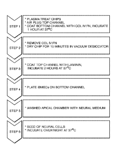
Figure 1 shows a schematic of one embodiment of a workflow for preparing and
seeding
a microfluidic chip comprising six steps. This embodiment addresses the
different surface
coating needs/preferences apparent for iBMECs and iMNPs based on experiments
such as those
illustrated in Tables 1 and 2. In particular, the workflow aims to provide, in
one embodiment,
different surface coatings for the top fluidic channel and bottom fluidic
channel of the device.
Figure 2 shows two schematics of microfluidic devices. In one embodiment of a
microfluidic device or chip (top), the device comprises top (apical) and
bottom (basal) channels
17
CA 03002399 2018-04-17
WO 2017/070224 PCT/US2016/057724
(the two Xs indicating that channels are blocked during at least part of the
protocol). The other
schematic (bottom) shows how the ports of a microfluidic device or chip (16)
can be utilized to
deposit fluids carrying surface coatings (e.g. dissolved proteins) and/or seed
the cells using
pipette tips. This image shows a modification to the typical chip ECM coating
protocol based on
the need in some embodiments to coat the top and bottom channels with
different ECM solutions
in wet and dry conditions. The procedure developed involved an "air dam" by
which perfusion of
ECM1 loaded into the bottom channel was prevented from perfusing through the
membrane to
the top channel by clamping flexible tubing and trapping air in the top
channel. The ports of a
second microfluidic channel can be air-filled and plugged up using clips, for
example.
Figure 3 provides a microscopic analysis of Chip 166 from Table 2, showing
neural cells
in the top channel of the microfluidic device (Figure 3A) and BMECs on the
bottom channel of
the microfluidic device (Figure 3B)
Figure 4 provides three images from a microfluidic chip where the cells have
been tested
for markers to confirm their identity. The top right image (Figure 4B) shows
good staining of
BMEC tight junctions indicating BBB formation on chip. On the top left (Figure
4A), the
staining shows neurons and astrocytes. Figure 4C is a vertical 2D projection
of a 3D confocal
stack of images slices, which allows for visualization of the neurons and
endothelial cells
together, even though they are not in the same plane on the microfluidic
device.
Figure 5 provides an image from a microfluidic chip wherein at least a portion
of an
apical astrocyte (i.e. the endfoot) has transmigrated the membrane and
contacted the BMECs on
the other side. Contact or interfacing between astrocytes and endothelial
cells is a recognized
feature of in vivo blood-brain barriers. To our knowledge, this interface has
never been
previously observed in in vitro models of the blood-brain barrier. The
potential for the formation
of astrocyte-endothelial contact observed in some of the embodiments disclosed
herein is desired
and advantageous, as it is believed that the in vivo contact/junction is
related to the tight barrier
properties characteristic of the blood-brain barrier.
Figure 6 shows a first image (Figure 6A) where iMNs were seeded on a plain (un-
patterned) surface, as well as a second image (Figure 6B) where the same cells
were seeded on a
nanopatterned surface, resulting in directed neurite growth. Such
nanopatterning can be applied
to the membrane or any surface of the BBB-on-chip. In particular embodiments,
the
nanopatterning is applied to the top surface of the membrane to direct neurite
growth for neuron
18
CA 03002399 2018-04-17
WO 2017/070224 PCT/US2016/057724
seeded on said surface. It is desired in some uses to direct neurite growth,
for example, in
studying neuron biology or disease (e.g. conditions that disturb neurite
growth or its
directionality), as a readout of neuron or blood-brain-barrier health (e.g. by
monitoring neurite
growth or its directionality) or in facilitating electrophysiological
measurements (e.g. using a
multi-electrode array or patch clamping). The preferred nanopattern is linear
valleys and ridges,
but alternatives such as circular, curved, or any other desired shape or
combination thereof are
also contemplated. Linear nanopatterning can include, for example, line
spacing ranging from
1 Onm to lum, 0.5um to 10um or 5um to 50um, and line depth ranging from lOnm
to 100nm,
50nm to 1000nm, 200nm to 5um or 2um to 50um.
Figure 7 show microscopic examination of the morphology of fresh (not frozen)
BMECs
seeded on a 4:1 mixture of collagen and fibronectin that has either been dried
(Figure 7A, top
left) or remained wet (Figure 7B, top right), as well as an example where the
same fresh cells
were seeded on laminin (Figures 7C and D, the arrow indicating contamination
of the cells with
neurons).
Figure 8 is a schematic showing one embodiment of a standard syringe pump and
reservoir setup for perfusion of the chips mediated by flexible tubing for
introducing flow into
the microfluidic chips. A plurality of fluid reservoirs are in fluidic
communication with a
corresponding plurality of microfluidic chips via inlet ports, with tubing
coming from the exit
ports and attached to a plurality of syringes used to draw fluid through the
chip at'a flow rate.
While a convenient method for creating flow conditions, other methods
involving different
pumping approaches or prcssure approaches to drive fluid are contemplated.
Figure 9 comprises photographs of microscopic examination of cell morphology
on the,
bottom (left-hand side) and top (right-hand side) of the membrane in a
microfluidic device where
the cells have been exposed to flow (using the system of Figure 8) for a
number of days (7 days).
Figure 9A and C show the results for Chip 664 where BMECs (on
collagen/fibronectin) and
iMPs (on wet laminin) were co-cultured. Figures 9B and D show the results for
Chip 663 where
iMPs (on laminin) were cultured alone.
Figure 10 is a photograph of fluorescent staining of cells in a microfluidic
device where
the cells have been exposed to flow (using the system of Figure 8) for a
number of days. The
image is a 3D image of the BMEC in the bottom channel showing a complete
contiguous BMEC
lumen being formed in the chip.
19
CA 03002399 2018-04-17
WO 2017/070224 PCT/US2016/057724
Figure 11 is a photograph of fluorescent staining of cells showing the
presence of neural
stem cells (red) in addition to neural filaments (green), with the nuclei
stained with DAPI. In the
preferred embodiment, the BBB-on-chip includes endothelial or endothelial-like
cells (preferably
brain-related endothelial cells) and optionally astrocytes or astrocyte-like
cells. However, in
some embodiments, the BBB-on-chip contains additional cells type such as, for
example,
neurons, pericytes and various progenitor cells. Such cells may be included as
an intended or
unintended bi-product of the stem cell differentiation process from which the
astrocytes or
endothelial cells are generated (whether on chip or preceding it), as stem
cells and progenitor
cells are typically capable of differentiating into a plurality of cells
types. The presence of
neurons is desirable in some embodiments because they can be used as readouts
of BBB function
(e.g. agents penetrating the barrier may affect the neurons in measurable
ways) or because they
may interact with other cells types or help generate a local environment that
improves the
function of the BBB-on-chip. Similarly, pericytes are desirable in some
embodiments, because it
is believed in the art that they help establish the blood-brain barrier and
can be potentially
monitored to evaluate BBB health. Neuronal- or endothelial-lineage progenitors
are desirable in
some embodiments, as they may replenish cell populations and be potentially
monitored to
evaluate BBB health. Accordingly, in some embodiments, neurons, pericytes,
neuronal-lineage
progenitors, endothelial-lineage progenitors or combinations thereof or
progenitors thereof may
be deposited in the BBB-on-chip. In other embodiments, a differentiation
process is employed
(whether on chip or preceding it) to generate one or more of these cells
types.
Figures 12A and 12B show graphs with functional measurements performed on BBB-
on-
chips. Figure 12A shows the results/readout from transepithelial electrical
resistance (TEER)
measurements on the microfluidic chip under flow, static, and control
conditions. Clearly, flow
is important for optimum results. Figure 12B show TEER measurements on
transwells. TEER is
a typical measure of in vitro BBB models and is used both for evaluating the
model as well as an
experimental readout (e.g. after subjecting the BBB model to an experimental
condition). Some
aspects of the present invention include measuring the TEER of one or more BBB-
on-chips.
This can be done, for example, to evaluate BBB-on-chip development, maturation
or quality, as a
readout for experiments involve an introduced agent, as a readout for
experiments involving
specific cells or cell types (e.g. patient specific, a disease population, or
treated to simulate a
disease or condition), etc. It is known in the art how to integrate electrodes
suitable for TEER
CA 03002399 2018-04-17
WO 2017/070224 PCT/US2016/057724
measurement into microfluidic devices. Douville et al., "Fabrication of Two-
Layered Channel
System with Embedded Electrodes to Measure Resistance Across Epithelial and
Endothelial
Barriers" Anal Chem. 2010 March 15; 82(6): 2505-2511.
Figure 13A and B show how TEER measurements were made in one embodiment.
Figure 13A is an enlarged schematic view showing how electrodes on the chip
were connected,
along with pipette tips engaging the chip; Figure 13B shows the same connected
chip to the right
of a Epithelial Voltohmmeter.
Figure 14A was a follow-up experiment on another round of prototype ;ITER
chips that
showed iBMEC barrier function increasing in the presence of flow on a chip
followed by a
weakening of barrier function with the exposure of the chips to TNFa, a
proinflammatory
cytokine. Higher TEER values generally indicate a tighter barrier, which is
typically desirable
for a blood-brain barrier. Figure 14B also involves TNF alpha exposure, but
the readout is
membrane permeability as measured by Dextran-FITC.
Figure 15 shows permeability results for (and the structure of) fluorescein
sodium. Some
aspects of the present invention include ascertaining permeability for various
additional agents
(e.g. drugs, chemicals, hormones, blood components, biomarkers). Such methods
can allow
qualitative or quantitative estimation of the permeability of the in vitro
blood-brain barrier to the
one or more agents. Furthermore, according to some aspects of the present
invention, the
permeability of one agent is measured in response to a second agent, treatment
or experimental
condition (for example, measuring the effect of a medication on the blood-
brain barrier
permeability of another medication).
Figure 16A shows the user interface and the conditions during the run of human
blood
across the blood brain barrier. Figure 16B shows the equipment setup for
measuring the
transport of solutes from human blood across the blood brain barrier (BBB), a
barrier created in
vitro in the microfluidie devices described herein using a layer of I3MECs. As
evidenced, some
embodiments include blood or blood components, optionally perfused through one
or more
fluidic channels within the device. The use of blood of blood components is
desired in some
embodiments, as the blood or blood components can improve BBB-on-chip
function, for
example, by providing biochemical cues, or conversely hurt the BBB-on-chip,
for example,
because the blood may contain a harmful agent that may be under investigation.
In some
aspects, permeability assays include blood or blood components in order to
provide a potentially
21
CA 03002399 2018-04-17
=
WO 2017/070224 PCT/US2016/057724
more in vivo like result. In other aspects, individual-specific blood or blood
components are
used in order to potentially provide individualized BBB-related measures. This
can include, for
example, the measurement of the permeability of one or more agents or
components from the
blood or components, the effect of the blood or components on the permeability
of one or more
agents that may be added to the blood or another fluid included in the device,
the effect of the
blood or components on the health of the BBB-on-chip or any of its components
(whether
positive or negative), etc. This may include diagnostic uses, for example, to
identify a disease,
biomarker or infectious agent carried by the blood or blood components.
Figure 17 shows the measurement of thyroid hormone transport by mass
spectrometry
(Figure 17A) using the setup shown in Figure 16, along with the graphed
results (Figure 17B).
After flowing patient blood through the microfluidic chips into the channel
under the BMECs, it
was possible to measure the transport of compounds from the blood into the
neural compartment,
i.e. through the BMEC barrier. In this case, the experiment included a control
set of BBB-on-
chips comprising iPS-derived cells originating from a non-diseased individual,
and a second set
of BBB-on-chips comprising iPS-derived cells originating from a patient
diagnosed with Allan-
Herndon-Dudley syndrome (AHDS). The mass-spectrometry data in Figure 17A is an
initial
experiment to confirm that the MCT8 transporter defect can be recapitulated on
an Organ-Chip.
Figure 18 shows electrophysiology recordings collected by patch-clamp from
neurons in
the microfluidic device ("BBB-on-Chip"). An arrow (Figure 18A) indicates
single action
potential. Current recordings (Figure 18B, right) show negative sodium channel
currents (Nat)
and positive potassium channel 00. These measurements on-chip can be used, for
example, to
provide an indication of neuronal maturation or as a readout of neuron health.
In turn, neuronal
maturation or health can be used as indicators of BBB-on-chip quality (for
example, before
starting an experiment) or as an experimental endpoint indicating, for
example, that an agent as
crossed the BBB, a disease condition has emerged, the BBB has been modified or
compromised,
or conversely, that the BBB or neural function or health have improved. Patch
clamping can be
performed on the BBB-on-chip by a variety of methods, including for example,
by inserting the
patch-clamp electrodes through the soft body of an elastomeric BBB-on-chip
device. Similarly
to patch-clamping, other electrophysiological readouts can be obtained, for
example by including
one or more electrodes in the device. In particular, a multi-electrode array
(MEA) can be
integrated on the membrane of embodiments that possess one or similarly in
fluidic channels or
22
CA 03002399 2018-04-17
WO 2017/070224 PCT/US2016/057724
cavities within the device. Electrophysiological measurements (patch-clamping,
MEA) can also
be applied to astrocytes, which have been shown in the art to be excitable.
Figure 19 show the results of calcium flux imaging in the neural channel. The
photograph
(Figure 19A, top left) is a single fluorescent image from a movie of such
images. The colored
circles indicate the positions that correspond to the time traces in the 3
graphs. The traces
(Figures 19B and C) show that it is possible to observe neuronal function in
the microfluidic
chips in real-time. The addition of tetrodotoxin (TTX), which is a potent
blocker of voltage-gated
calcium channels, ablates this activity (Figure 19D, bottom right). Calcium
imaging or imaging
using voltage-sensitive dyes or proteins offer similar advantages to
electrophysiological readouts
but offers the advantage that no electrodes are necessary. Accordingly, some
aspects of the
present invention include methods of measuring the BBB-on-chip by imaging in
the presence of
calcium or voltage-sensitive dyes or proteins, to allow the potential
recording and optional
manipulation of neuronal or astrocyte excitations. These measurements can be
used, for
example, to provide an indication of neuronal maturation or as a readout of
neuron health. In
turn, neuronal maturation or health can be used as indicators of BBB-on-chip
quality (for
example, before starting an experiment) or as an experimental endpoint
indicating, for example,
that an agent as crossed the BBB, a disease condition has emerged, the BBB has
been modified
or compromised, or conversely, that the BBB or neural function or health have
improved.
Figure 20 shows both a protocol for generating, and staining results
confirming the
generation of, neural cells from neural progenitors. Such techniques allow one
to make
multipotcnt neural stem cells and motor neuron precursor directly from iPSC,
allowing
differentiation into many neural cell types (neurons, astrocytes, etc.).
Figure 21 shows the corrected T3 concentration in the top channel of seven
different
chips, i.e. chips populated with normal cells (2280, 2289 and 2284) as
compared to chips
populated with cells from an MCT8 cell line or patient (2285 ¨ 2288).
Figure 22A is a schematic showing one embodiment of the microfluidic device or
chip
(16), comprising two microchannels (1), each with an inlet and outlet port
(2), as well as
(optional) vacuum ports. Figure 22B is a topside schematic of an embodiment of
the perfusion,
disposable or "pod" (10) featuring the transparent (or translucent) cover (11)
over the reservoirs,
with the chip (16) inserted in the carrier (17). The chip can be seeded with
cells and then placed
23
CA 03002399 2018-04-17
WO 2017/070224 PCT/US2016/057724
in a carrier for insertion into the perfusion disposable or pod, whereupon
culture media in the
reservoirs flows into the microchannels and perfuses the cells (e.g. both
BMECs and MNs).
Figure 23 shows a schematic of an illustrative microfluidic device or "organ-
on-chip"
(16) device. The assembled device is schematically shown in Figure 23A with
the top surface
(21) indicated. Figure 23B shows an exploded view of the device of Figure 23A,
showing a
bottom piece (97) having channels (98) in a parallel configuration, and a top
piece (99) with a
plurality of ports (2), with a tissue-tissue interface simulation region
comprising a membrane
(101) between the top (99) and bottom (97) pieces, where (in one embodiment)
cell behavior
and/or passage of gases, chemicals, molecules, particulates and cells are
monitored. In an
embodiment, an inlet fluid port and an outlet fluid port are in communication
with the first
central microchannel such that fluid can dynamically travel from the inlet
fluid port to the outlet
fluid port via the first central microchannel, independently of the second
central microchannel. It
is also contemplated that the fluid passing between the inlet and outlet fluid
ports may be shared
between the central microchannels. In either embodiment, characteristics of
the fluid flow, such
as flow rate and the like, passing through the first central microchannel is
controllable
independently of fluid flow characteristics through the second central
microchannel and vice
versa.
Figure 24 is a print out of electrophysiological data from neurons cultured in
a
microfluidic device or chip, showing highly complex spontaneous activity in a
chip.
Figure 25 shows print outs of electrophysiological data from neurons cultured
alone
(Figure 25A, top panel) and co-cultured with BMECs (Figure 25B, bottom panel)
in a
microfluidic device or chip, showing that neural tissue have more mature
electrophysiological
properties in the chip, and in co-culture with BMECs.
Figure 26 shows print outs of electrophysiological data from neurons cultured
alone
(Figure 26A, top panel) and co-cultured with BMECs (Figure 26B, bottom panel)
in a
microfluidic device or chip, showing that neural tissue have more mature
electrophysiological
properties in the chip when in co-culture with BMECs.
Figure 27 provides neural calcium measurement read-outs comparing neurons (MN)
co-
cultured with BMECs (Figure 27D, bottom panel), cultured alone (Figure 27C,
first panel up
from the bottom panel), cultured in endothelial cell conditioned medium or
ECCM in a (96-well)
static culture (Figure 27B, second panel up from the bottom panel), along with
an unconditioned
24
CA 03002399 2018-04-17
WO 2017/070224 PCT/US2016/057724
media (96-well) static control (Figure 27A, top panel). Each neuron's activity
is simultaneously
tracked and analyzed (calcium influx is an indirect measure for neuronal
activity that can be
observed live in the chip). The results show that co-culture increases di MN
neural calcium
transient activity, i.e. a significant increase in transient frequency is
observed upon contact of
MNs with iBMECs.
Figure 28 is a bar graph of neural calcium measurements (average frequency
events per
cell) comparing neurons (MN) co-cultured with BMECs (far right), cultured
alone (next bar to
the left), cultured in endothelial cell conditioned medium or ECCM in a static
culture (next bar to
the left), along with an unconditioned media static control (far left). The
results show that co-
culture increases diMN neural calcium transient activity, i.e. a significant
increase in transient
frequency is observed upon contact of MNs with iBMECs.
Figure 29 shows the results of a transcriptomic study of iMNs in a
microfluidic chip.
Neurons were either cultured alone (Figure 29A, top box) on the chip or in a
co-culture with
BMECs (Figure 29B, bottom box), and this was compared with a 96-well static
culture. The
MNs were sorted on a FACS and RNA was sequenced (i.e. gene expression was
detected). RNA-
Seq from FACS sorted MNs show that neural development gene pathways (PC1) are
upregulated
in chip. Vascular interaction genes (PC3) are recreated in co-culture with
iBMECs. In addition,
there are chip induced genes (PC2), i.e. gene activity induced in the cells
simply from being
cultured on the chip.
Figure 30 shows the detailed results from which Figure 34 was prepared,
showing the
names of various neural development genes (PC1), chip induced genes (PC2) and
vascular
interaction genes (PC3). The colored bars on the right in Figure 30 represent
the expression of
each gene (row) in each of the 5 conditions (columns). Column order is MN
Only, BMEC/MN,
96-well control, 96 well ECCM, MN progenitor. Red = high and blue = low. These
vascular
gene pathways have not been shown to be induced in any other culture system
and may be
inducing the observed increase in maturity and activity.
Description of the Invention
The invention relates to culturing endothelial cells (preferably brain-related
endothelial
cells) and optionally astrocytes, optionally neurons, and optionally pericytes
in a fluidic device
under conditions whereby the cells mimic one or more structural or functional
features (e.g. tight
CA 03002399 2018-04-17
WO 2017/070224 PCT/US2016/057724
junctions) of the blood brain barrier and/or the spinal cord. Culture of these
cells in a
microfluidic device, such as a microfluidic chip with flow as herein
described, whether alone or
in combination with other cells, drives maturation and/or differentiation
further than existing
systems. For example, a mature electrophysiology of the neurons includes
negative sodium
channel current, positive potassium channel current, and/or action potential
spikes of amplitude,
duration and frequency similar to neurons in a physiological environment or
when compared to
static culture neurons, static culture neurons lack one or more of the
aforementioned features.
The evidence also supports improved maturation of the astrocytes and BMECs. As
described
herein, astrocytes were observed to send out of processes to contact the
endothelial cells. As
described herein, improved and sustained barrier function indicates maturation
of the BMECs.
Good viability and function allow for measurements of barrier integrity and
physiology, whether
by TEER, permeability assays, patch clamp (or other electrophysiological
methods), calcium or
voltage imaging, or other testing measures. Observed characteristics of the in
vitro "BBB-on-
chip" of the present invention include: (1) tight junctions between
endothelial cells (which
creates selective permeability to substances); (2) optional cell-to-cell
communication
exemplified by contact of the endothelial cells with astrocytes (e.g. endfoot
contact by partial
transmigration of the membrane separating these cells); (3) optional extended
neurite projections,
(4) optional fluid flow that influences cell differentiation and tight
junction formation; and (5)
high electrical resistance representing the maturity and integrity of the BBB
components. With
respect to neurite projections, in one embodiment, the present invention
contemplates seeding on
nanopatterned surfaces which promote extended and direct (e.g. along a
relatively linear path)
neurite growth. The preferred nanopattern is linear valleys and ridges, but
alternatives such as
circular, curved, or any other desired shape or combination thereof are also
contemplated. With
respect to endothelial cells, in one embodiment, the present invention
contemplates BMECs
which form a lumen on the chip (for example, completely lining a flow channel,
at least for a
portion of its length). Among other advantage (e.g. endothelial layer
stability) this potentially
enables the use of the device with blood or' blood components. With respect to
selective
permeability, the present invention contemplates, in one embodiment,
introducing substances in
a channel under the BMECs such that at least one substance passes through the
BMEC barrier
(e.g. BMEC cells on the bottom side of the membrane) and into a channel above
the membrane,
and detecting said at least one substance (e.g. with antibodies, mass spec,
etc.).
26
=
CA 03002399 2018-04-17
WO 2017/070224 PCT/US2016/057724
Although there is a strong need for a model of the human blood-brain barrier,
it is also
desirable to develop models of blood-brain barriers of other organisms (not
limited to animals).
Of particular interest are models of, for example, mouse, rat, dog, and
monkey, as those are
typically used in drug development. Accordingly, the BBB-on-chip can make
advantage of not
only human-derived cells but also cells from other organisms. Moreover,
although it is
preferable that all cell types used originate from the same species (for
example, in order to
ensure that cell-cell communication is effective), it may be desirable at time
to mix species (for
example, if a desired cell type is scarce or possess technical challenges).
Description of Preferred Embodiments
In one embodiment, the present invention contemplates a BBB-on-chip where at
least one
population of cells is derived from a patient diagnosed with a disorder of the
nervous system.
While it is not intended that the present invention be limited to a particular
CNS disorder, in one
embodiment, the disorder is ALS. Amyotrophic lateral sclerosis (ALS) is a
severe
neurodegenerative condition characterized by loss of motor neurons in the
brain and spinal cord.
In one embodiment, the present invention contemplates generating induced
pluripotent stem cells
(iPSCs) from patients with ALS and differentiating them into motor neurons
progenitors for
seeding on a microfluidic device. There are currently no effective treatments
for ALS. In one
embodiment, the present invention contemplates the BBB-on-chip as a model
system for testing
drugs so as to predict success in subsequent clinical trials.
In another embodiment, the CNS disorder is Parkinson's disease (PD). PD is a
neurodegenerative disorder primarily characterized by a loss of dopamine
neurons, but which
also leads to many other pathological changes.
In yet another embodiment, the CNS disorder is Alzheimer's disease.
Alzheimer's is a
type of dementia that causes problems with memory, thinking and behavior.
Symptoms usually
develop slowly and get worse over time, becoming severe enough to interfere
with daily tasks.
It is contemplated that iPSC technology can be used together with microfluidic
chips to
mimic patient-specific phenotypes in disease states. For example, in another
embodiment, cells
derived from patients diagnosed with MCT8-specific thyroid hormone cell-
membrane
transporter deficiency are contemplated for use in microfluidic devices as at
least one of the
cellular components of the "BBB-on-chip." This disease is characterized by
severe cognitive
27
CA 03002399 2018-04-17
WO 2017/070224 PCT/US2016/057724
deficiency, infantile hypotonia, diminished muscle mass and generalized muscle
weakness,
progressive spastic quadriplegia, joint contractures, and dystonic and/or
athetoid movement with
characteristic paroxysms or kinesigenic dyskinesias. Seizures occur in about
25% of cases.
Patients exhibit pathognomonic thyroid test results including high serum
3,3',5-triiodothyronine
(T3 ) concentration and low serum 3,3',5'-triiodothyronine (reverse T3 or rT3)
concentration.
Serum tetraiodothyronine (thyroxine or 14) concentration is often reduced, but
may be within the
low normal range; serum TSH concentrations are normal or slightly elevated.
SLC16A2 (also
known as MCT8) is the only gene in which mutations are known to cause this
disorder.
Experimental
Example 1
Cells are prepared either directly from cultured iPSCs or from frozen lots of
pre-
differentiated cells. Cells are thawed (or dissociated fresh) and seeded into
the chip at day 8-9 (in
the case of BMECs differentiation) and at various points in neural
differentiation. In the case of
IDissoclato & re-plate I
Day 0 6 12 18
Stage iPSC Neuroepithelia MN precurosors IMNs
Markers 0CT3/4 SOX1 OLIG2+ MNX1
NANOG HOX? NKX2.2- CHAT
Day 0 - 6 DayB-12 Dav 12 -3oc
1:1 IMDM/F12 1:1 IMDM/F12 1:1 IMDM/F12
1% NEAA 1% NEAA 1% NEAA
2% B27 (+vit,A) 2% B27 (+vit.A) 2% 927 (+vit.A)
1%N2 1%N2 1%N2
1% PSA 1% PSA 1% PSA
0.2 pM L0N193189 0.1 pM All-trans RA 0,1 pM Compound E
pM S8431542 0.2 pM L0N193189 2.5 pM DAPT
3 pM CHIR99021 1 pM Purpmorphamine 0.5 pM All-trans RA
(or SAG) 0.1 pM Purpmorphamine
10 pM SB431542 (or SAG)
3 pM CHIR99021 0.1 pM db-cAMP
200 ng/ml Ascorbic Acid
10 ng/ml GDNF
10 ng/ml BDNF
0.5 mM VPA
MNs, for example, cells are seeded at day 12 of differentiation either from
freshly differentiated
cultures or directly from a thawed vial. iPSC-derived forebrain neural
progenitor cultures
(dubbed EZs) were cultured in chip either dissociated or as neural spheres
that attached and
28
CA 03002399 2018-04-17
WO 2017/070224 PCT/US2016/057724
extended in 3 dimensions (See Figure 3 apical).The various factors used in the
protocol (see
above chart and tabs) for the generation of motor neurons are provided (using
iPSCs as the
starting material).
Example 2
In this example, another protocol for the generation of motor neurons is
provided using
iPSCs as the starting material:
iPSC to Motor Neuron Differentiation
.41111111.44-
MN 'precursor tonnation
Acurtme EslAg00 pis) ANOMIE!'"
741141.11404N1+004=9144 Pool
camsoodforrnatia0 nhaN10 Neu,* impoots, PAN* dlimels000 EAU
I PSC colonlo. IA T7 $.014111504 041p4744.401411A CA1
T
days . 6 8 17 25 /
INC SaND + ITU; Ntil DM + TRAP139 9i.AJ amm-mirmitt *;
SaND
39 39-84
MN Maturation media
N oval SAD *1701 10100.0 !AIN enatunitio0 meth&
,1701ArenA1at1ext SOW Newsboys' 1:1 044EAVF 1 2
Isom CM -04.01 1 AM ATPA I%N3 111,
' Media11A N2
2'411 77
14414511 211.127 41414NAln Al
MOM 1 0 AgA01 COW
1% NE NOMA *TARP 1 onamti GIMP
744111271-141A1 NOWA 200 ingtml aNtublt add
1% MEM NEAA 1107 1 021e1D011.10
10. PTA 1 0 411 17 0 44 0.1 11/4 rethok add
1 0.4 EitaNnotiVhAftlia=
The various factors used in the protocol (see above chart and tabs) for the
generation of motor
neurons are provided (using iPSCs as the starting material).
Example 3
This example explores various conditions tested for seeding neural (EZ spheres
and
iMNPs) and endothelial cells (iBMECs) from frozen stocks of cells on surfaces
treated with
different extracellular matrices (ECMs). The best results for iBMECs were
achieved with a
mixture of collagen and fibronectin (4:1 ratio) using a seeding concentration
of 5x10⁶ cells/ml
(Table 1). Given these results, seeding was attempted on microfluidic devices,
i.e. chips. Table 2
shows various conditions tested for seeding neural (EZ spheres, iNPCs and
iMNPs) and
endothelial cells (iBMECs) on the apical and basal sides of a microfluidic
chip using frozen
stocks of cells.
29
CA 03002399 2018-04-17
WO 2017/070224 PCT/US2016/057724
While a variety of protocols were explored, one embodiment for preparing and
seeding a
microfluidic chip comprising six steps. Figure 1 shows the workflow. First,
the chip (or regions
thereof) are treated to promote wetting or protein adhesion (e.g. by plasma
treatment). One or
more channels are then plugged (see the top schematic of Figure 2, where an
"X" indicates a
channel is blocked in a microfluidic device or chip with top and bottom
channels). Figure 2
(bottom schematic) shows how the ports of a microfluidic device can be
utilized to introduce
fluid (e.g. with ECMs) or cells using pipette tips. Using the protocol, the
ECM mixture for the
bottom channel is introduced first, with the excess removed, and the remainder
dried. Thereafter
(step 3), the ECM for the top channel is introduced. The BMECs can be seeded
on the bottom
channel. The top channel can be washed. Finally, the neural cells can be
introduced and
incubated for attachment. Cultures were seeded into chips following the
seeding of BMECs
described above either on the same day or the following day after BMECs had
been seeded onto
the chip. The chips were cultured for 14 days and fixed and stained for
relevant markers.
Confocal imaging shows the transmigration in z-stack.
Figure 3 provides a microscopic analysis of Chip 166 from Table 2, showing
neural cells
in the top channel of the microfluidic device (left) and BMECs on the bottom
channel of the
microfluidic device (right). The neural cells and BMECs have attached.
The attached cells were then tested for markers to confirm their identity.
Figure 4 is a
vertical 2D projection of a 3D confocal stack of images slices, which allows
for visualization of
the neurons and endothelial cells together, even though they are not in the
same plane on the
microfluidic device. The BMECs display the Glut I marker, while the neurons
are positive for
NFH. DAPI was used to stain the nuclei.
Figure 5 provides an image from a microfluidic chip wherein at least a portion
of an
apical astrocyte (i.e. the endfoot) has transmigrated the membrane and
contacted the BMECs on
the other side. The astrocytes are shown in white against the red stained
BMECs.
Example 4
The present invention contemplates, in one embodiment, utilizing nanopatterned
surfaces
for seeding cells. Figure 6 shows a first image (top) where iMNPs were seeded
on a plain (un-
patterned) surface, as well as a second image (bottom) where the same cells
were seeded on a
CA 03002399 2018-04-17
WO 2017/070224 PCT/US2016/057724
nanopatterned surface. Clearly, the nanopatterned surface results in directed
neurite growth (e.g.
in a line pattern)
Example 5
While frozen stocks of cells can be used (particular for the neural cells), it
was found that
better results can be obtained (particularly for BMECs) when fresh cells are
used for seeding.
Figure 7 show microscopic examination of the morphology of fresh (not frozen)
BMECs seeded
on a mixture of collagen and fibronectin that has either been dried (Figure
7A, top left) or
remained wet (Figure 7B, top right), as well as an example where the same
fresh cells were
seeded on laminin (Figures 7C and D). Interestingly, when laminin was used,
the BMECs were
not free of neurons (see the arrow in Figure 7D indicating contamination of
the cells with
neurons).
Tables 3 and 4 show various conditions tested for seeding fresh neural (iMNPs)
and fresh
endothelial cells (iBMECs), where the particular conditions are associated by
microfluidic chip
number, allowing for a correlation of good tight junctions with the seeding
conditions. Staining
results (not shown) for microfluidic chip 574 (see Table 3 for conditions)
indicated the cells are
positive for Glut 1 (red stain), which is a marker of BMEC tight junctions
(the nuclei were also
stained blue from DAPI). The seeding conditions for chip 574 resulted in good
tight junctions.
Staining results (not shown) for microfluidic chip 665 (see Table 3 for
conditions) indicated that
the cells are positive for Glut I. Thus, the seeding conditions for chip 665
also resulted in good
tight junctions. Staining results (not shown) for microfluidic chip 667 (see
Table 3 for
conditions) indicated the cells are positive for Glut 1. Thus, the seeding
conditions for chip 667
resulted in good tight junctions. Staining results for microfluidic chip 693
(see Table 3 for
conditions) indicated the cells are positive for Glut 1. Thus, the seeding
conditions for chip 693
resulted in good tight junctions.
Staining results (not shown) for microfluidic chip 733 (see Table 4 for
conditions)
indicated the cells are positive for Glut 1. The results (not shown) also
revealed that coating with
laminin alone (before seeding) results in poor BMEC tight junction formation.
31
CA 03002399 2018-04-17
WO 2017/070224 PCT/US2016/057724
Example 6
Unlike conventional static cultures, the present invention contemplates
microfluidic
devices where the cells are exposed to a constant flow of media providing
nutrients and
removing waste. Figure 8 is a photograph showing one embodiment of a system
for introducing
flow into the microfluidic chips. A plurality of fluid reservoirs are in
fluidic communication
with a corresponding plurality of microfluidic chips via inlet ports, with
tubing coming from the
exit ports and attached to a plurality of syringes used to draw fluid through
the chip at a flow
rate. Figure 9 comprises photographs of microscopic examination of cell
morphology on the
bottom (left-hand side) and top (right-hand side) of the membrane in a
microfluidic device where
the cells have been exposed to flow (using the system of Figure 8) for a
number of days. Figure
shows fluorescent staining of cells in a microfluidic device where the cells
have been exposed
to flow (using the system of Figure 8) for a number of days. The image is a 3D
image of the
BMEC in the bottom channel showing a complete contiguous BMEC lumen being
formed in the
chip. Figure 11 is a photograph of fluorescent staining of cells showing the
presence of neural
stem cells (red) in addition to neurites (green), with the nuclei stained with
DAPI.
Example 7
Good cell viability and function on the BB-on-chip allow for measurements of
barrier
integrity and physiology, whether by TEER, patch clamp or other testing
measures.
TEER: Figure 12A shows the results/readout from transepithelial electrical
resistance
(TEER) measurements un the microfluidic chip under flow, static, and control
conditions. Cells
were plated on tall channel PDMS chips equipped with incorporated gold
electrodes on each
channel (see Figure 13A). Post seeding of BMECs, transendothelial electrical
resistance was
measured by connecting the electrodes to an EVOM2 voltohmmeter (see Figure
13B). Figure
12A displays preliminary data indicating the beneficial effect of flow in the
BBB-on-chip, i.e.
higher TEER in response to flow. In particular, at around the 40 hour time
point, the TEER
value observed for a BBB-on-chip under flow was significantly higher than a
similar chip under
static conditions, i.e. that the iPS brain microvascular endothelial cells
(iBMECs) formed tighter
cell-cell junctions or barrier function under flow conditions on a prototype
TEER-Chip as
compared to a chip maintained in static culture. The "damaged" chip was a
failure due to the
32
CA 03002399 2018-04-17
WO 2017/070224 PCT/US2016/057724
TEER-Chips being a prototype. Figure 12B shows TEER results where the cells
were cultured in
transwells.
Figure 14A was a follow-up experiment on another round of prototype TEER chips
that
showed iBMEC barrier function increasing in the presence of flow on a chip
followed by a
weakening of barrier function with the exposure of the chips to TNFa, a
proinflammatory
cytokine. Higher TEER values generally indicate a tighter barrier, which is
typically desirable
for a blood-brain barrier.
PATCH CLAMP: Figure 18 shows electrophysiology recordings collected by patch-
clamp from neurons in the microfluidic device ("BBB-on-Chip"). These
measurements on-chip
can be used to provide an indication of neuronal maturation, i.e. more
precisely describe the
maturity of a neuronal cell. Cells were cultured as described above in a
specially designed
"openable" chip (where the chips can be partially disassembled to expose
directly cells on the
semi-porous membrane) with a stiff PET membrane to aid in patch-clamp
recording. PDMS was
attempted, but was unsuccessful. PET membrane chips were opened at endpoint at
6 and 24 days
in chip. Individual neurons seeded into the chip were directly accessed with a
glass micropipette,
and cell electrophysiology was recorded including capacitance, membrane
resting voltage,
spontaneous activity and induced activity. Figure 18 is a whole cell patch
recording of an
induced action potential from a neuron cultured on the chip. An arrow (Figure
18A) indicates
single action potential. Current recordings (Figure 18B, right) show negative
sodium channel
currents (Na) and positive potassium channel (K+) are necessary for normal
neuron function and
become more pronounced as a neuron matures.
CALCIUM FLUX: Figure 19 show the results of calcium flux imaging in the neural
channel. Using a florescent calcium influx-activated dye (Fluo-4), neurons
seeded in chip were
imaged using high resolution high frame-rate camera. Florescence intensity
changes of up to
hundreds of neurons were analyzed simultaneously by recording average pixel
intensity over
time (dF/F). These values were plotted with respect to time and are analyzed
for waveform
properties, which correlate spontaneous neural activity and neural network
formation. This is
accomplished through multi-step video post-processing and signal analysis
(including video
compression, signal cleanup, automatic or manual ROI detection, etc. which can
be implemented
from open-source MATLAB software packages). The photograph (Figure 19A, top
left) is a
single fluorescent image from a movie of such images. The colored circles
indicate the positions
33
CA 03002399 2018-04-17
WO 2017/070224 PCT/US2016/057724
that correspond to the time traces in the 3 graphs. The traces show that it is
possible to observe
neuronal function in the microfluidic chips in real-time. In this case, it is
shown that Ca2+ fluxes
can be measured on chips to give a direct readout of neuronal activity. The
addition of
tetrodotoxin (TTX), which is a potent blocker of voltage-gated calcium
channels, ablates this
activity (Figure 19D, bottom right). This type of experiment will be important
when the
neuronal activity is modulated by pharmacological stimulation.
ICC overlay data: By overlaying images taken after staining the cells,
specific cell
identification can be combined with original activity traces to determine
specific activities of
individual cell types in the chip. The overlay data (not shown) indicates that
motor neurons are
indeed more active in the chip. This can also be accomplished with cell type
specific reporter
lines.
Example 8
Brain microvascular endothelial cells (BMECs) constitute the blood-brain
barrier (BBB)
which forms a dynamic interface between the blood and the central nervous
system (CNS) in
vivo. This highly specialized interface restricts paracellular diffusion of
fluids and solutes
including chemicals, toxins and drugs from entering the brain. In this
example, fluorescein
sodium is used in a paracellular permeability assay of the BMECs seeded on a
microfluidic
device.
Albumin or Dextran conjugated to a fluorescent probe (e.g., FITC or TRITC) are
frequently used to monitor changes in leakage, and thus barrier function. In
this case, Dextran-
FITC, a green fluorescent molecule of 4 KDa, or sodium fluorescein (a 0.3 KDa
molecule), was
added to the bottom ("blood side") channel. Paracellular permeability was
calculated by
measuring the permeability of the fluorescent molecule on the Top ("brain
side") channel. Low
permeability is an indication for proper barrier functions. Figure 14B
involves TNF-alpha
exposure, but the readout is membrane permeability as measured by Dextran-
FITC. Figure 14B
confirms that TNFa exposure results in a decrease in barrier function and TEER
by an increase in
permeability through the semi-porous membrane by dextran-FITC, a fluorescently
labeled small
molecule.
Figure 15 shows the results for (and structure of) fluorescein sodium from a
paracellular
permeability assay. Chips were seeded with iPSC-derived BMECs taken from
healthy controls
34
CA 03002399 2018-04-17
WO 2017/070224 PCT/US2016/057724
(CTR) or MCT8-deficient patients, and the paracellular permeability was
determined by
monitoring Blood to brain permeability of the sodium fluorescein tracer as
described above.
Flow is clearly important.
In the present experiment, the agent used was fluorescein. In some aspects of
the present
invention, it is contemplated that similar testing will be done to ascertain
permeability for
various additional agents (e.g. drugs, chemicals, hormones, blood components,
biomarkers).
Such methods can allow qualitative or quantitative estimation of the
permeability of the in vivo
blood-brain barrier to the one or more agents. Furthermore, according to some
aspects of the
present invention, the permeability of one agent is measured in response to a
second agent,
treatment or experimental condition (for example, measuring the effect of a
medication on the
blood-brain barrier permeability of another medication). It is important to
note that although we
refer to permeability, we do not mean to exclude active transport, pumping or
any other means
for an agent to pass from one side of the barrier to the other (regardless of
direction). The
penetration of an agent through the barrier can be measured, for example,
using mass
spectroscopy, antibody-based methods (e.g. ELISAs, Western blots, bead-based
assays), or
optical methods (e.g. fluorescence signature, Raman spectroscopy, absorbance).
Example 9
Some embodiments include blood or blood components, optionally perfused
through one
or more fluidic channels within the device. The use of blood of blood
components is desired as
the blood or blood components can improve BBB-on-chip function, for example,
by providing
biochemical cues, or conversely hurt the BBB-on-chip, for example, because the
blood may
contain a harmful agent that may be under investigation. In some aspects,
permeability assays
include blood or blood components in order to provide a potentially more in
vivo like result. In
other aspects, individual-specific blood or blood components are used in order
to potentially
provide individualized BBB-related measures. This can include, for example,
the measurement
of the permeability of one or more agents or components from the blood or
components, the
effect of the blood or components on the permeability of one or more agents
that may be added
to the blood or another fluid included in the device, the effect of the blood
or components on the
health of the BBB-on-chip or any of its components (whether positive or
negative), etc. This
CA 03002399 2018-04-17
WO 2017/070224 PCT/US2016/057724
may include diagnostic uses, for example, to identify a disease, biomarker or
infectious agent
carried by the blood or blood components.
In this example, hormone transport across the BMECs was measured in the "BBB-
on-
chip" in healthy and diseased tissue by mass spectrometry. Thyroid hormone was
added to the
bottom channel and measured on the top channel. Thyroid hormones (T3 and T4)
were detected
using Liquid chromatography tandem-mass spectrometry (LC-MS/MS).
BMECs from a MCT8 background were used. Figure 16A shows the user interface
and
the conditions during the run of human blood across the blood brain barrier.
Figure 16B shows
the setup for measuring the transport of solutes from human blood across the
blood brain barrier,
a barrier created in vitro in the microfluidic devices describes herein using
a layer of BMECs.
Figure 16B shows how human blood was perfused into the bottom channel of the
tall chip. In
this experiment thyroid hormones were measured by LC-MS/MS as described above.
This setup
will also be used to test the filtration of proteins across the BBB.
Figure 17 shows the measurement of thyroid hormone transport by mass
spectrometry
(Figure 17A) using the setup shown in Figure 16B, along with the graphed
results (Figure 17B).
After flowing patient blood through the microfluidic chips into the channel
under the BMECs, it
was possible to measure the transport of compounds from the blood into the
neural compartment,
i.e. through the BMEC barrier. In this case, the experiment included a control
set of BBB-on-
chips comprising iPS-derived cells originating from a non-diseased individual,
and a second set
of BBB-on-chips comprising iPS-derived cells originating from a patient
diagnosed with Allan-
Herndon-Dudley syndrome (AHDS). Briefly, iBMECs were generated from a patient
with an
inactivating genetic mutation in the MCT8 thyroid hormone transporter. This
mutation leads to a
defect in T3/T4 transport across the BBB and defects in neural development in
patients. The
mass-spectrometry data in Figure 17A is an initial experiment to confirm that
the MCT8
transporter defect can be recapitulated on an Organ-Chip.
Example 10
In this example, the disease model was further evaluated. Samples were
prepared by
taking 100 ul of each sample of T3 and mixing it with the equivalent sample of
T4. This was
done for each sample and also for the calibration curve. Proteins and salts
were precipitated from
36
CA 03002399 2018-04-17
WO 2017/070224 PCT/US2016/057724
the solution; the samples were dried and resuspended in the same volume. The
calibration curve
permitted the calculation of the concentrations (in mM) for both T3 and T4.
For the T3/T4 experiments, the following 4 conditions were tested in the
microfluidic
chip:
1. 1 nM T3 in normal media in the bottom channel and media without T3 on
top
channel. Both sides were running at a 30 ul/hr flow rate.
2. 100 nM T3 and T4 in normal media in the bottom channel and media without
T3 on
top channel. Again, both sides were running at a 30 ul/hr flow rate.
3. Human plasma on bottom channel at 90 ul/hr and media without T3 on top
channel
kept static for 1 hour.
4. Human plasma on bottom channel at 90 ul/hr and media without 13 on top
channel
kept static for 1 hour.
For each experiment, Dextran-FITC was used in the bottom channel to correct
for
paracellular diffusion.
From the above-mentioned 4 conditions, only 100 nM was significantly above
detection
and these worked well as shown in Figure 21. Chips 2280, 2289, and 2284 are
populated with
cells from a single control line. Chips 2285 and 2286 are populated with cells
from the isogenic
mutated MCT8 line. Chips 2287 and 2288 are populated with cells from a mutated
MCT8
patient. Figure 21 is a bar graph showing the corrected T3 concentration in
the top channel of
each chip. Clearly, there is reduced 13 transport in mutated MCT8 lines as
compared to normal,
demonstrating one aspect of disease modeling using the blood-brain barrier,
organ-on-chip
device.
Example 11
In one embodiment, the present invention contemplates contact of neurons and
brain
related vascular cells, and more preferably, direct contact of is and iBMECs
on the
microfluidic chip to enhance neuronal physiology as measured by
electrophysiology and
transcriptomics. It has been found that the chip accelerates diMN
electrophysiological
maturation.
In this experiment, diMNs seeded into the chip were recorded after 12 days
after seeding.
Figure 24 provides a whole cell patch clamp recording of a non-invoked
spontaneously active
37
CA 03002399 2018-04-17
WO 2017/070224 PCT/US2016/057724
neuron showing highly complex and repetitive bursts of neuronal activity
indicative of neuronal
networks being established in the chip.
When induced to fire by injecting current into the neuron at day 6 in chip,
more resolved
action potentials are observed (Figure 25B) compared to traditional culture
(Figure 25A).
Neurons that are co-cultured with BMECs in chip (MN/BMEC) show more pronounced
currents (Figure 26B) than MNs cultured alone (Figure 26A) on chip (MN Only)
as depicted by
current traces recorded as the neuron is induced to fire an action potential.
These observed
electrophyisiological properties are well established in the field as
indicating neurons are more
mature at this time point.
Example 12
In a controlled study, calcium influx live cell imaging was performed on
dilVINs that had
been cultured in the chip (MN Chip) and in co-culture with BMECs (MN/BMEC).
Neuron
calcium influx was recorded as described previously, and plotted with respect
to time (Figure 27,
right panels). Calcium influx events or peaks correspond to neural activity
and were counted by
both automated software and blinded human technician. Each event was assigned
a time-stamped
value and depicted for each tracked neuron with respect to time.
Figure 28 is a bar graph showing that the frequency of recorded neurons on the
chip is
significantly increased in both chip conditions compared to traditional 96
well culture control
(CTRL 96). This increase was not observed in 96 well cultures that had been
treated with media
preconditioned with BMECs (ECCM 96) indicating the increase in the neurons
ability to flux
was achieved exclusively in the chip. This effect was further increased with
the addition of
BMECs to the chip in co-culture. Increased frequency is known to occur in vivo
as MNs mature
and indicate neurons mature faster in the chip.
Example 13
In this experiment, diMNs were stably transfected with a nuclear-tagged GFP
reporter
transgene and seeded on the top channel. NON-GFP BMECs were seeded into the
bottom
channel. Chips were allowed to mature either in this configuration, or non-
BMEC controls (both
diMN only on chip and diMN in a standard 96 well plate). The cells were FACS
sorted to purify
the diMN cultures away from the NON-GFP BMECs after 6 days on the chip. These
purified
38
CA 03002399 2018-04-17
WO 2017/070224 PCT/US2016/057724
cells were mRNA sequenced in all conditions, and a non-biased principle
component analysis
(PCA) was conducted on all samples. The first principle components separated
the conditions by
different genes expressed. PC I separates all cultures from a progenitor pool
(black) PC2 genes
separated 96-well culture from diMNs in chip, and PC3 separated genes that
were exclusively
expressed in co-culture with BMECs (Figure 29).
The top 200 highly expressed genes and bottom 100 low expressed genes from
each PC
were entered into the non-biased gene ontology platform DAVID. The resulting
pathways
included increased neural differentiation in the chip-specific PC2 gene set
(Figure 30, middle
list). Vascular interaction gene pathways were found in the co-culture chips
indicating that
known in vivo gene pathways between the vascular system and neurons were
recapitulated in the
chip device. The colored bars on the right in Figure 30 represent the
expression of each gene
(row) in= each of the 5 conditions (columns). Column order is MN Only,
BMEC/MN, 96-well
control, 96 well ECCM, MN progenitor. Red = high and blue = low. These
vascular gene
pathways have not been shown to be induced in any other culture system and may
be inducing
the observed increase in maturity and activity.
39
Table 1. ECM Calibration on PDMS 24-well Plate
, ---
_______________________________________________________________________________
___________________________________ w
.
.
Collagen/fthronectip (4:1) 1:1 H20 (Transweil
031BIVIEC .
--1
0
conc.)
1x1 0A5./mL Poor --1
0
l=J
5x10A5
Poor w
4,.
a).
_______________________________________________________________________________
_____________________________
Collagen/fibronectin (4:1) 1:5 'H20 (Diluted)
03iBilliEC
_a
. ' .. , - . =
1x10A5/mL Poor = =
0
-0 =,
5x10A6 Best
C
III ii/latrigel 0.5mg/rn1
031BMEC
1x1 0A5/mL
Poor
5x10'6
Poor P
2
== Laminin s 6Ou9/ml. . 031:-:Z
==.-
= - ,
. . 5x10A4 Poor
... . =.. = .
. =
,
Laminirt 5ug/m I
031EZ
5x10⁴'4
Poor
Poor
1X10A5
Poor
ca
s_
r. =
=
:,..afri n in, , ,,0 ut/rn I ., 2511(1=`'p ,
a)5x10A4
= Good
=
=
Z .
f A5 Best
=
, == clt '1' 01 .0
n
,-i
Laminin 5ug/mI
2511VINp
cp
5x10A4
Poor w
=
c.,
1X10A5
Poor -a
u,
--1
--1
¨ Matrigel ==0.5rng/rn1 . 03i EZ = Poor
w
4,.
25i11/1Np
Poor .
Table 2. Experimental Conditions Tested
_______________________________________________________________________________
__________________________________________ V
--1
_
r - ,-.-,,, - - '' - ====,;- ' .,,,..7i=--, _ . ,Y_ -
.Ii.:=:, ' . Density = . ....*_77*-r^.:171,71.:=..77
: = ; '''''''''''''''7'== - -1 - : :i.-'..7=-r
basal . = Basal ; Chip . .. - --1
Conditi,,--::ApiCal . = ', Apical Cf,Basal' ' ='''' =.
! , - - , . . . ..._., - ,-.- =
. . i 'Apical Evaluation..1,
. .. .,, .,...... ,;..Cfl!s..Doasai ._ _,.. ..-,
-'p-Lie, sit .;=.;.i.-Eva l'ata.ion',_.-4_:.:--N ber4.,;,. t=.>
n - , ' . : = -... Coating . T
.e .',..=7-:,1,:, = ....A '=ical-: ',; ,,,-..---:-, ..-,----
7,--_:.-,-----:",'.COatl .- - ,,,,µ . -.;:- -,--: -..--= ..-. ,--
,..., . ,
-- -- '.1:-Aririsil,61.1
L,iarn-inir,h-
_ .. . .,...,,,õ. ,-,,,,,,t,õ--,.*3-,,:-,, _ ,viff,i-. ,-,,,-- ,--,--,-,T40--
. z. ';.-,,,,,3.2,;=..c..,-;', - -'..,-v: ,., . '.-- .."...:.:=':,..';'';--
, ,.= .-',..:':...-1' l'-'-'2','-'-'-','='''..,.':-I:-, :i.-=::'-r.,
..;,:-.,.:,. .;.-- .:-.= ",..:-.'. - : ,' '''' . =
',....''.:,",-,.1 '=.,...E.:-.?';,-:,,..?t,..,,-,--;.,..-f,.-,,i....-:
:...:_-,...,: .,,,,-.- Th.,--., .,:-.,. . ,_.:=_:..--,'; : ,. --.,
,,,,.,:.--..1.-,,,'.= t. --- -', ...- ,-.'.: .: :
-. - -.: :.,.=. ..(500g/00iORY, ..
BMEC ?? : -.- Poor 150
= - -,wr.,,,!-:1-.4': .
.: .-;,... -1 Laminin- , = _.= -..- Success (High
.
2' _I-.:. .:':..,õi--.:..(50u6/riil:) ' E203 (.).00E+05 Density)
COL/Fibro lx BMEC 031CTR 2.0E406 Poor 151
,
. , , T , ,' , .,... '., , ';. ' , :. . ' ..:-:: '''-= '
' = ' : ''. -.'.,jfit. .
. = '. L
1 _ :: I.Iminin =-= '-, . : == ..: - '
.. 2... =, .:=..., ''.: '- -, Success (k)l'ed , . -..-'= .: ;-
:,' . == -.,.: =-,i--.''2 '..- ' ' '--- = ... = 2. --. ',-
.: - '''' ' , == ' , ;.. ..... = = , ,,...s. ..`0.;:j;:=,,t;:-.. =
- .: r, '-:-.::- ',1(.50i..1g/. ni 6...1 iMI4P2' 51-:'.-..'' '
... ='".' - 2:5-6E-1-06.--.= Density)' :.:..- ,..- '
Col_IFibrii:1-Z" ". BMEC '5iCTR.'' .=-'== 2_05+06 -', . Poor . = = ,,õ
a,,ze.t=,...
-,:::::-.1, Laminin
=
0
4! '''. -::"---=:.!=!.--.:1 (5Oug/mL) iNPC 1.50E+06 Poor
,C()L/Eibro lx BMEC 03iCTR . 2_0E+06 ... _ Poor . . i0!..37
Lamtnn
õ,..,q.". ,
0
= --..:1-'1*:",!Eti. -
o
o
ro
...,
.., '. - . _.: . _-. _-.-: -..,.-= _.:' .=-.: .--.,: , :::-..,-
(.., 1,...w /, .,-.
.,. IN P s_. . '': .= . :-= .:-...,."... 150E' .0''6 '.;, 'e',' ife. .
.. 4.. c,o t jibt'z 1: fthEc -.0'.3 .;i .c
TR --,- 20E-i-06 , Poor .. , ,
n ,Laminin sUcceS(31erni :'
-(50u6/mL)
iiVINp25i 1.25E+06 enA......
6/1'..:!'''''=;,:;.., COL/Fibro lx BMEC OjiCTB . 2..0E+06 . Poor.
. _76. . . ,, =
c=
=
: = prri.inin =.,.:,,,.,.. :.... -,. .. :=.....
.:===,,,.::_.:'= '! ..11't ge ..iltf,It.,'"1.,,,:- = -
:,...:.:;.,,,=.:',...'2.,'-'::-..,,, i::::.,;,- ",'-' ',..,,-...,' ':::
' ?'' = ',.!=:;',-.: ' - ' ' . ' .. = , ' ''',-,s.:, ' ' .-
= .
..=
7' :..''..- -:.-,:i."'.,!.4.' St
0.-.-..u.g-'irnL-.). iMNO:2.:5i.:'Sp' h' e-r...e'=:.. '_'.1:. n'illifej.-
..;',:'.4,,,:4 7,.A.fiPir'i'sj:: ". "-COL/Fibro:1x` ,'. Eii.4ECO3iCTR-..
=., : .--.1.0E+Ob - --. Success .... :- ' ',-;µ,.16D = -
. ..
.,.
. ,.........õ..... .
=
' - - ---':- Laminin, -, Success (Med
- , ------.-= =.(50ug/mL) iVINp25i 1.25E+06
Density) = '... -- -.= " COL/Fibro lx BMEC 831CTR 10E+06.. .,, Poor
. . . .:.,,,-!=,6,..,. ,=..,-,.,.'.,,,',.'
. = ===f--,f4:-7.4i õ __ . . ... ..
' . .. ,,==-=. !.µr..,=;:-24,i%',.' J-4 ..,-
,i..1.:::,:ii_2.9;',v2ir.,., ...:'.. ,µ,.. õ ,; : ::.:.',...- -
,,.:.,' ..,- .:.', ...,..: I.. 7 ' ....'..,,:',.. ':, .... --
.:..,. % '2, ,,, , .. ;., . -,'. . , :: -...',.. =:. . . , .
-...,
, ;,-......-:.,.:Larntnin
- -=': ( jlg/rn Li).' EZ'03- ' ..-30''''''10=Y.14--'06 -";-
'g''''.ie''4''.''17.17:;':..s=,:. .C:.O. .L:/- F.i= o4 -0: -i . --.
B'mriE.c:- 5.8-1/.v..cT. '. f.OF 06 Poor
.
165 - =
-.-..,. . . .. , _ .
' ""== . . " ' Laminin 7 . Success
(Lowest = - .
v
11.)=:-.---:',--. ' (5Oug/mL) iNPC 3 µ.00i'-i-66 .
density) ' =- -' ' ' COL/Hbro lx BM EC 58iMCT8 .
,2:0E+06 . Poor . . 11.6 ....., ,:21,......i.;
(-5
== . ...q .
. ....... . . ,.,. ,. . ,. ,... . . õ . . .
,. = - -i
. ' 1 :=-''-'.-'-- - taimihiri ...:-.. = .-...
..--..;;.''-=,,...;:-:.:,ii...'=,i:..T.',72',..'.. -.',.-.:... 0ces,cHigt,.-
.....','''-',.. -'; = ,; _ :: : - .',' ; .:r.: '...1 ! '
.'..:',L '''' ''' '''' ' '-':' ' .-=
cn
''' ll'' :=:: '
5. 6ug711)4...... EZ' 6.31: (;'1,-...*ift,,
,,,_:,.r.,,µU-3.-.-,. li-ll,:dE-66." - density) --,, ' -:-,,---:-.'''' .:
COL/Fibro Tx .: - 3N'/IC".58ilvICT8-.= - 2.0E+06 ... -: Poor- . .. ,..;
. , .w.. s...... , ,
y.::, . ... . . . . . ...
_ t=.>
C
Laminin :
.. - .. ... Success (Lowest ..,
0.,
a
.,z,''.-.,.. :(µ60ug/ml_..)µ_ imNp25i-, .= , 5.00E-+ 0.6
density) , ,.. COL/Fibro lx ..., BMEC 03iC:..Rõ .....:,
2,.0,..E,--,0,6 Poor
r.; ..,::.,:;,....71. 2940.6
ci.
--1
- ''''.''''= - -==;:ll' -7' -; th'''''"iiiiiiit4.:;-
4t1":...LO',ØA;µe.::::- : :-':' =:'' . ' " =-=':j,i.z.?'' .;. ; .'= Z 5
UCCeSS (High -: .. =,' ' ... ' ' '... .. ' -:.--: ,..: ::----
._ ...-: . ..":, . == = . . . ...-0,õ,;f: '.. ..i.,t4'_,03... ',! ,-
,...k.',..44;41*TR,=,,..J7i.;. --1
-ip..v.it; .,,,-=1=7;fi t1'41'{Zir.6" '. ' . .
: , , ;;;;-.';.:QL. .=., . ..,..; , t,,...-õ. õ ...,;.-.::
,.=,1: ..; = .; ' ,-,..: ..., ....,,..'-= - ,-,--,ic.TR - .. . .. 1 0.F.-
+ 07 ---. Fairs! -,-. ' it.,7144.,41! r-A-4..'sre<or.4:4-.7-,.=:..7-
' . 13 - . = ;,;?'"tr - Aiwg2V .,..:. =
- it2 . - -7,1-,dert'sst*-44:,::tii-=,=4-4y,_- ,
COLfEraroiv..zx - bilvtrL u_> . , ,. . , t=.>
A
. = - = . .. . .
. . . õ
-.= = . ', - - Laminin .- - . -
.
_ .
. Success -(High:. . -
- . --.=..= - .. .-.... - - . 1 .... .. ' . . . .=
. . - . ..
.
-.-....-_..._. (500g/mL) im-Nt; 25i= ::::-' -:::::.
b'..013...E.-i:i. i.6. ::' ;: density) - -,'. :.. == ' COL/Fibro 0.2i BM
EC 03iCf13' . .µ:0E-1;06'.=...'--;..'.. Poor ' . - - 291 -
- - --- ¨ --
CA 03002399 2018-04-17
WO 2017/070224
PCT/US2016/057724
= to-e= -;-µ i .= 6.; = ,... i; .iA '' ''
'on'e''' = = '''-f ,J j ti .7.:',"' , -Ø .. e .r.. ' % ,
i1
.1..-..,:l.-...,:al.i.,..::81:,,L!'8..1:..' .71::
.: ; S , , ,w. . ,:ti = :. .,.:!4
,;= s '
''.i-- .:. = , .
!
= . ,.t ; t :'-' -.-":;::). :'., 3 .
"t;.,. , =.' ;!-- i.,. '''' : tfi '-'1 t tv! ,..c- ;..... .''. -.' =
','"!- !-
' ,. !,. ". ,Fi A; ,, rgk,',4 :...t.= "'., t
t.:=,!,8., ,.......4
' ''''' =I t-.'.'i ' ri.: ' ''=';='-.I--ri :".' r , f it.-.1 ,~11... -
'1'2: . I .
"C3 ,.., . , ,.,. ,.,...;.,õ; .: =
,.. - L.:
,Ii.,,,, ....4'-';.4 ; .= ;4' 1 ; ,..f:'= ' ,i'J.1=''f-', '....';:A -
i,..., ''....t., , .iii ::-.
(1.) cõ., . ;..,.= , ...,:,,, 4 -',e 4
, ,- - 1.. 4....!. -.T. ; r..ii. - ty. ' = - L -7.
! ' - l':i lis '''' !--',1; " 1. i ' ' .- 1 ''t :=41, . -,I , 1 .
-, ^ _,,, -4. = . ,.., ,
(f) . ' ' " ' ' ' , ..,' =:.:,=; r
.,.;===.., .1. ,.. . - := .,,- ; ., fo -:,... ..:; :;.,.=...
A 1,-i-ii.'"'' :', Ve 11: ' '7,',,:": .'; II:: I
':.:4.1,!,2-1!! `4,,.E..=-:''' AF.,.,' ; ...1=;
i (1) , '33 ' ! .1 -, .---g =,.. uk:Q5 " ' 04
'-' " 4 ''w- -1,1,4 ii. ,. - = -
, , ":, ;%; .. ... , ,'..;!,1*-:',f
`:'").,!, ft: 'ii.',.,7-.. 4- 4 ..t .,,.:.; = ,!,:; --;!
' ' '''.;;T:', ;=.:53::::`";
t.:::Y.4:r :=!...., ',; .!.. '; :-" l'-iv2-,.:7i .2' f'-..i. , .. i..
l' - ."..k. --: ,.., "4::...! " ';':-...,':;.i
',........,(4,.. 7.., t. .11 ', ',.=:,-. ' '`P,'..;', ,...-r i '''''.
'7::1. ir; :, ..-I.
... ';;;--;--.1.. ,'I....,-, =''. r. C...'-;-: !.`-'=-:',....
; r=. i....:.,.. .' 1.--=. 'µ .-=,-=7g.;,,, ' :i-i.',-:
2..; 4- '1-fr.'":-.. :k.:". = I's
(f) i. = I, ;.1:.,, '.r., .÷ ''''1.;#,, .
=,,,F, -.'' ' . .... -,2'....- ' ' : il ' . =-= =
' -: .-.7t. ' ' i 1- 1¨ '.4- ' ", =
'1,../:- e... t ..t.L.- !:..,, ..,:;.1. ; 7õ. ,
...e.,,1 .., 1,c..e,4i'''', ' -.1,-.*' 1 .1' ,7õ:
.'2,-.-',..i::,,,,,p 4 *.-
.¨.;1! :';'4''';',. 11-il- ',3'.,`' - a,. 1 3
9....,....g,,, ii ,s4 1,,..,=, -,...õ:,........,
0 = : it 4.,,, . : ,,....,--.1,, ,, . . ,,..,. il
...,..:ET....,.4 , , , A , 2,4.4z:iv . ... v.
.,,,; ,.t. -., ,,',. 4 , .i.. - ..,.;.,.2 . '2.:2'
42 .. 4: ...PI ''' 11'6' .2!
...,,,81 r -4-,,,:.4 -.:7. i! ...-. , .._.,;-= ' h..; .=:, 4
V -,,,j i....'N t..,:;,
' 1 i ,, .k ,f trl.,. i: = 1 71, . -. <
.... . i 1 . - 1., w - 1 .1 . , 17 :
= IIMIMM
.. a. ; .e - e = , '.: i. -,S '''' . 41: = + Ai, ' .- - = ... ,0=9; , =
= ' = f . . , ,, , '.. , , .. , .
Ail ' :' ' ' . 1 ' " ' ''' , ,,..; '
.1%.1 ' =.1 .' = ,-,',..,. .. ,.!.- -- :- f:- =,.=.. ',..
m mow .. ;,t : :"...*: : .. :-'ir',,ie. ,
1,1 .1,t
¨05 ia ,..,...,.õ, _ , .,..gt.:::-Jr-,,;. 41:-
1N-Ii; .;,-,,- ..r, ,,-., ,..¨ ,'....,.m. = .....gg , .,,,e, 7.
' ,fg =
;. ' 7- ',' ....(¨',..7., ::-.,
,t.' ,., ,--- = ',,,i._,.
. ' l',--1."4 '-'-'.i- '' 7, .4'71a 1 , i sµt ' .t t
,:',4.,'". =P -,,,,.., a; :.. '' 'õ
0 i 1; ..:,::+: :.' .:µ'::''' ',,,` f' ,I.,,,,',,,..;.
c!, ,..;.,*:.:,,,-,' 1-õ,,.=-.,.?..... ...1,-; 5.1:;:
q j,,-,=:,: = ,.e... ' :
i
(
- - r'..t.,; .:`,11:,!'õ L.T.,,,,;rj ',..._,,b`; '.i. -
'''=,..-.;',.i'1,-'T. :, ....i.:,...,.: ..) . : . ._ ' S , 4
.' :=1 '-= nt At = .: i V=''''!"'", 1. . "
= , w ... , . ' 0, .., , Ili = ,,,
.4.,, - µ,..,44. ,Q.= ; . -- . , =
,tg.' ', i= .... = -.õ _.,=. :,... ,,-. -
... - -!. .. ,;!. k=
;..=qt-4 `-'4: - =i,4 ,. '', _ n: iv .1..1
- = _ , 4, -= o......!....
;ir - . ' 0...õ...' ..j.::::,i .i. '177'; -
!;='!"'1', 7 c .7 "".t. i .2.;':.' ; 2-1,. , , '.7''' -41.,
õ.1i.C.V.
IN '' 111'....E '
CO .
i , . , ^ ' !...-: ',. ,1 '''' .:k:''''''''` 'F'a '
.:1-1.' . ", t ,--, - ,r =2=,-', ,,, '. 1
; ,I, ... , .., , g ti 1.:. I .1,,,, - 1.
z.,,,0 0,5 s., ..' =-, - l' ' 1 ....
"' ' :...T7.1.t ' 4..' 't. .?..1 ''' = ' ': :.,
, : ' ,,.,;' ' 'ik "1' 'i ''''',:, ' ,... '''4:.
. ' ' .8 - = = 8'' a) 4 .:4,,:.4 ' .i.:
,.. .. 7 - ;,'.= -..!,..õ...,...,õ ,,,,, - .. õ,.......,õ.õ,
..... ...õ. e " ' . 94 === - .,
a ' , - - =-=,= , :=1 1 '
.i...i. ';''' - - ' , ... " .4:' 0
I =1 4 ''t,', --:- '''' .,'' ;If '*" ' weril gz:;'-*V..:
, .; . ,=,= = it,' :=,' , ' " , i I., ' .
i '1 ,.!: J',.....%.,:ttf -," ,,,,'^'...
.t. ,=2 41 : i ,n,' -i= , :. ===,= .7. ,,. % ="' .... = .,
ses.....
; ..;. s. f. , tw
'',.=== 'F' '11.' Y' .,-. . 7,Mj : 41.A ? .r...,,.,:z.::: =Arit...4:1*., ..7,0
,,,...,
. :: ,,,,,,,!1,', &I.. ',.,. .
...= !.,.:, !..,..."',,, ,=,,,õ,,-....3 .. :a,,,,..".., ... ;; .,.
,,...t,,,,=,
' '.-0'..i; ,.. :-,.: :..-: ;.;....4.P =.'
,c,...,== r .. , 1 ..3.,s4.,t _. :A. ;.,. . 4.-= a) 1 i
Ci I i
,
..' ''',-;*i ---...-...... .,`..ik?.:=......
..',-;:,-,...,'.1- -:'..; .., 4"., : = .-'. .
- ... . 0 -.. 0:. .. =1 -,,i ,,, - ,,-,
. . - -1, __..1
I , . , 1 i
!' IQ' '' "4.5714-: .,--:--e ' '''4,-''''. 0( Z<?1 ';'2-#!'''. 1.: ' '
., '-'41',',.'
0, ...-t-'1' l' ;1-in ''' -'...' [ r , i; .:
' ; '''' ' ;;.t' . -::i: '77::;:i ''' : .' " T'S'''...
E 'gi
-.1, .J,..i.L...,i,,,,1 It. j A.,A n .-1-,,,'.-
- ,..,..i....": i .
1 =
.i' '..i'....:. ..,.µ.:., ' . ..t..= 1.,.. -- '_.; .. ..,
,... =,. .. .. 1.,,..e. ...:-...,.--- la)
.....,.. ,:.... .,. .. !
=..,,. o sj m ''l 4.0 0 'ic, ,,...
',! 0 :r.: Ps ' '..' 1:11!, 'i
1
=.P(\, 'f--
\X/
-....... L.)
. _ . . .. .
42
CA 03002399 2018-04-17
WO 2017/070224
PCT/US2016/057724
' :. : :-:1'::.,i1; ----.;,- !, ."¨;,,I._,! = :
¨ ;.% .,.,: ' ,' .
".,
' 71 '''-,1.-; -i- ,-1.."'-i-'
...
.,=-=--8,!;.. ,;..8... - ,8 , ,:.',:a -.
õ1 , = ,...71:=:i .1 : .- fi.:: . ,.,1%.=,=:
A. 1 1i -, 'µg1.1'; . !'- ' ,.
06 ! 700 : 1 , ',-,,,."1; --,g--. .7.... :.' :,!-- g.''
i
'i,',4' - --.f.,
1
1 . ti - r: 71.. i,,,pi
.
i
J = ,, , V ::.t. t: 6;., .8 ..... ..,t4t.
, 8, tt: :: .8:15 t.;õ1=.8 ..7
,
i ' ,,, ' :F. 1
"C:i ( - 4, - '
0 '' P.' 7 .k'T-
= 1 .,' 31,4 . t-,1.,-- õ1:
. I s-i.;,. t =
: ..4,-.
,, ; ..,!..,.. = 1 - '7'
(f) , 1 ''' V 't,14:',,.. ''' '-'-:
't ' ! ==-", g :. L' ' ;". - 1 t:
. .:+. .
i ICD == %, - g ,%Pr =,
- ' ' ... =F l'. it.. .., 1 :'. ' .
'1. ''; ''µ
.. . .p.= ti. ! 1 e-:, = ' .: " .r.-', -' '.--.' T'. ' : 2...,
'.i: ' ...Z - ' ,
.t:. 7. "",, ',:ii., = '' 'g-,'q - t
'.1if' =.-7
, , ,: . ,
(/) ,
: 1 = = W ,;= t ' it' ,
,õ., õ..õ.; ,-,..,õ...-. ,...
i =_,t,'', .Fli:-.7, --:-.1,' .
=
_1,-.-0
0 .., - ,- =
' ,- , 4.i.r. i ,,,-4.. .- .. . i ''
i:,` ' - .., , 15-7 ';':- = . ,..f..' ::' .
i
. --
A¨) ... ,. . . ,,,, . . .,, ., tb...
=
. . ,
.=
--
73 ,: f g .4-- . ,'==:õ 4 -7: '4,, O''' , ..g.y.,-:
:It : : 1,N ,,,,,
0 . ..
= 1: ". ., = ' . : ...' 11
,,,,," _ ' - -= 2 i . :
,':,.._ Y.. _ , - -,,,.., ,-., % ,,, -
t,'," , '''', 1 -1,;. - ,", = . -
j4.; .,,,, " f..,......
I V;
4 ...:-= 't . -'" , = :. '''F,'n
.. . ' ' a ,t. :
,
a) =! 4, .73 ,.;,., - = 4 õ,, ,=
t
, ,...,
tf'= = = , , -- ,.. -', ' V i ,:
ID : ,:,1-ii :.,-.94,,,, õ.=,..#,__!:. ,.,.-põ:.- .4,-
,bi ..); ,:'..iN,,,,
' v , 4 .' ...: - v,õõ: ',- ,,
,.,..-i-.4t -; ,.- ,". ',--= Q) I r,
J......0 I Z'
L . ' $ :i; '' '11'' r = 't.;.,- = i',
}
..'"
' ' .--f = 1.1tP, - .-,s,
' r
......J ,g al
.. -
-
i 2'.. _4' = i''' = :(1.7, ; t,' r': :-
P4 :::7-1 4 :, t'''''''' = D ! I
CD al
, .,.1.õ.
,.. ,
1
43