Note: Descriptions are shown in the official language in which they were submitted.
METHODS OF DIAGNOSING DISEASE USING MICROFLOW CYTOMETRY
FIELD OF THE INVENTION
Generally, the present invention relates to diagnostic methods and biomarkers
used therein. More
specifically, the present invention relates to the use of extracellular
vesicles for aggressive prostate
cancer diagnosis and biomarkers for predicting the same.
BACKGROUND OF THE INVENTION
Extracellular vesicles (EVs) hold great potential for diagnostics and
prognostics in a variety of fields
such as immunology, neurology, cardiology, and oncology. EVs include exosomes
(30-100 nm),
microvesicles (50-2,000 nm), apoptotic bodies (500-4,000 nm), and very large
oncosomes (1,000-
10,000nm). Healthy and diseased cells continuously release EVs which contain
many of the mRNA,
miRNA, and protein markers from their cells of origin. EVs have been found in
nearly all biological
fluids including blood, urine, semen, and cerebrospinal fluid, making them
promising targets for
minimally-invasive diagnostic assays.
Multiple methods exist for EV characterization (Szatenek R et al., Int J Mol
Sci 18(6), 2017). Electron
microscopy provides the highest resolution images of EVs but lacks high-
throughput data acquisition,
cannot easily measure many markers simultaneously, and may require time
consuming and
complicated data analysis since the raw data are images (Harris JR, Arch
Biochem Biophys 581; 3-18,
2015). Nanoparticle tracking analysis and tunable resistive pulse sensing
allow rapid enumeration and
sizing of particles but are not ideal for characterizing EV markers (Gardiner
C et al., J Extracell
Vesicles 2, 2013; Vogel R at al., Anal Chem 83(9): 3499-35-6, 2011). Microflow
cytometry (pFCM) or
nanoscale flow cytometry allows high-throughput characterization of the
optical properties of particles,
allowing quantification of particle size, concentration, and marker abundance
for millions of EVs in
minutes (Szatenek supra). These desirable characteristics make pFCIViwell-
suited for high-sensitivity
EV-based clinical assays.
uFCM generates large amounts of data which complicates analysis. A typical 10
pL plasma sample
can generate over 5,000,000 events each with over a dozen optical properties.
Traditional cell-based
flow cytometry analysis typically involves generating bivariate scatter plots
and quantifying event
concentration within user-defined regions of interest (ROls) over 4 quadrants
since many cells have
similar size and are characterized as marker positive or negative. Such
methods are too simplistic for
CA 3003032 2018-04-27
pFCM since EVs range in size and hence in marker abundance which necessitates
the development
of pFCM analysis tools that can rapidly process very large complex data sets.
When generating an EV-based diagnostic/prognostic assay, EVs must not only be
characterized
within biological samples but also analyzed for their ability to predict
clinically meaningful conditions
which can improve patient well-being and/or healthcare economics.
SUMMARY OF THE INVENTION
According to an aspect of the present invention, there is provided a method of
diagnosing disease in a
patient. The method involves the steps of: incubating a sample from the
patient with one or more
biomarkers for the disease of interest; subjecting the sample to microflow
cytometry; obtaining signal
intensities for the one or more biomarkers and, optionally, obtaining one or
more optical properties
associated with the sample; processing the signal intensities and, if
obtained, the one or more optical
properties using machine learning algorithms to achieve a particle phenotype
of the patient; and
diagnosing the patient with the disease based on the particle phenotype of the
patient.
According to another aspect of the present invention, there is provided a
method of identifying a disease
signature for a disease. The method involves the steps of: incubating a sample
from a healthy subject
and a sample from a subject with a known disease with one or more biomarkers;
subjecting the samples
to microflow cytometry; obtaining signal intensities for the one or more
biomarkers and, optionally,
obtaining one or more optical properties associated with each sample; log
transforming the signal
intensities from the one or more biomarkers and, and if present, the one or
more optical properties to
produce transformed signal intensities; binning similar transformed signal
intensities to produce a
binned signal intensity, wherein each binned signal intensity represents
particle concentration data in a
region of interest (ROI); comparing the particle concentration data in each
ROI between the sample
from the healthy subject and the sample from the subject with a known disease
using one or more
machine learning algorithms; determining receiver operator characteristic
(ROC) area under the curve
(AUC) values for each ROI from each combination of markers and machine
learning algorithms; and
selecting a combination of biomarkers that provides the highest AUC values to
obtain the disease
signature for the disease.
According to a further aspect of the present invention, there is provided a
method of diagnosing
aggressive prostate cancer in a patient. The method involves the steps of:
incubating a sample from
the patient with one or more biomarkers for aggressive prostate cancer;
subjecting the sample to
2
CA 3003032 2018-04-27
microflow cytometry; obtaining signal intensities for the one or more
biomarkers and, optionally,
obtaining one or more optical properties associated with the sample:
processing the signal intensities
and, if obtained, the one or more optical properties using machine learning
algorithms to achieve a
particle phenotype of the patient; and diagnosing the patient with the disease
based on the particle
phenotype of the patient.
In one embodiment, the processing involves: log transforming the signal
intensities to produce
transformed signal intensities; and binning particles with similar transformed
signal intensities into
regions of interest (ROI) for each optical property where each ROI is
considered a different particle
phenotype. In other embodiments, the log transforming and binning steps occur
simultaneously or
separately.
In another embodiment, binning the particles comprises binning using a set
number of bins per optical
property.
In a further embodiment, the method includes a plurality of ROls.
In a still further embodiment, the machine learning algorithm is an
individual/bagged/boosted decision
tree algorithm, linear/quadratic/cubic/Gaussian support vector machine
algorithm, logistic regression,
linear/quadratic,/subspace discriminant analysis, or k-nearest neighbors
algorithm. In one embodiment,
the machine learning algorithm is a boosted decision tree algorithm, such as
the XGBoost algorithm.
In one embodiment, the extreme gradient boosted decision tree algorithm
comprises an ensemble of at
least 100 models.
In another embodiment, the predictive score comprises a standard of care
score.
In a further embodiment, the one or more biomarkers are selected from Table 1
OF Table 2.
In a still further embodiment, the sample is a blood serum sample.
BRIEF DESCRIPTION OF THE DRAWINGS
These and other embodiments and features will be better understood with
reference to the following
description and drawings, in which:
3
CA 3003032 2018-04-27
BRIEF DESCRIPTION OF THE DRAWINGS
These and other embodiments and features will be better understood with
reference to the following
description and drawings, in which:
FIG. 1 represents a graphical overview of the method according to an
embodiment of the present
invention;
HG. 2 shows predictingfcorrelating clinical features using pFCM data. A)
receiver operator
characteristic area under the curve (ROC AUC) maps for predicting various
clinical features using the
LALS-PSMA, LALS-ghrelin, and PSMA-ghrelin data sets. The largest 10% AUCs in
each map were
averaged and compared; B) ROC AUG maps for predicting Pea grade group 14-, 2+,
3+, 4+ and 5 using
the LALS-PSMA data set; C) ROC AUC maps for predicting diabetes using the LALS-
ghrelin data set;
and D) correlation coefficient maps for PSA (right), tumor stage (middle), and
weight (right) using the
LALS-PSMA data set;
FIG. 3 shows variability of PSMA/ghrelin probe staining on particles from
plasma samples complicates
conventional manual gating analysis. A), B) and C) are representative scatter
plots and ROC AUG
maps of large angle light scatter (LALS) and PSMA (a), LALS and ghrelin (b),
and PSMA and ghrelin
(c) for non-aggressive and aggressive Pea patients; D) quantification of
PSMA/ghrelin probe positive
particles in patient plasma by manual gating; E) ROC curves for predicting
aggressive Pea (grade group
3+) using manual ROI data;
FIG. 4 shows a viSNE analysis of pFCM data. A) Equal number of particles
(30,000) from aggressive
and non-aggressive PCa patients were analyzed with viSNE; B) particles were
clustered using the fast
search/density peaks algorithm; C) viSNE cluster purity for aggressive PCa
particles. Some clusters
show enrichment for particles derived from aggressive PCa patients (arrow);
FIG. 5 shows optimizing machine learning of pFCM data to predict aggressive
PCa using the PSMA-
ghrelin data set; A), B), C) the optimal machine learning algorithm (a),
number of bins per optical
parameter (b) and number of XG Boost models in an ensemble (c); D) the effect
of grid searching
XGBoost parameters, feature selection, and ensembling on model performance; E)
ROC curves for
manual gating, CITRUS, and a custom binning-XGBoost algorithm for predicting
aggressive PCa.
Plotted values represent SEM with at least 10 repeats of 5-fold cross-
validation;
FIG. 6 shows the incorporation of clinical and pFCM data to predict aggressive
PCa. A) Waterfall
plot of predictions of aggressive PCa from a logistic regression model using p
FCM-based
4
CA 3003032 2018-04-27
and PSA density (e) in men with and without e larged prostates. Plotted values
are mean
SEM; F) Predictions of aggressive PCa in men with enlarged prostates using
pFCM + SOC
logistic regression model; and 0) Recommend;tion of whether men with enlarged
prostates
should receive biopsies using pFCM SOC moil el; and
FIG. 7 shows validation of PSMA/ghrelin probe staining on cells and EVs from
cultured cells.
A) Microscopy imaging of fixed LNCaP and P1 3 cells stained with scrambled IgG
or J591
antibody specific for PSMA; B) pFCM scatter clots of LALS and PSMA. stain
intensity from
LNCaP and PC3 culture media; C) Quantitatio of PSMA-positive particles from
LNCaP and
PC3 culture Media with or without excess unla= eled PSMA antibody; D)
Microscopy imaging
of fixed LNCaP and BPH cells stained with sera bled probe or ghrelin probe; E)
pFCM scatter
plots of LALS and ghrelin stain intensity fro LNCaP and BPH culture media; and
F)
Quantitation of ghrelin probe-positive particles rom LNCaP and BPH culture
media with or
without excess unlabeled ghrelin probe;
FIG. 8 shows a comparison of clustering algorithms of a viSNE plot of the LALS-
PSMA-ghrelin data set.
A), 13), and C) Clustering algorithms include K-means (a), expectation
maximization Gaussian
mixture model (b), and fast search/density peaks (c);
HG. 9 is a graphical representation of XGBoost model performance after PSMA-
ghrelin data
set transformations;
FIG. 10 shows the variable gain map from XGBoost model using PSMA-ghrelin data
set to
predict aggressive PCa (a), and overlay of AUC (color scale) and variable gain
(gray scale)
maps (b);
FIG. 11 represents a method and results from a highly sensitive detection of
single cancer cells
using microflow cytometry and ultrasound; and
FIG. 12 represents a method and results showing enhanced accuracy of clinical
predictions on
shifted microflow cytometry data.
CA 3003032 2018-04-27
DESCRIPTION OF THE INVENTION
Described herein are embodiments illustrative of biomarkers for diagnosing
disease, including
aggressive prostate cancer; methods of diagnosing disease, including
aggressive prostate cancer; and
methods of developing disease prediction models and diagnostic tests using the
same. It will be
appreciated that the embodiments and examples described herein are for
illustrative purposes intended
for those skilled in the art and are not meant to be limiting in any way. Al!
references to embodiments or
examples throughout the disclosure should be considered a reference to an
illustrative and non-limiting
embodiment or an illustrative and non-limiting example.
Unless defined otherwise, all technical and scientific terms used herein have
the same meaning as
commonly understood by one of ordinary skill in the art to which this
invention belongs. It must also be
noted that, as used in the specification and the appended claims, the singular
forms "a," "an" and "the"
include plural referents unless the context clearly dictates otherwise. For
example, reference to an
"antigen" or "antibody" is intended to include a plurality of antigen
molecules or antibodies.
A method of diagnosing disease, such as aggressive prostate cancer, in a
patient is provided. The
method involves the steps of: incubating a sample from the patient with one or
more biomarkers for the
disease of interest; subjecting the sample to microflow cytometry; obtaining
signal intensities for the one
or more biomarkers and, optionally, obtaining one or more optical properties
associated with the sample;
processing the signal intensities and, if obtained, the one or more optical
properties using machine
learning algorithms to achieve a particle phenotype of the patient; and
diagnosing the patient with the
disease based on the particle phenotype of the patient. In one embodiment, the
disease may be cancer,
in particular aggressive prostate cancer, and the biomarkers correlating to
cancer biomarkers, in
particular aggressive prostate cancer biomarkers.
A method of identifying a disease signature is also provided. The method
involves the steps of:
incubating a sample from a healthy subject and a sample from a subject with a
known disease with one
or more biomarkers; subjecting the samples to microflow cytometry; obtaining
signal intensities for the
one or more biomarkers and, optionally, obtaining one or more optical
properties associated with each
sample; log transforming the signal intensities from the one or more
biomarkers and, and if present, the
one or more optical properties to produce transformed signal intensities;
binning similar transformed
signal intensities to produce a binned signal intensity, wherein each binned
signal intensity represents
particle concentration data in a region of interest (ROI); comparing the
particle concentration data in
each ROI between the sample from the healthy subject and the sample from the
subject with a known
disease using one or more machine learning algorithms; determining receiver
operator characteristic
6
CA 3003032 2018-04-27
(ROC) area under the curve (AUC) values for each ROI from each combination of
markers and machine
learning algorithms; and selecting a combination of biomarkers that provides
the highest AUG values to
obtain the disease signature for the disease.
Samples that are useful in the present invention include, but are not limited
to, biological samples, such
as blood (or components thereof), semen, milk, etc_ In the present invention,
extracellular vesicles do
not need to be isolated and purified, as is required in other methods.
Instead, serum can be isolated
from blood and used, without further purification and processing, in the
methods described herein.
The samples are incubated with biomarkers associated with the disease of
interest or the disease being
diagnosed. Biomarkers can include one of a number of different labels that can
be used to specifically
identify a feature of a sample, and in this case, an extracellular vesicle
(EV). Typically, the biomarkers
will be specific to a biological molecule that is only or primarily expressed
in cells or tissues affected by
the disease of interest. However, the biomarkers can be specific for a
particular cell type. Moreover,
more than one biomarker can be used to identify more than one feature of the
disease of interest and/or
cell type.
In most cases, a secondary agent will be incubated with the sample to identify
the primary biomarker
used in the assay. This secondary agent will typically be conjugated to some
form of marker that can
be identified by fluorescence. For example, to detect PSMA, the sample can be
incubated with the
PSMA specific monoclonal antibody J591 (available through BZL Biologics, LLC)
and further incubated
with the Qdot565-conjugated donkey anti-mouse IgG antibody, which allows
detection in the pFCM
assay.
Cell characteristics or the presence of a biomarker can also be determined by
the light scatter of the
particle. The light scatter characteristics combined with the fluorescence
intensity described above can
provide a unique phenotype for each particle. These particle phenotypes can be
used singular or
combined with multiple biomarkers can provided a unique disease signature for
the disease of interest.
The samples are then subjected to pFCM, using a commercially available
machine, such as, but
not limited to, the Apogee A50 microflow cytometer or the Cyt0FLEX or DxFlex
Flow Cytometer. Raw
data obtained from the pFCM analysis can be extracted using algorithms, such
as MATLAB script, and
organized as individual particles as rows and light scatter and fluorescence
intensities as columns. The
time each particle was recorded can be represented in a separate column.
7
CA 3003032 2018-04-27
The minimum and maximum cut-offs for light scatter/fluorescence intensity for
each particle phenotype
can be determined through optimization experiments, which involve using a
range of different cut-offs
for a range of different light scatter/fluorescence intensities and
identifying the cut-offs that provide the
highest receiver operator characteristic under the curve from previously
acquired patient data.
The number of particles in each particle phenotype can be determined using
custom processing scripts
which groups particles with similar light scatter and marker intensity.
Particle phenotype concentrations
are calculated based on the length of time the sample was run, the sample flow
rate of the pFCM, and
the dilution factor of the sample. If the patient has more than one pFCM data
file (i.e. multiple replicates),
particle phenotype concentrations can be averaged across all replicate pFCM
date files.
From the data collected above, a data set for machine is constructed. A table
can be created with
particle phenotype concentrations for all patients. In one iteration, rows can
represent patients and
columns represent particle phenotype concentration. However, it will be clear
to a person skilled in the
art that the data can be represented in an opposite manner or in some other
tabular form.
Clinically relevant data can be added as additional columns, or rows depending
on how the data set is
created, to the table. This data can be used as additional features for
machine learning (e.g., does PSA
with the pFCM data provide better predictions of who has aggressive prostate
cancer?) or it may be
used as labels that the machine learning algorithms need to predict (e.g.,
identification of which patients
have aggressive prostate cancer).
Once the data set is created, an optimized machine learning model capable of
predicting clinical status
from pFCM with or without clinical data is generated. Software used for
machine learning can include,
but is not limited to, R, MATLAB, KNIME, and python. Machine learning models
can include single
decision tree, support vector machines, k-nearest neighbor, linear regression,
logistic regression,
discriminant analysis, random forest, neural networks, and XGBoost. The
algorithm providing the
highest ROC AUC for predicting a clinical condition can be further optimized.
All machine learning
algorithms are analyzed using 5-fold cross-validation which involves splitting
the data into 6 separate
groups. A model can be created using 4 of the 5 groups and model accuracy can
be determined against
the held-out group. The groups are shuffled and the process is repeated 4 more
times so that every
patient is used once in the held-out group. This ensures model accuracy is
determined on data that was
not used to create the model.
CA 3003032 2018-04-27
Machine learning algorithm optimization includes identifying which pFCIVI I
clinical features should be
kept / removed before model creation using recursive-feature elimination. This
algorithm identifies the
most important features from a model using all data (e.g., XGBoost feature
importance using the
xgb.importarice function in R). Multiple data sets are created which include
the top 10, 20, 30, 40, 50,
60, 70, 80, 90, or 100% most important features and the data set which
provides the highest ROC AUC
using 5-fold cross-validation contains the features which will be kept for the
final machine learning
model. Other feature selection algorithms including genetic algorithms and
simulated annealing can also
be used at this step.
After feature selection, the tunable parameters of the machine learning
algorithm can be optimized
through grid searching. This involves providing multiple values for each
tunable algorithm parameter
(e.g., nrounds: 100, 200, and 300 as well as max_depth: 3, 4, and 5 for
XGBoost) and testing every
combination of possible parameter values. The set of parameter values
providing the highest ROC AUC
using 5-fold cross-validation is used for the final machine learning model.
The final machine learning model optimization involves ensembling many
(typically >100) models
together by averaging the predictions from all models. All models will use the
optimized features and
parameters described above, but each model will use a slightly different
cohort of patients (e.g.,
randomly selected 80% of patients) for model creation. This causes each model
to be unique and the
average of all models' predictions will provide a more accurate and stable
prediction of clinical status
then using a single model with the full data set. The final optimized
ensembled model is saved on a
computer for future use.
The final machine learning model can be used to predict clinical status of new
patients. New patient
data which includes particle phenotype concentrations with or without clinical
data can be used as input
for the final machine learning model to predict the probability that that
patient has a specific clinical
condition.
Using the method described above, patients with previously diagnosed
aggressive prostate cancer were
studied to determine the particle phenotypes/biomarkers most commonly
associated with the disease.
These particle phenotypestiomarkers are shown in Table 1.
9
CA 3003032 2018-04-27
Table 1: Biomarkers associated with aggressive prostate cancer
GENE PROTEIN
JAG1 jagged 1
CDH11 cadherin 11, type 2, OB-cadherin (osteoblast)
SELE selectin E
MERTK MER proto-oncogene, tyrosine kinase
GABRA2 gamma-aminobutyric acid (GABA) A receptor,
alpha 2
TNFRSF1OB tumor necrosis factor receptor superfamily,
member 10b
ABCC5 ATP-binding cassette, sub-family C
(CFTFJMRP), member 5
LETMD1 LETM1 domain containing 1
CADM1 cell adhesion molecule 1
EMP2 epithelial membrane protein 2
ENTPD2 ectonucleoside triphosphate
diphosphohydrolase 2
CA 3003032 2018-04-27
ABCB11 ATP-binding cassette, sub-family 13
(MDR/TAP), member 11
. .
IL17RA interleukin 17 receptor A
RNF122 ring finger protein 122
S114 suppression Of tumorigenicity 14 (colon
carcinoma)
SYPL1 synaptophysin-like 1
LDLRAD3 low density lipoprotein receptor class A
domain containing 3
HTR1F 5-hydroxytryptamine (serotonin) receptor 1F,
G protein-coupled
EMP1 epithelial membrane protein 1
TRPV6 transient receptor potential cation channel,
subfamily V, member 6
KCNN2 potassium channel, calcium activated
intermediate/small conductance subfamily N
alpha, member 2
CLCN6 chloride channel, voltage-sensitive 6
11
CA 3003032 2018-04-27
SLC17A3 solute carrier family 17 (organic anion
transporter), member 3
SLC44A4 solute carrier family 44, member 4
5L022A23 solute carrier family 22, member 23
C9orf91 chromosome 9 open reading frame 91
RDH10 retinol dehydrogenase 10 (all-trans)
PNKD paroxysmal nonkinesigenic dyskinesia
TMEM229 transmembrane protein 229B
EXAMPLES
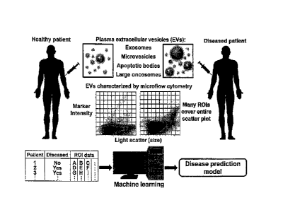
Due to the various sizes of different EVs, the goal was to separate the pFCM
data into many different
ROls, where each ROI represents the concentration of different EVs, and use
machine learning on the
ROl data to predict clinical conditions (Fig. 1). Before creating such models,
it was important to first
identify which clinical conditions the pFCM data can best predict. Automated
analysis scripts were used
to create AUG maps of the pFCM data for predicting 10 different clinical
conditions which were relevant
to the PSMA and ghrelin probes.
When averaging the highest 10% of AUCs within the LALS-PSMA, LALS-ghrelin, and
PSMA-ghrelin
AUC maps, predicting PCa grade group 5 and 4+ provided the highest averaged
AUCs (Fig. 2a).
Interestingly, all three bivariate AUCs maps provided top 10% AUCs above 0.7
for predicting these high
grade PCa with LALS-PSMA having AUCs above 0.8 for predicting grade group 5
PCa, The LALS-
PSMA AUC maps displayed an interesting pattern shift when comparing the
different PCa grade groups
(Fig. 2b). When estimating particle size using LALS, prediction of grade group
1+ displayed relatively
smaller PSMA-positive particles with AUCs above 0.5, meaning particle
concentration in these ROls in
12
CA 3003032 2018-04-27
general is higher in patients with grade group 1+ PCa, whereas larger PSMA-
positive particles mostly
displayed AUCs below 0.5, meaning particle concentration in these ROls in
general is lower in patients
with grade group 1+ PCa. The AUC maps for higher grade groups demonstrated a
progressive inversion
of this phenotype with grade group 5 PCa having AUCs >0.8 for larger PSMA-
positive particles and
AUCs approximately 0.3 for many smaller PSMA-positive particles. This
phenotype inversion became
quite noticeable with grade group 3+ AUC maps. Previous clinical trials have
shown that grade group 3
PCa patients receiving radical prostatectomy had a 10 year recurrence-free
progression of under 0.57
which was significantly lower than >0.75 for those patients with grade group 2
PCa (28). This suggests
that most men with grade group 3 PCa have metastatic disease at diagnosis
since surgical removal of
the primary tumor does not cure the patients of PCa. Without being limited by
theory, the greater
abundance of larger PSMA-positive particles in higher grade PCa patients may
be partly due to
circulating metastatic cells since larger EVs (>300 nm) from localized tumor
cells would have difficulty
intravasating into blood vessels.
Due to ghrelin's role in energy and glucose metabolism (Churm R et al., Obes
Rev 18(2): 140-148,
2017), AUC maps were created for predicting diabetes. A range of different
sized ghrelin-positive
particles displayed AUCs near 0.7, suggesting that diabetic men have EVs with
elevated levels of ghrelin
receptors (Fig. 2c).
Using the LALS-PSMA data, correlation maps were created for PSA, tumor stage,
and weight (Fig. 2d).
Relatively large particles slightly positive for PSMA demonstrated the highest
positive correlation with
PSA whereas large particles with strong PSMA positivity correlated best with
tumor stage. Such
correlations are not surprising since 1) prostate PSMA expression has been
shown to correlate with
PSA at diagnosis (Kasperzyk JL et al., Cancer Epidemiol Biomarkers Prey
22(12):2354-63, 2013), and
2) higher grade tumors are more likely to spread, explaining the similarity
between the higher grade
AUC maps and the tumor stage correlation map.
Given the results of the AUC/correlation maps, the pFCM data was used to
predict aggressive PCa
which were defined as grade group 3+ since these patients demonstrate
significantly worse outcome
than grade group 2 and lower PCa patients.
pFCM data was analyzed by manual gating to provide a benchmark of conventional
analysis. Creating
manual gates around specific particle populations is a non-trivial task since
different particle populations
exist on different patient scatter plots with some slight shifts in population
locations (Fig. 3a, b, c). For
simplicity, gates were created that grouped all marker-positive particles.
When compared to non-
aggressive PCa, only the concentration of ghrelin-positive particles was
significantly higher in
13
CA 3003032 2018-04-27
aggressive PCa by 2.1-fold (p < 0_05, Fig. 3d). The AUCs of PSMA-, ghrelin-,
and PSMA/ghrelin-positive
particle concentrations for predicting aggressive PCa were all below 0_6 (Fig.
3e). These low AUCs may
be explained by the AUC maps which show the gates encompassing particles with
AUCs above and
below 0.5 (Fig. 3a, b, c),
viSNE plots of both aggressive and non-aggressive particles together uncovered
more particle
populations than were visible with conventional scatter plots (Fig. 4a).
Particles were clustered using K-
means, expectation maximization Gaussian mixture model, and fast
search/density peaks algorithms,
and the last algorithm was the only one which could maintain large clusters
with irregular shapes (Fig.
4b and Fig. 8). Two clusters achieved >0.8 cluster purity for aggressive PCa,
suggesting that these
particle populations are in higher levels within aggressive PCa patients (Fig.
4c). Although these results
appear promising to exploit clinically, the non-reproducible nature of viSNE
requires all data to be
analyzed simultaneously. Since viSNE can only handle up to 100,000 events,
>99.99% of particles in
the 215 patient cohort would be removed from analysis.
In order to optimize the prediction of aggressive PCa from pFCM data, particle
concentrations from
ROls were used as training data for 24 different machine learning algorithms.
For LALS-PSMA, LALS-
ghrelin, and PSMA-ghrelin data sets, XGBoost provided the highest AUCs at
0.61, 0.62, and 0.66 (Fig.
5a). All subsequent analysis used the PSMA-ghrelin data set with XGBoost.
As expected for a decision tree-based model, monotonic transformations of the
pFCM data did not
improve XGBoost model performance (Fig. 9). The XGBoost variable gain map,
which displays the most
important ROls for XGBoost model accuracy, illustrated that many different
particle populations are
important for the XGBoost model (Fig. 10a). The ROls with relatively high
variable gain mostly
overlapped with regions on the AUG map that were well above and below 0,5,
suggesting that particle
populations which had higher and lower concentrations in aggressive PCa
patients were important for
the model (Fig. lob).
Changing the binning strategy to above or below 32 caused AUCs to decrease,
suggesting that this
level of resolution is preferred for predicting aggressive Pea. Creating
increasingly larger ensembles of
XGBoost models increased model performance (Hg. 5c). Compared to single
XGBoost models, an
ensemble of 100 models provided a 5% improvement in AUC and reduced model
variability by 95%.
Larger XGBoost ensembles could be made for greater model performance although
such small benefits
in accuracy would also have greater processing/memory requirements_ Grid
searching XGBoost
parameters and recursive feature elimination increased XGBoost AUCs by 3% and
5%, respectively
(Fig. 5d). Combining grid searching, feature selection, and ensembling
significantly increased the
14
CA 3003032 2018-04-27
XGBoost AUG by 12% (p < 0.05), suggesting an additive interaction between
model optimization
techniques_ Citrus and manual gating analysis of the PSMA-ghrelin data set
provided significantly lower
AUCs, 0.52 and 0.59, respectively, compared to our optimized XGBoost model at
0.75. (p < 0.05). The
present optimized XGBoost model also outperformed PSA which was the only
clinical features which
significantly differed between aggressive and non-aggressive Pea patients (p =
0.0015, Table 1).
To compare the present optimized model with SOC for predicting aggressive Pea,
logistic regression
models were created using SOC with or without our pFCM-based XGBoost model
predictions. A
waterfall plot of patient predictions from the SOC and pFCM model provided 89%
sensitivity and 49%
specificity when using a cutoff probability of 0.07332 (Fig. 6a and Table 1).
Adding SOC to pFCM
predictions slightly increased the AUC to 0.76 which was significantly greater
than the 0.68 AUC from
SOC alone (p < 0.05), demonstrating the clinical value of the pFCM-based
XGBoost model (Fig. 6b).
Table 2: Patient characteristics by Pea grade group
patienttharacteristics Grads grollp Gradegroup p-tralue- ROC ALIC Mon
iensitivity Specificity pPV NPV
by PCa grade group 3 mean (CI) (4 % (CI) %CC() Ifi (CI)
% (CI)
parents, n 088 27
Race, n (% black) a (1.6) 1(3.71 042
0.51 (0_38-0.61( - 3,7 OR54-1S) 93 (95-100) 25 (0.63-81) 86(82.92)
Family history of PC, n (%) 53 (29) (22) 0.65
0.2 (0.42-0.55) - 22(3.6-42) 71(64-78) 10(3.8-21) 86(80-91)
Previous nt.gative biopsy, n (56) 20 (I1) 1
(3,7) 0.49 054(0.42-0.45) - 3.7(0.034-19) 89 04-931 4.8(0.12-24)
88(81-91)
DRE, n abnormal) 48(25) 10(37) 0.25 MB (0.44-0.2) -
37 (18-58) 74 (67-ED) 27(84-29) 89 (83-93)
Age, yr, mean (CI) 62 (60-63) 65 (61-
68) 0.2 0.85(0.45-0,70) 53-95 39(71-53) 14(9.7.20) 13(3.9-19) 90(73-98)
PSA, fly/rat, mean (CI) 7.4
(5.2?-8.7) 15(3.8.29) 0.00/.5 0.69(0.54-0.79) 5.25 89(71-93) 42(35-49)
18(32-26) 915(90-59)
SOC score 12 (11-13) 17(11-
23) 0.0023 0.63 (0.57-0.73) 9.472 89(71-98) 30(24-37) 1.9 (10-22) 95(86-99)
Flow as store, mean (CI) 35(34-
33) 40(37-42) .c 0.0001 0.75 (0.66-0.34) 32.55 89(71-98) 40(41-50) 20(13-
28) 97(91-99)
Mow assay + SOC score, mean (CI) 11(9-
12) 24 (18-32) <0.0001 0.75 (0.67-0.86) 7.332 89 (71-93) 49 (42-56) 20(13-
23) 97(91-993
DRE, digital rectal exam; SOC, standard of care; Cl, 95% confidence interval;
ROC AUC, receiver
operator characteristic area under the CLINe; PPV, positive predictive value;
NPV, negative predictive
value;
Upon further analysis of the 215 patient cohort, it was observed that men with
enlarged prostates (>40
cc) were significantly less likely to have Pea, meaning that compared to men
with normal sized
prostates, a greater percentage of men with enlarged prostates underwent
unnecessary biopsies.
Based on current clinical practice, men primarily receive prostate biopsies
due to high PSA levels and/or
abnormal DRE. The fraction of patients with abnormal DRE was similar between
men with normal and
enlarged prostates (Fig. 6c) while PSA levels were significantly higher in men
with enlarged prostates
< 0.05, Fig. 6d), suggesting that elevated PSA was responsible for the
increased number of
unnecessary biopsies. Normalizing PSA levels using PSA density (PSA divided by
prostate volume)
may not he ideal since PSA density was significantly lower in men with
enlarged prostate (Fig. 6e). For
men with enlarged prostates, the SOC pFCM probability scores for aggressive
Pea were significantly
CA 3003032 2018-04-27
different between non-aggressive and aggressive PCa patients (p < 0.0005, Fig.
60, and using the
previously define probability cutoff threshold in Table 2, 100% and 49% of
patients with aggressive and
non-aggressive PCa would be recommended for biopsy, respectively, eliminating
approximately half of
unnecessary biopsies while still maintain 100% sensitivity for detecting
aggressive PCa (Fig. 6g).
A. Patient characteristics and sample acquisition
Pre-biopsy plasma samples were acquired from the Alberta Prostate Cancer
Research Initiative
(APCaRI) biorepository. The inclusion criteria were adult men without prior
prostate cancer diagnosis
who were: (1) referred to urology clinics in Alberta for prostate concerns and
were being scheduled for
a prostate biopsy; and (2) undergoing transurethral prostate surgery for
diagnosis or treatment of
prostate abnormalities. All patients provided written informed consent, and
the study was approved by
the scientific ethics committees at the Prostate Cancer Centre (Calgary,
Alberta, Canada) and the
Northern Alberta Urology Centre (Edmonton, Alberta, Canada). Patients were
enrolled between June
2014 and September 2015. Transrectal ultrasound guided prostate biopsies were
performed with a
median of 12 cores per patient and evaluated according to each hospital's
SOPs. Test results were not
provided to the clinical sites for patient care. Laboratory personnel who
acquired patient samples and
ran tests with them were blinded for patient characteristics. Blood was
collected and processed to collect
plasma as per institutional SOPs and time from arm to -80 C freezer was 2
hours or less.
8. pFCM assay
Frozen plasma samples were thawed, centrifuged at 16,000 x g for 30 minutes to
remove large debris
and platelet particles, and incubated with 400 pg/mL J591 antibody and 1/50
final dilution of secondary
Qdot565-conjugated donkey anti-mouse IgG antibody. Samples were also incubated
with 0,025 mM
Ghrelin Cy5 probe containing the first 18 amino adds of ghrelin. Thirty
minutes after probe incubation,
samples were diluted 100-fold in double filtered (0.22 pm) phosphate buffered
saline and analyzed with
the Apogee A50 microflow cytometer using a flow rate of 3.01 pUminute. Samples
were run for up to 2
minutes or until 5,000,000 events were recorded, whichever came first. Plasma
from each patient was
run in triplicate. Conventional manual gating analysis of pFCM data was
performed using Histogram
version 255Ø0.80 software (Apogee Flow Systems).
C. Processing pFCM data
Patient pFCM fcs files were analyzed using a custom MATLAB (version R2017a)
script. Within each fcs
file, signal intensities for all channels were log transformed and particles
with similar optical properties
were binned using 32-bins per optical property unless stated otherwise. Three
different bivariate
16
CA 3003032 2018-04-27
histograms of particle concentration were created: 1) large angle light
scatter (LALS) and PSMA stain
intensity, 2) LALS and ghrelin probe stain intensity, and 3) PSMA and ghrelin
probe stain intensity. Each
bivariate histogram contained 1024 ROls (32x32 bins). Particle concentration
in each ROI was averaged
over the three replicates per patient.
D. Predicting and correlating clinical features with pFCM data
The pFCM data was used to predict binary clinical features (e.g., patients
with or without diabetes,
normal or abnormal digital rectal exam) and correlate with ordinal or interval
clinical features (e.g., tumor
stage or PSA, respectively) using a custom MATLAB script. To minimize the code
needed for automated
analysis, an excel instruction file was created which described how the pFCM
data should be analyzed
for each clinical feature. Within the instruction file, each clinical feature
was a separate column and each
row contained specific information or instructions. Specific information
included the location of the
clinical feature within the database, the type of data for each clinical
feature (binary or ordinal/interval),
and the value which represents missing data for that clinical feature.
Instructions primarily involved how
the clinical feature should be transformed which included thresholding values
when binarizing features,
deriving the Pea grade groups from Gleason scores, and determining age from
dates of birth. Patients
missing data for the clinical feature were removed from analysis for that
clinical feature.
Once clinical feature data was retrieved from the database for all patients
and transformed, pFCM
particle concentration data for each ROI was used to predict or correlate with
clinical features. For binary
clinical features, receiver operator characteristic (ROC) area under the curve
(AUC) values were
determined for each ROI and AUC maps were generated for each bivariate data
set including LALS-
PSMA, LALS-ghrelin, and PSMA-ghrelin. For ordinal/interval clinical features.
Pearson correlation
coefficients were determined for each ROI and correlation maps were generated
for each bivariate data
set. The highest 10% of AUC values in each AUC map were averaged and these
values were compared
across clinical features.
viSNE analysis of pFCM data
viSNE plots were created using Cyt version 2.0 software run on MATLAB (25).
Each patient's triplicate
fcs files were concatenated into one fcs file. Two new fcs files were created:
one using events from
patients with grade group 2 and lower Pea (non-aggressive Pea), and the other
using events from
patients with grade group 3 and higher PCa (aggressive Pea). These two fcs
files had a total of
approximately 100,000 events with an equal number of events from each patient
within their group. With
Cyt software, 30,000 events from both of these two fcs files were randomly
subsampled and merged to
create 60,000 events which were visualized with viSNE using the bh-SNE
transformation using LALS,
17
CA 3003032 2018-04-27
PSMA, and ghrelin channels and clustered with the k-means and expectation
maximization Gaussian
mixture model algorithms. The viSNE results were exported from Cyt and also
clustered using the fast
search / density peaks algorithm using the DensityClust function for Matlab
(Rodriguez A and Laio A,
Science 344(8191):1492-6, 2014). Event pair Euclidean distances were
determined using the pd1st2
function. For setting delta and rho parameters using the paraSet function, the
percent neighbor variable
was set to 2% and a Gaussian kernel was used. Cluster centers were selected
using delta values
between 1.5 and 5 as well as rho values between 200 and 1900. For all
clustering algorithms, 248
clusters were created over the 60,000 events. Cluster purity for aggressive
PCa was defined as the
number of aggressive PCa events divided by the total number events within each
cluster. Only clusters
with at least 60 particles (0_1% of total particles) were analyzed.
F. Optimizing machine learning models for predicting aggressive PCa
MATLAB's classification learner app was used to test 23 different machine
learning algorithms to predict
aggressive PCa using particle concentration pFCM data. These algorithms
included
individual/bagged/boosted decision trees, linear/quadratic/cubic/Gaussian
support vector machines,
logistic regression, linear/quadratic/subspace discriminant analysis, and k-
nearest neighbors. XGBoost
was also tested using the `xgboosti package in R (version 3.3.3). All machine
learning algorithms used
default settings and 5-fold cross-validation repeated at least 10 times with
patient randomization
between repeats.
The machine learning algorithm with the highest AUC was then optimized by 1)
comparing 2, 4, 8, 18,
32, 64, and 128 bins when processing the pFCM data, 2) creating ensembles of
3, 6, 12, 25, 50, and
100 models using the same machine learning algorithm but randomly selecting
different subsets of
patients as training data and averaging model predictions, 3) selecting the
best subset of uFCM ROls
using recursive feature elimination with the R 'caret' package, and 4) grid
searching algorithm
parameters (XGBoost: nrounds 50, 100, 150, 200, 250, 300, 400; max_depth = 3,
4, 5, 6; eta 0,01,
0.1; gamma = 0; colsample_bytree = 1; min_child_weight = 1; subsample = 1).
The
binning/ensembling/features/parameters that provided the highest AUCs were
used together to create
a final model for predicting aggressive PCa. This model was compared to manual
gating analysis using
Histogram software and Citrus with default settings using R. Citrus predicts
clinical conditions from flow
cytometry data by using hierarchical clustering and lasso-regularized logistic
regression and nearest
shrunken centroid methods (Bruggner RV et al., Proc Natl Mad Sci USA
111(26):E2770-7, 2014).
To incorporating standard of care (SOC) clinical features, including PSA, age,
DRE, family history of
PCa, previous negative biopsy, and race (black 1, other races 0), with the
final pFCM model
18
CA 3003032 2018-04-27
probability predictions, a logistic regression model was created using all of
these features. This model
was compared to a similar logistic regression model without using pFCM data.
G. Statistical analysis
Unless stated otherwise, bar/dot plots with error bars represent mean
standard error of the mean.
When comparing 2 groups, unpaired two-tailed t-tests were used for interval
data and Fisher's exact
tests were used for contingency tables. One-way ANOVA was used for comparing 3
or more groups
using Tukey's multiple comparison test ROC curves were compared by DeLong's
method using the
'pROC' package in R. When possible, ROC cutoff values were determined using
¨90% sensitivity and
the resulting specificity and positive/negative predictive values were
determined using GraphPad Prism
version 6.01 software.
19
CA 3003032 2018-04-27