Note: Descriptions are shown in the official language in which they were submitted.
BLOOD PRESSURE MEASURING SURGICAL INSTRUMENT
CROSS-REFERENCE TO RELATED APPLICATIONS
[0001] This application claims the benefit of and priority to U.S.
Provisional Patent
Application No. 62/505,168 filed May 12, 2017, the entire disclosure of which
is incorporated
by reference herein.
BACKGROUND
1. Technical Field
[0002] The present disclosure relates to surgical instruments and, more
particularly, to
surgical instruments for grasping tissue and determining characteristics of
the grasped tissue in
preparation for performing various surgical procedures.
2. Background of Related Art
[0003] Surgical procedures sometimes involve the cutting and closure of
tissue. For
example, colorectal surgery sometimes requires anastomosis, which involves
resecting a piece
of diseased bowel tissue and creating a new connection between presumably two
healthy bowel
segments. Typically, before performing the anastomosis, the amount of tissue
to be resected
is estimated using visual indicia of the bowel. A goal of performing the
anastomosis is to
preserve as much healthy tissue as possible while at the same time removing
all of the diseased
tissue.
[0004] A risk involved in performing an anastomotic procedure is
anastomotic leaks.
The anastomotic leaks are typically caused by a failure to resect all of the
diseased tissue.
Current methods used in estimating the amount of tissue to be resected during
an anastomotic
procedure are sometimes inadequate in preventing all anastomotic leaks.
[0005] Accordingly, a need exists for surgical instruments that can sense
one or more
parameters (e.g., blood pressure) of the bowel tissue to aid a clinician in
performing a more
successful anastomotic surgical procedure.
1
CA 3003040 2018-04-27
,
SUMMARY
[0006] In one aspect of the present disclosure, an end effector for
determining blood
pressure is provided. The end effector includes a pair of jaw members. A first
jaw member of
the pair of jaw members includes a jaw body, a piston coupled to the jaw body,
a first inflatable
member coupled to the piston, and a first pressure sensor associated with the
first inflatable
member. The piston is configured to move relative to the jaw body to apply
pressure to tissue
disposed between the pair of jaw members. The first pressure sensor is
configured to detect
pressure fluctuations caused by blood flowing through tissue disposed between
the pair of jaw
members.
[0007] In some embodiments, the first pressure sensor may be further
configured to
determine a pressure within the first inflatable member.
[0008] In some embodiments, the first jaw member may further include a
second
inflatable member disposed adjacent the piston such that the second inflatable
member moves
the piston and the first inflatable member upon being inflated with an
inflation medium. The
first jaw member may further include a second pressure sensor associated with
the second
inflatable member. The second pressure sensor may be configured to determine a
pressure
within the second inflatable member.
[0009] In some embodiments, the first pressure sensor may have a first
portion in
communication with an interior of the first inflatable member, and a second
portion in
communication with an interior of the second inflatable member. The second
portion of the
first pressure sensor may be configured to determine a pressure within the
second inflatable
member.
[0010] In some embodiments, the piston may have an elongated
configuration and
define a hole having the first inflatable member captured therein.
2
CA 3003040 2018-04-27
[0011] In some embodiments, the first jaw member may further include a
housing fixed
within the jaw body. The housing may define an opening having the piston
movably disposed
therein.
[0012] In some embodiments, the piston may be configured to move relative
to the jaw
body between a first position and a second condition, in which the piston
protrudes a greater
distance relative to the jaw body than in the first position.
[0013] In another aspect of the present disclosure, a surgical instrument
for determining
blood pressure is provided. The surgical instrument includes a handle portion,
a shaft coupled
to the handle portion, and a pair of jaw members operably coupled to the
shaft. A first jaw
member of the pair of jaw members includes a jaw body, a piston coupled to the
jaw body, a
first inflatable member coupled to the piston, and a first pressure sensor
associated with the
first inflatable member. The piston is configured to move relative to the jaw
body to apply
pressure to tissue disposed between the pair of jaw members. The first
pressure sensor is
configured to detect pressure fluctuations caused by blood flowing through
tissue disposed
between the pair of jaw members.
[0014] In some embodiments, the first jaw member may further include a
second
inflatable member disposed adjacent the piston such that the second inflatable
member moves
the piston and the first inflatable member upon being inflated with an
inflation medium. The
piston may be coupled to the second inflatable member such that the piston
moves away from
the jaw body in response to an expansion of the second inflatable member and
the piston moves
toward the jaw body in response to a contraction of the second inflatable
member.
[0015] In some embodiments, the surgical instrument may further include a
processor
in communication with the first pressure sensor. The processor may be
configured to calculate
a blood pressure of tissue grasped by the pair ofjaw members based on the
pressure fluctuations
detected by the first pressure sensor.
3
CA 3003040 2018-04-27
=
[0016] It is contemplated that the surgical instrument may be
laparoscopic. The pair of
jaw members may be movable relative to one another between spaced and
approximated
positions in response to an actuation of the handle portion.
[0017] In yet another aspect of the present disclosure, a method of
determining local
blood pressure is provided. The method includes positioning tissue between a
pair of jaw
members of a surgical instrument. A piston having a first inflatable member
associated
therewith is moved relative to a jaw body of a first jaw member of the pair of
jaw members,
thereby applying pressure on the tissue with at least one of the piston or the
first inflatable
member. Pressure fluctuations in the first inflatable member are measured with
a first pressure
sensor associated with the first inflatable member. The local blood pressure
of the tissue is
determined using the measured pressure fluctuations.
[0018] Some methods may further include expanding a second inflatable
member of
the first jaw member to move the piston and the first inflatable member into
engagement with
the tissue.
[0019] Some methods may further include determining a pressure within the
second
inflatable member as the piston is being moved. The local blood pressure of
the tissue may be
determined using both the measured pressure fluctuations in the first
inflatable member and the
measured pressure within the second inflatable member.
100201 Some methods may further include moving the piston and the first
inflatable
member toward the jaw body to reduce the applied pressure on the tissue. The
first pressure
sensor may measure the pressure fluctuations as the pressure applied on the
tissue is reduced.
[0021] Some methods may further include contracting the second inflatable
member to
move the piston and the first inflatable member toward the jaw body, thereby
reducing the
pressure applied on the tissue by the piston.
4
CA 3003040 2018-04-27
[0022] These and other objects will be more clearly illustrated below by
the description
of the drawings and the detailed description of the preferred embodiments.
BRIEF DESCRIPTION OF THE DRAWINGS
[0023] The accompanying drawings, which are incorporated in and
constitute a part of
this specification, illustrate embodiments of the present disclosure and,
together with the
detailed description of the embodiments given below, serve to explain the
principles of the
disclosure.
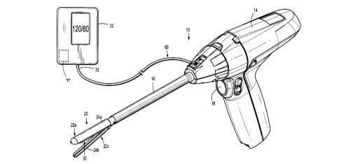
[0024] FIG. 1 is a perspective view of one embodiment of a surgical
instrument
including an end effector for grasping tissue and determining a local blood
pressure of the
grasped tissue;
[0025] FIG. 2 is an enlarged perspective view of the end effector of FIG.
1 illustrating
a blood pressure sensing assembly;
[0026] FIG. 3 is a cross-sectional view, taken along line 3-3 in FIG. 2,
of the end
effector illustrating components of the blood pressure sensing assembly;
[0027] FIG. 3A is a perspective view of the cross-section of the end
effector of FIG. 3
illustrating the blood pressure sensing assembly in an expanded configuration;
and
[0028] FIG. 4 is a cross-sectional view of another embodiment of an end
effector for
grasping tissue and determining a local blood pressure of the grasped tissue.
DETAILED DESCRIPTION
[0029] Embodiments of the presently disclosed surgical instruments and
end effectors
thereof will now be described in detail with reference to the drawing figures
wherein like
reference numerals identify similar or identical elements. As used herein and
as is traditional,
the term "distal" will refer to that portion which is further from the user
while the term
"proximal" will refer to that portion which is closer to the user.
CA 3003040 2018-04-27
,
'
'
[0030] The present disclosure is directed to a tissue grasper for
measuring blood
pressure in tissue, such as bowel tissue, using the oscillometric method. The
tissue grasper
includes a pair of jaw members for grasping tissue therebetween, a first
balloon member
disposed within a movable piston, and a second balloon member. The second
balloon member
is expanded to force the piston and the first balloon member into engagement
with tissue to
apply pressure on the grasped tissue. The first balloon member includes a
sensor that detects
pressure fluctuations (i.e., oscillations) caused by pressure pulses from
blood flowing through
the grasped tissue, and the second balloon member includes a sensor that
measures the air
pressure within the second balloon member. The second balloon member is first
inflated to a
pressure at which occlusion of blood flow through the tissue occurs. The
second balloon
member is gradually deflated to reduce the amount of pressure applied on the
tissue. As the
pressure applied is reduced to at or below the occlusion pressure, the first
pressure sensor senses
the oscillations created by the pulsatile flow of blood through the grasped
tissue, and the second
sensor measures the air pressure within the second inflatable member. The
blood pressure may
be calculated using the measured oscillations in air pressure in the first
balloon member and
the measured pressure in the second balloon member.
[0031] The basis for the oscillometric method of measuring blood
pressure is disclosed
in U.S. Pat. Nos. 4,349,034 and 4,360,029 both to Ramsey, III, the entire
contents of which are
incorporated by reference herein. As disclosed by Ramsey, III, the
oscillometric method of
measuring blood pressure includes applying an inflatable cuff around an
extremity of a
patient's body and inflating the cuff to a pressure that exceeds the patient's
systolic pressure
such that no blood flows through the artery of the tissue (i.e., the artery is
occluded). The
pressure within the cuff is incrementally reduced while a pressure sensor in
communication
with the interior of the cuff tracks the pressure therein. As the cuff is
deflated below the systolic
pressure, blood begins to flow through the artery creating vibrations or
pulses in the arterial
6
CA 3003040 2018-04-27
wall that are transferred to the cuff causing slight pressure variations
within the cuff. These
pressure variations within the cuff are detected by the pressure sensor. The
pressure sensor
produces an electrical signal which represents the pressure within the cuff
throughout the
measurement process, and the sensed pulsatile vibrations at each discreet
pressure within the
cuff. These pulsatile vibrations are called "oscillation complexes" or
"oscillations."
[0032] Local blood pressure may be estimated based on the oscillation
complexes
measured by the pressure sensor. For example, peak pulse amplitudes ("PPA")
may be
determined for each oscillometric complex. The PPA increases as the cuff
pressure is reduced
until a peak amplitude is reached. Once the peak amplitude is reached, the PPA
begins to
decrease with further reductions in cuff pressure. The cuff pressure at which
the oscillations
have a maximum value is representative of the patient's mean arterial pressure
("MAP"). The
systolic and diastolic pressures can be derived either as predetermined
fractions of MAP, or by
more sophisticated estimating techniques using direct processing of the
oscillation complexes.
[0033] FIGS. 1-3A illustrate a surgical instrument 10 for measuring local
blood
pressure of tissue grasped between a pair of jaw members 22a, 22b of the
surgical instrument
using the oscillometric method. In embodiments, the surgical instrument 10 may
be
configured to measure blood pressure using other methods, such as, for
example, the
auscultatory method. The surgical instrument 10 may include a visual display
unit 12 for
displaying the blood pressure determined by the surgical instrument 10.
[0034] The surgical instrument 10 generally includes a handle portion 14,
an elongated
shaft 16, and an end effector 20. The handle portion 14 of the surgical
instrument 10 may be
power-operated or manually-operated. An actuation of a switch or button 18 of
the handle
portion 12 is configured to effect closing of the jaw members 22a, 22b of the
end effector 20
to grasp tissue disposed between the jaw members 22a, 22b. The handle portion
14 may include
7
CA 3003040 2018-04-27
a processor for transforming an actuation of the switch 18 into a closing of
the jaw members
22a, 22b.
[0035] The end effector 20 is detachably coupled to a distal portion of
the elongated
shaft 16 or, in some embodiments, may be fixedly coupled to the distal portion
of the elongated
shaft 16. The end effector 40 includes the pair of opposing jaw members 22a,
22b which are
movable between spaced and approximated positions. A first jaw member 22a of
the end
effector 20 includes a jaw body 24a fixedly coupled to the distal portion of
the elongated shaft
16, and a second jaw member 22b of the end effector 20 includes a jaw body 24b
pivotably
coupled to the distal portion of the elongated shaft 16. In embodiments, one
or both of the jaw
bodies 24a, 24b may be movably coupled to the distal portion of the elongated
shaft 16.
[0036] The first jaw member 22a includes a blood pressure sensing
assembly 30 housed
within the jaw body 24a thereof. In embodiments, the blood pressure assembly
30 may be
housed within the jaw body 24b of the second jaw member 22b rather than the
first jaw member
22a. The blood pressure assembly 30 is in communication with a processor "P"
disposed within
the visual display unit 12. In some embodiments, the processor "P" may instead
be disposed
within the handle portion 14 of the surgical instrument 10. The processor "P"
may be operably
connected to a memory, which may include transitory type memory (e.g., RAM)
and/or non-
transitory type memory (e.g., flash media, disk media, etc.). The processor
"P" may include
software for running a blood pressure measurement sequence. Those skilled in
the art will
appreciate that the processor "P" may be substituted by using any logic
processor (e.g., control
circuit) adapted to perform the calculations and/or set of instructions
described herein
including, but not limited to, field programmable gate arrays, digital signal
processor, and
combinations thereof
[0037] With reference to FIGS. 2, 3, and 3A, the blood pressure sensing
assembly 30
generally includes a piston or block 36, two inflatable members 50a, 50b, and
two sensors 60a,
8
CA 3003040 2018-04-27
0
60b. The first jaw member 22a includes a piston housing or piston sleeve 32
fixedly disposed
within a cavity 26 defined in the jaw body 24a. The piston housing 32 defines
an elongate
channel 34 having the piston 36 disposed therein. The piston 36 is movable
within the elongate
channel 34 of the housing 32. The piston housing 32 may include a pin 38
projecting into the
elongate channel 34 thereof. The pin 38 extends into a slot 40 defined
partially through a distal
end of the piston 36 to prevent the piston 36 from dislodging from the piston
housing 32. In
particular, as the piston 36 moves or slides away from the jaw body 24a in a
direction indicated
by arrow "A" in FIG. 3, the pin 38 of the housing 32 rides in the slot 40 of
the piston 36 until
the pin 38 contacts an end wall of the slot 40 of the piston 36, thereby
preventing any further
travel of the piston 36.
[0038] The piston 36 has a substantially rectangular
configuration, but in some
embodiments, the piston 36 may assume any suitable shape, such as, for
example, square,
triangular, oblong, circular, or the like. The piston 36 defines a planar
tissue-contacting surface
42a that is coplanar with a tissue-oriented surface 33 of the housing 32 when
the piston 36 is
in a retracted position, as shown in FIG. 2. When the piston 36 protrudes from
the housing 32,
the tissue-contacting surface 42a of the piston 36 is disposed upward of or
above the tissue-
oriented surface 33 of the housing 32. The tissue-contacting surface 42a of
the piston 36
defines a hole 44 therein dimensioned for receipt of the first inflatable
member 50a of the blood
pressure sensing assembly 30.
[0039] The first inflatable member 50a is fixedly disposed within
the hole 44 of the
piston 36 such that the first inflatable member 50a moves with the piston 36
as the piston 36
slides relative to the jaw body 24a between retracted and deployed states. The
first inflatable
member 50a has a disc or saucer-shaped main body 52 and proximal and distal
tubes 54a, 54b
extending from opposite ends of the main body 52. In embodiments, the main
body 52 of the
first inflatable member 50a may assume a variety of shapes, such as, for
example, cylindrical,
9
CA 3003040 2018-04-27
=
rectangular, or the like. The main body 52 of the first inflatable member 50a
has a top or outer
surface 56 that is substantially flush with the tissue-contacting surface 42a
of the piston 36. In
some embodiments, the top surface 56 of the main body 52 of the first
inflatable member 50a
may protrude outwardly of the tissue-contacting surface 42a of the piston 36.
[0040] The first inflatable member 50a may be fabricated from a
biocompatible
material such as natural or synthetic elastomers, natural or synthetic rubbers
and silicone
materials, and/or compliant polyurethanes. The main body 52 of the first
inflatable member
50a defines a hollow inner chamber or void 58 that receives an inflation media
that changes or
moves the main body 52 of the first inflatable member 50a from a collapsed
configuration to
an expanded configuration. The proximal tube 54a may extend proximally through
the
elongated shaft 16 (FIG. 1) to couple to a pump (not shown) disposed in the
visual display unit
12 for adjusting the amount of inflation media in the first inflatable member
50a and, in turn,
the pressure in the first inflatable member 50a. In other embodiments, the
first inflatable
member 50a may not be connected to the pump such that the first inflatable
member 50a has a
fixed amount of inflation media therein.
100411 The first inflatable member 50a further includes a pressure sensor
60a disposed
in the proximal tube 54a for sensing air pressure fluctuations (e.g.,
oscillations) within the first
inflatable member 50a. In embodiments, the pressure sensor 60a may be disposed
in any
suitable location within the first inflatable member 50a, such as the main
body 52. The pressure
sensor 60a may be a piezoelectric transducer, a piezoresistive strain gauge,
an electromagnetic
optical sensor, or any other suitable pressure sensor that detects pressure
variations within the
first inflatable member 50a caused by pulse waves generated by the pulsatile
flow of blood
through an artery (e.g., oscillations).
[0042] In embodiments, in addition to the pressure sensor 60a being
configured to sense
oscillations, the pressure sensor 60a may also be configured to measure the
pressure (e.g., air
CA 3003040 2018-04-27
4
pressure) within the first inflatable member 50a. In other embodiments, the
first pressure
sensor 60a may be a perfusion sensor, for example, a Doppler flow sensor,
configured to
measure local perfusion (e.g., blood flow) through tissue grasped by the jaw
members 22a, 22b.
The pressure sensor 60a may measure perfusion of the grasped tissue on the
basis of known
techniques, such as Laser-Doppler Flowmetry ("LDF"), measuring light
scattering, and/or
measuring absorption of light from one or more LED's or other light sources.
For a detailed
description of LDF technology, reference may be made to U.S. Patent Nos.
4,109,647 and
4,862,894, the entire contents of each of which are incorporated by reference
herein.
[0043] In still other embodiments, instead of the first inflatable member
50a having a
pressure sensor, the first inflatable member 50a may include a stethoscope
head, an ultrasound
pickup, or a microphone for receiving auditory signals generated by blood
flowing through
tissue. It is contemplated that any of these sensors may be disposed within
the first inflatable
member 50a, on the first inflatable member 50a, on the tissue-contacting
surface 42a of the
piston 36, or at any other suitable location of the end effector 20.
[0044] The first pressure sensor 60a is in communication, via lead wires
or wireless
connection, with the processor "P" of the visual display unit 12, which
receives the pressure
fluctuation data collected by the first pressure sensor 60a. Upon the first
pressure sensor 60a
measuring the pressure oscillations in grasped tissue, the first pressure
sensor 60a transmits the
data to the processor "P." In some embodiments, the first pressure sensor 60a
may also be in
communication, via lead wires or wireless connection, with a computing device
or processor
(not shown) such as an oscilloscope, which processes the information collected
by the first
pressure sensor 60a. The computing device (e.g., an oscilloscope) may also be
in
communication, via lead wires or wireless connection, with the visual display
unit 12 to send
the processed information related to the blood pressure to a display screen so
that the display
screen can display the blood pressure.
11
CA 3003040 2018-04-27
=
[0045] With continued reference to FIGS. 2, 3, and 3A, the second
inflatable member
50b of the blood pressure sensing assembly 30 has a main body 64 and a tube 66
extending
from a proximal end of the main body 64. The main body 64 of the second
inflatable member
50b defines a hollow inner chamber or void 68 that receives an inflation media
(e.g., air) that
changes or moves the main body 64 of the second inflatable member 50a from a
collapsed
configuration, in which the main body 64 is substantially flat and
rectangular, to an expanded
configuration, in which the main body 64 is larger than in the collapsed
configuration and
assumes a bulbous configuration. In some embodiments, the main body 64 of the
second
inflatable member 50b may be configured to assume any suitable shape when in
the expanded
configuration, such as, for example, rectangular, dome-shaped, elliptical,
oblong, tubular,
square, triangular, cylindrical, rod-shaped, or the like. The tube or hose 66
of the second
inflatable member 50b is in fluid communication with the hollow inner chamber
68 of the main
body 64 and may extend proximally from the main body 64 through the elongated
shaft 16 and
out of the surgical instrument 10. The tube 66 may have an end 70 (FIG. 1)
coupled to a source
of inflation media, such as, for example, a pump (not explicitly shown), for
delivering a liquid
and/or gas into the hollow inner chamber 68 of the second inflatable member
50b.
[0046] The main body 64 of the second inflatable member 50b is captured
within the
cavity 26 of the jaw body 24a and may be fixed at its distal end to a distal
end of the jaw body
24a. The main body 64 of the second inflatable member 50b is disposed below or
under the
piston 36 and in abutting engagement with a bottom surface 42b of the piston
36. As such, an
expansion of the second inflatable member 50b effects movement or sliding of
the piston 36
and the attached first inflatable member 50a away from the jaw body 24a and
toward clamped
tissue in the direction indicated by arrow "A" in FIG. 3. In embodiments, the
first inflatable
member 50a may extend below the bottom surface 42b of the piston 36 to contact
a top surface
62 of the second inflatable member 50b.
12
CA 3003040 2018-04-27
. . ,
<
[0047] The top surface 62 of the second inflatable member 50b may
be fixed to the
bottom surface 42b of the piston 36 such that contraction of the second
inflatable member 50b
retracts the piston 36 and the first inflatable member 50a toward the jaw body
24a in the
direction indicated by arrow "B" in FIG. 3. The top surface 62 of the second
inflatable member
50b may be attached to the bottom surface 42b of the piston 36 and/or the
first inflatable
member 50a via an adhesive, a hook and loop fastener, a suture, or the like.
In other
embodiments, instead of the first and second inflatable members 50a, 50b being
attached to
one another to facilitate retraction of the first inflatable member 50a, a
biasing member (not
shown) may be provided that resiliently biases the piston 36/first inflatable
member 50a toward
a retracted position within the jaw body 24a such that even in the absence of
an outward
pressure applied on the piston 36/first inflatable member 50a by the second
inflatable member
50b, the piston 36/first inflatable member 50a are still biased toward the
retracted state.
[0048] The second inflatable member 50b further includes a second
pressure sensor
60b (e.g., a piezoresistive pressure sensor, a capacitive pressure sensor, a
MEMS device, etc.)
disposed within the proximal tube 66 thereof. In embodiments, the second
pressure sensor 60b
may instead be disposed within the main body 64 of the second inflatable
member 50b. The
second pressure sensor 60b is configured to measure the pressure (e.g., air
pressure) within the
second inflatable member 50b.
[0049] Since the pressure within the second inflatable member 50b
is responsible for
forcing the piston 36 and the first inflatable member 50a into engagement with
tissue, the
pressure within the second inflatable member 50b is substantially similar to
and/or directly
correlated with the pressure experienced on the arteries within tissue gasped
between the jaw
members 22a, 22b. Thus, by knowing the pressure within the second inflatable
member 50b,
via the second pressure sensor 60b, the pressure applied on grasped tissue by
the piston 36/first
inflatable member 50a will be known. After the second pressure sensor 60b
measures the
13
CA 3003040 2018-04-27
clamping pressure applied to the grasped tissue, the second pressure sensor
60b transmits the
measurement data to the processor "P," which together with the pressure
fluctuation data
determined by the first pressure sensor 60a, calculates blood pressure and
displays the
measurement on the display screen of the visual display unit 12, as will be
described in further
detail below.
[0050] In operation, the surgical instrument 10 may be used in a surgical
procedure in
which tissue is to be stapled, for example, an anastomotic surgical procedure,
to gather various
data about the subject tissue prior to effecting stapling. In some anastomotic
surgical
procedures, unhealthy or diseased bowel tissue is resected and the ends of the
remaining
healthy segments of bowel are stapled together to recreate a continuous bowel.
Prior to stapling
the ends of the separate bowel segments to one another, the viability of the
ends of the separate
bowel segments should be assessed in order to predict the likelihood of post-
surgery
anastomotic leaks or other adverse outcomes. It has been found that local
blood pressure of
bowel segments is an indicator of tissue viability. Accordingly, a clinician
may make use of
the blood pressure measuring surgical instrument 10 of the present disclosure
to aid in making
this viability assessment.
[0051] In use of the surgical instrument 10, the end effector 20 of the
surgical
instrument 10 is positioned through an access port (not shown) to gain entry
to a surgical site
in a minimally invasive manner. With the second inflatable member 50b of the
blood pressure
sensing assembly 30 in a collapsed or substantially un-inflated state, tissue
is disposed between
the tissue-contacting surface 42a of the piston 36 of the first jaw member 22a
and the tissue
contacting surface of the second jaw member 22b.
[0052] With the tissue disposed between the jaw members 22a, 22b, the
pump of the
visual display unit 12 conveys an inflation media (e.g., air) into the hollow
inner chamber 68
of the second inflatable member 50b via the tube 66 to expand the second
inflatable member
14
CA 3003040 2018-04-27
S . .. '
50b, as shown in FIG. 3A. As the second inflatable member 50b expands, the
second inflatable
member 50b applies an upward-oriented force on the piston 36 to raise the
piston 36 relative
to the jaw body 24a of the first jaw member 22a in the direction indicated by
arrow "A." Since
the first inflatable member 50a is captured within the piston 36, the first
inflatable member 50a
rises with the piston 36 relative to and away from the jaw body 24a to apply
pressure on the
grasped tissue.
[0053] Continued expansion of the second inflatable member 50b increases
the
distance the piston 36/first inflatable member 50a projects from the jaw body
24a and, in turn,
increases the clamping pressure on the tissue. The processor "P" may be pre-
programmed to
expand the second inflatable member to a threshold pressure known to occlude
an artery (e.g.,
a pressure that exceeds the systolic pressure of any patient).
[0054] Upon reaching the threshold pressure, the inflation media (e.g.,
air) is gradually
removed from the second inflatable member 50b in incremental steps to contract
the second
inflatable member 50b. As the second inflatable member 50b contracts, the
piston 36 and the
associated first inflatable member 50a retract back toward the jaw body 24a of
the first jaw
member 22a, in the direction indicated by arrow "B," to reduce the clamping
pressure on the
tissue. The surgical instrument 10 may be pre-programmed to reduce the
clamping pressure at
a predetermined rate via deflation of the second inflatable member 50b.
100551 As the pressure applied to the grasped tissue is gradually reduced,
the first
pressure sensor 50a continuously monitors any pressure fluctuations (e.g.,
oscillations)
generated by blood flowing through the arteries in the grasped tissue. Before
the clamping
pressure drops below the systolic pressure of the patient, the first pressure
sensor 60a should
not detect any oscillations since no blood is flowing through the arteries at
this clamping
pressure. However, the artery may be emitting slight percussion pulses due to
the blood hitting
the occluded artery in pulses. These slight percussive pulses are so low in
force that the
CA 3003040 2018-04-27
f ' '
vibrations they induce are absorbed by the movable piston 36, thereby damping
their impact
on the first pressure sensor 60a. In this way, these damped percussive pulses
will be too small
for the first pressure sensor 60a to detect, which may otherwise be confused
by the processor
"P" as oscillations from flowing blood rather than percussion pulses from an
occluded artery.
[0056] The moment the pressure applied on the tissue by the end
effector 20 falls below
the systolic pressure of the patient, blood begins to flow through the clamped
tissue and will
produce the oscillations described above. The first pressure sensor 60a in the
first inflatable
member 50a detects and measures these oscillations and transfers the
measurement data to the
processor "P." While the clamping pressure is gradually reduced, the second
pressure sensor
60b in the second inflatable member 50b continuously monitors the pressure
within the second
inflatable member 50b and sends this pressure measurement data to the
processor "P."
10057] Reduction of the clamping pressure, via deflation of the
second inflatable
member 50b, is continued until the pressure within the second inflatable
member 50b falls
below a threshold pressure corresponding to the diastolic pressure of any
patient. In
embodiments, instead of gradually decreasing the clamping pressure on the
tissue by deflating
the second inflatable member 50b, the jaw members 22a, 22b may be gradually
pivoted away
from one another. The processor "P" uses the data collected by the first and
second pressure
sensors 60a, 60b to compute the local blood pressure in the grasped tissue
using any suitable
algorithm. In embodiments, the processor "P" may be configured to compute the
blood
pressure from the measurements made by the first and second pressure sensors
60a, 60b using
the process described in U.S. Patent No. 4,360,029, the entire contents of
which are
incorporated by reference herein. Upon the surgical instrument 10 calculating
the blood
pressure, the visual display unit 12 displays the calculated blood pressure on
the display screen
for the clinician to view.
16
CA 3003040 2018-04-27
4 ' .
[0058] The blood pressure determined using the above-noted
technique may be used to
assess the viability of the gasped tissue by, for example, comparing the
measured local blood
pressure with a known local blood pressure associated with healthy or viable
tissue.
Additionally or alternately, the measured local blood pressure may be used in
combination with
other measurements, for example, a systemic blood pressure reading, to aid in
making the
determination of the viability of the tissue. The systemic blood pressure may
be taken using
any suitable device, for example, a blood pressure cuff, applied to any
suitable body portion of
the patient, for example, an arm of the patient. An index may be calculated by
taking a ratio
of the local blood pressure measured by the surgical instrument 10 and the
systemic blood
pressure taken using the blood pressure cuff The index may be calculated by
the computing
device in the visual display unit 12 and displayed as a number on the display
screen.
[0059] The calculated index may be predictive of whether an
anastomotic leak may
occur and/or the grade of an anastomotic leak. As such, a clinician can use
the index to make
a determination on whether the two ends of the presumed healthy bowel segments
are healthy
enough to be stapled together or whether more tissue needs to be resected. For
example, the
calculated index may be compared to a known index that is associated with
healthy tissue.
[0060] In some embodiments, the surgical instrument 10 may not
include the display
12, and instead, the surgical instrument 10 may be configured to be connected
to or be in
communication with another type of display, for example, a tablet, a cell
phone, a computer
monitor, a laptop, or any suitable display device. The surgical instrument 10
may be connected
to any of the aforementioned display devices via USB wires, Wi-Fi, or the
like. In other
embodiments, the visual display unit 12 may be integrated into the handle
portion 14 of the
surgical instrument 10 rather than being an auxiliary component.
[0061] In some embodiments, the second inflatable member 50b may
be replaced with
a powered actuator (e.g., a pusher, a sled, a screw, etc.) operably coupled to
the piston 36 to
17
CA 3003040 2018-04-27
selectively raise the piston 36 and the first inflatable member 50a relative
to the jaw body 24a.
The motorized actuator may be associated with a pressure sensor that senses
the amount of
pressure applied to the piston 36 by the motorized actuator. The pressure
sensor may be
disposed on one or both of the tissue-contacting surfaces of the first and
second jaw members
22a, 22b. In this alternate embodiment, it is also contemplated that the first
inflatable member
50a may include each of the first and second pressure sensors 60a, 60b.
[0062] The surgical instrument 10 or components thereof may be configured
to be
incorporated into a robotic surgical system (not shown). The robotic surgical
system is
powered locally or remotely, and has electronic control systems localized in a
console or
distributed within or throughout the robotic surgical system. The robotic
surgical system
permits a clinician to remotely manipulate the surgical instrument 10 to more
precisely control
the movement of the surgical instrument 10. The surgical instrument 10 may be
configured to
send the measurements gathered by the first and second pressure sensors 60a,
60b of the end
effector 20 to an interface of the robotic surgical system on which the
measurements may be
displayed for the clinician to read.
[0063] With reference to FIG. 4, another embodiment of an end effector
120 is
provided. The end effector 120 is similar to the end effector 20 described
with reference to
FIGS. 1-3A, and will therefore only be described with the detail necessary to
elucidate any
differences. The end effector 120 includes a pair of opposing jaw members
122a, 122b and a
blood pressure sensing assembly 130 disposed in a first jaw member 122a of the
pair of jaw
members 122a, 122b.
[0064] Similar to the pressure sensing assembly 30 described above, the
pressure
sensing assembly 130 of the presently described embodiment includes a piston
136 movably
disposed within a housing 132, and first and second inflatable members 150a,
150b. However,
instead of each of the first and second inflatable members 150a, 150b having
discreet pressure
18
CA 3003040 2018-04-27
=
sensors, a dual-function pressure sensor 160 is provided that extends through
each of the first
and second inflatable members 150a, 150b.
[0065] In particular, the dual-function pressure sensor 160 includes a
first portion 160a
disposed within the first inflatable member 150a, and a second portion 160b
extending within
the second inflatable member 150b. The first portion 160a of the pressure
sensor assembly 160
is configured as a first pressure sensor (e.g., a piezoelectric transducer, a
piezoresistive strain
gauge, an electromagnetic optical sensor, etc.) that detects pressure
variations within the first
inflatable member 150a caused by pulse waves generated by the pulsatile flow
of blood through
an artery (e.g., oscillations). The second portion 160b of the dual-function
pressure sensor 160
is configured as a second pressure sensor (e.g., a piezoresistive pressure
sensor, a capacitive
pressure sensor, a MEMS device, etc.), which measures the pressure (e.g., air
pressure) within
the second inflatable member 150b. The end effector 120 may determine blood
pressure in
grasped tissue using the measurements taken by the sensor 160 in a similar
manner as that
described above with respect to the end effector 20.
[0066] Although the illustrative embodiments of the present disclosure
have been
described herein, it is understood that the disclosure is not limited to those
precise
embodiments, and that various other changes and modifications may be affected
therein by one
skilled in the art without departing from the scope or spirit of the
disclosure. For example,
while described with respect to a grasper, it is envisioned that a blood
pressure sensing
assembly in accordance with the present disclosure may be incorporated into
other surgical
instruments, such as, for example, surgical staplers. All such changes and
modifications are
intended to be included within the scope of the disclosure.
19
CA 3003040 2018-04-27