Note: Descriptions are shown in the official language in which they were submitted.
CA 03003232 2018-04-25
WO 2017/072761
PCT/1L2016/051153
- 1 -
SYSTEMS FOR THROMBECTOMY
TECHNOLOGICAL FIELD
The present disclosure relates to anchoring and retrieval of a corpus in an
organ
of a subject, in particular narrow tubular organs such as small blood vessels.
BACKGROUND ART
References considered to be relevant as background to the presently disclosed
subject matter are listed below:
[1] Nogueira et al., AJNR 2009, 30, 649-661
[2] Grunwald et al. The American Journal of Neuro radiology 2011, 32, 238-
243
[3] Mordasini et al. The eJoumal of the European Society of minimally
invasive
Neurological Therapy, 2012: 1238000077
[4] US 7,766,921
[5] US 8,715,227
[6] US 6,685,722
[7] W02013/054324
[8] Gralla et al., Am J Neuroradiol 2006, 27, 1357-1361
[9] W02011/130256
[10] Gory et al., Am J Neuroradiol 2013, 34, 2192-2198
[11] Levy et al., Am J Neuroradiol 2006, 27, 2069-2072
Acknowledgement of the above references herein is not to be inferred as
meaning that these are in any way relevant to the patentability of the
presently disclosed
subject matter.
BACKGROUND
The removal of blood clots and plaque from blood vessels by use of minimally
invasive procedures is nowadays a well-established practice. A stroke event
associated
with a blood clot occurs as a result of disturbance in the blood vessels
supplying blood
CA 03003232 2018-04-25
WO 2017/072761 PCT/1L2016/051153
- 2 -
to the brain, leading to sudden death of brain cells. This can be due to
ischemia (lack of
glucose and oxygen supply) caused by thrombosis (-80% of strokes) or due to a
hemorrhage (-20% of strokes). The annual prevalence of stroke is estimated to
be 15
million people worldwide and it is one of the leading causes of death (-10% of
all
deaths) and long-term disability. Furthermore, stroke is one of the most
costly health
problems in America and the Western world, with estimated direct and indirect
costs of
$38.6 billion annually. The majority of the damage caused by a stroke is due
to
secondary stroke damage which threatens the functionally of the impaired
region that
surrounds the infarct core; the ischemic penumbra. Early medical intervention
(for re-
canalization) can inhibit this process and reduce the risk for irreversible
neurological
damage.
The goal of treatment for stroke resulting from thrombus remains the same:
safe
and rapid re-establishment of oxygenated blood flow to the affected tissue.
Guidelines
and protocols for the treatment of ischemic stroke are, for example, those
published by
the American Society of Neurology and the American Society of Neurosurgeons or
The
European Stroke Organization (ESO). More specifically, the pharmacologic
standard of
care for ischemic stroke patients to date is by intravenous (IV) tissue
plasminogen
activator (rt-PA). Improvement in re-canalization rate may be achieved when rt-
PA is
used intra-arterially (IA) within 6 hours of symptom onset, in patients with
occlusions
in a large-vessel (e.g., middle cerebral artery), or patients who have
contraindications
for the use of IV thrombolysis. However, this treatment may increase the risk
for
intracranial hemorrhage and is currently not approved for use worldwide.
Beyond the
failure rates of thrombolytic therapy, it is also limited in the time window
for treatment
and indicated population. Therefore, in patients who have either failed IV rt-
PA therapy
or who are either ineligible for or have contraindications to IV rt-PA use, or
are out of
the therapeutic window when medical support can be initiated,
neurothrombectomy
devices have been used for the re-establishment of blood flow.
Various mechanical approaches to fragment or retrieve clots have been utilized
and reported in the clinical literature. These include, inter alia,
endovascular
(intracranial) thrombectomy, endovascular thromboaspiration, mechanical
thrombus
disruption and thrombus entrapment devices [1-6]. Intracranial thrombectomy
may
provide rapid flow restoration with a potentially lower likelihood of clot
fragmentation
CA 03003232 2018-04-25
WO 2017/072761 PCT/1L2016/051153
- 3 -
and distal embolism, lessens and even preclude the use of chemical
thrombolytics - thus
reducing the risk of neurotoxicity and intracranial hemorrhage. By avoiding
the use of
chemical thrombolytics, it could be possible to extend the treatment window to
8 hours
and beyond. In addition, re-canalization occurs without the disruption of the
blood-
brain-barrier. For example, some systems are based on deployment of devices in
a
collapsed state, that are expanded for retrieval of the blood clot once
inserted into the
blood vessel [4]. Others comprise a plurality of strands, and have contracted
and
expanded configurations 115-7, 9].
Due to the variability in the properties of blood clots, many of the devices
described in the art are suitable for extraction of a specific type of clots.
Moreover, in
most cases the devices are designed to provide support for the artery as well
as function
to provide embolic protection, thereby necessitating direct contact with the
internal face
of the blood vessel. Such contact often causes additional damage to the blood
vessel
when the device is manipulated and moved within the vessel during the
different stages
of the procedure. Thus, there is a need for a device allowing extraction of
various clots
from a variety of blood vessels, reducing the risk of clot disintegration,
while providing
greater operational flexibility and minimal blood vessel damage.
GENERAL DECRIPTION
The present disclosure relates to a medical system, kits and methods for
retrieval
and/or extraction of a corpus located in a tubular organ. Thus, the system of
this
disclosure is suitable for carrying out various procedures for removal of
occlusive
corpus from tubular organs. An exemplary procedure may be thrombectomy (i.e.
removal of blood clots), typically in narrow blood vessels such as, but not
limited to,
those existing in the brain, by anchoring the device into the corpus in a
manner
permitting its extraction from the blood vessel without significant fracturing
of the
corpus or without significantly damaging the blood vessel.
In the context of the present disclosure, the term corpus encompasses blood
clots, plaque, cholesterol layers, thrombus, naturally-occurring foreign
bodies (e.g.
tissue portions trapped within or adhered to the inner face of the tubular
organ), non
CA 03003232 2018-04-25
WO 2017/072761 PCT/1L2016/051153
- 4 -
naturally-occurring foreign bodies (e.g. non-biological objects trapped
within, adhered
to or penetrating through the tubular organ), and the like.
The term tubular organ means to encompass any anatomical lumen of a subject
to be treated that enables flow of a bodily-fluid therethrough. The organ may
be a blood
vessel (a vain, an artery, micro blood vessels, etc.), or a non-vascular
anatomical organ,
such as fallopian tubes, urinary tract (e.g. ureter, urethra, kidneys),
biliary tract (bile
ducts), gastrointestinal tract, airways and any other anatomical lumen in
which partial or
full blockage may occur.
In one of its aspects this disclosure provides a medical system for anchoring
into
at least one corpus located in a tubular organ, the system comprising a
handling and
manipulation apparatus (HMA) and a corpus anchoring unit operable thereby. The
HMA is configured for manipulating the corpus anchoring unit into engagement
with
the corpus, and once in proximity to the corpus the anchoring unit is
manipulated into
operation by the HMA.
The corpus anchoring unit comprises a deployment wire defining a proximal-
distal axis, at least two generally cylindrical elongated bodies that are
spaced apart
along said deployment wire, and at least two axially displaceable tip tools.
Each
cylindrical body is constituted by at least one, typically a plurality of,
wound coiled
threads that form the cylindrical structure of the cylindrical body; each such
cylindrical
body has a proximal end and a distal end. The at least one wound coiled thread
(and
hence also the cylindrical body) has a deployment state, in which it is coiled
and wound
to form the cylindrical shape of the cylindrical body (i.e. forming the
general shape of a
tube) - each of said cylindrical bodies having a fixed end at either the
proximal or distal
end. The coiled wound threads are held at the cylindrical body's fixed end one
against
the other, to prevent the threads' deployment at the fixed end. The opposite
end of the
cylindrical body is a free end, that is configured for deploying the wound
coiled threads
(and hence also the cylindrical body) from their deployment state into at
least one
deployed state. In the deployed state, each of the threads unwinds in the
general radial
direction while tracing, during deployment, a generally helical path. Thus,
during
deployment, the free end of each of the unwinding coiled threads moves in a
general
screw-like movement, thereby anchoring the unwound threads into the corpus.
CA 03003232 2018-04-25
WO 2017/072761 PCT/1L2016/051153
- 5 -
A tip tool (of said at least two axially displaceable tip tools) is located
such that
it is associated with one of the proximal or distal ends of the tip's
corresponding
cylindrical body. Namely, each cylindrical body has an associated tip tool,
which is
associated either with the proximal end or with the distal end of the
cylindrical body.
The number of the tip tools corresponds to the number of the cylindrical
bodies. The
tips tools are mounted onto the deployment wire and are axially displaceable
thereby.
The tip tool is configured to unwind at least one coiled thread, at times all
of the coiled
threads simultaneously, from its corresponding cylindrical body upon axial
displacement of the tip tool, such that the wound coiled thread is unwound
from its
deployment state to at least one deployed state.
While each coiled thread has one deployment state, in which it is wound to
form
the cylindrical body, the thread may have several deployed states in which it
gradually
unwinds from the cylindrical body. The transition between the deployed states
(i.e. the
extent of deployment of the cylindrical body) occurs upon axial displacement
of the tip
tool and the relative positions of the tip tool and its associated cylindrical
body along
the deployment wire.
Unlike some of the thrombectomy devices known in the art, the anchoring unit
of a system of this disclosure does not merely forms a net or a mesh of
deployed wires
that form physical barriers in the blood vessel and by that permits the
retrieval of the
corpus, but rather the corpus anchoring unit of the system of this disclosure
anchors (i.e.
penetrates into) the corpus at various locations thereof. Thus, and as also
explained
herein, the corpus anchoring unit of this disclosure does not need to be
dimensioned to
encompass the entire-cross section of the organ. This allows for both a
relatively small
unit (having a small volume imprint) when introducing the unit into the blood
vessel, as
well as a relatively small volume imprint of the deployed unit. Such small
dimensions
reduce the risk of possible damaging the blood vessel during the corpus's
capturing and
retrieval procedure.
Therefore, the presently disclosed systems also provide effective removal of
occlusive corpus from a tubular organ while minimizing the risk of injury to
the organ's
wall during retrieval.
CA 03003232 2018-04-25
WO 2017/072761 PCT/1L2016/051153
- 6 -
The HMA is configured to axially, at times also rotationally (as explained
further below) displace the deployment wire, consequently axially and/or
rotationally
displacing at least one of the tip tools and/or the cylindrical bodies.
In the present disclosure, reference is made to proximal-distal
directionality. In
the system of the present disclosure, the deployment wire extends between a
proximal
end of the wire that is linked to the HMA, and a distal, typically free,
leading-end of the
wire. The proximal-distal axis is defined as the longitudinal axis extending
between the
wire's ends. Thus, the terms proximal and distal (or any lingual variation
thereof), refer
to the position of various elements along the proximal-distal axis.
Accordingly, axial
displacement is meant to refer to a movement of an element along the axis,
whether in
the proximal-distal direction or in the distal-proximal direction.
The wire is typically formed out of a biocompatible material, such as
polymeric
or metallic biocompatible materials known in the art. Examples of suitable
materials
include metal, metal alloy, a metal-polymer composite, combinations thereof,
and the
like, or any other suitable material.
Some examples of suitable metals and metal alloys include stainless steel,
316LV stainless steel; mild steel; nickel-titanium alloy such as linear-
elastic and/or
super-elastic nitinol; other nickel alloys such as nickel-chromium-molybdenum
alloys,
nickel-copper alloys, nickel-cobalt-chromium-molybdenum alloys, nickel-
molybdenum
alloys, other nickel-chromium alloys, other nickel-molybdenum alloys, other
nickel-
cobalt alloys, other nickel-iron alloys, other nickel-copper alloys, other
nickel-tungsten
or tungsten alloys, and the like; cobalt-chromium alloys; cobalt-chromium-
molybdenum
alloys; platinum enriched stainless steel; combinations thereof; and the like;
or any
other suitable material.
In some embodiments, the wire is flexible. The wire has a diameter which is
smaller than the diameter of the tubular organ, i.e. blood vessel, into which
the corpus
anchoring unit is inserted. By some embodiments, the diameter of the
deployment wire
is between about 0.0045 inches and 0.018 inches. It is of note that other
dimensions are
also contemplated.
The corpus anchoring unit may be inserted into the organ via a catheter, a
micro-
catheter or an endoscope. In some operational procedures, usually depending on
the
physical properties (i.e. geometry, density, etc.) of the corpus, a leading
bore may be
CA 03003232 2018-04-25
WO 2017/072761 PCT/1L2016/051153
- 7 -
formed in the corpus by a preliminary stage using a designated tool. The
leading bore
enables subsequent insertion of corpus anchoring unit of the system of this
disclosure,
such that a part of the corpus anchoring unit penetrates beyond the corpus in
the distal
direction.
In cases where the corpus has suitable consistency, the system of this
disclosure
may be used to form such a leading bore. Namely, where the consistency of the
corpus
is suitable, the leading end of the wire may be used to penetrate through the
corpus. In
such embodiments, the leading end of the deployment wire may be tapered,
slanted
and/or grooved to permit penetration through the corpus. The leading end may
be made
of the same material as the wire or of a different material.
The HMA is configured to axially (and/or rotationally) displace the deployment
wire, such that the corpus anchoring unit is brought into proximity with the
corpus.
Once in such proximity, the corpus anchoring unit is operated (i.e. deployed)
by axial
displacement the deployment wire induced by the HMA.
As noted above, the deployment wire is associated with at least two generally
cylindrical elongated bodies (also interchangeably referred to herein as
tubes), that
may typically have the form of a hollow cylinder having a longitudinal axis,
extending
between a proximal tube end and a distal tube end. In some embodiments, the
tubes are
coaxial with said deployment wire.
As noted above, the cylindrical bodies comprise at least one, typically a
plurality
of, pre-stressed helically coiled threads, which are tightly wound and held
one against
the other to form a shape of the elongated cylindrical body (i.e. a tube). As
the coiled
threads are wound and interact with one another by friction forces, the
cylindrical
bodies do not need external arrangements to maintain the threads in the wound
configuration. In other words, the cylindrical bodies are designed to have a
normally-
closed configuration in their deployment state, which require active
engagement with
the tip tool to transit into the deployed (unwound) state. This permits, as
discussed and
exemplified herein, highly controlled deployment of the threads during the
corpus
capturing process. Such a normally-closed configuration is contrary to common
devices
available on the market, in which the deployable units are configures to have
a normally
open configuration and are held in a collapsed state by external means (such
as external
sleeves or micro-catheters). Such units need to be inserted in the collapsed
state, and
CA 03003232 2018-04-25
WO 2017/072761 PCT/1L2016/051153
- 8 -
upon removal of the external means they automatically and immediately deploy
to their
original normally-open configuration without interaction with an additional
element ¨
such automatic deployment is often uncontrolled and may cause damage to the
blood
vessel or fragmentation of the corpus. As already noted, such damage and/or
fragmentation may be avoided by using the normally-closed tubes of the unit of
this
disclosure.
In some embodiments, the deployed threads are dimensioned to exert a radial
force of no more than about 1 N (in a conduit having a diameter of 2 mm) on
the
internal surface of the organ.
In some embodiments, the number of cylindrical bodies (and hence the number
of tip tools) is between 2 and 10. In other embodiments, the number of
cylindrical
bodies is 2, 3, 4, 5, 6, 7, 8, 9 or 10.
According to some embodiments, each cylindrical body, independently of the
others, may have a length of between about 1 and 10 mm, more typically between
2 and
mm. In other embodiments, the cylindrical bodies have an internal diameter of
between about 0.001 inches (0.0254mm) and about 0.13 inches (3.302mm).
Each cylindrical body may independently be constituted of a different number
of
threads. Thus, in some embodiments, each cylindrical body, independently of
the
others, may comprise between 1 and 120 wound coiled threads. In other
embodiments,
the number of threads in each cylindrical body is independently between 1 and
80, or
between 1 and 40, or between 1 and 20, or between 1 and 10, or even between 1
and 8
threads. In other embodiments, each cylindrical body in the anchoring unit
consists of
the same number of threads.
Each of the cylindrical bodies, independently of the others, may be a single
layer
tube or a multiple-layered tube. Namely, the wound coiled threads may be
arranged in a
single layer to form a single layer tube. Alternatively, several layers
(typically between
2 and 5 layers) of wound coiled threads may be stacked to form a multi-layered
tube. In
the multi-layered tube, the layers may be arranged such that the threads of
one layer are
parallel to the threads of the subsequent layer. In another arrangement, the
layers are
arranged such that the wound coiled threads in at least one layer are off-set
to the
threads of a subsequent layer. Another arrangement permits variability in
wounding
direction, the threads in all of the layers may be wound in the same
direction; or in at
CA 03003232 2018-04-25
WO 2017/072761 PCT/1L2016/051153
- 9 -
least one layer the threads are wound clockwise, while at an adjacent layer
the threads
are wound counter-clockwise. Such multi-layering enables tailored flexibility
of the
tubes as well as permits multi-stage deployment of the threads.
In some embodiments, the threads are made of a shape-memory metal or alloy,
for example nitinol or stainless steel, such that their transition from the
wound to the
unwound state (i.e. from the deployment to the deployed state) is facilitated
by the
shape-memory properties of the alloy. In other embodiments, the threads have a
wound
deployment state and an unwound deployed state, and may be biased to the
unwound
deployed state. However, as noted above, the transition from the wound state
to the
unwound state is not spontaneous, and requires a mechanical engagement to
overcome
the friction forces between the wound threads. The threads are held together
in the
wound state by compression and friction resulting from the geometry of the
cylindrical
body and friction forces between the threads, and assume the unwound state
upon axial
displacement of the tip tools, as explained herein.
The term free end, or open end, which is at times also interchangeably
referred
to herein as opening end, refers to an end of the cylindrical body in which
unwinding of
the coiled threads is enabled. Meaning, that once unwinding occurs, unwinding
will
advance from the free end to the opposite end of the tube. As can be
appreciated, the
free end is not fixedly attached to the deployment wire. According to some
embodiments, an opposite end of the tube, being the fixed end, is configured
to
maintain a section of the threads in a wound, coiled state, thereby preventing
their
deployment at the fixed end. In such embodiments, once unwound, the section
near the
opening end of the tube will be in an unwound state, while the section of the
threads
near the opposite (fixed) end will be maintained in a wound state. Maintaining
the fixed
end in the wound state may be enabled by any suitable means known in the art,
for
example, by fixedly associating the fixed end to the deployment wire, locally
welding
the threads one to the other, by association with an external or internal
unwinding
limiting element, etc.
According to some embodiments, at least one of the cylindrical bodies has a
free, open end. In other embodiments, in each cylindrical bodies one of the
proximal or
distal ends is a free, open end. In such embodiments, the distal ends may be
said open
ends. In other such embodiments, the proximal ends may be said open ends. In
some
CA 03003232 2018-04-25
WO 2017/072761 PCT/1L2016/051153
- 10 -
other embodiments, in all cylindrical bodies (i.e. all of the tubes) the
proximal end is the
open end; according to other embodiments, in all tubes the distal end is the
open end.
According to some embodiments, the cylindrical bodies may be arranged along
the deployment wire (from the proximal to the distal end) such that (i) odd
cylindrical
bodies have free ends at respective distal ends, while even cylindrical bodies
have free
ends at respective proximal ends, or (ii) odd cylindrical bodies have free
ends at
respective proximal ends, while even cylindrical bodies have free ends at
respective
distal ends.
According to some embodiments, external surface of at least one of the
cylindrical bodies may enveloped by a restricting layer, which may be
constituted by a
polymeric layer or sheet, that is positioned along a portion of the tube's
length (however
not over the entire length of the tube). The enveloped portion of the
cylindrical body is
typically distanced along said deployment wire from the free end of the
cylindrical
body. Typically, such restricting layer will be positioned to envelope a mid-
portion of
the cylindrical body, that encompasses no more than a half, at times a third,
of the
body's length. This restricting layer limits (or restricts) the extent of
unwinding of the
coiled threads. Such a restricting layer may be made of a flexible material
that will
permit deformation of the cylindrical body when the tip tool engages the
restricted
portion of the cylindrical body. Non-limiting examples of such materials may
be
polymeric materials, which may be selected from polytetrafluoroethylene
(PTFE),
polyethylene, polypropylene, polyurethanes, and others.
Such a restricting layer may be designed to enable deployment of the threads
in
two stages, by varying the force required to unwind the threads in different
sections of
the cylindrical body. Namely, a first stage of deployment will require an
initial force in
the non-restricted section of the cylindrical body to permit unwinding of the
threads to a
first length, and second stage of deployment that requires a larger applied
force for
further deployment in the restricted section of the cylindrical body, thus
unwinding the
threads to their final desired deployed length. Such two-stage deployment may
be used
for controlling and timing of the desired deployment sequence. It is to be
understood
that the restriction layer may not be present in all of the cylindrical
bodies. Further, the
restricting layer, if such exists, may vary from tube to tube (e.g. in the
type of material,
thickness, length, etc.) to allow tailoring of the deployment sequence.
CA 03003232 2018-04-25
WO 2017/072761 PCT/1L2016/051153
- 11 -
As explained herein, the unwinding of the threads results in anchoring of the
unwound coiled thread in the corpus by a helical movement (i.e. an axial-
rotational or
screw-like movement) of the thread during its unwinding. In other words, due
to its
helical geometry, during the transition of the thread from its wound
deployment state to
its unwound deployed state, a leading free end of each thread displaces in an
axial-
rotational manner (i.e. screw-like movement), to trace a generally helical
path and is
anchored into the corpus.
In the corpus anchoring unit, each cylindrical body is associated with a
corresponding tip tool, such that the tip tool is typically positioned at the
free end (being
either the proximal or the distal end) of its corresponding cylindrical body.
In some
embodiments, the tip tool is in abutment or in contact with the free end of
its
corresponding cylindrical body. The axial displacement of the tip tool
(induced by the
HMA), causes the threads in the cylindrical body associated with the tip tool
to unwind.
The tip tool typically has an ellipsoid or tear-drop shape, such that the
longitudinal axis of the tip tool coincides with the deployment wire. The tip
tool may
have a maximal diameter similar to that of the cylindrical body. In some
embodiments,
the maximal diameter of the tip tool is larger than the internal diameter of
the associated
cylindrical body, such that the maximal diameter of the tip tool will allow
only partial
penetration of the tip tool into the cylindrical body's lumen.
In some embodiments, the tip tool further comprises a tubular element
associated with one of its proximal or distal ends. As will also be discussed
and
demonstrated below, the tubular element functions to further control the
unwinding of
the wound coiled threads. When the tip tool is displaced into the cylindrical
body, the
angle of impact of the free edges of the wound threads with the tubular
element, which
may be varied by varying the dimensions of the tear-shaped section of the tip
tool,
results in a variance in the mechanical force applied on the free edges of the
wound
threads. Therefore, by varying the dimensions of the tip tool, one mechanism
to control
the force required for deployment of the tubes may be obtained.
Another parameter that may have an influence on the applied force to cause
unwinding is the dimensions of the ellipsoid section of the tip tool. For
example, and
without wishing to be bound by theory, the tip tool may be in the form of an
tri-axial or
oblate ellipsoid, wherein the relation between its semi-principle axes
dimensions
CA 03003232 2018-04-25
WO 2017/072761 PCT/1L2016/051153
- 12 -
determines the angle at which the surface of the tip tool engages its
corresponding
cylindrical body. Various angles may transfer different loads (and hence
difference
forces) to the cylindrical body. Other deployment control mechanisms are
detailed
below.
According to some embodiments, the tip tools are positioned at the distal end.
In
other embodiments, the tip tools are positioned at the proximal ends.
According to some
other embodiments, at least one of the tip tools is positioned at a proximal
end of its
corresponding cylindrical body and at least one other tip tool is position at
a distal end
of its corresponding cylindrical body.
According to some embodiments, when the proximal end of the cylindrical body
is the open, free end, the tip tool may be displaced in the distal direction
to unwind at
least one thread from the cylindrical body. According to other embodiments,
when the
open, free end is a distal end of the cylindrical body, the tip tools may be
displaced in
the proximal direction to unwind at least one thread from the cylindrical
body.
In some embodiments, the tip tool is made of a biocompatible material know to
a person of skill in the art. Some non-limiting examples are tip tools made of
metal,
metal alloys, soldering compositions, polymers, or polymer-coated metals. In
other
embodiments, the tip tool may be made from a different material from that of
the
cylindrical body's threads. In order to assist in monitoring the corpus
anchoring and
extraction process, at least one of the tip tools may comprise an radiopaque
marker, e.g.
a platinum iridium (Pt-Ir) or gold marker.
As noted above, the tip tools are associated with the deployment wire, such
that
operation of the HMA induces axial displacement of the tip tools, thereby
eventually
leading to selective unwinding of the threads from the tubes, as explained
herein.
In order to permit such selective unwinding, in some embodiments, each
cylindrical body is independently configured to unwind at a force applied
thereto upon
axial displacement of the tip tool. In some embodiments, each of the
cylindrical bodies
unwinds at a different force applied thereto. The force applied by the axial
displacement
of the tip tool may be selected, controlled and/or adjusted by the HMA via the
deployment wire.
In some embodiments, the force required for unwinding the threads is
determined by the winding pitch of the coiled threads in the cylindrical body.
By way of
CA 03003232 2018-04-25
WO 2017/072761 PCT/1L2016/051153
- 13 -
example, a coiled thread having a pitch of less than 45 degrees will require
less force to
initiate unwinding, while a coiled thread having a pitch higher than 45
degrees will
require a larger applied force for unwinding.
Another way to control the force required to unwind the threads is varying the
thickness of the threads. Namely, the thicker the threads are, the more
resistant they are
to unwinding, and hence a larger force will be required for unwinding.
Similarly, the
tubes may be made of materials having different moduli of elasticity, such
that for low
modulus materials unwinding will require application of less force than for
threads
made of a higher modulus material.
An additional means for controlling the sequence of opening is by eliminating
some of the threads composing the cylindrical body, e.g. by using a grooved
tube
wherein one or more of the threads have been removed. Without wishing to be
bound
by theory, a reduced number of threads results in less friction between the
threads,
requiring application of smaller forces to overcome the friction between the
threads and
cause their unwinding.
Any number of threads can be removed from the cylindrical body to obtain a
grooved tube, for example, one thread, or two threads, or three threads, or
four threads,
or five threads, or six threads, or seven threads and so on. The number of
threads that
can be removed is between 1 and the total number of threads in the cylindrical
body
minus 1 (i.e. n-1). In some embodiments, the number of threads removed is two.
As any person of skill in the art would appreciate, the mechanism that enables
variance in required unwinding force may be one of the above or any
combination
thereof.
As noted above, the unwinding of the threads is caused by axial displacement
of
the tip tools associated with the deployment wire. In some embodiments, at
least one of
the cylindrical bodies is fixedly associated with said deployment wire. In
other
embodiments, at least one of the cylindrical bodies is floating. According to
some other
embodiments, at least one of the tip tools is fixedly associated with said
deployment
wire. According to further embodiments, at least one other of the tip tools is
floating.
The term floating is meant to denote that the element is not in direct contact
with the deployment wire, such that relatively low friction axial displacement
(i.e.
sliding) is enabled. Such floating may be obtained, for example, by coating at
least a
CA 03003232 2018-04-25
WO 2017/072761 PCT/1L2016/051153
- 14 -
portion of the deployment wire by layer having a low friction coefficient,
such that the
layer becomes interposed between the wire and the elements mounted thereon
(i.e. the
tubes and the tip tools). However, as may be appreciated, other possible means
known
to a person of skill may be applicable.
In some embodiments, the distal cylindrical bodies are fixedly associated with
said deployment wire and proximal cylindrical bodies are floating. In other
embodiments, the proximal cylindrical bodies are fixedly associated with said
deployment wire and distal cylindrical bodies are floating. In such
embodiments,
floating tip tools may be associated with fixedly-associated cylindrical
bodies, and
floating cylindrical bodies may be associated with fixedly-associated tip
tools.
According to some embodiments, the cylindrical bodies are spaced apart by a
spacer, which may, for example, be constituted by a rigid tube of defined
length or by
sections of the deployment wire having larger diameter than the rest of the
wire. In
some embodiments, the spacing between cylindrical bodies is at least 2mm.
In order to facilitate increased capturing of the corpus, by some embodiments,
the threads comprising at least one of the cylindrical bodies are coiled in a
direction
permitting their unwinding helical movement in one rotational direction (e.g.
clockwise
or counterclockwise) upon axial displacement of the corresponding tip tool.
According to additional embodiments, (i) the threads comprising at least one
of
the cylindrical bodies may be coiled to permit helical unwinding in one
rotational
direction upon axial displacement of its corresponding tip tool, and (ii) the
threads
comprising a consecutive cylindrical body along the proximal-distal axis may
be coiled
to permit their helical unwinding in an opposite rotational direction upon
axial
displacement of its corresponding tip tool. In cases where the corpus
anchoring unit is
made of pairs of cylindrical bodies, such arrangement permits a "cage"
formation that
captures the corpus from both the distal and the proximal direction. Further,
in such
arrangements, the threads in each tube may vary in length in order to allow
their
tangling during or after unwinding. Namely, the distal cylindrical body in a
pair of
cylindrical bodies may consist of shorter threads, while the proximal
cylindrical body
may consist of longer threads (and vice versa).
CA 03003232 2018-04-25
WO 2017/072761 PCT/1L2016/051153
- 15 -
In another variant of the corpus anchoring unit in a system of this
disclosure, the
cylindrical bodies are arranged in oppositely-oriented pairs, such that a
plurality of cage
structures are formed upon deployment of the cylindrical bodies.
Thus, in another aspect, there is provided a medical system for anchoring into
at
least one corpus located in a tubular organ, the system comprising a handling
and
manipulation apparatus (HMA) and a corpus anchoring unit operable thereby, the
HMA
being configured for manipulating the corpus anchoring unit into engagement
with said
corpus,
the corpus anchoring unit comprising:
a deployment wire defining a proximal-distal axis;
at least one pair of generally cylindrical elongated bodies spaced apart
along said deployment wire, each of said bodies having a proximal end and a
distal
end, and being constituted by at least one wound coiled thread in a deployment
state, a
first of the pair of bodies being a proximal body with a proximal fixed end
and a distal
free end and a second of the pair of bodies being a distal body with a distal
fixed end
and a proximal free end, the free end of each body being configured for
deploying into a
deployed state in which each of the threads unwinds in the general radial
direction while
tracing, during deployment, a generally helical path, and
at least one pair of axially displaceable tip tools, each mounted onto the
deployment wire at the body's free end, such that axial displacement of the
tip tool
forces the wound coiled threads to unwind into said at least one deployed
state,
the HMA being configured to axially (and/or rotationally) displace the
deployment wire.
In some embodiments, the unwinding of the coiled threads of each pair of
cylindrical bodies causes entanglement of the unwound threads of one of the
bodies into
the unwound threads of the other body of said pair. Namely, the unwound
threads of
each pair of cylindrical bodies form a cage structure.
Such systems may comprise at least 2 pairs of cylindrical bodies, namely 4, 6,
8
or even 10 cylindrical bodies (and hence 4, 6, 8, or even 10 tip tools
associated
therewith). In some embodiments, the length of the first cylindrical body of
each pair of
cylindrical bodies is larger than the length of the second cylindrical body of
each pair of
cylindrical bodies. Namely, the proximal tube in each pair is configures to
have longer
CA 03003232 2018-04-25
WO 2017/072761 PCT/1L2016/051153
- 16 -
unwound coiled threads than its corresponding distal tube. As typically the
direction of
retrieval is movement in the distal-to-proximal direction (once the corpus has
been
captured), i.e. pulling of the deployment wire, such an arrangement will
limit, and at
times prevent, contact of the free edges of the distal deployed coiled threads
with the
inner face of the blood vessel, thereby minimizing and even preventing further
damage
to the blood vessel.
In addition to the pairs of cylindrical bodies, such systems can further
comprise
at least one additional (stand-alone) cylindrical body, spaced apart from said
at least one
pair of cylindrical bodies on the deployment wire, the additional cylindrical
body being
associated with an additional tip tool at a proximal end or a distal end of
the additional
cylindrical body. The additional cylindrical body may be used to further
anchor an end
of the corpus or function to capture emboli.
Typically, in systems comprising pairs of cylindrical bodies, (i) the threads
of
one cylindrical body of the pair may be coiled to permit their helical
unwinding in one
rotational direction upon axial displacement of its corresponding tip tool and
(ii) the
threads of the other cylindrical body of said pair may be coiled to permit
their helical
unwinding in an opposite rotational direction upon axial displacement of its
corresponding tip tool. Thus, in addition to deployment in different proximal-
distal
orientations, the unwinding of the coiled threads in a pair of such
cylindrical bodies also
results in oppositely rotational helical movements of the free ends of the
threads during
their unwinding (e.g. a clockwise rotation in one cylindrical body and an anti-
clockwise
rotation in the other cylindrical body), thereby enhancing penetration of the
unwound
coiled threads into the corpus and applying torque forces onto the corpus,
that result
both in stronger anchoring and compactization of the corpus.
It is of note that in embodiments where a plurality of pairs of tubes are
utilized,
each pair may form a cage structure (i.e. a primary cage). Once two such
primary cages
are brought into proximity one with the other, a secondary, larger cage may be
formed.
Therefore, with each proximating cage, the distance between such cages is
shortened,
further compacting the corpus and assisting in its retrieval.
It is further of note that the formation of cages permits a stable anchoring
into
the corpus; thus, if by any reason tension on the deployment wire is released
(or slack is
formed in the wire), the cages will remain anchored within the corpus due to
the
CA 03003232 2018-04-25
WO 2017/072761 PCT/1L2016/051153
- 17 -
interlocking interaction of the unwound threads of two adjacent deployed
cylindrical
bodies.
In some embodiments, the corpus anchoring units of the systems described
herein may further comprise at least one closed tube. The closed tube(s) mean
to denote
tubes made of a plurality of wound coiled threads, configured such that in
both the
proximal end and the distal end of the tube the threads are fixedly coupled
one to the
other. Each such closed tube may be associated with a tip tool, associated
with either a
proximal or distal end of the closed tube. Upon pulling (or pushing) the
deployment
wire, the tip tool will apply force onto the distal (or proximal) end of the
tube, causing
the distal end and the proximal end to proximate one another; since the
threads are held
together at the ends of the tube, such applied force will cause the tube to
radially expand
as a result of the shortening in length. Such deployed closed tubes may form a
barrier to
reduce the risk of embolization or allow local controllable expansion of the
blood
vessel's diameter.
The closed tube may be grooved, i.e. one or more of the threads may be
removed in order to obtain a grooved tube. In such configurations, several
distinct
radially expanded sections of the closed tube may be obtained once the tube is
deployed. The number of threads removed and/or their position will determine
the
distance between expanded sections, and at times also the maximal radial
expansion
possible for each section of the tube.
To provide further reduction in the risk of embolization of the thrombotic
material during endovascular recanalization procedures may be obtained by
inclusion of
one or more embolic protection elements. Thus, in some embodiments, the system
may
further comprise at least one embolic protection element, which may be
positioned
proximal and/or distal ends of the corpus anchoring unit.
One non-limiting example of such embolic protection elements include an
occlusion balloon, displaceable over a wire proximal to the thrombus in order
to trap
and aspirate thrombotic debris released during the thrombectomy procedure.
Another
non-limiting example may be an occlusion balloon or a filter displaceable over
a wire
distal to the thrombus, permitting trapping and aspiration (or capture and
retrieve)
thrombotic debris released during the thrombectomy procedure.
CA 03003232 2018-04-25
WO 2017/072761 PCT/1L2016/051153
- 18 -
In another embodiment, the embolic protection element may be a protective
sleeve that forms a closed or partially-closed cover surrounding the thrombus
during
retrieval. In another embodiment, the cover may further provide protection and
support
to the vessel wall, reducing the risk of vessel wall injury during retrieval.
The cover
may have a fixed section at the proximal end of the corpus anchoring unit, and
a free
section extending in a proximal direction. The cover may have a diameter equal
or
greater that the corpus anchoring unit. There may be friction between the
cover and the
vessel wall resisting proximal movement of the cover, causing the cover to
avert over
the corpus anchoring unit, permitting the free section of the cover to be
distal to the
capturing zone. Averting refers to inside-out turning of the cover due to
movement of
the corpus anchoring unit within the cover, causing the cover sleeve to
protects and
cover the corpus anchoring unit.
In some embodiments, the embolic protection element may be expandable
through self-expanding configurations, or via actuated expansion (e.g., a
shape memory
alloy, spring expansion, or other actuation), or any other suitable mechanism
known in
the art. Similar to the elements of the corpus anchoring unit, the embolic
protection
element(s) may include radiopaque markers, such as gold and platinum for
improved
visibility under fluoroscopic imaging. The embolic protection element may be
made of
any suitable material known in the art, for example, biocompatible polymer
sheet,
biocompatible metal or alloy, etc.
It is of note that at least one, optionally at least some, or even all of the
elements
of the corpus anchoring unit (i.e. the deployment wire, the cylindrical
bodies, the tip
tools, the embolic protection element, and/or any other element being part of
the corpus
anchoring unit), as well as elements of the HMA which are inserted and/or come
into
contact with bodily tissues, may be coated by a suitable biocompatible
coating. For
example, a polymeric coating, a hydrophilic coating etc.
Although some specific examples are provided above with respect to the
materials from which the different system's parts may be made of, it is noted
that such
examples are non-limiting. Namely, the different part of the system may
independently
comprise metals, polymers, ceramic materials; may comprise non-bioabsorbable
and/or
bioabsorbable materials; some or all of the elements coming into contact with
bodily
tissues may elute desired substances over time (such as drugs, biologics, anti-
CA 03003232 2018-04-25
WO 2017/072761 PCT/1L2016/051153
- 19 -
thrombotics, coagulants, anti-coagulants, anti-inflammatory drugs,
thrombolytic drugs,
anti-proliferative drugs, healing promotors, re-endothelialization promoters,
or others).
The handling and manipulation apparatus (HMA) may comprise an actuator
associated with a shaft pipe. As used herein the term shaft pipe denotes an
elongated,
typically tubular, element, which may be made of any material that can
withstand or
resist compression loads along the longitudinal axis, for example stainless
steel. The
shaft pipe typically has a longitudinal lumen, through which the deployment
wire is
threaded. The shaft pipe may be fixedly coupled at its proximal end to the
actuator via a
shaft pipe handle, and at its distal end to one or more of the cylindrical
bodies. The shaft
handle on the actuator allows pushing or rotation of the shaft tube, thereby
affecting the
unwinding of the threads or allowing the helical (i.e. axial-rotational)
movement of
unwound threads of threads, controlling the coil pitch of the winding and
allowing
increase of the outside diameter of the cylindrical bodies.
The HMA is configured to associate with the deployment wire of the corpus
anchoring unit, such that the corpus anchoring unit is operable by the HMA.
The term
operable denotes the manipulation of the corpus anchoring unit into engagement
with
the corpus in the conduit, axial movement of the tip tools, unwinding of the
cylindrical
bodies at a desired sequence for anchoring into the corpus, and extracting the
corpus.
Thus, the actuator may be used to operate the corpus anchoring unit for
anchoring and retrieval of a corpus disposed in the tubular organ. In an
exemplary
embodiment, the actuator may comprise two types of handles: a deployment wire
handle that is fixedly coupled to the proximal end of the deployment wire, and
a shaft
pipe handle that is fixedly coupled to the shaft pipe of the HMA. The actuator
is
designed to allow the following exemplary types of movements:
1. Rotation of the deployment wire - to allow rotation of unwound threads, to
control the coil pitch of the threads, and to allow increase of the outside
diameter
of the cylindrical bodies. Further, rotation of the deployment wire allows for
centralizing the corpus anchoring unit during insertion of the device (i.e.
guide
the corpus anchoring unit from a position between the blood-vessel inner wall
and the clot into a centralized position within the blood clot), thereby
increasing
the efficiency of anchoring during deployment of the cylindrical bodies.
CA 03003232 2018-04-25
WO 2017/072761 PCT/1L2016/051153
- 20 -
2. Axially pulling/pushing the deployment wire - to allow axial displacement
of
the tip tools and subsequently unwinding of threads
3. Rotation of the shaft or the deployment wire - to control navigation of the
wire's
leading end and the corpus anchoring unit, and to facilitate the unwinding of
the
threads.
Applying variable torque onto the different elements mounted onto the wire is
also contemplated and within the scope of the present disclosure.
The system of this disclosure may be provided as a unitary system. Namely, in
another aspect, this disclosure provides a kit comprising a system as
described herein an
instructions for use.
Alternatively, the HMA and the corpus anchoring unit may be provided
separately, and the practitioner associates between the HMA and the corpus
anchoring
unit prior to utilization. Such separate corpus anchoring unit enables
replacement of the
corpus anchoring unit at will. Thus, in an aspect, the disclosure provides a
kit
comprising a handling and manipulation apparatus (HMA), at least one corpus
anchoring unit, and instructions for assembly and/or use.
Further, the corpus anchoring unit may also be provided as separate element
for
self-assembly, enabling variance in the amount of operable elements and/or
their
sequence along the deployment wire. Thus, in another aspect, this disclosure
provides a
kit for assembly of the system as herein described, comprising a handling and
manipulation apparatus (HMA); at least one deployment wire; a plurality of
cylindrical
bodies, each cylindrical body being constituted by at least one shape-memory
metal or
alloy wound coiled thread; a plurality of tip tools; instructions for
assembly; and
optionally comprising a plurality of spacers.
In some embodiments, the kit further comprises means for associating the
deployment wire with (i) the HMA, (ii) the cylindrical bodies, and/or (iii)
the tip tools.
Another aspect of this disclosure provides a method for removal of a corpus
located in a tubular organ, comprising:
(a) manipulating a corpus anchoring unit by a handling and manipulation
apparatus (HMA) associated therewith, such that the corpus anchoring unit
CA 03003232 2018-04-25
WO 2017/072761
PCT/1L2016/051153
- 21 -
is brought into proximity with the corpus, the corpus anchoring unit
comprising:
a deployment wire defining a proximal-distal axis;
at least two generally cylindrical elongated bodies that are spaced apart
along said deployment wire, each body having a proximal end and a
distal end and being constituted by at least one wound coiled thread in a
deployment state, each of said bodies having a fixed end at either the
proximal or distal end and having a free, opposite end that is configured
for deploying into at least one deployed state in which each of the
threads unwinds in the general radial direction while tracing, during
deployment, a generally helical path and
at least two axially displaceable tip tools, each mounted onto the
deployment wire and associated with the free end of the bodies;
(b) axially displacing the deployment wire to axially displace at
least one tip
tool, thereby unwinding at least one coiled thread from its associated body
from the deployment state to said at least one deployed state , thereby
anchoring the unwound coiled thread into the corpus; and
removing the anchored corpus from the organ by manipulating the corpus
anchoring unit out of the organ.
Another aspect of this disclosure provides a method for removal of a corpus
located in a tubular organ, comprising:
(a) manipulating a corpus anchoring unit by a handling and manipulation
apparatus (HMA) associated therewith, such that the corpus anchoring unit
is brought into proximity with the corpus, the corpus anchoring unit
comprising:
a deployment wire defining a proximal-distal axis;
at least one pair of generally cylindrical elongated bodies that are spaced
apart along said deployment wire, each body having a proximal end and
a distal end and being constituted by at least one wound coiled thread in
a deployment state, a first of the pair of bodies being a proximal body
with a proximal fixed end and a distal free end and a second of the pair
CA 03003232 2018-04-25
WO 2017/072761
PCT/1L2016/051153
- 22 -
of bodies being a distal body with a distal fixed end and a proximal free
end, the free end of each body being configured for deploying into a
deployed state in which each of the threads unwinds in the general radial
direction while tracing, during deployment, a generally helical pathõ and
at least one pair of axially displaceable tip tools each mounted onto the
deployment wire, a first of said pair of tip tools being associated with the
distal free end of the first of the pair of bodies, and a second of said pair
of tip tools being associated with the proximal free end of the second of
the pair of bodies,
(b) axially displacing the deployment wire in the proximal direction to
axially
displace the first tip tool in the proximal direction, thereby unwinding at
least one coiled thread from the first body from its deployment state to at
least one deployed state, thereby anchoring the unwound coiled thread into
the corpus;
(c) axially displacing the deployment wire in the proximal direction to
axially
displace the second tip tool in the proximal direction, thereby unwinding at
least one coiled thread from the second body from its deployment state to at
least one deployed state, thereby anchoring the unwound coiled thread into
the corpus; and
removing the anchored corpus from the organ by manipulating the corpus
anchoring unit out of the organ.
In some embodiments, the system comprises two or more pairs of cylindrical
bodies and steps (b)-(c) are repeated for each such pair. Namely, steps (b)
and (c) are
carried out for the first pair, then steps (b) and (c) are carried out for
another pair, and so
forth.
In other embodiments, the methods may further comprise a step (c'), carried
out
between steps (c) and (d), that comprises (c') axially displacing the
deployment wire to
bring the second cylindrical body into proximity with the first cylindrical
body, thereby
forming a cage structure, and optionally entangling the unwound coiled threads
of the
second cylindrical body and the unwound coiled threads of the first
cylindrical body.
CA 03003232 2018-04-25
WO 2017/072761 PCT/1L2016/051153
- 23 -
In such embodiments, the method may further comprises a step (c"), carried out
between step (c') and (d), that comprises (c") axially displacing the
deployment wire to
bring two adjacent cage structures into proximity with one another.
Another aspect provides a system as described herein for use in removing a
corpus from an anatomical conduit.
As used herein, the term "about" is meant to encompass deviation of 10% from
the specifically mentioned value of a parameter, such as length, diameter,
force, etc.
Whenever a numerical range is indicated herein, it is meant to include any
cited
numeral (fractional or integral) within the indicated range. The term
"between" or
"ranging/ranges between" a first indicate number and a second indicate number
and
"ranging/ranges from" a first indicate number "to" a second indicate number
are used
herein interchangeably and are meant to include the first and second indicated
numbers
and all the fractional and integral numerals therebetween. It should be noted
that the
range is given as such merely for convenience and brevity and should not be
construed
as an inflexible limitation on the scope of the invention. Accordingly, the
description of
a range should be considered to have specifically disclosed all the possible
sub-ranges
as well as individual numerical values within that range.
BRIEF DESCRIPTION OF THE DRAWINGS
In order to better understand the subject matter that is disclosed herein and
to
exemplify how it may be carried out in practice, embodiments will now be
described,
by way of non-limiting example only, with reference to the accompanying
drawings, in
which:
Fig. 1 shows a perspective view of the device according to this disclosure.
Fig. 2 is a close-up view of the device of Fig. 1 in the deployment (i.e. non-
deployed) state of the cylindrical bodies (i.e. the tubes).
Figs. 3A-3C show various deployment configurations of the unwound coiled
threads (i.e. in a deployed state of the tube(s)): deployment of a single,
distal tube (Fig.
3A); deployment of several tubes in the same opening orientation (Fig. 3B);
and
deployment of several tubes having different opening orientations (Fig. 3C).
CA 03003232 2018-04-25
WO 2017/072761 PCT/1L2016/051153
- 24 -
Fig. 4A is a schematic representation of a grooved tube according to an
embodiment of this disclosure.
Fig. 4B shows the relation between the number of threads in the cylindrical
body and the force required for deploying the threads.
Fig. 4C is a schematic representation of a grooved, deployed closed tube.
Fig. 5A shows a comparative thrombectomy device - SolitaireTM, a
commercially available revascularization device; Fig. 5B shows a comparison
between
the radial forces applied by the device of this disclosure (named "golden" in
this figure)
and the SolitaireTM device.
Figs. 6A-6E show a step-by-step sequence of the engagement of the tip tool
with the cylindrical body.
Figs. 7 show the formation of a cage structure between two cylindrical bodies
opening to opposite directions.
Figs. 8A-8C show formation of cage structures formed by a plurality of
cylindrical bodies that are not limited by a PTFE sleeve.
Figs. 9A-9H show formation of cage structures formed by a plurality of
cylindrical bodies that are limited by a PTFE sleeve so as to form a 2-step
opening of
the tubes.
Figs. 10A-10D show capturing of a simulated blood clot extraction, showing the
deployed cylindrical bodies anchored into the blood clot, as well as the
compaction of
the blood clot during extraction as a result of the capturing.
Figs. 11A-11G show the maneuverability of the device within a simulated
curved blood vessel. As can be seen, the blood clot is captured and retrieved
without
fragmentation.
Fig. 12 is a perspective view of the device used in the animal tests described
herein.
Figs. 13A-13F show radiographic imaging (WC 128, WW: 256, zoom 117%)
during animal test 1 (performance study 1), as follows:
Fig. 13A shows the occlusive thrombus extending into branches of the
IMA artery;
Fig. 13B shows the 8F guiding catheter 0.014" wire crossing the
occlusive thrombus;
CA 03003232 2018-04-25
WO 2017/072761 PCT/1L2016/051153
- 25 -
Fig. 13C shows a 0.017" micro-catheter navigated over the 0.014" wire
crossing the occlusive thrombus;
Fig. 13D shows the device crossing the occlusive thrombus, with three
distal radiopaque markers positioned distal to the thrombus;
Fig. 13E shows the occlusive thrombus trapped and retrieved using the
device, pulled back into the guiding catheter. During deployment of the
device,
the deployed tubes proximate one another and compressed against each other,
resulting in compression of the occlusive thrombus; and
Fig. 13F shows achievement of full recanalization of the artery (zoom:
123%).
Figs. 14A-14E show radiographic imaging (WC 128, WW: 256, zoom 117%)
during animal test 2 (performance study 1), as follows:
Fig. 14A shows the occlusive thrombus extending into branches of the
IMA artery;
Fig. 14B shows the device crossing the occlusive thrombus, with three
distal radiopaque markers positioned distal to the thrombus and deployed;
Fig. 14C shows the first portion of the occlusive thrombus is trapped and
retrieved using the device and is pulled back into the guiding catheter;
Fig. 14D shows entrapment of the second portion of the occlusive
thrombus; and
Fig. 14E shows achievement of full recanalization of the artery.
DETAILED DESCRIPTION OF EMBODIMENTS
As described above, the system of this disclosure includes a handling and
manipulation apparatus (HMA) and a corpus anchoring unit operable thereby. The
corpus anchoring unit is typically inserted into the vessel to be treated in a
non-
deployed state via a pre-inserted catheter or micro-catheter. Once reaching
the corpus to
be extracted, the corpus anchoring unit is deployed for anchoring into the
corpus, to
enable its extraction from the vessel.
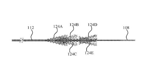
Turning first to Fig. 1, a system 100 according to an embodiment of this
disclosure is depicted. The system includes an exemplary HMA unit 102 and a
corpus
CA 03003232 2018-04-25
WO 2017/072761 PCT/1L2016/051153
- 26 -
anchoring unit, generally designated 104. It is of note that the HMA may have
various
designs (not shown), and the functionality of the HMA is not limited by any
specific
external design.
The corpus anchoring unit comprises a deployment wire 106, extending from the
HMA unit 102 to the flexible distal end 108 of the corpus anchoring unit. The
wire is
typically made of a flexible biocompatible material, which may, for example,
be a
metal, an alloy or a polymer material. Onto the wire, various functional unit
parts are
mounted, in a manner allowing insertion and navigation of the corpus anchoring
unit
within the vessel, as well as the unit's deployment for anchoring, entrapping
and
extracting of the clots, as will be described below.
The HMA further includes a main tubular pipe shaft 110 and a flexible tubular
pipe shaft 112, both mounted onto the deployment wire, typically coaxially
therewith.
Namely, the wire 106 is threaded through a longitudinal lumen formed within
main pipe
110 and flexible pipe 112, such that the wire may be pulled and pushed through
the
pipes. The main pipe is typically made of a material having limited
flexibility, for
example a biocompatible alloy such as nitinol, and is use to impart mechanical
strength
to the corpus anchoring unit upon insertion and extraction.
Associated to the distal end of pipe 110 is flexible pipe 112; pipe 112 has
increased flexibility (compared to pipe 110), and allows improved positioning
of the
deployable section 114 of the corpus anchoring unit. Both pipes 110 and 112
have a
diameter which is sufficient for free movement of the wire 106 and the
associated
deployable section 114, both in the non-deployed state and in the deployed
state for
extraction of the entrapped clot.
The deployable section 114 of the corpus anchoring unit is positioned distally
to
the flexible pipe 112. A close-up view of the corpus anchoring unit can be
seen in Fig.
2. The deployable section 114 comprises at least 2, in this specific example
5,
deployable cylindrical bodies 116A-116E (i.e. 5 tubes), each of the
cylindrical bodies
having an open, free end 118A-118E, respectively, positioned either at the
proximal or
distal end of each cylindrical body. Each of the cylindrical bodies is
constructed out of a
plurality of wound coiled threads, held together in the deployment state of
the
cylindrical body by friction forces. The cylindrical bodies are coaxially
mounted onto
the wire 106. Associated with each open free end 118A-118E are respective tip
tools
CA 03003232 2018-04-25
WO 2017/072761 PCT/1L2016/051153
- 27 -
120A-120E, which enable deployment of the threads, as will be explained
further
below.
Positioned distally to the outmost distal cylindrical body is a stopper 122.
The
stopper is fixedly attached to wire 106. Once the corpus anchoring unit is
brought into
proximity with the blood clot in the vessel, the system is operated to switch
the corpus
anchoring unit from the deployment to its deployed state. Several such states
are shown
in Figs. 3A-3C.
The manner by which the corpus anchoring unit is deployed will now be
described. Upon positioning of the corpus anchoring unit in adequate proximity
to a
corpus within vessel lumen, the wire 106 is manipulated by the HMA 102, such
that
movement of the wire switches the cylindrical bodies from a deployment state
to a
deployed state. Such manipulation typically involves pushing and/or pulling
the wire
106 (torqueing the wire is also contemplated). Pulling on the wire causes
fixedly
attached stopper 122 to bear onto the outmost distal cylindrical body (in the
exemplified
embodiment cylindrical body 116E). As at least some of the elements mounted
onto
wire 106 are floating (i.e. not fixedly attached to the wire), the pulling
force applied by
stopper 122 onto cylindrical body 116E causes all mounted elements of the
corpus
anchoring unit to proximate one another, and transfer the pulling force
between the
mounted elements.
In the example embodied by Fig. 3A, when the wire 106 is pulled, stopper 122
bears onto the fixed (closed) end of cylindrical body 116E, thereby causing
cylindrical
body 116E to move in the proximal direction. Such movement causes the open,
free end
of cylindrical body 116E to engage, or come into contact with, tip tool 120E,
which in
turn exerts an opposite force onto the open end of cylindrical body 116E. As
the coiled
threads of the cylindrical body are biased into their unwound state, the force
exerted by
tip tool 120E onto the open end is sufficient to switch the threads from their
wound to
their unwound state, resulting in deployment of cylindrical body 116E. As can
be seen
in Fig. 3A, the threads are welded to one another at the opposite end of
cylindrical body
116E (i.e. the fixed end opposite the open, free end), preventing the threads
from
completely unwinding. This results in a cone-like or funnel-like arrangement
of the
unwound threads that permits physical capturing of the clot once the
cylindrical body is
deployed and preventing the clot from drifting further into the blood vessel.
Further, to
CA 03003232 2018-04-25
WO 2017/072761 PCT/1L2016/051153
- 28 -
the physical encaging of the clot, the unwinding movement of the threads
during
unwinding will cause the threads to penetrate the clot and anchor it.
As can be seen in Figs. 3B and 3C, more than one cylindrical body may be
deployed in order to capture the clot. Deployment of several cylindrical
bodies, either in
the same opening direction (as seen in Fig. 3B) or in opposite directions (as
seen in Fig.
3C) will cause formation of capturing cages assisting in capturing and
retrieving larger
clots or clot fragments. Deployment of several tubes may be carried out by
additional
pulling onto the deployment wire, such that after the deployment of the
outmost distal
tube, another pull of the wire will cause deployment of the more proximal
tubes as force
is being linearly transferred between the elements mounted onto the wire.
Namely, once
wire 106 is pulled on and cylindrical body 116E is deployed by tip tool 120E,
another
pull will cause cylindrical body 116D to proximate tip tool 120D (or if
arranged
differently, will cause tip tool 120D to proximate cylindrical body 116D),
thereby
causing the threads in cylindrical body 116D to unwind and deploy. Thus, by
incremental pulling of the wire, the cylindrical bodies may be serially
deployed.
Another control of the deployment sequence may be obtained by using tubes
designed to deploy upon exertion of difference forces. For example, as can be
seen in
Fig. 4A, the tubes may be grooved by removing one or more of the threads. For
example, as can be clearly seen in Fig. 4B, reduction in the number of threads
results in
less friction between the threads, requiring application of smaller forces to
overcome the
friction between the threads and cause their unwinding. Thus, pulling onto the
wire at
different forces will control the deployment of different tubes.
Once the cylindrical bodies are deployed, pulling the deployment wire will
cause encaging of the clot. Generally speaking, in a first stage the
cylindrical bodies are
deployed such that the coiled threads unwind and anchor into the corpus.
Pulling onto
the deployment wire will cause deployed cylindrical bodies to proximate one
another,
resulting in initial compaction of the corpus. Upon further pulling, the
unwound threads
of adjacent cylindrical bodies will come into contact one with the other, at
times causing
entanglement of the threads, thus forming encaging of the captured corpus.
Further
proximation will increase compaction (thus decreasing the length of the
device) to allow
easier extraction of the device with reduced risks of corpus fragmentation. In
a specific,
non-limiting example, a combination of tubes having non-deployed lengths of
2.5 mm
CA 03003232 2018-04-25
WO 2017/072761 PCT/1L2016/051153
- 29 -
and 5 mm will eventually result in compaction of between 1.5-2 folds in length
due to
the proximation of the deployed tubes.
Further variation may be obtained by using tubes of various lengths, namely
some of the tubes may have threads of a first length while other tubes may
have threads
of a second length. Such variation in length may also assist in improved
entrapment of
the clot, as deployed tubes having shorter threads may be compactly arranged
within a
deployed cylindrical body having longer threads, encaging the clot between the
deployed cylindrical bodies in a compact manner. Such an arrangement is
demonstrated
in Fig. 3C, in which deployed cylindrical body 116E has shorter threads, while
cylindrical body 116D have longer threads. As can be seen, proximation of
cylindrical
body 116D and cylindrical body 116E one to the other causes a hermetically
closed
cage in which a clot may be entrapped. This will also be demonstrated further
below in
connection with Figs. 7B-9H.
As noted above, the corpus anchoring unit may comprise closed tubes, namely
tubes having both ends closed and capable of radial expansion when force is
applied by
the tip tool onto one (or both) of the closed tube's ends. Such a tube is
demonstrated in
Fig. 4C, which schematically depicts a deployed, grooved, closed-tub 130. Tube
130 is
associated with the deployment wire 106 and has a proximal end 132 and a
distal end
134, at least one of which being associated with a tip tool (not shown). The
wound
coiled threads of tube 130 are fixedly attached to one another at both ends
132 and 134,
such that when the tip tool applies a force onto one of the ends (resulting
from pushing
or pulling onto the deployment wire) the ends 132 and 134 proximate one
another,
causing reduction in length of tube 130 and radial expansion of the coiled
threads
between the two ends of the tube. In the example of Fig. 4C, some threads have
been
removed from the tube to form grooves 140A-140D, effectively dividing the tube
into
distinct sections, the number of threads removed determines the distance
between the
forms expanded sections of the tube, as well as the extent of radial extension
of each
section; for example, radially extended sections 138 are more radially
extended as more
threads have been removed as compared to radially extended sections 136.
As a man of the art may appreciate, although deployment of the device is the
examples described herein is exemplified by pulling onto the deployment wire
(i.e.
displacing the wire to the proximal direction), deployment by pushing is also
CA 03003232 2018-04-25
WO 2017/072761 PCT/1L2016/051153
- 30 -
contemplated under similar linear movement and transition of force principles.
Further,
deployment by rotational movement of the wire, i.e. applying variable torque
onto the
different elements mounted onto the wire is also contemplated and within the
scope of
the present disclosure.
As noted above, the device of this disclosure is designed as to exert minimal
radial force on the inner walls of the blood vessels. This is obtained by the
dimensions
of the cylindrical bodies and its controlled deployment. Fig. 5B shows a
comparative
measurement of the radial force exerted on a blood vessel when a tube of the
device of
this disclosure is deployed, compared to the SolitaireTM device (the structure
of which is
seen in Fig. 5A). As clearly seen, the radial force exerted by the deployed
threads of the
device of this disclosure is significantly lower than the radial force exerted
by the
SolitaireTM device. It is of note that the cylindrical bodies of the unit of
this disclosure
are introduced into the blood vessel in a normally-closed configuration,
controlled
deployment is permitted by proper manipulation of the HMA and its associated
deployment wire. This is contrary to the normally-open configuration of the
comparative standard device (which is introduced into the blood vessel in a
collapsed
configuration that is maintained by a microcatheter; upon removal of the
microcatheter
the device automatically and immediately assumes its extended configuration).
At times, the device of this disclosure is configured to have dimensions that
prevent the contact of the deployed cylindrical bodies (i.e. the unwound
coiled threads)
with the inner surface of the blood vessel. Meaning that in some embodiments,
the
cylindrical bodies of the unit are dimensioned such that one is the deployed
state, the
unwound coiled threads form a conical shape having a maximal diameter that is
smaller
than the inner diameter of the blood vessel.
To better understand the engagement of the tip tool and the cylindrical body,
reference is now made to Figs. 6A-6E, which show the sequence of engagement of
the
tip tool with the cylindrical body. Fig. 6A provides an exemplary system, in
which the
deployment wire 200 is associated with at least one cylindrical body 202 and
at least
one tip tool 206 which is located at the cylindrical body's distal free end
204. The
central portion of the cylindrical body 202 is enveloped by a polymeric sheet
(for
example a PTFE sheet) 214, the function of which will be described further
below. The
CA 03003232 2018-04-25
WO 2017/072761 PCT/1L2016/051153
- 31 -
tip tool 202 comprises an ellipsoid main body 208 and a distal tubular element
210, the
function of which will now be described.
Once the deployment wire is displaced in the proximal direction (designated by
arrow 212), i.e. the deployment wire is pulled, the ellipsoid section of the
tip tool
advances towards the distal, free end of the cylindrical body and engages the
distal end,
as seen in Fig. 6B. This engagement causes a primary unwinding of the wound
threads,
slightly separating the distal free ends of the wound threads one from the
other. As the
threads are made of a relatively flexible material (e.g. a flexible metal),
further
advancement of the ellipsoid portion of the tip tool causes elastic
deformation of the
wound threads, as seen in Fig. 6C, however, without further unwinding of the
threads.
Once the tip tool is further advanced in the proximal direction, the slightly
separated
edges of the wound threads engage the tubular element of the tip tool (Fig.
6C).
The dimensions of the ellipsoid portion and the tubular element are designed
such that once the free edges of the wound threads engage the tubular element,
such
engagement causes the wound threads to unwind and flare-out into a deployed
state,
seen in Fig. 6D. Further advancing of the tip tool in the proximal direction,
thus causes
further unwinding of the threads from the cylindrical body (Fig. 6E). Thus, by
controlling the angle of the impact of the edges of the wound threads with the
tubular
element of the tip tool, controlled deployment of the cylindrical body may be
obtained.
As also seen in Fig. 6E, the polymeric layer 214 functions to limit the extent
of
deployment of the cylindrical body by limiting the length of operable section
of the
cylindrical body. It is to be understood that the device may or may not
include such
polymeric layers.
Seen in Fig 7 is a system having at least one pair of oppositely oriented
cylindrical bodies. The system of Fig. 7 comprises a proximal cylindrical body
associated with a first tip tool at its distal free end, and a distal
cylindrical body
associated with a second tip toll as its proximal free end. Pulling the
deployment wire in
the direction of the arrow (i.e. in the proximal direction) causes the first
tip tool to
engage the distal end of the proximal cylindrical body, thereby causing its
deployment.
A further pull on the deployment wire causes the second cylindrical body to
engage the
second tip tool at the proximal end of the distal cylindrical body, thereby
causing
deployment of the distal cylindrical body. Once both cylindrical bodies are
deployed, a
CA 03003232 2018-04-25
WO 2017/072761 PCT/1L2016/051153
- 32 -
further pull in the proximal direction causes them to proximate one another,
thereby
causing a cage-formation. It is of note that the shaft may also be rotated
(i.e. torqued) in
order to cause rotating movement of the deployed cylindrical bodies, thereby
also
causing the cylindrical bodies to proximate one another and concomitantly
entangle
their unwound coiled threads for better capturing of the blood clot.
The dimensions of the cylindrical bodies in the pair of oppositely oriented
cylindrical bodies may be tailored, such that the unwound coiled threads of
one of the
deployed cylindrical bodies (typically the proximal cylindrical body of the
pair) are
longer than the unwound coiled threads of the other deployed cylindrical body
(typically the distal cylindrical body of the pair). As the extraction of the
device from
the blood vessel is done by pulling the corpus anchoring unit in the proximal
direction,
such a design minimizes the contact of the unwound coiled threads of the
distal
cylindrical bodies with the inner surface of the blood vessel, thereby
minimizing
damage to the blood vessel.
The sequence of deployment of a system containing a plurality of such
oppositely oriented pairs is shown in Figs. 8A-9H. The cylindrical bodies of
the system
shown in Figs. 8A-8C do not comprise a polymeric layer, and hence deployment
of
each cylindrical body is carried out to its full extent. Namely, each wound
thread in
each cylindrical body is deployed to its full operational length upon
engagement with its
corresponding tip tool (the most proximal tube is deployed first to its full
extent, then
the next-in-line distal cylindrical body is deployed to its full extent, then
the next-in-line
distal cylindrical body is opened to its full extent, and so on). After full
deployment of
the cylindrical bodies, proximation of the deployed bodies causes the
formation of
interlocked pairs of deployed bodies (i.e. primary cages). Upon further
proximation,
adjacent cages can form a further cage (i.e. secondary cage), etc. Each of
such cages is
anchored at a different section of the blood clot, and their proximation one
to the other
causes compaction of the blood clot for easier removal out of the vessel (as
discussed
further below).
In the exemplary system of Figs. 8A-8H, each proximal cylindrical body in each
pair of bodies is partially enveloped by a polymeric sheet, that limits the
extent of
deployment of these bodies. Thus, deployment of the device occurs in two
stages. First,
the all of the cylindrical bodies are partially deployed in a sequence along
the
CA 03003232 2018-04-25
WO 2017/072761 PCT/1L2016/051153
- 33 -
deployment wire (starting from the most proximal and ending with the most
distal along
the wire) to a substantially similar unwound coiled threads length, i.e. to a
thread length
similar to the non-restricted bodies. It is, however, also possible to
partially deploy all
of the bodies simultaneously in the first deployment stage (not shown). After
completion of the first stage, additional axial displacement of the deployment
wire
causes the restricted bodies to further deploy to a longer unwound coiled
threads length
in a second stage of deployment. Such a sequence of deployment divides the
forces
applied onto the blood clot during capturing, enable maintaining its integrity
during
capturing to minimize emboli.
Figs. 10A-10D show the capturing and retrieval of a simulated blood clot by a
system of this disclosure. The system is first fully penetrated into the blood
clot in a
deployment state (Fig. 10A). Then the anchoring unit is operated and the
various
cylindrical bodies are deployed and anchored into the blood clot (as can best
be seen in
Fig. 10D). As evident from Figs. 10A-10C, and as also explained above, the
anchoring
of the unit into the blood clot and the proximation of the deployed
cylindrical bodies
one to the other during the deployment and retrieval process, causes
significant
compaction of the blood clot, thereby further assisting its retrieval and
further
minimizes formation of emboli.
The ability of the system to maintain the captured blood clot integrity while
maneuvering in complex and highly curved vessels is shown in Figs. 11A-11G. In
these
figures, a step-by-step retrieval of a simulated blood clot is demonstrated,
showing the
flexibility of the corpus capturing unit while being maneuvered within a
simulated
blood vessel. No fragmentation or rupturing of the simulated blood clot was
observed
during the process and the clot was fully retrieved.
In vivo studies
All procedures were conducted according to international guidelines and were
approved by the responsible local ethics committee.
Performance Study 1
The retrieval of blood clots from blood vessels was demonstrated in vivo, as
follows. A 40 Kg swine was used in this study. Anesthesia was induced by an
intramuscular injection of ketamine (35 mg/kg) and xylazine (5 mg/kg), and
maintained
CA 03003232 2018-04-25
WO 2017/072761 PCT/1L2016/051153
- 34 -
with mechanical ventilation of oxygen with 1-2% isoflurane. Continuous
monitoring of
heart rate, respiration, oxygen saturation level (pulse oximetry), end tidal
CO2 and
temperature allowed real time assessment of the physiologic status of the
animal.
Common femoral artery access was subsequently obtained, and heparin bolus of
4,000 IU was intravenously administered. Anticoagulation was sustained with
maintenance administration of 1,000 IU heparin every hour.
Thrombus preparation and application were done as previously described in [8].
In brief, thrombi were created by mixing 10-mL autologous blood of the animal
with
1 gr barium sulfate (Sigma) and 0.25mL bovine thrombin solution (Sigma). The
mix was
injected into a 4mm inner diameter silicone tube, incubated for 1 hour at room
temperature and cut into lOmm length thrombi, which were injected into the
target
vessel. Due to the radiopacity gained by the added barium sulfate, the thrombi
were
visualized during angiography.
Device Configuration: the device configuration used in the animal study is
demonstrated in Fig. 12. The device (corpus anchoring unit) included a
stainless steel
deployment wire (2000mm, 0 0.0045"), onto which five 12-strands tubes (OD
0.0136",
ID 0.009"; strand 0 0.0023") were mounted. The tubes were arranged according
to
Table 1 (along the proximal-distal axis). The open ends of the tubes were
associated
with tin tip tools, having a rounded configuration (OD 0.0082", ID 0.0067");
the tip
tools contained Gold marker bands. The device was operated by an associated
HMA
(stopcock attached to a metal base or "guitar" handle).
Table 1: Arrangement of tubes in the device of Fig. 12
Tube 1 Tube 2 Tube 3 Tube 4 Tube 5
Length 5mm 5mm 3.5mm 5mm 5mm
Open end Distal Distal Proximal Distal Proximal
Experimental Design and Angio graphic Evaluation
The study was performed on a biplane angiography system. An 8F guiding
catheter was inserted through the femoral artery access into the target vessel
using a
0.014 inch wire. Selective occlusion of the internal maxillary artery and
lingual artery
CA 03003232 2018-04-25
WO 2017/072761 PCT/1L2016/051153
- 35 -
was performed, which simulated the anatomic setting of an occlusion of the
middle
cerebral artery and the basilar artery in the human circulation.
Pre-formed thrombi were injected by a syringe into the guiding catheter and
allowed to embolize distally into the swine internal maxillary and lingual
arteries. The
thrombi were allowed to mature in place for 10 min.
Vessel occlusion was confirmed by angiography and assessment of a
Thrombolysis In Cerebral Infarction (TICI) flow grade of 0 or 1 (persistent
occlusion or
trickle flow). A 0.017" micro-catheter was navigated over the 0.014" wire
across the
occlusive thrombus. The wire was removed and contrast injection was performed
to
confirm endoluminal positioning distal to the occlusion.
The device of Fig. 12 was then advanced into the microcatheter and navigated
to
the occlusive thrombus, until the distal radiopaque marker of the device was
observed in
adjacent to the microcatheter end. Then the microcatheter was retrieved back.
In order
to ensure optimal positioning of the device, the device was repositioned in a
way that
either the three distal radiopaque markers of the device were positioned
distal to the
occlusive thrombus, or the first (i.e. proximal) radiopaque marker was
positioned in
adjacent to the proximal end of the occlusive thrombus.
Then the HMA was activated, initiating deployment of the device and engaging
and trapping of the thrombus. During deployment of the device, the wire tubes
of the
device acted as trapping elements, creating closed geometrical cage-like shape
designed
to optimally trap the clot. In addition, the wire tubes were brought into
proximity with
one another (and in this instance compressed against each other), resulting in
compression and entrapment of the occlusive thrombus.
As notes above, the formation of cages permitted a stable anchoring into the
clots. The formation of primary cages between each pair of tubes followed by
the
formation of a secondary, larger cage between two proximating adjacent cages,
further
compacted the blood clot and enhanced the anchoring of the device for safe and
effective removal of the clot.
After the occlusive thrombus was trapped within the deployed device, the
microcatheter and the device were simultaneously pulled back into the guiding
catheter
under aspiration according to common practice.
CA 03003232 2018-04-25
WO 2017/072761 PCT/1L2016/051153
- 36 -
Two cases of vessel occlusions were obtained. In both cases, the occlusive
thrombus was extended into branches of the IMA artery. In test 1 (Figs. 13A-
13F), a
single recanalization attempt was performed and flow restoration was achieved
immediately after device deployment. In test 2 (Figs. 14A-14E), a first
recanalization
attempt resulted in splitting of the thrombus into two portions, retrieving
one portion of
the occluding thrombus. Then, a second device was introduced and deployed,
engaging
the remaining portion of the occluding thrombus, and a full recanalization was
achieved.
No distal thromboembolic events in the target vessel or in unaffected vessel
areas occurred during passing of the thrombus, deployment, and retrieval of
the device.
Control angiographies showed no signs of vessel perforation, dissection, nor
thrombus
embolization or fragmentation or device fraction.
Performance Study 2
In this study, swine were considered the model for evaluation. The device of
Animal Study I was tested for performance (i.e. clot trapping) by
instrumentation of
arterial segments similar to human indicated anatomies, with diameters ranging
from 2-
3.7mm. The segments included: Brachial, Subclavian, Axillary, Internal
Maxillary
Artery and the External carotid arteries, as detailed in Table 2.
Table 2: tested swine arterial segments
Procedure Artery Diameter (mm)
1 Internal maxillary 3.0
2 Left IMA 3.5
3 Brachial right 3.6
4 Brachial right 3.1
Brachial left 3.7
6 Subclavian left 3.1-3.4
7 Axillary left 2.0
8 Axillary left 3.7
9 Brachial left 3.3
IMA left 3.2
11 External carotid right 3.4
12 Brachial right 3.7
13 Externa carotid left 3.6-3.7
14 Axillary left 1.6
Axillary left 2.0-2.1
16 Brachial right 2.0-3.0
CA 03003232 2018-04-25
WO 2017/072761 PCT/1L2016/051153
- 37 -
The primary performance endpoint was re-canalization rate. Re-canalization
rate
was defined as successful capturing, trapping, and retrieval of thrombi as
determined by
angiographic evaluation (e.g., with TICI score >2 after thrombus retrieval in
maximum
3 attempts).
The usability (mechanical performance) of the device was assessed on a scale
of
1-5 (1=Poor, 5=Excellent), with respect to the following parameters:
= Catheter navigation - Ability to navigate to a specific vessel that
mimics
intracranial setting in human: Insertion, navigation, pushability
= Device retraction (withdrawal)
= Visibility of marker bands and tip
= Handle activation\Deployment
= Compatibility with commonly used catheterization tools (guide catheter,
micro
catheter, etc.)
= User experience
Pre-procedural preparation: The animals were housed for three days prior to
acute procedures. The animals were fed with commercial pellet food and water
was
administrated ad libitum. Each animal was examined for its general conditions,
weight
and health status, including Complete Blood Count (CBC).
Animals underwent overnight fasting period prior to procedure and an
anesthesia according to animal laboratory standard procedure. Induction:
Animals were
induced by an IM injection of Telazol (4.4mg/kg) mixed with xylazine
(2.2mg/kg).
Atropine (0.05mg/kg) was delivered intramuscularly as a premedication
following
induction. Maintenance was obtained by administration of 1-3% isoflurane via
endotracheal tube (mechanical ventilation). Heparin was administered with a
goal
activated coagulation time (ACT) between 250 and 300 seconds. Vital signs were
monitored following induction of general anesthesia; E.C.G., HR, Sp02,
capillary refill
time and Temp
The blood clots for the thromboembolization process were prepared by using a
20m1 whole blood obtained into a syringe, mixed with 2g barium sulfate powder
in a
gentile rotational movement. The mixture was incubated in the syringe at room
CA 03003232 2018-04-25
WO 2017/072761
PCT/1L2016/051153
- 38 -
temperature for 120 minutes until it showed a multiply-layered structure of
blood
constituent and barium. After the sedimentation, the solid component was
separated
from the serum component and a small piece of clot was carefully resected,
measuring
approximately 5 mm in diameter and 10-20 mm in length, from the aforementioned
solid component with both fibrin-rich and erythrocyte-rich layers. Finally,
each
prepared thrombus was filled into a silicone tube with saline for reservation
until
injection.
Surgical procedure: After groin opening (femoral) and insertion of an 8/9F
sheath, a balloon GC 8F was advanced over the wire (0.035" wire). Road mapping
of
the target vascular bed and selection of target vessels. The pre-prepared clot
was
injected to the pre-selected target vessel and left for embedding for at least
10 min prior
deployment. Selective pretreatment angiography was performed in order to check
that
no vascular damage was caused by the clot insertion procedure: TICI scoring
was
evaluated to record perfusion rate. Vessel dissection or perforation,
thromboembolic
event and clot fragmentation were checked for during this evaluation.
Thrombectomy procedure: the following procedure was carried out for every
treated vessel segment:
1. An 8F balloon GC was positioned as close as possible to the position of
thrombus employing a standard method.
2. A micro-catheter of 0.017" or 0.021" ID was advanced crossing the clot,
over a 0.014" or 0.016" guide wire.
3. The guide wire was retrieved
4. The device was removed from its packaging and inspected before
insertion.
5. The device was advanced through the micro-catheter till the distal tip
was angiograpically observed at the microcatheter distal end. The device was
positioned relatively to the clot so that the device proximal marker was at
the
clot proximal edge
6. The micro-catheter was retracted
7. Repositioning of the device relatively to the clot (if necessary):
device
proximal marker at the clot proximal edge
CA 03003232 2018-04-25
WO 2017/072761
PCT/1L2016/051153
- 39 -
8. Opening of the device (actuation of the corpus-anchoring units) was
performed after releasing the handle safety catch, by sliding the handle
activator, to
enable device deployment and clot engagement and trapping. It was verified
that the
radiopaque markers moved toward each other.
9. The balloon GC was inflated and vessel sealing verified.
10. The device was retracted with the micro-catheter as one unit into the
balloon GC, while aspirating by applying negative pressure when retraction
started The
balloon GC was deflated.
11. Retrieved/captured clots were observed and photographed and visual
inspection of device integrity was performed.
12. Angiographic assessment with TICI and vessel integrity (e.g.,
dissection/perforation), vasospasm scoring was performed
After each retrieval attempt, the vessel was examined for remaining and
additional occlusions (i.e. part of the clot that remained in place, distal
emboli and
affected new territories). In case the clot was located in a bifurcation,
occluding 2
branches of the vessel, only the main branch was treated. The procedure was
considered
successful when the main branch underwent recanalization (with TICI score >2)
In case of failed trapping, or partial success in clot retrieval, the device
was
withdrawn for an additional deployment, the device was cut in the distal shaft
coil
proximal to the activation area in order to be released from the micro-
catheter, the MC
integrity was verified for additional use and a new device was inserted. The
advancement to the occluded segment, engagement/ trapping and aspiration steps
were
repeated. Up to 3 attempts (3 devices) were allowed for a procedure to be
considered
successful.
Animal euthanasia and sacrifice was performed by IV bolus injection of an over
dose of sodium pentobarbital (100mg/kg) while the animal is under general
anesthesia.
Results
15 clots were treated with no vessel perforation or dissection after insertion
of
the clot and device. Out of the 15 clots, 14 were successfully retrieved with
post
thrombectomy recanalization TICI score of 2 or higher that was retrieved in 3
attempts
CA 03003232 2018-04-25
WO 2017/072761 PCT/1L2016/051153
- 40 -
or less, as detailed in Table 3. Table 4 summarizes the segmentation of the
number of
attempts for the 14 successful procedures.
Table 3: re-canalization assessment (TICI scores) after procedure
TICI score 2a 2b 3 Fail (0 or 1)
# of clots 0 10 4 1
Table 4: number of attempts
Number of attempts 1 2 3 Average
# of clots retrieved 12 1 1 1.2 0.56
As evident from Table 3, 93.3% of the procedures ended with successful
retrieval (based on the success criterion of TICI >2 with a maximum of three
attempts)
and good recanalization scores.
In terms of number of attempts, as detailed in Table 4, 12 out of the 14 (i.e.
85.7%) successful retrievals were achieved within the first attempt. No distal
occlusions
or affected new territories were documented in the study.
Mechanical performance (usability) evaluation was assessed in a scoring grade
on a 1 to 5 scale (1- Poor, 2- Mediocre, 3- Fair, 4- Good, 5- Excellent).
Table 6
provides a summary of the mechanical performance scoring.
Table 6: mechanical performance evaluation
Score Average
STD
1 2 3 4 5 score
Catheter navigation ¨ Ability to
navigate to a specific vessel that
mimics intracranial setting in 0 0 0 0 15 5 0
human: insertion, navigation,
pushability
Device retraction (withdrawal) 1 0 0 2 12 4.6 1
Visibility of markers and tip 0 0 0 0 15 5 0
Handle activationWeployment 0 0 1 0 14 4.9 0.5
Compatibility with commonly used
catheterization tools (Guide 0 0 1 0 14 4.9 0.5
catheter, micro catheter, etc.)
User experience 0 0 1 0 14 4.9 0.5
CA 03003232 2018-04-25
WO 2017/072761 PCT/1L2016/051153
- 41 -
Visual inspection under light microscope with magnification >30X was
performed to all devices that were used in the study. All devices were intact,
no
kinks/breaks or other damages were observed in any of the devices.
Safety
4 crossbred domestic female swine weighting 40-50Kg (approximately aged 3-4
Mo) were used. The device was tested for safety by simulated instrumentation
of
arterial segments (considered the test system) with diameters ranging from 2-
5.5mm
and with similar anatomy to humans. The following vessels were used:
Subclavian,
Axillary, common Carotid, Femoral and Saphenous.
Pre-procedural preparation: Each animal was examined for its general
condition, weight and health status, including Complete Blood Count (CBC).
Animals
were put in quarantine for a period of 3 days for acclimation purposes, and
had a 12
hours fasting period prior to index procedure. Anesthesia was performed
according to
the animal laboratory standard procedure. Once an appropriate level of
sedation was
induced, the animal was intubated with an endotracheal tube (dependent on the
size of
animal). The animal was connected to the anesthesia machine where isoflurane
gas was
used to maintain a surgical plane of anesthesia. Mechanical ventilation was
provided.
IV Heparin Loading dose of 100-150 IU/kg was administered and ACT was
monitored
throughout the procedure. Heparin was administered IV when ACT levels were <
250
sec. Following induction of general anesthesia, E.C.G., HR, 5p02 and
temperature
were monitored. Blood was collectedfor CBC and chemistry.
Surgical procedure: angiography was performed before, during, and after each
simulated thrombectomy procedure, for segment selection (according to
diameter, angle
of the vessels, side branched and tortuosity) and evaluation during and after
the
procedure. Vessel registration was performed based on anatomical landmarks and
specified in the angiograms.
Catheterization was performed according to the following procedure:
angiographic road mapping were performed as per common clinical practice:
Entry
artery (groin) puncturing for arterial access. Insertion of an 8F or 9F
vascular sheath to
access artery, followed by insertion of a 0.035" Guidewire (GW). Advancement
(Over
CA 03003232 2018-04-25
WO 2017/072761 PCT/1L2016/051153
- 42 -
the Wire, OTW) of a 6F/8F Guide Catheter (GC) and Road mapping of the target
vascular bed.
Thrombectomy procedure: The thrombectomy procedure was carried out as
described above in Performance Study 2, including angiographic assessment with
TICI
and vasospasm scoring. Final angiographic evaluation included angiographic
evaluation
and scoring for eventual vessel dissection or perforation, vasospasm,
thromboembolic
event. End of catheterization procedure with closure of entry groin till
hemostasis was
reached.
Follow-up assessment: Animals were assessed daily for general health
conditions (e.g. Animal injuries and visual infections, presence or absence of
feces,
bleeding and food intake). Animal weight was also followed. No adjuvant post
procedure therapy was indicated in these animal procedure. Post procedure
preventive
antibacterial therapy with antibiotics was given for 5 days post-surgery.
Prior to sacrifice, animal underwent standard diagnostic angiography to assess
all treated vessels, the arteries were specifically identified according to
anatomical
landmarks that were noted during procedure day and TICI scoring was attributed
for the
evaluation. Blood was collected, for CBC and bio chemistry. Blood collection,
from the
animal, was performed after sedation and prior to anesthesia and induction or
euthanasia. Animal euthanasia was performed by IV bolus injection of an over
dose of
potassium chloride (ad effect) while the animal was under general anesthesia.
Harvesting: Detection of instrumented segments was based on the registration
performed based on pre-selected anatomical landmarks and hardcopies of
angiograms.
The instrumented arterial segments were harvested. Harvesting of two native
untreated
segments was performed for comparative purposes from 2 animals. The harvested
segments were kept in a Formalin solution for further histological assessment.
Histological analysis: The samples were prepared by paraffin embedding
followed by H&E staining of selected sections. The samples were then submitted
for
histological evaluation.
In all procedure carried out with the device of this disclosure, no
angiographic
evidence of arterial wall injury such as dissection, perforation or thrombus
formation
CA 03003232 2018-04-25
WO 2017/072761 PCT/1L2016/051153
- 43 -
was observed. TICI-3 score was recorded in 6/6 (100%) vessels, following three
attempts in each vessel.
Vasospasm can normally occurs in endovascular procedures during guiding
catheter, wire and microcatheter manipulation and device retrieval. In fact,
the swine
model is much more prone to vasospasm compared with humans. Vasospasm is
usually
self-limited, and at follow up angiography after 1 hour vasospasm usually
resolves.
During the procedures only 2/6 simulated attempts (first attempts in each
vessel)
resulted in vasospasm.
Histological evaluation of the treated arteries at 30 days post procedure and
revealed no significant histological findings. Mild endothelial erosions were
noted
during the follow-up angiography at 30 day, and are considered acute in nature
and
related to wire or catheter passage. Such findings are within the limits of
similar
endovascular procedures [10,11].