Note: Descriptions are shown in the official language in which they were submitted.
CA 03003629 2018-04-27
WO 2017/074652
PCT/US2016/055255
OCCLUDER AND ANASTOMOSIS DEVICES
FIELD
[0001] This disclosure relates generally to implantable medical devices,
and
more specifically, to implantable medical devices for connecting tissue layers
to
create an anastomosis and to implantable devices for occluding inhibiting or
preventing material movement through tissue apertures, sealing, and allowing
healing of defects in tissues
BACKGROUND
[0002] Lesions of the gastrointestinal (GI) tract can be in the form of
polyps
that protrude from the mucosal lining with a mushroom-like shape, or flat
lesions that
are flush on the mucosa! lining. The need to remove lesions from the mucosal
lining
of the GI tract is common and growing worldwide. The likelihood of having
colon
lesions increases with age. Approximately half of the people over the age of
60 have
at least one colon lesion and often more. Some polyps are considered pre-
cancerous, which means that while they are not cancer, if left untreated they
may
develop into cancer. GI tract lesions are typically found during cancer
screening
tests, such as a colonoscopy or flexible sigmoidoscopy.
[0003] Benign and early malignant lesions of the GI tract can usually be
removed endoscopically using an electrocautery snare, hot snare, cold snare,
or
electrocautery knife devices. A saline-assisted polypectomy procedure is often
used
for the removal of large flat lesions in the GI tract. When lesions become
still larger
and invasively encompass more than just the mucosal layers of the GI tract, a
resection procedure is often performed whereby the full thickness of the wall
tissue is
removed along with the lesion. This procedure is typically performed using
laparoscopic or open surgery techniques rather than endoscopically. However,
open
surgery may not be an option for some patients, and laparoscopic procedures
may
not allow visualization within the lumen of the conduit being treated.
[0004] Large resections of the colon are not typically performed
endoscopically in part because tools and devices to adequately seal the
resulting
perforation in the colon wall are not available without approximating the
defect edges
which can result in lumen stricture (e.g., using clips, sutures, and the
like). Such
1
CA 03003629 2018-04-27
WO 2017/074652
PCT/US2016/055255
tools and devices are challenging to develop in part because of the relatively
hostile
colon environment that includes peristaltic movements and fecal matter.
[0005] An anastomosis is a cross-connection between two tubular tissue
structures, such as blood vessels or intestines. For example, when a portion
of an
intestine is resected, the resulting two ends can be sewn or stapled together
(anastomosed), using an intestinal anastomosis procedure. This procedure can
restore intestinal continuity after the resection of a bowel portion, or to
bypass a
portion of unresectable diseased bowel.
[0006] Anastomoses can be created in various manners including, but not
limited to: end-to-end, end-to-side, and side-to-side anastomoses. Often,
suturing is
used to create such anastomoses.
SUMMARY
[0007] One aspect of the invention relates to a medical device for sealing
a
defect or structure in tissue. The medical device includes a frame having
single
elongate member. The elongate member includes (1) a supporting portion
configured to conform to a geometry of a first tissue surface and to provide
an
apposition force against the first tissue surface, (2) an occluding portion
configured
to conform to a geometry of a second tissue surface and to provide an
apposition
force against the second tissue surface, and (3) a defect-occupying portion
disposed
between the supporting portion and the occluding portion. The defect-occupying
portion is configured to not provide a substantial apposition force against
tissue
around an aperture of the defect. The medical device also includes a sealing
material attached to at least a portion of the occluding portion. The sealing
material
is configured to inhibit material flow through the aperture.
[0008] A second aspect of the invention relates to a medical device for
sealing
a defect or structure in tissue. The medical device includes a wire member
that
includes a single wound wire. The wire includes (a) a sealing member, (2) an
apposition member, and (3) a defect-occupying portion disposed between the
sealing member and the apposition member. The medical device also includes a
covering material disposed on at least a portion of the sealing member.
2
CA 03003629 2018-04-27
WO 2017/074652
PCT/US2016/055255
[0009] A third aspect of the invention relates to a medical device system
that
includes (1) a frame including a single elongate member and (2) a delivery
sheath
defining a lumen. The elongate member forms (1) a supporting portion that is
configured to conform to a geometry of a first tissue surface and to provide
an
apposition force against the first tissue surface, (2) an occluding portion
that is
configured to conform to a geometry of a second tissue surface and to provide
an
apposition force against the second tissue surface, (3) a defect-occupying
portion
disposed between the supporting portion and the occluding portion, and (4) a
membrane attached to at least a portion of the occluding portion. The membrane
is
configured to inhibit material flow through the aperture. In addition, the
medical
device is configurable in a low-profile configuration such that the medical
device can
be contained within the lumen. Further, the medical device is configured to
expand
from the low-profile configuration when the device is liberated from the
lumen.
[0010] A fourth aspect of the invention relates to a method of sealing an
aperture
in a patient's body. The method includes inserting an implantable medical
device
into the aperture using a transcatheter technique. The device includes a
single
wound wire and a covering material. The wire forms (1) a sealing member, (2)
an
apposition member, and (3) a defect-occupying portion disposed between the
sealing member and the apposition member. The covering material is disposed on
at least a portion of the sealing member and is configured to fully overlay
the
aperture.
[0011] A fifth aspect of the invention relates to an implantable medical
device that
includes a single elongate member. The single elongate member forms (1) a
first
flange having a plurality of first arms configured about a central axis and
forming a
circumferential sealing portion at the outer edges of said first flange, (2) a
second
flange having a plurality of second arms configured about the central axis,
and (3) a
connecting region interconnecting the first and second flanges and adapted to
bridge
a defect in a lumen wall. In some embodiments, the first arms and the second
arms
have a pre-strained geometry such that an apposition force exists in the
presence of
the lumen wall and the apposition force does not exist in the absence of said
lumen
wall.
3
CA 03003629 2018-04-27
WO 2017/074652
PCT/US2016/055255
[0012] A sixth aspect of the invention relates to an implantable medical
device
that includes a single elongate member. The elongate member forms (1) a first
flange having a plurality of first arms configured about a central axis and
forming a
circumferential seal at outer edges of the first flange, (2) a second flange
having a
plurality of second arms configured about the central axis forming a
circumferential
sealing portion at outer edges of the second flange, and (3) a connecting
region
interconnecting the first and second flanges and adapted to cross a defect in
a
lumen wall. The connecting region fluidly connects the first and second
flanges.
The first arms and the second arms have a pre-strained geometry such that an
apposition force exists in the presence of the lumen wall and the apposition
force
does not exist in the absence of the lumen wall.
[0013] A seventh aspect of the invention relates to an implantable medical
device
that includes a single elongate member. The single elongate member forms (1) a
first flange having a plurality of first arms configured about a central axis
and forming
a circumferential sealing portion at the outer edges of the first flange, (2)
a second
flange having a plurality of second arms configured about the central axis,
and (3) a
connecting region interconnecting the first and second flanges and adapted to
cross
a defect in a lumen wall. The first arms and the second arms have a pre-
strained
geometry such that an apposition force exists in the absence of a lumen wall.
[0014] An eighth aspect of the invention relates to an implantable medical
device
that includes an apposition frame member that forms (1) a first flange having
a
plurality of first apposition petals configured about a central axis and
forming a first
circumferential sealing portion, (2) a second flange having a plurality of
second
apposition petals configured about the central axis and forming a second
circumferential sealing portion, and (3) a connecting region connecting the
first and
second flanges. The connecting region defines an aperture along the central
axis.
The implantable medical device also includes a support frame member that forms
a
plurality of apices and covering material disposed on at least a portion of
each of the
apposition frame member and the support frame member. In exemplary
embodiments, the support frame is disposed concentrically within the aperture.
4
CA 03003629 2018-04-27
WO 2017/074652
PCT/US2016/055255
[0015] A ninth aspect of the invention relates to a tissue-sealing device
that
includes a frame and a covering material disposed on at least a sealing
portion of the
frame. The frame includes an apposition portion, a sealing portion, and a
defect-
occupying portion. In exemplary embodiments, the apposition portion and the
sealing portion are configured dissimilarly. The frame defines diamond-shaped
petals that form the sealing portion and triangularly-shaped petals that form
the
apposition portion. The edges of the diamond-shaped petals in the sealing
portion
are substantially parallel to each other, which creates a line of physical
contact and a
sealing edge and reduces the presence of leakage channels between the sealing
petals. In contrast, the triangularly-shaped petals in the apposition portion
are
discrete and may tangentially contact each other. The tissue-sealing device
may be
configured to be implanted in a patient such that the covering material fully
overlays
and seals a tissue aperture.
[0016] A tenth aspect relates to a tissue sealing device that includes a
frame and
a covering material disposed on at least a portion of a sealing portion of the
frame.
The frame includes an apposition portion, a sealing portion, and a defect-
occupying
portion. In exemplary embodiments the apposition portion and the sealing
portion
are configured dissimilarly. The apposition petals and the sealing petals
include a
linear portion extending radially from the defect-occupying portion and an
essentially
diamond-shaped outer portion extending from the linear portion at the free
ends of
the petals. The sealing petals and the apposition petals are substantially
similar,
with the exception that the sealing petals have a more rounded outermost edge
than
the apposition petals. The outermost edges of the sealing petals tangentially
touch
each other. The abutment of the edges of the sealing petals creates a line of
physical contact and a sealing edge and reduces the presence of leakage
channels
between the sealing petals. The apposition petals in the apposition portion
are
discrete (not covered with a covering material) and may move relative to each
other.
The more rounded ends of the sealing petals (opposed to the less rounded ends
of
the apposition petals) creates a substantially uniform pressure distribution
at the
exterior circumference, and in the apposition portion, to facilitate loading
into a
delivery device. The tissue-sealing device may be configured to be implanted
in a
patient such that the covering material fully overlays and seals a tissue
aperture.
CA 03003629 2018-04-27
WO 2017/074652
PCT/US2016/055255
DESCRIPTION OF THE DRAWINGS
[0017] The accompanying drawings are included to provide a further
understanding of the disclosure and are incorporated in and constitute a part
of this
specification, illustrate embodiments, and together with the description serve
to
explain the principles of the disclosure.
[0018] FIG. 1A is a plan view of a wire frame of an exemplary tissue-
sealing
device in accordance with some embodiments;
[0019] FIG. 1B is an elevation view of the wire frame of FIG. 1A,
[0020] FIG. 2A is a plan view depicting the wire frame of FIG. 1A having
thereon
a covering material and engaged in an exemplary tissue defect.
[0021] FIG. 2B is a plan view showing the wire frame of FIG. 1A having
thereon a
covering material and engaged in another exemplary tissue defect.
[0022] FIG. 3A is a plan view of a wire frame of another exemplary tissue-
sealing
device in accordance with some embodiments;
[0023] FIG. 3B is an elevation view of the wire frame of FIG. 3A,
[0024] FIG. 4 is a plan view showing the wire frame of FIG. 3A having
thereon
covering material and engaged in an exemplary tissue defect;
[0025] FIG. 5 is a plan view of a wire frame of another exemplary tissue-
sealing
device in accordance with some embodiments;
[0026] FIG. 6 is a plan view showing the wire frame of FIG. 5 having
thereon a
covering material and engaged in an exemplary tissue defect;
[0027] FIG. 7 is a perspective view of a wire frame of another exemplary
tissue-
sealing device in accordance with some embodiments;
6
CA 03003629 2018-04-27
WO 2017/074652
PCT/US2016/055255
[0028] FIG. 8A is a perspective view of an exemplary sealing device made of
the
wire frame of FIG. 7 having a covering material disposed on an occluding
portion of
the wire frame;
[0029] FIG. 8B is an elevation view of the sealing device of FIG. 8A,
[0030] FIG. 80 is another perspective view of the sealing device of FIG.
8A,
[0031] FIG. 9A is a plan view of the sealing device of FIG. 8A engaged in
an
exemplary tissue defect;
[0032] FIG. 9B is another plan view of the sealing device of FIG. 8A
engaged in
the exemplary tissue defect;
[0033] FIG. 90 is a perspective view of the sealing device of FIG. 8A
engaged in
another exemplary tissue defect;
[0034] FIG. 9D is another perspective view of the sealing device of FIG. 8A
engaged in another exemplary tissue defect;
[0035] FIG. 10A is a plan view of another exemplary sealing device in
accordance
with some embodiments;
[0036] FIG. 10B is another plan view of the sealing device of FIG. 10A,
[0037] FIG. 11A is a plan view of the sealing device of FIG. 10A engaged in
an
exemplary tissue defect;
[0038] FIG. 11B is another plan view of the sealing device of FIG. 10A
engaged in
an exemplary tissue defect;
[0039] FIG. 12 is a perspective view of another exemplary sealing device in
accordance with some embodiments;
[0040] FIG. 13A is a perspective view showing the sealing device of FIG. 12
partially contained within an exemplary delivery sheath;
[0041] FIG. 13B is a side view showing the sealing device of FIG. 12 fully
contained within the delivery sheath of FIG. 13A,
7
CA 03003629 2018-04-27
WO 2017/074652
PCT/US2016/055255
[0042] FIG. 14A is a plan view of a sealing portion of another sealing
device
engaged in an exemplary tissue defect;
[0043] FIG. 14B is a plan view of an apposition portion of the sealing
device of
FIG. 14A,
[0044] FIG. 15A is a perspective view of a sealing portion of another
sealing
device engaged in an exemplary tissue defect;
[0045] FIG. 15B is a perspective view of an apposition portion of the
sealing
device of FIG. 15A,
[0046] FIG. 16A is a plan view of an exemplary anastomosis device in
accordance with some embodiments;
[0047] FIG. 16B is a plan view showing the exemplary anastomosis device of
FIG. 16A engaged with tissues to create an anastomosis,
[0048] FIG. 160 is an elevation view of the exemplary anastomosis device of
FIG.
16A,
[0049] FIG. 17A is a plan view of another exemplary anastomosis device in
accordance with some embodiments;
[0050] FIG. 17B is a plan view showing the exemplary anastomosis device of
FIG. 17A engaged with tissues to create an anastomosis,
[0051] FIG. 170 is an elevation view of the exemplary anastomosis device of
FIG.
17A,
[0052] FIG. 18 is a plan view of an exemplary apposition member frame in
accordance with some embodiments;
[0053] FIG. 19 is an elevation view of an exemplary support frame that may
be
used in conjunction with the apposition member frame of FIG. 18;
[0054] FIG. 20 is a plan view of the support frame of FIG. 19;
8
CA 03003629 2018-04-27
WO 2017/074652
PCT/US2016/055255
[0055] FIG. 21 is a plan view of another exemplary anastomosis device
including
a first arrangement of the apposition member frame of FIG. 18 and the support
frame
of FIG. 19;
[0056] FIG. 22 is a plan view of another exemplary anastomosis device
including
a second arrangement of the apposition member frame of FIG. 18 and the support
frame of FIG. 19;
[0057] FIG. 23 is an elevation view of another exemplary support frame that
can
be used in conjunction with the apposition member frame of FIG. 18;
[0058] FIG. 24 is a plan view of the support frame of FIG. 23;
[0059] FIG. 25 is a plan view of another exemplary anastomosis device
including
a first arrangement of the apposition member frame of FIG. 18 and the support
frame
of FIG. 19; and
[0060] FIG. 26 is a plan view of another exemplary apposition member frame.
[0061] FIG. 27 is a plan view of an exemplary tissue-sealing device
including a
frame and covering material where the apposition petals and sealing petals are
dissimilar;
[0062] FIG. 27A is a side view of the tissue-sealing device of FIG. 27;
[0063] FIG. 28 is a plan view of another exemplary tissue-sealing device
including
a frame and covering material where the apposition petals and sealing petals
are
dissimilar; and
[0064] FIG. 28A is a side view of the tissue-sealing device of FIG. 28.
DETAILED DESCRIPTION
[0065] Persons skilled in the art will readily appreciate that various
aspects of the
present disclosure can be realized by any number of methods and apparatus
configured to perform the intended functions. It should also be noted that the
accompanying figures referred to herein are not necessarily drawn to scale,
but may
9
CA 03003629 2018-04-27
WO 2017/074652
PCT/US2016/055255
be exaggerated to illustrate various aspects of the present disclosure, and in
that
regard, the drawing figures should not be construed as limiting.
[0066] This disclosure provides implantable medical devices and methods for
treating medical conditions using the implantable medical devices. For
example, this
disclosure provides implantable devices for occluding, sealing, and allowing
the
healing of tissue defects. Tissues that may be treated include, but are not
limited to,
those of the GI tract, peritoneum, vascular (arterial or venous) system,
cardiac
tissues, or the interface between one of these tissues and a synthetic
structure such
as a patch or vascular graft. Defects for which the implantable medical device
may
be applied include those that may be natural or artificially created, either
intentionally
or through some traumatic event or disease process. Defects may include, but
are
not limited to, perforations, ruptures, wounds, tears, endoleaks, fistulae,
and the like.
[0067] Additionally, this disclosure provides, inter alia, implantable
devices for
connecting tissue layers, such as for connecting a gallbladder and a portion
of a
gastrointestinal tract to create an anastomosis that facilitates material flow
therebetween. The devices are endoscopically deployable or deployable via a
catheter and can include self-expanding apposition mechanisms that facilitate
a
secure connection between the tissue structures (such a connection may also be
referred to herein as a "shunt," "passageway," "shunt passageway," or
"tunnel").
Such design features simplify implantation and reduce the likelihood of
complications. In some embodiments, the devices provided herein allow
treatment
to circumvent a conduit or organ blockage by creating a direct passage between
tissue structures, such as, for example, the gallbladder and a portion of the
gastrointestinal tract. In some embodiments, the devices provided herein are
implanted temporarily. As one example, the device is implanted and remains in
place until the gallbladder and/or its associated ducts are cleared of
blockages, after
which the device is removed. In another example, the device remains implanted
until the body grows a tissue-anastomosis around the device, and then the
device is
removed. In other embodiments, tissue ingrowth into and/or around the device
permanently implants the device, and the device is not removed. Such devices
can
provide an alternative treatment for patients who are not suitable candidates
for
CA 03003629 2018-04-27
WO 2017/074652
PCT/US2016/055255
other types of treatment (e.g., gallbladder removal surgery) and/or to avoid
known
complications of other types of treatment (e.g., external biliary drainage).
[0068] In reference to FIGS. 1A and 1B, a frame 100 of an exemplary tissue-
sealing device includes an elongate member 110. The elongate member 110 is
configured to form an apposition portion 120, a sealing portion 130, and a
defect-
occupying portion 140. The defect-occupying portion 140 is disposed between
the
apposition portion 120 and the sealing portion 130. As will be described
further, the
defect-occupying portion 140 is configured to traverse an opening or aperture
in one
or more layers of tissue, also referred to herein as a tissue defect. The
apposition
portion 120 and the sealing portion 130 are configured to be on opposite sides
of the
layer(s) of tissue. In some embodiments, the elongate member 110 comprises a
single continuous wire.
[0069] The elongate member 110 can comprise a variety of materials. The
elongate member 110 may be elastomeric, metallic, a spring wire, a shape
memory
alloy wire, a super-elastic alloy wire, or combinations thereof, to name a few
general
examples. In fact, any type of elongate member 110 that is suitably
biocompatible,
flexible, and resilient can generally be used for the tissue-sealing devices
provided
herein. For example, the elongate member 110 can comprise nitinol (NiTi), L605
steel, stainless steel, polymeric materials, or any other appropriate
biocompatible
material, including combinations of materials. In some embodiments,
bioresorbable
or bioabsorbable materials may be used, including, for example, a
bioresorbable or
bioabsorbable polymer. In some such embodiments, the elongate member 110, or
portions thereof, may eventually dissolve. In other embodiments, the elongate
member 110 is fully or partially coated to stimulate a biological reaction,
such as, but
not limited to, endothelial cell attachment, endothelial cell migration,
endothelial cell
proliferation, and resistance to or promotion of thrombosis.
[0070] It should be understood that suitable materials for the elongate
member
110 include a variety of metallic shape memory materials and super-elastic
alloys.
Shape memory refers to the ability of a material to revert or substantially
revert to an
originally memorized shape after plastic deformation by heating above a
critical
temperature. Super-elasticity refers to the ability of a material to deform
under strain
to a very large degree, without having this deformation become permanent. For
11
CA 03003629 2018-04-27
WO 2017/074652
PCT/US2016/055255
example, the super-elastic materials included in the frames of some tissue-
sealing
device embodiments provided herein are able to withstand a significant amount
of
bending and flexing and then return to the frame's original form (or
approximately
thereto) without deformation. In some embodiments, suitable shape memory and
super-elastic materials include various stainless steels which have been
physically,
chemically, and otherwise treated to produce high springiness, metal alloys
such as
cobalt chrome alloys (e.g., ELGILOYTM), platinum/tungsten alloys, and the NiTi
alloys.
[0071] The super-elastic properties of NiTi make it a suitable material for
the
elongate member 110 of some embodiments of the tissue-sealing devices provided
herein. NiTi elongate members 110 can be shape-set into a desired shape such
that
the NiTi elongate member 110 will tend to self-expand from a low-profile
delivery
configuration into the desired shape when deployed from a delivery sheath to a
target site within a body.
[0072] In some embodiments, the elongate member 110 can be treated in
various
ways to increase the radiopacity of the elongate member 110 for enhanced
radiographic visualization. In some embodiments, the elongate member 110 is at
least partially a drawn-filled type of NiTi containing a different material at
the core,
such as a material with enhanced radiopacity. In some embodiments, the
elongate
member 110 has a radiopaque cladding or plating on at least portions of the
elongate member 110. In some embodiments, one or more radiopaque markers are
attached to the elongate member 110 (and/or to a covering material that is
attached
to the elongate member 110).
[0073] In some embodiments, the diameter or thickness of the elongate
member
110 is within a range of about 0.1 mm to about 1.50 mm, but in some
embodiments
an elongate member 110 having smaller or larger diameters can be used. In some
embodiments, the diameter of thickness of the elongate member 110 is within a
range of about 0.2 mm to about 0.5 mm. Notwithstanding, it is to be
appreciated that
the elongate member 110, and the elongate members of other tissue-sealing
devices
provided herein, can have any suitable size or diameter.
12
CA 03003629 2018-04-27
WO 2017/074652
PCT/US2016/055255
[0074] In some embodiments, the elongate member 110 has a consistent
diameter along the length of the elongate member 110. In some embodiments, one
or more portions of the elongate member 110 are diametrically tapered or
otherwise
inconsistent in diameter. In some embodiments, the elongate member 110 may be
formed using a center-less grinding technique, such that the diameter of the
wire
varies along the length of the elongate member 110. The elongate member 110
may
have a round cross-sectional shape or may have a cross-sectional shape that is
not
round, such as a rectangle or other polygon. Examples of other cross-sectional
shapes that the elongate member 110 may have include a square, oval,
rectangle,
triangle, D-shape, trapezoid, or irregular cross-sectional shape formed by a
braided
or stranded construct. In some embodiments, the elongate member 110 may
comprise a flat wire. In some embodiments, a combination of such various types
of
elongate member 110 are used in a tissue-sealing device. While in some
embodiments the elongate member 110 of the device has a uniform cross-
sectional
shape and size, in some embodiments, some portions of the elongate member 110
have a different cross-sectional shape and/or size than other portions of the
elongate
member 110.
[0075] The elongate member 110 of the tissue-sealing devices provided
herein
may exhibit, for example, beneficial fatigue resistance and elastic
properties. In
some embodiments, the elongate member 110 allows the tissue-sealing devices to
be elastically crushed, folded, and/or collapsed into a low-profile delivery
configuration for containment within a lumen for transcatheter or
endoscopic/thorascopic delivery, and to self-expand to an operative size and
configuration once positioned at a desired target site within a body and
deployed
from the lumen. Further, in some embodiments the elongate member 110 of the
frame 100 (and the elongate members of the other frames described herein) can
be
over-distended without incurring damage to the frame 100. For example, in some
embodiments the elongate member 110 is capable of being deformed, such as when
an oversized device is placed through the frame 100, and the elongate member
110
will return (or substantially return) to its pre-deformed configuration
without
sustaining permanent deformation such as wrinkling or folding.
13
CA 03003629 2018-04-27
WO 2017/074652
PCT/US2016/055255
[0076] In some embodiments, the elongate member 110 may include one or more
fixation elements (e.g., anchors, barbs, protrusions, atraumatic members,
and/or
penetrating members, and combinations thereof). In exemplary embodiments, such
fixation elements advantageously reduce or inhibit in situ migration of the
tissue-
sealing devices after deployment to a target site within a body.
[0077] Still referring to FIGS. 1A and 1B, in some embodiments the
apposition
portion 120 (also referred to herein as the supporting portion or apposition
member)
includes multiple features that are configured to contact a surface of a
tissue around
a defect in the tissue, and to provide an apposition force to the tissue
surface. For
example, in the embodiment depicted in FIGS. 1A and 1B, the one or more
features
of the apposition portion 120 include elongate wire loops 122 (also referred
to herein
as "fingers" or "petals"). While in this embodiment, the apposition portion
120
includes eight wire loops 122, more or fewer than eight wire loops 122 may be
included. For example, in some embodiments one, two, three, four, five, six,
seven,
nine, ten, eleven, twelve, or more than twelve wire loops 122 may be included
in the
apposition portion 120.
[0078] In FIGS. 1A and 1B, the wire loops 122 of the apposition portion 120
are
depicted as being generally ovular in shape; however, it should be understood
that
an ovular shape is not required. For example, in some embodiments the wire
loops
122 can be circular, triangular, linear, rectangular, diamond-shaped, and the
like, or
combinations thereof. For example, in some embodiments the wire loops 122 can
have a first linear portion that projects radially from the defect-occupying
portion 140
and that is contiguous with a second diamond-shaped portion at the free end of
the
wire loops 122. Other combinations and are also envisioned and are considered
to
be within the purview of the invention. While in the depicted embodiment the
shape
and size of all of the individual wire loops 122 is generally uniform, such
uniformity is
not a requirement. For example, one or more of the wire loops 122 may be
shaped
or sized differently from one or more other wire loops 122 of the same tissue-
sealing
device.
[0079] In some embodiments, the wire loops 122 are configured to
independently
bear loads associated with tissue surface contact. That is, individual ones of
the
wire loops 122 can be independently deflected in accordance with the
topography of
14
CA 03003629 2018-04-27
WO 2017/074652
PCT/US2016/055255
the tissue surface without imparting a substantial force to any other ones of
the wire
loops 122. This feature can allow each of the wire loops 122 to provide an
appositional force even though the tissue surface topography is not planar.
Hence,
in some embodiments the apposition portion 120 is configured to be highly
conformable to irregular tissue surfaces (refer, e.g., to FIG. 130). In some
embodiments, portions of individual wire loops 122 may overlap with adjacent
wire
loops 122. In some such embodiments, some movements of the wire loops 122 may
induce forces on adjacent wire loops 122.
[0080] The elongate member 110 also forms the sealing portion 130 (also
referred to herein as the "occluding portion," "central portion," or "sealing
member").
As will be described further below, a generally fluid impermeable covering
material
may be disposed on the sealing portion 130. In some embodiments, the sealing
portion 130 includes one or more features that are configured to contact a
surface of
a tissue around a defect in the tissue, and to provide an apposition force to
the tissue
surface. For example, in the embodiment shown in FIGS. 1A and 1B, the one or
more features of the sealing portion 130 include elongate wire loops. Although
eight
wire loops 132 are shown, it is to be appreciated that more or fewer that
eight wire
loops 132 may be included. For example, in some embodiments one, two, three,
four, five, six, seven, nine, ten, eleven, twelve, or more than twelve wire
loops 132
may be included in the apposition portion 130. Additionally, the number of
wire loops
122 of the apposition portion 120 may be unequal to the number of wire loops
132 of
the sealing portion 130. Further, the shape of the wire loops 122 of the
apposition
portion 120 may be different than the shape of the wire loops 132 of the
sealing
portion 130.
[0081] Although the embodiment depicted in FIGS. 1A and 1B depicts the wire
loops 132 of the sealing portion 130 are having a generally ovular shape, it
should
be understood that the ovular shape is not required. For example, in some
embodiments, the wire loops 132 can be circular, triangular, linear,
rectangular,
diamond-shaped, and the like, and combinations thereof. For example, in some
embodiments the wire loops 132 can have a first linear portion that projects
radially
from the defect-occupying portion 140, and a second diamond-shaped portion at
the
free end of the wire loops 132. Other combinations and shapes are also
envisioned
CA 03003629 2018-04-27
WO 2017/074652
PCT/US2016/055255
and are considered to be within the purview of the invention. In addition, in
the
embodiment depicted in FIGS. 1A and 1B the shape and size of each of the wire
loops 132 generally uniform. However, it should be understood that such
uniformity
is not a requirement. For example, in some embodiments one or more of the wire
loops 132 are shaped or sized differently from one or more other wire loops
132.
[0082] In some embodiments, the wire loops 132 are configured to
independently
bear loads associated with tissue surface contact. That is, individual ones of
the
wire loops 132 can be independently deflected in accordance with the
topography of
the tissue surface without imparting a substantial force to any other ones of
the wire
loops 132. This feature can allow each of the wire loops 132 to provide an
appositional force even though the tissue surface topography is non-planar.
Hence,
in some embodiments the sealing portion 130 is configured to be highly
conformable
to irregular tissue surfaces (refer, e.g., to FIG. 13D). In some embodiments,
portions
of individual wire loops 132 may overlap with adjacent wire loops 132. In some
such
embodiments, some movements of the wire loops 132 may induce forces on
adjacent wire loops 132.
[0083] In some embodiments, at least portions of the wire loops 132 are
configured to overlap with each other. That is, at least portions of
individual ones of
the wire loops 132 can overlap with at least portions of the other wire loops
132 that
are adjacent thereto. In some embodiments, such overlap may enhance the
sealing
capabilities of the sealing portion 130.
[0084] While in the depicted embodiment of FIGS. 1A and 1B, the apposition
portion 120 and the sealing portion 130 each define a generally circular
circumference around their peripheries, a circular shape is not required in
all
embodiments. For example, in some embodiments the periphery of either of the
apposition portion 120 or the sealing portion 130 (or both) can define other
shapes
such as, but not limited to, an ellipse, rectangular, triangular, and other
geometric or
regular or irregular shapes.
[0085] In some embodiments, the wire loops 122 of the apposition portion
120
and corresponding wire loops 132 of the sealing portion 130 are not parallel.
For
example, in some embodiments the distance between the free ends of the wire
loops
16
CA 03003629 2018-04-27
WO 2017/074652
PCT/US2016/055255
122 and 132 is less than the distance between the wire loops 122 and 132 near
the
defect-occupying portion 140 (e.g., as shown in FIG. 1B). Such a configuration
provides an increased level of apposition force at the outer radius of the
frame 100
as compared to the apposition force nearer to the defect-occupying portion
140. In
some embodiments, the increased level of apposition force at the outer radius
of the
frame 100 can, in turn, facilitate conformance by the frame 100 to a
significantly non-
planar and irregular tissue surface. In some embodiments, to increase the
level of
apposition force provided by the frame 100. Further, the distance between the
free
ends of the wire loops 122 and 132 can be reduced to essentially zero. In some
embodiments, to increase the apposition force provided by the frame 100 still
further,
the wire loops 122 and 132 can cross over each other (e.g., refer to FIG. 10).
[0086] As described above, in some embodiments the elongate member 110 (and
the elongate members of some embodiments of the other devices described
herein)
is a single continuous element. Accordingly, the elongate member 110 includes
two
free ends or termini. In some embodiments, the two free ends of the elongate
member 110 can be conjoined such that the elongate member 110 forms a closed
wind pattern (i.e., a continuous loop). The free ends of the elongate member
110
can be joined together using a variety of techniques including, but not
limited to
bonding, welding (e.g., laser welding), gluing, using a sleeve coupling, and
the like,
and combinations thereof. In some embodiments, a butt joint is used to join
the free
ends of the elongate member 110. In some embodiments, other types of joints
can
be used to join the free ends of the elongate member 110, including but not
limited
to, an overlap joint, a twist joint, a crimp joint, and the like, and
combinations thereof.
The free ends can be conjoined prior to or after heat-setting (in those
embodiments
that use a heat-setting process). In some embodiments, the free ends are not
conjoined.
[0087] Referring now to FIGS. 1A-1B and 2A-2B, a covering material 210
(also
referred to herein as a sealing material or a membrane) can be disposed on or
around and/or attached to at least a portion of the sealing portion 130. In
addition,
the covering material 210 is attached to the sealing portion 130 of the frame
100 to
create the tissue-sealing device 200. The tissue-sealing device 200 is shown
sealing a large tissue aperture 230 in FIG. 2A, and the same tissue-sealing
device
17
CA 03003629 2018-04-27
WO 2017/074652
PCT/US2016/055255
200 is shown sealing a smaller tissue aperture 270 in FIG. 2B. Such tissue
defects
230 and 270 can result from a number of causes, such as a resection to remove
a
lesion, a burst aneurysm, a trauma-induced hole or tear, a fistula, diseases
such as
appendicitis or diverticulitis, Crohn's disease, and ulcers, to provide a few
non-
limiting examples.
[0088] Figures 2A and 2B illustrate how the design of the tissue-sealing
device
200 advantageously lends itself to sealing a wide variety of differently-sized
and
shaped apertures 230 and 270. This is accomplished, at least in part, because
the
defect-occupying portion 140 is configured to exert a low level of radial
force to the
perimeter tissue of the tissue apertures 230 and 270. Additionally, the
appositional
force that provides sealing and migration resistance is substantially
delivered by the
apposition portion 120 and the sealing portion 130, rather than the defect-
occupying
portion 140. In some embodiments the appositional forces provided by the
apposition portion 120 and the sealing portion 130 are substantially
independent of
the in situ device shape or diameter, thus providing reliable sealing across a
wide
variety of anatomies, and for dynamic anatomies (e.g., such as the GI tract).
[0089] The tissue-sealing device 200 may be configured to be implanted in a
patient such that the covering material 210 fully overlays and seals the
tissue
apertures 230 and 270. For example, the covering material 210 may be disposed
on
the sealing portion 130, but not on the apposition portion 120, nor the defect-
occupying portion 140. However, in some embodiments the covering material 210
may be disposed on all or portions of the apposition portion 120 and/or the
defect-
occupying portion 140 in addition to the sealing portion 130.
[0090] In one exemplary embodiment, tissue-sealing device 200 is used to
occlude/seal a defect in the wall of a body lumen such as an intestine or
blood
vessel. In such a case, tissue-sealing device 200 is deployed so that the
sealing
portion 130 with the covering material 210 is positioned on the inside of the
body
lumen. In that orientation, materials that are contained within the body lumen
are
occluded, i.e., prevented from leaking from the body lumen. In addition, in
that
orientation, the tissue-sealing device 200 provides separation of intralumenal
materials from the defect. The separation can, in some scenarios, allow
healing of
the defect, because contact of the biomaterials to the defect may tend to
inhibit or
18
CA 03003629 2018-04-27
WO 2017/074652
PCT/US2016/055255
prevent the healing process of the tissue surrounding the defect. For example,
fecal
matter within a colon would tend to inhibit the healing process of a
perforation in the
colon wall. In such circumstances, the tissue-sealing device 200 can be
temporarily
implanted in the colon such that the covering material 210 overlays the
perforation of
the colon wall. In result, the perforation will be sealed by the tissue-
sealing device
200 such that fecal matter will not escape from the colon to contaminate other
portions of the body, and the tissue surrounding the perforation will be
isolated from
fecal matter so that the tissue's healing process will not be inhibited. After
the
perforation has healed and/or closed, the tissue-sealing device 200, or
portions
thereof, can be removed from the patient. Alternatively, tissue-sealing device
200, or
portions thereof, may be naturally expelled by the body. In some embodiments,
the
tissue-sealing device 200 may be implanted permanently.
[0091] In addition, in some embodiments, portions of the tissue-sealing
device
200 are retrievable while other portions will remain at the defect site. For
example,
in some embodiments portions of the covering material 210 can provide a
scaffold
for tissue ingrowth or endothelialization to allow healing of the defect.
Then, those
portions of the covering material 210 can be made to separate from the tissue-
sealing device 200 and stay at the defect site when the other parts of the
tissue-
sealing device 200 are retrieved from the patient's body. In some embodiments,
the
tissue-sealing device 200, or portions thereof, are bioabsorbable such that
the
structure of the tissue-sealing device 200 will deteriorate in time. For
example, in
some such embodiments portions of the elongate member 110 may deteriorate by
bioabsorption, after which other portions of the tissue-sealing device 200 may
be
naturally expelled from the GI tract, or otherwise retrieved. In some cases,
the
elongate member 110 may need to be severed in one or more locations prior to
removal from the body. That may the case, for example, when tissue growth has
engulfed portions of the elongate member 110.
[0092] In some embodiments, the covering material 210 is made of a
membranous material that inhibits or reduces passage of blood, and other
bodily
fluids and substances. In some embodiments, the covering material 210 has a
material composition and configuration that inhibits or prevents
endothelialization
and tissue ingrowth to the covering material 210. Such a feature may be
19
CA 03003629 2018-04-27
WO 2017/074652
PCT/US2016/055255
advantageous, for example, for scenarios in which the tissue-sealing device
200 is
intended to be implanted temporarily in a patient and then retrieved from the
patient.
[0093] In some embodiments, the covering material 210, or portions thereof,
has
a microporous structure that promotes endothelialization and/or provides a
tissue
ingrowth scaffold for durable sealing and/or supplemental anchoring strength
of the
sealing device. Such a feature may be advantageous, for example, for scenarios
in
which the tissue-sealing device 200 is intended to be implanted in the patient
for a
long term or permanently.
[0094] In some embodiments, the covering material 210 comprises a
fluoropolymer, such as an expanded polytetrafluoroethylene (ePTFE) polymer. In
some embodiments, the covering material 210 comprises a polyester, a silicone,
a
urethane, other biocompatible polymer(s), Dacron, bioabsorbable systems,
copolymers, or combinations thereof.
[0095] In some embodiments, the covering material 210, or portions thereof,
used
in the tissue-sealing device 200 and other tissue-sealing device embodiments
is
modified by one or more chemical or physical processes that enhance one or
more
properties of the materials. For example, in some embodiments, a hydrophilic
coating may be applied to the covering material 210 to improve the wettability
and
echo translucency of the material 210. In some embodiments the covering
material
210, or portions thereof, may be modified with chemical moieties that promote
one or
more of endothelial cell attachment, endothelial cell migration, endothelial
cell
proliferation, and resistance to or promotion of thrombosis. In some
embodiments
the covering material 210, or portions thereof, may be modified with one or
more
covalently attached drug substances (e.g., heparin, antibiotics, and the like)
or
impregnated with the one or more drug substances. The drug substances can be
released in situ to promote healing, reduce tissue inflammation, reduce or
inhibit
infections, and to promote various other therapeutic treatments and outcomes.
In
some embodiments, the drug substance is a corticosteroid, a human growth
factor,
an anti-mitotic agent, an antithrombotic agent, a stem cell material, or
dexamethasone sodium phosphate, to name some examples. In some
embodiments, a pharmacological agent is delivered separately from the covering
material 210 to the target site to promote healing of the tissue defect.
CA 03003629 2018-04-27
WO 2017/074652
PCT/US2016/055255
[0096] Coatings and treatments may be applied to the covering material 210
before or after the covering material 210 is joined or disposed on the frame
100 of
the tissue-sealing device 200. Additionally, one or both sides of the covering
material 210, or portions thereof, may be coated. In some embodiments, certain
coatings and/or treatments are applied to the material(s) located on some
portions of
the tissue-sealing device 200, and other coatings and/or treatments are
applied to
the material(s) located on other portions of the tissue-sealing device 200. In
some
embodiments, a combination of multiple coatings and/or treatments are applied
to
the covering material 210, or portions thereof. In some embodiments, certain
portions of the tissue-sealing device 200 are left uncoated and/or untreated.
[0097] In some embodiments, a first portion of the covering material 210 is
formed of a first material and a second portion of the covering material 210
is formed
of a second material. In some embodiments, the covering material 210 is
comprised
of multiple layers of materials, which may be the same or different materials.
In
some embodiments, portions of the covering material 210 have one or more
radiopaque markers attached thereto to enhance in vivo radiographic
visualization of
the tissue-sealing device 200.
[0098] In some embodiments, at least a portion of the covering material 210
is
attached to the elongate member 110 of the sealing portion 130. The attachment
can be accomplished by a variety of techniques, such as by stitching the
covering
material 210 to the sealing portion 130, by adhering the covering material 210
to the
sealing portion 130, by laminating multiple layers of the covering material
210 to
encompass the sealing portion 130, by using clips or barbs, or by other such
techniques or combinations thereof. In some embodiments, the elongate member
110 of the sealing portion 130, or portions thereof, may be coated with a
bonding
agent, for example fluorinated ethylene propylene (FE F) or other suitable
adhesive
for bonding the covering material 210 to the sealing portion 130. The adhesive
may
be applied through contact coating, powder coating, dip coating, spray
coating, or
any other appropriate means. The sealing portion 130 thereby provides a
supportive
structural framework for the covering material 210 that may be otherwise
relatively
flaccid.
21
CA 03003629 2018-04-27
WO 2017/074652
PCT/US2016/055255
[0099] The design of the tissue-sealing device 200 facilitates a durable
ongoing
seal of a defect in a body lumen wall, notwithstanding the fact that some
anatomical
environments in which the tissue-sealing device 200 may be used are dynamic,
such
as the dynamic peristaltic motion environment of the GI tract. The tissue-
sealing
device 200 includes design features that facilitate the seal even in such
dynamic
environments. For example, the tissue-sealing device 200 is highly flexible
and
therefore highly conformable to irregular tissue topography. Furthermore, the
apposition forces provided by the apposition portion 120 and the sealing
portion 130
are substantially independent of the in situ device shape and/or diameter. In
some
embodiments, one or more auxiliary tissue anchorage features (e.g., anchors,
barbs,
protrusions, atraumatic members, and/or penetrating members, and combinations
thereof) are included on the elongate member 110. Such anchorage features can
provide increased fixation and to resistance to migration of the tissue-
sealing device
200 within the body.
[00100] As will be described further below, the configuration of the tissue-
sealing
device 200 (and other tissue-sealing device embodiments and anastomosis device
embodiments provided herein), as well as the flexibility and elasticity of the
elongate
member 110, make the tissue-sealing device 200 capable of transcatheter
deployment. That is, in some embodiments the tissue-sealing device 200 can be
elastically collapsed to a low-profile configuration for temporary containment
within a
lumen of a delivery catheter or sheath. To deploy the tissue-sealing device
200, the
sheath containing the tissue-sealing device 200 in the low-profile
configuration is
inserted into the body of a patient and directed to a target site¨typically
using
radiographic visualization (e.g., fluoroscopy), or using endoscopic optics for
direct
visualization. At the target site, the tissue-sealing device 200 is caused to
emerge
and become liberated from the sheath (e.g., using a pusher catheter), after
which the
tissue-sealing device 200 self-expands, or is caused to expand, to an enlarged
configuration. For example, FIGS. 1A and 1B show the frame 100 of the tissue-
sealing device 200 in the enlarged configuration that the frame 100 will
naturally tend
to seek in the absence of external constraining forces, such as those forces
from a
delivery sheath.
22
CA 03003629 2018-04-27
WO 2017/074652
PCT/US2016/055255
[00101] It should be understood that when the tissue-sealing device 200 (and
the
other devices described herein) is deployed in a patient's body, there will
typically be
constraining forces applied to the tissue-sealing device 200, such as from the
tissue
and tissue aperture in which the tissue-sealing device 200 resides. Because of
those constraining forces, the shape of the tissue-sealing device 200 within
the body
may tend to be different than the shapes shown in the figures of the instant
disclosure. Said another way, when the tissue-sealing device 200 is deployed
within
the body, the tissue-sealing device 200 will try to expand to its natural
fully enlarged
configuration, but the tissue-sealing device 200 may be constrained by the
contours
of the anatomy at the target site. In such circumstances, the shape of the
tissue-
sealing device 200 will tend to conform to the contours of the anatomy.
[00102] After the original deployment of the tissue-sealing device 200 at the
target
site, the contours of the anatomy may change over time. For example, if the
tissue-
sealing device 200 is deployed within the GI tract, the peristaltic wave
motion of the
intestines may change the contours of the anatomy at the target site. In that
circumstance, the flexibility and elasticity of the tissue-sealing device 200
can allow
the elongate member 110 to adapt in shape to thereby facilitate resilient
ongoing
contact between the covering material 210 and the tissue surrounding the
tissue
defect.
[00103] With reference to FIGS. 3A and 3B, a frame 400 of another exemplary
tissue-sealing device includes an elongate member 410. The elongate member 410
forms an apposition portion 420, a sealing portion 430, and a defect-occupying
portion 440. The defect-occupying portion 440 is disposed between the
apposition
portion 420 and the sealing portion 430. The defect-occupying portion 440 is
configured to traverse an opening or aperture in one or more layers of tissue.
The
apposition portion 420 and the sealing portion 430 are configured to be on
opposite
sides of the layer(s) of tissue. In some embodiments, the elongate member 410
comprises a single continuous wire that was formed to define the frame 400.
The
elongate member 410 defines apposition petals 422 that comprise the apposition
portion 420, and sealing petals 432 that comprise the sealing portion 430. In
the
depicted embodiment, the apposition petals 422 and the sealing petals 432 are
23
CA 03003629 2018-04-27
WO 2017/074652
PCT/US2016/055255
shaped essentially as triangles. In some embodiments, a variety of different
shapes
and/or combinations of different shapes can be used for the petals 422 and
432.
[00104] The frame 400 can share many of the same features and characteristics
as described above in reference to frame 100. However, one difference (in
addition
to the shape of the petals 422 and 432 as previously described) is that the
wind
pattern of the elongate member 410 results in a partial overlap of adjacent
petals
422 and 432. To be clear, the elongate member 410 is formed so that an
individual
apposition petal 422 partially overlaps with its adjacent apposition petals
422 on both
sides of the individual apposition petal 422. Similarly, the elongate member
410 is
formed so that an individual sealing petal 432 partially overlaps with the
adjacent
sealing petals 432 on both sides of the individual sealing petal 432. Such
overlap of
the adjacent sealing petals 432 can provide enhanced sealing performance in
some
embodiments.
[00105] It should be understood from the description herein that, while the
apposition portion 420 and the sealing portion 430 of the frame 400 are
equivalently
sized and shaped in the depicted embodiment, such similarities are not
required.
For example, in one non-limiting example, a frame of a tissue-sealing device
can
include an apposition portion comprised of the wire loops 122 of the frame 100
(referring to FIGS. 1A and 1B) and a sealing portion comprised of the sealing
petals
432 of the frame 400. All combinations of shapes, sizes, patterns, components,
features, etc. of one tissue-sealing device embodiment can be combined with
all
other shapes, sizes, patterns, components, features, etc. of all other tissue-
sealing
device embodiments described herein to create numerous iterations of hybrid
tissue-
sealing devices in addition to the individual embodiments described herein.
[00106] With reference to FIG. 4, a covering material 510 can be disposed on
or
around and/or attached to at least a portion of the elongate member 410 that
includes the sealing portion 430. The covering material 510 may be attached to
sealing petals 432 of the sealing portion 430 to create an exemplary tissue-
sealing
device 500. The tissue-sealing device 500 is shown in FIG. 4 as sealing a
tissue
aperture 530.
24
CA 03003629 2018-04-27
WO 2017/074652
PCT/US2016/055255
[00107] The covering material 510 can be a material as described above in
reference to covering material 210. The covering material 510 can be attached
to
the elongate member 410 as described above in reference to the attachment of
covering material 210 to elongate member 110.
[00108] While the exemplary tissue aperture 530 is depicted as generally
circular,
it should be understood that the design of the tissue-sealing device 500 (and
other
tissue-sealing device embodiments described herein) advantageously lends
itself to
sealing a wide variety of differently-sized and shaped apertures 530. That is
accomplished in part because the defect-occupying portion 440 is configured to
exert
a low level of radial force to the tissue aperture 530. Additionally, the
appositional
force for sealing and migration resistance is substantially delivered by the
apposition
portion 420 and the sealing portion 430, rather than the defect-occupying
portion
440. In fact, in some embodiments the appositional forces delivered by the
apposition portion 420 and the sealing portion 430 are substantially
independent of
the in situ device shape or diameter, thus providing reliable sealing across a
wide
variety of anatomies, and for dynamic anatomies (e.g., such as the GI tract).
[00109] The tissue-sealing device 500 is configured to be implanted in a
patient
such that the covering material 510 fully overlays and seals the tissue
aperture 530.
In the embodiment depicted in FIG. 4, the covering material 510 is disposed on
the
sealing portion 430, but not on the apposition portion 420, nor the defect-
occupying
portion 440. However, in some embodiments the covering material 510 may be
disposed on all or portions of the apposition portion 420 and/or the defect-
occupying
portion 440 in addition to the sealing portion 430.
[00110] With reference to FIGS. 3A, 3B, in some embodiments, the elongate
member 410 can be wound into the aforementioned shape to create frame 400
using
a winding mandrel. In some embodiments, after winding the elongate member 410
on the mandrel, the assembly can be heated to induce a memory shape in the
elongate member 410 corresponding to the shape of the frame 400 as-wound on
the
mandrel. Also, the two free ends of the elongate member 410 can be conjoined
as
described above.
CA 03003629 2018-04-27
WO 2017/074652
PCT/US2016/055255
[00111] With reference to FIG. 6, another exemplary tissue-sealing device 800
including a frame 700 and a covering material 810 is illustrated. The covering
material 810 is disposed on at least on a sealing portion 730 of the frame
700. The
tissue-sealing device 800 is shown sealing an exemplary tissue aperture 830.
The
wire frame 700 without the covering material 810 is depicted in FIG. 5.
[00112] The elongate member 710 forms the frame 700 that includes an
apposition
portion 720, a sealing portion 730, and a defect-occupying portion 740. In the
embodiment depicted in FIGS. 5 and 6, the apposition portion 720 and the
sealing
portion 730 are mirror images of each other. However, such mirror imagery is
not
required. Thus, in some embodiments, the apposition portion 720 and the
sealing
portion 730 are configured dissimilarly. The defect-occupying portion 740 is
disposed between the apposition portion 720 and the sealing portion 730.
Additionally, the defect-occupying portion 740 is configured to traverse the
defect or
aperture 830 in one or more layers of tissue. The apposition portion 720 and
the
sealing portion 730 are configured to be on opposite sides of the layer(s) of
tissue.
[00113] In some embodiments, the elongate member 710 includes a single
continuous wire that has been bent to form the frame 700. The elongate member
710 defines apposition petals 722 that form the apposition portion 720, and
sealing
petals 732 that form the sealing portion 730. In the embodiment shown in FIGS.
5
and 6, the apposition petals 722 and the sealing petals 732 are shaped
essentially
as trapezoids. In other embodiments, different shapes and combinations of
different
shapes can be used for the petals 722 and 732.
[00114] The frame 700 can share many of the same features and characteristics
as described above in reference to frames 100 and 400. However, one difference
(in
addition to the shape of the petals 722 and 732) is that the wind pattern of
the
elongate member 710 results in a peripheral frame 724. To be clear, the
elongate
member 710 is wound so that combined portions of the elongate member 710
define
an apposition portion peripheral frame 724. Similarly, the elongate member 710
is
wound so that combined portions of the elongate member 710 define a sealing
portion peripheral frame 734. Having a sealing portion peripheral frame 734
can
provide enhanced sealing performance in some embodiments.
26
CA 03003629 2018-04-27
WO 2017/074652
PCT/US2016/055255
[00115] It should be understood from the description herein that, although the
apposition portion 720 and the sealing portion 730 of the frame 700 may be
equivalently sized and shaped, such similarities are not required. For
instance, in
one non-limiting example, a frame of a tissue-sealing device may include an
apposition portion including the wire loops 122 of the frame 100 (referring to
FIGS.
1A and 1B), a sealing portion comprised of the sealing petals 732, and the
sealing
portion peripheral frame 734 of the frame 700. It is to be appreciated that
all
combinations of shapes, sizes, patterns, components, features, etc. of one
tissue-
sealing device embodiment can be combined with any other shapes, sizes,
patterns,
components, features, etc. of all other tissue-sealing device embodiments to
create
numerous iterations of hybrid tissue-sealing devices in addition to the
individual
embodiments described herein.
[00116] The covering material 810 may be a material as described above in
reference to covering material 210. The covering material 810 can be attached
to or
disposed on the elongate member 710 as described above in reference to the
attachment of covering material 210 to elongate member 110.
[00117] While the exemplary tissue aperture 830 is depicted as generally
circular,
it should be understood that the design of the tissue-sealing device 800 (and
other
embodiments described herein) advantageously lends itself to sealing a wide
variety
of differently-sized and shaped apertures 830. This is accomplished in part
because
the defect-occupying portion 740 is configured to exert a low level of radial
force to
the tissue aperture 830. Additionally, the appositional force for sealing and
migration
resistance is substantially provided by the apposition portion 720 and the
sealing
portion 730, rather than the defect-occupying portion 740. In fact, in some
embodiments, the appositional forces provided by the apposition portion 720
and the
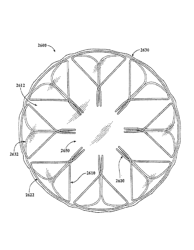
sealing portion 730 are substantially independent of the in situ device shape
or
diameter, thus providing reliable sealing across a wide variety of anatomies,
and for
dynamic anatomies (e.g., such as the GI tract).
[00118] The tissue-sealing device 800 is configured to be implanted in a
patient
such that the covering material 810 fully overlays and seals the tissue
aperture 830.
In the depicted embodiment, the covering material 810 is disposed on the
sealing
portion 730, but not on the apposition portion 720, nor the defect-occupying
portion
27
CA 03003629 2018-04-27
WO 2017/074652
PCT/US2016/055255
740. However, in some embodiments the covering material 810 can be disposed on
all or portions of the apposition portion 720 and/or the defect-occupying
portion 740
in addition to the sealing portion 730.
[00119] With reference to FIGS. 5 and 6, in some embodiments the elongate
member 710 may be wound into the aforementioned shape to create frame 700
using a winding mandrel. After winding the elongate member 710 on the mandrel,
the assembly can be heated to induce a memory shape in the elongate member 710
corresponding to the shape of the frame 700 as-wound on the mandrel. Also, the
two free ends of the elongate member 710 can be conjoined as described above.
[00120] With reference to FIGS. 7, 8A-80, and 9A-9D another exemplary tissue-
sealing device 1100 includes a frame 1000 and a covering material 1110. The
covering material 1110 is disposed at least on a sealing portion 1030 of the
frame
1000. The tissue-sealing device 1100 is shown sealing an exemplary tissue
aperture 1230. FIG. 7 is an illustration of the frame 1000 prior to attachment
of the
covering material 1110 thereto
[00121] The elongate member 1010 forms the frame 1000 that includes an
apposition portion 1020, a sealing portion 1030, and a defect-occupying
portion
1040. In the depicted embodiment, the elongate member 1010 is formed so that
the
apposition portion 1020 and the sealing portion 1030 are mirror images of each
other, however such mirror imagery is not required. Thus, in some embodiments,
the apposition portion 1020 and the sealing portion 1030 are configured
dissimilarly.
The defect-occupying portion 1040 is disposed between the apposition portion
1020
and the sealing portion 1030. Additionally, the defect-occupying portion 1040
is
configured to traverse the defect or aperture 1230 in one or more layers of
tissue.
The apposition portion 1020 and the sealing portion 1030 are configured to be
on
opposite sides of the layer(s) of tissue.
[00122] In some embodiments, the elongate member 1010 comprises a single
continuous wire that was formed in the shape of the frame 1000. The elongate
member 1010 defines one or more apposition petals 1022 that form the
apposition
portion 1020, and one or more sealing petals 1032 that form the sealing
portion
1030. In the depicted embodiment, the apposition petals 1022 and the sealing
28
CA 03003629 2018-04-27
WO 2017/074652
PCT/US2016/055255
petals 1032 include a linear portion extending radially from the defect-
occupying
portion 1040 and an essentially diamond-shaped outer portion extending from
the
linear portion at the free ends of the petals 1022 and 1032. In some
embodiments,
different shapes, and/or combinations of different shapes, can be used for the
petals
1022 and 1032.
[00123] The frame 1000 can share many of the same features and characteristics
as described above in reference to frames 100, 400, and 700. However, one
difference (in addition to the shape of the petals 1022 and 1032 as previously
described) is that the apposition portion 1020 and the sealing portion 1030
are
configured to be able to apply an increased level of appositional forces to
the
surfaces of the tissue surrounding the aperture 1230. That is because (as best
seen
in FIG. 10) the apposition petals 1022 and the sealing petals 1032 of the
frame 1000
are configured to overlap each other in their natural, unstressed states. In
other
words, the apposition petals 1022 and the sealing petals 1032 are formed to
have
concave shapes in opposite directions of each other such that the free ends of
the
apposition petals 1022 are located in the area of the sealing portion 1030 and
the
free ends of the sealing petals 1032 are located in the area of the apposition
portion
1020. This crisscrossing (or overlapping) of the apposition petals 1022 and
the
sealing petals 1032 may result in an exertion of an increased level of
appositional
forces applied to the surfaces of the tissue surrounding aperture 1230 by the
apposition petals 1022 and the sealing petals 1032. Accordingly, in some
embodiments the tissue-sealing device 1100 may tend to exhibit enhanced
conformability, sealing, and migration resistance.
[00124] When covering material 1110 is attached to sealing portion 1030, the
sealing portion 1030 (which was formed with a concaved shape as described
above)
may become partially or fully flattened. In other words, as exemplified in
FIG. 9B,
the sealing portion 1030 may become generally planar after the application of
the
covering material 1110 to the sealing petals 1032. However, in some
embodiments,
the sealing portion 1030 may remain concave (e.g., refer to FIG. 9D) after the
application of the covering material 1110 to the sealing petals 1032.
[00125] It should be understood from the description herein that, while the
apposition portion 1020 and the sealing portion 1030 of the frame 1000 are
29
CA 03003629 2018-04-27
WO 2017/074652
PCT/US2016/055255
equivalently sized and shaped in the depicted embodiment, such similarities
are not
required. For instance, in one non-limiting example, a frame of a tissue-
sealing
device can include an apposition portion including the wire loops 122 of the
frame
100 (referring to FIGS. 1A and 1B) and a sealing portion including of the
sealing
petals 1032 of the frame 1000.
[00126] The covering material 1110 can be a material as described above in
reference to covering material 210. The covering material 1110 can be attached
to
the elongate member 1010 as described above in reference to the attachment of
covering material 210 to elongate member 110.
[00127] While the exemplary tissue aperture 1230 is depicted as generally
circular,
it should be understood that the design of the tissue-sealing device 1100 (and
other
embodiments described herein) advantageously lends itself to sealing a wide
variety
of differently-sized and shaped apertures 1230. This is accomplished in part
because the defect-occupying portion 1040 is configured to exert a low level
of radial
force to the tissue aperture 1230. Additionally, the appositional force for
sealing and
migration resistance is substantially provided by the apposition portion 1020
and the
sealing portion 1030, rather than the defect-occupying portion 1040. In fact,
in some
embodiments the appositional forces provided by the apposition portion 1020
and
the sealing portion 1030 are substantially independent of the in situ device
shape or
diameter, thus providing reliable sealing across a wide variety of anatomies,
and for
dynamic anatomies (e.g., such as the GI tract).
[00128] The tissue-sealing device 1100 may be configured to be implanted in a
patient such that the covering material 1110 fully overlays and seals the
tissue
aperture 1230. In the embodiments depicted in FIGS. 8A and 80, the covering
material 1110 is disposed on the sealing portion 1030, but not on the
apposition
portion 1020, nor the defect-occupying portion 1040. However, in some
embodiments the covering material 1110 may be disposed on all or portions of
the
apposition portion 1020 and/or the defect-occupying portion 1040 in addition
to the
sealing portion 1030.
[00129] FIGS. 90 and 9D illustrate the tissue-sealing device 1100 treating a
defect
in a body lumen wall. The wall of the body lumen naturally has curvature
(i.e., it is a
CA 03003629 2018-04-27
WO 2017/074652
PCT/US2016/055255
non-planar surface). As shown in FIGS. 90 and 9D, the apposition portion 1020
is in
contact with the convexly-curved tissue wall (refer to FIG. 90) and that the
sealing
portion 1030 is in contact with the concavely-curved tissue wall (refer to
FIG. 9D).
Accordingly, the tissue-sealing device 1100 is well-suited for sealing defects
in body
lumen walls, and other tissue surfaces, that are curved or otherwise non-
planar.
[00130] Referring now to FIG. 9D, it can be seen that covering material 1110
overlays the aperture 1230 and contacts one or more portions of lumen wall
tissue
surrounding the aperture 1230. In this configuration, the covering material
1110 can
provide a scaffold to support tissue that is generated by the body's healing
process
to repair the aperture 1230. In other words, the covering material 1110 can
physically support tissue regrowth that makes the aperture 1230 smaller. In
some
cases, the aperture 1230 may have been created by a full thickness resection
of an
intestine. In other cases, other types of body tissues, or other types of
defect causes
can be treated in the aforementioned fashion.
[00131] The elongate member 1010 can be wound into the aforementioned shape
to create frame 1000 using an appropriate winding mandrel 1300. After winding
the
elongate member 1010 on the mandrel, the assembly can be heated to induce a
memory shape in the elongate member 1010 corresponding to the shape of the
frame 1000 as-wound on the mandrel. Also, the two free ends of the elongate
member 1010 can be conjoined as described above.
[00132] With reference to FIGS. 10A, 10B, 11A, and 11B, another exemplary
tissue-sealing device 1400 that includes a frame 1410 and a covering material
1412
is illustrated. The covering material 1412 is disposed at least on a sealing
portion
1430 of the frame 1410. The tissue-sealing device 1400 is shown sealing an
exemplary tissue aperture 1530.
[00133] The frame 1410 includes an apposition portion 1420, a sealing portion
1430, and a defect-occupying portion 1440. In the depicted embodiment, the
apposition portion 1420 and the sealing portion 1430 are configured
dissimilarly.
That is, the apposition portion 1420 includes one or more narrow wire loops
1422
and the sealing portion 1430 includes one or more wider petals 1432. The
defect-
occupying portion 1440 is disposed between the apposition portion 1420 and the
31
CA 03003629 2018-04-27
WO 2017/074652
PCT/US2016/055255
sealing portion 1430. In addition, the defect-occupying portion 1440 may be
configured to traverse the defect or aperture 1530 in one or more layers of
tissue.
Also, the apposition portion 1420 and the sealing portion 1430 may be
configured to
be on opposite sides of the layer(s) of tissue.
[00134] In some embodiments, the frame 1410 includes a single continuous wire
that was bent to form the frame 1410. The frame 1410 defines apposition wire
loops
1422 that form the apposition portion 1420, and sealing petals 1432 that form
the
sealing portion 1430. In the depicted embodiment, the apposition wire loops
1422
are shaped essentially as fingers, and the sealing petals 1432 are shaped
essentially
as diamonds on the ends of linear portions that extend from the central defect-
occupying portion 1440. In some embodiments, different shapes and combinations
of different shapes can be used for the wire loops 1422 and petals 1432. The
use of
dissimilar shapes for the apposition wire loops 1422 and the sealing petals
1432 can
beneficially provide the opportunity to individually optimize the
configurations of the
apposition portion 1420 independently from those of the sealing portion 1430.
For
example, the apposition portion 1420 may be optimized for crushability or for
conformability with irregular tissue topography, and the sealing portion 1430
may be
optimized for sealing. In some embodiments, other performance characteristics
or
combinations of performance characteristics can be selected for optimization
in
relation to the apposition portion 1420 and the sealing portion 1430,
individually.
[00135] The frame 1410 can share many of the same features and characteristics
as described above in reference to frames 100, 400, 700, and 1000. For
example,
the wind pattern of the frame 1410 results in defining a peripheral frame for
the
sealing portion 1430. In addition, the apposition wire loops 1422 of the
apposition
portion 1420 and the sealing petals 1432 of the sealing portion 1430 are
formed to
have overlap (crisscross) for enhanced apposition force capability. The
covering
material 1412 may be a material as described above in reference to covering
material 210. The covering material 1412 can be attached to the frame 1410 as
described above in reference to the attachment of covering material 210 to
elongate
member 110.
[00136] While the exemplary tissue aperture 1530 is depicted as generally
circular,
it should be understood that the design of the tissue-sealing device 1400 (and
other
32
CA 03003629 2018-04-27
WO 2017/074652
PCT/US2016/055255
embodiments described herein) advantageously lends itself to sealing a wide
variety
of differently-sized and shaped apertures 1530. This is accomplished in part
because the defect-occupying portion 1440 is configured to exert a low level
of radial
force to the tissue aperture 1530. Additionally, the appositional force for
sealing and
migration resistance is substantially provided by the apposition portion 1420
and the
sealing portion 1430, rather than the defect-occupying portion 1440. In fact,
in some
embodiments the appositional forces provided by the apposition portion 1420
and
the sealing portion 1430 are substantially independent of the in situ device
shape or
diameter, thus providing reliable sealing across a wide variety of anatomies,
and for
dynamic anatomies (e.g., such as the GI tract).
[00137] The tissue-sealing device 1400 may be configured to be implanted in a
patient such that the covering material 1412 fully overlays and seals the
tissue
aperture 1530. In the embodiments shown in FIGS. 10A and 10B, the covering
material 1412 is disposed on the sealing portion 1430, but not on the
apposition
portion 1420, nor the defect-occupying portion 1440. However, in some
embodiments the covering material 1412 may be disposed on all or portions of
the
apposition portion 1420 and/or the defect-occupying portion 1440 in addition
to the
sealing portion 1430.
[00138] With reference to FIG. 12, another exemplary tissue-sealing device
1600
is shown that includes a frame 1610 that defines one or more apposition wire
loops
1622 and one or more sealing petals 1632. In some embodiments, a covering
material 1612 is disposed on at least portions of the sealing petals 1632.
[00139] In some embodiments, the tissue-sealing device 1600 includes all of
the
characteristics and features of the tissue-sealing device 1400. In addition,
the
apposition wire loops 1622 of the tissue-sealing device 1600 include rings
1624 near
the free ends of the apposition wire loops 1622. In some embodiments, the
rings
1624 are integrally formed as part of the winding process of the frame 1610.
In
some embodiments, the rings 1624 are formed as separate components that are
subsequently attached to the frame 1610. It should be understood that the
rings
1624 can be combined with all embodiments of tissue-sealing device embodiments
and anastomosis device embodiments provided herein. In some embodiments, the
rings 1624 may be positioned on other locations of the frame 1610, and more or
33
CA 03003629 2018-04-27
WO 2017/074652
PCT/US2016/055255
fewer rings 1624 may be included. For example, in some embodiments, the rings
1624 may be positioned on the sealing petals 1632 instead of, or in addition
to,
having the rings 1624 positioned on the apposition wire loops 1622.
[00140] In some embodiments, a flexible member 1640 is threaded through each
of the rings 1624, so that the flexible member 1640 forms a closed and/or
tensionable loop. The flexible member 1640 may be a cord, wire, strap, suture,
and
the like. In some embodiments, the flexible member 1640 can be made of a
polymer
material including, but not limited to, nylon, polypropylene,
polytetrafluoroethylene
(PTFE), silk, and the like. In some embodiments, the flexible member 1640 may
be
made of a metallic material including, but not limited to, nitinol, aluminum,
stainless
steel, and the like. In additional embodiments, the flexible member 1640 can
be
made of a combination of materials. The flexible member 1640 may be made of
monofilament, twisted strands, braided strands, and the like. In some
embodiments,
the flexible member 1640 may be attached to one or more rings 1624, and
slidably
engaged with the other rings 1624. In some embodiments, the flexible member
1640
is slidably engaged with all of the rings 1624.
[00141] Pulling on (tensioning) the flexible member 1640 can cause a purse
string
effect. That is, pulling on the flexible member 1640 can draw the apposition
wire
loops 1622 towards each other. Such an action can be performed beneficially as
a
part of the process of crushing the tissue-sealing device 1600 to a low-
profile
configuration for the purpose of installing the device 1600 into a lumen of a
sheath.
That action can be performed when initially installing the device 1600 into a
delivery
sheath, or when recovering the device 1600 in situ so that the device 1600 can
be
retrieved and removed from a body using a transcatheter removal technique. For
example, applying tension to the flexible member 1640 using a grasping tool
can
cause the tissue-sealing device 1600 to collapse to a lower-profile
configuration for
insertion in a retrieval sheath.
[00142] For example, when retrieval of the tissue-sealing device 1600 from the
body is desired, a retrieval sheath containing a grasping tool can be routed
to the
location of the tissue-sealing device 1600 in the patient's body. The grasping
tool
can be used to temporarily couple with the flexible member 1640. As the
grasping
tool is thereafter retracted away from the tissue-sealing device 1600, tension
is
34
CA 03003629 2018-04-27
WO 2017/074652
PCT/US2016/055255
applied to the flexible member 1640. The tensioning and displacement of the
flexible
member 1640 caused by the grasping tool will cause the apposition wire loops
1622
to collapse to a lower-profile configuration. As the grasping tool is
retracted further,
including to within the retrieval sheath, the apposition wire loops 1622 will
be drawn
into the distal end of the retrieval sheath. A funnel can be included on the
distal end
portion of the retrieval sheath. The funnel will provide a wider initial
opening at the
distal tip of the retrieval sheath to facilitate the capture of all portions
of the
apposition wire loops 1622. As the grasping tool is further retracted, the
entire
tissue-sealing device 1600 can be pulled into the lumen of the retrieval
sheath. Then
the retrieval sheath, containing the tissue-sealing device 1600, can be
removed from
the patient. Retrieval features of various types and configurations, such as
the
flexible member 1640, may be included with any of the tissue-sealing device
embodiments provided herein, if so desired.
[00143] With reference to FIGS. 13A and 13B, the tissue-sealing device 1600
(as
well as some embodiments of the other tissue-sealing device embodiments and
anastomosis device embodiments provided herein) can be configured in a low-
profile
configuration for containment within a lumen of a sheath 1700. The sheath 1700
may be used for the initial installation of the tissue-sealing device 1600 in
a body, or
the sheath 1700 may be used for in situ retrieval of the tissue-sealing device
1600
from the body. The tissue-sealing device 1600 can be configured for self-
expansion
upon removal of the constraining forces resulting from containment within the
lumen
of the sheath 1700. That is, the tissue-sealing device 1600 can self-expand
once
liberated from the sheath 1700. To arrive at a low-profile (crushed)
configuration
such that the tissue-sealing device 1600 can fit within the sheath 1700, in
some
embodiments, portions of the tissue-sealing device 1600 may be folded one or
more
times.
[00144] In some embodiments, a sheath 1700 having about a 15 Fr. (5 mm) outer
diameter can be used. However, in some embodiments, sheaths that are smaller
or
larger than 15 Fr. can be used. For example, sheaths that have outer diameters
of 6
Fr., 7 Fr., 8 Fr., 9 Fr., 10 Fr., 11 Fr., 12 Fr., 13 Fr., 14 Fr., 16 Fr., 17
Fr., 18 Fr., 19
Fr., 20 Fr., and larger than 20 Fr., can be used in some embodiments.
CA 03003629 2018-04-27
WO 2017/074652
PCT/US2016/055255
[00145] In some embodiments, a pusher catheter 1710 is slidably disposed
within
the lumen of the sheath 1700. The pusher catheter 1710 can be, for example,
manually used by a clinician operator to force the tissue-sealing device 1600
out of
the lumen of the sheath 1700 when the distal tip of the sheath 1700 is
positioned as
desired at a target implantation site within a body, thereby deploying the
tissue-
sealing device 1600.
[00146] In the configuration shown in FIGS. 13A and 13B, the apposition
portion is
contained within the lumen of the sheath 1700 proximally of the sealing
portion. In
other words, deployment of the tissue-sealing device 1600 from the sheath 1700
will
result in the emergence of the sealing portion of the tissue-sealing device
1600 prior
to the emergence of the apposition portion. In some situations, it may be
important
to approach the target tissue defect from a direction with the orientation of
the tissue-
sealing device 1600 in relation to the sheath 1700 in mind. For example, when
a
defect in a body lumen wall is to be treated using the tissue-sealing device
1600,
generally the sealing portion should be positioned within the body lumen (to
seal the
body lumen contents within the body lumen). Therefore, when the orientation of
the
tissue-sealing device 1600 in relation to the sheath 1700 is as shown in FIGS
13A
and 13B, the approach to the body lumen should be from the outside of the body
lumen (e.g., laproscopically). That way, the sealing portion can be deployed
through
the defect so that the sealing portion is positioned inside of the body lumen.
Then,
by pulling back the sheath 1700, the apposition portion can be appropriately
positioned on the outside surface of the body lumen.
[00147] In other configurations, the tissue-sealing device 1600 may be
contained
within the sheath 1700 such that the sealing portion is proximal of the
apposition
portion. In that configuration, the approach to the body lumen defect can be
from
within the lumen (e.g., using an endoscopic technique). That way, the
apposition
portion can be deployed through the defect so that the apposition portion is
positioned outside of the body lumen. Then, by pulling back the sheath 1700,
the
sealing portion can be appropriately positioned on the inside surface of the
body
lumen.
[00148] In some medical procedures for deploying the tissue-sealing device
1600,
the deployment process is performed using radiographic visualization or
another
36
CA 03003629 2018-04-27
WO 2017/074652
PCT/US2016/055255
imaging modality. As described above, some embodiments of the tissue-sealing
device 1600 and other device embodiments provided herein are retrievable after
deployment. Therefore, if the initial deployment position is deemed
dissatisfactory,
the tissue-sealing device 1600 can be fully or partially retrieved into the
sheath 1700
and redeployed to a more desirable position.
[00149] With reference to FIGS. 14A and 14B, another exemplary tissue-sealing
device 1800 includes a frame 1810 and a covering material 1812 is shown. The
covering material 1812 is disposed on at least on a sealing portion 1830 of
the frame
1810. The tissue-sealing device 1800 is shown sealing an exemplary tissue
aperture
1850.
[00150] The frame 1810 includes an apposition portion 1820, a sealing portion
1830, and a defect-occupying portion positioned therebetween. In the
embodiment
depicted in FIGS. 14A and 14B, the apposition portion 1820 and the sealing
portion
1830 are configured dissimilarly. That is, the apposition portion 1820
includes one or
more narrow wire loops 1822, and the sealing portion 1830 includes one or more
wider petals 1832. The apposition portion 1820 and the sealing portion 1830
may be
configured to be on opposite sides of the layer(s) of tissue.
[00151] In some embodiments, the frame 1810 includes a single continuous wire
that has been bent to form the frame 1810. The frame 1810 defines apposition
wire
loops 1822 that form the apposition portion 1820, and sealing petals 1832 that
form
the sealing portion 1830. In the depicted embodiment, the apposition wire
loops
1822 are shaped essentially as fingers, and the sealing petals 1832 are shaped
essentially as sectors of a circle. In some embodiments, each petal 1832 of
the one
or more petals 1832 is configured to generally abut at least portions of
adjacent
petals 1832, while not overlapping adjacent petals 1832. In some embodiments,
one
or more of the petals 1832 may be configured to be separated from adjacent
petals
1832. In some embodiments, one or more of the petals 1832 may be configured to
at least partially overlap adjacent petals 1832.
[00152] In some embodiments, different shapes and combinations of different
shapes can be used for the wire loops 1822 and petals 1832. The use of
dissimilar
shapes for the apposition wire loops 1822 and the sealing petals 1832 can
37
CA 03003629 2018-04-27
WO 2017/074652
PCT/US2016/055255
beneficially provide the opportunity to individually optimize the
configurations of the
apposition portion 1820 independently from those of the sealing portion 1830.
For
example, in some embodiments the apposition portion 1820 may be optimized for
crushability or for conformability with irregular tissue topography, and the
sealing
portion 1830 may be optimized for sealing. In some embodiments, other
performance characteristics or combinations of performance characteristics can
be
selected for optimization in relation to the apposition portion 1820 and the
sealing
portion 1830, individually.
[00153] The frame 1810 can share many of the same features and characteristics
as described above in reference to frames 100, 400, 700, 1000, 1400, and 1600.
For example, the wind pattern of the frame 1810 results in defining a
peripheral
frame for the sealing portion 1830. The covering material 1812 can be a
material as
described above in reference to covering material 210. The covering material
1812
can be attached to the frame 1810 as described above in reference to the
attachment of covering material 210 to elongate member 110.
[00154] While the tissue aperture 1850 is depicted as generally circular, it
should
be understood that the design of the tissue-sealing device 1800 (and other
embodiments described herein) advantageously lends itself to sealing a wide
variety
of differently-sized and shaped apertures 1850. This is accomplished in part
because the appositional force for sealing and migration resistance is
substantially
provided by the apposition portion 1820 and the sealing portion 1830, rather
than the
defect-occupying portion. In fact, in some embodiments the appositional forces
provided by the apposition portion 1820 and the sealing portion 1830 are
substantially independent of the in situ device shape or diameter, thus
providing
reliable sealing across a wide variety of anatomies, and for dynamic anatomies
(e.g.,
such as the GI tract).
[00155] The tissue-sealing device 1800 may be configured to be implanted in a
patient such that the covering material 1812 fully overlays and seals the
tissue
aperture 1850. In the depicted embodiment, the covering material 1812 is
disposed
on the sealing portion 1830, but not on the apposition portion 1820, nor the
defect-
occupying portion. However, in some embodiments the covering material 1812 may
38
CA 03003629 2018-04-27
WO 2017/074652
PCT/US2016/055255
be disposed on all or portions of the apposition portion 1820 and/or the
defect-
occupying portion in addition to the sealing portion 1830.
[00156] The apposition wire loops 1822 of the tissue-sealing device 1800
include
rings 1824 near the free ends of the apposition wire loops 1822. In some
embodiments, the rings 1824 are integrally formed as part of the winding
process of
the frame 1810. In some embodiments, the rings 1824 are formed as separate
components that are subsequently attached to the frame 1810. It is to be
appreciated that the rings 1824 can be combined with all embodiments of tissue-
sealing devices provided herein. In some embodiments, the rings 1824 may be
positioned on other locations of the frame 1810, and more or fewer rings 1824
may
be included. For example, in some embodiments the rings 1824 may be positioned
on the sealing petals 1832 instead of, or in addition to, having the rings
1824
positioned on the apposition wire loops 1822.
[00157] In some embodiments, a flexible member 1860 is threaded through each
of the rings 1824 so that the flexible member 1860 forms a closed and/or
tensionable
loop. The flexible member 1860 may be a cord, wire, strap, suture, and the
like, and
can be constructed of the materials as described above in reference to the
flexible
member 1640. In some embodiments, the flexible member 1860 may be attached to
one or more rings 1824, and slidably engaged with the other rings 1824. In
some
embodiments, the flexible member 1860 is slidably engaged with all of the
rings
1824.
[00158] Pulling on (tensioning) the flexible member 1860 can cause a purse
string
effect. That is, pulling on the flexible member 1860 can draw the apposition
wire
loops 1824 towards each other. Such an action can be performed to crush the
tissue-sealing device 1800 to a low-profile configuration for installing the
device 1800
into a lumen of a sheath. The crushing action can be useful when initially
installing
the device 1800 into a delivery sheath, or when recovering the device 1800 in
situ so
that the device 1800 can be retrieved and removed from a body using a
transcatheter removal technique.
[00159] With reference to FIGS. 15A and 15B, another exemplary tissue-sealing
device 1900 includes a frame 1910 and a covering material 1912 is illustrated.
The
39
CA 03003629 2018-04-27
WO 2017/074652
PCT/US2016/055255
covering material 1912 is disposed at least on a sealing portion 1930 of the
frame
1910. The tissue-sealing device 1900 is shown sealing an exemplary tissue
aperture
1950.
[00160] The frame 1910 includes an apposition portion 1920, a sealing portion
1930, and a defect-occupying portion positioned therebetween. In the depicted
embodiment, the apposition portion 1920 and the sealing portion 1930 are
configured dissimilarly. That is, the apposition portion 1920 includes one or
more
narrow wire loops 1922, and the sealing portion 1930 includes one or more
wider
petals 1932. The apposition portion 1920 and the sealing portion 1930 may be
configured to be on opposite sides of the layer(s) of tissue.
[00161] In some embodiments, the frame 1910 includes a single continuous wire
that has been bent to form the frame 1910. The frame 1910 defines apposition
wire
loops 1922 that form the apposition portion 1920, and sealing petals 1932 that
form
the sealing portion 1930. In the depicted embodiment, the apposition wire
loops
1922 are shaped essentially as elongate wire loops, and the sealing petals
1932 are
shaped essentially as teardrops. In some embodiments, each petal 1932 of the
one
or more petals 1932 is configured to generally abut at least portions of
adjacent
petals 1932, while not overlapping adjacent petals 1932. In additional
embodiments,
one or more of the petals 1932 are configured to be separated from adjacent
petals
1932. In some embodiments, one or more of the petals 1932 are configured to at
least partially overlap adjacent petals 1932.
[00162] In some embodiments, different shapes and combinations of different
shapes can be used for the wire loops 1922 and petals 1932. The use of
dissimilar
shapes for the apposition wire loops 1922 and the sealing petals 1932 can
beneficially provide the opportunity to individually optimize the
configurations of the
apposition portion 1920 independently from those of the sealing portion 1930.
[00163] The frame 1910 can share many of the same features and characteristics
as described above in reference to frames 100, 400, 700, 1000, 1400, 1600, and
1800. For example, the wind pattern of the frame 1910 results in defining a
peripheral frame for the sealing portion 1930. The covering material 1912 can
be a
material as described above in reference to covering material 210. The
covering
CA 03003629 2018-04-27
WO 2017/074652
PCT/US2016/055255
material 1912 can be attached to the frame 1910 as described above in
reference to
the attachment of covering material 210 to elongate member 110.
[00164] While the exemplary tissue aperture 1950 is depicted as generally
circular,
it should be understood that the design of the tissue-sealing device 1900 (and
other
embodiments described herein) advantageously lends itself to sealing a wide
variety
of differently-sized and shaped apertures 1950.
[00165] The tissue-sealing device 1900 may be configured to be implanted in a
patient such that the covering material 1912 fully overlays and seals the
tissue
aperture 1950. In the embodiment shown in FIGS. 15A and 15B, the covering
material 1912 is disposed on the sealing portion 1930, but not on the
apposition
portion 1920, nor the defect-occupying portion. However, in some embodiments
the
covering material 1912 may be disposed on all or portions of the apposition
portion
1920 and/or the defect-occupying portion in addition to the sealing portion
1930.
[00166] The apposition wire loops 1922 of the tissue-sealing device 1900
include
rings 1924 near the free ends of the apposition wire loops 1922. In some
embodiments, the rings 1924 are integrally formed as part of the winding
process of
the frame 1910. In some embodiments, the rings 1924 are formed as separate
components that are subsequently attached to the frame 1910. It should be
understood that the rings 1924 can be combined with all embodiments of tissue-
sealing devices provided herein. In some embodiments, the rings 1924 are
positioned on other locations of the frame 1910, and more or fewer rings 1924
may
be included. For example, in some embodiments the rings 1924 can be positioned
on the sealing petals 1932 instead of, or in addition to, having the rings
1924
positioned on the apposition wire loops 1922.
[00167] In some embodiments, a flexible member 1960 is threaded through each
of the rings 1924, so that the flexible member 1960 forms a closed and/or
tensionable loop. The flexible member 1960 may be a cord, wire, strap, suture,
and
the like, and can be constructed of the materials as described above in
reference to
the flexible member 1640. In some embodiments, the flexible member 1960 may be
attached to one or more rings 1924, and slidably engaged with the other rings
1924.
41
CA 03003629 2018-04-27
WO 2017/074652
PCT/US2016/055255
In some embodiments, the flexible member 1960 is slidably engaged with all of
the
rings 1924.
[00168] Pulling on (tensioning) the flexible member 1960 can cause a purse
string
effect. That is, pulling on the flexible member 1960 can draw the apposition
wire
loops 1924 towards each other. Such an action can be performed to crush the
tissue-sealing device 1900 to a low-profile configuration for installing the
device 1900
into a lumen of a delivery sheath or when recovering the device 1900 in situ
so that
the device 1900 can be retrieved and removed from a body using a transcatheter
removal technique.
[00169] A wire winding mandrel may be used in some embodiments to create the
frame 1810 of tissue-sealing device 1800. That is, an elongate member can be
wound to create frame 1810 using a suitable winding mandrel. After forming the
frame 1810 on the mandrel, the assembly can be heated to induce a memory shape
in the frame 1810 corresponding to the shape of the mandrel. Also, the two
free
ends of the elongate member can be conjoined as described above. In some
embodiments, the two free ends of the elongate member are not conjoined.
Similarly, a wire winding mandrel 2100 can also be used to create the frame
1910 of
tissue-sealing device 1900. That is, an elongate member can be wound to create
frame 1910 using a suitable winding mandrel.
[00170] The use of occlusive devices in the environment of the GI tract, for
example, calls for occlusive devices that provide substantially continuous
lumen wall
contact with apposition force for effective sealing performance during
peristaltic
motion. Peristaltic motion can result in the application of large dynamic,
asymmetric,
and non-planar displacements to the occlusive devices in some circumstances,
as
well as normal and shear stresses from material transport. In some
embodiments,
the occlusive devices provided herein provide substantially continuous lumen
wall
contact with conformability and apposition force for effective occlusion and
sealing
performance during such conditions caused by peristaltic motion. For example,
in
some embodiments the occlusion device's provision of apposition force without
the
use of barbs or prongs allows the device to resist migration, seal, and be
safely
removed.
42
CA 03003629 2018-04-27
WO 2017/074652
PCT/US2016/055255
[00171] Referring now to FIGS. 16A-160, an exemplary anastomosis device 2200
can be constructed using many of the same design features, characteristics,
concepts, and methods of construction that also pertain to the tissue-sealing
devices
described above. However, the anastomosis device 2200 (and the other
anastomosis device embodiments provided herein) is distinct from the tissue-
sealing
devices at least because of a central aperture 2250 that can facilitate
material (e.g.,
biological materials) transfer therethrough. That is, while some portions of
the
anastomosis device 2200 are configured to substantially seal against
surrounding
tissue surfaces, the central aperture 2250 of the anastomosis device 2200 is
configured to not seal, but rather to facilitate the transfer of materials
(e.g., fluids,
solids, mixtures) through the central aperture 2250 generally along a central
axis
2216.
[00172] The anastomosis device 2200 includes a frame 2210 formed by an
elongate member 2212. In some embodiments, the elongate member 2212 is a
single element that is wound or otherwise formed to construct the frame 2210
of the
anastomosis device 2200 (e.g., as described above in reference to the tissue-
sealing
devices). In some embodiments, the elongate member 2212 can include two or
more elements that are cooperatively configured to define the frame 2210. The
elongate member 2212 may be constructed using the techniques, and can be made
of the types of materials, that are described above in reference to elongate
member
110, for example.
[00173] In some embodiments, the elongate member 2212 forms a first flange
2220, a second flange 2230, and a connecting region 2240. The flanges 2220 and
2230 may also be referred to herein as "apposition portions." The connecting
region
2240 may also be referred to herein as a "central portion." The connecting
region
2240 is disposed between and interconnects the first flange 2220 and the
second
flange 2230. The connecting region 2240 is configured to traverse an opening
or
aperture 2244 in one or more layers of tissue. The first flange 2220 and the
second
flange 2230 are configured to be on opposite sides of the layer(s) of tissue
and to
apply apposition forces against the tissue surfaces.
[00174] The elongate member 2212 defines one or more apposition petals 2222
that form the first flange 2220. The elongate member 2212 also forms one more
43
CA 03003629 2018-04-27
WO 2017/074652
PCT/US2016/055255
apposition petals 2232 that form the second flange 2230. In the depicted
embodiment, the first flange 2220 includes five apposition petals 2222 and the
second flange 2230 also includes five apposition petals 2232. The apposition
petals
2222 and 2232 may also be referred to herein as "arms," "fins," "loops,"
"apposition
members," or "fingers," for example.
[00175] While the depicted embodiment includes five apposition petals 2222 and
2232, it should be understood that some embodiments include other quantities
of
apposition petals 2222 and 2232. That is, the first flange 2220 and/or the
second
flange 2230 may include more than or less than five apposition petals 2222 and
2232. Further, in some embodiments the quantity of apposition petals 2222 may
be
different than the quantity of apposition petals 2232. Still further, the
sizes and
shapes (also referred to herein as the geometry) of the apposition petals 2222
may
be different than the sizes and shapes of the apposition petals 2232. In some
embodiments, the axes of one or more of the individual apposition petals 2222
may
be offset (e.g., skew) from the axes of one or more of the individual
apposition petals
2232. In some embodiments, the axes of one or more of the apposition petals
2222
may be parallel with the axes of one or more of the apposition petals 2232.
[00176] In the embodiment depicted in FIGS. 16A-160, the apposition petals
2222
and 2232 are shaped essentially as segments of an annulus (e.g., approximately
trapezoidal). In some embodiments, a variety of different petal geometries
and/or
combinations of different petal geometries can be used for the apposition
petals
2222 and 2232. In the depicted embodiment, the apposition petals 2222 and 2232
abut each other. In some embodiments, some or all of the apposition petals
2222
and 2232 may partially overlap adjacent apposition petals 2222 and 2232, or
some
or all of the apposition petals 2222 and 2232 may be spaced apart from
adjacent
apposition petals 2222 and 2232. Again, such configurations may be used either
uniformly or differently for the first flange 2220 in comparison to the second
flange
2230.
[00177] In the depicted embodiment, the apposition petals 2222 are generally
parallel to the apposition petals 2232 (as best seen in FIG. 160), and the
apposition
petals 2222 and 2232 are distanced apart from each other. However, in some
embodiments the apposition petals 2222 and 2232 may be formed to make at least
44
CA 03003629 2018-04-27
WO 2017/074652
PCT/US2016/055255
partial contact with each other when no materials are therebetween. This and
other
such configurations of the apposition petals 2222 and 2232 may also be
referred to
herein as a pre-strained geometry of the apposition petals 2222 and 2232. Such
a
configuration may increase the amount of apposition force applied by the
apposition
petals 2222 and 2232 in comparison to the embodiment having parallelism
between
the apposition petals 2222 and 2232. Further, in some embodiments that have
the
apposition petals 2222 and/or 2232 that are spaced apart from adjacent
apposition
petals 2222 and/or 2232, and that have an axially offset between the
individual
petals of the first flange 2220 and the second flange 2230, some or all of the
apposition petals 2222 and 2232 may be formed to crisscross each other when no
materials are therebetween. Such a configuration may further increase the
amount
of apposition force applied by the apposition petals 2222 and 2232 in
comparison to
the embodiment having contact between the apposition petals 2222 and 2232.
Combinations of all such configurations are also envisioned and are considered
to
be within the scope of this disclosure.
[00178] Still referring to FIGS. 16A, 16B, and 16C, the exemplary anastomosis
device 2200 also includes a covering material 2214. The covering material 2214
can
be disposed on and/or attached to at least portions of the elongate member
2212. In
the depicted embodiment, the covering material 2214 is attached to the
apposition
petals 2222 and 2232 and to the connecting region 2240, while leaving the
aperture
2250 uncovered. The covering material 2214 can be a material as described
above
in reference to covering material 210. The covering material 2214 can be
attached
to the elongate member 2212 as described above in reference to the attachment
of
covering material 210 to elongate member 110. In some embodiments, the
covering
material 2214 cooperates with the framework 2210 to provide a circumferential
seal
at the outer peripheral edge of the first flange 2220 and/or the second flange
2230.
[00179] Referring to FIG. 16B in particular, the exemplary anastomosis device
2200 is depicted as being deployed in one or more layers of tissue that have
the
opening 2244. While the exemplary tissue opening 2244 is depicted as generally
circular, it should be understood that the design of the anastomosis device
2200
(and other anastomosis device embodiments described herein) advantageously
lends itself to conforming to a wide variety of differently-sized and shaped
tissue
CA 03003629 2018-04-27
WO 2017/074652
PCT/US2016/055255
openings 2244. That is accomplished, at least in part, because the connecting
region 2240 is configured to exert a low level of radial force to the tissue
opening
2244. Additionally, the appositional force for sealing and migration
resistance is
substantially delivered by the first and second flanges 2220 and 2230, rather
than
the connecting region 2240. In fact, in some embodiments the appositional
forces
delivered by the first and second flanges 2220 and 2230 are substantially
independent of the in situ device shape or diameter, thus providing reliable
sealing,
migration resistance, and anastomosis performance across a wide variety of
anatomic topographies, and for dynamic anatomies (e.g., such as the GI tract).
[00180] The exemplary anastomosis device 2200 (and other anastomosis device
embodiments described herein) can be deployed using the devices and techniques
described above in reference to FIGS. 13A and 13B, for example.
[00181] In some embodiments, the exemplary anastomosis device 2200 (and other
anastomosis device embodiments described herein) substantially do not
interfere
with the healing response of the body, such as when two tissues that are
anastomosed using the devices provided herein grow together to form a tissue-
anastomosis. In some embodiments, the anastomosis devices described herein are
configured to be removable after deployment (such as after the anastomosed
tissues
have grown together). Therefore, in some such embodiments the anastomosis
devices described herein are configured to prevent or inhibit tissue ingrowth,
and are
designed for atraumatic withdrawal. For example, in some embodiments the
anastomosis devices described herein are configured to adequately seal and
resist
migration by exerting apposition forces without the use of barbs or prongs
(thereby
facilitating removal of the devices in a substantially atraumatic manner). In
some
such embodiments, the anastomosis devices described herein may include
features
to facilitate efficient repositioning and/or retrieval such as, but not
limited to, rings on
one or more of the apposition petals 2222 and 2232 and a flexible member that
is
threaded through such rings (e.g., refer to tissue-sealing devices 1800 and
1900
described above).
[00182] In reference to FIGS. 17A-170, another exemplary anastomosis device
2300 is shown that may be constructed using many of the same design features,
characteristics, concepts, and methods of construction that pertain to the
46
CA 03003629 2018-04-27
WO 2017/074652
PCT/US2016/055255
anastomosis device 2200 described above, as well as to the tissue-sealing
devices
described above. As with the anastomosis device 2200, the anastomosis device
2300 (and the other anastomosis device embodiments provided herein) is
distinct
from the tissue-sealing devices at least because of a central aperture 2350
that can
facilitate material transfer therethrough. That is, while some portions of the
anastomosis device 2300 are configured to substantially seal against
surrounding
tissue surfaces, the central aperture 2350 of the anastomosis device 2300 is
configured to not seal, but rather to facilitate the transfer of materials
(e.g., fluids,
solids, mixtures) through the central aperture 2350 along a central axis 2316.
[00183] The anastomosis device 2300 includes a frame 2310 formed by an
elongate member 2312. In some embodiments, the elongate member 2312 is a
single element that is wound or otherwise formed to construct the frame 2310
of the
anastomosis device 2300 (e.g., like described above in reference to the tissue-
sealing devices). In some embodiments, the elongate member 2312 can include
two
or more elements that are cooperatively configured to define the frame 2310.
The
elongate member 2312 may be constructed using the techniques, and may be made
of the types of materials that are described above in reference to elongate
member
110, for example.
[00184] In some embodiments, the elongate member 2312 forms a first flange
2320, a second flange 2330, and a connecting region 2340. The connecting
region
2340 is disposed between and interconnects the first flange 2320 and the
second
flange 2330. The connecting region 2340 is configured to traverse an opening
or
aperture 2344 in one or more layers of tissue. The first flange 2320 and the
second
flange 2330 may be configured to be on opposite sides of the layer(s) of
tissue and
to apply apposition forces against the tissue surfaces.
[00185] The elongate member 2312 defines one or more apposition petals 2322
that form the first flange 2320. The elongate member 2312 also forms one more
apposition petals 2332 that form the second flange 2330. In the embodiment
depicted in FIGS. 17A-170, the first flange 2320 includes five apposition
petals 2322
and the second flange 2330 includes five apposition petals 2332. In other
embodiments, fewer or more than five petals 2322 and/or 2332 may be included,
and the flanges 2320 and 2330 may have unequal numbers of petals 2322 and
47
CA 03003629 2018-04-27
WO 2017/074652
PCT/US2016/055255
2332. The geometry of the apposition petals 2322 may also be different than
the
geometry of the apposition petals 2332. In some embodiments, the axes of one
or
more of the individual apposition petals 2322 may be offset (e.g., skew) from
the
axes of one or more of the individual apposition petals 2332. In some
embodiments,
the axes of one or more of the apposition petals 2322 may be parallel with the
axes
of one or more of the apposition petals 2332.
[00186] While the frame 2310 can share many of the same features and
characteristics as described above in reference to frame 2210, one difference
is that
the apposition petals 2322 and 2332 may be configured with concave shapes (as
best seen in FIG. 170). Such concavity can allow the flanges 2320 and 2330 to
apply an increased level of appositional forces to the surfaces of the tissue
surrounding the aperture 2344, as compared to the appositional force caused by
generally planar petals. In addition, the concave shape of the flanges 2320
and
2330 concentrates the apposition force at the outer perimeter the frame 2310,
creating greater pressure on the tissue in comparison to planar flanges that
distribute
apposition force over a larger area. Additionally, the concave shape of the
flanges
2320 and 2330 enables the accommodation of a broad range of tissue thickness.
Further, in some embodiments the apposition petals 2322 and 2332 may be
configured to partially contact each other or to crisscross each other (as
described
above), thereby providing increased apposition force capabilities.
Accordingly, in
some embodiments the anastomosis device 2300 may exhibit enhanced
conformability, sealing, and migration resistance. Further, the concavity of
the
apposition petals 2322 and 2332 can allow the anastomosis device 2300 to be
used
effectively in conjunction with a broad range of tissue thicknesses. In some
embodiments, one or the other of the flanges 2320 and 2330 may include
concaved
petals while the other of the flanges 2320 and 2330 may include petals of
another
contour (e.g., planar petals).
[00187] The exemplary anastomosis device 2300 also includes a covering
material
2314. The covering material 2314 can be disposed on or around and/or attached
to
at least portions of the elongate member 2312. In the embodiment shown in
FIGS.
17A-170, the covering material 2314 is attached to the apposition petals 2322
and
2332 and to the connecting region 2340, leaving the aperture 2350 uncovered.
The
48
CA 03003629 2018-04-27
WO 2017/074652
PCT/US2016/055255
covering material 2314 can be a material as described above in reference to
covering material 210. Additionally, the covering material 2314 can be
attached to
the elongate member 2312 as described above in reference to the attachment of
covering material 210 to elongate member 110. In some embodiments, the
covering
material 2314 cooperates with the framework 2310 to provide a circumferential
seal
at the outer peripheral edge of the first flange 2320 and/or the second flange
2330.
[00188] Referring to FIG. 17B in particular, the exemplary anastomosis device
2300 is depicted as being deployed in one or more layers of tissue that have
an
opening 2344. While the exemplary tissue opening 2344 is depicted as generally
circular, it should be understood that the design of the anastomosis device
2300
(and other anastomosis device embodiments described herein) advantageously
lends itself to conforming to a wide variety of differently-sized and shaped
tissue
openings 2344. That is accomplished, at least in part, because the connecting
region 2340 is configured to exert a low level of radial force to the tissue
opening
2344. Additionally, the appositional force for sealing and migration
resistance is
substantially delivered by the first and second flanges 2320 and 2330, rather
than
the connecting region 2340. In fact, in some embodiments the appositional
forces
delivered by the first and second flanges 2320 and 2330 are substantially
independent of the in situ device shape or diameter, thus providing reliable
sealing,
migration resistance, and anastomosis performance across a wide variety of
anatomic topographies, and for dynamic anatomies (e.g., such as the GI tract).
[00189] In some embodiments, when the covering material 2314 is attached to
first
and second flanges 2320 and 2330, some or all of the apposition petals 2322
and
2332 (which were formed with a concaved shape as described above) may become
partially or fully flattened. In other words, the first and second flanges
2320 and
2330 may become more planar in some embodiments after the application of the
covering material 2314 to the apposition petals 2322 and 2332. However, in
other
embodiments, the first and second flanges 2320 and 2330 may remain concave
after
the application of the covering material 2314 to the apposition petals 2322
and 2332.
[00190] Referring to FIGS. 21-22, additional exemplary anastomosis devices
2400a and 2400b are shown which may be constructed using many of the same
design features, characteristics, concepts, and methods of construction that
pertain
49
CA 03003629 2018-04-27
WO 2017/074652
PCT/US2016/055255
to the anastomosis device 2200 and others described above, as well as to the
tissue-sealing devices described above. As with the anastomosis device 2200,
the
anastomosis devices 2400a and 2400b (and the other anastomosis device
embodiments provided herein) are distinct from the tissue-sealing devices at
least
because of the central apertures 2450a and 2450b that can facilitate material
transfer therethrough. That is, while some portions of the anastomosis devices
2400a and 2400b are configured to substantially seal against surrounding
tissue
surfaces, the central apertures 2450a and 2450b of the anastomosis devices
2400a
and 2400b are configured to not seal, but rather to facilitate the transfer of
materials
(e.g., fluids, solids, mixtures) through the central apertures 2450a and
2450b. The
central apertures 2450a and 2450b define longitudinal axes of the anastomosis
devices 2400a and 2400b respectively. One of skill in the art will appreciate
that the
anastomosis devices 2400a and 2400b can also be configured as tissue-sealing
devices.
[00191] In some embodiments, the frames of the anastomosis devices 2400a and
2400b are constructed from two distinct frame portions. That is, the frames of
the
anastomosis devices 2400a and 2400b include an apposition member frame 2410
(refer to FIG. 18) that is used in conjunction with a support frame 2411
(referto FIGS.
19 and 20). FIGS. 23 and 24 depict other support frame that can be used in
conjunction with the apposition member frame 2410. As shown in FIGS. 21 and
22,
in some embodiments the support frame 2411 may be concentrically nested within
the apposition member frame 2410 to construct the two-part frames of the
anastomosis devices 2400a and 2400b respectively. In some embodiments, the
frames of the anastomosis devices 2400a and 2400b can be formed unitarily from
a
single wire, or formed by cutting a precursor material such as a tubular or
sheet
material.
[00192] In some two-part frame embodiments of anastomosis devices 2400a
and/or 2400b, the support frame 2411 is affixed to the apposition member frame
2410. In some such embodiments, the support frame 2411 is affixed to the
apposition member frame 2410 using ties, crimp collars, welding (e.g., laser
welding), adhesives, and the like. In some such embodiments, the support frame
2411 is not directly affixed to the apposition member frame 2410, but the two
are
CA 03003629 2018-04-27
WO 2017/074652
PCT/US2016/055255
held together by virtue of a covering material that is disposed on at least
portions of
the support frame 2411 and the apposition member frame 2410.
[00193] The two anastomosis devices 2400a and 2400b are different from each
other in regard to the position of the support frame 2411 in relation to the
apposition
member frame 2410. The anastomosis device 2400a is arranged such that aligned
support is provided by the support frame 2411 in relation to the apposition
member
frame 2410. That is, contact by the support frame 2411 with the elongate
element
2412 of the apposition petals 2422 and 2432 provides additional rigidity to
the
apposition petals 2422 and 2432 of the apposition member frame 2410. In
contrast,
the anastomosis device 2400b is arranged such that offset support is provided
by the
support frame 2411 in relation to the apposition member frame 2410. That is,
the
support frame 2411 does not make contact with the elongate element 2412 of the
apposition petals 2422 and 2432, and therefore does not support the apposition
petals 2422 and 2432 directly. However, in some embodiments the support frame
2411 itself provides additional apposition petals within the apposition petals
2422
and 2432 (as best seen in FIG. 27). It should be understood that, as with the
other
anastomosis device embodiments described herein, a connecting region (not
shown)
extends between the apposition petals 2422 and 2432.
[00194] In some embodiments, the two-part frame construct of the anastomosis
devices 2400a and 2400b facilitates the provision of additional radial force
from the
anastomosis devices 2400a and 2400b in comparison to some single-part frame
constructs. That is the case at least because both frame portions, the support
frame
2411 and the apposition member frame 2410, can be configured to exert radial
force.
Because of the concentric relationship between the support frame 2411 and the
apposition member frame 2410, the radial forces from them are generally
additive.
[00195] In additional embodiments, one or more elongate element 2412 may be
used to construct the apposition member frame 2410, and one or more elongate
element 2413 may be used to construct the support frame 2411. In some
embodiments, the elongate element(s) 2412 is the same type of elongate element
as
the elongate element(s) 2413. In other embodiments, the elongate element(s)
2412
is a different type of elongate element as the elongate element(s) 2413. In
some
such embodiments, the radial force and the apposition force provided by the
51
CA 03003629 2018-04-27
WO 2017/074652
PCT/US2016/055255
anastomosis devices 2400a and 2400b can be individually and independently
tailored as desired, because the elongate element(s) 2412 is a different type
of
elongate element as the elongate element(s) 2413. For example, in some
embodiments, the elongate element(s) 2413 may have a greater stiffness than
the
elongate element(s) 2412. That may be the case, for example, because the
selected
elongate element(s) 2413 may have a larger diameter than the selected elongate
element(s) 2412. Or, the selected elongate element(s) 2413 may be made of a
stiffer material than the selected elongate element(s) 2412. By selecting such
an
arrangement, the radial force provided by the anastomosis devices 2400a and
2400b
can be tailored to a higher amount of force while the apposition force
provided by the
anastomosis devices 2400a and 2400b can be at a relatively lower amount of
force.
Further, by selecting the relative orientation of the apposition member frame
2410 in
relation to the support frame 2411 (i.e., whether to configure the two-part
frame like
anastomosis device 2400a or 2400b), the characteristics of the anastomosis
devices
2400a and/or 2400b can be tailored as desired.
[00196] In some embodiments, the one or more elongate element 2412 used to
construct the apposition member frame 2410 is a different material than the
one or
more elongate element 2413 used to construct the support frame 2411. For
example, in some embodiments the one or more elongate element 2412 is nitinol,
while the one or more elongate element 2413 is stainless steel. Moreover, in
some
such embodiments the apposition member frame 2410 may be self-expanding, while
the support frame 2411 may be balloon-expandable.
[00197] Referring to FIGS. 23 and 24, an alternative support frame 2470 is
depicted. The support frame 2470 can be used in conjunction with an apposition
member frame, such as, but not limited to, the apposition member frame 2410.
Accordingly, the advantages of the concentrically nested two-part frame
construct,
as described above, can be achieved using the apposition member frame 2500 as
well.
[00198] The support frame 2470 may be formed from one or more elongate
members 2472. The one or more elongate members 2472 may be made from any of
the materials and may be made using any of the techniques described above in
reference to the other elongate members provided herein. In some embodiments,
52
CA 03003629 2018-04-27
WO 2017/074652
PCT/US2016/055255
the support frame 2470 is formed as a wound-wire construct. In some
embodiments,
the support frame 2470 is formed by cutting a precursor material as described
above.
[00199] The support frame 2470 includes a plurality of apices 2484. In some
embodiments, the apices 2484 are positioned in relation to an apposition
member
frame in a desired arrangement
[00200] Referring to FIG. 25, the support frame 2470 can be concentrically
nested
within the apposition member frame 2410 to construct an anastomosis device
2480.
In this view it can be seen that, in some embodiments, the apices 2484 can be
in
alignment with the apposition member frame 2410 such that the support frame
2470
and the apposition member frame 2410 support each other.
[00201] Referring to FIG. 26, an alternate apposition member frame 2500 is
depicted. The apposition member frame 2500 may be used in conjunction with a
support frame, such as, but not limited to, support frame 2411 or support
frame
2470. Accordingly, the advantages of the concentrically nested two-part frame
construct, as described above, may be achieved using the apposition member
frame
2500 as well.
[00202] The apposition member frame 2500 may constructed of one or more
elongate elements 2502. The one or more elongate members 2502 can be made
from any of the materials and may be made using any of the techniques
described
above in reference to the other elongate members provided herein. The
apposition
member frame 2500 includes apposition petals 2512 and 2522. In some
embodiments, the apposition member frame 2500 is formed as a wound-wire
construct. In other embodiments, the apposition member frame 2500 is formed by
cutting a precursor material as described above.
[00203] Referring to FIGS. 27 and 27A, another exemplary tissue-sealing device
2600 including a frame 2610 and covering material 2612 is illustrated. The
covering
material 2612 is disposed on at least a sealing portion 2630 of the frame
2610. The
frame 2610 may be formed of one or more elongate element.
53
CA 03003629 2018-04-27
WO 2017/074652
PCT/US2016/055255
[00204] The frame 2610 includes an apposition portion 2620, a sealing portion
2630, and a defect-occupying portion 2650. In the depicted embodiment, the
apposition portion 2620 and the sealing portion 2630 are configured
dissimilarly. The
defect-occupying portion 2650 is disposed between the apposition portion 2620
and
the sealing portion 2630. In addition, the defect-occupying portion 2650 is
configured to traverse the defect or aperture in one or more layers of tissue.
The
apposition portion 2620 and the sealing portion 2630 may be configured to be
on
opposite sides of the layer(s) of tissue.
[00205] In some embodiments, the frame 2610 includes a single continuous wire
that has been bent to form the frame 2610. In the depicted embodiment, the
frame
2610 defines diamond-shaped petals 2632 that form the sealing portion 2630 and
triangularly-shaped petals 2622 that form the apposition portion 2620. As seen
in
FIG. 27, the edges of the diamond-shaped petals 2632 in the sealing portion
2630
are substantially parallel to each other, which creates a line of physical
contact and a
sealing edge and reduces the presence of leakage channels between the sealing
petals 2632. In contrast, the triangularly-shaped petals 2622 in the
apposition
portion 2620 are discrete and may tangentially contact each other.
Additionally, the
sealing petals 2632 and the apposition petals 2622 have a pre-strained
geometry
such that an apposition force exists in the absence of any tissue layer(s)
(e.g., prior
to implantation or in a resting state). The defect-occupying portion does not
provide
substantial apposition force against tissue surrounding an aperture of the
defect.
[00206] The use of dissimilar shapes for the apposition petals 2622 and
sealing
petals 2632 beneficially provide the opportunity to individually optimize the
configurations of the apposition portion 2620 independently from those of the
sealing
portion 2630. It is to be appreciated that in some embodiments, the axes of
one or
more of the individual apposition petals 2622 may be offset (e.g., skewed)
from the
axes of one or more of the individual sealing petals 2632. In other
embodiments, the
axes of one or more of the apposition petals 2622 may be parallel with the
axes of
one or more of the sealing petals 2632.
[00207] The covering material 2612 may be a material as described above in
reference to covering material 210. The covering material 2612 may be attached
to
54
CA 03003629 2018-04-27
WO 2017/074652
PCT/US2016/055255
or disposed on the frame 2610 as described above with respect to the
attachment of
covering material 210 to elongate member 110.
[00208] While the exemplary defect-occupying portion 2650 is depicted as
generally circular, it should be understood that the design of the tissue-
sealing
device 2600 (and other embodiments described herein) advantageously lends
itself
to sealing a wide variety of differently-sized and shaped apertures. In
addition, the
appositional force for sealing and migration resistance is substantially
provided by
the apposition portion 2620 and the sealing portion 2630, rather than the
defect-
occupying portion 2650. In fact, in some embodiments the appositional forces
provided by the apposition portion 2620 and the sealing portion 2630 are
substantially independent of the in situ device shape or diameter, thus
providing
reliable sealing across a wide variety of anatomies, and for dynamic anatomies
(e.g.,
such as the GI tract).
[00209] The tissue-sealing device 2600 may be configured to be implanted in a
patient such that the covering material 2612 fully overlays and seals the
tissue
aperture.
[00210] Turning to FIGS. 28 and 28A, another exemplary tissue-sealing device
2700 including a frame 2710 and a covering material 2712 is illustrated. The
covering material 2712 is disposed on at least a sealing portion 2730 of the
frame
2710. The frame 2710 may be formed of one or more elongate element.
[00211] The frame 2710 includes an apposition portion 2720, a sealing portion
2730, and a defect-occupying portion 2750. In the depicted embodiment, the
apposition portion 2720 and the sealing portion 2730 are configured
dissimilarly. The
defect-occupying portion 2750 is disposed between the apposition portion 2720
and
the sealing portion 2730. In addition, the defect-occupying portion 2750 is
configured to traverse the defect or aperture in one or more layers of tissue.
The
apposition portion 2720 and the sealing portion 2730 may be configured to be
on
opposite sides of the layer(s) of tissue.
[00212] In some embodiments, the frame 2710 includes a single continuous wire
that has been bent to form the frame 2710. The frame 2710 defines one or more
apposition petals 2722 that form the apposition portion 2720, and one or more
CA 03003629 2018-04-27
WO 2017/074652
PCT/US2016/055255
sealing petals 2732 that form the sealing portion 2730. In some embodiments,
the
axes of one or more of the individual apposition petals 2722 may be offset
(e.g.,
skewed) from the axes of one or more of the individual sealing petals 2732. In
some
embodiments, the axes of one or more of the apposition petals 2722 may be
parallel
with the axes of one or more of the sealing petals 2732.
[00213] In the depicted embodiment, the apposition petals 2722 include a
linear
portion extending radially from the defect-occupying portion 2750 and an
essentially
diamond-shaped outer portion extending from the linear portion at the free
ends of
the petals 2722. The sealing petals 2732 also include a linear portion
extending
radially from the defect-occupying portion 2750 and an essentially diamond-
shaped
portion extending from the linear portion at the free ends of the petals 2732.
In the
depicted embodiment, the sealing petals 2732 and the apposition petals 2722
are
substantially similar, with the exception that the sealing petals 2732 have a
more
rounded outermost edge than the apposition petals 2722. Additionally, the
sealing
petals 2732 and the apposition petals 2722 have a pre-strained geometry such
that
an apposition force exists in the absence of any tissue layer(s) (e.g., prior
to
implantation or in a resting state). The defect-occupying portion does not
provide
substantial apposition force against tissue surrounding an aperture of the
defect.
The use of dissimilar shapes for the apposition petals 2722 and sealing petals
2732
beneficially provide the opportunity to individually optimize the
configurations of the
apposition portion 2620 independently from those of the sealing portion 2730.
[00214] As seen in FIG. 28, the outermost edges of the sealing petals 2732
tangentially touch each other. The abutment of the edges of the sealing petals
2732
creates a line of physical contact and a sealing edge and reduces the presence
of
leakage channels between the sealing petals 2732. In the depicted embodiment,
the
sealing petals 2732 are forced into contact by the covering material 2712. In
contrast, the apposition petals 2722 in the apposition portion 2720 are
discrete (not
covered with a covering material) and may move relative to each other.
Alternatively,
the apposition petals 2722 may tangentially touch each other at the outer
edges
thereof. The more rounded ends of the sealing petals 2732 (opposed to the less
rounded ends of the apposition petals 2722) creates a substantially uniform
pressure
56
CA 03003629 2018-04-27
WO 2017/074652
PCT/US2016/055255
distribution at the exterior circumference, and in the apposition portion
2720, to
facilitate loading into a delivery device.
[00215] In addition, the shape of the apposition petals 2722 enables the
tissue-
sealing device 2700 to be easily inserted into a delivery sheath and/or into
or on a
delivery catheter. A flexible member may be threaded through at least one
apposition petal 2722. In at least one embodiment, the flexible member is
threaded
through each of the apposition petals 2722. The flexible member may be a cord,
wire, strap, or suture. The flexible member can be made of a polymer material
including, but not limited to, nylon, polypropylene, polytetrafluoroethylene
(PTFE),
silk, or a metallic material (e.g., nitinol, aluminum, and stainless steel).
The flexible
member may also be made of monofilament, twisted strands, or braided strands.
[00216] In placing the tissue-sealing device 2700 into a delivery sheath or
catheter,
the flexible member is tensioned (e.g., pulled), which causes the flexible
member to
be drawn to the apices 2740 of the apposition petals 2722. As a result of the
tensioning and the substantially even distribution of tension on the apices
2740, the
apposition petals 2722 are drawn towards each other and into a low-profile
configuration. The tissue-sealing device 2700 may then be inserted into the
delivery
sheath or catheter without entangling the petals 2722, 2732 of the tissue-
sealing
device 2700.
[00217] The covering material 2712 may be a material as described above in
reference to covering material 210. The covering material 2712 may be attached
to
or disposed on the frame 2710 as described above with respect to the
attachment of
covering material 210 to elongate member 110.
[00218] While the exemplary defect-occupying portion 2750 is depicted as
generally circular, it should be understood that the design of the tissue-
sealing
device 2700 (and other embodiments described herein) advantageously lends
itself
to sealing a wide variety of differently-sized and shaped apertures. In
addition, the
appositional force for sealing and migration resistance is substantially
provided by
the apposition portion 2720 and the sealing portion 2730, rather than the
defect-
occupying portion 2750. In fact, in some embodiments the appositional forces
provided by the apposition portion 2720 and the sealing portion 2730 are
57
CA 03003629 2018-04-27
WO 2017/074652
PCT/US2016/055255
substantially independent of the in situ device shape or diameter, thus
providing
reliable sealing across a wide variety of anatomies, and for dynamic anatomies
(e.g.,
such as the GI tract).
[00219] The tissue-sealing device 2700 may be configured to be implanted in a
patient such that the covering material 2712 fully overlays and seals the
tissue
aperture.
[00220] The tissue-sealing and anastomosis devices provided herein are
deployable to a target site within a patient using one or more catheters,
delivery
sheaths, and other suitable devices and techniques. In some implementations,
the
devices provided herein are deployable using an endoscopic or laparoscopic
approach.
[00221] It should be understood from the description herein that, all
combinations
of shapes, sizes, patterns, components, features, etc. of one tissue-sealing
device
embodiment and/or one occluder embodiment can be combined with any other
shapes, sizes, patterns, components, features, etc. of all other tissue-
sealing device
embodiments or occluder embodiments, to create an extensive scope of hybrid
tissue-sealing devices and occluder devices in addition to the individual
embodiments described herein, and such embodiments are considered to be in the
scope of the disclosure.
[00222] The invention of this application has been described above both
generically and with regard to specific embodiments. It will be apparent to
those
skilled in the art that various modifications and variations can be made in
the
embodiments without departing from the scope of the disclosure. Thus, it is
intended
that the embodiments cover the modifications and variations of this invention
provided they come within the scope of the appended claims and their
equivalents.
58