Note: Descriptions are shown in the official language in which they were submitted.
CA 03003728 2018-04-30
WO 2017/075440 PCT/US2016/059452
1
TARGETED CANCER THERAPY
RELATED APPLICATIONS
This application claims the benefit under 35 U.S.C. 119(e) of U.S.
provisional
application number 62/249,013, filed October 30, 2015, which is incorporated
by reference
herein in its entirety.
BACKGROUND
Adoptive cell transfer is a targeted immune cell therapy that often involves
engineering a patient's immune cells to recognize and attack his or her
tumor(s). Immune
cells collected from a patient's blood can be genetically engineered to
express receptors on
the immune cell surface, which permits recognition by the immune cells of
specific ligand
proteins (antigens) expressed on a tumor cell surface. In vitro-expanded
populations of these
genetically-engineered immune cells are infused back into the patient, the
immune cells
multiply in the patient's body and, with guidance from the engineered
receptors, recognize
and kill cancer cells that harbor the surface antigen.
SUMMARY
One approach to immunotherapy involves engineering a patient's own immune
cells
to recognize and attack his or her tumors. Often, however, the engineered
immune cells
attack normal cells as well as tumor cells, thus lowering the efficacy of the
immunotherapy
and increasing unwanted side-effects. This is in part because the tumor cells
and the normal
cells can express similar surface antigens at different levels. The present
disclosure provides
compositions and methods for selectively targeting immune cells to tumor cells
for the
treatment of cancer. This selectively results from engineering (e.g.,
genetically engineering)
tumor cells and immune cells of a subject in a complementary fashion resulting
in a highly
specific immunotherapeutic targeting system. In some embodiments, the tumor
cells are
engineered to express antigens (e.g., non-self antigens) that are not
expressed by normal
(non-tumor) cells, while in other embodiments, the tumor cells are engineered
to express
antigens (e.g., self-antigens) at an expression level higher than the
expression level at which a
normal tumor cell expresses the same antigen. These antigens are then
selectively bound by
immune cells engineered to express cognate receptors (receptors that bind
specifically to
those antigens). In some embodiments, tumor cells are engineered to express
antigens (e.g.,
self antigens) at a level similar to the level expressed on normal cells.
These normal cells
CA 03003728 2018-04-30
WO 2017/075440 PCT/US2016/059452
2
(e.g., B cells) are typically deleted along with the tumor cells, although
this causes little or no
toxicity in the patient.
Thus, embodiments of the present disclosure provide methods that include
delivering
to a subject an (at least one) engineered nucleic acid encoding an (at least
one) antigen,
wherein the engineered nucleic acid is delivered via a tumor-selective vehicle
or via
intratumoral injection, and delivering to the subject an immune cell (e.g.,
leukocyte)
expressing a receptor that binds to the antigen. In some embodiments, at least
two (e.g., at
least 3, at least 4, at least 5) engineered nucleic acids, each encoding a
different antigen, are
delivered to a subject.
In some embodiments, an antigen is a self-antigen, a non-self antigen or a
combination (recombinant chimera) of a self-antigen and a non-self antigen. A
non-self
antigen may be, for example, a bacterial, yeast, protozoan, viral, plant or
fish antigen. In
some embodiments, a non-self antigen is a synthetic (artificial) antigen.
In some embodiments, an antigen is a tumor antigen, such as a tumor-specific
antigen
(TSA) or a tumor-associated antigen (TAA). A tumor antigen may be or may
comprise, for
example, an epitope of CD19, CD20, CD21, CD22, CD45, BCMA, MART-1, MAGE-A3,
glycoprotein 100 (gp100), NY-ESO-1, HER2 (ErbB2), IGF2B3, EGFRvIII, Kallikrein
4,
KIF20A, Lengsin, Meloe, MUC-1, MUC5AC, MUC-16, B7-H3, B7-H6, CD70, CEA,
CSPG4, EphA2, EpCAM, EGFR family, FAP, FRa, glupican-3, GD2, GD3, HLA-
Al+MAGE1, IL-11Ra, IL-23Ra2, Lewis-Y, mesothelin, NKG2D ligands, PSMA, ROR1,
survivin, TAG72 or VEGFR2.
In some embodiments, an engineered nucleic acid encoding the antigen is
encapsulated within the tumor-selective vehicle.
In some embodiments, a tumor-selective vehicle is a virus (e.g., naturally-
occurring,
modified or hybrid virus), a virus-like particle or a pseudovirus. In some
embodiments, a
virus is an oncolytic virus, such as an adenovirus, a vaccinia virus, a
Sindbis virus, a Seneca
valley virus, a Coxsackie virus, a measles virus, a reovirus, a vaccinia
virus, a Newcastle
disease virus, a vesicular stomatitis virus, a herpes simplex virus, a
poliovirus, or a
parvovirus.
In some embodiments, a tumor-selective vehicle is a non-oncolytic virus that
is
modified to target tumor cells, such as an adeno-associated virus (AAV) that
is modified to
target tumor cells. In some embodiments, a tumor-selective vehicle is a
chimeric virus
between an eukaryotic and a prokaryotic virus, such as an adeno-associated
virus (AAV) and
a bacteriophage. For example, a chimeric virus may include elements (e.g., cis-
elements)
CA 03003728 2018-04-30
WO 2017/075440 PCT/US2016/059452
3
obtained from AAV and bacteriophage (phage). In some embodiments, the
bacteriophage
displays tumor targeting peptides.
In some embodiments, a tumor-selective vehicle is a papillomavirus or a
papilloma
pseudovirus. A papillomavirus may be a human papillomavirus, a modified human
papillomavirus, or a non-human papillomavirus, such as a bovine
papillomavirus.
In some embodiments, a tumor-selective vehicle is or comprises a natural
polymer, a
synthetic polymer, a cationic peptide, a cell-penetrating peptide, a
biodegradable
nanoparticle, a liposome, a lipoplex, a polyplex, a micelle, a dendrimer, a
gel, a
mucoadhesive or a silicon nanoneedle.
In some embodiments, a tumor-selective vehicle comprises a tumor-targeting
agent
(e.g., an alkylphosphocholine (APC) molecule). In some embodiments, a tumor
targeting
agent is a small molecule (drug or other chemical) or a peptide).
In some embodiments, an engineered nucleic acid encoding an antigen is a
deoxyribonucleic acid (DNA) or a ribonucleic acid (RNA), such as a messenger
RNA
(mRNA).
In some embodiments, an immune cell is a leukocyte. A leukocyte, in some
embodiments, is a neutrophil, an eosinophil, a basophil, a lymphocyte or a
monocyte. In
some embodiments, a leukocyte is a lymphocyte. A lymphocyte may be, for
example, a T
cell, a B cell, an NK cell, or an NKT cell. In some embodiments, an immune
cell is a
dendritic cell.
In some embodiments, a receptor expressed by an immune cell is a recombinant
antigen receptor. In some embodiments, a receptor expressed by an immune cell
(e.g., a T
cell) is a chimeric antigen receptor.
In some embodiments, a tumor-selective vehicle is delivered via a parenteral,
enteric
or topical route. For example, a tumor-selective vehicle may be delivered via
an intra-
abdominal, intra-amniotic, intra-arterial, intra-articular, intrabiliary,
intrabronchial,
intrabursal, intracardiac, intracartilaginous, intracaudal, intracavernous,
intracavitary,
intracerebral, intracisternal, intracorneal, intracoronal, intracoronary,
intracorporus,
intracranial, intradermal, intradiscal, intraductal, intraduodenal,
intradural, intraepidermal,
intraesophageal, intragastric, intragingival, intraileal, intralesional,
intraluminal,
intralymphatic, intramedullary, intrameningeal, intramuscular, intraocular,
intraovarian,
intrapericardial, intraperitoneal, intrapleural, intraprostatic,
intrapulmonary, intraocular,
intrasinal, intraspinal, intrasynovial, intratendinous, intratesticular,
intrathecal, intrathoracic,
CA 03003728 2018-04-30
WO 2017/075440 PCT/US2016/059452
4
intratubular, intratympanic, intrauterine, intravascular, intravenous (bolus
or drip),
intraventricular, intravesical or subcutaneous route.
In some embodiments, an engineered nucleic acid encoding an antigen is
injected into
the tumor.
Also provided herein are methods comprising delivering to a tumor an
engineered
nucleic acid that encodes an antigen (e.g. a non-self antigen), or delivering
to a tumor an
engineered nucleic acid that induces expression of a self-antigen, and
delivering to the tumor
an immune cell expressing a bispecific antigen receptor that binds two
antigens. In some
embodiments, the bispecific antigen receptor is a bispecific T cell receptor
or a bispecific
chimeric antigen receptor. In some embodiments, the bispecific antigen
receptor binds to an
antigen encoded by an engineered nucleic acid and binds to a self-antigen
naturally expressed
by the tumor. In some embodiments, the bispecific antigen binds to an antigen
encoded by an
engineered nucleic acid and binds to a non-tumor antigen present on a monocyte
and provides
an inhibitory signal.
Further provided herein are methods comprising delivering to a tumor two
engineered
nucleic acids that encode two different antigens, or delivering to a tumor two
engineered
nucleic acids that induce expression of two different self-antigens, and
delivering to the
tumor an immune cell expressing two different antigen receptors that
respectively bind to
each of the two different antigens. In some embodiments, the two different
antigen receptors
are recombinant T cell receptors or chimeric antigen receptors.
Also provided herein are methods comprising delivering to a tumor an
engineered
nucleic acids that encode an antigen, or delivering to a tumor an engineered
nucleic acids that
induce expression of a self-antigen, and delivering to the tumor an immune
cell expressing
two different antigen receptors, one of which binds to the antigen encoded by
the engineered
nucleic acid and the other of which binds to an antigen endogenously-expressed
in the tumor.
In some embodiments, the two different antigen receptors are recombinant T
cell receptors or
chimeric antigen receptors.
In some embodiments, a nucleic acid that induces expression of a self-antigen
is a
regulatory RNA or encodes a regulatory protein.
In some embodiments, an engineered nucleic acid that induces expression of a
self-
antigen contains a promoter (e.g., a natural promoter or a recombinant
promoter). A
promoter may be, for example, constitutive (e.g., CMV promoter) or inducible.
In some
embodiments, a promoter is a tissue-specific promoter.
CA 03003728 2018-04-30
WO 2017/075440 PCT/US2016/059452
BRIEF DESCRIPTION OF THE DRAWINGS
The following drawings form part of the present specification and are included
to
further demonstrate certain aspects of the present disclosure, which can be
better understood
by reference to one or more of these drawings in combination with the detailed
description of
specific embodiments presented herein. It is to be understood that the data
illustrated in the
drawings in no way limit the scope of the disclosure.
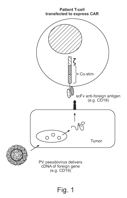
Fig. 1 depicts an example of a targeted cancer therapy of the present
disclosure. In
this example, a tumor-tropic papillomavirus (PV) pseudovirus (PsV) is
engineered to
encapsulate a nucleic acid encoding a CD19 antigen, and an immune cell (e.g.,
a T cell) is
engineered to express a chimeric antigen receptor (CAR) that comprises a
single chain
antibody fragment (scFv) that binds specifically to a CD19 antigen. Both the
PsV and the
immune cell are administered to a subject having a tumor/tumor cells. The PsV
serves as a
vehicle to deliver the nucleic acid to the tumor cell(s), and the immune cell
targets the tumor
cell(s) following expression of CD19 at the cell surface.
Fig. 2 shows results of CD19 expression in HaCaT human keratinocyte and PAM212
mouse keratinocyte cells following transfection of CD19 expression vector
clones (clones #B
and #D, and clones #6 and #8 for human and mouse CD19, respectively).
Transfection with
a red fluorescent protein (RFP) expression vector was used as a positive
controls, and non-
transfected cells were used as negative controls.
Fig. 3A (right image) is an electrophoretic gel image showing purification
fractions
obtained from cell lysates of 293TT cells transfected with two DNA expression
vectors:
modified HPV16/31 L1/L2 and (human) hCD19 (clone #B, see Fig. 2) or (mouse)
mCD19
(clone #6, see Fig. 2). The modified Li and L2 proteins are expressed and self-
assemble
preferentially encapsidating the CD19 expression vector to generate PsV. After
two days,
cells are lysed and the PsV is purified using density centrifugation and
fractions are collected
(PsV production and purification described in Buck 2007). Fig. 3A (left image)
is an
electrophoretic gel image showing purification fractions obtained from cell
lysates of 293TT
cells transfected with two DNA expression vectors: BPV1 Li/L2 and (human)
hCD19 (clone
#B, see Fig. 2) or (mouse) mCD19 (clone #6, see Fig. 2). The purest fractions
are denoted by
'''' and these were used in downstream validation experiments. Fig. 3B shows
graphs
demonstrating CD19 expression in 293TT cells infected with fractions (denoted
by * in Fig.
3A) of PsV that carry nucleic acid encoding CD19. 1 pi of the fractions for
modified
HPV16/31 PsV and BPV PsV were used for infection.
CA 03003728 2018-04-30
WO 2017/075440 PCT/US2016/059452
6
Figs. 4A and 4B show confirmation of infection and surface expression of human
CD19 in tumor cells of various cancer types after infection with modified
HPV16/31 PsV or
BPV PsV, each containing nucleic acid encoding hCD19. Fig. 4A. shows hCD19
surface
expression in human cells of different lineages or cancer types. Fig. 4B shows
mCD19
surface expression in murine TC-1 cells. For both Fig. 4A and Fig. 4B, surface
staining for
CD19 was completed 48 hours following infection with 1 pi of either modified
HPV16/13
PsV or BPV PsV. GFP is also expressed on each CD19 expression vector and
detection of its
expression serves as an internal positive control for gene delivery.
DETAILED DESCRIPTION
Tumor cells typically express tumor antigens that trigger an immune response
in a
host subject. These tumor antigens serve as markers for identifying tumor
cells and also
serve as candidates for targeted cancer therapies. In many instances, however,
the antigens
expressed by a tumor are also expressed by some normal cells. These antigens
are referred to
as tumor-associated antigens. Thus, therapies designed to use tumor-associated
antigens as
signals to guide therapeutics to tumors risk also targeting normal cells,
which can result in
unwanted side-effects and lower therapeutic efficacy.
Provided herein are therapies used to selectively target tumor cells without
also
targeting a substantial number of normal cells, thereby reducing or
eliminating unwanted
side-effects and increasing efficacy of treatment. In some embodiments, immune
cells and
tumor cells of a subject are genetically engineered to express a receptor and
cognate antigen,
respectively. Modification of tumor cells in vivo, in some embodiments, is
achieved by
delivering to a subject a tumor-selective vehicle (that selectively homes to
tumor cells)
containing an engineered nucleic acid (or more than one engineered nucleic
acid) that
encodes an antigen (or encodes more than one antigen). In other embodiments,
an engineered
nucleic acid encoding an antigen is delivered directed to a tumor in vivo via
intratumoral
injection. Following delivery of the nucleic acid (or preceding or in
combination with
delivery of an engineered nucleic acid), immune cells engineered to express a
cognate
receptor (or receptors) are delivered to the subject. The immune cells, guided
by receptor-
antigen (ligand) binding, selectively target tumor cells expressing the
antigen encoded by the
engineered nucleic acid. The engineered immune cells then kill the tumor
cells.
Antigens
CA 03003728 2018-04-30
WO 2017/075440 PCT/US2016/059452
7
Some embodiments of the present disclosure are directed to antigens, typically
encoded by an engineered (exogenous) nucleic acid delivered via a tumor-
selective vehicle or
intratumoral injection. An "antigen" is a molecule that serves as a ligand for
receptors of
immune cells, including leukocytes, such as T cells. An antigen may be a self-
antigen or a
non-self antigen.
A "self-antigen" refers to an antigen that originates from within a body. Self-
antigens
may be expressed by tumor cells as well as some normal cells. In some
embodiments, tumor
cells express self-antigens at an expression level higher than the expression
level at which a
normal tumor cell expresses the same self-antigen. That is, the self-antigen
expressed by a
tumor cell is overexpressed. In some embodiments, an engineered nucleic acid
encoding a
self-antigen is delivered to tumor cells (via a tumor-selective vehicle or
intratumoral
injection) that naturally express, or overexpress, the self-antigen for the
purpose of further
increasing the expression level of the self-antigen. Thus, immune cells
genetically
engineered to express the cognate receptor selectively target the tumor cells
over the normal
cells. It should be understood that while "self-antigens" originate from
within the body, a
recombinant form of that antigen is still referred to as "self-antigen" if it
is expressed in a
tumor by an engineered (exogenously delivered) nucleic acid. For example, CD19
and CD20
are self-antigens overexpressed by tumor cells. The present disclosure
encompasses
delivering to a subject an engineered nucleic acid encoding CD19 or CD20 - a
step referred
to herein as delivering to a subject an engineered nucleic acid encoding a
self-antigen.
In some embodiments, an engineered nucleic acid encoding a self-antigen is
delivered
to tumor cells and is expressed at a level higher than the level at which the
endogenous self-
antigen is expressed in non-modified tumor cells (a tumor cell that does not
contain an
engineered nucleic acid). For example, a self-antigen encoded by an engineered
nucleic acid
operably linked to a strong constitutive promoter, such as the CMV promoter
(e.g., CMV IE
promoter) or the Grp78 promoter, may be expressed in tumor cells at a level
that is 10%,
20%, 30%, 40%, 50%, 60%, 70%, 80%, 90%, 100%, 125%, 150%, 175% or 200% higher
than the level at which the endogenous self-antigen is expressed in non-
modified tumor cells.
In some embodiments, a self-antigen is a tumor antigen. A "tumor antigen" is
an
antigen expressed by tumor cells. Examples of tumor antigens of the present
disclosure
include, without limitation, CD19, CD20, CD21, CD22, CD45, BCMA, MART-1, MAGE-
A3, glycoprotein 100 (gp100), NY-ESO-1, HER2 (ErbB2), IGF2B3, EGFRvIII,
Kallikrein 4,
KIF20A, Lengsin, Meloe, MUC-1, MUC5AC, MUC-16, B7-H3, B7-H6, CD70, CEA,
CSPG4, EphA2, EpCAM, EGFR family, FAP, FRa, glupican-3, GD2, GD3, HLA-
CA 03003728 2018-04-30
WO 2017/075440 PCT/US2016/059452
8
Al+MAGE1, IL-11Ra, IL-23Ra2, Lewis-Y, mesothelin, NKG2D ligands, PSMA, ROR1,
survivin, TAG72 or VEGFR2. Other examples of tumor antigens are described (der
Bruggen
P et al. Peptide database: T cell-defined tumor antigens. Cancer Immun 2013.
URL:
cancerimmunity.org/peptide, incorporated herein by reference).
Tumor antigens include tumor-specific antigens (TSA) and tumor-associated
antigens
(TAA). "Tumor-specific antigens" are expressed only by tumor cells (not
expressed on any
other cell). "Tumor-associated antigens" are expressed by tumor cells and by
some normal
(non-tumor) cells.
Examples of tumor antigens include, without limitation, alpha-actinin-4,
ARTC1,
BCR-ABL, B-RAF, CASP-5, CASP-8, beta-catenin, Cdc27, CDK4, CDK12, CDKN2A,
CLPP, COA-1, CSNK1A1, dek-can, EFTUD2, Elongation factor 2, ETV6-AML1, FLT3-
ITD, FN1, GAS7, GPNMB, HAUS3, LDLR-fucosyltransferaseAS, HLA-A2, HLA-All,
hsp70-2, MART2, MATN, ME1, MUM-1, MUM-2, MUM-3, neo-PAP, Myosin class I,
NFYC, OGT, OS-9, p53, pmI-RARalpha, PPP1R3B, PRDX5, PTPRK, K-ras, N-ras,
RBAF600, SIRT2, SNRPD1, SYT-SSX1, SYT-SSX2, TGF-betaRII, Triosephosphate
isomerase, BAGE family antigens, CAGE family antigens, Cyclin-Al, GAGE family
antigens, HERV-K-MEL, KK-LC-1, KM-HN-1, LAGE-1, MAGE family antigens, NA88-A,
NY-ES0-1/LAGE-2, PRAME, SAGE family antigens, Sp17, SSX family antigens, TAG-
1,
TAG-2, TRAG-3, TRP2-INT2, XAGE family antigens, CEA, Gp100/pme117, mammaglobin-
A, Melan-A/MART-1, mesothilin, NY-BR-1, OA1, PAP, PSA, RAB38/NY-MEL-1, TRP-
1/gp75, TRP-2, tyrosinase, 9D7, adipophilin, AIM-2, ALDH1A1, BCLX (L), BING-4,
CALCA, CD45, CD274, CPSF, cyclin-B1, cyclin D1, DKK1, ENAH (hMena), EpCAM,
EphA3, EZH2, FGF5, Ganglioside GD3, glypican-3, G250/MN/CAIX, HER-2/neu, HLA-
DOB, Hepsin, IS01, IGF2B3, IL13Ralpha2, Intestinal carboxyl esterase, alpha-
foetoprotein,
Kallikrein 4, KIF20A, Lengsin, M-CSF, MCSP, mdm-2, Meloe, Midkine, MMP-2, MMP-
7,
MUC1, MUC5AC, p53, PAX5, PBF, PRAME, PSMA, RAGE-1, RGS5, RhoC, RNF43,
RU2AS, SAP-1, secernin 1, SOX10, STEAP1, survivin, Telomerase, TPBG, VEGF and
WT1. Other tumor antigens are encompassed by the present disclosure.
A "non-self antigen" is an antigen that originates from the external
environment
(outside the body). A non-self antigen is not naturally expressed in cells
(normal cells or
tumor cells) of a subject. With respect to a human subject, a non-self antigen
may be, for
example, a human antigen obtained from a different host/subject or a non-human
antigen,
such as a bacterial antigen, a yeast antigen, a protozoan antigen, a viral
antigen. A non-self
antigen may be a naturally-occurring antigen (naturally-occurring in another
organisms) or a
CA 03003728 2018-04-30
WO 2017/075440 PCT/US2016/059452
9
synthetic (non-naturally-occurring, e.g., artificial) antigen. Examples of non-
self antigens
include, without limitation, green fluorescent protein, KLH and avian
ovalbumin.
Engineered nucleic acids encoding non-self antigens delivered to tumor cells
via a tumor-
selective vehicle or intratumoral injection are expressed primarily in tumor
cells and not in
normal cells. Thus, immune cells genetically engineered to bind to the non-
self-antigen are
capable of selectively targeting tumor cells.
In some embodiments, an antigen is a peptide tag or an antigen comprises a
peptide
tag. Examples of peptide tags include, His tag, FLAG tag, viral peptides
(e.g., CMV
peptides, SV5 peptides), chitin binding protein, maltose binding protein,
glutathione-S-
transferase, thioredoxin, poly(NANP), V5-tag, Myc-tag, HA-tag, AviTag,
calmodulin-tag,
polyglutamate tag, E-tag, S-tag, SBP-tag, Softag 1, Strep-tag, TC tag, V5 tag,
VSV tag,
Xpress tag, isopeptag, Spytag, BCCP, Halo-tag, Nus-tag, Fc-tag and Ty tag.
Other peptide
tags are encompassed by the present disclosure.
Delivery Vehicles
In some embodiments, antigens are delivered to a subject via a tumor-selective
vehicle. A "tumor-selective vehicle" is a molecule, agent or matrix that
preferentially targets
(homes to) tumor cells or, with respect to viruses and pseudoviruses,
preferentially replicates
in and/or infects or pseudo-infects tumor cells. In some embodiments,
engineered nucleic
acids of the present disclosure are encapsulated within a tumor-selective
vehicle.
Examples of tumor-selective vehicles include, without limitation, viruses
(including
chimeric viruses and modified viruses), and pseudoviruses. Non-viral tumor-
selective
vehicles are also encompassed herein and described below.
A virus is a small infectious agent that replicates only inside the living
cells of other
organisms. A virus typically contains: (i) genetic material in the form of
viral DNA or viral
RNA; (ii) a protein coat, referred to as a capsid, which surrounds and
protects the genetic
material; and in some cases (iii) an envelope of lipids that surrounds the
protein coat. A
capsid, the protein shell of a virus, contains several structural subunits,
each referred to as a
cap somer.
Non-limiting examples of viruses of the present disclosure include oncolytic
viruses
and modified viruses (e.g., modified to preferentially infect tumor cells). In
some
embodiments, an engineered nucleic acid encoding an antigen is engineered to
be part of the
virus genome. In some embodiments an engineered nucleic acid encoding an
antigen is
encapsulated in a pseudovirus.
CA 03003728 2018-04-30
WO 2017/075440 PCT/US2016/059452
In some embodiments, a tumor-selective vehicle is an oncolytic virus. An
oncolytic
virus is a virus that preferentially infects and kills tumor cells. Examples
of oncolytic viruses
include, without limitation, adenoviruses, vaccinia viruses, Sindbis viruses,
Seneca valley
viruses, Coxsackie viruses, measles viruses, reoviruses, vaccinia viruses,
Newcastle disease
viruses, vesicular stomatitis viruses, herpes simplex viruses, polioviruses
and parvoviruses.
In some embodiments, a tumor-selective vehicle is a targeted chimeric virus. A
targeted chimeric virus is a recombinant virus containing components from at
least two
difference viruses. For example, a tumor-selective vehicle of the present
disclosure may
include a chimeric adeno-associated (AAV) and bacteriophage virus, referred to
as AAVP
(Hajitou A. et al. 2006 Cell 125: 385-398; Hajitou A. et al. 2007 Nat. Protoc.
2(3): 523-31;
and Hajitou A. et al. 2010 Adv. Genet. 69: 65-82, each of which is
incorporated herein by
reference).
In some embodiments, a tumor-selective vehicle is a naturally-occurring virus
or a
virus modified to preferentially infect (target) and kills tumor cells. A non-
limiting example
of a virus that may be modified to target tumor cells is an adeno-associated
virus (AAV). In
some embodiments, the capsid of the AAV is modified (e.g., receptor targeting,
mixed
capsids in the shell of the virion, or marker rescue to produce recombinant
virus; Chengwen
L et al. 2005 Cancer Gene Ther. 12(12): 913-25, incorporated by reference
herein). In some
embodiments, a T cell-stimulating epitope of an AAV is modified.
"Pseudoviruses" are synthetic viruses used to inject genetic material,
including DNA and RNA, with specific and desired traits into prokaryotic and
eukaryotic
cells. Pseudoviruses are closely related to viruses in structure and behavior
but lack many
characteristics exhibited by true viruses, including the capability to
replicate. In some
embodiments, an engineered nucleic acid encoding an antigen is encapsulated in
a
pseudovirus.
In some embodiments, a pseudovirus of the present disclosure comprises or
consists
of papillomavirus proteins (e.g., Li proteins, L2 proteins, or a combination
of Li and L2
proteins). The papillomavirus proteins (L1 and L2 capsid proteins) may be
human
papillomavirus proteins or non-human (e.g., bovine, murine, cotton-rabbit,
macaque or
rhesus) papillomavirus proteins. In some embodiments, these papillomavirus
proteins are
modified in a way that results in the pseudovirus having a modified
antigenicity relative to a
pseudovirus that comprises or consists of wild-type papillomavirus proteins.
For example, a
modified Li papillomavirus protein of the present disclosure may be a
recombinant protein
based on HPV serotype 16 and HPV serotype 31, referred to as a "modified
HPV16/31 Li
CA 03003728 2018-04-30
WO 2017/075440
PCT/US2016/059452
11
protein," which is described in International Pub. No. WO/2010/120266, the
entirety of
which is incorporated herein by reference.
Other examples of targeting vehicles include, without limitation, natural
polymers,
synthetic polymers, cationic peptides, cell-penetrating peptides,
biodegradable nanoparticles,
liposomes, lipoplexes (e.g., PEGylated lipoplexes), polyplexes, micelles and
dendrimers. In
some embodiments, a synthetic delivery vehicle is a gel, a mucoadhesive or a
silicon
nanoneedle. Other tumor-selective vehicles are encompassed by the present
disclosure.
In some embodiments, a tumor-selective vehicle is a liposome. Liposomes are
spherical vesicles having at least one lipid bilayer. The term "liposome"
encompasses
multilamellar vesicles (having several lamellar phase lipid bilayers), small
unilamellar
liposome vesicles (having one lipid bilayer), large unilamellar vesicles and
cochleate
vesicles. The term "lipoplex" refers to a cationic liposome that form with
DNA. The term
"polyplex" refers to a polymer that forms with DNA. In some embodiments, an
engineered
nucleic acid encoding an antigen is encapsulated in a liposome, lipoplex or
polyplex.
In some embodiments, a tumor-selective vehicle is a polymeric micelle.
Polymeric
micelles, by comparison, are prepared from certain amphiphilic co-polymers
consisting of
both hydrophilic and hydrophobic monomer units. In some embodiments, an
engineered
nucleic acid encoding an antigen is encapsulated in a polymeric micelle.
In some embodiments, a tumor-selective vehicle is a dendrimer. Dendrimers are
also
polymer-based delivery vehicles. They have a core that branches out in regular
intervals to
form a small, spherical and dense nanocarrier.
Delivery Routes
In some embodiments, engineered nucleic acids encoding antigens are delivered
to a
subject via intratumoral injection. "Intratumoral injection" is a route of
administration by
which an engineered nucleic acid, for example, is delivered directly to the
tumor via an
injection device (e.g., needle and syringe). In some embodiments, tumor-
selective vehicles,
immune cells, or both, are delivered to a subject via a parenteral route, an
enteral route or a
topical route.
Examples of parental routes include, without limitation, intra-abdominal,
intra-
amniotic, intra-arterial, intra-articular, intrabiliary, intrabronchial,
intrabursal, intracardiac,
intracartilaginous, intracaudal, intracavernous, intracavitary, intracerebral,
intracisternal,
intracorneal, intracoronal, intracoronary, intracorporus, intracranial,
intradermal, intradiscal,
intraductal, intraduodenal, intradural, intraepidermal, intraesophageal,
intragastric,
CA 03003728 2018-04-30
WO 2017/075440 PCT/US2016/059452
12
intragingival, intraileal, intralesional, intraluminal, intralymphatic,
intramedullary,
intrameningeal, intramuscular, intraocular, intraovarian, intrapericardial,
intraperitoneal,
intrapleural, intraprostatic, intrapulmonary, intraocular, intrasinal,
intraspinal, intrasynovial,
intratendinous, intratesticular, intrathecal, intrathoracic, intratubular,
intratympanic,
intrauterine, intravascular, intravenous (bolus or drip), intraventricular,
intravesical and
subcutaneous.
Enteral routes of administration include administration to the
gastrointestinal tract via
the mouth (oral), stomach (gastric) and rectum (rectal). Gastric
administration typically
involves the use of a tube through the nasal passage (NG tube) or a tube in
the belly leading
directly to the stomach (PEG tube). Rectal administration typically involves
rectal
suppositories.
Topical routes of administration include administration to a body surface,
such as skin
or mucous membranes. Delivery vehicles of the present disclosure may be
administered
topically via a cream, foam, gel, lotion or ointment, for example.
Other routes of delivery are encompassed by the present disclosure. For
example, an
engineered nucleic acid or a tumor-selective vehicle containing an engineered
nucleic acid
may be delivered via ultrasound-targeted microbubble destruction (UTMD) (Qiu
L. et al.
2012 Gene Therapy 19: 703-710, incorporated herein by reference).
In some embodiments, an engineered nucleic acid encoding an antigen is
delivered to
a subject (via a tumor-selective vehicle or via intratumoral injection) prior
to or after
delivering an immune cell. Thus, an engineered nucleic acid and an immune cell
of the
present disclosure may be delivered sequentially. In other embodiments,
however, an
engineered nucleic acid and an immune cell are delivered simultaneously.
Immune Cells
Some embodiments of the present disclosure are directed to immune cells, such
as
leukocytes (nucleated white blood cells), comprising (e.g., expressing) a
receptor that binds
to an antigen. A leukocyte of the present disclosure may be, for example, a
neutrophil,
eosinophil, basophil, lymphocyte or a monocyte. In some embodiments, a
leukocyte is a
lymphocyte. Examples of lymphocytes include T cells, B cells, Natural Killer
(NK) cells or
NKT cells. In some embodiments, a T cell is a CD4+ Th (T helper) cell, a CD8+
cytotoxic T
cell, a y6 T cell or a regulatory (suppressor) T cell. In some embodiments, an
immune cell is
a dendritic cell.
CA 03003728 2018-04-30
WO 2017/075440 PCT/US2016/059452
13
Immune cells of the present disclosure, in some embodiments, are genetically
engineered to express an antigen-binding receptor. A cell is considered
"engineered" if it
contains an engineered (exogenous) nucleic acid. Engineered nucleic acids of
the present
disclosure may be introduced into a cell by any known (e.g., conventional)
method. For
example, an engineered nucleic acid may be introduced into a cell by
electroporation (see,
e.g., Heiser W.C. Transcription Factor Protocols: Methods in Molecular
BiologyTM 2000;
130: 117-134), chemical (e.g., calcium phosphate or lipid), transfection (see,
e.g., Lewis
W.H., et al., Somatic Cell Genet. 1980 May; 6(3): 333-47; Chen C., et al., Mol
Cell Biol.
1987 August; 7(8): 2745-2752), fusion with bacterial protoplasts containing
recombinant
plasmids (see, e.g., Schaffner W. Proc Natl Acad Sci USA. 1980 Apr; 77(4):
2163-7),
microinjection of purified DNA directly into the nucleus of the cell (see,
e.g., Capecchi M.R.
Cell. 1980 Nov; 22(2 Pt 2): 479-88), or retrovirus transduction.
Some aspects of the present disclosure provide an "adoptive cell" approach,
which
involves isolating immune cells (e.g., T cells) from a subject, genetically
engineering the
cells (e.g., to express an antigen-binding receptor, such as a chimeric
antigen receptor),
expanding the cells ex vivo, and then re-introducing the cells into the
subject. This method
results in a greater number of engineered immune cells in the subject relative
to what could
be achieved by conventional gene delivery and vaccination methods. In some
embodiments,
immune cells are isolated from a subject, expanded ex vivo without genetic
modification, and
then re-introduced into the subject.
Antigen-Binding Receptors
Immune cells of the present disclosure comprise receptors that bind to
antigens, such
as an antigen encoded by an exogenously delivered nucleic acid, as provided
herein. In some
embodiments, a leukocyte is modified (e.g., genetically modified) to express a
receptor that
binds to an antigen. The receptor may be, in some embodiments, a naturally-
occurring
antigen receptor (normally expressed on the immune cell), recombinant antigen
receptor (not
normally expressed on the immune cell) or a chimeric antigen receptor (CAR).
Naturally-
occurring and recombinant antigen receptors encompassed by the present
disclosure include
T cell receptors, B cell receptors, NK cell receptors, NKT cell receptors and
dendritic cell
receptors. A "chimeric antigen receptor" refers to an artificial immune cell
receptor that is
engineered to recognize and bind to an antigen expressed by tumor cells.
Generally, a CAR
is designed for a T cell and is a chimera of a signaling domain of the T-cell
receptor (TcR)
CA 03003728 2018-04-30
WO 2017/075440 PCT/US2016/059452
14
complex and an antigen-recognizing domain (e.g., a single chain fragment
(scFv) of an
antibody) (Enblad et al., Human Gene Therapy. 2015; 26(8):498-505).
In some embodiments, an antigen binding receptor is a chimeric antigen
receptor
(CAR). A T cell that expressed a CAR is referred to as a "CAR T cell." A CAR T
cell
receptor, in some embodiments, comprises a signaling domain of the T-cell
receptor (TcR)
complex and an antigen-recognizing domain (e.g., a single chain fragment
(scFv) of an
antibody) (Enblad et al., Human Gene Therapy. 2015; 26(8):498-505).
There are four generations of CARs, each of which contains different
components.
First generation CARs join an antibody-derived scFv to the CD3zeta or z)
intracellular
signaling domain of the T-cell receptor through hinge and transmembrane
domains. Second
generation CARs incorporate an additional domain, e.g., CD28, 4-1BB (41BB), or
ICOS, to
supply a costimulatory signal. Third-generation CARs contain two costimulatory
domains
fused with the TcR CD3-t chain. Third-generation costimulatory domains may
include, e.g.,
a combination of CD3z, CD27, CD28, 4-1BB, ICOS, or 0X40. CARs, in some
embodiments, contain an ectodomain (e.g., CD3), commonly derived from a single
chain
variable fragment (scFv), a hinge, a transmembrane domain, and an endodomain
with one
(first generation), two (second generation), or three (third generation)
signaling domains
derived from CD3Z and/or co-stimulatory molecules (Maude et al., Blood. 2015;
125(26):4017-4023; Kakarla and Gottschalk, Cancer J. 2014; 20(2):151-155).
In some embodiments, the chimeric antigen receptor (CAR) is a T-cell
redirected for
universal cytokine killing (TRUCK), also known as a fourth generation CAR.
TRUCKs are
CAR-redirected T-cells used as vehicles to produce and release a transgenic
cytokine that
accumulates in the targeted tissue, e.g., a targeted tumor tissue. The
transgenic cytokine is
released upon CAR engagement of the target. TRUCK cells may deposit a variety
of
therapeutic cytokines in the target. This may result in therapeutic
concentrations at the
targeted site and avoid systemic toxicity.
CARs typically differ in their functional properties. The CD3t signaling
domain of
the T-cell receptor, when engaged, will activate and induce proliferation of T-
cells but can
lead to anergy (a lack of reaction by the body's defense mechanisms, resulting
in direct
induction of peripheral lymphocyte tolerance). Lymphocytes are considered
anergic when
they fail to respond to a specific antigen. The addition of a costimulatory
domain in second-
generation CARs improved replicative capacity and persistence of modified T-
cells. Similar
antitumor effects are observed in vitro with CD28 or 4-1BB CARs, but
preclinical in vivo
studies suggest that 4-1BB CARs may produce superior proliferation and/or
persistence.
CA 03003728 2018-04-30
WO 2017/075440 PCT/US2016/059452
Clinical trials suggest that both of these second-generation CARs are capable
of inducing
substantial T-cell proliferation in vivo, but CARs containing the 4-1BB
costimulatory domain
appear to persist longer. Third generation CARs combine multiple signaling
domains
(costimulatory) to augment potency. Fourth generation CARs are additionally
modified with
a constitutive or inducible expression cassette for a transgenic cytokine,
which is released by
the CAR T-cell to modulate the T-cell response. See, for example, Enblad et
al., Human
Gene Therapy. 2015; 26(8):498-505; Chmielew ski and Hinrich, Expert Opinion on
Biological Therapy. 2015;15(8): 1145-1154.
In some embodiments, a chimeric antigen receptor is a first generation CAR. In
some
embodiments, a chimeric antigen receptor is a third generation CAR. In some
embodiments,
a chimeric antigen receptor is a second generation CAR. In some embodiments, a
chimeric
antigen receptor is a third generation CAR. In some embodiments, the chimeric
antigen
receptor is a fourth generation CAR or a T-cell redirected for universal
cytokine killing
(TRUCK).
In some embodiments, a chimeric antigen receptor (CAR) comprises an
extracellular
domain comprising an antigen binding domain, a transmembrane domain, and a
cytoplasmic
domain. In some embodiments, a CAR is fully human. In some embodiments, the
antigen
binding domain of a CAR is specific for one or more antigens. In some
embodiments, a
"spacer" domain or "hinge" domain is located between an extracellular domain
(comprising
the antigen binding domain) and a transmembrane domain of a CAR, or between a
cytoplasmic domain and a transmembrane domain of the CAR. A "spacer domain"
refers to
any oligopeptide or polypeptide that functions to link the transmembrane
domain to the
extracellular domain and/or the cytoplasmic domain in the polypeptide chain. A
"hinge
domain" refers to any oligopeptide or polypeptide that functions to provide
flexibility to the
CAR, or domains thereof, or to prevent steric hindrance of the CAR, or domains
thereof. In
some embodiments, a spacer domain or hinge domain may comprise up to 300 amino
acids
(e.g., 10 to 100 amino acids, or 5 to 20 amino acids). In some embodiments,
one or more
spacer domain(s) may be included in other regions of a CAR.
In some embodiments, a CAR of the disclosure comprises an antigen binding
domain,
such as a single chain Fv (scFv) specific for a tumor antigen. The choice of
binding domain
depends upon the type and number of ligands that define the surface of a
target cell. For
example, the antigen binding domain may be chosen to recognize a ligand that
acts as a cell
surface marker on target cells associated with a particular disease state,
such as cancer or an
autoimmune disease. Thus, examples of cell surface markers that may act as
ligands for the
CA 03003728 2018-04-30
WO 2017/075440 PCT/US2016/059452
16
antigen binding domain in the CAR of the present disclosure include those
associated with
cancer cells and/or other forms of diseased cells. In some embodiments, a CAR
is engineered
to target a tumor antigen of interest by way of engineering a desired antigen
binding domain
that specifically binds to an antigen on a tumor cell encoded by an engineered
nucleic acid, as
provided herein.
An antigen binding domain (e.g., an scFv) that "specifically binds" to a
target or an
epitope is a term understood in the art, and methods to determine such
specific binding are
also known in the art. A molecule is said to exhibit "specific binding" if it
reacts or
associates more frequently, more rapidly, with greater duration and/or with
greater affinity
with a particular target antigen than it does with alternative targets. An
antigen binding
domain (e.g., an scFv) that specifically binds to a first target antigen may
or may not
specifically bind to a second target antigen. As such, "specific binding" does
not necessarily
require (although it can include) exclusive binding.
In some embodiments, immune cells expressing a CAR are genetically modified to
recognize multiple targets or antigens, which permits the recognition of
unique target or
antigen expression patterns on tumor cells. Examples of CARs that can bind
multiple targets
include: "split signal CARs," which limit complete immune cell activation to
tumors
expressing multiple antigens; "tandem CARs" (TanCARs), which contain
ectodomains
having two scFvs; and "universal ectodomain CARs," which incorporate avidin or
a
fluorescein isothiocyanate (FITC)-specific scFv to recognize tumor cells that
have been
incubated with tagged monoclonal antibodies (Mabs).
A CAR is considered "bispecific" if it recognizes two distinct antigens (has
two
distinct antigen recognition domains). In some embodiments, a bispecific CAR
is comprised
of two distinct antigen recognition domains present in tandem on a single
transgenic receptor
(referred to as a TanCAR; see, e.g., Grada Z et al. Molecular Therapy Nucleic
Acids
2013;2:e105, incorporated herein by reference). Thus, methods, in some
embodiments,
comprise delivering to a tumor an engineered nucleic acid that encode an
antigen, or
delivering to a tumor an engineered nucleic acid that induces expression of a
self-antigen, and
delivering to the tumor an immune cell expressing a bispecific CAR that binds
to two
antigens, one of which is encoded by the engineered nucleic acid.
In some embodiments, a CAR is an antigen-specific inhibitory CAR (iCAR), which
may be used, for example, to avoid off-tumor toxicity (Fedorov, VD et al. Sci.
Transl. Med.
published online Dec. 11, 2013, incorporated herein by reference). iCARs
contain an
antigen-specific inhibitory receptor, for example, to block nonspecific
immunosuppression,
CA 03003728 2018-04-30
WO 2017/075440 PCT/US2016/059452
17
which may result from extratumor target expression. iCARs may be based, for
example, on
inhibitory molecules CTLA-4 or PD-1. In some embodiments, these iCARs block T
cell
responses from T cells activated by either their endogenous T cell receptor or
an activating
CAR. In some embodiments, this inhibiting effect is temporary.
In some embodiments, CARs may be used in adoptive cell transfer, wherein
immune
cells are removed from a subject and modified so that they express receptors
specific to an
antigen, e.g., a tumor-specific antigen. The modified immune cells, which may
then
recognize and kill the cancer cells, are reintroduced into the subject (Pule,
et al., Cytotherapy.
2003; 5(3): 211-226; Maude et al., Blood. 2015; 125(26): 4017-4023, each of
which is
incorporated herein by reference).
Tumor Cells
The present disclosure encompasses the treatment of all types of tumors,
including
primary tumors and metastatic tumors. Tumors that arise from connective
tissue,
endothelium, mesothelium, blood cells, lymphoid cells, muscle, epithelial
tissue, neural tissue
and neural crest-derived cells are encompassed herein. The present disclosure
also
encompasses carcinomas, sarcomas, myelomas, leukemias, lymphomas, and cancers
of mixed
type (e.g., adenosquamous, carcinoma, mixed mesodermal tumor, carcinosarcoma
and
teratocarcinoma).
The following is a list of non-limiting examples of tumors/cancers encompassed
by
the present disclosure: acute lymphoblastic leukemia (ALL), acute myeloid
leukemia
(AML), adrenocortical carcinoma, AIDS-related cancers, Kaposi sarcoma, AIDS-
related
lymphoma, primary CNS lymphoma, anal cancer, appendix cancer, astrocytomas,
atypical
teratoid/rhabdoid tumor, basal cell carcinoma, bile duct cancer, bladder
cancer, bone cancer,
Ewing sarcoma family of tumors, osteosarcoma and malignant fibrous
histiocytoma, brain
stem glioma, brain tumor, astrocytomas, brain and spinal cord tumors, brain
stem glioma,
central nervous system atypical teratoid/rhabdoid tumor, central nervous
system embryonal
tumors, central nervous system germ cell tumors, craniopharyngioma,
ependymoma, breast
cancer, bronchial tumors, Burkitt lymphoma, carcinoid tumor, gastrointestinal,
carcinoma of
unknown primary, cardiac (heart) tumors, atypical teratoid/rhabdoid tumor,
embryonal
tumors, germ cell tumor, primary lymphoma, cervical cancer,
cholangiocarcinoma,
chordoma, chronic lymphocytic leukemia (CLL), chronic myelogenous leukemia
(CML),
chronic myeloproliferative neoplasms, colon cancer, colorectal cancer,
craniopharyngioma,
cutaneous T-cell lymphoma, ductal carcinoma in situ (dcis), embryonal tumors,
central
CA 03003728 2018-04-30
WO 2017/075440 PCT/US2016/059452
18
nervous system, endometrial cancer, ependymoma, esophageal cancer,
esthesioneuroblastoma, ewing sarcoma, extracranial germ cell tumor,
extragonadal germ cell
tumor, eye cancer, intraocular melanoma, retinoblastoma, fallopian tube
cancer, fibrous
histiocytoma of bone, malignant, and osteosarcoma, gallbladder cancer, gastric
(stomach)
cancer, gastrointestinal carcinoid tumor, gastrointestinal stromal tumors
(gist), germ cell
tumor, central nervous system, extracranial, extragonadal, ovarian,
testicular, gestational
trophoblastic disease, glioma, brain stem, hairy cell leukemia, head and neck
cancer, heart
cancer, hepatocellular (liver) cancer, histiocytosis, langerhans cell, Hodgkin
lymphoma,
hypopharyngeal cancer, intraocular melanoma, islet cell tumors, pancreatic
neuroendocrine
tumors, kaposi sarcoma, kidney, renal cell, Wilms tumor and other kidney
tumors,
langerhans cell histiocytosis, laryngeal cancer, acute lymphoblastic leukemia
(ALL), acute
myeloid leukemia (AML), chronic lymphocytic leukemia (CLL), chronic myeloid
leukemia
(CML), hairy cell, lip and oral cavity cancer, liver cancer (primary), lung
cancer, non-small
cell, small cell, lymphoma, Burkitt, cutaneous t-cell, Hodgkin, non-Hodgkin,
primary central
nervous system (CNS), macroglobulinemia, waldenstrom, male breast cancer,
malignant
fibrous histiocytoma of bone and osteosarcoma, melanoma, intraocular (eye),
merkel cell
carcinoma, mesothelioma, malignant, metastatic squamous neck cancer with
occult primary,
midline tract carcinoma involving nut gene, mouth cancer, multiple endocrine
neoplasia
syndromes, multiple myeloma/plasma cell neoplasm, mycosis fungoides,
myelodysplastic
syndromes, myelodysplastic/myeloproliferative neoplasms, myelogenous leukemia,
myeloma, myeloproliferative neoplasms, chronic, nasal cavity and paranasal
sinus cancer,
nasopharyngeal cancer, neuroblastoma, non-Hodgkin lymphoma, non-small cell
lung cancer,
ocular, oral cancer, oral cavity cancer, lip and, oropharyngeal cancer,
osteosarcoma and
malignant fibrous histiocytoma of bone, ovarian cancer, epithelial, germ cell
tumor, low
malignant potential tumor, pancreatic cancer, pancreatic neuroendocrine tumors
(islet cell
tumors), papillomatosis, paraganglioma, paranasal sinus and nasal cavity
cancer, parathyroid
cancer, penile cancer, pharyngeal cancer, pheochromocytoma, pituitary tumor,
plasma cell
neoplasm/multiple myeloma, pleuropulmonary blastoma, pregnancy and breast
cancer,
primary central nervous system (CNS) lymphoma, primary peritoneal cancer,
prostate cancer,
rectal cancer, renal cell (kidney) cancer, renal pelvis and ureter,
transitional cell cancer,
retinal cancer, retinoblastoma, rhabdomyosarcoma, salivary gland cancer,
sarcoma, ewing,
kaposi, osteosarcoma (bone cancer), rhabdomyosarcoma, soft tissue, uterine,
Sezary
syndrome, skin cancer, melanoma, merkel cell carcinoma, nonmelanoma, small
cell lung
cancer, small intestine cancer, soft tissue sarcoma, squamous cell carcinoma,
squamous neck
CA 03003728 2018-04-30
WO 2017/075440 PCT/US2016/059452
19
cancer with occult primary, metastatic, stomach (gastric) cancer, t-cell
lymphoma, cutaneous,
testicular cancer, throat cancer, thymoma and thymic carcinoma, thyroid
cancer, transitional
cell cancer of the renal pelvis and ureter, unknown primary, carcinoma of,
unusual cancers of,
ureter and renal pelvis, transitional cell cancer, urethral cancer, uterine
cancer, endometrial,
uterine sarcoma, vaginal cancer, vulvar cancer and waldenstrom
macroglobulinemia.
Nucleic Acids
Some embodiments of the present disclosure are directed to nucleic acids
encoding
antigens (e.g., non-self antigens). Such nucleic acids are delivered to a
subject and targeted
to tumor cells (e.g., via a tumor-selective vehicle or intratumoral injection)
where the nucleic
acid is expressed (e.g., overexpressed) in the tumor cells. In some
embodiments, a nucleic
acid encoding an antigen is a deoxyribonucleic acid (DNA) or a ribonucleic
acid (RNA), such
as a messenger RNA (mRNA). Nucleic acids of the present disclosure, in some
embodiments, are engineered nucleic acids. An "engineered nucleic acid" is a
nucleic acid
(e.g., at least two nucleotides covalently linked together, and in some
instances, containing
phosphodiester bonds, referred to as a phosphodiester "backbone") that does
not occur in
nature. Engineered nucleic acids include recombinant nucleic acids and
synthetic nucleic
acids. A "recombinant nucleic acid" is a molecule that is constructed by
joining nucleic acids
(e.g., isolated nucleic acids, synthetic nucleic acids or a combination
thereof) and, in some
embodiments, can replicate in a living cell. A "synthetic nucleic acid" is a
molecule that is
amplified or chemically, or by other means, synthesized. A synthetic nucleic
acid includes
those that are chemically modified, or otherwise modified, but can base pair
with (also
referred to as "binding to," e.g., transiently or stably) naturally-occurring
nucleic acid
molecules. Recombinant and synthetic nucleic acids also include those
molecules that result
from the replication of either of the foregoing.
While an engineered nucleic acid, as a whole, is not naturally-occurring, it
may
include wild-type nucleotide sequences. In some embodiments, an engineered
nucleic acid
comprises nucleotide sequences obtained from different organisms (e.g.,
obtained from
different species). For example, in some embodiments, an engineered nucleic
acid includes a
murine nucleotide sequence, a bacterial nucleotide sequence, a human
nucleotide sequence, a
viral nucleotide sequence, or a combination of any two or more of the
foregoing sequences.
In some embodiments, an engineered nucleic acid of the present disclosure may
comprise a backbone other than a phosphodiester backbone. For example, an
engineered
nucleic acid, in some embodiments, may comprise phosphoramide,
phosphorothioate,
CA 03003728 2018-04-30
WO 2017/075440 PCT/US2016/059452
phosphorodithioate, 0-methylphosphoroamidite linkages, peptide nucleic acids
or a
combination of any two or more of the foregoing linkages. An engineered
nucleic acid may
be single-stranded (ss) or double-stranded (ds), as specified, or an
engineered nucleic acid
may contain portions of both single-stranded and double-stranded sequence. In
some
embodiments, an engineered nucleic acid contains portions of triple-stranded
sequence. An
engineered nucleic acid may comprise DNA (e.g., genomic DNA, cDNA or a
combination of
genomic DNA and cDNA), RNA or a hybrid molecule, for example, where the
nucleic acid
contains any combination of deoxyribonucleotides and ribonucleotides (e.g.,
artificial or
natural), and any combination of two or more bases, including uracil, adenine,
thymine,
cytosine, guanine, inosine, xanthine, hypoxanthine, isocytosine and
isoguanine.
Delivery of modified mRNA is also encompassed by the present disclosure.
Modified
mRNA includes, for example, mRNA modified for improved codon usage, stability
and
antigen-processing characteristics of the encoded protein.
Engineered nucleic acids of the present disclosure may be produced using
standard
molecular biology methods (see, e.g., Green and Sambrook, Molecular Cloning, A
Laboratory Manual, 2012, Cold Spring Harbor Press). In some embodiments,
nucleic acids
are produced using GIBSON ASSEMBLY Cloning (see, e.g., Gibson, D.G. et al.
Nature
Methods, 343-345, 2009; and Gibson, D.G. et al. Nature Methods, 901-903, 2010,
each of
which is incorporated by reference herein). GIBSON ASSEMBLY typically uses
three
enzymatic activities in a single-tube reaction: 5' exonuclease, the 3'
extension activity of a
DNA polymerase and DNA ligase activity. The 5' exonuclease activity chews back
the 5'
end sequences and exposes the complementary sequence for annealing. The
polymerase
activity then fills in the gaps on the annealed regions. A DNA ligase then
seals the nick and
covalently links the DNA fragments together. The overlapping sequence of
adjoining
fragments is much longer than those used in Golden Gate Assembly, and
therefore results in a
higher percentage of correct assemblies. Other methods of producing engineered
nucleic
acids are known in the art and may be used in accordance with the present
disclosure.
Expression of engineered nucleic acids is typically driven by a promoter
operably
linked to the engineered nucleic acid. A "promoter" refers to a control region
of a nucleic
acid at which initiation and rate of transcription of the remainder of a
nucleic acid sequence
are controlled. A promoter drives transcription or of the nucleic acid
sequence that it
regulates, thus, it is typically located at or near the transcriptional start
site of a gene. A
promoter, in some embodiments, is 100 to 1000 nucleotides in length. A
promoter may also
contain sub-regions at which regulatory proteins and other molecules may bind,
such as RNA
CA 03003728 2018-04-30
WO 2017/075440 PCT/US2016/059452
21
polymerase and other transcription factors. Promoters may be constitutive,
inducible (also
referred to as activatable), repressible, tissue-specific, developmental stage-
specific or any
combination of two or more of the foregoing. Examples of constitutive promoter
for use in
accordance with the present disclosure include, without limitation, the CAG
promoter
(containing a cytomegalovirus (CMV) early enhancer element, a promoter
obtained from the
first exon and the first intron of chicken beta-actin gene, and a splice
acceptor of the rabbit
beta-globin gene), the CMV promoter, and the tumor-specific Grp78 promoter
(Kia A. 2012
Mo/. Cancer Ther. 11(12): 2566-77, incorporated herein by reference).
A promoter is considered to be "operably linked" when it is in a correct
functional
location and orientation relative to a sequence of nucleic acid that it
regulates (e.g., to control
("drive") transcriptional initiation and/or expression of that sequence).
A promoter, in some embodiments, is naturally associated with a nucleic acid
and
may be obtained by isolating the 5' non-coding sequence(s) located upstream of
the coding
region of the given nucleic acid. Such a promoter is referred to as an
"endogenous"
promoter.
A promoter, in some embodiments, is not naturally associated with a nucleic
acid.
Such a promoter is referred to as a "heterologous" promoter and includes, for
example,
promoters that regulate other nucleic acids and promoters obtained from other
cells. A
heterologous promoter may be synthetic or recombinant. Synthetic heterologous
promoters,
in some embodiments, contain various elements obtained from known
transcriptional
regulatory regions. Synthetic heterologous promoters, in some embodiments,
contain
mutations that alter expression through methods of genetic engineering that
are known in the
art. Recombinant heterologous promoters, in some embodiments, are produced by
recombinant cloning, nucleic acid amplification (e.g., polymerase chain
reaction (PCR)), or a
combination of recombinant cloning and nucleic acid amplification (see U.S.
Pat. No.
4,683,202 and U.S. Pat. No. 5,928,906). Other methods of producing synthetic
and
recombinant heterologous promoters are contemplated herein.
A promoter, in some embodiments, is an inducible promoter. An "inducible
promoter" regulates (e.g., activates or inactivates) transcriptional activity
of a nucleic acid to
which it is operably linked when the promoter is influenced by or contacted by
a
corresponding regulatory protein.
Thus, a "regulatory protein," as used herein, is a protein that modulates
(e.g., activates
or inactivates) transcriptional activity from a promoter (e.g., an inducible
promoter). In some
embodiments, a regulatory protein binds directly to an inducible promoter
(e.g., to a sequence
CA 03003728 2018-04-30
WO 2017/075440 PCT/US2016/059452
22
of nucleotides within a promoter). In some embodiments, a regulatory binds to
a region
upstream from an inducible promoter (e.g., within 50 to 100 nucleotides
upstream from an
inducible promoter). In some embodiments, a regulatory protein binds proximal
to (e.g.,
adjacent to) an inducible promoter. Examples of regulatory proteins include,
without
limitation, tetracycline-controlled transactivator (tTA) transcription factor,
reverse
tetracycline-controlled transactivator (rtTA) transcription factor, and Lac
repressor protein.
In some embodiments, a nucleic acid encoding an antigen is overexpressed or
misexpressed in a tumor cell. A nucleic acid or protein is considered
"overexpressed" if its
levels of expression exceed (e.g., by at least 10%, 50%, 100%, 200%, or more)
its normal
(wild-type) level of expression. A nucleic acid or protein is considered
"misexpressed" if it is
expressed in a cell or in a compartment of a cell in which it is not normally
expressed.
Additional Embodiments
Additional embodiments of the present disclosure are encompassed by the
following
number paragraphs:
1. A method comprising delivering to a subject an engineered nucleic acid
encoding an
antigen, wherein the engineered nucleic acid is delivered via a tumor-
selective vehicle or via
intratumoral injection, and delivering to the subject an immune cell
expressing a receptor that
binds to the antigen.
2. The method of paragraph 1, wherein the antigen is a self-antigen, a non-
self antigen,
or a combination thereof.
3. The method of paragraph 2, wherein the antigen is a non-self antigen.
4. The method of paragraph 3, wherein the non-self antigen is a bacterial,
yeast,
protozoan or viral antigen.
5. The method of paragraph 3, wherein the non-self antigen is a synthetic
antigen
6. The method of paragraph 1 or 2, wherein the antigen is a tumor antigen.
7. The method of paragraph 6, wherein the tumor antigen is a tumor-specific
antigen
(TSA) or a tumor-associated antigen (TAA).
8. The method of paragraph 6, wherein the tumor antigen is or comprises an
epitope of
CD19, CD20, CD21, CD22, CD45, BCMA, MART-1, MAGE-A3, glycoprotein 100 (gp100),
NY-ESO-1, HER2 (ErbB2), IGF2B3, EGFRvIII, Kallikrein 4, KIF20A, Lengsin,
Meloe,
MUC-1, MUC5AC, MUC-16, B7-H3, B7-H6, CD70, CEA, CSPG4, EphA2, EpCAM, EGFR
family, FAP, FRa, glupican-3, GD2, GD3, HLA-Al+MAGE1, IL-11Ra, IL-23Ra2, Lewis-
Y, mesothelin, NKG2D ligands, PSMA, ROR1, survivin, TAG72 or VEGFR2.
CA 03003728 2018-04-30
WO 2017/075440 PCT/US2016/059452
23
9. The method of paragraph 8, wherein the tumor antigen is or comprises an
epitope of
CD19.
10. The method of paragraph 9, wherein the tumor antigen is selected from
full length
CD19, a fragment of CD19, at least one C2 Ig-like domain of CD19, or a linear
epitope of
CD19.
11. The method of any one of paragraphs 1-10, wherein the engineered
nucleic acid
encoding the antigen is encapsulated within the tumor-selective vehicle.
12. The method of any one of paragraphs 1-11, wherein tumor-selective
vehicle is a virus
or a pseudovirus.
13. The method of paragraph 12, wherein the tumor-selective vehicle is an
oncolytic
virus.
14. The method of paragraph 13, wherein the oncolytic virus is an
adenovirus, a vaccinia
virus, a Sindbis virus, a Seneca valley virus, a Coxsackie virus, a measles
virus, a reovirus, a
vaccinia virus, a Newcastle disease virus, a vesicular stomatitis virus, a
herpes simplex virus,
a poliovirus, or a parvovirus.
15. The method of paragraph 12, wherein the tumor-selective vehicle is a
chimeric virus.
16. The method of paragraph 15,wherein the chimeric virus is obtained from
engineering
adeno-associated viruses and bacteriophages that display tumor selective
peptides.
17. The method of paragraph 12, wherein the tumor-selective vehicle is a
virus that is
modified to target tumor cells.
18. The method of paragraph 12, wherein the tumor-selective vehicle is an
adeno-
associated virus (AAV) that is modified to target tumor cells.
19. The method of paragraph 12, wherein the tumor-selective vehicle is a
papillomavirus.
20. The method of paragraph 19, wherein the papillomavirus is a human
papillomavirus.
21. The method of paragraph 19, wherein the papillomavirus is a modified
human
papillomavirus.
22. The method of paragraph 19, wherein the papillomavirus is a non-human
papillomavirus.
23. The method of paragraph 22, wherein the papillomavirus is a modified
non-human
papillomavirus.
24. The method of paragraph 12, wherein the tumor-selective vehicle is a
pseudovirus.
25. The method of any one of paragraphs 1-11, wherein tumor-selective
vehicle is or
comprises a natural polymer, a synthetic polymer, a cationic peptide, a cell-
penetrating
CA 03003728 2018-04-30
WO 2017/075440 PCT/US2016/059452
24
peptide, a biodegradable nanoparticle, a liposome, a lipoplex, a polyplex, a
micelle, a
dendrimer, a gel, a mucoadhesive or a silicon nanoneedle.
26. The method of any one of paragraphs 1-25, wherein the tumor-selective
vehicle
comprises a tumor-targeting agent.
27. The method of any one of paragraphs 1-26, wherein the engineered
nucleic acid
encoding an antigen is a deoxyribonucleic acid (DNA).
28. The method of any one of paragraphs 1-26, wherein the engineered
nucleic acid
encoding an antigen is a ribonucleic acid (RNA).
29. The method of paragraph 28, wherein the RNA is a messenger RNA (mRNA).
30. The method of any one of paragraphs 1-29, wherein the immune cell is
leukocyte.
31. The method of paragraph 30, wherein the leukocyte is a neutrophil,
eosinophil,
basophil, lymphocyte or a monocyte.
32. The method of paragraph 31, wherein the leukocyte is a lymphocyte.
33. The method of paragraph 32, wherein the lymphocyte is a T cell, a B
cell, an NK cell,
or an NKT cell.
34. The method of paragraph 33, wherein the lymphocyte is a T cell.
35. The method of any one of paragraphs 1-29, wherein immune cell is a
dendritic cell.
36. The method of any one of paragraphs 1-35, wherein the receptor is a
recombinant
antigen receptor.
37. The method of any one of paragraphs 1-35, wherein the receptor is a
chimeric antigen
receptor.
38. The method of any one of paragraphs 1-37, wherein the tumor-selective
vehicle is
delivered via a parenteral, enteric or topical route.
39. The method of paragraph 38, wherein the parenteral route is intra-
abdominal, intra-
amniotic, intra-arterial, intra-articular, intrabiliary, intrabronchial,
intrabursal, intracardiac,
intracartilaginous, intracaudal, intracavernous, intracavitary, intracerebral,
intracisternal,
intracorneal, intracoronal, intracoronary, intracorporus, intracranial,
intradermal, intradiscal,
intraductal, intraduodenal, intradural, intraepidermal, intraesophageal,
intragastric,
intragingival, intraileal, intralesional, intraluminal, intralymphatic,
intramedullary,
intrameningeal, intramuscular, intraocular, intraovarian, intrapericardial,
intraperitoneal,
intrapleural, intraprostatic, intrapulmonary, intraocular, intrasinal,
intraspinal, intrasynovial,
intratendinous, intratesticular, intrathecal, intrathoracic, intratubular,
intratympanic,
CA 03003728 2018-04-30
WO 2017/075440 PCT/US2016/059452
intrauterine, intravascular, intravenous (bolus or drip), intraventricular,
intravesical or
subcutaneous.
40. The method of any one of paragraphs 1-38, wherein the engineered
nucleic acid is
injected into the tumor.
41. The method of any one of paragraphs 1-40, wherein the method comprises
delivering
to a subject at least two engineered nucleic acids, each encoding a different
antigen.
42. A method comprising delivering to a subject an engineered nucleic acid
that induces
expression of a self-antigen, wherein the engineered nucleic acid is delivered
via a tumor-
selective vehicle or via intratumoral injection, and delivering to the subject
an immune cell
expressing a receptor that binds to the self-antigen.
43. The method of paragraph 42, wherein the self-antigen is a tumor
antigen.
44. The method of paragraph 43, wherein the tumor antigen is a tumor-
specific antigen
(TSA) or a tumor-associated antigen (TAA).
45. The method of paragraph 44, wherein the tumor antigen is or comprises
an epitope of
CD19, CD20, CD21, CD22, CD45, BCMA, MART-1, MAGE-A3, glycoprotein 100 (gp100),
NY-ESO-1, HER2 (ErbB2), IGF2B3, EGFRvIII, Kallikrein 4, KIF20A, Lengsin,
Meloe,
MUC-1, MUC5AC, MUC-16, B7-H3, B7-H6, CD70, CEA, CSPG4, EphA2, EpCAM, EGFR
family, FAP, FRa, glupican-3, GD2, GD3, HLA-Al+MAGE1, IL-11Ra, IL-23Ra2, Lewis-
Y, mesothelin, NKG2D ligands, PSMA, ROR1, survivin, TAG72 or VEGFR2.
46. The method of paragraph 44, wherein the tumor antigen is or comprises a
peptide tag.
47. The method of paragraph 46, wherein the peptide tag is selected from
the group
consisting of His tag, FLAG tag, CMV peptide, 5V5 peptide, chitin binding
protein, maltose
binding protein, glutathione-S-transferase, thioredoxin, poly(NANP), V5-tag,
Myc-tag, HA-
tag, AviTag, calmodulin-tag, polyglutamate tag, E-tag, S-tag, SBP-tag, Softag
1, Strep-tag,
TC tag, V5 tag, VSV tag, Xpress tag, isopeptag, Spytag, BCCP, Halo-tag, Nus-
tag, Fc-tag
and Ty tag.
48. The method of any one of paragraphs 42-47, wherein the engineered
nucleic acid that
induces expression of a self-antigen is encapsulated within the tumor-
selective vehicle.
49. The method of any one of paragraphs 42-48, wherein tumor-selective
vehicle is a
virus, a virus-like particle or a pseudovirus.
50. The method of paragraph 49, wherein the tumor-selective vehicle is an
oncolytic
virus.
Si. The method of paragraph 50, wherein the oncolytic virus is an
adenovirus, a vaccinia
virus, a Sindbis virus, a Seneca valley virus, a Coxsackie virus, a measles
virus, a reovirus, a
CA 03003728 2018-04-30
WO 2017/075440 PCT/US2016/059452
26
vaccinia virus, a Newcastle disease virus, a vesicular stomatitis virus, a
herpes simplex virus,
a poliovirus, or a parvovirus.
52. The method of paragraph 49, wherein the tumor-selective vehicle is a
chimeric virus.
53. The method of paragraph 52, wherein the chimeric virus is obtained from
engineering
adeno-associated viruses and bacteriophages that display tumor selective
peptides.
54. The method of paragraph 49, wherein the tumor-selective vehicle is a
virus that is
modified to target tumor cells.
55. The method of paragraph 49, wherein the tumor-selective vehicle is an
adeno-
associated virus (AAV) that is modified to target tumor cells.
56. The method of paragraph 49, wherein the tumor-selective vehicle is a
papillomavirus.
57. The method of paragraph 56, wherein the papillomavirus is a human
papillomavirus.
58. The method of paragraph 56, wherein the papillomavirus is a modified
human
papillomavirus.
59. The method of paragraph 56, wherein the papillomavirus is a non-human
papillomavirus.
60. The method of paragraph 59, wherein the papillomavirus is a modified
non-human
papillomavirus is a bovine papillomavirus.
61. The method of paragraph 49, wherein the tumor-selective vehicle is a
virus-like
particle.
62. The method of paragraph 49, wherein the tumor-selective vehicle is a
pseudovirus.
63. The method of any one of paragraphs 42-48, wherein tumor-selective
vehicle is or
comprises a natural polymer, a synthetic polymer, a cationic peptide, a cell-
penetrating
peptide, a biodegradable nanoparticle, a liposome, a lipoplex, a polyplex, a
micelle, a
dendrimer, a gel, a mucoadhesive or a silicon nanoneedle.
64. The method of any one of paragraphs 42-63, wherein the tumor-selective
vehicle
comprises a tumor-targeting agent.
65. The method of any one of paragraphs 42-64, wherein the engineered
nucleic acid that
induces expression of the self-antigen is a deoxyribonucleic acid (DNA).
66. The method of any one of paragraphs 42-64, wherein the engineered
nucleic acid that
induces expression of the self-antigen is a ribonucleic acid (RNA).
67. The method of paragraph 66, wherein the RNA is a messenger RNA (mRNA).
68. The method of paragraph 66, wherein the engineered nucleic acid that
induces
expression of the self-antigen is a regulatory RNA or encodes a regulatory
protein.
CA 03003728 2018-04-30
WO 2017/075440 PCT/US2016/059452
27
69. The method of paragraph 65, wherein the engineered nucleic acid that
induces
expression of the self-antigen contains a promoter.
70. The method of paragraph 69, wherein the promoter is a natural promoter.
71. The method of paragraph 69, wherein the promoter is a recombinant
promoter.
72. The method of paragraph 69, wherein the promoter is a constitutive
promoter.
73. The method of paragraph 72, wherein the constitutive promoter is a CMV
promoter.
74. The method of paragraph 69, wherein the promoter is an inducible
promoter.
75. The method of paragraph 69, wherein the promoter is a tissue-specific
promoter.
76. The method of any one of paragraphs 42-75, wherein the immune cell is
leukocyte.
77. The method of paragraph 76, wherein the leukocyte is a neutrophil,
eosinophil,
basophil, lymphocyte or a monocyte.
78. The method of paragraph 77, wherein the leukocyte is a lymphocyte.
79. The method of paragraph 78, wherein the lymphocyte is a T cell, a B
cell, an NK cell,
or an NKT cell.
80 The method of paragraph 79, wherein the lymphocyte is a T cell.
81. The method of any one of paragraphs 42-75, wherein immune cell is a
dendritic cell.
82. The method of any one of paragraphs 42-81, wherein the receptor is a
chimeric
antigen receptor.
83. The method of any one of paragraphs 42-82, wherein the tumor-selective
vehicle is
delivered via a parenteral, enteric or topical route.
84. The method of paragraph 83, wherein the parenteral route is intra-
abdominal, intra-
amniotic, intra-arterial, intra-articular, intrabiliary, intrabronchial,
intrabursal, intracardiac,
intracartilaginous, intracaudal, intracavernous, intracavitary, intracerebral,
intracisternal,
intracorneal, intracoronal, intracoronary, intracorporus, intracranial,
intradermal, intradiscal,
intraductal, intraduodenal, intradural, intraepidermal, intraesophageal,
intragastric,
intragingival, intraileal, intralesional, intraluminal, intralymphatic,
intramedullary,
intrameningeal, intramuscular, intraocular, intraovarian, intrapericardial,
intraperitoneal,
intrapleural, intraprostatic, intrapulmonary, intraocular, intrasinal,
intraspinal, intrasynovial,
intratendinous, intratesticular, intrathecal, intrathoracic, intratubular,
intratympanic,
intrauterine, intravascular, intravenous (bolus or drip), intraventricular,
intravesical or
subcutaneous.
85. The method of any one of paragraphs 42-83, wherein the engineered
nucleic acid is
injected into the tumor.
CA 03003728 2018-04-30
WO 2017/075440 PCT/US2016/059452
28
86. A method comprising delivering to a tumor an engineered nucleic acid
that encode an
antigen, or delivering to a tumor an engineered nucleic acid that induces
expression of a self-
antigen, and delivering to the tumor an immune cell expressing a bispecific
antigen receptor
that binds to the two antigens.
87. The method of paragraph 86, wherein the bispecific antigen receptor is
a bispecific T
cell receptor or a bispecific chimeric antigen receptor.
88. The method of paragraph 87, wherein the bispecific antigen receptor
also binds to a
self-antigen naturally expressed by the tumor.
89. The method of paragraph 87, wherein the bispecific antigen receptor
also binds to a
non-tumor antigen present on a monocyte and provides an inhibitory signal.
90. A method comprising delivering to a tumor two engineered nucleic acid
that encode
two different antigens, or delivering to a tumor two engineered nucleic acid
that induce
expression of two different self-antigens, and delivering to the tumor an
immune cell
expressing two different antigen receptors that respectively bind to each of
the two different
antigens.
91. The method of paragraph 90, wherein the two different antigen receptors
are
recombinant T cell receptors or chimeric antigen receptors.
92. The method of any one of paragraphs 1-91, wherein the tumor is an
ocular tumor, a
melanoma, a head and neck tumor, a lung tumor, a bladder tumor, a breast
tumor, a colorectal
tumor, a gastric tumor, an ovarian tumor, a pancreatic tumor, a prostate
tumor, a liver tumor,
or a renal tumor.
The present disclosure is further illustrated by the following Example, which
in no
way should be construed as further limiting.
EXAMPLES
Example 1: Papillomavirus pseudovirions (PsV) production
This and the following examples discuss methods involving delivery of
papillomavirus (PV) PsV comprising nucleic acid encoding CD19 as an antigen on
tumor
cells and targeting these CD19-expressing tumor cells with CAR T cells that
express a
receptor that recognizes and binds CD19. Fig. 1 depicts components for such a
treatment
method. The objective is to target tumors in a subject using PV pseudovirions
expressing
cDNA encoding CD19, and then administer to the subject T cells, derived from
the subject,
that have been engineered to express a chimeric antigen receipt (CAR)
containing an scFv
antibody fragment that binds specifically to CD19.
CA 03003728 2018-04-30
WO 2017/075440 PCT/US2016/059452
29
Human and mouse keratinocyte HaCaT and PAM212 were used to initially validate
expression of the corresponding CD19 gene from the expression vectors after
plasmid DNA
transfection. Fig. 2 provides confirmation of expression of CD19 after
transfection of vector
clones in HaCaT human keratinocyte and PAP212 mouse keratinocyte cells.
CD19 vector clones #B and #6 of human and mouse CD19, respectively, were then
co-transfected with a vector encoding PV Li and L2 proteins to produce PsV
that
encapsidated nucleic acid encoding CD19, as described below. 293TT cells were
cultured in
a T225 tissue culture flask for each PsV (modified HPV16/31 or BPV) for each
CD19
construct. Fig. 3A shows the optiprep purification fractions of the lysed
cells. Fractions
identified as optimal (high virion content with high purity) were selected for
use.
Confirmation of CD19 surface expression in cells containing PsV in the optimal
fractions is
shown in Fig. 3B.
Materials and Methods
Generation of CD19 expression vectors
Human CD19 (hCD19; Accession: NM 001770) and murine CD19 (mCD19;
Accession: NM 009844) cDNA sequences were purchased in pCMV6 vectors
(OriGene).
They were then cloned into a pCI based vector driven by the CMV promoter and
containing a
gene encoding green fluorescent protein (GFP). The cloning was as follows: the
luciferase
gene of plasmid pCLucF (home.ccr.cancer.gov/lco/pCLucf.htm) was removed with
EcoRI
and NotI. In the case of murine CD19, the vector was blunted prior to NotI
digestion.
hCD19 was removed from pCMV6-XL5 with EcoRI and NotI and ligated into the
vector.
mCD19 was removed from pCMV6-Entry with PvuI, blunted and then digested with
NotI
prior to ligation into the vector. Sequencing of clones was first performed
using promoters
within the CMV promoter and the poly A region, and from here two clones were
then sent for
internal sequencing using the primers of SEQ ID NO: 1-4.
Primers for sequencing CD19 expression vectors
AGCTGTATGTGTGGGC (SEQ ID NO: 1)
GCTCCACACTTTGGCTGT (SEQ ID NO: 2)
CTTCAAAGTGACGCCTCC (SEQ ID NO: 3)
TCTATGAGAACGACTCC (SEQ ID NO: 4)
CA 03003728 2018-04-30
WO 2017/075440 PCT/US2016/059452
PsV production of modified HPV16/31 and BPV-1
PsV, which packaged target plasmids encoding the cDNA for the target tumor
antigen
(e.g., encoding human CD19 protein), were made. The PsV were generated as
described in
Buck and Thompson (2007). Briefly, a large plasmid (> 8 Kb) co-expressing the
viral coat
proteins (L1 and L2) was co-transfected with the antigen expressing target
plasmid (<8 Kb;
e.g., expressing human CD19 protein) into 293TT cells. The cDNAs to be
packaged were
cloned into mammalian expression vector plasmids based on the pCI-neo backbone
using
standard molecular cloning techniques. Multiple proteins can be encoded on the
same or
individual plasmids and in this instance, both CD19 expression vectors also
express green
fluorescent protein (GFP). Over 48 hours, the viral coat proteins Li and L2
proteins self-
assembled into partially-assembled particles or protocapsids and encapsidated
the target
plasmid, preferentially packaging it due to its size. After 48 hours, the
cells were lysed, and
the partially-assembled particles or protocapsids were further matured and
purified by density
ultracentrifugation, as described in Buck and Thompson (2007). An optiprep
density gradient
can be used for purification.
293TT gene transduction ("infection") and detection of surface expressed CD19
2 x 10⁴ 293TT cells were pre-plated in 500 pi of media in a 24-well plate and
allowed
to incubate overnight. After 24hr, 1 pi of the pseudovirus preparations were
added to the
cells and allowed to incubate for 48hr. Cells were then detached from the
plates using 10
mM EDTA, washed with PBS+2% FBS and stained for surface expression of CD19
using the
following antibodies: anti-human CD19 APC, Clone SJ25C1 Biolegend Cat# 363006,
5u1/106 cells; anti-mouse CD19 APC, Clone 6D5 Biolegend Cat#115512, 1:100/106
cells.
Uninfected cells were stained with the antibodies, and infected cells were
stained with
isotype-matched antibodies in order to serve as background controls. Cells
were acquired on
a BD Facs Canto II and the data were analyzed in FlowJo V10. Data were
reported as
histograms showing raw cell counts as determined by fluorescent intensity of
the surface
bound antibody.
Example 2: In vitro validation of infectivity and surface expression of
antigen CD19
PsV delivered tumor antigen
Tumor cells were pre-plated overnight at low density (2 x 10⁴). Cells were
then
treated with 10 of PsV at 37 'C. After 48 hours, cells were harvested by
dissociation and cell
CA 03003728 2018-04-30
WO 2017/075440 PCT/US2016/059452
31
surface expression of the tumor antigen (e.g., CD19) was measured by
immunofluorescent
detection using fluorophore-conjugated antibodies directed against the antigen
followed by
detection using flow cytometry. Figs. 4A and 4B show surface expression of
CD19 in cells
of different lineage that were infected with modified HPV16/31 PsV or BPV PsV
carrying
nucleic acid encoding human CD19 or mouse CD19, as described in Example 1.
Fig. 4A
shows cells of various human cancer types successfully infected by PsV
carrying nucleic acid
encoding human CD19 cells. Modified HVP16/31 PsV infected H460 large cell lung
cancer
cells, HeLa cervical cancer cells, SK-MEL-2 melanoma cells and SK-OV-3 ovarian
carcinoma cells more efficiently than BPV PsV. Efficiency of both modified
HVP16/31 PsV
and BPV PsV is comparable for HSC-3 oral cancer cells and T24 bladder
carcinoma cells.
Fig. 4B shows murine tumor cells, TC-1, infected by PsV carrying nucleic acid
encoding
mouse CD19 however expression was below the detection threshold despite
evidence for
successful gene delivery as demonstrated by GFP expression.
Example 3: In vitro targeting of antigen expressing tumor cells with antigen-
specific
CAR T cells
Surface expression of antigen on tumor cells is validated, and antigen-
specific CAR T
cells are co-cultured with the transduced target tumor cells at varying ratios
(e.g., 1:1, 1:5,
5:1, 10:1, 1:10). Tumor cells of various cancers are tested. After two days,
supernatant and
cells are harvested to measure cytokine production and cell viability.
Inclusion of non-
specific T cells and/or non-infected tumor cells as controls demonstrates that
the targeted
tumor cells are killed in a CAR-specific and PsV-specific manner.
Example 4: In vivo validation of targeting of antigen expressing tumor cells
with
antigen-specific CAR T cells
PsV delivered tumor antigen
Syngeneic or xenograft tumors are established in matched mouse strains or
immunodeficient (e.g., NOD/SOD) mice and are allowed to grow until palpable
tumors are
obtained. Xenograft tumors will include human tumor cell lines, patient
derived (PDX) cell
lines and/or primary human patient tumors. Mice with tumors of 50-100mm3 are
randomized
for treatment. The optimal concentration of PsV is administered IV, IP or
intratumorally
(IT). Two days later, CAR T-cells (105¨ 109) are injected via various routes,
preferably IV,
IP or IT. Tumor volumes and mouse survival are measured. In additional
experiments,
tumor volume is allowed to exceed 100mm3 (e.g., 200 - 500mm3 i ) n order to
measure tumor
CA 03003728 2018-04-30
WO 2017/075440 PCT/US2016/059452
32
regression in the presence of CAR T cell therapy. An example of a tumor model
is a
xenograft model of ovarian cancer. For an established ovarian cancer model, 6-
to 12-week-
old female NOD/SOD or NOD/SCID/common gamma chain deficient mice are
inoculated
subcutaneously with 1 x 10⁶ A1847, SKOV3 or OVCAR (e.g., lines 2, 3, or 5)
cells on the
flank on day 0. After tumors become palpable at about 6 weeks, human primary T
cells, or
placenta-derived multipotent cells (PDMCs)are activated, and transduced with
the lentiviral
CAR expression vectors as described. After 2 weeks of T cell expansion, the
tumor burden
will be >100mm3. Mice are then injected IV, IP or IT with the optimal amount
of PsV,
followed two days later by an IV injection of CAR T cells. For the
intraperitoneal model of
ovarian cancer, 6 to 12-week-old NOD/SOD or NOD/SOD/common gamma chain
deficient
mice are injected IP with 10 x 106 A1847, SKOV3 or OVCAR (e.g., lines 2, 3, or
5) cells.
Two weeks after peritoneal inoculation, mice bearing established A1847 tumors
are given
PsV (IV, IP or IT) followed two days later with CAR T cells injected IV. Mice
are sacrificed
when they became distressed or moribund and the tumor mass is quantified,
preferentially by
imaging (e.g., of luciferase-expressing tumor cells). In all experiments,
blood is collected
throughout the duration to measure CAR T cell proliferation and expansion, and
to assess
cytokine secretion. Subsets of the animals are humanely euthanized at various
time points to
measure tumor infiltration and tumor viability.
Another example of a syngeneic tumor model is TC1, tumor cells derived from
primary murine lung cells transformed with oncogenes E6, E7 and mutant Ha-ras
(Lin 1996).
When subcutaneously implanted tumors reach 40-60mm3 in size, 107 ¨ 1010
infectious units
of tumor antigen expressing PsV is administered intravenously or
intratumorally. Two days
later, transduced and expanded antigen-specific CAR T cells (105 ¨ 109), or
placenta-derived
multipotent cells (PDMCs), are injected, e.g., via intravenous,
intraperitoneal and
intratumoral routes. Tumor growth is specifically inhibited by CAR T-cells
because they
specifically interact with the tumor's surface expressed foreign antigen
(e.g., CD19 delivered
by PsV). Epitope spreading can also be measured in the TC-1 model by measuring
T-cell
responses to endogenous tumor specific proteins (HPV16 E6 and E7) in order to
determine if
anti-tumor immunity is generated.
References
Brentjens RJ, Santos E, Nikhamin Y, Yeh R, Matsushita M, La Perle K, Quintas-
Cardama A, Larson SM,
Sadelain M. Genetically targeted T cells eradicate systemic acute
lymphoblastic leukemia xenografts. Clin
Cancer Res. 2007 Sep 15;13(18 Pt 1):5426-35.
CA 03003728 2018-04-30
WO 2017/075440 PCT/US2016/059452
33
Buck CB, Thompson CD. Production of papillomavirus-based gene transfer
vectors. CUff Protoc Cell Biol. 2007
Dec;Chapter 26:Unit 26.1.
Davila ML, Kloss CC, Gunset G, Sadelain M. CD19 CAR-targeted T cells induce
long-term remission and B
Cell Aplasia in an immunocompetent mouse model of B cell acute lymphoblastic
leukemia. PLoS One. 2013
Apr 9;8(4):e61338.
Gallardo HF, Tan C, Ory D, Sadelain M. Recombinant retroviruses pseudotyped
with the vesicular stomatitis
virus G glycoprotein mediate both stable gene transfer and pseudotransduction
in human peripheral blood
lymphocytes. Blood. 1997 Aug 1;90(3):952-7.
Lee J, Sadelain M, Brentjens R. Retroviral transduction of murine primary T
lymphocytes. Methods Mol Biol.
2009;506:83-96.
Lin KY, Guarnieri FG, Staveley-O'Canoll KF, Levitsky HI, August JT, Pardo11
DM, Wu TC. Treatment of
established tumors with a novel vaccine that enhances major histocompatibility
class II presentation of tumor
antigen. Cancer Res. 1996 Jan 1;56(1):21-6.
Salmon P, Trono D. Production and titration of lentiviral vectors. Curr Protoc
Hum Genet. 2007 Jul;Chapter
12:Unit 12.10.
EQUIVALENTS
Those skilled in the art will recognize, or be able to ascertain using no more
than
routine experimentation, many equivalents to the specific embodiments of the
disclosure
described herein. Such equivalents are intended to be encompassed by the
following claims.
All references, including patent documents, disclosed herein are incorporated
by
reference in their entirety.