Note: Descriptions are shown in the official language in which they were submitted.
CA 03003861 2018-05-01
WO 2016/073604
PCT/US2015/059032
RESPIRATORY PARAMETER GUIDED AUTOMATED IV
ADMINISTRATION AND IV TUBE CLAMP ACTIVATION
Reference to Related Applications
The present application claims priority to Provisional U.S. Application No.
62/075093, filed November 4, 2014 and entitled "Respiratory Parameter Guided
Automated IV Administration and IV Tube Clamp Activation," which is
incorporated
in its entirety.
Background of the Invention
1. Field of the Invention
This invention is directed to systems and methods of automating administration
of fluids with respiratory monitoring.
2. Description of the Background
Patient monitoring is essential for clinical care, playing a critical role in
patient
therapy by providing a quantitative assessment of patient status. Close
monitoring
directly contributes to clinical decisions by supplying early warning against
emergency
degeneration though the provision of continuous information that is relevant
to the
patient's condition. Physiological scores, such as Acute Physiology and
Chronic Health
Education (APACHE), Mortality Probability Model (MPM), and Therapeutic
Intervention Scoring System (TISS), have shown that monitoring significantly
improves patient outcomes. A key weakness of prior art and utilizing pulse
oximetry to
monitor and track respiration is that it is a very late indicator of a
patients breathing. It
represents the oxygen level in the blood and does not reflect the actual real
time
breathing levels of the patient.
It has been demonstrated that, in a person who stops breathing, it can take 5-
10
minutes before their 5p02 values fall out of the normal range. While this
parameter
has been used, it is a late and trailing indicator of respiration adequacy.
The idea that
continuous IV administration of powerful opiates could be allowed for
additional 10
minutes, is not optimal and, in certain situations, could be dangerous.
Knowing the
1
CA 03003861 2018-05-01
WO 2016/073604
PCT/US2015/059032
instantaneous amount of air which goes in and out of the lungs is the most
direct
measurement of respiration adequacy.
When analgesic medications are provided to a patient there is a significant
risk
during the administration period that a patient could have adverse reactions
(e.g.
overdose or side effects). These can significantly reduce respiration and can
lead to
respiratory depression, failure, and death. Current infusion/administration
products rely
on direct monitoring of the patient, by a nurse and/or clinician to ensure the
patient is
responding appropriately to the medication. An ultimate solution would be to
provide a
way to automatically stop the administration of medication/treatment/pain
killer
(Opiates) when the patients respiratory signs (e.g. minute ventilation (MV),
tidal
volume (TV), respiratory rate (RR), or end tidal CO2 (ETCO2)) vary from normal
range.
Summary of the Invention
The present invention overcomes the problems and disadvantages associated
with current strategies and designs and provides new tools and methods for
automating
administration of fluids with respiratory monitoring.
The system preferably includes a sensor for acquiring a physiological
bioelectrical impedance signal from a patient functionally connected to a
computing
device. The computing device preferably analyzes the physiological
bioelectrical
impedance signal and provides outputs an assessment of minute ventilation of
the
patient based on the analyzed bioelectrical impedance signal. Preferably, the
system
monitors the signal over time, provides a control signal to an IV pump that
instructs the
IV Pump to automatically adjust medication levels by automatically lowering
medication levels when respiration levels fall or completely stopping flow of
the
medication.
One embodiment of the invention is directed to an automated fluid
administration safety device. The device comprises a respiratory parameter
monitoring
device, a set of sensors adapted to obtain patient data coupled to the
respiratory
parameter monitoring device, and a fluid delivery system controlled by the
respiratory
parameter monitoring device and coupled to the patient.
2
CA 03003861 2018-05-01
WO 2016/073604
PCT/US2015/059032
In a preferred embodiment, the fluid delivery system comprises at least one of
an IV pump and a tube clamp. Preferably, the respiratory parameter monitoring
device
adjusts the administration of fluid by at least one of slowing fluid flow
through the IV
pump or closing the tube clamp. Preferably, fluid flow to the patient is
reduced or
stopped based on the monitored respiratory parameter. The fluid flow is
preferably
reduced upon the monitored respiratory parameter reaching a first threshold
and fluid
flow is stopped upon the monitored respiratory parameter reaching a second
threshold.
Preferably, the set of sensors is electrodes and the patient data is changes
in
impedance. In a preferred embodiment, the respiratory parameter is
variability,
variation, or complexity in at least one of the patient's minute volume, the
patient's
respiratory rate, the patient's respiratory pressure, the patient's
respiratory flow, a
patient's end tidal CO2, the patient's sublingual CO2, the patient's intensity
of
respiration, the patient's respiratory curve, change in the shape of the
patient's
respiratory curve, a respiratory curve based on the patient's inhaled volume,
a
respiratory curve based on the patient's exhaled volume, a respiratory curve
based on
the patient's inhaled pressure, a respiratory curve based on the patient's
exhaled
pressure, a respiratory curve based on the patient's inhaled flow, a
respiratory curve
based on the patient's exhaled flow, a respiratory curve based on motion of
the
patient's chest as measured by imaging, a respiratory curve based on motion of
the
patient's chest as measured by contact sensors placed on the chest, a
respiratory curve
based on motion of the patient's abdomen as measured by imaging, a respiratory
curve
based on motion of the patient's abdomen as measured by contact sensors placed
on the
abdomen, a respiratory curve based on motion of both the patient's chest and
abdomen
as measured by imaging, a respiratory curve based on motion of the patient's
chest and
abdomen as measured by contact sensors placed on the chest and abdomen,
variation of
the patient's interbreath intervals, phase lag between the patient's impedance
and
volume signal, variation of phase lag between the patient's impedance and
volume
signal, the patient's respiratory curve, change in the shape of the patient's
respiratory
curve, a respiratory curve based on the patient's inhaled volume, a
respiratory curve
based on the patient's exhaled volume, a respiratory curve based on the
patient's
3
CA 03003861 2018-05-01
WO 2016/073604
PCT/US2015/059032
inhaled pressure, a respiratory curve based on the patient's exhaled pressure,
a
respiratory curve based on the patient's inhaled flow, a respiratory curve
based on the
patient's exhaled flow, a respiratory curve based on motion of the patient's
chest as
measured by imaging, a respiratory curve based on motion of the patient's
chest as
measured by contact sensors placed on the chest, a respiratory curve based on
motion
of the patient's abdomen as measured by imaging, a respiratory curve based on
motion
of the patient's abdomen as measured by contact sensors placed on the abdomen,
a
respiratory curve based on motion of both the patient's chest and abdomen as
measured
by imaging, a respiratory curve based on motion of the patient's chest and
abdomen as
measured by contact sensors placed on the chest and abdomen, variation of the
patient's
interbreath intervals, phase lag between the subject's impedance and volume
signal,
variation of phase lag between the subject's impedance and volume signal, and
combinations thereof.
Preferably, the device further comprises a self-medication activation button,
wherein the self medication activation button is deactivated upon the
monitored
respiratory parameter reaching a threshold. In a preferred embodiment, the
device
further comprises at least one of an audible or visual alarm. The fluid is
preferably at
least one of a medication, saline solution, an antibiotic, blood, a blood
substitute, a
vitamin, a buffer, or a nutrient.
Another embodiment of the invention is directed to a method of automatically
administering fluid to patient. The method comprises the steps of coupling a
set of
sensors to the patient, obtaining patient data from the set of sensors,
monitoring for a
respiratory parameter from the patient data on a respiratory parameter
monitoring
device, coupling a fluid delivery system to the patient, and controlling the
fluid delivery
system based on the monitored respiratory parameter.
Preferably, the fluid delivery system comprises at least one of an IV pump and
a
tube clamp. In a preferred embodiment, the respiratory parameter monitoring
device
adjusts the administration of fluid by at least one of slowing fluid flow
through the IV
pump or closing the tube clamp. The method preferably, further comprises
reducing or
stopping fluid flow to the patient based on the monitored respiratory
parameter.
Preferably, the fluid flow is reduced upon the monitored respiratory parameter
reaching
4
CA 03003861 2018-05-01
WO 2016/073604
PCT/US2015/059032
a first threshold and fluid flow is stopped upon the monitored respiratory
parameter
reaching a second threshold.
In a preferred embodiment, the set of sensors is electrodes and the patient
data
is changes in impedance. Preferably, the respiratory parameter is variability,
variation,
or complexity in at least one of the patient's minute volume, the patient's
respiratory
rate, the patient's respiratory pressure, the patient's respiratory flow, a
patient's end
tidal CO2, the patient's sublingual CO2, the patient's intensity of
respiration, the
patient's respiratory curve, change in the shape of the patient's respiratory
curve, a
respiratory curve based on the patient's inhaled volume, a respiratory curve
based on
the patient's exhaled volume, a respiratory curve based on the patient's
inhaled
pressure, a respiratory curve based on the patient's exhaled pressure, a
respiratory curve
based on the patient's inhaled flow, a respiratory curve based on the
patient's exhaled
flow, a respiratory curve based on motion of the patient's chest as measured
by
imaging, a respiratory curve based on motion of the patient's chest as
measured by
contact sensors placed on the chest, a respiratory curve based on motion of
the patient's
abdomen as measured by imaging, a respiratory curve based on motion of the
patient's
abdomen as measured by contact sensors placed on the abdomen, a respiratory
curve
based on motion of both the patient's chest and abdomen as measured by
imaging, a
respiratory curve based on motion of the patient's chest and abdomen as
measured by
contact sensors placed on the chest and abdomen, variation of the patient's
interbreath
intervals, phase lag between the patient's impedance and volume signal,
variation of
phase lag between the patient's impedance and volume signal, the patient's
respiratory
curve, change in the shape of the patient's respiratory curve, a respiratory
curve based
on the patient's inhaled volume, a respiratory curve based on the patient's
exhaled
volume, a respiratory curve based on the patient's inhaled pressure, a
respiratory curve
based on the patient's exhaled pressure, a respiratory curve based on the
patient's
inhaled flow, a respiratory curve based on the patient's exhaled flow, a
respiratory
curve based on motion of the patient's chest as measured by imaging, a
respiratory
curve based on motion of the patient's chest as measured by contact sensors
placed on
the chest, a respiratory curve based on motion of the patient's abdomen as
measured by
imaging, a respiratory curve based on motion of the patient's abdomen as
measured by
5
CA 03003861 2018-05-01
WO 2016/073604
PCT/US2015/059032
contact sensors placed on the abdomen, a respiratory curve based on motion of
both the
patient's chest and abdomen as measured by imaging, a respiratory curve based
on
motion of the patient's chest and abdomen as measured by contact sensors
placed on
the chest and abdomen, variation of the patient's interbreath intervals, phase
lag
between the subject's impedance and volume signal, variation of phase lag
between the
subject's impedance and volume signal, and combinations thereof.
The method preferably further comprises deactivating a self-medication
activation button upon the monitored respiratory parameter reaching a
threshold.
Preferably, the method further comprises activating at least one of an audible
or visual
alarm upon the monitored respiratory parameter reaching a threshold.
Preferably, the
fluid is at least one of a medication, saline solution, an antibiotic, blood,
a blood
substitute, a vitamin, a buffer, or a nutrient.
Other embodiments and advantages of the invention are set forth in part in the
description, which follows, and in part, may be obvious from this description,
or may
be learned from the practice of the invention.
Description of the Figures
Figure 1. System setup for collecting impedance and spirometry waveforms
simultaneously.
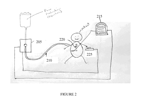
Figure 2. An embodiment of automated IV pump with feedback from a respiration
signal monitor.
Figure 3. An embodiment of an automated safety tube clamp.
Description of the Invention
As embodied and broadly described herein, the disclosures herein provide
detailed embodiments of the invention. However, the disclosed embodiments are
merely exemplary of the invention that may be embodied in various and
alternative
forms. Therefore, there is no intent that specific structural and functional
details should
be limiting, but rather the intention is that they provide a basis for the
claims and as a
representative basis for teaching one skilled in the art to variously employ
the present
invention
6
CA 03003861 2018-05-01
WO 2016/073604
PCT/US2015/059032
With reference to FIG. 1, an exemplary system includes at least one general-
purpose computing device 100, including a processing unit (CPU) 120 and a
system
bus 110 that couples various system components including the system memory
such as
read only memory (ROM) 140 and random access memory (RAM) 150 to the
processing unit 120. Other system memory 130 may be available for use as well.
It
can be appreciated that the invention may operate on a computing device with
more
than one CPU 120 or on a group or cluster of computing devices networked
together to
provide greater processing capability. The system bus 110 may be any of
several types
of bus structures including a memory bus or memory controller, a peripheral
bus, and a
local bus using any of a variety of bus architectures. A basic input/output
(BIOS)
stored in ROM 140 or the like, may provide the basic routine that helps to
transfer
information between elements within the computing device 100, such as during
start-
up. The computing device 100 further includes storage devices such as a hard
disk
drive 160, a magnetic disk drive, an optical disk drive, tape drive or the
like. The
storage device 160 is connected to the system bus 110 by a drive interface.
The drives
and the associated computer readable media provide nonvolatile storage of
computer
readable instructions, data structures, program modules and other data for the
computing device 100. The basic components are known to those of skill in the
art and
appropriate variations are contemplated depending on the type of device, such
as
whether the device is a small, handheld computing device, a desktop computer,
a
computer server, a handheld scanning device, or a wireless devices, including
wireless
Personal Digital Assistants ("PDAs"), tablet devices, wireless web-enabled or
"smart"
phones (e.g., Research in Motion's Blackberry, an Android m4 device, Apple's
iPhoneTm), other wireless phones, a game console (e.g, a Playstation TM, an
XboxTM, or
a WiiTm), a Smart TV, a wearable intern& connected device, etc. Preferably,
the
system is technology agnostic.
Although the exemplary environment described herein employs the hard disk, it
should be appreciated by those skilled in the art that other types of computer
readable
media which can store data that are accessible by a computer, such as magnetic
cassettes, flash memory cards, digital versatile disks, cartridges, random
access
7
CA 03003861 2018-05-01
WO 2016/073604
PCT/US2015/059032
memories (RAMs), read only memory (ROM), a cable or wireless signal containing
a
bit stream and the like, may also be used in the exemplary operating
environment.
To enable user interaction with the computing device 100, an input device 190
represents any number of input mechanisms, such as a microphone for speech, a
touch-
sensitive screen for gesture or graphical input, keyboard, mouse, motion
input, speech,
game console controller, TV remote and so forth. The output device 170 can be
one or
more of a number of output mechanisms known to those of skill in the art, for
example,
printers, monitors, projectors, speakers, and plotters. In some embodiments,
the output
can be via a network interface, for example uploading to a website, emailing,
attached
to or placed within other electronic files, and sending an SMS or MMS message.
In
some instances, multimodal systems enable a user to provide multiple types of
input to
communicate with the computing device 100. The communications interface 180
generally governs and manages the user input and system output. There is no
restriction on the invention operating on any particular hardware arrangement
and
therefore the basic features here may easily be substituted for improved
hardware or
firmware arrangements as they are developed.
For clarity of explanation, the illustrative system embodiment is presented as
comprising individual functional blocks (including functional blocks labeled
as a
"processor"). The functions these blocks represent may be provided through the
use of
either shared or dedicated hardware, including, but not limited to, hardware
capable of
executing software. For example the functions of one or more processors
presented in
FIG. 1 may be provided by a single shared processor or multiple processors.
(Use of the
term "processor" should not be construed to refer exclusively to hardware
capable of
executing software.) Illustrative embodiments may comprise microprocessor
and/or
digital signal processor (DSP) hardware, read-only memory (ROM) for storing
software performing the operations discussed below, and random access memory
(RAM) for storing results. Very large scale integration (VLSI) hardware
embodiments,
as well as custom VLSI circuitry in combination with a general purpose DSP
circuit,
may also be provided.
Embodiments within the scope of the present invention may also include
computer-readable media for carrying or having computer-executable
instructions or
8
CA 03003861 2018-05-01
WO 2016/073604
PCT/US2015/059032
data structures stored thereon. Such computer-readable media can be any
available
media that can be accessed by a general purpose or special purpose computer.
By way
of example, and not limitation, such computer-readable media can comprise RAM,
ROM, EEPROM, CD-ROM or other optical disk storage, magnetic disk storage or
other magnetic storage devices, or any other medium which can be used to carry
or
store desired program code means in the form of computer-executable
instructions or
data structures. When information is transferred or provided over a network or
another
communications connection (either hardwired, wireless, or combination thereof)
to a
computer, the computer properly views the connection as a computer-readable
medium.
Thus, any such connection is properly termed a computer-readable medium.
Combinations of the above should also be included within the scope of the
computer-
readable media.
Computer-executable instructions include, for example, instructions and data
which cause a general purpose computer, special purpose computer, or special
purpose
processing device to perform a certain function or group of functions.
Computer-
executable instructions also include program modules that are executed by
computers
in stand-alone or network environments. Generally, program modules include
routines,
programs, objects, components, and data structures, etc. that perform
particular tasks or
implement particular abstract data types. Computer-executable instructions,
associated
data structures, and program modules represent examples of the program code
means
for executing steps of the methods disclosed herein. The particular sequence
of such
executable instructions or associated data structures represents examples of
corresponding acts for implementing the functions described in such steps.
Those of skill in the art will appreciate the preferred embodiments of the
invention may be practiced in network computing environments with many types
of
computer system configurations, including personal computers, hand-held
devices,
multi-processor systems, microprocessor-based or programmable consumer
electronics,
network PCs, minicomputers, mainframe computers, and the like. Networks may
include the Internet, one or more Local Area Networks ("LANs"), one or more
Metropolitan Area Networks ("MANs"), one or more Wide Area Networks ("WANs"),
one or more Intranets, etc. Embodiments may also be practiced in distributed
9
CA 03003861 2018-05-01
WO 2016/073604
PCT/US2015/059032
computing environments where tasks are performed by local and remote
processing
devices that are linked (either by hardwired links, wireless links, or by a
combination
thereof) through a communications network, e.g. in the "cloud." In a
distributed
computing environment, program modules may be located in both local and remote
memory storage devices.
One embodiment of the invention is directed to an automated IV pump 205 or
tube clamp as depicted in figure 2. A tube 210 preferably provides pain
medication,
however other medication or fluids can be provided by through the tube. For
example
the tube can provide saline solutions, antibiotics, blood, blood substitutes,
vitamins,
buffers, or nutrients. Preferably, tube 210 is coupled to the patient by a
hypodermic
needle, a peripheral cannula, a central line, an implantable port, or another
coupling.
Preferably, the IV pump 205 or tube clamp is controlled by respiration monitor
signals
(e.g. impedance minute ventilation or ETCO2 (end tidal CO2)). Preferably, the
respiration signals are monitored by a monitoring device 215. Abnormal signals
might
be abnormal respiration (e.g. low minute ventilation (L/min) or high ETCO2
(mmHg,
kPa, %)). Preferably, the signal would activate to slow W administration or
tighten the
tube clamp (thereby allowing less fluid through the tube). The IV pump is
preferably
adjusted based on the respiratory signal. Alternately, the tube that provides
medication
could be pinched closed. This would halt or slow the flow of the medication.
An alarm
signal might be triggered and nurse or other medical practitioner would
preferably
arrive and see the alarming monitor signals and the tube in a closed sealed
safe
position. Preferably, the medical practitioner would adjust the devices or
fluids as
needed or perform any necessary medical procedures.
The automated IV pump 205 preferably receives signal from a respiration
volume monitor 215, the pump 205 is controlled based on the level of respired
air by
the patient which is preferably monitored by a set of electrodes 220 attached
to the skin
of the torso of the patient. The set of electrodes 220 can include one or more
electrodes
capable of transmitting and/or receiving an electronic signal. For example,
the
electrodes 220 may detect the impedance across the torso of the patient. As
the patient
breaths in and the chest expands, the impedance of the patient changes. Such
changes
in impedance can be measured by monitor 215. Based on the changes in
impedance,
CA 03003861 2018-05-01
WO 2016/073604
PCT/US2015/059032
respiratory parameters can be determined. For example, the respiratory
parameters
may be variability, variation, or complexity in at least one of the patient's
minute
volume, the patient's respiratory rate, the patient's respiratory pressure,
the patient's
respiratory flow, a patient's end tidal CO2, the patient's sublingual CO2, the
patient's
intensity of respiration, the patient's respiratory curve, change in the shape
of the
patient's respiratory curve, a respiratory curve based on the patient's
inhaled volume, a
respiratory curve based on the patient's exhaled volume, a respiratory curve
based on
the patient's inhaled pressure, a respiratory curve based on the patient's
exhaled
pressure, a respiratory curve based on the patient's inhaled flow, a
respiratory curve
based on the patient's exhaled flow, a respiratory curve based on motion of
the
patient's chest as measured by imaging, a respiratory curve based on motion of
the
patient's chest as measured by contact sensors placed on the chest, a
respiratory curve
based on motion of the patient's abdomen as measured by imaging, a respiratory
curve
based on motion of the patient's abdomen as measured by contact sensors placed
on the
abdomen, a respiratory curve based on motion of both the patient's chest and
abdomen
as measured by imaging, a respiratory curve based on motion of the patient's
chest and
abdomen as measured by contact sensors placed on the chest and abdomen,
variation of
the patient's interbreath intervals, phase lag between the patient's impedance
and
volume signal, variation of phase lag between the patient's impedance and
volume
signal, the patient's respiratory curve, change in the shape of the patient's
respiratory
curve, a respiratory curve based on the patient's inhaled volume, a
respiratory curve
based on the patient's exhaled volume, a respiratory curve based on the
patient's
inhaled pressure, a respiratory curve based on the patient's exhaled pressure,
a
respiratory curve based on the patient's inhaled flow, a respiratory curve
based on the
patient's exhaled flow, a respiratory curve based on motion of the patient's
chest as
measured by imaging, a respiratory curve based on motion of the patient's
chest as
measured by contact sensors placed on the chest, a respiratory curve based on
motion
of the patient's abdomen as measured by imaging, a respiratory curve based on
motion
of the patient's abdomen as measured by contact sensors placed on the abdomen,
a
respiratory curve based on motion of both the patient's chest and abdomen as
measured
by imaging, a respiratory curve based on motion of the patient's chest and
abdomen as
11
CA 03003861 2018-05-01
WO 2016/073604
PCT/US2015/059032
measured by contact sensors placed on the chest and abdomen, variation of the
patient's
interbreath intervals, phase lag between the subject's impedance and volume
signal,
variation of phase lag between the subject's impedance and volume signal, and
combinations thereof.
If the respiratory parameters is normal then the pain medication can continue
on
a standard dosage. When a monitored respiratory parameter drops below a set
level of
the normal range (e.g. 80%) for a person of that weight or other demographic,
then the
pump 205 will preferably reduce the rate of medication administration. If the
respiration parameter goes below a further threshold (e.g. 40%) then the
administration
of the fluid is preferably stopped. IV administration can be resumed once the
respiration parameter returns back above a certain level (e.g. 80% of normal
expected).
Preferably, the reduction, stoppage, and resumption of IV administration is
automatic.
Another aspect of the invention is once the respiration level goes below a
reference level, such as about 60% of normal range, then the patient's self
medication
activation button 250 will preferably be disabled until the respiration level
returns
above 80%. This will preferably provide a "smart" override to a patient who is
very
pain sensitive and does not realize the consequences of additional self doses.
Another embodiment of the invention is directed to an automated safety tube
clamp 330, as depicted in figure 3. The clamp 330 preferably receives signal
from a
respiration volume monitor 315, the clamp 330 is preferably controlled based
on the
level of respired air by the patient, which is monitored by a set of
electrodes 320
preferably attached to the skin of the torso of the patient. A tube clamp 330
is
preferably placed around the tube at beginning of a procedure and/or therapy
session.
The clamp 330 is normally open at beginning of procedure when the patient
vital signs
are normal. For example, if the minute volume is normal then the pain
medication can
continue on a standard dosage. When minute volume drops below a set level of
the
normal range (e.g. 40%) for a person of that weight or other demographic, then
the
clamp will cutoff administration of IV pain medication.
Other embodiments and uses of the invention will be apparent to those skilled
in
the art from consideration of the specification and practice of the invention
disclosed
herein. All references cited herein, including all publications, U.S. and
foreign patents
12
CA 03003861 2018-05-01
WO 2016/073604
PCT/US2015/059032
and patent applications, are specifically and entirely incorporated by
reference,
including, but not limited to U.S. Patent Application Publication Nos.
2010/0324437,
2012/0041279, 2013/0023781, 2014/0073895, and 2015/0254880. The term
comprising, where ever used, is intended to include the terms consisting and
consisting
essentially of. Furthermore, the terms comprising, including, and containing
are not
intended to be limiting. It is intended that the specification and examples be
considered
exemplary only with the true scope and spirit of the invention indicated by
the
following claims.
13