Note: Descriptions are shown in the official language in which they were submitted.
CA 03009053 2018-05-02
WO 2017/079400
PCT/US2016/060273
TITLE
METHODS AND COMPOSITIONS FOR GENE EDITING IN HEMATOPOIETIC STEM
CELLS
CROSS-REFERENCE TO RELATED APPLICATIONS
The present application claims priority under 35 U.S.C. 119(e) to U.S.
Provisional
Patent Application No. 62/250,561, filed November 4, 2015, which is
incorporated herein by
reference in its entirety.
BACKGROUND OF THE INVENTION
Powerful antigen-specific immunotherapies such as chimeric antigen receptor
(CAR)
T cells (CART cells), antibody-drug conjugates or bispecific T cell engaging
antibodies
(BITE), represent novel approaches to the treatment of cancer. Increased
potency is
associated with increased on-target off-minor toxicity, such as the prolonged
B cell aplasia
that results from CART19 treatment of B cell malignancies. In essence, this is
because none
of these modalities are able to discriminate between malignant cells and their
normal
counterparts that carry the same cell surface antigen. CART cells are a novel
therapy in
which T cells are genetically engineered to recognize and kill cells
expressing a specific
antigen on its surface. The CAR is a hybrid of an antigen-recognition domain
of an antibody
combined with the intracellular signaling domains of a T cell surface
receptor. CART cells
targeting CD19 have shown efficacy against B-cell malignancies in several
phase I clinical
trials (Grupp et al, New England Journal of Medicine. 2013;368: 1509-1518;
Brentjens et al,
Blood. 2011;118: 4817-4828; and Kochendeifer et al, Blood. 2010;116: 4099-
4102) and
deplete normal B cells. Since protracted B-Iymphophenia is well tolerated by
humans, this
particular toxicity has not been dose-limiting after CART19. However, CART
cells targeting
acute myeloid leukemia (AML) antigens, such as CD123 or CD33, eradicate
leukemia cells
and consequently deplete normal myeloid progenitors since these bear the same
surface
antigens, thus leading to bone marrow aplasia. The absence of surface antigens
that are
selectively expressed on AML cells and not on normal myeloid cells limits the
use of CART
cells in AML and other myeloid diseases (which include myelodysplastic and
myeloproliferative neoplasms).
Therefore a need exists for selectively targeting tumor cells without
depleting normal
myeloid progenitors. The present invention satisfies this need.
-1-
CA 03009053 2018-05-02
WO 2017/079400
PCT/US2016/060273
SUMMARY OF THE INVENTION
The present invention relates to compositions and methods of generating
modified
hematopoietic stem or progenitor cells.
In one aspect, the invention includes a method of protecting a hematopoietic
stem or
progenitor cell from a chimeric antigen receptor (CAR) T cell therapy in a
subject in need
thereof. The method of the invention comprises administering to the subject a
modified
hematopoietic stem or progenitor cell, wherein the stem or progenitor cell
comprises a
nucleic acid capable of decreasing expression of an endogenous gene or a
portion thereof,
wherein the endogenous gene encodes a polypeptide comprising an antigen domain
targeted
by a CAR. In one embodiment, the method of the invention further comprises
administering
the CART cell therapy to the subject in need thereof. In another embodiment,
the modified
cell further comprises a modified endogenous gene that encodes a modified
polypeptide
lacking the antigen domain targeted by the CAR.
In another aspect, the invention includes a method for generating a modified
hematopoietic stem or progenitor cell. The method of the invention comprises
introducing a
nucleic acid capable of decreasing expression of an endogenous gene or a
portion thereof into
the ccH, wherein the endogenous gene encodes a polypeptide comprising an
antigen domain
targeted by a chimeric antigen receptor (CAR). In one embodiment, the method
comprises
obtaining the cell from a subject in need of CAR T cell therapy. In another
embodiment, the
method further comprises introducing a modified endogenous gene into the
modified cell,
wherein the modified endogenous gene encodes a modified polypeptide lacking
the antigen
domain targeted by the CAR.
In one embodiment, the nucleic acid capable of decreasing the endogenous gene
expression is a CRISPR system. In one embodiment, the CRISPR system comprises
a Cas
expression vector and a guide nucleic acid sequence specific for the
endogenous gene.
In another embodiment, the CRISPR system comprises a Cas9 protein complexed
with a
guide nucleic acid sequence specific for the endogenous gene. In another
embodiment, the
CRISPR system comprises an inducible promoter. In a further embodiment, the
methods of
the invention as described herein further comprise exposing the hematopoietic
stem or
progenitor cell to an agent that activates the inducible promoter in the Cos
expression vector.
In one embodiment, the endogenous gene encodes a tumor antigen. In another
embodiment, the endogenous gene is expressed on a tumor cell targeted by the
CAR. In yet
another embodiment, the endogenous gene encodes CD33 or CD123.
-2-
CA 03004053 2018-05-02
WO 2017/079400
PCT/US2016/060273
In one embodiment, the modified polypeptide comprises at least one function
that is
equivalent to the function of the polypeptide encoded by the endogenous gene.
In one embodiment, the cell is obtained from a source selected from the group
consisting of peripheral blood mononuclear cells, cord blood cells, bone
marrow, lymph
node, and spleen.
In one embodiment, the cell is CD34+. In one embodiment, the method of the
invention as
described herein comprises expanding the cell. In another embodiment, the
expanding is
conducted prior to the step of introducing the nucleic acid. In another
embodiment, the
method of the invention as described herein comprises ciyopreserving the cell.
In yet another
embodiment, the method of the invention as described herein further comprises
thawing the
ciyopreserved cell prior to introducing the nucleic acid. In one embodiment,
introducing the
nucleic acid is conducted by a process selected from the group consisting of
transducing the
cell, transfecting the cell, and electroporating the cell. In another
embodiment, the modified
cell differentiates into at least one blood cell type in the subject. In yet
another embodiment,
the modified cell is capable of self-renewal after administration into the
subject.
In one aspect, the invention includes a composition comprising the modified
cell
generated according to the method described above herein.
In another aspect, the invention includes a pharmaceutical composition
comprising
the modified cell generated according to the method described above herein and
a
pharmaceutically acceptable carrier.
In another aspect, the invention includes a method for adoptive cell transfer
therapy.
The method comprises administering to a subject in need thereof an effective
amount of a
pharmaceutical composition comprising the modified cell generated according to
the method
described herein, wherein the subject is administered an effective amount of
the cell
described herein and a CAR therapy that targets the antigen domain of the
polypeptide
encoded by the endogenous gene thereby treating the subject.
In yet another aspect, the invention includes a method of treating a condition
in a
subject in need thereof The method comprises administering to the subject a
therapeutically
effective amount of a pharmaceutical composition comprising the modified cell
generated
according to the method described herein and administering a CAR T cell
therapy, wherein
the CAR comprises an antigen binding domain that specifically targets the
antigen domain of
the polypeptide encoded by the endogenous gene, thereby treating the
condition.
In one embodiment, the condition is an autoinunune disease. In another
embodiment,
the autoimmune disease is selected from the group consisting of Acquired
Immunodeficiency
-3-
Ch 03004053 2018-05-02
WO 2017/079400
PCT/US2016/060273
Syndrome (AIDS), alopecia areata, ankylosing spondylitis, antiphospholipid
syndrome,
autoimmune Addison's disease, autoimmune hemolytic anemia, autoimmune
hepatitis,
autoimmune inner ear disease (AIED), autoimmune lymphoproliferative syndrome
(ALPS),
autoimmune thrombocytopenic purpura (ATP). Behcet's disease, cardiomyopathy,
celiac
sprue-dermatitis hepetiformis; chronic fatigue inunune dysfunction syndrome
(CFIDS),
chronic inflammatory demyelinating polyneuropathy (CIPD), cicatricial
pemphigold, cold
agglutinin disease, crest syndrome, Crohn's disease, Degos' disease,
dermatomyositis-
juvenile, discoid lupus, essential mixed cryoglobulinemia, fibromyalgia-
fibromyositis,
Graves' disease, Guillain-Barre syndrome, Hashimoto's thyroiditis, idiopathic
pulmonary
fibrosis, idiopathic thrombocytopenia purpura (ITP), IgA nephropathy, insulin-
dependent
diabetes mellitus, juvenile chronic arthritis (Still's disease), juvenile
rheumatoid arthritis,
Meniere's disease, mixed connective tissue disease, multiple sclerosis,
myasthenia gravis,
pemacious anemia, polyarteritis nodosa, polychondritis, polyglandular
syndromes,
polymyalgia rheumatica, polymyositis and dermatomyositis, primary
agammaglobulinemia,
primary biliary cirrhosis, psoriasis, psoriatic arthritis, Raynaud's
phenomena, Reiter's
syndrome, rheumatic fever, rheumatoid arthritis, sarcoidosis, scleroderma
(progressive
systemic sclerosis (PSS), also known as systemic sclerosis (SS)), Sjogren's
syndrome, stiff-
man syndrome, systemic lupus eiythematosus, Takayasu arteritis, temporal
arteritis/giant cell
arteritis, ulcerative colitis, uveitis, vitiligo, Wegener's granulomatosis,
and any combination
thereof. In another embodiment, the condition is a cancer. In yet another
embodiment, the
cancer is selected from the group consisting of breast cancer, prostate
cancer, ovarian cancer,
cervical cancer, skin cancer, pancreatic cancer, colorectal cancer, renal
cancer, liver cancer,
brain cancer, lymphoma, leukemia, lung cancer, and any combination thereof.
BRIEF DESCRIPTION OF THE DRAWINGS
The following detailed description of preferred embodiments of the invention
will be
better understood when read in conjunction with the appended drawings. For the
purpose of
illustrating the invention, there are shown in the drawings embodiments which
are presently
preferred. It should be understood, however, that the invention is not limited
to the precise
arrangements and instrumentalities of the embodiments shown in the drawings.
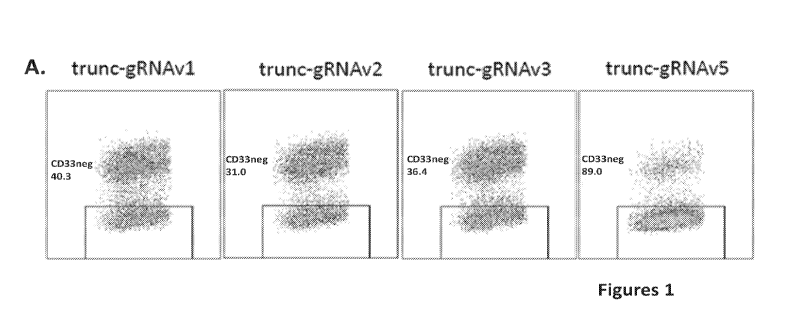
Figures 1A-1B are a set of plots and images showing the CD33 gRNA screen.
Molm14 cells were electroporated with Cas9 mRNA on day 1 and gRNAv1-5 on day
2.
Figure lA is a graph showing CD33 expression by flow cytometly 5 days after
electroporation. Figure 1B is an image showing mismatch cleavage assay
(Surveyor) of PCR
-4-
CA 03004053 2018-05-02
WO 2017/079400
PCT/US2016/060273
of genomic DNA across the gRNA cleavage site. Control cells were
electroporated with
gRNA against EMX1. Indero=1:14{1-(a+b)/(a+b+c))]*100 (a,b: relative
concentration of
cut bands, c: relative concentration of full-length band).
Figures 2A-2B are a set of plots showing CD34+ cells from mobilized peripheral
blood were either electroporated twice, initially with Cas9 mRNA and
subsequently with
CD33-targeted gRNA, or alternatively electroporated once with Cas9 protein
complexed with
the same gRNA. Figure 2A shows a representative plot of CD33 expression by
flow
cytometry 7 days after electroporation. Figure 2B is a graph showing indel
frequencies
measured by TIDE analysis of PCR amplicons spanning the gRNA target site,
averaged for
different donors; n=4 for RNA and n=2 for RNP.
Figures 3A-3B are a set of graphs showing that CART33 cells do not target CD33
negative cells. Figure 3A shows results from the flow cytometric degranulation
assay.
Figure 3B shows results from the luciferase-based killing. In all cases,
effectors are CART33
cells. TCM = T cell media alone (negative control); P-I = PMA and ionomycin
(positive
control); MOLM14wt = the CD33 expressing AML cell line MOLM14 without genetic
modification; D6 = MOLM14 KO for CD33; Jurkat = a CD33 negative cell line (an
additional negative control).
Figures 4A-4D are a panel of images showing CD33 KO in human CD34+ cells.
Figure 4A is a panel of flow diagrams showing CD33 and CD38 expression in CD33
KO
CD34+ cells. Figure 4B is an image showing mismatch mutation efficiency by
surveyor
DNA cleavage assay. Figure 4C is an image showing the Sanger sequencing
results of the
individual mutations as determined by TOPO cloning. Figure 4D shows the
percentages of
CD33 KO cells derived from G-CSF mobilized peripheral blood (mPB), cord blood,
and
bone marrow.
Figures 5A-5C area series of plots and images showing that KO of CD33 in HSCs
does not impair their normal growth and differentiation. After undergoing
CRISPR of the
EMX1 locus (control) or the CD33 locus, CD34+ cells were cultured in serum-
free media
with SCF, F1t3L, TPO, and IL-6 for 7 days, or alternatively plated on semi-
solid
methylcellulose media (Methocult) directly after electroporation. Figure 5A is
a graph
showing a growth curve of control (EMX1) vs. CD33 KO HSCs in vitro culture.
Figure 5B
is a graph showing myeloid and erythroid differentiation of control vs. CD33
KO HSCs in
methylcellulose medium. Figure 5C shows cytospun cells from the
methylcellulose colonies
derived from CD33 KO HSCs showing a typical monocytic and granulocytic
morphology
compared with control cells.
-5-
Ch 03004053 2018-05-02
WO 2017/079400
PCT/US2016/060273
Figure 6 is a panel of graphs of inununophenotyping of CD33 KO HSC
methylcellulose colonies and CD33+ HSC methylcellulose colonies, showing
identical
monocytic and granulocytic differentiation in control HSC-derived colonies,
CD33K0 HSC-
derived colonies, and in the residual CD33-expressing colonies from CD33K0
HSC.
Figures 7A-7B are a panel of graphs showing analysis of CD33 KO HSCs co-
cultured
with CART33 cells. Figure 7A shows expression of CD3 and CD45 on CD33 KO HSCs
compared to control HSCs, demonstrating that exposure to CART33 is more toxic
to control
HSC than to CD33K0 HSC, and this is quantified in Figure 7B.
Figure 8 is a graph showing that mice engrafted with either EMX1 or CD33 KO
HSCs exhibited normal myeloid development.
Figures 9A-9J are a series of plots and images showing that CD33 KO human
CD34+
cells are capable of long-term multi-lineage engraftment. Figure 9A is a
schematic of the
CD33 KO process in primary htunan CD34+ cells derived from G-CSF mobilized
peripheral
blood. Figure 9B displays results from a methocult colony formation assay of
control or
CD33 KO HSPCs. Representative images of colony-forming unit-granulocyte (CFU-
G),
colony-fonning unit-macrophage (CFU-M), and burst-forming unit-erythroid (BFU-
E) in
both groups are shown. Figure 9C shows 8-12 week old NSG mice injected with 1-
5x10⁵
control or CD33-K0 CD34+ cells and peripheral blood human CD45+ engraftment
was
measured after 12 weeks (n=68 mice; 6 independent experiments; 6 different
donors). Figure
9D shows gating on the human CD45+ cells from Figure 9C; B cells (CD19+) and
CD3+ T
cells (CD3+) were detected with no significant difference between the two
groups. Figure 9E
shows human myeloid cells in the CD33 KO HSPC-engrafted mice (gating on
hCD45+CD19-CD3- cells) have significantly reduced levels of CD33 expression
but no
difference in CD11b14+ expression compared to control HSPC-engrafted mice,
confirming
that loss of CD33 does not impact myeloid differentiation. Figure 9F
illustrates bone marrow
harvested after 16 weeks showed equal levels of human CD45+ engraftment in
control and
CD33 KO HSPC-engrafted mice. Figure 9G shows levels of human stem cells
(hCD45+1in-
negative CD34+38-) and myeloid progenitors (hCD45+1in-negative CD34+38+) in
the bone
marrow of mice engrafted with control or CD33 KO HSPCs. Figure 9H is a table
showing
bone marrow was harvested from NSG mice after 16 weeks of primary engraftment
and
transferred into secondary recipients and analyzed after 12 additional weeks;
sustained
human engraftment with persistent CD33 KO phenotype is observed. Figure 91
illustrates
bone marrow harvested after 16 weeks of primary engraftment with control and
CD33 KO
HSPCs, with equal levels of human CD45 expression (top left) and
differentiation into
-6-
CA 03004053 2018-05-02
WO 2017/079400
PCT/US2016/060273
lymphoid and myeloid lineages (top right), only differing in the expression of
CD33 (bottom
left), with no difference in the other myeloid markers CD1 lb and CD14 (bottom
right).
Figure 9J shows expression of CD33 on non-lymphoid human cells (gating for non-
T non-B
human cells not shown) at the end of the 16 week primary transplant,
indicating protracted,
stable absence of CD33 in marrows of xenografted mice.
Figures 10A-10D are a series of plots and images showing CD33 KO HSPCs are
resistant to CART33. Figure 10A is a schematic depicting NSG mice engrafted
with control
or CD33 KO HSPCs were given 5x10⁶ autologous CART33 cells, and residual human
myeloid cells were assessed after 4 weeks (n=30 mice; 2 independent
experiments; 2
different donors). Figure 10B shows CD33 is eliminated in the peripheral blood
of mice
treated with CART33, which leads to ablation of myeloid cells (CD1 lb+CD14+)
in the
control HSPC-engrafted mice, while in the CD33 KO HSPC-engrafted mice the
myeloid cells
are sustained. Figure 10C illustrates myeloid cells are detected in the
peripheral blood,
spleen, and bone marrow of the CD33 KO HSPC-engrafted mice after CART33
treatment, in
contrast to the myeloablation seen in control HSPC-engrafted mice. Figure IOD
shows
human progenitor cells are significantly increased in CD33 KO HSPC-engrafted
mice after
CART33 treatment compared to controls.
Figures 11A-11E are a series of plots and images showing CART33 can eradicate
AML while sparing CD33 KO HSPCs. Figure 11A is a schematic illustrating that
NSG mice
were first engrafted with control or CD33 KO HSPCs, then injected with Molm14,
an AML
cell line engineered to express green fluorescent protein and luciferase,
followed by CART33
treatment (n=8 mice). AML disease burden was measured by bioluminescent
imaging (BLI),
while human HSPCs were measured by flow cy-tometry of the peripheral blood.
Figure 11B is
a series of BLI images showing that both control and CD33 KO HSPC-engrafted
mice
achieve AML disease remission after CART33 treatment. Figure 11 C shows tumor
burden
over time as measured by BLI; each line represents one mouse. Dotted line
represents
background levels of radiance. Figure 11D shows CD33 KO HSPC-engrafted mice
show
persistent CD14+ myeloid cells after CART33 treatment of AML in the peripheral
blood
(PB), spleen, and bone marrow (BM), in contrast to controls. Figure 11E shows
human
progenitor cells are spared from CART33-mediated toxicity in the CD33 KO HSPC
group
only.
Figures 12A-12F are a series of plots and images showing CD33 KO HSPC progeny
have no functional defects. Figure 12A illustrates cytospin and Diff-Quick
staining of human
cells obtained from HSPC-engrafted mouse bone marrow show characteristic
morphologic
-7-
CA 03009053 2018-05-02
WO 2017/079400
PCT/US2016/060273
features of normal stem cell (blast), myeloid progenitor (promyelocyte), and
terminal effector
cells (monocytes and neutrophils). Figure 12B shows control or CD33 KO HSPCs
differentiated in vitro with myeloid cytokines (SCF, TPO, Flt3L, IL-6, GM-CSF,
IL-3) and
incubated with pHrodo green E. coli bioparticles that have green fluorescence
when acidified
in the phagosome; percent phagocytosis was measured by flow cytometry. Top,
representative flow plots from control and CD33 KO cells; bottom;
quantification of
phagocytosis from 2 independent experiments with 4 different donors. Figures
12C-12D
show control and CD33 KO CD34+ cells from 5 different mobilized peripheral
blood donors
were differentiated in vitro and gene expression was analyzed by RNA-seq.
Figure 12C
depicts fold-changes of differentially expressed genes shown as a heat map,
with each row
corresponding to genes and each column representing one sample from control
(ctrl) or CD33
KO (KO); numbers indicate donor of origin. Columns and rows are organized by
hierarchical
clustering; dendrogram branch length represents distances between samples and
clusters.
Figure 12D is a log-scale scatter plot of mean gene expression values of
control and CD33
KO samples. The coefficient of determination (R2) value is shown. Figure 12E
shows mice
engrafted with control or CD33 KO HSPCs were injected with rhG-CSF and
absolute
numbers of peripheral blood human monocytes (CD11b+14+) and neutrophils (CD1
lb+14-)
were measured; fold-change of cell numbers compared to baseline levels are
shown. Figure
12F illustrates mice engrafted with control or CD33 KO HSPCs were injected
with
lipopolysaccharide and serum levels of human cytokines were measured.
Figures 13A-13B are a series of images depicting off-target evaluation of CD33
KO
HSPCs. Figure 13A is a table showing the top 12 off-target sites predicted in
silico from two
web-based tools. Figure 13B, top panel, shows that SIGLEC2213, a pseudogene,
has a high
degree of homology to the CD33 gene, with a 100% identical binding site of the
CD33-
targeted gRNA used herein. Figure 13B, bottom panel, shows no mutations
detected by
Surveyor mismatch assay in other SIGLEC genes, while a high degree of on-
target mutations
are found in CD33 and S1GLEC22P.
Figures 14A-14B are a series of plots and images depicting autologous CD33 KO
stem cell transplant in Rhesus macaques. Figure 14A shows the experimental
schema,
whereby rhesus CD34+ HSPC are mobilized using G-CSF and plerixafor, removed by
apheresis, and gene edited with CRISPR/Cas9-based gene knockout of CD33. In
the
meantime, the monkey is conditioned with irradiation (TBI) and following that,
receives a re-
infusion of the edited HSPC. On the top right panel, flow cytometric
evaluation of CD33
expression in in vitro differentiated HSPC is shown in control and KO cells,
and TIDE
-8-
Ch 03004053 2018-05-02
WO 2017/079400
PCT/US2016/060273
analysis of sequencing of the CD33 locus is shown on the bottom right panel.
Figure 14B
shows expression of CD33 on selected sub-populations from the PB of the animal
transplanted in Figure 14A.
DETAILED DESCRIPTION
Definitions
Unless defined otherwise, all technical and scientific terms used herein have
the same
meaning as commonly understood by one of ordinary skill in the art to which
the invention
pertains. Although any methods and materials similar or equivalent to those
described herein
can be used in the practice for testing of the present invention, the
preferred materials and
methods are described herein. In describing and claiming the present
invention, the
following tenninology will be used.
It is also to be understood that the terminology used herein is for the
purpose of
describing particular embodiments only, and is not intended to be limiting.
The articles "a" and "an" are used herein to refer to one or to more than one
(i.e., to at
least one) of the grammatical object of the article. By way of example, "an
element" means
one element or more than one element.
"About" as used herein when referring to a measurable value such as an amount,
a
temporal duration, and the like, is meant to encompass variations of 20% or
10%, more
preferably 5 /0, even more preferably 1%, and still more preferably 0.1%
from the
specified value, as such variations are appropriate to perform the disclosed
methods.
The term "antibody," as used herein, refers to an immunoglobulin molecule
which
specifically binds with an antigen. Antibodies can be intact immunoglobulins
derived from
natural sources or from recombinant sources and can be immunoreactive portions
of intact
immunoglobulins. Antibodies are typically tetramers of immtmoglobulin
molecules. The
antibodies in the present invention may exist in a variety of forms including,
for example,
polyclonal antibodies, monoclonal antibodies, Fv, Fab and Rab)2, as well as
single chain
antibodies (scFv) and humanized antibodies (Harlow et al., 1999, In: Using
Antibodies: A
Laboratory Manual, Cold Spring Harbor Laboratory Press, NY; Harlow et al.,
1989, In:
Antibodies: A Laboratory Manual, Cold Spring Harbor, New York; Houston et al.,
1988,
Proc. Natl. Acad. Sci. USA 85:5879-5883; Bird et al., 1988, Science 242:423-
426).
The term "antigen" or "Ag" as used herein is defmed as a molecule that
provokes an
immune response. This immune response may involve either antibody production,
or the
-9-
Ch 03004053 2018-05-02
WO 2017/079400
PCT/US2016/060273
activation of specific immunologically-competent cells, or both. The skilled
artisan will
understand that any macromolecule, including virtually all proteins or
peptides, can serve as
an antigen. Furthermore, antigens can be derived from recombinant or genomic
DNA. A
skilled artisan will understand that any DNA, which comprises a nucleotide
sequences or a
partial nucleotide sequence encoding a protein that elicits an immune response
therefore
encodes an "antigen" as that term is used herein. Furthermore, one skilled in
the art will
understand that an antigen need not be encoded solely by a full length
nucleotide sequence of
a gene. It is readily apparent that the present invention includes, but is not
limited to, the use
of partial nucleotide sequences of more than one gene and that these
nucleotide sequences are
arranged in various combinations to elicit the desired immune response.
Moreover, a skilled
artisan will understand that an antigen need not be encoded by a "gene" at
all. It is readily
apparent that an antigen can be generated synthesized or can be derived from a
biological
sample. Such a biological sample can include, but is not limited to a tissue
sample, a tumor
sample, a cell or a biological fluid.
The term "anti-tumor effect" as used herein, refers to a biological effect
which can be
manifested by a decrease in tumor volume, a decrease in the number of tumor
cells, a
decrease in the number of metastases, an increase in life expectancy, or
amelioration of
various physiological symptoms associated with the cancerous condition. An
"anti-tumor
effect" can also be manifested by the ability of the peptides,
polynucleotides, cells and
antibodies of the invention in prevention of the occurrence of tumor in the
first place.
The term "auto-antigen" means, in accordance with the present invention, any
self-
antigen which is recognized by the immune system as being foreign. Auto-
antigens comprise,
but are not limited to, cellular proteins, phosphoproteins, cellular surface
proteins, cellular
lipids, nucleic acids, glycoproteins, including cell surface receptors.
The term "autoimmune disease" as used herein is defined as a disorder that
results
from an autoimmune response. An autoimmune disease is the result of an
inappropriate and
excessive response to a self-antigen. Examples of autoimmune diseases include
but are not
limited to, Addision's disease, alopecia areata, ankylosing spondylitis,
autoimmune hepatitis,
autoimmune parotitis, Crohn's disease, diabetes (Type I), dystrophic
epidermolysis bullosa,
epididymitis, glomerulonephiitis, Graves' disease, Guillain-Barr syndrome,
Hashimoto's
disease, hemolytic anemia, systemic lupus erythematosus, multiple sclerosis,
myasthenia
gravis, pemphigus vulgaris, psoriasis, rheumatic fever, rheumatoid arthritis,
sarcoidosis,
scleroderma, Sjogren's syndrome, spondyloarthropathies, thyroiditis,
vasculitis, vitiligo,
my-xedema, pernicious anemia, ulcerative colitis, among others.
-10-
CA 03004053 2018-05-02
WO 2017/079400
PCT/US2016/060273
As used herein, the term "autologous" is meant to refer to any material
derived from
the same individual to which it is later to be re-introduced into the
individual.
"Allogeneic" refers to a graft derived from a different animal of the same
species.
"Xenogeneic" refers to a graft derived from an animal of a different species.
The term "cancer" as used herein is defined as disease characterized by the
rapid and
uncontrolled growth of aberrant cells. Cancer cells can spread locally or
through the
bloodstream and lymphatic system to other parts of the body. Examples of
various cancers
include but are not limited to, breast cancer, prostate cancer, ovarian
cancer, cervical cancer,
skin cancer, pancreatic cancer, colorectal cancer, renal cancer, liver cancer,
brain cancer,
lymphoma, leukemia, lung cancer and the like. In certain embodiments, the
cancer is
medullary thyroid carcinoma.
The term "chimeric antigen receptor" or "CAR," as used herein, refers to an
artificial
T cell receptor that is engineered to be expressed on an immune effector cell
and specifically
bind an antigen. CARs may be used as a therapy with adoptive cell transfer. T
cells are
removed from a patient and modified so that they express the receptors
specific to a particular
lbrin of antigen. In some embodiments, the CARs have been expressed with
specificity to a
tumor associated antigen, for example. CARs may also comprise an intracellular
activation
domain, a transmembrane domain and an extracellular domain comprising a tumor
associated
antigen binding region. in some aspects, CARs comprise single-chain variable
fragments
(scFv) derived from monoclonal antibodies. The specificity of CAR designs may
be derived
from ligands of receptors (e.g... peptides). In some embodiments, a CAR can
target cancers
by redirecting the specificity of a T cell expressing the CAR specific for
tumor associated
antigens.
The term "cleavage" refers to the breakage of covalent bonds, such as in the
backbone
of a nucleic acid molecule. Cleavage can be initiated by a variety of methods,
including, but
not limited to, enzymatic or chemical hydrolysis of a phosphodiester bond.
Both single-
stranded cleavage and double-stranded cleavage are possible. Double-stranded
cleavage can
occur as a result of two distinct single-stranded cleavage events. DNA
cleavage can result in
the production of either blunt ends or staggered ends. In certain embodiments,
fusion
polypeptides may be used for targeting cleaved double-stranded DNA.
As used herein, the term "conservative sequence modifications" is intended to
refer to
amino acid modifications that do not significantly affect or alter the binding
characteristics of
the antibody containing the amino acid sequence. Such conservative
modifications include
amino acid substitutions, additions and deletions. Modifications can be
introduced into an
-11-
CA 03004053 2018-05-02
WO 2017/079400
PCT/US2016/060273
antibody of the invention by standard techniques known in the art, such as
site-directed
mutagenesis and PCR-mediated mutagenesis. Conservative amino acid
substitutions are ones
in which the amino acid residue is replaced with an amino acid residue having
a similar side
chain. Families of amino acid residues having similar side chains have been
defined in the
art. These families include amino acids with basic side chains (e.g., lysine,
arginine,
histidine), acidic side chains (e.g., aspartic acid, glutamic acid), uncharged
polar side chains
(e.g., glycine, asparagine, glutamine, serine, threonine, tyrosine, cysteine,
tryptophan),
nonpolar side chains (e.g., alanine, value, leucine, isoleucine, proline,
phenylalanine,
methionine), beta-branched side chains (e.g., threonine, valine, isoleucine)
and aromatic side
chains (e.g., tyrosine, phenylalanine, tryptophan, histidine). Thus, one or
more amino acid
residues within the CDR regions of an antibody can be replaced with other
amino acid
residues from the same side chain family and the altered antibody can be
tested for the ability
to bind antigens using the functional assays described herein.
The term "CRISPRICAS,' "clustered regularly interspaced short pahndromic
repeats
system," or "CRISPR" refers to DNA loci containing short repetitions of base
sequences.
Each repetition is followed by short segments of spacer DNA from previous
exposures to a
virus. Bacteria and archaea have evolved adaptive immune defenses termed
CR1SPR-
CRISPR¨associated (Cas) systems that use short RNA to direct degradation of
foreign
nucleic acids. In bacteria, the CRISPR system provides acquired immunity
against invading
foreign DNA via RNA-guided DNA cleavage.
In the type ii CRISPR/Cas system, short segments of foreign DNA, termed
"spacers"
are integrated within the CRISPR genomic loci and transcribed and processed
into short
CRISPR RNA (crRNA). These crRNAs anneal to trans-activating crRNAs (tracrRNAs)
and
direct sequence-specific cleavage and silencing of pathogenic DNA by Cas
proteins. Recent
work has shown that target recognition by the Cas9 protein requires a "seed"
sequence within
the crRNA and a conserved dinucleotide-containing protospacer adjacent motif
(PAM)
sequence upstream of the crRNA-binding region.
To direct Cas9 to cleave sequences of interest, crRNA-traerRN A. fusion
transcripts,
hereafter referred to as "guide RNAs" or "L4RNAs" may be designed, from human
116
polvmemse III promoter. CRISPR/CAS mediated genome editing and regulation,
highlighted
its transformative potential for basic science, cellular engineering and
therapeutics.
The term "CRISPRi" refers to a CRISPR system for sequence specific gene
repression or inhibition of gene expression, such as at the transcriptional
level.
CA 03009053 2018-05-02
WO 2017/079400
PCT/US2016/060273
A "disease" is a state of health of an animal wherein the animal cannot
maintain
homeostasis, and wherein if the disease is not ameliorated then the animal's
health continues
to deteriorate. In contrast, a "disorder" in an animal is a state of health in
which the animal is
able to maintain homeostasis, but in which the animal's state of health is
less favorable than it
would be in the absence of the disorder. Left untreated, a disorder does not
necessarily cause
a further decrease in the animal's state of health.
The term "downregulation" as used herein refers to the decrease or elimination
of
gene expression of one or more genes or a portion thereof.
"Effective amount" or "therapeutically effective amount" are used
interchangeably
herein, and refer to an amount of a compound, fonnulation, material, or
composition, as
described herein effective to achieve a particular biological result or
provides a therapeutic or
prophylactic benefit. Such results may include, but are not limited to, anti-
tumor activity as
determined by any means suitable in the art.
"Encoding" refers to the inherent property of specific sequences of
nucleotides in a
polynucleotide, such as a gene, a cDNA, or an mRNA, to serve as templates for
synthesis of
other polymers and macromolecules in biological processes having either a
defined sequence
of nucleotides (i.e., rRNA, tRNA and mRNA) or a defined sequence of amino
acids and the
biological properties resulting therefrom. Thus, a gene encodes a protein if
transcription and
translation of mRNA corresponding to that gene produces the protein in a cell
or other
biological system. Both the coding strand, the nucleotide sequence of which is
identical to
the mRNA sequence and is usually provided in sequence listings, and the non-
coding strand,
used as the template for transcription of a gene or cDNA, can be referred to
as encoding the
protein or other product of that gene or cDNA.
As used herein "endogenous" refers to any material from or produced inside an
organism, cell, tissue or system.
As used herein, the term "exogenous" refers to any material introduced from or
produced outside an organism, cell, tissue or system.
The term "expand" as used herein refers to increasing in number, as in an
increase in
the number of cells. In one embodiment, the cells that are expanded ex vivo
increase in
number relative to the number originally present in the culture. In another
embodiment, the
cells that are expanded ex vivo increase in number relative to other cell
types in the culture.
The term "ex vivo," as used herein, refers to cells that have been removed
from a living
organism, (e.g., a human) and propagated outside the organism (e.g., in a
culture dish, test
tube, or bioreactor).
-13-
CA 09004059 2018-05-02
WO 2017/079400
PCT/US2016/060273
The term "expression" as used herein is defined as the transcription and/or
translation
of a particular nucleotide sequence driven by its promoter.
"Expression vector" refers to a vector comprising a recombinant polynucleotide
comprising expression control sequences operatively linked to a nucleotide
sequence to be
expressed. An expression vector comprises sufficient cis-acting elements for
expression;
other elements for expression can be supplied by the host cell or in an in
vitro expression
system. Expression vectors include all those known in the art, such as
cosmids, plasmids
(e.g., naked or contained in liposomes) and viruses (e.g., Sendai viruses,
lentiviruses,
retroviruses, adenovimses, and adeno-associated viruses) that incorporate the
recombinant
polynucleotide.
The term "hematopoietic stem cell" or "HSC" refers to an undifferentiated
hematopoietic cell that is capable of differentiating into all blood cell
types, myeloid and
lymphoid cells. The HSC may reside in the bone marrow or be found elsewhere
e.g.
peripheral blood.
"Homologous" as used herein, refers to the subunit sequence identity between
two
polymeric molecules, e.g., between two nucleic acid molecules, such as, two
DNA molecules
or two RNA molecules, or between two poly-peptide molecules. When a subunit
position in
both of the two molecules is occupied by the same monomeric subunit; e.g., if
a position in
each of two DNA molecules is occupied by adenine, then they are homologous at
that
position. The homology between two sequences is a direct function of the
number of
matching or homologous positions; e.g., if half (e.g., five positions in a
polymer ten subunits
in length) of the positions in two sequences are homologous, the two sequences
are 50%
homologous; if 90% of the positions (e.g., 9 of 10), are matched or
homologous, the two
sequences are 90% homologous.
"Identity" as used herein refers to the subunit sequence identity between two
polymeric molecules particularly between two amino acid molecules, such as,
between two
polypeptide molecules. When two amino acid sequences have the same residues at
the same
positions; e.g., if a position in each of two polypeptide molecules is
occupied by an Arginine,
then they are identical at that position. The identity or extent to which two
amino acid
sequences have the same residues at the same positions in an alignment is
often expressed as
a percentage. The identity between two amino acid sequences is a direct
function of the
number of matching or identical positions; e.g., if half (e.g , five positions
in a polymer ten
amino acids in length) of the positions in two sequences are identical, the
two sequences are
-14-
CA 09004053 2018-05-02
WO 2017/079400
PCT/US2016/060273
50% identical; if 90% of the positions (e.g., 9 of 10), are matched or
identical, the two amino
acids sequences are 90% identical.
The term "immune response" as used herein is defined as a cellular response to
an
antigen that occurs when lymphocytes identify antigenic molecules as foreign
and induce the
formation of antibodies and/or activate lymphocytes to remove the antigen.
As used herein, an "instructional material" includes a publication, a
recording, a
diagram, or any other medium of expression which can be used to communicate
the
usefulness of the compositions and methods of the invention. The instructional
material of
the kit of the invention may, for example, be affixed to a container which
contains the nucleic
acid, peptide, and/or composition of the invention or be shipped together with
a container
which contains the nucleic acid, peptide, and/or composition. Alternatively,
the instructional
material may be shipped separately from the container with the intention that
the instructional
material and the compound be used cooperatively by the recipient.
"Isolated" means altered or removed from the natural state. For example, a
nucleic
acid or a peptide naturally present in a living animal is not "isolated," but
the same nucleic
acid or peptide partially or completely separated from the coexisting
materials of its natural
state is "isolated." An isolated nucleic acid or protein can exist in
substantially purified form,
or can exist in a non-native environment such as, for example, a host cell.
The term "knockout" or "KO" as used herein refers to the ablation of gene
expression
of one or more genes.
A "lentivirus" as used herein refers to a genus of the Retroviridae family.
Lentiviruses
are unique among the retroviruses in being able to infect non-dividing cells;
they can deliver
a significant amount of genetic information into the DNA of the host cell, so
they are one of
the most efficient methods of a gene delivery vector. HIV, SIV, and FIV are
all examples of
lentiviruses. Vectors derived from lentiviruses offer the means to achieve
significant levels of
gene transfer in vivo.
By the term "modified" as used herein, is meant a changed state or structure
of a
molecule or cell of the invention. Molecules may be modified in many ways,
including
chemically, structurally, and functionally. Cells may be modified through the
introduction of
nucleic acids.
By the term "modulating," as used herein, is meant mediating a detectable
increase or
decrease in the level of a response in a subject compared with the level of a
response in the
subject in the absence of a treatment or compound, and/or compared with the
level of a
response in an otherwise identical but untreated subject. The term encompasses
perturbing
-15-
Ch 03004053 2018-05-02
WO 2017/079400
PCT/US2016/060273
and/or affecting a native signal or response thereby mediating a beneficial
therapeutic
response in a subject, preferably, a human.
In the context of the present invention, the following abbreviations for the
commonly
occurring nucleic acid bases are used. "A" refers to adenosine, "C" refers to
cytosine, "G"
refers to guanosine, "T" refers to thymidine, and "U" refers to uridine.
Unless otherwise specified, a "nucleotide sequence encoding an amino acid
sequence"
includes all nucleotide sequences that are degenerate versions of each other
and that encode
the same amino acid sequence. The phrase nucleotide sequence that encodes a
protein or an
RNA may also include introns to the extent that the nucleotide sequence
encoding the protein
may in some version contain an intron(s).
The term "operably linked" refers to functional linkage between a regulatory
sequence and a heterologous nucleic acid sequence resulting in expression of
the latter. For
example, a first nucleic acid sequence is operably linked with a second
nucleic acid sequence
when the first nucleic acid sequence is placed in a functional relationship
with the second
nucleic acid sequence. For instance, a promoter is operably linked to a coding
sequence if the
promoter affects the transcription or expression of the coding sequence.
Generally, operably
linked DNA sequences are contiguous and, where necessary to join two protein
coding
regions, in the same reading frame.
The term "overexpressed" tumor antigen or "overexpression" of a tumor antigen
is
intended to indicate an abnormal level of expression of a tumor antigen in a
cell from a
disease area like a solid tumor within a specific tissue or organ of the
patient relative to the
level of expression in a normal cell from that tissue or organ. Patients
having solid tumors or
a hematological malignancy characterized by overexpression of the tumor
antigen can be
determined by standard assays known in the art.
"Parenteral" administration of an immunogenic composition includes, e.g.,
subcutaneous (s.c.), intravenous (i.v.), intramuscular (i.m.), or intrasternal
injection, or
infusion techniques.
As used herein, the terms "peptide," "polypeptide," and "protein" are used
interchangeably, and refer to a compound comprised of amino acid residues
covalently linked
by peptide bonds. A protein or peptide must contain at least two amino acids,
and no
limitation is placed on the maximum munber of amino acids that can comprise a
protein's or
peptide's sequence. Polypeptides include any peptide or protein comprising two
or more
amino acids joined to each other by peptide bonds. As used herein, the term
refers to both
short chains, which also commonly are referred to in the art as peptides,
oligopeptides and
-16-
Ch 03004053 2018-05-02
WO 2017/079400
PCT/US2016/060273
oligomers, for example, and to longer chains, which generally are referred to
in the art as
proteins, of which there are many types. "Polypeptides" include, for example,
biologically
active fragments, substantially homologous polypeptides, oligopeptides,
homodimers,
heterodimers, variants of polypeptides, modified polypeptides, derivatives,
analogs, fusion
proteins, among others. The polypeptides include natural peptides, recombinant
peptides,
synthetic peptides, or a combination thereof.
The term "polynucleotide" as used herein is defined as a chain of nucleotides.
Furthermore, nucleic acids are polymers of nucleotides. Thus, nucleic acids
and
polynucleotides as used herein are interchangeable. One skilled in the art has
the general
knowledge that nucleic acids are polynucleotides, which can be hydrolyzed into
the
monomeric "nucleotides." The monomeric nucleotides can be hydrolyzed into
nucleosides.
As used herein polynucleotides include, but are not limited to, all nucleic
acid sequences
which are obtained by any means available in the art, including, without
limitation,
recombinant means, i.e., the cloning of nucleic acid sequences from a
recombinant library or
a cell genome, using ordinary cloning technology and PCRTM, and the like, and
by synthetic
means.
The term "portion thereof' refers to a part of or a fragment of the whole.
The term "hematopoietic progenitor cell" refers to an undifferentiated
hematopoietic
cell capable of differentiating into at least one blood cell type to several
blood cell types, but
not all blood cells like a HSC. Examples of hematopoietic progenitor cells
include, but are
not limited to, a common myeloid progenitor cell, megakaryocyte-erythrocyte
progenitor cell,
granulocyte-macrophage progenitor cell, monocyte-dendritic progenitor cell,
and a common
lymphoid progenitor cell.
The term "promoter" as used herein is defined as a DNA sequence recognized by
the
synthetic machinery of the cell, or introduced synthetic machinery, required
to initiate the
specific transcription of a polynucleotide sequence.
As used herein, the term "promoter/regulatory sequence" means a nucleic acid
sequence which is required for expression of a gene product operably linked to
the
promoter/regulatory sequence. In some instances, this sequence may be the core
promoter
sequence and in other instances, this sequence may also include an enhancer
sequence and
other regulatory elements which are required for expression of the gene
product. The
promoter/regulatory sequence may, for example, be one which expresses the gene
product in
a tissue specific manner.
-17-
Ch 03004053 2018-05-02
WO 2017/079400
PCT/US2016/060273
A "constitutive" promoter is a nucleotide sequence which, when operably linked
with
a polynucleotide which encodes or specifies a gene product, causes the gene
product to be
produced in a cell under most or all physiological conditions of the cell.
An "inducible" promoter is a nucleotide sequence which, when operably linked
with a
polynucleotide which encodes or specifies a gene product, causes the gene
product to be
produced in a cell substantially only when an inducer which corresponds to the
promoter is
present in the cell.
A "tissue-specific" promoter is a nucleotide sequence which, when operably
linked
with a polynucleotide encodes or specified by a gene, causes the gene product
to be produced
in a cell substantially only if the cell is a cell of the tissue type
corresponding to the promoter.
A "Sendai virus" refers to a genus of the Paramyxoviridae family. Sendai
viruses are
negative, single stranded RNA viruses that do not integrate into the host
genotne or alter the
genetic information of the host cell. Sendai viruses have an exceptionally
broad host range
and are not pathogenic to humans. Used as a recombinant viral vector, Sendai
viruses are
capable of transient but strong gene expression.
A "signal transduction pathway" refers to the biochemical relationship between
a
variety of signal transduction molecules that play a role in the transmission
of a signal from
one portion of a cell to another portion of a cell. The phrase "cell surface
receptor" includes
molecules and complexes of molecules capable of receiving a signal and
transmitting signal
across the plasma membrane of a cell.
By the term "specifically binds," as used herein with respect to an antibody,
is meant
an antibody which recognizes a specific antigen, but does not substantially
recognize or bind
other molecules in a sample. For example, an antibody that specifically binds
to an antigen
from one species may also bind to that antigen from one or more species. But,
such cross-
species reactivity does not itself alter the classification of an antibody as
specific. In another
example, an antibody that specifically binds to an antigen may also bind to
different allelic
fonns of the antigen. However, such cross reactivity does not itself alter the
classification of
an antibody as specific. In some instances, the terms "specific binding" or
"specifically
binding," can be used in reference to the interaction of an antibody, a
protein, or a peptide
with a second chemical species, to mean that the interaction is dependent upon
the presence
of a particular structure (e.g., an antigenic determinant or epitope) on the
chemical species;
for example, an antibody recognizes and binds to a specific protein structure
rather than to
proteins generally. If an antibody is specific for epitope "A", the presence
of a molecule
-18-
CA 03004053 2018-05-02
WO 2017/079400
PCT/US2016/060273
containing epitope A (or free, unlabeled A), in a reaction containing labeled
"A" and the
antibody, will reduce the amount of labeled A bound to the antibody.
By the term "stimulation," is meant a primary response induced by binding of a
stimulatory molecule (e.g., a TCR/CD3 complex) with its cognate ligand thereby
mediating a
signal transduction event, such as, but not limited to, signal transduction
via the TCR/CD3
complex. Stimulation can mediate altered expression of certain molecules, such
as
downregulation of TGF-beta, and/or reorganization of cytoskeletal structures,
and the like.
"Substantially complementary," as used herein, refers to sequences of
nucleotides
where a majority or all of the bases in the primer sequence are complementary,
or one or
more bases are non-complementary, or mismatched.
The term "subject" is intended to include living organisms in which an immune
response can be elicited (e.g., mammals). A "subject" or "patient," as used
therein, may be a
human or non-human mammal. Non-human mammals include, for example, livestock
and
pets. such as ovine, bovine, porcine, canine, feline and murine mammals.
Preferably, the
subject is human.
As used herein, a "substantially purified" cell is a cell that is essentially
free of other
cell types. A substantially purified cell also refers to a cell which has been
separated from
other cell types with which it is normally associated in its naturally
occurring state. In some
instances, a population of substantially purified cells refers to a homogenous
population of
cells. In other instances, this term refers simply to cell that have been
separated from the
cells with which they are naturally associated in their natural state. In some
embodiments,
the cells are cultured in vitro. In other embodiments, the cells are not
cultured in vitro.
A "target site" or "target sequence" refers to a genoinic nucleic acid
sequence that
defines a portion of a nucleic acid to which a binding molecule may
specifically bind under
conditions sufficient for binding to occur.
The term "therapeutic" as used herein means a treatment and/or prophylaxis. A
therapeutic effect is obtained by suppression, remission, or eradication of a
disease state.
The term "transfected" or "transformed" or "transduced" as used herein refers
to a
process by which exogenous nucleic acid is transferred or introduced into the
host cell. A
"transfected" or "transformed" or "transduced" cell is one which has been
transfected,
transformed or transduced with exogenous nucleic acid. The cell includes the
primary
subject cell and its progeny.
To "treat" a disease as the term is used herein, means to reduce the frequency
or
severity of at least one sign or symptom of a disease or disorder experienced
by a subject.
-19-
Ch 03004053 2018-05-02
WO 2017/079400
PCT/US2016/060273
The phrase "under transcriptional control" or "operatively linked" as used
herein
means that the promoter is in the correct location and orientation in relation
to a
polynucleotide to control the initiation of transcription by RNA polymerase
and expression of
the polynucleotide.
A "vector" is a composition of matter which comprises an isolated nucleic acid
and
which can be used to deliver the isolated nucleic acid to the interior of a
cell. Numerous
vectors are known in the art including, but not limited to, linear
polynucleotides,
polynucleotides associated with ionic or amphiphilic compounds, plasmids, and
viruses.
Thus, the term "vector" includes an autonomously replicating plasmid or a
virus. The term
should also be construed to include non-plasmid and non-viral compounds which
facilitate
transfer of nucleic acid into cells, such as, for example, polylysine
compounds, liposomes,
and the like. Examples of viral vectors include, but are not limited to,
Sendai viral vectors,
adenoviral vectors, adeno-associated virus vectors, retroviral vectors,
lentiviral vectors, and
the like.
Ranges: throughout this disclosure, various aspects of the invention can be
presented
in a range format. It should be understood that the description in range
format is merely for
convenience and brevity and should not be construed as an inflexible
limitation on the scope
of the invention. Accordingly, the description of a range should be considered
to have
specifically disclosed all the possible subranges as well as individual
numerical values within
that range. For example, description of a range such as from 1 to 6 should be
considered to
have specifically disclosed subranges such as from 1 to 3, from 1 to 4, from 1
to 5, from 2 to
4, from 2 to 6, from 3 to 6 etc., as well as individual numbers within that
range, for example,
1, 2, 2.7, 3, 4, 5, 5.3, and 6. This applies regardless of the breadth of the
range.
Description
The invention described herein includes compositions and methods of generating
modified hematopoietic stem or progenitor cells that have decreased expression
of an
endogenous gene or a portion thereof. The endogenous gene encodes a
polypeptide
comprising an antigen domain targeted by a CAR or by any other antibody-based
modality
such as a monoclonal antibody, scFv, or bi-specific antibody (e.g. BITE). The
endogenous
gene or a portion thereof is downregulated via gene editing such that the
modified
hematopoietic stem or progenitor cells are rendered resistant to CART cell or
other antigen-
specific therapy.
-20-
CA 03009053 2018-05-02
WO 2017/079400
PCT/US2016/060273
Methods
One aspect of the invention includes a method of protecting a hematopoietic
stem or
progenitor cell from a chimeric antigen receptor (CAR) T cell therapy or other
antigen-
specific therapy in a subject in need thereof. The method comprises
administering a modified
hematopoietic stem or progenitor cell. The stem or progenitor cell comprises a
nucleic acid
capable of decreasing expression of an endogenous gene or a portion thereof
and the
endogenous gene encodes a polypeptide comprising an antigen domain targeted by
a CAR.
In one embodiment, the invention may further comprise administering the CART
therapy to
the subject in need thereof.
The modified cell may further comprise a modified endogenous gene that encodes
a
modified polypeptide lacking the antigen domain targeted by the CAR. The
modified
polypeptide may comprise at least one function that is equivalent to the
function of the
polypeptide encoded by the endogenous gene.
The invention also includes a method for generating a modified hematopoietic
stem or
progenitor cell. The method comprises introducing a nucleic acid capable of
decreasing
expression of an endogenous gene or a portion thereof into the cell, wherein
the endogenous
gene encodes a polypeptide comprising an antigen domain targeted by a chimeric
antigen
receptor (CAR). The invention may further comprise obtaining a cell from a
subject in need
of CART cell therapy. The cell may be obtained from peripheral blood
mononuclear cells,
cord blood cells, bone marrow, lymph nodes, and/or a spleen. The cell may be
CD34+.
In certain embodiments of the invention, the nucleic acid capable of
decreasing
endogenous gene expression is a CRISPR system. The CRISPR system may comprise
a Cas
expression vector and a guide nucleic acid sequence specific for the
endogenous gene and/or
a Cas9 protein complexed with a guide nucleic acid sequence specific for the
endogenous
gene. The CRISPR system may comprise an inducible promoter. The hematopoietic
stem or
progenitor cell may be exposed to an agent that activates the inducible
promoter in the Cas
expression vector.
In certain embodiments, the endogenous gene may encode a tumor antigen and/or
may be expressed on a tumor cell targeted by the CAR and/or may encode CD33,
CD123,
CD19, or CD22. A modified endogenous gene may be introduced into the modified
cell,
wherein the modified endogenous gene encodes a modified polypeptide lacking
the antigen
domain targeted by the CAR. The modified polypeptide may comprise at least one
function
that is equivalent to the function of the polypeptide encoded by the
endogenous gene.
-21-
CA 03004053 2018-05-02
WO 2017/079400
PCT/US2016/060273
Certain embodiments of the invention further comprise expanding the cells.
Expansion may be prior to the step of introducing the nucleic acid. The cells
may be
ciyopreserved then thawed prior to introducing the nucleic acids. The nucleic
acid may be
introduced by transducing the cell, or transfecting the cell, or
electroporating the cell.
The invention also includes a modified cell that is generated according to the
methods
described herein. A pharmaceutical composition comprising the modified cell
and a
pharmaceutically acceptable carrier generated according to the methods
described herein are
also included in the invention.
CR IS /Cas
Genome editing using programmable nucleases enables precise editing at
specific
genomic loci, which can be used to remove deleterious mutations or insert
protective
mutations. To date, there are three major classes of nucleases ¨ zinc finger
nucleases (ZFNs),
transcription activator-like effector nucleases (TALENs), and clustered,
regularly
interspaced, short palindromic repeat (CRISPR)-associated nucleases. Of these,
CRTSPR-
associated nucleases have proven to be markedly superior to the others in
terms of the ease
and simplicity of use.
The CRISPR/Cas system is a facile and efficient system for inducing targeted
genetic
alterations. Target recognition by the Cas9 protein requires a 'seed' sequence
within the
guide RNA (gRNA) and a conserved di-nucleotide containing protospacer adjacent
motif
(PAM) sequence upstream of the gRNA-binding region. The Cas9 protein, under
direction
from the gRNA, binds to its target DNA sequence and cuts both strands of the
DNA at a
specific locus. This double-stranded DNA break is repaired by either non-
homologous end
joining (NHEJ) or homology-directed repair (HDR). NHEJ frequently causes small
insertions or deletions (indels) at the breakage site that can lead to a
frameshift mutation of
the protein encoded by the gene. HDR utilizes a repair template that is copied
into the gene,
thus engineering specific mutations.
The CRISPR/CAS system can thereby be engineered to cleave virtually any DNA
sequence by redesigning the gRNA in cell lines (such as 293T cells), primary
cells, CAR T
cells, and stem and progenitor cells. In one aspect, the invention includes a
modified
hematopoietic stein or progenitor cell comprising a nucleic acid capable of
decreasing
expression of an endogenous gene or a portion thereof, wherein the endogenous
gene encodes
a polypeptide comprising an antigen domain targeted by a chimeric antigen
receptor (CAR).
-22-
Ch 03004053 2018-05-02
WO 2017/079400
PCT/US2016/060273
One example of a CRISPR/Cas system used to inhibit gene expression, CRISPRi,
is
described in U.S. Publication No.: 2014/0068797. CRISPRi induces permanent
gene
disruption that utilizes the RNA-guided Cas9 endonuclease to introduce DNA
double
stranded breaks which trigger error-prone repair pathways to result in frame
shift mutations.
A catalytically dead Cas9 lacks endonuclease activity. When coexpressed with a
guide RNA,
a DNA recognition complex is generated that specifically interferes with
transcriptional
elongation, RNA polymerase binding, or transcription factor binding. This
CRISPRi system
efficiently represses expression of targeted genes.
CRISPR/Cas gene disruption occurs when a guide nucleic acid sequence specific
for a
target gene and a Cas endonuclease are introduced into a cell and form a
complex that
enables the Cas endonuclease to introduce a double strand break at the target
gene. The
CRISPR/CAS system can also simultaneously taiget multiple genoinic loci by co-
expressing
a single CAS9 protein with two or more gRNAs, making this system uniquely
suited for
multiple gene editing or synergistic activation of target genes. In one
aspect, a modified
hematopoietic stem or progenitor cell is generated by introducing a nucleic
acid capable of
decreasing expression of an endogenous gene or a portion thereof into the
cell, wherein the
endogenous gene encodes a polypeptide comprising an antigen domain targeted by
a chimeric
antigen receptor (CAR). In such an embodiment, the nucleic acid capable of
decreasing
expression of the endogenous gene or a portion thereof is a CRISPR system. In
some
embodiments, the CRISPR system includes a Cas expression vector and a guide
nucleic acid
sequence specific for the endogenous gene. In another embodiment, the Cas
expression
vector induces expression of Cas9 endonuclease. Other endonucleases may also
be used,
including but not limited to, Ti, Cas3, Cas8a, Cas8b, Cas 10d, Csel, Csyl,
Csn2, Cas4,
Cas10, Csm2, Cmr5, Fokl, other nucleases known in the art, and any combination
thereof.
In one embodiment, introducing the CRISPR system comprises introducing an
inducible CRISPR system. The CRISPR system may be induced by exposing the
hematopoietic stem or progenitor cell to an agent that activates an inducible
promoter in the
CRISPR system, such as the Cas expression vector. In such an embodiment, the
Cas
expression vector includes an inducible promoter, such as one that is
inducible by exposure to
an antibiotic (e.g., by tetracycline or a derivative of tetracycline, for
example doxycycline).
However, it should be appreciated that other inducible promoters can be used.
The inducing
agent can be a selective condition (e.g., exposure to an agent, for example an
antibiotic) that
results in induction of the inducible promoter. This results in expression of
the Cas
expression vector.
-23-
CA 03004053 2018-05-02
WO 2017/079400
PCT/US2016/060273
The guide nucleic acid sequence is specific for a gene and targets that gene
for Cas
endonuclease-induced double strand breaks. The sequence of the guide nucleic
acid
sequence may be within a locus of the gene. In one embodiment, the guide
nucleic acid
sequence is at least 10, 11, 12, 13, 14, 15, 16, 17, 18, 19, 20, 21, 22, 23,
24, 25, 26, 27, 30,
31, 32, 33, 34, 35, 36, 37, 38, 39, 40 or more nucleotides in length.
The guide nucleic acid sequence may be specific for any gene, such as an
endogenous
gene that would reduce immunogenicity or reduce sensitivity to a CART therapy.
The
endogenous gene of the present invention encodes a polypeptide comprising an
antigen
domain targeted by a CAR. In one embodiment, the guide nucleic acid sequence
is specific
for the endogenous gene that encodes a tumor antigen. In yet another
embodiment, the guide
nucleic acid sequence is specific for the endogenous gene that encodes CD33 or
CD123.
The guide nucleic acid sequence includes a RNA sequence, a DNA sequence, a
combination thereof (a RNA-DNA combination sequence), or a sequence with
synthetic
nucleotides. The guide nucleic acid sequence can be a single molecule or a
double molecule.
In one embodiment, the guide nucleic acid sequence comprises a single guide
RNA.
Endogenous Gene Targets
CARS are typically used as a therapy in adoptive cell transfer. The CAR. is an
artificial receptor expressed on a T cell that is engineered to specifically
bind to an antigen
and activate the T cell as an immune efFect,or cell. In many instances, the
antigen targeted by
the CART cells is an endogenous gene that is expressed on normal and diseased
cells. Thus,
the CART cells target both normal and diseased cells for elimination.
The target of the CAR of the present invention encodes an endogenous (to the
cell)
polypeptide comprising an antigen domain expressed on cells. A CAR usually
includes an
extracellular domain that comprises an antigen binding domain. In some
embodiments, the
antigen binding domain of the CAR specifically binds to the antigen on a
target cell. In other
embodiments, the antigen binding domain of the CAR specifically binds to a
tumor antigen.
In one embodiment, the endogenous gene is expressed on a tumor cell tweeted by
the CAR.
In some embodiments, the endogenous gene encodes a cell surface molecule
comprising an
antigen domain targeted by the CAR. Cell surface molecules include endogenous
molecules
that may act as a binding partner associated with viral, bacterial and
parasitic infections.
Examples of endogenous genes may include, but are not limited to a gene that
encodes CD19; CD123; CD22; CD30; CD171; CS-1 (also referred to as CD2 subset
1,
CRACC, SLAMF7, CD319, and 19A24); C-type lectin-like molecule-1 (CLL-1 or
CLECL1);
-24-
Ch 03004053 2018-05-02
WO 2017/079400
PCT/US2016/060273
CD33; epidermal growth factor receptor variant III (EGFRvIII); ganglioside G2
(GD2);
ganglioside GD3 (aNeu5Ac(2-8)aNeu5Ac(2-3)bDGalp(1-4)bDGIcp(1-1)Cer); TNF
receptor
family member B cell maturation (BCMA); Tn antigen ((Tn Ag) or (GaINAca-
Ser/Thr));
prostate-specific membrane antigen (PSMA); Receptor tyrosine kinase-like
orphan receptor 1
(ROR1); Fms-Like Tyrosine Kinase 3 (FLT3); Tumor-associated glycoprotein 72
(TAG72);
CD38; CD44v6; Carcinoembryonic antigen (CEA); Epithelial cell adhesion
molecule
(EPCAM); B7H3 (CD276); KIT (CD117); Interleukin-13 receptor subunit alpha-2
(IL-13Ra2
or CD213A2); Mesothelin; Interleukin 11 receptor alpha (IL-11Ra); prostate
stem cell
antigen (PSCA); Protease Serine 21 (Testisin or PRSS21); vascular endothelial
growth factor
receptor 2 (VEGFR2); Lewis(Y) antigen; CD24; Platelet-derived growth factor
receptor beta
(PDGFR-beta); Stage-specific embryonic antigen-4 (SSEA-4); CD20; Folate
receptor alpha;
Receptor tyrosine-protein kinase ERBB2 (Her2/neu); Mucin 1, cell surface
associated
(MUC1); epidermal growth factor receptor (EGFR); neural cell adhesion molecule
(NCAM);
Prostase; prostatic acid phosphatase (PAP); elongation factor 2 mutated
(ELF2M); Ephrin
B2; fibroblast activation protein alpha (FAP); insulin-like growth factor 1
receptor (IGF-I
receptor), carbonic anhydrase IX (CA1X); Proteasome (Prosome, Macropain)
Subunit, Beta
Type, 9 (LMP2); glycoprotein 100 (gp100); oncogene fusion protein consisting
of breakpoint
cluster region (BCR) and Abelson murine leukemia viral oncogene homolog 1
(Abl) (bcr-
abl); tyrosinase; ephrin type-A receptor 2 (EphA2); Fucosyl GM!; sialyl Lewis
adhesion
molecule (sLe); ganglioside GM3 (aNeu5Ac(2-3)bDGalp( I -4)bDG1cp( I- I )Cer);
transglutaminase 5 (TGS5); high molecular weight-melanoma-associated antigen
(HMWMAA); o-acetyl-GD2 ganglioside (0AcGD2); Folate receptor beta; tumor
endothelial
marker 1 (TEM1/CD248); tumor endothelial marker 7-related (TEM7R); claudin 6
(CLDN6);
thyroid stimulating hormone receptor (TSHR); G protein-coupled receptor class
C group 5,
member D (GPRC5D); chromosome X open reading frame 61 (CXORF61); CD97; CD179a;
anaplastic lymphoma kinase (ALK); Polysialic acid; placenta-specific 1
(PLAC1);
hexasaccharide portion of globoH glycoceramide (GloboH); mammary gland
differentiation
antigen (NY-BR-I); uroplakin 2 (UPK2); Hepatitis A virus cellular receptor 1
(HAVCR1):
adrenoceptor beta 3 (ADRB3); pannexin 3 (PANX3); G protein-coupled receptor 20
(GPR20); lymphocyte antigen 6 complex, locus K 9 (LY6K); Olfactory receptor
51E2
(OR51E2); TCR Gamma Alternate Reading Frame Protein (TARP); Wilms tumor
protein
(WTI); Cancer/testis antigen 1 (NY-ES0-1); Cancer/testis antigen 2 (LAGE-1a);
Melanoma-
associated antigen 1 (MAGE-A1); ETS translocation-variant gene 6, located on
chromosome
12p (ETV6-AML); sperm protein 17 (SPA!?); X Antigen Family, Member IA ()CAGED;
-25-
Ch 03004053 2018-05-02
WO 2017/079400
PCT/US2016/060273
angiopoietin-binding cell surface receptor 2 (Tie 2); melanoma cancer testis
antigen-1
(MAD-CT- 1); melanoma cancer testis antigen-2 (MAD-CT-2); Fos-related antigen
1; tumor
protein p53 (p53); p53 mutant; prostein; surviving; telomerase: prostate
carcinoma tumor
antigen-1 (PCTA-1 or Galectin 8), melanoma antigen recognized by T cells 1
(MelanA or
MART!); Rat sarcoma (Ras) mutant; human Telomerase reverse transcriptase
(hTERT);
sarcoma translocation breakpoints; melanoma inhibitor of apoptosis (ML-1AP);
ERG
(transmembrane protease, serine 2 (TMF'RSS2) ETS fusion gene); N-Acetyl
glucosaminyl-
transferase V (NA17); paired box protein Pax-3 (PAX3); Androgen receptor;
Cyclin Bl; v-
myc avian myelocytomatosis viral oncogene neuroblastoma derived homolog
(MYCN); Ras
Homolog Family Member C (RhoC); Tyrosinase-related protein 2 (TRP-2): Cy-
tochrome
P450 1B! (CYP I CCCTC-Binding Factor (Zinc Finger Protein)-Like (BORIS or
Brother
of the Regulator of Imprinted Sites), Squamous Cell Carcinoma Antigen
Recognized By T
Cells 3 (SART3); Paired box protein Pax-5 (PAX5); proacrosin binding protein
sp32 (0Y-
TES 1); lymphocyte-specific protein tyrosine kinase (LCK); A lcinase anchor
protein 4
(AKAP-4); synovial sarcoma, X breakpoint 2 (SSX2); Receptor for Advanced
Glycation
Endproducts (RAGE-I); renal ubiquitous 1 (RU!); renal ubiquitous 2 (RU2);
legumain;
human papilloma virus E6 (HPV E6); human papilloma virus E7 (HPV E7);
intestinal
carboxyl esterase; heat shock protein 70-2 mutated (mut hsp70-2); CD79a;
CD79b; CD72;
Leukocyte-associated immunoglobulin-like receptor 1 (LAIR!); Fe fragment of
IgA receptor
(FCAR or CD89); Leukocyte inununoglobulin-like receptor subfamily A member 2
(LILRA2); CD300 molecule-like family member f (CD300LF): C-type lectin domain
family
12 member A (CLEC12A); bone marrow stromal cell antigen 2 (BST2); EGF-like
module-
containing mucin-like hormone receptor-like 2 (EMR2); lymphocyte antigen 75
(LY75);
Glypican-3 (GPC3); Fe receptor-like 5 (FCRL5); and immunoglobulin lambda-like
polypeptide 1 (IGLL1).
The choice of endogenous gene depends on the expression of the endogenous gene
in
normal cells, the presence of the expressed endogenous gene product on the
surface of
normal cells, and the effect CART therapy has on normal cells. For example,
the endogenous
gene may be highly expressed on diseased cells and have limited expression in
normal cells.
In another example, the endogenous gene may be widely expressed in normal
blood
cells and the CART therapy would potentially target all those cells in
addition to diseased
cells. In such circumstances, introduction of a modified endogenous gene may
be useful.
Introduction of a modified endogenous gene may be particularly useful when
expression of
the endogenous gene is critical for the health of a non-diseased cell, such as
a hematopoietic
-26-
Ch 03004053 2018-05-02
WO 2017/079400
PCT/US2016/060273
stem or progenitor cell. In one embodiment, the method of generating the
modified cell
described herein comprises decreasing expression of a portion of the
endogenous gene, such
as the portion comprising the antigen domain targeted by the CAR. In such an
embodiment,
the method can comprise introducing a modified endogenous gene that encodes a
modified
polypeptide lacking the antigen domain targeted by the CAR. In another
embodiment, the
modified cell described herein comprises a modified endogenous gene that
encodes a
modified polypeptide lacking the antigen domain targeted by the CAR. In yet
another
embodiment, the modified polypeptide comprises at least one function that is
equivalent to
the function of the polypeptide encoded by the endogenous gene. Thus, the
modified cell
expresses the modified endogenous gene, while maintaining resistance to CART
cell therapy.
Introduction of Nucleic Acids
Methods of introducing a nucleic acid into the hematopoietic stem or
progenitor cell
include physical, biological and chemical methods. Physical methods for
introducing a
polynucleotide, such as RNA, into a host cell include calcium phosphate
precipitation,
lipofection, particle bombardment, microinjection, electroporation, and the
like. RNA can be
introduced into target cells using commercially available methods which
include
electroporation (Ama.xa Nucleofector-II (Amaxa Biosystems, Cologne, Germany)),
(ECM
830 (BTX) (Harvard Instruments, Boston, Mass.) or the Gene Pulser II (BioRad,
Denver,
Colo.), Multiporator (Eppendort, Hamburg Germany). RNA can also be introduced
into cells
using cationic liposome mediated transfection using lipofection, using polymer
encapsulation, using peptide mediated transfection, or using biolistic
particle delivery
systems such as "gene guns" (see, for example. Nishikawa, et al. Hum Gene
Ther.,
12(8):861-70 (2001).
Biological methods for introducing a polynucleotide of interest into a host
cell include
the use of DNA and RNA vectors. Viral vectors, and especially retroviral
vectors, have
become the most widely used method for inserting genes into mammalian. e.g.,
Inunan cells.
Other viral vectors can be derived from lentivirus, poxviruses, herpes simplex
virus I,
adenoviruses and adeno-associated viruses, and the like. See, for example,
U.S. Pat. Nos.
5,350,674 and 5,585,362.
Chemical means for introducing a polynucleotide into a host cell include
colloidal
dispersion systems, such as macromolecule complexes, nanocapsules,
microspheres, beads,
and lipid-based systems including oil-in-water emulsions, micelles, mixed
micelles, and
-27-
Ch 03004053 2018-05-02
WO 2017/079400
PCT/US2016/060273
liposomes. An exemplary colloidal system for use as a delivery vehicle in
vitro and in vivo is
a liposome (e.g., an artificial membrane vesicle).
Lipids suitable for use can be obtained from commercial sources. For example,
dimyristyl phosphatidylcholine ("DMPC") can be obtained from Sigma, St. Louis,
MO;
dicetyl phosphate ("DCP") can be obtained from K & K Laboratories (Plainview,
NY);
cholesterol ("Choi") can be obtained from Calbiochem-Behring; dimyristyl
phosphatidylglycerol ("DMPG") and other lipids may be obtained from Avanti
Polar Lipids,
Inc. (Birmingham, AL). Stock solutions of lipids in chloroform or
chloroform/methanol can
be stored at about -20 C. Chloroform is used as the only solvent since it is
more readily
evaporated than methanol. "Liposome" is a generic term encompassing a variety
of single
and multilamellar lipid vehicles fonned by the generation of enclosed lipid
bilayers or
aggregates. Liposomes can be characterized as having vesicular structures with
a
phospholipid bilayer membrane and an inner aqueous medium. Multilamellar
liposomes have
multiple lipid layers separated by aqueous medium. They form spontaneously
when
phospholipids are suspended in an excess of aqueous solution. The lipid
components undergo
self-rearrangement before the formation of closed structures and entrap water
and dissolved
solutes between the lipid bilayers (Ghosh et al., 1991 Glycobiology 5: 505-
10). However,
compositions that have different structures in solution than the normal
vesicular structure are
also encompassed. For example, the lipids may assume a micellar structure or
merely exist as
nonuniform aggregates of lipid molecules. Also contemplated are lipofectamine-
nucleic acid
complexes.
Regardless of the method used to introduce the nucleic acid into the cell, a
variety of
assays may be performed to confirm the presence of the nucleic acid in the
cell. Such assays
include, for example, "molecular biological" assays well known to those of
skill in the art,
such as Southern and Northern blotting, RT-PCR and PCR; "biochemical" assays,
such as
detecting the presence or absence of a particular peptide, e.g., by
immunological means
(ELISAs and Western blots) or by assays described herein to identify agents
falling within
the scope of the invention.
In one aspect, the invention includes a method for generating a modified
hematopoietic stem or progenitor cell comprising introducing a nucleic acid
capable of
decreasing endogenous gene expression into the cell, wherein the endogenous
gene encodes a
polypeptide comprising an antigen domain to be targeted by a chimeric antigen
receptor
(CAR). In one embodiment, the method further comprises introducing a modified
-28-
Ch 03004053 2018-05-02
WO 2017/079400
PCT/US2016/060273
endogenous gene into the modified cell, wherein the modified endogenous gene
encodes a
modified polypeptide lacking the antigen domain targeted by the CAR. In such
an
embodiment, one nucleic acid may be introduced using the same or a different
method from
that used to introduce the modified endogenous gene into the cell.
RNA
In one embodiment, the nucleic acid introduced into the cell comprises a RNA.
In
another embodiment, at least one component of the CRISPR system comprises RNA.
In yet
another embodiment, the guide nucleic acid sequence is a RNA. In another
embodiment, the
RNA comprises in vitro transcribed RNA or synthetic RNA. The RNA is produced
by in
vitro transcription using a polymerase chain reaction (PCR)-generated
template. DNA of
interest from any source can be directly converted by PCR into a template for
in vitro mRNA
synthesis using appropriate primers and RNA polymerase. The source of the DNA
can be,
for example, genomic DNA, plasmid DNA, phage DNA, cDNA, synthetic DNA sequence
or
any other appropriate source of DNA.
PCR can be used to generate a template for in vitro transcription of RNA which
is
then introduced into cells. Methods for performing PCR are well known in the
art. Primers
for use in PCR are designed to have regions that are substantially
complementary to regions
of the DNA to be used as a template for the PCR. Substantially complementary
sequences
are able to anneal or hybridize with the intended DNA target under annealing
conditions used
for PCR. The primers can be designed to be substantially complementary to any
portion of
the DNA template. For example, the primers can be designed to amplify the
portion of a
gene that is normally transcribed in cells (the open reading frame), including
5' and 3' UTRs.
The primers can also be designed to amplify a portion of a gene that encodes a
particular
domain of interest. In one embodiment, the primers are designed to amplify the
coding
region of a human cDNA, including all or portions of the 5' and 3' UTRs.
Primers useful for
PCR are generated by synthetic methods that are well known in the art.
"Forward primers"
are primers that contain a region of nucleotides that are substantially
complementary to
nucleotides on the DNA template that are upstream of the DNA sequence that is
to be
amplified. "Upstream" is used herein to refer to a location 5, to the DNA
sequence to be
amplified relative to the coding strand. "Reverse primers" are primers that
contain a region
of nucleotides that are substantially complementary to a double-stranded DNA
template that
are downstream of the DNA sequence that is to be amplified. "Downstream" is
used herein
to refer to a location 3' to the DNA sequence to be amplified relative to the
coding strand.
-29-
CA 03004053 2018-05-02
WO 2017/079400
PCT/US2016/060273
Chemical structures that have the ability to promote stability and/or
translation
efficiency of the RNA may also be used. The RNA preferably has 5' and 3' UTRs.
In one
embodiment, the 5' UTR is between zero and 3000 nucleotides in length. The
length of 5'
and 3' UTR sequences to be added to the coding region can be altered by
different methods,
including, but not limited to, designing primers for PCR that anneal to
different regions of the
UTRs. Using this approach, one of ordinary skill in the art can modify the 5'
and 3' UTR
lengths required to achieve optimal translation efficiency following
transfection of the
transcribed RNA.
The 5' and 3' UTRs can be the naturally occurring, endogenous 5' and 3' UTRs
for the
template. Alternatively, UTR sequences that are not endogenous for the
template can be
added by incorporating the UTR sequences into the forward and reverse primers
or by any
other modifications of the template. The use of UTR sequences that are not
endogenous to
the template can be useful for modifying the stability and/or translation
efficiency of the
RNA. For example, it is known that AU-rich elements in 3' UTR sequences can
decrease the
stability of RNA. Therefore, 3' UTRs can be selected or designed to increase
the stability of
the transcribed RNA based on properties of UTRs that are well known in the
art.
In one embodiment, the 5' UTR can contain the Kozak sequence of the endogenous
gene. Alternatively, when a 5' UTR that is not endogenous to the gene of
interest is being
added by PCR as described above, a consensus Kozak sequence can be redesigned
by adding
the 5' UTR sequence. Kozak sequences can increase the efficiency of
translation of some
RNA transcripts, but does not appear to be required for all RNAs to enable
efficient
translation. The requirement for Kozak sequences for many RNAs is known in the
art. In
other embodiments the 5' UTR can be derived from an RNA virus whose RNA genome
is
stable in cells. In other embodiments various nucleotide analogues can be used
in the 3' or 5'
UTR to impede exonuclease degradation of the RNA.
To enable synthesis of RNA from a DNA template without the need for gene
cloning,
a promoter of transcription should be attached to the DNA template upstream of
the sequence
to be transcribed. When a sequence that functions as a promoter for an RNA
polymerase is
added to the 5' end of the forward primer, the RNA polymerase promoter becomes
incorporated into the PCR product upstream of the open reading frame that is
to be
transcribed. In one embodiment, the promoter is a T7 polymerase promoter, as
described
elsewhere herein. Other useful promoters include, but are not limited to. T3
and SP6 RNA
polymerase promoters. Consensus nucleotide sequences for 17, T3 and SP6
promoters are
known in the art.
-30-
CA 03009053 2018-05-02
WO 2017/079400
PCT/US2016/060273
In one embodiment, the mRNA has both a cap on the 5' end and a 3' poly(A) tail
hich determine ribosome binding, initiation of translation and stability mRNA
in the cell.
On a circular DNA template, for instance, plasmid DNA, RNA polymerase produces
a long
concatameric product which is not suitable for expression in eukaryotic cells.
The
transcription of plasmid DNA linearized at the end of the 3' UTR results in
normal sized
mRNA which is not effective in eukaryotic transfection even if it is
polyadenylated after
transcription.
On a linear DNA template, phage T7 RNA polymerase can extend the 3' end of the
transcript beyond the last base of the template (Schenbom and Mierendorf, Nuc
Acids Res.,
13:6223-36 (1985); Nacheva and Berzal-Herranz, Eur. J. Biochem., 270:1485-65
(2003).
The conventional method of integration of polyA/T stretches into a DNA
template is
molecular cloning. However polyA/T sequence integrated into plasmid DNA can
cause
plasmid instability, which is why plasmid DNA templates obtained from
bacterial cells are
often highly contaminated with deletions and other aberrations. This makes
cloning
procedures not only laborious and time consuming but often not reliable. That
is why a
method which allows construction of DNA templates with polyA/T 3' stretch
without cloning
highly desirable.
The polyAfT segment of the transcriptional DNA template can be produced during
PCR by using a reverse primer containing a polyT tail, such as 100T tail (size
can be 50-5000
T), or after PCR by any other method, including, but not limited to, DNA
ligation or in vitro
recombination. Poly(A) tails also provide stability to RNAs and reduce their
degradation.
Generally, the length of a poly(A) tail positively correlates with the
stability of the
transcribed RNA. In one embodiment, the poly(A) tail is between 100 and 5000
adenosines.
Poly(A) tails of RNAs can be further extended following in vitro transcription
with
the use of a poly(A) polymerase, such as E. coli polyA polymerase (E-PAP). In
one
embodiment, increasing the length of a poly(A) tail from 100 nucleotides to
between 300 and
400 nucleotides results in about a two-fold increase in the translation
efficiency of the RNA.
Additionally, the attachment of different chemical groups to the 3' end can
increase mRNA
stability. Such attachment can contain modified/artificial nucleotides,
aptamers and other
compounds. For example, ATP analogs can be incorporated into the poly(A) tail
using
poly(A) polymerase. ATP analogs can further increase the stability of the RNA.
5' caps also provide stability to RNA molecules. In a preferred embodiment,
RNAs
produced by the methods disclosed herein include a 5' cap. The 5' cap is
provided using
techniques known in the art and described herein (Cougot, et al., Trends in
Biochem. Sci.,
-31-
CA 03009053 2018-05-02
WO 2017/079400
PCIYUS2016/060273
29:436-444 (2001); Stepinski, et al., RNA, 7:1468-95 (2001); Elango, et al.,
Biochim.
Biophys. Res. Commun., 330:958-966 (2005)).
The RNAs produced by the methods disclosed herein can also contain an internal
ribosome entry site (IRES) sequence. The TRES sequence may be any viral,
chromosomal or
artificially designed sequence which initiates cap-independent ribosome
binding to mRNA
and facilitates the initiation of translation. Any solutes suitable for cell
electroporation, which
can contain factors facilitating cellular permeability and viability such as
sugars, peptides,
lipids, proteins, antioxidants, and surfactants can be included.
The RNAs described herein may be introduced into the cell by a variety of
methods
known in the art. In some embodiments, the RNA is electroporated into the
cells. In one
embodiment, the CRISPR system comprises a RNA that is electroporated into the
cells. In
yet another embodiment, the CRISPR system comprises at least one guide nucleic
acid
sequence that is a RNA and electroporated into the cells.
The disclosed methods can be applied to the modulation of cell activity in
order to
provide therapy to the subject in the fields of cancer, acute and chronic
infections, and
autoimmune diseases. The disclosed methods can involve targeting stem cells,
and also can
include methods for assessing the ability of the genetically modified cell to
kill a target
cancer cell.
The methods also provide the ability to control the level of expression over a
wide
range by changing, for example, the promoter or the amount of input RNA,
making it
possible to individually regulate the expression level.
One advantage of RNA transfection methods of the invention is that RNA
transfection
is essentially transient and a vector-free. A RNA transgene can be delivered
to a cell and
expressed therein, as a minimal expressing cassette without the need for any
additional viral
sequences. Under these conditions, integration of the transgene into the host
cell genome is
unlikely. Cloning of cells is not necessary because of the efficiency of
transfection of the
RNA and its ability to uniformly modify the entire lymphocyte population.
Genetic modification of the cells with in vitro-transcribed RNA (IVT-RNA)
makes
use of two different strategies both of which have been successively tested in
various animal
models. Cells are transfected with in vitro-transcribed RNA by means of
lipofection or
electroporation. It is desirable to stabilize 1VT-RNA using various
modifications in order to
achieve prolonged expression of transferred IVT-RNA.
Some IVT vectors are known in the literature which are utilized in a
standardized
manner as template for in vitro transcription and which have been genetically
modified in
-32-
Ch 03004053 2018-05-02
WO 2017/079400
PCT/US2016/060273
such a way that stabilized RNA transcripts are produced. Currently protocols
used in the art
are based on a plasmid vector with the following structure: a 5' RNA
polymerase promoter
enabling RNA transcription, followed by a gene of interest which is flanked
either 3' and/or 5'
by untranslated regions (UTR), and a 3' polyadenyl cassette containing 50-70 A
nucleotides.
Prior to in vitro transcription, the circular plasmid is linearized downstream
of the polyadenyl
cassette by type II restriction enzymes (recognition sequence corresponds to
cleavage site).
The polyadenyl cassette thus corresponds to the later poly(A) sequence in the
transcript. As a
result of this procedure, some nucleotides remain as part of the enzyme
cleavage site after
linearization and extend or mask the poly(A) sequence at the 3' end. It is not
clear, whether
this nonphysiological overhang affects the amount of protein produced
intracellularly from
such a construct.
In another aspect, the RNA construct is delivered into the cells by
electroporation.
See, e.g., the formulations and methodology of electroporation of nucleic acid
constructs into
mammalian cells as taught in US 2004/0014645, US 2005/0052630A1, US
2005/0070841A1,
US 2004/0059285A1, US 2004/0092907A1. The various parameters including
electric field
strength required for electroporation of any known cell type are generally
known in the
relevant research literature as well as numerous patents and applications in
the field. See e.g.,
U.S. Pat. No. 6,678,556, U.S. Pat. No. 7,171,264, and U.S. Pat. No. 7,173,116.
Apparatus for
therapeutic application of electroporation are available commercially, e.g.,
the MedPulserTm
DNA Electroporation Therapy System (Inovio/Genetronics, San Diego, Calif.),
and are
described in patents such as U.S. Pat. No. 6,567,694; U.S Pat. No. 6,516,223,
U.S. Pat. No.
5,993,434, U.S. Pat. No. 6,181,964, U.S. Pat. No. 6,241,701, and U.S. Pat. No.
6,233,482;
electroporation may also be used for transfection of cells in vitro as
described e.g. in
US20070128708A1. Electroporation may also be utilized to deliver nucleic acids
into cells
in vitro. Accordingly, electroporation-mediated administration into cells of
nucleic acids
including expression constructs utilizing any of the many available devices
and
electroporation systems known to those of skill in the art presents an
exciting new means for
delivering an RNA of interest to a target cell.
Sources of HSCs or Progenitor Cells
Prior to expansion, a source of the cells is obtained from a subject. Non-
limiting
examples of subjects include humans, dogs, cats, mice, rats, non-human
primates, swine and
transgenic species thereof. Preferably, the subject is a human. The cells can
be obtained
from a number of sources, including peripheral blood mononuclear cells, bone
marrow, cord
-33-
Ch 03004053 2018-05-02
WO 2017/079400
PCT/US2016/060273
blood, lymph node tissue, spleen tissue, umbilical cord, and tumors. In
certain embodiments,
a HSC or progenitor cell line available in the art, may be used. In certain
embodiments, the
cells can be obtained from a unit of blood collected from a subject using any
number of
techniques known to the skilled artisan, such as Ficoll separation. In one
embodiment, cells
from the circulating blood of an individual are obtained by apheresis or
leukapheresis. The
apheresis product typically contains lymphocytes, including T cells,
monocytes,
granulocytes, B cells, other nucleated white blood cells, red blood cells, and
platelets. The
cells collected by apheresis may be washed to remove the plasma fraction and
to place the
cells in an appropriate buffer or media, such as phosphate buffered saline
(PBS) or wash
solution lacks calcium and may lack magnesium or may lack many if not all
divalent cations,
for subsequent processing steps. After washing, the cells may be resuspended
in a variety of
biocompatible buffers, such as, for example, Ca-free, Mg-free PBS.
Alternatively, the
undesirable components of the apheresis sample may be removed and the cells
directly
resuspended in culture media.
In another embodiment, the cells are isolated from peripheral blood by lysing
the red
blood cells and depleting the monocytes, for example, by centrifugation
through a
PERCOLLTm gradient. Alternatively, the cells can be isolated from umbilical
cord. In any
event, a specific subpopulation of HSC or progenitor cells can be further
isolated by positive
or negative selection techniques.
The cord blood mononuclear cells so isolated can be depleted of cells
expressing
certain antigens, including, but not limited to, CD4, CD5, CD8, CD! lb, CD14,
CD19, CD24,
CD45, CD56, and CD66b. Depletion of these cells can be accomplished using an
isolated
antibody, a biological sample comprising an antibody. such as ascites, an
antibody bound to a
physical support, and a cell bound antibody.
Enrichment of a cell population by negative selection can be accomplished
using a
combination of antibodies directed to surface markers unique to the negatively
selected cells.
A preferred method is cell sorting and/or selection via negative magnetic
immunoadherence
or flow cytometry, that uses a cocktail of monoclonal antibodies directed to
cell surface
markers present on the cells negatively selected. For example, to enrich for
CD34+ cells by
negative selection, a monoclonal antibody cocktail typically includes
antibodies to CD4,
CDS, CD8, CD14, CD20, CD! lb, CD16, HLA-DR, and CD8.
For isolation of a desired population of cells by positive or negative
selection, the
concentration of cells and surface (e.g., particles such as beads) can be
varied. In certain
embodiments, it may be desirable to significantly decrease the volume in which
beads and
-34-
Ch 03004053 2018-05-02
WO 2017/079400
PCT/US2016/060273
cells are mixed together (i.e., increase the concentration of cells), to
ensure maximum contact
of cells and beads. For example, in one embodiment, a concentration of 2
billion cells/ml is
used. In one embodiment, a concentration of 1 billion cells/ml is used. In a
further
embodiment, greater than 100 million cells/ml is used. In a further
embodiment, a
concentration of cells of 10, 15, 20, 25, 30, 35, 40, 45, or 50 million
cells/ml is used. In yet
another embodiment, a concentration of cells from 75, 80, 85, 90, 95, or 100
million cells/ml
is used. In further embodiments, concentrations of 125 or 150 million cells/ml
can be used.
Using high concentrations can result in increased cell yield, cell activation,
and cell
expansion.
The cells can also be frozen after the washing step, which does not require
the
monocyte-removal step. While not wishing to be bound by theory, the freeze and
subsequent
thaw step provides a more uniform product by removing granulocytes and to some
extent
monocytes in the cell population. After the washing step that removes plasma
and platelets,
the cells may be suspended in a freezing solution. While many freezing
solutions and
parameters are known in the art and will be useful in this context, in a non-
limiting example,
one method involves using PBS containing 20% DMSO and 8% human serum albumin,
or
other suitable cell freezing media. The cells are then frozen to -80 C at a
rate of 10 per
minute and stored in the vapor phase of a liquid nitrogen storage tank. Other
methods of
controlled freezing may be used as well as uncontrolled freezing immediately
at -20 C or in
liquid nitrogen.
In one embodiment, the cell is obtained from cells selected from the group
consisting
of peripheral blood mononuclear cells, cord blood cells, bone marrow, lymph
nodes, and a
spleen. In another embodiment, the cell is CD34+.
/5
Expansion of 1-ISC or Progenitor Cells
The present invention includes a population of cells comprising the modified
cell
described herein. In one embodiment, the method for generating the modified
cell described
herein also includes expanding the cell or the modified cell. In one
embodiment, the
expansion is prior to the step of introducing the nucleic acid. In yet another
embodiment, the
expansion is prior to the step of introducing the nucleic acid. In some
embodiments, the cells
disclosed herein can be expanded by about 10 fold, 20 fold, 30 fold, 40 fold,
50 fold, 60 fold,
70 fold, 80 fold, 90 fold, 100 fold, 200 fold, 300 fold, 400 fold, 500 fold,
600 fold, 700 fold,
800 fold, 900 fold, 1000 fold, 2000 fold, 3000 fold, 4000 fold, 5000 fold,
6000 fold, 7000
-35-
Ch 03004053 2018-05-02
WO 2017/079400
PCT/US2016/060273
fold, 8000 fold, 9000 fold, 10,000 fold, 100,000 fold, 1,000,000 fold,
10,000,000 fold, or
greater, and any and all whole or partial integers therebetween. In one
embodiment, the cells
are expanded in the range of about 20 fold to about 50 fold.
The cells can be incubated in cell medium in a culture apparatus for a period
of time
or until the cells reach high cell density for optimal passage before passing
the cells to
another culture apparatus. The culturing apparatus can be of any culture
apparatus commonly
used for culturing cells in vitro. The cell medium may be replaced during the
culture of the
cells at any time. Preferably, the cell medium is replaced about every 2 to 3
days. The cells
are then harvested from the culture apparatus whereupon the cells can be used
immediately or
ciyopreserved to be stored for use at a later time. In one embodiment, the
invention includes
ciyopreserving the expanded cells. The cryopreserved cells are thawed prior to
introducing
nucleic acids into the cell.
In another embodiment, the method further comprises isolating the cell and
expanding
the cell. In another embodiment, the invention further comprises
cryopreserving the cell
prior to expansion. In yet another embodiment, the invention further comprises
thawing the
cryopreserved cell prior to introducing the nucleic acids.
The culturing step as described herein (contact with agents as described
herein) can be
very short, for example less than 24 hours such as 1, 2, 3, 4, 5, 6, 7, 8, 9,
10, 11, 12, 13, 14,
15, 16, 17, 18, 19, 20, 21, 22, or 23 hours. The culturing step as described
further herein
(contact with agents as described herein) can be longer, for example 1, 2, 3,
4, 5, 6, 7, 8, 9,
10, 11, 12, 13, 14, or more days.
Various terms are used to describe cells in culture. Cell culture refers
generally to
cells taken from a living organism and grown under controlled condition. A
primary cell
culture is a culture of cells, tissues or organs taken directly from an
organism and before the
first subculture. Cells are expanded in culture when they are placed in a
growth medium
under conditions that facilitate cell growth and/or division, resulting in a
larger population of
the cells. When cells are expanded in culture, the rate of cell proliferation
is typically
measured by the amount of time required for the cells to double in number,
otherwise known
as the doubling time.
Each round of subculturing is referred to as a passage. When cells are
subcultured,
they are referred to as having been passaged. A specific population of cells,
or a cell line, is
sometimes referred to or characterized by the number of times it has been
passaged. For
example, a cultured cell population that has been passaged ten times may be
referred to as a
PIO culture. The primary culture, i.e., the first culture following the
isolation of cells from
-36-
Ch 03004053 2018-05-02
WO 2017/079400
PCT/US2016/060273
tissue, is designated PO. Following the first subculture, the cells are
described as a secondary
culture (P1 or passage 1). After the second subculture, the cells become a
tertiary culture (P2
or passage 2), and so on. It will be understood by those of skill in the art
that there may be
many population doublings during the period of passnging; therefore the number
of
population doublings of a culture is greater than the passage number. The
expansion of cells
(i.e., the number of population doublings) during the period between passaging
depends on
many factors, including but is not limited to the seeding density, substrate,
medium, and time
between passaging.
In one embodiment, the cells may be cultured for several hours (about 3 hours)
to
about 14 days or any hourly integer value in between. Conditions appropriate
for HSC or
progenitor cell culture include an appropriate media (e.g., Minimal Essential
Media or RPMI
Media 1640 or, X-vivo 15, (Lonza)) that may contain factors necessary for
proliferation and
viability, such as but not limited to, serum (e.g., fetal bovine or human
serum), GM-
CSFinsulin, IFN-gamma, interleukin-1 (IL-1), IL-3, IL-4, IL-6, IL-7, IL-10, IL-
12, IL-15,
SCF, TGF-beta, TNF-a and TPO. or any other additives for the growth of cells
known to the
skilled artisan. In one embodiment, the cell culture includes 1L-3, 1L-6, GM-
CSF, SCF and
TPO. Other additives for the growth of cells include, but are not limited to,
surfactant,
plasmanate, and reducing agents such as N-acetyl-cysteine and 2-
mercaptoethanol. Media
can include RPMI 1640, AIM-V, DMEM, MEM, a-MEM, F-12, X-Vivo 15, and X-Vivo
20,
Optimizer, with added amino acids, sodium pyruvate, and vitamins, either serum-
free or
supplemented with an appropriate amount of serum (or plasma) or a defined set
of hormones,
and/or an amount of cytokine(s) sufficient for the growth and expansion of HSC
or progenitor
cells. Antibiotics, e.g., penicillin and streptomycin, are included only in
experimental
cultures, not in cultures of cells that are to be infused into a subject. The
target cells are
maintained under conditions necessary to support growth, for example, an
appropriate
temperature (e.g., 37 C) and atmosphere (e.g., air plus 5% CO2).
The medium used to culture the cells may include an agent that can stimulate
the
modified cells to expand. The cell modified by the methods disclosed herein
can be
expanded approximately 10 fold, 20 fold, 30 fold, 40 fold, 50 fold, 60 fold,
70 fold, 80 fold,
90 fold, 100 fold, 200 fold, 300 fold, 400 fold, 500 fold, 600 fold, 700 fold,
800 fold, 900
fold, 1000 fold, 2000 fold, 3000 fold, 4000 fold, 5000 fold, 6000 fold, 7000
fold, 8000 fold,
9000 fold, 10,000 fold, 100,000 fold, 1,000,000 fold, 10,000,000 fold, or
greater. In one
embodiment, the modified cell expands in the range of about 20 fold to about
50 fold, or
more by culturing the modified cell.
-37-
Ch 03004053 2018-05-02
WO 2017/079400
PCT/US2016/060273
'Therapy
The modified cells described herein may be included in a composition for
therapy.
The composition may include a pharmaceutical composition and further include a
pharmaceutically acceptable carrier. A therapeutically effective amount of the
pharmaceutical composition comprising the modified cells may be administered.
In one aspect, the invention includes a method of protecting a hematopoietic
stem or
progenitor cell from a chimeric antigen T cell (CART) therapy in a subject in
need thereof,
the method comprising administering a modified hematopoietic stem or
progenitor cell,
wherein the stem or progenitor cell comprises a nucleic acid capable of
decreasing expression
of an endogenous gene and the endogenous gene encodes a polypeptide comprising
an
antigen domain targeted by a chimeric antigen receptor (CAR).
In another aspect, the invention includes a method for adoptive cell transfer
therapy,
the method comprising administering to a subject in need thereof an effective
amount of a
pharmaceutical composition comprising the modified cell generated according to
a method
described herein, wherein the subject is administered an effective amount of
the cell
described herein and a CAR therapy that targets the antigen domain of the
polypeptide
encoded by the endogenous gene thereby treating the subject.
In another aspect, the invention includes a method of treating a condition in
a subject
in need thereof, the method comprising administering to the subject a
therapeutically
effective amount of a pharmaceutical composition comprising the modified cell
generated
according to a method described herein and administering a CAR therapy,
wherein the CAR
comprises an antigen binding domain that specifically targets the antigen
domain of the
polypeptide encoded by the endogenous gene, thereby treating the condition.
The modified cells described herein can be administered to a subject,
preferably a
mammal, even more preferably a human. In one embodiment, the modified cell
differentiates
into at least one blood cell type in the subject. In another embodiment, the
modified cell is
capable of self-renewal after administration into the subject.
In one embodiment, the condition is a cancer. Examples of various cancers
include
but are not limited to breast cancer, prostate cancer, ovarian cancer,
cervical cancer, skin
cancer, pancreatic cancer, colorectal cancer, renal cancer, liver cancer,
brain cancer,
lymphoma, leukemia, lung cancer and the like. In certain embodiments, the
cancer is a
leukemia, such as acute myeloid leukemia.
-38-
Ch 03004053 2018-05-02
WO 2017/079400
PCT/US2016/060273
Further, the modified cells can be administered to a subject, preferably a
mammal,
even more preferably a human, to suppress an immune reaction. The modified
cells can be
administered to suppress an immune reaction, such as those common to
autoimmune diseases
such as diabetes, psoriasis, rheumatoid arthritis, multiple sclerosis. GVHD,
enhancing
allograft tolerance induction, transplant rejection, and the like. In
addition, the cells of the
present invention can be used for the treatment of any condition in which a
diminished or
otherwise inhibited immune response, especially a cell-mediated immune
response, is
desirable to treat or alleviate the disease.
Further, the modified cells can be administered to a subject, preferably a
mammal,
even more preferably a human, to treat a condition, such as an autoimmune
disease.
Examples of various autoimmune diseases include but are not limited to
Examples of
autoimmune disease include but are not limited to, Acquired Immunodeficiency
Syndrome
(AIDS, which is a viral disease with an autoimmune component), alopecia
areata, ankylosing
spondylitis, antiphospholipid syndrome, autoimmune Addison's disease,
autoimmune
hemolytic anemia, autoimmune hepatitis, autoimmune inner ear disease (AIED),
autoimmune
lymphoproliferative syndrome (ALPS), autoimmune thrombocy-topenic purpura
(ATP),
Behcet's disease, cardiomyopathy, celiac sprue-dermatitis hepetiformis;
chronic fatigue
immune dysfimction syndrome (CFIDS), chronic inflammatory demyelinating
polyneuropathy (CIPD), cicatricial pemphigold, cold agglutinin disease, crest
syndrome,
Crohn's disease, Degos' disease, deimatomyositis-juvenile, discoid lupus,
essential mixed
cryoglobulinemia, fibromyalgia-fibromyositis, Graves' disease, Guillain-Barre
syndrome,
Hashimoto's thyroiditis, idiopathic pulmonary fibrosis, idiopathic
thrombocytopenia purpura
(ITP). IgA nephropathy, insulin-dependent diabetes mellitus, juvenile chronic
arthritis (Still's
disease), juvenile rheumatoid arthritis, Meniere's disease, mixed connective
tissue disease,
multiple sclerosis, myasthenia gravis, pemacious anemia, polyarteritis nodosa,
polychondritis, polyglandular syndromes, polymyalgia rheumatica, polymyositis
and
dermatomyositis, primary agammaglobulinemia, primary biliary cirrhosis,
psoriasis, psoriatic
arthritis, Raynaud's phenomena, Reiter's syndrome, rheumatic fever, rheumatoid
arthritis,
sarcoidosis, scleroderma (progressive systemic sclerosis (PSS), also known as
systemic
sclerosis (SS)), Sjogren's syndrome, stiff-man syndrome, systemic lupus
erythematosus,
Takayasu arteritis, temporal arteritis/giant cell arteritis, ulcerative
colitis, uveitis, vitiligo and
Wegener's granulomatosis.
The cells generated as described herein can also be modified and used to treat
inflammatory disorders. Examples of inflammatory disorders include but are not
limited to,
-39-
CA 03004053 2018-05-02
WO 2017/079400
PCT/US2016/060273
chronic and acute inflammatory disorders. Examples of inflammatory disorders
include
Alzheimer's disease, asthma, atopic allergy, allergy, atherosclerosis,
bronchial asthma,
eczema, glomerulonephritis, graft vs. host disease, hemolytic anemias,
osteoarthritis, sepsis,
stroke, transplantation of tissue and organs, vasculitis, diabetic retinopathy
and ventilator
induced lung injury.
In another embodiment, the modified cell described herein may be used for the
manufacture of a medicament for the treatment of an immune response in a
subject in need
thereof.
Cells of the invention can be administered in dosages and routes and at times
to be
determined in appropriate pre-clinical and clinical experimentation and
trials. Cell
compositions may be administered multiple times at dosages within these
ranges.
Administration of the cells of the invention may be combined with other
methods useful to
treat the desired disease or condition as determined by those of skill in the
art.
The cells of the invention to be administered may be autologous, allogeneic or
xenogeneic with respect to the subject undergoing therapy.
The administration of the cells of the invention may be carried out in any
convenient
manner known to those of skill in the art. The cells of the present invention
may be
administered to a subject by aerosol inhalation, injection, ingestion,
transfusion, implantation
or transplantation. The compositions described herein may be administered to a
patient
transarterially, subcutaneously, intradermally, intratumorally, intranodally,
intramedullary,
intramuscularly, by intravenous (i.v.) injection, or intraperitoneally. In
other instances, the
cells of the invention are injected directly into a site of inflammation in
the subject, a local
disease site in the subject, a lymph node, an organ, a tumor, and the like.
Pharmaceutical compositions
Pharmaceutical compositions of the present invention may comprise the modified
cell
as described herein, in combination with one or more pharmaceutically or
physiologically
acceptable carriers, diluents or excipients. Such compositions may comprise
buffers such as
neutral buffered saline, phosphate buffered saline and the like; carbohydrates
such as glucose,
mannose, sucrose or dextrans, mannitol; proteins; polypeptides or amino acids
such as
glycine; antioxidants; chelating agents such as EDTA or glutathione; adjuvants
(e.g.,
aluminum hydroxide); and preservatives. Compositions of the present invention
are
preferably formulated for intravenous administration.
-40-
Ch 03004053 2018-05-02
WO 2017/079400
PCT/US2016/060273
Pharmaceutical compositions of the present invention may be administered in a
manner appropriate to the disease to be treated (or prevented). The quantity
and frequency of
administration will be determined by such factors as the condition of the
patient, and the type
and severity of the patient's disease, although appropriate dosages may be
determined by
clinical trials.
When "an immunologically effective amount", "an anti-immune response effective
amount", "an immune response-inhibiting effective amount", or "therapeutic
amount" is
indicated, the precise amount of the compositions of the present invention to
be administered
can be determined by a physician with consideration of individual differences
in age, weight,
immune response, and condition of the patient (subject). It can generally be
stated that a
pharmaceutical composition comprising the modified cells described herein may
be
administered at a dosage of 104 to 109cells/kg body weight, preferably 105to
106cells/kg
body weight, including all integer values within those ranges. Cell
compositions may also be
administered multiple times at these dosages. The cells can be administered by
using
infusion techniques that are commonly known in immunotherapy (see, e.g.,
Rosenberg et a1.,
New Eng. J. of Med. 319:1676, 1988). The optimal dosage and treatment regime
for a
particular patient can readily be determined by one skilled in the art of
medicine by
monitoring the patient for signs of disease and adjusting the treatment
accordingly.
In certain embodiments of the present invention, the cells are expanded and
modified
using the methods described herein, or other methods known in the art where
the cells are
expanded to therapeutic levels, and administered to a patient in conjunction
with (e.g., before,
simultaneously or following) any number of relevant treatment modalities,
including but not
limited to treatment with agents such as antiviral therapy, cidofovir and
interleukin-2,
Cytarabine (also known as ARA-C) or natalizumab treatment for MS patients or
efalizumab
treatment for psoriasis patients or other treatments for PML patients. In
further
embodiments, the modified cells of the invention may be used in combination
with
chemotherapy, radiation, inununosuppressive agents, such as cyclosporin,
azathioprine,
methotrexate, mycophenolate, and FK506, antibodies, or other immunoablative
agents such
as CAM PATH, anti-CD3 antibodies or other antibody therapies, cytoxin,
fludaribine,
cyclosporin, FK506, rapamycin, mycophenolic acid, steroids, FR901228,
cytokines, and
irradiation. These drugs inhibit either the calcium dependent phosphatase
calcineurin
(cyclosporine and FK506) or inhibit the p7056 kinase that is important for
growth factor
induced signaling (mpamycin). (Liu et al., Cell 66:807-815, 1991; Henderson et
al., Immun.
73:316-321, 1991; Bierer et al., Curr. Opin. Immun. 5:763-773, 1993). In a
further
-41-
Ch 03004053 2018-05-02
WO 2017/079400
PCT/US2016/060273
embodiment, the cell compositions of the present invention are administered to
a patient in
conjunction with (e.g., before, simultaneously or following) bone marrow
transplantation, T
cell ablative therapy using either chemotherapy agents such as, fludarabine,
external-beam
radiation therapy (XRT), cyclophosphamide, or antibodies such as OKT3 or
CAMPATH. In
another embodiment, the cell compositions of the present invention are
administered
following B-cell ablative therapy such as agents that react with CD20, e.g.,
Rituxan. For
example, in one embodiment, subjects may undergo standard treatment with high
dose
chemotherapy followed by peripheral blood stem cell transplantation. In
certain
embodiments, following the transplant, subjects receive an infusion of the
expanded immune
cells of the present invention. In an additional embodiment, expanded cells
are administered
before or following surgery.
The dosage of the above treatments to be administered to a patient will vary
with the
precise nature of the condition being treated and the recipient of the
treatment. The scaling of
dosages for human administration can be performed according to art-accepted
practices. The
dose for CAMPATH, for example, will generally be in the range 1 to about 100
mg for an
adult patient, usually administered daily for a period between 1 and 30 days.
The preferred
daily dose is 1 to 10 mg per day although in some instances larger doses of up
to 40 mg per
day may be used (described in U.S. Patent No. 6,120,766).
It should be understood that the method and compositions that would be useful
in the
present invention are not limited to the particular formulations set forth in
the examples. The
following examples are put forth so as to provide those of ordinary skill in
the art with a
complete disclosure and description of how to make and use the cells,
expansion and culture
methods, and therapeutic methods of the invention, and are not intended to
limit the scope of
what the inventors regard as their invention.
The practice of the present invention employs, unless otherwise indicated,
conventional techniques of molecular biology (including recombinant
techniques),
microbiology, cell biology, biochemistry and immunology, which are well within
the purview
of the skilled artisan. Such techniques are explained fully in the literature,
such as,
"Molecular Cloning: A Laboratory Manual", fourth edition (Sambrook, 2012);
"Oligonucleotide Synthesis" (Gait, 1984); "Culture of Animal Cells" (Freslmey,
2010);
"Methods in Enzymology" "Handbook of Experimental Immunology" (Weir, 1997);
"Gene
Transfer Vectors for Mammalian Cells" (Miller and Cabs, 1987); "Short
Protocols in
Molecular Biology" (Ausubel, 2002); "Polymerase Chain Reaction: Principles,
Applications
and Troubleshooting", (Babar, 2011); "Current Protocols in Immunology"
(Coligan, 2002).
-42-
Ch 03004053 2018-05-02
WO 2017/079400
PCT/US2016/060273
These techniques are applicable to the production of the polynucleotides and
polypeptides of
the invention, and, as such, may be considered in making and practicing the
invention.
Particularly useful techniques for particular embodiments will be discussed in
the sections
that follow.
EXPERIMENTAL EXAMPLES
The invention is further described in detail by reference to the following
experimental
examples. These examples are provided for purposes of illustration only, and
are not intended
to be limiting unless otherwise specified. Thus, the invention should in no
way be construed
as being limited to the following examples, but rather, should be construed to
encompass any
and all variations which become evident as a result of the teaching provided
herein.
Without further description, it is believed that one of ordinary skill in the
art can,
using the preceding description and the following illustrative examples, make
and utilize the
compounds of the present invention and practice the claimed methods. The
following
working examples therefore, specifically point out the preferred embodiments
of the present
invention, and are not to be construed as limiting in any way the remainder of
the disclosure.
The materials and methods employed in these experiments are now described.
Construct cloning. Human codon optimized Cas9 expressed under the T7 promoter
was kindly provided by Dr. Yangbing Zhao. Cas9 mRNA was in vitro transcribed
using the
mMessage mMachine 77 Ultra kit (Ambion, AM1345). The guide RNAs (gRNAs) were
cloned into pUC57-sgRNA plasmid (Addgene 51132) using standard molecular
biology
techniques. gRNAs were in vitro transcribed using the T7-Scribe Standard RNA
IVT kit
C-AS2607). RNA was purified using the RNeasy Mini Kit (Qiagen, 74104).
Primary CD34+ cell isolation. Frozen CD34+ cells were purchased from the Stem
Cell and Xenograft Core at the University of Pennsylvania. Alternatively, G-
CSF mobilized
peripheral blood from autologous stem cell donors were obtained from clinical
specimens
that were no longer in use at the Hospital of the University of Pennsylvania,
and CD34+
selection was performed using the CD34 Microbead Kit (Miltenyi, 130-046-702).
CD34+ cell
purity was confirmed by flow cytometry to be >95%. Cells were rested overnight
prior to
electroporation.
Cell culture. Molm14 cells were cultured in RPMI-1640 medium supplemented with
10% FBS (R10). CD34+ cells were cultured in StemSpan SFEM (Stem Cell
Technologies,
09650) supplemented with human cytokines (SCF 10Ong/ul, Flt3 ligand 10Ong/ul,
TPO
5Ong/ul, IL-6 5Ong/u1).
-43-
Ch 03004053 2018-05-02
WO 2017/079400
PCT/US2016/060273
Electroporaiion. Mohn14 or CD34+ cells were washed once and resuspended in
Opti-MEM and electroporated with Cas9 mRNA with the BTX ECM 830 Square Wave
Electroporation System (Harvard Apparatus) using a single pulse of 400V and
5msec. Cells
were incubated at 32 C overnight and re-electroporated with gRNA the next day
using the
same machine and settings. Cells were kept at 32'C until the following day,
after which they
were cultured at 37'C until analysis. Alternatively, CD34+ cells were
electroporated once
with Cas9 protein (PNA Bio, CP02) complexed with a CD33-targeted gRNA using
the same
settings. Cells were incubated at 32'C overnight and then injected into NSG
mice or kept at
37'C for further analysis.
Mice transplantation studies. For in vivo studies, 8-12 week old NOD-SCID-
IL2re
(NSG) mice were originally obtained from Jackson Laboratories and purchased
from the
Stem Cell and Xenograft Core at the University of Pennsylvania. Mice were
injected with
busulfan 30mg/kg and the following day 1-5x10⁵ control or CD33 KO HSPCs were
injected.
Mice were bled retro-orbitally every 4 weeks to monitor the human engraftment
profile. After
12 weeks of engraftment mice were injected with 1-5x10⁶autologous CD33-
targeting CAR T
cells. At the end of the experiment bone marrow and spleen were harvested to
assess for
lineage composition.
Colony Forming Cell ((PC) Assay. One day after electroporation, 1000 CD34+
cells
were plated in 1.1m1 of methylcellulose (MethoCult H4435 Enriched, Stem Cell
Technologies) on 6 well plates in duplicate and cultured for two weeks at 37
C, 5% CO2,
95% humidity. Colonies were then counted and scored. Individual colonies were
picked and
lysed in 40111 of lysis buffer containing 50mM NaOH and 0.2 inM EDTA. Samples
were
heated to 95 C for 20 minutes then cooled down, after which 1 Al of 1M TrisC1
was added.
2 1 of reaction was used for PCR with AccuPrime Pfx SuperMix (Invitrogen,
12344-040) as
per manufacturer's instructions. Also, MethoCult wells were solubilized with
RIO media
overnight and flow cytometry was performed on single-cell suspensions. Cell
morphology
was analyzed by Cytospin and stained with DiffQuik staining procedure.
Flow cytometry. The following anti-human antibodies were used to evaluate the
CD34+ cells 7 days after electroporation: CD34-APC (BioLegend, 343510), CD38-
BV711
(BioLegend, 303528), CD33-PE (eBioscience, 12-0339-41), CD45-BV421 (BioLegend,
304032), and Live/Dead Fixable Aqua (Life Technologies, L34957). FMO control
was used
for gating negative cell percentage. For MethoCult differentiated cells, CD1
lb-FITC
(BioLegend, 301329), CD14-APC (BD, 340436) were used in addition to CD45-
BV421,
CD33-PE and Live/Dead Fixable Aqua as above. For mouse peripheral blood
analysis, mouse
-44-
Ch 03004053 2018-05-02
WO 2017/079400
PCT/US2016/060273
CD45-APC/Cy7, human CD45-BV421, CD3-BV605, CD19-PE/Cy7, CD33-PE, CD11b-
FITC, and CD14-APC were used.
DNA analysis. Genomic DNA was extracted from the Molm14 and CD34+ cells
using the High Pure PCR Template Preparation Kit (Roche, 11796828001). PCR was
performed using the following primers:
CD33F: 5'-AGCTGCTTCCTCAGACATGC-3-, SEQ ID NO: 1.
CD33R: 5'-CTGTATTI-GGTACITCCTCTCTCCA-3', SEQ ID NO: 2.
Surveyor Mutation Detection kit (Transgenomics) was used to detect mutations
and
band intensities were analyzed using ImageJ software. PCR amplicons were
analyzed by
Sanger sequencing and allele modification frequency was calculated using TIDE
(Tracking of
In/dels by Decomposition) software.
0,0W:deity. CD34+ cells electroporated with either control (EMX1) or CD33-
targeting gRNA were incubated at a 1:1 ratio with T cells for 72 hours. The
number of T cells
and CD34+ cells remaining in culture were analyzed by flow cytometry using the
following
antibodies: CD3-PE Cy7 (eBioscience, 24-0038-42), CD34-APC (BioLegend,
343510),
CD38-BV711 (BioLegend, 303528), CD33-PE (eBioscience, 12-0339-41), CD45-BV42 I
(BioLegend, 304032), and Live/Dead Fixable Aqua. Countbright absolute counting
beads
(Invitrogen, C36950) were added to quantify the absolute numbers of cell
fractions.
The results of the experiments are now described.
Example 1: Generation of CD33 knockout (KO) HSPCs and regeneration of
hematopoiesis
A prior study of CRISPR/Cas9 mediated gene editing of human HSCs achieved 30%
homozygous knockout of the CCR5 gene, demonstrating the feasibility of the
approach
described herein (Mandal et al. Cell Stem Cell. 2014;15: 643-652). The prior
study used
plasmid nucleofection to introduce Cas9 and sgRNA into G-CSF mobilized
peripheral blood
CD34+ cells. The study described herein furthers the insight into gene editing
in HSCs with
CRISPR/Cas9 by utilizing different methods of delivery of Cas9 and gRNA into
multiple
sources of CD34+ cells.
As proof-of-principle, human hematopoietic stem cells with knockout of the
CD33
gene (CD33 KO HSCs) were tested. CD33 is a cell surface receptor that is
expressed on cells
of the myeloid lineage and on most AML. It belongs to a family of sialic acid-
binding,
immunoglobulin-like lectins (siglecs), that recognize sialylated
glycoproteins. These proteins
carry immunoreceptor tyrosine-based inhibitory motifs (MMs) within their
cytoplasmic
-45-
CA 09004059 2018-05-02
WO 2017/079400
PCT/US2016/060273
domains, suggesting an inhibitory signaling function. The precise role of CD33
in the
hematopoietic system is not well known, and it may not be essential for
myeloid cell
function. The functional capacities of CD33 KO HSCs were assessed to
understand the
impact of CD33 loss.
Four highly active gRNAs against CD33 identified by Doench et al, (Nat
Biotechnol.
2014;32: 1262-1267) were screened in the Molm14 cell line. To determine if
CD33 CART
cells would recognize and target non-CD33 expressing cells, Molm14 cells were
electroporated with Cas9 mRNA on day 1 and gRNAv1-5 on day 2. gRNAv5 had the
highest
efficacy in generating phenotypic loss of CD33, as measured by flow cytometly
and the
Surveyor nuclease assay (Figures 1A-1B).
CD34+ cells from mobilized peripheral blood were either electroporated twice,
initially with Cas9 mRNA and subsequently with CD33-targeted gRNA, or
alternatively
electroporated once with Cas9 protein complexed with the same gRNA. Figure 2A
shows a
representative plot of CD33 expression by flow cytometry 7 days after
electroporation.
Figure 2B is a graph showing indel frequencies measured by TIDE analysis of
PCR
amplicons spanning the gRNA target site, averaged for different donors; n=4
for RNA and
n=2 for RNP.
CD33 positive (D6 and Jurkat cells) and negative cells (Molm14 cells) were co-
cultured with CD33 CART cells. Figures 3A-3B show that CD33 selectively
targeted CD33
positive cells, while CD33 negative cells did not display degranulation or
lysis.
This gRNA was used to generate CD33 knockout (KO) HSCs from G-CSF mobilized
peripheral blood (mPB) CD34+ cells using RNA electroporation of Cas9 mRNA and
gRNA
(Figures 4A-4D). The CD33 KO HSCs demonstrated loss of CD33 expression. Using
cells
from four donors, the method generated 30-60% edited cells, with 30-50%
viability.
Sanger sequencing of CD33 KO HSC colonies revealed that a large proportion of
mutant clones carried a single A nucleotide insertion (+A mutation) at the
Cas9 cut site
(Figure 4C). This frameshift mutant generated a stop codon early in the CD33
protein coding
sequence, thus ensuring that CD33 was not expressed. There are reports that
adding a
homology-directed repair (HDR) template to Cas9/gRNA can increase the
frequency of
mutations, likely because it provides an alternative repair template, rather
than the intact
sister chromosome. Therefore, a single-strand DNA oligonucleotide HDR repair
template
with a +A mutation was added to the most efficacious electroporation method,
with the goal
of further increasing the number of CD33 KO HSCs.
-46-
Ch 03004053 2018-05-02
WO 2017/079400
PCT/US2016/060273
In the clinical setting, different sources of CD34+ cells (mPB, cord blood,
and bone
marrow) need to be utilized based on the availability of HLA-matched donors.
Each source
has unique characteristics, and the efficacy of CRISPR-mediated gene editing
varies based on
the source. The efficacy of generating CD33 KO HSCs when using different
sources of
CD34+ cells was compared. The protocol of the current invention used herein
generated a
high percentage of CD33 KO in CD34+ cells regardless of the source (Figure
4D).
CD33 KO HSCs were generated with up to 60% efficacy using RNA electroporation.
The limitation of this method is the viability of the cells after two
electropomtions, which
requires a large starting population to obtain sufficient number of cells for
in vivo
engraftment experiments. Instead, a single electroporation with Cas9 protein
complexed with
the same gRNA was used to generate high efficiency electroporation with
increased cell
viability (Figures 2A-2B).
To date, no evidence has been found that CD33 KO HSCs are deficient in any way
compared to control HSCs. As a control, cells were electroporated with a gRNA
targeting
EMX1, a gene encoding a transcription factor involved in brain development.
Both EMX1
and CD33 KO HSCs grew at similar rates in culture (Figure 5A), and
methylcellulose
colony-fonning assay showed equivalent numbers of both myeloid and erythroid
colonies
(Figures 5B and 6). Cytospins of the CD33 KO cells showed normal morphology of
neutrophil and macrophage cells (Figure 5C)
Analysis of CD33 KO HSCs showed that the cells had comparable CD3 and CD45
expression as control HSCs (Figure 7A). When control HSCs were incubated with
CART33
in vitro there was a marked decrease in cell count due to CART33-mediated
killing of CD33
positive cell population, which was the majority of cells after several days
of in vitro culture.
In contrast, CD33 KO HSCs had a significant number of residual cells remaining
after
CART33 treatment (Figure 7B).
More detailed functional evaluation of CD33 KO HSCs was performed in
comparison
with control KO HSC. NSG mice were engrafted with either EMX1 or CD33 KO HSCs.
The mice engrafted with either EMX1 or CD33 KO HSCs showed normal myeloid
development. The CD33 KO HSCs differentiated into mature myeloid cells
(neutrophils and
macrophages). Cell morphology was analyzed by cytospin and characteristic cell
surface
markers (CD! lb, CD15, CD14, CD16, CD45, CD66b, and HLA-DR) by flow
cytometr3,7
(Figure 8).
CD33 KO human CD34+ cells were capable of long-term multi-lineage engraftment
(Figures 9A-9H). Primary human CD34+ cells were derived from G-CSF mobilized
-47-
Ch 03004053 2018-05-02
WO 2017/079400
PCT/US2016/060273
peripheral blood (Figure 9A) and led to equivalent number and morphology of
hematopoietic
myeloid and erythroid colonies (Figure 9B). 8-12 week old NSG mice were
injected with
either control or CD33-K0 CD34+ cells. Twelve weeks later, the percentage of
hCD45 in
peripheral blood (engraftment) was measured (Figure 9C). B cells (CD19+), CD3+
T cells
(CD3+), and non-lymphoid cells were detected with no significant difference
between the
two groups (Figure 9D). Human myeloid cells in CD33 KO HSPC-engrafted mice had
significantly reduced levels of CD33 expression, but no difference was
observed in
CD11b+14+ expression compared to control HSPC-engrafted mice (Figures 9E and
91). In
addition, bone marrow harvested after 16 weeks showed equal levels of human
CD45+
engraftment in control and CD33 KO HSPC-engrafted mice (Figure 9F). There were
no
significant differences in the levels of human stem cells and myeloid
progenitors in the bone
marrow of mice engrafted with either control or CD33 KO HSPCs (Figure 9G).
Bone marrow
was harvested from NSG mice after 16 weeks of primary engraftment then
transferred into
secondary recipients and analyzed after 12 additional weeks. Sustained human
engraftment
with persistent CD33 KO phenotype was observed (Figure 9H). In the bone
marrow, no
difference in total human engraftment between the CD33K0 or CD33WT groups was
observed (Figure 91, top left), with differentiation into lymphoid and myeloid
lineages
(Figure 91, top right), with the exception of decreased CD33 expression
(Figure 91, bottom
left). Myeloid cells from CD33K0 had expression of CD 1 lb and CD14 at levels
comparable
to controls (Figure 91, bottom right). At the end of the 16 week primary
transplant, expression
of CD33 on non-lymphoid human cells indicated protracted, stable absence of
CD33 in
marrows of xenografled mice (Figure 9J).
Example 2: CD33 KO HSPCs are Resistant to CD33-targeted therapy
NSG mice engrafted with control or CD33 KO HSPCs were given autologous
CART33 cells, and residual human myeloid cells were assessed after 4 weeks
(Figure 10A).
CD33 was eliminated in the peripheral blood of mice treated with CART33, which
leads to
ablation of myeloid cells (CD11b14+) in the control HSPC-engrafted mice, while
in the
CD33 KO HSPC-engrafted mice the myeloid cells were sustained (Figure 10B).
Myeloid
cells were detected in the peripheral blood, spleen, and bone marrow of the
CD33 KO HSPC-
engrafted mice after CART33 treatment, in contrast to the myeloablation seen
in control
HSPC-engrafted mice (Figure 10C). Human progenitor cells were significantly
increased in
CD33 KO HSPC-engrafted mice after CART33 treatment compared to controls
(Figure 10D).
-48-
Ch 03004053 2018-05-02
WO 2017/079400
PCT/US2016/060273
Furthermore, it was demonstrated herein that CART33 could eradicate AMC, while
sparing CD33 KO HSPCs. NSG mice were first engrafted with control or CD33 KO
HSPCs,
then injected with Molm14, an AML cell line engineered to express green
fluorescent protein
and luciferase, followed by CART33 treatment (Figure 11A). AML disease burden
was
measured by bioluminescent imaging (BLI), while human HSPCs were measured by
flow
cytometry of the peripheral blood. Both control and CD33 KO HSPC-engrafted
mice went
into AML disease remission after CART33 treatment (Figure 11B). Tumor burden
decreased
in both control and CD33 KO HSPC-engrafted mice within 1-2 weeks post-CART33
treatment (Figure 11C). CD33 KO HSPC-engrafted mice showed persistent CD14+
myeloid
cells after CART33 treatment of AML in the peripheral blood (PB), spleen, and
bone marrow
(BM), in contrast to controls (Figure 11D). Human progenitor cells were spared
from
CART33-mediated toxicity in the CD33 KO HSPC group only (Figure 11E).
Example 3: CD33 KO myeloid cells are able to retain normal function
Experiments described herein demonstrated that CD33 KO HSPC progeny had no
functional defects (Figures 12A-12F). Human cells obtained from HSPC-engrafted
mouse
bone marrow showed characteristic morphologic features of normal stem cell
(blast), myeloid
progenitor (promyeloc)rte), and terminal effector cells (monocytes and
neutrophils) (Figure
12A). Control or CD33 KO HSPCs were differentiated in vitro with myeloid
cytokines
(SCF, TPO, Flt3L, IL-6, GM-CSF, IL-3) and incubated with pHrodo green E coli
bioparticles that have green fluorescence when acidified in the phagosome
(Figure 12B). No
significant differences in phagocytosis percentages was seen between control
and CD33 KO
HSPCs (Figure 12B). Differentially expressed genes are depicted in Figure 12C,
with each
row corresponding to genes and each column representing one sample from
control (ctrl) or
CD33 KO (KO); numbers indicate donor of origin. These results indicate that
there is no
significant perturbation of individual gene or of pathways by KO of CD33
alone. Gene
expression values of control and CD33 KO samples were strongly correlated with
one
another (Figure 12D). Mice were engrafted with control or CD33 KO HSPCs and
injected
with rhG-CSF. Absolute numbers of peripheral blood human monocytes (CD]
lb+14+) and
neutrophils (CD11b+14-) were measured. There were no significant changes in
cell numbers
compared to baseline levels (Figure 12E). Mice were engrafted with control or
CD33 KO
HSPCs and injected with lipopolysaccharide. Serum levels of human cytokines
were similar
between the two groups (Figure 12F).
-49-
Ch 03004053 2018-05-02
WO 2017/079400
PCT/US2016/060273
Potential off-target mutations generated by the CD33 KO protocol were
investigated.
Using two web tools (http://cripsr.mit.edu, http://crispr.cos.uni-
heidelberg.de/), the top 12
off-target sites predicted in silico were identified (Figure 13A). It was
discovered that
SIGLEC22P, a pseudogene, has a high degree of homology to the CD33 gene, with
a 100%
identical binding site of the CD33-targeted gRNA (Figure 13B). Otherwise, no
mutations
were detected by the Surveyor mismatch assay in other SIGLEC genes, despite a
high degree
of on-target mutations in CD33 and SIGLEC22P (Figure 13B).
Example 4: Autologous CD33 KO stem cell transplant in Rhesus macaques
Rhesus macaque CD34+ HSPC are mobilized using G-CSF and plerixafor, removed
by apheresis, and gene edited with CRTSPR/Cas9-based gene knockout of CD33. In
the
meantime, the monkey is conditioned with irradiation (TBI) and following that,
receives a re-
infusion of the edited HSPC. CD33 expression of in vitro differentiated HSPC
is measured in
control and KO cells. TIDE analysis of sequencing of the CD33 locus is also
performed.
Expression of CD33 on select sub-populations from the PB of the transplanted
animal are
analyzed.
The compositions and methods described herein will permit CART therapy and
other
potent antibody-based therapeutics e.g. Bins to treat diseases without an
identifiable cell
surface marker selectively expressed on the diseased cells. The compositions
and methods
described herein provide a vertical advance in genetically engineered
treatments.
Other Embodiments
The recitation of a listing of elements in any definition of a variable herein
includes
definitions of that variable as any single element or combination (or
subcombination) of
listed elements. The recitation of an embodiment herein includes that
embodiment as any
single embodiment or in combination with any other embodiments or portions
thereof.
The disclosures of each and every, patent, patent application, and publication
cited
herein are hereby incorporated herein by reference in their entirety. While
this invention has
been disclosed with reference to specific embodiments, it is apparent that
other embodiments
and variations of this invention may be devised by others skilled in the art
without departing
from the true spirit and scope of the invention. The appended claims are
intended to be
construed to include all such embodiments and equivalent variations.
-50-