Note: Descriptions are shown in the official language in which they were submitted.
CA 03004148 2018-05-02
WO 2017/083612
PCT/US2016/061479
- 1 -
AN NKG2D-IG FUSION PROTEIN FOR CANCER IMMUNOTHERAPY
RELATED APPLICATIONS
This applications claims priority under 35 U.S.C. 119(e) to U.S. provisional
application
number 62/255,016, filed November 13, 2015, the contents of which are
incorporated herein by
reference in its entirety.
BACKGROUND OF INVENTION
NKG2D is a type II transmembrane glycoprotein having an extracellular lectin-
like
domain. This domain lacks the recognizable calcium-binding sites found in true
C-type lectins
and binds protein rather than carbohydrate ligands. NKG2D is an activating
receptor that is
expressed in a variety of immune cells. Human NKG2D is expressed on CD8+ af3 T
cells, y6 T
cells, NK cells and NKT cells. In mouse systems, NKG2D also occurs on
macrophages.
Human ligands for NKG2D include MHC class I chain-related molecules (MICA and
MICB),
UL16-binding proteins (ULBP1, ULBP2, ULBP 3 and ULBP4) and RAET-1G; and mouse
ligands for NKG2D include minor histocompatibility antigen 60 (H60) and
retinoic acid early
inducible transcript (RAE-1). Expression of NKG2D ligands also occurs in
intestinal epithelial
cells, tumor cells and under conditions of stress or infection.
NKG2D exists as a disulfide-linked homodimer that delivers an activating
signal upon
ligand binding. Signaling requires association with an adapter protein.
Alternative splicing of
the NKG2D mRNA results in isoforms with different cytoplasmic domains that can
associate
either with DAP12 to deliver a true activating signal or with DAP10 resulting
in a costimulatory
signal. NKG2D has been implicated in immune surveillance and immune response
against viral
infection. In addition, elevated levels of NKG2D ligands have been detected in
proliferating
cells and many types of cancer.
Certain NKG2D-Fc chimeras and their uses have been disclosed previously, for
example
in published PCT application WO/2010/080124, the entire content of which is
incorporated
herein by reference.
CA 03004148 2018-05-02
WO 2017/083612
PCT/US2016/061479
- 2 -
SUMMARY OF INVENTION
In the present disclosure, novel compositions and methods for cancer therapy
are
provided. The present invention is based, at least in part, on the surprising
discovery that a
chimeric molecule comprising two NKG2D fragments and an Fc fragment (e.g., a
dimeric
NKG2D-Fc chimera), which is capable of binding one or more NKG2D ligands,
induces tumor
cell death with improved efficacy compared to chimeric molecules comprising a
single NKG2D
fragment and an Fc fragment (e.g., a monomeric NKG2D-Fc chimera). In some
embodiments,
the dimeric NKG2D-Fc chimera described by this document binds with increased
avidity to an
NKG2D ligand as compared to a monomeric NKG2D-Fc chimera. In some embodiments,
the
avidity is increased 2-fold, 5-fold, 10-fold, 100-fold, or 1000-fold.
Accordingly, in some aspects the disclosure provides a dimeric NKG2D-Fc
chimera
comprising: NKG2D1-NKG2D2 -Fc, wherein NKG2D 1 and NKG2D2 each comprises NKG2D
or a fragment thereof and can bind an NKG2D ligand; and Fc comprises a
fragment
crystallizable region (Fc) of an immunoglobulin. In some aspects, the
disclosure provides a
composition comprising the dimeric NKG2D-Fc chimera as described herein and a
pharmaceutically acceptable carrier.
In some embodiments, the dimeric NKG2D-Fc chimera further comprises a drug
moiety.
In some embodiments, the drug moiety is attached to the amino terminus or the
carboxy
terminus of the chimera. In some embodiments, the drug moiety is attached to
the carboxy
terminus of the chimera.
In some embodiments, the dimeric NKG2D-Fc chimera further comprises at least
one
linking molecule, wherein the at least one linking molecule is not a
contiguous portion of the
NKG2D1, NKG2D2, Fc or drug moiety and which covalently joins: an amino acid of
NKG2D1
to an amino acid of NKG2D2, an amino acid of NKG2D2 to an amino acid of Fc, or
an amino
acid of Fc to the drug moiety.
In some embodiments, the at least one linking molecule is a peptide linker. In
some
embodiments, the peptide linker ranges from about 2 to about 25 amino acids in
length. In some
embodiments, the at least one linking molecule is a glycine-serine linker. In
some embodiments,
the glycine-serine linker is represented by the formula (GS)õ, wherein n is 1,
2, 3, 4, 5, 6, 7, 8, 9,
10, 11, or 12. In some embodiments, the glycine-serine linker is represented
by the formula
(GGGGS). (SEQ ID NO: 2), wherein n is 1, 2, 3, 4, or 5.
CA 03004148 2018-05-02
WO 2017/083612
PCT/US2016/061479
- 3 -
In some embodiments, the chimera comprises three linking molecules, Xi, X2 and
X3,
wherein X1 covalently joins an amino acid of NKG2D1 to an amino acid of NKGD2;
X2
covalently joins an amino acid of NKG2D2 to an amino acid of Fc; and X3
covalently joins an
amino acid of Fc to the drug moiety. In some embodiments, X1 is (GS)3 (SEQ ID
NO: 4) and
X2, X3, and X4 are each (GGGGS)4 (SEQ ID NO: 3).
In some embodiments, the NKG2D fragment comprises an extracellular fragment of
NKG2D. In some embodiments, the NKG2D extracellular fragment is represented by
SEQ ID
NO: 1.
In some embodiments, the Fc comprises a fragment crystallizable region (Fc) of
a human
immunoglobulin (IgG). In some embodiments, the human immunoglobulin is IgGl.
In some aspects, the disclosure provides a method for treating cancer
comprising
administering to a subject having an NKG2D ligand expressing cancer a dimeric
NKG2D-Fc
chimera as described by this document in an amount effective to treat the
cancer.
In some embodiments, the NKG2D ligand expressing cancer is melanoma, lung
cancer,
plasma cell cancer, leukemia, lymphoma, ovarian cancer, colon cancer,
pancreatic cancer or
prostate cancer. In some circumstances, one or more of these cancers may be
present in a
subject.
In some embodiments, the method further comprises treating the subject with an
additional anti-cancer therapy. In some embodiments, the additional anti-
cancer therapy is
selected from the group consisting of surgery, radiation therapy,
chemotherapy, gene therapy,
DNA therapy, viral therapy, RNA therapy, adjuvant therapy, and immunotherapy.
In some embodiments, the additional cancer therapy is a chemotherapy that
damages
DNA.
In some embodiments, the NKG2D ligand is MICA, MICB, ULBP1, ULBP2, ULBP3,
ULBP4, ULBP5, or ULBP6.
Each of the limitations of the invention can encompass various embodiments of
the
invention. It is, therefore, anticipated that each of the limitations of the
invention involving any
one element or combinations of elements can be included in each aspect of the
invention. This
invention is not limited in its application to the details of construction and
the arrangement of
components set forth in the following description or illustrated in the
drawings. The invention is
capable of other embodiments and of being practiced or of being carried out in
various ways.
Also, the phraseology and terminology used herein is for the purpose of
description and should
CA 03004148 2018-05-02
WO 2017/083612
PCT/US2016/061479
- 4 -
not be regarded as limiting. The use of "including," "comprising," or
"having," "containing",
"involving", and variations thereof herein, is meant to encompass the items
listed thereafter and
equivalents thereof as well as additional items.
BRIEF DESCRIPTION OF DRAWINGS
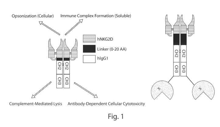
Fig. 1 shows a diagram of dimeric NKG2D-Fc chimeras with (right) and without
(left) a
drug moiety.
Fig. 2 shows that hNKG2Dx2-hIgGl-hIL15/Ra is produced as a single fusion
protein,
and is purified by protein A.
Fig. 3 shows that hNKG2Dx2-hIgGl-IL15/Ra promotes proliferation of human NK
cells
similarly to IL-15.
Fig. 4 shows that hNKG2Dx2-hIgGl-IL15/Ra promotes potent killing of multiple
cell
lines, and is superior to hNKG2Dx2-hIgG1 in cell lines with moderate ligand
expression. Panel
A shows that neither construct promotes killing of the B16 tumor cell line,
which does not
express NG2D-L. Panel B shows that both constructs equally promote killing of
a synthetic B16
tumor cell line expressing high levels of NKG2D ligand. Panel C shows that
various tumors
express different levels of NKG2D ligands on their cell surface, as measured
by NKG2D fusion
protein binding. Panel D shows that hNKG2Dx2-hIgGl-IL15/Ra kills cells
expressing
moderate NKG2D ligand more efficiently than hNKG2Dx2-hIgG1.
Fig. 5 shows that resting NK cells are activated by the fusion protein to
produce IFN-y,
but maximum production requires all three components: NKG2D, hIgGl, and IL-15.
N297Q is a
mutation in hIgG1 that prevents CD16 (expressed on NK) binding to hIgGl.
Fig. 6 shows that pre-activated NK cells require CD16 binding to kill target
cells, but do
not require IL-15.
Fig. 7 shows that optimal activation of, and killing by, resting NK cells
requires CD16
binding and IL-15 activation.
Fig. 8 shows ELISA analysis demonstrating that NKG2Dx2-hIgG1 binds to MICA*008
with improved avidity as compared to hNKG2Dx1-hIgGl, which is monomeric NKG2D-
Fc
chimera.
Fig. 9 shows flow cytometry analysis demonstrating that hNKG2Dx2-hIgG1 binds
with
improved avidity to NKG2D ligand-expressing cells as compared to hNKG2Dx1-
hIgGl.
CA 03004148 2018-05-02
WO 2017/083612
PCT/US2016/061479
- 5 -
Fig. 10 shows NKG2D-Fc drives NK cell killing of ligand-positive targets.
Dimeric
NKG2D-Fc chimeras are more effective in mediating killing than monomeric NKG2D-
Fc
chimeras. * depicts p<0.5, and ** depicts p<0.01.
Fig. 11 shows dimeric NKG2D-Fc chimeras (e.g., hNKG2Dx2-hIgG1) kill NKG2D
ligand-expres sing cells more efficiently than monomeric NKG2D-Fc chimeras
(e.g.,
hNKG2Dx1-hIgG1). ** depicts p<0.01.
Fig. 12 shows NKG2Dx2-hIgG1 neutralization of soluble MICA is superior to
NKG2Dx1-hIgGl. * depicts p<0.05, ** depicts p<0.01, *** depicts p<0.005, and
**** depicts
p<0.001.
Fig. 13 shows a structural model of dimeric hNKG2D-hIgG1 in complex with human
MICA (hMICA). (G4S)4is SEQ ID NO: 3; GGSGGGSG is SEQ ID NO: 5.
DETAILED DESCRIPTION OF INVENTION
Disclosed herein are novel compositions and methods for cancer immunotherapy.
Compositions and methods of the present invention are based, at least in part,
on the surprising
discovery that a chimeric molecule comprising two NKG2D fragments and an Fc
fragment (e.g.,
a dimeric NKG2D-Fc chimera), which is capable of binding one or more NKG2D
ligands,
induces tumor cell death with improved efficacy compared to chimeric molecules
comprising a
single NKG2D fragment and an Fc fragment (e.g., a monomeric NKG2D-Fc chimera).
Monomeric NKG2D-Fc chimeras described in the prior art (e.g., constructs
described in
published PCT application WO/2010/080124), exhibit a low binding avidity to
NKG2D ligands
(e.g., a low binding avidity index). The dimeric NKG2D-Fc constructs described
herein provide
increased binding avidity (e.g., an improved avidity index of at least 1.1, 2,
3, 4, 5, 6, 7, 8, 9, 10
or more) compared to the prior art monomeric constructs by providing multiple
NKG2D
receptors (or portions thereof) on the same molecule. Without wishing to be
bound by any
particular theory, the presence of multiple NKG2D receptors on a single
molecule is thought to
increase the number and duration of NKG2D-NKG2D ligand binding interactions,
leading to
increased anti-tumor activity. Indeed, as shown in the Examples section,
dimeric NKG2D-Fc
chimeras exhibit up to 100-fold improved binding avidity compared to the prior
art monomeric
NKG2D-Fc chimeras. However, the success of this approach was not predictable
because it was
not known whether increasing the number of receptors (or portions thereof) in
the chimeric
CA 03004148 2018-05-02
WO 2017/083612
PCT/US2016/061479
- 6 -
construct would inhibit binding interactions (e.g., via steric hindrance), or
cause aggregation of
the chimeras that could interfere with the stability of the molecule.
NKG2D ligand(s) are known to be expressed on cancer cells. Therefore, in some
embodiments, the disclosure provides methods for cancer therapy in a subject
(e.g., a human
subject), the method comprising administering to a subject having an NKG2D
ligand-expressing
cancer a dimeric NKG2D-Fc chimera as described herein. Unlike an immunotherapy
that
employs a monoclonal antibody against an NKG2D ligand, such as MICA, the
methods
provided herein are believed to have broad effects against cancer, on the
basis that NKG2D
binds to multiple ligands.
The dimeric NKG2D-Fc chimera can target any or all NKG2D ligands that are
expressed
on human tumor cells, and thus is capable of mediating tumor cell destruction
through
complement lysis and ADCC. The NKG2D-Fc chimera is also capable of opsonizing
any tumor
cells that express at least one NKG2D ligand. The NKG2D-Fc chimera can promote
efficient
cross-presentation (e.g., priming) by dendritic cells, leading to the
induction of potent T cell
responses against the tumor. Moreover, this chimera is capable of binding and
sequestering any
"shed" (e.g., soluble or released) NKG2D ligand(s) produced by tumor cells,
thereby alleviating
immune suppression due to down-regulation of NKG2D expression in response to
tumor-
derived soluble ligands.
NKG2D-Fc
In some aspects the disclosure provides a dimeric NKG2D-Fc chimera comprising:
NKG2D1-NKG2D2 -Fc, wherein NKG2D 1 and NKG2D2 each comprises NKG2D or a
fragment
thereof and can bind an NKG2D ligand; and Fc comprises a fragment
crystallizable region (Fc)
of an immunoglobulin. In some embodiments, the NKG2D fragment comprises an
extracellular
fragment of NKG2D. In some embodiments, the NKG2D extracellular fragment is
represented
by SEQ ID NO: 1.
As used herein, a "dimeric NKG2D-Fc chimera" is a chimeric molecule comprising
two
NKG2D ligand binding sites, wherein each ligand binding site comprises at
least a portion or all
of the NKG2D receptor and is capable of binding an NKG2D ligand. The ligand
binding sites
are fused to an Fc fragment. In the Examples section and the Figures, the two
NKG2D ligand
binding sites of dimeric NKG2D-Fc chimera are also referred to collectively as
"NKG2Dx2".
The monomeric NKG2D-Fc chimera described in the prior art can be referred to
as
CA 03004148 2018-05-02
WO 2017/083612
PCT/US2016/061479
- 7 -
"NKG2Dx1". The terms "chimera," "chimeric molecule," and the like generally
refer to a
molecule that is comprised of parts that are from multiple origins or sources.
In some
embodiments, dimeric NKG2D-Fc is produced as a recombinant chimeric fusion
protein.
In some embodiments, the dimeric NKG2D-Fc chimera described herein binds with
increased avidity to an NKG2D ligand as compared to a monomeric NKG2D-Fc
chimera. As
used herein, "avidity" refers to overall strength across multiple affinities
of individual non-
covalent binding interactions between a ligand and a receptor. Methods of
measuring binding
avidity are known in the art and include, for example, ELISA, surface plasmon
resonance
analysis, CD analysis, fluorescence quenching, size-exclusion binding assay
and isothermal
titration calorimetry. For brief descriptions of these assays, see, for
example, Lengyel et al.,
2007, J. Biol. Chem., 282: 30658-666). In some embodiments, binding avidity is
determined by
measuring avidity index. In some embodiments, the binding avidity of the
dimeric NKG2D-Fc
chimera to a NKG2D ligand is increased between about 2-fold and about 2000-
fold as compared
to the monomeric NKG2D-Fc chimera. In some embodiments, the binding avidity is
increased
between about 2-fold and 1000-fold. In some embodiments, the binding avidity
is increased
between about 2-fold and 100-fold. In some embodiments, the binding avidity is
increased
between about 5-fold and 1000-fold. In some embodiments, the binding avidity
is increased
between about 5-fold and 200-fold. In some embodiments, the binding avidity is
increased
between about 2-fold and about 20-fold. In some embodiments, the binding
avidity is increased
2-fold, 5-fold, 10-fold, 100-fold, or 1000-fold. In some embodiments, the
binding avidity is
increased at least 2-fold, at least 5-fold, at least 10-fold, at least 100-
fold, or at least 1000-fold.
In some embodiments, the dimeric NKG2D-Fc constructs has an increased binding
avidity index
as compared to the monomeric NKG2D-Fc chimera, e.g., an improved avidity index
of at least
1.1, 2, 3, 4, 5, 6, 7, 8, 9, 10 or more.
NKG2D
In some aspects the disclosure provides a dimeric NKG2D-Fc chimera comprising:
two
NKG2D or fragments thereof. NKG2D, also referred to as KLRK1; killer cell
lectin-like
receptor subfamily K, member 1; CD314; KLR; NKG2-D; FLJ17759; FLJ75772 or
D12S2489E, is one of the major triggering receptors of NK cells and is well
known in the art.
See, for example, Garrity et al. (2005). The portion of the NKG2D receptor
used for dimeric
NKG2D-Fc is based on the known sequences of NKG2D (e.g., Accession: NP 031386)
or
CA 03004148 2018-05-02
WO 2017/083612
PCT/US2016/061479
- 8 -
derivatives thereof that bind at least one ligand. Derivatives of NKG2D that
can be used in the
compositions and methods of the invention include, but are not limited to,
NKG2D sequences
containing one or more mutations, such as a point mutation, a substitution, a
deletion mutation
and/or an insertion mutation. One of ordinary skill in the art can readily
determine suitable
derivatives of NKG2D according to the teaching of the present disclosure and
knowledge
available in the art. At the cDNA level, such a mutation may be a silent
mutation.
Alternatively, the mutation may result in a change in the corresponding amino
acid residue.
Where the latter is the case, the change may constitute a conservative change,
such that an amino
acid residue is replaced with another amino acid residue of similar
characteristics. In some
cases, however, a mutation may result in a substitution that is non-
conservative. Such mutations
are acceptable to the extent that the dimeric NKG2D-Fc chimera is capable of
binding to an
NKG2D ligand.
In some embodiments, each NKG2D portion of a dimeric NKG2D-Fc chimera is a
full
length NKG2D polypeptide. The full length sequence of NKG2D has been described
in the
literature. See, for example, RefSeq Accession: NP 031386. Additionally,
alternative splice
variants of NKG2D have been described. For purposes of the instant invention,
any one of such
alternatively spliced variants may be used, provided that the resulting
polypeptide, when
constructed as a dimeric NKG2D-Fc chimera, is capable of binding its
ligand(s).
In some embodiments, each NKG2D portion of a dimeric NKG2D-Fc chimera is a
partial sequence (i.e., fragment) of the NKG2D receptor polypeptide, provided
that the resulting
polypeptide, when constructed as a dimeric NKG2D-Fc chimera, retains the
ability to bind its
ligand(s). For example, each NKG2D portion of the dimeric NKG2D-Fc construct
may be
shortened by either end of the NKG2D sequence by one or more amino acid
residues. More
specifically, the N-terminus of the NKG2D sequence may be deleted by 1, 2, 3,
4, 5, 6, 7, 8, 9,
10, 11, 12, 13, 14, 15, 16, 17, 18, 19, 20, about 30, about 40, about 50,
about 60, about 70, about
80 or more residues. Similarly, the C-terminus of the NKG2D sequence may be
deleted by 1, 2,
3, 4, 5, 6, 7, 8, 9, 10, 11, 12, 13, 14, 15, 16, 17, 18, 19, 20 or more
residues. In some
embodiments, both the N-terminus and the C-terminus may be shortened as
described.
It has been shown that the extracellular portion of NKG2D contributes to the
formation
of homodimers and forms a ligand-binding site(s). Thus, it is possible to
delete part or all of the
intracellular portion of NKG2D and still maintain the ability to bind its
ligand(s). For example,
the dimeric NKG2D-Fc chimera described in this disclosure may contain
predominantly an
CA 03004148 2018-05-02
WO 2017/083612
PCT/US2016/061479
- 9 -
extracellular fragment of the NKG2D receptor. Structural analyses have
revealed that amino
acid residues 78 to 216 of the human NKG2D sequence correspond to the
extracellular portion
of the NKG2D, containing ligand-binding sites. For a murine counterpart, the
extracellular
domain is amino acid residues 78-232, 94-232 or 92-232.
Accordingly, in some embodiments, each NKG2D of the dimeric NKG2D-Fc construct
comprises the extracellular portion of the NKG2D sequence, e.g., amino acid
residues 78-216 of
human NKG2D; 78-232, 94-232 or 92-232 of murine NKG2D. In some embodiments, a
dimeric NKG2D-Fc construct comprises a portion of the extracellular domain.
Thus, the
extracellular domain of the dimeric NKG2D-Fc construct may be shortened at the
N-terminus, at
the C-terminus, or both. For example, the N-terminus of the extracellular
domain used to
generate a dimeric NKG2D-Fc may be shortened by one or more amino acid
residues, e.g., 1, 2,
3, 4, 5, 6, 7, 8, 9, 10, 11, 12, 13, 14, 15, 16, 17, 18, 19, 20, about 30,
about 40, about 50, about
60, and so forth, relative to the full extracellular portion of the
polypeptide. The C-terminus of
the extracellular domain used to generate an NKG2D-Fc may be shortened by one
or more
amino acid residues, e.g., 1, 2, 3, 4, 5, 6, 7, 8, 9, 10, 11, 12, 13, 14, 15,
16, 17, 18, 19, 20, about
30, about 40, about 50, about 60, and so forth, relative to the full
extracellular portion of the
polypeptide. Using a human NKG2D as an example, the dimeric NKG2D-Fc construct
may
contain a fragment of the extracellular domain, wherein the N-terminus of the
domain begins at
amino acid residue 79, 80, 81, 90, 91, 92, 93, 94, 95, 96, 97, 98, 99, 100,
about 110, about 120,
about 130, about 140 or about 150. Similarly, the dimeric NKG2D-Fc construct
may contain a
fragment of the extracellular domain, wherein the C-terminus of the domain
ends at amino acid
residue 231, 230, 229, 228, 227, 226, 225, 224, 223, 222, 221, 220, 219, 218,
217, 216, 215,
214, 213, 212, 211, 210, 209, 208, 207, 206, 205, and so forth. Such deletions
at each end of the
extracellular domain of the NKG2D sequence may be combined.
The skilled artisan recognizes that dimeric NKG2D-Fc chimera described by the
disclosure may comprise two of the same NKG2D fragments or two different NKG2D
fragments. For example, in some embodiments, a dimeric NKG2D-Fc chimera
comprises two
NKG2D fragments corresponding to amino acid residues 78 to 216 of the human
NKG2D. In
some other embodiments, a dimeric NKG2D-Fc chimera comprises two NKG2D
fragments,
where the first fragment corresponds to amino acid residues 78 to 216 of the
human NKG2D
and the second fragment corresponds to a different portion of the NKG2D
extracellular domain
(e.g., amino acid positions 140 to 210 of the human NKG2D).
CA 03004148 2018-05-02
WO 2017/083612
PCT/US2016/061479
- 10 -
Also contemplated are dimeric NKG2D-Fc derivatives that include one or more
mutations in the NKG2D portion of the construct at the interface of the NKG2D-
ligand binding.
In particular, certain mutations are known to affect the binding affinity
between the NKG2D
receptor and its ligand (e.g., MICA). See, for example, Lengyel et al., 2007,
J. Biol. Chem.,
282: 30658-666. The three dimensional structure of a complex between NKG2D and
MICA has
been described. Accordingly, one of ordinary skill in the art may determine
the amino acid
residues of NKG2D that contribute to the interaction with its ligand and test
the effect of
mutations by systematically altering the key residues. In any of the
embodiments, the resulting
dimeric NKG2D-Fc chimera is capable of binding ligand(s). For a comprehensive
review of the
amino acid residues that are involved in receptor-ligand contact, see, for
example, Strong and
McFarland, 2004, Advances in Protein Chemistry, 68: 281-213. According to
published studies,
key residues that are thought to be important in the interaction with the
ligand have been
mapped to amino acid residues approximately from 150 to 207 in human NKG2D,
which
correspond to residues approximately from 166 to 223 in mouse NKG2D.
Therefore, each
NKG2D fragment of the dimeric NKG2D-Fc construct of the invention preferably
comprises a
fragment spanning at least most of these residues (e.g., residues 150 to 207
in human NKG2D).
Likewise, it will be understood that conservative substitutions, deletions or
mutations outside
these regions can potentially be tolerated with ease in many instances.
Some amino acid residues have been identified to be especially important for
mediating
ligand binding. Specifically, residues of human NKG2D important for binding to
MICA include
Y152, Q185, K197, Y199, E201 and N207. Residues of human NKG2D important for
binding
to ULBP3 include 1182, Y199 and Y152. Residues of murine NKG2D important for
binding to
RAE-10 include K166, Y168, Y215, K213, E217 and N223. In preferred
embodiments,
therefore, most or all of these residues (of a corresponding dimeric NKG2D
construct) are
maintained without a mutation or deletion at the position where broad
permissibility (e.g.,
specificity) for multiple ligands is desirable. However, it is also possible
to design a dimeric
NKG2D-Fc construct that preferentially binds one ligand over another ligand by
strategically
introducing a mutation at one or more of these key residues that confer
selective ligand-
recognition and binding. On the other hand, certain amino acid residues are
involved in the
binding of various ligands. For example, Y152 and Y199 in human NKG2D, which
are
equivalent to Y168 and Y215 respectively in the murine counterpart, contribute
to the binding of
CA 03004148 2018-05-02
WO 2017/083612
PCT/US2016/061479
- 11 -
MICA as well as ULBP3. Therefore, in some embodiments, these residues are
unmodified so as
to retain broad ligand specificity.
The Examples provided below present a representative dimeric NKG2D-Fc chimera,
wherein each NKG2D fragment corresponds to amino acid residues 78 to 216 of
the human
NKG2D. However, it should be appreciated that the same approach may be
employed for
NKG2D sequences derived from any other species that are known to develop
cancer. For
example, the NKG2D fragment of dimeric NKG2D-Fc may have 1, 2, 3, 4, 5, 6, 7,
8, 9, 10 or
more amino acid changes, such as deletions, insertions and substitutions, as
long as the dimeric
NKG2D-Fc retains its ligand binding activity.
The present invention includes variants of dimeric NKG2D-Fc constructs that
contain
one or more amino acid changes as described above, to the extent that the
dimeric NKG2D-Fc
chimera binds to its native ligand or ligands. To determine whether a dimeric
NKG2D-Fc
variant containing a particular mutation retains ligand binding activity,
binding assays can be
carried out, in which binding affinity and/or binding capacity of the
particular dimeric NKG2D-
Fc chimera to its ligand(s) may be evaluated. A number of methods are known in
the art by
which receptor-ligand interactions may be measured. These methods for assaying
ligand
binding include, without limitation, ELISA, surface plasmon resonance
analysis, CD analysis,
fluorescence quenching, size-exclusion binding assay and isothermal titration
calorimetry. For
brief descriptions of these assays, see, for example, Lengyel et al. (2007).
Fc fragment
In some embodiments, a dimeric NKG2D-Fc chimera comprises a fragment
crystallizable region (Fc) of an immunoglobulin. The Fc region of
immunoglobulins plays a
significant role in mediating immune defense. FcyRs are widely expressed as
transmembrane
glycoproteins on a number of cell types, including macrophages, NK cells,
dendritic cells, B
cells, neutrophils and mast cells. Fc-mediated activities include recruitment
of effector cells via
Fc-FcyR interactions. There are two classes of Fc receptors that can be
distinguished
functionally: the activating Fc receptor class and the inhibitory Fc receptor
class. Activating Fc
receptors include human FcyRIA, FcyRIIA and FcyRIIIA, as well as their murine
orthologues,
i.e., FcyRI, FcyRIII FcyRIV. Activating FcyRs mediate ADCC and ADCP, induce
endocytosis
of immune complexes leading to antigen presentation, and contribute to the
production and
release of cytokines and proinflammatory factors. For general review of the
IgG structure and
CA 03004148 2018-05-02
WO 2017/083612
PCT/US2016/061479
- 12 -
mechanisms of action, see Liu et al. (2008; Immunological Reviews, 222: 9-27).
As described
in more detail herein, the Fc portion of dimeric NKG2D-Fc is a domain that
binds an activating
Fc receptor, and preferably an activating Fc Ig domain and includes the hinge
region that allows
for dimerization.
The Fc portion of the dimeric NKG2D chimera useful for this disclosure can be
readily
adapted to render it species-specific. For use in a murine system, e.g., cells
derived from a
mouse, the Fc fragment used to generate dimeric NKG2D-Fc is preferably that of
a murine
origin. In some embodiments, an Fc fragment of the murine IgG2a is preferred.
For use in a human subject, e.g., for cancer treatment, the Fc fragment used
to generate
dimeric NKG2D-Fc is preferably that of a human origin. In particularly
preferred embodiments,
NKG2D-Fc comprises an activating Fc Ig domain. Among the four human IgG
isotypes, an
activating Fc domain of IgG1 is preferred for the preparation of dimeric NKG2D-
Fc. Thus, in
some embodiments, the Fc comprises a fragment crystallizable region (Fc) of a
human
immunoglobulin (IgG). In some embodiments, the human immunoglobulin is IgGl.
Experimental data relating to chimeric constructs containing an Fc region of
the human IgG1 are
provided in the Examples section.
The art appreciates that different antibody isotypes have a varying degree of
cytotoxic
potential in vivo (See, for example, Nimmerjahn F. & Ravetch JV., 2006,
Immunity, 24:19-28).
For example, the murine IgG2a and IgG2b isotypes are more efficient in
clearing infections such
as bacterial infections and viral infections and in killing tumor cells than
their IgG1 or IgG3
counterparts. This is attributable at least in part to differential ratios of
activating versus
inhibitory FcRs present in vivo. Similarly, with respect to human IgG
isotypes, IgG1 and IgG3
have a stronger interaction with FcRs than IgG2 or IgG4. Moreover, certain
polymorphic
allotypes of a given isotype may influence affinity for an Fc receptor.
Indeed, there are allelic
variants of activating FcRs that will significantly affect the affinity for
certain antibody isotypes.
For example, the FcyRIIIa receptor 158V allotype displays a higher affinity
for human IgG1 and
increased antibody-dependent cellular cytotoxicity (Cartron G. et al., 2002,
Blood, 99: 754-758).
Without wishing to be bound by any particular theory, it is possible to
optimize the
interaction between the Fc portion of the dimeric NKG2D-Fc chimera to its
corresponding Fc
receptor by strategically selecting or modifying the Fc allele used for
preparing the dimeric
NKG2D-Fc chimera. Accordingly, the invention contemplates using a mutant or an
allotype of
an Fc fragment. A number of useful mutations within an Fc domain have been
described, which
CA 03004148 2018-05-02
WO 2017/083612
PCT/US2016/061479
- 13 -
can affect the interaction of an Fc and its receptor, the effector function of
the Fc, as well as the
half-life of the Fc-containing molecule. These include specific amino acid
substitutions and/or
modifications to carbohydrate moieties in the Fc. For review, see, for
example, Liu et al., 2008,
Immunological Reviews, 222:9-27; Nimmerjahn & Ravetch, 2007, Curr. Opin.
Immunol., 19(2):
239-45.
The structure of Fc fragments generally is known in the art. Briefly, the Fc
region of a
typical IgG molecule is a symmetric homodimer of the carboxy-terminal portion
of heavy chains
and is composed of the CH2 and CH3 domains, which are separated from the Fab
by a flexible
hinge region. The Fc region is stabilized by non-covalent interactions between
domains. The Fc
region interacts with FcRs to exert effector functions or to regulate the
catabolism of IgG. The
heavy constant regions (Cy2 and Cy3) and the hinge region located between the
variable domain
and the constant regions interact with C lq and Fc receptors (FcRs). Thus, the
heavy constant
regions of the IgG molecule are responsible for its effector functions, since
they include binding
sites for complement and for FcRs on different effector cells. Recruitment of
effector cells is
therefore mediated via the Fc-FcyR interactions.
In general, the interaction of an antibody with complement initiates
complement-
dependent cytotoxicity (CDC), and FcyR interactions mediate antibody-dependent
cell toxicity
(ADCC) and antibody-dependent cell phagocytosis (ADCP). The classical
activation pathway
of CDC is triggered when Cl, the first component of the pathway, binds to the
hinge-Fc portion
of the IgG in an antigen-antibody complex. Subsequent activation of the
complement cascades
eventually induces the formation of a C5-C9 membrane attack complex that leads
to the death of
the target cell. ADCC, on the other hand, is dependent upon the ability of the
FcyR-bearing cells
of the innate immune system (e.g., NK cells, monocytes, macrophages and
granulocytes) to
recognize the Fc domain of antibody bound to target cells. This recognition
triggers effector
cells to release cytoplasmic perforin, granulysin, and granzymes that induce
apoptosis and lysis
of target cells. The major effector cells in ADCC are NK cells, which express
the type of FcyRs
that recognize the IgG1 and IgG3 subclasses and trigger cytotoxic effects in
vivo.
In the context of the present invention, as demonstrated in the Examples, the
dimeric
NKG2D-Fc chimeras described herein are capable of mediating equivalent
cellular effects by
virtue of having a functional Fc portion, coupled with the dimeric NKG2D
portion that can
broadly but specifically recognize and bind to its ligands.
CA 03004148 2018-05-02
WO 2017/083612
PCT/US2016/061479
- 14 -
As noted, there are activating receptors (FcyRI, FcyRIIA and FcyRIII) and
inhibitory
(FcyRIIB) receptors. In general, interaction of IgGs with activating FcyRs
triggers cell
activation, while interaction with FcyRIIB inhibits cell activation. With the
exception of B cells
and NK cells, activating and inhibitory FcyRs are co-expressed on the same
effector cells,
thereby generating a threshold for cell activation. B cells express only the
inhibitory FcyRIIB
and therefore cannot be activated by endogenous IgG under physiological
conditions. NK cells
express the activating FcyRIII so that they can kill target cells
independently of pre-activation
(or priming).
FcyRIIA and FcyRIII (CD16) have low affinities for monomeric IgG and are
thought to
be critical for triggering effector functions, leading to anti-tumor activity.
Thus, it is possible to
design a dimeric NKG2D-Fc such that it is genetically engineered to have
increased affinities for
the activating FcyRIII, and decreased affinities for the inhibitory FcyRIIB.
Accordingly, the amino acid residues of dimeric NKG2D-Fc molecules that
contribute to
their direct interaction with FcyRs, which are located primarily in the lower
hinge region and are
adjacent to the Cy2 region, may be modified, and such variants are embraced by
this invention.
It has been shown that the region corresponding to amino acid residues 234-237
of the IgG is
required for binding to FcyRs. In addition, other residues that are important
in IgG-FcyRs
interactions have been shown to be located in the Cy2 domain and include
Asp265, Asp270,
A1a327, Pro329 and Lys338.
Several strategies are contemplated to generate dimeric NKG2D-Fc chimeras with
enhanced activities. To engineer the dimeric NKG2D-Fc with an enhanced ADCC
capability, at
least two approaches are contemplated. First, based on the amino acid residues
in an IgG1 that
were identified to be critical for its binding to activating and inhibitory
FcyRs, the invention
provides variants of dimeric NKG2D-Fc chimeras that enhance or reduce,
respectively, the
affinity for these receptors. Accordingly, in one embodiment, the triple amino
acid substitution,
Ser298A1a/G1u333Ala/Lys334Ala, where the position of each residue is based on
IgGl, is
provided. The dimeric NKG2D-Fc containing this triple mutation should exhibit
a higher
affinity for FcyRIIIA but not for FcyRIIB, thereby promoting ADCC. Similarly,
in another
embodiment, the dimeric NKG2D-Fc variant contains the double mutation in the
Fc,
Ser239Asp/11e332G1u, which is expected to exert improved ADCC. Other mutations
for
enhancing ADCC include, without limitation, Ser239Asp/A1a330Leu/Ile332Glu and
Ser239Asp/Ser298A1a/I1e332Ala. Similarly, in some embodiments, mutations that
combine
CA 03004148 2018-05-02
WO 2017/083612
PCT/US2016/061479
- 15 -
increased binding to FcyRIIIA (e.g., activating receptors) and reduced binding
to FcyRIIB are
contemplated. Examples of such Fc mutations include
Phe243Leu/Arg292Pro/Tyr300Leu/Va1305I1e/Pro396Leu, without limitation (the
positions of
the residues are based on IgG1).
The second approach relates to modifying the carbohydrate moieties in the Fc
based on
the observation that some modifications significantly affect the affinity of
the Fc for FcyRs. It
has been shown that the Fc domain contains two asparagine N-linked
oligosaccharide sites
(reviewed in Liu et al., 2008). ADCC requires the presence of certain
oligosaccharides and is
dependent upon changes in the structure of the oligosaccharides. In
particular, previous studies
have shown that removing the fucose moiety attached to the innermost GlcNAc of
the
biantennary complex-type oligosaccharides dramatically increases ADCC by
improving the
binding of the Fc to FcyRIIIA without impairing CDC activity. Based on this
observation, in
one embodiment, the invention provides fucose-deficient dimeric NKG2D-Fc. In
some
embodiments, the chimera completely lacks the fucose moiety (i.e., non-
fucosylated). In other
embodiments, the chimera is hypofucosylated.
To make dimeric NKG2D-Fc containing modified carbohydrates, host cells may be
engineered to express the enzymes that catalyze the desired modification(s).
For example, host
cells, such as Chinese hamster ovary (CHO) cells may be transfected with the
enzyme, f3-(1,4)-
N-acetylglucosaminyltransferase III (GnT-III), which elevates the level of
bisected, non-
fucosylated oligosaccharides. The NKG2D-Fc product generated from these host
cells can have
a dramatically enhanced ADCC activity. In addition, in some embodiments, the
content of
fucose in NKG2D-Fc may be manipulated by a-1,6-fucosyltranferase (FUT8)-
knockout cells
lacking core-fucosyl transferase activity. Alternatively, small interfering
RNA may be used to
constitutively inhibit the expression of the FUT8 enzyme to achieve the same
effect. In some
embodiments, host cells deficient in guanosine diphosphate (GDP)-mannose 4,6-
dehydratase
(GMD) may be used to yield non-fucosylated NKG2D-Fc.
Next, to engineer the dimeric NKG2D-Fc with an enhanced complement activity,
various
mutations in the Fc domain are contemplated. Generally, complement can be
activated by at
least three pathways, leading to the formation of the membrane attach complex
C5b-9, which
forms pores in the plasma membranes of target cells and causes their lysis. C
lq binding to the
Fc domain is a critical step in this process. Among the human IgG subclasses,
only IgG1 and
IgG3 can initiate the complement cascade. In some embodiments, mutations are
introduced to
CA 03004148 2018-05-02
WO 2017/083612
PCT/US2016/061479
- 16 -
the Fc domain of the dimeric NKG2D-Fc, so as to promote Clq recruitment and
the Clq-Fc
interaction. The residues of the Fc targeted for such mutations include, but
are not limited to:
Asp270, Lys322, Pro329 and Pro331. These mutations involve substituting the
corresponding
residue(s) with nonpolar neutral amino acids, such as Ala, Met, or Trp. In a
specific
embodiment, the dimeric NKG2D-Fc contains the mutation, Lys326Trp, Clu333Ser
or both.
To achieve increased C 1 q binding and enhanced CDC, some embodiments of the
invention involve introducing a mutation or mutations to certain residues of
the hinge region of
human IgGl. Non-limiting examples of such mutations include:
Lys222Trp/Thr223Trp,
Cys220Asp/Asp221Cys, Cys220Asp/Asp221Cys/Lys222Trp/Thr223Trp,
Lys222Trp/Thr223Trp/His224Trp and Asp221Trp/Lys222Trp.
In addition, it should be noted that when fusion proteins with artificial
sequences and
activities are used as therapeutic agents, in some circumstances, patients
treated with such a
fusion protein trigger an unwanted immune response, such as development of
antibodies against
the agent. Certain structural modifications of an Fc fragment have been shown
to reduce
immunogenicity of a therapeutic fusion protein. See, for example, U.S. Patent
6,992,174 B2 by
Gillies et al., which is incorporated by reference herein; Liu et al., 2008,
Immunological
Reviews, 222:9-27. Such modifications may be useful for an effective design of
dimeric
NKG2D-Fc described in the present disclosure.
Linkers
The dimeric NKG2D-Fc construct used in the methods of the present disclosure
may
further comprise at least one linking moiety that connects a first NKG2D
portion (e.g.,
NKG2D1) with a second NKG2D portion (e.g., NKG2D2), an NKG2D portion (e.g.,
NKG2D 1 or
NKG2D2) with an Fc fragment, and/or an Fc fragment to a drug moiety. In some
embodiments,
a linking moiety (e.g., linking molecule) is referred to as X1, X2, or X3. In
some cases, a hinge
region of Fc fusion protein molecules serves as a spacer between the Fc region
and the fused
peptide (e.g., soluble receptor), allowing these two parts of the molecule to
function separately
(see, for example, Ashkenazi et al., 1997).
In some embodiments, the at least one linking moiety (e.g., linking molecule)
is not a
contiguous portion of the NKG2D1, NKG2D2, Fc, or drug moiety and covalently
joins: an amino
acid of NKG2D1 to an amino acid of NKG2D2, an amino acid of NKG2D2 to an amino
acid of
Fc, or an amino acid of Fc to the drug moiety. As used herein, a linking
molecule that is "not a
CA 03004148 2018-05-02
WO 2017/083612
PCT/US2016/061479
- 17 -
contiguous portion" means that the each NKG2D portion (e.g., NKG2D1 and
NKG2D2), a
NKG2D portion and the Fc portion, and/or the Fc portion and a drug moiety of
the chimera are
connected via an additional element that is not a part of the NKG2D or
immunoglobulin or drug
moiety, that is contiguous in nature with the portions of the chimera that it
joins, and functions
as a linker. Non-limiting examples of a linking molecule that is not a
contiguous portion of
either NKG2D, Fc, or drug moiety are described below.
The linking molecule may be a peptide linker. In some embodiments, the peptide
linker
ranges from about 2 to about 25 amino acids in length. In some embodiments,
the peptide linker
is 20 amino acids in length. In some embodiments, the peptide linker ranges
from about 4 to
about 16 amino acids in length. In some embodiments, the peptide linker is 2,
3, 4, 5, 6, 7, 8, 9,
10, 11, 12, 13, 14, 15, 16, 17, 18, 19, 20, 21, 22, 23, 24, or 25 amino acids
in length. In some
embodiments, the peptide linker is longer than 25 amino acids in length. Where
the linker is a
peptide linker, the dimeric NKG2D-Fc chimera may be produced as a single
recombinant
polypeptide using a conventional molecular biological/recombinant DNA method.
In some embodiments, a peptide linker provides a protease-dependent cleavable
site.
Examples of protease-cleavable peptide linkers include, without limitation,
the MMP sensitive
linker GGPLGLWAGG (SEQ ID NO: 6) and the factor Xa-sensitive linker IEGR (SEQ
ID NO:
7). The art is familiar with a variety of cleavable sequences that may be
employed for the
methods provided herein, for example those disclosed in Chen et al., Adv. Drug
Deliv. Rev.
(2013), 65(10): 1357-69).
In some embodiments of the present invention, a flexible peptide linker is
used. A
flexible peptide linker is preferably about 25 or fewer amino acids in length.
In some
embodiments, a flexible peptide linker is 20 amino acids in length. In some
embodiments, a
peptide linker contains about 20 or fewer amino acid residues, e.g., 2, 3,4,
5, 6,7, 8, 9, 10, 11,
12, 13, 14, 15, 16, 17, 18, 19, and 20. In some embodiments, a peptide linker
contains about 12
or fewer amino acid residues, e.g., 3, 4, 5, 6, 7, 8, 9, 10, 11, and 12. In
some cases, a peptide
linker comprises two or more of the following amino acids: glycine, serine,
alanine, and
threonine. In some embodiments, the flexible peptide linker is a glycine-
serine linker.
In some embodiments, the glycine-serine linker is represented by the formula
(GS)õ,
wherein n is 1,2, 3,4, 5, 6,7, 8, 9, 10, 11, or 12. In some embodiments, the
glycine-serine
linker is represented by the formula (GGGGS)õ (SEQ ID NO: 2), wherein n is 1,
2, 3, 4, or 5.
CA 03004148 2018-05-02
WO 2017/083612
PCT/US2016/061479
- 18 -
In some embodiments, a dimeric NKG2D-Fc chimera comprises three linking
molecules,
Xi, X2 and X3, wherein X1 covalently joins an amino acid of NKG2D 1 to an
amino acid of
NKGD2; X2 covalently joins an amino acid of NKG2D2 to an amino acid of Fc; and
X3
covalently joins an amino acid of Fc to a drug moiety. In some embodiments, X1
is (GS)3 (SEQ
ID NO: 4) and X2, X3, and X4 are each (GGGGS)4 (SEQ ID NO: 3).
In some embodiments, the dimeric NKG2D-Fc chimera contains an IEGR (SEQ ID NO:
7) peptide linker.
Alternatively, a linking molecule may be a non-peptide linker. As used herein,
a "non-
peptide linker" is a biocompatible polymer including two or more repeating
units linked to each
other. Examples of the non-peptide polymer include but are not limited to:
polyethylene glycol
(PEG) , polypropylene glycol (PPG), co-poly (ethylene/propylene) glycol,
polyoxyethylene
(POE), polyurethane, polyphosphazene, polysaccharides, dextran, polyvinyl
alcohol,
polyvinylpyrrolidones, polyvinyl ethyl ether, polyacrylamide, polyacrylate,
polycyanoacrylates,
lipid polymers, chitins, hyaluronic acid, and heparin. For more detailed
descriptions of non-
peptide linkers useful for Fc fusion molecules, see, for example,
WO/2006/107124, which is
incorporated by reference herein. Typically such linkers will have a range of
molecular weight
of from about 1 kDa to 50 kDa, depending upon a particular linker. For
example, a typical PEG
has a molecular weight of about 1 to 5 kDa, and polyethylene glycol has a
molecular weight of
about 5 kDa to 50 kDa, and more preferably about 10 kDa to 40 kDa.
Drug moieties
In some embodiments, a dimeric NKG2D-Fc chimera further comprises a drug
moiety.
As used herein, "drug moiety" refers to a therapeutic agent that is intended
for delivery to a
targeted cell (e.g., a cancer cell). Generally, a drug moiety is conjugated
(e.g., directly or
indirectly covalently bound) to the carboxy terminus of a dimeric NKG2D-Fc
chimera.
However, the skilled artisan recognizes that in some embodiments, a drug
moiety is conjugated
to the amino terminus of a dimeric NKG2D-Fc chimera. Examples of "drug
moieties" include
drugs (e.g., small molecules), toxins (e.g., molecules of the lymphotoxin
family), radionuclides,
enzymes, cytokines, chemokines, antibody single chain variable fragments
directed against
activating compounds or blocking angiogenesis, or essentially any anti-tumor
compound.
In some embodiments, the drug moiety comprises a cytokine or functional
portion
thereof. Cytokines are proteins and peptides that are capable of modulating
immune cell
CA 03004148 2018-05-02
WO 2017/083612
PCT/US2016/061479
- 19 -
function. A "functional portion" of a cytokine is a cytokine fragment that
retains the ability to
modulate immune cell function (e.g., bind to one or more cytokine receptors).
Examples of
cytokines include, but are not limited to interferon-alpha (IFN-a), interferon-
beta (IFN-13), and
interferon-gamma (IFN-y), interleukins (e.g., IL-1 to IL-29, in particular, IL-
2, IL-5, IL-6, IL-7,
IL-10, IL-12, IL-15 and IL-18), tumor necrosis factors (e.g., TNF-alpha and
TNF-beta),
erythropoietin (EPO), MIP3a, monocyte chemotactic protein (MCP)-1,
intracellular adhesion
molecule (ICAM), macrophage colony stimulating factor (M-CSF), granulocyte
colony
stimulating factor (G-CSF) and granulocyte-macrophage colony stimulating
factor (GM-CSF).
In some embodiments, the drug moiety comprises a cytokine selected from the
group consisting
of: IL-2, IL-12, IL-15, IL-18, IL-21 and IFN-a.
In some embodiments, a drug moiety comprises a cytokine/cytokine receptor
heterocomplex. Cytokine/cytokine receptor heterocomplexes are known in the art
and are
described, for example in Rowley et al., Eur J Immunol. 2009 Feb; 39(2): 491-
506. In some
embodiments, a dimeric NKG2D-Fc chimera includes a drug moiety comprising an
IL-15 (e.g.,
GenBank AAX37025)/IL-15Ra (e.g., GenBank AAP69528.1) heterocomplex. In some
embodiments, the drug moiety comprises amino acids 31-107 of the human IL-15
receptor alpha
(hIL15Ra, GenBank AAP69528.1) fused to amino acids 22-135 of IL-15 (GenBank
AAX37025). In some embodiments, the IL-15 and IL-15Ra are separated by a
linker, for
example, a 20- amino acid (G4S)4 (SEQ ID NO: 3) linker. A dimeric NKG2D-Fc
chimera
comprising an IL-15/IL-15Ra heterocomplex is further described in the Examples
section. In
some embodiments, a dimeric NKG2D-Fc chimera includes a drug moiety comprising
a
heterocomplex of IL-12p35 and IL-12p40. In some embodiments, the IL-12p35 and
IL-12p40
are separated by a linker, for example, a 20- amino acid (G45)4 (SEQ ID NO: 3)
linker. In some
embodiments, a dimeric NKG2D-Fc chimera includes a drug moiety comprising a
heterocomplex of IL-23p19 and IL-23p40. In some embodiments, the IL-23p19 and
IL-23p40
are separated by a linker, for example, a 20-amino acid (G45)4 (SEQ ID NO: 3)
linker. In some
embodiments, a dimeric NKG2D-Fc chimera includes a drug moiety comprising a
heterocomplex of IL-27p28 and EB1. In some embodiments, the IL-27p28 and EB1
are
separated by a linker, for example, a 20-amino acid (G45)4 (SEQ ID NO: 3)
linker. In some
embodiments, each subunit of a cytokine/cytokine receptor heterocomplex is on
a different chain
of the dimeric NKG2D-Fc chimera.
CA 03004148 2018-05-02
WO 2017/083612
PCT/US2016/061479
- 20 -
In some embodiments, the drug moiety is an antibody single chain variable
fragment
(ScFv). As used herein, an "antibody single chain variable fragment" refers to
a fusion protein
of the variable regions of the heavy (VH) and light chains (VL) of
immunoglobulins, connected
with a short linker peptide. ScFv proteins retain the specificity of the
original immunoglobulin,
despite removal of the constant regions and the introduction of the linker. In
some
embodiments, a ScFv binds to an immune checkpoint protein (e.g., PD1 or
CTLA4). In some
embodiments, an ScFv blocks angiogenesis (e.g., binds to a regulator of
angiogenesis, such as
VEGF).
In some embodiments, the drug moiety is a chemokine. As used herein,
"chemokines"
refers to low-molecular-weight proteins that stimulate recruitment of
leukocytes. Generally,
chemokines are secondary pro-inflammatory mediators that are induced by
primary pro-
inflammatory mediators such as interleukin-1 (IL-1) or tumor necrosis factor
(TNF).
Chemokines can be classified into four families: CC chemokines (e.g., CCL1 to
CCL-28), CXC
(e.g., CXCL1 to CXCL17),C (e.g., XCL1, XCL2), and CX3C (CX3CL1).
In some embodiments, the drug moiety is a small molecule. As used herein,
"small
molecule" refers to a non-peptidic, non-oligomeric organic compound either
synthesized in the
laboratory or found in nature. Non-limiting examples of small molecule drugs
include small
molecule kinase inhibitors (e.g., everolimus, gefitinib, imatinib, etc.),
bromodomain inhibitors
(e.g., JQ1, I-BET 151, RVX-208, etc.), antibiotics (e.g., kanamycin, neomycin,
ciprofloxacin,
etc.), and antivirals (e.g., ribavirin, rimantadine, zidovudine, etc.). In
some embodiments, the
small molecule is an anti-tumor compound. Anti-tumor compounds are discussed
in further
detail elsewhere in this disclosure.
In some embodiments, the drug moiety is a radionuclide. As used herein,
"radionuclide"
refers to medically useful radionuclides. Examples of radionuclides include
99mTc, 188Re, 186Re,
9
153Sm, 166 90 8 67 68 111 183 59 225 212 211 45
60 61 Ho, Y, Sr, Ga, Ga, In, Gd, Fe, Ac, Bi, At, Ti, Cu,
Cu, and
67Cu.
Other moieties
In some embodiments, dimeric NKG2D-Fc chimeras useful for the methods
described
herein may further comprise one or more accessory moieties, such as a tag
sequence and a signal
sequence. For example, a tag sequence can be used for detecting and/or
isolating the
polypeptide. Examples of tags include, without limitation: HA, Flag, Myc, Glu,
His and
CA 03004148 2018-05-02
WO 2017/083612
PCT/US2016/061479
- 21 -
Maltose basic protein. The tag sequence may be located at the amino terminus,
carboxyl
terminus, or located somewhere in the middle of the dimeric NKG2D-Fc chimeric
molecule
(e.g., between modular peptide fragments), provided that the presence of such
a tag does not
interfere with the function of the dimeric NKG2D-Fc molecule. In some cases, a
tag sequence is
cleavable.
In some embodiments, dimeric NKG2D-Fc chimeras may optionally comprise a
signal
sequence. A signal sequence is a short (typically about 3-60 amino acids long)
peptide chain
that directs the post-translational transport of a polypeptide, thereby
allowing a greater yield of
the polypeptide. The amino acid sequences of a signal sequence direct
polypeptides (which are
synthesized in the cytosol) to certain subcellular compartments, e.g.,
organelles. A signal
sequence is also referred to as a targeting signal, a signal peptide, a
transit peptide, or a
localization signal. In some embodiments, a signal sequence is cleaved from
the polypeptide by
signal peptidase after the polypeptide is transported.
In some embodiments, the dimeric NKG2D chimera contains an N-terminal modified
IL-
2 signal sequence, which allows for optimal expression and secretion of NKG2D-
Fc construct.
See, for example, Zhang et al., 2004, J. Gene Med., 7:354-65. In some
embodiments, the
dimeric NKG2D chimera contains a signal peptide derived from the polypeptide
sequence of
CD33. For example, the CD33 signal peptide may correspond to amino acid
residues 1-16 of
the CD33 polypeptide sequence. One of ordinary skill in the art will
understand that there are a
number of other suitable signal peptide sequences that may be used to practice
the methods
provided in this disclosure. In addition, where there is a signal peptide
present in the NKG2D
chimera, extra amino acid residues, e.g., a spacer, may be optionally inserted
between the N-
terminus signal peptide and the Fc portion of the chimera. In some
embodiments, for example, a
signal sequence is followed by a Met-Asp dipeptide spacer.
Preparation of dimeric NKG2D-Fc
The art is familiar with molecular biological and biochemical techniques for
preparing a
dimeric NKG2D-Fc chimera with desired features. Preferably, dimeric NKG2D-Fc
chimeric
constructs are produced by conventional recombinatory DNA methods. In
preferred
embodiments, a dimeric NKG2D-Fc chimera is produced as a single (e.g.,
contiguous)
recombinant polypeptide. In other embodiments, two or more portions of dimeric
NKG2D-Fc
are produced as separate fragments and are subsequently linked together to
yield a dimeric
CA 03004148 2018-05-02
WO 2017/083612
PCT/US2016/061479
- 22 -
NKG2D-Fc molecule. For example, each NKG2D portion (e.g., NKG2D 1, NKG2D2) of
the
chimera and an Fc portion of the dimeric NKG2D-Fc are each produced as
separate recombinant
polypeptides then fused together by a chemical linking means to yield dimeric
NKG2D-Fc. This
production methodology may be preferred particularly in situations where a non-
peptide linking
molecule is employed. Similarly, this production methodology may be also
preferred if a
dimeric NKG2D-Fc chimera does not fold correctly (e.g., does not properly bind
a ligand) when
made as a single contiguous polypeptide.
For the production of recombinant polypeptides, a variety of host organisms
may be
used. Suitable hosts include, but are not limited to: bacteria such as E.
coli, yeast cells, insect
cells, plant cells, and mammalian cells. Choice of a suitable host organism
will depend on the
particular application of the dimeric NKG2D-Fc chimera. The skilled artisan
will understand
how to take into consideration certain criteria in selecting a suitable host
for producing the
recombinant polypeptide. Factors affecting selection of a suitable host
include, for example,
post-translational modifications, such as phosphorylation and glycosylation
patterns, as well as
technical factors, such as the general expected yield and the ease of
purification. Host-specific
post-translational modifications of a dimeric NKG2D-Fc, which is to be used in
vivo, should be
carefully considered because certain post-specific modifications are known to
be highly
immunogenic (antigenic).
Once produced, dimeric NKG2D-Fc can be purified by any suitable means, such as
chromatographic methods known to those of skill in the art. Examples of
chromatographic
methods include gel filtration chromatography. See, for example, Caine et al.,
Protein Expr.
Purif., 1996, 8:159-66. In some embodiments, dimeric NKG2D-Fc is purified by
Protein A
immunoaffinity chromatography.
As will be recognized by one of ordinary skill in the art, dimeric NKG2D
chimera
portions also can be prepared and isolated separately, and joined by chemical
synthesis.
NKG2D receptor ligands
In any of the embodiments described in this disclosure, dimeric NKG2D-Fc is
capable of
binding the endogenous ligand of the NKG2D receptor. Known NKG2D-ligands in
humans
include MICA, MICB, RAET-1G, ULBP1, ULBP2, ULBP3, ULBP4, ULBP5, and ULBP6.
Preferably, the dimeric NKG2D-Fc chimera descried in the present disclosure is
capable of
binding more than one type of NKG2D receptor ligand.
CA 03004148 2018-05-02
WO 2017/083612
PCT/US2016/061479
-23 -
In some embodiments, the dimeric NKG2D-Fc chimeric molecules bind ligands with
high affinity of 10-4 M or less, 10-7M or less, or with subnanomolar affinity,
e.g., 0.9, 0.8, 0.7,
0.6, 0.5, 0.4, 0.3, 0.2, 0.1 nM or even less. In some embodiments, the binding
affinity of the
dimeric NKG2D-Fc molecule for its ligands is at least 5 x 10⁶ Ka, at least 1 x
107 Ka, at least 2 x
107 Ka, at least 1 x 10⁸ Ka, or greater.
In some embodiments, NKG2D-Fc binds preferentially to (e.g., with higher
affinity for)
a subset of NKG2D receptor ligands. 3D structural data in combination with
mutagenesis
analyses have revealed that NKG2D is permissive in the recognition and binding
of a diverse
array of its endogenous ligands.
A ligand for NKG2D may be expressed on a cell surface. Alternatively, a ligand
for
NKG2D may be "shed" from the cell surface and is present as a soluble ligand.
It has been
known in certain cancers that NKG2D ligands such as MICA are over-expressed
and in some
cases released (e.g., shed) into the bloodstream or surrounding tissues in a
soluble form, e.g., in
sera. It is believed that this contributes, at least in part, to the
pathogenesis and/or progression of
cancer. Thus, the dimeric NKG2D-Fc is useful for binding such ligand, either
present on cell
surface or as a released form, in counterbalancing the expression of the
ligands that are present
at an abnormally elevated level by functioning as a neutralizing agent.
Where an NKG2D ligand is expressed on the surface of cancer cells of a
subject, dimeric
NKG2D-Fc described in the present disclosure binds to the cell surface ligand
when
administered to the subject. The binding of the dimeric NKG2D-Fc chimera to
its ligand may
prevent activation of endogenous NKG2D receptors present on NK cells. Where an
NKG2D
ligand is "shed" from cancer cells, e.g., released into the bloodstream of a
subject, dimeric
NKG2D-Fc described herein binds to the soluble ligand, sequestering it from
further action.
Therapeutic applications
Normally, expression of the NKG2D ligands appears to be confined to the
gastrointestinal epithelium. Little expression is observed in quiescent
epithelial cells, but higher
levels of expression occur in rapidly proliferating cells. Expression of the
NKG2D ligands is
also up-regulated in various transformed cells, particularly those of
epithelial origin.
Accordingly, provided herein are methods for treating cancer or symptoms of
cancer in a
subject. The methods comprise administering to the subject a therapeutically
effective amount
of dimeric NKG2D-Fc that binds NKG2D ligands in vivo.
CA 03004148 2018-05-02
WO 2017/083612
PCT/US2016/061479
- 24 -
The terms "treating," "treatment," and "treat" and the like in the context of
a cancer
therapy refer to the administration of a composition comprising dimeric NKG2D-
Fc as
described herein to a subject who has cancer. The composition is administered
to the subject in
an amount that is therapeutically effective. As used herein, a therapeutically
effective amount
refers to an amount of the therapeutic that is believed to effectuate a
beneficial effect with
statistical significance on the subject having the disease or disorder, such
as certain types of
cancer. Generally, a therapeutically effective amount is determined by
administering the
composition to a population of subjects with specified conditions (such as
progression or stage
of a disease) and evaluating the outcome in response. As used herein,
therapeutic treatment
shall include, for example, complete prevention or abolishment of the symptoms
of a disease, a
delay in onset of the symptoms of a disease, or lessening in the severity of a
disease.
Cancer
Dimeric NKG2D-Fc chimeras are believed to be broadly useful for immunotherapy
for a
wide variety of cancers, where the expression of one or more NKG2D ligands is
elevated in a
subject. Cancer broadly refers to a proliferative disease involving
transformed cells, including
both pre-malignant and malignant disorders. The present invention is useful
for treating a
subject having cancer that is characterized by over-expression of one or more
NKG2D ligands.
In some embodiments, the cancer is characterized by over-expression of one (or
predominantly
one) ligand of the NKG2D receptor. In other embodiments, the cancer is
characterized by over-
expression of two or more NKG2D ligands.
The methods disclosed herein are useful therapeutics for the treatment of pre-
malignant
disorders that carry with them a risk of progressing to malignancy.
Examples of such disorders include, without limitation, dysplasia,
hyperplasia, and
plasma cell disorders such as monoclonal gammopathy of undetermined
significance (MGUS)
and smoldering multiple myeloma (SMM). In some embodiments, the cancer is
melanoma,
lung, breast, kidney, ovarian, prostate, pancreatic, gastric, and colon
carcinoma, lymphoma or
leukemia. In some embodiments, the cancer is melanoma. In some embodiments,
the cancer is
a plasma cell malignancy, for example, multiple myeloma (MM) or pre-malignant
condition of
plasma cells. In some embodiments, the cancer is melanoma, lung cancer, plasma
cell cancer,
leukemia, lymphoma, ovarian cancer, colon cancer, pancreatic cancer or
prostate cancer. In
some embodiments, the subject has been diagnosed as having a cancer or as
being predisposed
CA 03004148 2018-05-02
WO 2017/083612
PCT/US2016/061479
- 25 -
to cancer. Thus, methods disclosed herein are also useful to treat a subject
who has had a
metastasis and is therefore susceptible to a relapse or recurrence. The
methods are particularly
useful in high-risk individuals who, for example, have a family history of
cancer or
metastasizing tumors, or show a genetic predispositions for a cancer
metastasis. Specifically,
the methods are directed to treating cancer that is associated with NKG2D
ligand expression. In
some embodiments, an NKG2D ligand is MICA. Thus, in some embodiments, the
cancer
causes MICA-related tumors.
Whether a particular subject (e.g., patient) should receive a cancer therapy
comprising
NKG2D-Fc can be determined by testing for aberrant expression of one or more
NKG2D
ligands in the subject. "Aberrant expression of one or more NKG2D ligands" in
the subject
means over-expression of the ligand(s) in a biological sample obtained from
the subject. In
some embodiments, a biological sample may include a biopsy sample taken from a
tissue of the
subject suspected to be cancerous. For example, in some cases, a biological
sample is collected
from a solid tumor to test for malignancy. In other cases, a biological sample
may constitute a
blood sample, e.g., serum, a stool sample, urine sample, etc. A biological
sample may be any
cell or tissue sample that is collected from a subject for the purpose of
testing for the diagnosis
or progression of a disease, such as cancer.
One of ordinary skill in the art is familiar with a variety of laboratory
techniques and
protocols used to assay for the presence of and the levels of one or more
markers present in a
biological sample. To determine whether a subject has cancer that is
associated with over-
expression of NKG2D ligand(s), typically immunoaffinity assays are performed.
In certain
situations, depending on the type of biological samples that are available,
immunohistological or
immunocytochemical analyses may be carried out. A number of antibodies are
commercially
available for performing these analyses. Methods commonly employed for this
purpose include,
but are not limited to, ELISA, immunoblotting, and immunohistochemistry.
Subjects
The methods disclosed herein can be applied to a wide range of species, e.g.,
humans,
non-human primates (e.g., monkeys), horses, cattle, pigs, sheep, deer, elk,
goats, dogs, cats,
rabbits, guinea pigs, hamsters, rats, and mice, which are known to develop
cancer. Thus, a
"subject" as used herein is a mammalian subject having a disease, or at risk
of developing a
disease associated with an abnormal expression of at least one NKG2D ligand,
such as cancer.
CA 03004148 2018-05-02
WO 2017/083612
PCT/US2016/061479
- 26 -
In preferred embodiments, the subject is a human subject having a cancer
presenting elevated
levels of one or more NKG2D ligands. In some embodiments, the NKG2D ligands
include
MICA.
If a subject has been shown to express an elevated level of one or more NKG2D
ligands,
the subject may be treated with the methods described herein. In some
circumstances, a subject
has received or is receiving another cancer therapy. In some embodiments, the
cancer may be in
remission. In some cases, the subject is at risk of having recurrence, e.g.,
metastasis. In some
embodiments, the over-expression of one or more NKG2D ligands is limited to
cancerous cells,
e.g., tumors. In some embodiments, at least one of the NKG2D ligands expressed
by cancer
cells are shed into the blood stream, and thus detectable in the serum of the
subject.
Depending on the phenotype of a particular cancer, it may be possible to
target one or more
ligands which are over-expressed (expressed by tumor cells) over the other
ligands, whose
expression is not significantly affected.
Modes of action
The instant invention is based, in part, on the surprising discovery that that
a chimeric
molecule comprising two NKG2D fragments and an Fc fragment (e.g., a dimeric
NKG2D-Fc
chimera), which is capable of binding one or more NKG2D ligands, induces tumor
cell death
with improved efficacy compared to chimeric molecules comprising a single
NKG2D fragment
and an Fc fragment (e.g., a monomeric NKG2D-Fc chimera).
Without being limited by any particular theory, it appears that dimeric NKG2D-
Fc
chimeras can function through the two major components of the immune system:
innate
immunity and adaptive immunity. As used herein, innate immunity or the innate
immune
system refers to non-specific host defense mechanisms against foreign
pathogens. Innate
immunity includes both physical barriers (e.g., skin, gastric acid, mucus or
tears, as well as cells)
and active mechanisms such as NK cells, phagocytes and the complement system.
NK cells
represent a major component of the innate immune system. NK cells are
cytotoxic, e.g., are able
to attack cells that have been infected by microbes, as well as some kinds of
tumor cells. The
cytotoxic activity of NK cells is mediated through cell-surface receptors that
recognize MHC
class I alleles. A number of receptor types are known in the art, including
NKG2D, which is one
receptor subtype. Phagocytic cells include neutrophils, monocytes,
macrophages, basophils and
CA 03004148 2018-05-02
WO 2017/083612
PCT/US2016/061479
- 27 -
eosinophils. The complement system is a biochemical cascade of the immune
system that helps
clear pathogens from a host organism.
In general, adaptive immunity or the adaptive immune system refers to an
antigen-
specific antibody-mediated immune response. Adaptive immunity is generally
mediated via
specific antibody production by B lymphocytes and antigen-specific activity of
T lymphocytes.
The humoral response mediated by B lymphocytes defends primarily against
extracellular
pathogens through the production of circulating antibodies that mark foreign
cells and molecules
for destruction by other specialized cells and proteins. The cellular response
mediated by T
lymphocytes defends predominantly against intracellular pathogens and cancer
cells by directly
binding to and destroying the affected cells. According to the present
disclosure, dimeric
NKG2D-Fc, which is a non-antibody molecule, is believed to functionally mimic
what is
ordinarily the function of specific antibodies.
The present invention thus contemplates methods for cancer treatment, wherein
dimeric
NKG2D-Fc binds directly to tumor cells that are expressing NKG2D ligands on
the cell surface.
In this mode of action, dimeric NKG2D-Fc can specifically identify for
destruction tumor cells
that over-express NKG2D ligands, but not healthy cells that do not.
Dimeric NKG2D-Fc can target any or all NKG2D ligands that are expressed on
human
tumor cells in at least two ways. One mechanism of mediating tumor cell
destruction is through
the process of complement lysis (also referred to as complement dependent
lysis, complement-
dependent cytotoxicity or CDC). A second way of mediating tumor cell
destruction is by
triggering antibody dependent cellular cytotoxicity (ADCC).
In some embodiments, dimeric NKG2D-Fc acts as an opsonizing agent.
Opsonization is
the process where cells or particles become coated with molecules which allow
them to bind to
receptors on other cells, such as dendritic cells or phagocytes, to promote
the uptake. For
antigen-presenting cells such as dendritic cells and macrophages, opsonization
promotes
efficient processing and presentation of antigens. Opsonizing agents that are
capable of
specifically binding to both the target (e.g., ligands) and particular
receptors on antigen-
presenting cells (e.g., FcRs) that can mediate internalization and subsequent
antigen processing
are particularly useful.
Tumor cells that express one or more ligands of the NKG2D receptor on the cell
surface
can become opsonized, e.g., coated, with dimeric NKG2D-Fc molecules. For
example, the
NKG2D portion of the chimera can bind to the ligands on the tumor cell
surface, while leaving
CA 03004148 2018-05-02
WO 2017/083612
PCT/US2016/061479
- 28 -
the Fc portion of the chimera exposed. Dendritic cells have FcyRs and
therefore can bind to and
internalize the tumor antigen (e.g., NKG2D ligands), which then results in
antigen presentation
to cytotoxic T cells, also known as CD8+ T cells. This is referred to as cross-
priming.
Similarly, opsonization results in the generation of MHC class II-restricted
CD4+ T cell
responses. Through opsonization, therefore, the NKG2D-Fc chimera can promote
efficient
cross-presentation (e.g., priming) by dendritic cells, leading to the
induction of potent T cell
responses against the tumor.
Cancer patients often suffer from immune suppression. In some cases, it is
believed that
the immune suppression, at least in part, may be caused by impaired NKG2D
receptor signaling.
Based on a prevailing model, for example, shed MICA impairs host defense by
inducing the
internalization of NKG2D receptor molecules on lymphocytes. Thus, according to
this model,
tumor cell shedding of MICA results in immune suppression through down-
regulation of
NKG2D surface expression.
Therefore, the methods provided herein are useful for counteracting or
relieving immune
suppression by administering a composition comprising dimeric NKG2D-Fc,
particularly in
situations where a patient exhibits elevated levels of soluble (i.e., shed)
NKG2D ligand or
ligands that are detectable in sera. The mode of action is that NKG2D-Fc
administered to the
patient binds to (thus sequestering) excess soluble ligands of NKG2D that were
shed from
tumors, thereby reversing the down-expression of NKG2D receptors on cell
surface that led to
immune suppression.
Thus, the dimeric NKG2D-Fc chimera can have multiple therapeutic functions,
including
neutralizing soluble ligands that are shed by tumor cells, promoting ADCC
and/or CDC in tumor
cells expressing the cell surface ligands and mediating cross presentation and
priming of the
adaptive immune system, including CD8 cytotoxic T-lymphocytes (CTLs) and tumor-
specific
antibody producing B-cells.
Administration
The dimeric NKG2D-Fc composition can be administered directly to a subject.
The
subject is preferably a mammal. The terms "administration" and "administer"
refer to a means
of providing a pharmaceutical agent to a subject such that the pharmaceutical
agent is to contact
its target cells, e.g., cancer cells, in vivo, i.e., in the body of the
subject. In some embodiments,
the composition comprising NKG2D-Fc is systematically administered to a
subject. In preferred
CA 03004148 2018-05-02
WO 2017/083612
PCT/US2016/061479
- 29 -
embodiments, a systematic administration is delivered via an intravenous
injection. In some
embodiments, the composition comprising dimeric NKG2D-Fc is administered
locally. For
example, in some cases, the composition may be delivered directly to or within
close proximity
of a solid tumor.
Pharmaceutically-acceptable carriers
In some aspects, the disclosure provides a composition comprising the dimeric
NKG2D-
Fc chimera as described by this document and a pharmaceutically acceptable
carrier. Generally,
the composition comprising dimeric NKG2D-Fc can be suspended in a
pharmaceutically-
acceptable carrier (e.g., physiological saline). Such carriers can include,
without limitation,
sterile aqueous or non-aqueous solutions, suspensions, and emulsions. Examples
of non-
aqueous solvents include mineral oil, propylene glycol, polyethylene glycol,
vegetable oils, and
injectable organic esters, for example. Aqueous earners include, without
limitation, water,
alcohol, saline, and buffered solutions. Preservatives, flavorings, and other
additives such as, for
example, antimicrobials, anti-oxidants, chelating agents, inert gases, and the
like also may be
present. It will be appreciated that any material described herein that is to
be administered to a
mammal can contain one or more pharmaceutically acceptable carriers.
Routes of administration
Any composition described herein can be administered to any part of the
subject's body
via various administration routes. The composition can be administered by
intravenous,
intraperitoneal, intramuscular, subcutaneous, intramuscular, intrarectal,
intravaginal, intrathecal,
intratracheal, intradermal, or transdermal injection, by oral or nasal
administration, by
inhalation, or by gradual perfusion over time. The composition can be
delivered to specific
tissue. For example, the composition can be delivered to, without limitation,
the joints, nasal
mucosa, blood, lungs, intestines, muscle tissues, skin, or peritoneal cavity
of a mammal. In a
further example, an aerosol preparation of a composition can be given to a
subject by inhalation.
Dosage
The dosage required depends on the route of administration, the nature of the
formulation, the nature of the patient's illness, the subject's size, weight,
surface area, age, and
sex, other drugs being administered, and the judgment of the attending
physician. Suitable
CA 03004148 2018-05-02
WO 2017/083612
PCT/US2016/061479
- 30 -
dosages are typically in the range of 0.01-1,000 [tg/kg. Wide variations in
the needed dosage are
to be expected in view of the variety of dimeric NKG2D-Fc compositions
available and the
differing efficiencies of various routes of administration. Variations in
these dosage levels can
be adjusted using standard empirical routines for optimization as is well
understood in the art.
Administrations can be single or multiple (e.g., 2- or 3-, 4-, 6-, 8-, 10-, 20-
, 50-, 100, 150-, or
more fold). Encapsulation of the composition in a suitable delivery vehicle
(e.g., polymeric
microparticles or implantable devices) may increase the efficiency of
delivery.
Treatment regimen
The duration of treatment with any composition provided herein can be any
length of
time from as short as one day to as long as the life span of the host (e.g.,
many years). For
example, dimeric NKG2D-Fc compositions can be administered once a month for
three months,
or once a year for a period of ten years. It is also noted that the frequency
of treatment can be
variable. For example, dimeric NKG2D-Fc compositions can be administered once
(or twice,
three times, etc.) daily, weekly, monthly, or yearly. Dimeric NKG2D-Fc
compositions can be
administered together, e.g., at the same point in time or sequentially, with
one or more other
cancer therapies. For example, a patient can receive an autologous tumor cell
vaccine followed
by an anti-CTL4 antibody, followed by a dimeric NKG2D-Fc therapy, separated by
intervals of
hours, days, months or years.
Effective amounts
An effective amount of any composition described herein can be administered to
a
subject. The term "effective" as used herein refers to any amount that induces
a desired
therapeutic effect, such as an immune response, while not inducing significant
toxicity in the
subject. Such an amount can be determined by assessing a subject's biological
reaction, e.g.,
immune response and improvement in a symptom, after administration of a known
amount of a
particular composition. In addition, the level of toxicity, if any, can be
determined by assessing
a subject's clinical symptoms before and after administering a known amount of
a particular
composition. It is noted that the effective amount of a particular composition
administered to a
subject can be adjusted according to a desired outcome as well as the host's
response and level of
toxicity. Significant toxicity can vary for each particular host and depends
on multiple factors
including, without limitation, the subject's disease state, age, and tolerance
to pain.
CA 03004148 2018-05-02
WO 2017/083612
PCT/US2016/061479
-31 -
Combination therapy
In some embodiments, the subject in need of cancer treatment is treated with
the dimeric
NKG2D-Fc composition described herein in conjunction with additional cancer
therapy. In
some embodiments, the additional cancer therapy includes a cytotoxic agent
and/or non-
cytotoxic agent. A "cytotoxic agent" refers to a substance that inhibits or
prevents the function
of cells and/or causes destruction of cells. The term is intended to include
radioactive isotopes
(e.g., 1311, 1251, 90Y and 186Re), chemotherapeutic agents, and toxins such as
enzymatically active
toxins of bacterial, fungal, plant or animal origin or synthetic toxins, or
fragments thereof. A
non-cytotoxic agent refers to a substance that does not inhibit or prevent the
function of cells
and/or does not cause destruction of cells. A "non-cytotoxic agent" may
include an agent that
can be activated to be cytotoxic. A non-cytotoxic agent may include a bead,
liposome, matrix or
particle (see, e.g., U. S. Patent Publications 2003/0028071 and 2003/0032995,
which are
incorporated by reference herein). Such agents may be conjugated, coupled,
linked or associated
with a dimeric NKG2D-Fc composition described herein.
In some embodiments, conventional cancer medicaments are administered with the
compositions described herein. In some cases, the subject in need of cancer
treatment is treated
with the dimeric NKG2D-Fc composition described herein in conjunction with one
or more
additional agents directed to target cancer cells. Highly suitable agents
include those agents that
promote DNA-damage, e.g., double stranded breaks in cellular DNA, in cancer
cells. Any form
of DNA-damaging agent know to those of skill in the art can be used. DNA
damage can
typically be produced by radiation therapy and/or chemotherapy. DNA-damaging
agents are
also referred to as genotoxic agents. As used herein, "in conjunction with"
shall mean that
dimeric NKG2D-Fc is administered to a subject concurrently with one or more
additional
therapies (either simultaneously or separately but in close proximity), prior
to, or after
administration of one or more additional therapies.
Examples of radiation therapy include, without limitation, external radiation
therapy and
internal radiation therapy (also called brachytherapy) Energy sources for
external radiation
therapy include x-rays, gamma rays and particle beams, energy sources used in
internal radiation
include radioactive iodine (iodine125 or iodine131), strontium89, or
radioisotopes of phosphorous,
palladium, cesium, indium, phosphate, or cobalt. Methods of administering
radiation therapy
are well known to those of skill in the art.
CA 03004148 2018-05-02
WO 2017/083612
PCT/US2016/061479
- 32 -
Examples of DNA-damaging chemotherapeutic agents that may be particularly
useful
include, without limitation: Busulfan (Myleran), Carboplatin (Paraplatin),
Carmustme (BCNU),
Chlorambucil (Leukeran), Cisplatin (Platmol), Cyclophosphamide (Cytoxan,
Neosar),
Dacarbazme (DTIC-Dome), Ifosfamide (Ifex), Lomustme (CCNU), Mechlorethamme
(nitrogen
mustard, Mustargen), Melphalan (Alkeran), and Procarbazine (Matulane).
A number of other chemotherapeutic agents may be also used for the method
described
herein, either alone or in combination. These include: methotrexate,
vincristine, adriamycin,
cisplatin, non-sugar containing chloroethylnitrosoureas, 5-fluorouracil,
mitomycin C,
bleomycin, doxorubicin, dacarbazine, taxol, fragyline, Meglamine GLA,
valrubicin, carmustaine
and poliferposan, MMI270, BAY 12-9566, RAS farnesyl transferase inhibitor,
farnesyl
transferase inhibitor, MMP, MTA/LY231514, LY264618/Lometexol, Glamolec, CI-
994, TNP-
470, Hycamtin/Topotecan, PKC412, Valspodar/PSC833, Novantrone/Mitroxantrone,
Metaret/Suramin, Batimastat, E7070, BCH-4556, CS-682, 9-AC, AG3340, AG3433,
Incel/VX-
710, VX-853, ZD0101, IS1641, ODN 698, TA 2516/Marmistat, BB2516/Marmistat, CDP
845,
D2163, PD183805, DX8951f, Lemonal DP 2202, FK 317, Picibanil/OK-432, AD
32/Valrubicin,
Metastron/strontium derivative, Temodal/Temozolomide, Evacet/liposomal
doxorubicin,
Yewtaxan/Paclitaxel, Taxol/Paclitaxel, Xeload/Capecitabine,
Furtulon/Doxifluridine,
Cyclopax/oral paclitaxel, Oral Taxoid, SPU-077/Cisplatin, HMR
1275/Flavopiridol, CP-358
(774)/EGFR, CP-609 (754)/RAS oncogene inhibitor, BMS-182751/oral platinum,
UFT(Tegafur/Uracil), Ergamisol/Levamisole, Eniluraci1/776C85/5FU enhancer,
Campto/Levamisole, Camptosar/Irinotecan, Tumodex/Ralitrexed,
Leustatin/Cladribine,
Paxex/Paclitaxel, Doxil/liposomal doxorubicin, Caelyx/liposomal doxorubicin,
Fludara/Fludarabine, Pharmarubicin/Epirubicin, DepoCyt, ZD1839, LU 79553/Bis-
Naphtalimide, LU 103793/Dolastain, Caetyx/liposomal doxorubicin,
Gemzar/Gemcitabine, ZD
0473/Anormed, YM 116, iodine seeds, CDK4 and CDK2 inhibitors, PARP inhibitors,
D4809/Dexifosamide, Ifes/Mesnex/Ifosamide, Vumon/Teniposide,
Paraplatin/Carboplatin,
Plantinol/cisplatin, Vepeside/Etoposide, ZD 9331, Taxotere/Docetaxel, prodrug
of guanine
arabinoside, Taxane Analog, nitrosoureas, alkylating agents such as melphelan
and
cyclophosphamide, Aminoglutethimide, Asparaginase, Busulfan, Carboplatin,
Chlorombucil,
cisplatin, Cytarabine HC1, Dactinomycin, Daunorubicin HC1, Estramustine
phosphate sodium,
Etoposide (VP16-213), Floxuridine, Fluorouracil (5-FU), Flutamide, Hydroxyurea
(hydroxycarbamide), Ifosfamide, Interferon Alfa-2a, Alfa-2b, Leuprolide
acetate (LHRH-
CA 03004148 2018-05-02
WO 2017/083612
PCT/US2016/061479
- 33 -
releasing factor analog), Lomustine (CCNU), Mechlorethamine HC1 (nitrogen
mustard),
Mercaptopurine, Mesna, Mitotane (o.p'-DDD), Mitoxantrone HC1, Octreotide,
Plicamycin,
Procarbazine HC1, Streptozocin, Tamoxifen citrate, Thioguanine, Thiotepa,
Vinblastine sulfate,
Amsacrine (m-AMSA), Azacitidine, Erthropoietin, Hexamethylmelamine (HMM),
Interleukin
2, Mitoguazone (methyl-GAG; methyl glyoxal bis-guanylhydrazone; MGBG),
Pentostatin
(2'deoxycoformycin), Semustine (methyl-CCNU), Teniposide (VM-26), and
Vindesine sulfate,
but it is not so limited.
In addition, the following agents may be also useful for the instant
invention: alkylating
agents, such as carboplatin and cisplatin, nitrogen mustard alkylating agents,
nitrosourea
alkylating agents, such as carmustine (BCNU), antimetabolites, such as
methotrexate, folinic
acid, purine analog antimetabolites, mercaptopurine, pyrimidine analog
antimetabolites, such as
fluorouracil (5-FU) and gemcitabine (Gemzar ), hormonal antineoplastics, such
as goserelin,
leuprolide, and tamoxifen, natural antineoplastics, such as aldesleukin,
mterleukin-2, docetaxel,
etoposide (VP- 16), interferon alfa, paclitaxel (Taxol ), and tretinoin
(ATRA), antibiotic natural
antineoplastics, such as bleomycin, dactmomycin, daunorubicin, doxorubicin,
daunomycin and
mitomycins including mitomycin C, and vinca alkaloid natural antineoplastics,
such as
vinblastine, vincristine, vindesine, hydroxyurea, acetone, adriamycin,
ifosfamide, enocitabine,
epitiostanol, aclarubicin, ancitabine, nimustine, procarbazine hydrochloride,
carboquone,
carboplatin, carmofur, chromomycin A3, antitumor polysaccharides, antitumor
platelet factors,
cyclophosphamide (CytoxanC)), Schizophyllan, cytarabine (cytosine
arabinoside), dacarbazine,
thiomosine, thiotepa, tegafur, dolastatins, dolastatin analogs such as
auristatin, CPT-11
(irinotecan), mitozantrone, vinorelbine, teniposide, aminopterin, carbomycin,
esperamicins (See,
e.g., U S Patent No 4,675,187, which is incorporated by reference herein),
neocarzinostatin, OK
432, bleomycin, furtulon, broxundine, busulfan, honvan, peplomycin, bestatin
(Ubenimex ),
interferon-0, mepitiostane, mitobromtol, melphalan, laminin peptides,
lentinan, Coriolus
versicolor extract, tegafur/uracil, estramustine (estrogen/mechlorethamine),
thalidomide, and
lenalidomide (Revlmid ).
Other suitable chemotherapeutics include proteasome inhibiting agents.
Proteasome
inhibitors block the action of proteasomes, cellular complexes that degrade
proteins, particularly
those short-lived proteins that are involved in cell maintenance, growth,
division, and cell death.
Examples of proteasome inhibitors include bortezomib (Velcade ), lactacystin
(AG Scientific,
Inc, San Diego, CA), MG132 (Biomol International, Plymouth Meeting, PA) PS-
519,
CA 03004148 2018-05-02
WO 2017/083612
PCT/US2016/061479
- 34 -
eponemycin, epoxomycin, aclacinomycin A, the dipeptide benzamide, CVT-63417,
and vinyl
sulfone tripeptide proteasome inhibitors.
In some embodiments, the methods described herein are used in conjunction with
one or
more other cancer treatments, including cancer immunotherapy. Cancer
immunotherapy is the
use of the immune system to reject cancer. The main premise is stimulating the
subject's
immune system to attack the tumor cells that are responsible for the disease.
This can be either
through immunization of the subject, in which case the subject's own immune
system is
rendered to recognize tumor cells as targets to be destroyed, or through the
administration of
therapeutics, such as antibodies, as drugs, in which case the subject's immune
system is
recruited to destroy tumor cells by the therapeutic agents. Cancer
immunotherapy includes an
antibody-based therapy and cytokine-based therapy.
A number of therapeutic monoclonal antibodies have been approved by the FDA
for use
in humans, and more are underway. The FDA-approved monoclonal antibodies for
cancer
immunotherapy include antibodies against CD52, CD33, CD20, ErbB2, vascular
endothelial
growth factor and epidermal growth factor receptor. These and other antibodies
targeting one or
more cancer-associated antigen are thus suitable for use in a combination
therapy to be
administered in conjunction with dimeric NKG2D-Fc. Examples of monoclonal
antibodies
approved by the FDA for cancer therapy include, without limitation: Rituximab
(available as
RituxanTm), Trastuzumab (available as HerceptinTm), Alemtuzumab (available as
Campath-
IHTm), Cetuximab (available as ErbituxTm), Bevacizumab (available as
AvastinTm),
Panitumumab (available as VectibixTm), Gemtuzumab ozogamicin (available as
MylotargTm),
Ibritumomab tiuxetan (available as ZevalinTM) and Tositumomab (available as
BexxarTm).
Examples of monoclonal antibodies currently undergoing human clinical testing
for cancer
therapy in the United States include, without limitation: WX-G250 (available
as RencarexTm),
Ipilimumab (available as MDX-010), Zanolimumab (available as HuMax-CD4),
Ofatunumab
(available as HuMax-CD20), ch14.18, Zalutumumab (available as HuMax-EGFr),
Oregovomab
(available as B43.13, OvalRexTm), Edrecolomab (available as IGN-101,
PanorexTm), 1311_
chTNT-JIB (available as CotaraTm), Pemtumomab (available as R-1549,
TheragynTm),
Lintuzumab (available as SGN-33), Labetuzumab (available as hMN14, CEAcideTm),
Catumaxomab (available as RemovabTm), CNTO 328 (available as cCLB8), 3F8,
177Lu-J591,
Nimotuzumab, SGN-30, Ticilimumab (available as CP-675206), Daclizumab
(available as
ZenapaxTm), Epratuzumab (available as hLL2, LymphoCideTm), 90Y-Epratuzumab,
Galiximab
CA 03004148 2018-05-02
WO 2017/083612
PCT/US2016/061479
- 35 -
(available as IDEC-114), MDX-060, CT-011, CS-1008, SGN-40, Mapatumumab
(available as
TRM-I), Apolizumab (available as HuID10, RemitogenTM) and Volociximab
(available as
M200).
Cancer immunotherapy also includes a cytokine-based therapy. The cytokine-
based
cancer therapy utilizes one or more cytokines that modulate a subject's immune
response. Non-
limiting examples of cytokines useful in cancer treatment include interferon-a
(IFN-a),
interleukin-2 (IL-2), Granulocyte-macrophage colony-stimulating factor (GM-
CSF) and
interleukin-12 (IL-12).
The entire contents of all of the references (including literature references,
issued
patents, published patent applications, and co pending patent applications)
cited throughout this
application are hereby expressly incorporated by reference.
EXAMPLES
Constructs
hIgG1
This construct is the hIgG1 portion from the parental pFUSE-hIgG1 vector
(Invivogen).
hNKG2Dx2-hIgG1
Two copies of human NKG2D (F78-V216), with an amino acid spacer between them,
were cloned via restriction-free cloning 5' of the pFUSE-hIgG1 vector
(Invivogen). A
schematic of this construct is depicted in Fig. 1, left panel.
hNKG2D, RefSeq NP 031386.2 , amino acids 78-216:
FLNSLFNQEVQIPLTESYCGPCPKNWICYKNNCYQFFDESKNWYESQASCMSQNASLLK
VYSKEDQDLLKLVKS YHWMGLVHIPTNGSWQWEDGSILSPNLLTIIEMQKGDCALYAS
SFKGYIENCSTPNTYICMQRTV (SEQ ID NO: 1)
hIgG1 -X or hNKG2Dx2-hIgG1 -X
Using either the hIgG1 vector or the hNKG2Dx2-hIgG1 parental vectors,
described
above, various constructs were cloned 3' of the hIgG1 segment (denoted by "X"
above). These
constructs are described below.
CA 03004148 2018-05-02
WO 2017/083612
PCT/US2016/061479
- 36 -
h1L15/h1L15Ra
A codon optimized version of a portion of the human IL-15 receptor alpha
(hIL15Ra,
GenBank AAP69528.1, amino acids 31-107, was fused to a codon optimized version
of IL-15
(IL15, GenBank AAX37025, amino acids 22-135. The hIL15Ra and hIL-15 were
separated by
a twenty amino acid (G4S)4 (SEQ ID NO: 3) linker. Amino acid sequences are
shown below.
hIL15Ra, GenBank AAP69528.1, amino acids 31-107:
ITCPPPMSVEHADIWVKSYSLYSRERYICNSGFKRKAGTSSLTECVLNKATNVAHWTTP
SLKCIRDPALVHQRPAPP (SEQ ID NO: 8)
IL15, GenBank AAX37025, amino acids 22-135:
NWVNVISDLKKIEDLIQSMHIDATLYTESDVHPSCKVTAMKCFLLELQVISLESGDASIH
DTVENLIILANNSLSSNGNVTESGCKECEELEEKNIKEFLQSFVHIVQMFINTS (SEQ ID
NO: 9)
aPD1
The ScFv for anti-mouse PD1 was cloned 3' of the hIgG1 segment in the hIgG1
vector
or the hNKG2Dx2-hIgG1 vector.
aCTLA4
The ScFv for anti-mouse CTLA4 was cloned 3' of the hIgG1 segment in the hIgG1
vector or the hNKG2Dx2-hIgG1 vector.
Heterologous expression and purification
The indicated fusion constructs were produced in 293FT cells by calcium
phosphate transfection
of plasmids encoding the constructs. Supernatants were collected, and the
fusion constructs were
purified by Protein A chromatography. 1 i.t.g of each construct (reduced with
2-mercaptoethanol
and heated to 70 C for 10 minutes or not) was loaded onto an 8-10% SDS-PAGE
gel, run at
100V for 60-120 minutes, and proteins were visualized with Coomassie Blue
staining. Fig. 2
shows that hNKG2Dx2-hIgGl-hIL15/Ra is produced as a single fusion protein, and
is easily
purified by protein A.
CA 03004148 2018-05-02
WO 2017/083612
PCT/US2016/061479
- 37 -
Proliferation Assay
Human NK cells were isolated from normal donors using RosetteSep (StemCell
Technologies). NK cells were labeled with 5 i.t.M carboxyfluorescein
succinimidyl ester (CFSE)
(Invitrogen). Cells were then cultured in RPMI + 10% FBS with various
dilutions of the IL-15
constructs (hIgGl-hIL15/Ra or hNKG2Dx2-20AA-hIgGl-hIL15/Ra) or IL-15 for 4
days, and
CFSE dilution was measured using a FACSCanto (BD). Fig. 3 provides results
showing that
incubation with hNKG2Dx2-hIgGl-IL15/Ra promotes proliferation of human NK
cells similarly
to incubation with IL-15.
Killing Assay
Human NK cells were isolated from normal donors using RosetteSep (StemCell
Technologies), and frozen in BamBanker (Wako). NK cells were thawed, and
allowed to
recover overnight in RPMI + 10% FBS + 200 IU/mL hIL-2. Cells were then washed
three times
in PBS, and added to various tumor targets previously labeled with CFSE
(Invitrogen). Cells
were centrifuged for 1 minute at 1000 rpm, and co-cultured for 5 hours at 37
degrees in a
humidified CO2 incubator. After 4 hours, 7-aminoactinomycin D (7-AAD) was
added, and
tumor target cell death was analyzed.
Fig. 4 shows that hNKG2Dx2-hIgGl-IL15/Ra promotes potent killing of multiple
cell
lines, and is superior to hNKG2Dx2-hIgG1 in cell lines with moderate ligand
expression. Panel
A shows that constructs do not promote killing of the B16 tumor cell line,
which does not
express NG2D-L. Both hNKG2Dx2-hIgG1 and hNKG2Dx2-hIgGl-IL15/Ra constructs
equally
promote killing of a synthetic B16 tumor cell line expressing high levels of
NKG2D ligand (Fig.
4, panel B). Various tumors express different levels of NKG2D ligands on their
cell surface, as
measured by NKG2D fusion protein binding (Fig. 4, panel C). The hNKG2Dx2-hIgG1
and
hNKG2Dx2-hIgGl-IL15/Ra constructs also promote killing of tumors naturally
expressing high
levels of NKG2D ligands (K562), but the hNKG2Dx2-hIgGl-IL15/Ra construct
drives superior
killing, in a graded manner inversely correlated with NKG2D ligand expression
(Fig. 4, panel
D).
IFNy ELISA
Human NK cells were isolated, frozen, and added to tumor targets as described
above,
except that there was no overnight recovery period for the NK cells (e.g.,
RPMI + 10% FBS +
CA 03004148 2018-05-02
WO 2017/083612
PCT/US2016/061479
- 38 -
200 IU/mL hIL-2), and tumor targets were not labeled with CFSE. After 24 hours
of co-culture,
the plates were spun down and supernatant was aspirated for analysis by IFN-y
ELISA (Becton
Dickinson).
Fig. 5 shows that resting NK cells are activated by the fusion protein to
produce IFN-y,
but maximum production requires all three components: NKG2D, hIgGl, and IL-15.
Note that
N297Q is a mutation in hIgG1 that prevents CD16 (expressed on NK) binding to
hIgGl.
CD16 and IL-15 Activation
Fig. 6 shows that pre-activated NK cells (e.g., incubated overnight with hIL-
2) require
CD16 binding to kill target cells, but do not require IL-15.
Human NK cells were isolated, frozen, and added to tumor targets, as described
above.
There was no overnight recovery period for the NK cells. Tumor targets were
labeled with
CFSE. Fig. 7 shows that optimal activation of, and killing by, resting NK
cells requires CD16
binding and IL-15 activation.
Improved characteristics of dimeric NKG2D-Fc constructs
A protein model showing NKG2Dx2-hIgG1 in complex with the NKG2D ligand MICA
is shown in Fig. 13. Binding of NKG2Dx2-hIgGland hNKG2Dx1-hIgG1 constructs to
MICA*008 was assayed by ELISA and flow cytometry. Fig. 8 shows ELISA data
demonstrating that NKG2Dx2-hIgG1 binds to MICA*008 with improved avidity as
compared to
hNKG2Dx1-hIgGl. Fig. 9 depicts flow cytometry data showing that hNKG2Dx2-hIgG1
binds
with improved avidity to NKG2D ligand-expressing cells as compared to hNKG2Dx1-
hIgGl.
Fig. 10 shows NKG2D-Fc drives NK cell killing of ligand-positive (e.g., NKG2D
ligand
expressing) targets. In particular, hNKG2Dx2-20AA-hIgG1 mediated significantly
higher
killing of B16 cells than hNKG2Dx1-20AA-hIgG1.
Fig. 11 shows hNKG2Dx2-hIgG1 improves killing of NKG2D ligand-expressing cells
as
compared to hNKG2Dx1-hIgGl. B16-ULBP2OE and HeyA8 cells were tested.
Tumors can shed their NKG2D ligands, which further impairs the immune
response.
One potential solution to this issue is to "sponge up" soluble NKG2D ligands
(e.g., soluble
MICA) to restore immune system function. Fig. 12 shows NKG2Dx2-hIgG1
neutralization of
soluble MICA is superior to NKG2Dx1-hIgGl.
CA 03004148 2018-05-02
WO 2017/083612 PCT/US2016/061479
- 39 -
Equivalents
Those skilled in the art will recognize, or be able to ascertain using no more
than routine
experimentation, many equivalents of the specific embodiments of the invention
described
herein. Such equivalents are intended to be encompassed by the following
claims.