Note: Descriptions are shown in the official language in which they were submitted.
CA 03004590 2018-05-03
WO 2017/077542
PCT/1L2016/051196
DEVICE AND METHOD FOR TREATMENT OF AN ARTIFICIAL BONE
IMPLANT WITH BLOOD
CROSS-REFERENCE TO RELATED APPLICATIONS
The present application claims priority to United States provisional patent
application
No. 62/279,480, filed November 3, 2015.
FIELD
The present subject matter relates to artificial bone implants. More
particularly, the
present subject matter relates to treatment of artificial bone implants before
placing the
artificial bone implants in a patient's body.
BACKGROUND
Sometimes there is a need to treat artificial bone implants, for example
dental
implants and bone substitutes, with various types of preparations and
substances in order to
enhance rehabilitation of the tissue where the artificial bone implant is
planted. Of particular
interest is the treatment of artificial bone implants with preparations that
promote accelerated
osseointegration of the artificial bone implants.
Artificial bone implants are made of biocompatible materials. For example,
dental
implants are made of titanium. One of the processes that promote proper
settlement of the
artificial bone implant in a bone tissue is osseointegration, also known as
osteointegration.
Osseointegration is a direct structural and functional connection between a
living bone and a
surface of an artificial bone implant. In other words, osseointegration may be
defined as
formation of a direct interface between an artificial bone implant and bone,
without
intervening soft tissue. This is achieved by a structural linkage made at a
contact point
between a bone and a surface of an artificial bone implant.
Osseointegrated implants have been used to treat edentulism, and for head and
neck
reconstruction to facilitate retention of auricularmandibular, maxillary,
nasal, and orbital
implants, and for bone-anchored hearing aids.
CA 03004590 2018-05-03
WO 2017/077542
PCT/1L2016/051196
Referring specifically to artificial dental implants, osseointegration is the
main
requirement for installed dental implant stability. Similar to traumatic
insults to bony tissues,
the drilling of an implant cavity leads to distinct phases comprising a
cascade of complex
physiological mechanisms similar to direct fracture healing. At first, fibrin
polymerization
and the formation of a blood clot occurs due to mechanisms of cellular and
plasmatic
hemostasis. The blood clot serves as an extracellular matrix supporting
invading bone-
forming cells and neoangiogenesis. Then, osteogenic cells generate new bone
tissue within
the borders of the drill hole, onto the surface of the installed implant.
Osteoblasts migrate to
the surface of the implant cavity, differentiate, and lead to the formation of
new bone tissue in
an appositional manner. The degree of new bone formation at the implant-drill
hole interface
largely dictates the stability of installed dental implants. After a three to
six-month period
remodeling phase, the dental implant surface is 60-70% covered by newly formed
bone,
which closely reflects the degree of osseointegration.
It is appreciated that acceleration of the osseointegration process of
artificial bone
implants is of importance, for example in order to shorten the recovery period
after placing an
artificial bone implant in a bone tissue. Of great importance is the
acceleration of
osseointegration of dental implants, as further steps are required following
placing the dental
implant, such as attachment of a dental prosthetic, for example a tooth, a
bridge or a denture,
to the implant, or placing an abutment that will hold a dental prosthetic.
However, advance to
these further steps depends on healing of the tissue surrounding the implant.
Accelerated
osseointegration of the dental implant shortens the healing time after placing
the dental
implant and expedites the entire process of dental implantation.
One way of accelerating osseointegration of an artificial bone implant, for
example a
dental implant, is covering the surface of the artificial bone implant with
substances or
preparations that promote osseointegration, for example growth factors.
Furthermore, treating
the artificial bone implant with additional types of materials or preparations
is advantageous.
Examples of such additional type of materials or preparations include, but not
limited to,
materials or preparations that affect healing of a surrounding tissue of an
implant, enhance
healing time after implantation, and improve the condition of a patient
undergone
implantation ¨ for example antibiotic substances, pain relievers, and the
like.
2
CA 03004590 2018-05-03
WO 2017/077542
PCT/1L2016/051196
The currently available devices and methods for pre-treating artificial bone
implants
are cumbersome and time consuming.
Therefore, there is a need for a device and methods for pre-treating easily
and shortly
an artificial bone implant, for example a dental implant and bone substitute,
before placement
of the artificial bone implant in a target bone tissue.
SUMMARY
Unless otherwise defined, all technical and scientific terms used herein have
the same
meaning as commonly understood by one of ordinary skill in the art to which
this subject
matter belongs. Although methods and materials similar or equivalent to those
described
herein can be used in the practice or testing of the present subject matter,
suitable methods
and materials are described below. In case of conflict, the patent
specification, including
definitions, will control. In addition, the materials, methods, and examples
are illustrative
only and not intended to be limiting.
According to one aspect of the present subject matter, there is provided a
device for
treating an artificial bone implant with blood, the device comprising: a
container configured
to accommodate an artificial bone implant and be filled with blood, wherein
the container
comprises an opening; and a cover configured to cover the opening of the
container.
According to one embodiment, the device is configured to be centrifuged.
According to another embodiment, the container and the cover are configured to
maintain a negative air pressure in the container compared to an ambient air
pressure.
According to yet another embodiment, the cover is configured to attach to an
artificial
bone implant so that the artificial bone implant is held by the cover.
According to a further embodiment, the artificial bone implant is a dental
implant.
3
CA 03004590 2018-05-03
WO 2017/077542 PCT/1L2016/051196
According to yet a further embodiment, the container further comprises a
separator
separating the container to an upper part and a bottom part, wherein the
separator comprises
holes that allow contact of blood in the upper part with blood in the bottom
part.
According to an additional embodiment, the artificial bone implant is a bone
material.
According to yet an additional embodiment, the separator is configured to
maintain
the bone material in the upper part of the container.
According to another aspect of the present subject matter, there is provided a
method
for covering an artificial bone implant with osseointegration accelerators,
the method
comprising: withdrawing blood from a patient; transferring the blood into a
device for
treating an artificial bone implant with blood, wherein the device comprises a
container
configured to accommodate an artificial bone implant and be filled with blood,
and wherein
the container comprises an opening; and a cover configured to cover the
opening of the
container, and wherein the container contains the artificial bone implant;
centrifuging the
device; and removing the implant from the device.
According to one embodiment, the container and the cover are configured to
maintain
a negative air pressure in the container compared to an ambient air pressure,
and the blood is
transferred into the container due to a negative air pressure in the
container.
According to another embodiment, after withdrawing the blood from the patient,
the
blood is centrifuged, and plasma separated from the centrifuged blood is
transferred in to the
device, and wherein instead of centrifuging the device, the plasma is allowed
to coagulate.
According to yet another embodiment, the plasma is allowed to coagulate in an
accelerated manner.
According to still another embodiment, the coagulation of the blood or plasma
is
accelerated by shaking, or ultra-sonication, or any combination thereof.
BRIEF DESCRIPTION OF THE DRAWINGS
4
CA 03004590 2018-05-03
WO 2017/077542
PCT/1L2016/051196
Embodiments are herein described, by way of example only, with reference to
the
accompanying drawings. With specific reference now to the drawings in detail,
it is stressed
that the particulars shown are by way of example and for purposes of
illustrative discussion
of the preferred embodiments, and are presented in the cause of providing what
is believed to
be the most useful and readily understood description of the principles and
conceptual aspects
of the embodiments. In this regard, no attempt is made to show structural
details in more
detail than is necessary for a fundamental understanding, the description
taken with the
drawings making apparent to those skilled in the art how several forms may be
embodied in
practice.
In the drawings:
- Figs. 1A-B illustrate, according to an exemplary embodiment, a side view
and a perspective
view, respectively, of a device for treating an artificial bone implant with
whole blood.
- Fig. 2. schematically illustrates, according to an exemplary embodiment, a
system for
withdrawing whole blood from a vein directly from a vein of a patient into a
device
containing an artificial bone implant.
- Fig. 3 schematically illustrates, according to an exemplary embodiment, a
preferred
embodiment of the method for covering an artificial bone implant with
osseointegration
accelerators before implanting the artificial bone implant in a target bone
tissue, using the
device of the present subject matter.
- Fig. 4A illustrates a whole blood sample after centrifugation in a test
tube in the absence of
an anti-coagulating agent.
- Fig. 4B shows a clotted CGF layer separated from the rest of the
centrifuged whole blood.
- Fig. 4C shows a clotted CGF layer laid onto a gauze.
- Fig. 5A schematically illustrates and Fig. 6A is a photograph of a device
comprising a
container covered with a cover to which an artificial bone implant is
attached, when the
container is filled with whole blood.
- Fig. 5B schematically illustrates and Fig. 6B is a photograph of a device
comprising a
container covered with a cover to which an artificial bone implant is
attached, when the
container is filled with whole blood after centrifugation.
- Fig. 5C schematically illustrates and Fig. 6C is a photograph of an
artificial bone implant
attached to a cover of the device after the cover and artificial bone implant
were separated
from the container, after centrifugation with whole blood.
CA 03004590 2018-05-03
WO 2017/077542
PCT/1L2016/051196
- Fig. 7 is a scanning electron micrograph (SEM) of the coating layer
covering the artificial
bone implant after centrifugation with whole blood seen in Fig. 6C.
- Fig. 8 is a photograph of another embodiment of an artificial bone
implant coated with CGF
following centrifugation with whole blood.
- Fig. 9 shows graphs of cumulative release of growth factors from coated
implants over time.
- Figs. 10A-D are SEM images of CGF-coated dental implants cultured with
MSCs. Fig.
10A) A dental implant. Fig. 10B) A CGF-coated dental implant. Fig. 10C) An MSC-
seeded
dental implant. Fig. 10D) A CGF-coated dental implant incubated with MSCs.
- Figs. 11A-D are SEM images of treated dental implant surfaces. Fig. 11A)
A dental implant
surface. Fig. 11B) A CGF-coated dental implant surface. The arrow indicates a
platelet. Fig.
11C) An MSC-seeded dental implant surface. The arrow indicates a seeded cell.
Fig. 11D) A
CGF-coated dental implant incubated with MSCs.
- Fig. 12 is a graph showing number of MSCs growing on implant surfaces for
two days.
- Figs. 13A-C schematically illustrate another exemplary embodiment of the
device
configured to coat a dental implant.
- Figs. 14A-B illustrate a Boyden chamber to assess the effect of CGF-
coated implants on
MSC migration and growth rate.
- Fig. 15 schematically illustrates, another exemplary embodiment of the
device configured to
form a putty bone from a bone material, for example bone substitute.
- Fig. 16 schematically illustrates, according to an exemplary embodiment, a
disassembled
device configured to form a putty bone from a bone material.
- Fig. 17 schematically illustrates, according to an exemplary embodiment,
an assembled
device configured to form a putty bone from a bone material.
- Fig. 18. schematically illustrates, according to an exemplary embodiment,
a system for
withdrawing whole blood from a vein directly from a vein of a patient into a
device
configured to form a putty bone from a bone material.
- Figs. 19A-I are SEM of a putty bone 700 prepared by using the device 1
configured to form
a putty bone from a bone material.
DESCRIPTION OF THE PREFERRED EMBODIMENTS
Before explaining at least one embodiment in detail, it is to be understood
that the
subject matter is not limited in its application to the details of
construction and the
arrangement of the components set forth in the following description or
illustrated in the
6
CA 03004590 2018-05-03
WO 2017/077542
PCT/1L2016/051196
drawings. The subject matter is capable of other embodiments or of being
practiced or carried
out in various ways. Also, it is to be understood that the phraseology and
terminology
employed herein is for the purpose of description and should not be regarded
as limiting. In
discussion of the various figures described herein below, like numbers refer
to like parts. The
drawings are generally not to scale.
For clarity, non-essential elements were omitted from some of the drawings.
The term "artificial bone implant" as disclosed herein refers to any type of
artificial
bone implant known in the art. Examples of an artificial bone implant include,
but not limited
to, an artificial bone implant made of a biocompatible material, like
titanium, that is used in
various procedures ¨ for example an artificial dental implant. Other examples
include, but not
limited to, bone substitute in any form known in the art ¨ powder, granules,
and the like.
One of the currently available methods for accelerating osseointegration of an
artificial bone implant is covering the artificial bone implant with whole
blood before the
placement of the artificial bone implant in the target bone tissue. Whole
blood comprises
components, designated hereinafter "osseointegration accelerators", that
accelerate
osseointegration, for example progenitor cells, growth factors that are
contained in a blood
plasma, and the like.
Preferably, the blood sample used for treating the artificial bone implant is
an
autologous blood sample, namely a whole blood sample taken from the patient
that is subject
to the implantation of the artificial bone implant.
It should be noted that the term "patient" as disclosed herein refers to any
organism
that is subject to artificial bone implantation, namely any animal patient,
including a human
patient.
The current practice is to immerse the artificial bone implant in a sample of
whole
blood, preferably an autologous whole blood sample, just before placing the
artificial bone
implant in a target bone tissue. During the immersion, whole blood including
osseointegration accelerators is adhered to the artificial bone implant. Then,
the artificial
bone implant is separated from the remaining whole blood sample and placed in
a target bone
7
CA 03004590 2018-05-03
WO 2017/077542
PCT/1L2016/051196
tissue. However, it is appreciated that this method is cumbersome, messy, time
consuming,
and may expose the artificial bone implant covered with the whole blood to non-
sterile
conditions that may increase the chance of contamination of the artificial
bone implant, for
example with pathogenic viruses and bacteria that may cause local infection of
the tissue
surrounding the implantation site, and even systemic life threatening
infection.
The present subject matter provides a device for treating an artificial bone
implant
with whole blood, in a simple, rapid and easy to use way.
The present subject matter yet further provides a device for biologically
activating a
surface of an artificial bone implant with whole blood, in a simple, rapid and
easy to use way.
The present subject matter still further provides a device for biologically
activating a
surface of an artificial bone implant with whole blood, in order to accelerate
osseointegration
of the artificial bone implant, in a simple, rapid and easy to use way.
More particularly, the present subject matter provides a device for
biologically
activating a surface of an artificial bone implant with whole blood, in order
to enhance
osteoblastic migration, adhesion, proliferation, and differentiation, all key
to improved
osseointegration as well as shorten the period of the implant site
rehabilitation.
The present subject matter additionally provides methods for treating an
artificial
bone implant with whole blood, using the subject matter's device.
95 The present subject matter yet additionally provides methods for
biologically
activating a surface of an artificial bone implant with whole blood, using the
subject matter's
device.
The present subject matter still additionally provides methods for
biologically
activating a surface of an artificial bone implant with whole blood, in order
to accelerate
osseointegration of the artificial bone implant, using the subject matter's
device.
More particularly, the present subject matter provides methods for
biologically
activating a surface of an artificial bone implant with whole blood, in order
to enhance
8
CA 03004590 2018-05-03
WO 2017/077542
PCT/1L2016/051196
osteoblastic migration, adhesion, proliferation, and differentiation, all key
to improved
osseointegration as well as shorten the period of the implant site
rehabilitation.
It should be noted that for the sake of simplicity only, whole blood will be
occasionally designated hereinafter shortly as "blood".
Figs. 1A-B illustrate, according to an exemplary embodiment, a side view and a
perspective view, respectively, of a device 1 for treating an artificial bone
implant with whole
blood. According to a preferred embodiment, the device 1 is configured to
allow coverage of
an artificial bone implant with osseointegration accelerators that are present
in the whole
blood. According to one embodiment, the device 1 comprises a container 10
having an
opening 15 (not seen), and a cover 20 configured to cover the opening 15 of
the container 10.
The container 10 is configured to accommodate any artificial bone implant 30
known in the
art, in any size and structure. According to a preferred embodiment, the
artificial bone
implant 30 is made of a biocompatible material, for example titanium. For the
sake of
simplicity only, the artificial bone implant 30 is occasionally designated
hereinafter "implant
30". Fig. 1 further illustrates an exemplary implant 30 in the form of a
dental implant 30 that
is accommodated in the container 10. According to a preferred embodiment, the
cover 20 is
configured to attach to the implant 30 so that the implant 30 is held by the
cover 20.
According to one embodiment, the height of the container 10 is similar to the
length of the
implant 30. According to another embodiment, the height of the container 10 is
higher than
the length of implant 30, as illustrated in Figs. 1A-B. According to yet
another embodiment,
the container 10 is configured to be filled with whole blood in a manner that
allows
immersion of an implant 30 in the whole blood when the implant 30 is attached
to the cover
20 and the cover 20 covers the container 10.
According to one embodiment, whole blood is transferred into the container 10
through the opening 15, for example with a syringe or a pipette. However, this
way of
transferring whole blood into the container 10 is tedious, time consuming, and
more
importantly increases the chance of exposure of the whole blood sample and the
artificial
bone implant 30 to non-sterile conditions. In order to overcome this problem,
according to
another embodiment, whole blood is transferred into the container 10 while
keeping the
container 10 sealed by the cover 20. This may be achieved, for example, by
maintaining a
negative air pressure inside the container 10, and transferring whole blood
into the container
9
CA 03004590 2018-05-03
WO 2017/077542
PCT/1L2016/051196
using a device that that penetrates into the interior of the container 10, for
example
through the cover 20, or through the a bottom 17 of the container (see Figs.
1A-B), or through
any part of the container 10.
5 It
should be noted that the term "negative air pressure" will occasionally be
referred to
hereinafter shortly as "vacuum". Accordingly, a component, for example a
container 10,
having a negative air pressure will occasionally be referred to hereinafter
shortly as
"vacuumed" component, for example "vacuumed container".
10
Therefore, according to a further embodiment, the container 10 and the cover
20 are
configured to maintain a negative air pressure in the container 10 compared to
an ambient air
pressure. According to a yet further embodiment, the cover 20 is configured to
allow
penetration of a needle-like device into an interior of the container 10.
According to still a
further embodiment, the negative air pressure in the container 10 is in a
level that allows
entrance of a quantity of whole blood into the container 10 that is enough to
cover an
artificial bone implant 30 held by the cover 20. These embodiments allow
transfer of whole
blood into the container 10 to cover an artificial bone implant 30 held inside
the container 10
by the cover 20, without a need to open the cover 20, thus avoiding exposure
of the whole
blood and the artificial bone implant 30 to non-sterile conditions.
According to one embodiment, the whole blood is transferred into the sealed
container 10 having a negative air pressure within, from a whole blood source.
Examples of a
whole blood source include a syringe containing whole blood, a blood bag, a
vein of a
patient, and the like. A preferred embodiment of the whole blood source is a
vein of a patient.
According to another preferred embodiment, the whole blood is transferred from
a vein of a
patient that is subject to the implantation of the artificial bone implant 30,
namely the whole
blood sample that is used to treat the artificial bone implant 30 is an
autologous blood
sample.
According to one embodiment, there is provided a method for covering an
artificial
bone implant 30 with osseointegration accelerators before implanting the
artificial bone
implant 30 in a target bone tissue, the method comprising:
CA 03004590 2018-05-03
WO 2017/077542
PCT/1L2016/051196
- providing a device 1 for treating an artificial bone implant with whole
blood, the
device comprising a container 10 having an opening 15, and a cover 20
configured
to cover the opening 15 and hold an artificial bone implant 30;
- attaching an artificial bone implant 30 to the cover 20;
- providing a whole blood sample;
- filling the container 10 with the whole blood sample;
- covering the container 10 with the cover 20 holding the artificial bone
implant 30
in a manner that at least part of the artificial bone implant 30 is immersed
in the
whole blood;
- incubating the artificial bone implant 30 in the whole blood for a period of
time
that allows covering of the artificial bone implant 30 with osseointegration
accelerators;
- removing the cover 20 with the osseointegration accelerators-covered
artificial
bone implant 30 from the container 10; and
- detaching the osseointegration accelerators-covered artificial bone implant
from
the cover.
When a device 1 in which there is a negative air pressure in the container 10
is used
for covering an artificial bone implant 30 with osseointegration accelerators
before
implanting the artificial bone implant 30 in a target bone tissue, the
following embodiments
apply.
According to one embodiment, there is provided a method for storing an
artificial
bone implant 30 in a device 1 for treating an artificial bone implant with
whole blood under
negative air pressure conditions, the method comprising:
- providing a device 1 for treating an artificial bone implant with whole
blood, the
device comprising a container 10 having an opening 15, and a cover 20
configured
to seal the opening 15, wherein the container 10 and the cover 20 are
configured
to maintain a negative air pressure in the container 10, the cover 20 is
configured
to allow penetration of a needle-like device into an interior of the container
10,
and the cover 20 is further configured to hold an artificial bone implant 30;
- attaching an artificial bone implant 30 to the cover 20;
11
CA 03004590 2018-05-03
WO 2017/077542
PCT/1L2016/051196
- sealing the opening 15 of the container 10 with the cover 20 to which an
artificial
bone implant 30 is attached, in a manner that the artificial bone implant 30
is
contained in the container 10; and
- creating a negative air pressure in the container 10.
The creation of a negative air pressure in the container 10 is by any method
known in
the art, for example, withdrawing air from the container 10 using a needle-
like device that
penetrates the cover 20, while maintaining the container 10 sealed by the
cover 20.
The method for storing an artificial bone implant 30 in a device 1 under
negative air
pressure conditions may be performed, for example, during the manufacturing of
devices 1
for treating an artificial bone implant with whole blood that contain the
artificial bone implant
30 under negative air pressure conditions. Such devices 1 are ready-to-use,
thus rendering the
process of treating the artificial bone implant 30 with whole blood a rapid
and easy to use
procedure, preventing exposure of the artificial bone implant 30 and the whole
blood to non-
sterile conditions.
Usage of a device 1 prepared by the aforementioned method may be according to
the
following exemplary embodiments.
According to one embodiment, there is provided a method for covering an
artificial
bone implant 30 with osseointegration accelerators before implanting the
artificial bone
implant 30 in a target bone tissue, the method comprising:
- providing a device 1 for treating an artificial bone implant with whole
blood,
comprising a container 10 sealed with a cover 20, and containing an artificial
bone
implant 30 attached to the cover 20 under negative air pressure conditions;
- inserting into an interior of the container 10 a needle-like device
fluidically
connected to a whole blood source;
- letting the whole blood filling the interior of the container 10 and
covering at least
part of the artificial bone implant;
- incubating the artificial bone implant 30 in the whole blood for a period
of time
that allows covering of the artificial bone implant 30 with osseointegration
accelerators;
12
CA 03004590 2018-05-03
WO 2017/077542
PCT/1L2016/051196
- removing the cover 20 with the osseointegration accelerators-covered
artificial
bone implant 30 from the container 10; and
- detaching the osseointegration accelerators-covered artificial bone
implant from
the cover.
According to one embodiment, the whole blood source contains autologous whole
blood sample.
According to another embodiment, the whole blood source is a syringe
containing
whole blood.
According to yet another embodiment, the whole blood source is a blood bag
containing whole blood.
According to a preferred embodiment, the whole blood source is a vein of a
patient.
When the whole blood source is a vein of a patient, preferably a vein of a
patient that
is subject to the implantation of the artificial bone implant 30, a needle-
like device that is
inserted in to the patient's vein is fluidically connected with a conduit to
the needle-like
device that is inserted into the interior of the container 10 through the
cover 20. As a result of
the negative air pressure inside the container 10, whole blood is withdrawn
from the patient's
vein, through the conduit, into the container 10.
Fig. 2. schematically illustrates, according to an exemplary embodiment, a
system for
withdrawing whole blood from a vein directly from a vein of a patient into a
device 1
containing an artificial bone implant 30. According to a preferred embodiment,
whole blood
is withdrawn from a vein of a patient, for example a vein in a patient's hand
50, by
venipuncture as known in the art, using for example a hypodermic needle 52,
configured to
be inserted into a vein, fluidically attached to a sheath 54, configured to
receive whole blood.
The sheath is provided with a plug 56, configured to plug the sheath and allow
insertion of a
tube 58 into the interior of the sheath 54. The tube 58 comprises a first end,
configured to be
inserted into the sheath 54 through the plug 56, and a second end configured
to penetrate into
the container 10 of the device. This is achieved, for example, by a second
needle 60 attached
to the second end of the tube 58. The second needle 60 is configured to
penetrate into the
13
CA 03004590 2018-05-03
WO 2017/077542
PCT/1L2016/051196
container 10 of the device 1, and the device 1 is configured to allow
penetration of a second
needle 60 through it. This is achieved, for example, by using a container
comprising, for
example, a rubber membrane 66 at its base, namely at the side of the container
10 opposite to
the cover 20 to which the artificial bone implant 30 is attached. The rubber
membrane 66 is
configured to allow penetration of a second needle 60 into the interior of the
container 10.
Thus, upon insertion of the hypodermic needle 52 into a patient's vein,
penetration of the first
end of the tube 58 into the sheath 54 fluidically connected to the hypodermic
needle 52, and
penetration of the second needle 60, that is attached to the second end of the
tube 58, into the
container 10 interior, there is provided a direct route through which whole
blood is
withdrawn from a patient's vein directly into the container 10 containing the
artificial bone
implant 30. The negative air pressure in the container 10 assists in
withdrawing blood from
the patient's vein directly into the container 10.
One advantage of this embodiment is that it eliminates exposure of the whole
blood
and of the artificial bone implant to non-sterile conditions in one hand, and
allows coverage
of the artificial bone implant with whole blood in a simple, rapid and easy
way.
During experimentation of the aforementioned method for covering an artificial
bone
implant 30 with osseointegration accelerators before implanting the artificial
bone implant 30
in a target bone tissue, it was surprisingly found that using a whole blood
sample devoid of an
anti-coagulating agent is beneficial over the usage of a whole blood sample
comprising anti-
coagulating agents, for example heparin, citrate, and the like. It was found
that clotting of
blood over the artificial bone implant 30 causes more efficient coverage of
the artificial bone
implant 30 with osseointegration accelerators.
Thus, according to another preferred embodiment, relating to the method for
covering
an artificial bone implant 30 with osseointegration accelerators before
implanting the
artificial bone implant 30 in a target bone tissue, the whole blood sample is
devoid of anti-
coagulating agents.
During experimentation it was further found that centrifugation of device 1
for
treating an artificial bone implant 30 with whole blood, while in the
container there is an
artificial bone implant 30 covered with whole blood devoid of an anti-
coagulating agent,
14
CA 03004590 2018-05-03
WO 2017/077542
PCT/1L2016/051196
causes a very efficient coverage of the artificial bone implant 30 with
osseointegration
accelerators.
Thus, according to an additional embodiment, the device 1 for treating an
artificial
bone implant with whole blood is configured to be centrifuged while containing
an artificial
bone implant covered with whole blood.
According to yet an additional embodiment, relating to the method for covering
an
artificial bone implant 30 with osseointegration accelerators before
implanting the artificial
bone implant 30 in a target bone tissue, the incubating is centrifuging the
device 1 for a
period of time that allows covering of the artificial bone implant 30 with
osseointegration
accelerators comprises.
According to still an additional embodiment, the centrifuging is at a range of
substantially 2,500-3,500 g and at a time range of substantially 7-10 min.
Fig. 3 schematically illustrates, according to an exemplary embodiment, a
preferred
embodiment of the method for covering an artificial bone implant 30 with
osseointegration
accelerators before implanting the artificial bone implant 30 in a target bone
tissue, using the
device 1 of the present subject matter, the method 100 comprises:
- withdrawing blood from a patient (112) - preferably the blood is
withdrawn from
a vein of a patient in the body of which the artificial bone implant 30 is to
be
implanted;
- transferring the blood into a device 1 containing an implant (114) ¨
preferably the
95 device comprises a vacuumed container containing an implant, and the
blood is
transferred into the vacuumed container in order to cover the implant;
- centrifuging the device 1 (116) ¨ according to a preferred embodiment,
the
container is centrifuged at a range of substantially 2,500-3,000g for
substantially
7-10 minutes; and
- removing the implant from the device.
Fig. 4A illustrates a whole blood sample after centrifugation in a test tube
in the
absence of an anti-coagulating agent. During centrifugation, whole blood is
separated to
three major layers: an upper plasma layer, also known as platelet poor plasma
(PPP), a lower
CA 03004590 2018-05-03
WO 2017/077542
PCT/1L2016/051196
red blood cells (RBC) layer, and a solid middle layer, designated hereinafter
concentrated
growth factors (CGF) layer. The solid CGF layer comprises three parts: an
upper white part
(WP), comprising white blood cells (WBC) and platelets, a lower red part (RP),
and a middle
"buffy coat" (BC) part. Since the whole blood was centrifuged in the absence
of an anti-
coagulating agent, the CGF layer is clotted.
Fig. 4B shows a clotted CGF layer separated from the rest of the centrifuged
whole
blood.
Fig. 4C shows a clotted CGF layer laid onto a gauze.
The biomaterial for coating dental implants is mainly composed of autologous
concentrated growth factors (CGF). It is prepared of whole venous blood
collected in sterile
tubes without an anticoagulant. After centrifugation, a dense fibrin
clot/block rich in growth
factors, is produced. Fibrin clot/block is produced as a result of high
concentration of
fibrinogen, factor XIII and thrombin. Factor XIII, which is activated by
thrombin, crosslinks
fibrinogen to fibrin clot, increases stability and strength as well as
protects against plasmin-
mediated degradation. Essentially, the strengthened fibrin matrix captures
multiple growth
factors such as platelet-derived growth factor, transforming growth factor-B,
vascular
endothelial growth factor and epidermal growth factor.
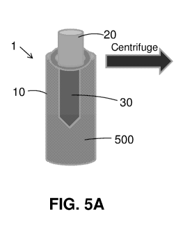
Fig. 5A schematically illustrates and Fig. 6A is a photograph of a device 1
comprising
a container 10 covered with a cover 20 to which an artificial bone implant 30
is attached,
when the container 10 is filled with whole blood 500.
Fig. 5B schematically illustrates and Fig. 6B is a photograph of a device 1
comprising
a container 10 covered with a cover 20 to which an artificial bone implant 30
is attached,
when the container 10 is filled with whole blood after centrifugation, for
example at a range
of substantially 2,500-3,000g for substantially 7-10 minutes. As a result of
the centrifugation
30 the whole blood 500 is separated to two main layer ¨ a lower layer 510
comprising red blood
cells and platelets, and an upper layer 520 comprising plasma.
Fig. 5C schematically illustrates and Fig. 6C is a photograph of an artificial
bone
implant 30 attached to a cover 20 of the device 1 after the cover 20 and
artificial bone implant
16
CA 03004590 2018-05-03
WO 2017/077542
PCT/1L2016/051196
30 were separated from the container 10, after centrifugation with whole blood
500. The
artificial bone implant 30 is covered with some of the upper layer 520
comprising plasma.
The artificial bone implant 30 is held by the cover 20 inside the container 10
in a manner that
after centrifugation the artificial bone implant 30 is in contact with the
upper plasma layer,
the middle buffy coat layer, and some upper part of the lower red blood cells
layer. Thus, the
centrifugation separates the artificial bone implant from most of the red
blood cells, but
allows direct contact of the artificial bone implant mostly with the plasma,
and the white
blood cells and platelets of the whole blood sample.
As a result of the centrifugation, plasma and buffy coat is adhered to the
surface of the
artificial bone implant. Thus, components of plasma and buffy coat adhere to
the surface of
the artificial bone implant, including osseointegration accelerators such as
growth factors that
are contained in the plasma layer, and progenitor cells that are contained in
the buffy coat
layer.
Fig. 7 is a scanning electron micrograph (SEM) of the coating layer covering
the
artificial bone implant after centrifugation with whole blood seen in Fig. 6C.
The coating
layer comprises a fibrin network (without interlocked RBCs due to
centrifugation). The
coating layer on the artificial bone implant is well-woven and characterized
by a dense fibrin
texture with thin fibers with pores of about 0.1 micrometer.
Fig. 8 is a photograph of another embodiment of an artificial bone implant
coated
with CGF following centrifugation with whole blood. The fibrin clot coating
the artificial
bone implant is readily seen.
To clinically justify the use of CGF-coated implants, the biological activity
of the
coating layer was evaluated using enzyme-linked immunosorbent assay (ELISA).
The
cumulative release rate of growth factors from the CGF coating layer was
studied in vitro, by
incubating coated implants in medium, and quantitating the growth factors
released into the
medium. We chose to study certain growth factors that have a biological effect
on cell
adhesion, proliferation and osteogenic differentiation, all central to implant
osseointegration.
These growth factors are mainly released from platelets that are interlocked
within the fibrin
network. Fibrin clot formation is initiated during centrifugation, where the
heavy RBCs
17
CA 03004590 2018-05-03
WO 2017/077542
PCT/1L2016/051196
sediment first and are therefore not interlocked within the fibrin network,
while the lighter
WBCs and platelets sediment later, concomitant to fibrin clot formation, and
therefore
become interlocked in the network. These platelet-rich concentrates within the
fibrin clot
differentially release growth factors and affect cell differentiation and
functions. These
growth factors include: platelet derived growth factor (PDGF), which enhance
cell growth,
blood vessel repair and generation and collagen production; vascular
endothelial growth
factor (VEGF), which promotes growth and new generation of vascular cells;
tumor necrosis
factor-alpha (TNF-a), which is involved in systemic inflammation; transforming
growth
factor-betal (TGF-I31), which improves growth and neogenesis of epithelial
cells; vascular
cells and wound healing; and insulin-like growth factor-1 (IGF-1), which is
crucial in
healing and cell growth. The kinetics of cumulative release of growth factors
from coated
implants is presented in Fig. 9.
Fig. 9 shows graphs of cumulative release of growth factors from coated
implants
over time. Coated implants were incubated in medium at 37 C, for varying time
intervals (5
h, 1, 3, 6, 7 or 8 days) and growth factors released into the medium were
quantitated. The
release test was conducted using blood from three different donors, each
incubated with an
implant in a separate vacuum container (v1, v2, and v3). The individual and
mean results are
presented. A) PDGF-AB release over time. B) VEGF release over time. C) TNF-a
release
over time. D) TGF-I31 release over time. E) IGF-1 release over time.
All tested growth factors were present in the CGF coating layer and released
at a slow
rate. PDGF-AB and VEGF release seemed to increase over the eight-day period,
whereas
TNF-a, TGF-I31, and IGF-1 release seemed to be constant over time. Future
studies will
measure the release of growth factors in media that contain protease
inhibitors, to account
also for degraded growth factor release. The release of growth factors will be
assessed for a
longer time; up to 20 days.
SEM was employed to characterize the surface of CGF-coated dental implants
seeded
with cells. Mesenchymal stem cells (MSCs) isolated from bone marrow were
seeded onto
CGF-coated implants, at a density of 100,000 MSCs/ml/dental implant, and then
cultured for
two days. This was performed to verify the effect of CGF coat on cell adhesion
and growth.
The samples were then fixed in and the three-dimensional morphology of the
implant coating
and distribution of cells were visualized (Figs. 10A-D and 11A-D).
18
CA 03004590 2018-05-03
WO 2017/077542
PCT/1L2016/051196
Figs. 10A-D are SEM images of CGF-coated dental implants cultured with MSCs.
Fig. 10A) A dental implant. Fig. 10B) A CGF-coated dental implant. Fig. 10C)
An MSC-
seeded dental implant. Fig. 10D) A CGF-coated dental implant incubated with
MSCs.
Figs. 11A-D are SEM images of treated dental implant surfaces. Fig. 11A) A
dental
implant surface. Fig. 11B) A CGF-coated dental implant surface. The arrow
indicates a
platelet. Fig. 11C) An MSC-seeded dental implant surface. The arrow indicates
a seeded cell.
Fig. 11D) A CGF-coated dental implant incubated with MSCs. The arrows indicate
seeded
cells.
The bare titanium dental implant surface (Fig. 11A) differed from CGF-coated
implant surface, with the latter displaying fibrin fibers (Fig. 11B). Cells
attached to the bare
implant surface (Fig. 11C), but were observed in greater numbers on CGF-coated
surfaces
(Fig. 11D).
After demonstrating the three-dimensional structure of the CGF coating layer
and the
growth factors release rate, we investigated the biological activity of the
CGF coating on
bone marrow-derived MSCs. As the CGF coating contains fibrin matrix and growth
factors,
the adhesion and proliferation of MSCs on the layer was tested by the
AlamarBlue metabolic
activity assay. MSCs were seeded at a density of 100,000 MSCs/ml/dental
implant, and the
number of cells which adhered to implant surfaces and proliferated for two
days was
evaluated (Fig. 12).
Fig. 12 is a graph showing number of MSCs growing on implant surfaces for two
days. The CGF coating significantly enhanced MSCs adhesion and proliferation
as compared
to the control samples. The biocompatibility of the fibrin matrix and the
effect of the
interlocked factors on cell growth are projected to enhance MSC osteogenic
differentiation.
We investigate the effect of the CGF coating on osteogenic genes, using
techniques such as
fluorescence-activated cell scanner (FACS) and real-time PCR.
Figs. 13A-C schematically illustrate another exemplary embodiment of the
device 1
configured to coat a dental implant. Fig. 13A illustrates a vacuum container
comprising two
tubes. Blood is transferred into the inner tube, in which a dental implant is
placed. The larger
19
CA 03004590 2018-05-03
WO 2017/077542
PCT/1L2016/051196
tube protects the inner tube during centrifugation. Fig. 13B illustrates an
assembled vacuum
container centrifuged with a silicon cap facing down. Fig. 13C illustrates a
silicon cap from
which blood is transferred.
Figs. 13A-C illustrate the vacuum container in which a dental implant is
positioned
such that root surface coating occurs upon centrifugation. The appropriate
pressure of
vacuum within the vacuum container is set up to withdraw the precise amount of
blood for
optimal coating. Acceleration of blood clotting may be achieved by coating the
inner lumen
of the vacuum container with silicone and micronized silica. As such, the
blood withdrawn
into the vacuum container undergoes a complex clotting cascade forming long
strands of
fibrin around the implant that eventually results in a homogeneous net-like
texture. Taken
together, the parameters of vacuum and centrifugation (e.g., relative
centrifugal force (rcf),
time, speed, orientation of container in centrifuge etc.) allow a coating
process that yields a
400-500 micron-thick layer of fibrin that entraps bioactive components such
platelets and
WBCs, but is deprived of RBCs.
In order to assure the effectiveness of the procedure and the bioactivity of
the coated
implant, designed to be implanted immediately following coating, the CGF
coating is further
characterized. The potential of growth, proliferation, migration, and
differentiation of MSCs
seeded onto CGF coating layer is studied. MSCs migration is assessed using the
Boyden
chamber assay.
Figs. 14A-B illustrate a Boyden chamber to assess the effect of CGF-coated
implants
on MSC migration and growth rate. Fig. 14A illustrates the effect of growth
factors released
from CGF-coated implants on MSCs migration. Fig. 14B illustrates the effect of
growth
factors released from CGF-coated implants on MSCs growth rate.
Cells are placed in the cell culture insert or the upper chamber, separated by
a porous
membrane from the lower chamber, which contains the CGF-coated implant. The
cells and
the implant are submerged in a shared serum-free medium. The cells are then
allowed to
migrate from the upper chamber to the underside surface of the insert, over 4
hours under
incubated conditions. The cells on the upper membrane surface of the insert
are then
mechanically removed, and the migrated cells on the underside surface of the
insert are fixed,
stained and counted. This technique enables assessment of the percentage of
MSCs migration
CA 03004590 2018-05-03
WO 2017/077542
PCT/1L2016/051196
toward growth factors released from CGF-coated implant. The effect of these
factors on the
growth rate of MSCs in the lower chamber in the presence versus absence of CGF-
coated
implants in the upper chamber can also be assessed and compared over time
(Fig. 14B). The
expression of osteogenic genes in MSCs seeded onto CGF coating layer is
determined using
techniques such as FACS and real-time PCR. The osteogenic potential of MSCs
seeded onto
the CGF coating layer is tested in vitro using alkaline phosphatase activity
assays and
assessment of mineralization of cells.
Mini and standard implants with the appropriate containers for CGF coating are
used
for proof of concept studies to verify acceleration of osseointegration
following placement of
CGF-coated implants in bone tissue of rat tibia. Standard-size implants are
also tested in
humans, along with the appropriate container for CGF coating. These coated
implants are
placed in bone tissue of the dog mandible, to establish clinical protocol for
human patients.
Following in vitro studies characterizing MSC differentiation in the presence
of the
CGF coating, feasibility studies are conducted, in which CGF-coated implants
are
transplanted into an the rat tibia to test their osseointegration rate within
bone tissue. Wistar
nude rats are anesthetized and an incision is made over the right anterior-
proximal tibia
surface. Care is taken to preserve the periosteal surface. Holes are drilled
through one cortex,
using a 1 mm drill bit and implants are placed. The skin is closed around the
implant with
non-absorbable sutures and pain is managed. Two groups of animal are tested
and compared,
i.e., those receiving the CGF-coated implants versus those receiving the non-
coated implants.
The CGF coating procedure is performed with human blood. Implant
osseointegration is
assessed at various time points (e.g., 2, 4, 6, and 10 weeks after installing
implants). A total
of 40 rats is needed for the study. At the end of experiment, rats are
anesthetized; the tibia
bone in which implants are installed is excised, fixed in formalin, and
embedded in paraffin
for histological analysis, or analyzed with an ex vivo micro-CT scanners to
assess bone tissue
formation or implant osseointegration. Rats are sacrificed by intracardiac
administration of
pentobarbital sodium salt.
osseointegration of CGF-coated implants is evaluated in a canine model. Two
groups
comprised of four male beagle dogs each, approximately two years old and
weighing 15-18
kg, are radiographically screened before tooth extraction to rule out any
pathology. Two
mandibular implants are implanted in each dog, with one group receiving CGF-
coated
CA 03004590 2018-05-03
WO 2017/077542
PCT/1L2016/051196
implants and the other receiving bare implants. CGF implant coating is
performed with
autologous blood, collected from the dog being treated. Dogs undergo surgery
under
halothane gas anesthesia. Heart rate, temperature, and respiration rate are
monitored during
surgery. The edentulation procedure of dogs is assessed radiographically.
Venous blood of
the dogs is used for implant coating. Osseointegration and bone tissue healing
around the
implants is radiographically assessed at 1, 3, 6 months post-implantation. At
the 6 month time
point, implants are also histologically evaluated. Histomorphometric analysis
is conducted to
determine the percent bone contact length along the implant.
Fig. 15 schematically illustrates, another exemplary embodiment of the device
1
configured to form a putty bone from a bone material, for example bone
substitute. A bone
material 600, in the form of powder, fragments, and the like, is placed within
a vacuumed
container 10. The container 10 is provided with a fixed separator 40 that
separates the
container 10 to two parts, a bottom part and an upper part. The separator 40
is provided with
holes so that when the container 10 is filled with whole blood 500, the blood
500 in the
bottom part of the container 10 is in full contact with the blood 500 in the
upper part of the
container 10. The bone material 600 is in the upper part of the container 10,
on top of the
separator 40. After the vacuum in the container 10 is replaced with a
patient's blood 500, the
container 10 is centrifuged in similar conditions as mentioned herein above.
As a result of the
centrifugation, the blood 500 is separated to a bottom phase 510 comprising
red blood cells
and platelets, mostly concentrated in the bottom part of the container 10,
under the separator
40, and an upper phase 520 comprising plasma in the upper part of the
container 10, above
the separator 40. Again, the plasma contains osseointegration accelerators
that adhere to the
bone material 600 so as to form a putty bone 700, ready to be used.
Fig. 16 schematically illustrates, according to an exemplary embodiment, a
disassembled device 1 configured to form a putty bone from a bone material.
The container
10, exemplary components of the cover 20 and the separator 40 are illustrated.
Fig. 17 schematically illustrates, according to an exemplary embodiment, an
assembled device 1 configured to form a putty bone from a bone material. The
container 10,
exemplary components of the cover 20 and the separator 40 are illustrated.
22
CA 03004590 2018-05-03
WO 2017/077542
PCT/1L2016/051196
Fig. 18. schematically illustrates, according to an exemplary embodiment, a
system
for withdrawing whole blood from a vein directly from a vein of a patient into
a device 1
configured to form a putty bone from a bone material. The components of the
system are as
described in Fig. 2, and the device 1 configured to form a putty bone from a
bone material is
as described in Fig. 15.
Figs. 19A-I are SEM of a putty bone 700 prepared by using the device 1
configured to
form a putty bone from a bone material.
It is appreciated that certain features of the subject matter, which are, for
clarity,
described in the context of separate embodiments, may also be provided in
combination in a
single embodiment. Conversely, various features of the subject matter, which
are, for brevity,
described in the context of a single embodiment, may also be provided
separately or in any
suitable sub combination.
Although the subject matter has been described in conjunction with specific
embodiments thereof, it is evident that many alternatives, modifications and
variations will be
apparent to those skilled in the art. Accordingly, it is intended to embrace
all such
alternatives, modifications and variations that fall within the spirit and
broad scope of the
appended claims.
23