Note: Descriptions are shown in the official language in which they were submitted.
84281644
1
DEVICES AND METHODS FOR DETECTING ANALYTES
USING THERMAL WAVES
TECHNICAL FIELD
Embodiments of the present disclosure relate generally to devices and methods
of
detecting analytes using polymer materials, such as over a heat sink
configured to
produce a thermal wave.
BACKGROUND
Molecularly imprinted polymers (MIPs) can be used for detecting chemical
substances in complex mixtures. In modern research, these polymers are of
increasing
interest for bioanalytical applications. Advantages of using these MIPs
include easy and
cheap production; mechanical, chemical, and thermal stability; reusability;
and long shelf
life. In recent years, the concept of molecular imprinting has been extended
to surface
.. imprinting of thin polymer films with micrometer-sized cells to create so-
called "surface
imprinted polymers" (SIPs) for the detection of proteins, glycoproteins, plant
viruses,
human viruses, bacteria, pollen, yeast cells, and even mammalian red blood
cells. SIPs
are polymeric materials with indentations at the surface, with a form and
function matching
part of a desired target. SIPs are suitable for bonding with larger objects
(e.g., cells, bacteria,
etc.), which do not diffuse quickly through pores of an MIP. Imprinting may
occur after
polymerization by softening the polymer. The detection of cells using
biosensors described
in literature is conventionally done by gravimetric detection, electronic read-
out platforms
or micro-fluidic techniques. However, these techniques are often time-
consuming, provide
difficulties for analysis, or require expensive equipment.
For example, temperature resistance of substrates having MIPs attached thereto
based on the concentration of analytes is described in U.S. Patent Application
Publication
2014/0011198 Al, "Heat-Transfer Resistance Based Analysis Bioparticles,"
published
January 9, 2014.
A low-cost sensor platform providing the capability to differentiate between
cells
with slight differences in shape, size, and functionalities in functional
groups on their
surface would be a valuable tool for modern research and industry.
Date Recut/Date Received 2023-03-16
CA 03004786 2018-05-09
WO 2017/084885 PCT/EP2016/076571
2
DISCLOSURE
In some embodiments, a device for detecting an analyte includes a substrate
having a polymer material formed on a surface thereof; a heat sink thermally
coupled to a
surface of the substrate opposite the polymer material; a temperature
modification device
thermally coupled to the heat sink; a controller configured to cause the
temperature
modification device to produce a thermal wave emanating from the heat sink;
and a flow
cell located and configured to pass a liquid over the polymer material of the
substrate.
The device may further include a temperature sensor located and configured to
detect a
temperature of the liquid passing over the polymer material and a processor
configured to
calculate a concentration of an analyte in the liquid based at least in part
on a phase shift
between the thermal wave at the heat sink and an attenuated thermal wave in
the liquid.
A method for detecting an analyte includes passing a liquid containing an
analyte
over a polymer material on a substrate; binding the analyte to the polymer
material; providing
a thermal wave from a heat sink to the polymer material through the substrate;
detecting a
temperature of the liquid; and calculating a concentration of the analyte in
the liquid based at
least in part on a phase shift between the thermal wave produced by the heat
sink and an
attenuated thermal wave in the liquid.
A method of forming a device for detecting an analyte includes forming a
polymer
material over a surface of a substrate; thermally coupling a heat sink to a
surface of the
substrate opposite the polymer material; thermally coupling a temperature
modification
device to the heat sink; configuring a controller to cause the temperature
modification device
to produce a thermal wave emanating from the heat sink; configuring a flow
cell to pass a
liquid over the polymer material of the substrate; configuring a temperature
sensor to detect a
temperature of the liquid passing over the polymer material; and configuring a
processor to
calculate a concentration of an analyte in the liquid based at least in part
on a phase shift
between the thermal wave at the heat sink and an attenuated thermal wave in
the liquid.
In some embodiments, a method for characterizing bacteria includes passing a
liquid containing an analyte comprising a first bacteria and a second bacteria
over and in
contact with a polymer material on a substrate. The polymer material is
formulated to bind
to the first bacteria, and the first bacteria binds to the polymer material
with a higher
affinity than the second bacteria. A heat transfer property of the polymer
material varies
based on an amount of the analyte bound thereto. The method further includes
binding a
portion of the first bacteria and the second bacteria of the analyte to the
polymer material,
removing at least a portion of the second bacteria from the polymer material,
detecting a
84281644
3
temperature of the substrate, and calculating a concentration of the first
bacteria in the liquid
based at least in part on the temperature of the substrate.
In other embodiments, a method for characterizing a liquid comprising bacteria
includes
passing a liquid containing a first strain of bacteria and at least a second
strain of bacteria over
and in contact with a polymer material on a substrate. The polymer material is
formulated to
bind to the first strain of bacteria, and the first bacteria binds to the
polymer material with a
higher affinity than the at least a second bacteria. A heat transfer property
of the polymer
material varies based on an amount of material bound thereto. The method
further includes
binding a portion of the first bacteria and a portion of the at least a second
bacteria to the
polymer material, washing the polymer material to remove the at least a second
bacteria
therefrom, passing the liquid over the polymer material after washing the
polymer material,
washing the polymer material at least a second time to remove the at least a
second bacteria
therefrom, detecting a temperature of the substrate, and calculating a
concentration of the first
bacteria in the liquid based at least in part on the temperature of the
polymer material.
Thus, there is provided a device for detecting an analyte, the device
comprising: a
substrate having a polymer material formed on a surface thereof, the polymer
material formulated
to bind to the analyte, wherein a heat transfer property of the polymer
material is formulated to
vary based on an amount of the analyte bound thereto; a heat transfer element
thermally coupled
to a surface of the substrate opposite the polymer material; a temperature
modification device
thermally coupled to the heat transfer element; a controller configured to
cause the temperature
modification device to produce a thermal wave emanating from the heat transfer
element; a flow
cell located and configured to pass a liquid over the polymer material of the
substrate; a
temperature sensor configured to detect a temperature (T2) of the liquid
passing over the polymer
material; and a processor configured to calculate a concentration of an
analyte in the liquid based
at least in part on a phase shift between the thermal wave at the heat
transfer element and an
attenuated thermal wave in the liquid.
There is also provided a method for detecting an analyte, the method
comprising: passing
a liquid containing an analyte over a polymer material on a substrate, the
polymer material
formulated to bind to the analyte, wherein a heat transfer property of the
polymer material is
formulated to vary based on an amount of the analyte bound thereto; binding
the analyte to the
polymer material; providing a thermal wave from a heat transfer element to the
polymer material
through the substrate; detecting a temperature (12) of the liquid; and
calculating a concentration
of the analyte in the liquid based at least in part on a phase shift between
the thermal wave
produced by the heat transfer element and an attenuated thermal wave in the
liquid.
There is also provided a method of forming a device for detecting an analyte,
the method
comprising: forming a polymer material over a surface of a substrate, the
polymer material
Date Recue/Date Received 2023-03-16
84281644
3a
formulated to bind to the analyte, wherein a heat transfer property of the
polymer material is
formulated to vary based on an amount of the analyte bound thereto; thermally
coupling a heat
transfer element to a surface of the substrate opposite the polymer material;
thermally coupling
a temperature modification device to the heat transfer element; configuring a
controller to cause
the temperature modification device to produce a thermal wave emanating from
the heat
transfer element; configuring a flow cell to pass a liquid over the polymer
material of the
substrate; configuring a temperature sensor to detect a temperature (T2) of
the liquid passing
over the polymer material; and configuring a processor to calculate a
concentration of an
analyte in the liquid based at least in part on a phase shift between the
thermal wave at the heat
transfer element and an attenuated thermal wave in the liquid.
BRIEF DESCRIPTION OF THE DRAWINGS
FIG. 1 is a simplified schematic diagram showing a device for detecting an
analyte;
FIG. 2 is a simplified schematic representation showing how a thermal wave may
travel in
the device of FIG. 1;
FIG. 3 is a simplified schematic diagram showing another device for detecting
an
analyte;
FIG. 4 is a graph showing binding isotherms for dopamine as measured according
to an
embodiment of the disclosure;
FIG. 5 is a graph showing calibration curves for dopamine as measured
according to an
embodiment of the disclosure;
FIG. 6 is a graph showing changes in temperature as measured according to an
embodiment of the disclosure;
FIG. 7 is a graph showing time-dependent values of thermal resistance as
measured
according to an embodiment of the disclosure;
FIG. 8 is a graph showing the thermal resistance data of FIG. 7 in the form of
a
dose-response curve;
FIG. 9 is a graph showing a thermal wave generated according to an embodiment
of the
disclosure;
Date Recue/Date Received 2023-03-16
CA 03004786 2018-05-09
WO 2017/084885
PCT/EP2016/076571
4
FIG. 10 is a graph showing thermal waves measured after passing through a
substrate according to an embodiment of the disclosure;
FIG. 11 is a graph showing the phase shift of the thermal waves shown in FIG.
10 as
measured according to an embodiment of the disclosure;
FIG. 12 is a graph showing thermal waves measured after passing through a
substrate according to an embodiment of the disclosure;
FIG. 13 is a graph showing the phase shift of thermal waves shown in FIG. 12
as
measured according to an embodiment of the disclosure;
FIGS. 14 and 15 are optical microscopic analyses of polymers imprinted with E.
coil and S. aureus, respectively;
FIG. 161s a graph showing thermal response of a device alternately exposed to
dead and living E. coil, with flushing in between exposures;
FIG. 17 is a boxplot summarizing the thermal responses shown in FIG. 5;
FIGS. 18 and 19 are graphs showing thermal responses of devices alternately
exposed to S. aureus and E. coil, with flushing in between exposures;
FIG. 20 is a boxplot summarizing the thermal responses shown in FIGS. 7 and 8;
FIG. 21 is a graph showing thermal response of a device exposed to increasing
concentrations of E. coil, with flushing in between exposures;
FIG. 22 is a dose-response curve derived from the thermal responses shown in
FIG. 10;
FIG. 23 is a graph showing thermal responses of a device exposed to a mixture
of
E. coil and S. aureus, with flushing in between exposures, as well as a
boxplot
summarizing the thermal responses;
FIGS. 24 through 30 are graphs showing changes in temperature of devices as
measured according to an embodiment of the disclosure;
FIGS. 31 through 37 are graphs showing thermal waves measured after passing
through substrates according to an embodiment of the disclosure;
FIGS. 38 and 40 are graphs showing changes in temperature of devices when
exposed to analogue and target bacteria, as measured according to embodiments
of the
disclosure;
FIGS. 39 and 41 are graphs showing phase shifts at 0.03 Hz for the data
depicted in
FIGS. 38 and 40, respectively;
FIG. 42 is a graph showing changes in temperature of a device as measured
during
repeated exposure to a mixture of bacteria, according to embodiments of the
disclosure;
CA 03004786 2018-05-09
WO 2017/084885 PCT/EP2016/076571
FIG. 43 is a graph showing thermal responses of the device for which the
temperature changes are shown in FIG. 42, as well as a boxplot summarizing the
thermal
responses; and
FIG. 44 is a graph showing phase shift at 0.03 Hz for the data depicted in
FIG. 42.
5
MODE(S) FOR CARRYING OUT THE INVENTION
The illustrations presented herein are not actual views of any particular
device or
method, but are merely idealized representations employed to describe example
embodiments of the present disclosure. Elements common between figures may
retain
the same numerical designation.
As used herein, the terms "template molecule" and "template bacteria"
respectively
refer to molecules or bacteria used to form a molecularly imprinted polymer
(MIP) or
surface imprinted polymer (SIP). Such MIPs or SIPs can then detect "target
molecules" or
"binding partners," which have functionality corresponding to the template
molecules used
to form the MIP or SIP.
As used herein, the term "may" encompasses the word "can," and the term "may
be"
encompasses the words "is" or "are," depending on context. Furthermore,
presence of the
word "may" is intended to indicate options for practicing or implementing
embodiments of the
disclosure, without limitation.
FIG. 1 is a simplified schematic diagram showing a device 100 for detecting an
analyte. In some embodiments, the device 100 may be configured to detect a
target
molecule, a nucleic acid such as DNA and/or RNA, single-nucleotide
polymorphisms (SNPs)
in DNA and/or RNA, small molecules, proteins, bacteria, etc.
The device 100 may include a substrate 110 having a polymer material 112
located over a surface thereof. For example, the polymer material 112 may be
formed or
disposed over a generally planar surface of the substrate 110, and another,
opposite
generally planar surface of the substrate 110 may be free of the polymer
material 112. In
some embodiments, the substrate 110 may include a metal (e.g., aluminum), an
alloy, a
semiconductor (e.g., silicon, doped diamond, etc.), an electrically insulating
material (e.g.,
undoped diamond). The polymer material 112 may include any material for which
a heat
transfer property varies based on an amount of the analyte bound thereto. For
example,
the thermal conductivity, thermal diffusivity, heat capacity, or another
property of the
polymer material 112 may vary with concentration of the analyte on the surface
thereof.
In some embodiments, the polymer material 112 may include an imprinted
polymer, such as a molecularly imprinted polymer (MIP) or a surface imprinted
polymer
84281644
6
(SIP). MIPs and SIPs may also be referred to in the art as "plastic"
antibodies. MIPs
typically possess a high affinity for a specific binding partner, so that when
such binding
partners are contacted with the MIP, the molecules bind with the MIP. MIPs are
synthetic
receptors that contain nanocavities with high affinity for their respective
target molecules.
Imprinting (i.e., formation of the nanocavities) is often part of the
polymerization process.
MIPs are able to specifically bind targets, including bacteria, varying from
small ions to
large cells in complex matrices. Binding of molecules to the MIP may alter
some
properties of the MIP, such as thermal properties, mechanical properties,
electrical
properties, etc. The altered property of an MIP may, therefore, be used to
detect a
presence of such molecules at relatively low concentrations. MIPs are
described in, for
example, U.S. Patent Application Publication 2009/0281272 Al, "Monodisperse
Molecularly Imprinted Polymer Beads," published November 12, 2009.
Similarly, SIPs typically possess a high affinity for a specific binding
partner, but
may typically bind to relatively larger objects (e.g., cells, bacteria, etc.)
that do not diffuse
quickly through pores of an MIP. SIPs may be polymer materials formed over a
surface,
then imprinted after polymerization by softening the polymer.
In certain embodiments, the polymer material 112 may include DNA, RNA,
proteins, or portions or analogs thereof. For example, the device 100 may
include a
substrate 110 (e.g., a diamond surface) functionalized with a polymer material
112 such
as DNA, RNA, a protein, a polypeptide, a nucleic acid polymer, a probe, or a
portion or
analog thereof (e.g., complementary DNA, antibodies, etc.). The polymer
material 112
may be formulated to possess a high affinity for a specific binding partner,
so that when
such binding partners are contacted with the surface of the substrate 110, the
molecules
bind with the polymer material 112. The polymer material 112 may also bind to
analogues
of the binding partner (e.g., a material having similar functionality as the
binding partner),
though not necessarily with the same affinity as binding with the binding
partner itself. In
some embodiments, the polymer material 112 may include at least about seven
(7)
repeating units, such as ten (10) repeating units or more.
In some embodiments, the polymer material 112 may include a material screen-
printed onto the substrate 110. Screen-printed materials may be manufactured
efficiently
and in mass quantities, with relatively high uniformity in comparison with
other materials.
The device 100 may further include a heat sink 114 thermally coupled to a
surface
of the substrate 110, such as a surface opposite the polymer material 112.
Though
referred to as a heat "sink" for the sake of simplicity, the heat sink 114 may
be configured
Date Recut/Date Received 2023-03-16
CA 03004786 2018-05-09
WO 2017/084885 PCT/EP2016/076571
7
to provide heat to or remove heat from the substrate 110 and, so, may also be
characterized as a heat transfer element 114. The heat sink or heat transfer
element 114
may be a material having a high thermal conductivity, such as a transition
metal (e.g.,
copper, silver, etc.) or an alloy or mixture thereof. In some embodiments, the
polymer
material 112 may be applied to the heat sink 114 itself. The heat sink 114 may
be
thermally coupled to a temperature sensor 116 (e.g., a thermocouple or another
device)
configured to detect a temperature of the heat sink 114, and to a temperature
modification
device 118 configured to maintain the temperature of the heat sink 114. The
temperature
modification device 118 may include, for example, a thermoelectric device, a
heat
.. exchanger, a fan, a resistance heater, etc. The temperature sensor 116 may
be a resistor
having a resistance that varies with temperature. If the properties of the
heat sink 114 are
known (e.g., if a relationship between a control signal to the modification
device 118 and
the temperature of the heat sink 114 is well characterized), the temperature
sensor 116
may be omitted. In some embodiments, the temperature sensor 116 may be
integral to
the temperature modification device 118. For example, the internal resistance
of the
temperature modification device 118 itself may be measured to determine its
temperature.
The temperature sensor 116 and the temperature modification device 118 may be
connected to a controller 121 configured (i.e., programmed) to control the
temperature
modification device 118 to cause the heat sink 114 to produce a thermal wave
emanating
.. from the heat sink 114 and through the substrate 110 (including the polymer
material 112
thereon). For example, the controller 121 and a processor 123 may be
incorporated into a
computer 120 (e.g., the controller 121 may be an input-output card configured
to receive
and provide electrical signals, and may be configured to receive signals from
the
processor 123). In some embodiments, the controller 121 may be a proportional-
integral-
derivative (PID) controller capable of changing the temperature of the heat
sink 114 by a
small amount on a relatively short time scale. For example, the controller 121
may
change the temperature of the heat sink 114 by about 0.5 C or less, about 0.2
C or less,
or even about 0.05 C or less. Thus, the thermal wave may have an amplitude of
about
1.0 C or less, about 0.4 C or less, or even about 0.10 C or less. The
controller 121 may
be capable of changing the temperature of the heat sink 114 via the
temperature
modification device 118 from one set point to another and back to form a
thermal wave
having a frequency from about 0.001 to about 0.5 Hz, such as from about 0.005
to about
0.1 Hz, or from about 0.01 to about 0.05 Hz. In some embodiments, the
controller 121,
the temperature modification device 118, and the heat sink 114 may together
produce a
thermal wave having a variable frequency. Based on a measurement from the
84281644
8
temperature sensor 116 (if present), a known input to the temperature
modification device
118, or other means, properties of the thermal wave may be known (e.g., a
phase,
amplitude, frequency at a specific time, rate of frequency change, etc.).
In other embodiments, the controller 121 may be configured to maintain the
heat
sink 114 at a constant temperature. Detection of analytes using a heat sink at
constant
temperature is described in U.S. Patent Application Publication 2015/0219584
Al,
"Biosensor Using Impedimentric Real-Time Monitoring," published August 6,
2015.
The device 100 may further include a flow cell 122 configured to pass a liquid
124
over the polymer material 112 of the substrate 110. The flow cell 122 may
define a void
126 adjacent the polymer material 112 of the substrate 110, as well as an
inlet 128 and an
outlet 130 through which the liquid 124 may flow. An 0-ring 131 or another
appropriate
sealing mechanism may retain the liquid 124 within the flow cell 122 adjacent
the polymer
material 112 over the substrate 110.
The liquid 124 may include an analyte 132 that specifically binds to the
polymer
material 112 and change thermal properties thereof, as described above. For
example, the
analyte 132 may include one or more strains of bacteria. The analyte 132
(which may
include multiple analytes 132a and 132b) may specifically bind to the polymer
material
112 and changes thermal properties thereof, as described above. If multiple
analytes
132a and 132b are present in the liquid 124, the analytes 132a, 132b may have
similar
functionalities, such that each of the analytes 132a, 132b bind to the polymer
material
112. The analytes 132a, 132b may bind to the polymer material 112 with
different
affinities. In some embodiments, the first analyte 132a may include living
bacteria, and
the second analyte 132b may include dead bacteria of the same species. In
other
embodiments, the first analyte 132a may include bacteria, and the second
analyte 132b
may include an analogue bacteria.
A temperature sensor 134 (e.g., a thermocouple or another device) may be
configured to detect a temperature of the liquid 124 in (e.g., flowing
through) the flow cell
122. The computer 120 may record the temperature of the liquid 124 by, for
example,
measuring a resistance of the temperature sensor 134 via the controller 121
and/or the
processor 123, and correlating that resistance to a temperature. The
temperature of the
liquid 124 may be different from the temperature of the heat sink 114, and may
vary based
at least in part on the presence or absence of the analyte 132 and its
concentration in the
liquid 124. For example, temperature resistance of substrates based on the
concentration
of analytes is described in U.S. Patent Application Publication 2014/0011198
Al, "Heat-
Date Recue/Date Received 2023-03-16
84281644
9
Transfer Resistance Based Analysis Bioparticles," published January 9, 2014.
In some embodiments, the processor 123 may be configured to calculate a
concentration of the analyte 132 in the liquid 124 based at least in part on a
phase shift
between the thermal wave produced by the heat sink 114 and an attenuated
thermal wave
in the liquid 124 after the thermal wave passes through the substrate 110 and
the polymer
material 112.
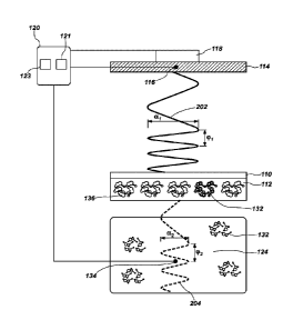
FIG. 2 is a simplified schematic representation showing how the thermal wave
may
travel in the device 100 of FIG. 1. FIG. 2 includes some of the components
shown in FIG. 1,
but shows them separated to allow representation of thermal waves traveling
through and
between the components. In particular, FIG. 2 shows the heat sink 114
thermally coupled to
the temperature modification device 118 and the temperature sensor 116, which
are
connected to the computer 120. The concentration of the analyte 132 may be
measured
based on the differences between the thermal wave at the heat sink 114 and the
thermal
wave in the liquid 124, without a separate calibration step.
The heat sink 114 may produce a thermal wave 202 and transfer the thermal wave
202 to the substrate 110 and the polymer material 112 thereon. For example, if
the heat sink
114 is initially maintained at a constant temperature of 37 C, the thermal
wave 202 may be
produced by heating the heat sink 114 to a temperature of 37.1 C and then
cooling the heat
sink 114 to a temperature of 36.9 C. The heating and cooling of the heat sink
114, driven by
the temperature modification device 118, may cause the substrate 110 and the
polymer
material 112 to heat and cool in a corresponding manner. The thermal wave 202
may have
an amplitude al and a frequency (pi. The amplitude ai and/or the frequency (pi
may vary with
time. For example, the thermal wave 202 may have a continuously varying
frequency cpi.
As discussed above, the presence or absence of the analyte 132 on the
substrate
.. 110 may change the thermal conductivity, thermal diffusivity, heat
capacity, or another
property of the polymer material 112. FIG. 2 illustrates conceptually that the
polymer
material 112 may define cavities 136 therein adapted to interact with at least
a portion of the
analyte 132. Without being bound to any particular theory, the cavities 136
may be
configured to act to specifically bind the analyte 132. Thus, the polymer
material 112 may
.. receive particles or molecules of the analyte 132 from the liquid 124 in
some of the cavities
136, based on the concentration of the analyte 132 in the liquid 124. The
liquid 124 and the
polymer material 112 may reach equilibrium at a given temperature, such that
the analyte
132 binds to and separates from the polymer material 112 at equal rates. The
thermal
Date Recut/Date Received 2023-03-16
CA 03004786 2018-05-09
WO 2017/084885
PCT/EP2016/076571
properties of the polymer material 112 may depend in part on the fraction of
the cavities 136
bound to particles or molecules of the analyte 132.
The substrate 110 and/or the polymer material 112 thereon may alter the
thermal
wave 202 passing therethrough to form an attenuated thermal wave 204. The
attenuated
5 thermal wave 204 may be detected by the temperature sensor 134, and
recorded by the
computer 120. The attenuated thermal wave 204 may have an amplitude 02 and a
frequency
92, which may be different from the amplitude al and a frequency (pi of the
thermal wave
202. The differences in the amplitudes al, 02 and/or the frequencies (pi,
(1:12 may be
correlated to the amount of the analyte 132 bound to the polymer material 112,
and thus, to
10 the concentration of the analyte 132 in the liquid 124. Measurement of
the differences in the
amplitudes al, 02 and/or the frequencies (pi, 92 may allow the device 100 to
detect relatively
lower amounts of the analyte 132 bound to the polymer material 112
(corresponding to lower
concentrations of the analyte 132 in the liquid 124) as compared with
conventional methods
of measuring the temperature of the liquid 124 at steady state.
In other embodiments, the processor 123 may be configured to calculate a
concentration of the analyte 132 based on a steady-state temperature
difference between
the heat sink 114 and the liquid 124.
In certain embodiments, the analyte 132 may bind to a non-planar surface. For
example, FIG. 3 is a simplified schematic diagram showing another device 200
for
detecting the analyte 132. The device 200 may include a thermocouple 210
having a
base material 212 formed over a surface thereof. For example, the base
material 212
may be formed over a generally cylindrical surface of the thermocouple 210,
such that an
entire end of the thermocouple 210 is enclosed. The thermocouple 210 may
include a
junction between two materials formulated to provide a temperature-dependent
voltage
between electrical contacts 216, 218. In some embodiments, the thermocouple
210 may
include one or more of a metal (e.g., platinum, gold, iridium, palladium,
etc.) or an alloy
(e.g., a nickel alloy, a copper alloy, a rhodium alloy, a rhenium alloy, an
iron alloy, a
molybdenum alloy, etc.).
The base material 212 may be a polymer material such as polylactic-(L)-acid,
which may be referred to in the art as PLLA. PLLA is transparent, inexpensive
to produce
from environmentally renewable sources (e.g., starch or sugar-containing
agricultural
products), biodegradable, and biocompatible. Furthermore, PLLA can be
solubilized in
chloroform to enable application to the thermocouple 210. Another material,
rather than
PLLA, may be selected to be the base material 212, based on desired
properties. In
some embodiments, the base material 212 may include polyurethane, polylactic
acid,
84281644
11
polycaprolactone, poly(lactic-co-glycolic acid), poly(D,L-lactide-co-
glycolide), or another
selected polymer. The base material 212 may be in the form of a thin, smooth,
and
homogeneous coating over the exterior of the thermocouple 210. Uniformity of
the
coating by base material 212 may enable to the device 200 to yield
reproducible results.
The thickness of the base material 212 may be selected in view of the thermal
resistance
of the base material 212 to affect the rate at which heat may flow toward or
away from the
thermocouple 210. Thus, a thinner base material 212 may be beneficial for
applications in
which a fast response is desired or temperature differentials are small.
The base material 212 may be selected to exhibit at least some elasticity,
such
that the device 200 may be flexible to allow bending of the thermocouple 210
without
breaking the base material 212. This may enable the device 200 to be used for
applications requiring tight clearance or bends (e.g., in vivo use in
catheters).
An assay polymer 214 may be on a surface of the base material 212. In some
embodiments, the assay polymer 214 may be directly bonded to the surface of
the
thermocouple 210, and the base material 212 may be omitted. The assay polymer
214
may include a material for which a heat transfer property varies responsive to
an amount
of the analyte bound thereto. For example, the thermal conductivity, thermal
diffusivity,
heat capacity, or another property of the assay polymer 214 may vary with
concentration
of the analyte on the surface thereof.
In some embodiments, the assay polymer 214 may include an imprinted polymer
(an MIP or SIP), DNA, RNA, proteins, or portions or analogs thereof (e.g.,
antibodies).
The assay polymer 214 may be configured to possess a high affinity for a
specific binding
partner, so that when such binding partners are contacted with the surface of
the
thermocouple 210, the molecules bind with the assay polymer 214. In some
.. embodiments, the assay polymer 214 may include at least about seven (7)
repeating units,
such as ten (10) repeating units or more.
In some embodiments, the device 200 may include a processor 223 programmed
to calculate an amount of the analyte bound to the assay polymer 214. The
processor 223
may calculate a concentration of the analyte in a liquid in contact with the
device 200 based
at least in part on the amount of the analyte bound to the assay polymer 214.
For example,
the processor 223 may calculate the amount of the analyte by a method as
disclosed in
U.S. Patent Application Publication 2014/0011198 Al, "Heat-Transfer Resistance
Based
Analysis Bioparticles," published January 9, 2014; or U.S. Patent Application
Publication
2014/0242605 Al, "Heat-Transfer Resistance Based Analysis of Bioparticles,"
published
August 28, 2014. In certain embodiments, the processor 223 may be used to
detect a
Date Recut/Date Received 2023-03-16
84281644
12
phase shift between a thermal wave at or emanating from a heat sink and an
attenuated thermal
wave at the thermocouple 210. The processor 223 may then calculate the
concentration of
the analyte in the liquid based at least in part on a difference in amplitude
between the
thermal wave at the heat sink and the attenuated thermal wave at the
thermocouple 210.
Returning again to FIG. 1, the polymer material 112 may be formed or otherwise
provided over the substrate 110. For example, the polymer material 112 may be
screen-
printed onto a metal substrate 110. Screen-printing may be performed
efficiently and
scaled to produce mass quantities, with relatively high uniformity in
comparison with other
methods. Screen-printing of substrates is described in, for example, U.S.
Patent
Application Publication 2012/0186999 Al, "Electrochemical Sensor," published
July 26,
2012.
The heat sink 114 may be thermally coupled to the substrate 110 at a surface
opposite the polymer material 112. For example, the heat sink 114 may be
placed in
direct physical contact with the substrate 110 such that heat can flow from
the heat sink
114 to the substrate 110 by conduction. In some embodiments, a thermally
conductive
material (e.g., a polymerizable liquid matrix having a thermally conductive
filler) may be
placed in physical contact with the heat sink 114 and the substrate 110 to
eliminate air
gaps between the heat sink 114 and the substrate 110. Similarly, the
temperature
modification device 118 may be thermally coupled to the heat sink 114 by
direct physical
contact, through a thermally conductive material, or by other appropriate
means.
The controller 121 (e.g., a PID controller) may be electrically connected to
the
temperature modification device 118 to provide power sufficient to drive the
temperature of
the heat sink 114, and to cause the temperature modification device 118 to
change the
temperature of the heat sink 114 to produce the thermal wave 202 (FIG. 2).
The flow cell 122 may be secured adjacent the substrate 110 such that the
liquid
124 enters the flow cell 122 through the inlet 128, contacts the polymer
material 112, and
then leaves the flow cell 122 through the outlet 130. In some embodiments, the
flow cell
122 may be connected to the heat sink 114 by one or more fasteners 138 (e.g.,
screws).
In other embodiments, the flow cell 122 may be connected to the heat sink 114
by integral
threads or by a slip-fit joint. The 0-ring 131 or other seal may be configured
to keep the
liquid 124 from contacting the heat sink 114, the temperature modification
device 118, or
the back side of the substrate 110 directly.
The temperature sensor 134 may be disposed within the void 126 of the flow
cell
122 to measure the temperature of the liquid 124 flowing through the flow cell
122. The
Date Recut/Date Received 2023-03-16
CA 03004786 2018-05-09
WO 2017/084885
PCT/EP2016/076571
13
temperature sensor 134 may be secured to the flow cell 122 by an adhesive or
other
appropriate means. The temperature sensor 134 may be electrically connected to
the
processor 123, which may include an ohmmeter. The processor 123 may be
configured
to continuously detect the temperature at the temperature sensor 134, and to
calculate the
concentration of the analyte 132 in the liquid 124 based at least in part on a
phase shift
between the thermal wave 202 (FIG. 2) produced by the heat sink 114 and the
attenuated
thermal wave 204 (FIG. 2) in the liquid 124.
The device 100 shown in FIG. 1 and described above may be used to detect any
selected analyte 132, such as bacteria. For example, the device 100 may be
used for
detecting, sensing, and quantifying biological analytes or other chemicals in
the liquid 124.
The device 100 may be used for detecting, sensing, and quantifying particular
strains of
bacteria, whether bacteria are living or dead, or discriminating types of
bacteria in a
complex mixture. The analyte 132 may be a gas, liquid, or solid dissolved or
otherwise
mixed with the liquid 124. For example, the device 100 may be used for
detecting, sensing,
quantifying analytes, antibodies, antigens, nucleic acids, (e.g., DNA, RNA,
etc.), including
nucleic acids with particular sequences (e.g., SNPs), proteins, small
molecules (e.g.,
dopamine, histamine, etc.) or other substances. In some embodiments, the
device 100 may
be used for detecting histamine, dopamine, serotonin, adrenalin,
methylphenidate, etc.
One of the many attractive features of molecular imprinting methods as
disclosed
herein is that methods can be applied to a diverse range of analytes. The
imprinting of small,
organic molecules (e.g., pharmaceuticals, pesticides, amino acids and
peptides, nucleotide
bases, steroids, sugars, etc.) is described in, for example, K. Haupt and K.
Mosbach,
"Molecularly Imprinted Polymers and Their Use in Biomimetic Sensors," Chem.
Rev. 100,
2495-2504 (2000); and G. Mustafa and P. Lieberzeit, "MIP Sensors on the Way to
Real-World Applications," in Springer Series on Chemical Sensors and
Biosensors, vol. 12,
pp. 167-187 (Springer, 2012). Somewhat larger organic compounds (e.g.,
peptides) can
also be imprinted via similar approaches. Protocols for imprinting larger
structures, such as
proteins, cells, and mineral crystals have been proposed in, for example, M.
Kempe, M.
Glad, and K. Mosbach, "An Approach Towards Surface Imprinting Using the Enzyme
Ribonuclease A," J. Molecular Recognition, 8, 35-39 (1995); S. Hjerten et al.,
"Gels
Mimicking Antibodies in Their Selective Recognition of Proteins,"
Chromatographia 44,
227-234 (1997); H. Shi et al., "Template-Imprinted Nanostructured Surfaces for
Protein
Recognition," Nature 398, 593-597 (1999); A. Aherne et al. "Bacteria-Mediated
Lithography
of Polymer Surfaces," J. Am. Chem. Soc. 118, 8771-8772 (1996); and S. M.
D'Souza, et al.,
"Directed Nucleation of Calcite at a Crystal-Imprinted Polymer Surface,"
Nature 398, 312-316
84281644
14
(1999). Molecular imprinting as a bridge to drug advanced drug delivery is
described in B.
Sellergren and C. Allender, "Molecularly Imprinted Polymers: A Bridge to
Advanced Drug
Delivery," Advanced Drug Delivery Reviews 57, 1733-1741 (2005).
To detect the analyte 132, the liquid 124 containing the analyte 132 may be
passed through the flow cell 122, adjacent and in contact with the polymer
material 112
over the substrate 110. The analyte 132 (e.g., particles, molecules, or
bacteria) binds to
the polymer material 112, changing one or more thermal properties of the
polymer
material 112. The liquid 124 may flow continuously through the flow cell 122
during
detection, or the flow may terminate before detection begins. The thermal wave
202 (FIG.
2) and the attenuated thermal wave 204 may travel through the liquid 124
whether the liquid
124 is flowing or stagnant. The thermal properties of liquid 124 may differ
for flowing and
stagnant liquids 124, but can be determined based on flow properties. In some
embodiments, the flow cell 122 and the liquid 124 therein may be brought to a
test
temperature before detection of the analyte 132. As discussed above, the
polymer
material 112 may be a molecularly imprinted polymer formulated to bind a
particular
analyte 132 of interest.
The thermal wave 202 (FIG. 2) is provided from the heat sink 114 to the
polymer
material 112 through the substrate 110. The controller 121 (e.g., a PID
controller) may
change the temperature of the heat sink 114 via the temperature modification
device 118,
such as by raising the temperature and lowering the temperature of the heat
sink 114 by a
preselected amount and at a preselected frequency. The change in the
temperature of the
heat sink 114 may be small enough that the change does not interfere
significantly with other
measurements that may occur simultaneously. For example, the average
temperature of the
liquid 124 in the flow cell 122 may be measured even though the temperature of
the heat
sink 114 is varying, so long as the time scale of the average temperature
measurement is
longer than the frequency of the variation and/or the amount of the
temperature variation is
small in comparison with the temperature change induced by the interaction of
the analyte
132 with the polymer material 112. In some embodiments, the heat sink 114 may
provide a
thermal wave 202 having a frequency from about 0.001 to about 0.5 Hz, such as
from about
0.005 to about 0.1 Hz, or from about 0.01 to about 0.05 Hz. Furthermore, the
frequency of
the thermal wave 202 may vary during testing (e.g., the frequency may be
continuously
varied from a low frequency to a high frequency or vice versa). The thermal
wave 202 may
have an amplitude of about 1.0 C or less, about 0.4 C or less, or even about
0.10 C or less.
Date Recut/Date Received 2023-03-16
CA 03004786 2018-05-09
WO 2017/084885
PCT/EP2016/076571
The temperature of the liquid 124 in the flow cell 122 may be tested, and the
result
may be compared with the temperature of the heat sink 114.
The concentration of the analyte 132 in the liquid 124 may be calculated at
least in
part on a phase shift between the thermal wave 202 produced by the heat sink
114 and the
5 attenuated thermal wave 204 wave in the liquid 124. A comparison of the
thermal wave 202
and the attenuated thermal wave 204 may be performed by the processor 123
based on
responses of liquids of known concentration. In some embodiments, the
comparison of the
thermal wave 202 with the attenuated thermal wave 204 may be based at least in
part on the
amplitudes the phase shift, or another property.
10 Measurement of the thermal wave enables measurement of thermal
resistance
without significantly changing the overall temperature of the polymer material
112. Without
being bound to any particular theory, such a measurement appears to be a
thermal analog to
the measurement of capacitance or inductance in the field of electronics. For
example,
measuring resistance reveals some information about an electronic device or
material, but
15 measuring capacitance or impedance reveals additional information, such
as how the device
or material responds to a load. Similarly, measuring thermal resistance by the
methods
disclosed herein can reveal additional information that measuring a steady-
state temperature
difference cannot.
For example, when applying a thermal wave, different types of information are
available in the form of a change in amplitude, frequency and/or phase of the
attenuated
thermal wave in the liquid upon binding of a target to the receptor. The phase
shift may vary
based on the frequency of the input. The amount of information provided by a
thermal wave
is much greater than steady-state analysis, and the information may enable
detection or
differentiation of a wider variety of materials.
Furthermore, and again without being bound to any particular theory, an
increase in
thermal mass of the polymer material 112 may occur upon binding of the analyte
132 onto its
receptor (i.e., the cavities 136). Before binding of the analyte 132, the
cavities 136 may be
filled with liquid. Upon binding of the analyte 132 into its receptor, the
liquid may be replaced
by the analyte 132, thus increasing the thermal mass of the entire transducer
system.
In some embodiments, the first analyte 132a may be distinguished from the
second analyte 132b by removing the second analyte 132b from the polymer
material
112. For example, if the first analyte 132a is living bacteria, and the second
analyte 132b
is dead bacteria, the dead bacteria may be washed or rinsed from polymer
material 112
(e.g., with a buffer), leaving the living bacteria behind. Differences in
affinity between the
first analyte 132a and the second analyte 132b may facilitate such
discrimination. In
CA 03004786 2018-05-09
WO 2017/084885
PCT/EP2016/076571
16
some embodiments, the first analyte 132a may be the template molecule used to
form the
polymer material 112, and the second analyte 132b may be a molecule or
bacteria having
some similar functionality. Therefore the second analyte 132b may bind, at
least weakly,
to the polymer material 112.
EXAMPLES
Examples 1 through 5 build on aspects of biosensing devices described
generally
in U.S. Patent Application Publication 2014/0011198 Al, "Heat-Transfer
Resistance
Based Analysis Bioparticles," published January 9, 2014.
Example 1: Preparation of MIP having a template for detecting dopamine.
Ethylene glycol dimethacrylate (EGDM), methacrylic acid (MAA), dopamine
hydrochloride salt (99%), and methanol were purchased from Acros Organics
(Loughborough, United Kingdom). Prior to polymerization, the stabilizers in
the MAA and
EGDM were removed by filtration over alumina. 4,4'-azobis(4-cyanovaleric acid)
and
serotonin creatinine sulfate monohydrate (98%) were purchased from Sigma-
Aldrich
(Gillingham, United Kingdom). For the heat-transfer measurements, a lx
phosphate
buffered saline (PBS) solution was prepared with Dulbecco tablets obtained
from Oxoid
Limited (Basingstoke, United Kingdom).
A mixture of MAA (0.54 g, 6.6 mmol), EGDM (2.96 g, 14.9 mmol), and
4,4'-azobis(4-cyanovaleric acid) (65 mg) was dissolved in methanol (3.67 ml)
and water (0.57
ml) together with dopamine (0.063 g, 0.33 mmol), the template molecule. This
mixture was
degassed with N2 and heated to initiate polymerization. To allow full
completion of the
reaction, the mixture was kept at 65 C for 12 hours. After polymerization, the
bulk polymer
was ground and sieved to obtain microparticles having diameters smaller than
10 pm.
Dopamine was removed from the MIP powders by continuous extraction with a
50/50 mixture
of methanol and water. After 6 hours, the MIP was substantially free of
dopamine, as verified
by AT-IR spectroscopy with a NICOLETTm 380 FT-IR device from Thermo Scientific
(Loughborough, United Kingdom). Subsequently, the MIP powder was dried in an
oven for
12 hours at 100 C. A non-imprinted polymer (NIP) was synthesized as a control
according
to the same method, but without the presence of the dopamine.
Example 2: Testing of MIP for detecting dopamine
Specificity and binding isotherms of the MIP and NIP particles were determined
by
optical batch rebinding experiments with an Agilent 8453 spectrophotometer
(Stockport,
CA 03004786 2018-05-09
WO 2017/084885
PCT/EP2016/076571
17
United Kingdom). For the rebinding experiments, 20 mg of MIP or NIP powder was
added to
ml of aqueous dopamine solutions in concentrations between 0.3 to 1.0 mM. The
resulting
suspensions were shaken for 12 hours on a rocking table at room temperature.
Subsequently, the suspensions were filtered and the free concentration of
dopamine (Cf) was
5 determined by UV-vis spectroscopy. The bound concentrations (Sb) of
dopamine were
calculated per gram of MIP and NIP and binding isotherms, and are shown in
FIG. 4. By
fitting the binding isotherms, the specificity of the MIP toward the template
dopamine was
determined. To test the selectivity, the competitor molecule serotonin was
used, since its
structure is very similar to dopamine. For these experiments, 20 mg of MIP
powder was
added to 5 ml of aqueous serotonin solutions and binding isotherms were
determined after
filtration of the suspensions.
FIG. 4 shows that there is a significant difference in binding between the MIP
and its
reference, the NIP. To determine the specificity, the imprint factor (IF) was
used, which is the
amount bound to the MIP divided by the amount bound to the reference NIP at a
selected
concentration. The binding isotherms were fitted with a two-parameter fit of
the following type
to analyze the imprint factor at a specific concentration (Equation 1):
Equation 1: Sb = A = C f"
Equation 1 corresponds to the Freundlich isotherm and may be used for fitting
of MIP
binding isotherms if the distribution of the binding sites and affinity
constants are assumed to
be heterogeneous. At Cf = 0.3 mM, the IF was 3.1 0.1, whereas higher
concentrations
yielded slightly lower IF values (-2.5) due to saturation of the binding
sites. The results were
comparable to other dopamine MI Ps in literature. The response of the MIP to
the competitor
serotonin was not significantly different than the reference, demonstrating
the selectivity of
the system.
Example 3: Preparation of MIP-coated Screen-Printed Electrodes (SPEs)
Experiments carried out throughout the following Examples utilize Screen-
Printed
Electrodes (SPEs) (41 mm x 7 mm), which comprise a three-electrode
configuration with a
3-mm graphite working electrode, a graphite counter electrode and an Ag/AgCI
reference
electrode.
SPEs were fabricated with stencil designs to form a 3-mm diameter working
electrode, using a screen-printing machine (MicroDEK 176ORS, available from
DEK,
Weymouth, UK). First, a carbon-graphite ink formulation (C2000802P2, available
from
Gwent Electronic Materials Ltd, UK) was printed onto a polyester substrate
having a
thickness of 250 pm. The carbon-graphite ink was cured in a fan-oven at 60 C
for 30
CA 03004786 2018-05-09
WO 2017/084885
PCT/EP2016/076571
18
minutes. A dielectric paste (02070423D5, available from Gwent Electronic
Materials Ltd)
was printed onto the polyester substrate to cover the connections. The
dielectric paste was
cured at 60 C for 30 minutes. The reproducibility of this batch of sensors was
found to
correspond to less than 4% RSD toward a redox probe, [Ru(NH3)]2 /3+/0.1 M KCI,
using an
edge connector.
The MIPs were incorporated into the ink of the SPEs on the basis of the weight
percent of Mp and MI, where Mp is the mass of particulate and MI is the mass
of the ink
formulation used in the printing process. For the purposes of these Examples,
the weight
percent of Mp was 30% and the weight percent of MI was 70%.
Example 4: Cyclic voltammetry measurements of SPEs
Cyclic voltammetric measurements were carried out using a potentiostat
(Autolab
PG-STAT, available from Metrohm, Utrecht, The Netherlands), using three
electrodes.
Graphitic screen-printed electrodes and MIP-coated SPEs as described in
Example 3 were
used as the defined working electrodes. A platinum counter and a saturated
calomel
electrode (SCE) as the reference electrode complete the circuit. This
electroanalytical
protocol was studied over a range of dopamine concentrations from 0 to 50 pM,
in steps of 5
pM, within a nitrogen-degassed pH-7.4 phosphate-buffered saline (PBS)
solution. The
oxidation peak at +0.20 V was used as the analytical parameter. This
experimental
procedure was carried out over the potential range from ¨0.2 V to +0.8 V at a
scan rate of 50
mV/sec. The resulting calibration curves are shown in FIG. 5. Analysis of the
oxidation peak
height versus dopamine concentration shows that the response in both
electrodes was
approximately linear.
The response of both electrodes to dopamine can be represented with a linear
fit (R2
= 0.97), indicating the sensitive regime of the sensor platform. For the bare
SPEs, the
gradient was 0.023 pA/pM dopamine, while for the MIP-modified SPE the gradient
was 0.025
pA/pM dopamine. The limit of detection was defined as the concentration at
which the signal
is three times the standard deviation. The limit of detection was 4.7 0.05 pM
for the
MIP-coated SPE and 4.0 0.06 pM for the bare SPE.
Example 5: Heat-Transfer Method (HTM)
A flow cell having an inside diameter of 6 mm and a height of 4 mm, with a
total
interior volume of 110 pl, was made of acrylic (available under the trademark
PERSPEX ,
from Lucite International, of Lancashire, United Kingdom). The flow cell was
coupled to
the potentiostat system described in Example 4, and was sealed with an 0-ring.
The
CA 03004786 2018-05-09
WO 2017/084885
PCT/EP2016/076571
19
contact area between the flow cell and the potentiostat system was 28 mm2. The
MIP-coated SPEs (described in Example 3) were mounted horizontally and pressed
mechanically onto a copper block, which served as a heat sink. The temperature
T1 of the
copper block was actively controlled by a proportional-integral-derivative (PI
D) controller
with control parameters P = 8, I = 1, and D = 0, and measured by a
thermocouple. The
temperature T1 of the copper block was maintained at 37.00 C.
A second thermocouple was positioned above the surface of the MIP-coated
SPEs, which measured the temperature T2 in the liquid. The thermal resistance,
abbreviated as Rth ( C/W), was determined by dividing the temperature
difference (T1¨T2)
by the input power P (in Watts) consumed while keeping the temperature
constant at
37.00 C (Equation 2).
Equation 2: Rth
The MIP-coated SPEs were stabilized in phosphate-buffered saline (PBS)
solutions,
and then increasing concentrations of dopamine (0 to 900 nM) in the solution
were added to
the flow cell. After stabilization of the signal, the Rth values at each
concentration were
determined. Corresponding dose-response curves were constructed, and are shown
in FIG.
4.
The flow cell was placed in an environment with a stable ambient temperature
of
20.00 0.02 C. The temperature of the copper block, T1, was strictly
controlled at 37.00
0.02 C by a PID controller. The flow cell was filled with pure PBS solution;
after stabilization
of T2, increasing concentrations of dopamine in PBS solutions were added (0 to
1000 nM).
As shown in FIG. 6, a change in the concentration of the solution flowing into
the flow cell
resulted in a quick drop in T2. After reaching a stable plateau level, the
sensor cell was left to
stabilize for at least 15 minutes. The decrease in T2 can then be solely
attributed to the
binding of the target molecules to the MIP layer. FIG. 7 shows the time-
dependent thermal
resistance values, and FIG. 8 shows the corresponding Rth data in the form of
a
dose-response curve. The normalized values shown in FIG. 8 were calculated by
dividing
Rth observed after each addition to the base-line signal.
FIG. 7 illustrates that the thermal resistance Rth increased stepwise from
6.80
0.10 C/W to 7.92 0.09 C/W by gradually increasing the dopamine concentration
to 900 nM
in PBS. This corresponds to a percentage increase of 16.5%, significantly
higher than the
noise on the signal (1.1%), indicating that the effect is due to binding of
the target to the
nanocavities of the MIP. When the same test was performed on the reference NIP
electrode, the thermal resistance did not significantly change with increasing
concentrations
of dopamine. Thus, the MIP appears to be specific to dopamine.
CA 03004786 2018-05-09
WO 2017/084885
PCT/EP2016/076571
As shown in the calculated dose¨response curve in FIG. 8; at concentrations up
to
800 nM, the binding effect increased linearly with the concentration. At
higher
concentrations, a trend toward saturation was exhibited, which may be
attributed to
increasing occupation of the binding sites. With a linear fit, the limit of
detection was
5 determined to be 350 30 nM, which is a significant improvement compared
to cyclic
voltammetry (having a limit of detection of 4700 50 nM, see Example 4).
Example 6: Thermal Wave Transport Analysis (TWTA)
Besides analyzing the heat-transport through the functionalized chip, the
phase shift
10 in response to the heat sink was studied simultaneously on the same
sample as on which
the HTM (Example 5) was performed.
At four chosen dopamine concentrations in PBS (0, 300 nM, 400 nM, and 800 nM)
the PID controller transmitted a thermal wave through the heat sink by a 22-0
radial leaded
high-power resistor (Type MPR Series, available from TE Connectivity, of
Schaffhausen,
15 Switzerland) through a thermally conductive silicone paste (SILCOTHERM
5G502, available
from ACC Silicones Ltd., of Somerset, UK). The thermal wave had an amplitude
of 0.1 C
and variable frequency from 0.01 Hz to 0.05 Hz, as shown in FIG. 9. When
dopamine was
bound to the MIP particles, a delay in the phase ((P1 0 (p2) and a reduction
in amplitude (al 0
02) of the thermal wave output were measured at T2, as shown in FIG. 10.
Because the
20 thermal wave had an amplitude of only 0.1 C and was applied at no more
than four distinct
points it time, the thermal wave did not affect the stability of the system or
the thermal
resistance values calculated.
In FIG. 10, the phase shift observed between the input thermal wave (Ti) and
resulting wave passing through the MIP-coated SPE exposed to a pure PBS buffer
solution
was due to the time required to transfer heat from the heat sink to the center
of the liquid
compartment. A slight increase of the phase shift, accompanied with a decrease
of the
amplitude of the signal, was observed when the MIP-coated SPE was exposed to a
300 nM
solution of dopamine in PBS. With higher concentrations of dopamine, the
measured phase
shift increased more and the amplitude decreased more. Without being bound to
any
particular theory, it appears that binding of the neurotransmitter to the MIP-
layer resulted in a
rise in the heat-transfer resistance at the solid-liquid interface. This leads
to slower
dissipation of the heat from the heat sink to the liquid compartment and
appears to explain
the results observed in FIG. 10.
FIG. 11 shows the observed phase as a function of the frequency of the applied
thermal wave. As shown in FIG. 11, a large change in the phase shift appears
between 0.02
CA 03004786 2018-05-09
WO 2017/084885
PCT/EP2016/076571
21
Hz and 0.03 Hz, with smaller changes between 0 Hz and 0.02 Hz and between 0.03
Hz and
0.05 Hz. Thus, a frequency of 0.03 Hz was selected to measure target-receptor
dynamics in
subsequent Examples. At concentrations above 300 nM, a significant effect in
the thermal
wave output was measured at 0.03 Hz. At this frequency, a phase shift of -57
1 was
observed in PBS, while at 800 nIVI this increased to -75 2 , corresponding
to a 31% 2%
percent increase.
As shown in FIG. 8, the detection limit for dopamine by the heat transfer
method
(HTM, Example 5) was about 350 mN. However, measuring the phase-shift
response, as
described in Example 6, dopamine was successfully measured at 300 nM. At a
higher
concentration of 800 nM, the heat transfer method produced an effect of 16
1%, which is
nearly a factor of two lower than for the phase-shift response. Thus, the
Thermal Wave
Transfer Analysis (TWTA, Example 6) can improve detection of dopamine.
Example 7: Detection of dopamine in bananas
Bananas were ground for 4 min in a combined steamer and blender (Avent model
5CF870/20, available from Royal Philips, of Eindhoven, The Netherlands) and
subsequently
centrifuged at 3200 rpm for 5 minutes. The supernatant was filtered to obtain
a clear liquid,
which was spiked with increasing concentrations of dopamine (62.5, 125, 250,
500, 1000,
2000 nM). At concentrations of 500 nM and higher, a significant effect on the
thermal
resistance was observed.
The test described in Example 6 was repeated using the banana-derived liquid
spiked with dopamine. The result of the thermal wave outputs normalized to the
initial
temperature of 37.00 C and corresponding phase shifts are shown in FIG. 12.
Only the
results for 500 nM and higher concentrations are provided because at lower
concentrations
no significant difference was observed. A gentle filter (10 point median) was
applied to the
data to correct for viscosity effects. FIG. 13 shows the observed phase as a
function of the
frequency of the applied thermal wave. At the spiked concentration of 500 nM,
a phase shift
of -55 3 Hz was measured compared to 37 2 Hz in a pure, non-spiked
solution. In
percentage increase, a difference of 46% 2% was measured, which is a
combination of the
effect of the spiked dopamine concentration and of the initial dopamine
present in the
banana. Because 500 nM is still in the concentration range in which dopamine
is present in
biological samples, this Example 7 shows that the Thermal Wave Transfer
Analysis (TWTA)
technique may be used to measure biologically relevant dopamine
concentrations.
Conventional methods are difficult to implement to measure food-related
samples
because of the high viscosity and the presence of other interfering compounds
in food
CA 03004786 2018-05-09
WO 2017/084885
PCT/EP2016/076571
22
samples, such as large proteins. For example, the limit of detection of
certain compounds
may increase due to non-specific binding and higher noise levels (compare
Example 6,
wherein concentrations of 300 nM in buffer were detectable, with Example 7,
wherein
concentrations of 500 nM were detectable in spiked banana fluid).
Table 1 below compares the detection limits for MIP-modified SPEs of dopamine
in
buffer solutions and in a food sample. Table 1 shows that thermal methods can
provide
advantages over conventional electrochemical methods because the limit of
detection in
buffer solutions is approximately an order of magnitude lower. Furthermore,
thermal
methods enable measurement of complex food samples. Compared to HTM, analyzing
the
transport of thermal waves had a significantly higher effect size (31% vs 16%
at 800 nM in
dopamine buffer solutions) and enhanced the detection limit by requiring less
stringent
temperature control.
Table 1: Detection limits of MIP-modified SPEs of dopamine
Detection technique Detection limit of Detection limit of food
buffer solutions sample spiked with
(nM) dopamine (nM)
Cyclic voltammetry 4700 50 (Example -
4)
Heat-transfer method (HTM) 350 30 (Example 5) ¨500 nM (Example 7)
Thermal wave transport 300 35 (Example 6) ¨500 nM (Example 7)
analysis (TWTA)
The direct mixing of MIP particles with screen-printing ink may eliminate some
steps
in preparation of electrodes, and may enable mass-production of functionalized
electrodes.
Thermal wave transport analysis (TWTA) may result in limits of detection for
dopamine in the
nanomolar regime for not only buffer solutions, but also with a relevant food
sample. An
additional benefit is that this technique can be performed simultaneously with
the
heat-transfer method, allowing direct validation of the results. The described
methodology
offers a new approach for fast and cost-effective detection of
neurotransmitters, which may
be used in the fields of biomedical and clinical research.
Example 8: Bacterial culturing and sample preparation
Characterized strains of Escherichia coli (ATCCO 8739TM) and Staphylococcus
aureus (ATCCO 6538-m) were obtained from Leibniz Institute DSMZ, of
Braunschweig,
Germany. 20 ml of nutrient broth (item number x929.1, from Carl Roth GmbH + Co
KG, of
Karlsruhe, Germany) was inoculated with a single colony of E. coll. 20 ml of
Caso broth
CA 03004786 2018-05-09
WO 2017/084885
PCT/EP2016/076571
23
(item number x938.1, from Carl Roth) was inoculated with a single colony of S.
aureus.
Both colonies were allowed to grow overnight at 37 C while subject to
agitation.
1 ml of each overnight culture was diluted in 20 ml of the respective broth,
and
allowed to grow at 37 C for 3 hours or until 0D600 (i.e., optical density
measured at a
wavelength of 600 nm, a measurement correlated to concentration of the
bacteria) of 1
was obtained. Afterwards, the cells were harvested by centrifuging to form
pellets, which
were washed one time with phosphate buffered saline (PBS), and then
resuspended in
PBS to achieve desired concentrations.
Example 9: Preparation of bacteria-imprinted polyurethane layers
A spin-coating solution was prepared by dissolving 122 mg of
4,4'-diisocyanatodiphenylmethane, 222 mg of bisphenol A, and 25 mg of
phloroglucinol in
500 pL of anhydrous tetrahydrofuran (THF). All reagents had a purity of at
least 99.9%
and were used as received from Sigma-Aldrich N.V., of Diegem, Belgium. The
solution
was polymerized up to its gel point at 65 C for 200 minutes while gently
stirring. The
solution was diluted in anhydrous THF in a 1:5 ratio. Polyurethane layers with
an average
thickness of 1.2 0.1 pm, as measured with a profilometer (Dektak 35T, Sloan
Instruments Corporation, Santa Barbara, California, USA) were formed by spin-
coating
the solution for 60 s at 2000 rpm onto aluminum substrates each having a
surface area of
1 cm2.
Polydimethylsiloxane (PDMS) stamps were made using a Dow Corning
SYLGARD 184 silicone elastomer kit purchased from Malvom N.V., of Schelle,
Belgium.
Bacteria-covered PDMS stamps were formed by applying 400 pL of a bacteria
suspension
in PBS to each stamp. The bacteria were allowed to settle to the surface of
the stamp for
60 s. The excess fluid was removed by spin-coating the stamps at 3000 rpm for
60 s to
create a dense monolayer of bacteria on the stamp surface.
The bacteria-covered stamps were each pressed into the polyurethane layer on
one of the aluminum substrates at a pressure of 70 Pa. The polyurethane was
cured for
18 hours at 65 C in an inert atmosphere, after which the stamps were removed
from the
surfaces of the substrates. Template bacteria were washed off with ethanol and
PBS,
leaving behind selective binding cavities on the surfaces of the substrates.
Thus, surface-
imprinted polymers (SIPs) were prepared to be selective for each of E. coli
and S. aureus.
CA 03004786 2018-05-09
WO 2017/084885
PCT/EP2016/076571
24
Example 10: Heat-Transfer Method (HTM)
A flow cell having an inside diameter of 6 mm and a height of 4 mm, with a
total
interior volume of 110 pl, was made of acrylic (available under the trademark
PERSPEXO,
from Lucite International, of Lancashire, United Kingdom). The flow cell was
coupled to a
potentiostat, and was sealed with an 0-ring. The contact area between the flow
cell and
the potentiostat system was 28 mm2. The SIP-coated substrates (described in
Example
9) were mounted horizontally and pressed mechanically onto a copper block,
which
served as a heat sink. The temperature TI of the copper block was actively
controlled by
a proportional-integral-derivative (PID) controller with control parameters P
= 8, I = 1, and
D = 0, and measured by a thermocouple. The temperature Tt of the copper block
was
maintained at 37.00 C.
A second thermocouple was positioned above the surface of the SIP-coated
substrates, which measured the temperature T2 in the liquid. The thermal
resistance,
abbreviated as Rth ( C/W), was determined by dividing the temperature
difference (T1¨T2)
by the input power P (in Watts) consumed while keeping the temperature
constant at
37.00 C, as shown in Equation 2 (see Example 5).
The SIP-coated substrates were stabilized in PBS buffer (pH = 7.4) at the
beginning of each experiment. Bacteria were introduced to the system by
injecting 3 mL
of a bacteria solution (1 x 10⁷ CFU/mL in PBS) at a controlled flow rate of
2.5 mL/min.
The SIP-coated substrates were stabilized, after which the SIP-coated
substrates were
flushed with PBS at a flow rate of 0.25 mL/min for 12 minutes (total volume 3
mL) to
remove any unbound bacteria from the SIP layer. The HTM setup monitors the
thermal
resistance (Rth) at the solid-liquid interface at a rate of one measurement
per second.
Example 11: Microscopic imaging
Microscopic imaging of the SIP-coated substrates was performed with a DM750
optical microscope, available from Leica Microsystems, of Diegem, Belgium. The
SIP-
coated substrates were imaged at magnifications 640x and 1000x. Software
(ImageJ
version 1.44p, available from National Institutes of Health, Bethesda,
Maryland, USA) was
used to determine the number of cell imprints per unit area on microscopic
images of the
SIP-coated substrates. The average surface coverage of cell imprints was
calculated
based on cell imprint counts of three different samples for each type of SIP-
coated
substrate and at five locations on each SIP-coated substrate.
Optical microscopic analysis of a SIP surface imprinted with E. coli (FIG. 11)
clearly reveals rod-shaped imprints with a length varying from 1.5 to 3 pm and
a width of
CA 03004786 2018-05-09
WO 2017/084885
PCT/EP2016/076571
0.5 to 1.5 pm corresponding to the dimensions of the bacteria. A calculated
surface
coverage of 1.11 x 10⁶ 6.62 x 10⁶ imprints/cm2 corresponds to a total
surface coverage
of 6.02 1.6 %. Optical microscopic analysis of an S. aureus SIP (FIG. 15)
shows a
heterogeneous distribution of spherical imprints with a diameter of 500 nm ¨
800 nm.
5 The imprint surface coverage of 2.91 x 10⁶ 8.73 x 10⁵ imprints/cm2
corresponds to a
total surface coverage of 9.12 2.1 %.
Example 12: Discrimination between live and dead bacteria
A SIP-coated substrate was formed and imprinted with living E. coil cells in
PBS
10 (concentration 1 x 10⁷CFU/mL) as described in Examples 8 and 9. The SIP-
coated
substrate was mechanically pressed with its non-coated, polished backside onto
a copper
block, to ensure thermal contact between the SIP-coated substrate and the
copper block.
The SIP-coated substrate was placed in a flow cell, which was filled with PBS.
The Rth
signal of the SIP-coated substrate was allowed to stabilize for 60 minutes.
Dead bacteria
15 were introduced into the flow cell for 72 s at a flow rate of 2.5
mL/min. The flow was
stopped, and the Rth signal was allowed to stabilize for 60 min, allowing the
bacteria to
sediment towards the SIP surface. Any unbound bacteria were removed by
flushing the
flow cell with PBS for 12 minutes at a rate of 0.25 mUmin. After a 60-minute
stabilization
interval, the experiment was repeated with living E. coil cells. The results
of this
20 experiment are shown in FIGS. 16 and 17.
FIG. 16 shows that both exposure events (i.e., exposure to living and dead E.
coil
cells) result in an increase in thermal resistance at the solid-liquid
interface of the SIP-coated
substrate. The increase associated with an addition of dead bacteria can be
partially
reversed by flushing with PBS, whereas the increase caused by adding living E.
coil cells
25 appears irreversible. FIG. 17 is a boxplot summarizing the data. Error
bars indicate the
standard deviation of the noise on the signal.
FIGS. 16 and 17 indicate that the signal (Rth) increases upon addition of a
solution of
dead bacteria in PBS by 0.67 0.15 C/VV. Upon flushing the chamber with PBS
the signal
drops back to a value 0.36 0.16 C/W above the baseline. After infusing the
live bacteria
into the measuring chamber the signal increases again to a value 0.91 0.21
C/W. Flushing
with buffer solution does not cause a measurable decrease in Rth, and the
signal remains at
0.93 0.19 CNV above the baseline.
The thermal resistance tests described in Example 12 and in FIGS. 16 and 17
show comparable responses upon initial exposure to dead and living bacteria,
although
the increase in Rth is somewhat lower for dead cells. The morphology of the
dead bacteria
CA 03004786 2018-05-09
WO 2017/084885
PCT/EP2016/076571
26
cells appears to be compatible with the dimensions of microcavities on the
imprinted
polymer surface. Additionally, dead bacteria express some bacteria-specific
functional
groups on their outer membranes, which may provide a partial functional match
between
the dead bacteria and the imprinted surface. Both living and dead cells alter
heat flow
properties through microcavities of the polymer, typically increasing thermal
resistance at
the solid-liquid interface. Rinsing the imprinted surface may provide
sufficient shear forces
to remove the dead bacteria from microcavities on the imprinted surface.
Exposure of the
imprinted surface to living E. coil, on the other hand, may produce an
increase in thermal
resistance that cannot be reversed by a simple flushing. The bond between the
imprints
and living bacteria appears to be more stable than the bond between imprints
and dead
bacteria. Differentiation between dead and living bacteria from the same
species may be
based on chemical functionalization created within microcavities during
imprinting.
Example 13: Selectivity between E. coli and S. aureus
SIP-coated substrates were formed and imprinted with S. aureus cells (gram-
positive bacteria) and E. coil cells (gram-negative bacteria) as described in
Examples 8
and 9. The SIP-coated substrates were mechanically pressed with their non-
coated,
polished backsides onto copper blocks, to ensure thermal contact between the
SIP-coated
substrates and the copper blocks. The SIP-coated substrates were placed in a
flow cell,
which was filled with PBS. Time-dependent Rth data were acquired by
consecutively
exposing the SIP-coated substrates to analogue non-target bacteria and target
bacteria.
The flow cell was flushed at a controlled velocity between both exposure
events.
FIG. 18 shows that exposing an E. coll-imprinted SIP to a suspension of S.
aureus
cells in PBS (concentration 1 x 10⁷CFU/mL) increased the thermal resistance at
the solid-
liquid interface with by 0.62 0.14 C/W. Rinsing the flow cell with PBS
returned the
signal back to baseline (ARth = 0.07 0.21 C/W). Repeating the cycle with an
E. coil
solution having the same concentration produced an irreversible increase in
Rth of 0.96
0.16 C/W (ARth upon flushing = 0.94 0.12 C/W). A similar trend was observed
when
exposing an S. aureus¨imprinted SIP to E. coil-followed by S. aureus, as shown
in FIG.
19. Exposure to a solution of E. coli cells increased the Rth signal with 0.76
0.09 C/W
but upon rinsing the flow cell with PBS, the thermal resistance stabilized at
a value 0.12
0.11 C/VV above the baseline. Exposing the SIP to a solution of target cells,
led to an
increase in thermal resistance of 0.91 0.17 C/W. Flushing the cell with PBS
did not
significantly change the signal (0.87 0.19 C/W).
CA 03004786 2018-05-09
WO 2017/084885
PCT/EP2016/076571
27
Thus, FIGS. 18 and 19 each shown time-dependent Rh measurements of SIPs
imprinted with either E.coli (FIG. 18) or S. aureus (FIG. 19) during
consecutive bacterial
exposure events to analogue non-target bacteria and finally to target
bacteria. In both
cases, addition of non-target bacteria species led to an increase in thermal
resistance, but
the signal returned to near baseline upon flushing the flow cell with buffer
solution.
Binding of target bacteria to the SIP led to an irreversible rise in Rth. The
results of these
experiments are summarized in a box plot in FIG. 20.
Example 14: Sensitivity test and dose-response curve
Portions of a stock solution of E. coli cells in PBS with a concentration of
lx107
CFU/mL were diluted 100, 50, 20 and 10 times, and a SIP-coated substrate
(imprinted
with E. coil, as described in Examples 8 and 9) was consecutively exposed to
an
increasing concentration of target E. co//cells in a flow cell. In between
each exposure
step, the flow cell was rinsed with ethanol for 12 minutes at a rate of 0.25
mL/min,
.. followed by a rinse with PBS for 12 minutes at a rate of 0.25 mL/min. The
results of this
experiment are shown in FIG. 21. The results identify the limit-of-detection
(LoD) of the
SIP-coated substrate.
The thermal resistance increased when the E. coli cells were added, and the
increases appear to be concentration-dependent. The time-dependent thermal
resistance
data shown in FIG. 21 indicate that exposing the SIP-coated substrate to a
concentration
of 1x10⁵ CFU/mL did not result in a measurable increase in Rth. Upon addition
of a
concentration of 2 x 10⁵ CFU/mL, the signal started to increase. The signal
appeared to
start saturating at a concentration of 5 x 10⁵ CFU/mL. These results combined
with the
results from the previous experiment were used to establish a dose-response
curve
shown in FIG. 22 showing a response in Rth as a function of the added target-
bacteria
concentration on a logarithmic scale.
The dose-response curve follows an empirical, exponential fit function
according to
the formula:
ARth(c) =A¨Bxexpf¨T.c),
where c is the concentration of E. coli, and A, B, and C are constants. The
exponential fit
drawn through the obtained data in FIG. 22 has an R2-value of 0.9901.
The sensitivity tests described in Example 14 and FIGS. 21 and 22 reveal that
sensors as described herein qualitatively respond to an elevated concentration
of target
bacteria species in a sample and that the response can be quantified. At
relatively low
concentrations, the sensor's response may remain within noise levels. But
starting from a
CA 03004786 2018-05-09
WO 2017/084885
PCT/EP2016/076571
28
threshold concentration (about 2 x 10⁵ CFU/mL in Example 14), the Rth signal
increases to
a value high enough above the baseline to be statistically distinguishable
(indicating that a
sufficient amount of cells interacts with and binds to the microcavities on
the imprinted
polymer, blocking heat flow through the polymer and thereby increasing the
heat-transfer
resistance). This effect becomes more pronounced with an increasing
concentration, but
the polymer seems to saturate (at concentrations above 5 x 10⁵ CFU/mL in
Example 14).
Using the exponential fit to the data and defining the detection limit as the
concentration at
which the signal-to-noise ratio is larger than 3, the limit of detection (LoD)
for the sample
in Example 14 was 1.5 x 10⁵ CFU/mL. The LoD may be affected by, for example,
the
synthesis protocol for bacterial imprinting, including sedimentation time,
spin-coat velocity
and acceleration, template concentration, and surface functionalization of the
stamp
surface. In addition, the noise of the signal may be improved by electronic
noise
reduction, shielding, insulation, etc.
Example 15: Detection of E. coil in a semi-complex matrix
A solution was prepared containing both E. coli and S. aureus cells in a 1:99
ratio.
The total concentration of bacteria was 1 x 10⁷ CFU/mL. This mixture was used
in a
progressive enrichment experiment.
A SIP-coated substrate was imprinted with E. coli, as described in Example 9.
The
substrate was exposed three consecutive times to the mixture, and the
substrate was
flushed with buffer between each exposure event. The results are shown in FIG.
23, and
indicate that the signal (Rth) does not significantly increase in comparison
to the baseline
after the first exposure event. Rth increases after the second and third
exposure steps.
After exposure to the bacteria mixture, the Rth signal initially increased to
saturation.
The saturation level at each step (indicated using the scale on the right of
FIG. 23)
was determined as the ratio of A Rth after exposure to the mixture and after
flushing with
buffer respectively. The LoD is illustrated as a dashed line and is defined as
three times
the standard deviation on the signal, corresponding to 26.4%. After the first
two cycles,
the signal only reaches 0.8 8.1% and 11.8 7.8%, well below the detection
limit. After a
third exposure round, the signal exceeds the limit of detection at a
saturation level of 32.1
8.0%
Without being bound to any particular theory, it appears that both target and
analogue cells bound to the SIP-coated substrate in the first exposure. After
flushing, the
signal fell back to a value that did not significantly differ from the
baseline value. The total
concentration of target cells (E. coil) in the mixture was only 1 x 10⁵
CFU/mL, which is
CA 03004786 2018-05-09
WO 2017/084885
PCT/EP2016/076571
29
below the LoD determined in Example 14. Moreover, the E. coil cells were
outnumbered
99:1 by S. aureus cells, an analogue bacteria that also bind to the
microcavities in the
SIP-coated substrate. E. coil cells cannot bind to microcavities that are
already occupied
by S. aureus cells. The analogue bacteria may also prevent the target bacteria
from
interacting with the SIP-coated substrate, due to steric hindrance.
These problems may be at least partially overcome by increasing the number of
exposure cycles. With each cycle, the signal appeared to saturate and
eventually reach
the LoD, indicating that enrichment may improve the sensitivity of the SIP-
coated
substrate and may enable it to detect lower concentrations of bacteria in
increasingly
complex mixtures.
Example 16: Thermal wave analysis to detect bacterial species
Seven bacteria-imprinted polyurethane layers selective to E. coil, S. aureus,
K.
pneumonia , P. aeruginosa, S. epidermidis, A. baumannii, and E. coif K-12 were
formed
as described in Example 9. The polyurethane layers were placed on aluminum
substrates
in flow cells as described in Example 10. The flow cells were each configured
to vary the
temperature Tt of the copper block a function of time.
Each substrate was subjected to increasing concentrations of target bacteria
in
buffer solution. For each concentration of target bacteria, the temperature T1
was kept
constant for a period of time, then varied to apply a thermal wave. The
temperature
under the substrate was kept constant at 37 C by applying power P. The
temperature T2
of the liquid flow cell was monitored in time. The thermal resistance (i.e.,
Rth=(Ti-T2)/P)
was also monitored over time. The results are shown in FIGS. 24 through 30.
These results show that the temperature (T2) in the liquid flow cell decreases
when
the amount of target bacteria in the flow cell increases. This appears to
indicate that
bacteria are binding to the polyurethane on the substrate, increasing the
thermal
resistance (Rth) at the solid-liquid interface, which in turn causes T2 to
drop.
The thermal waves at each concentration were analyzed, and are shown in FIGS.
31 through 37. The relative change in T2 was determined for each wave and the
results
were plotted in time, relative to the input wave.
The data in FIGS. 31 through 37 show that increasing the concentration of
target
bacteria in the flow cell leads to a phase shift in the thermal wave
transmitted through the
substrate and a decrease in amplitude of the thermal wave. Without being bound
to any
particular theory, it appears that as bacteria bind to the polyurethane over
the substrate,
the thermal resistance at the interface increases, inhibiting thermal energy
to transfer to
CA 03004786 2018-05-09
WO 2017/084885
PCT/EP2016/076571
the liquid. This can be seen from the amplitude change of the wave.
Additionally, the
thermal wave dissipates slower over the chip resulting in the observed phase
shift. The
phase shift and/or amplitude change can be linked to the concentration of
bacteria in the
sample, and may be used to characterize the sample.
5
Example 17: Selectivity of imprinted SIPs to bacteria
Seven bacteria-imprinted polyurethane layers selective to the bacteria,
identical to
those tested in Example 16, were used for a cross-selectivity test. In
addition, other SIPs
were imprinted with C. difficile and E. cecorum, such that nine total
substrates could be
10 tested.
In order to determine the selectivity of the substrates, each of the SIPs was
consecutively exposed to eight analog bacteria and finally, the target (Le.,
the bacteria
with which it had been imprinted) in a flow cell as described in Example 10.
At each
exposure step, a bacterial suspension in PBS (pH 7.4, concentration 1 x 10⁷
CFU/mL)
15 was injected into the flow cell. For each bacteria, the temperature T1
was kept constant
for a period of time, then varied to apply a thermal wave. The temperature
under the
substrate was kept constant at 37 C by applying power P. The temperature T2 of
the
liquid flow cell was monitored in time. The thermal resistance (i.e., Rth=(Ti-
T2)/P) was also
monitored over time. Upon stabilization of the signal, the SIPs were flushed
with buffer
20 solution to remove any unbound material. The process was repeated until
each bacteria
had been tested of the eight analog bacteria, and finally, with the target.
The time-dependent temperature profile and TWTA analysis for an E. coil SIP
are
shown in FIG. 38. These results show that the temperature (T2) in the liquid
flow cell
decreases when the amount of target bacteria in the flow cell increases. This
appears to
25 indicate that bacteria are binding to the polyurethane on the substrate,
increasing the
thermal resistance (Rth) at the solid-liquid interface, which in turn causes
T2 to drop.
When the substrate is flushed with PBS, the bacteria¨other than the
target¨tend to be
washed away.
The addition of analogue cells to the flow cell leads to a decrease in T2,
which can
30 be readily reversed by flushing with buffer, which corresponds to the
results of Example
13. However, upon addition of E. coli K-12 cells, the signal does not fully
return back to
baseline and stabilizes at an intermediate value. Addition of target E. coli
cells further
decreases the signal to a minimum, after which the signal stays constant upon
flushing
with buffer. These findings are confirmed by TWTA. FIG. 39 shows the phase
shift at
0.03 Hz for the TWTA test for each of the bacteria on the E. coil imprinted
SIP. The first
CA 03004786 2018-05-09
WO 2017/084885
PCT/EP2016/076571
31
seven analogue bacteria do not cause a phase shift in the transmitted wave
compared to
the input wave, but exposure to both the analogue E. coil K-12 and the target
E. coil cells
results in a measurable phase shift with a maximum being observed for E. coil
cells.
A similar experiment on an E. coil K-12 imprinted SIP confirms these results,
as
shown in FIGS. 40 and 41. In addition to these experiments, SIPs were
imprinted for
each of the bacteria under study and exposed consecutively to target and
analogue
bacteria. The data indicate that no cross-selectivity is observed in similar
experiments
using these SIPs, The results of these experiments are summarized in Table 2.
Table 2: Cross-selectivity of SIPs imprinted with different bacteria
Target E c.
SIP A. b. K. p. S. e. S. a. C. d. E.
cec. P. a. E. c.
K-12
A. baumannii S
none none none none none none none none
K. pneumoniae none S
none none none none none none none
S. epidermidis none none S
none none _none none none none
S. aureus none none none S
none none none none none
C. difficile none none none none S
none none none none
E. cecorum none none none none none S
none none none
P. aeruginosa none none none none none none S
none none
E. coli K-12 none none none none none none none S NS
E. coil none none none none none none none NS
S = specific cell binding, NS = non-specific cell binding, none = no cell
binding
The results described in Example 17 indicate that a sensor platform having one
or
more imprinted SIPs may selectively discriminate between various types of
bacteria in
buffer in a quantitative manner.
Example 18: Selectivity of imprinted SIPs when exposed to a complex mixture
A sample encountered during on-site bacterial detection and identification
might be
expected to contain an excess of competitor molecules and cells in addition to
a trace
amount of the target. In an attempt to simulate this condition, a SIP
imprinted with S.
aureus, as described in Example 9, was selected for a progressive enrichment
experiment
exposing the SIP to a mixture of bacteria. A mixture of the nine bacteria
tested in
Example 19 was prepared, containing S. aureus and an excess of the eight non-
target
bacteria. The ratio of S. aureus to each of the non-target bacteria was 1:99,
and the total
concentration of bacteria was 1 x 10⁷ CFU/mL. The SIP was exposed to the
mixture five
consecutive times, and was flushed with buffer (PBS) between each of the
exposure
events.
CA 03004786 2018-05-09
WO 2017/084885
PCT/EP2016/076571
32
The time-dependent temperature profile is shown in FIG. 42. In order to
validate
TWTA as a measuring technique for SIP-based bacteria detection, HTM was used
as a
reference technique. Therefore, the thermal resistance Rth was calculated from
the
temperature profile according to Equation 2 (see Example 5).
The thermal resistance data are shown FIG. 43, simplified by applying a median
filter as a guide to the eye. To visualize the effect of progressive
enrichment more clearly,
the saturation level of the Rth response was calculated for each exposure
cycle (consisting
of cell exposure followed by flushing) by dividing the net effect size after
flushing by the
maximal effect size upon addition of the mixture to the flow cell. These
results indicate
that the net signal gradually increases with each exposure cycle until the
limit-of-detection
is reached after four-to-five exposure cycles. The limit-of-detection value is
indicated as
the dashed line in FIG. 43, and is defined as three times the maximal error on
the Rth
signal throughout the measurement (i.e., the 3cY method).
The TWTA data, depicted in FIG. 44, show a similar trend. The net phase shift
observed in the transmitted wave after each exposure cycle increases gradually
and after
the third exposure cycle, the signal reaches the limit-of-detection.
Due to the excess of competitor bacteria in the mixture, it appears that only
a small
amount of target bacteria can bind to the SIP. Therefore, both the responses
in thermal
resistance Rth (FIG. 43) and phase shift (FIG. 44) are less pronounced than
they would be
when exposed to only the target bacteria. As the number of exposure events
increases,
the response gradually increases and eventually reaches the limit-of-detection
level
apparently because the non-target bacteria are washed from the SIP, exposing
binding
sites free to accept target bacteria on the next cycle. Because the noise on
the signal for
HTM is significantly higher, the LoD for HTM is only reached after four or
even five
consecutive cycles, whereas a measurable signal that can be regarded as
significant is
already apparent after two-to-three cycles when using TWTA as a measurement
technique. It appears that because of the low amount of noise on the thermal
wave, the
development of the TWTA principle can be considered as a valuable advance in
thermal
bacterial identification.
It has been unexpectedly discovered that the methods and devices described
herein may be used to discriminate not only between strains of similar
bacteria, but also
between living and dead bacteria of the same strain. Without being bound to
any
particular theory, it appears that the difference in surface chemistry between
living and
dead E. coil is sufficient to discriminate between them, despite their
morphological
similarities.
CA 03004786 2018-05-09
WO 2017/084885
PCT/EP2016/076571
33
Furthermore, it has been unexpectedly discovered that rinsing non-target
analytes
(e.g., bacteria similar but not identical to a target analyte bacteria) can
increase the
detection capability of a polymer material by freeing binding sites of non-
target analytes
without removing target analytes from other binding sites. Thus, binding sites
that were
initially occupied by target analytes may remain filled, and binding sites
that were initially
occupied by non-target (but analogue) analytes may be cleared for re-binding
with another
analyte (in particular, with the target analyte). Analogue bacteria may bind
to imprints to
some extent, possibly due to the presence of bacteria-specific functional
groups on the
membrane of the cells that are compatible to some of the functional groups
inside the
imprints. However, the bond does not appear to withstand shear forces provided
by
flushing. The target bacteria, on the other hand, appear to remain firmly
bound to the
polymer, such that the thermal resistance remains at an elevated level even
after flushing.
Such clearing and re-binding may be useful for characterizing complex mixtures
of similar
or related analytes because related analytes may tend to weakly bind to sites
imprinted for
one another. By clearing and re-binding analytes, lower concentrations of the
target
analyte may be detected.
The methods and devices described herein may be used in conjunction with
steady-state or thermal-wave analysis techniques. Various shapes of substrates
may be
used, and data (e.g., temperature) may be collected at various points, such as
in the liquid
to be analyzed, in a substrate coated with polymer material, or in a coated
thermocouple.
Methods described herein may be used to provide real-time or nearly real-time
characterization of bacteria that is conventionally performed in laboratories
having
complex equipment and highly trained personnel. Thus, the methods and devices
may
enable faster and cheaper data collection, and may enable improved outcomes
by, for
.. example, identifying bacterial outbreaks within a population. Such methods
may be
beneficial in health care, environmental and food safety (e.g., by detecting
water-, air-,
and food-borne bacteria), and counter-terrorism (e.g., by detecting anthrax,
etc.).