Note: Descriptions are shown in the official language in which they were submitted.
CA 03004918 2018-05-10
WO 2017/091850 PCT/A1J2016/051168
Novel Anti-Angiogenic Fusion Polypeptides
I. BACKGROUND
[0001] Angiogenesis, the formation of new blood vessels from existing ones,
is
essential to many physiological and pathological processes. Normally,
angiogenesis is
tightly regulated by pro- and anti- angiogenic factors, but in the case of
diseases such
as cancer, ocular neovascular diseases, arthritis, and psoriasis, the process
can go
awry. (Folkman, J., Nat. Med., 1:27-31 (1995).) There are a number of diseases
known
to be associated with deregulated or undesired angiogenesis. Such diseases
include,
but are not limited to, ocular neovascularisation, such as retinopathies
(including
diabetic retinopathy), age-related macular degeneration, psoriasis,
hemangioblastoma,
hemangioma, arteriosclerosis, inflammatory disease, such as a rheumatoid or
rheumatic inflammatory disease, especially arthritis (including rheumatoid
arthritis), or
other chronic inflammatory disorders, such as chronic asthma, arterial or post-
transplantational atherosclerosis, endometriosis, and neoplastic diseases, for
example
so-called solid tumors and liquid (or hematopoietic) tumors (such as leukemias
and
lymphomas). Other diseases associated with undesired angiogenesis will be
apparent
to those skilled in the art.
[0002] Although many signal transduction systems have been implicated in
the
regulation of angiogenesis, one of the best-characterized and most endothelial
cell-
selective systems involves the Tie-2 receptor tyrosine kinase that is
selectively
expressed within the vascular endothelium (referred to as "Tie-2" or "Tie-2R"
(also
referred to as "ORK"), murine Tie-2 is also referred to as "tek") and its
ligands, the
angiopoietins (Yancopoulos, G. D., et al., Nature 407:242-48 (2000); Gale, N.
W. and
Yancopoulos, G. D., Genes Dev. 13:1055-1066 (1999)).
[0003] There are four known angiopoietins; angiopoietin-1 ("Ang-1,"
alternatively
abbreviated as ANGPT1 or Ang1) through angiopoietin-4 ("Ang-4"). These
angiopoietins are also referred to as "Tie-2 ligands" (Davis, S., et al.,
Cell, 7:1161-
1169 (1996); Grosios, K., et al., Cytogenet Cell Genet, 4:118-120 (1999);
Holash, J., et
1
CA 03004918 2018-05-10
WO 2017/091850 PCT/AU2016/051168
al., Investigative Ophthalmology & Visual Science, 42:1611-1625 (1999);
Koblizek, TI.,
et al., Current Biology, S:529-532 (1998); Lin, P., et al., Proc Natl Acad Sci
USA,
95:8829-8834 (1998); Maisonpierre, P.C., et al., Science, 277:55-60 (1997);
Papapetropoulos, A., et al., Lab Invest, 79:213-223 (1999); Sato, T. N., et
al., Nature,
375:70-74 (1998); Shyu, K.G., et al., Circulation, 95:2081-12087 (1998); Sun,
C, et al.,
Cell, 37:1171-1180 (1996); Sun, C., et al., Science, 252:468-471 (1998);
Valenzuela,
D.M., et al., Proc Natl Acad Sci USA, 96:1904-1909 (1999); Witzenbichler, B.,
et al., J
Biol Chem, 273:18514-18521 (1998)).
[0004] Both Ang-1 and -2 bind to Tie-2 with an affinity of 3 nM (Kd)
(Maisonpierre,
P.C., et al., Science 277 (1997) 55-60). Whereas Ang-1 binding to Tie-2
stimulates
receptor phosphorylation in cultured endothelial cells, Ang-2 has been
observed to both
agonize and antagonize Tie-2 receptor phosphorylation (Davis, S., et al.,
(1996), supra;
Maisonpierre, P.C., et al., (1997), supra; Kim, I, J.H. Kim, et al., Oncogene
19(39):
4549-4552 (2000); Teichert-Kuliszewska, K., P.C. Maisonpierre, et al.,
Cardiovascular
Research 49(3): 659-70 (2001)). The phenotypes of mouse Tie-2 and Ang-1
knockouts
are similar and suggest that Ang-1 -stimulated Tie-2 phosphorylation mediates
remodeling and stabilization of developing vessels in utero through
maintenance of
endothelial cell-support cell adhesion (Dumont, D. J., et al., Genes &
Development,
8:1897-1909 [1994]; Sato, T. N., et al., Nature, 376:10-14 (1995); Sun, C, et
al., (1996),
supra). The role of Ang-1 in vessel stabilization is thought to be conserved
in the adult,
where it is expressed widely and constitutively (Hanahan, D., Science, 277:48-
50
(1997); Zagzag, D., et al., Experimental Neurology, 59:391-400 (1999)). In
contrast,
Ang-2 expression is primarily limited to sites of vascular remodeling, where
it is thought
to block Ang-1 function, thereby inducing a state of vascular plasticity
conducive to
angiogenesis (Hanahan, D., (1997), supra; Holash, J., et al., Science,
284:1994-1998
(1999); Maisonpierre, P. C, et al., (1997), supra).
[0005] Human angiopoietin-2 ("Ang-2," alternatively abbreviated as ANGPT2
or
Ang2) is described in Maisonpierre, P.C., et al., Science 277 (1997) 55-60 and
Cheung,
A. H., et al., Genomics 48 (1998) 389-91. Numerous published studies have
purportedly
demonstrated vessel-selective Ang-2 expression in disease states associated
with
2
CA 03004918 2018-05-10
WO 2017/091850 PCT/AU2016/051168
deregulated angiogenesis (Bunone, G., et al., American Journal of Pathology,
155:1961-1916 (1999); Etoh, T., et al., Cancer Research, 67:2145-2153 (2001);
Hangai,
M., et al., Investigative Ophthalmology & Visual Science, 42:1611-1625 (2001);
Holash,
J., et al., (1999) supra; Kuroda, K., et al., Journal of Investigative
Dermatology, 116:113-
120 (2001); Otani, A., et al., Investigative Ophthalmology & Visual Science,
40:1912-
1920 (1999); Stratmann, A., et al., American Journal of Pathology, 153: 1459-
1466
(1998); Tanaka, S., et al., J Clin Invest, 203:34-345 (1999); Yoshida, Y., et
al.,
International Journal of Oncology, 25:1221-1225 (1999); Yuan, K., et al.,
Journal of
Periodontal Research, 35:165-171 (2000); Zagzag, D., et al., (1999) supra). An
effective
anti-Ang-2 therapy will benefit a vast population of patients with
angiogenesis-
associated diseases, such as cancer, retinopathies, arthritis, and psoriasis.
[0006] A prominent factor also involved in physiological angiogenesis and
various
diseases and disorders associated with deregulated angiogenesis, for example
solid
tumor growth, is the vascular endothelial growth factor VEGF-A (also known as
VEGF)
(Ferrara, Nature (2005) 438, 967-974). Indeed, experiments with neutralizing
antibodies
and other inhibitors have shown that blockade of the VEGF-A pathway can be
sufficient
to significantly suppress the angiogenesis associated with tumor growth in
many models
(Willett, Cancer Cell (2007) 10(2), 145-147; Batchelor, Cancer Cell (2007)
11(1), 83-95)
and many therapies targeting this factor have been successful as vascular
normalization agents in patients suffering from various conditions arising
from
pathological angiogenesis, including neovascular age-related macular
degeneration
(nAMD) (Rosenfeld, N Engl J Med (2006) 355(14), 1419-1431; Trichonas G,
Ophtalmol
Ther (2013) 2(2), 89-98; Martin, N Engl J Med (2011) 364(2), 1897-1908;
Solomon,
Cochrane Database Syst Rev (2013) 8, Art.No.:CD005139).
[0007] However, recent research on targeted VEGF blockade therapy has
revealed that such therapies may promote a more invasive cellular phenotype
and
enhance tumor cell dissemination (Casanovas, Cancer Cell (2005) 8(4), 299-309;
Pae-
Ribes, Cancer Cell (2009) 15, 220-231). One theory that accounts for this
effect relies
on the severe restriction of the oxygen supply to the tumor that occurs when
anti-
angiogenic agents are used, creating a state of hypoxia. Hypoxia can lead to
the
3
CA 03004918 2018-05-10
WO 2017/091850 PCT/AU2016/051168
transcriptional activation of a number of genes through the stabilization of
the HIF-1 a
transcription factor, which, in the presence of oxygen, is earmarked for
proteosomal
destruction by oxygen-dependent prolyl-hydroxylases. Targeted genes for
activation
include VEGF-A, itself, which would, in a normal situation, promote
angiogenesis in
order to overcome the hypoxia. It has also been reported that Ang-2's
expression can
be induced by VEGF-A under hypoxic conditions, and may thus further contribute
to
destabilising vessels in the process of physiological or pathological
angiogenesis
(Simon, J Cell Physiol (2008) 217(3), 809-818).
[0008] More recently, a functional link has been further drawn between Ang-
2
and VEGF-A when it was proposed that Ang-2 could be responsible for
compensatory
tumor revascularization and growth during anti-VEGF therapy and was shown to
interfere with anti-VEGFR-2¨induced vessel normalization (Bullock, J Clin
Oncol (2010)
28, abstr 4630). Data also support a complementary mode of action of
antagonists of
Ang-2 and VEGF in the context preventing tumor angiogenesis and growth
(Hashizume,
Cancer Res (2010) 70, 2213-2223).
[0009] Given the overlapping and compensatory modes of action of key
angiogenic factors with high therapeutic potential such as Ang-2 and VEGF-A,
the
clinical potential of current monotherapies is clearly limited (Bergers, Nat
Rev Cancer
(2008) 8, 592-603). Indeed, pre-clinical data recently showed that the
simultaneous
block of both factors result in enhanced antitumor, antiangiogenic and
antimetastatic
activity when compared to that of certain monospecific agents used alone
(Kienast, Clin
Cancer Res (2013) 19(24), 6730-6740). In this context, there is a clear need
for the
development of potent, dual targeting agents such as the polypeptide disclosed
herein.
[0010] The present invention satisfies this need and provides anti VEGF-
A/Ang2
bispecific therapeutic proteins. Current bispecific antibodies specific for
VEGF-A and
Ang-2 are, however, sub-optimal, being limited, e.g., by monovalency and
geometry,
which can impact target engagement and efficacy, not to mention having a
relatively
high molecular weight of approximately 150 kDa. The present disclosure
overcomes
these and other limitations by providing novel bi- and multi-specific fusion
polypeptides
4
CA 03004918 2018-05-10
WO 2017/091850 PCT/AU2016/051168
that are at least bivalent for each of the desired therapeutic targets and
have unique
geometry for improved target engagement.
II. DEFINITIONS
[0011] The following list defines terms, phrases, and abbreviations used
throughout the instant specification. All terms listed and defined herein are
intended to
encompass all grammatical forms.
[0012] As used herein, "Ang-1", unless specified as being from a non-human
species (e.g., "mouse Ang-1," "monkey Ang-1," etc.), means human Ang-1, a full-
length
protein defined by Swiss Prot Q15389 or a biologically active fragment thereof
(e.g., a
fragment of the Ang-1 protein which is capable of inducing angiogenesis in
vitro or in
vivo).
[0013] As used herein, "Ang-2", unless specified as being from a non-human
species (e.g., "mouse Ang-2," "monkey Ang-2," etc.), means human Ang-2, a full-
length
protein defined by Swiss Prot 015123, (also see, Figure 6 of U.S. Patent No.
6,166,185;
incorporated herein by reference in its entirety) or a biologically active
fragment thereof
(e.g., a fragment of the Ang-2 protein which is capable of inducing
angiogenesis in vitro
or in vivo).
[0014] The term "Tie-2" (also referred to in the art as "tek") unless
specified as
being from a non-human species (e.g., "mouse Tie-2," "monkey Tie-2," etc.),
refers to
human Tie-2 or a biologically active fragment thereof. Human Tie-2 has the
amino acid
sequence as set forth in the NCB! protein sequence database under Accession
No.
AAA61130.
[0015] As used herein, "VEGF-A" may be the human protein with the amino
acid
sequence of Swiss Prot Data Bank Accession No. P15692, the hamster protein
with the
amino acid sequence of Swiss Prot Data Bank Accession No. Q99PS1, the bovine
protein with the amino acid sequence of Swiss Prot Data Bank Accession No.
P15691,
the pig protein with the amino acid sequence of Swiss Prot Data Bank No.
P49151, the
horse protein with the amino acid sequence of Swiss Prot Data Bank Accession
No.
CA 03004918 2018-05-10
WO 2017/091850 PCT/AU2016/051168
Q9GKRO, the sheep protein with the amino acid sequence of Swiss Prot Data Bank
Accession No. P50412, the mouse protein with the amino acid sequence of Swiss
Prot
Data Bank Accession No. Q00731, the rat protein with the amino acid sequence
of
Swiss Prot Data Bank Accession No. P16612, the chicken protein with the amino
acid
sequence of Swiss Prot Data Bank Accession No. P67964, the guinea pig protein
with
the amino acid sequence of Swiss Prot Data Bank Accession No. P26617, or of a
fragment of the respective protein. The term "VEGF" as mentioned herein
includes
VEGF-A, VEGF-B, VEGF-C, VEGF-D and/or PLGF. Examples of VEGF proteins are
described herein. Preferably, said term, when used in the context of the
disclosure and
in particular when used in the context of one of the lipocalin muteins of the
disclosed
combination, refers to VEGF-A. The term "VEGF" thus includes full-length VEGF,
but
also includes fragments of VEGF, preferably of VEGF-A, and/or variants such as
splice
variants of VEGF, preferably VEGF-A. Preferably, said fragments or variants
are
functional, i.e., they have VEGF, preferably VEGF-A activity/function as
described
herein. Accordingly, when one of the lipocalin muteins of the disclosed
combination is
referred to as being specific to VEGF, it means that such lipocalin mutein can
bind
VEGF, preferably VEGF-A, (a) fragment(s) of VEGF, preferably VEGF-A and/or (a)
variant(s) of VEGF-A, preferably VEGF-A.
[0016] As used herein, "detectable affinity" means the ability to bind to a
selected
target with an affinity constant of generally at least about 10-5 M or below.
Lower
affinities are generally no longer measurable with common methods such as
ELISA and
therefore of secondary importance.
[0017] As used herein, "binding affinity" of a protein of the disclosure
(e.g. a
mutein of human lipocalin 2) to a selected target (in the present case, Ang-1
or Ang-2),
can be measured (and thereby Kd values of a mutein-ligand complex be
determined) by
a multitude of methods known to those skilled in the art. Such methods
include, but are
not limited to, fluorescence titration, direct EL ISA, competition ELISA,
calorimetric
methods, such as isothermal titration calorimetry (ITC), and surface plasmon
resonance
(Biacore). Such methods are well established in the art and examples thereof
are also
detailed below.
6
CA 03004918 2018-05-10
WO 2017/091850 PCT/AU2016/051168
[0018] It is also noted that the complex formation between the respective
binder
and its ligands is influenced by many different factors such as the
concentrations of the
respective binding partners, the presence of competitors, pH and the ionic
strength of
the buffer system used, and the experimental method used for determination of
the
dissociation constant Kd (for example fluorescence titration, direct ELISA,
competition
ELISA or surface plasmon resonance, just to name a few) or even the
mathematical
algorithm which is used for evaluation of the experimental data.
[0019] Therefore, it is also clear to the skilled person that the Kd values
(dissociation constant of the complex formed between the respective binder and
its
target/ligand) may vary within a certain experimental range, depending on the
method
and experimental setup that is used for determining the affinity of a
particular mutein for
a given ligand. This means that there may be a slight deviation in the
measured Kd
values or a tolerance range depending, for example, on whether the Kd value
was
determined by surface plasmon resonance (Biacore), by competition ELISA, or by
"direct ELISA."
[0020] As used herein, a compound such as a mutein of the disclosure
"specifically binds" a target (for example, Ang-1 or Ang-2) or has "binding
specificity" for
a target if it is able to discriminate between that target and one or more
reference
targets, since binding specificity is not an absolute, but a relative
property. "Specific
binding" can be determined, for example, in accordance with Western blots,
ELISA-,
RIA-, ECL-, IRMA-tests, IHC and peptide scans.
[0021] The term "human lipocalin 2" or "human Len 2" or "human NGAL" or
"hNGAL" as used herein refers to the mature human neutrophil gelatinase-
associated
lipocalin (NGAL) with the SWISS-PROT/UniProt Data Bank Accession Number
P80188.
A human lipocalin 2 mutein of the disclosure may also be designated herein as
"an
hNGAL mutein". The amino acid sequence shown in SWISS-PROT/UniProt Data Bank
Accession Number P80188 may be used as a preferred "reference sequence", more
preferably the amino acid sequence shown in SEQ ID NO: 1 is used as reference
sequence.
7
=
CA 03004918 2018-05-10
WO 2017/091850 PCT/AU2016/051168
[0022] As used herein, a "mutein," a "mutated" entity (whether protein or
nucleic
acid), or "mutant" refers to the exchange, deletion, or insertion of one or
more
nucleotides or amino acids, compared to the naturally occurring (wild-type)
nucleic acid
or protein "reference" scaffold. The term "mutein," as used herein, also
includes its
functional fragments or variants. Fragments or variants of particular muteins
described
in the present disclosure preferably retain the function of binding to Ang-1
or Ang-2, e.g.
with detectable or even higher affinity, and such fragments or variants are
"functional
fragments or variants" of the reference muteins disclosed herein.
[0023] The term "fragment" as used herein in connection with the lipocalin
muteins of the disclosure relates to proteins or peptides derived from full-
length mature
human lipocalin 2 that are N-terminally and/or C-terminally shortened, i.e.
lacking at
least one of the N-terminal and/or C-terminal amino acids. Such fragments may
include
at least 10 or more such as 20 or more or 30 or more consecutive amino acids
of the
primary sequence of the mature human lipocalin 2 and are usually detectable in
an
immunoassay of the mature human lipocalin 2. Such a fragment may lack up to 2,
up to
3, up to 4, up to 5, up to 10, up to 15, up to 20, up to 25, or up to 30
(including all
numbers in between) of the N-terminal and/or C-terminal amino acids. It is
understood
that the fragment is preferably a functional fragment of the full-length
mature human
lipocalin 2 (mutein), which means that it preferably comprises the binding
pocket of the
full length mature human lipocalin 2 (mutein) it is derived from. As an
illustrative
example, such a functional fragment may comprise at least amino acids 28-134,
preferably at least amino acids 13-157 of the linear polypeptide sequence of
the full
length mature human lipocalin 2.
[0024] In general, the term "fragment", as used herein with respect to the
corresponding protein ligand of a mutein of the disclosure or of the
combination
according to the disclosure, relates to N-terminally and/or C-terminally
shortened protein
or peptide ligands, which retain the capability of the full length ligand to
be recognized
and/or bound by a mutein according to the disclosure.
[0025] The term "mutagenesis" as used herein means that the experimental
8
CA 03004918 2018-05-10
WO 2017/091850 PCT/AU2016/051168
conditions are chosen such that the amino acid naturally occurring at a given
sequence
position of the mature human lipocalin 2 can be substituted by at least one
amino acid
that is not present at this specific position in the respective natural
polypeptide
sequence. The term "mutagenesis" also includes the (additional) modification
of the
length of sequence segments by deletion or insertion of one or more amino
acids. Thus,
it is within the scope of the disclosure that, for example, one amino acid at
a chosen
sequence position is replaced by a stretch of three random mutations, leading
to an
insertion of two amino acid residues compared to the length of the respective
segment
of the wild type protein. Such an insertion or deletion may be introduced
independently
from each other in any of the peptide segments that can be subjected to
mutagenesis in
the disclosure.
[0026] The term "random mutagenesis" means that no predetermined single
amino acid (mutation) is present at a certain sequence position but that at
least two
amino acids can be incorporated with a certain probability at a predefined
sequence
position during mutagenesis.
[0027] "Identity" is a property of sequences that measures their similarity
or
relationship. The term "sequence identity" or "identity" as used in the
present disclosure
means the percentage of pair-wise identical residues - following (homologous)
alignment of a sequence of a polypeptide of the disclosure with a sequence in
question
- with respect to the number of residues in the longer of these two sequences.
Sequence identity is measured by dividing the number of identical amino acid
residues
by the total number of residues and multiplying the product by 100.
[0028] The term "homology" is used herein in its usual meaning and includes
identical amino acids as well as amino acids which are regarded to be
conservative
substitutions (for example, exchange of a glutamate residue by an aspartate
residue) at
equivalent positions in the linear amino acid sequence of a polypeptide of the
disclosure
(e.g., any mutein of the disclosure).
[0029] The percentage of sequence homology or sequence identity can, for
example, be determined herein using the program BLASTP, version blastp 2.2.5
9
CA 03004918 2018-05-10
WO 2017/091850 PCT/AU2016/051168
(November 16, 2002; cf. Altschul, S. F. et al. (1997) Nucl. Acids Res. 25,
3389-3402). In
this embodiment the percentage of homology is based on the alignment of the
entire
polypeptide sequences (matrix: BLOSUM 62; gap costs: 11.1; cutoff value set to
10-3)
including the propeptide sequences, preferably using the wild type protein
scaffold as
reference in a pairwise comparison. It is calculated as the percentage of
numbers of
"positives" (homologous amino acids) indicated as result in the BLASTP program
output
divided by the total number of amino acids selected by the program for the
alignment.
[0030] Specifically, in order to determine whether an amino acid residue of
the
amino acid sequence of a mutein different from the wild-type human lipocalin 2
corresponds to a certain position in the amino acid sequence of the wild-type
human
lipocalin 2, a skilled artisan can use means and methods well-known in the
art, e.g.,
alignments, either manually or by using computer programs such as BLAST2.0,
which
stands for Basic Local Alignment Search Tool or ClustalW or any other suitable
program
which is suitable to generate sequence alignments. Accordingly, the wild-type
human
lipocalin 2 can serve as "subject sequence" or "reference sequence", while the
amino
acid sequence of a mutein different from the wild-type human lipocalin 2
described
herein serves as "query sequence". The terms "reference sequence" and "wild
type
sequence" are used interchangeably herein.
[0031] "Gaps" are spaces in an alignment that are the result of additions
or
deletions of amino acids. Thus, two copies of exactly the same sequence have
100%
identity, but sequences that are less highly conserved, and have deletions,
additions, or
replacements, may have a lower degree of sequence identity. Those skilled in
the art
will recognize that several computer programs are available for determining
sequence
identity using standard parameters, for example Blast (Altschul, et al. (1997)
Nucleic
Acids Res. 25, 3389-3402), Blast2 (Altschul, et al. (1990) J. Mol. Biol. 215,
403-410),
and Smith-Waterman (Smith, et al. (1981) J. Mol. Biol. 147, 195-197).
[0032] The term "variant" as used in the present disclosure relates to
derivatives
of a protein or peptide that include modifications of the amino acid sequence,
for
example by substitution, deletion, insertion or chemical modification. Such
modifications
CA 03004918 2018-05-10
WO 2017/091850 PCT/AU2016/051168
do in some embodiments not reduce the functionality of the protein or peptide.
Such
variants include proteins, wherein one or more amino acids have been replaced
by their
respective D-stereoisomers or by amino acids other than the naturally
occurring 20
amino acids, such as, for example, ornithine, hydroxyproline, citrulline,
homoserine,
hydroxylysine, norvaline. However, such substitutions may also be
conservative, i.e. an
amino acid residue is replaced with a chemically similar amino acid residue.
Examples
of conservative substitutions are the replacements among the members of the
following
groups: 1) alanine, serine, and threonine; 2) aspartic acid and glutamic acid;
3)
asparagine and glutamine; 4) arginine and lysine; 5) isoleucine, leucine,
methionine,
and valine; and 6) phenylalanine, tyrosine, and tryptophan.
[0033] By a "native sequence" human lipocalin 2 is meant human lipocalin 2
that
has the same amino acid sequence as the corresponding polypeptide derived from
nature. Thus, a native sequence human lipocalin 2 can have the amino acid
sequence
of the respective naturally-occurring human lipocalin 2. Such native sequence
polypeptide can be isolated from nature or can be produced by recombinant or
synthetic
means. The term "native sequence" polypeptide specifically encompasses
naturally-
occurring truncated or secreted forms of the human lipocalin 2, naturally-
occurring
variant forms such as alternatively spliced forms and naturally-occurring
allelic variants
of human lipocalin 2. A polypeptide "variant" means a biologically active
polypeptide
having at least about 50%, 60%, 70%, 80% or at least about 85% amino acid
sequence
identity with the native sequence polypeptide. Such variants include, for
instance,
polypeptides in which one or more amino acid residues are added or deleted at
the N-
or C- terminus of the polypeptide. Generally, a variant has at least about
70%, including
at least about 80%, such as at least about 85% amino acid sequence identity,
including
at least about 90% amino acid sequence identity or at least about 95% amino
acid
sequence identity with the native sequence polypeptide.
[0034] The term "position" when used in accordance with the disclosure
means
the position of either an amino acid within an amino acid sequence depicted
herein or
the position of a nucleotide within a nucleic acid sequence depicted herein.
To
understand the term " correspond" or "corresponding" as used herein in the
context of
11
CA 03004918 2018-05-10
WO 2017/091850 PCT/AU2016/051168
the amino acid sequence positions of one or more muteins, a corresponding
position is
not only determined by the number of the preceding nucleotides/amino acids.
Accordingly, the position of a given amino acid in accordance with the
disclosure which
may be substituted may vary due to deletion or addition of amino acids
elsewhere in a
(mutant or wild-type) human lipocalin 2. Similarly, the position of a given
nucleotide in
accordance with the present disclosure which may be substituted may vary due
to
deletions or additional nucleotides elsewhere in a mutein or wild type human
lipocalin 2
5'-untranslated region (UTR) including the promoter and/or any other
regulatory
sequences or gene (including exons and introns).
[0035] Thus, for a corresponding position in accordance with the
disclosure, it is
preferably to be understood that the positions of nucleotides/amino acids may
differ in
the indicated number than similar neighbouring nucleotides/amino acids, but
said
neighbouring nucleotides/amino acids, which may be exchanged, deleted, or
added, are
also comprised by the one or more corresponding positions.
[0036] In addition, for a corresponding position in a mutein based on a
reference
scaffold in accordance with the disclosure, it is preferably to be understood
that the
positions of nucleotides/amino acids are structurally corresponding to the
positions
elsewhere in a mutein or wild-type human lipocalin 2, even if they may differ
in the
indicated number.
[0037] The term "organic molecule" or "small organic molecule" as used
herein
for the non-natural target denotes an organic molecule comprising at least two
carbon
atoms, but preferably not more than 7 or 12 rotatable carbon bonds, having a
molecular
weight in the range between 100 and 2000 Dalton, preferably between 100 and
1000
Dalton, and optionally including one or two metal atoms.
[0038] The word "detect", "detection", "detectable" or "detecting" as used
herein is
understood both on a quantitative and a qualitative level, as well as a
combination
thereof. It thus includes quantitative, semi-quantitative and qualitative
measurements of
a molecule of interest.
12
CA 03004918 2018-05-10
WO 2017/091850 PCT/AU2016/051168
[0039] A "subject" is a vertebrate, preferably a mammal, more preferably a
human. The term "mammal" is used herein to refer to any animal classified as a
mammal, including, without limitation, humans, domestic and farm animals, and
zoo,
sports, or pet animals, such as sheep, dogs, horses, cats, cows, rats, pigs,
apes such
as cynomolgus monkeys and etc., to name only a few illustrative examples.
Preferably,
the mammal herein is human.
[0040] An "effective amount" is an amount sufficient to effect beneficial
or desired
results. An effective amount can be administered in one or more
administrations.
[0041] A "sample" is defined as a biological sample taken from any subject.
Biological samples include, but are not limited to, blood, serum, urine,
feces, semen, or
tissue.
[0042] The term "metastasis" according to the disclosure refers to the
transmission of cancerous cells from the primary tumor to one or more sites
elsewhere
in a patient where secondary tumors develop. Means to determine if a cancer
has
metastasized are known in the art and include bone scan, chest X-ray, CAT
scan, MRI
scan, and tumor marker tests. The term "prevention of metastasis" means that
the
metastasis of the primary, tumor or cancer is prevented, delayed, or reduced
and thus
the development of secondary tumors is prevented, delayed, or reduced.
Preferably the
metastasis i.e. secondary tumors of the lung are prevented or reduced, which
means
that metastatic transmission of cancerous cells from the primary tumor to the
lung is
prevented or reduced.
[0043] The term "cancer" as used herein refers to proliferative diseases,
such as
lymphomas, lymphocytic leukemias, lung cancer, non-small cell lung (NSCL)
cancer,
bronchioloalviolar cell lung cancer, bone cancer, pancreatic cancer, skin
cancer, cancer
of the head or neck, cutaneous or intraocular melanoma, uterine cancer,
ovarian
cancer, rectal cancer, cancer of the anal region, stomach cancer, gastric
cancer, colon
cancer, breast cancer, uterine cancer, carcinoma of the fallopian tubes,
carcinoma of
the endometrium, carcinoma of the cervix, carcinoma of the vagina, carcinoma
of the
vulva, Hodgkin's Disease, cancer of the oesophagus, cancer of the small
intestine,
13
CA 03004918 2018-05-10
WO 2017/091850 PCT/AU2016/051168
cancer of the endocrine system, cancer of the thyroid gland, cancer of the
parathyroid
gland, cancer of the adrenal gland, sarcoma of soft tissue, cancer of the
urethra, cancer
of the penis, prostate cancer, cancer of the bladder, cancer of the kidney or
ureter, renal
cell carcinoma, carcinoma of the renal pelvis, mesothelioma, hepatocellular
cancer,
biliary cancer, neoplasms of the central nervous system (CNS), spinal axis
tumors,
brain stem glioma, glioblastoma multiforme, astrocytomas, schwanomas,
ependymonas, medulloblastomas, meningiomas, squamous cell carcinomas,
pituitary
adenoma and Ewings sarcoma, including refractory versions of any of the above
cancers, or a combination of one or more of the above cancers.
[0044] The term "vascular diseases" includes Cancer, Inflammatory diseases,
Atherosclerosis, lschemia, Trauma, Sepsis, COPD, Asthma, Diabetes, AMD,
Retinopathy, Stroke, Adipositas, Acute lung injury, Hemorrhage, Vascular leak
e.g.
Cytokine induced, Allergy, Graves' Disease, Hashimoto's Autoimmune
Thyroiditis,
Idiopathic Thrombocytopenic Purpura, Giant Cell Arteritis, Rheumatoid
Arthritis,
Systemic Lupus Erythematosus (SLE), Lupus Nephritis, Crohn's Disease, Multiple
Sclerosis, Ulcerative Colitis, especially to solid tumors, intraocular
neovascular
syndromes (such as proliferative retinopathies or age- related macular
degeneration
(AMD)), rheumatoid arthritis, and psoriasis (Folkman, J., et al., J. Biol.
Chem. 267
(1992) 10931- 10934; Klagsbrun, M., et al., Annu. Rev. Physiol. 53 (1991) 217-
239; and
Garner, A., Vascular diseases, In: Pathobiology of ocular disease, A dynamic
approach,
Garner, A., and Klintworth, G. K. (eds.), 2nd edition, Marcel Dekker, New York
(1994),
pp 1625-1710).
[0045] The term "antibody", as used herein, is intended to refer to
immunoglobulin molecules comprising four polypeptide chains, two heavy (H)
chains
and two light (L) chains interconnected by disulfide bonds, as well as
multimers thereof
(e.g., IgM). Each heavy chain comprises a heavy chain variable region
(abbreviated
herein as HCVR or VH) and a heavy chain constant region. The heavy chain
constant
region comprises three domains, CH1, CH2and CH3. Each light chain comprises a
light
chain variable region (abbreviated herein as LCVR or VL) and a light chain
constant
region. The light chain constant region comprises one domain (CL1). The VH and
VL
14
CA 03004918 2018-05-10
WO 2017/091850 PCT/AU2016/051168
regions can be further subdivided into regions of hyper variability, termed
complementarity determining regions (CDRs), interspersed with regions that are
more
conserved, termed framework regions (FR). Each VH and Vi is composed of three
CDRs and four FRs, arranged from amino-terminus to carboxyl-terminus in the
following
order: FR1, CDR1, FR2, CDR2, FR3, CDR3, FR4. In different embodiments of the
disclosure, the FRs of the anti-Ang-2 antibody (or antigen-binding portion
thereof) may
be identical to the human germline sequences, or may be naturally or
artificially
modified. An amino acid consensus sequence may be defined based on a side-by-
side
analysis of two or more CDRs. CDR sequences can be easily determined based on
the
sequences of the light chain and/or heavy chain variable regions. The
preferred method
in the context of the invention is the IMGT method as described in Lefranc, M.-
P., The
Immunologist, 7, 132-136 (1999). CDR1 consists of positions 27 to 38, CDR2
consists
of positions 56 to 65, CDR3 for germline V-genes consists of positions 105 to
116,
CDR3 for rearranged V-J-genes or V-D-J-genes consists of positions 105 to 117
(position preceding J-PHE or J-TRP 118) with gaps at the top of the loop for
rearranged
CDR3-IMGT with less than 13 amino acids, or with additional positions 112.1,
111.1,
112.2, 111.2, etc. for rearranged CDR3-IMGT with more than 13 amino acids. The
positions given in this paragraph are according to the IMGT numbering
described in
Lefranc, M.-P., The Immunologist, 7, 132-136 (1999).
[0046] The term "antibody," as used herein, also includes antigen-binding
fragments of full antibody molecules. The terms "antigen-binding portion" of
an
antibody, "antigen- binding fragment" of an antibody, and the like, as used
herein,
include any naturally occurring, enzymatically obtainable, synthetic, or
genetically
engineered polypeptide or glycoprotein that specifically binds an antigen to
form a
complex. Antigen-binding fragments of an antibody may be derived, e.g., from
full
antibody molecules using any suitable standard techniques such as proteolytic
digestion
or recombinant genetic engineering techniques involving the manipulation and
expression of DNA encoding antibody variable and optionally constant domains.
Such
DNA is known and/or is readily available from, e.g., commercial sources, DNA
libraries
(including, e.g., phage-antibody libraries), or can be synthesized. The DNA
may be
CA 03004918 2018-05-10
WO 2017/091850 PCT/AU2016/051168
sequenced and manipulated chemically or by using molecular biology techniques,
for
example, to arrange one or more variable and/or constant domains into a
suitable
configuration, or to introduce codons, create cysteine residues, modify, add
or delete
amino adds, etc. Non-limiting examples of antigen-binding fragments include:
(i) Fab
fragments; (ii) F(al31)2 fragments; (iii) Fd fragments; (iv) Fv fragments; (v)
single-chain
Fv (scFv) molecules; (vi) dAb fragments; and (vii) minimal recognition units
consisting of
the amino acid residues that mimic the hypervariable region of an antibody
(e.g., an
isolated complementarity determining region (CDR)). Other engineered
molecules, such
as diabodies, triabodies, tetrabodies and minibodies, are also encompassed
within the
expression "antigen-binding fragment," as used herein. An antigen-binding
fragment of
an antibody will typically comprise at least one variable domain. The variable
domain
may be of any size or amino acid composition and will generally comprise at
least one
CDR which is adjacent to or in frame with one or more framework sequences. In
antigen-binding fragments having a VH domain associated with a VL domain, the
VH
and VL domains may be situated relative to one another in any suitable
arrangement.
For example, the variable region may be dimeric and contain VH-VH, VH-VL or VL-
VL
dimers. Alternatively, the antigen-binding fragment of an antibody may contain
a
monomeric VH or VL domain.
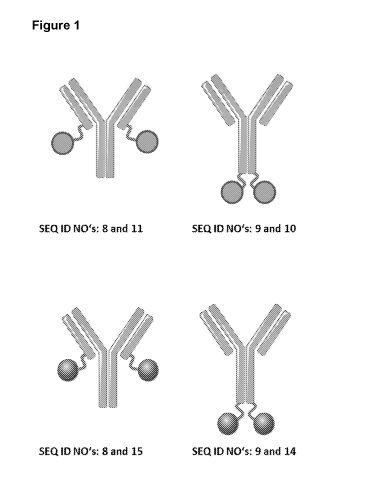
III. DESCRIPTIONS OF FIGURES
[0047] Figure 1: provides an overview of the design of the representative
fusion
polypeptides described in this application, which are bispecific with regard
to the targets
VEGF-A and Ang-2. Representative fusion polypeptides were made based on an
antibody specific for VEGF-A (SEQ ID NOs: 8 and 9) and a lipocalin mutein
specific for
Ang-2 (SEQ ID NO: 2 or SEQ ID NO: 3). Lipocalin muteins were fused to either
one of
the two C-termini of the antibody. The resulting fusion polypeptides have the
SEQ ID
NOs: 9 and 10, SEQ ID NOs: 8 and 11, SEQ ID NOs: 9 and 14, SEQ ID NOs: 8 and
15.
[0048] Figure 2: depicts the results of ELISA experiments in which the
affinity of
representative fusion polypeptides, the benchmark bispecific antibody (SEQ ID
NOs:
20, 21, 22 and 23) and positive control antibody (SEQ ID NOs: 8 and 9) against
VEGF-
16
CA 03004918 2018-05-10
WO 2017/091850 PCT/AU2016/051168
A was determined. Recombinant VEGF-A was coated on a microtiter plate, and the
tested agents were titrated starting from a concentration of 100 nM. Bound
agents
under study were detected via an anti-human IgG Fc antibody as described in
Example
2. The data was fit with a 1:1 binding model with EC50 value and the maximum
signal
as free parameters, and a slope that was fixed to unity.
[0049] Figure 3: shows the results of EL ISA experiments in which the
affinity of
representative fusion polypeptides (SEQ ID NOs: 9 and 10, SEQ ID NOs: 8 and
11,
SEQ ID NOs: 9 and 14, SEQ ID NOs: 8 and 15), benchmark bispecific antibody
(SEQ
ID NOs: 20, 21, 22 and 23) and the positive control lipocalin muteins against
Ang-2
(SEQ ID NOs: 2 and 3) was determined. Recombinant Ang-2 was coated on a
microtiter
plate, and the tested agents were titrated starting from a concentration of
100 nM.
Bound agents under study were detected via an anti-human-IgG-Fc antibody or
anti-
lipocalin antibody as described in Example 3. The data was fit with a 1:1
binding model
with EC50 value and the maximum signal as free parameters, and a slope that
was
fixed to unity.
[0050] Figure 4: illustrates the results of an ELISA experiment in which
the ability
of representative fusion polypeptides (SEQ ID NOs: 9 and 10, SEQ ID NOs: 8 and
11,
SEQ ID NOs: 9 and 14, SEQ ID NOs: 8 and 15), to simultaneously bind both
targets,
VEGF-A and Ang-2, was determined. (A) Recombinant VEGF-A was coated on a
microtiter plate, followed by a titration of the fusion polypeptides starting
from a
concentration of 100 nM. Subsequently, a constant concentration of
biotinylated human
Ang-2 was added, which was detected via extravidin as described in Example 4.
(B) An
alternative format was also used where Ang-2 was coated on a microtiter plate,
followed
by a titration of the fusion polypeptides starting from a concentration of 100
nM.
Subsequently, a constant concentration of biotinylated human VEGF-A was added.
[0051] Figure 5: demonstrates fusion polypeptides (SEQ ID NOs: 9 and 10,
SEQ
ID NOs: 8 and 11, SEQ ID NOs: 9 and 14, SEQ ID NOs: 8 and 15) and benchmark
control (SEQ ID NOs: 6 and 7) are capable of blocking the interaction between
human
Ang-2 and its receptor human Tie-2, over expressed on HEK cells. A constant
concentration of human Ang-2 was pre-incubated with variable concentrations of
fusion
17
CA 03004918 2018-05-10
WO 2017/091850 PCT/AU2016/051168
polypeptides (SEQ ID NOs: 9 and 10, SEQ ID NOs: 8 and 11, SEQ ID NOs: 9 and
14,
SEQ ID NOs: 8 and 15) or benchmark control (SEQ ID NOs: 6 and 7). Non-
neutralized
Ang-2 was detected via an anti-HIS-tag antibody. The data were fitted with a
single-site
binding model.
[0052] Figure 6: demonstrates that the fusion polypeptides (SEQ ID NOs: 9
and
10, SEQ ID NOs: 8 and 11, SEQ ID NOs: 9 and 14, SEQ ID NOs: 8 and 15) and
benchmark controls (SEQ ID NOs: 6 and 7; SEQ ID NOs: 8 and 9) are capable of
blocking the biological activity of VEGF-A and/or hAng-2 in a cell-based
proliferation
assay. In the assay, the fusion polypeptides, an IgG isotype negative control
and two
benchmark antibodies were added to VEGF-A supplemented Human Lymphatic
Endothelial Cells (LEC). The experiment shows that LEC proliferation is
blocked by the
fusion polypeptides (SEQ ID NOs: 9 and 10, SEQ ID NOs: 8 and 11, SEQ ID NOs: 9
and 14, SEQ ID NOs: 8 and 15) with IC50 values ranging from 1.2-0.5 nM. The
VEGF-A
benchmark antibody control (SEQ ID NOs: 8 and 9) inhibited proliferation with
an IC50
value of 1.2 nM. The lipocalin muteins (SEQ ID NO: 2 or SEQ ID NO: 3)
partially
inhibited cell proliferation with an IC50 of 1.9-1.7 nM while the Ang-2
benchmark
antibody (SEQ ID NOs: 6 and 7) had an IC50 of 4.2nM. The IgG isotype and SEQ
ID
NO: 1 negative controls had no effect on cell proliferation. Data were fitted
with a
sigmoidal dose-response model.
[0053] Figure 7: provides the result of a pharmacokinetic analysis of the
bispecific fusion polypeptides (SEQ ID NOs: 9 and 14, SEQ ID NOs: 8 and 15) in
rabbits. Female rabbits received test articles as an intravitreal injection in
the right eye
at a dose of 100 ug / eye. Drug levels were detected using a Sandwich ELISA
detecting
the full bispecific construct via the targets VEGF-A and Ang-2. The data were
fitted
using a non-compartmental model.
IV. DETAILED DESCRIPTION OF THE DISCLOSURE
[0054] In some embodiments, a fusion polypeptide of the disclosure contains
at
least two subunits in any order: a first subunit that comprises a full-length
immunoglobulin or an antigen-binding domain thereof specific for VEGF-A, and a
18
CA 03004918 2018-05-10
WO 2017/091850 PCT/AU2016/051168
second subunit that comprises a lipocalin mutein specific for Ang-2. The
subunits can
be linked via a covalent bond, e.g., a peptide bond.
[0055] In some embodiments, one subunit can be linked to another subunit as
essentially shown in Figure 1. For example, one lipocalin mutein can be
linked, via a
peptide bond, to the C-terminus of the immunoglobulin heavy chain, the N-
terminus of
the immunoglobulin heavy chain, the C-terminus of the of the immunoglobulin
light
chain, and/or the N-terminus of the immunoglobulin light chain. In some
particular
embodiments, a lipocalin mutein subunit can, therefore, be fused at its N-
terminus
and/or its C-terminus to an immunoglobulin subunit. In some still further
embodiments,
two subunits can be joined by a peptide linker. The linker can be of any make-
up and
size and will be apparent to the skilled worker. A preferred linker is a
(G4S)3 linker, for
example, as shown in SEQ ID NO: 19.
[0056] In some embodiments, the fusion polypeptide also may contain a third
or
additional subunit. For instance, the polypeptide may contain a third subunit
comprising
a lipocalin mutein specific for a target other than Ang-2 or VEGF-A, which
third subunit
may be attached at its N or C terminus to the C or N terminus, respectively,
of either the
first or second subunit.
[0057] In some embodiments, in a fusion polypeptide of the disclosure, a
VEGF-
A-specific subunit is fused to a Ang-2-specific subunit.
[0058] In some more specific embodiments, the VEGF-A specific subunit
comprises a full-length immunoglobulin (such as a monoclonal antibody) or an
antigen-
binding domain thereof and the Ang-2-specific subunit comprises a lipocalin
mutein. In
some embodiments, the fusion polypeptide comprises amino acid sequences
selected
from the group consisting of SEQ ID NOs: 9 and 10, SEQ ID NOs: 8 and 11, SEQ
ID
NOs: 9 and 12, SEQ ID NOs: 8 and 13, SEQ ID NOs: 9 and 14, SEQ ID NOs: 8 and
15,
SEQ ID NOs: 9 and 16, SEQ ID NOs: 8 and 17, SEQ ID NOs 9 and 24, SEQ ID NOs 8
and 25, SEQ ID NOs 9 and 26, SEQ ID NOs 8 and 27, SEQ ID NOs 9 and 28, SEQ ID
NOs 8 and 29, SEQ ID NOs 9 and 30, SEQ ID NOs 8 and 31, SEQ ID NOs 9 and 32,
SEQ ID NOs 8 and 33, SEQ ID NOs 9 and 34, SEQ ID NOs 8 and 35, SEQ ID NOs 9
19
CA 03004918 2018-05-10
WO 2017/091850 PCT/AU2016/051168
and 36, SEQ ID NOs 8 and 37, SEQ ID NOs 9 and 38, SEQ ID NOs 8 and 39, SEQ ID
NOs 9 and 40, SEQ ID NOs 8 and 41, SEQ ID NOs 9 and 42, SEQ ID NOs 8 and 43,
SEQ ID NOs 9 and 44, SEQ ID NOs 8 and 45, SEQ ID NOs 9 and 46, SEQ ID NOs 8
and 47, SEQ ID NOs 9 and 48, SEQ ID NOs 8 and 49, SEQ ID NOs 9 and 50, SEQ ID
NOs 8 and 51, SEQ ID NOs 9 and 52, SEQ ID NOs 8 and 53, SEQ ID NOs 9 and 54,
SEQ ID NOs 8 and 55, SEQ ID NOs 9 and 56, SEQ ID NOs 8 and 57, SEQ ID NOs 9
and 58, SEQ ID NOs 8 and 59, SEQ ID NOs 9 and 60, SEQ ID NOs 8 and 61, SEQ ID
NOs 9 and 62, SEQ ID NOs 8 and 63, SEQ ID NOs 9 and 64, SEQ ID NOs 8 and 65,
SEQ ID NOs 9 and 66, SEQ ID NOs 8 and 67, SEQ ID NOs 9 and 68, SEQ ID NOs 8
and 69, SEQ ID NOs 9 and 70 and SEQ ID NOs 8 and 71. In some embodiments, the
VEGF-A specific subunit comprises a full-length immunoglobulin (such as a
monoclonal
antibody) or an antigen-binding domain thereof wherein the monoclonal antibody
has
the heavy chain complementarity-determining regions (CDRs) and the and the
light
chain CDRs contained in an antibody selected from the group consisting of SEQ
ID
NOs: 9 and 10, SEQ ID NOs: 8 and 11, SEQ ID NOs: 9 and 12, SEQ ID NOs: 8 and
13,
SEQ ID NOs: 9 and 14, SEQ ID NOs: 8 and 15, SEQ ID NOs: 9 and 16, SEQ ID NOs:
8
and 17, SEQ ID NOs 9 and 24, SEQ ID NOs 8 and 25, SEQ ID NOs 9 and 26, SEQ ID
NOs 8 and 27, SEQ ID NOs 9 and 28, SEQ ID NOs 8 and 29, SEQ ID NOs 9 and 30,
SEQ ID NOs 8 and 31, SEQ ID NOs 9 and 32, SEQ ID NOs 8 and 33, SEQ ID NOs 9
and 34, SEQ ID NOs 8 and 35, SEQ ID NOs 9 and 36, SEQ ID NOs 8 and 37, SEQ ID
NOs 9 and 38, SEQ ID NOs 8 and 39, SEQ ID NOs 9 and 40, SEQ ID NOs 8 and 41,
SEQ ID NOs 9 and 42, SEQ ID NOs 8 and 43, SEQ ID NOs 9 and 44, SEQ ID NOs 8
and 45, SEQ ID NOs 9 and 46, SEQ ID NOs 8 and 47, SEQ ID NOs 9 and 48, SEQ ID
NOs 8 and 49, SEQ ID NOs 9 and 50, SEQ ID NOs 8 and 51, SEQ ID NOs 9 and 52,
SEQ ID NOs 8 and 53, SEQ ID NOs 9 and 54, SEQ ID NOs 8 and 55, SEQ ID NOs 9
and 56, SEQ ID NOs 8 and 57, SEQ ID NOs 9 and 58, SEQ ID NOs 8 and 59, SEQ ID
NOs 9 and 60, SEQ ID NOs 8 and 61, SEQ ID NOs 9 and 62, SEQ ID NOs 8 and 63,
SEQ ID NOs 9 and 64, SEQ ID NOs 8 and 65, SEQ ID NOs 9 and 66, SEQ ID NOs 8
and 67, SEQ ID NOs 9 and 68, SEQ ID NOs 8 and 69, SEQ ID NOs 9 and 70 and SEQ
ID NOs 8 and 71.
CA 03004918 2018-05-10
WO 2017/091850 PCT/AU2016/051168
[0059] In some embodiments, the second subunit is comprised of a lipocalin
mutein which comprises amino acid sequences selected from the group consisting
of
SEQ ID NOs: 2, 3, 86, 87, 88, 89, 90, 91, 92, 93, 94, 95, 96 and 97.
[0060] In some embodiments, the second subunit is comprised of a lipocalin
mutein which comprises nucleic acid sequences selected from the group
consisting of
SEQ ID NOs: 72-85.
[0061] In some embodiments, the Fc portion of the immunoglobulin included
in a
fusion polypeptide of the disclosure may contribute to maintaining the serum
levels of
the fusion polypeptide, critical for its stability and persistence in the
body. For example,
when the Fc portion binds to Fc receptors on endothelial cells and on
phagocytes, the
fusion polypeptide may become internalized and recycled back to the blood
stream,
enhancing its half-life.
[0062] In some embodiments, a fusion polypeptide of the disclosure may be
able
to bind VEGF-A with an EC50 value of about 1 nM or less, such as about 0.5 nM,
about
0.3 nM or about 0.15 nM, for example, when said affinity for VEGF-A is
measured in an
ELISA assay essentially as described in Example 2.
[0063] In some embodiments, a fusion polypeptide of the disclosure may be
able
to bind VEGF-A with an EC50 value, comparable to the EC50 value of the
immunoglobulin specific for VEGF-A as included in such fusion polypeptide,
such as the
antibody having the heavy and light chains provided by SEQ ID NOs: 8 and 9,
for
example, when said immunoglobulin and the fusion polypeptide are measured in
as
ELISA assay essentially as described in Example 2.
[0064] In some embodiments, a fusion polypeptide of the disclosure may be
able
to bind Ang-2 with an EC50 value of about 1 nM or less, such as about 0.5 nM,
about
0.25 nM or about 0.1 nM, for example, when said affinity for Ang-2 is measured
in an
ELISA assay essentially as described in Example 3. A fusion polypeptide of the
disclosure may be able to bind Ang-2 with an EC50 value comparable to the EC50
value of the lipocalin mutein specific for Ang-2 as included in such fusion
polypeptide,
such as lipocalin muteins of SEQ ID NOs: 2 and 3, for example, when said
lipocalin
21
CA 03004918 2018-05-10
WO 2017/091850 PCT/AU2016/051168
mutein and the fusion polypeptide are measured in as ELISA assay essentially
as
described in Example 3.
[0065] In some
embodiments, the fusion polypeptides of the disclosure specific
for both VEGF-A and Ang-2 may be capable of simultaneously binding VEGF-A and
Ang-2, for example, when said fusion polypeptide is measured in an ELISA assay
essentially described in Example 4.
[0066] In some
embodiments, the fusion polypeptides of the disclosure may be
able to block the binding of human Ang-2 to human Tie-2 expressing cells in a
competition cell electrochemoluminescence (ECL) assay format as essentially
described in Example 5.
[0067] In some
embodiments, the fusion polypeptide of the disclosure is able to
block VEGF-A dependent cell proliferation, in particular neutralize the
biological activity
of VEGF-A in a short-term proliferation assay using lymphatic microvascular
endothelial
cells (LEC) as essentially described in Example 6.
A. Exemplary immunoglobulins as included in the fusion polypeptides.
[0068] In some
embodiments, with respect to the fusion polypeptide, the first
subunit comprises a full-length immunoglobulin or an antigen-binding domain
thereof
specific for VEGF-A. The immunoglobulin, for example, may be IgG1, IgG2, IgG3
or
IgG4, and preferentially is IgG1. In further embodiments, the immunoglobulin
is a
monoclonal antibody against VEGF-A. A few illustrative examples for such
immunoglobulins include bevacizumab (trade name Avastin) and ranibizumab
(trade
name Lucentis), for example.
B. Exemplary lipocalin muteins as included in the fusion polypeptides.
[0069] As used
herein, a "lipocalin" is defined as a monomeric protein of
approximately 18-20 kDA in weight, having a cylindrical 13-pleated sheet
supersecondary structural region comprising a plurality of (preferably eight)
13 -strands
connected pair-wise by a plurality of (preferably four) loops at one end to
define thereby
a binding pocket. It is the diversity of the loops in the otherwise rigid
lipocalin scaffold
22
CA 03004918 2018-05-10
WO 2017/091850 PCT/AU2016/051168
that gives rise to a variety of different binding modes among the lipocalin
family
members, each capable of accommodating targets of different size, shape, and
chemical character (reviewed, e.g., in Flower, D.R. (1996), supra; Flower,
D.R. et al.
(2000), supra, or Skerra, A. (2000) Biochim. Biophys. Acta 1482, 337-350).
Indeed, the
lipocalin family of proteins have naturally evolved to bind a wide spectrum of
ligands,
sharing unusually low levels of overall sequence conservation (often with
sequence
identities of less than 20%) yet retaining a highly conserved overall folding
pattern. The
correspondence between positions in various lipocalins is well known to one of
skill in
the art. See, for example, U.S. Patent No. 7,250,297.
[0070] As noted above, a lipocalin is a polypeptide defined by its
supersecondary
structure, namely cylindrical p-pleated sheet supersecondary structural region
comprising eight p-strands connected pair-wise by four loops at one end to
define
thereby a binding pocket. The present disclosure is not limited to lipocalin
muteins
specifically disclosed herein. In this regard, the disclosure relates to a
lipocalin mutein
having a cylindrical p-pleated sheet supersecondary structural region
comprising eight
p-strands connected pair-wise by four loops at one end to define thereby a
binding
pocket, wherein at least one amino acid of each of at least three of said four
loops has
been mutated and wherein said lipocalin is effective to bind Ang-2 with
detectable
affinity.
[0071] In one particular embodiment, a lipocalin mutein disclosed herein is
a
mutein of human lipocalin 2. The term "human lipocalin 2" or "human Lcn 2" or
"human
NGAL" as used herein refers to the mature human neutrophil gelatinase-
associated
lipocalin (NGAL) with the SWISS-PROT/UniProt Data Bank Accession Number
P80188.
A human lipocalin 2 mutein of the disclosure may also be designated herein as
"an
hNGAL mutein". The amino acid sequence shown in SWISS-PROT/UniProt Data Bank
Accession Number P80188 may be used as a preferred "reference sequence", more
preferably the amino acid sequence shown in SEQ ID NO: 1 is used as reference
sequence.
[0072] In some embodiments, a lipocalin mutein comprised in the fusion
polypeptide of the disclosure binding Ang-2 with detectable affinity may
include at least
23
CA 03004918 2018-05-10
WO 2017/091850 PCT/AU2016/051168
one amino acid substitution of a native cysteine residue by another amino
acid, for
example, a serine residue. In some other embodiments, a lipocalin mutein
binding Ang-
2 with detectable affinity may include one or more non-native cysteine
residues
substituting one or more amino acids of a wild-type lipocalin. In a further
particular
embodiment, a lipocalin mutein according to the disclosure includes at least
two amino
acid substitutions of a native amino acid by a cysteine residue, hereby to
form one or
more cysteine bridges. In some embodiments, said cysteine bridge may connect
at
least two loop regions. The definition of these regions is used herein in
accordance with
Flower (Flower, 1996, supra, Flower, et al., 2000, supra) and Breustedt et al.
(2005,
supra).
[0073] Polypeptides of the disclosure, which are, in part, directed against
or
specific for Ang-2, include any number of specific-binding protein muteins
that are
based on a defined protein scaffold. Preferably, the number of nucleotides or
amino
acids, respectively, that is exchanged, deleted or inserted is 1, 2, 3, 4, 5,
6, 7, 8, 9, 10,
11, 12, 13, 14, 15, 16, 17, 18, 19, 20 or more such as 25, 30, 35, 40, 45 or
50, with 1, 2,
3, 4, 5,6, 7, 8, 9, 10, or 11 being preferred and 9, 10 or 11 being even more
preferred.
However, it is preferred that a lipocalin mutein of the disclosure is still
capable of binding
Ang-2.
[0074] In one aspect, the present fusion polypeptide of the disclosure
comprises
various lipocalin muteins that bind Ang-2 with at least detectable affinity.
In this sense,
Ang-2 can be regarded a non-natural ligand of the reference wild-type
lipocalin, where
"non-natural ligand" refers to a compound that does not bind to wild type
lipocalins
under physiological conditions. By engineering wild type lipocalins with one
or more
mutations at certain sequence positions, the present inventors have
demonstrated that
high affinity and high specificity for the non-natural ligand, Ang-2, is
possible. In some
embodiments, at 1, 2, 3, 4, 5, 6, 7, 8, 9, 10, 11, 12 or even more nucleotide
triplet(s)
encoding certain sequence positions on wild type lipocalins, a random
mutagenesis
may be carried out through substitution at these positions by a subset of
nucleotide
triplets.
[0075] Further, the lipocalin muteins comprised in the fusion polypeptide
of the
24
CA 03004918 2018-05-10
WO 2017/091850 PCT/AU2016/051168
disclosure may have a mutated amino acid residue at any one or more, including
at
least at any one, two, three, four, five, six, seven, eight, nine, ten, eleven
or twelve, of
the sequence positions corresponding to certain sequence positions of the
linear
polypeptide sequence of the reference lipocalin.
[0076] A fusion polypeptide of the disclosure may include the wild-type
(natural)
amino acid sequence of the "parental" protein scaffold (such as a lipocalin)
outside the
mutated amino acid sequence positions. In some embodiments, a lipocalin mutein
comprised in the fusion polypeptide of the disclosure may also carry one or
more amino
acid mutations at a sequence position/ positions as long as such a mutation
does, at
least essentially not hamper or not interfere with the binding activity and
the folding of
the mutein. Such mutations can be accomplished very easily on DNA level using
established standard methods (Sambrook, J. et al. (2001) Molecular Cloning: A
Laboratory Manual, 3rd Ed., Cold Spring Harbor Laboratory Press, Cold Spring
Harbor,
NY). Illustrative examples of alterations of the amino acid sequence are
insertions or
deletions as well as amino acid substitutions. Such substitutions may be
conservative,
i.e. an amino acid residue is replaced with an amino acid residue of
chemically similar
properties, in particular with regard to polarity as well as size. Examples of
conservative
substitutions are the replacements among the members of the following groups:
1)
alanine, serine, and threonine; 2) aspartic acid and glutamic acid; 3)
asparagine and
glutamine; 4) arginine and lysine; 5) isoleucine, leucine, methionine, and
valine; and 6)
phenylalanine, tyrosine, and tryptophan. On the other hand, it is also
possible to
introduce non-conservative alterations in the amino acid sequence. In
addition, instead
of replacing single amino acid residues, it is also possible to either insert
or delete one
or more continuous amino acids of the primary structure of the hNGAL as long
as these
deletions or insertion result in a stable folded/functional mutein (for
example, hNGAL
muteins with truncated N- and C-terminus). In such mutein, for instance, one
or more
amino acid residues are added or deleted at the N- or C- terminus of the
polypeptide.
Generally, such a mutein may have about at least 70%, including at least about
80%,
such as at least about 85% amino acid sequence identity, with the amino acid
sequence
of the mature hNGAL. As an illustrative example, the present disclosure also
encompasses NGAL muteins as defined above, in which amino acid residues (Lys-
Asp-
CA 03004918 2018-05-10
WO 2017/091850 PCT/AU2016/051168
Pro, positions 46-48) of the linear polypeptide sequence of the mature human
lipocalin 2
(hNGAL) have been deleted (SEQ ID NO: 1).
[0077] The amino acid sequence of a lipocalin mutein comprised in the
fusion
polypeptide disclosed herein has a high sequence identity to the reference
lipocalin
when compared to sequence identities with other lipocalins. In this general
context, the
amino acid sequence of a lipocalin mutein of the disclosure is at least
substantially
similar to the amino acid sequence of the reference lipocalin, with the
proviso that
possibly there are gaps (as defined below) in an alignment that are the result
of
additions or deletions of amino acids. A respective sequence of a lipocalin
mutein of the
disclosure, being substantially similar to the sequences of the reference
lipocalin, has,
in some embodiments, at least 70% identity or sequence homology, at least 75%
identity or sequence homology, at least 80% identity or sequence homology, at
least
82% identity or sequence homology, at least 85% identity or sequence homology,
at
least 87% identity or sequence homology, or at least 90% identity or sequence
homology including at least 95% identity or sequence homology, to the sequence
of the
reference lipocalin, with the proviso that the altered position or sequence is
retained and
that one or more gaps are possible.
[0078] As used herein, a lipocalin mutein comprised in the fusion
polypeptide of
the disclosure "specifically binds" a target (for example, Ang-2) if it is
able to
discriminate between that target and one or more reference targets, since
binding
specificity is not an absolute, but a relative property. "Specific binding"
can be
determined, for example, in accordance with Western blots, ELISA-, RIA-, ECL-,
IRMA-
tests, FACS, IHC and peptide scans.
[0079] In one embodiment, the lipocalin muteins of the disclosure are fused
at its
N-terminus and/or its C-terminus to a fusion partner which is a protein domain
that
extends the serum half-life of the mutein. In further particular embodiments,
the protein
domain is a Fc part of an immunoglobulin, a CH3 domain of an immunoglobulin, a
CH4
domain of an immunoglobulin, an albumin binding peptide, or an albumin binding
protein.
26
CA 03004918 2018-05-10
WO 2017/091850 PCT/AU2016/051168
[0080] In another embodiment, the lipocalin muteins of the disclosure are
conjugated to a compound that extends the serum half-life of the mutein. More
preferably, the mutein is conjugated to a compound selected from the group
consisting
of a polyalkylene glycol molecule, a hydroethylstarch, an Fc part of an
immunoglobulin,
a CH3 domain of an immunoglobulin, a CH4 domain of an immunoglobulin, an
albumin
binding peptide, and an albumin binding protein.
[0081] In yet another embodiment, the current disclosure relates to a
nucleic acid
molecule comprising a nucleotide sequence encoding a fusion polypeptide
comprising a
lipocalin mutein disclosed herein. The disclosure encompasses a host cell
containing
said nucleic acid molecule.
[0082] In one aspect, the present disclosure provides a fusion polypeptide
comprising a lipocalin mutein that binds Ang-2 and useful applications
therefor. The
disclosure also provides methods of making such fusion polypeptide comprising
an
Ang-2 binding subunit described herein as well as compositions comprising such
a
fusion polypeptide. The Ang-2 binding subunit of the disclosure as well as
compositions
thereof may be used in methods of detecting Ang-2 in a sample or in methods of
binding of Ang-2 in a subject. No such fusion polypeptide comprising such
human
lipocalin muteins having these features attendant to the uses provided by
present
disclosure have been previously described.
1. Exemplary lipocalins muteins specific for Ang-2
[0083] In one aspect, the present disclosure provides a fusion polypeptide
comprising an Ang-2 binding human lipocalin 2 (human Lcn2 or hNGAL) muteins.
[0084] One embodiment of the current disclosure relates to a fusion
polypeptide
comprising a mutein that is capable of binding Ang-2 with detectable affinity,
such as an
affinity measured by a Kd of about 200 nM or lower, such as about 150 nM or
lower.
[0085] In one aspect, the current disclosure provides a fusion polypeptide
comprising an hNGAL mutein that is capable of binding Ang-2 with a Kd of about
5 nM
or lower, for example when measured by Biacore T200 instrument in a Surface
27
CA 03004918 2018-05-10
WO 2017/091850 PCT/AU2016/051168
Plasmon Resonance (SPR).
[0086] In some further embodiments, one or more hNGAL muteins comprised in
the fusion polypeptide of the disclosure are capable of binding Ang-2 with an
affinity
measured by an EC50 value of about 5 nM or lower, when measured in an ELISA
assay.
[0087] In some other embodiments, one or more hNGAL muteins comprised in
the fusion polypeptide of the disclosure are capable of binding Ang-2 with an
affinity
measured by an IC50 value of about 5 nM or lower, when measured in a
competition
ELISA format assay.
[0088] In some other embodiments, one or more hNGAL muteins comprised in
the fusion polypeptide of the disclosure are capable of inhibiting or reducing
lymphatic
microvascular endothelial cells proliferation mediated by Ang-2 with an IC50
value of
about 5 nM or lower in a cell-based proliferation assay.
[0089] In some other embodiments, one or more hNGAL muteins comprised in
the fusion polypeptide of the disclosure are cross-reactive with both human
Ang-2 and
mouse Ang-2. In some embodiments, one or more such muteins are capable binding
both human Ang-2 and mouse Ang-2 with detectable affinity, such as an affinity
measured by a Kd of about 200 nM or lower, such as about 150 nM or lower.
[0090] In some still further embodiments, one or more such muteins
comprised in
the fusion polypeptide of the disclosure are capable of binding mouse Ang-2
with an
affinity measured by an IC50 value of about 5 nM or lower, when measured in an
ELISA
assay.
[0091] In some still further embodiments, one or more such muteins
comprised in
the fusion polypeptide of the disclosure are capable of blocking binding of
human Ang-2
to hTie-2 and mouse Ang-2 to hTie-2 with an IC50 value of about 25 nM or
lower,
respectively, in a competition cell ECL format.
[0092] In some embodiments, one or more hNGAL muteins comprised in the
fusion polypeptide of the disclosure are not cross-reactive with human Ang-4.
In some
28
CA 03004918 2018-05-10
WO 2017/091850 PCT/AU2016/051168
embodiments, one or more hNGAL muteins comprised in the fusion polypeptide of
the
disclosure are not cross-reactive with mouse Ang-3. In some embodiments, one
or more
hNGAL muteins comprised in the fusion polypeptide of the disclosure are not
cross-
reactive with human VEGF-A.
[0093] In this
regard, the disclosure relates to a fusion polypeptide, wherein said
fusion polypeptide includes an hNGAL mutein, and said hNGAL in comparison with
the
linear polypeptide sequence of the mature hNGAL, comprises at least 1, 2, 3,
4, 5, 6, 7,
8,9, 10, 11, 12, 13, 14, 15, 16, 17, 18, 19, 20, 21, or even more, mutated
amino acid
residues at the sequence positions 28, 36, 40, 41, 49, 52, 65, 68, 70, 72-74,
77, 79, 81,
87, 96, 100, 103, 106, 116, 125, 126, 127, 129, 132 and 134, and wherein said
polypeptide binds Ang-2 with detectable affinity.
[0094] In some
embodiments, an Ang-2-binding hNGAL mutein comprised in the
fusion polypeptide of the disclosure includes, at any one or more of the
sequence
positions 36, 40, 41, 49, 52, 68, 70, 72-73, 77, 79, 81, 96, 100, 103, 106,
125, 127, 132
and 134 of the linear polypeptide sequence of the mature hNGAL (SEQ ID NO: 1),
one
or more of the following mutated amino acid residues: Leu 36 Gin, Glu,
His, Val, Met
or Phe; Ala 40 Val, Tyr,
His or Trp; Ile 41 His, Tyr, Trp or Val; Gin 49 -4 Gly, Ile, Val,
Glu or Val; Tyr 52 Trp, His,
Thr or Ser; Ser 68 Gly, Asp, Gin, Glu or Ile; Leu 70 -4
Ser, Thr, Gly, Arg, Tyr or Ala; Arg 72 Gly, Ala,
Trp, Thr or Glu; Lys 73 Pro, Phe,
Leu, Arg, Ala or Gin; Asp 77 -4 Asn, Lys, Ser or Val; Trp 79 Thr, Arg, Ser or
Asn; Arg
81 -* Trp, His or Tyr; Asn 96 -> Gly, Ala, Pro, Gin or Asp; Tyr 100 -4 Pro,
Trp, Gly, Ser,
Leu or Asp; Leu 103 Gly, Glu, Asp, Met or Gin; Tyr 106 Thr, Leu
or Phe; Lys 125
His, Thr or Gly; Ser 127 Leu or
Met; Tyr 132 -4 Phe, Trp or Val; and Lys 134 -4
Ala, Glu or Trp. In some embodiments, an hNGAL mutein of the disclosure
includes two
or more, such as 3, 4, 5, 6, 7, 8, 9, 10, 11, 12, 13, 14, 15, 16, or even more
or all mutated
amino acid residues at these sequence positions of the mature hNGAL.
[0095]
Additionally, an Ang-2-binding hNGAL mutein comprised in the fusion
polypeptide of the disclosure may also comprise the following substitution in
comparison
with the linear polypeptide sequence of the mature hNGAL: Gin 28 -+ His; Asn
65
Asp; Lys 74 Glu; Cys 87 Ser; Asn 116 Asp; Val 126 -+ Met and Asn 129 Asp.
29
CA 03004918 2018-05-10
WO 2017/091850 PCT/AU2016/051168
[0096] In some
additional embodiments, an hNGAL mutein comprised in the
fusion polypeptide of the disclosure which binds to Ang-2 includes the
following amino
acid replacements in comparison with the linear polypeptide sequence of the
mature
hNGAL:
(a) Leu 36 -) Gin; Ala 40 -)Tyr; Gln 49 -) Gly; Tyr 52 -> Trp; Ser 68 --, Gly;
Leu 70
Ser; Arg 72 -4 Gly; Lys 73 Pro; Asp 77-4 Asn; Trp 79 -4 Thr; Arg 81 Trp;
Asn 96 Gly; Tyr
100 -> Pro; Leu 103 --, Gly; Tyr 106 -> Thr, Lys 125 -4 His;
Ser 127 --, Leu; Tyr 132 -> Phe; Lys 134 Glu;
(b) Leu 36 Phe; Ala
40 -)His, Ile 41 --, Arg; Gln 49 Gly; Tyr 52 --, His; Ser 68
--, Asp; Leu 70 -4 Thr; Arg 72 --, Ala; Lys 73 --, Phe; Asp 77-, Asn; Trp 79 -
4
Arg; Arg 81 -4 His; Tyr 100 -4 Trp; Leu 103 --) Glu; Tyr 106 --, Thr; Lys 125 -
4
Thr; Ser 127 --, Met; Tyr 132 -, Trp; Lys 134 --, Trp;
(c) Leu 36 -> Val; Ala 40 Trp; Ile
41 -, Tyr, Gln 49 --, Ile; Tyr 52 --) Thr; Ser 68 -4
Gin; Leu 70 Gly; Arg
72 -) Glu; Lys 73 ---) Gin; Asp 77 --) Lys; Trp 79 --, Ser,
Arg 81 His; Tyr
100 -4 Trp; Leu 103 --, Asp; Tyr 106 --) Leu; Lys 125 --) Gly;
Ser 127 --, Met; Tyr 132 Val; Lys 134 -3 Ala;
(d) Leu 36 Glu, Ala
40 -Val; Ile 41 -) Glu; Gln 49 --) Val; Tyr 52 Thr Ser 68 --)
Glu; Leu 70 --) Arg; Arg 72 --, Trp; Lys 73 --) Leu; Asp 77 --, Lys; Trp 79 -4
Asn;
Arg 81 -4 His; Asn 96 -) Ala; Tyr 100 -, Gly; Leu 103 --, Met; Tyr 106 -, Thr;
Lys
125 Thr; Ser 127 --, Met; Tyr 132 --) Trp; Lys 134 --, Trp;
(e) Leu 36 --, Gln; Ala 40 -) Tyr; Ile 41 --, Trp; Gln 49 -4 Ile; Tyr 52 ->
Ser, Ser 68 -)
Ile; Leu 70 --, Tyr; Arg 72 Thr; Lys
73 -4 Arg; Asp 77 --, Ser; Trp 79 Arg; Arg
81 --, Tyr; Asn 96 --) Pro; Leu 103 --) Asp; Tyr 106 -) Thr; Lys 125 --, His;
Ser
127 -, Tyr; Tyr 132 --, Trp; Lys 134-' Glu;
(f) Leu 36 --, Gln; Ala 40 ---, Tyr; Gln 49 -4 Glu; Tyr 52 Trp; Asn
65 -4 Asp; Ser 68
-4 Gly; Leu 70 --, Ser; Arg 72 -4 Gly; Lys 73 --, Pro; Asp 77 -4 Asn; Trp 79 --
)
Arg; Arg 81 --) Trp; Asn 96 --) Gly; Tyr 100 -) Ser; Leu 103 --, Gin; Tyr 106 -
-)
Thr; Lys 125 His; Ser 127 -, Leu; Tyr 132 --, Phe; Lys 134 Glu;
CA 03004918 2018-05-10
WO 2017/091850 PCT/AU2016/051168
(g) Leu 36 --o His; Ala 40 -*Tyr; Gin 49 --o Glu; Tyr 52 -o Trp; Asn 65 --4
Asp; Ser 68
Glu; Leu 70 ---o Ser; Arg 72 ---o Gly; Lys 73 --o Pro; Asp 77 -o Asn; Trp 79 --
--o
Arg; Arg 81 --0 Trp; Asn 96 -4 Gly; Tyr 100 -4 Pro; Leu 103 Asp; Tyr
106 -4
Thr; Lys 125 His; Ser 127 --o Leu; Tyr 132 --o Phe; Lys 134 -4 Glu;
(h) Leu 36 ---o Gin; Ala 40 --oTyr, Gin 49 --+ Gly; Tyr 52 Trp; Asn 65
-*Asp; Ser 68
--+ Glu; Leu 70 -4 Ser; Arg 72 --o Gly; Lys 73 Ala; Asp 77
--4 Asn; Trp 79 -3
Arg; Arg 81 --o Trp; Asn 96 --o Gly; Tyr 100 -o Asp; Leu 103 -4 Gly; Tyr 106
Thr; Lys 125 His; Ser 127 --4 Leu; Tyr 132 Phe; Lys 134 -4 Glu;
(i) Leu 36 -4 His; Ala 40 -Tyr; Gin 49 --4 Gly; Tyr 52 -4 Trp; Asn 65 -4 Asp;
Ser 68
--o Glu; Leu 70 -+ Ser; Arg 72 -4 Gly; Lys 73 -o Pro; Asp 77 -4 Asn; Trp 79
Arg; Arg 81 Trp; Asn 96 --o Gly; Tyr 100 --o Pro; Leu 103 --o Gly; Tyr 106
Thr; Lys 125 --o His; Ser 127 ---o Leu; Tyr 132 -> Phe; Lys 134 -4 Glu;
(j) Leu 36 -3 Gin; Ala 40 -4Tyr; Gin 49 --o Gly; Tyr 52 -4 Trp; Asn 65 --o
Asp; Ser 68
Gly; Leu 70 -+ Ser; Arg 72 --o Gly; Lys 73 --o Ala; Asp 77 -o Val; Trp 79 -4
Arg;
Arg 81 -0 Trp; Asn 96 --o Gly; Tyr 100 --o Pro; Leu 103 -4 Gly; Tyr 106 -o
Thr; Lys
125 -o His; Ser 127 --o Leu; Tyr 132 --o Phe; Lys 134 --o Glu;
(k) Leu 36 --o Gin; Ala 40 -Tyr; Gin 49 --o Val; Tyr 52 --o Trp; Asn 65 --o
Asp; Ser 68
-o Glu; Leu 70 --o Ser; Arg 72 --o Gly; Lys 73 --o Pro; Asp 77 --o Asn; Trp 79
Arg; Arg 81 -o Trp; Asn 96 -o Gly; Tyr 100 --o Leu; Leu 103 --o Gly; Tyr 106 --
o
Thr; Lys 125 --o His; Ser 127 --o Leu; Tyr 132 ---o Phe; Lys 134 --o Glu;
(I) Leu 36 -o Val; Ala 40 --oTyr; Ile 41 -4 Tyr; Gin 49 --o Ile; Tyr 52 --o
Thr; Asn 65 --o
Asp; Ser 68 --o Gin; Leu 70 --o Gly; Arg 72 -> Glu; Lys 73 --o Gin; Lys 74 --o
Glu;
Asp 77 -3 Lys; Trp 79 --o Ser; Arg 81 --4 His; Tyr 100 --o Trp; Leu 103 -o
Asp; Tyr
106 -o Pro; Asn 116 -4 Asp; Lys 125 -o Gly; Ser 127 --o Met; Asn 129 --o Asp;
Tyr
132 --o Val; Lys 134 --o Ala;
(m) Leu 36 --o Val; Ala 40 -Tyr; Ile 41 -o Tyr; Gin 49 --o Ile; Tyr 52 --o
Thr; Asn 65 --o
Asp; Ser 68 --4 Gin; Leu 70 -4 Gly; Arg 72 --4 Glu; Lys 73 -4 Gin; Lys 74 --o
Glu;
Asp 77 --0 Lys; Trp 79 --+ Ser; Arg 81 --o His; Asn 96 --o Asp; Tyr 100 --o
Trp; Leu
31
CA 03004918 2018-05-10
WO 2017/091850 PCT/AU2016/051168
103 Asp; Tyr 106 Pro; Lys
125 -> Gly; Val 126 -> Met; Ser 127 -> Met; Asn
129 -Asp; Tyr 132 --4 Val; Lys 134 -'Ala; or
(n) Leu 36 Met; Ala 40 -*Tyr, Ile 41 Asp; Gln 49 -4 Ile; Tyr 52 -> Thr;
Asn 65
Asp; Ser 68 -> Gln; Leu 70 -4 Gly; Arg 72 Glu; Lys 73 Gln; Asp
77 Lys;
Trp 79 -+ Ser; Arg 81 His; Asn 96 Gln; Tyr
100 Trp; Leu 103 Asp; Tyr
106-. Pro; Lys 125 Gly; Ser 127 Met; Tyr 132 Val; Lys 134
Ala.
Each of these sets of amino acid replacements may further include the
replacements
Gln 28 -4 His and Cys 87 -> Ser.
[0097] In the
residual region, i.e. the region differing from sequence positions 28,
36, 40, 41, 49, 52, 65, 68, 70, 72-74, 77, 79, 81, 87, 96, 100, 103, 106, 116,
125, 126,
127, 129, 132 and 134, an hNGAL mutein comprised in the fusion polypeptide of
the
disclosure may include the wild type (natural) amino acid sequence outside the
mutated
amino acid sequence positions.
[0098] In
further particular embodiments, a mutein comprised in the fusion
polypeptide of the disclosure according to the current disclosure comprises an
amino
acid sequence selected from the group consisting of SEQ ID NOs: 2, 3, 86, 87,
88, 89,
90, 91, 92, 93, 94, 95, 96 or 97 or a functional fragment or variant thereof.
In some
embodiments, such fragment or variant is a structural homologue of a mutein
defined in
any one of SEQ ID NOs: 2, 3, 86, 87, 88, 89, 90, 91, 92, 93, 94, 95, 96 or 97.
[0099] The amino
acid sequence of an Ang-2-binding hNGAL mutein comprised
in the fusion polypeptide of the disclosure may have a high sequence identity,
such as
at least 70%, at least 75%, at least 80%, at least 82%, at least 85%, at least
87%, at
least 90% identity, including at least 95% identity, to a sequence selected
from the
group consisting of SEQ ID NOs: 2, 3, 86, 87, 88, 89, 90, 91, 92, 93, 94, 95,
96 or 97.
[00100] In some
still embodiments, an hNGAL mutein cross-reactive with human
Ang-2 and/or mouse Ang-2 according to the disclosure comprises an amino acid
sequence selected from the group consisting of SEQ ID NOs: 2, 3, 86, 87, 88,
89, 90,
91, 92, 93, 94, 95, 96 or 97 and functional fragments or variants thereof.
32
CA 03004918 2018-05-10
WO 2017/091850 PCT/AU2016/051168
[001011 The disclosure also includes structural homologues of an hNGAL
mutein
comprised in the fusion polypeptide of the disclosure having an amino acid
sequence
selected from the group consisting of SEQ ID NOs: 2, 3, 86, 87, 88, 89, 90,
91, 92, 93,
94, 95, 96 or 97, which structural homologues have an amino acid sequence
homology
or sequence identity of more than about 60%, preferably more than 65%, more
than
70%, more than 75%, more than 80%, more than 85%, more than 90%, more than 92%
and most preferably more than 95% in relation to said hNGAL mutein.
[00102] An Ang-2-binding hNGAL mutein comprised in the fusion polypeptide
of
the disclosure can be obtained by means of mutagenesis of a naturally
occurring form
of human lipocalin 2 (or hNGAL). In some embodiments of the mutagenesis, a
substitution (or replacement) is a conservative substitution. Nevertheless,
any
substitution - including non-conservative substitution or one or more from the
exemplary
substitutions below - is envisaged as long as the mutein retains its
capability to bind to
Ang-2, and/or it has an identity to the then substituted sequence in that it
is at least
60%, such as at least 65%, at least 70%, at least 75%, at least 80%, at least
85% or
higher identity to the amino acid sequence of the mature human lipocalin 2
(SWISS-
PROT Data Bank Accession Number P80188).
[00103] The present disclosure also relates to a pharmaceutical composition
that
includes at least one fusion polypeptide of the disclosure comprising an Ang-2-
binding
hNGAL mutein, or conjugate or fusion protein thereof as described herein, and
optionally, a pharmaceutically acceptable excipient.
[00104] Accordingly, the Ang-2-binding hNGAL muteins comprised in the
fusion
polypeptide of the disclosure can be formulated into compositions using
pharmaceutically acceptable ingredients as well as established methods of
preparation
(Gennaro and Gennaro (2000) Remington: The Science and Practice of Pharmacy,
20th
Ed., Lippincott Williams & Wilkins, Philadelphia, PA). To prepare the
pharmaceutical
compositions, pharmaceutically inert inorganic or organic excipients can be
used.
C. Muteins comprised in the fusion polypeptide of the disclosure.
[00105] When used herein in the context of the fusion polypeptide of the
present
33
CA 03004918 2018-05-10
WO 2017/091850 PCT/AU2016/051168
disclosure, the term "specific for", in reference to a mutein that binds to
Ang-2, includes
that the mutein is directed against, binds to, or reacts with Ang-2. Thus,
being directed
to, binding to or reacting with includes that the mutein specifically binds to
Ang-2. The
term "specifically" in this context means that the mutein engages Ang-2, as
described
herein, but essentially not with another target. Whether the mutein
specifically binds or
engages a target as defined above can easily be tested, inter alia, by
comparing the
reaction of a hNGAL mutein comprised in the fusion polypeptide of the present
disclosure with Ang-2 and the reaction of said mutein with (an) other
target(s). "Specific
binding" can also be determined, for example, in accordance with Western
blots, ELISA-
, RIA-, ECL-, IRMA-tests, FACS, IHC and peptide scans.
[00106] The amino acid sequence of a mutein comprised in the fusion
polypeptide
of the disclosure has a high sequence identity to human lipocalin 2 when
compared to
sequence identities with another lipocalin (see also above). In this general
context the
amino acid sequence of a mutein of the combination according to the disclosure
is at
least substantially similar to the amino acid sequence of the corresponding
lipocalin (the
wild-type hNGAL). A respective sequence of a mutein of the combination
according to
the disclosure, being substantially similar to the sequence of mature hNGAL,
such as at
least 65%, at least 70%, at least 75%, at least 80%, at least 82%, at least
85%, at least
87%, at least 90% identity, including at least 95% identity to the sequence of
mature
hNGAL. In this regard, a mutein of the disclosure of course may contain, in
comparison
substitutions as described herein which renders the mutein capable of binding
to Ang-2.
Typically, a mutein of hNGAL includes one or more mutations ¨ relative to the
native
sequence of hNGAL ¨ of amino acids in the four loops at the open end of the
ligand
binding site of hNGAL. As explained above, these regions are essential in
determining
the binding specificity of a mutein for Ang-2. A mutein derived hNGAL or a
homologue
thereof, may have one, two, three, four or more mutated amino acid residues at
any
sequence position in the N-terminal region and/or in the three peptide loops
BC, DE,
and FG arranged at the end of the 13-barrel structure that is located opposite
to the
natural binding pocket.
[00107] A mutein comprised in the fusion polypeptide of the disclosure
includes
34
CA 03004918 2018-05-10
WO 2017/091850 PCT/AU2016/051168
one or more, such as two, three, four, five, six, seven, eight, nine, ten,
eleven, twelve,
thirteen, fourteen, fifteen, sixteen, seventeen, eighteen, nineteen or even
twenty
substitutions in comparison to the corresponding native hNGAL lipocalin,
provided that
such a mutein should be capable of binding to Ang-2. For example, a mutein can
have a
substitution at a position corresponding to a distinct position (i.e. at a
corresponding
position) of hNGAL. In some embodiments a mutein of the combination according
to the
disclosure includes at least two amino acid substitutions, including 2, 3, 4,
5, or even
more, amino acid substitutions of a native amino acid by an arginine residue.
Accordingly, the nucleic acid of a protein 'reference' scaffold as described
herein is
subject to mutagenesis with the aim of generating a mutein which is capable of
binding
to Ang-2.
[00108] Also, a mutein comprised in the fusion polypeptide of the present
disclosure can comprise a heterologous amino acid sequence at its N-or C-
Terminus,
preferably C-terminus, such as a Strep-tag, e.g., Strep II tag without
affecting the
biological activity (binding to its target e.g. Ang-2) of the mutein.
[00109] Specifically, in order to determine whether an amino acid residue
of the
amino acid sequence of a mutein comprised in the fusion polypeptide of the
disclosure
different from wild-type hNGAL corresponds to a certain position in the amino
acid
sequence of wild-type hNGAL, a skilled artisan can use means and methods well-
known in the art, e.g., alignments, either manually or by using computer
programs such
as BLAST2.0, which stands for Basic Local Alignment Search Tool or ClustalW or
any
other suitable program which is suitable to generate sequence alignments.
Accordingly,
wild-type hNGAL can serve as "subject sequence" or "reference sequence", while
the
amino acid sequence of a mutein different from the wild-type hNGAL described
herein
serves as "query sequence". The terms "reference sequence" and "wild type
sequence"
are used interchangeably herein.
[00110] In some embodiments a substitution (or replacement) is a
conservative
substitution. Nevertheless, any substitution - including non-conservative
substitution or
one or more from the exemplary substitutions listed below - is envisaged as
long as the
mutein comprised in the fusion polypeptide of the disclosure retains its
capability to bind
CA 03004918 2018-05-10
WO 2017/091850
PCT/AU2016/051168
Ang-2, and/or it has an identity to the then substituted sequence in that it
is at least
60%, such as at least 65%, at least 70%, at least 75%, at least 80%, at least
85 % or
higher identical to the "original" sequence.
[00111]
Conservative substitutions are generally the following substitutions, listed
according to the amino acid to be mutated, each followed by one or more
replacement(s) that can be taken to be conservative: Ala ¨> Gly, Ser, Val; Arg
¨> Lys;
Asn Gln, His; Asp Glu; Cys Ser; Gln Asn; Glu -
4 Asp; Gly -+ Ala; His Arg,
Asn, Gln; Ile Leu, Val; Leu Ile, Val;
Lys ¨> Arg, Gin, Glu; Met Leu, Tyr, Ile; Phe
-3 Met, Leu, Tyr; Ser Thr; Thr Ser; Trp
¨> Tyr; Tyr ¨> Tip, Phe; Val Ile, Leu.
Other substitutions are also permissible and can be determined empirically or
in accord
with other known conservative or non-conservative substitutions. As a further
orientation, the following eight groups each contain amino acids that can
typically be
taken to define conservative substitutions for one another:
a. Alanine (Ala), Glycine (Gly);
b. Aspartic acid (Asp), Glutamic acid (Glu);
c. Asparagine (Asn), Glutamine (Gin);
d. Arginine (Arg), Lysine (Lys);
e. lsoleucine (Ile), Leucine (Leu), Methionine (Met), Valine (Val);
f. Phenylalanine (Phe), Tyrosine (Tyr), Tryptophan (Tip);
9. Serine (Ser), Threonine (Thr); and
h. Cysteine (Cys), Methionine (Met)
[00112] If such
substitutions result in a change in biological activity, then more
substantial changes, such as the following, or as further described below in
reference to
amino acid classes, may be introduced and the products screened for a desired
characteristic. Examples of such more substantial changes are: Ala ¨> Leu,
Ile; Arg
Gin; Asn Asp, Lys, Arg, His; Asp Asn; Cys Ala; Gin Glu; Glu
Gin; His
Lys; Ile ¨ Met, Ala, Phe; Leu Ala, Met,
Norleucine; Lys ¨> Asn; Met ¨> Phe; Phe
36
CA 03004918 2018-05-10
WO 2017/091850 PCT/AU2016/051168
Val, Ile, Ala; Tip Phe; Tyr -4 Thr, Ser; Val ¨> Met, Phe, Ala.
[00113] Substantial modifications in the biological properties of hNGAL are
accomplished by selecting substitutions that differ significantly in their
effect on
maintaining (a) the structure of the polypeptide backbone in the area of the
substitution,
for example, as a sheet or helical conformation, (b) the charge or
hydrophobicity of the
molecule at the target site, or (c) the bulk of the side chain. Naturally
occurring residues
are divided into groups based on common side-chain properties: (1)
hydrophobic:
norleucine, methionine, alanine, valine, leucine, iso-leucine; (2) neutral
hydrophilic:
cysteine, serine, threonine; (3) acidic: asparitic acid, glutamic acid; (4)
basic:
asparagine, glutamine, histidine, lysine, arginine; (5) residues that
influence chain
orientation: glycine, proline; and (6) aromatic: tryptophan, tyrosine,
phenylalanine.
[00114] Non-conservative substitutions will entail exchanging a member of
one of
these classes for another class. Any cysteine residue not involved in
maintaining the
proper conformation of hNGAL also may be substituted, generally with serine,
to
improve the oxidative stability of the molecule and prevent aberrant
crosslinking.
Conversely, cysteine bond (s) may be added to improve its stability.
[00115] Any mutation, including an insertion as discussed above, can be
accomplished very easily on the nucleic acid, e.g. DNA level using established
standard
methods. Illustrative examples of alterations of the amino acid sequence are
insertions
or deletions as well as amino acid substitutions. Such substitutions may be
conservative, i.e. an amino acid residue is replaced with an amino acid
residue of
chemically similar properties, in particular with regard to polarity as well
as size.
Examples of conservative substitutions are the replacements among the members
of
the following groups: 1) alanine, serine, and threonine; 2) aspartic acid and
glutamic
acid; 3) asparagine and glutamine; 4) arginine and lysine; 5) iso-leucine,
leucine,
methionine, and valine; and 6) phenylalanine, tyrosine, and tryptophan. On the
other
hand, it is also possible to introduce non-conservative alterations in the
amino acid
sequence. In addition, instead of replacing single amino acid residues, it is
also possible
to either insert or delete one or more continuous amino acids of the primary
structure of
hNGAL as long as these deletions or insertion result in a stable
folded/functional
37
CA 03004918 2018-05-10
WO 2017/091850 PCT/AU2016/051168
mutein.
[00116] Modifications of the amino acid sequence include directed
mutagenesis of
single amino acid positions in order to simplify sub-cloning of the mutated
hNGAL gene
or its parts by incorporating cleavage sites for certain restriction enzymes.
In addition,
these mutations can also be incorporated to further improve the affinity of a
mutein for a
given target such as Ang-2. Furthermore, mutations can be introduced in order
to
modulate certain characteristics of the mutein such as to improve folding
stability, serum
stability, protein resistance or water solubility or to reduce aggregation
tendency, if
necessary. For example, naturally occurring cysteine residues may be mutated
to other
amino acids to prevent disulphide bridge formation. It is also possible to
deliberately
mutate other amino acid sequence position to cysteine in order to introduce
new
reactive groups, for example for the conjugation to other compounds, such as
polyethylene glycol (PEG), hydroxyethyl starch (HES), biotin, peptides or
proteins, or for
the formation of non-naturally occurring disulphide linkages. The generated
thiol moiety
may be used to PEGylate or HESylate the mutein, for example, in order to
increase the
serum half-life of a respective mutein.
[00117] It is also possible to mutate other amino acid sequence positions
to
cysteine in order to introduce new reactive groups, for example, for the
conjugation to
other compounds, such as polyethylene glycol (PEG), hydroxyethyl starch (HES),
biotin,
peptides or proteins, or for the formation of non-naturally occurring
disulphide linkages.
[00118] In some embodiments, if one of the above moieties is conjugated to
a
mutein comprised in the fusion polypeptide of the disclosure, conjugation to
an amino
acid side chain can be advantageous. Suitable amino acid side chains may occur
naturally in the amino acid sequence of hNGAL or may be introduced by
mutagenesis.
In case a suitable binding site is introduced via mutagenesis, one possibility
is the
replacement of an amino acid at the appropriate position by a cysteine
residue.
[00119] With respect to a mutein of human lipocalin 2 comprised in the
fusion
polypeptide of the disclosure, exemplary possibilities of such a mutation to
introduce a
cysteine residue into the amino acid sequence of a lipocalin including human
lipocalin 2
38
CA 03004918 2018-05-10
WO 2017/091850 PCT/AU2016/051168
mutein to include the introduction of a cysteine (Cys) residue at at least at
one of the
sequence positions that correspond to sequence positions 14, 21, 60, 84, 88,
116, 141,
145, 143, 146 or 158 of the wild type sequence of human NGAL. In some
embodiments
where a human lipocalin 2 mutein of the disclosure has a sequence in which, in
comparison to the sequence of the SWISS-PROT/UniProt Data Bank Accession
Number P80188, a cysteine has been replaced by another amino acid residue, the
corresponding cysteine may be reintroduced into the sequence. As an
illustrative
example, a cysteine residue at amino acid position 87 may be introduced in
such a case
by reverting to a cysteine as originally present in the sequence of SWISS-PROT
accession No. P80188. The generated thiol moiety at the side of any of the
amino acid
positions 14, 21, 60, 84, 88, 116, 141, 145, 143, 146 and/or 158 may be used
to
PEGylate or HESylate the mutein, for example, in order to increase the serum
half-life
of a respective human lipocalin 2 mutein.
[00120] In another embodiment, in order to provide suitable amino acid side
chains for conjugating one of the above compounds to a mutein comprised in the
fusion
polypeptide of the present disclosure, artificial amino acids may be
introduced by
mutagenesis. Generally, such artificial amino acids are designed to be more
reactive
and thus to facilitate the conjugation to the desired compound. One example of
such an
artificial amino acid that may be introduced via an artificial tRNA is para-
acetyl-
phenylalanine.
[00121] In some embodiments, a mutein comprised in the fusion polypeptide
of the
disclosure may be fused at its N-terminus or its C-terminus to a protein, a
protein
domain or a peptide, for instance, a signal sequence and/or an affinity tag.
[00122] Affinity tags such as the Strep-tage or Strep-tage II (Schmidt,
T.G.M. et
al. (1996) J. Mol. Biol. 255, 753-766), the myc-tag, the FLAG-tag, the His6-
tag or the
HA-tag or proteins such as glutathione-S-transferase also allow easy detection
and/or
purification of recombinant proteins are further examples of suitable fusion
partners.
Finally, proteins with chromogenic or fluorescent properties such as the green
fluorescent protein (GFP) or the yellow fluorescent protein (YFP) are suitable
fusion
39
CA 03004918 2018-05-10
WO 2017/091850 PCT/AU2016/051168
partners for muteins of the disclosure as well.
[00123] In general, it is possible to label the muteins comprised in the
fusion
polypeptide of the disclosure with any appropriate chemical substance or
enzyme,
which directly or indirectly generates a detectable compound or signal in a
chemical,
physical, optical, or enzymatic reaction. An example for a physical reaction
and at the
same time optical reaction/marker is the emission of fluorescence upon
irradiation or the
emission of X-rays when using a radioactive label. Alkaline phosphatase,
horseradish
peroxidase and fi-galactosidase are examples of enzyme labels (and at the same
time
optical labels) which catalyze the formation of chromogenic reaction products.
In
general, all labels commonly used for antibodies (except those exclusively
used with the
sugar moiety in the Fc part of immunoglobulins) can also be used for
conjugation to the
muteins comprised in the fusion polypeptide of the disclosure. The muteins
comprised
in the fusion polypeptide of the disclosure may also be conjugated with any
suitable
therapeutically active agent, e.g., for the targeted delivery of such agents
to a given cell,
tissue or organ or for the selective targeting of cells, e.g., of tumor cells
without affecting
the surrounding normal cells. Examples of such therapeutically active agents
include
radionuclides, toxins, small organic molecules, and therapeutic peptides (such
as
peptides acting as agonists/antagonists of a cell surface receptor or peptides
competing
for a protein binding site on a given cellular target). The muteins comprised
in the fusion
polypeptide of the disclosure may, however, also be conjugated with
therapeutically
active nucleic acids such as antisense nucleic acid molecules, small
interfering RNAs,
micro RNAs or ribozymes. Such conjugates can be produced by methods well known
in
the art.
[00124] As indicated above, a mutein comprised in a fusion polypeptide of
the
disclosure may in some embodiments be conjugated to a moiety that extends the
serum
half-life of the mutein (in this regard see also PCT publication WO 2006/56464
where
such conjugation strategies are described with references to muteins of human
neutrophile gelatinase-associated lipocalin with binding affinity for CTLA-4).
The moiety
that extends the serum half-life may be a polyalkylene glycol molecule,
hydroxyethyl
starch, fatty acid molecules, such as palmitic acid (Vajo & Duckworth 2000,
Pharmacol.
CA 03004918 2018-05-10
WO 2017/091850 PCT/AU2016/051168
Rev. 52, 1-9), an Fc part of an immunoglobulin, a CH3 domain of an
immunoglobulin, a
CH4 domain of an immunoglobulin, an albumin binding peptide, or an albumin
binding
protein, transferrin to name only a few. The albumin binding protein may be a
bacterial
albumin binding protein, an antibody, an antibody fragment including domain
antibodies
(see US patent 6,696,245, for example), or a mutein with binding activity for
albumin.
Accordingly, suitable conjugation partners for extending the half-life of a
mutein
comprised in a fusion polypeptide of the disclosure include an albumin binding
protein,
for example, a bacterial albumin binding domain, such as the one of
streptococcal
protein G (KOnig, T., & Skerra, A. (1998) J. Immunol. Methods 218, 73-83).
Other
examples of albumin binding peptides that can be used as conjugation partner
are, for
instance, those having a Cys-Xaa1-Xaa2-Xaa3-Xaa4-Cys consensus sequence,
wherein
Xaal is Asp, Asn, Ser, Thr, or Trp; Xaa2 is Asn, Gln, His, Ile, Leu, or Lys;
Xaa3 is Ala,
Asp, Phe, Trp, or Tyr; and Xaa4 is Asp, Gly, Leu, Phe, Ser, or Thr as
described in U.S.
Patent Application 2003/0069395 (incorporated herein by reference in its
entirety) or
Dennis et al. (Dennis, M. S., Zhang, M., Meng, Y. G., Kadkhodayan, M.,
Kirchhofer, D.,
Combs, D. & Damico, L. A. (2002) J Biol Chem 277, 35035-35043).
[00125] In other embodiments, albumin itself (Osbom, B.L. et al., 2002, J.
Pharmacol. Exp. Ther. 303, 540-548), or a biological active fragment of
albumin can be
used as conjugation partner of a mutein comprised in a fusion polypeptide of
the
disclosure. The term "albumin" includes all mammal albumins such as human
serum
albumin or bovine serum albumin or rat albumin. The albumin or fragment
thereof can
be recombinantly produced as described in U.S. Patent No. 5,728,553 or
European
Patent Applications EP 0 330 451 and EP 0 361 991 (incorporated herein by
reference
in their entirety). Recombinant human albumin (Recombumine) Novozymes Delta
Ltd.
(Nottingham, UK) can be conjugated or fused to a mutein of the disclosure in
order to
extend the half-life of the mutein.
[00126] If the albumin-binding protein is an antibody fragment it may be a
domain
antibody. Domain Antibodies (dAbs) are engineered to allow precise control
over
biophysical properties and in vivo half-life to create the optimal safety and
efficacy
product profile. Domain Antibodies are for example commercially available from
41
CA 03004918 2018-05-10
WO 2017/091850 PCT/AU2016/051168
Domantis Ltd. (Cambridge, UK and MA, USA).
[00127] Using transferrin as a moiety to extend the serum half-life of the
muteins
comprised in a fusion polypeptide of the disclosure, the muteins can be
genetically
fused to the N or C terminus, or both, of non-glycosylated transferrin. Non-
glycosylated
transferrin has a half-life of 14-17 days, and a transferrin fusion protein
will similarly
have an extended half-life. The transferrin carrier also provides high
bioavailability,
biodistribution and circulating stability. This technology is commercially
available from
BioRexis (BioRexis Pharmaceutical Corporation, PA, USA). Recombinant human
transferrin (DeltaFerrinTM) for use as a protein stabilizer/half-life
extension partner is
also commercially available from Novozymes Delta Ltd. (Nottingham, UK).
[00128] If an Fc part of an immunoglobulin is used for the purpose to
prolong the
serum half-life of the muteins comprised in a fusion polypeptide of the
disclosure, the
SynFusionTM technology, commercially available from Syntonix Pharmaceuticals,
Inc
(MA, USA), may be used. The use of this Fc-fusion technology allows the
creation of
longer-acting biopharmaceuticals and may for example consist of two copies of
the
mutein linked to the Fc region of an antibody to improve pharmacokinetics,
solubility,
and production efficiency.
[00129] Yet another alternative to prolong the half-life of the muteins of
the
disclosure is to fuse to the N-or C-terminus of the muteins long,
unstructured, flexible
glycine-rich sequences (for example poly-glycine with about 20 to 80
consecutive
glycine residues). This approach disclosed in W02007/038619, for example, has
also
been term "rPEG" (recombinant PEG).
[00130] If polyalkylene glycol is used as conjugation partner, the
polyalkylene
glycol can be substituted, unsubstituted, linear or branched. It can also be
an activated
polyalkylene derivative. Examples of suitable compounds are polyethylene
glycol (PEG)
molecules as described in WO 99/64016, in US Patent 6,177,074 or in US Patent
6,403,564 in relation to interferon, or as described for other proteins such
as PEG-
modified asparaginase, PEG-adenosine deaminase (PEG-ADA) or PEG-superoxide
dismutase (see for example, Fuertges et al. (1990) The Clinical Efficacy of
42
CA 03004918 2018-05-10
WO 2017/091850 PCT/AU2016/051168
Poly(Ethylene Glycol)-Modified Proteins J. Control. Release 11, 139-148). The
molecular weight of such a polymer, such as polyethylene glycol, may range
from about
300 to about 70.000 Dalton, including, for example, polyethylene glycol with a
molecular
weight of about 10.000, of about 20.000, of about 30.000 or of about 40.000
Dalton.
Moreover, as e.g. described in US patents 6,500,930 or 6,620,413, carbohydrate
oligo-
and polymers such as starch or hydroxyethyl starch (H ES) can be conjugated to
a
mutein of the disclosure for the purpose of serum half-life extension.
[00131] In addition, a mutein comprised in a fusion polypeptide of the
disclosure
may be fused to a moiety which may confer new characteristics to the muteins
of the
disclosure such as enzymatic activity or binding affinity for other molecules.
Examples
of suitable fusion partners are alkaline phosphatase, horseradish peroxidase,
gluthation-S-transferase, the albumin-binding domain of protein G, protein A,
antibody
fragments, oligomerization domains or toxins.
[00132] In particular, it may be possible to fuse a mutein comprised in a
fusion
polypeptide of the disclosure herein with a separate enzyme active site such
that both
"components" of the resulting fusion protein together act on a given
therapeutic target.
The binding domain of the mutein comprised in a fusion polypeptide of the
disclosure
attaches to the disease-causing target, allowing the enzyme domain to abolish
the
biological function of the target.
[00133] The present disclosure also relates to nucleic acid molecules (DNA
and
RNA) that include nucleotide sequences encoding the muteins comprised in the
fusion
polypeptide of the disclosure. Since the degeneracy of the genetic code
permits
substitutions of certain codons by other codons specifying the same amino
acid, the
disclosure is not limited to a specific nucleic acid molecule encoding a
mutein as
described herein but encompasses all nucleic acid molecules that include
nucleotide
sequences encoding a functional mutein. In this regard, the present disclosure
provides
nucleotide sequences, as shown in SEQ ID NOs: 72-85, encoding some muteins
comprised in a fusion polypeptide of the disclosure.
[00134] In one embodiment of the disclosure, the method includes subjecting
the
43
CA 03004918 2018-05-10
WO 2017/091850 PCT/AU2016/051168
nucleic acid molecule to mutagenesis at nucleotide triplets coding for at
least one, or
even more, of the sequence positions corresponding to the sequence positions
28, 36,
40, 41, 49, 52, 65, 68, 70, 72-74, 77, 79, 81, 87, 96, 100, 103, 106, 116,
125, 126, 127,
129, 132 and 134 of the linear polypeptide sequence of human NGAL (SEQ ID NO:
1).
[00135] The disclosure also includes nucleic acid molecules encoding the
muteins
comprised in the fusion polypeptide of the disclosure, which include
additional mutations
outside the indicated sequence positions of experimental mutagenesis. Such
mutations
are often tolerated or can even prove to be advantageous, for example if they
contribute
to an improved folding efficiency, serum stability, thermal stability or
ligand binding
affinity of the muteins.
[00136] A nucleic acid molecule disclosed in this application may be
"operably
linked" to a regulatory sequence (or regulatory sequences) to allow expression
of this
nucleic acid molecule.
[00137] A nucleic acid molecule, such as DNA, is referred to as "capable of
expressing a nucleic acid molecule" or capable "to allow expression of a
nucleotide
sequence" if it includes sequence elements which contain information regarding
to
transcriptional and/or translational regulation, and such sequences are
"operably linked"
to the nucleotide sequence encoding the polypeptide. An operable linkage is a
linkage
in which the regulatory sequence elements and the sequence to be expressed are
connected in a way that enables gene expression. The precise nature of the
regulatory
regions necessary for gene expression may vary among species, but in general
these
regions include a promoter which, in prokaryotes, contains both the promoter
per se, i.e.
DNA elements directing the initiation of transcription, as well as DNA
elements which,
when transcribed into RNA, will signal the initiation of translation. Such
promoter
regions normally include 5' non-coding sequences involved in initiation of
transcription
and translation, such as the -35/-10 boxes and the Shine-Dalgamo element in
prokaryotes or the TATA box, CAAT sequences, and 5'-capping elements in
eukaryotes. These regions can also include enhancer or repressor elements as
well as
translated signal and leader sequences for targeting the native polypeptide to
a specific
compartment of a host cell.
44
CA 03004918 2018-05-10
WO 2017/091850 PCT/AU2016/051168
[00138] In addition, the 3' non-coding sequences may contain regulatory
elements
involved in transcriptional termination, polyadenylation or the like. If,
however, these
termination sequences are not satisfactory functional in a particular host
cell, then they
may be substituted with signals functional in that cell.
[00139] Therefore, a nucleic acid molecule of the disclosure can include a
regulatory sequence, such as a promoter sequence. In some embodiments a
nucleic
acid molecule of the disclosure includes a promoter sequence and a
transcriptional
termination sequence. Suitable prokaryotic promoters are, for example, the tet
promoter, the /acUV5 promoter or the T7 promoter. Examples of promoters useful
for
expression in eukaryotic cells are the SV40 promoter or the CMV promoter.
[00140] The nucleic acid molecules of the disclosure can also be part of a
vector
or any other kind of cloning vehicle, such as a plasmid, a phagemid, a phage,
a
baculovirus, a cosmid or an artificial chromosome.
[00141] In one embodiment, the nucleic acid molecule is included in a
phasmid. A
phasmid vector denotes a vector encoding the intergenic region of a temperent
phage,
such as M13 or f1, or a functional part thereof fused to the cDNA of interest.
After
superinfection of the bacterial host cells with such an phagemid vector and an
appropriate helper phage (e.g. M13K07, VCS-M13 or R408) intact phage particles
are
produced, thereby enabling physical coupling of the encoded heterologous cDNA
to its
corresponding polypeptide displayed on the phage surface (see e.g. Lowman,
H.B.
(1997) Annu. Rev. Biophys. BiomoL Struct 26, 401-424, or Rodi, D.J., and
Makowski,
L. (1999) Curr. Opin. Biotechnol. 10, 87-93).
[00142] Such cloning vehicles can include, aside from the regulatory
sequences
described above and a nucleic acid sequence encoding a mutein as described
herein,
replication and control sequences derived from a species compatible with the
host cell
that is used for expression as well as selection markers conferring a
selectable
phenotype on transformed or transfected cells. Large numbers of suitable
cloning
vectors are known in the art, and are commercially available.
[00143] The DNA molecule encoding a mutein comprised in a fusion
polypeptide
CA 03004918 2018-05-10
WO 2017/091850 PCT/AU2016/051168
as described herein, and in particular a cloning vector containing the coding
sequence
of such a mutein can be transformed into a host cell capable of expressing the
gene.
Transformation can be performed using standard techniques. Thus, the
disclosure is
also directed to a host cell containing a nucleic acid molecule as disclosed
herein.
[00144] The transformed host cells are cultured under conditions suitable
for
expression of the nucleotide sequence encoding a fusion protein of the
disclosure.
Suitable host cells can be prokaryotic, such as Escherichia coli (E. col!) or
Bacillus
subtilis, or eukaryotic, such as Saccharomyces cerevisiae, Pichia pastoris,
SF9 or
High5 insect cells, immortalized mammalian cell lines (e.g., HeLa cells or CHO
cells) or
primary mammalian cells.
[00145] The disclosure also relates to a method for the production of a
mutein
comprised in a fusion polypeptide as described herein, wherein the mutein or
polypeptide, a fragment of the mutein or a fusion protein of the mutein is
produced
starting from the nucleic acid coding for the mutein or polypeptide by means
of genetic
engineering methods. The method can be carried out in vivo, the mutein or
polypeptide
can for example be produced in a bacterial or eukaryotic host organism and
then
isolated from this host organism or its culture. It is also possible to
produce a protein in
vitro, for example by use of an in vitro translation system.
[00146] When producing the mutein comprised in the fusion polypeptide of
the
disclosure in vivo a nucleic acid encoding such mutein or polypeptide is
introduced into
a suitable bacterial or eukaryotic host organism by means of recombinant DNA
technology (as already outlined above). For this purpose, the host cell is
first
transformed with a cloning vector that includes a nucleic acid molecule
encoding a
mutein as described herein using established standard methods. The host cell
is then
cultured under conditions, which allow expression of the heterologous DNA and
thus the
synthesis of the corresponding polypeptide. Subsequently, the polypeptide is
recovered
either from the cell or from the cultivation medium.
[00147] In some embodiments, a nucleic acid molecule, such as DNA,
disclosed in
this application may be "operably linked" to another nucleic acid molecule of
the
46
CA 03004918 2018-05-10
WO 2017/091850 PCT/AU2016/051168
disclosure to allow expression of a fusion protein of the disclosure. In this
regard, an
operable linkage is a linkage in which the sequence elements of the first
nucleic acid
molecule and the sequence elements of the second nucleic acid molecule are
connected in a way that enables expression of the fusion protein as a single
polypeptide.
[00148] In addition, in some embodiments, the naturally occurring disulfide
bond
between Cys 76 and Cys 175 may be removed in hNGAL muteins comprised in the
fusion polypeptide of the disclosure. Accordingly, such muteins can be
produced in a
cell compartment having a reducing redox milieu, for example, in the cytoplasm
of
Gram-negative bacteria.
[00149] In case a mutein comprised in the fusion polypeptide of the
disclosure
includes intramolecular disulfide bonds, it may be preferred to direct the
nascent
polypeptide to a cell compartment having an oxidizing redox milieu using an
appropriate
signal sequence. Such an oxidizing environment may be provided by the
periplasm of
Gram-negative bacteria such as E. coli, in the extracellular milieu of Gram-
positive
bacteria or in the lumen of the endoplasmic reticulum of eukaryotic cells and
usually
favors the formation of structural disulfide bonds.
[00150] It is, however, also possible to produce a mutein comprised in the
fusion
polypeptide of the disclosure in the cytosol of a host cell, preferably E.
coll. In this case,
the mutein or polypeptide can either be directly obtained in a soluble and
folded state or
recovered in form of inclusion bodies, followed by renaturation in vitro. A
further option
is the use of specific host strains having an oxidizing intracellular milieu,
which may thus
allow the formation of disulfide bonds in the cytosol (Venturi et al. (2002)
J. MoL Biol.
315, 1-8.).
[00151] However, the mutein or polypeptide comprised in the fusion
polypeptide
as described herein may not necessarily be generated or produced only by use
of
genetic engineering. Rather, such mutein or polypeptide can also be obtained
by
chemical synthesis such as Merrifield solid phase polypeptide synthesis or by
in vitro
transcription and translation. It is for example possible that promising
mutations are
47
CA 03004918 2018-05-10
W02017/091850 = PCT/AU2016/051168
identified using molecular modeling and then to synthesize the wanted
(designed)
polypeptide in vitro and investigate the binding activity for Ang-2. Methods
for the solid
phase and/or solution phase synthesis of proteins are well known in the art
(see e.g.
Bruckdorfer, T. et at. (2004) Curr. Pharm. Biotechnol. 5, 29-43).
[00152] In another embodiment, the mutein or polypeptide comprised in the
fusion
polypeptide of the disclosure may be produced by in vitro
transcription/translation
employing well-established methods known to those skilled in the art.
[00153] The skilled worker will appreciate methods useful to prepare
muteins or
polypeptides comprised in a fusion polypeptide thereof contemplated by the
present
disclosure but whose protein or nucleic acid sequences are not explicitly
disclosed
herein. As an overview, such modifications of the amino acid sequence include,
e.g.,
directed mutagenesis of single amino acid positions in order to simplify sub-
cloning of a
mutated hNGAL gene or its parts by incorporating cleavage sites for certain
restriction
enzymes. In addition, these mutations can also be incorporated to further
improve the
affinity of a mutein for its target (e.g. Ang-2 or Ang-1, respectively).
Furthermore,
mutations can be introduced to modulate certain characteristics of the mutein
such as to
improve folding stability, serum stability, protein resistance or water
solubility or to
reduce aggregation tendency, if necessary. For example, naturally occurring
cysteine
residues may be mutated to other amino acids to prevent disulphide bridge
formation.
[00154] The muteins or polypeptides comprised in the fusion polypeptide
thereof
disclosed herein and their derivatives can be used in many fields similar to
antibodies or
fragments thereof. For example, the muteins can be used for labeling with an
enzyme,
an antibody, a radioactive substance or any other group having biochemical
activity or
defined binding characteristics. By doing so, their respective targets or
conjugates or
fusion proteins thereof can be detected or brought in contact with them. In
addition,
muteins or polypeptides thereof of the disclosure can serve to detect chemical
structures by means of established analytical methods (e.g., ELISA or Western
Blot) or
by microscopy or immunosensorics. In this regard, the detection signal can
either be
generated directly by use of a suitable mutein conjugate or fusion protein or
indirectly by
immunochemical detection of the bound mutein via an antibody.
48
CA 03004918 2018-05-10
WO 2017/091850 PCT/AU2016/051168
[00155] Additional objects, advantages, and features of this disclosure
will become
apparent to those skilled in the art upon examination of the following
Examples and the
attached Figures thereof, which are not intended to be limiting. Thus, it
should be
understood that although the present disclosure is specifically disclosed by
exemplary
embodiments and optional features, modification and variation of the
disclosures
embodied therein herein disclosed may be resorted to by those skilled in the
art, and that
such modifications and variations are considered to be within the scope of
this disclosure.
D. Exemplary uses, applications and production of the fusion polypeptides.
[00156] Angiogenesis requires the binding of signaling molecules, such as
vascular endothelial growth factor (VEGF), to receptors on the surface of
normal
endothelial cells. When VEGF and other endothelial growth factors bind to
their
receptors on endothelial cells, signals within these cells are initiated that
promote the
growth and survival of new blood vessels. One approach to developing an
effective anti-
angiogenic treatment modality has been to combine agents that act on different
targets
involved in angiogenesis, preferably targets that act on well isolated
signaling pathways.
[00157] The present disclosure, therefore, encompasses the use of a fusion
polypeptide comprising two subunits, where a first subunit comprises an
immunoglobulin or a functional fragment thereof specific for VEGF -A and a
second
subunit which comprises a hNGAL mutein specific for Ang-2. In some
embodiments, the
fusion polypeptide is capable of blocking or contributing to block at least
one of such
signals that promote the growth and survival of new blood vessels.
[00158] In some further embodiments, the fusion polypeptide may be used in
combination with one or more further anti-angiogenic agents. As used here, an
"anti-
angiogenic agent" means any substance capable of inhibiting or interfering
with the
binding of one of such signaling molecules to its receptor. In some
embodiments the
anti-angiogenic agent is capable of blocking or contributes to block the one
of signals
that promotes the growth and survival of new blood vessels.
[00159] In some particular embodiments, such further anti-angiogenic agents
comprise (i) antagonists of Ang-1, Ang-3, Ang-4 and/or Tie-2; (ii) antagonists
of Fltl,
49
CA 03004918 2018-05-10
WO 2017/091850 PCT/AU2016/051168
KDR, F1t4, VEGF-B, VEGF-C, VEGF-D, VEGF-E, PIGF and/or EG-VEGF; (iii) delta
like
ligand 4 (DLL4, a vascular-specific Notch ligand) antagonists, (iv) epidermal
growth
factor receptor (EGFR) antagonists and (v) cytokine inhibitors. In some
further
embodiments, the DLL4 antagonist may be an anti-DLL4 antibody (e.g., an anti-
DLL4
antibody disclosed in U.S. Patent Application No. 2009/0142354 such as REGN421
and
etc.). In some further embodiments, the EGFR antagonist may be an anti-EGFR
antibody or small molecule inhibitor of EGFR activity. Other anti-angiogenic
agents that
may be beneficially administered in combination with the anti-Ang-2 hNGAL
muteins of
the disclosure include cytokine inhibitors, including small- molecule cytokine
inhibitors
and antibodies that bind to cytokines such as IL-1, IL-2, IL-3, IL-4, IL-5, IL-
6, IL-8, IL-9,
IL-11, IL-12, IL-13, IL-17, IL-18, and/or to their respective receptors.
[00160] In this regard, the present disclosure also includes therapeutic
combinations comprising any of the fusion polypeptides mentioned herein and an
anti-
angiogenic agent such as an antagonist of one or more of DLL4, EGFR, or any of
the
aforementioned cytokines, wherein the antagonist may be an aptamer, an
antisense
molecule, a ribozyme, an siRNA, a peptibody, a nanobody, an antibody, an
antibody
fragment (e.g., Fab fragment; F(a131)2 fragment; Fd fragment; Fv fragment;
scFv; dAb
fragment; an engineered molecule (such as diabody, triabody, tetrabody,
minibody and
minimal recognition unit); an antiviral, an antibiotic, an analgesic, a
corticosteroids
and/or an nonsteroidal anti-inflammatory drug (NSAID).
[00161] In some embodiments, said engineered molecule may be an EGF-like
domain, a Kringle-domain, a fibronectin type I domain, a fibronectin type II
domain, a
fibronectin type III domain, a PAN domain, a G1a domain, a SRCR domain, a
Kunitz/Bovine pancreatic trypsin Inhibitor domain, tendamistat, a Kazal-type
serine
protease inhibitor domain, a Trefoil (P-type) domain, a von Willebrand factor
type C
domain, an Anaphylatoxin- like domain, a CUB domain, a thyroglobulin type I
repeat,
LDL-receptor class A domain, a Sushi domain, a Link domain, a Thrombospondin
type I
domain, an immunoglobulin domain or a an immunoglobulin-like domain (for
example,
domain antibodies or camel heavy chain antibodies), a C-type lectin domain, a
MAM
domain, a von VVillebrand factor type A domain, a Somatomedin B domain, a WAP-
type
CA 03004918 2018-05-10
WO 2017/091850 PCT/AU2016/051168
four disulfide core domain, a F5/8 type C domain, a Hemopexin domain, an SH2
domain, an SH3 domain, a Laminin-type EGF-like domain, a C2 domain, a
"kappabody"
(Ill. et at. "Design and construction of a hybrid immunoglobulin domain with
properties of
both heavy and light chain variable regions" Protein Eng 10:949-57 (1997)), a
"minibody" (Martin et al. "The affinity-selection of a minibody polypeptide
inhibitor of
human interleukin-6" EMBO J 13:5303-9 (1994)), a "diabody" (Holliger et at.
"Diabodies': small bivalent and bispecific antibody fragments" PNAS USA
90:6444-
6448 (1993)), a "janusin" (Traunecker et at. "Bispecific single chain
molecules
(Janusins) target cytotoxic lymphocytes on HIV infected cells" EMBO J 10:3655-
3659
(1991) and Traunecker et al. "Janusin: new molecular design for bispecific
reagents" Int
J Cancer Suppl 7:51-52 (1992), a nanobody, an adnectin, a tetranectin, a
microbody, an
affilin, an affibody an ankyrin, a crystallin, a knottin, ubiquitin, a zinc-
finger protein, an
autofluorescent protein, an ankyrin or ankyrin repeat protein or a leucine-
rich repeat
protein, or an avimer (Silverman, Lu Q, Bakker A, To W, Duguay A, Alba BM,
Smith R,
Rivas A, Li P, Le H, Whitehorn E, Moore KW, Swimmer C, Perlroth V, Vogt M,
Kolkman
J, Stemmer WP 2005, Nat Biotech, Dec;23(12):1556-61, E-Publication in Nat
Biotech.
2005 Nov 20 edition).
[00162] When combined with one or more additional agents of the disclosure,
the
fusion polypeptide of the disclosure may be administered prior to,
simultaneous with
(e.g., in the same formulation or in separate formulations), or subsequent to
the
administration of the other agent(s). The fusion polypeptide and the anti-
angiogenic
agent may be administered in combination, including concurrently,
concomitantly or in
series. In some embodiments, the combinations of the disclosure, the fusion
polypeptide of the disclosure and the anti-angiogenic agents, may be included
in a
single composition that may be administered. The composition may include an
effective
amount of the fusion polypeptide of the disclosure and the anti-angiogenic
agent as
active ingredients, in association with at least one pharmaceutically
acceptable
adjuvant, diluent or carrier. In this regard, the combinations of the
disclosure can be
formulated into compositions using pharmaceutically acceptable ingredients as
well as
established methods of preparation (Gennaro and Gennaro (2000) Remington: The
Science and Practice of Pharmacy, 20th Ed., Lippincott Williams & Wilkins,
51
CA 03004918 2018-05-10
WO 2017/091850 PCT/AU2016/051168
Philadelphia, PA). To prepare the pharmaceutical compositions,
pharmaceutically inert
inorganic or organic excipients can be used.
[00163] The fusion polypeptide of the disclosure and the anti-angiogenic
agent
may also be administered independent from each other, including at individual
intervals
at independent points of time. The combinations of the fusion polypeptide of
the
disclosure and the anti-angiogenic agent may be provided in various forms and
in any
orientation.
[00164] The fusion polypeptide of the disclosure and combinations thereof
may
also be administered as part of a treatment regimen that also includes
radiation
treatment and/or conventional chemotherapy.
[00165] In some particular embodiments, the anti-angiogenic agent is an
antagonist of any component of the VEGFNEGF receptor systems and
Angiopoietinffie-2 receptor system; that is any one of Fltl, KDR, F1t4, VEGF-
A, VEGF-B,
VEGF-C, VEGF-D, VEGF-E, PIGF, EG-VEGF, Ang-1, Ang-2, Ang-3, Ang-4 or Tie-2.
The VEGF-VEGFR pathway and the Tie-2 pathway should be considered as two
independent mediators essential for the process of in vivo angiogenesis
(Siemeister, G.,
et al., Cancer Res. 59:3 (1999) 3185-91; Jendreyko, N., et al., Journal of
Biological
Chemistry, 278:47812-47819 (2003); Jendreyko, N., et al., PNAS, 102:8293-8298
(2005)).
[00166] In some embodiments, the present disclosure encompasses the use of
(i)
a fusion polypeptide of the disclosure and (ii) one or more anti-angiogenic
agents, for
inhibiting deregulated angiogenesis in a subject. Such use includes a step of
administering to a subject an effective amount of (i) a fusion polypeptide of
the
disclosure and (ii) one or more anti-angiogenic agents.
[00167] Similarly, the present disclosure discloses the use of (i) fusion
polypeptide
of the disclosure and (ii) one or more anti-angiogenic agents for the
treatment,
prevention or alleviation of diseases or disorders associated with deregulated
angiogenesis in a subject. In some further embodiments, the diseases or
disorders
52
CA 03004918 2018-05-10
WO 2017/091850 PCT/AU2016/051168
associated with deregulated angiogenesis include cancer, ocular neovascular
diseases
(such as retinopathies), arthritis, and psoriasis. In some embodiments, one
anti-
angiogenic agent is a VEGF-A antagonist mentioned herein and the second anti-
angiogenic agent is a VEGF-C antagonist mentioned herein.
[00168] Further details on fusion polypeptides of the disclosure comprising
a
subunit with a detectable affinity for Ang-2 can be found in Section B of the
current
disclosure.
[00169] In a particularly preferred embodiment, a fusion polypeptide that
comprises a subunit specific for Ang-2 is selected from the group consisting
of SEQ ID
NOs: 2, 3, 86, 87, 88, 89, 90, 91, 92, 93, 94, 95, 96 or 97 or functional
fragments or
variants thereof. In some embodiments, such fragments or variants are
structural
homologues of a mutein defined in any one of SEQ ID NO: 1.
[00170] The present disclosure also relates to a pharmaceutical composition
comprising at least one of the following: (i) a fusion polypeptide of the
disclosure and (ii)
one or more anti-angiogenic agents, which composition can be used in for
inhibiting
deregulated angiogenesis. In some embodiments, one anti-angiogenic agent is a
VEGF-A antagonist mentioned herein and the second anti-angiogenic agent is a
VEGF-
C antagonist mentioned herein.
[00171] In this regard, the combinations of the disclosure can be
formulated into
compositions using pharmaceutically acceptable ingredients as well as
established
methods of preparation (Gennaro and Gennaro (2000) Remington: The Science and
Practice of Pharmacy, 20th Ed., Lippincott Williams & Wilkins, Philadelphia,
PA). To
prepare the pharmaceutical compositions, pharmaceutically inert inorganic or
organic
excipients can be used.
[00172] In still another aspect, the present disclosure features a method
of
treating, preventing or ameliorating diseases or disorders associated with
deregulated
angiogenesis in a subject, comprising administering to said subject an
effective amount
of a composition that comprises at least the following: (i) a fusion
polypeptide of the
53
CA 03004918 2018-05-10
WO 2017/091850 PCT/AU2016/051168
disclosure and (ii) one or more anti-angiogenic agents. In some further
embodiments,
the diseases or disorders associated with deregulated angiogenesis include
cancer,
ocular neovascular diseases (such as retinopathies), arthritis, and psoriasis.
In some
embodiments, one anti-angiogenic agent is a VEGF-A antagonist mentioned herein
and
the second anti-angiogenic agent is a VEGF-C antagonist mentioned herein.
[00173] In still another aspect, the present disclosure involves a method
of
inhibiting or reducing deregulated angiogenesis in a subject comprising
administering to
said subject an effective amount of a composition that comprises at least the
following:
(i) a fusion polypeptide of the disclosure and (ii) one or more anti-
angiogenic agents.
The invention is further characterized by following items:
Item 1. A fusion polypeptide comprising a first subunit and a second subunit,
wherein the first subunit is comprised of an immunoglobulin or an antigen-
binding
domain thereof specific for VEGF-A, wherein the second subunit is comprised of
a
human neutrophil gelatinase associated lipocalin (hNGAL) mutein specific for
Ang-2.
Item 2. The fusion polypeptide of item 1, wherein the fusion polypeptide is
capable of binding VEGF-A with an EC50 value of about 1 nM or lower.
Item 3. The fusion polypeptide of item 1, wherein the fusion polypeptide is
capable of binding VEGF-A with an EC50 value of about 1 nM or lower, when
measured
in an ELISA assay essentially described in Example 2.
Item 4. The fusion polypeptide of item 1, wherein the fusion polypeptide is
capable of binding VEGF-A with an EC50 value of about 200 pM or lower.
Item 5. The fusion polypeptide of item 1, wherein the fusion polypeptide is
capable of binding VEGF-A with an EC50 value of about 200 pM or lower, when
measured in an ELISA assay as essentially described in Example 2.
Item 6. The fusion polypeptide of item 1, wherein the fusion polypeptide is
capable of binding Ang-2 with an EC50 value of about 1 nM or lower.
Item 7. The fusion polypeptide of item 1, wherein the fusion polypeptide is
capable of binding Ang-2 with an EC50 value of about 1 nM or lower, when
measured in
an ELISA assay as essentially described in Example 3.
54
CA 03004918 2018-05-10
WO 2017/091850 PCT/AU2016/051168
Item 8. The fusion polypeptide of item 1, wherein the fusion polypeptide is
capable of binding Ang-2 with an EC50 value of about 250 pM or lower.
Item 9. The fusion polypeptide of item 1, wherein the fusion polypeptide is
capable of binding Ang-2 with an EC50 value of about 250 pM or lower, when
measured
in an ELISA assay as essentially described in Example 3.
Item 10. The fusion polypeptide of item 1, wherein the fusion polypeptide is
capable of simultaneously binding VEGF-A and Ang-2, wherein the subunit
binding
VEGF-A can bind its target with an EC50 value of about 1 nM or lower.
Item 11. The fusion polypeptide of item 1, wherein the fusion polypeptide is
capable of simultaneously binding VEGF-A and Ang-2, wherein the subunit
binding
VEGF-A can bind its target with an EC50 value of about 1 nM or lower, when
measured
in an ELISA assay as essentially described in Example 4.
Item 12. The fusion polypeptide of item 1, wherein the fusion polypeptide is
capable of simultaneously binding VEGF-A and Ang-2, wherein the subunit
binding
VEGF-A can bind its target with an EC50 value of about 500 pM or lower.
Item 13. The fusion polypeptide of item 1, wherein the fusion polypeptide is
capable of simultaneously binding VEGF-A and Ang-2, wherein the subunit
binding
VEGF-A can bind its target with an EC50 value of about 500 pM or lower, when
measured in an ELISA assay as essentially described in Example 4.
Item 14. The fusion polypeptide of item 1, wherein the fusion polypeptide is
capable of simultaneously binding VEGF-A and Ang-2, wherein the subunit
binding
Ang-2 can bind its target with an EC50 value of about 1.5 nM or lower.
Item 15. The fusion polypeptide of item 1, wherein the fusion polypeptide is
capable of simultaneously binding VEGF-A and Ang-2, wherein the subunit
binding
Ang-2 can bind its target with an EC50 value of about 1.5 nM or lower, when
measured
in an ELISA assay as essentially described in Example 4.
Item 16. The fusion polypeptide of item 1, wherein the fusion polypeptide is
capable of simultaneously binding VEGF-A and Ang-2, wherein the subunit
binding
Ang-2 can bind its target with an EC50 value of about 600 pM or lower.
Item 17. The fusion polypeptide of item 1, wherein the fusion polypeptide is
capable of simultaneously binding VEGF-A and Ang-2, wherein the subunit
binding
CA 03004918 2018-05-10
WO 2017/091850 PCT/AU2016/051168
Ang-2 can bind its target with an EC50 value of about 600 pM or lower, when
measured
in an ELISA assay as essentially described in Example 4.
Item 18. The fusion polypeptide of item 1, wherein the fusion polypeptide is
capable of blocking the binding of human Ang-2 to human Tie-2 with an EC50
value of
about 1 nM or lower.
Item 19. The fusion polypeptide of item 1, wherein the fusion polypeptide is
capable of blocking the binding of human Ang-2 to human Tie-2 with an EC50
value of
about 1 nM or lower in a competition cell ECL format as essentially described
in
Example 5.
Item 20. The fusion polypeptide of item 1, wherein the fusion polypeptide is
capable of reducing or blocking at least one of the biological functions of
VEGF-A.
Item 21. The fusion polypeptide of item 1, wherein the fusion polypeptide is
capable of reducing or blocking at least one of the biological functions of
VEGF-A, in
particular VEGF-A-dependent cell-proliferation such as measured in a
functional cell-
based proliferation assay essentially described in Example 6.
Item 22. The fusion polypeptide of item 1, wherein the fusion polypeptide has
a
half-life in a rabbit of about 3 to 5 days.
Item 23. The fusion polypeptide of item 1, wherein the fusion polypeptide has
a
half-life in a rabbit of about 3 to 5 days, such as measured in a
pharmacokinetics assay
essentially described in Example 7.
Item 24. The fusion polypeptide of item 1, wherein the fusion polypeptide has
a
half-life in a human subject of about 3 to 5 days.
Item 25. The fusion polypeptide of any one of the preceding items, wherein
said
mutein comprises one or more mutated amino acid residues at positions
corresponding
to positions 28, 36, 40, 41, 49, 52, 65, 68, 70, 72-74, 77, 79, 81, 87, 96,
100, 103, 106,
116, 125, 126, 127, 129, 132 and 134 of the linear polypeptide sequence of the
mature
hNGAL (SEQ ID NO: 1).
Item 26. The fusion polypeptide of any one of the preceding items, wherein
said
mutein comprises one or more mutated amino acid residues at positions 36, 40,
41, 49,
52, 68, 70, 72-73, 77, 79, 81, 96, 100, 103, 106, 125, 127, 132 and 134 of the
linear
polypeptide sequence of the mature hNGAL.
56
CA 03004918 2018-05-10
WO 2017/091850 PCT/AU2016/051168
Item 27. The fusion polypeptide of any one of the preceding items, wherein
said
mutein comprises one of the following sets of amino acid substitutions in
comparison
with the linear polypeptide sequence of the mature hNGAL: Leu 36 -4 Gin, Glu,
His,
Val, Met or Phe; Ala 40 -4 Val, Tyr, His or Trp; Ile 41 -4 His, Tyr, Trp or
Val; Gin 49
Gly, Ile, Val, Glu or Val; Tyr 52 -4 Trp, His, Thr or Ser; Ser 68 -4 Gly, Asp,
Gin, Glu or
Ile; Leu 70 -4 Ser, Thr, Gly, Arg, Tyr or Ala; Arg 72 Gly, Ala,
Trp, Thr or Glu; Lys 73
--4 Pro, Phe, Leu, Arg, Ala or Gin; Asp 77 -4 Asn, Lys, Ser or Val; Trp 79 -4
Thr, Arg,
Ser or Asn; Arg 81 Trp, His or Tyr; Asn 96 Gly, Ala,
Pro, Gin or Asp; Tyr 100 -4
Pro, Trp, Gly, Ser, Leu or Asp; Leu 103 -4 Gly, Glu, Asp, Met or Gin; Tyr 106
Thr,
Leu or Phe; Lys 125 -4 His, Thr or Gly; Ser 127 -* Leu or Met; Tyr 132 -4 Phe,
Trp or
Val; and Lys 134 Ala, Glu or Trp.
Item 28. The fusion polypeptide any one of the preceding items, wherein said
mutein comprises one of the following sets of mutated amino acid substitutions
in
comparison with the linear polypeptide sequence of the mature hNGAL: Gin 28 -
.His;
Asn 65 Asp; Lys 74 Glu; Cys 87 Ser; Asn 116 Asp; Val 126 Met and Asn
129 Asp.
Item 29. The fusion polypeptide any one of the preceding items, wherein said
mutein comprises at least 1,2, 3, 4, 5, 6, 7, 8, 9, 10, 11, 12, 13, 14, 15,
16, 17, 18, 19,
20 or 21 mutated amino acid residues at the sequence positions 28, 36, 40, 41,
49, 52,
65, 68, 70, 72-74, 77, 79, 81, 87, 96, 100, 103, 106, 116, 125, 126, 127, 129,
132 and
134 of the linear polypeptide sequence of the mature hNGAL (SEQ ID NO: 1).
Item 30. The fusion polypeptide any one of the preceding items, wherein said
mutein comprises one of the following sets of amino acid substitutions in
comparison
with the linear polypeptide sequence of the mature hNGAL:
(a) Leu 36 -4 Gin; Ala 40 -Jyr, Gin 49 -4 Gly; Tyr 52 Trp;
Ser 68 Gly; Leu 70 --4 Ser; Arg 72 -4 Gly; Lys 73 Pro; Asp
77-4 Asn;
Trp 79 -4 Thr; Arg 81 -4 Trp; Asn 96 -4 Gly; Tyr 100 -4 Pro;
Leu 103 -4 Gly; Tyr 106 --4 Thr; Lys 125 -4 His; Ser 127 -4 Leu; Tyr 132 -4
Phe; Lys 134 -4 Glu;
(b) Leu 36 -4 Phe; Ala 40 Ile 41 -4
Arg; Gln 49 -4 Gly;
Tyr 52 His; Ser 68 -> Asp; Leu 70 Thr; Arg 72 -4 Ala; Lys 73 Phe;
57
CA 03004918 2018-05-10
WO 2017/091850 PCT/AU2016/051168
Asp 77-4 Asn; Trp 79 -> Arg; Arg 81 -4 His; Tyr 100 Trp;
Leu 103 Glu; Tyr 106 -4 Thr; Lys 125 Thr; Ser 127 Met; Tyr
132 -4
Trp; Lys 134 -4 Trp;
(c) Leu 36 Val; Ala 40 Trp; Ile 41 Tyr; Gin 49 Ile; Tyr
52 Thr; Ser 68 Gin; Leu 70 Gly; Arg
72 Glu; Lys 73 --4 Gin; Asp 77
---4 Lys; Trp 79 Ser; Arg
81 -4 His; Tyr 100 --4 Trp; Leu 103
--4 Asp; Tyr 106 --4 Leu; Lys 125 Gly; Ser
127 --4 Met; Tyr 132 Val; Lys
134 Ala;
(d) Leu 36 -4 Glu, Ala 40 -4Val; Ile 41 Glu; Gin
49 -4 Val; Tyr
52 -4ThrSer68-4Glu, Leu70-4Arg,Arg72-*Trp;Lys73--Leu,Asp77-- Lys; Trp
79 Asn; Arg 81 --4 His; Asn 96 Ala; Tyr 100 Gly; Leu
103 Met; Tyr 106 -> Thr; Lys 125 Thr; Ser 127 Met; Tyr
132 -4 Trp;
Lys 134 --,Trp;
(e) Leu 36 -4 Gin; Ala 40 --4 Tyr; Ile 41 -4 Trp; Gln 49 Ile; Tyr
52 -> Ser; Ser 68 -4 Ile; Leu 70 -4 Tyr; Arg 72 Thr; Lys
73 --4 Arg; Asp 77
- Ser; Trp 79 -4 Arg; Arg 81 -4 Tyr; Asn 96 -4 Pro; Leu 103
--4 Asp; Tyr 106 Thr; Lys 125 His; Ser
127 -4 Tyr; Tyr 132 Trp; Lys
134 -4 Glu;
(f) Leu 36 -4 Gin; Ala 40 Tyr; Gin
49 --4 Glu; Tyr 52 Trp;
Asn 65 -4 Asp; Ser 68 -4 Gly; Leu 70 -4 Ser; Arg 72 -4 Gly; Lys 73 -4 Pro;
Asp 77 Asn; Trp 79 -4 Arg;
Arg 81 -4 Trp; Asn 96 Gly;
Tyr 100 -4 Ser; Leu 103 -4 Gin; Tyr 106 --4 Thr; Lys 125 His; Ser
127 -4
Leu; Tyr 132 -4 Phe; Lys 134 -4 Glu;
(g) Leu 36 -4 His; Ala 40 -Jyr; Gin 49 -4 Glu; Tyr 52 -4 Trp;
Asn 65 -4 Asp; Ser 68 -4 Glu; Leu 70 --4 Ser; Arg 72 -4 Gly; Lys 73 -4 Pro;
Asp 77 -4 Asn; Trp 79 Arg; Arg 81 Trp; Asn 96 Gly;
Tyr 100 Pro; Leu 103 -4 Asp; Tyr 106 --4 Thr; Lys 125 --4 His; Ser 127
Leu; Tyr 132 -4 Phe; Lys 134 -4 Glu;
58
CA 03004918 2018-05-10
WO 2017/091850 PCT/AU2016/051168
(h) Leu 36 -4 Gin; Ala 40 -4Tyr; Gin 49 -4 Gly; Tyr 52 -4 Trp;
Asn 65 --4 Asp; Ser 68 -4 Glu; Leu 70 -4 Ser; Arg 72 -4 Gly; Lys 73 -4 Ala;
Asp 77 -> Asn; Trp 79 --4 Arg; Arg 81 Trp, Asn
96 --4 Gly;
Tyr 100 -4 Asp; Leu 103 --4 Gly; Tyr 106 Thr; Lys 125 His; Ser
127 -4
Leu; Tyr 132 --4Phe; Lys 134 Glu;
(i) Leu 36 -4 His; Ala 40 --4Tyr; Gin 49 -4 Gly; Tyr 52 -4 Trp;
Asn 65 Asp; Ser
68 -4 Glu; Leu 70 -4 Ser; Arg 72 -4 Gly; Lys 73 -4 Pro;
Asp 77 -4 Asn; Trp 79 -> Arg; Arg 81 -4 Trp; Asn 96 -4 Gly;
Tyr 100 -4 Pro; Leu 103 -4 Gly; Tyr 106 -4 Thr; Lys 125 --4 His; Ser 127
Leu; Tyr 132 Phe; Lys 134 -4 Glu;
(j) Leu 36 -4 Gin; Ala 40 -4Tyr; Gin 49 -4 Gly; Tyr 52 -4 Tip;
Asn 65 ---4 Asp; Ser 68 -4 Gly; Leu 70 -4 Ser; Arg 72 ---4 Gly; Lys 73 Ala;
Asp 77 -4 Val; Trp 79 -> Arg; Arg 81 --4 Trp; Asn 96 -4 Gly; Tyr
100 --4 Pro; Leu 103 -4 Gly; Tyr 106 -4 Thr, Lys 125 -4 His; Ser 127 --4 Leu;
Tyr 132 -# Phe; Lys 134 -4 Glu;
(k) Leu 36 --4 Gin; Ala 40 ---,Tyr; Gin 49 -4 Val; Tyr 52 -4 Trp;
Asn 65 -4 Asp; Ser 68 -4 Glu; Leu 70 -4 Ser; Arg 72 -> Gly; Lys 73 -4 Pro;
Asp 77 -4 Asn; Trp 79 --4 Arg; Arg 81 -4 Trp; Asn 96 Gly;
Tyr 100 --4 Leu; Leu 103 --4 Gly; Tyr 106 -4 Thr; Lys 125 -4 His; Ser 127 ---4
Leu; Tyr 132 -4 Phe; Lys 134 -4 Glu;
(I) Leu 36 Val; Ala
40 --,Tyr; Ile 41 -4 Tyr; Gin 49 -4 Ile; Tyr
52 -> Thr; Asn 65 Asp; Ser 68 -4 Gin; Leu 70 -4 Gly; Arg 72 Glu; Lys
73
---4
Gin; Lys 74 --4 Glu; Asp 77 -4 Lys; Trp 79 -4 Ser; Arg 81 His; Tyr
100 --4 Trp;
Leu 103 Asp; Tyr
106 --4 Pro; Asn 116 --4 Asp; Lys 125
- Gly; Ser 127 -4 Met; Asn 129 Asp; Tyr 132 -4 Val; Lys 134 -*Ala;
(m) Leu 36 -4 Val; Ala 40 --Tyr; Ile 41 -4 Tyr; Gin 49 -4 Ile; Tyr
52 -4 Thr; Asn 65 -4 Asp; Ser 68 --4 Gin; Leu 70 --4 Gly; Arg 72 -4 Glu; Lys
73
--4 Gin; Lys 74 -4 Glu; Asp 77 -4 Lys; Trp 79 -4 Ser; Arg 81 -4 His; Asn 96 -4
Asp;
59
CA 03004918 2018-05-10
WO 2017/091850 PCT/AU2016/051168
Tyr 100 Tip; Leu 103 Asp; Tyr
106 -4 Pro; Lys 125
Gly; Val 126 Met; Ser 127 -4 Met; Asn 129 ¨> Asp; Tyr 132 Val; Lys
134 Ala; or
(n) Leu 36 -4 Met; Ala 40 ¨Tyr; Ile 41 -4
Asp;
Gin 49 -4 Ile; Tyr 52 ¨> Thr; Asn 65 ¨> Asp; Ser 68 Gin; Leu
70 Gly; Arg
72 Glu; Lys
73 Gin; Asp 77 ¨> Lys; Trp 79 ¨> Ser; Arg 81 ¨> His; Asn 96 -4 Gin;
Tyr 100 Tip; Leu 103 Asp; Tyr
106 ¨> Pro; Lys
125 -4 Gly; Ser 127 Met; Tyr 132 -4 Val; Lys 134 Ala;
wherein each of the sets of amino acid substitutions optionally further
includes the
substitutions Gin 28 His and Cys 87 -4 Ser.
Item 31. The fusion polypeptide any one of the preceding items, wherein the
second
subunit comprises a mutein with an amino acid sequence selected from the group
consisting of SEQ ID NOs: 2 and 3 and functional fragments or variants
thereof.
Item 32. The fusion polypeptide any one of the preceding items, wherein the
amino acid sequence of the mutein has at least 85%, at least 90%, at least
95%, at
least 97%, or at least 98% sequence identity to an amino acid sequence
selected from
the group consisting of SEQ ID NOs: 2 and 3.
Item 33. The fusion polypeptide any one of the preceding items, wherein one or
more subunits have one or more elements which are conjugated to a compound
selected from the group consisting of an organic molecule, an enzyme label, a
radioactive label, a colored label, a fluorescent label, a chromogenic label,
a
luminescent label, a hapten, digoxigenin, biotin, a cytostatic agent, a toxin,
a metal
complex, a metal, and colloidal gold.
Item 34. The fusion polypeptide of item 33, wherein the second subunit
comprises a mutein which is cross-reactive with both human Ang-2 and mouse Ang-
2.
Item 35. The fusion polypeptide of item 34, wherein the second subunit
comprises a mutein capable of binding mouse Ang-2 with an affinity measured by
an
CA 03004918 2018-05-10
WO 2017/091850 PCT/AU2016/051168
EC50 value of about 5 nM or lower.
Item 36. The fusion polypeptide of item 34, wherein the second subunit
comprises a mutein capable of binding mouse Ang-2 with an affinity measured by
an
EC50 value of about 5 nM or lower, when measured in a standard ELISA assay.
Item 37. The fusion polypeptide of any one of the preceding items, wherein the
mutein from the second subunit can be conjugated to a compound that extends
the
serum half-life of the polypeptide.
Item 38. The fusion polypeptide of item 37, wherein the compound that extends
the serum half-life is selected from the group consisting of a polyalkylene
glycol
molecule, hydroethylstarch, a Fc part of an immunoglobulin, a CH3 domain of an
immunoglobulin, a CH4 domain of an immunoglobulin, an albumin binding peptide,
and
an albumin binding protein.
Item 39. The fusion polypeptide of item 38, wherein the polyalkylene glycol is
polyethylene (PEG) or an activated derivative thereof.
Item 40. The fusion polypeptide of any one of the preceding items, wherein
said
first and second subunits are linked via a peptide bond.
Item 41. The fusion polypeptide of any one of the preceding items, wherein the
first subunit and the second subunit are linked via a peptide bond between the
N-
terminus of the lipocalin mutein of the second subunit and the C-terminus of a
heavy
chain constant region (CH) of the immunoglobulin of the first subunit.
Item 42. The fusion polypeptide of any one of the preceding items, wherein the
first subunit is linked to the second subunit via a peptide bond between the N-
terminus
of the lipocalin mutein of the second subunit and the C-terminus of a light
chain
constant region (CL) of the immunoglobulin of the first subunit.
Item 43. The fusion polypeptide of any one of the preceding items, wherein the
first subunit is linked to the second subunit via a peptide bond between the C-
terminus
of the lipocalin mutein of the second subunit and the N-terminus of a light
chain
61
CA 03004918 2018-05-10
WO 2017/091850 PCT/AU2016/051168
constant region (CH) of the immunoglobulin of the first subunit.
Item 44. The fusion polypeptide of any one of the preceding items, wherein the
first subunit is linked to the second subunit via a peptide bond between the C-
terminus
of the lipocalin mutein of the second subunit and the N-terminus of a light
chain
constant region (CL) of the immunoglobulin of the first subunit.
Item 45. The fusion polypeptide of any one of the preceding items, wherein the
peptide bond is provided by an (G4S)3 peptide linker.
Item 46. The fusion polypeptide of any one of the preceding items, wherein the
peptide bond comprises at least 4 glycine residues and at least one serine
residue.
Item 47. The fusion polypeptide of any one of the preceding items, wherein the
peptide bond comprises at least one (G4S) unit.
Item 48. The fusion polypeptide of item 48, wherein said (G4S) unit can be
repeated n times, where n = 1-10.
Item 49. The fusion polypeptide of any one of the preceding items, wherein the
immunoglobulin is a monoclonal antibody.
Item 50. The fusion polypeptide of item 51, wherein the monoclonal antibody
has
the heavy chain complementarity-determining regions (CDRs) contained in the
heavy
chain of SEQ ID NO: 8 and the light chain CDRs contained in the light chain of
SEQ ID
N09.
Item 51. The fusion polypeptide of any one of the preceding items, wherein the
monoclonal antibody has a heavy chain determined by SEQ ID NO: 8 and a light
chain
determined by SEQ ID NO 9.
Item 52. The fusion polypeptide of any one of the preceding items, wherein the
monoclonal antibody has an IgG1 backbone.
Item 53. The fusion polypeptide of any one of the preceding items, wherein the
monoclonal antibody is bevacizumab.
62
CA 03004918 2018-05-10
WO 2017/091850 PCT/AU2016/051168
Item 54. The fusion polypeptide of any one of the preceding items, wherein
said
polypeptide comprises the amino acids shown in SEQ ID NO: 9 and 10, or the
amino
acids shown in SEQ ID NO: 8 and 11, or the amino acids shown in SEQ ID NO: 9
and
12, or the amino acids shown in SEQ ID NO: 8 and 13, or the amino acids shown
in
SEQ ID NO: 9 and 14, or the amino acids shown in SEQ ID NO: 8 and 15, or the
amino
acids shown in SEQ ID NO: 9 and 16, or the amino acids shown in SEQ ID NO: 8
and
17.
Item 55. A nucleic acid molecule comprising a nucleotide sequence encoding the
polypeptide of any one of items 1 to 54.
Item 56. The nucleic acid molecule of item 55, wherein the nucleic acid
molecule
is operably linked to a regulatory sequence to allow expression of said
nucleic acid
molecule.
Item 57. The nucleic acid molecule of any one of items 55-56, wherein the
nucleic acid molecule is comprised in a vector or in a phagemid vector.
Item 58. A host cell containing a nucleic acid molecule of any one of items 56-
57.
Item 59. A method of producing the fusion polypeptide of any one of the
preceding items, comprising the step of expressing the nucleic acid sequence
coding for
the mutein.
Item 60. The method of item 59, wherein the fusion polypeptide is produced in
a
bacterial or eukaryotic host organism and is isolated from this host organism
or its
culture.
Item 61. A pharmaceutical composition comprising the fusion polypeptide of any
one of items 1-54.
Item 62. A use of the fusion polypeptide of any one of items 1-54 for the
manufacture of a pharmaceutical composition suited for the treatment,
prevention
and/or amelioration of a disease or disorder associated with deregulated
angiogenesis.
63
CA 03004918 2018-05-10
WO 2017/091850 PCT/AU2016/051168
Item 63. A use of the fusion polypeptide of any one of items 1-54 for the
simultaneous binding of Ang-2 and VEGF-A in a subject.
Item 64. A use of the fusion polypeptide of any one of items 1-54 or a
composition comprising such fusion polypeptide for the simultaneous binding of
Ang-2
and VEGF-A in a subject.
Item 65. A use of the fusion polypeptide of any one of items 1-54 or a
composition comprising such fusion polypeptide for inhibiting angiogenesis in
a subject.
Item 66. A use of the polypeptide of any one of items 1-54 or a composition
comprising such fusion polypeptide for treating a patient suffering from "wet"
type of age-
related macular degeneration (ARMD).
Item 67. A use of the polypeptide of any one of items 1-54 or a composition
comprising such fusion polypeptide in the detection of angiogenesis factors
for
diagnostic purposes.
Item 69. A method of simultaneously binding Ang-2 and VEGF-A in a subject
comprising administering to said subject the fusion polypeptides of any one of
items 1-54
or a composition comprising such fusion polypeptide.
Item 70. A method of inhibiting or reducing angiogenesis in a subject,
comprising
administering to said subject an effective amount of the fusion polypeptide of
any one of
items 1-54 or a composition comprising such fusion polypeptide.
Item 71. A method of treating, preventing or ameliorating a disease or
disorder
associated with deregulated angiogenesis in a subject, comprising
administering to said
subject an effective amount of the fusion polypeptide of any one of items 1-54
or a
composition comprising such fusion polypeptide.
Item 72. The method of item 71 or the use item 62, wherein the disease or
disorder is selected from the group consisting of: tumor growth, eye
disorders, vascular
diseases, inflammatory or infectious diseases, cancer, ocular neovascular
diseases,
arthritis, and psoriasis.
64
CA 03004918 2018-05-10
WO 2017/091850 PCT/AU2016/051168
Item 73. A diagnostic or analytical kit comprising a fusion polypeptide
according to
any one of items 1-54 or a composition comprising such fusion polypeptide.
V. EXAMPLES
[00174] Example 1: Expression and analysis of fusion polypeptides
[00175] To engage VEGF-A and Ang-2 at the same time, we generated several
representative antibody-lipocalin mutein fusion polypeptides, fusing together
the
antibody having the heavy and light chains provided by SEQ ID NOs: 8 and 9,
and one
of the lipocalin muteins of SEQ ID NO: 2 or SEQ ID NO: 3 via an unstructured
(G4S)3
linker (SEQ ID NO: 19). The different formats that were designed are depicted
in Figure
1. Such fusion polypeptides (SEQ ID NOs: 9 and 10, SEQ ID NOs: 8 and 11, SEQ
ID
NOs: 9 and 14, SEQ ID NOs: 8 and 15) were generated via fusion of the one of
the
lipocalin muteins of SEQ ID NO: 2 or SEQ ID NO: 3 to either one of the two C-
termini of
the antibody.
[00176] The constructs were generated by gene synthesis and cloned into a
mammalian expression vector. They were then transiently expressed in CHO
cells. The
concentration of fusion polypeptides in the cell culture medium was measured
using a
ForteBio Protein A sensor (Pall Corp.) and quantified using a human IgG1
standard.
[00177] The fusion polypeptides were purified using Protein A
chromatography
followed by size-exclusion chromatography (SEC) in phosphate-buffered saline
(PBS).
After SEC purification the fractions containing monomeric protein were pooled
and
analyzed again using analytical SEC. According to this analysis, the fusion
polypeptides
were fully monomeric without detectable multimeric species or aggregates.
[00178] Example 2: Specificity of fusion polypeptides towards VEGF-A
[00179] We employed an ELISA assay to determine the affinity of the fusion
proteins to recombinant VEGF-A. The target was dissolved in PBS (5 pg/mL) and
coated overnight on microtiter plates at 4 C. The plate was washed after each
incubation step with 80 pL PBS supplemented with 0.05% (v/v) Tween 20 (PBS-T)
five
times. The plates were blocked with 2% BSA (w/v) in PBS for 1 h at room
temperature
CA 03004918 2018-05-10
WO 2017/091850 PCT/AU2016/051168
and subsequently washed. Different concentrations of the benchmark bispecific
antibody (SEQ ID NOs: 20, 21, 22 and 23) and positive control antibody (SEQ ID
NOs:
8 and 9) or the fusion polypeptides (SEQ ID NOs: 9 and 10, SEQ ID NOs: 8 and
11,
SEQ ID NOs: 9 and 14, SEQ ID NOs: 8 and 15) were added to the wells and
incubated
for 1 h at room temperature, followed by a wash step. Bound agents under study
were
detected after incubation with 1:5000 diluted anti-human IgG Fc-HRP (#109-035-
098,
Jackson Laboratory) in PBS-T. After an additional wash step, fluorogenic HRP
substrate
(QuantaBlu, Thermo) was added to each well and the fluorescence intensity was
detected using a fluorescence microplate reader.
[00180] The result of the experiment was plotted in Figure 2, together with
the fit
curves resulting from a 1:1 binding sigmoidal fit, where the EC50 value and
the
maximum signal were free parameters, and the slope was fixed to unity. The
observed
EC50 values were in a similar range for all tested fusion polypeptides and
benchmark
antibodies (0.8¨ 0.15 nM).
[00181] Example 3: Specificity of fusion polypeptides towards Ang-2
[00182] We employed an ELISA assay to determine the affinity of the fusion
polypeptides and the positive control lipocalin muteins of SEQ ID NO: 2 or SEQ
ID NO:
3 to recombinant human Ang-2 (Creative BioMart). The target was dissolved in
PBS
(5 pg/mL) and coated overnight on microtiter plates at 4 C. The plate was
washed after
each incubation step with 80 pL PBS-T five times. The plates were blocked with
2%
BSA (w/v) in PBS for 1 h at room temperature and subsequently washed.
Different
concentrations of the Ang-2-specific lipocalin mutein in monomeric form (SEQ
ID NO: 2
or SEQ ID NO: 3) or the fusion polypeptides (SEQ ID NOs: 9 and 10, SEQ ID NOs:
8
and 11, SEQ ID NOs: 9 and 14, SEQ ID NOs: 8 and 15) or bispecific benchmark
control
(SEQ ID NOs: 20, 21, 22 and 23) were added to the wells and incubated for 1 h
at room
temperature, followed by a wash step. Bound agents under study were detected
after
incubation for 1 h at room temperature with 1:1000 diluted anti-lipocalin
antibody
conjugated to HRP in PBS-T or 1:5000 diluted anti-human IgG Fc-HRP (#109-035-
098,
Jackson Laboratory) in PBS-T. After an additional wash step, fluorogenic HRP
substrate
66
CA 03004918 2018-05-10
WO 2017/091850 PCT/AU2016/051168
(QuantaBlu, Thermo) was added to each well and the fluorescence intensity was
detected using a fluorescence microplate reader.
[00183] The result of the experiment was plotted in Figure 3, together with
the fit
curves resulting from a 1:1 binding sigmoidal fit, where the EC50 value and
the
maximum signal were free parameters, and the slope was fixed to unity. The
observed
EC50 values for all tested fusion polypeptides (SEQ ID NOs: 9 and 10, SEQ ID
NOs: 8
and 11, SEQ ID NOs: 9 and 14, SEQ ID NOs: 8 and 15) were similar and ranged
from
0.21 nM to 0.23 nM. The value obtained for the positive control lipocalin
muteins of SEQ
ID NO: 2 or SEQ ID NO: 3, was 0.35-0.5 nM while the bispecific benchmark
antibody
had an EC50 of 0.83 nM.
[00184] Example 4: Demonstration of simultaneous target binding in an
ELISA-based setting
[00185] In order to demonstrate the simultaneous binding of the fusion
polypeptides to VEGF-A and Ang-2, a dual-binding ELISA format was used.
Recombinant VEGF-A (R&D Systems) in PBS (5 pg/mL) was coated overnight on
microtiter plates at 4 C. The plate was washed five times after each
incubation step with
80 pL PBS supplemented with 0.05% (v/v) Tween 20 (PBS-T) using a Biotek ELx405
select CW washer. The plates were blocked with 2% BSA (w/v) in PBS for 1 h at
room
temperature and subsequently washed again. Different concentrations of the
fusion
polypeptides were added to the wells and incubated for 1 h at room
temperature,
followed by a wash step. Subsequently, biotinylated human Ang-2 was added at a
constant concentration of 1 pg/mL in PBS-T for 1 h. After washing, Extravidin-
HRP
(Sigma-Adrich, 1:5000 in PBS-T) was added to the wells for 1 h. After an
additional
wash step, fluorogenic HRP substrate (QuantaBlu, Thermo) was added to each
well
and the fluorescence intensity was detected using a fluorescence microplate
reader.
[00186] Alternatively, recombinant Ang-2 (Creative BioMart) in PBS (5
pg/mL) was
coated overnight on microtiter plates at 4 C. The plate was washed five times
after each
incubation step with 80 pL PBS supplemented with 0.05% (v/v) Tween 20 (PBS-T)
using a Biotek ELx405 select CW washer. The plates were blocked with 2% BSA
(w/v)
67
CA 03004918 2018-05-10
WO 2017/091850 PCT/AU2016/051168
in PBS for 1 h at room temperature and subsequently washed again. Different
concentrations of the fusion polypeptides (SEQ ID NOs: 9 and 10, SEQ ID NOs: 8
and
11, SEQ ID NOs: 9 and 14, SEQ ID NOs: 8 and 15) were added to the wells and
incubated for 1 h at room temperature, followed by a wash step. Subsequently,
biotinylated human VEGF-A was added at a constant concentration of 1 pg/mL in
PBS-
T for 1 h. After washing, Extravidin-HRP (Sigma-Adrich, 1:5000 in PBS-T) was
added to
the wells for 1 h. After an additional wash step, fluorogenic HRP substrate
(QuantaBlu,
Thermo) was added to each well and the fluorescence intensity was detected
using a
fluorescence microplate reader.
[00187] The respective experimental data was plotted in Figure 4. All
tested fusion
polypeptides showed clear binding signals with EC50 values ranging from 0.36 ¨
0.55 nM when VEGF-A was coated on the plate, demonstrating that these fusion
polypeptides are able to engage VEGF-A and Ang-2 simultaneously. The
bispecific
benchmark molecule (SEQ ID NOs: 20, 21, 22 and 23) showed a lower binding
signal
with EC50 value of 1.8 nM (Figure 4A). Using the alternative assay set up,
where Ang-
2 was coated on the plate and biotinylated VEGF-A was used for detection, all
tested
fusion polypeptides showed clear binding signals with EC50 values ranging from
0.95 ¨
1.3 nM, while the bispecific benchmark molecule again showed a lower binding
signal
with EC50 value of 14 nM (Figure 4B). This inferior binding of the bispecific
benchmark
antibody may be due to its monovalent target engagement.
[00188] Example 5: Fusion polypeptides block the binding of human Ang-2 to
hTie-2 expressing cells
[00189] Binding of fusion polypeptides to human Ang-2 in a competitive mode
was
tested on hTie-2 overexpressing HEK cells using a competition cell
electrochemoluminescence (ECL) assay format (Figure 5). In this experiment, a
constant concentration of human Ang-2 was incubated with variable
concentrations of
fusion polypeptides (SEQ ID NOs: 9 and 10, SEQ ID NOs: 8 and 11, SEQ ID NOs: 9
and 14, SEQ ID NOs: 8 and 15), positive control lipocalin muteins (SEQ ID NO:
2 or
SEQ ID NO: 3), benchmark antibody (SEQ ID NOs: 6 and 7) and negative controls
(SEQ ID NO: 1 & hIgG1) for 1 h. After this pre-incubation in solution, an
aliquot of the
68
CA 03004918 2018-05-10
WO 2017/091850 PCT/AU2016/051168
binding moiety/ Ang-2 mixture was transferred to an MSD plate coated with hTie-
2
overexpressing HEK cells to measure the concentration of hAng-2 that was not
blocked
to bind to hTie-2 respectively.
[00190] All incubation steps were performed at room temperature, and the
plate
was washed after each incubation step with 80 pl PBS buffer for two times
using a
Biotek EL405 select CW washer (Biotek). In the first step, a 384 well plate
was
precoated for 5 minutes with poly D lysine and washed twice with PBS. 104
HEK:hTie-2
cells per well were seeded and allowed to adhere to the surface of the wells
overnight at
37 C. After washing, cell coated wells were blocked with 60 pl PBS /Casein (2%
Casein
in PBS) for 1 h at room temperature.
[00191] A fixed concentration of human Ang-2 was incubated in solution with
varying concentrations of fusion polypeptides, positive control lipocalin
muteins,
benchmark antibody and negative controls using a suitable starting
concentration which
was serially diluted at a 1:3 ratio down to the picomolar range in PBS-/Casein
buffer.
After lh incubation at room temperature, 20 pl of the reaction mixture was
transferred to
the HEK:hTie2-coated plate to capture competitively unbound hAng-2 for 1 hour
min at
RT. A standard curve containing varying concentrations of hAng-2 was prepared
in PBS
/Casein and incubated for 1 hour the same plate as well.
[00192] To allow for detection and quantitation of bound hAng-2, the
residual
supernatants were discarded and 20 pl of a mixture anti-HIS-tag antibody
(Abcam) and
Sulfotag labelled anti-goat antibody (Mesoscale Discovery) was added at a
concentration of 1 pg/ml in PBS/casein and incubated for 1 h at RT. After
washing, 35 pl
surfactant-free reading buffer was added to each well and the ECL signal of
every well
was read using a Mesoscale Discovery reader.
[00193] The respective experimental data was plotted in Figure 5. All
tested fusion
polypeptides showed clear inhibition of hAng-2 binding to hTie2 with EC50
values
ranging from 0.3 ¨ 0.4 nM, while the benchmark molecule and lipocalin muteins
also
showed similar values of between 0.3 ¨ 0.6 nM.
[00194] Example 6: Fusion polypeptide-mediated blockade of VEGF-A in cell-
69
CA 03004918 2018-05-10
WO 2017/091850 PCT/AU2016/051168
based proliferation assay
[00195] The ability of fusion polypeptides (SEQ ID NOs: 9 and 10, SEQ ID
NOs: 8
and 11, SEQ ID NOs: 9 and 14, SEQ ID NOs: 8 and 15), positive control
lipocalin
muteins (SEQ ID NO: 2 or SEQ ID NO: 3), benchmark antibodies (SEQ ID NOs: 6
and
7; SEQ ID NO: 8 and 9) and negative controls (SEQ ID NO: 1 & hIgG1) to
neutralize the
biological activity of VEGF-A was assessed by the application of a short-term
proliferation bioassay employing lymphatic microvascular endothelial cells
(LEC). The
LEC proliferation can be inhibited by agents having either a hVEGF-A or hAng-2-
neutralizing effect. While hAng-2 is not directly added to the assay
endogenous hAng-2
is released by the LEC cells.
[00196] LEC were maintained in EBM, 5% fetal calf serum and MV2
supplemental
kit under standard conditions according to manufacturer's instructions (PAA
Laboratories), 37 C, 5% CO2 atmosphere). On day 1 of the experiment, the
adherent
cells were dissociated from their substrate with trypsin/EDTA according to the
manufacturer's instructions. Subsequently, cells were centrifuged down for 5
minutes at
1000 rpm, resuspended in EBM and filtered through a 100pm cell strainer
(Falcon) to
remove cell aggregates. Cells were then seeded in 96-well flat bottom tissue
culture
plates (Greiner) at a density of 3200 cells per well using an end volume of
100 pl. They
were incubated 1 hour under standard conditions. After 1 h dilution series of
all test
agents (pre-incubated with hVEGF-A (R&D systems) at 5Ong/m1 for 30 minutes)
were
added to LEC cells in culture. After three days in culture, the extent of
proliferation was
assessed by quantifying the number of viable cells. This was performed using
the
CeliTiter-Glo Luminescent Cell Viability Assay (Promega) to measure ATP
levels, which
correlate with the number of metabolically active cells. The ability of fusion
polypeptides
(SEQ ID NOs: 9 and 10, SEQ ID NOs: 8 and 11, SEQ ID NOs: 9 and 14, SEQ ID NOs:
8 and 15) and VEGF-A specific benchmark (SEQ ID NO: 8 and 9) to neutralize
VEGF-A
induced proliferation was assessed. The ability of Ang-2 specific lipocalin
muteins (SEQ
ID NO: 2 or SEQ ID NO: 3) and benchmark antibody (SEQ ID NOs: 6 and 7) to
neutralize hAng-2 was also assessed by their 1050 value, i.e. the
concentration of the
lipocalin muteins that lead to half-maximal inhibition of hAng-2 mediated
proliferation.
CA 03004918 2018-05-10
WO 2017/091850 PCT/AU2016/051168
[00197] IC50 values were determined using GraphPad Prism software (GraphPad
Software Inc.) by plotting standardized signal against samples concentration
and non-
linear regression of the data with a sigmoidal dose-response model.
[00198] The respective experimental data was plotted in Figure 6. All
tested fusion
polypeptides showed clear inhibition of proliferation with EC50 values ranging
from 1.2
¨ 0.5 nM, while the VEGF-A benchmark molecule had an EC50 of 1.2. The hAng-2
specific lipocalin muteins and hAng-2 benchmark control (SEQ ID NOs: 6 and 7)
partially inhibited proliferation with IC50 values between 1.2 ¨ 4.2 nM. The
negative
controls had no effect on proliferation.
[00199] Example 7: In vivo pharmacokinetic assessment of fusion
polypeptides
[00200] The in vivo assessment of fusion polypeptides involved intravitreal
injection of test agent in rabbits (HY79b; pigmented) followed by sampling of
blood,
vitreous humor and retinal tissue over a 336-hour period. In brief, rabbits
(n=16 per
treatment) were injected of 100 ug fusion polypeptide (SEQ ID NOs: 9 and 14,
SEQ ID
NOs: 8 and 15) into one eye (right) while the other eye (left) was used a
control (i.e.,
untreated). Ophthalmic examinations were performed periodically to assess the
overall
health of the rabbits' eyes.
[00201] Pharmacokinetics of test agents was determined based on plasma and
vitreous humor collected from rabbits over a 336-hour period following
injection. In
particular, blood and vitreous humor was collected from rabbits at 2 h, 8 h,
24 h, 72 h,
96 h, 168 h, 216 h and 336 h post-dose (n=2 rabbits per treatment sacrificed
at each
time point).
[00202] Drug levels were detected using a Sandwich ELISA detecting the full
bispecific construct via the targets VEGF-A and Ang-2, as described in example
4. The
data were fitted using a non-compartmental model using Prism GraphPad 5
software.
Figure 7 shows linear plots of the vitreal concentration over time for the
constructs SEQ
ID NOs: 9 and 14, SEQ ID NOs: 8 and 15. The data show that the bispecific
fusions
have terminal half-lives in rabbit vitreous ranging from approximately 3.7 ¨
4.5 days.
71
CA 03004918 2018-05-10
WO 2017/091850 PCT/AU2016/051168
[00203] Embodiments illustratively described herein may suitably be
practiced in
the absence of any element or elements, limitation or limitations, not
specifically
disclosed herein. Thus, for example, the terms "comprising", "including",
"containing",
etc. shall be read expansively and without limitation. Additionally, the terms
and
expressions employed herein have been used as terms of description and not of
limitation, and there is no intention in the use of such terms and expressions
of
excluding any equivalents of the features shown and described or portions
thereof, but it
is recognized that various modifications are possible within the scope of the
invention
claimed. Thus, it should be understood that although the present embodiments
have
been specifically disclosed by preferred embodiments and optional features,
modification and variations thereof may be resorted to by those skilled in the
art, and
that such modifications and variations are considered to be within the scope
of this
invention. All patents, patent applications, textbooks and peer-reviewed
publications
described herein are hereby incorporated by reference in their entirety.
Furthermore,
where a definition or use of a term in a reference, which is incorporated by
reference
herein is inconsistent or contrary to the definition of that term provided
herein, the
definition of that term provided herein applies and the definition of that
term in the
reference does not apply. Each of the narrower species and sub-generic
groupings
falling within the generic disclosure also forms part of the invention. This
includes the
generic description of the invention with a proviso or negative limitation
removing any
subject matter from the genus, regardless of whether or not the excised
material is
specifically recited herein. In addition, where features are described in
terms of Markush
groups, those skilled in the art will recognize that the disclosure is also
thereby
described in terms of any individual member or subgroup of members of the
Markush
group. Further embodiments will become apparent from the following claims.
[00204] Equivalents: Those skilled in the art will recognize, or be able to
ascertain
using no more than routine experimentation, many equivalents to the specific
embodiments of the invention described herein. Such equivalents are intended
to be
encompassed by the following claims. All publications, patents and patent
applications
mentioned in this specification are herein incorporated by reference into the
specification to the same extent as if each individual publication, patent or
patent
72
CA 03004918 2018-05-10
WO 2017/091850 PCT/AU2016/051168
application was specifically and individually indicated to be incorporated
herein by
reference.
73