Note: Descriptions are shown in the official language in which they were submitted.
CA 03004957 2018-05-10
WO 2017/081213 PCT/EP2016/077363
1
IMPLANTABLE ENDOLUMINAL PROSTHESIS
Field of the invention
The present invention relates to implantable endoluminal prostheses. More
particularly, it relates to
an endoluminal prosthesis for treatment of aneurysm involving branches.
Background of the invention
Endovascular repair is known as a relatively new and minimally invasive
technique for treatment of
aortic aneurysm. It delivers an impermeable tube (graft) supported with
metallic or plastic frame
(stent) via a remote vessel. However, because of its impermeability, this
technique cannot be applied
to aneurysm repair in which the aneurysm involves important branches (e.g. the
coronary arteries,
the supra aortic branches, renal and middle suprarenal arteries, visceral
arteries and internal iliac),
otherwise it causes serious complications with occlusion of the branches.
Summary of the invention
A first object of the present invention is to provide a device implantable by
endovascular approach for
treatment of aneurysm involving branches.
Another object of the invention is ensuring patency of the branches while
treating an aneurysm.
The subject of the present invention is defined in the appended independent
claims. Preferred
embodiment are defined in the depended claims.
A subject of the present invention is an implantable endoluminal prosthesis
having a multilayer
configuration and comprising at least one self-expandable braided framework.
Said braided
framework extends along an axis being able to expand from a radially
compressed state in a delivery
configuration to a radially expanded state. The braided framework is formed
with at most 196 wires
having a given diameter 021. The braid framework is devoid of any impermeable
cover layer and forms
CA 03004957 2018-05-10
WO 2017/081213 PCT/EP2016/077363
2
a wall of the endoluminal prosthesis. The braided framework comprises a lumen
in a cylindrical form
with a circular cross-section and a constant diameter. A ratio T1/021 of
thickness T1 of a wall of said
endoluminal prosthesis in radially expanded state to the diameter 021 of wire
being greater than 2.0,
preferably at least 2.5, more preferably at least 3.0, even more preferably at
least 3.5, still even more
preferably 4Ø The surface coverage ratio (SCR) of said endoluminal
prosthesis is more than 30% and
less than 70%, preferably more than 35% and less than 50% in radially expanded
state.
The self-expandable braided framework preferably comprises at least 90 wires
and at most 130 wires;
and the diameter of the wires is at least 120 um, preferably at least 200 um
and at most 220 um.
In another preferred embodiment, in radially expanded state, the self-
expandable framework
comprises a plurality of layers of wires made of biocompatible material; each
layer forming a mesh;
the meshes forming a lattice with a plurality of wires of said layers; the
meshes being interlocked, the
wires being integrated in the mesh of at least one of the adjacent layers.
Brief Description of the Figures
Other particularities and advantages of the invention will be developed
hereinafter, reference being
made to the appended drawings wherein:
FIG.1 is a schematic front view of an endoluminal prosthesis according to the
present invention
FIG.la is a schematic magnified view of a portion of the front view shown in
FIG.1.
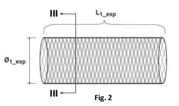
FIG.2 is a side view of the endoluminal prosthesis shown in FIG.1.
FIG.3 is a section view of the endoluminal prosthesis shown in FIGs 1, la and
2 accroding to a cutting
plane III-Ill.
FIG.3a is a schematic magnified view of an embodiment of a portion of the
cross-section shown in
FIG.3.
FIG.3b is a schematic magnified view of another embodiment of a portion of the
cross-section shown
in FIG.3.
FIG. 4 is a schematic magnified view of another portion of an endoluminal
prosthesis according to the
present invention.
FIGs 5 and 6 represent two stages of the healing process of an aneurysm
wherein an endoluminal
prosthesis according to the present invention has been implanted.
CA 03004957 2018-05-10
WO 2017/081213 PCT/EP2016/077363
3
FIGs 7 and 8 show simulations of blood velocity at an orifice of an aortic
branch respectively according
to prior art stents and with an endoluminal prosthesis according to the
present invention.
FIGs 9a and 9b show simulation of blood velocity in an aortic model
respectively according to the prior
art (without stent) and with an endoluminal prosthesis according to the
present invention.
FIG5.10a and 10b are magnified views at the supra aortic branches orifices of
the simulations shown
in FIG5.9a and 9b, respectively.
FIG5.11a and 11b are a magnified views at the coronaries orifice of the
simulation shown in FIG5.9a
and 9b, respectively.
FIG.12 is a schematic cross-section view of the aorta showing how to measure
the width and height
of the aortic arch.
FIG.13 (a-d) show the different phases of the healing process of a saccular
aneurysm involving a branch
with an endoluminal prosthesis according to the present invention.
FIG.14 (a-d) shows the different phases of the healing process of a fusiform-
shaped aneurysm
involving a branch with an endoluminal prosthesis according to the present
invention.
Detailed Description of the Invention
As used hereinafter, the term "implantable" refers to an ability of a medical
device to be positioned
at a location within a body vessel. Implantable medical device can be
configured for transient
placement within a body vessel during a medical intervention (e.g., seconds,
minutes, hours), or to
remain in a body vessel permanently.
The terms "endoluminal" or "transluminal" prosthesis refers to a device
adapted for placement in a
curved or straight body vessel by procedures wherein the prosthesis is
advanced within and through
the lumen of a body vessel from a remote location to a target site within the
body vessel. In vascular
procedures, a medical device can typically be introduced "endovascularly"
using a catheter over a wire
guide under fluoroscopic guidance. The catheters and wire guides may be
introduced through
conventional access sites in the vascular system.
The term "catheter" refers to a tube that is inserted into a blood vessel to
access the target site. In the
present description, a "catheter" will designate either a catheter per se, or
a catheter with its
accessories, meaning needle, guide wire, introducer sheath and other common
suitable medical
devices known by the man skilled in the art.
CA 03004957 2018-05-10
WO 2017/081213 PCT/EP2016/077363
4
The term "permanent" refers to a medical device which may be placed in a blood
vessel and will
remain in the blood vessel for a long period of time (e.g. months, years) and
possibly for the remainder
of the patient's life.
The endoluminal prosthesis 1 is configured to take a compressed shape having a
relatively small and
relatively uniform diameter when disposed within a delivery system (i.e., "in
compressed state"), and
to spontaneously take a deployed shape with radially expanded diameter within
the delivery location
such as a body lumen (i.e., "in deployed state"). As used herein the terms
"expanded shape" or
"expanded state" refer to a shape or state resulting from the self-expanding
properties of a self-spring-
back object (e.g., braided framework 20) when it is allowed to expand without
any outer compression
force (i.e., non-constricted state). Beside these definitions, the term
"nominal diameter" designates
the diameter of the implantable endoluminal prosthesis when placed in the
targeted vessel. Generally,
the nominal diameter (0,,or) of a self-expandable device designed to be placed
permanently inside a
body lumen is 10 to 25% smaller than the external diameter of said device when
deployed without
external compression force (0exp).
The implantable endoluminal prosthesis 1 according to the present invention
comprises at least one
self-expandable braided framework 20 able to expand from a radially compressed
state in a delivery
configuration to a radially expanded state. The implantable endoluminal
prosthesis 1 has a multilayer
configuration either comprising at least two of the self-expandable braided
frameworks 20 or
comprising at least one self-expandable braided framework 20 having a
plurality of interlocked layers
(interlocked multilayer configuration) formed by braiding a plurality of
wires. The braided framework
20 comprises a lumen in a cylindrical form with a circular cross-section and a
constant diameter shown
in FIGs 1, la and 2.
When the endoluminal prosthesis 1 having the multilayer configuration is
observed normal with
respect to a wall, meshes of the braided framework(s) 20 form a lattices with
a plurality of level of
wires 21. FIG.3 shows a schematic cross-section of the endoluminal prosthesis
1 according to the
present invention. FIG.3a shows a schematic magnified view of a portion of the
endoluminal
prosthesis 1 comprising a self-expandable framework 20, and FIG.3b showing a
portion of the
endoluminal prosthesis 1 comprising two self-expandable frameworks 20. A ratio
T1/021 of the
thickness T1 of a wall of the endoluminal prosthesis 1 to the diameter 021 of
wire 21 should be greater
than 2Ø1t characterizes the endoluminal prosthesis 1 having more than a
single layer of mesh, namely
multilayer configuration. The braided framework 20 is preferably made of a
multilayer braid having a
CA 03004957 2018-05-10
WO 2017/081213 PCT/EP2016/077363
5 thickness T20. The term "interlocked multi-layer" refers to a framework
comprising multiple layers,
whose plies are not distinct at the time of braiding, for example a given
number of wires of the plies
of the first layer 22 being interlocked with the plies of the second layer 23
and/or other layers, for
example, as schematically illustrated in FIG.4. Said interlocked multi-layer,
for example, can be formed
by using the braiding machine described in EP1248372.
Thanks to the thicker wall T1 of the multilayered endoluminal prosthesis 1 as
compared with the wall
thickness of a conventional stent, endoluminal prosthesis 1 exhibits a three
dimensional (3D) porosity.
The thicker the wall is (regarding a given wire diameter 021) the greater the
3D porosity effect..
One of the technical effects provided by the 3D porosity of endoluminal
prosthesis 1, is that the
present endoluminal prosthesis 1 lets the blood flow into the aneurysm sac
converts owing to its
multilayer configuration, an undesired damaging turbulence in the aneurysmal
sac into a smooth
laminar flow 11 (as shown FIG.5), instead of mechanically/physically keeping
out the blood flow from
the aneurysm as would do a conventional stent-graft techniques. It results in
excluding the aneurysm
by forming a protecting organized thrombus 12, known as layers of Zhan (see
FIG.6), while keeping
the branches and collaterals unobstructed. Thanks to the permeable multilayer
structure of the
endoluminal prosthesis 1, additional repairs such as open debranching-bypass
procedure and custom-
made fenestrated/branched configuration for maintaining a blood flow are not
required.
The surface coverage ratio (SCR) of endoluminal prosthesis 1 is between 30%
and 70%, preferably
more than 35% and less than 50%, even more preferably less than 45% in
radially expanded state. The
SCR of the endoluminal prosthesis is defined by the relation:
SCR = Sw/St
Wherein "Sw" is the actual surface covered by wires 21 composed in the
endoluminal prosthesis 1,
and "Si" is the total surface are of the wall of the endoluminal prosthesis 1
when observed normal
with respect to the wall.
Studies and experiments carried by the inventor led to surprising and
unexpected conclusions. The
perfusion in branches is improved in accordance with the increase of the ratio
T1/021 having the SCR
of the endoluminal prosthesis between 30% and 70% instead of occluding these
blanches. "Perfusion"
is, in physiology, the process of a body delivering blood to capillary bed in
its biological tissue. The
terms "hypoperfusion" and "hyperperfusion" measure the perfusion level
relative to a tissue's current
CA 03004957 2018-05-10
WO 2017/081213 PCT/EP2016/077363
6
need to meet its metabolic needs. For example, the endoluminal prosthesis of
the invention increases
the perfusion in the supra aortic branches 30 when it covers the branches,
resulting in that the
functioning of the organs to which the supra aortic branches 30 carries the
blood is improved. As
shown in a simulation of FIG.7, a heavy turbulence is created at an orifice 34
of branch. On the contray,
when the endoluminal prosthesis is placed in front of the orifice 34, the
chaotic flow is eliminated by
passing through a wall of the endoluminal prosthesis and converted to a
regulated laminar flow. It
accelerates the flow in the branches covered by the endoluminal prosthesis 1.
Accordingly, the ratio
T1/021 of the present endoluminal prosthesis 1 should be more than 2.0,
preferably at least 2.5, more
preferably at least 3.0, even more preferably at least 3.5, still even more
preferably 4.0 while the SCR
is between 30% and 70%, preferably between 35% and 50% in radially expanded
state. A competed
simulation of blood flow in an aorta model without and with the endoluminal
prosthesis having more
than 2.0 of T1/021 are shown in FIG5.9a and 9b, respectively. The aortic model
was created based on
an actual pathology of a patient. In FIG.9b, the endoluminal prosthesis is
placed so as to cover the wall
of the vessel from the coronaries 31 up to the supra aortic branches 30.
Processing so, surprisingly,
the velocities of blood flow entering into the supra aortic branches 30 are
notably increased of
between 21% and 24% as shown in FIG.10b (magnified view of FIG.9b) at the
orifices 34 of supra aortic
branches 30, when compared with the velocity without device shown in FIG.10a
as a(magnified view
of FIG.9a). The flow velocity in the coronaries are also increased up to 20%
as shown in FIG.11a and
11b.
Further distinguishing improvement of "perfusion" in the branches covered by
the endoluminal
prosthesis 1 was observed with this interlocked multilayer configuration. The
braided framework 20
of the endoluminal prosthesis 1 is made of at most 196 wires 21, preferably at
least 90 wires at most
130 wires. The wires preferably have a diameter (021) of at least 120 um,
preferably at least 150 um,
more preferably at least 180 um, even more at least 200 um and at most 220 um.
Another advantages of the present invention is that the implantable
endoluminal prosthesis 1, having
higher value of the ratio T1/021, can effectively form a thrombus in the
aneurysmal sac in comparison
with a braided framework having lower T1/021 ratio. The ratio T1/021 of the
wall thickness T1 of the
endoluminal prosthesis 1 to the wire diameter 021 of wire 21 being more than
2.0 characterizes the
endoluminal prosthesis 1 having more than a single layer of mesh. The greater
the ratio T1/021, the
more layers the endoluminal prosthesis 1 will comprise. Each wire forming
multiple-layers works to
make the blood flow be laminated which gets through the wall of the
endoluminal prosthesis 1.
CA 03004957 2018-05-10
WO 2017/081213 PCT/EP2016/077363
7
The curve of the aortic arch 32 is generally defined by measuring the width
W32 and height H32 of the
curve as described by Ou et al. in J. Thrac. Cardiovasc. Surg. 2006; 132:1105-
1111. Width W32 is
measured as the maximal horizontal distance between the midpoints 35 of the
ascending and
descending aorta 32 close to the axial plane going through the right pulmonary
artery; and height H32
of the aortic arch is measured maximal vertical distance between W32 and the
highest midpoint 35 of
the aortic arch W32 as depicted in FIG.12.
Interlocked multiple-layer configuration having a ratio T1/021 of at least 2.5
brings an important
advantageous technical property. When the aneurysm is located at the outer
side of the curve, it is
most important to set an optimal SCR and an optimal opening size of mesh at
the outer side of the
curve in order to form a protecting organized thrombus in the aneurysmal sac
by converting an
undesired damaging turbulence 33 into a smooth laminar flow 36 while keeping
branches, such as
supra aortic branches 30, patent. Wires of the interlocked multiple-layer
configuration of the
invention shift to keep a regular distance between adjacent parallel,
resulting in that the SCR can stays
almost the same between in a curved state and in straight configuration. On
the Contrary, when a
conventional single-layer mesh-like tube having less than 2.0 of T1/021 is
deployed in a curved lumen,
the SCR at the outer side of the curve are much lower than the SCR in a
straight configuration.
Therefore, the ratio T1/021 of the present endoluminal prosthesis 1 should be
more than 2.0,
preferably at least 2.5, more preferably at least 3.0, even more preferably at
least 3.5, still even more
preferably at least 4Ø
As another surprising effect provided the present endoluminal prosthesis 1
having interlocked
multiple-layer configuration, against the "normal" expectation that a space
between an aneurysmal
wall and endoluminal prosthesis would be occluded by thrombus as shown in
FIG.6, the aneurysm
including branches shrinks directly instead of forming thrombus in the
aneurysmal sac while still
maintaining the blood flow into the branches as shown FIGs 13 and 14. The
inventor assumes that by
sealing the beginning of the aorta with the enlarged, undesired turbulence 33
are eliminated and
desired smooth flow 11 are created in this volume. It accelerates the non-
turbulent blood flow
entering the branches while decreasing the pressure under Venturi effect,
resulting in shrinkage of
the aneurysmal sac.
The biocompatible material used in the invention is preferably a metallic
substrate selected from a
group consisting of stainless steels (e.g., 316, 316L or 304); nickel-titanium
alloys including shape
memory or superelastic types (e.g., nitinol, Nitinol-DFr-Platinum); cobalt-
chrome alloys (e.g.,
CA 03004957 2018-05-10
WO 2017/081213 PCT/EP2016/077363
8
elgiloy); cobalt-chromium-nickel alloys (e.g., phynox); alloys of cobalt,
nickel, chromium and
molybdenum (e.g., MP35N or MP2ON); cobalt-chromium-vanadium alloys; cobalt-
chromium-tungsten
alloys; magnesium alloys; titanium alloys (e.g., TiC, TiN); tantalum alloys
(e.g., TaC, TaN); L605;. Said
metallic substrate is preferably selected from the group consisting of
titanium, nickel-titanium alloys
such as nitinol and Nitinol-DFr-Platinum, any type of stainless steels, or a
cobalt-chromium-nickel
alloys such as Phynox .