Note: Descriptions are shown in the official language in which they were submitted.
CA 03005259 2018-05-11
WO 2017/087563
PCT/US2016/062342
- 1 -
METHODS, COMPOSITIONS, AND SYSTEMS FOR CULTURING AND
CHARACTERIZING FASTIDIOUS PLANT MICROBES
CROSS-REFERENCE TO RELATED APPLICATIONS
This application claims priority to U.S. Application No. 62/255,823 filed
November
16, 2015, the entire contents of which are hereby incorporated in this
disclosure by reference.
FIELD OF THE DISCLOSURE
The present disclosure relates, in some embodiments, to methods, compositions,
and
systems for culturing and characterizing fastidious plant microbes.
BACKGROUND OF THE DISCLOSURE
Estimates of food and agriculture organizations project that global food
production
has to increase 60% by 2050 to meet the demands of the rising world
population. On top of
the rising demand, annual agricultural crop losses caused by plant pathogens
may run into
upwards of 40%. Fastidious and obligate phytopathogens alone may be
devastating to
several food and commodity crops. For example, xylem-limited Xylella
fastidiosa infects
over 100 plant species including grapevine, citrus, coffee and almonds.
Similarly, phloem-
limited Candidatus Liberibacter spp. are emerging destructive pathogens
causing yield losses
(e.g., severe losses) in diverse plant families, including Solanaceae (potato,
tomato, pepper
and tobacco), Apiaceae (carrot and celery), Rosaceae (pear, apple and
blackthorn), and
Rutaceae (citrus spp.). Zebra chip disease in potato may be caused by
Candidatus
Liberibacter solanacearum (Lso), which also infects tomatoes, pepper and
tobacco. Lso is
transmitted by an insect vector, potato psyllid (Bactericera cockerelli).
Since being
designated as an emerging disease in 2004, zebra chip disease has been
documented in
several commercial potato growing regions of the United States, Mexico,
Central America,
and New Zealand. In Texas alone, zebra chip disease is estimated to affect 35%
of cultivated
potato acreage, causing annual crop losses of approximately S25 million USD.
Similarly,
citrus greening or Huanglongbing (HLB) disease caused by the Candidatus
Liberibacter
asiaticus (Las) may be the most devastating disease of citrus today, and
threatens citrus
production worldwide. HLB is transmitted by the insect vector, Asian Citrus
psyllid
(Diaphorina citri). In 2006-2011, in Florida alone, HLB caused losses upwards
of S4.5
CA 03005259 2018-05-11
WO 2017/087563
PCT/US2016/062342
- 2 -
billion USD. These and other diseases caused by fastidious plant pathogens are
a major
threat to U.S. and global agriculture production. It is imperative to curtail
these agricultural
losses in order to overcome the impending global food security challenge.
CA 03005259 2018-05-11
WO 2017/087563
PCT/US2016/062342
- 3 -
SUMMARY
The inability to culture fastidious vascular-limited microbes is a major
bottleneck in
research relating to several destructive agricultural pathogens, such as Lso
and Las.
Accordingly, a need has arisen for improved (e.g., easy, rapid, and/or
scalable) culturing and
functional characterization of fastidious vascular-colonizing microbes.
The present disclosure relates, in some embodiments, to methods, compositions,
and
systems for culturing and characterizing fastidious microbes using hairy roots
(e.g., R.
rhizogenes-mediated hairy roots induced directly from infected plants or plant
tissues). R.
rhizogenes-mediated hairy roots induced directly from the infected plants or
plant tissues may
provide an easy, rapid and scalable platform to culture and characterize the
fastidious
vascular-colonizing microbes. For example, Lso and Las may be cultured in
hairy roots
induced directly from Lso- and Las-infected tomato, potato and citrus plants
or plant tissues.
Because hairy roots are organized and stable tissues, the microbe-colonized
hairy root tissues
may be clonally propagated for numerous applications (e.g., characterization
of the fastidious
microbes).
The present disclosure relates, in some embodiments, to methods for
cultivating a
fastidious plant microbe (e.g., a fastidious plant pathogen). For example, a
method may
comprise contacting a plant (e.g., a tomato plant, a potato plant, a citrus
plant) colonized by a
fastidious plant microbe (e.g., Xylella fastidiosa, Candidatus Liberibacter
spp.) with a
suspension of R. rhizogenes under conditions that permit induction of hairy
roots colonized
with the fastidious plant microbe. A fastidious plant microbe may then be
cultivated by
propagating the colonized microbial hairy roots. In some embodiments, a
cultivated plant
microbe may be Candidatus Liberibacter spp. (e.g., Candidatus Liberibacter
solanacearum
(Lso) and/or Candidatus Liberibacter asiaticus (Las)). According to some
embodiments, R.
rhizogenes may include both a Ri-DNA plasmid and a T-DNA plasmid and the T-DNA
plasmid may encode a first exogenous transgene (e.g., a gene encoding an
autofluorescent
protein). According to some embodiments, a plant may include a first exogenous
transgene.
In some embodiments, contacting a plant colonized by a fastidious microbe with
a
suspension of R. rhizogenes may include wounding one or more surfaces of a
plant, forming
a wound site, and exposing the wound site to a suspension of R. rhizogenes.
Contacting a
plant colonized by a fastidious plant microbe with a suspension of R.
rhizogenes, in some
CA 03005259 2018-05-11
WO 2017/087563
PCT/US2016/062342
- 4 -
embodiments, may include removing a photosynthetic portion of the plant to
generate a
wound site. In some embodiments, a method may include covering a wound site
with a rock
wool matrix and exposing the rock wool matrix to a suspension of R.
rhizogenes. According
to some embodiments, a method may include submerging a wound site in a
suspension of R.
rhizogenes, vacuum infiltrating at least a portion of the suspension of R.
rhizogenes into the
wound site, and covering the wounds site with a vermiculite matrix. A method
may
comprise, in some embodiments, contacting any desired portion of a plant
colonized with the
fastidious plant microbe with R. rhizogenes including, for example, a
cotyledon, a hypocotyl,
an immature shoot, an immature root, a mature shoot, or mature root.
Propagating a colonized hairy root, according to some embodiments, may include
exposing an attached hairy root or a harvested hairy root to one or more
selective consitions.
In some embodiments, propagating may include transferring an attached hairy
root or a
harvested hairy root to at least one of: a vermiculite matrix, a hydroponic
system, an in vitro
system, and a bioreactor system. A method may comprise assessing the presence
of the
fastidious plant pathogen in the propagated microbial hairy roots, according
to some
embodiments.
The present disclosure relates, in some embodiments, to methods for assessing
an
effect of a test composition on a fastidious plant microbe. A method may
comprise, for
example, contacting a plant (e.g., a tomato plant, a potato plant, a citrus
plant) infected with
the fastidious plant microbe (e.g., Xylella fastidiosa, Candidatus
Liberibacter spp.) with R.
rhizogenes under conditions that permit induction of hairy roots colonized by
the fastidious
plant microbe, propagating the colonized microbial hairy roots, contacting the
propagated
colonized microbial hairy roots with the test composition, and/or assessing at
least one metric
of the presence and/or vitality of the fastidious plant microbe. In some
embodiments, a
cultivated plant microbe may be Candidatus Liberibacter spp. (e.g., Candidatus
Liberibacter
solanacearum (Lso) and/or Candidatus Liberibacter asiaticus (Las)).
A method may comprise, in some embodiments, contacting Rhizobium rhizogenes
with any desired portion of a plant colonized with a fastidious plant microbe
including, for
example, a cotyledon, a hypocotyl, an immature shoot, an immature root, a
mature shoot, or
mature root. In some embodiments, a test composition may be applied to and/or
injected into
an infected microbial hairy root. A test composition, in some embodiments, may
be
CA 03005259 2018-05-11
WO 2017/087563
PCT/US2016/062342
- 5 -
conditionally or constitutively present in an infected plant (e.g., a
propagated infected hairy
root).
According to some embodiments, a colonized plant (e.g., a propagated colonized
hairy root) may comprise an exogenous transgene (e.g., NPR1, GFP) and the test
composition
may be or may comprise a gene product of the transgene. In some embodiments, a
colonized
plant (e.g., a propagated colonized microbial hairy root) may include at least
one of an
exogenous transgene, a CRISPR/Cas. A TALEN, and an RNAi construct, and
accordingly a
test composition may be or may comprise a gene product of the transgene, a
CRISPR/Cas, a
TALEN, and an RNAi construct. In some embodiment, a test composition may
include an
NPR1 protein.
The present disclosure relates, in some embodiments, to methods for culturing
fastidious plant microbes using hairy roots. A method may comprise, for
example, selecting
tissues from one or more parts of a plant as an explant source; cultivating
the explant source
in an in vitro medium comprising Rhizobium rhizogenes (R. rhizogenes); cutting
the explant
source into a plurality of pieces; inducing growth of hairy roots from at
least some pieces of
the explant source over a period of time; performing polymerase chain reaction
(PCR)
amplification of R. rhizogenes root locus B (rolB) and R. rhizogenes root
locus C (rolC)
marker genes to confirm the induction of the hairy roots; and/or performing
PCR
amplification of a 16S rDNA marker genes to determine presence of one or more
fastidious
microbes. In some embodiments, an infected plant may comprise a first
exogenous transgene
and a second exogenous transgene. A first exogenous transgene may encode, for
example, an
auto-fluorescent protein (e.g., green fluorescent protein, yellow fluorescent
protein, red
fluorescent protein, and the like). According to some embodiments, cultivating
the explant
source in a medium comprising R. rhizogenes may comprise cultivating the
explant source in
a medium free of antibiotics (e.g., free of at least cefatoxime, carbencillin
and kanamycin).
CA 03005259 2018-05-11
WO 2017/087563
PCT/US2016/062342
- 6 -
BRIEF DESCRIPTION OF THE DRAWINGS
The file of this disclosure contains at least one drawing executed in color.
Copies of
this patent with color drawing(s) will be provided by the Patent and Trademark
Office upon
request and payment of the necessary fee. Some embodiments of the disclosure
may be
understood by referring, in part, to the present disclosure and the
accompanying drawings,
wherein:
FIGURE 1 illustrates a workflow for generating microbial hairy roots for
downstream
studies and applications, according to example embodiments of the disclosure;
FIGURE 2A illustrates adult psyllids being propagated in contained traps using
eggplants as host plants, according to an embodiment of the disclosure;
FIGURE 2B illustrates adult psyllids carrying Lso being propagated in
contained
traps using eggplants as host plants, according to an embodiment of the
disclosure;
FIGURE 2C illustrates healthy tomato leaves at four weeks post-psyllid
exposure,
according to an embodiment of the disclosure;
FIGURE 2D illustrates Lso-infected tomato leaves at four weeks post-psyllid
exposure, according to an embodiment of the disclosure;
FIGURE 2E illustrates healthy potato leaves at four weeks post-psyllid
exposure,
according to an embodiment of the disclosure;
FIGURE 2F illustrates Lso-infected tomato leaves at four weeks post-psyllid
exposure, according to an embodiment of the disclosure;
FIGURE 2G illustrates polymerase chain reaction (PCR)-based confirmation of
Lso
in infected tomato and potato leaves;
FIGURES 3A-3E illustrate, according to specific example embodiments of the
disclosure, a schematic of an in vitro microbial hairy root platform;
FIGURE 3A shows cutting of surface-sterilized plant tissues into smaller
pieces and
gentle wounding using a fine forceps, according to an embodiment of the
disclosure;
FIGURE 3B shows immersing explants in a suspension of R. rhizogenes, according
to
an embodiment of the disclosure;
FIGURE 3C shows explants following three-day co-cultivation of the plant
tissues on
nutrient media, according to an embodiment of the disclosure;
CA 03005259 2018-05-11
WO 2017/087563
PCT/US2016/062342
- 7 -
FIGURE 3D shows an osmotic stress treatment of explants, according to an
example
embodiment of the disclosure;
FIGURE 3E shows incubation of the explants in nutrient selection media
following
the treatment of FIGURE 3D, according to an embodiment of the disclosure;
FIGURE 4A illustrates in vitro induction of hairy roots on a tomato explant
transformed with Rhizobium rhizogenes (strain ATCC 15834), according to an
embodiment
of the disclosure;
FIGURE 4B shows PCR validation results of hairy roots on a tomato explant,
according to an embodiment of the disclosure;
FIGURE 5A illustrates in vitro induction of hairy roots on a potato explant
transformed with Rhizobium rhizogenes, according to an embodiment of the
disclosure;
FIGURE 5B shows PCR validation results of hairy roots on a potato explant,
according to an embodiment of the disclosure;
FIGURES 6A illustrates in vitro induction of hairy roots on citrus (Eureka
lemon)
explants transformed with Rhizobium rhizogenes (strain ATCC 15834), according
to an
embodiment of the disclosure;
FIGURE 6B shows PCR validation results of hairy roots on citrus (Eureka lemon)
explants transformed with Rhizobium rhizogenes (strain ATCC 15834), according
to an
embodiment of the disclosure;
FIGURE 7A shows aerial hairy roots induced on a healthy tomato by Rhizobium
rhizogenes, according to an embodiment of the disclosure;
FIGURE 7B shows aerial microbial hairy roots induced on an Lso-colonized
tomato
by Rhizobium rhizogenes, according to an embodiment of the disclosure;
FIGURE 7C shows PCR validation results of aerial hairy roots on tomato,
according
to an embodiment of the disclosure;
FIGURE 8A shows aerial hairy roots induced on an Lso-colonized potato by
Rhizobium rhizogenes, according to an embodiment of the disclosure;
FIGURE 8B shows PCR validation results of aerial hairy roots on potato,
according to
an embodiment of the disclosure;
FIGURE 8C shows PCR validation results of aerial microbial hairy roots on
potato,
according to an embodiment of the disclosure;
CA 03005259 2018-05-11
WO 2017/087563
PCT/US2016/062342
- 8 -
FIGURE 9A shows microbial hairy roots induced on an Lso-colonized tomato using
a
rock wool method, according to an embodiment of the disclosure;
FIGURE 9B shows PCR validation results of microbial hairy roots on tomato
induced
using a rock wool method, according to an embodiment of the disclosure;
FIGURE 10A shows microbial hairy roots induced on a tomato plant using a
vermiculite method, according to an embodiment of the disclosure;
FIGURE 10B shows PCR validation results of microbial hairy roots on tomato
induced using a vermiculite method, according to an embodiment of the
disclosure;
FIGURE 11 shows microbial hairy roots induced on a Las infected citrus (sour
orange) using a vermiculite method, according to an embodiment of the
disclosure;
FIGURE 12 illustrates propagated hairy roots growing in a vermiculite matrix,
according to an embodiment of the disclosure;
FIGURE 13 illustrates propagated hairy roots growing in a hydroponic culture,
according to an embodiment of the disclosure;
FIGURE 14 illustrates propagated hairy roots growing in an in vitro culture,
according to an embodiment of the disclosure;
FIGURE 15 illustrates propagated hairy roots growing in a bioreactor system,
according to an embodiment of the disclosure;
FIGURE 16A shows healthy and Lso-colonized hairy roots harvested from an in
vitro
or in planta system, according to an embodiment of the disclosure;
FIGURE 16B shows healthy and Lso-colonized microbial hairy roots distributed
into
a multi-well plate, according to an embodiment of the disclosure;
FIGURE 16C shows Quantitative Real Time PCR results of an Lso titer after a 2
day
treatment of hairy roots with penicillin; and
FIGURE 16D shows Quantitative Real Time PCR results of an Lso titer after a 7
day
treatment of hairy roots with penicillin.
CA 03005259 2018-05-11
WO 2017/087563
PCT/US2016/062342
- 9 -
DETAILED DESCRIPTION
Despite the huge economic significance of fastidious microbes (e.g., plant
pathogens),
little is known of their biology, genetics, and the vector-pathogen-plant
interactions. This
knowledge may enable development of effective disease and pest management
strategies to
limit yield losses (e.g., tremendous losses). One bottleneck (e.g., a major
bottleneck) in
characterizing these fastidious microbes is their inability to grow outside
their natural hosts,
as they are obligate parasites of plants. It is estimated that > 99% of
microorganisms from
any environment are non-cultivable in the laboratory. Numerous attempts have
been made to
create suitable artificial growth media and culture conditions for cultivating
fastidious
microbes; however, to date these approaches have had only limited success.
Plant hairy roots can be readily induced from diverse plant tissues upon
infection by a
soil bacterium, Rhizobium rhizogenes (recently revised from Agrobacterium
rhizogenes). In a
manner similar to its related cousin, A. tumefaciens, R. rhizogenes introduces
its root-
inducing (Ri) transfer-DNA (Ri-DNA plasmid), which encodes the root locus
(rol) genes
(e.g., rolB, rolC) into the plant genome. The expression of rol genes in
planta overproduces
the plant hormone, auxin, and induces hairy root initiation and proliferation.
Hairy airy roots are anatomically, morphologically, and metabolically similar
to
normal roots. Hairy roots are connected to a plant tissue from which they are
generated by
Intact xylem and phloem vasculature, thereby allowing continued transport of
water,
nutrients, cellular signaling¨and as shown here fastidious microbes¨through
the
vasculature. Typically hairy roots are smaller in diameter than a plant tissue
from which they
derive (e.g., stem, root) and are often numerous. A large number of plant
genuses may be
transformed by R. rhizogenes and generate hairy roots, including but not
limited to: Citrus
(e.g., lemon), Solanaceae (e.g., potato, tomato), Daucus (e.g., carrot),
Taxus, Cinchona,
Gmelina, Glycine (e.g., soybean), Rutaceae (e.g., Bad l tree), Nyctaginaceae,
and Rosaceae
(e. g. , apple).
The present disclosure relates, in some embodiments, to a method of
cultivating a
fastidious microbe (e.g., in vitro, in planta). The present disclosure
relates, in some
embodiments, to a hairy root system that may be directly used to cultivate and
characterize
fastidious microbes. The present disclosure relates, in some embodiments, to a
hairy root
system that may be used for high-throughput functional genetic and genomic
studies of the
CA 03005259 2018-05-11
WO 2017/087563
PCT/US2016/062342
- 10 -
fastidious microbe-plant interactions. In some embodiments, a hairy root
system may be
used for chemical genetic screens (e.g., screening antibiotics, essential
oils, oxylipins) to
combat devastating plant diseases. According to some embodiments, a hairy root
system
may include a genetically modified plant and may be beneficial in identifying
gene related
susceptibility and/or resistance to fastidious microbes in plant species.
According to some embodiments, Rhizobium rhizo genes-mediated hairy root
cultures
of a host plant (e.g., tomato, potato, pepper, citrus) may be infected with a
fastidious microbe
(e.g., Candidatus spp., Xylella spp., Clavibacter spp.). Explant source
material may include
any desired organ or tissue of a plant including, for example, cotyledons,
hypocotyls,
immature and mature shoots and roots). An optimal source may be identified for
a particular
set of conditions by testing multiple infected plant tissues. Hairy root
multiplication
techniques (e.g., techniques for scalability and/or multiplexing) may be
included for high-
throughput diagnostics and/or molecular characterization, according to some
embodiments.
Functional characterization for a hairy-root system may include, in some
embodiments,
genetic gain-of-function (e.g., overexpression) and loss-of-function (e.g.,
clustered, regularly
interspaced, short palindromic repeat-associated (CRISPR/Cas), Transcription
activator-like
effector nuclease (TALEN) and Ribonucleic Acid inference (RNAi) knockdown)
studies of
candidate plant and pathogen-encoded genes. Gene constructs (e.g.,
representative gene
constructs) or gene libraries may be transiently delivered into established
hairy-roots by any
desired method including, for example, vacuum infiltration and/or by DNA
bombardment. In
some embodiments, gene constructs may be inserted into the R. rhizogenes T-DNA
prior to
hairy-root induction, thus completing the process in a single step.
According to some embodiments, a hairy root system may be used for propagation
and functional studies of any desired plant-microbe association (e.g., beyond
vascular-
colonizing phytobacteria) including viruses (e.g., Tomato spotted wilt virus,
Cucumber
mosaic virus, Cauliflower mosaic virus, Turnip yellow mosaic virus) , viroids
(e.g., Potato
spindle tuber viroid, Tomato apical stunt viroid, Citrus exocortis viroid),
and endophytic
microbes (e.g., Acidovorax facilis, Azoarcus sp. BH72, Azospirillum sp. B510,
Fusarium
spp., Colletotrichum spp, Curvularia spp., Beauveria bassiana, Lecanicillium
spp, Pythium
oligandrum).
CA 03005259 2018-05-11
WO 2017/087563
PCT/US2016/062342
- 11 -
Previous studies have not reported the application of hairy roots to culture
or to the
study of fastidious plant microbes. Thus, the present disclosure showing that
hairy roots can
support the growth of fastidious microbes may have a significant impact on
large-scale
propagation of microbial hairy roots for high-throughput applications. The
disclosed hairy
root system resolves a significant bottleneck in culturing and propagating
fastidious
phytopathogens and may result in the initiation of numerous biological studies
of fastidious
plant pathogens. Such studies offer the promise of new transformative
developments in plant
disease and pest management, as well as, agriculture in general.
The present disclosure, in some embodiments, may help establish and optimize
Candidatus Liberibacter spp. microbial hairy root cultures in any suitable
plant such as
potato, tomato and citrus, and allow researchers to perform genetic and/or
chemical analysis
using the disclosed microbial hairy root system.
According to some embodiments disclosed here, hairy roots induced directly
from
infected plants may provide an easy, rapid, and scalable platform to culture
and characterize
fastidious vascular-colonizing plant pathogens. For example, Lso and Las are
prevalent in
South Texas and infect several Solanaceous crops including potato, tomato, and
Citrus spp.
Thus, Las-infected citrus trees in South Texas may be used to obtain Las-
infected plant
material. Plant tissues from Las-infected citrus trees may be collected and
tested for Las
presence by polymerase chain reaction (PCR) amplification of 16S rDNA, an
established
DNA marker for detecting Las. Las-positive citrus tissues may be further used
as an explant
source for microbial hairy root induction.
The present disclosure relates, in some embodiments, to a biological mimic
hairy root
platform for rapid culture, propagation, and functional studies of fastidious
vascular-
colonizing microbes (e.g., plant pathogens). Fastidious vascular-colonizing
plant pathogens
such as Candidatus spp., Xylella spp., viruses, phytoplasmas and spiroplasmas
result in
billions of dollars of annual crop losses. The inability to culture these and
other vascular-
limited microbes (e.g., pathogens) presents a tremendous challenge to
researchers attempting
to study these organisms. Plant hairy roots induced by Rhizobium rhizo genes
are organized,
stable tissues anatomically and metabolically similar to roots, and they
possess intact xylem
and phloem vasculature. According to some embodiments, microbe-containing
hairy roots
induced directly from infected plants may provide an easy, rapid, and scalable
platform to
CA 03005259 2018-05-11
WO 2017/087563
PCT/US2016/062342
- 12 -
culture, propagate, and characterize fastidious vascular-limited plant
pathogens (e.g., in the
laboratory).
According to some embodiments, Candidatus Liberibacter spp. microbial hairy
root
cultures in a plant (e.g., potato, tomato, citrus) may enable genetic analysis
using a microbial
hairy root system. In addition to alleviating previous challenges of culturing
fastidious
vascular-limited plant pathogens, a hairy root platform may be readily
exploited for
transformative, high-throughput functional studies including but not limited
to genetic and
chemical screens for novel antimicrobials and bactericides. Because R.
rhizogenes may
effectively induce hairy roots in diverse monocot and dicot plants, microbial
hairy root
systems, methods, and compositions for microbial cultivation may be applied to
other
agronomic crops and plant microbe associations beyond vascular-colonizing
phytobacteria
such as fungi, viruses, viroids, and endophytic microbes.
Generating an Explant and/or Inoculum Source
Insect vectors may spread fastidious microbes from plant to plant. Plant
material
colonized by a fastidious microbe (e.g., explant) may be generated, according
to some
embodiments, by allowing or introducing an insect vector carrying the
fastidious microbe to
feed upon a plant and thereby transferring the fastidious microbe to the plant
tissue. Once
transferred into a plant vascular system, a fastidious microbe can colonize
and replicate.
According to some embodiments, a fastidious microbe carrying insect vectors
may be
collected from an environment (e.g., agricultural fields) and tested to
determine whether they
carry the fastidious microbe (e.g., 16S rDNA PCR). Insect vectors (e.g.,
fastidious microbe
carrying insect vectors, non-carrying insect vectors) may be maintained (e.g.,
in insect cages)
and propagated, in some embodiments. A fastidious microbe carrying insect
vector, in some
embodiments, may be generated in a laboratory setting, for example, by
allowing a non-
carrying insect vector to feed upon plant tissue infected by a fastidious
microbe. Numerous
methods are known for transmitting a fastidious microbe to an insect vector,
as well as,
maintaining and propagating an insect vector population, all of which are
within the scope of
the present disclosure.
In some embodiments, an insect vector population may be tested for the
presence or
absence of colonization by a fastidious microbe. Any suitable technique may be
used to test
CA 03005259 2018-05-11
WO 2017/087563
PCT/US2016/062342
- 13 -
an insect vector for the presence or absence of colonization by a fastidious
microbe, for
example 16S rDNA PCR.
At least one fastidious microbe carrying insect vector may be permitted to
feed upon an
uninfected plant for a period of time (e.g., seven days); thereby transferring
at least one
fastidious microbe to the plant vasculature. Within the scope of this
disclosure, numerous
methods may be used to successfully transfer at least one fastidious microbe
to a plant
vasculature. Such methods may vary depending upon at least: the species and
developmental
stage of an insect vector, a level of fastidious microbe within an insect
vector, a rate at which
a fastidious microbe replicates within an insect vector, and a species and
developmental stage
of an uninfected plant. According to some embodiments, after a designated
feeding period
(e.g., seven days), a microbe carrying insect vector may be removed from the
plant and the
plant may be monitored for colonization and/or disease development. In some
embodiments,
disease symptoms may indicate colonization by a fastidious microbe. Any number
of
standard laboratory procedures may be used to identify the presence or absence
of a
fastidious microbe within a plant tissue. According to some embodiments, plant
tissue may
be tested for the presence or absence of fastidious microbe populations using
16S rDNA
PCR. A plant or plant tissue (e.g., cotyledon) which tests positive for the
presence of a
fastidious microbe (e.g., PCR positive) may be used as a source of explant for
microbial hairy
root induction, according to some embodiments. According to some embodiments,
a method
for cultivating a fastidious plant microbe may include colonizing a plant with
a fastidious
microbe to generate an explant source. In some embodiments, colonizing a plant
with a
fastidious microbe may include exposing one or more surfaces of the plant to
at least one
fastidious microbe carrying insect vector (e.g., an Lso carrying psyllid). An
explant, in some
embodiments, may include a plant tissue, such as a cotyledon, a hypocotyl, an
immature
shoot, an immature root, a mature shoot, a mature root, or any combination
thereof.
A Microbial Hairy Root Platform
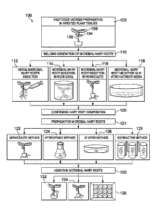
FIGURE 1 illustrates a microbial hairy root platform workflow 100 including:
propagating a fastidious microbe in one or more plant tissues 102; inducing
microbial hairy
root production from infected plant tissues 110; propagating microbial hairy
roots 120, and
CA 03005259 2018-05-11
WO 2017/087563
PCT/US2016/062342
- 14 -
applying microbial hairy roots to downstream studies and applications 130,
according to
example embodiments of the present disclosure.
As shown in FIGURE 1, according to some embodiments, a fastidious microbe may
be propagated in one or more plant tissues 102. One or more plant tissues of
an infected
plant may be colonized by a fastidious microbe including a cotyledon 104, a
hypocotyl 106, a
leaf 108, an immature shoot, an immature root, a mature shoot, a mature root,
or any
combination thereof. Propagating a fastidious microbe (e.g., Lso, Las) in one
or more plant
tissues may include exposing a healthy plant to one or more vector species
(e.g., psyllid) that
are colonized by the fastidious microbe and that are known to feed on plant
tissues, according
to some embodiments. According to some embodiments, propagating a fastidious
microbe
may include various methods of asexual plant propagation. According to some
embodiments,
propagating a fastidious microbe may include identifying infected plants from
an
environment and maintaining the identified plants.
A microbial hairy root platform workflow 100, may include inducing microbial
hairy
root generation from an infected plant or plant tissue 110. According to some
embodiments,
inducing microbial hairy root generation may include culturing Rhizobium
rhizogenes.
Numerous methods are appropriate for culturing R. rhizogenes and are
encompassed by this
disclosure. Culturing R. rhizogenes may include growing R. rhizogenes in any
appropriate
culture medium (e.g., Luria-Bertani medium (LB)) to any appropriate optical
density (e.g.,
0.3). In some embodiments, a culture of R. rhizogenes may be grown to an O.D.
of about 0.2,
or about 0.3, or about 0.4, or about 0.5, or about 0.6. According to some
embodiments, a
culture of R. rhizogenes may be grown to an O.D. between 0.2 and 0.6, or
between 0.3 and
0.6, or between 0.2 and 0.4. Upon reaching an elected optical density, a
culture of R.
rhizogenes may be removed from a medium (e.g., via centrifugation) and
resuspended in a
volume of plant culture medium (e.g., 1/2 MS, 1/2 B5 + 3% sucrose) or water
(e.g., sterile
water) to a desired concentration. In some embodiments, a culture of R.
rhizogenes may be
resuspended at an O.D. of about 0.2, or about 0.3, or about 0.4, or about 0.5,
or about 0.6.
According to some embodiments, a culture of R. rhizogenes may be resuspended
at an O.D.
between 0.2 and 0.6, or between 0.3 and 0.6, or between 0.2 and 0.4.
According to some embodiments, a strain of R. rhizogenes may be selected based
on
specific characteristics. For example, different R. rhizogenes strains may
have varying
CA 03005259 2018-05-11
WO 2017/087563
PCT/US2016/062342
- 15 -
potentials to induce hairy roots from plants. A suitable strain for
propagating microbial hairy
roots in a plant or explant (e.g., tomato, potato, citrus) may be empirically
determined, in
some embodiments, based on, for example, a percent induction of hairy roots in
a selected
explant tissue type and/or plant species (e.g., tomato, potato, citrus).
Suitable strains of R.
rhizogenes for evaluation and/or use may include, in some embodiments,
American Type
Cell Culture (ATCC) 15834, ATCC 43056, ATCC 43057, ATCC 1333, K599, or any
combination thereof. According to some embodiments, for each combination of
explant
tissue and R. rhizogenes strain, induction efficiency may be determined by
measuring
parameters such as the following: (a) hairy root induction percentage per
total explants; (b)
hairy root initiation days per total explants; (c) hairy root induction
frequency per single
explant, and (d) fastidious microbe (e.g., Las and Lso) populations in the
hairy roots. For
accurate comparison of microbial titers amongst different samples (e.g.,
explant tissue type,
plant species type), quantitative PCR techniques (e.g., q-PCR, quantitative
Real-Time PCR)
may be used, according to some embodiments. Statistical analysis, such as an
analysis of
variance (ANOVA) and Student's T-test, may be employed to determine
significant
differences between populations amongst different samples.
As illustrated in FIGURE 1, both in planta 110, 112, 114 and in vitro 116
approaches
may be used to induce microbial hairy root generation directly from one or
more infected
plant tissues. In planta approaches for inducing microbial hairy root
generation include aerial
induction of microbial hairy roots 110, rock wool induction of microbial hairy
roots 112, and
vermiculite induction of microbial hairy roots 114.
In planta methods of inducing microbial hairy roots
Inducing microbial hairy root generation 110, in some embodiments, may include
selecting an infected plant (e.g., Lso) and preparing one or more surfaces of
the infected plant
(e.g., surface sterilization, wounding). Preparing an infected plant may
include surface
sterilization of one or more surfaces of the infected plant. Any appropriate
surface
sterilization techniques may be used including, in some embodiments, exposure
of one or
more surfaces of an infected plant for a designated period of time (e.g., 1 to
10 minutes) to a
solution containing an alcohol (e.g., 70% ethanol), NaC10 (bleach) (e.g., 2%,
10%), a non-
phytotoxic anti-fungal (e.g., amphotericin B), an anti-bacterial (e.g., 200
mg/L cefotaxime or
100 mg/L carbenicillin) compound, or any combination thereof.
CA 03005259 2018-05-11
WO 2017/087563
PCT/US2016/062342
- 16 -
According to some embodiments, preparing an infected plant (e.g., colonized by
Lso
or Las) may include wounding one or more surfaces of the infected plant. Any
suitable tools
may be used to wound one or more surfaces of an infected plant, including
scissors, a scalpel,
forceps (e.g., fine gauge), a syringe, a needle, or any combination thereof.
Wounded and
exposed surfaces of an infected plant may serve as active sites for R.
rhizogenes
transformation and hairy root induction, in some embodiments.
In some embodiments, inducing microbial hairy root generation 110 may include
contacting an infected plant or a portion of an infected plant with at least
one R. rhizogenes
cell (in planta approach), as shown in FIGURE 1 at 112, 114, and 116.
Contacting an
infected plant with at least one R. rhizogenes cell may include directly
exposing one or more
parts (e.g., a wound site) of an infected plant to a R. rhizogenes suspension
(e.g., O.D. 0.3)
(e.g., to generate aerial hairy roots) or vacuum infiltrating one or more
parts of an infected
plant with a R. rhizogenes suspension, according to some embodiments.
According to some embodiments, one or more portions of an infected plant may
be
directly contacted by a suspension containing at least one cell of R.
rhizogenes (e.g., O.D.
0.3). Various methods may be used to contact one or more portion of an
infected plant with a
suspension containing at least one cell of R. rhizogenes. According to some
embodiments,
contacting one or more portions of an infected plant may include: applying a
suspension
containing at least one cell of R. rhizogenes to a wound site (e.g., using a
dropper), dipping a
fine needle in a suspension of R. rhizogenes and using the fine needle to
wound one or more
locations on an infected plant; injecting a suspension of R. rhizogenes into
an infected plant
using a syringe. As shown in FIGURE 1 112, contacting an infected plant at a
location above
soil level may generate one or more aerial microbial hairy roots.
According to some embodiments, contacting one or more portions of an infected
plant
with at least one cell of R. rhizogenes (e.g., an exposed wound site) may
include wrapping or
covering (e.g., with aluminum foil) a contact site. Wrapping or covering a
contact site may
reduce exposure to light and/or maintain desired humidity levels, in some
embodiments.
In some embodiments, contacting one or more portions of an infected plant with
at
least one cell of R. rhizogenes may include infiltrating the one or more
portions of the
infected plant using vacuum pressure. As shown in FIGURE 1 at 116, inducing
microbial
hairy root generation may include removing a portion of an infected plant
(e.g., a shoot) to
CA 03005259 2018-05-11
WO 2017/087563
PCT/US2016/062342
- 17 -
form a wound site and contacting the wound site with a solution containing at
least one cell
of R. rhizogenes (e.g., O.D. 0.3). In some embodiments, contacting a portion
of an infected
plant with a solution containing at least one cell of R. rhizogenes (e.g.,
O.D. 0.3) may include
submerging a wound site in the solution and exposing the portion of the
infected plant to a
vacuum environment for a period of time. Any vacuum environment that permits
infiltration
of at least one plant cell with at least one cell of R. rhizogenes may be
used. In some
embodiments, a vacuum environment may be about 20 inHg, or about 25 inHg, or
about 30
inHg. According to some embodiments, a period of time for which a vacuum
environment
may any suitable length of time that permits infiltration of at least one
plant cell with at least
one cell of R. rhizogenes. A period of time for which a vacuum may be held may
be any
length of time. For example, in some embodiments, a vacuum may be held for at
least about
30 sec, or at least about 1 mm, or at least about 5 min, or at least about 10
min, or at least
about 30 mm, or at least about 60 mm, or at least about 3 hours, or at least
about 6 hours, or
at least about 12 hours.
According to some embodiments, and as shown at 114 of FIGURE 1, following
vacuum infiltration a wound site may be removed from a solution of R.
rhizogenes and
covered by (e.g., completely covered, partially covered) inserting in a
vermiculite matrix. In
some embodiments, inducing microbial hairy root generation may include
removing a portion
of an infected plant (e.g., a shoot) to form a wound site, contacting the
wound site with a
solution containing at least one cell of R. rhizogenes (e.g., O.D. 0.3),
removing the wound
site from the solution, and covering the wound site with a vermiculite matrix.
In some
embodiments, a portion of an infected plant covered by a vermiculite matrix
may be
maintained in conditions suitable for formation of one or more microbial hairy
roots. A
vermiculite matrix may be periodically changed, in some embodiments.
As shown in FIGURE 1 at 114, inducing microbial hairy root generation may
include
removing a portion of an infected plant (e.g., a shoot) to form a wound site,
covering the
wound site in a rock wool matrix, and exposing the rock wool matrix to a
solution containing
at least one cell of R. rhizogenes. Exposing a rock wool matrix to a solution
containing at
least one cell of R. rhizogenes (e.g., O.D. 0.3) may include providing a
sufficient volume of
the solution to partially saturate or fully saturate the rock wool matrix.
According to some
embodiments, a portion of an infected plant covered by a rock wool matrix may
be
CA 03005259 2018-05-11
WO 2017/087563
PCT/US2016/062342
- 18 -
maintained in conditions suitable for co-cultivation of the R. rhizogenes with
one or more
plant cells for a co-cultivation period. According to some embodiments, a co-
cultivation
period may be at least about 12 hours, or at least about 24 hours, or at least
about 48 hours, or
at least about 72 hours. Following a co-cultivation period, in some
embodiments, a rock
wool matrix may be dried (e.g., air dried, vacuum dried) to form a dried rock
wool matrix. A
dried rock wool matrix may retain some moisture, according to some
embodiments. In some
embodiments, a dry rock wool matrix may have a reduced population of R.
rhizogenes when
compared to the same rock wool matrix prior to drying. According to some
embodiments,
drying a rock wool matrix may be performed by exposing the rock wool matrix to
one or
more drying conditions (e.g., temperatures, air currents) for a period of at
least 6 hours, or at
least 12 hours, or at least 24 hours, or at least 36 hours, or at least 48
hours, or at least 72
hours. In some embodiments, a dried rock wool matrix may be rehydrated.
Contacting one or more portions of an infected plant (e.g., 112, 114, 116)
with at least
one cell of R. rhizogenes may include maintaining a contacted plant in
conditions appropriate
for formation of microbial hairy roots (e.g., incubator, growth chamber,
greenhouse) until at
least one hairy root generates. Conditions appropriate for generation of one
or more
microbial hairy roots may vary depending upon factors including: a species of
infected plant,
a portion of an infected root contacted, a method of contacting an infected
plant, a strain of R.
rhizogenes selected, a concentration of R. rhizogenes in a contact solution,
or any
combination thereof. According to some embodiments, an infected plant may be
maintained
at a temperature of between about 21 C and about 25 C. In some embodiments,
an infected
plant may be maintained in conditions having a light/dark cycle of about 8
hours of light to
about 16 hours of light per 24 hour period, according to some embodiments. In
some
embodiments microbial hairy roots may appear about 10 to 21 days after
contacting an
infected plant with a suspension of R. rhizogenes.
In vitro methods of inducing microbial hairy roots
As illustrated in FIGURE 1 at 118, inducing microbial hairy root generation
may be
performed in vitro, according to some embodiments of the present disclosure.
In vitro
induction of microbial hairy roots, according to some embodiments, may
include: preparing a
culture of R. rhizogenes, preparing an explant, contacting (e.g., co-
cultivation) the explant
CA 03005259 2018-05-11
WO 2017/087563
PCT/US2016/062342
- 19 -
with a solution containing at least one R. rhizogenes cell, and exposing the
explant to one or
more selective conditions.
According to some embodiments, in vitro methods 118 of inducing microbial
hairy
root generation 110 may include selecting one or more infected tissues (e.g.,
leaf, cotyledon,
hypocotyl, and/or root) from a plant colonized by a fastidious microbe to
serve as an explant.
In some embodiments, inducing microbial hairy root generation may include
preparing an
explant (e.g., surface sterilization, wounding). Preparing an explant may
include surface
sterilization of one or more surfaces of the explant (e.g., a cotyledon). Any
appropriate
surface sterilization techniques may be used including, in some embodiments,
exposure of
one or more surfaces of an infected plant or an explant to a solution
containing an alcohol
(e.g., 70% ethanol), NaC10 (bleach) (e.g., 2.5%, 10% solution), a non-
phytotoxic anti-fungal
(e.g., amphotericin B), an anti-bacterial (e.g., cefotaxime or carbenicillin)
compound, or any
combination thereof for a designated period of time (e.g., 1 to 10 minutes).
In some embodiments, preparing an explant (e.g., a cotyledon) may include
cutting an
explant into smaller pieces (e.g., about 2 centimeter (cm) long)), wounding at
least one
portion of an explant (e.g., using forceps), or any combination thereof. Any
suitable tools
may be used to prepare an explant, including scissors, a scalpel, forceps
(e.g., fine gauge), a
syringe, a needle, or any combination thereof. Wounded and exposed surfaces of
an explant
(e.g., a cotyledon) may serve as active sites for R. rhizogenes transformation
and hairy root
induction.
In some embodiments, inducing microbial hairy root generation may include
contacting (e.g., co-cultivating) an explant (e.g., a surface sterilized and
wounded cotyledon)
with at least one R. rhizogenes cell (in vitro approach) 116. Contacting an
explant (e.g., a
prepared explant) with at least one cell of R. rhizogenes may include
immersing the explant
or prepared explant in a suspension containing at least one cell of R.
rhizogenes (e.g., OD
0.3) for a period of time (e.g., 20 min), in some embodiments. An explant may
be immersed
in a suspension containing at least one cell of R. rhizogenes for any period
of time, for
example for at least 1 mm, or for at least 5 mm, or for at least 10 min, or
for at least 15 min,
or for at least 20 mm, or for at least 25 min, or for at least 30 mm,
according to some
embodiments. In some embodiments, contacting (e.g., ) co-cultivating an
explant may
include immersing an explant (e.g., a prepared explant) in a suspension
containing at least
CA 03005259 2018-05-11
WO 2017/087563
PCT/US2016/062342
- 20 -
one cell of R. rhizogenes (e.g., O.D. 0.3) for a period of about 1 mm to about
30 min, or about
mm to about 25 mm, or about 10 mm to about 25 mm, or about 15 mm to about 25
mm, or
about 15 mm to about 20 mm.
In some embodiments, contacting an explant with at least one cell of R.
rhizogenes
5 may
include transferring an explant from a suspension containing at least one cell
of R.
rhizogenes (e.g., O.D. 0.3) to a co-cultivation medium (e.g., 1/2 MS, 1/2 B5 +
3% sucrose) and
incubating the explant for a period of at least 12 hours, or at least 24
hours, or at least 36
hours, or at least 48 hours, or at least 72 hours. Incubating an explant or
prepared explant
may be performed under any suitable conditions for the survival of both the
explant and the
R. rhizogenes. In some embodiments, incubating an explant may be performed at
a
temperature of about 21 C, or about 22 C, or about 23 C, or about 24 C, or
about 25 C.
According to some embodiments, incubating an explant may be performed at a
temperature
of about 21 C to about 25 C. A co-cultivation medium may include any medium
that
permits growth of R. rhizogenes, for example: al/2 MS, 1/2 B5 + 3% sucrose
medium.
According to some embodiments, contacting an explant may include immersing the
explant in a suspension containing at least one cell of R. rhizogenes for a
period of 20 min,
transferring the explant to a co-cultivation medium of 1/2 MS, 1/2 B5 + 3%
sucrose, and
incubating the explant at a temperature of 21 C to 25 C for a period of 72
hours.
In some embodiments, following contacting and incubation an explant may be
exposed to one or more selective conditions. According to some embodiments, an
explant or
prepared explant may be transferred from a co-cultivation medium to selection
medium. A
selection medium may be any medium that inhibits (e.g., reduce a concentration
or growth
of) R. rhizogenes, untransformed tissue, untransformed roots, or any
combination thereof.
According to some embodiments, a selection medium may inhibit a population of
R.
rhizogenes but not inhibit a fastidious microbe (e.g., Lso). According to some
embodiments,
various measures may be used to avoid the potential pitfall of using common
antibiotics (e.g.,
cefatoxime, carbencillin and kanamycin). For example, some antibiotics may
inhibit a
fastidious microbe residing inside an explant. Therefore, in some embodiments,
alternative
antibiotics, such as streptomycin (e.g., at 200 mg/L concentration), neomycin
(e.g., at 100
mg/L), penicillin (e.g., at 100 mg/L) and hygromycin (e.g., at 100 mg/L), that
are phloem-
immobile and/or non-phytotoxic may be used. An explant may be transferred from
a co-
CA 03005259 2018-05-11
WO 2017/087563
PCT/US2016/062342
-21 -
cultivation medium (e.g., about 1/2 MS, 1/2 B5 + 3% sucrose) to a selection
medium (e.g.,
about 1/2 MS, 1/2 B5 + 3% sucrose + 200 mg/L cefotaxime or 100 mg/L
carbenicillin) which
may inhibit (e.g., reduce a concentration or growth of) R. rhizogenes.
According to some embodiments, following contacting and incubation an explant
may
be exposed to osmotic stress. Exposing an explant to osmotic stress may be
effective in
inhibiting R. rhizogenes (e.g., reduce a concentration of). Various methods
exist for exposing
an explant to osmotic stress. For example, an explant may be exposed to
osmotic stress by
repeatedly rinsing the explant in sterilized de-ionized water for a period of
time (e.g., about
30 minutes). In some embodiments, de-ionized water used for exposing an
explant to
osmotic stress may include an antibiotic compound, for example, 200 mg/L
cefotaxime or
100 mg/L carbenicillin.
After contacting and exposing an explant to one or more selective conditions,
an
explant may be placed in an appropriate growing environment (e.g., incubator,
growth
chamber, greenhouse) until at least one hairy root generates. An explant may
be
subsequently monitored for hairy root induction. Depending on a plant species,
an explant
source, and a R. rhizogenes strain used, hairy roots may emerge within two to
four weeks,
according to some embodiments.
Microbial Hairy Root Induction Efficiency
In some embodiments, a hairy root induction efficiency may vary (e.g.,
significantly
vary) based on various factors including: plant variety, strain of R.
rhizogenes, and/or explant
source (e.g., a cotyledon, a hypocotyl, an immature shoot, an immature root, a
mature shoot,
and a mature root). Empirical data may be used to optimize hairy root
induction efficiencies.
For accurate comparison of microbial populations amongst different samples
(e.g., explant
tissue type, plant species type), quantitative PCR techniques (e.g., q-PCR,
quantitative Real-
Time PCR) may be used, according to some embodiments. Statistical analysis,
such as an
analysis of variance (ANOVA) and Student's T-test, may be employed to
determine
significant differences between populations amongst different samples.
According to some embodiments, hairy root induction efficiencies may range
from
about 10% to about 90% depending on the plant variety, explant tissue, and R.
rhizogenes
strain used. The preferred explant tissue and R. rhizogenes strain may
maximize microbial
hairy root induction efficiency in various plants such as citrus, tomato, and
potato.
CA 03005259 2018-05-11
WO 2017/087563
PCT/US2016/062342
- 22 -
Confirmation of a Hairy Root
As shown in FIGURE 1 at 120, a microbial hairy root platform workflow 100, may
include confirming that a microbial hairy root (e.g., generated in plant or in
vitro) was
induced by transformation by R. rhizogenes. Various molecular methods may be
used in
confirming that a microbial hairy root (e.g., generated in plant or in vitro)
was induced by
transformation by R. rhizogenes. For example, according to some embodiments,
PCR
amplification of known root inducing (Ri) DNA genes (e.g., rolB, rolC) may be
conducted to
confirm that a microbial hairy root (e.g., generated in planta or in vitro)
was induced by
transformation by R. rhizogenes.
Confirmation of Colonization of a Hairy Root by a Fastidious Microbe
A microbial hairy root platform workflow 100, may include confirming that a
microbial hairy root generated from an infected plant is colonized by the
fastidious microbe
(e.g., Lso, Las), as shown in FIGURE 1 at 120. Numerous scientific methods may
be used to
confirm that a microbial hairy root from an explant or an infected plant is
colonized by a
fastidious microbe without deviating from this disclosure, including but not
limited to PCR,
q-PCR, quantitative Real-Time PCR, reverse-transcription qPCR, enzyme-linked
immunosorbent assay (ELISA), or any combination thereof.
Propagation of Microbial Hairy Roots Colonized by a Fastidious Microbe
According to some embodiments, a microbial hairy root platform workflow 100,
may
include propagating microbial hairy roots for downstream studies and
applications. To
successfully utilize microbial hairy roots for high-throughput biological
studies, it may be
desirable to propagate a microbial hairy root inoculum in sufficiently large
quantities,
according to some embodiments. Hairy roots systems are amenable to large-scale
propagation. According to some embodiments, a harvested hairy root may be
clonally
propagated. Clonal propagation of a harvested hairy root may provide an
increased source of
a fastidious microbe contained within a harvested hairy root (e.g., a
microbial hairy root
inoculum). According to some embodiments of the disclosure, propagating
microbial hairy
roots may be performed using a vermiculite method 122, a hydroponic method
124, an in
vitro media method 126, a bioreactor method 128, or any combination thereof.
According to some embodiments, propagating microbial hairy roots may be
performed when a microbial hairy root attached to an infected plant or an
explant has reached
CA 03005259 2018-05-11
WO 2017/087563
PCT/US2016/062342
- 23 -
a desired length. In some embodiments, propagating microbial hairy roots may
be performed
when a microbial hairy root attached to an infected plant or an explant has
reached a length of
at least about 1 cm, or at least about 1.5 cm, or at least about 2 cm, or at
least about 2.5 cm, or
at least about 3 cm, or at least about 3.5 cm, or at least about 4 cm, or at
least about 4.5 cm, or
at least about 5 cm.
As described above, there are multiple in planta approaches that may be used
to
generate one or more microbial hairy roots. Such in planta generated microbial
hairy roots
may still be attached to at least a portion of an infected plant capable of
photosynthetic
activity (i.e., an attached hairy root). According to some embodiments,
propagating
microbial hairy roots 121 may include exposing an attached hairy root to one
or more
selective conditions (e.g., prior to being propagated). Exposing an attached
hairy root to one
or more selective conditions may include exposing one or more surfaces of the
attached hairy
root to a solution containing an alcohol (e.g., 70% ethanol), NaC10 (bleach)
(e.g., 2.5%,
10%), a non-phytotoxic anti-fungal (e.g., amphotericin B), an anti-bacterial
(e.g., cefotaxime,
carbenicillin) compound, or any combination thereof for a designated period of
time (e.g., 1
to 10 minutes), according to some embodiments. In some embodiments, exposing
an
attached hairy root to one or more selective conditions may include exposing
the attached
hairy root to osmotic stress. Exposing an attached hairy root to one or more
selective
conditions, in some embodiments, may include transferring the attached hairy
root to a
selection media (e.g., about 1/2 MS, 1/2 B5 + 3% sucrose + 200 mg/L cefotaxime
or 100 mg/L
carbenicillin) to inhibit (e.g., reduce a concentration of) R. rhizogenes.
According to some embodiments, propagating microbial hairy roots 121 may
include
harvesting one or more microbial hairy roots from an explant, an infected
plant, a portion of
an infected plant, or an attached hairy root to form a harvested hairy root.
Propagating
microbial hairy roots 121 may include exposing a harvested hairy root to
selective conditions
which, in some embodiments, may reduce a concentration of R. rhizogenes.
Exposing a
harvested hairy root to one or more selective conditions may include exposing
one or more
surfaces of the harvested hairy root to a solution containing an alcohol
(e.g., 70% ethanol),
NaC10 (bleach) (e.g., 2.5%, 10%), a non-phytotoxic anti-fungal (e.g.,
amphotericin B), an
anti-bacterial (e.g., cefotaxime or carbenicillin) compound, or any
combination thereof for a
designated period of time (e.g., 1 to 10 minutes), according to some
embodiments. In some
CA 03005259 2018-05-11
WO 2017/087563
PCT/US2016/062342
- 24 -
embodiments, exposing a harvested hairy root to one or more selective
conditions may
include transferring a harvested hairy root to a selection media (e.g., about
1/2 MS, 1/2 B5 + 3%
sucrose + 200 mg/L cefotaxime or 100 mg/L carbenicillin). In some embodiments,
exposing
a harvested hairy root to one or more selective conditions may include
exposing a harvested
hairy root to osmotic stress. For example, a harvested hairy root may be
exposed to osmotic
stress by repeated rinsing in sterilized de-ionized water for a period of time
(e.g., about 30
minutes).
As shown in FIGURE 1 at 122, propagating microbial hairy roots may be
performed
using a vermiculite method 122. A vermiculite method 122, in some embodiments,
may
include transplanting an attached hairy root (e.g., a surface sterilized
attached hairy root) into
a vermiculite matrix. According to some embodiments, a vermiculite method 122
of
propagating microbial hairy roots may include periodically transferring an
attached hairy root
to a fresh vermiculite matrix. A transplanted attached hairy root in a
vermiculite matrix may
be placed in suitable conditions for maintenance of a photosynthetic portion
of the attached
hairy root. For example, the vermiculite hairy roots may be propagated in a
growth chamber
with a diurnal cycle of 14 hour of light (intensity: 100 pmol 111-2 S-1), 10
hour dark, and 21 C
to about 25 C.
According to some embodiments, propagating microbial hairy roots may be
performed using a hydroponic method 124. A hydroponic method 124 may include,
according to some embodiments, placing an attached hairy root (e.g., exposed
to osmotic
stress) or a harvested hairy root (e.g., surface sterilized) in a nutrient
rich medium (e.g., 1/2
MS, 1/2 B5 + 3% sucrose media) to generate a hydroponic culture. In some
embodiments, a
nutrient rich medium may include antibiotics or antifungal components. A
hydroponic
culture may be maintained at any appropriate conditions for growth of an
attached hairy root
or a harvested hairy root. In some embodiments, a hydroponic culture may be
agitated.
According to some embodiments, a hydroponic culture may be periodically
supplemented
with an additional nutritional source (e.g., a fresh media supply). A
hydroponic culture, in
some embodiments, may be maintained at a temperature of about 21 C to about
25 C.
According to some embodiments, supplying an external light source is
unnecessary for
growth of a hydroponic culture. As shown in FIGURE 1 at 126, propagating
microbial hairy
roots may be performed using an in vitro media method 126. An in vitro media
method 126
CA 03005259 2018-05-11
WO 2017/087563
PCT/US2016/062342
- 25 -
may include, according to some embodiments, placing an attached hairy root
(e.g., exposed to
osmotic stress) or a harvested hairy root (e.g., surface sterilized) on a
plate of nutrient rich
media (e.g., 1/2 MS, 1/2 B5 + 3% sucrose media) to generate an in vitro
culture. In some
embodiments, a nutrient rich medium may include antibiotics or antifungal
components. An
in vitro culture may be maintained at any appropriate conditions for growth of
an attached
hairy root or a harvested hairy root. According to some embodiments, an in
vitro media
method 126 may include periodically transplanting an attached hairy root or a
harvested hairy
root to a fresh plate of nutrient rich media. An in vitro culture, in some
embodiments, may be
maintained at a temperature of about 21 C to about 25 C. According to some
embodiments,
supplying an external light source is unnecessary for growth of an in vitro
culture.
As shown in FIGURE 1 at 128, according to some embodiments, propagating
microbial hairy roots may be performed using a bioreactor method 128. A
bioreactor method
128 may include, according to some embodiments, placing an attached hairy root
(e.g.,
exposed to osmotic stress) or a harvested hairy root (e.g., surface
sterilized) in a bioreactor
system containing a nutrient rich medium (e.g., 1/2 MS, 1/2 B5 + 3% sucrose
media) to generate
a bioreactor culture. In some embodiments, a nutrient rich medium may include
antibiotics
or antifungal components.
Various bioreactor systems may be used without deviating from the present
disclosure. For example, in some embodiments, liquid-phase and/or gas-phase
bioreactors
may be used in a bioreactor method of propagating microbial hairy roots.
According to some
embodiments, an immersion system bioreactor or a temporary immersion system
bioreactor
(e.g., the SETIS system) may be used in a bioreactor method of propagating
microbial hairy
roots. In some embodiments, an attached hairy root or a harvested hairy root
may be
periodically immersed in a nutrient rich medium for a period of time
calculated to allow
sufficient uptake of nutrients (e.g., a temporary immersion system
bioreactor). In some
embodiments, a temporary immersion system may contribute to an improvement of
gas
exchange and avoidance of hypoxia/aeration issues when compared to an
immersion system
where plant tissues are perpetually immersed in a nutrient medium. A temporary
immersion
system (e.g., SETIS system) may be inexpensive, easy to establish, and/or
scalable (e.g.,
highly scalable). Individual units of a temporary immersion system may be
designed to
function independently, in some embodiments. According to some embodiments,
individual
CA 03005259 2018-05-11
WO 2017/087563
PCT/US2016/062342
- 26 -
units of a bioreactor may be multiplexed such that a first bioreactor unit is
attached to at least
a second bioreactor unit. A multiplexed bioreactor set-up may reduce loss due
to
contamination when compared to a single bioreactor unit as contamination can
be prevented
from spreading from unit to unit.
A bioreactor culture may be maintained at any appropriate conditions for
growth of an
attached hairy root or a harvested hairy root. In some embodiments, a
bioreactor culture may
be aerated. According to some embodiments, a bioreactor culture may be
periodically
supplemented with an additional nutritional source (e.g., a fresh media
supply). A bioreactor
culture, in some embodiments, may be maintained at a temperature of about 21
C to about
25 C. According to some embodiments, supplying an external light source is
unnecessary
for growth of a bioreactor hairy root culture.
According to some embodiments, propagating microbial hairy roots may be
achieved
in a relatively short period of time. In some embodiments, a microbial hairy
root population
may be generated from an infected plant or an explant to a propagated mass of
microbial
hairy roots in about six to ten weeks.
A Hairy-Root Genetic Screening System
As illustrated in FIGURE 1 at 130, in addition to alleviating previous
challenges of
culturing fastidious vascular-limited plant microbes, a hairy root platform
may be readily
exploited for transformative, high-throughput functional studies including but
not limited to
genetic and chemical screens for novel antimicrobials and bactericides.
Because R.
rhizo genes may effectively induce hairy roots in diverse monocot and dicot
plants, microbial
hairy root systems, methods, and compositions for microbial cultivation may be
applied to
other agronomic crops and plant microbe associations beyond vascular-
colonizing
phytobacteria such as fungi, viruses, viroids, and endophytic microbes.
Assays may be performed using microbial hairy roots that remain attached to
plant
tissue, such as aerial hairy root shown in FIGURE 1 at 132 or microbial hairy
roots generated
using a rock wool or vermiculite method as shown in FIGURE 1 at 134, according
to some
embodiments. In some embodiments, assays may be performed using harvested
microbial
hairy roots, as shown in FIGURE 1 at 136 where a multi-well assay is
illustrated.
CA 03005259 2018-05-11
WO 2017/087563
PCT/US2016/062342
-27 -
Microbial hairy root systems, methods, and compositions, according to some
embodiments, may solve a long standing problem in plant pathology to culture
fastidious
microbes and/or enable transformative biological and genetic studies of the
fastidious
microbes. For example, a microbial hairy root system may be deployed for rapid
screening of
novel resistance genes, antimicrobial compounds, bactericides, etc. Such
screening may help
fight devastating diseases such as ZC and HLB. Microbial hairy roots can also
be leveraged
to better understand the interactions occurring between the host-pathogen-
vector. Therefore,
microbial hairy root systems, methods, and compositions disclosed herein may
advance U.S.
agriculture and plant disease management by aiding in developing control
strategies of
potentially-devastating fastidious plant pathogens.
A microbial hairy root platform and system disclosed herein may be integrated
into
programs towards identification of novel resistance genes and antimicrobial
compounds. For
example, the system may help achieve rapid functional and chemical genetic
screening of
candidate disease resistance genes and antimicrobial molecules (e.g.,
antibiotics, essential
oils, oxylipins) in a plant (e.g., tomato, potato, citrus) microbial hairy
root systems. Because
R. rhizo genes may effectively induce hairy roots in diverse dicot and
monocots, the disclosed
principles may also establish microbial hairy root systems for other crops and
plant microbe
associations beyond vascular-limited phytobacteria, such as fungi, viruses,
viroids, and
beneficial endophytes. In some embodiments, a hairy root system may be used to
study plant
pathogens (e.g., economically important plant pathogens) and their
corresponding diseases
(e.g., zebra chip (ZC), HLB).
The present disclosure relates, in some embodiments, to a microbial hairy root
platform for rapid culture, propagation, and functional studies of fastidious
vascular
colonizing plant microbes (e.g., pathogens). According to some embodiments,
microbe-
colonized hairy roots induced directly from colonized plants may provide an
easy, rapid, and
scalable platform to culture, propagate, and characterize fastidious vascular-
limited plant
microbes (e.g., in the laboratory).
According to some embodiments disclosed herein, genetic analysis (e.g., an
analysis
of transgene function) may be performed using a microbial hairy root system.
In some
embodiments, a hairy root system may comprise cultivating hairy root from a
genetically
modified plant by transforming the genetically modified plant with a strain of
R. rhizo genes.
CA 03005259 2018-05-11
WO 2017/087563
PCT/US2016/062342
- 28 -
For example, a plant that is genetically modified to overexpress a plant Broad-
complex,
Tramtrack and Bric-abrac (BTB) domain family protein, NPR1, or green
florescent protein
(GFP) may be transformed with a strain of R. rhizogenes to induce production
of a hairy root.
In some embodiments, a genetically modified plant transformed by R. rhizogenes
may be
colonized by a fastidious microbe (e.g., Las, Lso).
A hairy root system may comprise, in some embodiments, transforming a plant
with a
R. rhizogenes strain having one or more modified T-DNA plasmids. For example,
in some
embodiments, a modified R. rhizogenes may be formed. A modified R. rhizogenes
may
comprise one or more T-DNA plasmids, each encoding at least one of a target
gene (e.g.,
NPR1, GFP), a CRISPR/Cas, a TALEN, or an RNAi construct, in some embodiments.
According to some embodiments, a hairy root system may comprise transforming a
plant
with a R. rhizogenes strain having a first modified T-DNA plasmid and a second
modified T-
DNA plasmid, with each of the first T-DNA plasmid and the second T-DNA plasmid
encoding at least one of a target gene, a CRISPR/Cas, a TALEN, or an RNAi
construct. In
some embodiments, a T-DNA plasmid may encode a library of target genes.
Because R.
rhizogenes can simultaneously transfer T-DNA and Ri-DNA into plant cells,
transformation
of a plant with a modified R. rhizogenes may result in production of a plant
producing hairy
roots that express (e.g., overexpress) at least one of a target gene, a
CRISPR/Cas, a TALEN,
or an RNAi construct.
According to some embodiments, one or more T-DNA plasmid encoding at least one
of a target gene (e.g., NPR1, GFP), a CRISPR/Cas, a TALEN, or an RNAi
construct, may be
delivered transiently into a hairy root using mild vacuum infiltration or DNA
bombardment;
thereby forming a genetically modified hairy root.
In some embodiments, a T-DNA plasmid encoding at least one of a target gene
(e.g.,
NPR1, GFP), a CRISPR/Cas, a TALEN, or an RNAi construct may further comprise a
reporter/marker gene (e.g., GFP, 0-glucuronidase [GUS], antibiotic resistance
gene).
Because the use of standard antibiotics for the induction and selection of
microbial hairy
roots is controlled, green fluorescent protein (GFP)-based or GUS-based
screening may be
used (e.g., optionally, exclusively) to identify microbial hairy roots
harboring a modified T-
DNA construct.
CA 03005259 2018-05-11
WO 2017/087563
PCT/US2016/062342
- 29 -
In some embodiments, transformation of a hairy root with at least one of a
target gene
(e.g., NPR1, GFP), a CRISPR/Cas, a TALEN, or an RNAi construct may be
confirmed using
reverse-transcription PCR (RT-PCR), PCR, DNA-sequencing, Southern blot
analysis,
northern blot analysis, and/or western blot analysis. Hairy roots co-
transformed or infiltrated
with an empty or GFP containing binary T-DNA vector may be used as a negative
control. A
person having skill in the art would understand that other methods of
confirmation of
transformation with a target gene may be used without deviating from the
present disclosure.
Evaluation of at least one of a target gene (e.g., NPR1, GFP), a CRISPR/Cas, a
TALEN, or an RNAi construct may include quantitative analysis of titers of a
fastidious plant
microbe colonizing a hairy root system. For example, a target gene that is
involved in
resistance mechanisms may have reduced titers of the fastidious plant microbe
when
compared to a colonized hairy root that does not contain the target gene.
Evaluation of titers
of a fastidious plant microbe may include qualitative or quantitative
evaluations.
As an example, a hairy root system may be used to perform a genetic analysis
of a
NPR1, GFP gene. The Broad-complex, Tramtrack and Bric-abrac (BTB) domain
family of
proteins are well-conserved in plants and are involved in diverse plant
signaling pathways.
For example, a plant BTB protein, NPR1, may be modulated by diverse abiotic
and biotic
stress signals including salicylic-acid, methyl-jasmonate, reactive oxygen
species, and
wounding in Arabidopsis. Therefore, to evaluate the NPR1 gene's potential role
in a
response to a fastidious plant microbe (e.g., Lso, Las), a hairy root system
may be used. For
example, a potato, a tomato, and a citrus plant cultivated by their respective
Lso or Las
pathogen may be transformed with a R. rhizogenes strain having a T-DNA plasmid
containing a NPR1 gene or a GFP gene. To determine the effect of NPR1 on Lso
and Las,
bacterial titers in the hairy roots overexpressing NPR1 may be quantified and
compared to the
control hairy roots expressing only GFP. For example, the resulting titers may
indicate that
expression (e.g., overexpression) of the NPR1 gene promoted resistance (or
tolerance) to Lso
and Las in the hairy roots. Together, these tests may provide rapid functional
applications
using the disclosed microbial hairy roots.
As will be understood by those skilled in the art who have the benefit of the
instant
disclosure, other equivalent or alternative compositions, devices, methods,
and systems for
cultivating fastidious plant pathogens can be envisioned without departing
from the
CA 03005259 2018-05-11
WO 2017/087563
PCT/US2016/062342
- 30 -
description contained herein. Accordingly, the manner of carrying out the
disclosure as
shown and described is to be construed as illustrative only.
Persons skilled in the art may make various changes in the nature, number,
and/or
arrangement of steps without departing from the scope of the instant
disclosure. Each
disclosed method and method step may be performed in association with any
other disclosed
method or method step and in any order according to some embodiments. Where
the verb
"may" appears, it is intended to convey an optional and/or permissive
condition, but its use is
not intended to suggest any lack of operability unless otherwise indicated.
Where open terms
such as "having" or "comprising" are used, one of ordinary skill in the art
having the benefit
of the instant disclosure will appreciate that the disclosed features or steps
optionally may be
combined with additional features or steps. Such option may not be exercised
and, indeed, in
some embodiments, disclosed systems, compositions, apparatuses, and/or methods
may
exclude any other features or steps beyond those disclosed herein. Elements,
compositions,
devices, systems, methods, and method steps not recited may be included or
excluded as
desired or required. Persons skilled in the art may make various changes in
methods of
preparing and using a composition, device, and/or system of the disclosure.
Also, where ranges have been provided, the disclosed endpoints may be treated
as
exact and/or approximations as desired or demanded by the particular
embodiment. Where
the endpoints are approximate, the degree of flexibility may vary in
proportion to the order of
magnitude of the range. For example, on one hand, a range endpoint of about 50
in the
context of a range of about 5 to about 50 may include 50.5, but not 52.5 or 55
and, on the
other hand, a range endpoint of about 50 in the context of a range of about
0.5 to about 50
may include 55, but not 60 or 75. In addition, it may be desirable, in some
embodiments, to
mix and match range endpoints. Also, in some embodiments, each figure
disclosed (e.g., in
one or more of the examples, tables, and/or drawings) may form the basis of a
range (e.g.,
depicted value +/- about 10%, depicted value +/- about 50%, depicted value +/-
about 100%)
and/or a range endpoint. With respect to the former, a value of 50 depicted in
an example,
table, and/or drawing may form the basis of a range of, for example, about 45
to about 55,
about 25 to about 100, and/or about 0 to about 100.
All or a portion of a device and/or system for cultivating a fastidious
microbes may be
configured and arranged to be disposable, serviceable, interchangeable, and/or
replaceable.
CA 03005259 2018-05-11
WO 2017/087563
PCT/US2016/062342
-31 -
These equivalents and alternatives along with obvious changes and
modifications are
intended to be included within the scope of the present disclosure.
Accordingly, the
foregoing disclosure is intended to be illustrative, but not limiting, of the
scope of the
disclosure as illustrated by the appended claims.
The title, abstract, background, and headings are provided in compliance with
regulations and/or for the convenience of the reader. They include no
admissions as to the
scope and content of prior art and no limitations applicable to all disclosed
embodiments.
CA 03005259 2018-05-11
WO 2017/087563
PCT/US2016/062342
- 32 -
EXAMPLES
EXAMPLE 1: Generating an Explant and/or Inoculum Source
FIGURES 2A through 2G illustrate an example of using fastidious microbe
carrying
insect vectors to generate plants colonized by the fastidious microbe for use
as explants in
hairy root methods and systems. Specifically, FIGURES 2A and 2B illustrate Lso-
carrying
and Lso-free potato psyllid colonies (Central haplotype), maintained in insect
cages at the
Texas A&M AgriLife Center-Weslaco. The psyllids were originally collected from
commercial potato fields near Dalhart, TX in 2007. As shown in FIGURE 2A and
FIGURE
2B, the psyllids were fed on eggplants and kept at 25 C with a 12:12
light:darkness (L:D)
hour photoperiod and about 50% relative humidity in a controlled growth
chamber.
Periodically, a psyllid colony (e.g., Lso-carrying, Lso- free) was tested for
the presence or
absence of Lso colonization using 16S rDNA PCR.
To generate explant material, ten adult Lso-carrying psyllids were released
into cages
containing two-month to three-month-old potato and tomato plants and permitted
to feed. As
a control, ten Lso-free psyllids were released into a separate set of cages
containing two-
month to three-month-old potato and tomato plants and permitted to feed. After
a period of
three days, the psyllids were removed and foliar symptoms (chlorosis and
necrosis) on the
infected potato and tomato plants were monitored. As shown in FIGURE 2D and 2F
respectively, typical disease symptoms began to appear on the tomato and
potato plants
exposed to the feeding of Lso-carrying psyllids within two to four weeks after
feeding. By
contrast, those tomato (FIGURE 2C) and potato (FIGURE 2E) plants that were
exposed to
Lso-free psyllid feeding did not exhibit disease symptoms (FIGURE 2C and
FIGURE 2E
respectively).
As shown in FIGURE 2G, the presence of Lso in the infected plants (e.g.,
tomato in
FIGURE 2D and potato in FIGURE 2F) was validated by PCR amplification of 16S
rDNA.
Plant material that tested positive for Lso infection was used as a source of
explant (for in
vitro approaches) and as a colonized plant (for in planta approaches) for
microbial hairy root
induction.
CA 03005259 2018-05-11
WO 2017/087563
PCT/US2016/062342
-33 -
EXAMPLE 2: Culturing Rhizobium rhizogenes
A fresh culture of R. rhizogenes was cultured to an optical density (OD) of
about 0.3.
The culture was pelleted by centrifugation and the R. rhizogenes cells were re-
suspended in a
sterile 1/2 MS or 1/2 B5 + 3% sucrose medium to an O.D. of 0.3.
EXAMPLE 3: In vitro induction of microbial hairy roots
Portions of both healthy plants and plants colonized with Lso were harvested
including cotyledon, hypocotyl, immature shoot, and immature root regions. The
plant
portions were surface sterilized using a solution containing 70% ethanol, 2.5%
or 10%
NaC10, and water. As shown in FIGURE 3A, portions of the surface sterilized
plants were
cut using a scalpel into pieces having a length of about 2 cm each.
Additionally, each of the
pieces was gently wounded using a sterilized pair of fine forceps to generate
prepared
explants.
As shown in FIGURE 3B, the prepared explants were contacted with a suspension
of
R. rhizogenes by immersion in a suspension prepared as described in EXAMPLE 2.
The
immersed explants were agitated for a period of 20 min. As shown in FIGURE 3C,
the
explants were then removed from the suspension of R. rhizogenes and placed on
a plate of 1/2
MS or 1/2 B5+3% sucrose medium. The explants were incubated on plate for 72
hours at
about 21-25 C thereby allowing the explant and R. rhizogenes to co-cultivate.
After co-cultivation, the explants were subjected to osmotic stress to reduce
the
concentration of R. rhizogenes. The explants were removed from the plate of
1/2 MS or 1/2 B5
+ 3% sucrose media and placed in a volume of sterilized de-ionized water, as
shown in
FIGURE 3D. The suspension of explants in the sterilized deionized water was
agitated for
about 30 minutes. The explants were then removed and transferred to a
selection medium of
1/2 MS or 1/2 B5 + 3% sucrose + 200 mg/L cefotaxime or 100 mg/L carbenicillin,
as shown in
FIGURE 3E.
The plates of selection media containing the explants were then placed in an
incubator
at 25 C and monitored for hairy root induction. Depending on the plant
species, the explant
source, and the R. rhizogenes strain used, hairy roots emerged within two to
six weeks.
FIGURE 4A illustrates in vitro induction of hairy roots on a tomato explant
transformed with Rhizobium rhizogenes (strain ATCC 15834). FIGURE 5A
illustrates in
CA 03005259 2018-05-11
WO 2017/087563
PCT/US2016/062342
- 34 -
vitro induction of hairy roots on a potato explant transformed with Rhizobium
rhizo genes.
FIGURE 6 illustrates in vitro induction of hairy roots on citrus (Eureka
lemon) explants
transformed with Rhizobium rhizogenes (strain ATCC 15834).
The structures were confirmed as hairy roots using PCR amplification of known
root
inducing (Ri) DNA genes rolB and rolC.
FIGURE 4B shows PCR validation results of tomato hairy roots induced in vitro
with
Rhizobium rhizogenes (strain ATCC 15834), where rolB and rolC are marker genes
for
transformed hairy roots. As shown in FIGURE 4B: lane WT shows PCR
amplification of
DNA from wild type tomato that was not transformed with R. rhizogenes
(negative control);
lane 1 shows PCR amplification of DNA from a hairy root of an explant
transformed by R.
rhizogenes strain Castlemart; lane 2 shows PCR amplification of DNA from a
hairy root of an
explant transformed by a R. rhizogenes strain from University of California 82
(UC82); lane
3 shows a PCR amplification of DNA from a hairy root of a second explant
transformed by R.
rhizogenes strain UC82; and lane Rh shows a PCR amplification of DNA from R.
rhizogenes
cells (positive control).
FIGURE 5B shows PCR validation of potato (Atlantic) hairy roots from an
explant
transformed with Rhizobium rhizogenes (strain ATCC 15834), where rolB and rolC
are
marker genes for transformed hairy roots. Of the five lanes shown in FIGURE
5B, Lane WT
represents DNA from wild type potato (negative control), Lane 1 represents
potato hairy root
sample 1, Lane 2 represents potato hairy root sample 2, Lane 3 represents
potato hairy root
sample 3, and Lane Rh represents R. rhizogenes cells (positive control).
FIGURE 6B shows PCR validation of citrus (Eureka lemon) hairy roots from an
explant transformed with Rhizobium rhizogenes (strain ATCC 15834). Of the two
lanes for
each of rolB and rolC shown in FIGURE 6B, lanes marked HR (hairy roots)
represent DNA
from hairy roots, and Lane WT represents DNA from wild type citrus (negative
control).
EXAMPLE 4: In planta induction of aerial microbial hairy roots and transgene
delivery
Healthy plants (control) and plants infected with Lso were selected, and
select plant
surfaces were surface sterilized using a solution containing 70% ethanol, 2.5%
or 10%
NaC10 (bleach), and water. A suspension of R. rhizogenes was prepared as
described in
EXAMPLE 2.
CA 03005259 2018-05-11
WO 2017/087563
PCT/US2016/062342
- 35 -
Induction of aerial microbial hairy roots was performed by gently wounding
stem
tissue and/or leaf tissue of a surface sterilized region of an Lso colonized
plant using a needle
dipped in the R. rhizo genes solution. The R. rhizogenes strain used contained
both an Ri-
DNA plasmid and a T-DNA plasmid with the T-DNA plasmid encoding a green
fluorescent
protein (GFP). Healthy plants were also induced for aerial hairy root
formation using the
method described above for use as an experimental control. The exposed wound
sites were
wrapped in aluminum foil to reduce exposure to light and help maintain desired
humidity
levels at the area of exposure. The plants were maintained in a growth chamber
with
appropriate temperature, light, and humidity conditions for each plant
species. Plants were
monitored for the formation of microbial hairy roots. FIGURE 7A illustrates
the formation
of aerial hairy roots on a healthy tomato plant. FIGURE 7B illustrates the
formation of aerial
microbial hairy roots on an Lso colonized tomato plant. FIGURE 8A illustrates
the
formation of aerial microbial hairy roots on an Lso colonized potato plant.
The structures were confirmed as microbial hairy roots using PCR amplification
of
known root inducing (Ri) DNA genes rolB and rolC. As shown in FIGURE 7C, PCR
validation of aerial microbial hairy root tissue was performed using DNA
samples from
microbial hairy roots pictured in FIGURES 7A and 7B. Primers were designed to
amplify
16S rDNA of Lso (labelled in the row marked 16S rDNA), rolB and rolC marker
genes from
hair root transformation, an endogenous tomato gene RPL (control), and GFP
gene encoded
on the T-DNA plasmid. DNA from healthy plant tissue was gathered and amplified
using the
primer sets. The lanes of FIGURE 7C representing healthy plant DNA are as
follows: WT
represents DNA from a wild type tomato plant (negative control), CL represents
DNA from a
healthy tomato leaf (negative control), and CHR represents DNA from a healthy
hairy root
(negative control for 16SrDNA primers). DNA was also extracted from Lso
infected plant
tissue including: a top leaf of an Lso infected tomato (TL), a middle leaf of
an Lso infected
tomato (ML), and an aerial microbial hairy root from an Lso infected tomato
(HR). Three
replicates were performed for the Lso infected tissue. As shown in FIGURE 7C
the CHR and
HR samples replicated the rolB and rolC genes; thus showing that these were in
fact hairy
roots. The HR, but not CHR, amplified the 16S rDNA segments, establishing that
the HR
were Lso colonized.
CA 03005259 2018-05-11
WO 2017/087563
PCT/US2016/062342
- 36 -
A separate PCR gel was run to confirm the co-transformation of both the Ri-DNA
plasmid (encoding rolB and rolC) and the T-DNA plasmid (encoding GFP). For
this gel
plasmid DNA comprising the T-DNA vector (labelled V) was extracted and
amplified using
the GFP primer set. As shown in FIGURE 7C this served as a positive control
for the
amplification of GFP. FIGURE 7C shows that in the CHR and HR samples the GFP
gene
was amplified, establishing that the Ri-DNA and T-DNA were co-transformed
during the
generation of the aerial hairy roots.
FIGURE 8B illustrates the PCR amplification of 16S rDNA in 21 day old Lso-
infected aerial hairy root samples taken from the Lso infected potato shown in
FIGURE 8A.
Amplification of the RPL gene served as a positive control. FIGURE 8C
illustrates the PCR
amplification of the rolB, rolC, and GFP genes in hairy root tissues from
healthy ("H") potato
plants and Lso infected ("L") potato plants; thus establishing that the Ri-DNA
and T-DNA
were co-transformed during the generation of the aerial hairy roots.
EXAMPLE 5: In planta induction of microbial hairy roots using a rock wool
method and
trans gene delivery
Healthy tomato plants (control) and tomato plants infected with Lso were
selected,
and select plant surfaces were surface sterilized using a solution containing
70% ethanol,
2.5% or 10% NaC10 (bleach), and water. A suspension of R. rhizogenes was
prepared as
described in EXAMPLE 2. The R. rhizogenes contained both an Ri-DNA plasmid
(including
rolB and rolC genes) and a T-DNA plasmid encoding GFP.
A shoot portion of the selected tomato plants was removed using a scalpel and
the
wounded portion of the shoot was inserted in a rock wool matrix. A volume of
the R.
rhizogenes suspension was used to saturate the rock wool matrix, and the
matrix was placed
in a culture vessel to maintain a high humidity level. The R. rhizogenes was
permitted to co-
cultivate with the wound sites of the healthy tomato plants and the Lso
infected tomato plants
for 72 hours. After 72 hours the rock wool matrix was removed and the rock
wool was
permitted to dry, thereby killing most of the R. rhizogenes. The rock wool
matrix was
exposed to the ambient environment for approximately 24 hours before being
rehydrated.
The treated shoot and rock wool matrix was transferred to a new plastic
magenta box culture
CA 03005259 2018-05-11
WO 2017/087563
PCT/US2016/062342
-37 -
vessel with nutrient solution (1/2 MS or 1/2 B5), and placed in a diurnal
growth chamber and
monitored for generation of microbial hairy roots.
FIGURE 9A shows the generation of microbial hairy roots on an Lso infected
tomato
plant.
PCR validation was performed to confirm the microbial hairy root constructs,
the
colonization of the microbial roots with Lso, and the co-transformation with
both the Ri-
DNA and T-DNA plasmids. FIGURE 9B illustrates the PCR amplification of rolB,
rolC, and
GFP genes in hairy root tissues from healthy ("H") potato plants and Lso
infected ("L")
potato plants; thus establishing that the Ri-DNA and T-DNA were co-transformed
during the
co-cultivation with R. rhizogenes. As expected, the 16SrDNA was only amplified
in the Lso
infected microbial hairy roots.
EXAMPLE 6: In planta induction of microbial hairy roots using a vermiculite
method and
trans gene delivery
Healthy plants (control) and plants infected with Lso were selected, and
select plant
surfaces were surface sterilized using a solution containing 70% ethanol, 2.5%
or 10%
NaC10 (bleach), and water. A suspension of R. rhizogenes was prepared as
described in
EXAMPLE 2. The R. rhizogenes contained both an Ri-DNA plasmid (including rolB
and
rolC genes) and a T-DNA plasmid encoding GFP.
A shoot portion of the selected plants was removed using a scalpel and the
wounded
portion of the shoot was submerged in the R. rhizogenes suspension. A vacuum
environment
of about 30 inHg was generated and held for 30 minutes. After release of the
vacuum, the
shoot was removed from the R. rhizogenes suspension and placed in a
vermiculite matrix.
The shoots were placed in a covered tray in a growth chamber at 25 C with a
light/dark cycle
of 14 hours of light followed by 10 hours of dark. The shoots were monitored
for microbial
hairy root generation.
FIGURE 10A illustrated microbial hairy root growth from a tomato plant induced
using the vermiculite method. PCR validation was performed to confirm the
microbial hairy
root constructs, the colonization of the microbial roots with Lso, and the co-
transformation
with both the Ri-DNA and T-DNA plasmids. FIGURE 10B illustrates the PCR
amplification
CA 03005259 2018-05-11
WO 2017/087563
PCT/US2016/062342
- 38 -
of rolB, rolC, and GFP genes in hairy root tissues from healthy ("H") tomato
plants and Lso
infected ("L") tomato plants; thus establishing that the Ri-DNA and T-DNA were
co-
transformed during the co-cultivation with R. rhizogenes. As expected, the 16S
rDNA was
only amplified in the Lso infected microbial hairy roots.
FIGURE 11 illustrates the generation of microbial hairy roots from shoots of
citrus
(Sour orange) using the vermiculite method.
EXAMPLE 7: Propagation of Hairy Roots- Vermiculite Method
A harvested hairy root may be clonally propagated. Clonal propagation of a
harvested
hairy root may provide an increased source of a fastidious microbe contained
within a
harvested hairy root (e.g., a microbial hairy root inoculum).
Hairy roots generated from both healthy tomato plants and Lso infected tomato
plants,
as described above in EXAMPLE 4, were selected for propagation. The tomato
plants were
cut directly below the site where aerial hairy roots generated and the lower
stem and root
portion of the plant was discarded. The shoot portion attached to the aerial
hairy roots was
maintained (i.e., an attached hairy root).
The aerial hairy roots were surface sterilized by submerging the roots in a
70%
ethanol solution, 2.5% or 10% bleach solution, and then rinsing with de-
ionized water. The
attached hairy roots were then transplanted into a vermiculite matrix. The
transplanted
attached hairy roots were placed in a growth chamber at 25 C with a
light/dark cycle of 14
hours of light followed by 10 hours of dark. The shoots were monitored for
propagation of
the hairy roots. FIGURE 12 illustrates the propagated hairy roots growing from
the bottom
of a vermiculite containing pot.
EXAMPLE 8: Propagation of Microbial Hairy Roots- Hydroponic Method
Hairy roots generated from both healthy tomato plants and Lso infected tomato
plants,
as described above in EXAMPLE 4, were selected for propagation. The aerial
hairy roots of
the tomato plants were harvested when they reached a length of at least 3 cm.
The harvested
aerial hairy roots were surface sterilized by submerging the harvested roots
in a 70% ethanol
solution, 2.5% or 10% bleach solution, and then rinsing with de-ionized water.
The harvested
hairy roots were then placed in a beaker containing 1/2 MS or 1/2 B5 + 3%
sucrose + 200 mg/L
CA 03005259 2018-05-11
WO 2017/087563
PCT/US2016/062342
- 39 -
cefotaxime or 100 mg/L carbenicillin, and 2.5 mg/L amphotericin B. The
hydroponic culture
was maintained at 25 C with gentle agitation at 50 or 100 rpm. FIGURE 13
illustrates the
propagated hairy roots growing in a hydroponic culture.
EXAMPLE 9: Propagation of Microbial Hairy Roots- In vitro Method
Hairy roots generated from both healthy tomato plants and Lso infected tomato
plants,
as described above in EXAMPLE 4, were selected for propagation. The aerial
hairy roots of
the tomato plants were harvested when they reached a length of at least 3 cm.
The harvested
aerial hairy roots were surface sterilized by submerging the harvested roots
in a 70% ethanol
solution, 2.5% or 10% bleach solution, and then rinsing with de-ionized water.
The harvested
hairy roots were then placed on a plate containing 1/2 MS or 1/2 B5 + 3%
sucrose + 200 mg/L
cefotaxime or 100 mg/L carbenicillin, and 2.5 mg/L amphotericin B. The in
vitro culture was
maintained at 25 C. FIGURE 14 illustrates the propagated hairy roots growing
in a
hydroponic culture. The nutrient media was replaced with fresh media on a
weekly basis.
EXAMPLE 10: Propagation of Microbial Hairy Roots- Bioreactor Method
Hairy roots generated from both healthy tomato plants and Lso infected tomato
plants,
as described above in EXAMPLE 9, were selected for propagation using a
bioreactor method.
The hairy roots were harvested from the in vitro culture or in planta methods,
were surface
sterilized by submerging the harvested roots 70% ethanol solution, 2.5% or 10%
bleach
solution, and then rinsing with de-ionized water. and placed in a SETIS system
containing 1/2
MS or 1/2 B5 + 3% sucrose + 200 mg/L cefotaxime or 100 mg/L carbenicillin, and
2.5 mg/L
amphotericin B. The bioreactor culture was maintained at 25 C. FIGURE 15
illustrates the
propagated hairy roots growing in a bioreactor system. The nutrient media was
replaced with
fresh media every three to four weeks.
EXAMPLE 11: High-throughput antimicrobial assays
Microbial hairy roots were used to analyze whether Lso is inhibited by
antimicrobials
such as penicillin. As shown in FIGURE 16A, healthy hairy roots and Lso
colonized
microbial hairy roots were harvested from either in vitro or in planta
propagation. The hairy
roots were separated and weighed into equal quantities (e.g., 50 or 100 mg/per
well) without
CA 03005259 2018-05-11
WO 2017/087563
PCT/US2016/062342
- 40 -
damaging the structural integrity. Healthy hairy roots and Lso colonized
microbial hairy
roots were similarly distributed into three or more biological replicates into
wells of a multi-
well plate, as shown in FIGURE 16B. Two milliliters of 1/2 MS or 1/2 B5 media
was placed
into each of the control wells of the multi-well plate, submerging the hairy
roots, serving as
the negative control. Two milliliters of 1/2 MS or 1/2 B5 + 100 mg/L
penicillin media was
placed into each of the experimental wells of the multi-well plate, submerging
the hairy roots.
The multi-well plate was placed in a vacuum environment and a pressure of 25
inHg was
drawn for a period of 15 to 30 minutes. The multi-welled plates with hairy
roots were
covered with aluminum foil to prevent light exposure and possible degradation
of antibiotics.
The multi-welled plates with their respective treatments were then incubated
at a temperature
of 25 C with gentle shaking on a shaker (50 rpm) for a period of 2 days and 7
days.
Quantitative Real Time PCR was used to evaluate the Lso titer using primers to
amplify 16s rDNA sequences. As shown in FIGURES 16C and 16D, the Lso colonized
microbial hairy roots exposed to penicillin showed a significantly lower titer
of Lso than the
Lso colonized microbial hairy roots that were not exposed to penicillin after
both 2 days and
7 days of exposure.