Note: Descriptions are shown in the official language in which they were submitted.
CA 03005281 2018-05-11
WO 2017/083833 PCT/US2016/061865
Laparoscopic Suture Device with Stripper Plate
Cross-Reference to Related Applications
[0001] This application is an International Patent Application filed under the
Patent
Cooperation Treaty and claims priority under the Paris Convention and US
Patent Application
Serial No. 14/939,851 filed on November 13, 2015.
Field of the Disclosure
[0002] The present disclosure generally relates to medical fastening devices,
and more
particularly, relates to sutures and suturing devices for fastening tissue
and/or prosthetic
material.
Background of the Disclosure
[0003] The fastening of tissues has long been a need in the medical industry,
and
correspondingly, a finite number of fastening devices have been developed for
different
applications and uses. Among these devices are laparoscopic fastening devices
or tackers
which are often used with minimally invasive procedures such as laparoscopic
repair of
hernias, and the like. A typical laparoscopic procedure involves the insertion
of thin,
elongated instruments into relatively small incisions or access ports in the
abdomen to access
hernia defects in the abdominal wall from the inside. Moreover, the
laparoscopic instruments
are used to position a prosthetic mesh over the defect and fasten the
prosthetic mesh against
the inner abdominal wall using tacks, or the like.
[0004] Conventional laparoscopic tackers provide a relatively thin and
elongated tubular
member containing deployable tacks and having an end-firing mechanism
positioned at the
-1-
CA 03005281 2018-05-11
WO 2017/083833 PCT/US2016/061865
distal tip thereof In particular, the end-firing mechanism is configured to
deploy tacks
directly from the tip of the elongated member in an axial manner, and thus,
ideal application
suggests positioning the elongated member perpendicularly against the tissue
surface to be
tacked. However, due to several factors, such as the relatively rigid and
elongated nature of
the laparoscopic tacker, the limited locations and number of access ports
available, and the
typical location of hernia defects, it is difficult to position the end of the
laparoscopic device
squarely against the inner wall of the abdomen. In practice, a surgeon using a
laparoscopic
tacker typically positions the tacker with one hand, sometimes even slightly
bending the
instrument, while using his other hand to press against the outer wall of the
abdomen in order
to achieve the best possible angle for installing the tacks.
[0005] Furthermore, due to the limited access to hernia defects and the
minimally invasive
nature of typical hernia repairs, laparoscopic tackers tend to use simple-
action type
mechanisms to deploy tacks, and correspondingly, employ tacks with basic means
for
fastening prosthetic mesh to the inner abdominal wall. More specifically,
conventional
tackers employ screw-type or simple push-type actions to install tacks with
threads or barbs
which help embed the tacks within abdominal tissue. Over time, especially in
the case of
metal, coil-like tacks, these tacks may cause irritation or pain to the
patient, become dislodged
from the abdominal wall, or cause other complications post-surgery. To address
such
drawbacks associated with metal tacks, absorbable tacks have been developed
and employed.
Absorbable tacks are designed to be eventually absorbed by the body, and thus,
cause less
irritation or pain to the patient over time. However, absorbable tacks also
tend to provide
holding or tensile strength that is less than optimal. In such cases, suturing
the hernia defects
or suturing prosthetic mesh to the abdominal wall may prove to be more
effective. Even still,
-2-
CA 03005281 2018-05-11
WO 2017/083833 PCT/US2016/061865
the relatively complex nature involved with suturing makes it difficult to use
sutures on hernia
defects via laparoscopic or otherwise minimally invasive procedures.
[0006] Accordingly, there is a need for minimally invasive or laparoscopic
means of tissue
fastening or installing sutures in tissue which substantially facilitates the
installation process
for the surgeon or user. There is also a need for a medical fastening device
which provides a
more effective and reliable means for closing tissue and/or fastening
prosthetic mesh to tissue.
Furthermore, there is a need for a medical fastening device which employs
fasteners that
reduce irritation, pain, and other complications to the patient without
adversely affecting
tissue holding strength.
Summary of the Disclosure
[0007] In accordance with one aspect of the disclosure, a suturing device is
provided. The
suturing device may include a firing aperture having at least one needle and a
stripper plate,
the needle being rotatable relative to the stripper plate and the firing
aperture, and configured
to engage a suture for deployment; and a drive mechanism operatively coupled
to the needle
and configured to advance the needle from a retracted position to an extended
position during
engagement, and retract the needle from the extended position to the retracted
position during
disengagement.
[0008] In accordance with another aspect of the disclosure, a suturing device
is provided.
The suturing device may include a firing aperture having at least one needle
configured to be
rotatable relative to the firing aperture and engage a suture for deployment;
a stripper plate
coupled to the firing aperture, the stripper plate having at least a guard
member and one or
more extension elements extending therefrom; and a drive mechanism operatively
coupled to
the needle and configured to advance the needle from a retracted position to
an extended
-3-
CA 03005281 2018-05-11
WO 2017/083833 PCT/US2016/061865
position during engagement, and retract the needle from the extended position
to the retracted
position during disengagement.
[0009] In accordance with yet another aspect of the disclosure, a suturing
device is provided.
The suturing device may include an elongate member extending between a working
end and a
control end and configured to receive one or more deployable sutures, the
working end having
a firing aperture, a distal needle, a proximal needle, and a stripper plate
coupled to the firing
aperture; and a drive mechanism disposed within the elongate member and
operatively
coupling the control end with each of the distal and proximal needles, the
drive mechanism
being configured to advance the distal and proximal needles from retracted
positions to
extended positions during engagement, and retract the distal and proximal
needles from the
extended positions to the retracted positions during disengagement.
[0010] These and other aspects and features of the disclosure will be better
understood upon
reading the following detailed description when taken into conjunction with
the
accompanying drawings.
Brief Description of the Drawings
[0011] FIG. 1 is a perspective view of a suturing device constructed in
accordance with the
teachings of the present disclosure;
[0012] FIG. 2 is a partial perspective view of the working end of a suturing
device with
fully retracted first and second needles;
[0013] FIG. 3 is a partial perspective view of the working end of a suturing
device with
partially extended first and second needles;
[0014] FIG. 4 is a cross-sectional side plan view of the working end of a
suturing device
with first and second needles disposed in the fully retracted positions;
-4-
CA 03005281 2018-05-11
WO 2017/083833 PCT/US2016/061865
[0015] FIG. 5 is a partial perspective view of the working end of a suturing
device with first
and second needles disposed in the fully retracted positions;
[0016] FIG. 6 is a cross-sectional side plan view of the working end of a
suturing device
with first and second needles disposed in partially extended positions;
[0017] FIG. 7 is a partial perspective view of the working end of a suturing
device with first
and second needles disposed in partially extended positions;
[0018] FIG. 8 is a cross-sectional side plan view of the working end of a
suturing device
with first and second needles disposed in fully extended positions;
[0019] FIG. 9 is a partial perspective view of the working end of a suturing
device with first
and second needles disposed in fully extended positions;
[0020] FIG. 10 is a cross-sectional side plan view of the control end and
triggering
mechanism of a suturing device;
[0021] FIG. 11 is an exploded perspective view of the control end and
triggering
mechanism of a suturing device;
[0022] FIG. 12 is a partial perspective view of the control end and triggering
mechanism of
a suturing device;
[0023] FIG. 13 is a partial perspective view of the control end and triggering
mechanism of
a suturing device in the engaged state;
[0024] FIG. 14 is a partial perspective view of the control end and triggering
mechanism of
a suturing device in the disengaged state;
[0025] FIG. 15 is a partial top plan view of the working end, autoloading
mechanism and
first and second needles of a suturing device;
[0026] FIG. 16 is a partial top plan view of the autoloading mechanism of a
suturing device;
-5-
CA 03005281 2018-05-11
WO 2017/083833 PCT/US2016/061865
[0027] FIG. 17 is a partial side plan view of the working end, elongate member
and
autoloading mechanism of a suturing device during engagement;
[0028] FIG. 18 is a partial side plan view of the working end, elongate member
and
autoloading mechanism of a suturing device during disengagement;
[0029] FIG. 19 is a partial perspective view of the autoloading mechanism of a
suturing
device;
[0030] FIG. 20 is a partial perspective view of the shuttle of the autoloading
mechanism of a
suturing device;
[0031] FIG. 21 is a partial perspective view of the working end, first and
second needles and
autoloading mechanism of a suturing device;
[0032] FIG. 22 are partial side plan views of the autoloading mechanism of a
suturing
device during engagement;
[0033] FIG. 23 is a partial side plan view of the autoloading mechanism of a
suturing device
during disengagement;
[0034] FIG. 24 is a partial top plan view of the working end and autoloading
mechanism of
a suturing device retrieving a suture for deployment;
[0035] FIG. 25 is a partial top plan view of the working end and autoloading
mechanism of
a suturing device sending a retrieved suture for deployment;
[0036] FIG. 26 is a partial top plan view of the working end and autoloading
mechanism of
a suturing device positioning a retrieved suture for deployment;
[0037] FIG. 27 is a partial perspective view of the working end, first and
second needles and
autoloading mechanism of a suturing device during engagement;
[0038] FIG. 28 is a perspective view of one exemplary embodiment of a fastener
having
constriction elements constructed in accordance with the teachings of the
present disclosure;
-6-
CA 03005281 2018-05-11
WO 2017/083833 PCT/US2016/061865
[0039] FIG. 29 is a top plan view of a fastener having constriction elements;
[0040] FIG. 30 is a partial perspective view of a fastener with constriction
elements as
engaged by first and second needles of a suturing device;
[0041] FIG. 31 is a partial perspective view of a fastener with constriction
elements as
engaged by first and second needles of a suturing device;
[0042] FIG. 32 is a partial perspective view of a fastener with constriction
elements as
engaged by first and second needles of a suturing device;
[0043] FIG. 33 is a perspective view of another exemplary embodiment of a
fastener having
constriction elements;
[0044] FIG. 34 is a top plan view of a fastener having constriction elements;
[0045] FIG. 35 is a perspective view of another exemplary embodiment of a
fastener having
constriction elements;
[0046] FIG. 36 is a top plan view of a fastener having constriction elements;
[0047] FIG. 37 is a perspective view of another exemplary embodiment of a
fastener having
constriction elements;
[0048] FIG. 38 is a top plan view of a fastener having constriction elements;
[0049] FIG. 39 is a perspective view of another exemplary embodiment of a
fastener having
constriction elements;
[0050] FIG. 40 is a top plan view of a fastener having constriction elements;
[0051] FIG. 41 is a perspective view of yet another exemplary embodiment of a
fastener
having constriction elements, breakaway tabs and nesting elements;
[0052] FIG. 42 is a top plan view of a fastener having constriction elements,
breakaway tabs
and nesting elements;
-7-
CA 03005281 2018-05-11
WO 2017/083833 PCT/US2016/061865
[0053] FIG. 43 is a perspective view of a string of fasteners, each having
constriction
elements, breakaway tabs and nesting elements;
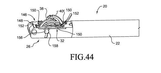
[0054] FIG. 44 is a partial perspective view of one exemplary embodiment of a
stripper
plate as provided on a working end of a suturing device;
[0055] FIG. 45 is a partial perspective view demonstrating deployment of a
suture while a
stripper plate stabilizes prosthetic material during deployment of a suture;
[0056] FIG. 46 is a partial perspective view demonstrating deployment of a
suture through
prosthetic material without a stripper plate;
[0057] FIG. 47 is a perspective view of one exemplary embodiment of a stripper
plate
having a guard member and a tail member;
[0058] FIG. 48 is a side plan view of one exemplary embodiment of a stripper
plate having
a guard member and a tail member;
[0059] FIG. 49 is a top plan view of one exemplary embodiment of a stripper
plate having a
guard member and a tail member;
[0060] FIG. 50 is a partial perspective view of a guard member of a stripper
plate having a
cutout, extension elements and coupling mechanisms;
[0061] FIG. 51 is a partial perspective view of another guard member of a
stripper plate
having a cutout, extension elements and coupling mechanisms;
[0062] FIG. 52 is a partial perspective view of another guard member of a
stripper plate
having a cutout, extension elements and coupling mechanisms;
[0063] FIG. 53 is a partial perspective view of another guard member of a
stripper plate
having a cutout, extension elements and coupling mechanisms;
[0064] FIG. 54 is a partial perspective view of another guard member of a
stripper plate
having a cutout, extension elements and coupling mechanisms;
-8-
CA 03005281 2018-05-11
WO 2017/083833 PCT/US2016/061865
[0065] FIG. 55 is a partial perspective view of another guard member of a
stripper plate
having cutouts, extension elements and coupling mechanisms;
[0066] FIG. 56 is a partial perspective view of another guard member of a
stripper plate
having cutouts, extension elements and a coupling mechanism;
[0067] FIG. 57 is a partial perspective view of another guard member of a
stripper plate
having cutouts, extension elements and a coupling mechanism;
[0068] FIG. 58 is a partial perspective view of another guard member of a
stripper plate
having cutouts, extension elements and a coupling mechanism;
[0069] FIG. 59 is a partial perspective view of another guard member of a
stripper plate
having cutouts, extension elements and a coupling mechanism;
[0070] FIG. 60 is a partial perspective view of another guard member of a
stripper plate
having cutouts, extension elements and coupling mechanisms;
[0071] FIG. 61 is a partial perspective view of another guard member of a
stripper plate
having cutouts, extension elements and coupling mechanisms; and
[0072] FIG. 62 is a partial perspective view of yet another guard member of a
stripper plate
having cutouts, an extension element and coupling mechanisms.
[0073] While the present disclosure is susceptible to various modifications
and alternative
constructions, certain illustrative embodiments thereof have been shown in the
drawings and
will be described below in detail. It should be understood, however, that
there is no intention
to limit the present invention to the specific forms disclosed, but on the
contrary, the intention
is to cover all modifications, alternative constructions and equivalents
falling within the spirit
and scope of the present disclosure.
-9-
CA 03005281 2018-05-11
WO 2017/083833 PCT/US2016/061865
Detailed Description
[0074] Referring now to the drawings, and with specific reference to FIG. 1, a
medical
fastening or suturing device constructed in accordance with the teachings of
the present
disclosure is generally referred to by reference numeral 20. The suturing
device 20, as will be
described in further detail herein, may advantageously enable convenient yet
effective means
of providing fasteners within a surgical environment. The disclosed
embodiments may
additionally facilitate the installation of fasteners or sutures during
minimally invasive
surgical procedures, such as laparoscopic procedures, and the like. As used
for laparoscopic
treatment of a hernia, the embodiment of FIG. 1, for example, may be employed
to reach
beneath sections of tissue, within or around the abdominal region, to fasten
tissues of the
abdominal wall or to fasten prosthetic material, such as prosthetic mesh, to
the abdominal
wall from the inside. Although the embodiments disclosed herein demonstrate
tissue
fastening as applied to laparoscopic applications, it will be understood that
the present
disclosure may be equally or similarly applied to other medical procedures.
[0075] As shown in FIG. 1, the suturing device 20 may generally include an
elongate
member 22 which extends between a control end 24 disposed at a proximal end
thereof, and a
working end 26 disposed at a distal end thereof. The control end 24 may
generally include a
grip 28 as well as a triggering mechanism 30, or any other suitable means for
receiving input
or triggering actions from a user and converting the input or actions into a
suturing action that
is performed at the working end 26 of the suturing device 20. The working end
26 may
generally be configured with a firing aperture 32, or a fastening interface
disposed at a
longitudinal side thereof, through which fasteners or sutures 34 may be
deployed or installed
in tissue and/or prosthetic material. Furthermore, one or more of the sutures
34 to be
deployed may be provided along the elongate member 22 and distally advanced or
fed toward
-10-
CA 03005281 2018-05-11
WO 2017/083833 PCT/US2016/061865
the firing aperture 32 of the working end 26, for example, along one or more
guides or tracks
36 longitudinally disposed within the elongate member 22.
[0076] As shown in more detail in FIGS. 2 and 3, the working end 26 of the
suturing device
20 of FIG. 1 may at least partially enclose a first needle 38 and a second
needle 40, each of
which may be substantially concealed within the firing aperture 32 of the
working end 26 in a
default or fully retracted position. More specifically, the first needle 38
may be rotatably and
pivotally disposed about a first fixed axis 42, and the second needle 40 may
be rotatably and
pivotally disposed about a second fixed axis 44. Moreover, the first axis 42
may be axially
offset but substantially parallel to the second axis 44, for example, such
that the first needle
38 is distally positioned relative to the suturing device 20 and the second
needle 40 is
proximally positioned relative to the suturing device 20. In other alternative
embodiments,
each of the first and second needles 38, 40 may be coaxially disposed about a
common axis.
In still further embodiments, a single needle or more than two needles may be
disposed within
the firing aperture 32 and comprise any one of a plurality of different
arrangements.
[0077] Still referring to FIGS. 2 and 3, each of the first and second needles
38, 40 may be
configured to rotate in opposing directions between respective retracted and
extended
positions. For example, during advancement, the first or distal needle 38 may
be configured
to proximally rotate toward the elongate member 22, while the second or
proximal needle 40
may be configured to distally rotate away from the elongate member 22.
Conversely, during
retraction, the first needle 38 may be configured to distally rotate away from
the elongate
member 22, while the second needle 40 may be configured to proximally rotate
toward the
elongate member 22. Moreover, each of the first and second needles 38, 40 may
be
configured to advance and retract between respective retracted and extended
positions
simultaneously, or in substantially equal increments or at substantially equal
rates of angular
-11-
CA 03005281 2018-05-11
WO 2017/083833 PCT/US2016/061865
displacement. Each of the first and second needles 38, 40 may further comprise
a low-profile
arcuate geometry which enables the needles 38, 40 to be substantially
concealed within the
firing aperture 32 while in the fully retracted position, and have maximized
reach during
advancement. Furthermore, each arcuate needle 38, 40 may be shaped and/or
otherwise
configured to rotate in a cammed fashion such that, it creates a progressively
tighter pull as it
travels through the tissue, and thus, creates a tighter fastening of the
tissue.
[0078] In addition, each of the first and second needles 38, 40 of FIGS. 2 and
3 may include
one or more of needle hooks 46, grooves, tines, recesses, canted surfaces, or
any other
suitable structure configured to enable engagement with a fastener or suture
34, or one or
more needle guides 48 thereof. As shown in FIGS. 2 and 3, for example, a hook
46 may be
disposed on an outer edge of each of the first and second needles 38, 40 and
configured to
engage with a needle guide 48 of a suture 34 as the respective needle 38, 40
is retracted from
the fully extended position. While the embodiments of FIGS. 2 and 3 may depict
the needles
38, 40 with retrograde-type hooks 46 configured to engage a suture 34 during
retraction, it
will be understood that other configurations may be equally or similarly
employed, such as
antegrade-type hooks configured to engage a suture 34 during advancement, or
the like. In
still further alternatives, one or more hooks may be disposed on an inner edge
of each of the
needles 38, 40.
[0079] Turning now to FIGS. 4-9, more detailed drawings of the first and
second needles 38,
40 are provided illustrating the relative rotational positions thereof as the
needles 38, 40 are
advanced from fully retracted positions to fully extended positions. As shown,
each of the
first and second needles 38, 40 may be operatively coupled to a drive
mechanism 50 that is
configured to advance the needles 38, 40 from the retracted positions to the
extended
positions during an engagement of the drive mechanism 50 received via the
control end 24 of
-12-
CA 03005281 2018-05-11
WO 2017/083833 PCT/US2016/061865
the suturing device 20, and conversely, to retract the needles 38, 40 from the
extended
positions to the retracted positions during a disengagement of the drive
mechanism 50
received via the control end 24. Furthermore, the drive mechanism 50 may
include a multi-
bar linkage, such as a three-bar linkage, or the like, which operatively
couples the control end
24 to each of the first and second needles 38, 40.
[0080] As shown in FIGS. 4-9, the drive mechanism 50 may include at least a
first drive
link 52 for driving the first needle 38 and a second drive link 54 for driving
the second needle
40, each of which may be slidably disposed within the elongate member 22 and
in operative
communication between the control end 24 and the working end 26. The drive
mechanism 50
may additionally include a first intermediate link 56 for driving the first
needle 38 and a
second intermediate link 58 for driving the second needle 40, each of which
may configured
to pivotally couple the corresponding drive link 52, 54 to the corresponding
needle 38, 40. In
other modifications, one or more links may be omitted or added to the drive
mechanism 50.
As the needles 38, 40 are opposedly arranged, the drive links 52, 54 and the
intermediate links
56, 58 may be configured to be slidably and pivotally driven in substantially
equal increments
or rates of displacement, but in opposing directions relative to one another.
For example,
during advancement, the first drive link 52 of the first needle 38 may be
slidably driven
distally toward the working end 26 at substantially the same rate or in
similar increments as
the second drive link 54 of the second needle 40 being driven proximally away
from the
working end 26.
[0081] In the fully retracted positions, as shown in FIGS. 4 and 5 for
example, each of the
first and second needles 38, 40 may be substantially concealed beneath the
firing aperture 32
and within the working end 26 of the suturing device 20 so as to facilitate
insertion thereof
into minimal incisions or access ports, or the like. The first and second
needles 38, 40 may
-13-
CA 03005281 2018-05-11
WO 2017/083833 PCT/US2016/061865
further include a low-profile geometry which enables the working end 26 of the
suturing
device 20 as well as the access ports to be generally smaller in size. During
advancement or
during engagement of the drive mechanism 50, as shown in FIGS. 6 and 7 for
example, the
first drive link 52 may drive or push the first intermediate link 56 toward
the distal end of the
firing aperture 32 thereby causing the first needle 38 to rotate about the
first fixed axis 42 and
upwardly extend from the distal end of the firing aperture 32, while the
second drive link 54
may drive or pull the second intermediate link 58 toward the proximal end of
the firing
aperture 32 thereby causing the second needle 40 to rotate about the second
fixed axis 44 and
upwardly extend from the proximal end of the firing aperture 32. Moreover, the
drive
mechanism 50 may be configured to rotatably extend the needles 38, 40 such
that the reach of
each needle 38, 40 is maximally extended during advancement even with a low-
profile
geometry so as to sufficiently penetrate tissue and/or prosthetic material to
be fastened or
sutured.
[0082] The drive mechanism 50 may continue advancing each of the first and
second
needles 38, 40 until the needles 38, 40 respectively reach the fully extended
positions, as
shown for example in FIGS. 8 and 9. In particular, the drive mechanism 50 may
be
configured such that each of the first and second needles 38, 40 extend until
at least one or
more of the hooks 46 thereof engage with a fastener or suture 34 for
deployment. For
example, positioning of the first and second needles 38, 40, the drive
mechanism 50, the firing
aperture 32, and the sutures 34 may be configured such that retrograde-type
hooks 46 on the
outer edges of the needles 38, 40 are able to fully engage with one or more
corresponding
needle guides 48 of a given suture 34. In other alternatives, each of the
needles 38, 40 may
employ a retrograde-type hook disposed on the inner edge thereof, an antegrade-
type hook
disposed on the outer edge thereof, an antegrade-type hook disposed on the
inner edge thereof,
-14-
CA 03005281 2018-05-11
WO 2017/083833 PCT/US2016/061865
or any other suitable variation thereof, to which each of the drive mechanism
50, the firing
aperture 32, and the like, may be modified to enable sufficient engagement
with the
corresponding needle guide 48 of a given suture 34.
[0083] Once the first and second needles 38, 40 respectively reach the fully
extended
positions thereof as shown for example in FIGS. 8 and 9, and once a suture 34
is fully
engaged, the drive mechanism 50 may be released or disengaged, so as to
retract the needles
38, 40 and deploy the engaged suture 34 within tissue and/or prosthetic
material to be
fastened. Moreover, the needles 38, 40 may be retracted toward the positions
shown in FIGS.
4 and 5 by substantially reversing the drive mechanism 50. During retraction
or during
disengagement of the drive mechanism 50, for example, the first drive link 52
may drive or
pull the first intermediate link 56 toward the proximal end of the firing
aperture 32 thereby
causing the first needle 38 to rotate in reverse about the first fixed axis 42
and downwardly
retract into the distal end of the firing aperture 32. Correspondingly, the
second drive link 54
may drive or push the second intermediate link 58 toward the distal end of the
firing aperture
32 thereby causing the second needle 40 to rotate in reverse about the second
fixed axis 44
and downwardly retract into the proximal end of the firing aperture 32.
Furthermore, each of
the first and second needles 38, 40 may be retracted until the needles 38, 40
return to the fully
retracted positions of FIGS. 4 and 5 and until a previously engaged suture 34
is completely
deployed and released therefrom, at which point the needles 38, 40 may be
advanced again to
engage with a new suture 34 for deployment.
[0084] While one possible implementation is provided in the drawings, other
drive
mechanisms and configurations therefor will be apparent to those skilled in
the art without
departing from the scope of the appended claims. For example, in other
modifications, the
suturing device 20 may employ more than two needles which, for instance,
partially oppose
-15-
CA 03005281 2018-05-11
WO 2017/083833 PCT/US2016/061865
one another, or alternatively, rotate in like manner and direction relative to
one another. In
alternative modifications, the needles 38, 40 may be configured to be rotated
sequentially
rather than simultaneously relative to one another, and/or configured to be
rotated at non-
identical rates of angular displacement relative to one another. In additional
modifications,
the needles 38, 40 may be configured to rotate about a common axis rather than
axially offset.
In further modifications, the suturing device 20 may provide a needle that is
configured to
rotate about an axis that is parallel, or otherwise generally not
perpendicular, to the elongate
member 22. In still further modifications, the working end 26 of the suturing
device 20 may
be articulated, such as pivotable or otherwise movable, relative to the
elongate member 22
about one or more axes.
[0085] Referring now to FIGS. 10-14, one exemplary triggering mechanism 60
that may be
employed to operate the drive mechanism 50 of FIGS. 2-9 is provided. As shown,
the
triggering mechanism 60 may be disposed within a housing 62 provided at the
control end 24
of the suturing device 20 and configured to interface with the first and
second needles 38, 40
via the elongate member 22 and the drive mechanism 50 disposed therein.
Furthermore, one
or more of the elongate member 22 and the drive mechanism 50 therein may be
rotatably
coupled to the housing 62 via a rotating collar 64 which may be used to adjust
the radial
position of the firing aperture 32 relative to the control end 24. The housing
62 may further
provide a grip 66 relative to which a trigger 68 of the triggering mechanism
60 may be
pivotally anchored by an anchoring pin 70 and movable in one of two
directions. For
example, the trigger 68 may be configured to engage the drive mechanism 50 and
advance the
needles 38, 40 when pulled toward the grip 66, and disengage the drive
mechanism 50 and
retract the needles 38, 40 when pushed away from the grip 66. Correspondingly,
as shown in
FIG. 10, the trigger 68 may be provided with a proximal handle 72 for pulling
the trigger 68
-16-
CA 03005281 2018-05-11
WO 2017/083833 PCT/US2016/061865
toward the grip 66, as well as a distal handle 74 for pushing the trigger 68
away from the grip
66.
[0086] Still referring to FIG. 10, the triggering mechanism 60 may further
include a yoke 76
that is rigidly and axially coupled to the elongate member 22 and rotatably
disposed within
the housing 62. The triggering mechanism 60 may additionally include a drive
collar 78 that
is axially movable relative to the yoke 76 and pivotally anchored to the
trigger 68 via a lynch
pin 80. Furthermore, as shown in FIGS. 10-14, the interface between the drive
collar 78 and
the lynch pin 80 may be configured such that the drive collar 78 is pivotally
anchored to the
trigger 68 irrespective of the rotational position of the drive collar 78
relative to the trigger 68
and the housing 62. The drive collar 78 may additionally be linked to the yoke
76 via a collar
link 82 and a reversing lever 84 such that the rotational position of the
drive collar 78 follows
the rotational position of the yoke 76. As shown in FIGS. 10-14, for example,
the proximal
end of the collar link 82 may be pivotally as well as radially coupled to the
drive collar 78,
and the distal end of the collar link 82 may be pivotally and radially coupled
to the yoke 76.
[0087] The triggering mechanism 60 of FIGS. 10-14 may further provide means
for
translating a single action received by a user at the control end 24 of the
suturing device 20
into two or more simultaneous but opposing actions effectuated at the working
end 26. For
example, the distal end of the collar link 82 may be coupled to the yoke 76
via a reversing
lever 84, the substantial center of which may be pivotally anchored to the
yoke 76. In
particular, a first end of the reversing lever 84 may be pivotally coupled to
a first sliding block
86 that is rigidly coupled to the first drive link 52 but slidably movable
relative to the yoke 76.
Correspondingly, a second end of the reversing lever 84, opposite the first
end, may be
pivotally coupled to a second sliding block 88 that is rigidly coupled to the
second drive link
54 but also slidably movable relative to the yoke 76. In addition, the collar
link 82 may be
-17-
CA 03005281 2018-05-11
WO 2017/083833 PCT/US2016/061865
pivotally coupled proximate and biased to one of the first and second ends of
the reversing
lever 84 such that, for example, pushing the collar link 82 in a distal
direction rotates the
reversing lever 84 relative to the yoke 76 in a first direction, and pulling
the collar link 82 in a
proximal direction rotates the reversing lever 84 in a second direction
opposite to the first
direction.
[0088] As illustrated in FIGS. 13 and 14, for example, the collar link 82 may
be coupled
proximate to the second end of the reversing lever 84 which may further be
coupled to the
second sliding block 88. In this particular arrangement, when the trigger 68
is moved toward
the grip 66 as indicated by the arrow in FIG. 13, the drive collar 78 and the
collar link 82 may
be pulled toward the control end 24 of the suturing device 20, thereby causing
the reversing
lever 84 to pivot in the manner shown and slidably urge the first sliding
block 86, as well as
the first drive link 52 coupled thereto, in the distal direction while
simultaneously urging the
second sliding block 88, as well as the second drive link 54 coupled thereto,
in the proximal
direction. Moving the trigger 68 in the manner shown in FIG. 13 may thus cause
the drive
mechanism 50 to engage and actuate the first and second needles 38, 40.
Conversely, when
the trigger 68 is moved away from the grip 66 as indicated by the arrow in
FIG. 14, the drive
collar 78 and the collar link 82 may be pushed toward the working end 26 of
the suturing
device 20, thereby causing the reversing lever 84 to pivot in the opposite
direction and
slidably urge the first sliding block 86, as well as the first drive link 52,
in the proximal
direction while simultaneously urging the second sliding block 88, as well as
the second drive
link 54, in the distal direction. Correspondingly, moving the trigger 68 in
the manner shown
in FIG. 14 may cause the drive mechanism 50 to disengage and retract the first
and second
needles 38, 40.
-18-
CA 03005281 2018-05-11
WO 2017/083833 PCT/US2016/061865
[0089] Turning to FIGS. 15-27, the suturing device 20 may additionally include
an
autoloading mechanism 90 for successively feeding and automatically loading
one of a
plurality of sutures 34 into position relative to the firing aperture 32 for
deployment. As
shown in FIGS. 15 and 16, for example, a plurality of successively deployable
sutures 34, in
the form of replaceable suture cartridges, suture ribbons, suture strings, or
the like, may be
removably inserted along guides or tracks 36 disposed within the elongate
member 22. The
autoloading mechanism 90 may provide a pusher member 92 that is also slidably
disposed
along the tracks 36 and configured to successively or incrementally urge the
sutures 34
toward the firing aperture 32 for deployment. As shown in FIGS. 17 and 18 for
example, the
pusher member 92 may include at least one flexible pusher tab 94 extending
therefrom that is
biased so as to unidirectionally interface with one or more catches 96 that
are disposed along
one of the first and second drive links 52, 54 of the drive mechanism 50.
Moreover, the
pusher tab 94 and the catches 96 may be configured such that the pusher member
92 urges the
sutures 34 toward the firing aperture 32 during engagement of the drive
mechanism 50 or
advancement of the needles 38, 40.
[0090] As shown in the particular arrangement of FIG. 17 and 18, for example,
the pusher
member 92 may be configured such that at least one pusher tab 94 engages with
one of the
catches 96 disposed on the first drive link 52, and thereby moves the pusher
member 92 in
direct correspondence with the first drive link 52. In this configuration, as
shown in FIG. 17,
the pusher member 92 may be urged to push the sutures 34 toward the firing
aperture 32 while
the first and second drive links 52, 54 are being engaged and while the first
and second
needles 38, 40 are being advanced. Furthermore, in this particular
configuration, when the
drive mechanism 50 is being disengaged and when the needles 38, 40 are being
retracted, as
shown in FIG. 18, the catches 96 of the first drive link 52 may be free to
return and move
-19-
CA 03005281 2018-05-11
WO 2017/083833 PCT/US2016/061865
away from the working end 26 while the pusher member 92 remains stationary
relative to the
sutures 34 and the firing aperture 32. Moreover, the pusher member 92 may
include support
members 97 as shown in FIG. 15 configured to essentially wedge the pusher
member 92
within the guides or tracks 36 of the elongate member 22 and provide the
pusher member 92
at least some resistance against longitudinal movement therealong. The
positioning of the
catches 96 along the first drive link 52 may be spaced according to the
distance allotted for
each suture 34. In addition, the number of catches 96 and the freedom of
travel of the pusher
member 92 may also be configured so as to sufficiently adapt to the changing
length of the
string of available sutures 34 which incrementally shortens after each
deployment.
[0091] While the embodiments shown may disclose interactions between the
pusher tab 94
and catches 96 provided on the first drive link 52, the pusher tab 94 may
alternatively interact
with catches 96 disposed on the second drive link 54 or any combination of the
first and
second drive links 52, 54. In still further alternative embodiments, the
pusher member 92
may be configured to interact with the drive mechanism 50 in other manners not
shown, so
long as the drive mechanism 50 is able to engage the pusher member 92 to
timely and
appropriately urge one or more sutures 34 toward the firing aperture 32 for
deployment upon
deployment of a prior suture 34.
[0092] While the pusher member 92 and the catches 96 of the first drive link
52 of FIGS.
15-18 may aid in urging the string of sutures 34 toward the working end 26 for
deployment,
the extent to which the sutures 34 are pushed may be limited so as not to
obstruct the firing
aperture 32 through which the first and second needles 38, 40 will need to
extend in order to
deploy a prior suture 34. Accordingly, the autoloading mechanism 90, as shown
in FIGS. 19-
27, may further include a shuttle 98 configured to retrieve the next suture 34
in line for
deployment and position the suture 34 over the firing aperture 32 in alignment
with the
-20-
CA 03005281 2018-05-11
WO 2017/083833 PCT/US2016/061865
needles 38, 40 upon full deployment and release of a prior suture 34. As shown
in FIG. 19,
the shuttle 98 may be slidably disposed along the elongate member 22 and
beneath the string
of sutures 34 to be deployed. Moreover, the shuttle 98 may be movably disposed
in
communication between the working end 26 and the elongate member 22 such that
the
distance of travel of the shuttle 98 extends between at least the firing
aperture 32 and the next
suture 34 in line for deployment.
[0093] As shown in FIG. 20, the shuttle 98 may further include one or more
suture pawls
100 for engaging with a suture 34 prior to deployment. More specifically, the
suture pawls
100 may be configured such that the shuttle 98 is engaging when traveling in
one direction
but non-engaging when traveling in the opposite direction. In the embodiments
of FIGS. 19
and 20, for example, each of the suture pawls 100 may include a ramped edge
102 facing the
proximal direction and a hooked edge 104 facing the opposite, distal
direction. In addition,
each of the suture pawls 100 may be formed of a partially flexible material
and allowed to
deflect within recesses 106 formed within the shuttle 98. In such a way, the
deflectable
ramped edges 102 may enable the suture pawls 100 and the shuttle 98 to
proximally travel
from the firing aperture 32 to beneath the sutures 34 without substantial
obstruction and
without adversely affecting the position of the sutures 34. Once the shuttle
98 is in the
appropriate position beneath the next suture 34 in line for deployment, as
shown in FIG. 19,
the hooked edges 104 may be upright and in position to slidably engage with
the suture 34.
As the shuttle 98 returns toward the working end 26, the hooked edges 104 of
the suture
pawls 100 may distally slide the next suture 34 onto the firing aperture 32.
Moreover, the
suture 34 may be positioned such that any needle guides 48 thereof are
appropriately aligned
with one or more corresponding needles 38, 40.
-21-
CA 03005281 2018-05-11
WO 2017/083833 PCT/US2016/061865
[0094] Turning to FIG. 21, the autoloading mechanism 90 may further interface
with the
drive mechanism 50 to at least cause the shuttle 98 of FIGS. 19 and 20 to move
between the
firing aperture 32 and the string of suture 34. As shown, the autoloading
mechanism 90 may
include a shuttle pawl 108 that is generally disposed beneath the shuttle 98
and coupled to one
of the first and second drive links 52, 54 of the drive mechanism 50. While
other
configurations are possible, in the particular embodiments shown, for example,
the shuttle
pawl 108 may be coupled to the first drive link 52. Moreover, the shuttle pawl
108 may
include a ramped edge 110 facing the distal direction that is configured such
that the first
drive link 52 and the shuttle pawl 108 are freely movable in the distal
direction relative to the
shuttle 98 without substantial obstruction or interference therewith, such as
during
advancement of the needles 38, 40. As illustrated, the shuttle pawl 108 may be
formed of a
flexible material that can be deflected within a recess 112 of the first drive
link 52. The
shuttle pawl 108 may additionally include a hooked edge 114 facing the
proximal direction
that is configured such that the shuttle pawl 108 pulls the shuttle 98 with
the first drive link 52
when the first drive link 52 moves in the proximal direction, such as during
retraction of the
needles 38, 40.
[0095] As shown more particularly in FIG. 22, during engagement of the drive
mechanism
50 or during advancement of the first and second needles 38, 40, the first
drive link 52 along
with the shuttle pawl 108 may be distally pushed toward the working end 26 of
the suturing
device 20 in the manner shown. As the shuttle pawl 108 approaches the shuttle
98, the
ramped edge 110 thereof may enable the shuttle pawl 108 to deflect into the
recess 112 of the
first drive link 52, and further, enable the shuttle pawl 108 to glide under
the shuttle 98
without altering the position of the shuttle 98 relative to the sutures 34.
Each of the first drive
link 52 and the shuttle pawl 108 may progress in such a way at least until the
hooked edge
-22-
CA 03005281 2018-05-11
WO 2017/083833 PCT/US2016/061865
114 of the shuttle pawl 108 reaches and interfaces with the distal end of the
shuttle 98. Both
the first drive link 52 and the shuttle pawl 98 may be sized and configured
such that the
hooked edge 114 interfaces with distal end of the shuttle 98 once the needles
38, 40 are in the
fully extended positions and ready to engage and deploy the prior suture 34 as
shown in FIG.
21. Correspondingly, during disengagement of the drive mechanism 50 or during
retraction
of the needles 38, 40, the first drive link 52 along with the shuttle pawl 108
and the engaged
shuttle 98 may be proximally pulled toward the string of sutures 34 so as to
retrieve the next
suture 34 in line for subsequent deployment.
[0096] Once the shuttle 98 is sufficiently pulled beneath the next suture 34
to be deployed,
the shuttle pawl 108 may be configured to automatically release the shuttle 98
so as to enable
the shuttle 98 to return to the working end 26 and send the retrieved suture
34 therewith to the
appropriate position over the firing aperture 32. As shown in FIG. 23, for
example, the
autoloading mechanism 90 may thus provide a declutch feature, such as a
declutch pin 116, or
the like, configured to release the shuttle pawl 108, or release the shuttle
98 from the first
drive link 52, once the shuttle 98 is appropriately positioned beneath the
next suture 34 in line
for deployment. For example, the declutch pin 116 may be coupled within the
elongate
member 22 and fixedly positioned relative to the shuttle pawl 108 such that,
as the shuttle
pawl 108 proximally passes thereby, the shuttle pawl 108 is caused to deflect
within the
recess 112 of the first drive link 52 and allow the shuttle 98 to return to
the working end 26.
Furthermore, the shuttle pawl 108 may further provide a ramped interface 118
which
proximally precedes the hooked edge 114 and is configured to sufficiently
deflect and release
the shuttle pawl 108 from the shuttle 98 at the appropriate moment, for
instance, when the
suture pawls 100 of the shuttle 98 are ready to engage with the next suture 34
in line for
deployment.
-23-
CA 03005281 2018-05-11
WO 2017/083833 PCT/US2016/061865
[0097] Still referring to FIG. 23, once the shuttle pawl 108 is fully
deflected, the shuttle 98
and the retrieved suture 34 may be sent to the firing aperture 32 by a bias
mechanism 120
configured to continuously bias or urge the shuttle 98 toward the working end
26. As shown,
the bias mechanism 120 may employ a compression spring, or the like, that is
longitudinally
disposed within the elongate member 22 and configured to distally push the
shuttle 98 away
therefrom. In further modifications, the proximal end of the shuttle 98 may
further provide a
centering rod 122 longitudinally extending therefrom configured to interface
with the
compression spring of the bias mechanism 120 and maintain centering of the
shuttle 98
relative to the elongate member 22 and the firing aperture 32. Similarly,
other bias
mechanisms 120 may be employed to achieve comparable results so long as the
biasing force
applied upon the shuttle 98 in the distal direction does not exceed the force
exerted thereon in
the proximal direction by the shuttle pawl 108 and the first drive link 52.
[0098] Turning now to FIGS. 24-26, one exemplary embodiment of the autoloading
mechanism 90 is shown as it progressively retrieves the next suture 34 in line
for deployment,
and appropriately positions the suture 34 upon the firing aperture 32. More
specifically, as
shown in FIG. 24, the shuttle 98 as well as the suture pawls 100 are
proximally pulled toward
the string of sutures 34 as the drive mechanism 50 is disengaged or as the
needles 38, 40 are
retracted. As illustrated, the shuttle 98 is proximally pulled until at least
the suture pawls 100
are in position to slidably engage respective sections of the suture 34. For
instance, each
suture pawl 100 may be configured to engage an exterior of a needle guide 48
of the suture 34,
an interior of a needle guide 48, or any other portion of the suture 34 that
is suitable for
carrying the suture 34 to the firing aperture 32. Once released, the shuttle
98 and the suture
pawls 100, as well as the next suture 34 to be deployed, may be distally
pushed toward the
firing aperture 32 while leaving the remaining string of sutures 34 behind, as
shown for
-24-
CA 03005281 2018-05-11
WO 2017/083833 PCT/US2016/061865
instance in FIG. 25. Furthermore, as shown in FIG. 26, the shuttle 98 may
continue carrying
the suture 34 toward the firing aperture 32 until each of the needle guides 48
of the suture 34
is appropriately aligned to be engaged by the corresponding needle 38, 40.
[0099] In addition, as shown in FIG. 27, the autoloading mechanism 90 may also
provide
one or more release mechanisms 124, 126 for completely deploying or releasing
an engaged
suture 34 from the first and second needles 38, 40 during retraction thereof.
For example,
each release mechanism 124, 126 may employ a blade or a cutting edge 128 that
is
longitudinally disposed within the firing aperture 32 and fixedly positioned
proximate the
retracted position of the corresponding needle 38, 40 such that, as the needle
38, 40 is
retracted back into the firing aperture 32 and restored to its fully retracted
position, the
movement thereof relative to the cutting edge 124 causes the needle guide 48
of the suture 34
to be cut and released therefrom. In the particular embodiment of FIG. 24, for
instance, a first
release mechanism 124 is fixedly disposed within the firing aperture 32 and
proximate the
first needle 38, while the second release mechanism 126 is fixedly disposed
within the firing
aperture 32 and proximate the second needle 40. Moreover, in each release
mechanism 124,
126, the cutting edge 128 may be specifically positioned such that an engaged
suture 34 is cut
and completely released by the time the corresponding needle 38, 40 returns to
its retracted
position. While only cutting edges 128 are shown, the release mechanisms 124,
126 may
alternatively employ hooks, pawls, ramped edges, or any suitable device
capable of releasing
the suture 34 from the needles 38, 40 or hooks 46 thereof either by cutting or
unlatching the
suture 34 therefrom.
[00100] Referring now to FIGS. 28 and 29, one exemplary embodiment of a tissue
fastener
or suture 34 constructed in accordance with the teachings of the present
disclosure is provided.
As shown, the suture 34 may generally comprise an elongated filament 130
extending
-25-
CA 03005281 2018-05-11
WO 2017/083833 PCT/US2016/061865
between a first end and a second end, and at least one needle guide 48
disposed at one or more
of the first and second ends of the elongated filament 130. The suture 34 may
be unitarily
formed of a material that is sufficiently flexible and compliant so as to be
appropriately
deployable by a suturing device 20, while also providing sufficient resilience
or rigidity to
maintain closure between tissue and/or prosthetic material upon deployment.
Additionally,
the elongated filament 130 may be formed with one or more planar curves, such
as the S-
shaped curve shown, or the like, so as to provide for a more compact overall
package and to
increase the number of sutures 34 that can be made available for deployment,
for example,
along the elongated member 22 of a given suturing device 20. Furthermore, the
planar curves
of the elongated filament 130 may be configured according to the anticipated
geometry of the
suture 34 once deployed and installed within tissue and/or prosthetic
material.
[00101] Still referring to the sutures 34 of FIGS. 28 and 29, each needle
guide 48 may be
sufficiently sized and configured to be engaged by, for example, one of the
needles 38, 40 of
the suturing device 20 of FIGS. 1-27, or one the needle hooks 46 thereof,
while also being
sufficiently easily released from the needles 38, 40, for example, via any of
the release
mechanisms 124, 126 provided in FIGS. 24-27. The needle guides 48 may further
be shaped,
for example, with a relatively tapered tip that is configured to facilitate
advancement thereof
through tissue and/or prosthetic material during deployment, as well as resist
retraction
thereof to promote a secure closure. For example, the needle guides 48 may be
shaped in the
substantial form of an oval, an ellipse, a circle, a semi-circle, a triangle,
a polygon, or the like.
As shown, each needle guide 48 may additionally include one or more retention
elements 132
that are also configured to facilitate advancement thereof through sections of
tissue and/or
prosthetic material, and further aid in resisting retraction thereof once
deployed. The
-26-
CA 03005281 2018-05-11
WO 2017/083833 PCT/US2016/061865
retention elements 132 may be shaped in the form of a tine, a fin, a canted
element, or any
design sufficiently capable resisting retraction through tissue and/or
prosthetic material.
[00102] Each of the needle guides 48 in FIGS. 28 and 29 may further be
provided with one
or more constriction elements 134 configured to further secure an engagement
between the
needle guide 48 and a corresponding needle 38, 40 or needle hook 46 thereof.
More
specifically, the constriction element 134 may be disposed within the needle
guide 48 in a
manner configured to at least partially bias or constrict the needle guide 48
against one of the
needles 38, 40 received therethrough. As shown in FIGS. 28 and 29, for
example, the
constriction element 134 may take the form of a tab, flap, or the like, that
is disposed within
the needle guide 48 and extending toward the tapered end of the needle guide
48 or extending
toward any other the portion of the needle guide 48 that is anticipated to be
engaged by a
needle hook 46. Moreover, the constriction elements 134 may be formed of a
material that is
sufficiently flexible and compliant so as to receive a needle 38, 40
therethrough, but also
formed of a material with sufficient resilience and rigidity so as to bias the
needle guide 48
against the needle 38, 40 and needle hook 46.
[00103] Turning to FIGS. 30-32, one exemplary interaction between the suture
34 of FIGS.
28 and 29 and a given set of needles 38, 40 and respective needle hooks 46 is
provided. As
shown, once the first and second needles 38, 40 are advanced into the fully
extended positions
and received through the respective needle guides 48, the constriction
elements 134 are
caused to bend, thereby pushing or exerting an outward force against the inner
edge of the
needles 38, 40. This outward pushing force exerted by the constriction element
134 may
effectively exert a substantially equal and opposite inward force on the
tapered end of the
needle guide 48, thereby biasing the needle guide 48 into the needle hook 46
of the respective
needle 38, 40. Thus, the constriction elements 134 of the sutures 34 may
provide an
-27-
CA 03005281 2018-05-11
WO 2017/083833 PCT/US2016/061865
otherwise absent constricting force on a received needle 38, 40, which may
further serve to
secure an engagement between the needle hook 46 and the needle guide 48 of the
suture 34.
While disclosed in the form of a tab or flap, the constriction elements 134
may be provided on
the needle guides 48 in any one of variety of different forms, sizes and
configurations.
Alternatively, the constriction element 134 may be configured to substantially
close the
needle guide 48 except for one or more slots, apertures or other voids
disposed toward the
tapered end thereof in a manner which would effectively bias the needle guide
48 against a
given needle hook 46. In still further alternatives, the constriction element
134 may be
completely closed but penetrable by a needle 38, 40 in a manner which would
effectively bias
the needle guide 48 against the needle hook 46.
[00104] As shown in FIGS. 33 and 34, another exemplary embodiment of a tissue
fastener
or suture 34-1 that may be used in association with a suturing device 20 is
provided. Similar
to the suture 34 of FIGS. 28 and 29, the suture 34-1 shown may generally
comprise an
elongated filament 130-1 extending between a first end and a second end, and
at least one
needle guide 48-1 disposed at one or more of the first and second ends of the
elongated
filament 130-1. The suture 34-1 may be formed of a material that is
sufficiently flexible and
compliant so as to be appropriately deployable by a suturing device 20, while
also providing
sufficient resilience or rigidity to maintain closure between tissue and/or
prosthetic material
upon deployment. The elongated filament 130-1 of the suture 34-1 may further
include a
cross member 136 as well as filament guides 138 configured to stabilize the
suture 34-1 as it
is moved within the tracks 36 and along the elongate member 22 of a suturing
device 20. For
example, the cross member 136 may aid in increasing the structural integrity
laterally across
the suture 34-1 and reduce binding, while the filament guides 138 may be sized
and
configured to interface with the tracks 36 of the elongate member 22 of a
suturing device 20
-28-
CA 03005281 2018-05-11
WO 2017/083833 PCT/US2016/061865
so as to provide the suture 34-1 with additional lateral support and maintain
proper alignment
thereof Furthermore, any one or more of the cross member 136 and the filament
guides 138
may be configured with retention features configured to aid in resisting
retraction thereof once
deployed into tissue and/or prosthetic material.
[00105] As in previous embodiments, the needle guides 48-1 of FIGS. 33 and 34
may be
sufficiently sized and configured to be engaged by a needle 38, 40 of a
suturing device 20, or
one of the needle hooks 46 thereof, while also being sufficiently thin or
easily released from
the needles 38, 40, for example, via any of the release mechanisms 124, 126
provided in FIGS.
24-27. As shown, the needle guides 48-1 may be provided with a relatively
tapered tip, as
well as provided with one or more retention elements 132-1, configured to
facilitate
advancement thereof through tissue and/or prosthetic material during
deployment, and resist
retraction thereof to promote a secure closure. Each of the needle guides 48-1
in FIGS. 33
and 34 may be provided with constriction elements 134-1 which substantially
conform to the
shape of the needle guides 48-1 and serve to secure an engagement between the
needle guide
48-1 and a corresponding needle 38, 40 or needle hook 46 thereof Specifically,
each
constriction element 134-1 may be configured to increase the integrity or
lateral rigidity of
each needle guide 48-1 when a needle 38, 40 is not inserted therethrough, such
as when the
suture 34-1 is being moved along the tracks 36 of the elongate member 22 of a
suturing
device 20, but also configured to effectively reduce the lateral rigidity of
each needle guide
48-1 when a needle 38, 40 is received therethrough, such as during advancement
through
tissue and/or prosthetic material. As shown in FIGS. 33 and 34, for example,
the constriction
elements 134-1, when in the non-deflected state, may substantially fill the
width of the needle
guides 48-1, and thereby provide lateral support thereacross. When in the
deflected state, the
constriction elements 134-1 may enable the needle guides 48-1 to substantially
collapse and
-29-
CA 03005281 2018-05-11
WO 2017/083833 PCT/US2016/061865
narrow so as to promote insertion or advancement thereof through a tissue, and
the like.
Furthermore, the constriction elements 134-1 may continue to provide lateral
rigidity and
support for the retention elements 132-1 once deployed and released into
tissue and/or
prosthetic material. For example, once the suture 34-1 is deployed and needle
guides 48-1 are
released, for instance cut, from the corresponding needles 38, 40, the
constriction elements
134-1 may be configured to return to the non-deflected default state and
thereby substantially
prevent the retention elements 132-1 from collapsing and retracting from the
tissue and/or
prosthetic material.
[00106] As additionally shown in FIGS. 35 and 36, another exemplary embodiment
of a
tissue fastener or suture 34-2 that may be used in association with a suturing
device 20 is
provided. As in previous embodiments, the suture 34-2 may generally comprise
an elongated
filament 130-2 extending between a first end and a second end, and at least
one needle guide
48-2 disposed at one or more of the first and second ends of the elongated
filament 130-2.
The suture 34-2 may be formed of a material that is sufficiently flexible and
compliant so as
to be appropriately deployable by a suturing device 20, while also providing
sufficient
resilience or rigidity to maintain closure between tissue and/or prosthetic
material upon
deployment. The elongated filament 130-2 of the suture 34-2 may further
include a cross
member 136 as well as filament guides 138 configured to stabilize the suture
34-2 as it is
moved within the tracks 36 and along the elongate member 22 of a suturing
device 20.
Additionally, any one or more of the cross member 136 and the filament guides
138 may be
configured with retention features configured to aid in resisting retraction
thereof once
deployed into tissue and/or prosthetic material.
[00107] The needle guides 48-2 of FIGS. 35 and 36 may be sufficiently sized
and
configured to be engaged by a needle 38, 40 of a suturing device 20, or one of
the needle
-30-
CA 03005281 2018-05-11
WO 2017/083833 PCT/US2016/061865
hooks 46 thereof, while also being sufficiently thin or easily released from
the needles 38, 40,
for example, via any of the release mechanisms 124, 126 provided in FIGS. 24-
27. The
needle guides 48-2 may be provided with a relatively tapered tip, as well as
provided with one
or more retention elements 132-2, configured to facilitate advancement thereof
through tissue
and/or prosthetic material during deployment, and resist retraction thereof to
promote a secure
closure. Each of the needle guides 48-2 in FIGS. 35 and 36 may be provided
with
constriction elements 134-2 configured to further secure an engagement between
the needle
guide 48-2 and a corresponding needle 38, 40 or needle hook 46 thereof
Specifically, each
constriction element 134-2 may be provided with a widened feature configured
to increase the
integrity or lateral rigidity of each needle guide 48-2 when a needle 38, 40
is not inserted
therethrough, such as when the suture 34-2 is being moved along the tracks 36
of the elongate
member 22 of a suturing device 20, but also configured to effectively reduce
the lateral
rigidity of each needle guide 48-2 when a needle 38, 40 is received
therethrough, such as
during advancement through tissue and/or prosthetic material. As shown in
FIGS. 35 and 36,
for example, the widened feature of the constriction element 134-2, when in
the non-deflected
state, may substantially abut the inner walls of the needle guide 48-2, and
thereby provide
lateral support thereacross. When in the deflected state, the constriction
element 134-2 may
enable the needle guide 48-2 to substantially collapse and narrow so as to
promote insertion or
advancement thereof through a tissue, and the like. Furthermore, the
constriction elements
134-2 may continue to provide lateral rigidity and support for the retention
elements 132-2
once deployed and released into tissue and/or prosthetic material. For
example, once the
suture 34-2 is deployed and needle guides 48-2 are released, for instance cut,
from the
corresponding needles 38, 40, the constriction elements 134-2 may be
configured to return to
-31-
CA 03005281 2018-05-11
WO 2017/083833 PCT/US2016/061865
the non-deflected default state and thereby substantially prevent the
retention elements 132-2
from collapsing and retracting from the tissue and/or prosthetic material.
[00108] In still further alternatives, another exemplary embodiment of a
tissue fastener or
suture 34-3 is provided in FIGS. 37 and 38. As in previous embodiments, the
suture 34-3
may generally comprise an elongated filament 130-3 extending between a first
end and a
second end, and at least one needle guide 48-3 disposed at one or more of the
first and second
ends of the elongated filament 130-3. The suture 34-3 may be formed of a
material that is
sufficiently flexible and compliant so as to be appropriately deployable by a
suturing device
20, while also providing sufficient resilience or rigidity to maintain closure
between tissue
and/or prosthetic material upon deployment. The elongated filament 130-3 of
the suture 34-3
may further include a cross member 136 as well as filament guides 138
configured to stabilize
the suture 34-3 as it is moved within the tracks 36 and along the elongate
member 22 of a
suturing device 20. Additionally, any one or more of the cross member 136 and
the filament
guides 138 may be configured with retention features configured to aid in
resisting retraction
thereof once deployed into tissue and/or prosthetic material.
[00109] The needle guides 48-3 of FIGS. 37 and 38 may be sufficiently sized
and
configured to be engaged by a needle 38, 40 of a suturing device 20, or one of
the needle
hooks 46 thereof, while also being sufficiently thin or easily released from
the needles 38, 40,
for example, via any of the release mechanisms 124, 126 provided in FIGS. 24-
27. The
needle guides 48-3 may be provided with a relatively tapered tip, as well as
provided with one
or more retention elements 132-3, configured to facilitate advancement thereof
through tissue
and/or prosthetic material during deployment, and resist retraction thereof to
promote a secure
closure. Each of the needle guides 48-3 in FIGS. 37 and 38 may be provided
with
constriction elements 134-3 configured to further secure an engagement between
the needle
-32-
CA 03005281 2018-05-11
WO 2017/083833 PCT/US2016/061865
guide 48-3 and a corresponding needle 38, 40 or needle hook 46 thereof
Specifically, each
constriction element 134-3 may be provided with a substantially webbed feature
configured to
increase the integrity or lateral rigidity of each needle guide 48-3 when a
needle 38, 40 is not
inserted therethrough, such as when the suture 34-3 is being moved along the
tracks 36 of the
elongate member 22 of a suturing device 20, but also configured to effectively
reduce the
lateral rigidity of each needle guide 48-3 when a needle 38, 40 is received
therethrough, such
as during advancement through tissue and/or prosthetic material. As shown in
FIGS. 37 and
38, for example, the webbed feature of the constriction element 134-3, when in
the non-
deflected state, may provide rigidity and lateral support against the inner
walls of the needle
guide 48-3. When the constriction element 134-3 is at least partially
deflected state due to the
insertion of a needle 38, 40, the needle guide 48-3 may be enabled to
substantially collapse
and narrow so as to promote insertion or advancement thereof through a tissue,
and the like.
Furthermore, the constriction elements 134-3 may continue to provide lateral
rigidity and
support for the retention elements 132-3 once deployed and released into
tissue and/or
prosthetic material. For example, once the suture 34-3 is deployed and needle
guides 48-3 are
released, for instance cut, from the corresponding needles 38, 40, the
constriction elements
134-3 may be configured to return to the non-deflected default state and
thereby substantially
prevent the retention elements 132-3 from collapsing and retracting from the
tissue and/or
prosthetic material.
[00110] Referring now to FIGS. 39 and 40, another exemplary embodiment of a
tissue
fastener or suture 34-4 is provided. As in previous embodiments, the suture 34-
4 may
generally comprise an elongated filament 130-4 extending between a first end
and a second
end, and at least one needle guide 48-4 disposed at one or more of the first
and second ends of
the elongated filament 130-4. The suture 34-4 may be formed of a material that
is sufficiently
-33-
CA 03005281 2018-05-11
WO 2017/083833 PCT/US2016/061865
flexible and compliant so as to be appropriately deployable by a suturing
device 20, while
also providing sufficient resilience or rigidity to maintain closure between
tissue and/or
prosthetic material upon deployment. The elongated filament 130-4 of the
suture 34-4 may
further include a cross member 136 as well as filament guides 138 configured
to stabilize the
suture 34-4 as it is moved within the tracks 36 and along the elongate member
22 of a
suturing device 20. Additionally, any one or more of the cross member 136 and
the filament
guides 138 may be configured with retention features configured to aid in
resisting retraction
thereof once deployed into tissue and/or prosthetic material.
[00111] The needle guides 48-4 of FIGS. 39 and 40 may be sufficiently sized
and
configured to be engaged by a needle 38, 40 of a suturing device 20, or one of
the needle
hooks 46 thereof, while also being sufficiently thin or easily released from
the needles 38, 40,
for example, via any of the release mechanisms 124, 126 provided in FIGS. 24-
27. The
needle guides 48-4 may be provided with a relatively tapered tip, as well as
provided with one
or more retention elements 132-4, configured to facilitate advancement thereof
through tissue
and/or prosthetic material during deployment, and resist retraction thereof to
promote a secure
closure. Each of the needle guides 48-4 in FIGS. 39 and 40 may be provided
with
constriction elements 134-4 configured to further secure an engagement between
the needle
guide 48-4 and a corresponding needle 38, 40 or needle hook 46 thereof As in
the suture 34-
3 of FIGS. 37 and 38, the constriction elements 134-4 of FIGS. 39 and 40 may
be provided
with a webbed feature configured to increase the integrity or lateral rigidity
of each needle
guide 48-4 when a needle 38, 40 is not inserted therethrough, such as when the
suture 34-4 is
being moved along the tracks 36 of the elongate member 22 of a suturing device
20, but also
configured to effectively reduce the lateral rigidity of each needle guide 48-
4 when a needle
38, 40 is received therethrough, such as during advancement through tissue
and/or prosthetic
-34-
CA 03005281 2018-05-11
WO 2017/083833 PCT/US2016/061865
material. Unlike the previous suture 34-3, however, the constriction elements
134-4 of the
suture 34-4 of FIGS. 39 and 40 may be arched or otherwise contrasted with the
general plane
of the suture 34-4 and biased to exert a lateral force against the inner walls
of the needle guide
48-4 when in the non-deflected state. When the constriction element 134-4 is
at least partially
deflected due to the insertion of a needle 38, 40, the needle guide 48-4 may
be enabled to
substantially collapse and narrow so as to promote insertion or advancement
thereof through a
tissue, and the like. Furthermore, the constriction elements 134-4 may
continue to provide
lateral rigidity and support for the retention elements 132-4 once deployed
and released into
tissue and/or prosthetic material. For example, once the suture 34-4 is
deployed and needle
guides 48-4 are released, for instance cut, from the corresponding needles 38,
40, the
constriction elements 134-4 may be configured to return to the non-deflected
default state and
thereby substantially prevent the retention elements 132-4 from collapsing and
retracting from
the tissue and/or prosthetic material.
[00112] Referring further to FIGS. 41 and 42, yet another exemplary embodiment
of a
tissue fastener or suture 34-5 is provided. Similar to previous embodiments,
the suture 34-5
may generally comprise an elongated filament 130-5 extending between a first
end and a
second end, and at least one needle guide 48-5 disposed at one or more of the
first and second
ends of the elongated filament 130-5. The suture 34-5 may be formed of a
material that is
sufficiently flexible and compliant so as to be appropriately deployable by a
suturing device
20, while also providing sufficient resilience or rigidity to maintain closure
between tissue
and/or prosthetic material upon deployment. The elongated filament 130-5 of
the suture 34-5
may also include breakaway tabs 140 configured to help stabilize the suture 34-
5 as it is
moved within the tracks 36 and along the elongate member 22 of a suturing
device 20. As
shown, each breakaway tab 140 may be coupled between a needle guide 48-5 and a
-35-
CA 03005281 2018-05-11
WO 2017/083833 PCT/US2016/061865
corresponding section of the elongated filament 130-5 in the folded position,
and configured
to be detachable upon deployment. In particular, the breakaway tabs 140 may be
sized and
configured to provide, not only sufficient planar and lateral rigidity to the
suture 34-5 prior to
deployment, but also configured with sufficient detachability so as not to
interfere with the
deployment thereof As better seen in Fig. 42, for example, each of the
breakaway tabs 140
may incorporate attenuated features 142, such as in the form of grooves,
slits, perforations, or
the like. Furthermore, the breakaway tabs 140 may be angled or otherwise
positioned relative
to the needle guides 48-5 in a way to help resist retraction thereof once
deployed into tissue
and/or prosthetic material.
[00113] The needle guides 48-5 of FIGS. 41 and 42 may be sufficiently sized
and
configured to be engaged by a needle 38, 40 of a suturing device 20, or one of
the needle
hooks 46 thereof, while also being sufficiently thin or easily released from
the needles 38, 40,
for example, via any of the release mechanisms 124, 126 provided in FIGS. 24-
27. The
needle guides 48-5 may be provided with a relatively tapered tip, as well as
provided with one
or more retention elements 132-5, configured to facilitate advancement thereof
through tissue
and/or prosthetic material during deployment, and resist retraction thereof to
promote a secure
closure. As shown, the edges of the needle guides 48-5 may additionally be
beveled, rounded,
or otherwise configured to further facilitate advancement thereof In addition,
each of the
needle guides 48-5 in FIGS. 41 and 42 may be provided with generally linear
constriction
elements 134-5 positioned to further secure an engagement between the needle
guide 48-5 and
a corresponding needle 38, 40 or needle hook 46 thereof Moreover, the
constriction elements
134-5 may serve to increase the integrity or lateral rigidity of each needle
guide 48-5 when a
needle 38, 40 is not inserted therethrough, such as when the suture 34-5 is
being moved along
the tracks 36 of the elongate member 22 of a suturing device 20. Furthermore,
the
-36-
CA 03005281 2018-05-11
WO 2017/083833 PCT/US2016/061865
constriction elements 134-5 may continue to provide lateral rigidity and
support for the
retention elements 132-5 once deployed and released into tissue and/or
prosthetic material.
For example, once the suture 34-5 is deployed and needle guides 48-5 are
released from the
corresponding needles 38, 40, the constriction elements 134-5 may be
configured to prevent
the retention elements 132-5 from collapsing and retracting from the tissue
and/or prosthetic
material.
[00114] In addition, the suture 34-5 of FIGS. 41 and 42 may further include
one or more
nesting elements 144, or extended features disposed along the elongated
filament 130-5,
which may be sized and configured to detachably couple to a counterpart
section of an
adjacent suture 34-5 in a string of sutures 34-5. As shown in FIG. 43, for
example, each
nesting element 144 may be configured to couple to the tip of the needle guide
48-5 of an
adjacent suture 34-5. Correspondingly, the tips of each needle guide 48-5 may
be beveled,
rounded, or otherwise sized and shaped to be mateably received within the
nesting elements
144 of an adjacent suture 34-5. In such a way, each suture 34-5 may include
two sets of
nesting elements 144, such as forward-facing nesting elements 144 for coupling
to the trailing
needle guide 48-5 of a preceding suture 34-5, and rearward-facing nesting
elements 144 for
coupling to the leading needle guide 48-5 of a subsequent suture 34-5.
Furthermore, the
nesting elements 144 may be coupled to corresponding sections of adjacent
sutures 34-5 using,
for example, flexible bonding material or adhesives, friction fitments,
attenuated connections,
or any other suitable arrangement that is, not only capable of maintaining
rigidity of the string
of sutures 34-5 prior to deployment, but also capable of being easily detached
so as not to
interfere with deployment.
[00115] Turning further to FIG. 44, another embodiment of the suturing device
20 is
provided with a stripper plate 146. As shown, the stripper plate 146 may be
longitudinally
-37-
CA 03005281 2018-05-11
WO 2017/083833 PCT/US2016/061865
disposed within the working end 26 and at least partially provided along the
elongate member
22. More specifically, the stripper plate 146 may include a guard member 148
that is
disposed or positioned over the firing aperture 32 and the retracted arcuate
needles 38, 40 in a
manner configured to at least partially cover and protect the internal
components of the
working end 26 from foreign objects. As shown, the guard member 148 may
include one or
more cutouts 150 sized, shaped, or otherwise configured, to allow the arcuate
needles 38, 40
to be advanced and retracted therethrough, as well as to allow deployment of a
suture 34
therethrough. The stripper plate 146 may further include one or more extension
elements 152
at least partially extending outwardly from the guard member 148 for engaging
a prosthetic
mesh 154. As demonstrated in FIG. 45, for example, the guard member 148 and
the
extension elements 152 may be configured such that, when the working end 26 is
pressed
against a prosthetic mesh 154 and tissue, the extension elements 152 keep the
prosthetic mesh
154 taut at least during the installation or deployment of the suture 34. In
particular, at least
one extension element 152 may be distally positioned relative to the firing
aperture 32 so as to
engage a distal end of the prosthetic mesh 154 and to prevent the prosthetic
mesh 154 from
sliding proximally relative thereto, while at least one additional extension
element 152 may be
proximally positioned relative to the firing aperture 32 so as to engage a
proximal end of the
prosthetic mesh 154 and prevent the prosthetic mesh 154 from sliding distally
relative thereto.
In such a way, the guard member 148 and the extension elements 152 of the
stripper plate 146
may resist undesirable bunching of the prosthetic mesh 154 during deployment,
as shown for
example in FIG. 46.
[00116] Referring back to FIG. 44, the guard member 148 of the stripper plate
146 may also
include one or more coupling mechanisms 156 extending therefrom configured to
couple the
stripper plate 146 to the working end 26 of the suturing device 20 and onto to
the firing
-38-
CA 03005281 2018-05-11
WO 2017/083833 PCT/US2016/061865
aperture 32. As shown in FIG. 44, for example, the coupling mechanisms 156 may
be
provided in the form of retention tabs which extend over one or more sides of
the working end
26 and are held in place by recesses 158 formed in the body the working end
26. Other types
of coupling mechanisms 156 will be apparent to those of ordinary skill in the
art. For
example, the stripper plate 146 may employ screws, clips, hooks, adhesives, or
the like, while
the working end 26 may employ corresponding apertures, grooves, or other
designs
configured to sufficiently retain the stripper plate 146 thereagainst. In
still further alternatives,
the stripper plate 146 and one or more non-moving components of the working
end 26 may be
formed as a unitary body rather than separate parts that are later assembled
together.
Furthermore, as shown in FIGS. 47-49, the stripper plate 146 may further
include a tail
member 160 proximally extending longitudinally from the guard member 148,
which may be
installed along and within the elongate member 22 of the suturing device 20.
The tail member
160 may be positioned in the tracks 36 and over one or more of the sutures 34
awaiting
deployment disposed within the elongate member 22. Moreover, the tail member
160 may be
designed to further support the sutures 34 as they are incrementally advanced
toward the
working end 26 and aid the operability of the drive mechanism 50.
[00117] Turning to FIG. 50, one exemplary embodiment of the stripper plate 146
is shown
in more detail. As shown, the stripper plate 146 may include a guard member
148 having
extension elements 152 extending upward therefrom, or away from the working
end 26, two
coupling mechanisms 156 extending downward therefrom, or toward the working
end 26, and
a tail member 160 extending proximally therefrom, or toward the control end 24
of the
suturing device 20. More particularly, as shown in FIG. 50, the stripper plate
146 may
include an arrangement of six extension elements 152 positioned along the
edges of the cutout
150, where each of the extension elements 152 may be canted, angled, curved,
beveled, or
-39-
CA 03005281 2018-05-11
WO 2017/083833 PCT/US2016/061865
otherwise shaped to sufficiently engage a prosthetic mesh 154 and keep the
engaged
prosthetic mesh 154 taut against the tissue at least during deployment of a
suture 34.
Furthermore, any one or more of the guard member 148, cutouts 150, extension
elements 152,
and coupling mechanisms 156 of the stripper plate 146, may be modified to
accommodate
different brands or types of prosthetic meshes 154, different types of needles
38, 40 or
configurations thereof, different types of sutures 34, and the like.
[00118] As further shown in FIGS. 51-62, the position, size, number, or shape
of the
individual extension elements 152 may be varied based on the type of
prosthetic mesh 154
intended to be used. In FIGS. 51-55, 57 and 62, for example, each of the
extension elements
152 may be substantially perpendicular to the guard member 148. Alternatively,
one or more
of the extension elements 152 may extend at an acute angle relative to the
guard member 148
as shown in FIGS. 56 and 58-61. Additionally, while the stripper plate 146 may
include a
plurality of extension elements 152 as shown in FIGS. 51-61, the stripper
place 146 may serve
to sufficiently engage a prosthetic mesh 154 using a single extension element
152 as shown in
FIG. 62. Each of the stripper plates 146 may also incorporate different
configurations of
cutouts 150 based on the type of suture 34 being used with the suturing device
20. For
example, the stripper plate 146 may incorporate a single enlarged cutout 150
as shown in
FIGS. 51-54, or multiple cutouts 150 as shown in FIGS. 55-62. Furthermore, the
cutouts 150
may incorporate different designs that are specifically optimized for a given
set of sutures 34
and/or needles 38, 40.
[00119] Still further, the stripper plates 146 may incorporate any one of a
variety of
different coupling mechanisms 156. As shown in FIGS. 51-55, for example, the
coupling
mechanisms 156 may be provided in the form of retention tabs which extend over
the sides of
the working end 26. The stripper plate 146 may also incorporate coupling
mechanisms 156
-40-
CA 03005281 2018-05-11
WO 2017/083833 PCT/US2016/061865
that are provided in the form of braces, or the like, designed to extend over
and hook onto the
tip of the working end 26 of the suturing device 20, as those disclosed in
FIGS. 56-58.
Additionally, the coupling mechanism 156 may employ a tabbed design configured
to be
inserted into and held by the tip of the working end 26, as shown in FGI. 59.
Furthermore, the
coupling mechanism 156 may be configured to couple the stripper plate 146 to
the working
end 26 of the suturing device 20 via fasteners, such as screws, or the like,
and provide
apertures configured to receive the fasteners. Furthermore, the coupling
mechanism 156 may
employ apertures configured to receive fasteners, such as screws, or the like,
by which the
stripper plate 146 may be attached to the working end 26 of the suturing
device 20. In still
further modifications, the stripper plate 146, or any component thereof, may
be adjustable or
interchangeable such that a single suturing device 20 can easily adapt to
different types of
prosthetic meshes 154.
[00120] From the foregoing, it can be seen that the present disclosure sets
forth a medical
fastening or suturing device adapted to rapidly and reliably install fasteners
or sutures to
secure tissue and/or any applicable prosthetic material. The device not only
greatly reduces
the time required for fastening tissues, but also results in improved ease of
use relative to
other methods. Furthermore, through the unique combination of elements set
forth in the
present disclosure, the tissue fastening or suturing is more reliably retained
with reduced
irritation and other complications to the patient and without adversely
affecting the integrity
of the attachment and/or closure.
-41-